EP4137794A1 - Colorants de cyanine et leur utilisation pour la coloration in vivo de micro-organismes et d'autres cellules vivantes - Google Patents
Colorants de cyanine et leur utilisation pour la coloration in vivo de micro-organismes et d'autres cellules vivantes Download PDFInfo
- Publication number
- EP4137794A1 EP4137794A1 EP22191279.3A EP22191279A EP4137794A1 EP 4137794 A1 EP4137794 A1 EP 4137794A1 EP 22191279 A EP22191279 A EP 22191279A EP 4137794 A1 EP4137794 A1 EP 4137794A1
- Authority
- EP
- European Patent Office
- Prior art keywords
- cells
- cyanine dye
- whereat
- formula
- viability
- Prior art date
- Legal status (The legal status is an assumption and is not a legal conclusion. Google has not performed a legal analysis and makes no representation as to the accuracy of the status listed.)
- Pending
Links
- 239000000975 dye Substances 0.000 title claims abstract description 159
- 238000010186 staining Methods 0.000 title claims abstract description 50
- ANRHNWWPFJCPAZ-UHFFFAOYSA-M thionine Chemical compound [Cl-].C1=CC(N)=CC2=[S+]C3=CC(N)=CC=C3N=C21 ANRHNWWPFJCPAZ-UHFFFAOYSA-M 0.000 title claims abstract 32
- 244000005700 microbiome Species 0.000 title abstract description 12
- 238000001727 in vivo Methods 0.000 title description 6
- 230000035899 viability Effects 0.000 claims abstract description 52
- 230000027455 binding Effects 0.000 claims abstract description 10
- 108020004707 nucleic acids Proteins 0.000 claims abstract description 8
- 102000039446 nucleic acids Human genes 0.000 claims abstract description 8
- 150000007523 nucleic acids Chemical class 0.000 claims abstract description 8
- 210000004027 cell Anatomy 0.000 claims description 159
- IAZDPXIOMUYVGZ-UHFFFAOYSA-N Dimethylsulphoxide Chemical compound CS(C)=O IAZDPXIOMUYVGZ-UHFFFAOYSA-N 0.000 claims description 38
- 238000000034 method Methods 0.000 claims description 38
- -1 methoxy, ethoxy, propoxy, amino, methylamino, ethylamino Chemical group 0.000 claims description 38
- RIOQSEWOXXDEQQ-UHFFFAOYSA-N triphenylphosphine Chemical compound C1=CC=CC=C1P(C=1C=CC=CC=1)C1=CC=CC=C1 RIOQSEWOXXDEQQ-UHFFFAOYSA-N 0.000 claims description 34
- 150000001875 compounds Chemical class 0.000 claims description 31
- WEVYAHXRMPXWCK-UHFFFAOYSA-N Acetonitrile Chemical compound CC#N WEVYAHXRMPXWCK-UHFFFAOYSA-N 0.000 claims description 30
- 125000001424 substituent group Chemical group 0.000 claims description 29
- 229910001868 water Inorganic materials 0.000 claims description 27
- XLYOFNOQVPJJNP-UHFFFAOYSA-N water Substances O XLYOFNOQVPJJNP-UHFFFAOYSA-N 0.000 claims description 26
- 210000003470 mitochondria Anatomy 0.000 claims description 24
- 108090000765 processed proteins & peptides Proteins 0.000 claims description 24
- 230000000149 penetrating effect Effects 0.000 claims description 19
- 239000000243 solution Substances 0.000 claims description 19
- 238000009395 breeding Methods 0.000 claims description 18
- 230000001488 breeding effect Effects 0.000 claims description 18
- 239000011541 reaction mixture Substances 0.000 claims description 16
- WKGZJBVXZWCZQC-UHFFFAOYSA-N 1-(1-benzyltriazol-4-yl)-n,n-bis[(1-benzyltriazol-4-yl)methyl]methanamine Chemical class C=1N(CC=2C=CC=CC=2)N=NC=1CN(CC=1N=NN(CC=2C=CC=CC=2)C=1)CC(N=N1)=CN1CC1=CC=CC=C1 WKGZJBVXZWCZQC-UHFFFAOYSA-N 0.000 claims description 15
- 125000002496 methyl group Chemical group [H]C([H])([H])* 0.000 claims description 15
- 230000012010 growth Effects 0.000 claims description 14
- 238000005259 measurement Methods 0.000 claims description 12
- 230000000813 microbial effect Effects 0.000 claims description 12
- 239000007795 chemical reaction product Substances 0.000 claims description 10
- 125000000217 alkyl group Chemical group 0.000 claims description 9
- 230000000269 nucleophilic effect Effects 0.000 claims description 9
- 239000000126 substance Substances 0.000 claims description 9
- 125000001495 ethyl group Chemical group [H]C([H])([H])C([H])([H])* 0.000 claims description 8
- 239000002243 precursor Substances 0.000 claims description 8
- 230000000063 preceeding effect Effects 0.000 claims description 7
- 125000001607 1,2,3-triazol-1-yl group Chemical class [*]N1N=NC([H])=C1[H] 0.000 claims description 6
- 125000003178 carboxy group Chemical group [H]OC(*)=O 0.000 claims description 6
- 125000000623 heterocyclic group Chemical group 0.000 claims description 6
- 125000001449 isopropyl group Chemical group [H]C([H])([H])C([H])(*)C([H])([H])[H] 0.000 claims description 6
- 125000001436 propyl group Chemical group [H]C([*])([H])C([H])([H])C([H])([H])[H] 0.000 claims description 6
- 238000006243 chemical reaction Methods 0.000 claims description 5
- 238000006467 substitution reaction Methods 0.000 claims description 5
- QWENRTYMTSOGBR-UHFFFAOYSA-N 1H-1,2,3-Triazole Chemical compound C=1C=NNN=1 QWENRTYMTSOGBR-UHFFFAOYSA-N 0.000 claims description 4
- AFVFQIVMOAPDHO-UHFFFAOYSA-N Methanesulfonic acid Chemical compound CS(O)(=O)=O AFVFQIVMOAPDHO-UHFFFAOYSA-N 0.000 claims description 4
- QAOWNCQODCNURD-UHFFFAOYSA-L Sulfate Chemical compound [O-]S([O-])(=O)=O QAOWNCQODCNURD-UHFFFAOYSA-L 0.000 claims description 4
- DTQVDTLACAAQTR-UHFFFAOYSA-M Trifluoroacetate Chemical compound [O-]C(=O)C(F)(F)F DTQVDTLACAAQTR-UHFFFAOYSA-M 0.000 claims description 4
- 125000006193 alkinyl group Chemical group 0.000 claims description 4
- 125000001118 alkylidene group Chemical group 0.000 claims description 4
- 125000000484 butyl group Chemical group [H]C([*])([H])C([H])([H])C([H])([H])C([H])([H])[H] 0.000 claims description 4
- PBWZKZYHONABLN-UHFFFAOYSA-M difluoroacetate Chemical compound [O-]C(=O)C(F)F PBWZKZYHONABLN-UHFFFAOYSA-M 0.000 claims description 4
- QEWYKACRFQMRMB-UHFFFAOYSA-N fluoroacetic acid Chemical compound OC(=O)CF QEWYKACRFQMRMB-UHFFFAOYSA-N 0.000 claims description 4
- 229910052739 hydrogen Inorganic materials 0.000 claims description 4
- 125000000959 isobutyl group Chemical group [H]C([H])([H])C([H])(C([H])([H])[H])C([H])([H])* 0.000 claims description 4
- 230000009467 reduction Effects 0.000 claims description 4
- 230000002194 synthesizing effect Effects 0.000 claims description 4
- 125000000999 tert-butyl group Chemical group [H]C([H])([H])C(*)(C([H])([H])[H])C([H])([H])[H] 0.000 claims description 4
- JOXIMZWYDAKGHI-UHFFFAOYSA-N toluene-4-sulfonic acid Chemical compound CC1=CC=C(S(O)(=O)=O)C=C1 JOXIMZWYDAKGHI-UHFFFAOYSA-N 0.000 claims description 4
- ITMCEJHCFYSIIV-UHFFFAOYSA-M triflate Chemical compound [O-]S(=O)(=O)C(F)(F)F ITMCEJHCFYSIIV-UHFFFAOYSA-M 0.000 claims description 4
- 238000003026 viability measurement method Methods 0.000 claims description 4
- 238000002360 preparation method Methods 0.000 claims description 3
- IVRMZWNICZWHMI-UHFFFAOYSA-N Azide Chemical compound [N-]=[N+]=[N-] IVRMZWNICZWHMI-UHFFFAOYSA-N 0.000 claims description 2
- WKBOTKDWSSQWDR-UHFFFAOYSA-N Bromine atom Chemical compound [Br] WKBOTKDWSSQWDR-UHFFFAOYSA-N 0.000 claims description 2
- ZAMOUSCENKQFHK-UHFFFAOYSA-N Chlorine atom Chemical compound [Cl] ZAMOUSCENKQFHK-UHFFFAOYSA-N 0.000 claims description 2
- 150000001345 alkine derivatives Chemical class 0.000 claims description 2
- 238000010461 azide-alkyne cycloaddition reaction Methods 0.000 claims description 2
- GDTBXPJZTBHREO-UHFFFAOYSA-N bromine Substances BrBr GDTBXPJZTBHREO-UHFFFAOYSA-N 0.000 claims description 2
- 229910052794 bromium Inorganic materials 0.000 claims description 2
- 239000000460 chlorine Substances 0.000 claims description 2
- 229910052801 chlorine Inorganic materials 0.000 claims description 2
- 125000004122 cyclic group Chemical group 0.000 claims description 2
- 125000004663 dialkyl amino group Chemical group 0.000 claims description 2
- 229910052736 halogen Inorganic materials 0.000 claims description 2
- 150000002367 halogens Chemical class 0.000 claims description 2
- PNDPGZBMCMUPRI-UHFFFAOYSA-N iodine Chemical compound II PNDPGZBMCMUPRI-UHFFFAOYSA-N 0.000 claims description 2
- 238000002955 isolation Methods 0.000 claims description 2
- 125000000449 nitro group Chemical group [O-][N+](*)=O 0.000 claims description 2
- 125000000020 sulfo group Chemical group O=S(=O)([*])O[H] 0.000 claims description 2
- 230000003319 supportive effect Effects 0.000 claims description 2
- 125000003275 alpha amino acid group Chemical group 0.000 claims 4
- 238000011835 investigation Methods 0.000 abstract description 5
- 244000052769 pathogen Species 0.000 abstract description 5
- 230000008611 intercellular interaction Effects 0.000 abstract 1
- QGKMIGUHVLGJBR-UHFFFAOYSA-M (4z)-1-(3-methylbutyl)-4-[[1-(3-methylbutyl)quinolin-1-ium-4-yl]methylidene]quinoline;iodide Chemical compound [I-].C12=CC=CC=C2N(CCC(C)C)C=CC1=CC1=CC=[N+](CCC(C)C)C2=CC=CC=C12 QGKMIGUHVLGJBR-UHFFFAOYSA-M 0.000 description 72
- IAZDPXIOMUYVGZ-WFGJKAKNSA-N Dimethyl sulfoxide Chemical compound [2H]C([2H])([2H])S(=O)C([2H])([2H])[2H] IAZDPXIOMUYVGZ-WFGJKAKNSA-N 0.000 description 72
- YMWUJEATGCHHMB-UHFFFAOYSA-N Dichloromethane Chemical compound ClCCl YMWUJEATGCHHMB-UHFFFAOYSA-N 0.000 description 56
- OKKJLVBELUTLKV-UHFFFAOYSA-N Methanol Chemical compound OC OKKJLVBELUTLKV-UHFFFAOYSA-N 0.000 description 51
- 238000000655 nuclear magnetic resonance spectrum Methods 0.000 description 33
- 230000015572 biosynthetic process Effects 0.000 description 23
- 239000000523 sample Substances 0.000 description 23
- 239000002904 solvent Substances 0.000 description 23
- 238000003786 synthesis reaction Methods 0.000 description 23
- 230000005284 excitation Effects 0.000 description 22
- 239000000047 product Substances 0.000 description 20
- 239000007787 solid Substances 0.000 description 19
- 238000005160 1H NMR spectroscopy Methods 0.000 description 17
- 238000001644 13C nuclear magnetic resonance spectroscopy Methods 0.000 description 16
- IOJUPLGTWVMSFF-UHFFFAOYSA-N benzothiazole Chemical compound C1=CC=C2SC=NC2=C1 IOJUPLGTWVMSFF-UHFFFAOYSA-N 0.000 description 16
- 238000003919 heteronuclear multiple bond coherence Methods 0.000 description 14
- 238000001228 spectrum Methods 0.000 description 14
- 239000011347 resin Substances 0.000 description 13
- 229920005989 resin Polymers 0.000 description 13
- CSNNHWWHGAXBCP-UHFFFAOYSA-L Magnesium sulfate Chemical compound [Mg+2].[O-][S+2]([O-])([O-])[O-] CSNNHWWHGAXBCP-UHFFFAOYSA-L 0.000 description 12
- PXIPVTKHYLBLMZ-UHFFFAOYSA-N Sodium azide Chemical compound [Na+].[N-]=[N+]=[N-] PXIPVTKHYLBLMZ-UHFFFAOYSA-N 0.000 description 12
- 238000002372 labelling Methods 0.000 description 12
- 241000894006 Bacteria Species 0.000 description 11
- 239000000203 mixture Substances 0.000 description 11
- 108020004414 DNA Proteins 0.000 description 10
- 238000002474 experimental method Methods 0.000 description 10
- 238000002953 preparative HPLC Methods 0.000 description 10
- WFDIJRYMOXRFFG-UHFFFAOYSA-N Acetic anhydride Chemical compound CC(=O)OC(C)=O WFDIJRYMOXRFFG-UHFFFAOYSA-N 0.000 description 9
- 241000588724 Escherichia coli Species 0.000 description 9
- RTZKZFJDLAIYFH-UHFFFAOYSA-N Diethyl ether Chemical compound CCOCC RTZKZFJDLAIYFH-UHFFFAOYSA-N 0.000 description 8
- JGFZNNIVVJXRND-UHFFFAOYSA-N N,N-Diisopropylethylamine (DIPEA) Chemical compound CCN(C(C)C)C(C)C JGFZNNIVVJXRND-UHFFFAOYSA-N 0.000 description 8
- 230000001580 bacterial effect Effects 0.000 description 8
- 150000001413 amino acids Chemical group 0.000 description 7
- OISVCGZHLKNMSJ-UHFFFAOYSA-N 2,6-dimethylpyridine Chemical compound CC1=CC=CC(C)=N1 OISVCGZHLKNMSJ-UHFFFAOYSA-N 0.000 description 6
- CSCPPACGZOOCGX-UHFFFAOYSA-N Acetone Chemical compound CC(C)=O CSCPPACGZOOCGX-UHFFFAOYSA-N 0.000 description 6
- LFQSCWFLJHTTHZ-UHFFFAOYSA-N Ethanol Chemical compound CCO LFQSCWFLJHTTHZ-UHFFFAOYSA-N 0.000 description 6
- CDBYLPFSWZWCQE-UHFFFAOYSA-L Sodium Carbonate Chemical compound [Na+].[Na+].[O-]C([O-])=O CDBYLPFSWZWCQE-UHFFFAOYSA-L 0.000 description 6
- 238000003818 flash chromatography Methods 0.000 description 6
- 229910052943 magnesium sulfate Inorganic materials 0.000 description 6
- 235000014469 Bacillus subtilis Nutrition 0.000 description 5
- PLXBWHJQWKZRKG-UHFFFAOYSA-N Resazurin Chemical compound C1=CC(=O)C=C2OC3=CC(O)=CC=C3[N+]([O-])=C21 PLXBWHJQWKZRKG-UHFFFAOYSA-N 0.000 description 5
- HEDRZPFGACZZDS-MICDWDOJSA-N Trichloro(2H)methane Chemical class [2H]C(Cl)(Cl)Cl HEDRZPFGACZZDS-MICDWDOJSA-N 0.000 description 5
- 230000003833 cell viability Effects 0.000 description 5
- 230000008878 coupling Effects 0.000 description 5
- 238000010168 coupling process Methods 0.000 description 5
- 238000005859 coupling reaction Methods 0.000 description 5
- 239000012043 crude product Substances 0.000 description 5
- 238000000799 fluorescence microscopy Methods 0.000 description 5
- HEDRZPFGACZZDS-UHFFFAOYSA-N Chloroform Chemical compound ClC(Cl)Cl HEDRZPFGACZZDS-UHFFFAOYSA-N 0.000 description 4
- KFZMGEQAYNKOFK-UHFFFAOYSA-N Isopropanol Chemical compound CC(C)O KFZMGEQAYNKOFK-UHFFFAOYSA-N 0.000 description 4
- GLUUGHFHXGJENI-UHFFFAOYSA-N Piperazine Chemical compound C1CNCCN1 GLUUGHFHXGJENI-UHFFFAOYSA-N 0.000 description 4
- 238000003556 assay Methods 0.000 description 4
- UDLLFLQFQMACJB-UHFFFAOYSA-N azidomethylbenzene Chemical compound [N-]=[N+]=NCC1=CC=CC=C1 UDLLFLQFQMACJB-UHFFFAOYSA-N 0.000 description 4
- 230000009977 dual effect Effects 0.000 description 4
- 230000000694 effects Effects 0.000 description 4
- 230000003993 interaction Effects 0.000 description 4
- 125000002346 iodo group Chemical group I* 0.000 description 4
- BDAGIHXWWSANSR-UHFFFAOYSA-N methanoic acid Natural products OC=O BDAGIHXWWSANSR-UHFFFAOYSA-N 0.000 description 4
- ACOJCCLIDPZYJC-UHFFFAOYSA-M thiazole orange Chemical compound CC1=CC=C(S([O-])(=O)=O)C=C1.C1=CC=C2C(C=C3N(C4=CC=CC=C4S3)C)=CC=[N+](C)C2=C1 ACOJCCLIDPZYJC-UHFFFAOYSA-M 0.000 description 4
- 108091003079 Bovine Serum Albumin Proteins 0.000 description 3
- 108091034117 Oligonucleotide Proteins 0.000 description 3
- 206010057249 Phagocytosis Diseases 0.000 description 3
- 241000508269 Psidium Species 0.000 description 3
- ZMANZCXQSJIPKH-UHFFFAOYSA-N Triethylamine Chemical compound CCN(CC)CC ZMANZCXQSJIPKH-UHFFFAOYSA-N 0.000 description 3
- 239000012298 atmosphere Substances 0.000 description 3
- 230000008033 biological extinction Effects 0.000 description 3
- 238000001460 carbon-13 nuclear magnetic resonance spectrum Methods 0.000 description 3
- 238000003776 cleavage reaction Methods 0.000 description 3
- 238000005100 correlation spectroscopy Methods 0.000 description 3
- 235000018417 cysteine Nutrition 0.000 description 3
- XUJNEKJLAYXESH-UHFFFAOYSA-N cysteine Natural products SCC(N)C(O)=O XUJNEKJLAYXESH-UHFFFAOYSA-N 0.000 description 3
- 230000007717 exclusion Effects 0.000 description 3
- 239000012091 fetal bovine serum Substances 0.000 description 3
- 238000000684 flow cytometry Methods 0.000 description 3
- 230000002452 interceptive effect Effects 0.000 description 3
- 239000010410 layer Substances 0.000 description 3
- 238000011068 loading method Methods 0.000 description 3
- 239000012044 organic layer Substances 0.000 description 3
- 230000008782 phagocytosis Effects 0.000 description 3
- 239000002244 precipitate Substances 0.000 description 3
- JKANAVGODYYCQF-UHFFFAOYSA-N prop-2-yn-1-amine Chemical compound NCC#C JKANAVGODYYCQF-UHFFFAOYSA-N 0.000 description 3
- 238000000425 proton nuclear magnetic resonance spectrum Methods 0.000 description 3
- 235000010378 sodium ascorbate Nutrition 0.000 description 3
- PPASLZSBLFJQEF-RKJRWTFHSA-M sodium ascorbate Substances [Na+].OC[C@@H](O)[C@H]1OC(=O)C(O)=C1[O-] PPASLZSBLFJQEF-RKJRWTFHSA-M 0.000 description 3
- 229960005055 sodium ascorbate Drugs 0.000 description 3
- 229910000029 sodium carbonate Inorganic materials 0.000 description 3
- PPASLZSBLFJQEF-RXSVEWSESA-M sodium-L-ascorbate Chemical compound [Na+].OC[C@H](O)[C@H]1OC(=O)C(O)=C1[O-] PPASLZSBLFJQEF-RXSVEWSESA-M 0.000 description 3
- ABZLKHKQJHEPAX-UHFFFAOYSA-N tetramethylrhodamine Chemical compound C=12C=CC(N(C)C)=CC2=[O+]C2=CC(N(C)C)=CC=C2C=1C1=CC=CC=C1C([O-])=O ABZLKHKQJHEPAX-UHFFFAOYSA-N 0.000 description 3
- CZDYPVPMEAXLPK-UHFFFAOYSA-N tetramethylsilane Chemical compound C[Si](C)(C)C CZDYPVPMEAXLPK-UHFFFAOYSA-N 0.000 description 3
- 238000005406 washing Methods 0.000 description 3
- OZFAFGSSMRRTDW-UHFFFAOYSA-N (2,4-dichlorophenyl) benzenesulfonate Chemical compound ClC1=CC(Cl)=CC=C1OS(=O)(=O)C1=CC=CC=C1 OZFAFGSSMRRTDW-UHFFFAOYSA-N 0.000 description 2
- XWKFPIODWVPXLX-UHFFFAOYSA-N 2-methyl-5-methylpyridine Natural products CC1=CC=C(C)N=C1 XWKFPIODWVPXLX-UHFFFAOYSA-N 0.000 description 2
- 238000005084 2D-nuclear magnetic resonance Methods 0.000 description 2
- OSWFIVFLDKOXQC-UHFFFAOYSA-N 4-(3-methoxyphenyl)aniline Chemical compound COC1=CC=CC(C=2C=CC(N)=CC=2)=C1 OSWFIVFLDKOXQC-UHFFFAOYSA-N 0.000 description 2
- VHUUQVKOLVNVRT-UHFFFAOYSA-N Ammonium hydroxide Chemical compound [NH4+].[OH-] VHUUQVKOLVNVRT-UHFFFAOYSA-N 0.000 description 2
- 244000063299 Bacillus subtilis Species 0.000 description 2
- 102000053602 DNA Human genes 0.000 description 2
- 239000012591 Dulbecco’s Phosphate Buffered Saline Substances 0.000 description 2
- 239000006144 Dulbecco’s modified Eagle's medium Substances 0.000 description 2
- 241000588747 Klebsiella pneumoniae Species 0.000 description 2
- 238000005481 NMR spectroscopy Methods 0.000 description 2
- NQRYJNQNLNOLGT-UHFFFAOYSA-N Piperidine Chemical compound C1CCNCC1 NQRYJNQNLNOLGT-UHFFFAOYSA-N 0.000 description 2
- VYPSYNLAJGMNEJ-UHFFFAOYSA-N Silicium dioxide Chemical compound O=[Si]=O VYPSYNLAJGMNEJ-UHFFFAOYSA-N 0.000 description 2
- FAPWRFPIFSIZLT-UHFFFAOYSA-M Sodium chloride Chemical compound [Na+].[Cl-] FAPWRFPIFSIZLT-UHFFFAOYSA-M 0.000 description 2
- 239000007983 Tris buffer Substances 0.000 description 2
- 241000251539 Vertebrata <Metazoa> Species 0.000 description 2
- 235000001014 amino acid Nutrition 0.000 description 2
- 239000011230 binding agent Substances 0.000 description 2
- 239000012267 brine Substances 0.000 description 2
- 238000005119 centrifugation Methods 0.000 description 2
- 239000003086 colorant Substances 0.000 description 2
- 230000000052 comparative effect Effects 0.000 description 2
- ARUVKPQLZAKDPS-UHFFFAOYSA-L copper(II) sulfate Chemical compound [Cu+2].[O-][S+2]([O-])([O-])[O-] ARUVKPQLZAKDPS-UHFFFAOYSA-L 0.000 description 2
- 238000001514 detection method Methods 0.000 description 2
- 229960004132 diethyl ether Drugs 0.000 description 2
- 239000012153 distilled water Substances 0.000 description 2
- 229940079593 drug Drugs 0.000 description 2
- 239000003814 drug Substances 0.000 description 2
- 238000002330 electrospray ionisation mass spectrometry Methods 0.000 description 2
- 238000005516 engineering process Methods 0.000 description 2
- 235000019253 formic acid Nutrition 0.000 description 2
- 239000001963 growth medium Substances 0.000 description 2
- 238000005570 heteronuclear single quantum coherence Methods 0.000 description 2
- 208000015181 infectious disease Diseases 0.000 description 2
- 238000009830 intercalation Methods 0.000 description 2
- 210000002540 macrophage Anatomy 0.000 description 2
- 238000004519 manufacturing process Methods 0.000 description 2
- 239000002609 medium Substances 0.000 description 2
- 210000003463 organelle Anatomy 0.000 description 2
- 230000001717 pathogenic effect Effects 0.000 description 2
- 238000011160 research Methods 0.000 description 2
- 230000007017 scission Effects 0.000 description 2
- 239000011780 sodium chloride Substances 0.000 description 2
- HPALAKNZSZLMCH-UHFFFAOYSA-M sodium;chloride;hydrate Chemical compound O.[Na+].[Cl-] HPALAKNZSZLMCH-UHFFFAOYSA-M 0.000 description 2
- UCSJYZPVAKXKNQ-HZYVHMACSA-N streptomycin Chemical compound CN[C@H]1[C@H](O)[C@@H](O)[C@H](CO)O[C@H]1O[C@@H]1[C@](C=O)(O)[C@H](C)O[C@H]1O[C@@H]1[C@@H](NC(N)=N)[C@H](O)[C@@H](NC(N)=N)[C@H](O)[C@H]1O UCSJYZPVAKXKNQ-HZYVHMACSA-N 0.000 description 2
- 239000000725 suspension Substances 0.000 description 2
- 238000012360 testing method Methods 0.000 description 2
- 238000004448 titration Methods 0.000 description 2
- LENZDBCJOHFCAS-UHFFFAOYSA-N tris Chemical compound OCC(N)(CO)CO LENZDBCJOHFCAS-UHFFFAOYSA-N 0.000 description 2
- HNICLNKVURBTKV-MUUNZHRXSA-N (2r)-5-[[amino-[(2,2,4,6,7-pentamethyl-3h-1-benzofuran-5-yl)sulfonylamino]methylidene]amino]-2-(9h-fluoren-9-ylmethoxycarbonylamino)pentanoic acid Chemical compound C12=CC=CC=C2C2=CC=CC=C2C1COC(=O)N[C@@H](C(O)=O)CCCN=C(N)NS(=O)(=O)C1=C(C)C(C)=C2OC(C)(C)CC2=C1C HNICLNKVURBTKV-MUUNZHRXSA-N 0.000 description 1
- 125000003088 (fluoren-9-ylmethoxy)carbonyl group Chemical group 0.000 description 1
- JFLSOKIMYBSASW-UHFFFAOYSA-N 1-chloro-2-[chloro(diphenyl)methyl]benzene Chemical compound ClC1=CC=CC=C1C(Cl)(C=1C=CC=CC=1)C1=CC=CC=C1 JFLSOKIMYBSASW-UHFFFAOYSA-N 0.000 description 1
- DGPBVJWCIDNDPN-UHFFFAOYSA-N 2-(dimethylamino)benzaldehyde Chemical compound CN(C)C1=CC=CC=C1C=O DGPBVJWCIDNDPN-UHFFFAOYSA-N 0.000 description 1
- HCZMHWVFVZAHCR-UHFFFAOYSA-N 2-[2-(2-sulfanylethoxy)ethoxy]ethanethiol Chemical compound SCCOCCOCCS HCZMHWVFVZAHCR-UHFFFAOYSA-N 0.000 description 1
- FWBHETKCLVMNFS-UHFFFAOYSA-N 4',6-Diamino-2-phenylindol Chemical compound C1=CC(C(=N)N)=CC=C1C1=CC2=CC=C(C(N)=N)C=C2N1 FWBHETKCLVMNFS-UHFFFAOYSA-N 0.000 description 1
- ZCYVEMRRCGMTRW-UHFFFAOYSA-N 7553-56-2 Chemical group [I] ZCYVEMRRCGMTRW-UHFFFAOYSA-N 0.000 description 1
- 241001502050 Acis Species 0.000 description 1
- 239000012103 Alexa Fluor 488 Substances 0.000 description 1
- RYGMFSIKBFXOCR-UHFFFAOYSA-N Copper Chemical compound [Cu] RYGMFSIKBFXOCR-UHFFFAOYSA-N 0.000 description 1
- OHOQEZWSNFNUSY-UHFFFAOYSA-N Cy3-bifunctional dye zwitterion Chemical compound O=C1CCC(=O)N1OC(=O)CCCCCN1C2=CC=C(S(O)(=O)=O)C=C2C(C)(C)C1=CC=CC(C(C1=CC(=CC=C11)S([O-])(=O)=O)(C)C)=[N+]1CCCCCC(=O)ON1C(=O)CCC1=O OHOQEZWSNFNUSY-UHFFFAOYSA-N 0.000 description 1
- ODKSFYDXXFIFQN-SCSAIBSYSA-N D-arginine Chemical compound OC(=O)[C@H](N)CCCNC(N)=N ODKSFYDXXFIFQN-SCSAIBSYSA-N 0.000 description 1
- 230000004568 DNA-binding Effects 0.000 description 1
- 241000196324 Embryophyta Species 0.000 description 1
- 102000004533 Endonucleases Human genes 0.000 description 1
- 108010042407 Endonucleases Proteins 0.000 description 1
- 241000233866 Fungi Species 0.000 description 1
- 108010043121 Green Fluorescent Proteins Proteins 0.000 description 1
- 101100030361 Neurospora crassa (strain ATCC 24698 / 74-OR23-1A / CBS 708.71 / DSM 1257 / FGSC 987) pph-3 gene Proteins 0.000 description 1
- 229930182555 Penicillin Natural products 0.000 description 1
- JGSARLDLIJGVTE-MBNYWOFBSA-N Penicillin G Chemical compound N([C@H]1[C@H]2SC([C@@H](N2C1=O)C(O)=O)(C)C)C(=O)CC1=CC=CC=C1 JGSARLDLIJGVTE-MBNYWOFBSA-N 0.000 description 1
- 101000686495 Platymeris rhadamanthus Venom redulysin 2 Proteins 0.000 description 1
- 240000004808 Saccharomyces cerevisiae Species 0.000 description 1
- FZWLAAWBMGSTSO-UHFFFAOYSA-N Thiazole Chemical group C1=CSC=N1 FZWLAAWBMGSTSO-UHFFFAOYSA-N 0.000 description 1
- 108700019146 Transgenes Proteins 0.000 description 1
- KPFBUSLHFFWMAI-HYRPPVSQSA-N [(8r,9s,10r,13s,14s,17r)-17-acetyl-6-formyl-3-methoxy-10,13-dimethyl-1,2,7,8,9,11,12,14,15,16-decahydrocyclopenta[a]phenanthren-17-yl] acetate Chemical compound C1C[C@@H]2[C@](CCC(OC)=C3)(C)C3=C(C=O)C[C@H]2[C@@H]2CC[C@](OC(C)=O)(C(C)=O)[C@]21C KPFBUSLHFFWMAI-HYRPPVSQSA-N 0.000 description 1
- 238000002835 absorbance Methods 0.000 description 1
- 150000001412 amines Chemical class 0.000 description 1
- 230000003698 anagen phase Effects 0.000 description 1
- 239000003242 anti bacterial agent Substances 0.000 description 1
- 229940088710 antibiotic agent Drugs 0.000 description 1
- 238000013459 approach Methods 0.000 description 1
- 125000000852 azido group Chemical group *N=[N+]=[N-] 0.000 description 1
- 244000309466 calf Species 0.000 description 1
- 229910052799 carbon Inorganic materials 0.000 description 1
- 125000004432 carbon atom Chemical group C* 0.000 description 1
- 238000004113 cell culture Methods 0.000 description 1
- 239000006285 cell suspension Substances 0.000 description 1
- 238000003570 cell viability assay Methods 0.000 description 1
- 238000012512 characterization method Methods 0.000 description 1
- 229910000365 copper sulfate Inorganic materials 0.000 description 1
- 229910000366 copper(II) sulfate Inorganic materials 0.000 description 1
- JZCCFEFSEZPSOG-UHFFFAOYSA-L copper(II) sulfate pentahydrate Chemical compound O.O.O.O.O.[Cu+2].[O-]S([O-])(=O)=O JZCCFEFSEZPSOG-UHFFFAOYSA-L 0.000 description 1
- 230000001419 dependent effect Effects 0.000 description 1
- 238000011161 development Methods 0.000 description 1
- 230000018109 developmental process Effects 0.000 description 1
- 238000002451 electron ionisation mass spectrometry Methods 0.000 description 1
- 239000003480 eluent Substances 0.000 description 1
- 210000003527 eukaryotic cell Anatomy 0.000 description 1
- 238000001914 filtration Methods 0.000 description 1
- 238000001917 fluorescence detection Methods 0.000 description 1
- 238000001943 fluorescence-activated cell sorting Methods 0.000 description 1
- 239000007850 fluorescent dye Substances 0.000 description 1
- 238000001215 fluorescent labelling Methods 0.000 description 1
- 238000004128 high performance liquid chromatography Methods 0.000 description 1
- 238000000589 high-performance liquid chromatography-mass spectrometry Methods 0.000 description 1
- SMWDFEZZVXVKRB-UHFFFAOYSA-O hydron;quinoline Chemical compound [NH+]1=CC=CC2=CC=CC=C21 SMWDFEZZVXVKRB-UHFFFAOYSA-O 0.000 description 1
- 238000011534 incubation Methods 0.000 description 1
- 229910052740 iodine Inorganic materials 0.000 description 1
- 239000011630 iodine Substances 0.000 description 1
- 239000007788 liquid Substances 0.000 description 1
- 230000028744 lysogeny Effects 0.000 description 1
- 235000019341 magnesium sulphate Nutrition 0.000 description 1
- 238000004949 mass spectrometry Methods 0.000 description 1
- 239000012528 membrane Substances 0.000 description 1
- 238000012544 monitoring process Methods 0.000 description 1
- ZHOBJWVNWMQMLF-UHFFFAOYSA-N n,n-bis(prop-2-ynyl)prop-2-yn-1-amine Chemical compound C#CCN(CC#C)CC#C ZHOBJWVNWMQMLF-UHFFFAOYSA-N 0.000 description 1
- 239000012299 nitrogen atmosphere Substances 0.000 description 1
- 125000004433 nitrogen atom Chemical group N* 0.000 description 1
- 231100000252 nontoxic Toxicity 0.000 description 1
- 230000003000 nontoxic effect Effects 0.000 description 1
- 231100000956 nontoxicity Toxicity 0.000 description 1
- 238000010534 nucleophilic substitution reaction Methods 0.000 description 1
- 235000015097 nutrients Nutrition 0.000 description 1
- 239000012074 organic phase Substances 0.000 description 1
- 229910052760 oxygen Inorganic materials 0.000 description 1
- 230000037361 pathway Effects 0.000 description 1
- 239000008188 pellet Substances 0.000 description 1
- 229940049954 penicillin Drugs 0.000 description 1
- 238000010647 peptide synthesis reaction Methods 0.000 description 1
- 230000035699 permeability Effects 0.000 description 1
- 239000012466 permeate Substances 0.000 description 1
- 230000008569 process Effects 0.000 description 1
- 102000004196 processed proteins & peptides Human genes 0.000 description 1
- 235000018102 proteins Nutrition 0.000 description 1
- 108090000623 proteins and genes Proteins 0.000 description 1
- 102000004169 proteins and genes Human genes 0.000 description 1
- 238000000746 purification Methods 0.000 description 1
- WPPDXAHGCGPUPK-UHFFFAOYSA-N red 2 Chemical compound C1=CC=CC=C1C(C1=CC=CC=C11)=C(C=2C=3C4=CC=C5C6=CC=C7C8=C(C=9C=CC=CC=9)C9=CC=CC=C9C(C=9C=CC=CC=9)=C8C8=CC=C(C6=C87)C(C=35)=CC=2)C4=C1C1=CC=CC=C1 WPPDXAHGCGPUPK-UHFFFAOYSA-N 0.000 description 1
- 239000001022 rhodamine dye Substances 0.000 description 1
- 229910052979 sodium sulfide Inorganic materials 0.000 description 1
- GRVFOGOEDUUMBP-UHFFFAOYSA-N sodium sulfide (anhydrous) Chemical compound [Na+].[Na+].[S-2] GRVFOGOEDUUMBP-UHFFFAOYSA-N 0.000 description 1
- 239000007790 solid phase Substances 0.000 description 1
- 241000894007 species Species 0.000 description 1
- 230000003595 spectral effect Effects 0.000 description 1
- 230000000638 stimulation Effects 0.000 description 1
- 238000003756 stirring Methods 0.000 description 1
- 229960005322 streptomycin Drugs 0.000 description 1
- 229910052717 sulfur Inorganic materials 0.000 description 1
- 210000001541 thymus gland Anatomy 0.000 description 1
- 231100000419 toxicity Toxicity 0.000 description 1
- 230000001988 toxicity Effects 0.000 description 1
- 238000000870 ultraviolet spectroscopy Methods 0.000 description 1
- 230000003612 virological effect Effects 0.000 description 1
- ORQXBVXKBGUSBA-QMMMGPOBSA-N β-cyclohexyl-alanine Chemical compound OC(=O)[C@@H](N)CC1CCCCC1 ORQXBVXKBGUSBA-QMMMGPOBSA-N 0.000 description 1
Images


Classifications
-
- G—PHYSICS
- G01—MEASURING; TESTING
- G01N—INVESTIGATING OR ANALYSING MATERIALS BY DETERMINING THEIR CHEMICAL OR PHYSICAL PROPERTIES
- G01N21/00—Investigating or analysing materials by the use of optical means, i.e. using sub-millimetre waves, infrared, visible or ultraviolet light
- G01N21/62—Systems in which the material investigated is excited whereby it emits light or causes a change in wavelength of the incident light
- G01N21/63—Systems in which the material investigated is excited whereby it emits light or causes a change in wavelength of the incident light optically excited
- G01N21/64—Fluorescence; Phosphorescence
- G01N21/6486—Measuring fluorescence of biological material, e.g. DNA, RNA, cells
-
- C—CHEMISTRY; METALLURGY
- C09—DYES; PAINTS; POLISHES; NATURAL RESINS; ADHESIVES; COMPOSITIONS NOT OTHERWISE PROVIDED FOR; APPLICATIONS OF MATERIALS NOT OTHERWISE PROVIDED FOR
- C09B—ORGANIC DYES OR CLOSELY-RELATED COMPOUNDS FOR PRODUCING DYES, e.g. PIGMENTS; MORDANTS; LAKES
- C09B23/00—Methine or polymethine dyes, e.g. cyanine dyes
- C09B23/02—Methine or polymethine dyes, e.g. cyanine dyes the polymethine chain containing an odd number of >CH- or >C[alkyl]- groups
- C09B23/04—Methine or polymethine dyes, e.g. cyanine dyes the polymethine chain containing an odd number of >CH- or >C[alkyl]- groups one >CH- group, e.g. cyanines, isocyanines, pseudocyanines
-
- C—CHEMISTRY; METALLURGY
- C09—DYES; PAINTS; POLISHES; NATURAL RESINS; ADHESIVES; COMPOSITIONS NOT OTHERWISE PROVIDED FOR; APPLICATIONS OF MATERIALS NOT OTHERWISE PROVIDED FOR
- C09B—ORGANIC DYES OR CLOSELY-RELATED COMPOUNDS FOR PRODUCING DYES, e.g. PIGMENTS; MORDANTS; LAKES
- C09B67/00—Influencing the physical, e.g. the dyeing or printing properties of dyestuffs without chemical reactions, e.g. by treating with solvents grinding or grinding assistants, coating of pigments or dyes; Process features in the making of dyestuff preparations; Dyestuff preparations of a special physical nature, e.g. tablets, films
- C09B67/0071—Process features in the making of dyestuff preparations; Dehydrating agents; Dispersing agents; Dustfree compositions
- C09B67/0083—Solutions of dyes
-
- G—PHYSICS
- G01—MEASURING; TESTING
- G01N—INVESTIGATING OR ANALYSING MATERIALS BY DETERMINING THEIR CHEMICAL OR PHYSICAL PROPERTIES
- G01N1/00—Sampling; Preparing specimens for investigation
- G01N1/28—Preparing specimens for investigation including physical details of (bio-)chemical methods covered elsewhere, e.g. G01N33/50, C12Q
- G01N1/30—Staining; Impregnating ; Fixation; Dehydration; Multistep processes for preparing samples of tissue, cell or nucleic acid material and the like for analysis
-
- G—PHYSICS
- G01—MEASURING; TESTING
- G01N—INVESTIGATING OR ANALYSING MATERIALS BY DETERMINING THEIR CHEMICAL OR PHYSICAL PROPERTIES
- G01N33/00—Investigating or analysing materials by specific methods not covered by groups G01N1/00 - G01N31/00
- G01N33/48—Biological material, e.g. blood, urine; Haemocytometers
- G01N33/50—Chemical analysis of biological material, e.g. blood, urine; Testing involving biospecific ligand binding methods; Immunological testing
- G01N33/58—Chemical analysis of biological material, e.g. blood, urine; Testing involving biospecific ligand binding methods; Immunological testing involving labelled substances
- G01N33/582—Chemical analysis of biological material, e.g. blood, urine; Testing involving biospecific ligand binding methods; Immunological testing involving labelled substances with fluorescent label
Definitions
- the invention relates to cyanine dyes, the production of cyanine dyes and their use for in vivo-staining of microorganisms, especially pathogens, and other living cells.
- US 2016/340717 A1 reveals a dye resp. probe for detecting bacterial or viral endonucleases which is composed of an oligonucleotide, a fluorophore connected to the oligonucleotide and a fluorescence quencher, also being connected to the oligonucleotide. Fluorescence is emitted only upon cleavage of the dye/probe. US 2016/340717 A1 nowhere reveals that it is intended to keep cells living after being stained by the dye/probe. So, US 2016/340717 A1 is not addressing the problem to be solved by the invention revealed herein.
- US 2011/0262904 A1 describes dyes having a good solubility in water and a good live cell permeability. But nowhere within US 2011/0262904 A1 it is stated or claimed that the dyes do not negatively interfere with the viability of the stained cells. Staining cells without negatively affecting their viability is not addressed at all in US 2011/0262904 A1 , indicating that this problem is not concerned about. In US 2011/0262904 A1 it is only important that the dyes can permeate the membranes of live cells for staining, but the destiny of the cells, their viability upon being stained is of no importance. The technical teaching of this document is also completely different from the invention revealed herein below.
- US 2010/0151509 A1 is revealing staining and counting leucocytes, for which purpose it is not necessary that stained cells stay alive after having been stained. Consequently interference of staining with the viability of the stained cells (leucocytes) is not addressed at all in US 2010/0151509 A1 .
- Leucocytes by nature cannot multiply (grow) like other cells or microorganisms, so that this document is dealing with a totally different subject for which the parameter "viability" is not applicable at all.
- the technical teaching of this document is also completely different from the invention revealed herein below.
- Tinghan Zhao et al. describe a very special dye being composed of a tetramethylrhodamin moiety being chemically bound to a specific MPP (mitochondria penetrating protein), rFrFrF.
- Rhodamine dyes are known mitochondria markers by themselves, e.g. tetramethylrhodamine ethyl ester (TMRE), which is commercially available for the purpose of staining mitochondria in living cells. It is structural very close to TAMRA. The known applicability of TMRE for staining of living cells is misleading to the application of dyes of similar structure - like TAMRA. Also, TMRE is not interacting with the DNA and it is not a light-up dye, so background is always higher than with cyanine dyes. Thus, application of cyanine dyes for staining according to the invention will improve the background-to-signal ratio.
- TMRE tetramethylrhodamine ethyl ester
- the article (cf. Bioconjugate Chem. 2019, 30, 2312 - 2316 ) provides only one specific dye-component (TAMRA), belonging to a class of dyes which is known to be usable for marking/staining mitochondria, and no hint at all that conjugates for staining mitochondria may be possible to be composed of cyanine dyes and MPP's. Cyanine dyes were unknown heretofore to be applicable for marking/staining mitochondria in live cells without negatively interfering with the viability of the stained cells.
- TAMRA specific dye-component
- Patent application EP 3 572 468 A1 reveals cyanine dyes binding to DNA and exerting fluorescence upon excitation with light of suitable wavelength, e.g. cyanine dye 3, exerting red fluorescence.
- suitable wavelength e.g. cyanine dye 3
- An objective of the present invention is to provide a method for staining living cells in such a way that different sample populations of cells are stained with different (at least two) cyanine dyes (most preferred cyanine dyes which are binding to nucleic acid) which are not reducing the viability of the stained cells, so that interaction of the stained population of cells can be observed via multichannel fluorescence microscopy.
- a second objective of the present invention is to provide substances (cyanine dyes) that can be used for in vivo-staining of cells, especially microorganisms, without significantly reducing the viability of the stained cells (e. g. microbes, yeasts) according to the inventive method for staining living cells.
- the term "microorganism” (and all of its grammatical forms) as used with regard to the present invention comprehends not only all types of bacteria, fungi etc., but also pathogenic structures of all types.
- the terms “microorganism”, “pathogen”, “bacteria” etc. are therefore synonymously used throughout this whole revelation of the current invention.
- the term “cell” (and all of its grammatical forms) as used with regard to the present invention comprehends not only cells of microorganisms and its synonyms but also cells of vertebrates and plants.
- a method for staining living cells is revealed herein applying at least two nucleic acid binding cyanine dyes with different fluorescence emission wavelengths which are not reducing the viability of the stained cells substantially.
- a cyanine dye (most preferredly a cyanine dye which is binding to nucleic acid) is not reducing the viability of cells substantially according to the invention if: a) microbial (e. g. E. Coli) growth after 10h under identical breeding conditions at dye concentration of 2.5 ⁇ M is at least 90% compared to DMSO-reference and at dye concentration of 5 ⁇ M is at least 70% compared to DMSO-reference and/or b) growth of HeLa cells under identical breeding conditions at dye concentration of 10 ⁇ M is at least 50% of cell viability compared to reference (for example: concentration of 10 ⁇ M TriTO-1 leads to viability of about 62%, concentration of 10 ⁇ M 6-TramTO-1 (1) leads to viability of about 97%, concentration of 10 ⁇ M 6-TramTO-3 (3) leads to viability of about 97%).
- concentration of 10 ⁇ M TriTO-1 leads to viability of about 62%
- concentration of 10 ⁇ M 6-TramTO-1 (1) leads to viability of about 97%
- the present invention provides new types of cyanine dyes of formula (I), allowing in vivo staining of pathogens without negatively interfering with the viability of the pathogens.
- the cyanine dyes from previous research exhibited mainly red fluorescence, enabling them to be detected in the far-red fluorescence channels of fluorescence microscopes or other fluorescence detection apparatuses only.
- the work revealed herein has led to new compounds (e. g. cyanine dyes 1, 2) exerting excellent characteristics for live-staining (in vivo-staining) of cells, i.e. the new compounds exert distinctively less interference with the viability of cells upon staining in that they can also be used for staining cells of vertebrates, e.g. HeLa cells.
- the new compounds exert red, green and yellow fluorescence, thus providing the possibility of staining live cells with several cyanine dyes exerting different fluorescence colours at the same time and thus allowing a multichannel fluorescence observation of the behaviour of, for example, living microbial cells, to a much greater extent than previously known.
- different species of microbial cells may be stained with different cyanine dyes, exerting different fluorescence, and being simultaneously observed within the same sample at the same time. This process of staining cells, basically without reducing the viability of the cells, is herein referred to as "in vivo staining".
- Scheme 2 shows cyanine dyes according to the state-of-the-art exerting green, yellow and red fluorescence upon excitation with light of suitable wavelength, which are used as control samples within the experiments.
- Scheme 3 shows further examples of cyanine dyes according to the state of the art, being modified regarding substituent X, whereat general formula Ia represents examples of red fluorescent cyanine dyes and general formula IIa represents examples of green fluorescent cyanine dyes.
- the scope of the invention is comprising compounds where substituent R" is (for example): -(CH 2 ) 5 -PPh 3 (e. g. 15 (green fluorescence), 16 (yellow fluorescence), 17 (red fluorescence)).
- substituent -PPh 3 at the end of the side chain attached to the N-atom of the thiazole ring is introducing the possibility of staining organelles, especially e.g. mitochondria. This effect was heretofore unknown to be working in combination with cyanine dyes, thus this development, revealed herein, provides the possibility of staining organelles with cyanine dyes.
- MPP's being comprised by the scope of the invention are: H 2 N-Cha-dArg-Cha-dArg-COOH (SEQ ID 2), H 2 N-dArg-Cha-dArg-Cha-COOH (SEQ ID 3), H 2 N-dArg-Phe-dArg-Phe-dArg-Phe-COOH (SEQ ID 4).
- SEQ ID 2 H 2 N-Cha-dArg-Cha-dArg-COOH
- SEQ ID 3 H 2 N-dArg-Phe-dArg-Phe-dArg-Phe-COOH
- SPPS solid phase peptide synthesis
- any MPP to be applied may be acetylated at the N-terminus, whereat the examples are not meant to be confining the scope of the invention to the use of acetylated or otherwise at the N-terminus substituted MPP's.
- Table 1 shows some of the molecular extinction coefficients that are exemplarily measured.
- Table 1 Molar extinction coefficients Compound solvent Molar extinction coefficient 6-TramTO-3 (3) H 2 O 59403 M -1 cm -1 CH 3 TO-3 (6) DMSO 37153 M -1 cm -1 6-TramTAS (2) MeCN 70463 M -1 cm -1 CH 3 TAS (5) MeCN lit. 82200 M -1 cm -1 6-TramTO-1 (1) H 2 O 61813 M -1 cm -1 CH 3 TO-1 (4) DMSO lit. 63000 M -1 cm -1
- the subject matter of the invention also comprises a method for staining living cells as described before, characterized in that the method comprises the following steps i) and ii) and the additional step iii):
- the subject matter of the invention also comprises any one method as described before, characterized in that the cyanine dye is a cyanine dye for use in binding to nucleic acid.
- the subject matter of the invention also comprises any one method as described before, characterized in that the method comprises the following additional preparation step before step i):
- the subject matter of the invention also comprises a cyanine dye for usage in any one method as is described before, the chemical structure of the cyanine dye being according to general formula (I), wherein
- the subject matter of the invention also comprises a cyanine dye as is described before, characterized in that the mitochondria penetrating peptide has the amino acid sequence H 2 N-Cys-Cha-dArg-Cha-dArg-COOH according to SEQ ID 1 or the amino acid sequence according to SEQ ID 2 or the amino acid sequence according to SEQ ID 3 or the amino acid sequence according to SEQ ID 4, whereat each mitochondria penetrating peptide is acetylated at the N-terminus.
- the subject matter of the invention also comprises a cyanine dye as is described before, characterized in that it has the chemical structure according to formula ( A ), or according to formula ( B ) or according to formular ( C ),
- the subject matter of the invention also comprises any cyanine dye as is described before in its protonated or deprotonated form, whereat the counterion, resp. counterions, of the protonated or deprotonated form is, resp. are, independently selected from the list comprising halogenate, sulfate, triflate, trifluoro acetate, difluoro acetate or fluoro acetate.
- the subject matter of the invention also comprises any cyanine dye as is described before, characterized in that it is for use in binding to oligomeric DNA or primer-DNA or DNA-aptameres.
- the subject matter of the invention also comprises the usage of a cyanine dye as is described before for staining nucleic acid within a living cell.
- the subject matter of the invention also comprises a kit for carrying out any one of the methods described above, the kit comprising the cyanine dye and optionally supportive substances.
- the subject matter of the invention also comprises the synthesizing method of a cyanine dye as is described above, characterized in that the synthesizing method comprises the following steps:
- the subject matter of the invention also comprises a method as described before, characterized in that the nucleophilic compound in step a) is triphenylphosphine or 1,2,3-triazole or a mitochondria penetrating peptide.
- the subject matter of the invention also comprises a method as described above, characterized in that the nucleophilic compound in step a) is the azide ion and the reaction product from step a) or the extracted reaction product from step b) or the purified reaction product from step c) is further reacted in an additional step d), the additional step d) being inserted between steps a) and b) or between steps b) and c) or being placed after step c).
- the subject matter of the invention also comprises a method as described before, characterized in that the additional step d) comprises the reaction of a precursor compound according to formula (V),
- the subject matter of the invention also comprises a method as described above, characterized in that the additional step d) comprises the reaction of a precursor compound according to formula (V),
- 6-TramTO-1 (1), 6-TramTAS (2) and 6-TramTO-3 (3) do not interfere with the growth of Escherichia coli (E. coli) NEB5a (New England Biolabs), while the previously described dyes, without the triazole amine side chain (4-6) do. Even at 5 ⁇ M concentration no toxicity is observed regarding compounds 1-3, clearly proving that TramTOs and TramTAS (1-3) are non-toxic to microorganisms (cf. Fig. 2 ).
- Fig. 3 exemplarily shows the viability of HeLa cells upon staining with cyanine dyes. HeLa cells are eukaryotic cells, therefore these experiments show the widespread applicability of the invention.
- HeLa cells are grown in a humidified cell-culture incubator (New Brunswick Scientific, USA) at 37 °C under CO 2 (g) (5%, v/v).
- Growth medium is DMEM nutrient medium supplemented with fetal bovine serum (FBS) (10%, v/v), penicillin (100 units/mL), and streptomycin (100 ⁇ g/mL).
- FBS fetal bovine serum
- HeLa cells (2 ⁇ 10 4 ) are seeded into black 96-well microtiter plates (Greiner Bio-One, Austria) in DMEM (200 ⁇ L, 2.5% FBS).
- FBS fetal bovine serum
- FBS fetal bovine serum
- penicillin 100 units/mL
- streptomycin 100 ⁇ g/mL
- HeLa cells (2 ⁇ 10 4 ) are seeded into black 96-well microtiter plates (Greiner Bio-One, Austria) in DMEM (200 ⁇ L, 2.5% FBS).
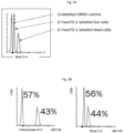
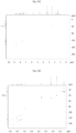
- Measurements of cell viability is exemplarily performed with compounds 18, 19 and 20: HeLa cells are treated with different concentrations (10 ⁇ M, 7.5 ⁇ M, 5 ⁇ M, 2.5 ⁇ M, 1 ⁇ M and 0.5 ⁇ M) of the dyes (18, 19, 20) in triplicated. Viability is then examined after 21h incubation time by resazurin-based assay. The results of the tests are depicted in Figs. 10A and 10B.
- E. coli NEB5a are cultured in lysogeny broth (LB) liquid medium and incubated at 37 °C, 200 rpm.
- LB lysogeny broth
- the affinity of the TramTO derivatives towards dsDNA is usually higher than the affinity of the derivatives without the modified side chain according to this invention.
- the TramTOs according to the invention were the stronger binders compared to the corresponding controls (4-6) with the similar fluorescence, however the green control CH 3 TO-1 (4) is a stronger binder then the 6-TramTAS, the 6-TramTO-1 is superior to 6-TramTO-3 (about x2) and 6-TramTAS (about x3).
- K D values are determined as is well known to any person being of ordinary skill in the art. Table 2 shows the results obtained.
- Table 2 Exemplary results of fluorescence titrations (affinity towards dsDNA) RFU -/+ DNA Slits (nm) K D 6-TramTO-1 (1) 1.2/460 5/5 8.63 ⁇ 4.34 ⁇ M CH 3 TO-1 (4) 0.2/125 10/10 (for 5/5: -10 RFU) 20.56 ⁇ 6.67 ⁇ M 6-TramTAS (2) 2.9/90 10/10 (for 5/5: ⁇ 8 RFU) 30.09 ⁇ 4.61 ⁇ M or 28.07 ⁇ 2.33 ⁇ M CH 3 TAS (5) 2.3/33 10/10 (for 5/5: ⁇ 2 RFU) Not calculable 6-TramTO-3 (3) 11.8/280 5/5 22.96 ⁇ 1.81 ⁇ M / 19.5 ⁇ 2.6 ⁇ M (literature value) CH 3 TO-3 (6) 6.8/210 5/5 44.98 ⁇ 1.05 ⁇ M
- the flow cytometric measurements are performed by use of the LSR Fortessa cell analyser (BD Biosciences, USA) or other commercially available apparatuses (e.g. FACSAria III (BD Biosciences, USA) and Guava easyCyte (Merck Millipore, USA)). Any person of ordinary skill in the art knows how to adjust the different apparatuses commercially available in order to perform comparable experiments exerting the experimental parameters described herein. A minimum of 10000 events per sample is analysed. The measurements are each performed in at least two independent experiments. Bacteria are labelled for 15 min with 5 ⁇ M of the corresponding dyes. All dyes showed > 99.9% labelling.
- Fig. 4 shows graphs of the results, exemplarily obtained with E. Coli (NEB5a) and B . Subtilis (NCIB 3610).
- Culture plates are then centrifuged for 10 min at 250 g and 37°C to establish physical contact between macrophages and bacteria. Subsequently, culture plates are transferred back into a 37°C incubator with CO 2 atmosphere for 50 min. After the stimulation period the cells are washed once with DPBS prior to flow cytometry. Macrophages are detached carefully from the culture dishes by using a rubber scraper.
- Electrospray ionization mass spectrometry is performed on a Finnigan LTQ-FT (Thermo Fischer Scientific). EI-MS is performed on an AccuTOF GCv (JEOL).
- Scheme 5 shows the general retrosynthetic pathway of the production of the cyanine dyes according to the invention.
- Benzothiazolium 37 (117mg, 400 ⁇ mol, 1.00 eq) and quinilinium 24 (128 mg, 400 ⁇ mol, 1.00 eq) are suspended in ethanol (6.0 mL), diisopropylethylamine (0.15 mL, 884 ⁇ mol, 2.21 eq) is added and the solution stirred at room temperature in the dark for 2 h. The solvent is evaporated, and the crude purified by flash column chromatography (basic allox, MeOH in CH 2 Cl 2 : 0% ⁇ 0.2% ⁇ 0.5% ⁇ 0.7%) to yield the desired product 4 (156 mg, 350 ⁇ mol, 87 %) as red solid.
- Benzothiazolium 37 (147 mg, 505 ⁇ mol, 1.26 eq) and quinilinium 38 (161 mg, 400 ⁇ mol, 1.00 eq) are dissolved in CH 2 Cl 2 /Methanol (1:1, 4 mL), Acetic anhydride (0.4 mL) and triethylamine (0.4 mL) are added and the reaction is stirred for 3.5 h at room temperature. The crude dye is precipitated by pouring the reaction mixture in diethylether.
- lodo-derivative ( 22 , 500 mg, 0.81 mmol) and sodium azide (210 mg, 3.24 mmol) are dissolved in acetone (3.3 mL) and water (3.3 mL) and the mixture is stirred at r.t. for 17 h.
- Sodium azide (105 mg, 1.62 mmol) is added and the mixture is stirred at r.t. 25 h.
- Sodium azide (210 mg, 3.24 mmol) is added and the mixture is stirred at 50 °C for 3 h.
- the solvent is evaporated and the crude extracted in CH 2 Cl 2 and water.
- Tris(benzyltriazolmethyl)amine (TBTA, 54): 7.5 mL MeCN and 13.1 mg CuOAc 2 H 2 O (66.0 ⁇ mol, 0.02 eq.) was stirred at room temperature until a blue solution was obtained. After addition of 0.7 mL benzylazide (56, 0.74 g, 5.6 mmol, 1.70 eq.) and 0.46 mL tripropargylamine ( 55 , 430 mg, 3.28 mmol, 1.00 eq.) in 2.5 mL MeCN the reaction mixture was stirred for further 5 min at room temperature.
- the following exemplary synthesis route is based on the nucleophilic attack of the cysteine in peptide 36 at the iodine substituted carbon atom of the alkyl chain of the dye-derivative (21, 22, 23) (cf. Fig. 29 , scheme 12).
- the resin is washed with DMF (5x) and capping is performed by adding 2,6-lutidine/Ac 2 O/DMF (6:5:89, 500 ⁇ L) to the resin and shaking for 5 min.
- the first coupling cycle begins mainly analogous to the synthesis described for peptide 26.
- Deprotecting of the Fmoc-groups is performed by adding piperidine (20% in DMF, 2 x 500 ⁇ L) and shaking for 2 x 5 min.
- the resin is washed with DMF (5x), CH 2 Cl 2 (5x), DMF (5x) and the coupling solution consisting of the Fmoc-amino acid (66.0 ⁇ mol, 4.00 equiv.), Oxyma (9.4 mg, 66.0 ⁇ mol, 4.00 equiv.) and DIC (10 ⁇ L, 66.0 ⁇ mol, 4.00 equiv.) in DMF (200 ⁇ L) is added to the resin which is shaken at room temperature for 45 min.
- the resin is washed with DMF (5x), CH 2 Cl 2 (5x), DMF (5x) and capped by adding 2,6-lutidine/Ac 2 O/DMF (6:5:89, 500 ⁇ L) to the resin and shaking for 5 min. After washing with DMF (5x), CH 2 Cl 2 (5x), DMF (5x), the next cycle begins. After the last coupling cycle, the final Fmoc group is deprotected as described above and the resin is washed with DMF (5x), CH 2 Cl 2 (5x), DMF (5x) and capped as described above.
- the peptide is cleaved from the resin by adding 1.5 mL SH-cleavage mix (2.5% DODT, 2.5% H 2 O, 1% TIPS, 94% TFA) and shaking for 2.5 h. Afterward, the resin is washed with cleavage mix (500 ⁇ L) and TFA (2 x 500 ⁇ L) and the combined solutions are drained into ice-cold Et 2 O ( ⁇ 25 mL). After centrifugation, the precipitate is washed with cold Et 2 O (2x) and the obtained pellet is dried in vacuo.
- SH-cleavage mix 2.5% DODT, 2.5% H 2 O, 1% TIPS, 94% TFA
- Red MPP (20): Peptide (36, 0.9 mg, 801 nmol, 1.00 equiv.) and lodo derivative 6-lodoTO-3 (14, 1.5 mg, 2.24 ⁇ mol, 2.80 equiv.) are dissolved in DMF (100 ⁇ L), DIPEA (1.6 ⁇ L, 8.91 ⁇ mol, 11.1 equiv.) is added and the reaction mixture is stirred at room temperature for 18 h. Afterward, the solvent is removed via centrifugation in vacuo and the crude is purified by preparative HPLC (Varian, 35-95%) to yield the desired Red MPP (20, 44 nmol, 6%) as a blue solid. R t 23.4 min (gradient 1).
- Green MPP (18): Peptide ( 36 , 0.9 mg, 801 nmol, 1.00 equiv.) and lodo derivative 6-lodoTO-1 (5, 1.0 mg, 1.56 ⁇ mol, 1.95 equiv.) are dissolved in DMF (100 ⁇ L), DIPEA (1.6 ⁇ L, 8.91 ⁇ mol, 11.1 equiv.) is added and the reaction mixture is stirred at room temperature for 18 h. Afterward, the solvent is removed via centrifugation in vacuo and the crude is purified by preparative HPLC (Varian, 35-95%) to yield the desired Green MPP (18, 116 nmol, 15%) as a red solid. R t 22.3 min (gradient 1).
- Yellow MPP (19): Peptide (36, 0.9 mg, 801 nmol, 1.00 equiv.) and lodo derivative 22 (1.0 mg, 1.62 ⁇ mol, 2.02 equiv.) are dissolved in DMF (100 ⁇ L), DIPEA (1.6 ⁇ L, 8.91 ⁇ mol, 11.1 equiv.) is added and the reaction mixture is stirred at room temperature for 18 h. Afterward, the solvent is removed via centrifugation in vacuo and the crude is purified by preparative HPLC (Varian, 35-95%) to yield the desired Yellow MPP (19, 44 nmol, 6%) as a red solid. R t 16.9 min (gradient 1).
- Table 3 Abbreviations used herein Cha L -cyclohexylalanine dArg D -arginine MFI mean fluorescence intensity MOI multiplicity of infection MPP mitochondria penetrating peptide NT nucleoli tracker RFU relative fluorescence untis TBTA tris((1-benzyl-4-triazolyl)methyl)amine TO thiazole orange TAS benzoThiazole AminoStyryl
Landscapes
- Health & Medical Sciences (AREA)
- Life Sciences & Earth Sciences (AREA)
- Chemical & Material Sciences (AREA)
- Engineering & Computer Science (AREA)
- Molecular Biology (AREA)
- Immunology (AREA)
- Biomedical Technology (AREA)
- General Health & Medical Sciences (AREA)
- Pathology (AREA)
- Physics & Mathematics (AREA)
- Analytical Chemistry (AREA)
- Biochemistry (AREA)
- General Physics & Mathematics (AREA)
- Organic Chemistry (AREA)
- Urology & Nephrology (AREA)
- Hematology (AREA)
- Biotechnology (AREA)
- Cell Biology (AREA)
- Microbiology (AREA)
- Nuclear Medicine, Radiotherapy & Molecular Imaging (AREA)
- Food Science & Technology (AREA)
- Medicinal Chemistry (AREA)
- Chemical Kinetics & Catalysis (AREA)
- Measuring Or Testing Involving Enzymes Or Micro-Organisms (AREA)
Applications Claiming Priority (1)
| Application Number | Priority Date | Filing Date | Title |
|---|---|---|---|
| EP21192451 | 2021-08-20 |
Publications (1)
| Publication Number | Publication Date |
|---|---|
| EP4137794A1 true EP4137794A1 (fr) | 2023-02-22 |
Family
ID=77431255
Family Applications (1)
| Application Number | Title | Priority Date | Filing Date |
|---|---|---|---|
| EP22191279.3A Pending EP4137794A1 (fr) | 2021-08-20 | 2022-08-19 | Colorants de cyanine et leur utilisation pour la coloration in vivo de micro-organismes et d'autres cellules vivantes |
Country Status (2)
| Country | Link |
|---|---|
| US (1) | US20230096750A1 (fr) |
| EP (1) | EP4137794A1 (fr) |
Citations (6)
| Publication number | Priority date | Publication date | Assignee | Title |
|---|---|---|---|---|
| US20100151509A1 (en) | 2008-12-17 | 2010-06-17 | Shenzhen Mindray Bio-Medical Electronics Co., Ltd. | Reagent, kit and method for differentating and counting leukocytes |
| US20110262904A1 (en) | 2010-01-05 | 2011-10-27 | Xiaojun Peng | Fluorescent dyes,methods of synthesis and applications thereof |
| WO2013189043A1 (fr) | 2012-06-20 | 2013-12-27 | 大连理工大学 | Colorants de cyanine verts fluorescents, leur procédé de préparation et leurs utilisations |
| US20150185182A1 (en) | 2013-10-03 | 2015-07-02 | Biotium, Inc. | Nucleic acid binding dyes with improved safety |
| US20160340717A1 (en) | 2014-02-07 | 2016-11-24 | University Of Iowa Research Foundation | Oligonucleotide-based probes and methods for detection of microbes |
| EP3572468A1 (fr) | 2018-05-24 | 2019-11-27 | Philipps-Universität Marburg | Colorants de cyanine et leur utilisation pour la coloration in vivo de micro-organismes |
-
2022
- 2022-08-19 US US17/821,007 patent/US20230096750A1/en active Pending
- 2022-08-19 EP EP22191279.3A patent/EP4137794A1/fr active Pending
Patent Citations (6)
| Publication number | Priority date | Publication date | Assignee | Title |
|---|---|---|---|---|
| US20100151509A1 (en) | 2008-12-17 | 2010-06-17 | Shenzhen Mindray Bio-Medical Electronics Co., Ltd. | Reagent, kit and method for differentating and counting leukocytes |
| US20110262904A1 (en) | 2010-01-05 | 2011-10-27 | Xiaojun Peng | Fluorescent dyes,methods of synthesis and applications thereof |
| WO2013189043A1 (fr) | 2012-06-20 | 2013-12-27 | 大连理工大学 | Colorants de cyanine verts fluorescents, leur procédé de préparation et leurs utilisations |
| US20150185182A1 (en) | 2013-10-03 | 2015-07-02 | Biotium, Inc. | Nucleic acid binding dyes with improved safety |
| US20160340717A1 (en) | 2014-02-07 | 2016-11-24 | University Of Iowa Research Foundation | Oligonucleotide-based probes and methods for detection of microbes |
| EP3572468A1 (fr) | 2018-05-24 | 2019-11-27 | Philipps-Universität Marburg | Colorants de cyanine et leur utilisation pour la coloration in vivo de micro-organismes |
Non-Patent Citations (3)
| Title |
|---|
| BIOCONJUGATE CHEM., vol. 30, 2019, pages 2312 - 2316 |
| JIEQIONG QIU ET AL., NUCLEIC ACIS RESEARCH, vol. 44, no. 17, 2016 |
| LEON N SCHULTE ET AL: "A Far-Red Fluorescent DNA Binder for Interaction Studies of Live Multidrug-Resistant Pathogens and Host Cells", ANGEWANDTE CHEMIE, WILEY - V C H VERLAG GMBH & CO. KGAA, DE, vol. 130, no. 36, 6 August 2018 (2018-08-06), pages 11738 - 11742, XP071374875, ISSN: 0044-8249, DOI: 10.1002/ANGE.201804090 * |
Also Published As
| Publication number | Publication date |
|---|---|
| US20230096750A1 (en) | 2023-03-30 |
Similar Documents
| Publication | Publication Date | Title |
|---|---|---|
| US10215751B2 (en) | Carboxy X rhodamine analogs | |
| EP2942352A1 (fr) | SYNTHÈSE DE RHODAMINE Si ASYMÉTRIQUE ET DE RHODOL | |
| CS273617B2 (en) | Method of resorufine's derivatives production | |
| Kele et al. | Clickable fluorophores for biological labeling—with or without copper | |
| Palmioli et al. | Glyco-functionalized dinuclear rhenium (i) complexes for cell imaging | |
| Song et al. | Development of an endoplasmic reticulum-targeting fluorescent probe for the imaging of polarity in living cells and tissues | |
| JP2012526086A (ja) | 新規インジケータプラットフォーム | |
| WO2007038251A1 (fr) | Inhibiteurs de proteines kinases d'alkoxy indolinone | |
| JP2015038114A (ja) | ポリアミンの新規な蛍光誘導体、その製造方法、および癌性腫瘍の治療における診断手段としてのその使用 | |
| EP4137794A1 (fr) | Colorants de cyanine et leur utilisation pour la coloration in vivo de micro-organismes et d'autres cellules vivantes | |
| Proverbio et al. | Luminescent conjugates between dinuclear rhenium complexes and 17α-ethynylestradiol: synthesis, photophysical characterization, and cell imaging | |
| KR101476639B1 (ko) | 감마-l-글루타밀-l-시스테닐글라이신 검출용 조성물 | |
| CN104031055B (zh) | 一种可用作Wnt信号途径激活剂的化合物及其制备与应用 | |
| EP3572468B1 (fr) | Colorants de cyanine et leur utilisation pour la coloration in vivo de micro-organismes | |
| CN114702447B (zh) | 一种萘酰亚胺衍生物及其制备方法与应用 | |
| JP5240704B2 (ja) | 新規蛍光化合物およびそれを用いた細胞内コレステロールの検出方法 | |
| JP6675125B2 (ja) | pH依存性蛍光化合物 | |
| CN114436947B (zh) | 一种对粘度和硝基还原酶双响应的荧光探针及其制备方法和应用 | |
| KR101125058B1 (ko) | 물질 표지용 화합물 및 그 제조방법 | |
| US8133902B2 (en) | Ammosamides as anticancer agents | |
| JP2004101389A (ja) | アルミニウムイオン及び/又は第二鉄イオン測定用プローブ | |
| US20130344607A1 (en) | Pi-conjugated fluoroionophores and method for determining an alkali ion | |
| WO2021153772A1 (fr) | Sonde fluorescente bleue pour la détection d'aldéhyde déshydrogénase 1a1 | |
| CN113943264B (zh) | 抑制g3bp1应激颗粒形成的化合物、制备方法及其用途 | |
| EP3981778A1 (fr) | Sonde fluorescente pour la détection de l'activité de l'enpp |
Legal Events
| Date | Code | Title | Description |
|---|---|---|---|
| PUAI | Public reference made under article 153(3) epc to a published international application that has entered the european phase |
Free format text: ORIGINAL CODE: 0009012 |
|
| STAA | Information on the status of an ep patent application or granted ep patent |
Free format text: STATUS: THE APPLICATION HAS BEEN PUBLISHED |
|
| AK | Designated contracting states |
Kind code of ref document: A1 Designated state(s): AL AT BE BG CH CY CZ DE DK EE ES FI FR GB GR HR HU IE IS IT LI LT LU LV MC MK MT NL NO PL PT RO RS SE SI SK SM TR |
|
| STAA | Information on the status of an ep patent application or granted ep patent |
Free format text: STATUS: REQUEST FOR EXAMINATION WAS MADE |
|
| 17P | Request for examination filed |
Effective date: 20230822 |
|
| RBV | Designated contracting states (corrected) |
Designated state(s): AL AT BE BG CH CY CZ DE DK EE ES FI FR GB GR HR HU IE IS IT LI LT LU LV MC MK MT NL NO PL PT RO RS SE SI SK SM TR |
|
| RIN1 | Information on inventor provided before grant (corrected) |
Inventor name: PLOCHER, BENEDIKT Inventor name: LINDEN, GRETA Inventor name: SCHULTE, LEON Inventor name: VAZQUEZ, OLALLA |