EP4067364A1 - Neue verbindung auf gadoliniumbasis, verfahren zu ihrer herstellung und mrt-kontrastmittel damit - Google Patents
Neue verbindung auf gadoliniumbasis, verfahren zu ihrer herstellung und mrt-kontrastmittel damit Download PDFInfo
- Publication number
- EP4067364A1 EP4067364A1 EP20881219.8A EP20881219A EP4067364A1 EP 4067364 A1 EP4067364 A1 EP 4067364A1 EP 20881219 A EP20881219 A EP 20881219A EP 4067364 A1 EP4067364 A1 EP 4067364A1
- Authority
- EP
- European Patent Office
- Prior art keywords
- compound
- chemical formula
- gadolinium
- based compound
- contrast agent
- Prior art date
- Legal status (The legal status is an assumption and is not a legal conclusion. Google has not performed a legal analysis and makes no representation as to the accuracy of the status listed.)
- Granted
Links
- 150000001875 compounds Chemical class 0.000 title claims abstract description 93
- 229910052688 Gadolinium Inorganic materials 0.000 title claims abstract description 29
- UIWYJDYFSGRHKR-UHFFFAOYSA-N gadolinium atom Chemical compound [Gd] UIWYJDYFSGRHKR-UHFFFAOYSA-N 0.000 title claims abstract description 29
- 239000002616 MRI contrast agent Substances 0.000 title claims abstract description 18
- 238000004519 manufacturing process Methods 0.000 title abstract 2
- 239000000126 substance Substances 0.000 claims abstract description 52
- 102000001763 Vesicular Glutamate Transport Proteins Human genes 0.000 claims description 16
- 108010040170 Vesicular Glutamate Transport Proteins Proteins 0.000 claims description 16
- 238000000034 method Methods 0.000 claims description 13
- XLYOFNOQVPJJNP-UHFFFAOYSA-N water Substances O XLYOFNOQVPJJNP-UHFFFAOYSA-N 0.000 claims description 10
- 208000014644 Brain disease Diseases 0.000 claims description 9
- 230000003412 degenerative effect Effects 0.000 claims description 9
- 125000006239 protecting group Chemical group 0.000 claims description 9
- 208000024827 Alzheimer disease Diseases 0.000 claims description 6
- 239000002253 acid Substances 0.000 claims description 6
- 150000003839 salts Chemical class 0.000 claims description 6
- 125000000008 (C1-C10) alkyl group Chemical group 0.000 claims description 5
- -1 alkali metal salt Chemical class 0.000 claims description 4
- 238000003745 diagnosis Methods 0.000 claims description 4
- UFHFLCQGNIYNRP-UHFFFAOYSA-N Hydrogen Chemical compound [H][H] UFHFLCQGNIYNRP-UHFFFAOYSA-N 0.000 claims description 3
- 125000000217 alkyl group Chemical group 0.000 claims description 3
- CDKFGSDHWMPTKX-UHFFFAOYSA-N gadolinium;hydrate Chemical compound O.[Gd] CDKFGSDHWMPTKX-UHFFFAOYSA-N 0.000 claims description 3
- 229910052736 halogen Inorganic materials 0.000 claims description 3
- 150000002367 halogens Chemical class 0.000 claims description 3
- 229910052739 hydrogen Inorganic materials 0.000 claims description 3
- 239000001257 hydrogen Substances 0.000 claims description 3
- 125000006755 (C2-C20) alkyl group Chemical group 0.000 claims description 2
- 150000001335 aliphatic alkanes Chemical class 0.000 claims description 2
- 229910052783 alkali metal Inorganic materials 0.000 claims description 2
- 239000003795 chemical substances by application Substances 0.000 claims 2
- 125000005647 linker group Chemical group 0.000 abstract description 6
- 210000004027 cell Anatomy 0.000 description 25
- OKKJLVBELUTLKV-UHFFFAOYSA-N Methanol Chemical compound OC OKKJLVBELUTLKV-UHFFFAOYSA-N 0.000 description 21
- 239000002872 contrast media Substances 0.000 description 18
- WHUUTDBJXJRKMK-VKHMYHEASA-N L-glutamic acid Chemical compound OC(=O)[C@@H](N)CCC(O)=O WHUUTDBJXJRKMK-VKHMYHEASA-N 0.000 description 14
- 229930195712 glutamate Natural products 0.000 description 14
- 108091006283 SLC17A7 Proteins 0.000 description 13
- 102000046052 Vesicular Glutamate Transport Protein 1 Human genes 0.000 description 13
- YMWUJEATGCHHMB-UHFFFAOYSA-N Dichloromethane Chemical compound ClCCl YMWUJEATGCHHMB-UHFFFAOYSA-N 0.000 description 12
- ZMXDDKWLCZADIW-UHFFFAOYSA-N N,N-Dimethylformamide Chemical compound CN(C)C=O ZMXDDKWLCZADIW-UHFFFAOYSA-N 0.000 description 12
- 210000004556 brain Anatomy 0.000 description 12
- 239000000872 buffer Substances 0.000 description 11
- 238000006243 chemical reaction Methods 0.000 description 11
- 238000002474 experimental method Methods 0.000 description 11
- 238000001727 in vivo Methods 0.000 description 10
- 238000002360 preparation method Methods 0.000 description 10
- ZPDFIIGFYAHNSK-UHFFFAOYSA-K gadobutrol Chemical compound [Gd+3].OCC(O)C(CO)N1CCN(CC([O-])=O)CCN(CC([O-])=O)CCN(CC([O-])=O)CC1 ZPDFIIGFYAHNSK-UHFFFAOYSA-K 0.000 description 9
- 239000000243 solution Substances 0.000 description 9
- 108091003079 Bovine Serum Albumin Proteins 0.000 description 8
- 239000012981 Hank's balanced salt solution Substances 0.000 description 8
- 239000000203 mixture Substances 0.000 description 7
- 239000002904 solvent Substances 0.000 description 7
- 210000001519 tissue Anatomy 0.000 description 7
- CSCPPACGZOOCGX-UHFFFAOYSA-N Acetone Chemical compound CC(C)=O CSCPPACGZOOCGX-UHFFFAOYSA-N 0.000 description 6
- WEVYAHXRMPXWCK-UHFFFAOYSA-N Acetonitrile Chemical compound CC#N WEVYAHXRMPXWCK-UHFFFAOYSA-N 0.000 description 6
- RTZKZFJDLAIYFH-UHFFFAOYSA-N Diethyl ether Chemical compound CCOCC RTZKZFJDLAIYFH-UHFFFAOYSA-N 0.000 description 6
- IAZDPXIOMUYVGZ-UHFFFAOYSA-N Dimethylsulphoxide Chemical compound CS(C)=O IAZDPXIOMUYVGZ-UHFFFAOYSA-N 0.000 description 6
- XEKOWRVHYACXOJ-UHFFFAOYSA-N Ethyl acetate Chemical compound CCOC(C)=O XEKOWRVHYACXOJ-UHFFFAOYSA-N 0.000 description 6
- 230000008859 change Effects 0.000 description 6
- 229940126214 compound 3 Drugs 0.000 description 6
- 238000002000 high resolution fast-atom bombardment mass spectrometry Methods 0.000 description 6
- 238000003384 imaging method Methods 0.000 description 6
- 238000003125 immunofluorescent labeling Methods 0.000 description 6
- 238000001228 spectrum Methods 0.000 description 6
- 238000012360 testing method Methods 0.000 description 6
- 241000699670 Mus sp. Species 0.000 description 5
- 230000003833 cell viability Effects 0.000 description 5
- 238000005119 centrifugation Methods 0.000 description 5
- 229940125904 compound 1 Drugs 0.000 description 5
- 229940125782 compound 2 Drugs 0.000 description 5
- 201000010099 disease Diseases 0.000 description 5
- 208000037265 diseases, disorders, signs and symptoms Diseases 0.000 description 5
- 230000001965 increasing effect Effects 0.000 description 5
- 230000005291 magnetic effect Effects 0.000 description 5
- 210000002569 neuron Anatomy 0.000 description 5
- 239000002244 precipitate Substances 0.000 description 5
- ZDXPYRJPNDTMRX-VKHMYHEASA-N L-glutamine Chemical compound OC(=O)[C@@H](N)CCC(N)=O ZDXPYRJPNDTMRX-VKHMYHEASA-N 0.000 description 4
- 102000004142 Trypsin Human genes 0.000 description 4
- 108090000631 Trypsin Proteins 0.000 description 4
- 230000015572 biosynthetic process Effects 0.000 description 4
- 229940098773 bovine serum albumin Drugs 0.000 description 4
- 239000012091 fetal bovine serum Substances 0.000 description 4
- 238000002073 fluorescence micrograph Methods 0.000 description 4
- 238000002514 liquid chromatography mass spectrum Methods 0.000 description 4
- 238000005259 measurement Methods 0.000 description 4
- 239000011734 sodium Substances 0.000 description 4
- 238000003786 synthesis reaction Methods 0.000 description 4
- 230000008685 targeting Effects 0.000 description 4
- 125000000999 tert-butyl group Chemical group [H]C([H])([H])C(*)(C([H])([H])[H])C([H])([H])[H] 0.000 description 4
- 238000004809 thin layer chromatography Methods 0.000 description 4
- 239000012588 trypsin Substances 0.000 description 4
- FWBHETKCLVMNFS-UHFFFAOYSA-N 4',6-Diamino-2-phenylindol Chemical compound C1=CC(C(=N)N)=CC=C1C1=CC2=CC=C(C(N)=N)C=C2N1 FWBHETKCLVMNFS-UHFFFAOYSA-N 0.000 description 3
- IICCLYANAQEHCI-UHFFFAOYSA-N 4,5,6,7-tetrachloro-3',6'-dihydroxy-2',4',5',7'-tetraiodospiro[2-benzofuran-3,9'-xanthene]-1-one Chemical compound O1C(=O)C(C(=C(Cl)C(Cl)=C2Cl)Cl)=C2C21C1=CC(I)=C(O)C(I)=C1OC1=C(I)C(O)=C(I)C=C21 IICCLYANAQEHCI-UHFFFAOYSA-N 0.000 description 3
- 229930182816 L-glutamine Natural products 0.000 description 3
- 241001465754 Metazoa Species 0.000 description 3
- 238000002835 absorbance Methods 0.000 description 3
- 238000010586 diagram Methods 0.000 description 3
- OXZOLXJZTSUDOM-UHFFFAOYSA-N fluoro 2,2,2-trifluoroacetate Chemical compound FOC(=O)C(F)(F)F OXZOLXJZTSUDOM-UHFFFAOYSA-N 0.000 description 3
- 238000004128 high performance liquid chromatography Methods 0.000 description 3
- 238000002114 high-resolution electrospray ionisation mass spectrometry Methods 0.000 description 3
- 210000001320 hippocampus Anatomy 0.000 description 3
- 125000004435 hydrogen atom Chemical group [H]* 0.000 description 3
- 238000000338 in vitro Methods 0.000 description 3
- 238000013507 mapping Methods 0.000 description 3
- 239000000047 product Substances 0.000 description 3
- 238000011160 research Methods 0.000 description 3
- 239000007787 solid Substances 0.000 description 3
- ILJSQTXMGCGYMG-UHFFFAOYSA-N triacetic acid Chemical compound CC(=O)CC(=O)CC(O)=O ILJSQTXMGCGYMG-UHFFFAOYSA-N 0.000 description 3
- OZFAFGSSMRRTDW-UHFFFAOYSA-N (2,4-dichlorophenyl) benzenesulfonate Chemical compound ClC1=CC(Cl)=CC=C1OS(=O)(=O)C1=CC=CC=C1 OZFAFGSSMRRTDW-UHFFFAOYSA-N 0.000 description 2
- 125000003837 (C1-C20) alkyl group Chemical group 0.000 description 2
- 238000011818 5xFAD mouse Methods 0.000 description 2
- 241000283707 Capra Species 0.000 description 2
- HEDRZPFGACZZDS-UHFFFAOYSA-N Chloroform Chemical compound ClC(Cl)Cl HEDRZPFGACZZDS-UHFFFAOYSA-N 0.000 description 2
- 206010012289 Dementia Diseases 0.000 description 2
- QOSSAOTZNIDXMA-UHFFFAOYSA-N Dicylcohexylcarbodiimide Chemical compound C1CCCCC1N=C=NC1CCCCC1 QOSSAOTZNIDXMA-UHFFFAOYSA-N 0.000 description 2
- IAZDPXIOMUYVGZ-WFGJKAKNSA-N Dimethyl sulfoxide Chemical compound [2H]C([2H])([2H])S(=O)C([2H])([2H])[2H] IAZDPXIOMUYVGZ-WFGJKAKNSA-N 0.000 description 2
- 239000012591 Dulbecco’s Phosphate Buffered Saline Substances 0.000 description 2
- 102000034575 Glutamate transporters Human genes 0.000 description 2
- 108091006151 Glutamate transporters Proteins 0.000 description 2
- PIWKPBJCKXDKJR-UHFFFAOYSA-N Isoflurane Chemical compound FC(F)OC(Cl)C(F)(F)F PIWKPBJCKXDKJR-UHFFFAOYSA-N 0.000 description 2
- CSNNHWWHGAXBCP-UHFFFAOYSA-L Magnesium sulfate Chemical compound [Mg+2].[O-][S+2]([O-])([O-])[O-] CSNNHWWHGAXBCP-UHFFFAOYSA-L 0.000 description 2
- 241000699666 Mus <mouse, genus> Species 0.000 description 2
- 229930040373 Paraformaldehyde Natural products 0.000 description 2
- 208000018737 Parkinson disease Diseases 0.000 description 2
- PXIPVTKHYLBLMZ-UHFFFAOYSA-N Sodium azide Chemical compound [Na+].[N-]=[N+]=[N-] PXIPVTKHYLBLMZ-UHFFFAOYSA-N 0.000 description 2
- 201000004810 Vascular dementia Diseases 0.000 description 2
- 230000009471 action Effects 0.000 description 2
- 239000003242 anti bacterial agent Substances 0.000 description 2
- 229940088710 antibiotic agent Drugs 0.000 description 2
- 238000004364 calculation method Methods 0.000 description 2
- 238000010609 cell counting kit-8 assay Methods 0.000 description 2
- 238000004113 cell culture Methods 0.000 description 2
- 210000004289 cerebral ventricle Anatomy 0.000 description 2
- 239000000460 chlorine Substances 0.000 description 2
- 238000004440 column chromatography Methods 0.000 description 2
- 230000003013 cytotoxicity Effects 0.000 description 2
- 231100000135 cytotoxicity Toxicity 0.000 description 2
- 230000006378 damage Effects 0.000 description 2
- 230000007423 decrease Effects 0.000 description 2
- 238000013399 early diagnosis Methods 0.000 description 2
- 230000000694 effects Effects 0.000 description 2
- 238000011156 evaluation Methods 0.000 description 2
- 230000002964 excitative effect Effects 0.000 description 2
- 210000003722 extracellular fluid Anatomy 0.000 description 2
- 238000001914 filtration Methods 0.000 description 2
- 238000002075 inversion recovery Methods 0.000 description 2
- 229960002725 isoflurane Drugs 0.000 description 2
- 238000004895 liquid chromatography mass spectrometry Methods 0.000 description 2
- 239000011159 matrix material Substances 0.000 description 2
- 239000002609 medium Substances 0.000 description 2
- 239000007758 minimum essential medium Substances 0.000 description 2
- 238000000655 nuclear magnetic resonance spectrum Methods 0.000 description 2
- 229920002866 paraformaldehyde Polymers 0.000 description 2
- 239000011736 potassium bicarbonate Substances 0.000 description 2
- 229910000028 potassium bicarbonate Inorganic materials 0.000 description 2
- TYJJADVDDVDEDZ-UHFFFAOYSA-M potassium hydrogencarbonate Chemical compound [K+].OC([O-])=O TYJJADVDDVDEDZ-UHFFFAOYSA-M 0.000 description 2
- 238000012545 processing Methods 0.000 description 2
- 238000010992 reflux Methods 0.000 description 2
- 229930187593 rose bengal Natural products 0.000 description 2
- 229940081623 rose bengal Drugs 0.000 description 2
- STRXNPAVPKGJQR-UHFFFAOYSA-N rose bengal A Natural products O1C(=O)C(C(=CC=C2Cl)Cl)=C2C21C1=CC(I)=C(O)C(I)=C1OC1=C(I)C(O)=C(I)C=C21 STRXNPAVPKGJQR-UHFFFAOYSA-N 0.000 description 2
- 210000002966 serum Anatomy 0.000 description 2
- 159000000000 sodium salts Chemical class 0.000 description 2
- 230000000087 stabilizing effect Effects 0.000 description 2
- 210000000225 synapse Anatomy 0.000 description 2
- 238000005406 washing Methods 0.000 description 2
- 125000000923 (C1-C30) alkyl group Chemical group 0.000 description 1
- QBPPRVHXOZRESW-UHFFFAOYSA-N 1,4,7,10-tetraazacyclododecane Chemical group C1CNCCNCCNCCN1 QBPPRVHXOZRESW-UHFFFAOYSA-N 0.000 description 1
- ASOKPJOREAFHNY-UHFFFAOYSA-N 1-Hydroxybenzotriazole Chemical compound C1=CC=C2N(O)N=NC2=C1 ASOKPJOREAFHNY-UHFFFAOYSA-N 0.000 description 1
- 238000005160 1H NMR spectroscopy Methods 0.000 description 1
- RZESKRXOCXWCFX-UHFFFAOYSA-N 2-[bis[2-[carboxymethyl-[2-(methylamino)-2-oxoethyl]amino]ethyl]amino]acetic acid Chemical compound CNC(=O)CN(CC(O)=O)CCN(CC(O)=O)CCN(CC(O)=O)CC(=O)NC RZESKRXOCXWCFX-UHFFFAOYSA-N 0.000 description 1
- ZCYVEMRRCGMTRW-UHFFFAOYSA-N 7553-56-2 Chemical compound [I] ZCYVEMRRCGMTRW-UHFFFAOYSA-N 0.000 description 1
- BKJFDZSBZWHRNH-UHFFFAOYSA-N 8-bromooctanoic acid Chemical compound OC(=O)CCCCCCCBr BKJFDZSBZWHRNH-UHFFFAOYSA-N 0.000 description 1
- 239000012103 Alexa Fluor 488 Substances 0.000 description 1
- 239000012099 Alexa Fluor family Substances 0.000 description 1
- 238000010173 Alzheimer-disease mouse model Methods 0.000 description 1
- 206010002091 Anaesthesia Diseases 0.000 description 1
- WKBOTKDWSSQWDR-UHFFFAOYSA-N Bromine atom Chemical compound [Br] WKBOTKDWSSQWDR-UHFFFAOYSA-N 0.000 description 1
- 238000011740 C57BL/6 mouse Methods 0.000 description 1
- ZAMOUSCENKQFHK-UHFFFAOYSA-N Chlorine atom Chemical compound [Cl] ZAMOUSCENKQFHK-UHFFFAOYSA-N 0.000 description 1
- SLWPVSJVOXUYAK-UHFFFAOYSA-N ClC1=C(C(=O)OCCCCCCCC(=O)O)C(=C(C(=C1Cl)Cl)Cl)C=1C2=CC(=C(C(=C2OC2=C(C(C(=CC=12)I)=O)I)I)O)I Chemical compound ClC1=C(C(=O)OCCCCCCCC(=O)O)C(=C(C(=C1Cl)Cl)Cl)C=1C2=CC(=C(C(=C2OC2=C(C(C(=CC=12)I)=O)I)I)O)I SLWPVSJVOXUYAK-UHFFFAOYSA-N 0.000 description 1
- 108090000790 Enzymes Proteins 0.000 description 1
- 102000004190 Enzymes Human genes 0.000 description 1
- PIICEJLVQHRZGT-UHFFFAOYSA-N Ethylenediamine Chemical compound NCCN PIICEJLVQHRZGT-UHFFFAOYSA-N 0.000 description 1
- PXGOKWXKJXAPGV-UHFFFAOYSA-N Fluorine Chemical compound FF PXGOKWXKJXAPGV-UHFFFAOYSA-N 0.000 description 1
- 108010027915 Glutamate Receptors Proteins 0.000 description 1
- 201000005569 Gout Diseases 0.000 description 1
- 101710088172 HTH-type transcriptional regulator RipA Proteins 0.000 description 1
- 239000012580 N-2 Supplement Substances 0.000 description 1
- 206010029260 Neuroblastoma Diseases 0.000 description 1
- 206010029350 Neurotoxicity Diseases 0.000 description 1
- 241000700159 Rattus Species 0.000 description 1
- 206010044221 Toxic encephalopathy Diseases 0.000 description 1
- 229920004890 Triton X-100 Polymers 0.000 description 1
- 239000013504 Triton X-100 Substances 0.000 description 1
- 230000005856 abnormality Effects 0.000 description 1
- 230000032683 aging Effects 0.000 description 1
- 150000001413 amino acids Chemical class 0.000 description 1
- 230000037005 anaesthesia Effects 0.000 description 1
- 238000004458 analytical method Methods 0.000 description 1
- 238000010171 animal model Methods 0.000 description 1
- 125000003236 benzoyl group Chemical group [H]C1=C([H])C([H])=C(C([H])=C1[H])C(*)=O 0.000 description 1
- 125000001797 benzyl group Chemical group [H]C1=C([H])C([H])=C(C([H])=C1[H])C([H])([H])* 0.000 description 1
- 230000033228 biological regulation Effects 0.000 description 1
- 230000000903 blocking effect Effects 0.000 description 1
- GDTBXPJZTBHREO-UHFFFAOYSA-N bromine Substances BrBr GDTBXPJZTBHREO-UHFFFAOYSA-N 0.000 description 1
- 229910052794 bromium Inorganic materials 0.000 description 1
- 125000003178 carboxy group Chemical group [H]OC(*)=O 0.000 description 1
- 230000005779 cell damage Effects 0.000 description 1
- 208000037887 cell injury Diseases 0.000 description 1
- 238000013043 cell viability test Methods 0.000 description 1
- 230000005754 cellular signaling Effects 0.000 description 1
- 210000003710 cerebral cortex Anatomy 0.000 description 1
- 210000001175 cerebrospinal fluid Anatomy 0.000 description 1
- 238000012512 characterization method Methods 0.000 description 1
- 239000013522 chelant Substances 0.000 description 1
- 229910052801 chlorine Inorganic materials 0.000 description 1
- 210000000795 conjunctiva Anatomy 0.000 description 1
- 210000004087 cornea Anatomy 0.000 description 1
- 210000004748 cultured cell Anatomy 0.000 description 1
- 230000003247 decreasing effect Effects 0.000 description 1
- 230000018044 dehydration Effects 0.000 description 1
- 238000006297 dehydration reaction Methods 0.000 description 1
- 238000001514 detection method Methods 0.000 description 1
- 238000011161 development Methods 0.000 description 1
- 230000018109 developmental process Effects 0.000 description 1
- 238000009826 distribution Methods 0.000 description 1
- 229940079593 drug Drugs 0.000 description 1
- 239000003814 drug Substances 0.000 description 1
- 238000001035 drying Methods 0.000 description 1
- 239000000975 dye Substances 0.000 description 1
- 238000002592 echocardiography Methods 0.000 description 1
- 230000008451 emotion Effects 0.000 description 1
- 238000005516 engineering process Methods 0.000 description 1
- 230000002708 enhancing effect Effects 0.000 description 1
- 238000006911 enzymatic reaction Methods 0.000 description 1
- PQJJJMRNHATNKG-UHFFFAOYSA-N ethyl bromoacetate Chemical compound CCOC(=O)CBr PQJJJMRNHATNKG-UHFFFAOYSA-N 0.000 description 1
- 125000001495 ethyl group Chemical group [H]C([H])([H])C([H])([H])* 0.000 description 1
- 239000003889 eye drop Substances 0.000 description 1
- HJUFTIJOISQSKQ-UHFFFAOYSA-N fenoxycarb Chemical compound C1=CC(OCCNC(=O)OCC)=CC=C1OC1=CC=CC=C1 HJUFTIJOISQSKQ-UHFFFAOYSA-N 0.000 description 1
- 230000001605 fetal effect Effects 0.000 description 1
- 210000003754 fetus Anatomy 0.000 description 1
- 229910052731 fluorine Inorganic materials 0.000 description 1
- 239000011737 fluorine Substances 0.000 description 1
- 230000008014 freezing Effects 0.000 description 1
- 238000007710 freezing Methods 0.000 description 1
- 230000006870 function Effects 0.000 description 1
- OCDAWJYGVOLXGZ-VPVMAENOSA-K gadobenate dimeglumine Chemical compound [Gd+3].CNC[C@H](O)[C@@H](O)[C@H](O)[C@H](O)CO.CNC[C@H](O)[C@@H](O)[C@H](O)[C@H](O)CO.OC(=O)CN(CC([O-])=O)CCN(CC([O-])=O)CCN(CC(O)=O)C(C([O-])=O)COCC1=CC=CC=C1 OCDAWJYGVOLXGZ-VPVMAENOSA-K 0.000 description 1
- HZHFFEYYPYZMNU-UHFFFAOYSA-K gadodiamide Chemical compound [Gd+3].CNC(=O)CN(CC([O-])=O)CCN(CC([O-])=O)CCN(CC([O-])=O)CC(=O)NC HZHFFEYYPYZMNU-UHFFFAOYSA-K 0.000 description 1
- LGMLJQFQKXPRGA-VPVMAENOSA-K gadopentetate dimeglumine Chemical compound [Gd+3].CNC[C@H](O)[C@@H](O)[C@H](O)[C@H](O)CO.CNC[C@H](O)[C@@H](O)[C@H](O)[C@H](O)CO.OC(=O)CN(CC([O-])=O)CCN(CC([O-])=O)CCN(CC(O)=O)CC([O-])=O LGMLJQFQKXPRGA-VPVMAENOSA-K 0.000 description 1
- GFSTXYOTEVLASN-UHFFFAOYSA-K gadoteric acid Chemical compound [Gd+3].OC(=O)CN1CCN(CC([O-])=O)CCN(CC([O-])=O)CCN(CC([O-])=O)CC1 GFSTXYOTEVLASN-UHFFFAOYSA-K 0.000 description 1
- DPNNNPAKRZOSMO-UHFFFAOYSA-K gadoteridol Chemical compound [Gd+3].CC(O)CN1CCN(CC([O-])=O)CCN(CC([O-])=O)CCN(CC([O-])=O)CC1 DPNNNPAKRZOSMO-UHFFFAOYSA-K 0.000 description 1
- 239000011521 glass Substances 0.000 description 1
- ZDXPYRJPNDTMRX-UHFFFAOYSA-N glutamine Natural products OC(=O)C(N)CCC(N)=O ZDXPYRJPNDTMRX-UHFFFAOYSA-N 0.000 description 1
- 239000001963 growth medium Substances 0.000 description 1
- 238000010438 heat treatment Methods 0.000 description 1
- 238000010191 image analysis Methods 0.000 description 1
- 238000011532 immunohistochemical staining Methods 0.000 description 1
- 238000003364 immunohistochemistry Methods 0.000 description 1
- 238000011534 incubation Methods 0.000 description 1
- 239000007924 injection Substances 0.000 description 1
- 238000002347 injection Methods 0.000 description 1
- 239000011630 iodine Substances 0.000 description 1
- 229910052740 iodine Inorganic materials 0.000 description 1
- 239000010410 layer Substances 0.000 description 1
- 210000004185 liver Anatomy 0.000 description 1
- 229910052943 magnesium sulfate Inorganic materials 0.000 description 1
- 238000002595 magnetic resonance imaging Methods 0.000 description 1
- 238000004949 mass spectrometry Methods 0.000 description 1
- 239000000463 material Substances 0.000 description 1
- 239000012528 membrane Substances 0.000 description 1
- 125000002496 methyl group Chemical group [H]C([H])([H])* 0.000 description 1
- 239000012120 mounting media Substances 0.000 description 1
- 230000001537 neural effect Effects 0.000 description 1
- 210000004498 neuroglial cell Anatomy 0.000 description 1
- 230000007135 neurotoxicity Effects 0.000 description 1
- 231100000228 neurotoxicity Toxicity 0.000 description 1
- 238000001208 nuclear magnetic resonance pulse sequence Methods 0.000 description 1
- 238000001543 one-way ANOVA Methods 0.000 description 1
- 239000012044 organic layer Substances 0.000 description 1
- 230000002018 overexpression Effects 0.000 description 1
- 230000036542 oxidative stress Effects 0.000 description 1
- 125000004430 oxygen atom Chemical group O* 0.000 description 1
- 230000005298 paramagnetic effect Effects 0.000 description 1
- 230000001766 physiological effect Effects 0.000 description 1
- 230000035935 pregnancy Effects 0.000 description 1
- 125000002924 primary amino group Chemical group [H]N([H])* 0.000 description 1
- 230000005855 radiation Effects 0.000 description 1
- 238000010814 radioimmunoprecipitation assay Methods 0.000 description 1
- 239000003642 reactive oxygen metabolite Substances 0.000 description 1
- 102000005962 receptors Human genes 0.000 description 1
- 230000001105 regulatory effect Effects 0.000 description 1
- 230000000241 respiratory effect Effects 0.000 description 1
- 230000019491 signal transduction Effects 0.000 description 1
- 230000011664 signaling Effects 0.000 description 1
- 210000004872 soft tissue Anatomy 0.000 description 1
- 238000000264 spin echo pulse sequence Methods 0.000 description 1
- 238000012453 sprague-dawley rat model Methods 0.000 description 1
- 238000010186 staining Methods 0.000 description 1
- 238000003756 stirring Methods 0.000 description 1
- 238000003860 storage Methods 0.000 description 1
- 239000006228 supernatant Substances 0.000 description 1
- 230000000946 synaptic effect Effects 0.000 description 1
- NMHVTLJFPDOJOD-UHFFFAOYSA-N tert-butyl 2-[4,7-bis[2-[(2-methylpropan-2-yl)oxy]-2-oxoethyl]-1,4,7,10-tetrazacyclododec-1-yl]acetate Chemical compound CC(C)(C)OC(=O)CN1CCNCCN(CC(=O)OC(C)(C)C)CCN(CC(=O)OC(C)(C)C)CC1 NMHVTLJFPDOJOD-UHFFFAOYSA-N 0.000 description 1
- 230000001988 toxicity Effects 0.000 description 1
- 231100000419 toxicity Toxicity 0.000 description 1
- 230000009261 transgenic effect Effects 0.000 description 1
- 238000003260 vortexing Methods 0.000 description 1
- 238000011816 wild-type C57Bl6 mouse Methods 0.000 description 1
Images


Classifications
-
- C—CHEMISTRY; METALLURGY
- C07—ORGANIC CHEMISTRY
- C07F—ACYCLIC, CARBOCYCLIC OR HETEROCYCLIC COMPOUNDS CONTAINING ELEMENTS OTHER THAN CARBON, HYDROGEN, HALOGEN, OXYGEN, NITROGEN, SULFUR, SELENIUM OR TELLURIUM
- C07F5/00—Compounds containing elements of Groups 3 or 13 of the Periodic Table
-
- A—HUMAN NECESSITIES
- A61—MEDICAL OR VETERINARY SCIENCE; HYGIENE
- A61K—PREPARATIONS FOR MEDICAL, DENTAL OR TOILETRY PURPOSES
- A61K49/00—Preparations for testing in vivo
- A61K49/06—Nuclear magnetic resonance [NMR] contrast preparations; Magnetic resonance imaging [MRI] contrast preparations
- A61K49/08—Nuclear magnetic resonance [NMR] contrast preparations; Magnetic resonance imaging [MRI] contrast preparations characterised by the carrier
- A61K49/085—Nuclear magnetic resonance [NMR] contrast preparations; Magnetic resonance imaging [MRI] contrast preparations characterised by the carrier conjugated systems
-
- A—HUMAN NECESSITIES
- A61—MEDICAL OR VETERINARY SCIENCE; HYGIENE
- A61K—PREPARATIONS FOR MEDICAL, DENTAL OR TOILETRY PURPOSES
- A61K49/00—Preparations for testing in vivo
- A61K49/06—Nuclear magnetic resonance [NMR] contrast preparations; Magnetic resonance imaging [MRI] contrast preparations
- A61K49/08—Nuclear magnetic resonance [NMR] contrast preparations; Magnetic resonance imaging [MRI] contrast preparations characterised by the carrier
- A61K49/10—Organic compounds
- A61K49/101—Organic compounds the carrier being a complex-forming compound able to form MRI-active complexes with paramagnetic metals
- A61K49/106—Organic compounds the carrier being a complex-forming compound able to form MRI-active complexes with paramagnetic metals the complex-forming compound being cyclic, e.g. DOTA
- A61K49/108—Organic compounds the carrier being a complex-forming compound able to form MRI-active complexes with paramagnetic metals the complex-forming compound being cyclic, e.g. DOTA the metal complex being Gd-DOTA
-
- C—CHEMISTRY; METALLURGY
- C07—ORGANIC CHEMISTRY
- C07B—GENERAL METHODS OF ORGANIC CHEMISTRY; APPARATUS THEREFOR
- C07B59/00—Introduction of isotopes of elements into organic compounds ; Labelled organic compounds per se
- C07B59/004—Acyclic, carbocyclic or heterocyclic compounds containing elements other than carbon, hydrogen, halogen, oxygen, nitrogen, sulfur, selenium or tellurium
-
- Y—GENERAL TAGGING OF NEW TECHNOLOGICAL DEVELOPMENTS; GENERAL TAGGING OF CROSS-SECTIONAL TECHNOLOGIES SPANNING OVER SEVERAL SECTIONS OF THE IPC; TECHNICAL SUBJECTS COVERED BY FORMER USPC CROSS-REFERENCE ART COLLECTIONS [XRACs] AND DIGESTS
- Y02—TECHNOLOGIES OR APPLICATIONS FOR MITIGATION OR ADAPTATION AGAINST CLIMATE CHANGE
- Y02P—CLIMATE CHANGE MITIGATION TECHNOLOGIES IN THE PRODUCTION OR PROCESSING OF GOODS
- Y02P20/00—Technologies relating to chemical industry
- Y02P20/50—Improvements relating to the production of bulk chemicals
- Y02P20/55—Design of synthesis routes, e.g. reducing the use of auxiliary or protecting groups
Definitions
- the present disclosure relates to a novel gadolinium-based compound, a method for preparation of the same, and an MRI contrast agent containing the same.
- the present disclosure relates to a novel gadolinium-based compound having a structure in which a gadolinium complex and Rose Bengal are bonded to each other via a linking group, a method for preparing the same, and an MRI contrast agent containing the same.
- Degenerative brain diseases include Parkinson's disease, vascular dementia, Alzheimer's disease, and the like. Neurotoxicity due to overexpression of glutamate is considered as one of the causes of the disease.
- Glutamate is responsible for more than 70% of excitatory signaling in the human brain.
- the glutamate is an important amino acid that regulates learning, memory, motor performance, and emotion.
- Glutamate-induced excitatory signal transduction at synapses is tightly regulated by several glutamate transporters and receptors.
- the vesicular glutamate transporter (VGLUT) is known to play a key role in storage and concentration regulation of the glutamate in neuronal cells. Normally, the concentration of glutamate in the synaptic cleft is in a range of 1 to 3 ⁇ M. When the glutamate stored in the vesicle is released, the concentration thereof increases to hundreds to thousands of ⁇ M or higher.
- Magnetic Resonance Image refers to a method of obtaining anatomical, physiological, and biochemical information images of the body using a phenomenon in which the distributions of hydrogen atoms in different tissues of the body are different from each other and the hydrogen atoms are relaxed in a magnetic field.
- MRI Magnetic Resonance Image
- CT or PET MRI does not use radiation harmful to the human body and creates images inside the body using the gradient of the magnetic field and radio waves under a strong magnetic field.
- the MRI is non-invasive, has high resolution, and has excellent soft tissue examination capabilities.
- a contrast agent is injected into a subject to obtain an MRI image.
- the contrast between tissues on the MRI image is a phenomenon that occurs because the relaxation actions in which the nuclear spins of water molecules to return to the equilibrium state in the different tissues are different from each other.
- the contrast agent uses a paramagnetic or superparamagnetic material to affect the relaxation action to enhance the difference in relaxation between tissues and thus induce change in the MRI signal to make the contrast between the tissues clearer.
- contrast agent based on gadolinium (Gd) chelate.
- Gd-DTPA Magneticnevist ®
- Gd-DOTA Dotaram ®
- Gd(DTPA-BMA) Omniscan ®
- Gd(DO3A-HP) ProHance ®
- Gd(BOPTA) MultiHance ®
- most of the commercially available contrast agents are non-specific contrast agents distributed in the extracellular fluid (ECF). Only a liver-specific contrast agent is used as a specific contrast agent.
- Recent research is related to the development of a contrast agent that has a specific target or that may exhibit signal enhancement due to physiological activity (pH change, enzyme activity).
- pH change Currently, sufficient results about MRI contrast agents specific to degenerative brain diseases have not been obtained.
- One purpose of the present disclosure is to provide a gadolinium-based compound that may be used as an MRI contrast material and, in particular, has specificity to degenerative brain disease.
- Another purpose of the present disclosure is to provide an MRI contrast agent containing the compound.
- Another purpose of the present disclosure is to provide a method for preparation of the compound.
- RB represents a following Chemical Formula 2: ⁇ indicates a connection site.
- an MRI contrast agent containing the gadolinium-based compound represented by the Chemical Formula 1 is provided.
- novel gadolinium-based compound according to the present disclosure not only has sufficient self-relaxation properties to be used as an MRI contrast material, but also binds to VGLUT, especially VGLUT1, so that the compound has an MRI contrast enhancing effect in the presence of VGLUT, and thus may be used for diagnosis of diseases related to VGLUT, specifically, the degenerative brain disease.
- a gadolinium-based compound according to the present disclosure may be represented by a following Chemical Formula 1:
- RB represents a following Chemical Formula 2: ⁇ indicates a connection site.
- n may represent any integer from 1 to 5
- a 1 may represent ⁇ -CONH- ⁇ .
- L 1 may represent linear or branched (C1-C10)alkyl
- L 2 may represent linear or branched (C2-C20)alkyl
- gadolinium may be coordinated with at least one water molecule.
- gadolinium may coordinate with one or two water molecules.
- an oxygen atom may form a coordination bond with gadolinium.
- the Chemical Formula 2 of the present disclosure is a portion derived from Rose Bengal (4,5,6,7-tetrachloro-2',4',5',7'-tetraiodofluorescein).
- the Rose Bengal is generally used as a dye, and in particular, it has been used as an eye drop for staining cells of the conjunctiva and cornea to identify damage thereto.
- Use of Rose Bengal for an MRI contrast agent with a VGLUT targeting function has not been considered until now.
- the gadolinium-based compound of the Chemical Formula 1 of the present disclosure may specifically bind to mammalian vesicular glutamate transporter (VGLUT), as shown in Examples to be described later.
- VGLUT mammalian vesicular glutamate transporter
- the compound according to the present disclosure is water-soluble and coordinates with at least one or more water molecules and thus has self-relaxation properties.
- the compound increases the relaxation of at least one or more water molecules and hydrogen atoms in the human body to improve the image contrast, and thus may be used as an MRI contrast material.
- the compound according to the present disclosure may exhibit magnetic relaxivity of 5 mM -1 s -1 or greater, for example, in 3T magnetic resonance imaging.
- an MRI contrast agent containing a gadolinium-based compound represented by the Chemical Formula 1 is provided. Further, since the compound according to the present disclosure is capable of binding to VGLUT, the MRI contrast agent according to the present disclosure may be used to diagnose a disease associated with VGLUT, more specifically, degenerative brain diseases such as Parkinson's disease, vascular dementia, Alzheimer's disease. Accordingly, according to one embodiment of the present disclosure, a specific MRI contrast agent for diagnosing degenerative brain disease containing the compound of the Chemical Formula 1 may be provided. Moreover, as described above, according to a recent study, glutamate is overexpressed in the early onset of Alzheimer's disease, and the concentration of glutamate decreases due to nerve cell damage as the disease progresses. Therefore, an MRI contrast agent containing the compound according to the present disclosure targeting the VGLUT may act as a specific MRI contrast agent for the diagnosis of Alzheimer's disease, especially for early diagnosis thereof.
- the gadolinium-based compound of the Chemical Formula 1 of the present disclosure may be prepared by a method including following steps:
- the salt of Rose Bengal is an alkali metal salt of Rose Bengal, for example, a sodium salt of Rose Bengal.
- the halogen-substituted alkanoic acid may be an alkanoic acid derived from a linear or branched (C1-C30)alkane and substituted by one halogen, wherein the halogen is chlorine, fluorine, bromine, or iodine.
- PT represents a protecting group, and may be a protecting group commonly used for protecting the -COOH group, for example, methyl, benzyl, tert-butyl, or the like.
- 1,4,7-tris(tert-butoxycarbonylmethyl)-1,4,7,10-tetraazacyclododecane ⁇ HBr (2.53 g, 4.25 mmol) was dissolved in acetonitrile (ACN) (50 mL). Then, KHCO 3 (1.29 g, 12.9 mmol) and ethyl bromoacetate (518 ⁇ L, 4.68 mmol) were sequentially added to the solution, and the mixture was heated to reflux at 60°C for 24 hours.
- the obtained compound 2 (0.25 g, 0.22 mmol) was dissolved in dichloromethane (DCM) (20 mL), and N,N'-dicyclohexylcarbodiimide (0.07 g, 0.32 mmol) was added thereto. The mixture was stirred at room temperature for 15 minutes. Then, 1-hydroxybenzotriazole (0.04 g, 0.32 mmol) was added to the solution. The mixture was stirred at room temperature for further 10 minutes, and then the obtained compound 1 (0.14 g, 0.22 mmol) was added thereto. The mixture was stirred for 18 hours at room temperature.
- DCM dichloromethane
- the obtained compound 3 (0.36 g, 0.21 mmol) was dissolved in tetrafluoroacetic acid (TFA) (15 mL), followed by stirring at room temperature for 24 hours. The completion of the reaction was determined using TLC. 20 mL of CHCl 3 and 20 mL of MeOH were added thereto, and drying under reduced pressure was repeated three times to remove TFA. After removal of the solvent, the resultant was dried. The obtained crude of the red oil was dissolved in DMF (3mL), and acetone (130 mL) was added thereto, and a precipitate was produced.
- TFA tetrafluoroacetic acid
- the resulting precipitate was obtained via centrifugation, washed three more times with acetone, and the resultant was identified using mass spectrometry, and the resulting compound 4 was used in a next reaction.
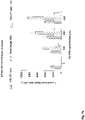
- the HR-FAB-MS spectrum of the obtained compound 4 is shown in FIG. 4 .
- the representative compound according to the present disclosure as prepared above may be used as an MRI contrast material, and at the same time, characteristic evaluation thereof was performed to determine whether or not the compound has a targeting ability to VGLUT1 (vesicular glutamate transporter 1) as one of the glutamate transporters.
- VGLUT1 vesicular glutamate transporter 1
- the compound Gd-RB obtained in the above preparation example was dissolved in dimethyl sulfoxide (DMSO)/water (5:5) at a concentration of 10 mM, and then diluted with water to obtain samples having five concentration levels between 0 and 1 mM. Three sample sets were prepared, and were used to obtain MRI images. The relaxivity thereof was measured using MRI of each of 3.0 T and 9.4 T, and the measurement of the T1 value was performed using the inversion recovery method. Specifically, at 3.0 T, more than 10 different inversion times (TI) in a range from 50 to 1800 msec were used in the FSE-IR (fast spin echo-inversion recovery) sequence.
- TI inversion times
- T2 measurement was performed using different echo time (TE) values while applying a CPMG (Carr-Purcell-Meiboon-Gill) pulse sequence to multiple spin-echo measurements. Specifically, the T2 was measured using 10 or more different TEs in a range of 8.5 to 135 msec in the T2 MAP sequence at 3.0 T. The T2 was measured using 10 or more different TEs in a range of 10 to 700 msec in the MSME (multi slice multi echo) sequence at 9.4 T.
- TE echo time
- CPMG Carr-Purcell-Meiboon-Gill
- T1 and T2 relaxation times were obtained from the non-linear least-square of mean pixel values for multiple spin-echo measurements of each echo time. Then, the relaxivity (R1 and R2) was calculated as the inverse of the relaxation time per mM. Finally, image-work was done with the calculated relaxation times (T1 and T2) and relaxivity (R1 and R2) to create a relaxation time map and a relaxivity map, respectively, and the relaxivity (r1 and r2) were obtained. The results are shown in the following [Table 1]. Table 1 r 1 (mM -1 s -1 ) r 2 (mM -1 s -1 ) 3 T 5.42 9.14 9.4 T 2.78 14.44
- the obtained cells were suspended in neurobasal media containing 1 ⁇ N2 supplement, 1 ⁇ B27, 4 mM L-glutamine, and 1% antibiotics at 7 ⁇ 10 6 cells per E-tube.
- Gd-RB or Gadovist was added to each E-tube at a concentration of 0, 50, 100, 200, 400 ⁇ M, and reacted at 37°C in a shaker for 24 hours. After 24 hours, centrifugation was performed at 4000 rpm and 4°C for 5 minutes to remove the supernatant. 1 mL of PBS was added thereto and the cells were released via pipetting. Centrifugation was repeated 3 times under the same conditions to remove substances which were not coupled to the cells. RIPA solution (150 uL) was added thereto.
- the cells were disrupted via vortexing at 4 °C for 1 hour at 15 minute intervals.
- 350 uL of PBS was added to the disrupted cells to produce a total sample of 500 uL, and MR imaging was performed thereon.
- MRI intensity was analyzed using ImageJ program.
- the MR phantom image performed in 2)-(1) is shown in FIG. 6A
- the change in the phantom signal intensity based on the concentration of each of the compound GD-RB8 according to the present disclosure and the commercial contrast agent Gadovist is shown as a graph in FIG. 6B .
- a control is directed to a sample containing only cells and no other contrast agent.
- FIG. 6A and FIG. 6B when Gadovist was applied to a certain number of cells, there was little difference in brightness based on the Gd concentration.
- the prepared compound Gd-RB according to the present disclosure was applied thereto based on each Gd concentration, the brightness of the phantom increased as the Gd concentration increased.
- the compound Gd-RB binds to VGLUT1 in the presence of VGLUT1, and that the binding amount increases in proportion to the concentration of the compound Gd-RB.
- the change in signal intensity based on the Gd concentration is expressed as a graph based on a measuring result of the signal intensity in each phantom.
- the primary cultured cells were seeded in a Cell Culture Slide 4well (cat. No. 30104, SPL) based on Neurobasal TM Medium (cat. No. 21103049, Gibco) containing 4mM L-glutamine at 4 ⁇ 10 5 per well. After incubation for 24 hours, the cells were cultured in Neurobasal TM Medium that does not contain L-glutamine while the light is blocked. The compound Gd-RB was applied thereto at each of 0, 100, 200, and 400 uM concentration for 10 minutes, and the cells were washed 3 times using the DPBS buffer.
- the cells were fixed in 4% paraformaldehyde (PFA) for 15 minutes, washed with TBS buffer 3 times for 5 minutes. Then, blocking was performed using TBS buffer containing 5% normal gout serum (NGS) and 5% bovine serum albumin (BSA) for 1 hour.
- VGLUT1 Polyclonal Antibody (Cat. No. 48-2400, invitrogen) was diluted with TBS buffer containing 5% NGS and 5% BSA at a ratio of 1:250 and reaction occurred overnight at 4 degrees C. The cells were 3 times washed for 15 minutes with TBS buffer.
- the secondary antibody, Goat anti-Rabbit Alexa Fluor 488 Cat. No.
- A-11008, invitrogen was diluted with TBS buffer containing 5% NGS and 5% BSA at a ratio of 1:500. The dark reaction was carried out for 1 hour and 30 minutes at 25 degrees C. After washing 3 times for 10 minutes with TBS buffer, mounting was performed with VECTASHIELD ® Phardset TM Antifade Mounting Medium with DAPI (Cat. No. H-1500 VECTOR), and images of fluorescently stained cells were obtained using lionphart equipment. A 20x objective lens was used. Regarding a fluorescence intensity, ROI values were analyzed using lionphart software.
- FIG. 7A shows a fluorescence image
- FIG. 7B shows a graph of each fluorescence intensity according to the compound Gd-RB concentration.
- Wild-type C57BL/6 mice were used as a 5XFAD transgenic AD mouse model and control under the approval of the Institutional Animal Care and Committee of the Daegu Gyeongbuk Advanced Medical Industry Promotion Foundation (DGMIF), and all in vivo experiments were performed according to approved protocols.
- DGMIF Daegu Gyeongbuk Advanced Medical Industry Promotion Foundation
- 12 months old male 5XFAD mice and C57BL/6 mice were anesthetized with isoflurane. After pre-image acquisition, 50 mM Gd-RB in DMSO was injected into cerebral ventricle (ICV) thereof at a dose of 0.1 mmol/kg and imaging was carried out for 3 hours.
- ICV cerebral ventricle
- mouse brains were harvested 3 hours after injection.
- OCT compound tissue-Tek, Sakura Finetek, USA
- the tissue was sectioned into several partial frozen-sections with a thickness of about 20 ⁇ m using a freezing microtome (Leica Biosystems, Wetzlar, Germany). The sections were mounted on microscope glass slides. For immunohistochemical staining, average 6 to 8 sections including the hippocampus per brain were used and analyzed blindly by the investigator. All sections including the hippocampus were collected in DPBS solution containing 0.1% sodium azide and stored at 4 °C.
- Brain sections were blocked with 3% goat serum (Gibco Co., Grand Island, NY, USA) and TBS containing 0.1% Triton X-100 and incubated with primary antibody (anti-VGLUT1) for 2 hours at 25 °C. Brain sections were then washed with TBS and were incubated in the presence of an IgG-labeled secondary antibody together with Alexa Fluor (Cell Signaling Technology Inc., Beverly, MA, USA) for 1 hour. The sections were mounted in DAPI solution for 10 min and then observed under a fluorescence microscope (ECLIPSE Ti, Nikon, NY, USA). Images were analyzed using NIS-Elements Basic Research imaging software (version 4.50).
- FIG. 8A shows the results of immunofluorescence staining of the brain as extracted 3 hours later (In other words, there is a change in signal intensity in more areas in normal mice; for example, in the drawing shown under the mark "Merge" in FIG.
- the area with the difference in intensity is indicated by a white arrow, and the number of the white arrows is larger in the normal mouse). That is, from FIG. 8B , it was identified that based on the results of immunofluorescence staining, the signals of VGLUT1 antibody (shown under the label "VGLUT1 antibody” in FIG. 8B ) and Gd-RB (shown under the label “Gd-RB” in FIG. 8B ) overlap each other, indicating that Gd-RB targets the VGLUT1 in in vivo experiments.
- Human-derived neuroblastoma cells (SH-SY5Y) were cultured in MEM (Minimum Essential Medium) containing 10% Fetal bovine serum (FBS), 4 mM glutamine, and 1% antibiotics at 1 ⁇ 10 4 per well of a 96-well plate. After attaching and stabilizing the cells for 24 hours, each of Gd-RB and Gadovist was diluted and applied at a concentration of 0, 50, 75, 100, 150, 200, 400 ⁇ M to 100 ⁇ l of growth medium per well, and the cells were incubated for 22 hours.
- MEM Minimum Essential Medium
- FBS Fetal bovine serum
- 4 mM glutamine 4 mM glutamine
- antibiotics 1 ⁇ 10 4 per well of a 96-well plate
- CCK-8 Cell Counting Kit-8, Dojindo Laboratories, Kumamoto, Japan
- the calculated values were graphed using the GraphPad Prism application. The statistical significance of the obtained values was identified via one-way ANOVA with Dunnett's multiple comparison test. The significance of ⁇ p ⁇ 0.05 ⁇ p ⁇ 0.01, ⁇ p ⁇ 0.001 vs. control is indicated.
- the cell viability obtained according to the above calculation method is shown in FIG. 9 as a graph based on the concentration of each of the compound Gd-RB and Gadovist.
Landscapes
- Health & Medical Sciences (AREA)
- Chemical & Material Sciences (AREA)
- Organic Chemistry (AREA)
- Nuclear Medicine, Radiotherapy & Molecular Imaging (AREA)
- Public Health (AREA)
- Epidemiology (AREA)
- Life Sciences & Earth Sciences (AREA)
- Animal Behavior & Ethology (AREA)
- General Health & Medical Sciences (AREA)
- Radiology & Medical Imaging (AREA)
- Veterinary Medicine (AREA)
- Medicinal Chemistry (AREA)
- Medicines Containing Antibodies Or Antigens For Use As Internal Diagnostic Agents (AREA)
Applications Claiming Priority (3)
| Application Number | Priority Date | Filing Date | Title |
|---|---|---|---|
| KR20190134992 | 2019-10-29 | ||
| KR1020200114620A KR102386595B1 (ko) | 2019-10-29 | 2020-09-08 | 신규한 가돌리늄계 화합물, 이의 제조 방법, 및 이를 함유하는 mri 조영제 |
| PCT/KR2020/013347 WO2021085875A1 (ko) | 2019-10-29 | 2020-09-29 | 신규한 가돌리늄계 화합물, 이의 제조 방법, 및 이를 함유하는 mri 조영제 |
Publications (3)
| Publication Number | Publication Date |
|---|---|
| EP4067364A1 true EP4067364A1 (de) | 2022-10-05 |
| EP4067364A4 EP4067364A4 (de) | 2023-08-30 |
| EP4067364B1 EP4067364B1 (de) | 2024-08-28 |
Family
ID=75715319
Family Applications (1)
| Application Number | Title | Priority Date | Filing Date |
|---|---|---|---|
| EP20881219.8A Active EP4067364B1 (de) | 2019-10-29 | 2020-09-29 | Neue verbindung auf gadoliniumbasis, verfahren zu ihrer herstellung und mrt-kontrastmittel damit |
Country Status (3)
| Country | Link |
|---|---|
| US (1) | US20220380386A1 (de) |
| EP (1) | EP4067364B1 (de) |
| WO (1) | WO2021085875A1 (de) |
Family Cites Families (2)
| Publication number | Priority date | Publication date | Assignee | Title |
|---|---|---|---|---|
| KR101469900B1 (ko) * | 2013-04-18 | 2014-12-09 | 경북대학교 산학협력단 | Do3a-디아미노바이페닐 화합물 및 이를 리간드로 포함하는 가돌리늄 착물 |
| CN106986920A (zh) * | 2017-03-09 | 2017-07-28 | 北京市心肺血管疾病研究所 | 四型胶原作为靶点在早期诊断主动脉瘤/夹层中的应用 |
-
2020
- 2020-09-29 US US17/755,143 patent/US20220380386A1/en active Pending
- 2020-09-29 WO PCT/KR2020/013347 patent/WO2021085875A1/ko unknown
- 2020-09-29 EP EP20881219.8A patent/EP4067364B1/de active Active
Also Published As
| Publication number | Publication date |
|---|---|
| US20220380386A1 (en) | 2022-12-01 |
| EP4067364A4 (de) | 2023-08-30 |
| WO2021085875A1 (ko) | 2021-05-06 |
| EP4067364B1 (de) | 2024-08-28 |
Similar Documents
| Publication | Publication Date | Title |
|---|---|---|
| Boros et al. | Gd (DOTAla): a single amino acid Gd-complex as a modular tool for high relaxivity MR contrast agent development | |
| US10759817B2 (en) | Gadolinium complex, method for synthesis of the gadolinium complex, and MRI contrast agent including the gadolinium complex | |
| EP4095142A1 (de) | Neuartige gadoliniumverbindung, herstellungsverfahren dafür und mrt-kontrastmittel damit | |
| KR102386595B1 (ko) | 신규한 가돌리늄계 화합물, 이의 제조 방법, 및 이를 함유하는 mri 조영제 | |
| Shao et al. | Identification of fatty liver disease at diverse stages using two-photon absorption of triphenylamine-based BODIPY analogues | |
| EP4067364B1 (de) | Neue verbindung auf gadoliniumbasis, verfahren zu ihrer herstellung und mrt-kontrastmittel damit | |
| US20230094602A1 (en) | Novel gadolinium-based compound, preparation method therefor, and mri contrast agent containing same | |
| US20230233714A1 (en) | Novel gadolinium-based compound, method for producing same, and mri contrast agent containing same | |
| Kim et al. | Rose bengal conjugated gadolinium complex as a new multimodal imaging agent targeting presynaptic vesicular glutamate transporters | |
| Hai et al. | Molecular fMRI | |
| KR102323277B1 (ko) | 신규한 가돌리늄계 화합물, 이를 함유하는 염증성 질환의 치료 또는 예방용 약제학적 조성물, 및 mri 조영 조성물 | |
| EP4095143A1 (de) | Neue verbindung auf gadoliniumbasis, verfahren zu ihrer herstellung und mrt-kontrastmittel damit | |
| EP3770149A1 (de) | Verbindung mit neuartiger struktur, entzündungshemmendes mittel damit und cyclooxygenase-2-inhibitor damit | |
| EP4374883A1 (de) | Gadoliniumbasierte verbindung und mrt-kontrastmittel damit | |
| EP3015855A1 (de) | Metall-Biosensoren auf Basis von Verbindungen mit metallempfindlichen chemischen Verschiebungen für magnetische Resonanzspektroskopie und Magnetresonanzbildgebung | |
| KR102668430B1 (ko) | 글루타티온 감응형 신규 화합물 및 이의 의학적 용도 | |
| KR102646267B1 (ko) | 가돌리늄계 화합물, 이를 포함하는 mri 조영제 | |
| KR102659253B1 (ko) | 가돌리늄계 화합물, 이를 포함하는 mri 조영제 | |
| EP4374881A1 (de) | Gadoliniumbasierte verbindung und mrt-kontrastmittel damit | |
| KR102683452B1 (ko) | 가돌리늄계 화합물, 이를 포함하는 mri 조영제 | |
| KR102667904B1 (ko) | 가돌리늄계 화합물, 이를 포함하는 mri 조영제 | |
| KR102659229B1 (ko) | 가돌리늄계 화합물, 이를 포함하는 mri 조영제 | |
| EP4374882A1 (de) | Gadoliniumbasierte verbindung und mrt-kontrastmittel damit | |
| DE10040380B4 (de) | Verwendung von perfluoralkylhaltigen Metallkomplexen als Kontrastmittel im MR-Imaging zur Darstellung von Plaques | |
| Wang | PH-Responsive MRI Agents that can be Activated Beyond the Tissue MT Window |
Legal Events
| Date | Code | Title | Description |
|---|---|---|---|
| STAA | Information on the status of an ep patent application or granted ep patent |
Free format text: STATUS: THE INTERNATIONAL PUBLICATION HAS BEEN MADE |
|
| PUAI | Public reference made under article 153(3) epc to a published international application that has entered the european phase |
Free format text: ORIGINAL CODE: 0009012 |
|
| STAA | Information on the status of an ep patent application or granted ep patent |
Free format text: STATUS: REQUEST FOR EXAMINATION WAS MADE |
|
| 17P | Request for examination filed |
Effective date: 20220404 |
|
| AK | Designated contracting states |
Kind code of ref document: A1 Designated state(s): AL AT BE BG CH CY CZ DE DK EE ES FI FR GB GR HR HU IE IS IT LI LT LU LV MC MK MT NL NO PL PT RO RS SE SI SK SM TR |
|
| DAV | Request for validation of the european patent (deleted) | ||
| DAX | Request for extension of the european patent (deleted) | ||
| P01 | Opt-out of the competence of the unified patent court (upc) registered |
Effective date: 20230526 |
|
| A4 | Supplementary search report drawn up and despatched |
Effective date: 20230728 |
|
| RIC1 | Information provided on ipc code assigned before grant |
Ipc: A61K 49/08 20060101ALI20230724BHEP Ipc: A61K 49/10 20060101ALI20230724BHEP Ipc: C07F 5/00 20060101AFI20230724BHEP |
|
| GRAP | Despatch of communication of intention to grant a patent |
Free format text: ORIGINAL CODE: EPIDOSNIGR1 |
|
| STAA | Information on the status of an ep patent application or granted ep patent |
Free format text: STATUS: GRANT OF PATENT IS INTENDED |
|
| INTG | Intention to grant announced |
Effective date: 20240410 |
|
| GRAS | Grant fee paid |
Free format text: ORIGINAL CODE: EPIDOSNIGR3 |
|
| GRAA | (expected) grant |
Free format text: ORIGINAL CODE: 0009210 |
|
| STAA | Information on the status of an ep patent application or granted ep patent |
Free format text: STATUS: THE PATENT HAS BEEN GRANTED |
|
| AK | Designated contracting states |
Kind code of ref document: B1 Designated state(s): AL AT BE BG CH CY CZ DE DK EE ES FI FR GB GR HR HU IE IS IT LI LT LU LV MC MK MT NL NO PL PT RO RS SE SI SK SM TR |
|
| REG | Reference to a national code |
Ref country code: CH Ref legal event code: EP |
|
| REG | Reference to a national code |
Ref country code: DE Ref legal event code: R096 Ref document number: 602020036858 Country of ref document: DE |