EP4063864A2 - Verfahren und mittel zum nachweis eines autoantikörpers - Google Patents
Verfahren und mittel zum nachweis eines autoantikörpers Download PDFInfo
- Publication number
- EP4063864A2 EP4063864A2 EP22163971.9A EP22163971A EP4063864A2 EP 4063864 A2 EP4063864 A2 EP 4063864A2 EP 22163971 A EP22163971 A EP 22163971A EP 4063864 A2 EP4063864 A2 EP 4063864A2
- Authority
- EP
- European Patent Office
- Prior art keywords
- seq
- polypeptide
- autoantibody
- antibody
- variant
- Prior art date
- Legal status (The legal status is an assumption and is not a legal conclusion. Google has not performed a legal analysis and makes no representation as to the accuracy of the status listed.)
- Pending
Links
Images



Classifications
-
- G—PHYSICS
- G01—MEASURING; TESTING
- G01N—INVESTIGATING OR ANALYSING MATERIALS BY DETERMINING THEIR CHEMICAL OR PHYSICAL PROPERTIES
- G01N33/00—Investigating or analysing materials by specific methods not covered by groups G01N1/00 - G01N31/00
- G01N33/48—Biological material, e.g. blood, urine; Haemocytometers
- G01N33/50—Chemical analysis of biological material, e.g. blood, urine; Testing involving biospecific ligand binding methods; Immunological testing
- G01N33/68—Chemical analysis of biological material, e.g. blood, urine; Testing involving biospecific ligand binding methods; Immunological testing involving proteins, peptides or amino acids
- G01N33/6854—Immunoglobulins
-
- G—PHYSICS
- G01—MEASURING; TESTING
- G01N—INVESTIGATING OR ANALYSING MATERIALS BY DETERMINING THEIR CHEMICAL OR PHYSICAL PROPERTIES
- G01N33/00—Investigating or analysing materials by specific methods not covered by groups G01N1/00 - G01N31/00
- G01N33/48—Biological material, e.g. blood, urine; Haemocytometers
- G01N33/50—Chemical analysis of biological material, e.g. blood, urine; Testing involving biospecific ligand binding methods; Immunological testing
- G01N33/53—Immunoassay; Biospecific binding assay; Materials therefor
- G01N33/564—Immunoassay; Biospecific binding assay; Materials therefor for pre-existing immune complex or autoimmune disease, i.e. systemic lupus erythematosus, rheumatoid arthritis, multiple sclerosis, rheumatoid factors or complement components C1-C9
-
- C—CHEMISTRY; METALLURGY
- C07—ORGANIC CHEMISTRY
- C07K—PEPTIDES
- C07K14/00—Peptides having more than 20 amino acids; Gastrins; Somatostatins; Melanotropins; Derivatives thereof
- C07K14/435—Peptides having more than 20 amino acids; Gastrins; Somatostatins; Melanotropins; Derivatives thereof from animals; from humans
- C07K14/46—Peptides having more than 20 amino acids; Gastrins; Somatostatins; Melanotropins; Derivatives thereof from animals; from humans from vertebrates
- C07K14/47—Peptides having more than 20 amino acids; Gastrins; Somatostatins; Melanotropins; Derivatives thereof from animals; from humans from vertebrates from mammals
-
- C—CHEMISTRY; METALLURGY
- C07—ORGANIC CHEMISTRY
- C07K—PEPTIDES
- C07K16/00—Immunoglobulins [IG], e.g. monoclonal or polyclonal antibodies
- C07K16/18—Immunoglobulins [IG], e.g. monoclonal or polyclonal antibodies against material from animals or humans
-
- G—PHYSICS
- G01—MEASURING; TESTING
- G01N—INVESTIGATING OR ANALYSING MATERIALS BY DETERMINING THEIR CHEMICAL OR PHYSICAL PROPERTIES
- G01N33/00—Investigating or analysing materials by specific methods not covered by groups G01N1/00 - G01N31/00
- G01N33/48—Biological material, e.g. blood, urine; Haemocytometers
- G01N33/50—Chemical analysis of biological material, e.g. blood, urine; Testing involving biospecific ligand binding methods; Immunological testing
- G01N33/53—Immunoassay; Biospecific binding assay; Materials therefor
- G01N33/543—Immunoassay; Biospecific binding assay; Materials therefor with an insoluble carrier for immobilising immunochemicals
- G01N33/54306—Solid-phase reaction mechanisms
-
- G—PHYSICS
- G01—MEASURING; TESTING
- G01N—INVESTIGATING OR ANALYSING MATERIALS BY DETERMINING THEIR CHEMICAL OR PHYSICAL PROPERTIES
- G01N33/00—Investigating or analysing materials by specific methods not covered by groups G01N1/00 - G01N31/00
- G01N33/48—Biological material, e.g. blood, urine; Haemocytometers
- G01N33/50—Chemical analysis of biological material, e.g. blood, urine; Testing involving biospecific ligand binding methods; Immunological testing
- G01N33/68—Chemical analysis of biological material, e.g. blood, urine; Testing involving biospecific ligand binding methods; Immunological testing involving proteins, peptides or amino acids
- G01N33/6875—Nucleoproteins
-
- G—PHYSICS
- G01—MEASURING; TESTING
- G01N—INVESTIGATING OR ANALYSING MATERIALS BY DETERMINING THEIR CHEMICAL OR PHYSICAL PROPERTIES
- G01N33/00—Investigating or analysing materials by specific methods not covered by groups G01N1/00 - G01N31/00
- G01N33/48—Biological material, e.g. blood, urine; Haemocytometers
- G01N33/50—Chemical analysis of biological material, e.g. blood, urine; Testing involving biospecific ligand binding methods; Immunological testing
- G01N33/68—Chemical analysis of biological material, e.g. blood, urine; Testing involving biospecific ligand binding methods; Immunological testing involving proteins, peptides or amino acids
- G01N33/6893—Chemical analysis of biological material, e.g. blood, urine; Testing involving biospecific ligand binding methods; Immunological testing involving proteins, peptides or amino acids related to diseases not provided for elsewhere
-
- C—CHEMISTRY; METALLURGY
- C07—ORGANIC CHEMISTRY
- C07K—PEPTIDES
- C07K2317/00—Immunoglobulins specific features
- C07K2317/30—Immunoglobulins specific features characterized by aspects of specificity or valency
- C07K2317/34—Identification of a linear epitope shorter than 20 amino acid residues or of a conformational epitope defined by amino acid residues
-
- G—PHYSICS
- G01—MEASURING; TESTING
- G01N—INVESTIGATING OR ANALYSING MATERIALS BY DETERMINING THEIR CHEMICAL OR PHYSICAL PROPERTIES
- G01N2800/00—Detection or diagnosis of diseases
- G01N2800/10—Musculoskeletal or connective tissue disorders
- G01N2800/101—Diffuse connective tissue disease, e.g. Sjögren, Wegener's granulomatosis
-
- G—PHYSICS
- G01—MEASURING; TESTING
- G01N—INVESTIGATING OR ANALYSING MATERIALS BY DETERMINING THEIR CHEMICAL OR PHYSICAL PROPERTIES
- G01N2800/00—Detection or diagnosis of diseases
- G01N2800/10—Musculoskeletal or connective tissue disorders
- G01N2800/101—Diffuse connective tissue disease, e.g. Sjögren, Wegener's granulomatosis
- G01N2800/102—Arthritis; Rheumatoid arthritis, i.e. inflammation of peripheral joints
-
- G—PHYSICS
- G01—MEASURING; TESTING
- G01N—INVESTIGATING OR ANALYSING MATERIALS BY DETERMINING THEIR CHEMICAL OR PHYSICAL PROPERTIES
- G01N2800/00—Detection or diagnosis of diseases
- G01N2800/10—Musculoskeletal or connective tissue disorders
- G01N2800/101—Diffuse connective tissue disease, e.g. Sjögren, Wegener's granulomatosis
- G01N2800/104—Lupus erythematosus [SLE]
-
- G—PHYSICS
- G01—MEASURING; TESTING
- G01N—INVESTIGATING OR ANALYSING MATERIALS BY DETERMINING THEIR CHEMICAL OR PHYSICAL PROPERTIES
- G01N2800/00—Detection or diagnosis of diseases
- G01N2800/20—Dermatological disorders
-
- G—PHYSICS
- G01—MEASURING; TESTING
- G01N—INVESTIGATING OR ANALYSING MATERIALS BY DETERMINING THEIR CHEMICAL OR PHYSICAL PROPERTIES
- G01N2800/00—Detection or diagnosis of diseases
- G01N2800/24—Immunology or allergic disorders
Definitions
- ANAs are a group of autoantibodies that targets the nuclear components of mammalian cells. They are mainly associated with systemic autoimmune rheumatic diseases (SARD) and some of the ANAs are biomarkers for certain SARDs. For example, anti-double-strand DNA (dsDNA), anti-Sm and anti-ribosomal P are tightly associated with systemic lupus erythematosus (SLE), while anti-Scl70, anti-centromere and anti-RNA polymerase III are biomarkers for systemic sclerosis (SSc). Meanwhile, increasing evidence shows that elevated level of ANAs in cancer patients is significant in confronting tumor cells.
- SARD systemic autoimmune rheumatic diseases
- the immunoassay for the detection of ANA is dated back to 1948, Hargraves et al. discovered and applied lupus erythematosus (LE) cells in order to aid the diagnosis of SLE patients, though it was found later that the LE cell test was neither sensitive nor specific enough ( R. B. Conn, "Practice Parameter - The Lupus Erythematosus Cell Test: An Obsolete Test Now Superseded by Definitive Immunologic Tests," American Journal of Clinical Pathology, pp. 65-66, 1 January 1994 ).
- the indirect immunofluorescence assay (IFA) based on animal tissue substrates, such as kidney from rats, was established.
- HEp-2 HeLa-derived human epithelial type 2 cells, which show better sensitivity and specificity, were applied in the IFA.
- HEp-2 cells When compared to cryosection of animal tissues, HEp-2 cells have the advantage of a large nucleus and a better visibility of other organelles, high division rate and an expression profile with more than 100 autoantigens, which contributes to easier observation and interpretation of the results.
- a negative IFA result indicates a non-SARD condition while a positive result should be further differentiated with available monospecific tests.
- ANAs The occurrence of high titers of ANAs may be years before the development of clinical syndromes. Arbuckle and colleagues have reported that ANAs could be detected in 88% of SLE patients in a mean of 3.3 years before onset of the disease ( Arbuckle et al., "Development of Autoantibodies before the Clinical Onset of Systemic Lupus Erythematosus," N Engl J Med, pp. 349:1526-33, 2003 ). This demonstrates the significance of ANAs in early diagnosis of the disease and a possible specific treatment. However, the utility of ANA diagnostics differs from one to another. Some ANAs showed high disease specificity and are included in the diagnostic criteria and/or reflecting disease activity, while some others show weak association with SARD or even negative relations.
- ANAs like anti-Scl70
- anti-DFS70 certain ANAs like anti-DFS70 are negatively related with SARD. Therefore, additional effort is required to identify the unknown ANAs and to study the corresponding clinical association.
- Zonula occludens-1 (ZO-1), also known as Tight junction protein-1 (TJP1) is a 220-kD peripheral membrane protein that is encoded by the TJP1 gene in humans. It belongs to the family of zona occludens proteins (ZO-1, ZO-2, and ZO-3), which are tight junction-associated proteins and of which ZO-1 is the first to be cloned. It was first isolated in 1986 by Stevenson and Goodenough using a monoclonal antibody raised in rodent liver to recognize a 225-kD polypeptide in whole liver homogenates and in tight junction-enriched membrane fractions. TJP1 is specifically enriched at the tight junctions of epithelial and endothelial cells.
- Another problem underlying the present invention is to increase the sensitivity of diagnostics for the diagnosis of systemic autoimmune rheumatic diseases, preferably for distinguishing autoimmune types of systemic autoimmune rheumatic diseases.
- the patient has or is supposed to have a systemic autoimmune rheumatic disease, preferably SSc.
- a systemic autoimmune rheumatic disease preferably SSc.
- the problem underlying the present invention is solved by a carrier, preferably a solid carrier, comprising a polypeptide having the sequence SEQ ID NO1, SEQ ID NO11 or SEQ ID NO24 or a variant thereof, wherein preferably the polypeptide is immobilized on the carrier, and/or wherein the polypeptide is preferably a recombinant protein, more preferably a purified recombinant protein.
- the carrier comprises a polypeptide consisting of or comprising at least one of the sequences according to SEQ ID NO1, SEQ ID NO35, SEQ ID NO38, SEQ ID NO41, SEQ ID NO44, SEQ ID NO45, SEQ ID NO46, SEQ ID NO47, SEQ ID NO48, SEQ ID NO49, SEQ ID NO50, SEQ ID NO51, SEQ ID NO52, SEQ ID NO86, SEQ ID NO87, SEQ ID NO88, SEQ ID NO89, SEQ ID NO90, SEQ ID NO91, SEQ ID NO92 and SEQ ID NO93.
- the carrier according to the present invention is a solid carrier, wherein the polypeptide is immobilized on the carrier, and wherein the polypeptide is preferably a recombinant protein, more preferably a purified recombinant protein.
- the carrier according to the present invention is a test strip comprising spatially separate reagents which includes a polypeptide having the sequence SEQ ID NO1, SEQ ID NO11 or SEQ ID NO24 or a variant thereof, and one or more from the group comprising one or more calibrators and a reagent indicating the presence of a secondary antibody, preferably a secondary antibody recognizing IgG class antibodies.
- the problem underlying the present invention is solved by a use of an autoantibody according to the present invention for the diagnosis of a disease, preferably of a systemic autoimmune rheumatic disease, more preferably of systemic sclerosis.
- the use comprises the step detecting in a sample an autoantibody binding to a polypeptide having the sequence SEQ ID NO1, SEQ ID NO11 or SEQ ID NO24 or a variant thereof.
- the use comprises the step detecting in a sample an autoantibody binding to a polypeptide consisting of or comprising at least one of the sequences according to SEQ ID NO1, SEQ ID NO35, SEQ ID NO38, SEQ ID NO41, SEQ ID NO44, SEQ ID NO45, SEQ ID NO46, SEQ ID NO47, SEQ ID NO48, SEQ ID NO49, SEQ ID NO50, SEQ ID NO51, SEQ ID NO52, SEQ ID NO86, SEQ ID NO87, SEQ ID NO88, SEQ ID NO89, SEQ ID NO90, SEQ ID NO91, SEQ ID NO92 and SEQ ID NO93.
- the problem underlying the present invention is solved by a pharmaceutical composition or medical device, preferably diagnostic device, comprising a polypeptide having the sequence SEQ ID NO1, SEQ ID NO11 or SEQ ID NO24 or a variant thereof.
- the problem underlying the present invention is solved by a method comprising the step contacting a medical or diagnostic device comprising a polypeptide having the sequence SEQ ID NO1, SEQ ID NO11 or SEQ ID NO24 or a variant thereof with a buffered solution comprising an antibody binding specifically to said polypeptide having the sequence SEQ ID NO1, SEQ ID NO11 or SEQ ID NO24 or a variant thereof, which solution is not a sample from a patient in need of a diagnosis comprising an unknown concentration of said antibody, wherein preferably
- the problem underlying the present invention is solved by an ex vivo method for removing an autoantibody according to the present invention, from blood, preferably serum of a patient.
- one or more epitopes recognized by anti-NVL autoantibodies is or are located on one, two, three, four, five, six, seven or all C-terminal fragments according to SEQ ID NO86, SEQ ID NO87, SEQ ID NO88, SEQ ID NO89, SEQ ID NO90, SEQ ID NO91, SEQ ID NO92 and SEQ ID NO93.
- the teachings of the present invention are carried out using a polypeptide having the sequence of a polypeptide referred to such as H8-NVL[human](aa1-117) (SEQ ID NO49), H8-NVL[human](aa106-292) (SEQ ID NO50), H8-NVL[human](aa281-560) (SEQ ID NO51), H8-NVL[human](aa549-856) (SEQ ID NO52), or a variant thereof.
- teachings of the present invention are carried out using a polypeptide consisting of or comprising at least one of the sequences according to SEQ ID NO86, SEQ ID NO87, SEQ ID NO88, SEQ ID NO89, SEQ ID NO90, SEQ ID NO91, SEQ ID NO92 and SEQ ID NO93.
- teachings of the present invention are carried out using a polypeptide having the sequence of a polypeptide referred to such as H8-CD2BP2[human](aa117-260) (SEQ ID NO66), H8-CD2BP2[human](aa249-341) (SEQ ID NO67) or a variant thereof.
- the term "variant”, as used herein, may refer to at least one fragment of the full length sequence referred to, more specifically one or more amino acid or nucleic acid sequence which is, relative to the full-length sequence, truncated at one or both termini by one or more amino acids.
- a fragment comprises or encodes for a peptide having at least 6, 7, 8, 10, 12, 15, 20, 25, 50, 75, 100, 150, 200, 300, 400, 500, 600, 700, 800, 900, 1000, 1100, 1200, 1300, 1400, 1500, 1600 or 1650 successive amino acids of the original sequence or a variant thereof.
- polypeptide and variants thereof may, in addition, comprise chemical modifications, for example isotopic labels or covalent modifications such as glycosylation, phosphorylation, acetylation, decarboxylation, citrullination, methylation, hydroxylation and the like.
- chemical modifications for example isotopic labels or covalent modifications such as glycosylation, phosphorylation, acetylation, decarboxylation, citrullination, methylation, hydroxylation and the like.
- Any modification is designed such that it does not abolish the biological activity of the variant.
- the variant of the polypeptide has biological activity.
- biological activity is the ability to bind specifically to an autoantibody binding specifically to the autoantigen of interest, preferably the autoantigen having the sequence of SEQ ID NO1, SEQ ID NO11, SEQ ID NO24, SEQ ID NO10, SEQ ID NO23 or SEQ ID NO82 or a variant thereof as found preferably in a patient suffering from an autoimmune disease associated with such autoantibody, preferably SARD, more preferably SSc.
- a polypeptide provided or used according to the present invention such as a polypeptide having the sequence SEQ ID NO1, SEQ ID NO11 or SEQ ID NO24 or a variant thereof or an autoantibody according to the present invention is an isolated polypeptide, wherein the term "isolated” means that the polypeptide has been enriched compared to its state upon production using a biotechnological or synthetic approach and is preferably pure, i.e. at least 60, 70, 80, 90, 95 or 99 percent of the polypeptide in the respective liquid consists of said polypeptide as judged by SDS polyacrylamide gel electrophoresis followed by Coomassie blue staining and visual inspection.
- any polypeptide on a carrier used as a means to capture an antibody is pure.
- the term "overexpressing”, as used herein, means that the cell has been transfected with a nucleic acid, either transiently or stably in the sense that the nucleic acid has been incorporated in the genome of the cell, that comprises a nucleic acid sequence encoding a polypeptide having the sequence SEQ ID NO1, SEQ ID NO11 or SEQ ID NO24 or a variant thereof or another SARD-related autoantigen or a variant thereof under the control of a promotor.
- the cell overexpressing a polypeptide having the sequence SEQ ID NO1, SEQ ID NO11 or SEQ ID NO24 or a variant thereof may additionally overexpress a polypeptide to another SARD-related autoantigen or a variant thereof.
- the transfected cell expresses more polypeptide recognized by the autoantibody to be detected than the same type of cell normally would, probably at least 10, 20, 30, 50, 100, 200 or 500 % more as judged by quantitative Western Blot.
- the promotor may be an inducible promotor, which allows for the induction of expression by addition of an inducer.
- the person skilled in the art is familiar with protocols and vectors for transiently overexpressing a polypeptide in a eukaryotic cell, for example the pTriEx system from Novagen and with protocols and vectors for stably transfecting a eukaryotic cell, for example the pcDNA TM 4/TO vector system from Invitrogen.
- a fixed mammalian cell may be used.
- the term "fixed" cell refers to a cell that has been treated with a reactive chemical compound to the effect that the cell is no longer metabolically active, but still presents its epitopes for immunostaining with antibodies and their subsequent detection, for example by fluorescence.
- the reactive chemical compound is selected from the group comprising acetone, formalin, methanol and ethanol or mixtures thereof, preferably all of them.
- the person skilled in the art is familiar with protocols that may be used to prepare fixed cells. Essentially, the cell which is attached to a solid support is washed by using washing buffer, followed by contacting with the reactive compound, for example immersion. Pure acetone or formalin or aqueous dilutions of the reactive chemical compound may be used.
- the cell is on a carrier for microscopic immunofluorescence analysis.
- a carrier may be a glass slide.
- the cell on the glass slide may be covered with a mounting buffer.
- a mounting medium is a liquid which helps maintain a near physiological pH to maintain the molecular structure of any diagnostically relevant molecular and their epitopes, is compatible with the emission of a fluorescence signal and prevents a premature loss of fluorescence due to bleaching of the fluorophore.
- its optical properties are matched with other buffers used, in particular its refractive index which allows for an efficient microscopic fluorescence analysis.
- compositions and methods are described in the state of the art, for example in " Mountants and Antifades", published by Wright Cell Imaging Facility, Toronto Western Research Institute University Health Network, (https://de.scribd.com/document/47879592/Mountants-Antifades ), Krenek et al. (1989) Comparison of antifading agents used in immunofluorescence, J. Immunol. Meth 117, 91-97 and Nairn et al. (1969) Microphotometry in Immunofluorescence, Clin. Exp. Immunol. 4, 697-705 .
- a cover glass may be placed on top of the composition comprising the sample and the mounting medium.
- Slides with cover glasses (FB 112d-1005-1 or ZZ 3000-0112) are available from EUROIMMUN GmbH, Lübeck, Germany.
- any carrier compatible with microscopic analysis of the fluorescence pattern may be used.
- the carrier may comprise a mock-transfected cell, which has been transfected with the same vector as the cell overexpressing a polypeptide having the sequence SEQ ID NO1, SEQ ID NO11 or SEQ ID NO24 or a variant thereof, but without the nucleic acid encoding for the latter.
- Such mock-transfected cell may serve as a negative control.
- the carrier is configured for analysis using an immunofluorescence microscope.
- a secondary antibody is an antibody binding specifically to all antibodies from an antibody class, preferably a mammalian antibody class, more preferably human antibody class such as IgG.
- Secondary antibodies typically recognize the constant domain of said class, but may also recognize other epitopes shared by antibodies from the class of interest, for example a conformational epitope across the 3D structure. A wide range of them is commercially available, for example from Thermo Fisher. It may be a monoclonal or a polyclonal antibody.
- the term "recognized" means that the secondary antibody binds specifically to the antibody or antibodies to be detected.
- a secondary antibody may bind specifically to all isotypes from the antibody class.
- a secondary antibody to IgG class antibodies may bind to IgG1, IgG2, IgG3 and IgG4 isotypes.
- This may be achieved by using as a secondary antibody to the class, preferably to IgG class antibodies, a mixture comprising an antibody binding specifically to each IgG isotype or a single antibody which reacts with all isotypes of interest.
- the use of secondary antibodies is explained in Kruger, N. J., Detection of Polypeptides on Blots Using Secondary Antibodies, in The Protein Protocols Handbook (ed. J. M. Walker), page 967, volume 1996, Springer . Briefly, such secondary antibodies may be generated by immunizing a laboratory animal with the antibody to be recognized or a mixture of the antibodies to be recognized.
- binding specifically preferably means that the binding reaction is stronger than a binding reaction characterized by a dissociation constant of 1 ⁇ 10 -5 M, more preferably 1 ⁇ 10 -7 M, more preferably 1 ⁇ 10 -8 M, more preferably 1 ⁇ 10 -9 M, more preferably 1 ⁇ 10 -10 M, more preferably 1 ⁇ 10 -11 M, more preferably 1 ⁇ 10 -12 M, as determined by surface plasmon resonance using Biacore equipment at 25 °C in PBS buffer at pH 7.
- the term "diagnosis”, as used herein, is to be used in its broadest possible sense and may to any kind of procedure aiming to obtain information instrumental in the assessment whether a patient, known or an anonymous subject from a cohort, suffers or is likely or more likely than the average or a comparative subject, the latter preferably having similar symptoms, to suffer from certain a disease or disorder in the past, at the time of the diagnosis or in the future, to find out how the disease is progressing or is likely to progress in the future or to evaluate the responsiveness of a patient or patients in general with regard to a certain treatment, for example the administration of immunosuppressive drugs, or to find out whether a sample is from such a patient.
- a certain treatment for example the administration of immunosuppressive drugs
- the detection of an autoantibody to a polypeptide having the sequence SEQ ID NO1, SEQ ID NO11 or SEQ ID NO24 or a variant thereof is considered to imply a definitive diagnosis of SARD, preferably SSc, because of the presence of the autoantibody.
- the methods and products according to the present invention may be used for interaction studies, including determining whether a drug candidate or other compound may interfere with the binding of an autoantibody to a polypeptide having the sequence SEQ ID NO1, SEQ ID NO11 or SEQ ID NO24 or a variant thereof or may affect any downstream process or the strength of its binding to its target.
- any symptoms or diseases referred to throughout this application is in line with the person skilled in the art's understanding as of the filing date or, preferably, earliest priority date of this application as evidenced by text books and scientific publications. It should be mentioned that the inventive methods or uses or products, taken alone, cannot be used to arrive at a definite, final diagnosis.
- a device for removing an autoantibody to a polypeptide having the sequence SEQ ID NO1, SEQ ID NO11 or SEQ ID NO24 or a variant thereof, from blood, preferably serum of a SARD patient, preferably of an SSc patient, wherein the device comprises a carrier coated with a polypeptide having the sequence SEQ ID NO1, SEQ ID NO11 or SEQ ID NO24 or a variant thereof is provided as an ex vivo method for removing an autoantibody to a polypeptide having the sequence SEQ ID NO1, SEQ ID NO11 or SEQ ID NO24 or a variant thereof, from blood, preferably serum of a SARD patient, preferably of a SSc patient.
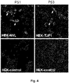
- Fig. 4 shows IFA with NVL-His, TJP1-His or control transfected HEK293 cells and PS1 (1:100) or PS3, respectively (1:10).
- Anti-human-IgG-Alexa488 (1:500) was applied as secondary antibody. Arrows indicate the specific fluorescence signal (magnification 200x).
- NNL Nuclear VCP-like protein
- CD2BP2 CD2 antigen cytoplasmic tail-binding protein 2
- TJP1 tight junction protein ZO-1
- PS1 and PS3 reacted with the individual antigen in recombinant HEK293 cell IFA.
- ANAs against NVL, CD2BP2 and TJP1 present novel markers for SARD, especially SSc, or other rheumatic diseases.
- the immunoprecipitation was performed with 500 ⁇ l cell HEp-2 cells (10 ⁇ 10 6 cells/ml) and 5 ⁇ l patient or control sera.
- HEp-2 cells were centrifuged at 16000 x g for 10 minutes at 4°C.
- the sediment together with patient or control sera was resuspended in 500 ⁇ l RIPA lysis buffer (PBS pH 7.4, 0.1% (v/v) Nonidet TM P 40, 1% (w/v) Deoxycholate, 0.1% (w/v) SDS, 1 mM Protease inhibitor cocktail [Sigma]) and rotated for 1 h at 4°C.
- the suspension was spun down at 16000 x g for 20 minutes at 4°C.
- the Mascot search engine Mascot Server 2.3 (Matrix Science, London, UK) was used for protein identification by searching against the NCBI or SwissProt database limited to Mammalia. Search parameters were as follows: Mass tolerance was set to 80 ppm, one missed cleavage site was accepted, and carbamidomethylation of cysteine residues as well as oxidation of methionine residues were set as fixed and variable modifications, respectively. To evaluate the protein hits, a significance threshold of p ⁇ 0.05 was chosen.
- the cDNA clone IRAUp969F0866D encoding a partial sequence (amino acid 206-856) of human NVL-isoform1 (SEQ ID NO 1) was obtained from Source BioScience UK Limited. This cDNA clone is coding NVL-isoform3 (Acc. BC012105, SEQ ID NO 2). A synthetic DNA fragment was ordered coding for the missing amino acids 1-205 of NVL-isoform1 from Eurofins Genomics Germany GmbH (Ebersberg). The gene synthesis product (SEQ ID NO 3) was digested with Bsal (R3733, New England Biolabs, Frankfurt am Main, Germany) and the 614 bp fragment was isolated by Gel-purification.
- the amplification product was digested with Bsal and together with the gene synthesis fragment ligated with Ncol and Xhol linearized pTriEx-1 vector (Merck, Darmstadt, Germany) resulting in SEQ ID NO 7, coding for NVL[human]-IF1 fused to an octa Histidin-tag (H8) (SEQ ID NO 8) or pET24d vector (Merck) resulting in SEQ ID NO 9, coding for NVL[human]-IF1 fused to a hexa Histidin-tag (H6) (SEQ ID NO 10).
- Both gene synthesis products (SEQ ID NO 13 and SEQ ID NO 14) were digested with Bbsl (R3539, New England Biolabs) resulting in 517 bp fragment-1 and 510 bp fragment-2 (SEQ ID NO 15 and SEQ ID NO 16) that were both Gel-purified.
- CD2BP2 was amplified using the gene synthesis product (SEQ ID NO 14) as template and the DNA oligonucleotide primers sense CD2BP2-fragment-2 (SEQ ID NO 17) and asense CD2BP2-fragment-2 (SEQ ID NO 18), resulting in SEQ ID NO 19 without the Stop-codon.
- the amplification product was digested with Bbsl (R3539, New England Biolabs).
- the fragments SEQ ID NO 15 and SEQ ID NO 16 were ligated with Ncol and Xhol linearized pTriEx-1 vector (Merck) resulting in SEQ ID NO 20, coding for CD2BP2human] fused to a C-terminal octa Histidin-tag (H8) (SEQ ID NO 21).
- the fragment SEQ ID NO 15 and the Bbsl-digested fragment SEQ ID NO 19 were ligated with Ncol and Xhol linearized pET24d vector (Merck) resulting in SEQ ID NO 22, coding for CD2BP2human] fused to a N-terminal octa Histidin-tag (H8) (SEQ ID NO 23).
- SEQ ID NO 29 One fragment was digested with Bsal (R3733, New England Biolabs) and the other fragment (SEQ ID NO 30) was digested with Esp3I (R0734, New England Biolabs). Both fragments were ligated with Ncol and Xhol linearized pTriEx-1 vector (Merck) resulting in SEQ ID NO 31, coding for human TJP1-isoform2 fused to a C-terminal octa Histidin-tag (H8) (SEQ ID NO 32).
- NVL-His and TJP1-His were expressed in the human cell line HEK293 after ExGen500-mediated transfection (ThermoFisher Scientific, Dreieich, Germany) according to the manufacturer's instructions (Biomol GmbH, Hamburg, Germany).
- ExGen500-mediated transfection ThermoFisher Scientific, Dreieich, Germany
- Biomol GmbH Hamburg, Germany
- immunofluorescence substrates the cells were grown on cover slides and acetone fixed two days after transfection.
- cells were harvested 5 d after transfection and lysed with NuPage LDS sample buffer (ThermoFisher Scientific, Schrö, Germany).
- the amplification products were digested with Bsal (R3733, New England Biolabs) and ligated with Ncol and Xhol linearized pET24d vector (Merck) resulting in pET24d-N-TJP1[human](aa1-575) (SEQ ID NO 78), pET24d-N-TJP1[human](aa1-156) (SEQ ID NO 79), pET24d-N-TJP1[human](aa144-306) (SEQ ID NO 80) and pET24d-N-TJP1[human](aa294-575) (SEQ ID NO 81) coding for four proteins fused to a N-terminal octa Histidin-tag (H8) (SEQ ID NO 82 to SEQ ID NO 85).
- Bacterial expression was essentially performed as described in the pET system manual (Merck) employing E. coli strain RosettaBlue(DE3)pLacl (Merck). Protein expression was induced by the addition of 2 mM isopropyl ⁇ -D-thiogalactoside (IPTG) for 3 hours at 37°C.
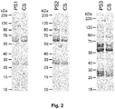
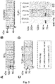
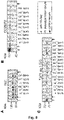
- Purified recombinant antigens (SEQ ID NO 10, 23, 82) in the respective buffer or recombinant antigen fragments (SEQ ID NO 49, 50, 51, 52, 66, 67, 83, 84, 85) expressing E.coli or recombinant antigens (SEQ ID NO 32) containing HEK-cell lysates were incubated with NuPage LDS sample buffer (ThermoFisher Scientific, Schrö, Germany) containing 25 mmol/L dithiothreitol at 70°C for 10 min, followed by SDS-PAGE (NuPAGE, ThermoFisher Scientific).
- the magnification of the selected area shows that autoantibodies from PS3 also localized in the area of intercellular contacts, indicating that PS3 recognizes, besides of an unknown nuclear antigen, endogenous TJP1 at cell junctions.
Landscapes
- Health & Medical Sciences (AREA)
- Life Sciences & Earth Sciences (AREA)
- Immunology (AREA)
- Chemical & Material Sciences (AREA)
- Engineering & Computer Science (AREA)
- Molecular Biology (AREA)
- Hematology (AREA)
- Urology & Nephrology (AREA)
- Biomedical Technology (AREA)
- Biochemistry (AREA)
- General Health & Medical Sciences (AREA)
- Medicinal Chemistry (AREA)
- Physics & Mathematics (AREA)
- Analytical Chemistry (AREA)
- Biotechnology (AREA)
- Cell Biology (AREA)
- Microbiology (AREA)
- Pathology (AREA)
- Food Science & Technology (AREA)
- General Physics & Mathematics (AREA)
- Proteomics, Peptides & Aminoacids (AREA)
- Organic Chemistry (AREA)
- Biophysics (AREA)
- Genetics & Genomics (AREA)
- Rehabilitation Therapy (AREA)
- Rheumatology (AREA)
- Chemical Kinetics & Catalysis (AREA)
- Toxicology (AREA)
- Zoology (AREA)
- Gastroenterology & Hepatology (AREA)
- Peptides Or Proteins (AREA)
Applications Claiming Priority (1)
| Application Number | Priority Date | Filing Date | Title |
|---|---|---|---|
| EP21165011 | 2021-03-25 |
Publications (2)
| Publication Number | Publication Date |
|---|---|
| EP4063864A2 true EP4063864A2 (de) | 2022-09-28 |
| EP4063864A3 EP4063864A3 (de) | 2022-11-16 |
Family
ID=75252296
Family Applications (1)
| Application Number | Title | Priority Date | Filing Date |
|---|---|---|---|
| EP22163971.9A Pending EP4063864A3 (de) | 2021-03-25 | 2022-03-24 | Verfahren und mittel zum nachweis eines autoantikörpers |
Country Status (5)
| Country | Link |
|---|---|
| US (1) | US20220317121A1 (de) |
| EP (1) | EP4063864A3 (de) |
| CN (1) | CN115128275A (de) |
| CA (1) | CA3151417A1 (de) |
| TW (1) | TW202305362A (de) |
Citations (1)
| Publication number | Priority date | Publication date | Assignee | Title |
|---|---|---|---|---|
| EP3477300A1 (de) | 2017-10-25 | 2019-05-01 | Euroimmun Medizinische Labordiagnostika AG | Zellulose-basierter immunadsorber |
Family Cites Families (1)
| Publication number | Priority date | Publication date | Assignee | Title |
|---|---|---|---|---|
| JP2010510528A (ja) * | 2006-11-22 | 2010-04-02 | ライフ テクノロジーズ コーポレーション | 自己免疫疾患のバイオマーカー |
-
2022
- 2022-03-08 CA CA3151417A patent/CA3151417A1/en active Pending
- 2022-03-18 TW TW111110136A patent/TW202305362A/zh unknown
- 2022-03-23 US US17/701,779 patent/US20220317121A1/en active Pending
- 2022-03-24 CN CN202210298165.6A patent/CN115128275A/zh active Pending
- 2022-03-24 EP EP22163971.9A patent/EP4063864A3/de active Pending
Patent Citations (1)
| Publication number | Priority date | Publication date | Assignee | Title |
|---|---|---|---|---|
| EP3477300A1 (de) | 2017-10-25 | 2019-05-01 | Euroimmun Medizinische Labordiagnostika AG | Zellulose-basierter immunadsorber |
Non-Patent Citations (23)
| Title |
|---|
| "The Immunoassay Handbook", 2005, ELSEVIER |
| "Uniprot", Database accession no. Q07157-2 |
| ALBERT ET AL.: "The GYF domain protein CD2BP2 is critical for embryogenesis and podocyte function", JOURNAL OF MOLECULAR CELL BIOLOGY, vol. 7, no. 5, 16 June 2015 (2015-06-16), pages 402 - 414 |
| ARBUCKLE ET AL.: "Development of Autoantibodies before the Clinical Onset of Systemic Lupus Erythematosus", N ENGL J MED, vol. 349, 2003, pages 1526 - 33, XP008060325, DOI: 10.1056/NEJMoa021933 |
| ARTHURLESK: "Introduction to bioinformatics", 2008, OXFORD UNIVERSITY PRESS |
| BROWN T. A.: "Gene Cloning - an introduction", 1986, CHAPMAN & HALL |
| C. PROBSTW. SCHLUMBERGERW. STOCKER ET AL.: "Development of ELISA for the specific determination of autoantibodies against envoplakin and periplakin in paraneoplastic pemphigus", CLINICA CHIMICA ACTA, vol. 410, no. 1-2, 2009, pages 13 - 18, XP026738580, DOI: 10.1016/j.cca.2009.08.022 |
| EISEI NOIRINORIA HANAFUSA: "The Concise Manual of Apharesis Therapy", 2014, SPRINGER TOKYO |
| FUJIWARA ET AL.: "Structure and Function of the N-terminal Nucleolin Binding Domain of Nuclear Valosin-containing Protein-like 2 (NVL2) Harboring a Nucleolar Localization Signal", JOURNAL OF BIOLOGICAL CHEMISTRY, 17 June 2011 (2011-06-17), pages 21732 - 21741 |
| HAMILTON, P., KANIGICHERLA, D.HANUMAPURA, P.WALZ, L.KRAMER, D.FISCHER, M.BRENCHLEY, P.MITRA, S., J. CLIN. APH., vol. 33, no. 3, 2018, pages 283 - 290 |
| HEINZE: "Investigating the functional role of CD2BP2 in T cells", INTERNATIONAL IMMUNOLOGY, vol. 19, no. 11, November 2007 (2007-11-01), pages 1313 - 1318 |
| HOU: "The Paracellular Channel,", 2019, ACADEMIC PRESS, article "Paracellular Channel Formation", pages: 9 - 27 |
| KAVANAUGH ET AL.: "Guidelines for Clinical Use of the Antinuclear Antibody Test and Tests for Specific Autoantibodies to Nuclear Antigens", ARCHIVES OF PATHOLOGY & LABORATORY MEDICINE, vol. 124, no. 1, August 2000 (2000-08-01), pages 71 - 81 |
| KRENEK ET AL.: "Comparison of antifading agents used in immunofluorescence", J. IMMUNOL. METH, vol. 117, 1989, pages 91 - 97 |
| KRUGER, N. J.: "The Protein Protocols Handbook", vol. 1996, SPRINGER, article "Detection of Polypeptides on Blots Using Secondary Antibodies", pages: 967 |
| LARKIN, M. A.BLACKSHIELDS, G.BROWN, N. P.CHENNA, R.MCGETTIGAN, P. A.MCWILLIAM, H.VALENTIN, F.WALLACE, I. M.WILM, A.LOPEZ, R.: "Clustal Wand Clustal X version 2.0", BIOINFORMATICS, vol. 23, 2007, pages 2947 - 2948 |
| LUDWIG ET AL.: "Mechanisms of autoantibody-induced pathology", FRONTIERS IN IMMUNOLOGY, vol. 8, 31 May 2017 (2017-05-31), pages 603 |
| NAIRN ET AL.: "Microphotometry in Immunofluorescence", CLIN. EXP. IMMUNOL., vol. 4, 1969, pages 697 - 705 |
| NIELSEN ET AL.: "Structural Basis for the Bifunctionality of the U5 snRNP 52K Protein (CD2BP2", JOURNAL OF MOLECULAR BIOLOGY, vol. 369, no. 4, 4 April 2007 (2007-04-04), pages 902 - 908, XP022083568, DOI: 10.1016/j.jmb.2007.03.077 |
| R. B. CONN: "Practice Parameter - The Lupus Erythematosus Cell Test: An Obsolete Test Now Superseded by Definitive Immunologic Tests", AMERICAN JOURNAL OF CLINICAL PATHOLOGY, 1 January 1994 (1994-01-01), pages 65 - 66 |
| RIPKE ET AL.: "A mega-analysis of genome-wide association studies for major depressive disorder", MOLECULAR PSYCHIATRY, April 2013 (2013-04-01), pages 497 - 511, XP055075174, DOI: 10.1038/mp.2012.21 |
| TAN ET AL.: "Characteristics of a Soluble Nuclear Antigen Precipitating with Sera of Patients with Systemic Lupus Erythematosus", J IMMUNOL, vol. 96, no. 3, 1 March 1966 (1966-03-01), pages 464 - 471 |
| WANG ET AL.: "The NVL gene confers risk for both major depressive disorder and schizophrenia in the Han Chinese population", PROGRESS IN NEURO-PSYCHOPHARMACOLOGY AND BIOLOGICAL PSYCHIATRY, 1 October 2015 (2015-10-01), pages 7 - 13 |
Also Published As
| Publication number | Publication date |
|---|---|
| CN115128275A (zh) | 2022-09-30 |
| TW202305362A (zh) | 2023-02-01 |
| US20220317121A1 (en) | 2022-10-06 |
| CA3151417A1 (en) | 2022-09-25 |
| EP4063864A3 (de) | 2022-11-16 |
Similar Documents
| Publication | Publication Date | Title |
|---|---|---|
| US10670597B2 (en) | Methods and kits for monitoring membranous nephropathy | |
| US8148084B2 (en) | Diagnosis of autoimmune disease | |
| JP5998318B2 (ja) | 新規血管炎の検査方法および検査用試薬 | |
| EP3018478B1 (de) | Neuartiger, diagnostisch relevanter autoantikörper | |
| EP3392656A1 (de) | Diagnose einer neuroautoimmunkrankheit | |
| JP7326108B2 (ja) | 自己免疫性水疱症の診断 | |
| CN110824156B (zh) | 神经自身免疫疾病的诊断 | |
| EP3842802A1 (de) | Verfahren zum nachweis eines autoantikörpers gegen den acetylcholinrezeptor | |
| EP2668502B1 (de) | Verfahren und mittel zur diagnose von vaskulitis | |
| US12442823B2 (en) | Method and reagents for diagnosing membranous nephropathy | |
| EP4080215A2 (de) | Verfahren und reagenzien zum nachweis eines autoantikörpers | |
| EP4063864A2 (de) | Verfahren und mittel zum nachweis eines autoantikörpers | |
| CN108948153A (zh) | 一种瓜氨酸修饰肽抗原组合及其应用 | |
| WO2013008930A1 (ja) | 皮膚筋炎の検査方法 | |
| US10989712B2 (en) | Diagnosis of a neuroautoimmune disease | |
| JP5211315B2 (ja) | 腫瘍マーカ、腫瘍診断キットおよび腫瘍マーカの測定方法 | |
| US8895697B2 (en) | Isolated monophosphorylated peptide derived from human alpha-enolase useful for diagnosis and treatment of pancreatic adenocarcinoma, antibodies directed against the said monophosphorylated peptide, and uses thereof | |
| US20080219981A1 (en) | Diagnostic Kit for Solid Cancer and Medicament for Solid Cancer Therapy | |
| US20040180385A1 (en) | Method of diagnosing systemic lupus erythematosus | |
| CN119064588A (zh) | 用于检测哺乳动物抗体的方法和试剂 | |
| JP2012224594A (ja) | 環状シトルリン化グルコース−6−リン酸イソメラーゼペプチドおよび関節リウマチ症診断方法 |
Legal Events
| Date | Code | Title | Description |
|---|---|---|---|
| PUAI | Public reference made under article 153(3) epc to a published international application that has entered the european phase |
Free format text: ORIGINAL CODE: 0009012 |
|
| STAA | Information on the status of an ep patent application or granted ep patent |
Free format text: STATUS: THE APPLICATION HAS BEEN PUBLISHED |
|
| AK | Designated contracting states |
Kind code of ref document: A2 Designated state(s): AL AT BE BG CH CY CZ DE DK EE ES FI FR GB GR HR HU IE IS IT LI LT LU LV MC MK MT NL NO PL PT RO RS SE SI SK SM TR |
|
| PUAL | Search report despatched |
Free format text: ORIGINAL CODE: 0009013 |
|
| AK | Designated contracting states |
Kind code of ref document: A3 Designated state(s): AL AT BE BG CH CY CZ DE DK EE ES FI FR GB GR HR HU IE IS IT LI LT LU LV MC MK MT NL NO PL PT RO RS SE SI SK SM TR |
|
| RIC1 | Information provided on ipc code assigned before grant |
Ipc: G01N 33/68 20060101ALI20221007BHEP Ipc: C07K 16/00 20060101ALI20221007BHEP Ipc: C07K 14/00 20060101ALI20221007BHEP Ipc: G01N 33/564 20060101AFI20221007BHEP |
|
| STAA | Information on the status of an ep patent application or granted ep patent |
Free format text: STATUS: REQUEST FOR EXAMINATION WAS MADE |
|
| 17P | Request for examination filed |
Effective date: 20230420 |
|
| RBV | Designated contracting states (corrected) |
Designated state(s): AL AT BE BG CH CY CZ DE DK EE ES FI FR GB GR HR HU IE IS IT LI LT LU LV MC MK MT NL NO PL PT RO RS SE SI SK SM TR |
|
| STAA | Information on the status of an ep patent application or granted ep patent |
Free format text: STATUS: EXAMINATION IS IN PROGRESS |
|
| 17Q | First examination report despatched |
Effective date: 20260306 |