EP4043555A1 - Use of rhodococcus ruber cell wall skeleton in regenerative medicine - Google Patents
Use of rhodococcus ruber cell wall skeleton in regenerative medicine Download PDFInfo
- Publication number
- EP4043555A1 EP4043555A1 EP21744220.1A EP21744220A EP4043555A1 EP 4043555 A1 EP4043555 A1 EP 4043555A1 EP 21744220 A EP21744220 A EP 21744220A EP 4043555 A1 EP4043555 A1 EP 4043555A1
- Authority
- EP
- European Patent Office
- Prior art keywords
- stem cells
- mesenchymal stem
- rhodococcus ruber
- cell wall
- product derived
- Prior art date
- Legal status (The legal status is an assumption and is not a legal conclusion. Google has not performed a legal analysis and makes no representation as to the accuracy of the status listed.)
- Pending
Links
- 241000187563 Rhodococcus ruber Species 0.000 title claims abstract description 143
- 108010039939 Cell Wall Skeleton Proteins 0.000 title abstract description 19
- 210000004520 cell wall skeleton Anatomy 0.000 title abstract description 19
- 239000003814 drug Substances 0.000 title description 26
- 230000001172 regenerating effect Effects 0.000 title description 2
- 210000002901 mesenchymal stem cell Anatomy 0.000 claims abstract description 103
- 210000000130 stem cell Anatomy 0.000 claims abstract description 83
- 210000004504 adult stem cell Anatomy 0.000 claims abstract description 31
- 230000001737 promoting effect Effects 0.000 claims abstract description 20
- 230000004069 differentiation Effects 0.000 claims abstract description 15
- 230000035755 proliferation Effects 0.000 claims abstract description 14
- 230000004083 survival effect Effects 0.000 claims abstract description 14
- 230000012010 growth Effects 0.000 claims abstract description 6
- 230000005012 migration Effects 0.000 claims abstract description 5
- 238000013508 migration Methods 0.000 claims abstract description 5
- 210000002421 cell wall Anatomy 0.000 claims description 98
- 238000000034 method Methods 0.000 claims description 34
- 239000008194 pharmaceutical composition Substances 0.000 claims description 29
- 108090000623 proteins and genes Proteins 0.000 claims description 22
- 206010052428 Wound Diseases 0.000 claims description 21
- 208000027418 Wounds and injury Diseases 0.000 claims description 21
- 238000002360 preparation method Methods 0.000 claims description 20
- 102000004169 proteins and genes Human genes 0.000 claims description 19
- 230000029663 wound healing Effects 0.000 claims description 19
- 206010012601 diabetes mellitus Diseases 0.000 claims description 16
- XLYOFNOQVPJJNP-UHFFFAOYSA-N water Substances O XLYOFNOQVPJJNP-UHFFFAOYSA-N 0.000 claims description 16
- 239000006143 cell culture medium Substances 0.000 claims description 15
- 150000002632 lipids Chemical class 0.000 claims description 14
- 108020004707 nucleic acids Proteins 0.000 claims description 11
- 102000039446 nucleic acids Human genes 0.000 claims description 11
- 150000007523 nucleic acids Chemical class 0.000 claims description 11
- 241000316848 Rhodococcus <scale insect> Species 0.000 claims description 10
- 238000011166 aliquoting Methods 0.000 claims description 10
- 210000001185 bone marrow Anatomy 0.000 claims description 9
- 239000003153 chemical reaction reagent Substances 0.000 claims description 9
- 210000003074 dental pulp Anatomy 0.000 claims description 6
- 210000004700 fetal blood Anatomy 0.000 claims description 6
- 210000004185 liver Anatomy 0.000 claims description 6
- 210000004072 lung Anatomy 0.000 claims description 6
- 210000003205 muscle Anatomy 0.000 claims description 6
- 230000003169 placental effect Effects 0.000 claims description 6
- 210000003954 umbilical cord Anatomy 0.000 claims description 6
- 210000004027 cell Anatomy 0.000 description 76
- 239000000203 mixture Substances 0.000 description 66
- 239000000047 product Substances 0.000 description 58
- WQZGKKKJIJFFOK-GASJEMHNSA-N Glucose Natural products OC[C@H]1OC(O)[C@H](O)[C@@H](O)[C@@H]1O WQZGKKKJIJFFOK-GASJEMHNSA-N 0.000 description 18
- 230000006907 apoptotic process Effects 0.000 description 18
- 230000000694 effects Effects 0.000 description 18
- WQZGKKKJIJFFOK-VFUOTHLCSA-N beta-D-glucose Chemical compound OC[C@H]1O[C@@H](O)[C@H](O)[C@@H](O)[C@@H]1O WQZGKKKJIJFFOK-VFUOTHLCSA-N 0.000 description 17
- 241000699670 Mus sp. Species 0.000 description 16
- 239000008103 glucose Substances 0.000 description 16
- 241001465754 Metazoa Species 0.000 description 15
- 241000894006 Bacteria Species 0.000 description 14
- LFQSCWFLJHTTHZ-UHFFFAOYSA-N Ethanol Chemical compound CCO LFQSCWFLJHTTHZ-UHFFFAOYSA-N 0.000 description 14
- 235000018102 proteins Nutrition 0.000 description 14
- -1 tincture Substances 0.000 description 14
- 201000010099 disease Diseases 0.000 description 13
- 208000037265 diseases, disorders, signs and symptoms Diseases 0.000 description 13
- 239000000243 solution Substances 0.000 description 13
- 238000012360 testing method Methods 0.000 description 13
- PEDCQBHIVMGVHV-UHFFFAOYSA-N Glycerine Chemical compound OCC(O)CO PEDCQBHIVMGVHV-UHFFFAOYSA-N 0.000 description 12
- 238000009472 formulation Methods 0.000 description 11
- FAPWRFPIFSIZLT-UHFFFAOYSA-M Sodium chloride Chemical compound [Na+].[Cl-] FAPWRFPIFSIZLT-UHFFFAOYSA-M 0.000 description 10
- 238000002474 experimental method Methods 0.000 description 10
- 239000008176 lyophilized powder Substances 0.000 description 10
- 239000000872 buffer Substances 0.000 description 9
- 210000001519 tissue Anatomy 0.000 description 9
- 102000004190 Enzymes Human genes 0.000 description 8
- 108090000790 Enzymes Proteins 0.000 description 8
- 229940088598 enzyme Drugs 0.000 description 8
- 239000001963 growth medium Substances 0.000 description 8
- 230000002159 abnormal effect Effects 0.000 description 7
- 210000000577 adipose tissue Anatomy 0.000 description 7
- 238000006243 chemical reaction Methods 0.000 description 7
- 238000000684 flow cytometry Methods 0.000 description 7
- 239000007924 injection Substances 0.000 description 7
- 238000002347 injection Methods 0.000 description 7
- 239000002674 ointment Substances 0.000 description 7
- 239000000546 pharmaceutical excipient Substances 0.000 description 7
- 239000000843 powder Substances 0.000 description 7
- 210000003491 skin Anatomy 0.000 description 7
- 208000024891 symptom Diseases 0.000 description 7
- 108020004465 16S ribosomal RNA Proteins 0.000 description 6
- OKTJSMMVPCPJKN-UHFFFAOYSA-N Carbon Chemical compound [C] OKTJSMMVPCPJKN-UHFFFAOYSA-N 0.000 description 6
- FBPFZTCFMRRESA-KVTDHHQDSA-N D-Mannitol Chemical compound OC[C@@H](O)[C@@H](O)[C@H](O)[C@H](O)CO FBPFZTCFMRRESA-KVTDHHQDSA-N 0.000 description 6
- MSFSPUZXLOGKHJ-UHFFFAOYSA-N Muraminsaeure Natural products OC(=O)C(C)OC1C(N)C(O)OC(CO)C1O MSFSPUZXLOGKHJ-UHFFFAOYSA-N 0.000 description 6
- 108010013639 Peptidoglycan Proteins 0.000 description 6
- DNIAPMSPPWPWGF-UHFFFAOYSA-N Propylene glycol Chemical compound CC(O)CO DNIAPMSPPWPWGF-UHFFFAOYSA-N 0.000 description 6
- SRBFZHDQGSBBOR-UHFFFAOYSA-N beta-D-Pyranose-Lyxose Natural products OC1COC(O)C(O)C1O SRBFZHDQGSBBOR-UHFFFAOYSA-N 0.000 description 6
- 229910052799 carbon Inorganic materials 0.000 description 6
- 239000012528 membrane Substances 0.000 description 6
- 239000002244 precipitate Substances 0.000 description 6
- 235000002639 sodium chloride Nutrition 0.000 description 6
- 239000007787 solid Substances 0.000 description 6
- HDTRYLNUVZCQOY-UHFFFAOYSA-N α-D-glucopyranosyl-α-D-glucopyranoside Natural products OC1C(O)C(O)C(CO)OC1OC1C(O)C(O)C(O)C(CO)O1 HDTRYLNUVZCQOY-UHFFFAOYSA-N 0.000 description 5
- HDTRYLNUVZCQOY-WSWWMNSNSA-N Trehalose Natural products O[C@@H]1[C@@H](O)[C@@H](O)[C@@H](CO)O[C@@H]1O[C@@H]1[C@H](O)[C@@H](O)[C@@H](O)[C@@H](CO)O1 HDTRYLNUVZCQOY-WSWWMNSNSA-N 0.000 description 5
- HDTRYLNUVZCQOY-LIZSDCNHSA-N alpha,alpha-trehalose Chemical compound O[C@@H]1[C@@H](O)[C@H](O)[C@@H](CO)O[C@@H]1O[C@@H]1[C@H](O)[C@@H](O)[C@H](O)[C@@H](CO)O1 HDTRYLNUVZCQOY-LIZSDCNHSA-N 0.000 description 5
- 210000004369 blood Anatomy 0.000 description 5
- 239000008280 blood Substances 0.000 description 5
- 210000003850 cellular structure Anatomy 0.000 description 5
- 239000006071 cream Substances 0.000 description 5
- 230000006378 damage Effects 0.000 description 5
- 238000001514 detection method Methods 0.000 description 5
- 210000002950 fibroblast Anatomy 0.000 description 5
- 235000011187 glycerol Nutrition 0.000 description 5
- 239000003102 growth factor Substances 0.000 description 5
- FZWBNHMXJMCXLU-BLAUPYHCSA-N isomaltotriose Chemical compound O[C@@H]1[C@@H](O)[C@H](O)[C@@H](CO)O[C@@H]1OC[C@@H]1[C@@H](O)[C@H](O)[C@@H](O)[C@@H](OC[C@@H](O)[C@@H](O)[C@H](O)[C@@H](O)C=O)O1 FZWBNHMXJMCXLU-BLAUPYHCSA-N 0.000 description 5
- 230000003902 lesion Effects 0.000 description 5
- 235000010355 mannitol Nutrition 0.000 description 5
- 239000002609 medium Substances 0.000 description 5
- 239000002245 particle Substances 0.000 description 5
- 239000011780 sodium chloride Substances 0.000 description 5
- 230000001225 therapeutic effect Effects 0.000 description 5
- 229940074410 trehalose Drugs 0.000 description 5
- IJGRMHOSHXDMSA-UHFFFAOYSA-N Atomic nitrogen Chemical compound N#N IJGRMHOSHXDMSA-UHFFFAOYSA-N 0.000 description 4
- 241000192125 Firmicutes Species 0.000 description 4
- DHMQDGOQFOQNFH-UHFFFAOYSA-N Glycine Chemical compound NCC(O)=O DHMQDGOQFOQNFH-UHFFFAOYSA-N 0.000 description 4
- 229930195725 Mannitol Natural products 0.000 description 4
- 108010087230 Sincalide Proteins 0.000 description 4
- 229930006000 Sucrose Natural products 0.000 description 4
- 102000004887 Transforming Growth Factor beta Human genes 0.000 description 4
- 108090001012 Transforming Growth Factor beta Proteins 0.000 description 4
- 238000002835 absorbance Methods 0.000 description 4
- 102000055102 bcl-2-Associated X Human genes 0.000 description 4
- 108700000707 bcl-2-Associated X Proteins 0.000 description 4
- 238000010609 cell counting kit-8 assay Methods 0.000 description 4
- 210000000170 cell membrane Anatomy 0.000 description 4
- HVYWMOMLDIMFJA-DPAQBDIFSA-N cholesterol Chemical compound C1C=C2C[C@@H](O)CC[C@]2(C)[C@@H]2[C@@H]1[C@@H]1CC[C@H]([C@H](C)CCCC(C)C)[C@@]1(C)CC2 HVYWMOMLDIMFJA-DPAQBDIFSA-N 0.000 description 4
- 229940119744 dextran 40 Drugs 0.000 description 4
- 235000014113 dietary fatty acids Nutrition 0.000 description 4
- 238000005516 engineering process Methods 0.000 description 4
- 239000000284 extract Substances 0.000 description 4
- 239000000194 fatty acid Substances 0.000 description 4
- 229930195729 fatty acid Natural products 0.000 description 4
- 239000000499 gel Substances 0.000 description 4
- 238000000338 in vitro Methods 0.000 description 4
- 238000010348 incorporation Methods 0.000 description 4
- 239000012669 liquid formulation Substances 0.000 description 4
- 239000006210 lotion Substances 0.000 description 4
- 239000000594 mannitol Substances 0.000 description 4
- 238000004519 manufacturing process Methods 0.000 description 4
- HEBKCHPVOIAQTA-UHFFFAOYSA-N meso ribitol Natural products OCC(O)C(O)C(O)CO HEBKCHPVOIAQTA-UHFFFAOYSA-N 0.000 description 4
- 238000009629 microbiological culture Methods 0.000 description 4
- 108090000765 processed proteins & peptides Proteins 0.000 description 4
- 238000012163 sequencing technique Methods 0.000 description 4
- IZTQOLKUZKXIRV-YRVFCXMDSA-N sincalide Chemical compound C([C@@H](C(=O)N[C@@H](CCSC)C(=O)NCC(=O)N[C@@H](CC=1C2=CC=CC=C2NC=1)C(=O)N[C@@H](CCSC)C(=O)N[C@@H](CC(O)=O)C(=O)N[C@@H](CC=1C=CC=CC=1)C(N)=O)NC(=O)[C@@H](N)CC(O)=O)C1=CC=C(OS(O)(=O)=O)C=C1 IZTQOLKUZKXIRV-YRVFCXMDSA-N 0.000 description 4
- 238000010186 staining Methods 0.000 description 4
- UCSJYZPVAKXKNQ-HZYVHMACSA-N streptomycin Chemical compound CN[C@H]1[C@H](O)[C@@H](O)[C@H](CO)O[C@H]1O[C@@H]1[C@](C=O)(O)[C@H](C)O[C@H]1O[C@@H]1[C@@H](NC(N)=N)[C@H](O)[C@@H](NC(N)=N)[C@H](O)[C@H]1O UCSJYZPVAKXKNQ-HZYVHMACSA-N 0.000 description 4
- 239000000126 substance Substances 0.000 description 4
- 239000005720 sucrose Substances 0.000 description 4
- 239000006228 supernatant Substances 0.000 description 4
- ZRKFYGHZFMAOKI-QMGMOQQFSA-N tgfbeta Chemical compound C([C@H](NC(=O)[C@H](C(C)C)NC(=O)CNC(=O)[C@H](CCC(O)=O)NC(=O)[C@H](CCCNC(N)=N)NC(=O)[C@H](CC(N)=O)NC(=O)[C@H](CC(C)C)NC(=O)[C@H]([C@@H](C)O)NC(=O)[C@H](CCC(O)=O)NC(=O)[C@H]([C@@H](C)O)NC(=O)[C@H](CC(C)C)NC(=O)CNC(=O)[C@H](C)NC(=O)[C@H](CO)NC(=O)[C@H](CCC(N)=O)NC(=O)[C@@H](NC(=O)[C@H](C)NC(=O)[C@H](C)NC(=O)[C@@H](NC(=O)[C@H](CC(C)C)NC(=O)[C@@H](N)CCSC)C(C)C)[C@@H](C)CC)C(=O)N[C@@H]([C@@H](C)O)C(=O)N[C@@H](C(C)C)C(=O)N[C@@H](CC=1C=CC=CC=1)C(=O)N[C@@H](C)C(=O)N1[C@@H](CCC1)C(=O)N[C@@H]([C@@H](C)O)C(=O)N[C@@H](CC(N)=O)C(=O)N[C@@H](CCC(O)=O)C(=O)N[C@@H](C)C(=O)N[C@@H](CC=1C=CC=CC=1)C(=O)N[C@@H](CCCNC(N)=N)C(=O)N[C@@H](C)C(=O)N[C@@H](CC(C)C)C(=O)N1[C@@H](CCC1)C(=O)N1[C@@H](CCC1)C(=O)N[C@@H](CCCNC(N)=N)C(=O)N[C@@H](CCC(O)=O)C(=O)N[C@@H](CCCNC(N)=N)C(=O)N[C@@H](CO)C(=O)N[C@@H](CCCNC(N)=N)C(=O)N[C@@H](CC(C)C)C(=O)N[C@@H](CC(C)C)C(O)=O)C1=CC=C(O)C=C1 ZRKFYGHZFMAOKI-QMGMOQQFSA-N 0.000 description 4
- 229940124597 therapeutic agent Drugs 0.000 description 4
- 238000005406 washing Methods 0.000 description 4
- QTBSBXVTEAMEQO-UHFFFAOYSA-N Acetic acid Chemical compound CC(O)=O QTBSBXVTEAMEQO-UHFFFAOYSA-N 0.000 description 3
- CSCPPACGZOOCGX-UHFFFAOYSA-N Acetone Chemical compound CC(C)=O CSCPPACGZOOCGX-UHFFFAOYSA-N 0.000 description 3
- 229920001817 Agar Polymers 0.000 description 3
- SHZGCJCMOBCMKK-UHFFFAOYSA-N D-mannomethylose Natural products CC1OC(O)C(O)C(O)C1O SHZGCJCMOBCMKK-UHFFFAOYSA-N 0.000 description 3
- LYCAIKOWRPUZTN-UHFFFAOYSA-N Ethylene glycol Chemical compound OCCO LYCAIKOWRPUZTN-UHFFFAOYSA-N 0.000 description 3
- RFSUNEUAIZKAJO-ARQDHWQXSA-N Fructose Chemical compound OC[C@H]1O[C@](O)(CO)[C@@H](O)[C@@H]1O RFSUNEUAIZKAJO-ARQDHWQXSA-N 0.000 description 3
- 241000282412 Homo Species 0.000 description 3
- PNNNRSAQSRJVSB-UHFFFAOYSA-N L-rhamnose Natural products CC(O)C(O)C(O)C(O)C=O PNNNRSAQSRJVSB-UHFFFAOYSA-N 0.000 description 3
- HEMHJVSKTPXQMS-UHFFFAOYSA-M Sodium hydroxide Chemical compound [OH-].[Na+] HEMHJVSKTPXQMS-UHFFFAOYSA-M 0.000 description 3
- 108090000631 Trypsin Proteins 0.000 description 3
- 102000004142 Trypsin Human genes 0.000 description 3
- 239000002253 acid Substances 0.000 description 3
- 239000008272 agar Substances 0.000 description 3
- 235000001014 amino acid Nutrition 0.000 description 3
- 238000010171 animal model Methods 0.000 description 3
- 239000003963 antioxidant agent Substances 0.000 description 3
- 230000003078 antioxidant effect Effects 0.000 description 3
- 235000006708 antioxidants Nutrition 0.000 description 3
- PYMYPHUHKUWMLA-UHFFFAOYSA-N arabinose Natural products OCC(O)C(O)C(O)C=O PYMYPHUHKUWMLA-UHFFFAOYSA-N 0.000 description 3
- 230000037396 body weight Effects 0.000 description 3
- 150000001720 carbohydrates Chemical class 0.000 description 3
- 235000014633 carbohydrates Nutrition 0.000 description 3
- KRKNYBCHXYNGOX-UHFFFAOYSA-N citric acid Chemical compound OC(=O)CC(O)(C(O)=O)CC(O)=O KRKNYBCHXYNGOX-UHFFFAOYSA-N 0.000 description 3
- 238000012136 culture method Methods 0.000 description 3
- 238000012258 culturing Methods 0.000 description 3
- PRAKJMSDJKAYCZ-UHFFFAOYSA-N dodecahydrosqualene Natural products CC(C)CCCC(C)CCCC(C)CCCCC(C)CCCC(C)CCCC(C)C PRAKJMSDJKAYCZ-UHFFFAOYSA-N 0.000 description 3
- 229940079593 drug Drugs 0.000 description 3
- 150000004665 fatty acids Chemical class 0.000 description 3
- 239000012091 fetal bovine serum Substances 0.000 description 3
- 230000035876 healing Effects 0.000 description 3
- 230000000877 morphologic effect Effects 0.000 description 3
- 235000015097 nutrients Nutrition 0.000 description 3
- 210000000056 organ Anatomy 0.000 description 3
- 150000003904 phospholipids Chemical class 0.000 description 3
- 230000008569 process Effects 0.000 description 3
- 239000012264 purified product Substances 0.000 description 3
- 229960002668 sodium chloride Drugs 0.000 description 3
- 239000002904 solvent Substances 0.000 description 3
- 235000000346 sugar Nutrition 0.000 description 3
- 238000002054 transplantation Methods 0.000 description 3
- 239000012588 trypsin Substances 0.000 description 3
- 230000000007 visual effect Effects 0.000 description 3
- 238000001262 western blot Methods 0.000 description 3
- YBJHBAHKTGYVGT-ZKWXMUAHSA-N (+)-Biotin Chemical compound N1C(=O)N[C@@H]2[C@H](CCCCC(=O)O)SC[C@@H]21 YBJHBAHKTGYVGT-ZKWXMUAHSA-N 0.000 description 2
- WQZGKKKJIJFFOK-SVZMEOIVSA-N (+)-Galactose Chemical compound OC[C@H]1OC(O)[C@H](O)[C@@H](O)[C@H]1O WQZGKKKJIJFFOK-SVZMEOIVSA-N 0.000 description 2
- BJEPYKJPYRNKOW-REOHCLBHSA-N (S)-malic acid Chemical compound OC(=O)[C@@H](O)CC(O)=O BJEPYKJPYRNKOW-REOHCLBHSA-N 0.000 description 2
- 108091032973 (ribonucleotides)n+m Proteins 0.000 description 2
- GVJHHUAWPYXKBD-UHFFFAOYSA-N (±)-α-Tocopherol Chemical compound OC1=C(C)C(C)=C2OC(CCCC(C)CCCC(C)CCCC(C)C)(C)CCC2=C1C GVJHHUAWPYXKBD-UHFFFAOYSA-N 0.000 description 2
- VBICKXHEKHSIBG-UHFFFAOYSA-N 1-monostearoylglycerol Chemical compound CCCCCCCCCCCCCCCCCC(=O)OCC(O)CO VBICKXHEKHSIBG-UHFFFAOYSA-N 0.000 description 2
- ALYNCZNDIQEVRV-UHFFFAOYSA-N 4-aminobenzoic acid Chemical compound NC1=CC=C(C(O)=O)C=C1 ALYNCZNDIQEVRV-UHFFFAOYSA-N 0.000 description 2
- CFKMVGJGLGKFKI-UHFFFAOYSA-N 4-chloro-m-cresol Chemical compound CC1=CC(O)=CC=C1Cl CFKMVGJGLGKFKI-UHFFFAOYSA-N 0.000 description 2
- DLFVBJFMPXGRIB-UHFFFAOYSA-N Acetamide Chemical compound CC(N)=O DLFVBJFMPXGRIB-UHFFFAOYSA-N 0.000 description 2
- GUBGYTABKSRVRQ-XLOQQCSPSA-N Alpha-Lactose Chemical compound O[C@@H]1[C@@H](O)[C@@H](O)[C@@H](CO)O[C@H]1O[C@@H]1[C@@H](CO)O[C@H](O)[C@H](O)[C@H]1O GUBGYTABKSRVRQ-XLOQQCSPSA-N 0.000 description 2
- CIWBSHSKHKDKBQ-JLAZNSOCSA-N Ascorbic acid Chemical compound OC[C@H](O)[C@H]1OC(=O)C(O)=C1O CIWBSHSKHKDKBQ-JLAZNSOCSA-N 0.000 description 2
- 108091003079 Bovine Serum Albumin Proteins 0.000 description 2
- FERIUCNNQQJTOY-UHFFFAOYSA-N Butyric acid Chemical compound CCCC(O)=O FERIUCNNQQJTOY-UHFFFAOYSA-N 0.000 description 2
- 241000700198 Cavia Species 0.000 description 2
- 229920001661 Chitosan Polymers 0.000 description 2
- HEBKCHPVOIAQTA-NGQZWQHPSA-N D-Arabitol Natural products OC[C@H](O)C(O)[C@H](O)CO HEBKCHPVOIAQTA-NGQZWQHPSA-N 0.000 description 2
- RFSUNEUAIZKAJO-VRPWFDPXSA-N D-Fructose Natural products OC[C@H]1OC(O)(CO)[C@@H](O)[C@@H]1O RFSUNEUAIZKAJO-VRPWFDPXSA-N 0.000 description 2
- FBPFZTCFMRRESA-FSIIMWSLSA-N D-Glucitol Natural products OC[C@H](O)[C@H](O)[C@@H](O)[C@H](O)CO FBPFZTCFMRRESA-FSIIMWSLSA-N 0.000 description 2
- HEBKCHPVOIAQTA-QWWZWVQMSA-N D-arabinitol Chemical compound OC[C@@H](O)C(O)[C@H](O)CO HEBKCHPVOIAQTA-QWWZWVQMSA-N 0.000 description 2
- FBPFZTCFMRRESA-JGWLITMVSA-N D-glucitol Chemical compound OC[C@H](O)[C@@H](O)[C@H](O)[C@H](O)CO FBPFZTCFMRRESA-JGWLITMVSA-N 0.000 description 2
- 238000000116 DAPI staining Methods 0.000 description 2
- 108020004414 DNA Proteins 0.000 description 2
- RTZKZFJDLAIYFH-UHFFFAOYSA-N Diethyl ether Chemical compound CCOCC RTZKZFJDLAIYFH-UHFFFAOYSA-N 0.000 description 2
- LCGLNKUTAGEVQW-UHFFFAOYSA-N Dimethyl ether Chemical compound COC LCGLNKUTAGEVQW-UHFFFAOYSA-N 0.000 description 2
- 239000006144 Dulbecco’s modified Eagle's medium Substances 0.000 description 2
- 102000009024 Epidermal Growth Factor Human genes 0.000 description 2
- 239000004386 Erythritol Substances 0.000 description 2
- UNXHWFMMPAWVPI-UHFFFAOYSA-N Erythritol Natural products OCC(O)C(O)CO UNXHWFMMPAWVPI-UHFFFAOYSA-N 0.000 description 2
- 108090000379 Fibroblast growth factor 2 Proteins 0.000 description 2
- 108010010803 Gelatin Proteins 0.000 description 2
- 239000004471 Glycine Substances 0.000 description 2
- 102100021866 Hepatocyte growth factor Human genes 0.000 description 2
- 101000898034 Homo sapiens Hepatocyte growth factor Proteins 0.000 description 2
- 101001076408 Homo sapiens Interleukin-6 Proteins 0.000 description 2
- 101000868152 Homo sapiens Son of sevenless homolog 1 Proteins 0.000 description 2
- VEXZGXHMUGYJMC-UHFFFAOYSA-N Hydrochloric acid Chemical compound Cl VEXZGXHMUGYJMC-UHFFFAOYSA-N 0.000 description 2
- KFZMGEQAYNKOFK-UHFFFAOYSA-N Isopropanol Chemical compound CC(C)O KFZMGEQAYNKOFK-UHFFFAOYSA-N 0.000 description 2
- GUBGYTABKSRVRQ-QKKXKWKRSA-N Lactose Natural products OC[C@H]1O[C@@H](O[C@H]2[C@H](O)[C@@H](O)C(O)O[C@@H]2CO)[C@H](O)[C@@H](O)[C@H]1O GUBGYTABKSRVRQ-QKKXKWKRSA-N 0.000 description 2
- 241000699660 Mus musculus Species 0.000 description 2
- LRHPLDYGYMQRHN-UHFFFAOYSA-N N-Butanol Chemical compound CCCCO LRHPLDYGYMQRHN-UHFFFAOYSA-N 0.000 description 2
- 229910002651 NO3 Inorganic materials 0.000 description 2
- NHNBFGGVMKEFGY-UHFFFAOYSA-N Nitrate Chemical compound [O-][N+]([O-])=O NHNBFGGVMKEFGY-UHFFFAOYSA-N 0.000 description 2
- 229910019142 PO4 Inorganic materials 0.000 description 2
- 229930040373 Paraformaldehyde Natural products 0.000 description 2
- 229930182555 Penicillin Natural products 0.000 description 2
- JGSARLDLIJGVTE-MBNYWOFBSA-N Penicillin G Chemical compound N([C@H]1[C@H]2SC([C@@H](N2C1=O)C(O)=O)(C)C)C(=O)CC1=CC=CC=C1 JGSARLDLIJGVTE-MBNYWOFBSA-N 0.000 description 2
- 108091005804 Peptidases Proteins 0.000 description 2
- 239000001888 Peptone Substances 0.000 description 2
- 108010080698 Peptones Proteins 0.000 description 2
- NBIIXXVUZAFLBC-UHFFFAOYSA-N Phosphoric acid Chemical compound OP(O)(O)=O NBIIXXVUZAFLBC-UHFFFAOYSA-N 0.000 description 2
- 102100024616 Platelet endothelial cell adhesion molecule Human genes 0.000 description 2
- 101710098940 Pro-epidermal growth factor Proteins 0.000 description 2
- 239000004365 Protease Substances 0.000 description 2
- 102100037486 Reverse transcriptase/ribonuclease H Human genes 0.000 description 2
- 235000021355 Stearic acid Nutrition 0.000 description 2
- CZMRCDWAGMRECN-UGDNZRGBSA-N Sucrose Chemical compound O[C@H]1[C@H](O)[C@@H](CO)O[C@@]1(CO)O[C@@H]1[C@H](O)[C@@H](O)[C@H](O)[C@@H](CO)O1 CZMRCDWAGMRECN-UGDNZRGBSA-N 0.000 description 2
- 239000013504 Triton X-100 Substances 0.000 description 2
- 229920004890 Triton X-100 Polymers 0.000 description 2
- TVXBFESIOXBWNM-UHFFFAOYSA-N Xylitol Natural products OCCC(O)C(O)C(O)CCO TVXBFESIOXBWNM-UHFFFAOYSA-N 0.000 description 2
- 230000001464 adherent effect Effects 0.000 description 2
- 239000000443 aerosol Substances 0.000 description 2
- PYMYPHUHKUWMLA-LMVFSUKVSA-N aldehydo-D-ribose Chemical compound OC[C@@H](O)[C@@H](O)[C@@H](O)C=O PYMYPHUHKUWMLA-LMVFSUKVSA-N 0.000 description 2
- AWUCVROLDVIAJX-UHFFFAOYSA-N alpha-glycerophosphate Natural products OCC(O)COP(O)(O)=O AWUCVROLDVIAJX-UHFFFAOYSA-N 0.000 description 2
- 150000001413 amino acids Chemical class 0.000 description 2
- 239000003708 ampul Substances 0.000 description 2
- 230000003698 anagen phase Effects 0.000 description 2
- 238000004458 analytical method Methods 0.000 description 2
- YZXBAPSDXZZRGB-DOFZRALJSA-N arachidonic acid Chemical compound CCCCC\C=C/C\C=C/C\C=C/C\C=C/CCCC(O)=O YZXBAPSDXZZRGB-DOFZRALJSA-N 0.000 description 2
- 230000001580 bacterial effect Effects 0.000 description 2
- WPYMKLBDIGXBTP-UHFFFAOYSA-N benzoic acid Chemical compound OC(=O)C1=CC=CC=C1 WPYMKLBDIGXBTP-UHFFFAOYSA-N 0.000 description 2
- 239000012472 biological sample Substances 0.000 description 2
- 210000004271 bone marrow stromal cell Anatomy 0.000 description 2
- 230000015556 catabolic process Effects 0.000 description 2
- 238000004113 cell culture Methods 0.000 description 2
- 230000010261 cell growth Effects 0.000 description 2
- 230000006037 cell lysis Effects 0.000 description 2
- 238000002659 cell therapy Methods 0.000 description 2
- 230000001413 cellular effect Effects 0.000 description 2
- 238000005119 centrifugation Methods 0.000 description 2
- 229940045110 chitosan Drugs 0.000 description 2
- 229940107161 cholesterol Drugs 0.000 description 2
- 235000012000 cholesterol Nutrition 0.000 description 2
- 230000001684 chronic effect Effects 0.000 description 2
- 238000006731 degradation reaction Methods 0.000 description 2
- 230000003111 delayed effect Effects 0.000 description 2
- 238000011161 development Methods 0.000 description 2
- 230000018109 developmental process Effects 0.000 description 2
- 238000013118 diabetic mouse model Methods 0.000 description 2
- 238000010790 dilution Methods 0.000 description 2
- 239000012895 dilution Substances 0.000 description 2
- XBDQKXXYIPTUBI-UHFFFAOYSA-N dimethylselenoniopropionate Natural products CCC(O)=O XBDQKXXYIPTUBI-UHFFFAOYSA-N 0.000 description 2
- 239000012153 distilled water Substances 0.000 description 2
- 239000002552 dosage form Substances 0.000 description 2
- 210000001671 embryonic stem cell Anatomy 0.000 description 2
- UNXHWFMMPAWVPI-ZXZARUISSA-N erythritol Chemical compound OC[C@H](O)[C@H](O)CO UNXHWFMMPAWVPI-ZXZARUISSA-N 0.000 description 2
- 229940009714 erythritol Drugs 0.000 description 2
- 235000019414 erythritol Nutrition 0.000 description 2
- 150000002148 esters Chemical class 0.000 description 2
- 238000000799 fluorescence microscopy Methods 0.000 description 2
- 239000007850 fluorescent dye Substances 0.000 description 2
- 239000008273 gelatin Substances 0.000 description 2
- 229920000159 gelatin Polymers 0.000 description 2
- 235000019322 gelatine Nutrition 0.000 description 2
- 235000011852 gelatine desserts Nutrition 0.000 description 2
- IPCSVZSSVZVIGE-UHFFFAOYSA-N hexadecanoic acid Chemical compound CCCCCCCCCCCCCCCC(O)=O IPCSVZSSVZVIGE-UHFFFAOYSA-N 0.000 description 2
- BJRNKVDFDLYUGJ-RMPHRYRLSA-N hydroquinone O-beta-D-glucopyranoside Chemical compound O[C@@H]1[C@@H](O)[C@H](O)[C@@H](CO)O[C@H]1OC1=CC=C(O)C=C1 BJRNKVDFDLYUGJ-RMPHRYRLSA-N 0.000 description 2
- 239000012535 impurity Substances 0.000 description 2
- 238000011503 in vivo imaging Methods 0.000 description 2
- 230000004054 inflammatory process Effects 0.000 description 2
- 238000007689 inspection Methods 0.000 description 2
- NOESYZHRGYRDHS-UHFFFAOYSA-N insulin Chemical compound N1C(=O)C(NC(=O)C(CCC(N)=O)NC(=O)C(CCC(O)=O)NC(=O)C(C(C)C)NC(=O)C(NC(=O)CN)C(C)CC)CSSCC(C(NC(CO)C(=O)NC(CC(C)C)C(=O)NC(CC=2C=CC(O)=CC=2)C(=O)NC(CCC(N)=O)C(=O)NC(CC(C)C)C(=O)NC(CCC(O)=O)C(=O)NC(CC(N)=O)C(=O)NC(CC=2C=CC(O)=CC=2)C(=O)NC(CSSCC(NC(=O)C(C(C)C)NC(=O)C(CC(C)C)NC(=O)C(CC=2C=CC(O)=CC=2)NC(=O)C(CC(C)C)NC(=O)C(C)NC(=O)C(CCC(O)=O)NC(=O)C(C(C)C)NC(=O)C(CC(C)C)NC(=O)C(CC=2NC=NC=2)NC(=O)C(CO)NC(=O)CNC2=O)C(=O)NCC(=O)NC(CCC(O)=O)C(=O)NC(CCCNC(N)=N)C(=O)NCC(=O)NC(CC=3C=CC=CC=3)C(=O)NC(CC=3C=CC=CC=3)C(=O)NC(CC=3C=CC(O)=CC=3)C(=O)NC(C(C)O)C(=O)N3C(CCC3)C(=O)NC(CCCCN)C(=O)NC(C)C(O)=O)C(=O)NC(CC(N)=O)C(O)=O)=O)NC(=O)C(C(C)CC)NC(=O)C(CO)NC(=O)C(C(C)O)NC(=O)C1CSSCC2NC(=O)C(CC(C)C)NC(=O)C(NC(=O)C(CCC(N)=O)NC(=O)C(CC(N)=O)NC(=O)C(NC(=O)C(N)CC=1C=CC=CC=1)C(C)C)CC1=CN=CN1 NOESYZHRGYRDHS-UHFFFAOYSA-N 0.000 description 2
- 238000002955 isolation Methods 0.000 description 2
- 210000002510 keratinocyte Anatomy 0.000 description 2
- 239000008101 lactose Substances 0.000 description 2
- 229960001375 lactose Drugs 0.000 description 2
- 239000000865 liniment Substances 0.000 description 2
- 229940040145 liniment Drugs 0.000 description 2
- 239000007788 liquid Substances 0.000 description 2
- 238000009630 liquid culture Methods 0.000 description 2
- KWGKDLIKAYFUFQ-UHFFFAOYSA-M lithium chloride Chemical compound [Li+].[Cl-] KWGKDLIKAYFUFQ-UHFFFAOYSA-M 0.000 description 2
- HQKMJHAJHXVSDF-UHFFFAOYSA-L magnesium stearate Chemical compound [Mg+2].CCCCCCCCCCCCCCCCCC([O-])=O.CCCCCCCCCCCCCCCCCC([O-])=O HQKMJHAJHXVSDF-UHFFFAOYSA-L 0.000 description 2
- 230000007246 mechanism Effects 0.000 description 2
- 239000002207 metabolite Substances 0.000 description 2
- 230000000813 microbial effect Effects 0.000 description 2
- 229910052757 nitrogen Inorganic materials 0.000 description 2
- 238000011580 nude mouse model Methods 0.000 description 2
- GLDOVTGHNKAZLK-UHFFFAOYSA-N octadecan-1-ol Chemical compound CCCCCCCCCCCCCCCCCCO GLDOVTGHNKAZLK-UHFFFAOYSA-N 0.000 description 2
- QIQXTHQIDYTFRH-UHFFFAOYSA-N octadecanoic acid Chemical compound CCCCCCCCCCCCCCCCCC(O)=O QIQXTHQIDYTFRH-UHFFFAOYSA-N 0.000 description 2
- OQCDKBAXFALNLD-UHFFFAOYSA-N octadecanoic acid Natural products CCCCCCCC(C)CCCCCCCCC(O)=O OQCDKBAXFALNLD-UHFFFAOYSA-N 0.000 description 2
- SECPZKHBENQXJG-FPLPWBNLSA-N palmitoleic acid Chemical compound CCCCCC\C=C/CCCCCCCC(O)=O SECPZKHBENQXJG-FPLPWBNLSA-N 0.000 description 2
- 229920002866 paraformaldehyde Polymers 0.000 description 2
- 239000006072 paste Substances 0.000 description 2
- 229940049954 penicillin Drugs 0.000 description 2
- 235000019319 peptone Nutrition 0.000 description 2
- YBYRMVIVWMBXKQ-UHFFFAOYSA-N phenylmethanesulfonyl fluoride Chemical compound FS(=O)(=O)CC1=CC=CC=C1 YBYRMVIVWMBXKQ-UHFFFAOYSA-N 0.000 description 2
- 235000021317 phosphate Nutrition 0.000 description 2
- 239000011505 plaster Substances 0.000 description 2
- 229920000136 polysorbate Polymers 0.000 description 2
- 238000000746 purification Methods 0.000 description 2
- 230000008439 repair process Effects 0.000 description 2
- 238000009256 replacement therapy Methods 0.000 description 2
- 238000011160 research Methods 0.000 description 2
- 238000000926 separation method Methods 0.000 description 2
- 210000002966 serum Anatomy 0.000 description 2
- 229960002920 sorbitol Drugs 0.000 description 2
- 241000894007 species Species 0.000 description 2
- 239000012192 staining solution Substances 0.000 description 2
- 238000007619 statistical method Methods 0.000 description 2
- 239000008117 stearic acid Substances 0.000 description 2
- 229960005322 streptomycin Drugs 0.000 description 2
- 210000002536 stromal cell Anatomy 0.000 description 2
- 239000000829 suppository Substances 0.000 description 2
- 239000000725 suspension Substances 0.000 description 2
- 229940098465 tincture Drugs 0.000 description 2
- 230000001988 toxicity Effects 0.000 description 2
- 231100000419 toxicity Toxicity 0.000 description 2
- 230000002792 vascular Effects 0.000 description 2
- 230000035899 viability Effects 0.000 description 2
- 239000008215 water for injection Substances 0.000 description 2
- 239000012224 working solution Substances 0.000 description 2
- 230000037314 wound repair Effects 0.000 description 2
- 239000000811 xylitol Substances 0.000 description 2
- 235000010447 xylitol Nutrition 0.000 description 2
- 229960002675 xylitol Drugs 0.000 description 2
- HEBKCHPVOIAQTA-SCDXWVJYSA-N xylitol Chemical compound OC[C@H](O)[C@@H](O)[C@H](O)CO HEBKCHPVOIAQTA-SCDXWVJYSA-N 0.000 description 2
- LNAZSHAWQACDHT-XIYTZBAFSA-N (2r,3r,4s,5r,6s)-4,5-dimethoxy-2-(methoxymethyl)-3-[(2s,3r,4s,5r,6r)-3,4,5-trimethoxy-6-(methoxymethyl)oxan-2-yl]oxy-6-[(2r,3r,4s,5r,6r)-4,5,6-trimethoxy-2-(methoxymethyl)oxan-3-yl]oxyoxane Chemical compound CO[C@@H]1[C@@H](OC)[C@H](OC)[C@@H](COC)O[C@H]1O[C@H]1[C@H](OC)[C@@H](OC)[C@H](O[C@H]2[C@@H]([C@@H](OC)[C@H](OC)O[C@@H]2COC)OC)O[C@@H]1COC LNAZSHAWQACDHT-XIYTZBAFSA-N 0.000 description 1
- AAWZDTNXLSGCEK-LNVDRNJUSA-N (3r,5r)-1,3,4,5-tetrahydroxycyclohexane-1-carboxylic acid Chemical compound O[C@@H]1CC(O)(C(O)=O)C[C@@H](O)C1O AAWZDTNXLSGCEK-LNVDRNJUSA-N 0.000 description 1
- YYGNTYWPHWGJRM-UHFFFAOYSA-N (6E,10E,14E,18E)-2,6,10,15,19,23-hexamethyltetracosa-2,6,10,14,18,22-hexaene Chemical compound CC(C)=CCCC(C)=CCCC(C)=CCCC=C(C)CCC=C(C)CCC=C(C)C YYGNTYWPHWGJRM-UHFFFAOYSA-N 0.000 description 1
- OYHQOLUKZRVURQ-NTGFUMLPSA-N (9Z,12Z)-9,10,12,13-tetratritiooctadeca-9,12-dienoic acid Chemical compound C(CCCCCCC\C(=C(/C\C(=C(/CCCCC)\[3H])\[3H])\[3H])\[3H])(=O)O OYHQOLUKZRVURQ-NTGFUMLPSA-N 0.000 description 1
- WRIDQFICGBMAFQ-UHFFFAOYSA-N (E)-8-Octadecenoic acid Natural products CCCCCCCCCC=CCCCCCCC(O)=O WRIDQFICGBMAFQ-UHFFFAOYSA-N 0.000 description 1
- XUCIJNAGGSZNQT-JHSLDZJXSA-N (R)-amygdalin Chemical compound O[C@@H]1[C@@H](O)[C@H](O)[C@@H](CO)O[C@H]1OC[C@@H]1[C@@H](O)[C@H](O)[C@@H](O)[C@H](O[C@@H](C#N)C=2C=CC=CC=2)O1 XUCIJNAGGSZNQT-JHSLDZJXSA-N 0.000 description 1
- LVGUZGTVOIAKKC-UHFFFAOYSA-N 1,1,1,2-tetrafluoroethane Chemical compound FCC(F)(F)F LVGUZGTVOIAKKC-UHFFFAOYSA-N 0.000 description 1
- PORPENFLTBBHSG-MGBGTMOVSA-N 1,2-dihexadecanoyl-sn-glycerol-3-phosphate Chemical compound CCCCCCCCCCCCCCCC(=O)OC[C@H](COP(O)(O)=O)OC(=O)CCCCCCCCCCCCCCC PORPENFLTBBHSG-MGBGTMOVSA-N 0.000 description 1
- TZCPCKNHXULUIY-RGULYWFUSA-N 1,2-distearoyl-sn-glycero-3-phosphoserine Chemical compound CCCCCCCCCCCCCCCCCC(=O)OC[C@H](COP(O)(=O)OC[C@H](N)C(O)=O)OC(=O)CCCCCCCCCCCCCCCCC TZCPCKNHXULUIY-RGULYWFUSA-N 0.000 description 1
- GZCWLCBFPRFLKL-UHFFFAOYSA-N 1-prop-2-ynoxypropan-2-ol Chemical compound CC(O)COCC#C GZCWLCBFPRFLKL-UHFFFAOYSA-N 0.000 description 1
- OWEGMIWEEQEYGQ-UHFFFAOYSA-N 100676-05-9 Natural products OC1C(O)C(O)C(CO)OC1OCC1C(O)C(O)C(O)C(OC2C(OC(O)C(O)C2O)CO)O1 OWEGMIWEEQEYGQ-UHFFFAOYSA-N 0.000 description 1
- KNENSDLFTGIERH-UHFFFAOYSA-N 2,2,4,4-tetramethyl-3-phenylpentan-3-ol Chemical compound CC(C)(C)C(O)(C(C)(C)C)C1=CC=CC=C1 KNENSDLFTGIERH-UHFFFAOYSA-N 0.000 description 1
- NHBKXEKEPDILRR-UHFFFAOYSA-N 2,3-bis(butanoylsulfanyl)propyl butanoate Chemical compound CCCC(=O)OCC(SC(=O)CCC)CSC(=O)CCC NHBKXEKEPDILRR-UHFFFAOYSA-N 0.000 description 1
- CHHHXKFHOYLYRE-UHFFFAOYSA-M 2,4-Hexadienoic acid, potassium salt (1:1), (2E,4E)- Chemical compound [K+].CC=CC=CC([O-])=O CHHHXKFHOYLYRE-UHFFFAOYSA-M 0.000 description 1
- IQUCNXSZNHPPML-UHFFFAOYSA-N 2-chloro-n-[(4-chlorophenyl)-phenylmethyl]acetamide Chemical compound C=1C=C(Cl)C=CC=1C(NC(=O)CCl)C1=CC=CC=C1 IQUCNXSZNHPPML-UHFFFAOYSA-N 0.000 description 1
- VBUYCZFBVCCYFD-JJYYJPOSSA-M 2-dehydro-D-gluconate Chemical compound OC[C@@H](O)[C@@H](O)[C@H](O)C(=O)C([O-])=O VBUYCZFBVCCYFD-JJYYJPOSSA-M 0.000 description 1
- LQJBNNIYVWPHFW-UHFFFAOYSA-N 20:1omega9c fatty acid Natural products CCCCCCCCCCC=CCCCCCCCC(O)=O LQJBNNIYVWPHFW-UHFFFAOYSA-N 0.000 description 1
- CYDQOEWLBCCFJZ-UHFFFAOYSA-N 4-(4-fluorophenyl)oxane-4-carboxylic acid Chemical compound C=1C=C(F)C=CC=1C1(C(=O)O)CCOCC1 CYDQOEWLBCCFJZ-UHFFFAOYSA-N 0.000 description 1
- HIQIXEFWDLTDED-UHFFFAOYSA-N 4-hydroxy-1-piperidin-4-ylpyrrolidin-2-one Chemical compound O=C1CC(O)CN1C1CCNCC1 HIQIXEFWDLTDED-UHFFFAOYSA-N 0.000 description 1
- QSBYPNXLFMSGKH-UHFFFAOYSA-N 9-Heptadecensaeure Natural products CCCCCCCC=CCCCCCCCC(O)=O QSBYPNXLFMSGKH-UHFFFAOYSA-N 0.000 description 1
- 108010051457 Acid Phosphatase Proteins 0.000 description 1
- 102000013563 Acid Phosphatase Human genes 0.000 description 1
- NIXOWILDQLNWCW-UHFFFAOYSA-N Acrylic acid Chemical compound OC(=O)C=C NIXOWILDQLNWCW-UHFFFAOYSA-N 0.000 description 1
- 102100022900 Actin, cytoplasmic 1 Human genes 0.000 description 1
- 108010085238 Actins Proteins 0.000 description 1
- 108020004774 Alkaline Phosphatase Proteins 0.000 description 1
- 102000002260 Alkaline Phosphatase Human genes 0.000 description 1
- 239000004382 Amylase Substances 0.000 description 1
- 102000013142 Amylases Human genes 0.000 description 1
- 108010065511 Amylases Proteins 0.000 description 1
- WZPBZJONDBGPKJ-UHFFFAOYSA-N Antibiotic SQ 26917 Natural products O=C1N(S(O)(=O)=O)C(C)C1NC(=O)C(=NOC(C)(C)C(O)=O)C1=CSC(N)=N1 WZPBZJONDBGPKJ-UHFFFAOYSA-N 0.000 description 1
- 101000588395 Bacillus subtilis (strain 168) Beta-hexosaminidase Proteins 0.000 description 1
- 108010081589 Becaplermin Proteins 0.000 description 1
- 239000005711 Benzoic acid Substances 0.000 description 1
- 102100026189 Beta-galactosidase Human genes 0.000 description 1
- 102100031168 CCN family member 2 Human genes 0.000 description 1
- 102100032912 CD44 antigen Human genes 0.000 description 1
- OYPRJOBELJOOCE-UHFFFAOYSA-N Calcium Chemical compound [Ca] OYPRJOBELJOOCE-UHFFFAOYSA-N 0.000 description 1
- 241000283707 Capra Species 0.000 description 1
- 102000003952 Caspase 3 Human genes 0.000 description 1
- 108090000397 Caspase 3 Proteins 0.000 description 1
- 102100035882 Catalase Human genes 0.000 description 1
- 108010053835 Catalase Proteins 0.000 description 1
- 241000700199 Cavia porcellus Species 0.000 description 1
- 108010008951 Chemokine CXCL12 Proteins 0.000 description 1
- 108090000317 Chymotrypsin Proteins 0.000 description 1
- 102000029816 Collagenase Human genes 0.000 description 1
- 108060005980 Collagenase Proteins 0.000 description 1
- AAWZDTNXLSGCEK-UHFFFAOYSA-N Cordycepinsaeure Natural products OC1CC(O)(C(O)=O)CC(O)C1O AAWZDTNXLSGCEK-UHFFFAOYSA-N 0.000 description 1
- 102000004127 Cytokines Human genes 0.000 description 1
- 108090000695 Cytokines Proteins 0.000 description 1
- GUBGYTABKSRVRQ-CUHNMECISA-N D-Cellobiose Chemical compound O[C@@H]1[C@@H](O)[C@H](O)[C@@H](CO)O[C@H]1O[C@@H]1[C@@H](CO)OC(O)[C@H](O)[C@H]1O GUBGYTABKSRVRQ-CUHNMECISA-N 0.000 description 1
- GSXOAOHZAIYLCY-UHFFFAOYSA-N D-F6P Natural products OCC(=O)C(O)C(O)C(O)COP(O)(O)=O GSXOAOHZAIYLCY-UHFFFAOYSA-N 0.000 description 1
- MTCFGRXMJLQNBG-UWTATZPHSA-N D-Serine Chemical compound OC[C@@H](N)C(O)=O MTCFGRXMJLQNBG-UWTATZPHSA-N 0.000 description 1
- 229930195711 D-Serine Natural products 0.000 description 1
- SHZGCJCMOBCMKK-SVZMEOIVSA-N D-fucopyranose Chemical compound C[C@H]1OC(O)[C@H](O)[C@@H](O)[C@H]1O SHZGCJCMOBCMKK-SVZMEOIVSA-N 0.000 description 1
- RGHNJXZEOKUKBD-SQOUGZDYSA-M D-gluconate Chemical compound OC[C@@H](O)[C@@H](O)[C@H](O)[C@@H](O)C([O-])=O RGHNJXZEOKUKBD-SQOUGZDYSA-M 0.000 description 1
- WQZGKKKJIJFFOK-QTVWNMPRSA-N D-mannopyranose Chemical compound OC[C@H]1OC(O)[C@@H](O)[C@@H](O)[C@@H]1O WQZGKKKJIJFFOK-QTVWNMPRSA-N 0.000 description 1
- QWIZNVHXZXRPDR-UHFFFAOYSA-N D-melezitose Natural products O1C(CO)C(O)C(O)C(O)C1OC1C(O)C(CO)OC1(CO)OC1OC(CO)C(O)C(O)C1O QWIZNVHXZXRPDR-UHFFFAOYSA-N 0.000 description 1
- SRBFZHDQGSBBOR-SOOFDHNKSA-N D-ribopyranose Chemical compound O[C@@H]1COC(O)[C@H](O)[C@@H]1O SRBFZHDQGSBBOR-SOOFDHNKSA-N 0.000 description 1
- LKDRXBCSQODPBY-OEXCPVAWSA-N D-tagatose Chemical compound OCC1(O)OC[C@@H](O)[C@H](O)[C@@H]1O LKDRXBCSQODPBY-OEXCPVAWSA-N 0.000 description 1
- SRBFZHDQGSBBOR-IOVATXLUSA-N D-xylopyranose Chemical compound O[C@@H]1COC(O)[C@H](O)[C@H]1O SRBFZHDQGSBBOR-IOVATXLUSA-N 0.000 description 1
- 235000001809 DL-alpha-tocopherylacetate Nutrition 0.000 description 1
- 239000011626 DL-alpha-tocopherylacetate Substances 0.000 description 1
- ZAKOWWREFLAJOT-UHFFFAOYSA-N DL-alpha-tocopherylacetate Chemical compound CC(=O)OC1=C(C)C(C)=C2OC(CCCC(C)CCCC(C)CCCC(C)C)(C)CCC2=C1C ZAKOWWREFLAJOT-UHFFFAOYSA-N 0.000 description 1
- 102000007260 Deoxyribonuclease I Human genes 0.000 description 1
- 108010008532 Deoxyribonuclease I Proteins 0.000 description 1
- 108010053770 Deoxyribonucleases Proteins 0.000 description 1
- 102000016911 Deoxyribonucleases Human genes 0.000 description 1
- 229920002307 Dextran Polymers 0.000 description 1
- 208000008960 Diabetic foot Diseases 0.000 description 1
- 102100037241 Endoglin Human genes 0.000 description 1
- 108090000371 Esterases Proteins 0.000 description 1
- PIICEJLVQHRZGT-UHFFFAOYSA-N Ethylenediamine Chemical compound NCCN PIICEJLVQHRZGT-UHFFFAOYSA-N 0.000 description 1
- 108010040476 FITC-annexin A5 Proteins 0.000 description 1
- 102000009123 Fibrin Human genes 0.000 description 1
- 108010073385 Fibrin Proteins 0.000 description 1
- BWGVNKXGVNDBDI-UHFFFAOYSA-N Fibrin monomer Chemical compound CNC(=O)CNC(=O)CN BWGVNKXGVNDBDI-UHFFFAOYSA-N 0.000 description 1
- 102000018233 Fibroblast Growth Factor Human genes 0.000 description 1
- 108050007372 Fibroblast Growth Factor Proteins 0.000 description 1
- 102000003971 Fibroblast Growth Factor 1 Human genes 0.000 description 1
- 108090000386 Fibroblast Growth Factor 1 Proteins 0.000 description 1
- 102100024785 Fibroblast growth factor 2 Human genes 0.000 description 1
- 102000003974 Fibroblast growth factor 2 Human genes 0.000 description 1
- 102100037362 Fibronectin Human genes 0.000 description 1
- 108010067306 Fibronectins Proteins 0.000 description 1
- 229930091371 Fructose Natural products 0.000 description 1
- 239000005715 Fructose Substances 0.000 description 1
- IECPWNUMDGFDKC-UHFFFAOYSA-N Fusicsaeure Natural products C12C(O)CC3C(=C(CCC=C(C)C)C(O)=O)C(OC(C)=O)CC3(C)C1(C)CCC1C2(C)CCC(O)C1C IECPWNUMDGFDKC-UHFFFAOYSA-N 0.000 description 1
- 102000053187 Glucuronidase Human genes 0.000 description 1
- 108010060309 Glucuronidase Proteins 0.000 description 1
- JZNWSCPGTDBMEW-UHFFFAOYSA-N Glycerophosphorylethanolamin Natural products NCCOP(O)(=O)OCC(O)CO JZNWSCPGTDBMEW-UHFFFAOYSA-N 0.000 description 1
- ZWZWYGMENQVNFU-UHFFFAOYSA-N Glycerophosphorylserin Natural products OC(=O)C(N)COP(O)(=O)OCC(O)CO ZWZWYGMENQVNFU-UHFFFAOYSA-N 0.000 description 1
- 229920002527 Glycogen Polymers 0.000 description 1
- 102100031573 Hematopoietic progenitor cell antigen CD34 Human genes 0.000 description 1
- SQUHHTBVTRBESD-UHFFFAOYSA-N Hexa-Ac-myo-Inositol Natural products CC(=O)OC1C(OC(C)=O)C(OC(C)=O)C(OC(C)=O)C(OC(C)=O)C1OC(C)=O SQUHHTBVTRBESD-UHFFFAOYSA-N 0.000 description 1
- 101000777550 Homo sapiens CCN family member 2 Proteins 0.000 description 1
- 101000868273 Homo sapiens CD44 antigen Proteins 0.000 description 1
- 101000881679 Homo sapiens Endoglin Proteins 0.000 description 1
- 101000777663 Homo sapiens Hematopoietic progenitor cell antigen CD34 Proteins 0.000 description 1
- 101000800116 Homo sapiens Thy-1 membrane glycoprotein Proteins 0.000 description 1
- 102000004157 Hydrolases Human genes 0.000 description 1
- 108090000604 Hydrolases Proteins 0.000 description 1
- 206010020710 Hyperphagia Diseases 0.000 description 1
- 206010021143 Hypoxia Diseases 0.000 description 1
- DGAQECJNVWCQMB-PUAWFVPOSA-M Ilexoside XXIX Chemical compound C[C@@H]1CC[C@@]2(CC[C@@]3(C(=CC[C@H]4[C@]3(CC[C@@H]5[C@@]4(CC[C@@H](C5(C)C)OS(=O)(=O)[O-])C)C)[C@@H]2[C@]1(C)O)C)C(=O)O[C@H]6[C@@H]([C@H]([C@@H]([C@H](O6)CO)O)O)O.[Na+] DGAQECJNVWCQMB-PUAWFVPOSA-M 0.000 description 1
- 206010061218 Inflammation Diseases 0.000 description 1
- 108090001061 Insulin Proteins 0.000 description 1
- 102000004877 Insulin Human genes 0.000 description 1
- 108090000723 Insulin-Like Growth Factor I Proteins 0.000 description 1
- 229920001202 Inulin Polymers 0.000 description 1
- LKDRXBCSQODPBY-AMVSKUEXSA-N L-(-)-Sorbose Chemical compound OCC1(O)OC[C@H](O)[C@@H](O)[C@@H]1O LKDRXBCSQODPBY-AMVSKUEXSA-N 0.000 description 1
- XUJNEKJLAYXESH-REOHCLBHSA-N L-Cysteine Chemical compound SC[C@H](N)C(O)=O XUJNEKJLAYXESH-REOHCLBHSA-N 0.000 description 1
- SHZGCJCMOBCMKK-PQMKYFCFSA-N L-Fucose Natural products C[C@H]1O[C@H](O)[C@@H](O)[C@@H](O)[C@@H]1O SHZGCJCMOBCMKK-PQMKYFCFSA-N 0.000 description 1
- HEBKCHPVOIAQTA-IMJSIDKUSA-N L-arabinitol Chemical compound OC[C@H](O)C(O)[C@@H](O)CO HEBKCHPVOIAQTA-IMJSIDKUSA-N 0.000 description 1
- LEVWYRKDKASIDU-IMJSIDKUSA-N L-cystine Chemical compound [O-]C(=O)[C@@H]([NH3+])CSSC[C@H]([NH3+])C([O-])=O LEVWYRKDKASIDU-IMJSIDKUSA-N 0.000 description 1
- SHZGCJCMOBCMKK-DHVFOXMCSA-N L-fucopyranose Chemical compound C[C@@H]1OC(O)[C@@H](O)[C@H](O)[C@@H]1O SHZGCJCMOBCMKK-DHVFOXMCSA-N 0.000 description 1
- ROHFNLRQFUQHCH-YFKPBYRVSA-N L-leucine Chemical compound CC(C)C[C@H](N)C(O)=O ROHFNLRQFUQHCH-YFKPBYRVSA-N 0.000 description 1
- SRBFZHDQGSBBOR-OWMBCFKOSA-N L-ribopyranose Chemical compound O[C@H]1COC(O)[C@@H](O)[C@H]1O SRBFZHDQGSBBOR-OWMBCFKOSA-N 0.000 description 1
- KZSNJWFQEVHDMF-BYPYZUCNSA-N L-valine Chemical compound CC(C)[C@H](N)C(O)=O KZSNJWFQEVHDMF-BYPYZUCNSA-N 0.000 description 1
- ROHFNLRQFUQHCH-UHFFFAOYSA-N Leucine Natural products CC(C)CC(N)C(O)=O ROHFNLRQFUQHCH-UHFFFAOYSA-N 0.000 description 1
- OJMMVQQUTAEWLP-UHFFFAOYSA-N Lincomycin Natural products CN1CC(CCC)CC1C(=O)NC(C(C)O)C1C(O)C(O)C(O)C(SC)O1 OJMMVQQUTAEWLP-UHFFFAOYSA-N 0.000 description 1
- 108090001060 Lipase Proteins 0.000 description 1
- 239000004367 Lipase Substances 0.000 description 1
- 102000004882 Lipase Human genes 0.000 description 1
- 102100024295 Maltase-glucoamylase Human genes 0.000 description 1
- GUBGYTABKSRVRQ-PICCSMPSSA-N Maltose Natural products O[C@@H]1[C@@H](O)[C@H](O)[C@@H](CO)O[C@@H]1O[C@@H]1[C@@H](CO)OC(O)[C@H](O)[C@H]1O GUBGYTABKSRVRQ-PICCSMPSSA-N 0.000 description 1
- 241000124008 Mammalia Species 0.000 description 1
- 108010052285 Membrane Proteins Proteins 0.000 description 1
- 102000018697 Membrane Proteins Human genes 0.000 description 1
- 229920000168 Microcrystalline cellulose Polymers 0.000 description 1
- 102000016943 Muramidase Human genes 0.000 description 1
- 108010014251 Muramidase Proteins 0.000 description 1
- 241000699666 Mus <mouse, genus> Species 0.000 description 1
- TUNFSRHWOTWDNC-UHFFFAOYSA-N Myristic acid Natural products CCCCCCCCCCCCCC(O)=O TUNFSRHWOTWDNC-UHFFFAOYSA-N 0.000 description 1
- 108010062010 N-Acetylmuramoyl-L-alanine Amidase Proteins 0.000 description 1
- OVRNDRQMDRJTHS-UHFFFAOYSA-N N-acelyl-D-glucosamine Natural products CC(=O)NC1C(O)OC(CO)C(O)C1O OVRNDRQMDRJTHS-UHFFFAOYSA-N 0.000 description 1
- MNLRQHMNZILYPY-MDMHTWEWSA-N N-acetyl-alpha-D-muramic acid Chemical compound OC(=O)[C@@H](C)O[C@H]1[C@H](O)[C@@H](CO)O[C@H](O)[C@@H]1NC(C)=O MNLRQHMNZILYPY-MDMHTWEWSA-N 0.000 description 1
- OVRNDRQMDRJTHS-FMDGEEDCSA-N N-acetyl-beta-D-glucosamine Chemical compound CC(=O)N[C@H]1[C@H](O)O[C@H](CO)[C@@H](O)[C@@H]1O OVRNDRQMDRJTHS-FMDGEEDCSA-N 0.000 description 1
- MBLBDJOUHNCFQT-LXGUWJNJSA-N N-acetylglucosamine Natural products CC(=O)N[C@@H](C=O)[C@@H](O)[C@H](O)[C@H](O)CO MBLBDJOUHNCFQT-LXGUWJNJSA-N 0.000 description 1
- 239000000020 Nitrocellulose Substances 0.000 description 1
- 101710163270 Nuclease Proteins 0.000 description 1
- 108091028043 Nucleic acid sequence Proteins 0.000 description 1
- 108091005461 Nucleic proteins Proteins 0.000 description 1
- AYRXSINWFIIFAE-UHFFFAOYSA-N O6-alpha-D-Galactopyranosyl-D-galactose Natural products OCC1OC(OCC(O)C(O)C(O)C(O)C=O)C(O)C(O)C1O AYRXSINWFIIFAE-UHFFFAOYSA-N 0.000 description 1
- 239000005642 Oleic acid Substances 0.000 description 1
- ZQPPMHVWECSIRJ-UHFFFAOYSA-N Oleic acid Natural products CCCCCCCCC=CCCCCCCCC(O)=O ZQPPMHVWECSIRJ-UHFFFAOYSA-N 0.000 description 1
- 102000004316 Oxidoreductases Human genes 0.000 description 1
- 108090000854 Oxidoreductases Proteins 0.000 description 1
- 235000021314 Palmitic acid Nutrition 0.000 description 1
- 235000021319 Palmitoleic acid Nutrition 0.000 description 1
- 102000003992 Peroxidases Human genes 0.000 description 1
- 206010057249 Phagocytosis Diseases 0.000 description 1
- 241001425800 Pipa Species 0.000 description 1
- 108010038512 Platelet-Derived Growth Factor Proteins 0.000 description 1
- 102000010780 Platelet-Derived Growth Factor Human genes 0.000 description 1
- 208000004880 Polyuria Diseases 0.000 description 1
- 239000004372 Polyvinyl alcohol Substances 0.000 description 1
- AAWZDTNXLSGCEK-ZHQZDSKASA-N Quinic acid Natural products O[C@H]1CC(O)(C(O)=O)C[C@H](O)C1O AAWZDTNXLSGCEK-ZHQZDSKASA-N 0.000 description 1
- MUPFEKGTMRGPLJ-RMMQSMQOSA-N Raffinose Natural products O(C[C@H]1[C@@H](O)[C@H](O)[C@@H](O)[C@@H](O[C@@]2(CO)[C@H](O)[C@@H](O)[C@@H](CO)O2)O1)[C@@H]1[C@H](O)[C@@H](O)[C@@H](O)[C@@H](CO)O1 MUPFEKGTMRGPLJ-RMMQSMQOSA-N 0.000 description 1
- 108010083644 Ribonucleases Proteins 0.000 description 1
- 102000006382 Ribonucleases Human genes 0.000 description 1
- HJYYPODYNSCCOU-ZDHWWVNNSA-N Rifamycin SV Natural products COC1C=COC2(C)Oc3c(C)c(O)c4c(O)c(NC(=O)C(=C/C=C/C(C)C(O)C(C)C(O)C(C)C(OC(=O)C)C1C)C)cc(O)c4c3C2=O HJYYPODYNSCCOU-ZDHWWVNNSA-N 0.000 description 1
- 101000945873 Saccharomyces cerevisiae (strain ATCC 204508 / S288c) Lipid droplet hydrolase 1 Proteins 0.000 description 1
- BUGBHKTXTAQXES-UHFFFAOYSA-N Selenium Chemical compound [Se] BUGBHKTXTAQXES-UHFFFAOYSA-N 0.000 description 1
- 238000012300 Sequence Analysis Methods 0.000 description 1
- UIIMBOGNXHQVGW-DEQYMQKBSA-M Sodium bicarbonate-14C Chemical compound [Na+].O[14C]([O-])=O UIIMBOGNXHQVGW-DEQYMQKBSA-M 0.000 description 1
- 229920002385 Sodium hyaluronate Polymers 0.000 description 1
- 229920002125 Sokalan® Polymers 0.000 description 1
- 102000013275 Somatomedins Human genes 0.000 description 1
- 229920002472 Starch Polymers 0.000 description 1
- 102100021669 Stromal cell-derived factor 1 Human genes 0.000 description 1
- 238000000692 Student's t-test Methods 0.000 description 1
- LSNNMFCWUKXFEE-UHFFFAOYSA-N Sulfurous acid Chemical compound OS(O)=O LSNNMFCWUKXFEE-UHFFFAOYSA-N 0.000 description 1
- 239000004098 Tetracycline Substances 0.000 description 1
- BHEOSNUKNHRBNM-UHFFFAOYSA-N Tetramethylsqualene Natural products CC(=C)C(C)CCC(=C)C(C)CCC(C)=CCCC=C(C)CCC(C)C(=C)CCC(C)C(C)=C BHEOSNUKNHRBNM-UHFFFAOYSA-N 0.000 description 1
- 102100033523 Thy-1 membrane glycoprotein Human genes 0.000 description 1
- 102000004338 Transferrin Human genes 0.000 description 1
- 108090000901 Transferrin Proteins 0.000 description 1
- 102000056172 Transforming growth factor beta-3 Human genes 0.000 description 1
- 108090000097 Transforming growth factor beta-3 Proteins 0.000 description 1
- GSEJCLTVZPLZKY-UHFFFAOYSA-N Triethanolamine Chemical compound OCCN(CCO)CCO GSEJCLTVZPLZKY-UHFFFAOYSA-N 0.000 description 1
- 102100039094 Tyrosinase Human genes 0.000 description 1
- 108060008724 Tyrosinase Proteins 0.000 description 1
- MUPFEKGTMRGPLJ-UHFFFAOYSA-N UNPD196149 Natural products OC1C(O)C(CO)OC1(CO)OC1C(O)C(O)C(O)C(COC2C(C(O)C(O)C(CO)O2)O)O1 MUPFEKGTMRGPLJ-UHFFFAOYSA-N 0.000 description 1
- KZSNJWFQEVHDMF-UHFFFAOYSA-N Valine Natural products CC(C)C(N)C(O)=O KZSNJWFQEVHDMF-UHFFFAOYSA-N 0.000 description 1
- 108010059993 Vancomycin Proteins 0.000 description 1
- 108010019530 Vascular Endothelial Growth Factors Proteins 0.000 description 1
- 102000005789 Vascular Endothelial Growth Factors Human genes 0.000 description 1
- 102100023543 Vascular cell adhesion protein 1 Human genes 0.000 description 1
- 102100035071 Vimentin Human genes 0.000 description 1
- 108010065472 Vimentin Proteins 0.000 description 1
- 229930003427 Vitamin E Natural products 0.000 description 1
- IJCWFDPJFXGQBN-RYNSOKOISA-N [(2R)-2-[(2R,3R,4S)-4-hydroxy-3-octadecanoyloxyoxolan-2-yl]-2-octadecanoyloxyethyl] octadecanoate Chemical compound CCCCCCCCCCCCCCCCCC(=O)OC[C@@H](OC(=O)CCCCCCCCCCCCCCCCC)[C@H]1OC[C@H](O)[C@H]1OC(=O)CCCCCCCCCCCCCCCCC IJCWFDPJFXGQBN-RYNSOKOISA-N 0.000 description 1
- ATBOMIWRCZXYSZ-XZBBILGWSA-N [1-[2,3-dihydroxypropoxy(hydroxy)phosphoryl]oxy-3-hexadecanoyloxypropan-2-yl] (9e,12e)-octadeca-9,12-dienoate Chemical compound CCCCCCCCCCCCCCCC(=O)OCC(COP(O)(=O)OCC(O)CO)OC(=O)CCCCCCC\C=C\C\C=C\CCCCC ATBOMIWRCZXYSZ-XZBBILGWSA-N 0.000 description 1
- 230000001133 acceleration Effects 0.000 description 1
- 238000009825 accumulation Methods 0.000 description 1
- DPXJVFZANSGRMM-UHFFFAOYSA-N acetic acid;2,3,4,5,6-pentahydroxyhexanal;sodium Chemical compound [Na].CC(O)=O.OCC(O)C(O)C(O)C(O)C=O DPXJVFZANSGRMM-UHFFFAOYSA-N 0.000 description 1
- 239000008186 active pharmaceutical agent Substances 0.000 description 1
- 230000009815 adipogenic differentiation Effects 0.000 description 1
- 239000002671 adjuvant Substances 0.000 description 1
- 230000002411 adverse Effects 0.000 description 1
- PNNNRSAQSRJVSB-BXKVDMCESA-N aldehydo-L-rhamnose Chemical compound C[C@H](O)[C@H](O)[C@@H](O)[C@@H](O)C=O PNNNRSAQSRJVSB-BXKVDMCESA-N 0.000 description 1
- PYMYPHUHKUWMLA-WISUUJSJSA-N aldehydo-L-xylose Chemical compound OC[C@H](O)[C@@H](O)[C@H](O)C=O PYMYPHUHKUWMLA-WISUUJSJSA-N 0.000 description 1
- 229930195726 aldehydo-L-xylose Natural products 0.000 description 1
- HFVAFDPGUJEFBQ-UHFFFAOYSA-M alizarin red S Chemical compound [Na+].O=C1C2=CC=CC=C2C(=O)C2=C1C=C(S([O-])(=O)=O)C(O)=C2O HFVAFDPGUJEFBQ-UHFFFAOYSA-M 0.000 description 1
- WQZGKKKJIJFFOK-DVKNGEFBSA-N alpha-D-glucose Chemical compound OC[C@H]1O[C@H](O)[C@H](O)[C@@H](O)[C@@H]1O WQZGKKKJIJFFOK-DVKNGEFBSA-N 0.000 description 1
- SRBFZHDQGSBBOR-STGXQOJASA-N alpha-D-lyxopyranose Chemical compound O[C@@H]1CO[C@H](O)[C@@H](O)[C@H]1O SRBFZHDQGSBBOR-STGXQOJASA-N 0.000 description 1
- 102000005840 alpha-Galactosidase Human genes 0.000 description 1
- 108010030291 alpha-Galactosidase Proteins 0.000 description 1
- 108010028144 alpha-Glucosidases Proteins 0.000 description 1
- 102000019199 alpha-Mannosidase Human genes 0.000 description 1
- 108010012864 alpha-Mannosidase Proteins 0.000 description 1
- BJEPYKJPYRNKOW-UHFFFAOYSA-N alpha-hydroxysuccinic acid Natural products OC(=O)C(O)CC(O)=O BJEPYKJPYRNKOW-UHFFFAOYSA-N 0.000 description 1
- DTOSIQBPPRVQHS-PDBXOOCHSA-N alpha-linolenic acid Chemical compound CC\C=C/C\C=C/C\C=C/CCCCCCCC(O)=O DTOSIQBPPRVQHS-PDBXOOCHSA-N 0.000 description 1
- 235000020661 alpha-linolenic acid Nutrition 0.000 description 1
- XAGFODPZIPBFFR-UHFFFAOYSA-N aluminium Chemical compound [Al] XAGFODPZIPBFFR-UHFFFAOYSA-N 0.000 description 1
- 229910052782 aluminium Inorganic materials 0.000 description 1
- 229910000147 aluminium phosphate Inorganic materials 0.000 description 1
- 229960004050 aminobenzoic acid Drugs 0.000 description 1
- 150000003863 ammonium salts Chemical class 0.000 description 1
- 230000003321 amplification Effects 0.000 description 1
- 229940089837 amygdalin Drugs 0.000 description 1
- YZLOSXFCSIDECK-UHFFFAOYSA-N amygdalin Natural products OCC1OC(OCC2OC(O)C(O)C(O)C2O)C(O)C(O)C1OC(C#N)c3ccccc3 YZLOSXFCSIDECK-UHFFFAOYSA-N 0.000 description 1
- 235000019418 amylase Nutrition 0.000 description 1
- 230000033115 angiogenesis Effects 0.000 description 1
- 230000002491 angiogenic effect Effects 0.000 description 1
- 230000002424 anti-apoptotic effect Effects 0.000 description 1
- 230000001640 apoptogenic effect Effects 0.000 description 1
- 229940114079 arachidonic acid Drugs 0.000 description 1
- 235000021342 arachidonic acid Nutrition 0.000 description 1
- 229960000271 arbutin Drugs 0.000 description 1
- 235000010323 ascorbic acid Nutrition 0.000 description 1
- 229960005070 ascorbic acid Drugs 0.000 description 1
- 239000011668 ascorbic acid Substances 0.000 description 1
- 238000003556 assay Methods 0.000 description 1
- 238000011717 athymic nude mouse Methods 0.000 description 1
- QVGXLLKOCUKJST-UHFFFAOYSA-N atomic oxygen Chemical compound [O] QVGXLLKOCUKJST-UHFFFAOYSA-N 0.000 description 1
- 229960003644 aztreonam Drugs 0.000 description 1
- WZPBZJONDBGPKJ-VEHQQRBSSA-N aztreonam Chemical compound O=C1N(S([O-])(=O)=O)[C@@H](C)[C@@H]1NC(=O)C(=N/OC(C)(C)C(O)=O)\C1=CSC([NH3+])=N1 WZPBZJONDBGPKJ-VEHQQRBSSA-N 0.000 description 1
- 235000010233 benzoic acid Nutrition 0.000 description 1
- 229960004365 benzoic acid Drugs 0.000 description 1
- VOIFKEWOFUNPBN-QIUUJYRFSA-N beta-D-glucuronamide Chemical compound NC(=O)[C@H]1O[C@@H](O)[C@H](O)[C@@H](O)[C@@H]1O VOIFKEWOFUNPBN-QIUUJYRFSA-N 0.000 description 1
- 108010005774 beta-Galactosidase Proteins 0.000 description 1
- 102000006995 beta-Glucosidase Human genes 0.000 description 1
- 108010047754 beta-Glucosidase Proteins 0.000 description 1
- DLRVVLDZNNYCBX-ZZFZYMBESA-N beta-melibiose Chemical compound O[C@@H]1[C@@H](O)[C@@H](O)[C@@H](CO)O[C@@H]1OC[C@@H]1[C@@H](O)[C@H](O)[C@@H](O)[C@H](O)O1 DLRVVLDZNNYCBX-ZZFZYMBESA-N 0.000 description 1
- 239000011230 binding agent Substances 0.000 description 1
- 239000012148 binding buffer Substances 0.000 description 1
- 238000005842 biochemical reaction Methods 0.000 description 1
- 238000007622 bioinformatic analysis Methods 0.000 description 1
- 230000008827 biological function Effects 0.000 description 1
- 230000015572 biosynthetic process Effects 0.000 description 1
- 229960002685 biotin Drugs 0.000 description 1
- 235000020958 biotin Nutrition 0.000 description 1
- 239000011616 biotin Substances 0.000 description 1
- 230000000903 blocking effect Effects 0.000 description 1
- QQWGVQWAEANRTK-UHFFFAOYSA-N bromosuccinic acid Chemical compound OC(=O)CC(Br)C(O)=O QQWGVQWAEANRTK-UHFFFAOYSA-N 0.000 description 1
- 235000010633 broth Nutrition 0.000 description 1
- 239000006189 buccal tablet Substances 0.000 description 1
- 229940046011 buccal tablet Drugs 0.000 description 1
- 239000011575 calcium Substances 0.000 description 1
- 229910052791 calcium Inorganic materials 0.000 description 1
- 239000002775 capsule Substances 0.000 description 1
- 229960001631 carbomer Drugs 0.000 description 1
- 239000001768 carboxy methyl cellulose Substances 0.000 description 1
- 239000004359 castor oil Substances 0.000 description 1
- 235000019438 castor oil Nutrition 0.000 description 1
- 230000005779 cell damage Effects 0.000 description 1
- 230000024245 cell differentiation Effects 0.000 description 1
- 230000006727 cell loss Effects 0.000 description 1
- 230000004663 cell proliferation Effects 0.000 description 1
- 230000003833 cell viability Effects 0.000 description 1
- 239000007795 chemical reaction product Substances 0.000 description 1
- 238000000546 chi-square test Methods 0.000 description 1
- 229960004926 chlorobutanol Drugs 0.000 description 1
- OSASVXMJTNOKOY-UHFFFAOYSA-N chlorobutanol Chemical compound CC(C)(O)C(Cl)(Cl)Cl OSASVXMJTNOKOY-UHFFFAOYSA-N 0.000 description 1
- 229960002242 chlorocresol Drugs 0.000 description 1
- 229960002376 chymotrypsin Drugs 0.000 description 1
- 230000004087 circulation Effects 0.000 description 1
- SECPZKHBENQXJG-UHFFFAOYSA-N cis-palmitoleic acid Natural products CCCCCCC=CCCCCCCCC(O)=O SECPZKHBENQXJG-UHFFFAOYSA-N 0.000 description 1
- 229960004106 citric acid Drugs 0.000 description 1
- 238000003501 co-culture Methods 0.000 description 1
- 229960002424 collagenase Drugs 0.000 description 1
- 238000002316 cosmetic surgery Methods 0.000 description 1
- 238000004132 cross linking Methods 0.000 description 1
- XUJNEKJLAYXESH-UHFFFAOYSA-N cysteine Natural products SCC(N)C(O)=O XUJNEKJLAYXESH-UHFFFAOYSA-N 0.000 description 1
- 235000018417 cysteine Nutrition 0.000 description 1
- 229960003067 cystine Drugs 0.000 description 1
- 230000003247 decreasing effect Effects 0.000 description 1
- 239000008367 deionised water Substances 0.000 description 1
- 229910021641 deionized water Inorganic materials 0.000 description 1
- 230000001419 dependent effect Effects 0.000 description 1
- 230000008021 deposition Effects 0.000 description 1
- 229960002086 dextran Drugs 0.000 description 1
- 230000029087 digestion Effects 0.000 description 1
- 239000007884 disintegrant Substances 0.000 description 1
- 229940117373 dl-alpha tocopheryl acetate Drugs 0.000 description 1
- JRBPAEWTRLWTQC-UHFFFAOYSA-N dodecylamine Chemical compound CCCCCCCCCCCCN JRBPAEWTRLWTQC-UHFFFAOYSA-N 0.000 description 1
- 230000002500 effect on skin Effects 0.000 description 1
- 239000003995 emulsifying agent Substances 0.000 description 1
- 239000000839 emulsion Substances 0.000 description 1
- 230000007613 environmental effect Effects 0.000 description 1
- YGHHWSRCTPQFFC-UHFFFAOYSA-N eucalyptosin A Natural products OC1C(O)C(O)C(CO)OC1OC1C(OC(C#N)C=2C=CC=CC=2)OC(CO)C(O)C1O YGHHWSRCTPQFFC-UHFFFAOYSA-N 0.000 description 1
- 230000001747 exhibiting effect Effects 0.000 description 1
- 238000010195 expression analysis Methods 0.000 description 1
- 238000000605 extraction Methods 0.000 description 1
- 239000003889 eye drop Substances 0.000 description 1
- 229940012356 eye drops Drugs 0.000 description 1
- 229950003499 fibrin Drugs 0.000 description 1
- 239000000945 filler Substances 0.000 description 1
- 238000011049 filling Methods 0.000 description 1
- 239000000834 fixative Substances 0.000 description 1
- 210000003495 flagella Anatomy 0.000 description 1
- 239000000796 flavoring agent Substances 0.000 description 1
- MHMNJMPURVTYEJ-UHFFFAOYSA-N fluorescein-5-isothiocyanate Chemical compound O1C(=O)C2=CC(N=C=S)=CC=C2C21C1=CC=C(O)C=C1OC1=CC(O)=CC=C21 MHMNJMPURVTYEJ-UHFFFAOYSA-N 0.000 description 1
- 230000004907 flux Effects 0.000 description 1
- 235000013355 food flavoring agent Nutrition 0.000 description 1
- 238000004108 freeze drying Methods 0.000 description 1
- 230000006870 function Effects 0.000 description 1
- IECPWNUMDGFDKC-MZJAQBGESA-M fusidate Chemical compound O[C@@H]([C@@H]12)C[C@H]3\C(=C(/CCC=C(C)C)C([O-])=O)[C@@H](OC(C)=O)C[C@]3(C)[C@@]2(C)CC[C@@H]2[C@]1(C)CC[C@@H](O)[C@H]2C IECPWNUMDGFDKC-MZJAQBGESA-M 0.000 description 1
- 229960004675 fusidic acid Drugs 0.000 description 1
- BTCSSZJGUNDROE-UHFFFAOYSA-N gamma-aminobutyric acid Chemical compound NCCCC(O)=O BTCSSZJGUNDROE-UHFFFAOYSA-N 0.000 description 1
- WIGCFUFOHFEKBI-UHFFFAOYSA-N gamma-tocopherol Natural products CC(C)CCCC(C)CCCC(C)CCCC1CCC2C(C)C(O)C(C)C(C)C2O1 WIGCFUFOHFEKBI-UHFFFAOYSA-N 0.000 description 1
- DLRVVLDZNNYCBX-CQUJWQHSSA-N gentiobiose Chemical compound O[C@@H]1[C@@H](O)[C@H](O)[C@@H](CO)O[C@H]1OC[C@@H]1[C@@H](O)[C@H](O)[C@@H](O)C(O)O1 DLRVVLDZNNYCBX-CQUJWQHSSA-N 0.000 description 1
- 229940050410 gluconate Drugs 0.000 description 1
- 125000002791 glucosyl group Chemical group C1([C@H](O)[C@@H](O)[C@H](O)[C@H](O1)CO)* 0.000 description 1
- 229950004542 glucuronamide Drugs 0.000 description 1
- ZEMPKEQAKRGZGQ-XOQCFJPHSA-N glycerol triricinoleate Natural products CCCCCC[C@@H](O)CC=CCCCCCCCC(=O)OC[C@@H](COC(=O)CCCCCCCC=CC[C@@H](O)CCCCCC)OC(=O)CCCCCCCC=CC[C@H](O)CCCCCC ZEMPKEQAKRGZGQ-XOQCFJPHSA-N 0.000 description 1
- 229940075507 glyceryl monostearate Drugs 0.000 description 1
- 229960002449 glycine Drugs 0.000 description 1
- 229940096919 glycogen Drugs 0.000 description 1
- 108091005608 glycosylated proteins Proteins 0.000 description 1
- 102000035122 glycosylated proteins Human genes 0.000 description 1
- BXWNKGSJHAJOGX-UHFFFAOYSA-N hexadecan-1-ol Chemical compound CCCCCCCCCCCCCCCCO BXWNKGSJHAJOGX-UHFFFAOYSA-N 0.000 description 1
- 239000003906 humectant Substances 0.000 description 1
- 150000005828 hydrofluoroalkanes Chemical class 0.000 description 1
- 229920003063 hydroxymethyl cellulose Polymers 0.000 description 1
- 229940031574 hydroxymethyl cellulose Drugs 0.000 description 1
- 239000001866 hydroxypropyl methyl cellulose Substances 0.000 description 1
- 235000010979 hydroxypropyl methyl cellulose Nutrition 0.000 description 1
- 229920003088 hydroxypropyl methyl cellulose Polymers 0.000 description 1
- UFVKGYZPFZQRLF-UHFFFAOYSA-N hydroxypropyl methyl cellulose Chemical compound OC1C(O)C(OC)OC(CO)C1OC1C(O)C(O)C(OC2C(C(O)C(OC3C(C(O)C(O)C(CO)O3)O)C(CO)O2)O)C(CO)O1 UFVKGYZPFZQRLF-UHFFFAOYSA-N 0.000 description 1
- 230000007954 hypoxia Effects 0.000 description 1
- 238000003384 imaging method Methods 0.000 description 1
- 238000003125 immunofluorescent labeling Methods 0.000 description 1
- 238000001727 in vivo Methods 0.000 description 1
- 229940031551 inactivated vaccine Drugs 0.000 description 1
- 230000002779 inactivation Effects 0.000 description 1
- 238000011534 incubation Methods 0.000 description 1
- 238000010832 independent-sample T-test Methods 0.000 description 1
- 208000014674 injury Diseases 0.000 description 1
- CDAISMWEOUEBRE-GPIVLXJGSA-N inositol Chemical compound O[C@H]1[C@H](O)[C@@H](O)[C@H](O)[C@H](O)[C@@H]1O CDAISMWEOUEBRE-GPIVLXJGSA-N 0.000 description 1
- 229960000367 inositol Drugs 0.000 description 1
- 229940125396 insulin Drugs 0.000 description 1
- 230000002452 interceptive effect Effects 0.000 description 1
- 238000007912 intraperitoneal administration Methods 0.000 description 1
- 239000007928 intraperitoneal injection Substances 0.000 description 1
- 229940029339 inulin Drugs 0.000 description 1
- JYJIGFIDKWBXDU-MNNPPOADSA-N inulin Chemical compound O[C@H]1[C@H](O)[C@@H](CO)O[C@@]1(CO)OC[C@]1(OC[C@]2(OC[C@]3(OC[C@]4(OC[C@]5(OC[C@]6(OC[C@]7(OC[C@]8(OC[C@]9(OC[C@]%10(OC[C@]%11(OC[C@]%12(OC[C@]%13(OC[C@]%14(OC[C@]%15(OC[C@]%16(OC[C@]%17(OC[C@]%18(OC[C@]%19(OC[C@]%20(OC[C@]%21(OC[C@]%22(OC[C@]%23(OC[C@]%24(OC[C@]%25(OC[C@]%26(OC[C@]%27(OC[C@]%28(OC[C@]%29(OC[C@]%30(OC[C@]%31(OC[C@]%32(OC[C@]%33(OC[C@]%34(OC[C@]%35(OC[C@]%36(O[C@@H]%37[C@@H]([C@@H](O)[C@H](O)[C@@H](CO)O%37)O)[C@H]([C@H](O)[C@@H](CO)O%36)O)[C@H]([C@H](O)[C@@H](CO)O%35)O)[C@H]([C@H](O)[C@@H](CO)O%34)O)[C@H]([C@H](O)[C@@H](CO)O%33)O)[C@H]([C@H](O)[C@@H](CO)O%32)O)[C@H]([C@H](O)[C@@H](CO)O%31)O)[C@H]([C@H](O)[C@@H](CO)O%30)O)[C@H]([C@H](O)[C@@H](CO)O%29)O)[C@H]([C@H](O)[C@@H](CO)O%28)O)[C@H]([C@H](O)[C@@H](CO)O%27)O)[C@H]([C@H](O)[C@@H](CO)O%26)O)[C@H]([C@H](O)[C@@H](CO)O%25)O)[C@H]([C@H](O)[C@@H](CO)O%24)O)[C@H]([C@H](O)[C@@H](CO)O%23)O)[C@H]([C@H](O)[C@@H](CO)O%22)O)[C@H]([C@H](O)[C@@H](CO)O%21)O)[C@H]([C@H](O)[C@@H](CO)O%20)O)[C@H]([C@H](O)[C@@H](CO)O%19)O)[C@H]([C@H](O)[C@@H](CO)O%18)O)[C@H]([C@H](O)[C@@H](CO)O%17)O)[C@H]([C@H](O)[C@@H](CO)O%16)O)[C@H]([C@H](O)[C@@H](CO)O%15)O)[C@H]([C@H](O)[C@@H](CO)O%14)O)[C@H]([C@H](O)[C@@H](CO)O%13)O)[C@H]([C@H](O)[C@@H](CO)O%12)O)[C@H]([C@H](O)[C@@H](CO)O%11)O)[C@H]([C@H](O)[C@@H](CO)O%10)O)[C@H]([C@H](O)[C@@H](CO)O9)O)[C@H]([C@H](O)[C@@H](CO)O8)O)[C@H]([C@H](O)[C@@H](CO)O7)O)[C@H]([C@H](O)[C@@H](CO)O6)O)[C@H]([C@H](O)[C@@H](CO)O5)O)[C@H]([C@H](O)[C@@H](CO)O4)O)[C@H]([C@H](O)[C@@H](CO)O3)O)[C@H]([C@H](O)[C@@H](CO)O2)O)[C@@H](O)[C@H](O)[C@@H](CO)O1 JYJIGFIDKWBXDU-MNNPPOADSA-N 0.000 description 1
- QXJSBBXBKPUZAA-UHFFFAOYSA-N isooleic acid Natural products CCCCCCCC=CCCCCCCCCC(O)=O QXJSBBXBKPUZAA-UHFFFAOYSA-N 0.000 description 1
- 229940074928 isopropyl myristate Drugs 0.000 description 1
- 238000005304 joining Methods 0.000 description 1
- GSXOAOHZAIYLCY-HSUXUTPPSA-N keto-D-fructose 6-phosphate Chemical compound OCC(=O)[C@@H](O)[C@H](O)[C@H](O)COP(O)(O)=O GSXOAOHZAIYLCY-HSUXUTPPSA-N 0.000 description 1
- 229940116298 l- malic acid Drugs 0.000 description 1
- 238000002372 labelling Methods 0.000 description 1
- 230000000670 limiting effect Effects 0.000 description 1
- OJMMVQQUTAEWLP-KIDUDLJLSA-N lincomycin Chemical compound CN1C[C@H](CCC)C[C@H]1C(=O)N[C@H]([C@@H](C)O)[C@@H]1[C@H](O)[C@H](O)[C@@H](O)[C@@H](SC)O1 OJMMVQQUTAEWLP-KIDUDLJLSA-N 0.000 description 1
- 229960005287 lincomycin Drugs 0.000 description 1
- 229960004488 linolenic acid Drugs 0.000 description 1
- KQQKGWQCNNTQJW-UHFFFAOYSA-N linolenic acid Natural products CC=CCCC=CCC=CCCCCCCCC(O)=O KQQKGWQCNNTQJW-UHFFFAOYSA-N 0.000 description 1
- 235000019421 lipase Nutrition 0.000 description 1
- 238000007443 liposuction Methods 0.000 description 1
- 229940073577 lithium chloride Drugs 0.000 description 1
- 230000007774 longterm Effects 0.000 description 1
- 239000000314 lubricant Substances 0.000 description 1
- 239000004325 lysozyme Substances 0.000 description 1
- 235000010335 lysozyme Nutrition 0.000 description 1
- 229960000274 lysozyme Drugs 0.000 description 1
- 235000019359 magnesium stearate Nutrition 0.000 description 1
- 235000011090 malic acid Nutrition 0.000 description 1
- 229960002160 maltose Drugs 0.000 description 1
- 239000003550 marker Substances 0.000 description 1
- 239000000463 material Substances 0.000 description 1
- 238000005259 measurement Methods 0.000 description 1
- QWIZNVHXZXRPDR-WSCXOGSTSA-N melezitose Chemical compound O([C@@]1(O[C@@H]([C@H]([C@@H]1O[C@@H]1[C@@H]([C@@H](O)[C@H](O)[C@@H](CO)O1)O)O)CO)CO)[C@H]1O[C@H](CO)[C@@H](O)[C@H](O)[C@H]1O QWIZNVHXZXRPDR-WSCXOGSTSA-N 0.000 description 1
- HOVAGTYPODGVJG-VEIUFWFVSA-N methyl alpha-D-mannoside Chemical compound CO[C@H]1O[C@H](CO)[C@@H](O)[C@H](O)[C@@H]1O HOVAGTYPODGVJG-VEIUFWFVSA-N 0.000 description 1
- ZBDGHWFPLXXWRD-JGWLITMVSA-N methyl beta-D-xylopyranoside Chemical compound CO[C@@H]1OC[C@@H](O)[C@H](O)[C@H]1O ZBDGHWFPLXXWRD-JGWLITMVSA-N 0.000 description 1
- 229920000609 methyl cellulose Polymers 0.000 description 1
- 239000001923 methylcellulose Substances 0.000 description 1
- 235000010981 methylcellulose Nutrition 0.000 description 1
- 244000005700 microbiome Species 0.000 description 1
- 229940016286 microcrystalline cellulose Drugs 0.000 description 1
- 235000019813 microcrystalline cellulose Nutrition 0.000 description 1
- 239000008108 microcrystalline cellulose Substances 0.000 description 1
- 239000001788 mono and diglycerides of fatty acids Substances 0.000 description 1
- 239000000178 monomer Substances 0.000 description 1
- WQEPLUUGTLDZJY-UHFFFAOYSA-N n-Pentadecanoic acid Natural products CCCCCCCCCCCCCCC(O)=O WQEPLUUGTLDZJY-UHFFFAOYSA-N 0.000 description 1
- JXTPJDDICSTXJX-UHFFFAOYSA-N n-Triacontane Natural products CCCCCCCCCCCCCCCCCCCCCCCCCCCCCC JXTPJDDICSTXJX-UHFFFAOYSA-N 0.000 description 1
- 229950006780 n-acetylglucosamine Drugs 0.000 description 1
- MHWLWQUZZRMNGJ-UHFFFAOYSA-N nalidixic acid Chemical compound C1=C(C)N=C2N(CC)C=C(C(O)=O)C(=O)C2=C1 MHWLWQUZZRMNGJ-UHFFFAOYSA-N 0.000 description 1
- 229960000210 nalidixic acid Drugs 0.000 description 1
- 229940097496 nasal spray Drugs 0.000 description 1
- 239000007922 nasal spray Substances 0.000 description 1
- 239000013642 negative control Substances 0.000 description 1
- 229920001220 nitrocellulos Polymers 0.000 description 1
- QJGQUHMNIGDVPM-UHFFFAOYSA-N nitrogen group Chemical group [N] QJGQUHMNIGDVPM-UHFFFAOYSA-N 0.000 description 1
- 239000002736 nonionic surfactant Substances 0.000 description 1
- 238000003199 nucleic acid amplification method Methods 0.000 description 1
- 239000002773 nucleotide Substances 0.000 description 1
- 125000003729 nucleotide group Chemical group 0.000 description 1
- 239000003921 oil Substances 0.000 description 1
- ZQPPMHVWECSIRJ-KTKRTIGZSA-N oleic acid Chemical compound CCCCCCCC\C=C/CCCCCCCC(O)=O ZQPPMHVWECSIRJ-KTKRTIGZSA-N 0.000 description 1
- 229960002969 oleic acid Drugs 0.000 description 1
- 235000021313 oleic acid Nutrition 0.000 description 1
- 238000001543 one-way ANOVA Methods 0.000 description 1
- 229940100688 oral solution Drugs 0.000 description 1
- 150000002894 organic compounds Chemical class 0.000 description 1
- 239000003960 organic solvent Substances 0.000 description 1
- 239000002357 osmotic agent Substances 0.000 description 1
- 230000009818 osteogenic differentiation Effects 0.000 description 1
- 230000036542 oxidative stress Effects 0.000 description 1
- 239000001301 oxygen Substances 0.000 description 1
- 229910052760 oxygen Inorganic materials 0.000 description 1
- BJRNKVDFDLYUGJ-UHFFFAOYSA-N p-hydroxyphenyl beta-D-alloside Natural products OC1C(O)C(O)C(CO)OC1OC1=CC=C(O)C=C1 BJRNKVDFDLYUGJ-UHFFFAOYSA-N 0.000 description 1
- 238000004806 packaging method and process Methods 0.000 description 1
- 230000000242 pagocytic effect Effects 0.000 description 1
- 239000012188 paraffin wax Substances 0.000 description 1
- 230000001575 pathological effect Effects 0.000 description 1
- 239000008188 pellet Substances 0.000 description 1
- 230000003836 peripheral circulation Effects 0.000 description 1
- 230000002093 peripheral effect Effects 0.000 description 1
- 208000033808 peripheral neuropathy Diseases 0.000 description 1
- 108040007629 peroxidase activity proteins Proteins 0.000 description 1
- 230000008782 phagocytosis Effects 0.000 description 1
- NBIIXXVUZAFLBC-UHFFFAOYSA-K phosphate Chemical compound [O-]P([O-])([O-])=O NBIIXXVUZAFLBC-UHFFFAOYSA-K 0.000 description 1
- 239000010452 phosphate Substances 0.000 description 1
- 239000008055 phosphate buffer solution Substances 0.000 description 1
- WTJKGGKOPKCXLL-RRHRGVEJSA-N phosphatidylcholine Chemical compound CCCCCCCCCCCCCCCC(=O)OC[C@H](COP([O-])(=O)OCC[N+](C)(C)C)OC(=O)CCCCCCCC=CCCCCCCCC WTJKGGKOPKCXLL-RRHRGVEJSA-N 0.000 description 1
- 150000008104 phosphatidylethanolamines Chemical class 0.000 description 1
- 150000003013 phosphoric acid derivatives Chemical class 0.000 description 1
- 108010000685 platelet-derived growth factor AB Proteins 0.000 description 1
- 229920002401 polyacrylamide Polymers 0.000 description 1
- 206010036067 polydipsia Diseases 0.000 description 1
- 229920000223 polyglycerol Polymers 0.000 description 1
- 229920000642 polymer Polymers 0.000 description 1
- 208000022530 polyphagia Diseases 0.000 description 1
- 229920002451 polyvinyl alcohol Polymers 0.000 description 1
- 239000001267 polyvinylpyrrolidone Substances 0.000 description 1
- 229920000036 polyvinylpyrrolidone Polymers 0.000 description 1
- 235000013855 polyvinylpyrrolidone Nutrition 0.000 description 1
- 239000013641 positive control Substances 0.000 description 1
- 239000004302 potassium sorbate Substances 0.000 description 1
- 235000010241 potassium sorbate Nutrition 0.000 description 1
- 229940069338 potassium sorbate Drugs 0.000 description 1
- BFPJYWDBBLZXOM-UHFFFAOYSA-L potassium tellurite Chemical compound [K+].[K+].[O-][Te]([O-])=O BFPJYWDBBLZXOM-UHFFFAOYSA-L 0.000 description 1
- 239000003755 preservative agent Substances 0.000 description 1
- 230000002335 preservative effect Effects 0.000 description 1
- 239000003380 propellant Substances 0.000 description 1
- 230000000069 prophylactic effect Effects 0.000 description 1
- 235000019260 propionic acid Nutrition 0.000 description 1
- ROSDSFDQCJNGOL-UHFFFAOYSA-N protonated dimethyl amine Natural products CNC ROSDSFDQCJNGOL-UHFFFAOYSA-N 0.000 description 1
- 238000010791 quenching Methods 0.000 description 1
- 230000000171 quenching effect Effects 0.000 description 1
- IUVKMZGDUIUOCP-BTNSXGMBSA-N quinbolone Chemical compound O([C@H]1CC[C@H]2[C@H]3[C@@H]([C@]4(C=CC(=O)C=C4CC3)C)CC[C@@]21C)C1=CCCC1 IUVKMZGDUIUOCP-BTNSXGMBSA-N 0.000 description 1
- 230000005855 radiation Effects 0.000 description 1
- MUPFEKGTMRGPLJ-ZQSKZDJDSA-N raffinose Chemical compound O[C@H]1[C@H](O)[C@@H](CO)O[C@@]1(CO)O[C@@H]1[C@H](O)[C@@H](O)[C@H](O)[C@@H](CO[C@@H]2[C@@H]([C@@H](O)[C@@H](O)[C@@H](CO)O2)O)O1 MUPFEKGTMRGPLJ-ZQSKZDJDSA-N 0.000 description 1
- 239000002994 raw material Substances 0.000 description 1
- 239000011541 reaction mixture Substances 0.000 description 1
- 238000006722 reduction reaction Methods 0.000 description 1
- 230000002829 reductive effect Effects 0.000 description 1
- 230000001105 regulatory effect Effects 0.000 description 1
- 238000005067 remediation Methods 0.000 description 1
- HJYYPODYNSCCOU-ODRIEIDWSA-N rifamycin SV Chemical compound OC1=C(C(O)=C2C)C3=C(O)C=C1NC(=O)\C(C)=C/C=C/[C@H](C)[C@H](O)[C@@H](C)[C@@H](O)[C@@H](C)[C@H](OC(C)=O)[C@H](C)[C@@H](OC)\C=C\O[C@@]1(C)OC2=C3C1=O HJYYPODYNSCCOU-ODRIEIDWSA-N 0.000 description 1
- 229940109171 rifamycin sv Drugs 0.000 description 1
- 150000003839 salts Chemical class 0.000 description 1
- 239000000523 sample Substances 0.000 description 1
- CDAISMWEOUEBRE-UHFFFAOYSA-N scyllo-inosotol Natural products OC1C(O)C(O)C(O)C(O)C1O CDAISMWEOUEBRE-UHFFFAOYSA-N 0.000 description 1
- 238000007789 sealing Methods 0.000 description 1
- 230000003248 secreting effect Effects 0.000 description 1
- 230000028327 secretion Effects 0.000 description 1
- 230000035945 sensitivity Effects 0.000 description 1
- 239000004017 serum-free culture medium Substances 0.000 description 1
- 239000010865 sewage Substances 0.000 description 1
- 238000002791 soaking Methods 0.000 description 1
- 239000011734 sodium Substances 0.000 description 1
- 229940083542 sodium Drugs 0.000 description 1
- 229910052708 sodium Inorganic materials 0.000 description 1
- 235000015424 sodium Nutrition 0.000 description 1
- XUXNAKZDHHEHPC-UHFFFAOYSA-M sodium bromate Chemical compound [Na+].[O-]Br(=O)=O XUXNAKZDHHEHPC-UHFFFAOYSA-M 0.000 description 1
- MFBOGIVSZKQAPD-UHFFFAOYSA-M sodium butyrate Chemical compound [Na+].CCCC([O-])=O MFBOGIVSZKQAPD-UHFFFAOYSA-M 0.000 description 1
- 235000019812 sodium carboxymethyl cellulose Nutrition 0.000 description 1
- 229920001027 sodium carboxymethylcellulose Polymers 0.000 description 1
- 229940010747 sodium hyaluronate Drugs 0.000 description 1
- 239000001540 sodium lactate Substances 0.000 description 1
- 235000011088 sodium lactate Nutrition 0.000 description 1
- 229940005581 sodium lactate Drugs 0.000 description 1
- 159000000000 sodium salts Chemical class 0.000 description 1
- 229960000776 sodium tetradecyl sulfate Drugs 0.000 description 1
- YWIVKILSMZOHHF-QJZPQSOGSA-N sodium;(2s,3s,4s,5r,6r)-6-[(2s,3r,4r,5s,6r)-3-acetamido-2-[(2s,3s,4r,5r,6r)-6-[(2r,3r,4r,5s,6r)-3-acetamido-2,5-dihydroxy-6-(hydroxymethyl)oxan-4-yl]oxy-2-carboxy-4,5-dihydroxyoxan-3-yl]oxy-5-hydroxy-6-(hydroxymethyl)oxan-4-yl]oxy-3,4,5-trihydroxyoxane-2- Chemical compound [Na+].CC(=O)N[C@H]1[C@H](O)O[C@H](CO)[C@@H](O)[C@@H]1O[C@H]1[C@H](O)[C@@H](O)[C@H](O[C@H]2[C@@H]([C@@H](O[C@H]3[C@@H]([C@@H](O)[C@H](O)[C@H](O3)C(O)=O)O)[C@H](O)[C@@H](CO)O2)NC(C)=O)[C@@H](C(O)=O)O1 YWIVKILSMZOHHF-QJZPQSOGSA-N 0.000 description 1
- XGRLSUFHELJJAB-JGSYTFBMSA-M sodium;[(2r)-2-hydroxy-3-[(z)-octadec-9-enoyl]oxypropyl] hydrogen phosphate Chemical compound [Na+].CCCCCCCC\C=C/CCCCCCCC(=O)OC[C@@H](O)COP(O)([O-])=O XGRLSUFHELJJAB-JGSYTFBMSA-M 0.000 description 1
- UPUIQOIQVMNQAP-UHFFFAOYSA-M sodium;tetradecyl sulfate Chemical compound [Na+].CCCCCCCCCCCCCCOS([O-])(=O)=O UPUIQOIQVMNQAP-UHFFFAOYSA-M 0.000 description 1
- 239000002689 soil Substances 0.000 description 1
- 238000000527 sonication Methods 0.000 description 1
- 239000001589 sorbitan tristearate Substances 0.000 description 1
- 235000011078 sorbitan tristearate Nutrition 0.000 description 1
- 229960004129 sorbitan tristearate Drugs 0.000 description 1
- 239000000600 sorbitol Substances 0.000 description 1
- 235000010356 sorbitol Nutrition 0.000 description 1
- 238000001228 spectrum Methods 0.000 description 1
- 239000007921 spray Substances 0.000 description 1
- 229940032094 squalane Drugs 0.000 description 1
- 229940031439 squalene Drugs 0.000 description 1
- TUHBEKDERLKLEC-UHFFFAOYSA-N squalene Natural products CC(=CCCC(=CCCC(=CCCC=C(/C)CCC=C(/C)CC=C(C)C)C)C)C TUHBEKDERLKLEC-UHFFFAOYSA-N 0.000 description 1
- 239000003381 stabilizer Substances 0.000 description 1
- 239000008107 starch Substances 0.000 description 1
- 235000019698 starch Nutrition 0.000 description 1
- 229940032147 starch Drugs 0.000 description 1
- 238000000528 statistical test Methods 0.000 description 1
- 239000011550 stock solution Substances 0.000 description 1
- MNQYNQBOVCBZIQ-JQOFMKNESA-A sucralfate Chemical compound O[Al](O)OS(=O)(=O)O[C@@H]1[C@@H](OS(=O)(=O)O[Al](O)O)[C@H](OS(=O)(=O)O[Al](O)O)[C@@H](COS(=O)(=O)O[Al](O)O)O[C@H]1O[C@@]1(COS(=O)(=O)O[Al](O)O)[C@@H](OS(=O)(=O)O[Al](O)O)[C@H](OS(=O)(=O)O[Al](O)O)[C@@H](OS(=O)(=O)O[Al](O)O)O1 MNQYNQBOVCBZIQ-JQOFMKNESA-A 0.000 description 1
- 229960004291 sucralfate Drugs 0.000 description 1
- 229960004793 sucrose Drugs 0.000 description 1
- 239000001797 sucrose acetate isobutyrate Substances 0.000 description 1
- UVGUPMLLGBCFEJ-SWTLDUCYSA-N sucrose acetate isobutyrate Chemical compound CC(C)C(=O)O[C@H]1[C@H](OC(=O)C(C)C)[C@@H](COC(=O)C(C)C)O[C@@]1(COC(C)=O)O[C@@H]1[C@H](OC(=O)C(C)C)[C@@H](OC(=O)C(C)C)[C@H](OC(=O)C(C)C)[C@@H](COC(C)=O)O1 UVGUPMLLGBCFEJ-SWTLDUCYSA-N 0.000 description 1
- 235000010983 sucrose acetate isobutyrate Nutrition 0.000 description 1
- 239000004094 surface-active agent Substances 0.000 description 1
- 239000003826 tablet Substances 0.000 description 1
- 229960002180 tetracycline Drugs 0.000 description 1
- 229930101283 tetracycline Natural products 0.000 description 1
- 235000019364 tetracycline Nutrition 0.000 description 1
- MUUHXGOJWVMBDY-UHFFFAOYSA-L tetrazolium blue Chemical compound [Cl-].[Cl-].COC1=CC(C=2C=C(OC)C(=CC=2)[N+]=2N(N=C(N=2)C=2C=CC=CC=2)C=2C=CC=CC=2)=CC=C1[N+]1=NC(C=2C=CC=CC=2)=NN1C1=CC=CC=C1 MUUHXGOJWVMBDY-UHFFFAOYSA-L 0.000 description 1
- RONADMZTCCPLEF-UHFFFAOYSA-M tetrazolium violet Chemical compound [Cl-].C1=CC=CC=C1C(N=[N+]1C=2C3=CC=CC=C3C=CC=2)=NN1C1=CC=CC=C1 RONADMZTCCPLEF-UHFFFAOYSA-M 0.000 description 1
- 238000002560 therapeutic procedure Methods 0.000 description 1
- RTKIYNMVFMVABJ-UHFFFAOYSA-L thimerosal Chemical compound [Na+].CC[Hg]SC1=CC=CC=C1C([O-])=O RTKIYNMVFMVABJ-UHFFFAOYSA-L 0.000 description 1
- 229940033663 thimerosal Drugs 0.000 description 1
- 230000036962 time dependent Effects 0.000 description 1
- 230000000699 topical effect Effects 0.000 description 1
- 239000011573 trace mineral Substances 0.000 description 1
- 235000013619 trace mineral Nutrition 0.000 description 1
- 239000012581 transferrin Substances 0.000 description 1
- 230000008733 trauma Effects 0.000 description 1
- 238000002525 ultrasonication Methods 0.000 description 1
- 239000004474 valine Substances 0.000 description 1
- 229960003165 vancomycin Drugs 0.000 description 1
- MYPYJXKWCTUITO-UHFFFAOYSA-N vancomycin Natural products O1C(C(=C2)Cl)=CC=C2C(O)C(C(NC(C2=CC(O)=CC(O)=C2C=2C(O)=CC=C3C=2)C(O)=O)=O)NC(=O)C3NC(=O)C2NC(=O)C(CC(N)=O)NC(=O)C(NC(=O)C(CC(C)C)NC)C(O)C(C=C3Cl)=CC=C3OC3=CC2=CC1=C3OC1OC(CO)C(O)C(O)C1OC1CC(C)(N)C(O)C(C)O1 MYPYJXKWCTUITO-UHFFFAOYSA-N 0.000 description 1
- MYPYJXKWCTUITO-LYRMYLQWSA-O vancomycin(1+) Chemical compound O([C@@H]1[C@@H](O)[C@H](O)[C@@H](CO)O[C@H]1OC1=C2C=C3C=C1OC1=CC=C(C=C1Cl)[C@@H](O)[C@H](C(N[C@@H](CC(N)=O)C(=O)N[C@H]3C(=O)N[C@H]1C(=O)N[C@H](C(N[C@@H](C3=CC(O)=CC(O)=C3C=3C(O)=CC=C1C=3)C([O-])=O)=O)[C@H](O)C1=CC=C(C(=C1)Cl)O2)=O)NC(=O)[C@@H](CC(C)C)[NH2+]C)[C@H]1C[C@](C)([NH3+])[C@H](O)[C@H](C)O1 MYPYJXKWCTUITO-LYRMYLQWSA-O 0.000 description 1
- 210000003462 vein Anatomy 0.000 description 1
- 210000005048 vimentin Anatomy 0.000 description 1
- 235000019165 vitamin E Nutrition 0.000 description 1
- 229940046009 vitamin E Drugs 0.000 description 1
- 239000011709 vitamin E Substances 0.000 description 1
- 208000016261 weight loss Diseases 0.000 description 1
- 230000004580 weight loss Effects 0.000 description 1
- 239000003871 white petrolatum Substances 0.000 description 1
- 238000012070 whole genome sequencing analysis Methods 0.000 description 1
- 239000002023 wood Substances 0.000 description 1
- 239000002676 xenobiotic agent Substances 0.000 description 1
- 230000002034 xenobiotic effect Effects 0.000 description 1
Images

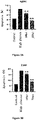

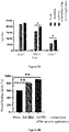
Classifications
-
- C—CHEMISTRY; METALLURGY
- C12—BIOCHEMISTRY; BEER; SPIRITS; WINE; VINEGAR; MICROBIOLOGY; ENZYMOLOGY; MUTATION OR GENETIC ENGINEERING
- C12N—MICROORGANISMS OR ENZYMES; COMPOSITIONS THEREOF; PROPAGATING, PRESERVING, OR MAINTAINING MICROORGANISMS; MUTATION OR GENETIC ENGINEERING; CULTURE MEDIA
- C12N5/00—Undifferentiated human, animal or plant cells, e.g. cell lines; Tissues; Cultivation or maintenance thereof; Culture media therefor
- C12N5/06—Animal cells or tissues; Human cells or tissues
- C12N5/0602—Vertebrate cells
- C12N5/0652—Cells of skeletal and connective tissues; Mesenchyme
- C12N5/0662—Stem cells
- C12N5/0667—Adipose-derived stem cells [ADSC]; Adipose stromal stem cells
-
- A—HUMAN NECESSITIES
- A61—MEDICAL OR VETERINARY SCIENCE; HYGIENE
- A61K—PREPARATIONS FOR MEDICAL, DENTAL OR TOILETRY PURPOSES
- A61K35/00—Medicinal preparations containing materials or reaction products thereof with undetermined constitution
- A61K35/12—Materials from mammals; Compositions comprising non-specified tissues or cells; Compositions comprising non-embryonic stem cells; Genetically modified cells
- A61K35/28—Bone marrow; Haematopoietic stem cells; Mesenchymal stem cells of any origin, e.g. adipose-derived stem cells
-
- A—HUMAN NECESSITIES
- A61—MEDICAL OR VETERINARY SCIENCE; HYGIENE
- A61K—PREPARATIONS FOR MEDICAL, DENTAL OR TOILETRY PURPOSES
- A61K35/00—Medicinal preparations containing materials or reaction products thereof with undetermined constitution
- A61K35/12—Materials from mammals; Compositions comprising non-specified tissues or cells; Compositions comprising non-embryonic stem cells; Genetically modified cells
- A61K35/48—Reproductive organs
- A61K35/54—Ovaries; Ova; Ovules; Embryos; Foetal cells; Germ cells
- A61K35/545—Embryonic stem cells; Pluripotent stem cells; Induced pluripotent stem cells; Uncharacterised stem cells
-
- A—HUMAN NECESSITIES
- A61—MEDICAL OR VETERINARY SCIENCE; HYGIENE
- A61K—PREPARATIONS FOR MEDICAL, DENTAL OR TOILETRY PURPOSES
- A61K35/00—Medicinal preparations containing materials or reaction products thereof with undetermined constitution
- A61K35/66—Microorganisms or materials therefrom
- A61K35/74—Bacteria
-
- A—HUMAN NECESSITIES
- A61—MEDICAL OR VETERINARY SCIENCE; HYGIENE
- A61P—SPECIFIC THERAPEUTIC ACTIVITY OF CHEMICAL COMPOUNDS OR MEDICINAL PREPARATIONS
- A61P17/00—Drugs for dermatological disorders
- A61P17/02—Drugs for dermatological disorders for treating wounds, ulcers, burns, scars, keloids, or the like
-
- C—CHEMISTRY; METALLURGY
- C12—BIOCHEMISTRY; BEER; SPIRITS; WINE; VINEGAR; MICROBIOLOGY; ENZYMOLOGY; MUTATION OR GENETIC ENGINEERING
- C12N—MICROORGANISMS OR ENZYMES; COMPOSITIONS THEREOF; PROPAGATING, PRESERVING, OR MAINTAINING MICROORGANISMS; MUTATION OR GENETIC ENGINEERING; CULTURE MEDIA
- C12N1/00—Microorganisms, e.g. protozoa; Compositions thereof; Processes of propagating, maintaining or preserving microorganisms or compositions thereof; Processes of preparing or isolating a composition containing a microorganism; Culture media therefor
- C12N1/20—Bacteria; Culture media therefor
- C12N1/205—Bacterial isolates
-
- C—CHEMISTRY; METALLURGY
- C12—BIOCHEMISTRY; BEER; SPIRITS; WINE; VINEGAR; MICROBIOLOGY; ENZYMOLOGY; MUTATION OR GENETIC ENGINEERING
- C12N—MICROORGANISMS OR ENZYMES; COMPOSITIONS THEREOF; PROPAGATING, PRESERVING, OR MAINTAINING MICROORGANISMS; MUTATION OR GENETIC ENGINEERING; CULTURE MEDIA
- C12N5/00—Undifferentiated human, animal or plant cells, e.g. cell lines; Tissues; Cultivation or maintenance thereof; Culture media therefor
- C12N5/06—Animal cells or tissues; Human cells or tissues
- C12N5/0602—Vertebrate cells
-
- C—CHEMISTRY; METALLURGY
- C12—BIOCHEMISTRY; BEER; SPIRITS; WINE; VINEGAR; MICROBIOLOGY; ENZYMOLOGY; MUTATION OR GENETIC ENGINEERING
- C12N—MICROORGANISMS OR ENZYMES; COMPOSITIONS THEREOF; PROPAGATING, PRESERVING, OR MAINTAINING MICROORGANISMS; MUTATION OR GENETIC ENGINEERING; CULTURE MEDIA
- C12N2500/00—Specific components of cell culture medium
- C12N2500/30—Organic components
- C12N2500/34—Sugars
-
- C—CHEMISTRY; METALLURGY
- C12—BIOCHEMISTRY; BEER; SPIRITS; WINE; VINEGAR; MICROBIOLOGY; ENZYMOLOGY; MUTATION OR GENETIC ENGINEERING
- C12N—MICROORGANISMS OR ENZYMES; COMPOSITIONS THEREOF; PROPAGATING, PRESERVING, OR MAINTAINING MICROORGANISMS; MUTATION OR GENETIC ENGINEERING; CULTURE MEDIA
- C12N2500/00—Specific components of cell culture medium
- C12N2500/70—Undefined extracts
- C12N2500/72—Undefined extracts from bacteria
-
- C—CHEMISTRY; METALLURGY
- C12—BIOCHEMISTRY; BEER; SPIRITS; WINE; VINEGAR; MICROBIOLOGY; ENZYMOLOGY; MUTATION OR GENETIC ENGINEERING
- C12R—INDEXING SCHEME ASSOCIATED WITH SUBCLASSES C12C - C12Q, RELATING TO MICROORGANISMS
- C12R2001/00—Microorganisms ; Processes using microorganisms
- C12R2001/01—Bacteria or Actinomycetales ; using bacteria or Actinomycetales
Definitions
- the present application relates to the field of regenerative medicine. Specifically, it relates to a use of Rhodococcus ruber (especially the cell wall skeleton) in promoting the proliferation of stem cells and promoting the differentiation of stem cells.
- Rhodococcus ruber especially the cell wall skeleton
- Stem cells are primitive cells with self-renewal ability and multi-directional differentiation potential. Under certain conditions, they can proliferate and differentiate into different functional cells. Therefore, stem cell plays a very important role in the renewal and damage repair of various tissues and organs of living organisms, and has become the hope of many incurable diseases, especially diseases with cell and tissue loss or damage.
- Stem cells include embryonic stem cells and adult stem cells.
- the application of embryonic stem cells are greatly limited due to ethical issues; meanwhile, adult stem cells can differentiate into functional cells and tissues, providing a basis for the wide application of stem cells; providing a new cell source for cell replacement therapy for many diseases. Therefore, adult stem cells have become the focus of research.
- stem cells in normal adult mammals are small, and the differentiation is affected by a variety of intrinsic mechanisms and microenvironmental factors; and long-term culture (especially serum-free expansion culture) of these cells in large quantities in vitro is very difficult, making these cells unsuitable for practical treatment.
- Stem cell related drugs can prevent and treat diseases caused by cell loss or damage by regulating the proliferation and differentiation of stem cells in living organisms. Stem cell related drugs are used to regulate the proliferation and directed differentiation potential of autologous stem cells, so as to reconstruct damaged functional cells and restore their biological functions.
- Ad-MSCs Adipose-derived mesenchymal stem cells
- Ad-MSCs Adipose-derived mesenchymal stem cells
- a significant obstacle to the successful use of Ad-MSCs in potential cell therapies is the viability ratio of cells after transplantation. When cells are transplanted into damaged skin tissue, they experience adverse conditions including hypoxia, inflammation, oxidative stress or others, which inevitably lead to poor survival of seeded cells after transplantation, interfering with the therapeutic effect of Ad-MSCs.
- Rhodococcus ruber is a gram-positive bacterium. Generally speaking, its colony is orange-yellow or orange-red in color, and round in shape; the size of the colony is about 1 mm to 2 mm; the cells are spherical or short rod-shaped; it can form primary branched mycelium; and it has no flagella. Rhodococcus ruber is aerobic and chemically heterotrophic.
- Fan Xin et al. performed whole gene sequencing for Rhodococcus ruber SD3 strain and performed bio-informatic analysis.
- the length of the whole genome of SD3 strain is about 5.37 Mb
- the GC content is about 70.63%
- the GenBank accession number is CP029146 ( Fan Xin, Whole-genome sequencing and expression analysis of heat shocking protein DnaK from Rhodococcus ruber SD3, Genomics and Applied Biology, January 2019 ).
- Rhodococcus can adapt to a variety of living environments due to its strong tolerance to organic substances and its wide degradation spectrum. Therefore, Rhodococcus is widely used in the fields of pollution remediation, organic compound degradation, sewage treatment and the like.
- the main application field of Rhodococcus ruber lies in environmental management, see CN108862590A , CN107151635A , CN102250796A , CN1519312A , CN103627653A , CN101033454A , CN108130288A , CN104830738A , CN101619299A , CN103509833A , CN106434466A , CN101580808A , CN102604875A , CN103160491A , CN106591168A , CN106591172A and CN105820982A .
- CN109576180A discloses a bacterium RDC-01 screened from the red soil in the suburbs near Panyu District, Guangzhou.
- the strain was identified as Rhodococcus ruber by 16S rRNA gene sequence analysis and cultivation characteristics identification. After inactivation, the bacterium was added as an immune adjuvant to an inactivated vaccine for animals, and it was found to be able to promote the production of antibodies in animals.
- Rhodococcus ruber in the field of human medicine has not yet been reported.
- the present application provides an active component for the regulation of stem cells and the application thereof.
- an isolated Rhodococcus ruber is provided.
- Rhodococcus ruber which was deposited at China General Microbiological Culture Collection Center (CGMCC) on March 22, 2019 (Yard No.1(3), West Beichen Road, Chaoyang District, Beijing, Institute of Microbiology, Chinese Academy of Sciences; postal code: 100101), under deposit number CGMCC No. 17431.
- CGMCC General Microbiological Culture Collection Center
- the deposit meets the requirements of the Budapest Treaty on the International Recognition of the Deposit of Microorganisms for the Purposes of Patent Procedure.
- Rhodococcus ruber and a derivative product thereof.
- the derivative product is derived from Rhodococcus ruber and comprises the components of Rhodococcus ruber (such as proteins, nucleic acids, lipids, cell walls and components thereof, carbohydrates or metabolites).
- Rhodococcus ruber refers to the strain under the deposit number CGMCC No. 17431.
- provided is a product derived from Rhodococcus ruber cell wall.
- provided is an isolated Rhodococcus ruber cell wall skeleton.
- Rhodococcus ruber refers to the strain under the deposit number CGMCC No. 17431.
- a pharmaceutical composition comprising the Rhodococcus ruber cell wall or Rhodococcus ruber cell wall skeleton according to the present disclosure.
- a product derived from Rhodococcus ruber cell wall which comprises a product obtained by disruption of Rhodococcus ruber.
- a product derived from Rhodococcus ruber cell wall which comprises a product obtained by disruption and purification (removing lipids, nucleic acids and proteins) of Rhodococcus ruber.
- a pharmaceutical composition or a medical device which comprises a product derived from Rhodococcus ruber cell wall.
- a pharmaceutical composition or a medical device which comprises a product obtained by disruption and purification (removing lipids, and/or nucleic acids, and/or proteins) of Rhodococcus ruber.
- a pharmaceutical composition or a medical device which comprises Rhodococcus ruber cell wall.
- a pharmaceutical composition or a medical device which comprises Rhodococcus ruber cell wall skeleton.
- a pharmaceutical composition or a medical device which comprises the above-mentioned product derived from Rhodococcus ruber cell wall.
- the pharmaceutical composition further comprises a pharmaceutically acceptable excipient.
- the product derived from Rhodococcus ruber cell wall is 1 part by weight
- the pharmaceutically acceptable excipient is 50 to 5000 parts by weight (for example, 50, 100, 200, 210, 220, 230, 240, 250, 260, 270, 280, 290, 300, 500, 600, 700, 1000, 1500, 2000, 2500, 3000, 3500, 4000, 4500, 5000 and any value within the range between any two values above).
- the Rhodococcus ruber cell wall is 1 part by weight
- the pharmaceutically acceptable excipient is 50 to 5000 parts by weight (for example, 50, 100, 200, 210, 220, 230, 240, 250, 260, 270, 280, 290, 300, 500, 600, 700, 1000, 1500, 2000, 2500, 3000, 3500, 4000, 4500, 5000 and any value within the range between any two values above).
- the Rhodococcus ruber cell wall skeleton is 1 part by weight
- the pharmaceutically acceptable excipient is 50 to 5000 parts by weight (for example, 50, 100, 200, 210, 220, 230, 240, 250, 260, 270, 280, 290, 300, 500, 600, 700, 1000, 1500, 2000, 2500, 3000, 3500, 4000, 4500, 5000 and any value within the range between any two values above).
- the pharmaceutical composition can be formulated into a liquid (liquid formulation).
- the pharmaceutical composition can be formulated into a solid (powder formulation or lyophilized powder formulation).
- the liquid formulation and the powder formulation can be converted into each other, and the difference lies only in the water content.
- the powder formulation (or lyophilized powder formulation) is obtained by removing most or all water from the liquid formulation.
- the liquid formulation is obtained by dissolving (or reconstituting) the powder formulation (or lyophilized powder formulation).
- the medicament or pharmaceutical composition is formulated into a dosage form selected from the following group consisting of ointment, cream, emulsion, suspension, paste, gel, lotion, tincture, salve, tablet, aerosol, spray, liniment and powder; wherein, the ointment is selected from the group consisting of soft ointment, plaster and cream.
- the pharmaceutically acceptable excipient relates to, but is not limited to: filler, stabilizer, flavoring agent, disintegrant, binder and lubricant.
- the pharmaceutically acceptable excipient is for example but not limited to: dextran, lactose, microcrystalline cellulose, trehalose, glycine, xylitol, sodium carboxymethyl cellulose, erythritol, gelatin, magnesium stearate, propellant, humectant, solvent, solubilizer, emulsifier, antioxidant, pH regulator and preservative.
- non-limiting examples also include: white vaseline, carbomer, hydroxypropyl methylcellulose, methyl cellulose, sodium hydroxymethyl cellulose, chitosan, sucralfate chitosan, polyvinylpyrrolidone, polyvinyl alcohol, sodium hyaluronate, dimethyl ether, tetrafluoroethane, hydrofluoroalkane, glycerin, propylene glycol, deionized water, water for injection, distilled water, ethanol, hexadecanol, octadecanol, p-aminobenzoic acid, acetamide, isopropanol, Tween, polyoxyethyl hydrogenated castor oil, stearic acid, glyceryl monostearate, triglycerol monostearate, sucrose fatty acid ester, sucrose ester, sucrose acetate isobutyrate, sorbitan tristearate, isopropyl
- a method for the preparation of a product derived from Rhodococcus ruber cell wall which comprises or consists of the following steps:
- step (5) can further include a step of removing cell membrane (for example by using a non-ionic surfactant).
- Rhodococcus ruber is not limited to particular culture medium and culture parameters.
- the skilled person can use well-known and appropriate methods for culture, and can use petri dishes, culture flasks and fermenters according to the preparation scale.
- Rhodococcus ruber For the disruption of Rhodococcus ruber, the purpose is to remove the substances inside the cells. Therefore, ultrasonication, lysozyme and other technologies can be used. The skilled person understands that any known or future method suitable for disrupting gram-positive bacteria is suitable for the technical solution of the present disclosure.
- the skilled person has the ability to adjust the particular parameters and instruments for culture, disruption, separation, collection, removal of impurity, and aliquoting according to the subsequent application (e.g., oral administration, injection, topical application, etc.) of the active component (the cell wall and components thereof), so as to avoid introducing factors that affect subsequent applications in the preparation steps.
- the subsequent application e.g., oral administration, injection, topical application, etc.
- the active component the cell wall and components thereof
- an organic solvent is used to remove lipids from the disrupted product.
- a nuclease is used to remove DNA and RNA from the disrupted product.
- a hydrolase is used to degrade proteins in the disrupted product.
- a surfactant is used to remove cell membranes from the disrupted product.
- the average particle size of disruption is 10 nm to 1000 nm; mention may be made of 10, 20, 30, 40, 50, 60, 70, 80, 90, 100, 110, 120, 130, 140, 150, 160, 170, 180, 190 nm ⁇ 10 nm, and the ranges between any two of the above values.
- There are many methods for measuring the particle size Hu Songqing et al., Modern technology of particle size measurement, Modern Chemical Industry, 2002, 22:1 ).
- the average particle size of disruption is 10 nm to 800 nm. In some other particular embodiments, the average particle size of disruption is 10 nm to 500 nm.
- the aliquoting refers to aliquoting into containers or onto solid supports.
- the container is selected from the group consisting of vial, tube, package, bag, plate, ampoule, injection device, aluminum film packaging, dressing and capsule.
- the aliquoting refers to aliquoting into vials/ampoules. Solvent is added to the vial/ampule just before use.
- a product derived from Rhodococcus ruber cell wall which is obtained by the preparation method according to present disclosure.
- a pharmaceutical composition or a medical device which comprises a product derived from Rhodococcus ruber cell wall obtained by the preparation method according to present disclosure.
- an isolated Rhodococcus ruber cell wall which is used for the regulation of adult stem cells.
- the regulation is selected from one of the following or a combination thereof: promoting the proliferation of stem cells, promoting the growth of stem cells, promoting the differentiation of stem cells, promoting the migration of stem cells, and improving the survival rate of stem cells; the stem cells are selected from the group consisting of adult stem cells, iPSCs and mesenchymal stem cells.
- the mesenchymal stem cells are selected from the group consisting of bone marrow mesenchymal stem cells, adipose-derived mesenchymal stem cells, synovial mesenchymal stem cells, umbilical cord mesenchymal stem cells, umbilical cord blood mesenchymal stem cells, placental mesenchymal stem cells, amniotic mesenchymal stem cells, liver mesenchymal stem cells, muscle mesenchymal stem cells, lung mesenchymal stem cells, pancreatic mesenchymal stem cells and dental pulp mesenchymal stem cells.
- Rhodococcus ruber cell wall which is used for the regulation of adult stem cells.
- a pharmaceutical composition or a medical device which is used for the regulation of adult stem cells.
- Rhodococcus ruber cell wall according to the present disclosure for the regulation of adult stem cells.
- Rhodococcus ruber cell wall of the present disclosure in the preparation of a medicament/medical device for the regulation of adult stem cells.
- the medicament is used for the regulation of adult stem cells.
- the medical device (such as dressing, patch, bandage, film, patch, etc.) is used for the regulation of adult stem cells.
- a method for the regulation of adult stem cells comprising exposing a subject to a therapeutically effective amount (or a prophylactically effective amount) of any one selected from of the following:
- a method for the regulation of stem cells comprising a step of exposing the stem cells to a product derived from Rhodococcus ruber cell wall.
- the ratio of the number of stem cells / the product derived from Rhodococcus ruber cell wall is: 1 to 1000 stem cells / 1 ng of the product derived from Rhodococcus ruber cell wall; preferably 5 to 50 stem cells / 1 ng of the product derived from Rhodococcus rubercell wall.
- the ratio of the number of stem cells / the product derived from Rhodococcus ruber cell wall is: 1 to 1000 stem cells / 1 ng of the product derived from Rhodococcus ruber cell wall; preferably 5 to 50 stem cells / 1 ng of the product derived from Rhodococcus rubercell wall.
- the ratio of the number of stem cells / the product derived from Rhodococcus ruber cell wall is: 1 to 1000 stem cells / 1 ng of the product derived from Rhodococcus ruber cell wall; preferably 5 to 50 stem cells / 1 ng of the product derived from Rhodococcus rubercell wall.
- a method for promoting wound healing comprising a step of exposing the wound of a subject to a therapeutically effective amount of mesenchymal stem cells and the product derived from Rhodococcus ruber cell wall.
- the ratio of the number of mesenchymal stem cells / the product derived from Rhodococcus ruber cell wall is: 1 to 100 stem cells / 1 ng of the product derived from Rhodococcus ruber cell wall; preferably 5 to 50 stem cells / 1 ng of the product derived from Rhodococcus ruber cell wall.
- the wound is a wound related to diabetes.
- the medicament is administered to the lesion according to the area and depth of the lesion, for example:
- the period of exposure lasts 2 days to 2 months or longer.
- the period of exposure lasts 2 days to 2 months or longer.
- the active component is administered to the subject for 3 to 4 weeks.
- the active component is administered at a frequency selected from the following: administering 1 to 3 times per day, 1 to 6 times per two days, 1 to 9 times per three days, 1 to 14 times per week, 1 to 60 times per month. In some embodiments, the active component is administered twice a day, or once a day, or once every two days.
- different doses are applied depending on the particular conditions of the subject, usually 1 ⁇ g to 1000 ⁇ g/unit dose/each administration; specifically speaking, for example 1, 5, 10, 15, 20, 25, 30, 35 , 40, 45, 50, 55, 60, 65, 70, 75, 80, 85, 90, 95, 100, 110, 120, 130, 140, 150, 160, 170, 180, 190, 200 ⁇ g/unit dose/each administration, and the ranges between any two of the above values.
- the exposure is achieved by the following routes, for example but not limited to: oral, mucosal, percutaneous, transdermal, intraperitoneal, puncture, nasal spray, eye drops, suppository and sublingual.
- the subject is an animal other than human, for example farm animal, pet, working animal, ornamental animal and production animal.
- the subject is a human.
- the subject is suspected of having, confirmed to have, has suffered from, or is susceptible to the target disease or symptoms thereof.
- a cell culture medium comprising one or a combination selected from the following:
- a cell culture medium which further comprises other components well known in the art suitable for culturing stem cells, especially mesenchymal stem cells.
- stem cells especially mesenchymal stem cells.
- the culture process be free of xenobiotic animal components, for example, by using serum-free culture medium.
- the skilled person can add cytokines, such as one or a combination of FGF, PDGF, TGF- ⁇ , HGF, EGF, CTGF, VEGF, insulin and insulin-like growth factors, to the cell culture medium as needed (for example, to maintain stemness, or to promote differentiation).
- cytokines such as one or a combination of FGF, PDGF, TGF- ⁇ , HGF, EGF, CTGF, VEGF, insulin and insulin-like growth factors
- the content of FGF (in final concentration) is preferably 0.1 to 100 ng/ml.
- FGF refers to a growth factor in the fibroblast growth factor family, preferably FGF-1, FGF-2 (bFGF).
- the content of PDGF (final concentration) is preferably 0.5 to 100 ng/ml.
- PDGF refers to a growth factor in the platelet-derived growth factor family, preferably PDGF-BB or PDGF-AB.
- the content of TGF- ⁇ (final concentration) is preferably 0.5 to 100 ng/ml.
- TGF- ⁇ refers to a growth factor in the transforming growth factor-beta family, preferably TGF- ⁇ 3.
- the content of HGF (final concentration) is preferably 0.1 to 50 ng/ml. In some particular embodiments, the content of EGF (final concentration) is preferably 0.5 to 200 ng/ml.
- a cell culture medium which further comprises at least one phospholipid, and/or at least one fatty acid.
- the phospholipid includes, for example, phosphatidic acid, lysophosphatidic acid, phosphatidyl cyclohexanol, phosphatidyl serine, phosphatidyl ethanolamine, phosphatidyl choline and phosphatidyl glycerol.
- the total content of phospholipid (final concentration) is preferably 0.1 to 30 ⁇ g/ml.
- the fatty acid includes, for example, linoleic acid, oleic acid, linolenic acid, arachidonic acid, tetradecanoic acid, palmitoleic acid, palmitic acid, stearic acid, and the like.
- the total content of fatty acid is preferably 1/1000 to 1/10 of the medium.
- a cell culture medium which further comprises an antioxidant.
- the antioxidant includes, for example, DL- ⁇ -tocopheryl acetate (vitamin E).
- a cell culture medium which further comprises amino acids, nucleotides and trace elements required to maintain cells.
- the composition of the present application is added to a known commercially available mesenchymal stem cell culture medium; for example, the commercially available mesenchymal stem cell culture medium is selected from the group consisting of MesenPRO RSTM, StemPro ® MSC SFM, StemPro ® MSC SFM XenoFree and StemPro ® human adipose-derived stem cell medium.
- the commercially available mesenchymal stem cell culture medium is selected from the group consisting of MesenPRO RSTM, StemPro ® MSC SFM, StemPro ® MSC SFM XenoFree and StemPro ® human adipose-derived stem cell medium.
- a therapeutic composition comprising: mesenchymal stem cells, and a product derived from Rhodococcus ruber cell wall.
- the ratio of the number of mesenchymal stem cells / the product derived from Rhodococcus ruber cell wall is: 1 to 100 stem cells / 1 ng of the product derived from Rhodococcus ruber cell wall; preferably 5 to 50 stem cells / 1 ng of the product derived from Rhodococcus ruber cell wall.
- the mesenchymal stem cells are selected from the group consisting of bone marrow mesenchymal stem cells, adipose-derived mesenchymal stem cells, synovial mesenchymal stem cells, umbilical cord mesenchymal stem cells, umbilical cord blood mesenchymal stem cells, placental mesenchymal stem cells, amniotic mesenchymal stem cells, liver mesenchymal stem cells, muscle mesenchymal stem cells, lung mesenchymal stem cells, pancreatic mesenchymal stem cells and dental pulp mesenchymal stem cells.
- a method for improving stem cell apoptosis comprising a step of exposing the stem cells to an effective amount of the above-mentioned product derived from Rhodococcus ruber cell wall.
- a method for improving stem cell survival comprising a step of exposing the stem cells to an effective amount of the above-mentioned product derived from Rhodococcus ruber cell wall.
- the only therapeutic (or prophylactic) active component in the medicament or medical device is a product derived from Rhodococcus ruber cell wall, notably comprising Rhodococcus ruber components (such as proteins, nucleic acids, lipids, cell wall and components thereof, carbohydrates or metabolites), specifically speaking, a product comprising Rhodococcus ruber cell wall, more preferably Rhodococcus ruber cell wall skeleton or components thereof.
- Rhodococcus ruber components such as proteins, nucleic acids, lipids, cell wall and components thereof, carbohydrates or metabolites
- Isolation/isolated refers to the separation of the Rhodococcus ruber of the present disclosure from its original growth environment.
- the cell wall structures of gram-positive bacteria and gram-negative bacteria are different. Specifically speaking, the cell wall of gram-positive bacteria is thicker (usually 20 nm to 80 nm), comprising about 90% peptidoglycan and about 10% teichoic acid (a polymer formed by alcohol and phosphoric acid molecules, usually existing in the form of sugar ester or amino acid ester).
- the peptidoglycan layer is dense, even as many as 20 layers.
- the cell wall of gram-negative bacteria is much thinner than that of gram-positive bacteria, and the structure is more complex, divided into outer membrane and peptidoglycan layer (usually 2 nm to 3 nm).
- the peptidoglycan layer is a unique component of the bacterial cell wall and is a derivative of heteropolysaccharide.
- Each peptidoglycan monomer comprises 3 moieties: the sugar unit (for example, at least two sugar molecules are connected by glycosidic bonds to form the framework of peptidoglycan), the peptide tail (a short peptide chain formed by linking several amino acids, which is connected to a N-acetylmuramic acid molecule), and the peptide bridge (which crosslinks the adjacent "peptide tails" to form a high-strength network structure).
- Different bacteria have different peptide bridges, peptide tails and cross-linking manners.
- isolated Rhodococcus ruber cell wall can be interpreted as either a complete cell wall or an incomplete cell wall (for example, disrupted or partially degraded).
- the components exhibiting the desired activity are derived from Rhodococcus ruber cell wall (for example, the cell wall itself or components thereof). Therefore, complete cell wall, disrupted cell wall, incompletely degraded product of cell wall, cell wall components, cell wall extracts and other various forms are allowed to be used in clinical applications, which are all encompassed in the scope of the present disclosure.
- a component that constitutes the main structure of the cell wall cannot be interpreted as merely representing the cross-linked network-like entity in the cell wall, and the skilled person understands that other cell wall components adsorbed onto, bound to and carried by the cross-linked network-like entity are not excluded.
- Rhodococcus ruber used in the embodiments of the present disclosure refers to the Rhodococcus ruber species of the Rhodococcus genus, and is not limited to a particular cell strain.
- Non-limiting examples include the TOY7 strain (Agricultural Environment Microbiological Culture Collection, Nanjing Agricultural University), CGMCC No. 4795, DSM43338, CCTCC No. 2012035, CGMCC No. 16640 and CGMCC No. 17431.
- the skilled person can perform taxonomic identification on a strain of bacteria.
- the available identification techniques include morphology, physiological and biochemical characteristics, 16S rRNA, and the like.
- identification techniques involve different methods. In the earlier period, morphological and biochemical identification methods were mainly used, but the reliability of these methods is not high. After the advent of sequencing technology, the skilled person can identify bacteria strains in a more reliable way.
- the medicament or pharmaceutical composition or active component or product of the present disclosure can be formulated into, but not limited to, the following forms: ointment, cream, plaster, gel, lotion, tincture, liniment, salve, paste, lyophilized powder, aerosol, suppository, patch, suspension, oral solution, buccal tablet and skin care product (cleanser, toning lotion, serum, lotion, cream and mask).
- the medicament or pharmaceutical composition or active component or product of the present disclosure can be formulated into the form of a formulation unit.
- the unit dose of the medicament comprises:
- Particular examples of the unit dose are 1, 2, 5, 10, 15, 20, 25, 30, 40, 50, 55, 56, 57, 58, 59, 60, 61, 62, 63, 65, 66, 67, 68, 69, 70, 80, 90, 100, 150, 200, 250, 300, 350, 400, 450, 500 mg ⁇ 10%, and the ranges between any two of the above values.
- administering when applied to animals, humans, cells, tissues, organs or biological samples, refer to the contact of the medicament or medical device with the animals, humans, cells, tissues, organs or biological samples.
- Treatment means administrating an internal or external medicament (therapeutic agent, active component or composition) (such as the Rhodococcus ruber cell wall or pharmaceutical composition thereof according to the present disclosure) or a medical device to a subject, in order to alleviate (relieve, delay, improve, cure) one or more disease symptoms to a clinically measurable degree in the subject (or population) to be treated, wherein the subject has, is suspected of having, or is susceptible to one or more diseases or symptoms thereof.
- an internal or external medicament therapeutic agent, active component or composition
- a medical device such as the Rhodococcus ruber cell wall or pharmaceutical composition thereof according to the present disclosure
- the amount of the medicament (therapeutic agent, active component or composition) that can effectively alleviate any disease symptoms is called the therapeutically effective amount. It can vary depending on a variety of factors, such as the disease state, age and body weight of the subject. It should be understood that the medicament (therapeutic agent, active component or composition) may be ineffective in alleviating the target disease or symptoms thereof of a single subject, but is statistically effective for the target disease or symptoms thereof according to any statistical test method known in the art (such as Student t-test, chi-square test and U test according to Mann and Whitney).
- aliquoting means that the product is allowed to be aliquoted, but is not necessarily aliquoted; whether the product is aliquoted or not does not affect the realization of the technical effects.
- Rhodococcus ruber used in the following examples is the CGMCC No. 17431 strain, which was deposited at China General Microbiological Culture Collection Center (CGMCC, Institute of Microbiology Chinese Academy of Sciences, Yard No.1(3), West Beichen Road, Chaoyang District, Beijing, China) on March 22, 2019.
- CGMCC General Microbiological Culture Collection Center
- the strain was cultured on a glycerol agar medium at 30 to 37°C (specifically 32 to 35°C) for 12 to 72 (specifically 36 to 60, such as 40 to 50) hours, and the following was observed: the colonies plumped up; were orange-red in color (slightly different depending on influences of light, the color of the culture medium, etc.); the surface was dry and wrinkled, slightly shiny (slightly different depending on differences in culture conditions); were fragile to touch; and the colony size was about 1 mm to 2 mm (slightly different depending on differences in culture conditions).
- the hyphae grew in a branching structure with septate, and formed mycelium (slightly different depending on differences in culture conditions);
- the strain was gram stain positive.
- the strain was cultured on a glycerol agar slant medium at 30 to 37°C (specifically 32 to 35°C) for 12 to 72 (specifically 36 to 60, such as 40 to 50) hours. Then, the following tests were performed on the culture.
- Nitrate reduction reaction positive, catalase: positive, tyrosinase: positive, amylase: negative, oxidase: negative, gelatin liquefaction: negative.
- Carbon source Biolog Gen II growth experiment positive for: glucuronamide, ⁇ -hydroxy-DL butyric acid, D-fructose-6-phosphate, ⁇ -D-glucose, D-fructose, D-mannitol, D-arabitol, D-sorbitol, quinic acid, y-aminobutyric acid, citric acid, L-malic acid, bromosuccinic acid, Tween 40, propionic acid and acetic acid
- Biolog Gen III chemical sensitivity experiment sensitive to: dimethylamine tetracycline, sodium tetradecyl sulfate, rifamycin SV, pH 5.0, 8% sodium chloride, lincomycin, fusidic acid, D-serine, vancomycin, tetrazolium violet and tetrazolium blue tolerate to: sodium bromate, 1% sodium lactate, pH 6.0, 1%-4% sodium chloride, nalidixic acid, lithium chloride
- the 15 strains isolated from the working seed tube and the 10 different strains isolated from the original seed tube were subjected to genome extraction, 16S rRNA amplification and sequencing. In these 25 strains, the 16S rRNA gene identity was 100%.
- the neighbor-joining strain phylogenetic tree constructed based on the Kimura2-parameter algorithm showed that the strain was classified as Rhodococcus ruber.
- the bacteria obtained in Preparation Example 1 were collected and the cells were disrupted (for example, but not limited to by sonication). Any appropriate well-known method in the art is also allowed to disrupt the bacteria, such as CN101250490A or CN101323865A .
- the disruption state was checked under a microscope. There should be no more than 5 intact bacteria in each visual field. The disruption was deemed as qualified when several (10 to 30) visual fields checked met this standard.
- the precipitate was reconstituted in water for injection, and then set aside till use. Optionally, it could be sterilized at 115°C for 20-30 minutes as the stock solution of the cell wall skeleton (mainly comprising the cell wall skeleton and components thereof).
- the pharmaceutical composition can be formulated into various forms Composition Capacity of each vial Components and amount Composition 1 2 ml Active component 60 ⁇ g Dextran 40 15 mg Composition 2 2 ml Active component 60 ⁇ g Dextran 40 12 mg Composition 3 2 ml Active component 120 ⁇ g Dextran 40 36 mg Composition 4 2 ml Active component 60 ⁇ g Trehalose 12 mg Composition 5 2 ml Active component 120 ⁇ g Trehalose 36 mg Composition 6 2 ml Active component 120 ⁇ g Mannitol 36 mg Composition 7 2 ml Active component 60 ⁇ g Mannitol 12 mg
- compositions of item 1 were lyophilized to prepare lyophilized powders (numbered as lyophilized powder Composition 1 to lyophilized powder Composition 7, respectively).
- composition was deemed as qualified if the body weight of each mouse increased at the due date Abnormal toxicity in guinea pigs all the guinea pigs should survive and have no abnormal reactions during the observation period. The composition was deemed as qualified if the body weight of each guinea pig increased at the due date
- Adipose tissue samples were obtained from liposuction aspirate of subjects (age range 25-35 years) and Ad-MSCs were isolated and cultured. Subjects were chosen from plastic surgery patients in the affiliated Hospital of Xuzhou Medical University. The experiment were approved by the ethics committee and informed consent of the patients.
- the obtained fresh adipose tissue extract was digested with 0.25% trypsin-EDTA, filtered, and centrifuged to retain the cell pellet.
- DMEM Invitrogen
- FBS fetal bovine serum
- penicillin/streptomycin was added.
- the cell culture dish was placed in a 37°C, 5% CO2 incubator to culture the cells. After that, the culture medium was changed every 2-3 days after washing the cells with PBS. The cells were passaged when growing to 80%.
- Ad-MSCs in logarithmic growth phase were seeded in a 96-well plate at 4 ⁇ 103 cells/well; DMEM + 10% fetal bovine serum + 1% penicillin/streptomycin was used for co-culture; the composition of the present application (Composition 1) was dissolved and diluted to 10 ⁇ g/ml with PBS buffer. After the Ad-MSCs adhered to the wall, for each sample to be tested, the experiment was divided into four groups: control, 25 ⁇ l, 50 ⁇ l and 75 ⁇ l. After 24 h, 48 h, 72 h or 96 h,10 ⁇ l of CCK-8 reagent was added to each well, and incubated for 2 h.
- absorbance of each well at a wavelength of 450 nm was detected by an enzyme-linked immunometric meter.
- the cell growth curve was plotted with the culture time as the horizontal axis and the cell number (absorbance) as the vertical axis.
- EdU incorporation assay was performed by using Cell-Light EdU 567 in vitro imaging kit (RiboBio).
- Ad-MSCs at the logarithmic growth phase were seeded in a 96-well plate at 4x10³ cells/well. 10 ⁇ g/ml, 50 ⁇ l of the composition of the present application was applied onto the Ad-MSCs.
- 100 ⁇ l of EdU medium was added to each well after 72 h or 96 h and incubated for two hours, and the wells were washed with PBS 1-2 times. 4% paraformaldehyde fixative was added to each well and the plate was incubated at room temperature for 30 min.
- Ad-MSCs When Ad-MSCs reached 80% confluence, they were seeded at a density of 1 ⁇ 104 cells/well in a 6-well plate. The experiment was divided into four groups. After the cells adhered to the wall, 100 ⁇ l of 50% sucrose was added to each experimental group to induce apoptosis of Ad-MSCs; and 100 ⁇ l or 250 ⁇ l of the composition of the present application at 10 ⁇ g/ml was respectively added to two experimental groups among these groups. After 48 h and 72 h, the supernatant of each group of cells was collected in flow tubes. The adherent cells were digested with EDTA-free trypsin and collected into flow tubes of the same group.
- the tubes were centrifuged at 2000 rpm for 5 min, washed twice with PBS buffer, shaken and mixed well. 500 ⁇ l of binding buffer (Annexin V-FITC apoptosis detection kit, Shanghai Beyotime Biotechnology Co., Ltd.), 5 ⁇ l of FITC and 5 ⁇ l of PI were successively added to each tube. The tubes were shaken and mixed well and incubated in the dark at 4°C for 5-15 min. The results were detected and analyzed by flow cytometry (BD Biosciences).
- TUNEL reaction solution 500 ⁇ l of TUNEL reaction solution (TUNEL apoptosis kit, Roche) was prepared. 50 ⁇ l of enzyme solution and 450 ⁇ l of labeling solution were mixed to prepare reagent A. 50 ⁇ l of reagent A was added to the negative control group, which was placed in a 37°C incubator in the dark for 60 min. DNase I was added to the positive control group, which was incubated at room temperature for 10 min. The cells were washed 3 times with PBS buffer and 50 ⁇ l/well of TUNEL reaction mixture was added. The cells were incubated in a 37°C incubator in the dark for 60 min and washed 3 times with PBS buffer.
- DAPI staining solution 50 ⁇ l/well of DAPI staining solution was added and the cells were incubated at room temperature for 3 min. The samples were photographed under a fluorescence microscope for analysis, and the wavelength range of detection light was 570-620 nm (maximum wavelength 580 nm).
- the treated cells were collected. 300 ⁇ l of cell lysis mixture (dissolve PIPA cell lysis solution on ice, add PMSF at a ratio of 100:1) was added to each dish and then placed on ice. The cells were fully lysed and adherent cells were scraped. The mixture was centrifuged in a Centrifuge-5810R refrigerated high-speed centrifuge at 4°C, 12,000 rpm for 20 min and the supernatant was collected.
- the cell extracts were separated on SDS-polyacrylamide gels. Then the proteins were transferred to nitrocellulose membranes and incubated with the following antibodies: rabbit-anti-human caspase-3 monoclonal antibody (1:400; CST, USA), rabbit-anti-human Bax monoclonal antibody (1:400; CST, USA). After incubation with the primary antibody, the membranes were washed for 5 minx3 times and incubated with diluted secondary antibody (1:10000) at room temperature in the dark for 2 h. The secondary antibody was discarded and the membranes were washed for 5 minx3 times. Equal amounts of ECL luminescent solution A and B were pipetted respectively and mixed well to prepare the ECL working solution. The ECL working solution was evenly dropped on the membranes. The membranes were put into a TANON gel imager for exposure and development, photographed, and analyzed by ImageJ software.
- mice were 4-week-old BALB/c athymic nude mice, SPF grade. All animal studies were approved by the Animal Care and Use Committee. The experimental mice were kept in SPF grade animal room. Mice were fasted for 12 h before model establishment, weighed and recorded. Diabetic mice were induced by intraperitoneal injection of 2% STZ (Sigma) at a dose of 150 mg/kg.
- Blood glucose was measured on day 7 after injection. Blood was collected through tail vein, and the blood glucose concentration of the mice was measured with a blood glucose meter and recorded. From day 7, the blood glucose concentration of the mice was greater than 16.7 mmol/L and the typical symptoms of diabetes "polydipsia, polyphagia, polyuria and weight loss" were developed, which was regarded as successful model establishment.
- the experimental animals were divided into 3 groups (5 animals in each group):
- mice were anesthetized and wounds with a diameter of 1.5 cm were prepared.
- the pretreated Ad-MSCs were injected into the skin of mice in each group by multi-point intradermal injection, with six points on each wound and 0.1 ml of cells injected at each point.
- the wound surface was covered with sterile gauze, and feeding and observation were continued.
- the survival of cells in the wound and the skin healing of mice were observed by using LB983 in vivo imaging system.
- Ad-MSCs were labeled with CM-Dil before injection. According to the method recommended by the supplier, CM-Dil live cell stain was added to the cells and incubated for 30 minutes. The supernatant was discarded after centrifugation and the cells were washed 3 times with PBS buffer. Finally, an appropriate amount of PBS was added and mixed well, and the labeled Ad-MSCs were placed in an ice box for later use.
- tissue paraffin sections were dewaxed and incubated with 3% H2O2 at room temperature for 5-10 min to eliminate endogenous peroxidase activity.
- the sections were rinsed with distilled water, soaked twice in PBS for 5 min each time, and blocked with 10% normal goat serum (diluted in PBS) at room temperature for 10 min. The blocking solution was discarded without washing the sections.
- CD31 primary antibody (dilution ratio 1:300) was added dropwise and incubated overnight.
- the sections were rinsed 3 times with PBS for 5 min each time.
- An appropriate amount of biotin-labeled fluorescent secondary antibody (dilution ratio 1:400) was added dropwise in the dark.
- the sections were incubated at 37°C in the dark for 1 h and rinsed 3 times with PBS for 5 min each time. An appropriate amount of DAPI staining solution was added dropwise. The sections were incubated at room temperature in the dark for 3 min and rinsed 3 times with PBS for 5 min each time. Finally, anti-fluorescence quenching sealing solution was added dropwise to each section. The sections were covered with coverslips, fixed, and stored in the dark.
- Test Example 1 Identification of morphology, surface markers and induced differentiation ability of human Ad-MSCs
- the collagenase digestion method was used.
- Primary Ad-MSCs extracted from adipose tissue extracts were seeded in cell culture dishes and passaged when they reached 80% confluence. The proliferation rate of the cells increased significantly after passage. The cells were uniform in terms of morphology and spindle-shaped.
- Well-grown Ad-MSCs of the P3 generation were used for adipogenic differentiation and osteogenic differentiation, respectively. Observation of the cells stained with oil red after 2 weeks under a microscope showed obvious presence of red lipid droplets of different sizes in the cells; observation of the cells stained with alizarin S stain showed obvious deposition of red calcium nodules. This indicated that the isolated and extracted cells had the characteristics of stem cell differentiation potential.
- composition 1 of the present application was applied to Ad-MSCs for 24 h, 48 h, 72 h and 96 h. Then the absorbance of the stem cells at a wavelength of 450 nm was detected by an enzyme-linked immunometric meter using CCK-8 reagent. The results showed that the viability of Ad-MSCs was significantly enhanced at 72 h and 96 h after introducing the composition of the present application into the stem cells, at a concentration of 10 ⁇ g/ml and a volume of 50 ⁇ l ( Figure 1 ).
- Test Example 3 The composition of the present application inhibits the high glucose-induced apoptosis of Ad-MSCs
- composition of the present application could inhibit the high glucose-induced apoptosis of stem cells ( Figure 3A, Figure 3B ).
- Test Example 4 Detection of expression levels of apoptosis marker proteins
- the cells were treated with different concentrations of the composition of the present application for 48 h and 72 h, and cellular proteins were extracted.
- the protein expression levels of c-caspase-3 and Bax (two major apoptotic markers) were detected by Western blotting experiments, and ⁇ -actin was used as an internal reference. The results showed that the expression levels of c-caspase-3 and Bax proteins were decreased after treatment with the composition of the present application ( Figure 4A, Figure 4B ).
- Test Example 5 The composition of the present application improves the survival rate of Ad-MSCs and accelerates wound healing in nude mice
- mice After the successful establishment of diabetic mouse model, the mice were anesthetized and wounds with a diameter of 1.5 cm were prepared.
- the pretreated cells labeled with fluorescent dye CM-Dil were injected into the wound skin of mice in treatment group by intradermal injection.
- the survival of cells was observed by using LB983 in vivo imaging system.
- diabetic refractory wounds There are many reasons for the formation of diabetic refractory wounds, e.g., the absence of cellular and molecular signals required for normal wound healing process.
- peripheral neuropathy, peripheral circulation damage and disturbance of protease balance are all important factors for refractory healing of diabetic wounds ( Rathur HM et al., The diabetic foot. Clin Dermatol. 2007; 25(1): 109-120 ).
- the abnormal vascular microenvironment under high glucose conditions can lead to abnormal cell growth environment, and ultimately damage the vascular reconstruction in the trauma area ( Guo WY et al., Acceleration of diabetic wood healing by low-dose radiation is associated with peripheral mobilization of bone marrow stem cells. Radiat Res.
- Ad-MSCs are a kind of stem cells with multi-directional differentiation potential extracted from adipose tissue ( Kato Y et al., Creation and transplantation of an adipose-derived stem cell (ASC) sheet in a diabetic wound-healing model. Jove-J Vis Exp. 2017; 12(6): 1-10 ). Ad-MSCs can migrate to damaged sites through differentiation potential, repair damaged skin with differentiated cells, and secrete a variety of growth factors ( Rehman J et al., Secretion of angiogenic and anti-apoptotic factors by human adipose stromal cells. Circulation. 2004; 109: 1292-8 ).
- Ad-MSCs can differentiate into fibroblasts, showing not only morphological similarity, but also the ability to express fibroblast surface proteins, including vimentin and fibronectin ( Kim WS et al., Wound healing effect of adipose-derived stem cells: a critical role of secretory factors on human dermal fibroblasts. J Dermatol Sci. 2007; 48:15-24 ). Meanwhile, Ad-MSCs can also be directly transformed into fibroblasts and keratinocytes for wound repair ( Unnikrishnan S et al., Constitution of fibrin-based niche for in vitro differentiation of adipose-derived mesenchymal stem cells to keratinocytes. Biores Open Access.
- Ad-MSCs undergo apoptosis when injected into wounds of diabetic mice, resulting in delayed wound healing.
- Ad-MSCs cultured under high glucose conditions undergo apoptosis in a time-dependent manner ( Li Q et al., Stromal cell-derived factor-1 promotes human adipose tissue-derived stem cell survival and chronic wound healing. Exp Ther Med. 2016; 12: 45-50 ).
- the effects of the composition of present application on in vitro activity and proliferation ability of Ad-MSCs were detected by CCK-8 and EdU.
- the results showed that the composition of the present application could improve the activity and proliferation ability of Ad-MSCs.
- Apoptosis of cells was detected by flow cytometry, and it was found that the composition of the present application could inhibit the high glucose-induced apoptosis of Ad-MSCs in a time- and concentration-dependent manner.
- the effect of the composition of the present application on the survival rate of Ad-MSCs was tested in vivo in animals, and the results showed that the survival rate of Ad-MSCs treated with the composition of the present application was higher than that treated with Ad-MSCs alone, and the wound healing speed was faster.
Landscapes
- Health & Medical Sciences (AREA)
- Life Sciences & Earth Sciences (AREA)
- Engineering & Computer Science (AREA)
- Chemical & Material Sciences (AREA)
- Biomedical Technology (AREA)
- Zoology (AREA)
- Biotechnology (AREA)
- General Health & Medical Sciences (AREA)
- Developmental Biology & Embryology (AREA)
- Organic Chemistry (AREA)
- Bioinformatics & Cheminformatics (AREA)
- Cell Biology (AREA)
- Medicinal Chemistry (AREA)
- Wood Science & Technology (AREA)
- Genetics & Genomics (AREA)
- Public Health (AREA)
- Animal Behavior & Ethology (AREA)
- Veterinary Medicine (AREA)
- Pharmacology & Pharmacy (AREA)
- Microbiology (AREA)
- Immunology (AREA)
- Epidemiology (AREA)
- Virology (AREA)
- Biochemistry (AREA)
- General Engineering & Computer Science (AREA)
- Molecular Biology (AREA)
- Mycology (AREA)
- Hematology (AREA)
- Chemical Kinetics & Catalysis (AREA)
- General Chemical & Material Sciences (AREA)
- Dermatology (AREA)
- Nuclear Medicine, Radiotherapy & Molecular Imaging (AREA)
- Tropical Medicine & Parasitology (AREA)
- Reproductive Health (AREA)
- Rheumatology (AREA)
- Gynecology & Obstetrics (AREA)
- Medicines Containing Material From Animals Or Micro-Organisms (AREA)
- Micro-Organisms Or Cultivation Processes Thereof (AREA)
- Medicinal Preparation (AREA)
- Preparation Of Compounds By Using Micro-Organisms (AREA)
Abstract
Description
- The present application claims the priority of Chinese patent application No.
CN 202010068249.1 filed on January 21, 2020 - The present application relates to the field of regenerative medicine. Specifically, it relates to a use of Rhodococcus ruber (especially the cell wall skeleton) in promoting the proliferation of stem cells and promoting the differentiation of stem cells.
- Stem cells are primitive cells with self-renewal ability and multi-directional differentiation potential. Under certain conditions, they can proliferate and differentiate into different functional cells. Therefore, stem cell plays a very important role in the renewal and damage repair of various tissues and organs of living organisms, and has become the hope of many incurable diseases, especially diseases with cell and tissue loss or damage.
- Stem cells include embryonic stem cells and adult stem cells. The application of embryonic stem cells are greatly limited due to ethical issues; meanwhile, adult stem cells can differentiate into functional cells and tissues, providing a basis for the wide application of stem cells; providing a new cell source for cell replacement therapy for many diseases. Therefore, adult stem cells have become the focus of research.
- However, the number of stem cells in normal adult mammals is small, and the differentiation is affected by a variety of intrinsic mechanisms and microenvironmental factors; and long-term culture (especially serum-free expansion culture) of these cells in large quantities in vitro is very difficult, making these cells unsuitable for practical treatment.
- Many diseases can be traced back to the loss or damage of functional cells, and cell replacement therapy is an effective treatment for these diseases. Stem cell related drugs can prevent and treat diseases caused by cell loss or damage by regulating the proliferation and differentiation of stem cells in living organisms. Stem cell related drugs are used to regulate the proliferation and directed differentiation potential of autologous stem cells, so as to reconstruct damaged functional cells and restore their biological functions.
- Adipose-derived mesenchymal stem cells (Ad-MSCs) are adult stem cells derived from adipose tissue stroma with considerable self-renewal capacity and can differentiate into various types of functional cells. Evidence has shown that Ad-MSCs have great potential in stem cell-based chronic wound therapies. However, a significant obstacle to the successful use of Ad-MSCs in potential cell therapies is the viability ratio of cells after transplantation. When cells are transplanted into damaged skin tissue, they experience adverse conditions including hypoxia, inflammation, oxidative stress or others, which inevitably lead to poor survival of seeded cells after transplantation, interfering with the therapeutic effect of Ad-MSCs.
- Rhodococcus ruber is a gram-positive bacterium. Generally speaking, its colony is orange-yellow or orange-red in color, and round in shape; the size of the colony is about 1 mm to 2 mm; the cells are spherical or short rod-shaped; it can form primary branched mycelium; and it has no flagella. Rhodococcus ruber is aerobic and chemically heterotrophic.
- At present, researchers have performed whole gene sequencing for Rhodococcus ruber. For example, Fan Xin et al. performed whole gene sequencing for Rhodococcus ruber SD3 strain and performed bio-informatic analysis. The length of the whole genome of SD3 strain is about 5.37 Mb, the GC content is about 70.63%, and the GenBank accession number is CP029146 (Fan Xin, Whole-genome sequencing and expression analysis of heat shocking protein DnaK from Rhodococcus ruber SD3, Genomics and Applied Biology, January 2019).
- The genus Rhodococcus can adapt to a variety of living environments due to its strong tolerance to organic substances and its wide degradation spectrum. Therefore, Rhodococcus is widely used in the fields of pollution remediation, organic compound degradation, sewage treatment and the like. At present, the main application field of Rhodococcus ruber lies in environmental management, see
CN108862590A ,CN107151635A ,CN102250796A ,CN1519312A ,CN103627653A ,CN101033454A ,CN108130288A ,CN104830738A ,CN101619299A ,CN103509833A ,CN106434466A ,CN101580808A ,CN102604875A ,CN103160491A ,CN106591168A ,CN106591172A andCN105820982A . -
CN109576180A discloses a bacterium RDC-01 screened from the red soil in the suburbs near Panyu District, Guangzhou. The strain was identified as Rhodococcus ruber by 16S rRNA gene sequence analysis and cultivation characteristics identification. After inactivation, the bacterium was added as an immune adjuvant to an inactivated vaccine for animals, and it was found to be able to promote the production of antibodies in animals. However, the application of Rhodococcus ruber in the field of human medicine has not yet been reported. - Therefore, finding active components that specifically regulate the proliferation and differentiation of adult stem cells is a research focus of stem cell related drugs.
- The present application provides an active component for the regulation of stem cells and the application thereof.
- According to some embodiments of the present disclosure, provided is an isolated Rhodococcus ruber.
- According to some particular embodiments of the present disclosure, provided is a Rhodococcus ruber, which was deposited at China General Microbiological Culture Collection Center (CGMCC) on March 22, 2019 (Yard No.1(3), West Beichen Road, Chaoyang District, Beijing, Institute of Microbiology, Chinese Academy of Sciences; postal code: 100101), under deposit number CGMCC No. 17431. The deposit meets the requirements of the Budapest Treaty on the International Recognition of the Deposit of Microorganisms for the Purposes of Patent Procedure.
- According to some embodiments of the present disclosure, provided are a Rhodococcus ruber and a derivative product thereof. The derivative product is derived from Rhodococcus ruber and comprises the components of Rhodococcus ruber (such as proteins, nucleic acids, lipids, cell walls and components thereof, carbohydrates or metabolites).
- In particular embodiments, provided is an isolated Rhodococcus ruber cell wall.
- In particular embodiments, provided is an isolated Rhodococcus ruber cell wall, the Rhodococcus ruber refers to the strain under the deposit number CGMCC No. 17431.
- In particular embodiments, provided is a product derived from Rhodococcus ruber cell wall. In particular embodiments, provided is an isolated Rhodococcus ruber cell wall skeleton.
- In particular embodiments, provided is an isolated Rhodococcus ruber cell wall skeleton, the Rhodococcus ruber refers to the strain under the deposit number CGMCC No. 17431.
- According to some embodiments of the present disclosure, provided is a pharmaceutical composition comprising the Rhodococcus ruber cell wall or Rhodococcus ruber cell wall skeleton according to the present disclosure.
- According to some embodiments of the present disclosure, provided is a product derived from Rhodococcus ruber cell wall, which comprises a product obtained by disruption of Rhodococcus ruber.
- According to some other embodiments of the present disclosure, provided is a product derived from Rhodococcus ruber cell wall, which comprises a product obtained by disruption and purification (removing lipids, nucleic acids and proteins) of Rhodococcus ruber.
- According to some other embodiments of the present disclosure, provided is a product derived from Rhodococcus ruber cell wall, which comprises Rhodococcus ruber cell wall. According to some other embodiments of the present disclosure, provided is a product derived from Rhodococcus ruber cell wall, which comprises Rhodococcus ruber cell wall skeleton.
- According to some embodiments of the present disclosure, provided is a pharmaceutical composition or a medical device, which comprises a product derived from Rhodococcus ruber cell wall.
- According to some other embodiments of the present disclosure, provided is a pharmaceutical composition or a medical device, which comprises a product obtained by disruption and purification (removing lipids, and/or nucleic acids, and/or proteins) of Rhodococcus ruber.
- According to some other embodiments of the present disclosure, provided is a pharmaceutical composition or a medical device, which comprises Rhodococcus ruber cell wall. According to some other embodiments of the present disclosure, provided is a pharmaceutical composition or a medical device, which comprises Rhodococcus ruber cell wall skeleton.
- According to some other embodiments of the present disclosure, provided is a pharmaceutical composition or a medical device, which comprises the above-mentioned product derived from Rhodococcus ruber cell wall.
- In particular embodiments, the pharmaceutical composition further comprises a pharmaceutically acceptable excipient.
- In some embodiments of the pharmaceutical composition, the product derived from Rhodococcus ruber cell wall is 1 part by weight, and the pharmaceutically acceptable excipient is 50 to 5000 parts by weight (for example, 50, 100, 200, 210, 220, 230, 240, 250, 260, 270, 280, 290, 300, 500, 600, 700, 1000, 1500, 2000, 2500, 3000, 3500, 4000, 4500, 5000 and any value within the range between any two values above).
- In some other embodiments of the pharmaceutical composition, the Rhodococcus ruber cell wall is 1 part by weight, and the pharmaceutically acceptable excipient is 50 to 5000 parts by weight (for example, 50, 100, 200, 210, 220, 230, 240, 250, 260, 270, 280, 290, 300, 500, 600, 700, 1000, 1500, 2000, 2500, 3000, 3500, 4000, 4500, 5000 and any value within the range between any two values above).
- In still other embodiments of the pharmaceutical composition, the Rhodococcus ruber cell wall skeleton is 1 part by weight, and the pharmaceutically acceptable excipient is 50 to 5000 parts by weight (for example, 50, 100, 200, 210, 220, 230, 240, 250, 260, 270, 280, 290, 300, 500, 600, 700, 1000, 1500, 2000, 2500, 3000, 3500, 4000, 4500, 5000 and any value within the range between any two values above).
- In some embodiments, the pharmaceutical composition can be formulated into a liquid (liquid formulation).
- In some other embodiments, the pharmaceutical composition can be formulated into a solid (powder formulation or lyophilized powder formulation).
- The skilled person understands that, for the pharmaceutical composition of the present disclosure, the liquid formulation and the powder formulation (or lyophilized powder formulation) can be converted into each other, and the difference lies only in the water content. The powder formulation (or lyophilized powder formulation) is obtained by removing most or all water from the liquid formulation. The liquid formulation is obtained by dissolving (or reconstituting) the powder formulation (or lyophilized powder formulation).
- In some embodiments, the medicament or pharmaceutical composition is formulated into a dosage form selected from the following group consisting of ointment, cream, emulsion, suspension, paste, gel, lotion, tincture, salve, tablet, aerosol, spray, liniment and powder; wherein, the ointment is selected from the group consisting of soft ointment, plaster and cream.
- In some embodiments, the pharmaceutically acceptable excipient relates to, but is not limited to: filler, stabilizer, flavoring agent, disintegrant, binder and lubricant.
- In some embodiments, the pharmaceutically acceptable excipient is for example but not limited to: dextran, lactose, microcrystalline cellulose, trehalose, glycine, xylitol, sodium carboxymethyl cellulose, erythritol, gelatin, magnesium stearate, propellant, humectant, solvent, solubilizer, emulsifier, antioxidant, pH regulator and preservative. Specifically, non-limiting examples also include: white vaseline, carbomer, hydroxypropyl methylcellulose, methyl cellulose, sodium hydroxymethyl cellulose, chitosan, sucralfate chitosan, polyvinylpyrrolidone, polyvinyl alcohol, sodium hyaluronate, dimethyl ether, tetrafluoroethane, hydrofluoroalkane, glycerin, propylene glycol, deionized water, water for injection, distilled water, ethanol, hexadecanol, octadecanol, p-aminobenzoic acid, acetamide, isopropanol, Tween, polyoxyethyl hydrogenated castor oil, stearic acid, glyceryl monostearate, triglycerol monostearate, sucrose fatty acid ester, sucrose ester, sucrose acetate isobutyrate, sorbitan tristearate, isopropyl myristate, cholesterol, squalene, squalane, n-butanol, ethylene glycol, ethanol, propylene glycol, polyglycerol ester, sulfite, cysteine, di-tert-butyl hydroxytoluene, potassium sorbate, phosphate buffer solution, triethanolamine, sodium hydroxide, ethylenediamine, laurylamine, sodium bicarbonate, hydrochloric acid, nipagins, thimerosal, chlorocresol, trichlorobutanol, benzoic acid and sodium salt thereof.
- According to some embodiments of the present disclosure, provided is a method for the preparation of a product derived from Rhodococcus ruber cell wall, which comprises or consists of the following steps:
- (1) providing a Rhodococcus ruber,
- (2) optionally, culturing the Rhodococcus ruber,
- (3) optionally, collecting the cultured Rhodococcus ruber,
- (4) disrupting the cultured Rhodococcus ruber to obtain a disrupted product;
- (5.1) optionally, performing the operation of removing lipids from the disrupted product;
- (5.2) optionally, performing the operation of removing nucleic acids from the disrupted product;
- (5.3) optionally, performing the operation of removing proteins from the disrupted product;
- (5.4) obtaining a purified product;
- (6) optionally, removing water from the purified product, preferably removing water from the purified product by lyophilization;
- (7) optionally, aliquoting;
- (8) obtaining the product derived from Rhodococcus ruber cell wall;
- Optionally, step (5) can further include a step of removing cell membrane (for example by using a non-ionic surfactant).
- Culture of Rhodococcus ruber is not limited to particular culture medium and culture parameters. The skilled person can use well-known and appropriate methods for culture, and can use petri dishes, culture flasks and fermenters according to the preparation scale.
- For the disruption of Rhodococcus ruber, the purpose is to remove the substances inside the cells. Therefore, ultrasonication, lysozyme and other technologies can be used. The skilled person understands that any known or future method suitable for disrupting gram-positive bacteria is suitable for the technical solution of the present disclosure.
- The skilled person has the ability to adjust the particular parameters and instruments for culture, disruption, separation, collection, removal of impurity, and aliquoting according to the subsequent application (e.g., oral administration, injection, topical application, etc.) of the active component (the cell wall and components thereof), so as to avoid introducing factors that affect subsequent applications in the preparation steps.
- In some embodiments, an organic solvent is used to remove lipids from the disrupted product. In some embodiments, a nuclease is used to remove DNA and RNA from the disrupted product. In some embodiments, a hydrolase is used to degrade proteins in the disrupted product. In some embodiments, a surfactant is used to remove cell membranes from the disrupted product.
- In some embodiments, the average particle size of disruption is 10 nm to 1000 nm; mention may be made of 10, 20, 30, 40, 50, 60, 70, 80, 90, 100, 110, 120, 130, 140, 150, 160, 170, 180, 190 nm ± 10 nm, and the ranges between any two of the above values. There are many methods for measuring the particle size (Hu Songqing et al., Modern technology of particle size measurement, Modern Chemical Industry, 2002, 22:1).
- In some particular embodiments, the average particle size of disruption is 10 nm to 800 nm. In some other particular embodiments, the average particle size of disruption is 10 nm to 500 nm.
- In some particular embodiments, the aliquoting refers to aliquoting into containers or onto solid supports. The container is selected from the group consisting of vial, tube, package, bag, plate, ampoule, injection device, aluminum film packaging, dressing and capsule.
- For example, in particular embodiments, the aliquoting refers to aliquoting into vials/ampoules. Solvent is added to the vial/ampule just before use.
- According to some embodiments of the present disclosure, provided is a product derived from Rhodococcus ruber cell wall, which is obtained by the preparation method according to present disclosure.
- According to some embodiments of the present disclosure, provided is a pharmaceutical composition or a medical device, which comprises a product derived from Rhodococcus ruber cell wall obtained by the preparation method according to present disclosure.
- According to some embodiments of the present disclosure, provided is an isolated Rhodococcus ruber cell wall, which is used for the regulation of adult stem cells. The regulation is selected from one of the following or a combination thereof: promoting the proliferation of stem cells, promoting the growth of stem cells, promoting the differentiation of stem cells, promoting the migration of stem cells, and improving the survival rate of stem cells; the stem cells are selected from the group consisting of adult stem cells, iPSCs and mesenchymal stem cells.
- In some embodiments, the mesenchymal stem cells are selected from the group consisting of bone marrow mesenchymal stem cells, adipose-derived mesenchymal stem cells, synovial mesenchymal stem cells, umbilical cord mesenchymal stem cells, umbilical cord blood mesenchymal stem cells, placental mesenchymal stem cells, amniotic mesenchymal stem cells, liver mesenchymal stem cells, muscle mesenchymal stem cells, lung mesenchymal stem cells, pancreatic mesenchymal stem cells and dental pulp mesenchymal stem cells.
- According to some embodiments of the present disclosure, provided is a product derived from Rhodococcus ruber cell wall, which is used for the regulation of adult stem cells.
- According to some embodiments of the present disclosure, provided is a pharmaceutical composition or a medical device, which is used for the regulation of adult stem cells.
- According to some embodiments of the present disclosure, provided is a use of the Rhodococcus ruber cell wall according to the present disclosure for the regulation of adult stem cells.
- Also provided is a use of the Rhodococcus ruber cell wall of the present disclosure in the preparation of a medicament/medical device for the regulation of adult stem cells.
- According to some embodiments of the present disclosure, provided is a use of the product derived from Rhodococcus ruber cell wall according to the present disclosure for the regulation of adult stem cells; also provided is a use of the product derived from Rhodococcus ruber cell wall of the present disclosure in the preparation of a medicament/medical device for the regulation of adult stem cells.
- According to some embodiments of the present disclosure, provided is a use of the pharmaceutical composition according to the present disclosure for the regulation of adult stem cells; also provided is a use of the pharmaceutical composition according to the present disclosure in the preparation of a medicament/medical device for the regulation of adult stem cells.
- According to some embodiments of the present disclosure, provided is use of any one selected from the following in the preparation of a medicament (or medical device):
- the Rhodococcus ruber according to the present disclosure,
- the isolated Rhodococcus ruber cell wall according to the present disclosure,
- the product derived from Rhodococcus rubercell wall according to the present disclosure,
- the pharmaceutical composition according to the present disclosure.
- In some particular embodiments, the medicament is used for the regulation of adult stem cells.
- In some particular embodiments, the medical device (such as dressing, patch, bandage, film, patch, etc.) is used for the regulation of adult stem cells.
- According to some embodiments of the present disclosure, also provided is a method for the regulation of adult stem cells, comprising exposing a subject to a therapeutically effective amount (or a prophylactically effective amount) of any one selected from of the following:
- the isolated Rhodococcus ruber cell wall according to the present disclosure,
- the product derived from Rhodococcus rubercell wall according to the present disclosure,
- the pharmaceutical composition according to the present disclosure,
- the medical device according to the present disclosure.
- In some particular embodiments, provided is a method for the regulation of stem cells, comprising a step of exposing the stem cells to a product derived from Rhodococcus ruber cell wall.
- In some particular embodiments, the ratio of the number of stem cells / the product derived from Rhodococcus ruber cell wall is: 1 to 1000 stem cells / 1 ng of the product derived from Rhodococcus ruber cell wall; preferably 5 to 50 stem cells / 1 ng of the product derived from Rhodococcus rubercell wall. For example, but not limited to 1, 2, 3, 4, 5, 6, 7, 8, 9, 10, 20, 30, 40, 50, 60, 70, 80, 90, 100, 150, 200, 250, 300, 350, 400, 450, 500, 550, 600, 650, 700, 750, 800, 850, 900, 950, 1000 stem cells / 1 ng of the product derived from Rhodococcus ruber cell wall, and any ranges between them.
- In some particular embodiments, provided is a method for promoting wound healing, comprising a step of exposing the wound of a subject to a therapeutically effective amount of mesenchymal stem cells and the product derived from Rhodococcus ruber cell wall.
- In some embodiments, the ratio of the number of mesenchymal stem cells / the product derived from Rhodococcus ruber cell wall is: 1 to 100 stem cells / 1 ng of the product derived from Rhodococcus ruber cell wall; preferably 5 to 50 stem cells / 1 ng of the product derived from Rhodococcus ruber cell wall. In some particular embodiments, the wound is a wound related to diabetes.
- In some particular embodiments, the medicament (or medical device) is administered to the lesion according to the area and depth of the lesion, for example:
- applying a medicament comprising Rhodococcus ruber cell wall skeleton; or
- covering the lesion with a patch (film or gauze) impregnated with Rhodococcus ruber cell wall skeleton; or
- directly administering a lyophilized powder comprising Rhodococcus ruber cell wall skeleton to the lesion; or
- administering an ointment/cream comprising Rhodococcus ruber cell wall skeleton to the lesion.
- In some particular embodiments, the period of exposure lasts 2 days to 2 months or longer. Specifically speaking, for example 2, 4, 6, 8, 10, 15, 20, 25, 30, 35, 40, 45, 50, 55, 60 days; or for example, mention may be made of 1 week, 2 weeks, 3 weeks, 4 weeks, 5 weeks, 6 weeks, 7 weeks, 8 weeks, 9 weeks, 10 weeks, 11 weeks, 12 weeks, 13 weeks, 14 weeks, 15 weeks, 16 weeks, 17 weeks, 18 weeks or longer. In particular embodiments, the active component is administered to the subject for 3 to 4 weeks.
- In some embodiments, the active component is administered at a frequency selected from the following: administering 1 to 3 times per day, 1 to 6 times per two days, 1 to 9 times per three days, 1 to 14 times per week, 1 to 60 times per month. In some embodiments, the active component is administered twice a day, or once a day, or once every two days.
- Regarding the amount of each administration, different doses are applied depending on the particular conditions of the subject, usually 1 µg to 1000 µg/unit dose/each administration; specifically speaking, for example 1, 5, 10, 15, 20, 25, 30, 35 , 40, 45, 50, 55, 60, 65, 70, 75, 80, 85, 90, 95, 100, 110, 120, 130, 140, 150, 160, 170, 180, 190, 200 µg/unit dose/each administration, and the ranges between any two of the above values.
- In some particular embodiments, the exposure is achieved by the following routes, for example but not limited to: oral, mucosal, percutaneous, transdermal, intraperitoneal, puncture, nasal spray, eye drops, suppository and sublingual.
- In some particular embodiments, the subject is an animal other than human, for example farm animal, pet, working animal, ornamental animal and production animal.
- In particular embodiments, the subject is a human.
- In some particular embodiments, the subject is suspected of having, confirmed to have, has suffered from, or is susceptible to the target disease or symptoms thereof.
- In some embodiments, provided is a cell culture medium comprising one or a combination selected from the following:
- the Rhodococcus ruber according to the present disclosure,
- the isolated Rhodococcus ruber cell wall according to the present disclosure,
- the product derived from Rhodococcus rubercell wall according to the present disclosure,
- the pharmaceutical composition according to the present disclosure.
- In some particular embodiments, provided is a cell culture medium, which further comprises other components well known in the art suitable for culturing stem cells, especially mesenchymal stem cells. When applied to humans, in order to provide safer cells, it is recommended that the culture process be free of xenobiotic animal components, for example, by using serum-free culture medium.
- In some particular embodiments, the skilled person can add cytokines, such as one or a combination of FGF, PDGF, TGF-β, HGF, EGF, CTGF, VEGF, insulin and insulin-like growth factors, to the cell culture medium as needed (for example, to maintain stemness, or to promote differentiation).
- In some particular embodiments, the content of FGF (in final concentration) is preferably 0.1 to 100 ng/ml. FGF refers to a growth factor in the fibroblast growth factor family, preferably FGF-1, FGF-2 (bFGF).
- In some particular embodiments, the content of PDGF (final concentration) is preferably 0.5 to 100 ng/ml. PDGF refers to a growth factor in the platelet-derived growth factor family, preferably PDGF-BB or PDGF-AB.
- In some particular embodiments, the content of TGF-β (final concentration) is preferably 0.5 to 100 ng/ml. TGF-β refers to a growth factor in the transforming growth factor-beta family, preferably TGF-β3.
- In some particular embodiments, the content of HGF (final concentration) is preferably 0.1 to 50 ng/ml. In some particular embodiments, the content of EGF (final concentration) is preferably 0.5 to 200 ng/ml.
- In some particular embodiments, provided is a cell culture medium, which further comprises at least one phospholipid, and/or at least one fatty acid.
- The phospholipid includes, for example, phosphatidic acid, lysophosphatidic acid, phosphatidyl cyclohexanol, phosphatidyl serine, phosphatidyl ethanolamine, phosphatidyl choline and phosphatidyl glycerol. The total content of phospholipid (final concentration) is preferably 0.1 to 30 µg/ml.
- The fatty acid includes, for example, linoleic acid, oleic acid, linolenic acid, arachidonic acid, tetradecanoic acid, palmitoleic acid, palmitic acid, stearic acid, and the like. The total content of fatty acid is preferably 1/1000 to 1/10 of the medium.
- In some particular embodiments, provided is a cell culture medium, which further comprises cholesterol. In some particular embodiments, provided is a cell culture medium, which further comprises ascorbic acid.
- In some particular embodiments, provided is a cell culture medium, which further comprises an antioxidant. The antioxidant includes, for example, DL-α-tocopheryl acetate (vitamin E).
- In some particular embodiments, provided is a cell culture medium, which further comprises transferrin. In some particular embodiments, provided is a cell culture medium, which further comprises selenate.
- In some particular embodiments, provided is a cell culture medium, which further comprises amino acids, nucleotides and trace elements required to maintain cells.
- In a particular example, the composition of the present application is added to a known commercially available mesenchymal stem cell culture medium; for example, the commercially available mesenchymal stem cell culture medium is selected from the group consisting of MesenPRO RSTM, StemPro® MSC SFM, StemPro® MSC SFM XenoFree and StemPro® human adipose-derived stem cell medium.
- According to some embodiments, provided is a therapeutic composition comprising: mesenchymal stem cells, and a product derived from Rhodococcus ruber cell wall.
- In some embodiments of therapeutic compositions, the ratio of the number of mesenchymal stem cells / the product derived from Rhodococcus ruber cell wall is: 1 to 100 stem cells / 1 ng of the product derived from Rhodococcus ruber cell wall; preferably 5 to 50 stem cells / 1 ng of the product derived from Rhodococcus ruber cell wall.
- In some embodiments of therapeutic compositions, the mesenchymal stem cells are selected from the group consisting of bone marrow mesenchymal stem cells, adipose-derived mesenchymal stem cells, synovial mesenchymal stem cells, umbilical cord mesenchymal stem cells, umbilical cord blood mesenchymal stem cells, placental mesenchymal stem cells, amniotic mesenchymal stem cells, liver mesenchymal stem cells, muscle mesenchymal stem cells, lung mesenchymal stem cells, pancreatic mesenchymal stem cells and dental pulp mesenchymal stem cells.
- According to some embodiments, provided is a method for improving stem cell apoptosis, comprising a step of exposing the stem cells to an effective amount of the above-mentioned product derived from Rhodococcus ruber cell wall.
- According to some embodiments, provided is a method for improving stem cell survival, comprising a step of exposing the stem cells to an effective amount of the above-mentioned product derived from Rhodococcus ruber cell wall.
- In the context of the present application, the only therapeutic (or prophylactic) active component in the medicament or medical device is a product derived from Rhodococcus ruber cell wall, notably comprising Rhodococcus ruber components (such as proteins, nucleic acids, lipids, cell wall and components thereof, carbohydrates or metabolites), specifically speaking, a product comprising Rhodococcus ruber cell wall, more preferably Rhodococcus ruber cell wall skeleton or components thereof.
-
-
Figure 1 : 10 µg/ml of Composition 1 of the present application was applied to Ad-MSCs for 24 h, 48 h, 72 h and 96 h. Then the absorbance (OD) of the Ad-MSCs at a wavelength of 450 nm was detected by an enzyme-linked immunometric meter using CCK-8. -
Figure 2 : Ad-MSCs were treated with 10 µg/ml, 50 µl of Composition 1 of the present application for 72 h and 96 h. Then the EdU incorporation analysis was performed by using EdU kits, the cell proliferation rate of each group was statistically analyzed, and the experiment was repeated for 3 times. The data are represented by bar graphs as mean ± SD (*P<0.05, **P<0.01, vs. control). -
Figure 3A to Figure 3B : The apoptosis rate of stem cells in blank control group, high glucose group, and the group treated with the composition of the present application at 48 h and 72 h was detected by flow cytometry. The experiment was repeated for 3 times. The data are represented by bar graphs as mean ± SD (**P<0.01, vs. high glucose). -
Figure 4A and Figure 4B : High glucose was added to the cells to induce apoptosis. Western blotting was performed to detect the protein expression levels of c-caspase-3 and Bax. The experiment was repeated for 3 times. The data are represented by bar graphs as mean ± SD (*P<0.05, **P<0.01, vs. high glucose). -
Figure 5A : Cells were inoculated into the wounds of diabetic mice. Then the survival rate of Ad-MSCs in each group was observed according to the cells labeled with the fluorescent dye CM-Dil onday 0,day 2 andday 7. The fluorescence flux results of CM-Dil-labeled cells were statistically analyzed. -
Figure 5B : Wound healing rate in each group was measured and calculated (*P<0.05, **<0.01, vs. blank). - "Isolation/isolated" refers to the separation of the Rhodococcus ruber of the present disclosure from its original growth environment.
- The skilled person knows that the cell wall structures of gram-positive bacteria and gram-negative bacteria are different. Specifically speaking, the cell wall of gram-positive bacteria is thicker (usually 20 nm to 80 nm), comprising about 90% peptidoglycan and about 10% teichoic acid (a polymer formed by alcohol and phosphoric acid molecules, usually existing in the form of sugar ester or amino acid ester). The peptidoglycan layer is dense, even as many as 20 layers. However, the cell wall of gram-negative bacteria is much thinner than that of gram-positive bacteria, and the structure is more complex, divided into outer membrane and peptidoglycan layer (usually 2 nm to 3 nm).
- The peptidoglycan layer is a unique component of the bacterial cell wall and is a derivative of heteropolysaccharide. Each peptidoglycan monomer comprises 3 moieties: the sugar unit (for example, at least two sugar molecules are connected by glycosidic bonds to form the framework of peptidoglycan), the peptide tail (a short peptide chain formed by linking several amino acids, which is connected to a N-acetylmuramic acid molecule), and the peptide bridge (which crosslinks the adjacent "peptide tails" to form a high-strength network structure). Different bacteria have different peptide bridges, peptide tails and cross-linking manners.
- In the present disclosure, "isolated Rhodococcus ruber cell wall" can be interpreted as either a complete cell wall or an incomplete cell wall (for example, disrupted or partially degraded). Under the teaching of the present disclosure, the skilled person will understand that the components exhibiting the desired activity are derived from Rhodococcus ruber cell wall (for example, the cell wall itself or components thereof). Therefore, complete cell wall, disrupted cell wall, incompletely degraded product of cell wall, cell wall components, cell wall extracts and other various forms are allowed to be used in clinical applications, which are all encompassed in the scope of the present disclosure.
- A component that constitutes the main structure of the cell wall; however, it cannot be interpreted as merely representing the cross-linked network-like entity in the cell wall, and the skilled person understands that other cell wall components adsorbed onto, bound to and carried by the cross-linked network-like entity are not excluded.
- The Rhodococcus ruber used in the embodiments of the present disclosure refers to the Rhodococcus ruber species of the Rhodococcus genus, and is not limited to a particular cell strain.
- Non-limiting examples include the TOY7 strain (Agricultural Environment Microbiological Culture Collection, Nanjing Agricultural University), CGMCC No. 4795, DSM43338, CCTCC No. 2012035, CGMCC No. 16640 and CGMCC No. 17431.
- According to known or future microbial identification techniques, the skilled person can perform taxonomic identification on a strain of bacteria. For example, the available identification techniques include morphology, physiological and biochemical characteristics, 16S rRNA, and the like. The skilled person understands that with the development of science and technology, identification techniques involve different methods. In the earlier period, morphological and biochemical identification methods were mainly used, but the reliability of these methods is not high. After the advent of sequencing technology, the skilled person can identify bacteria strains in a more reliable way. For example, when the DNA sequences of 16S rRNA are identified as having more than 97% (inclusive) of identity, it is determined that the two bacteria belong to the same species (Hua Gougen et al., The taxonomy and application of Rhodococcus, Microbiology China, 2003: 30 (4)). In terms of Rhodococcus ruber, the known strains deposited in international (or national) collection authorities of strains are used as model strains, and the strains to be identified are compared with the model strains.
- The medicament or pharmaceutical composition or active component or product of the present disclosure can be formulated into, but not limited to, the following forms: ointment, cream, plaster, gel, lotion, tincture, liniment, salve, paste, lyophilized powder, aerosol, suppository, patch, suspension, oral solution, buccal tablet and skin care product (cleanser, toning lotion, serum, lotion, cream and mask).
- The medicament or pharmaceutical composition or active component or product of the present disclosure can be formulated into the form of a formulation unit.
- In some embodiments, the unit dose of the medicament (or formulation, or therapeutic agent, or medical device) comprises:
- 1 µg to 1000 µg of the product derived from Rhodococcus ruber cell wall; or
- 1 µg to 1000 µg of the Rhodococcus ruber cell wall; or
- 1 µg to 1000 µg of the Rhodococcus ruber cell wall skeleton.
- Particular examples of the unit dose are 1, 2, 5, 10, 15, 20, 25, 30, 40, 50, 55, 56, 57, 58, 59, 60, 61, 62, 63, 65, 66, 67, 68, 69, 70, 80, 90, 100, 150, 200, 250, 300, 350, 400, 450, 500 mg ± 10%, and the ranges between any two of the above values.
- "Administering", "giving", "be provided with" and "treating", when applied to animals, humans, cells, tissues, organs or biological samples, refer to the contact of the medicament or medical device with the animals, humans, cells, tissues, organs or biological samples.
- "Treatment" means administrating an internal or external medicament (therapeutic agent, active component or composition) (such as the Rhodococcus ruber cell wall or pharmaceutical composition thereof according to the present disclosure) or a medical device to a subject, in order to alleviate (relieve, delay, improve, cure) one or more disease symptoms to a clinically measurable degree in the subject (or population) to be treated, wherein the subject has, is suspected of having, or is susceptible to one or more diseases or symptoms thereof.
- The amount of the medicament (therapeutic agent, active component or composition) that can effectively alleviate any disease symptoms is called the therapeutically effective amount. It can vary depending on a variety of factors, such as the disease state, age and body weight of the subject. It should be understood that the medicament (therapeutic agent, active component or composition) may be ineffective in alleviating the target disease or symptoms thereof of a single subject, but is statistically effective for the target disease or symptoms thereof according to any statistical test method known in the art (such as Student t-test, chi-square test and U test according to Mann and Whitney).
- The term "optionally" means that the event following this term can happen, but will not necessarily happen; it depends on the situation. For example, "optionally, aliquoting" means that the product is allowed to be aliquoted, but is not necessarily aliquoted; whether the product is aliquoted or not does not affect the realization of the technical effects.
- "A", "an", "single" and "the", if not explicitly stated, also involve the plural forms.
- The present disclosure is further described below with reference to the examples, preparation examples and test examples. However, these examples, preparation examples and test examples do not limit the scope of the present disclosure. When the particular conditions are not specified, operation should be performed in accordance with the normal conditions and the conditions recommended by the raw material supplier. The reagents without giving particular sources are conventional reagents purchased on the market.
- The Rhodococcus ruber used in the following examples is the CGMCC No. 17431 strain, which was deposited at China General Microbiological Culture Collection Center (CGMCC, Institute of Microbiology Chinese Academy of Sciences, Yard No.1(3), West Beichen Road, Chaoyang District, Beijing, China) on March 22, 2019.
- The skilled person especially understands that although a particular cell strain is used in the following particular examples, the realization of the technical effects does not depend on the particular cell line, and any species belonging to the Rhodococcus genus, Rhodococcus ruber species is applicable.
- The strain was cultured on a glycerol agar medium at 30 to 37°C (specifically 32 to 35°C) for 12 to 72 (specifically 36 to 60, such as 40 to 50) hours, and the following was observed: the colonies plumped up; were orange-red in color (slightly different depending on influences of light, the color of the culture medium, etc.); the surface was dry and wrinkled, slightly shiny (slightly different depending on differences in culture conditions); were fragile to touch; and the colony size was about 1 mm to 2 mm (slightly different depending on differences in culture conditions).
- The hyphae grew in a branching structure with septate, and formed mycelium (slightly different depending on differences in culture conditions);
- Division of the hyphae formed regular short and thick cells (slightly different depending on differences in culture conditions);
- After culturing for 4 to 5 days, the bacteria became short rod-shaped or spherical (slightly different depending on differences in culture conditions).
- The strain was gram stain positive.
- The strain was cultured on a glycerol agar slant medium at 30 to 37°C (specifically 32 to 35°C) for 12 to 72 (specifically 36 to 60, such as 40 to 50) hours. Then, the following tests were performed on the culture.
-
Table 1. Test of acid production positive for: glycerin, mannitol, sorbitol, D-arabitol, D-fructose and D-glucose negative for: inositol, inulin, lactose, sucrose, starch, maltose, glycogen, xylitol, gluconate, trehalose, erythritol, melezitose, melibiose, raffinose, cellobiose, amygdalin, gentiobiose, adonol, arbutin, D-arabinose, L-arabinose, -methyl-D-glucoside, α-methyl-D-mannoside, D-ribose, D-xylose, L-xylose, N-acetyl-glucosamine, D-turbiose, D-lyxose, β-methyl-D-xyloside, D-galactose, D-tagatose, D-fucose, L-fucose, D-mannose, L-sorbose, L-arabinitol, L-rhamnose and 2-keto-gluconate -
Table 2. Enzyme activity determination positive for: alkaline phosphatase, lipid esterase (C8), lipase (C14), leucine araminase, valine araminase, cystine araminase, trypsin, chymotrypsin, acid phosphatase, naphthol-AS-B1-phosphohydrolase and α-glucosidase negative for: N-acetyl-glucosaminidase, esterase (C4), β-galactosidase, β-glucuronidase, β-glucosidase, α-galactosidase, α-mannosidase and β-fucosidase - 4.3 Nitrate reduction reaction: positive, catalase: positive, tyrosinase: positive, amylase: negative, oxidase: negative, gelatin liquefaction: negative.
-
Table 3. Carbon source Biolog Gen II growth experiment: positive for: glucuronamide, β-hydroxy-DL butyric acid, D-fructose-6-phosphate, α-D-glucose, D-fructose, D-mannitol, D-arabitol, D-sorbitol, quinic acid, y-aminobutyric acid, citric acid, L-malic acid, bromosuccinic acid, Tween 40, propionic acid and acetic acidBiolog Gen III chemical sensitivity experiment: sensitive to: dimethylamine tetracycline, sodium tetradecyl sulfate, rifamycin SV, pH 5.0, 8% sodium chloride, lincomycin, fusidic acid, D-serine, vancomycin, tetrazolium violet and tetrazolium blue tolerate to: sodium bromate, 1% sodium lactate, pH 6.0, 1%-4% sodium chloride, nalidixic acid, lithium chloride, potassium tellurite, aztreonam and sodium butyrate - The 15 strains isolated from the working seed tube and the 10 different strains isolated from the original seed tube were subjected to genome extraction, 16S rRNA amplification and sequencing. In these 25 strains, the 16S rRNA gene identity was 100%.
- Further, the neighbor-joining strain phylogenetic tree constructed based on the Kimura2-parameter algorithm showed that the strain was classified as Rhodococcus ruber.
-
- 1. Rhodococcus ruber can be cultured by conventional microbial methods.
- 2. The culture method can be solid culture or liquid culture.
- 3. There are no special requirements on the nutrient sources in the culture medium. The culture medium can contain carbon sources, nitrogen sources and other nutrient sources that are commonly used for microbial culture. The carbon source can be any carbon source that can be consumed by Rhodococcus ruber, for example fructose, glucose, etc. The nitrogen source can be broth, peptone, ammonium salt, nitrate and other organic or inorganic nitrogen-containing compounds. For other nutrient sources, some inorganic salts can be added appropriately, for example NaCl and phosphates.
- 4. There are no strict limitations on the culture conditions (temperature, time, etc.). Those skilled in the art can choose the conditions that maximize the yield based on the preliminary small-scale pilot test data.
- 5. As an example, the following culture conditions was used to ferment Rhodococcus ruber.
- (1) The culture medium composition comprising: peptone, broth, sodium chloride, phosphate, glycerin (and, optionally, agar, when in solid culture).
- (2) Parameters of the culture method: After the working strain was recovered, it was transferred to a solid culture medium for 3-5 days, and then transferred to liquid culture (30-37°C, maintained for 3-5 days). The fed-batch semi-continuous mode or the batch mode can be used. The pH, bacterial density, dissolved oxygen and carbon source consumption were monitored during culture.
- The bacteria obtained in Preparation Example 1 were collected and the cells were disrupted (for example, but not limited to by sonication). Any appropriate well-known method in the art is also allowed to disrupt the bacteria, such as
CN101250490A orCN101323865A . - The disruption state was checked under a microscope. There should be no more than 5 intact bacteria in each visual field. The disruption was deemed as qualified when several (10 to 30) visual fields checked met this standard.
-
- 1. Removal of nucleic acids: The supernatant after disruption was centrifuged. DNase and RNase were added to the obtained precipitate, and nucleic acids were removed according to the operation recommended by the supplier of the enzymes.
- 2. Removal of proteins: Common protease (such as trypsin) was added to the precipitate, and proteins were removed according to the operation recommended by the supplier of the enzyme.
- 3. Removal of lipids: Organic reagents (for example, but not limited to, any one of acetone, ether and ethanol or a combination thereof) were added to the precipitate, and lipids were removed according to conventional operations in the art.
- 4. Removal of cell membranes: Triton X-100 was added to the precipitate, and the precipitate was collected by centrifugation according to conventional operations in the art, and rinsed with PBS.
- It should be understood that among the above steps for removing impurities, those skilled in the art can adjust the order of the steps to make them compatible with each other.
- After removing the non-cell wall components, the precipitate was reconstituted in water for injection, and then set aside till use. Optionally, it could be sterilized at 115°C for 20-30 minutes as the stock solution of the cell wall skeleton (mainly comprising the cell wall skeleton and components thereof).
- An excipient (such as
dextran 40, mannitol or trehalose) was added to the product obtained in Preparation Example 3. The resulting product was referred to as the pharmaceutical composition after filling into aliquots.Table 4. The pharmaceutical composition can be formulated into various forms Composition Capacity of each vial Components and amount Composition 1 2 ml Active component 60µg Dextran 40 15mg Composition 2 2 ml Active component 60µg Dextran 40 12mg Composition 3 2 ml Active component 120 µg Dextran 40 36 mgComposition 4 2 ml Active component 60 µg Trehalose 12 mgComposition 5 2 ml Active component 120 µg Trehalose 36 mg Composition 6 2 ml Active component 120 µg Mannitol 36 mg Composition 7 2 ml Active component 60 µg Mannitol 12 mg - The pharmaceutical compositions of item 1 were lyophilized to prepare lyophilized powders (numbered as lyophilized powder Composition 1 to lyophilized
powder Composition 7, respectively). -
Table 5. Quality inspection items Appearance White unconsolidated solid or powder Water content ≤6% Solubility the product was deemed qualified if it dissolved within 1 min when 2.0 ml of NaCl injection was added Residual amount of protein 0.4 µg/vial (criteria: ≤ 9.0 µg/vial) Residual amount of RNA 0.8% (criteria: not more than 5%) Residual amount of DNA 0.9% (criteria: not more than 5%) Residual amount of Triton X-100 Undetectable (criteria: not more than 5%) Residual amount of lipids 3.8% (criteria: not more than 5%) Phagocytosis rate 75% (criteria: ≥ 40%) Phagocytic index 1.05 (criteria: ≥ 0.50) Abnormal toxicity in mice all the mice should survive and have no abnormal reactions during the observation period. The composition was deemed as qualified if the body weight of each mouse increased at the due date Abnormal toxicity in guinea pigs all the guinea pigs should survive and have no abnormal reactions during the observation period. The composition was deemed as qualified if the body weight of each guinea pig increased at the due date - Adipose tissue samples were obtained from liposuction aspirate of subjects (age range 25-35 years) and Ad-MSCs were isolated and cultured. Subjects were chosen from plastic surgery patients in the Affiliated Hospital of Xuzhou Medical University. The experiment were approved by the ethics committee and informed consent of the patients.
- The obtained fresh adipose tissue extract was digested with 0.25% trypsin-EDTA, filtered, and centrifuged to retain the cell pellet. DMEM (Invitrogen) culture medium containing 10% fetal bovine serum (FBS, Gibco) and 1% penicillin/streptomycin was added. The cell culture dish was placed in a 37°C, 5% CO2 incubator to culture the cells. After that, the culture medium was changed every 2-3 days after washing the cells with PBS. The cells were passaged when growing to 80%.
- Ad-MSCs in logarithmic growth phase were seeded in a 96-well plate at 4×10³ cells/well; DMEM + 10% fetal bovine serum + 1% penicillin/streptomycin was used for co-culture; the composition of the present application (Composition 1) was dissolved and diluted to 10 µg/ml with PBS buffer. After the Ad-MSCs adhered to the wall, for each sample to be tested, the experiment was divided into four groups: control, 25 µl, 50 µl and 75 µl. After 24 h, 48 h, 72 h or 96 h,10 µl of CCK-8 reagent was added to each well, and incubated for 2 h. Then, absorbance of each well at a wavelength of 450 nm was detected by an enzyme-linked immunometric meter. The cell growth curve was plotted with the culture time as the horizontal axis and the cell number (absorbance) as the vertical axis.
- EdU incorporation assay was performed by using Cell-Light EdU 567 in vitro imaging kit (RiboBio). Ad-MSCs at the logarithmic growth phase were seeded in a 96-well plate at 4x10³ cells/well. 10 µg/ml, 50 µl of the composition of the present application was applied onto the Ad-MSCs. 100 µl of EdU medium was added to each well after 72 h or 96 h and incubated for two hours, and the wells were washed with PBS 1-2 times. 4% paraformaldehyde fixative was added to each well and the plate was incubated at room temperature for 30 min. Then 2 mg/ml glycine solution was added and the plate was shaken on a shaker for 5 minutes. After washing with PBS, osmotic agent was added, the plate was shaken on a shaker for 10 min and washed with PBS. Apollo staining reaction solution was added and the plate was incubated at room temperature in the dark for 30 minutes. The staining reaction solution was discarded. Penetrant (0.5% TritonX-100 in PBS) was added and the plate was shaken on a shaker 2-3 times, 10 minutes each time, and the penetrant was discarded. The plate was washed with PBS again and Hoechest 33342 reaction solution was added. The plate was incubated at room temperature in the dark for 30 minutes, and the reaction solution was discarded. After washing 1-3 times with PBS, positive cells were observed by fluorescence microscopy.
- When Ad-MSCs reached 80% confluence, they were seeded at a density of 1 ×10⁴ cells/well in a 6-well plate. The experiment was divided into four groups. After the cells adhered to the wall, 100 µl of 50% sucrose was added to each experimental group to induce apoptosis of Ad-MSCs; and 100 µl or 250 µl of the composition of the present application at 10 µg/ml was respectively added to two experimental groups among these groups. After 48 h and 72 h, the supernatant of each group of cells was collected in flow tubes. The adherent cells were digested with EDTA-free trypsin and collected into flow tubes of the same group. The tubes were centrifuged at 2000 rpm for 5 min, washed twice with PBS buffer, shaken and mixed well. 500 µl of binding buffer (Annexin V-FITC apoptosis detection kit, Shanghai Beyotime Biotechnology Co., Ltd.), 5 µl of FITC and 5 µl of PI were successively added to each tube. The tubes were shaken and mixed well and incubated in the dark at 4°C for 5-15 min. The results were detected and analyzed by flow cytometry (BD Biosciences).
- Several 18mm × 18mm coverslips were sterilized by soaking in 75% ethanol. When using, the coverslips were placed in a 6-well plate and rinsed with PBS buffer several times until the ethanol residue was completely removed. Ad-MSCs were cultured in a 6-well plate at a cell density of 1×10⁴ cells per well. The experimental grouping was the same as that of flow cytometry. The cells were washed 3 times with PBS buffer and fixed in 4% paraformaldehyde at room temperature for 30 min; washed 3 times with PBS buffer and incubated with 0.1% TritionX-100 at 2°C-8°C for 10 min; washed 3 times with PBS buffer. 500 µl of TUNEL reaction solution (TUNEL apoptosis kit, Roche) was prepared. 50 µl of enzyme solution and 450 µl of labeling solution were mixed to prepare reagent A. 50 µl of reagent A was added to the negative control group, which was placed in a 37°C incubator in the dark for 60 min. DNase I was added to the positive control group, which was incubated at room temperature for 10 min. The cells were washed 3 times with PBS buffer and 50 µl/well of TUNEL reaction mixture was added. The cells were incubated in a 37°C incubator in the dark for 60 min and washed 3 times with PBS buffer. 50 µl/well of DAPI staining solution was added and the cells were incubated at room temperature for 3 min. The samples were photographed under a fluorescence microscope for analysis, and the wavelength range of detection light was 570-620 nm (maximum wavelength 580 nm).
- The treated cells were collected. 300 µl of cell lysis mixture (dissolve PIPA cell lysis solution on ice, add PMSF at a ratio of 100:1) was added to each dish and then placed on ice. The cells were fully lysed and adherent cells were scraped. The mixture was centrifuged in a Centrifuge-5810R refrigerated high-speed centrifuge at 4°C, 12,000 rpm for 20 min and the supernatant was collected.
- The cell extracts were separated on SDS-polyacrylamide gels. Then the proteins were transferred to nitrocellulose membranes and incubated with the following antibodies: rabbit-anti-human caspase-3 monoclonal antibody (1:400; CST, USA), rabbit-anti-human Bax monoclonal antibody (1:400; CST, USA). After incubation with the primary antibody, the membranes were washed for 5 minx3 times and incubated with diluted secondary antibody (1:10000) at room temperature in the dark for 2 h. The secondary antibody was discarded and the membranes were washed for 5 minx3 times. Equal amounts of ECL luminescent solution A and B were pipetted respectively and mixed well to prepare the ECL working solution. The ECL working solution was evenly dropped on the membranes. The membranes were put into a TANON gel imager for exposure and development, photographed, and analyzed by ImageJ software.
- The experimental animals were 4-week-old BALB/c athymic nude mice, SPF grade. All animal studies were approved by the Animal Care and Use Committee. The experimental mice were kept in SPF grade animal room. Mice were fasted for 12 h before model establishment, weighed and recorded. Diabetic mice were induced by intraperitoneal injection of 2% STZ (Sigma) at a dose of 150 mg/kg.
- Blood glucose was measured on
day 7 after injection. Blood was collected through tail vein, and the blood glucose concentration of the mice was measured with a blood glucose meter and recorded. Fromday 7, the blood glucose concentration of the mice was greater than 16.7 mmol/L and the typical symptoms of diabetes "polydipsia, polyphagia, polyuria and weight loss" were developed, which was regarded as successful model establishment. - The experimental animals were divided into 3 groups (5 animals in each group):
- Blank control group,
- Ad-MSC group,
- The composition of the present application + Ad-MSC group.
- Once the diabetic mouse model was successfully established, the mice were anesthetized and wounds with a diameter of 1.5 cm were prepared. The pretreated Ad-MSCs were injected into the skin of mice in each group by multi-point intradermal injection, with six points on each wound and 0.1 ml of cells injected at each point. The wound surface was covered with sterile gauze, and feeding and observation were continued. The survival of cells in the wound and the skin healing of mice were observed by using LB983 in vivo imaging system.
- Ad-MSCs were labeled with CM-Dil before injection. According to the method recommended by the supplier, CM-Dil live cell stain was added to the cells and incubated for 30 minutes. The supernatant was discarded after centrifugation and the cells were washed 3 times with PBS buffer. Finally, an appropriate amount of PBS was added and mixed well, and the labeled Ad-MSCs were placed in an ice box for later use.
- The tissue paraffin sections were dewaxed and incubated with 3% H2O2 at room temperature for 5-10 min to eliminate endogenous peroxidase activity. The sections were rinsed with distilled water, soaked twice in PBS for 5 min each time, and blocked with 10% normal goat serum (diluted in PBS) at room temperature for 10 min. The blocking solution was discarded without washing the sections. CD31 primary antibody (dilution ratio 1:300) was added dropwise and incubated overnight. The sections were rinsed 3 times with PBS for 5 min each time. An appropriate amount of biotin-labeled fluorescent secondary antibody (dilution ratio 1:400) was added dropwise in the dark. The sections were incubated at 37°C in the dark for 1 h and rinsed 3 times with PBS for 5 min each time. An appropriate amount of DAPI staining solution was added dropwise. The sections were incubated at room temperature in the dark for 3 min and rinsed 3 times with PBS for 5 min each time. Finally, anti-fluorescence quenching sealing solution was added dropwise to each section. The sections were covered with coverslips, fixed, and stored in the dark.
- Statistical analysis was performed by using SPSS software (SPSS 16.0). The experimental results were represented as mean ± SD. The comparison between two groups was performed by independent samples t-test, and the comparison of means among multiple groups was performed by one-way ANOVA. α=0.05 was the level of significance, and the difference with P<0.05 was considered to be statistically significant.
- The collagenase digestion method was used. Primary Ad-MSCs extracted from adipose tissue extracts were seeded in cell culture dishes and passaged when they reached 80% confluence. The proliferation rate of the cells increased significantly after passage. The cells were uniform in terms of morphology and spindle-shaped. Well-grown Ad-MSCs of the P3 generation were used for adipogenic differentiation and osteogenic differentiation, respectively. Observation of the cells stained with oil red after 2 weeks under a microscope showed obvious presence of red lipid droplets of different sizes in the cells; observation of the cells stained with alizarin S stain showed obvious deposition of red calcium nodules. This indicated that the isolated and extracted cells had the characteristics of stem cell differentiation potential.
- 6 different cell surface markers were detected by flow cytometry. The results showed that the cells were positive for CD105, CD90 and CD44; negative for CD31, CD34 and CD106. These results were consistent with the characteristics of the Ad-MSC immunophenotype.
- In order to detect the effect of the composition of the present application on the activity of Ad-MSCs, Composition 1 of the present application was applied to Ad-MSCs for 24 h, 48 h, 72 h and 96 h. Then the absorbance of the stem cells at a wavelength of 450 nm was detected by an enzyme-linked immunometric meter using CCK-8 reagent. The results showed that the viability of Ad-MSCs was significantly enhanced at 72 h and 96 h after introducing the composition of the present application into the stem cells, at a concentration of 10 µg/ml and a volume of 50 µl (
Figure 1 ). - In order to verify the effect of the composition of the present application on the proliferation of Ad-MSCs, 10 µg/ml, 50 µl of the composition of the present application was applied to stem cells. The incorporation of EdU was analyzed by using the EdU kit after 72 h and 96 h, respectively. The positive cells were observed by fluorescence microscopy. The results showed that the composition of the present application could increase the proliferation rate of Ad-MSCs (
Figure 2 ). - High glucose was added to Ad-MSCs to induce apoptosis. After treating the cells with different concentrations of Composition 1 of the present application for 48 h and 72 h, respectively, apoptosis was detected by using FITC-PI flow cytometry apoptosis detection kit.
- The results showed that the composition of the present application could inhibit the high glucose-induced apoptosis of stem cells (
Figure 3A, Figure 3B ). - High glucose was added to the cells to induce apoptosis. The cells were treated with different concentrations of the composition of the present application for 48 h and 72 h, and cellular proteins were extracted. The protein expression levels of c-caspase-3 and Bax (two major apoptotic markers) were detected by Western blotting experiments, and β-actin was used as an internal reference. The results showed that the expression levels of c-caspase-3 and Bax proteins were decreased after treatment with the composition of the present application (
Figure 4A, Figure 4B ). - In order to further explore the effect of the composition of the present application on Ad-MSCs in diabetic wound healing, a wound model simulating the mechanism of wound repair in human skin was established in diabetic nude mice.
- After the successful establishment of diabetic mouse model, the mice were anesthetized and wounds with a diameter of 1.5 cm were prepared. The pretreated cells labeled with fluorescent dye CM-Dil were injected into the wound skin of mice in treatment group by intradermal injection. The survival of cells was observed by using LB983 in vivo imaging system.
- The results showed that the Ad-MSCs in the group treated with the composition of the present application had a higher survival rate than the group using Ad-MSCs alone (
Figure 5A ). The wound healing process of diabetic mice treated with Ad-MSCs was also evaluated for 14 days. Compared with the blank group, the wounds of the mice treated with Ad-MSCs healed well, while the group treated with the composition of the present application healed better, with accelerated wound healing (Figure 5B ) and an increased wound healing rate (Table 6).Table 6. Wound healing on day 14 after operation (mean ± SD, n=3) Group Wound healing rate (%) Blank 58.5±1.8 Ad-MSC 78.0±1.5** Ad-MSC + the composition of the present application 90.5±0.8** **P<0.01, vs. blank. - There are many reasons for the formation of diabetic refractory wounds, e.g., the absence of cellular and molecular signals required for normal wound healing process. In addition, peripheral neuropathy, peripheral circulation damage and disturbance of protease balance are all important factors for refractory healing of diabetic wounds (Rathur HM et al., The diabetic foot. Clin Dermatol. 2007; 25(1): 109-120). On the other hand, the abnormal vascular microenvironment under high glucose conditions can lead to abnormal cell growth environment, and ultimately damage the vascular reconstruction in the trauma area (Guo WY et al., Acceleration of diabetic wood healing by low-dose radiation is associated with peripheral mobilization of bone marrow stem cells. Radiat Res. 2010; 174(4): 467-479). In addition, the reduced number of fibroblasts, increased glycosylated proteins, abnormal growth factor expression, delayed inflammatory process, and accumulation of glycosylated end products in traumatized tissues under diabetic high glucose conditions all affect the migration and function of bone marrow-derived cells (Fiorina P et al., The mobilization and effect of endogenous bone marrow progenitor cells diabetic wound healing. Cell Transplant. 2010; 19(11): 1369-1381).
- Ad-MSCs are a kind of stem cells with multi-directional differentiation potential extracted from adipose tissue (Kato Y et al., Creation and transplantation of an adipose-derived stem cell (ASC) sheet in a diabetic wound-healing model. Jove-J Vis Exp. 2017; 12(6): 1-10). Ad-MSCs can migrate to damaged sites through differentiation potential, repair damaged skin with differentiated cells, and secrete a variety of growth factors (Rehman J et al., Secretion of angiogenic and anti-apoptotic factors by human adipose stromal cells. Circulation. 2004; 109: 1292-8). They accelerate wound angiogenesis and promote wound healing (Ebrahimian TG et al., Cell therapy based on adipose tissue-derived stromal cells promotes physiological and pathological wound healing. Arterioscler Thromb Vasc Biol. 2009; 29(4): 503-510).
- Some scholars have also found that Ad-MSCs can differentiate into fibroblasts, showing not only morphological similarity, but also the ability to express fibroblast surface proteins, including vimentin and fibronectin (Kim WS et al., Wound healing effect of adipose-derived stem cells: a critical role of secretory factors on human dermal fibroblasts. J Dermatol Sci. 2007; 48:15-24). Meanwhile, Ad-MSCs can also be directly transformed into fibroblasts and keratinocytes for wound repair (Unnikrishnan S et al., Constitution of fibrin-based niche for in vitro differentiation of adipose-derived mesenchymal stem cells to keratinocytes. Biores Open Access. 2014; 3(6): 339-347). However, previous studies have found that most Ad-MSCs undergo apoptosis when injected into wounds of diabetic mice, resulting in delayed wound healing. Ad-MSCs cultured under high glucose conditions undergo apoptosis in a time-dependent manner (Li Q et al., Stromal cell-derived factor-1 promotes human adipose tissue-derived stem cell survival and chronic wound healing. Exp Ther Med. 2016; 12: 45-50).
- In the test examples, the effects of the composition of present application on in vitro activity and proliferation ability of Ad-MSCs were detected by CCK-8 and EdU. The results showed that the composition of the present application could improve the activity and proliferation ability of Ad-MSCs. Apoptosis of cells was detected by flow cytometry, and it was found that the composition of the present application could inhibit the high glucose-induced apoptosis of Ad-MSCs in a time- and concentration-dependent manner.
- The effect of the composition of the present application on the survival rate of Ad-MSCs was tested in vivo in animals, and the results showed that the survival rate of Ad-MSCs treated with the composition of the present application was higher than that treated with Ad-MSCs alone, and the wound healing speed was faster.
Claims (13)
- A method for the regulation of stem cells, comprising a step of exposing the stem cells to a product derived from Rhodococcus ruber cell wall, wherein:the ratio of the number of stem cells / the product derived from Rhodococcus ruber cell wall is: 1 to 100 stem cells / 1 ng of the product derived from Rhodococcus ruber cell wall; preferably 5 to 50 stem cells / 1 ng of the product derived from Rhodococcus ruber cell wall;the regulation is selected from one of the following or a combination thereof:
promoting the proliferation of stem cells, promoting the growth of stem cells, promoting the differentiation of stem cells, promoting the migration of stem cells, and improving the survival rate of stem cells;the stem cells are selected from the group consisting of adult stem cells, iPSCs and mesenchymal stem cells;preferably, the stem cells are mesenchymal stem cells;the mesenchymal stem cells are selected from the group consisting of bone marrow mesenchymal stem cells, adipose-derived mesenchymal stem cells, synovial mesenchymal stem cells, umbilical cord mesenchymal stem cells, umbilical cord blood mesenchymal stem cells, placental mesenchymal stem cells, amniotic mesenchymal stem cells, liver mesenchymal stem cells, muscle mesenchymal stem cells, lung mesenchymal stem cells, pancreatic mesenchymal stem cells and dental pulp mesenchymal stem cells;preferably, the mesenchymal stem cells are adipose-derived mesenchymal stem cells;preferably, the adult stem cells are epithelial adult stem cells. - The method according to claim 1, wherein the product derived from Rhodococcus ruber cell wall is Rhodococcus ruber cell wall or a component thereof.
- The method according to claim 1 or 2, wherein the product derived from Rhodococcus ruber cell wall is obtained by the following method comprising the following steps:(1) providing a Rhodococcus ruber,(2) disrupting the Rhodococcus ruber to obtain a disrupted product;(3.1) optionally, removing lipids from the disrupted product;(3.2) optionally, removing nucleic acids from the disrupted product;(3.3) optionally, removing proteins from the disrupted product;(3.4) obtaining a product derived from Rhodococcus ruber cell wall;(4) optionally, removing water from the product derived from Rhodococcus ruber cell wall, preferably lyophilizing the product derived from Rhodococcus ruber cell wall;(5) optionally, aliquoting;
wherein, steps (3.1), (3.2) and (3.3) are interchangeable in order or performed in parallel; step (4) and step (5) are interchangeable in order. - The method according to claim 1 or 2, wherein the Rhodococcus ruber was deposited in CGMCC under the number CGMCC No. 17431 on March 22, 2019.
- Use of a product derived from Rhodococcus ruber cell wall in the preparation of a reagent, wherein:the reagent is used for one of the following or a combination thereof: promoting the proliferation of stem cells, promoting the growth of stem cells, promoting the differentiation of stem cells, promoting the migration of stem cells, and improving the survival rate of stem cells;the stem cells are selected from the group consisting of adult stem cells, iPSCs and mesenchymal stem cells;preferably, the stem cells are mesenchymal stem cells;the mesenchymal stem cells are selected from the group consisting of bone marrow mesenchymal stem cells, adipose-derived mesenchymal stem cells, synovial mesenchymal stem cells, umbilical cord mesenchymal stem cells, umbilical cord blood mesenchymal stem cells, placental mesenchymal stem cells, amniotic mesenchymal stem cells, liver mesenchymal stem cells, muscle mesenchymal stem cells, lung mesenchymal stem cells, pancreatic mesenchymal stem cells and dental pulp mesenchymal stem cells;preferably, the mesenchymal stem cells are adipose-derived mesenchymal stem cells;preferably, the adult stem cells are epithelial adult stem cells.
- The use according to claim 5, wherein the product derived from Rhodococcus ruber cell wall is Rhodococcus ruber cell wall or a component thereof.
- The use according to claim 5 or 6, wherein the product derived from Rhodococcus ruber cell wall is obtained by the following method comprising the following steps:(1) providing a Rhodococcus ruber,(2) disrupting the Rhodococcus ruber to obtain a disrupted product;(3.1) optionally, removing lipids from the disrupted product;(3.2) optionally, removing nucleic acids from the disrupted product;(3.3) optionally, removing proteins from the disrupted product;(3.4) obtaining a product derived from Rhodococcus ruber cell wall;(4) optionally, removing water from the product derived from Rhodococcus ruber cell wall, preferably lyophilizing the product derived from Rhodococcus ruber cell wall;(5) optionally, aliquoting;
wherein, steps (3.1), (3.2) and (3.3) are interchangeable in order or performed in parallel; step (4) and step (5) are interchangeable in order. - The use according to claim 5 or 6, wherein the Rhodococcus ruber was deposited in CGMCC under the number CGMCC No. 17431 on March 22, 2019.
- A cell culture medium comprising a product derived from Rhodococcus ruber cell wall; preferably, the product derived from Rhodococcus ruber cell wall is Rhodococcus ruber cell wall or a component thereof.
- The cell culture medium according to claim 9, wherein the product derived from Rhodococcus ruber cell wall is obtained by the following method comprising the following steps:(1) providing a Rhodococcus ruber,(2) disrupting the Rhodococcus ruber to obtain a disrupted product;(3.1) optionally, removing lipids from the disrupted product;(3.2) optionally, removing nucleic acids from the disrupted product;(3.3) optionally, removing proteins from the disrupted product;(3.4) obtaining a product derived from Rhodococcus ruber cell wall;(4) optionally, removing water from the product derived from Rhodococcus ruber cell wall, preferably lyophilizing the product derived from Rhodococcus ruber cell wall;(5) optionally, aliquoting;
wherein, steps (3.1), (3.2) and (3.3) are interchangeable in order or performed in parallel; step (4) and step (5) are interchangeable in order. - The cell culture medium according to claim 9, wherein the Rhodococcus ruberwas deposited in CGMCC under the number CGMCC No. 17431 on March 22, 2019.
- A method for promoting wound healing, comprising a step of exposing wound of a subject to a therapeutically effective amount of stem cells along with a product derived from Rhodococcus ruber cell wall, wherein:the stem cells are selected from the group consisting of adult stem cells, mesenchymal stem cells and iPSCs;the ratio of the number of stem cells / the product derived from Rhodococcus ruber cell wall is: 1 to 100 stem cells / 1 ng of the product derived from Rhodococcus ruber cell wall; preferably 5 to 50 stem cells / 1 ng of the product derived from Rhodococcus ruber cell wall;the mesenchymal stem cells are selected from the group consisting of bone marrow mesenchymal stem cells, adipose-derived mesenchymal stem cells, synovial mesenchymal stem cells, umbilical cord mesenchymal stem cells, umbilical cord blood mesenchymal stem cells, placental mesenchymal stem cells, amniotic mesenchymal stem cells, liver mesenchymal stem cells, muscle mesenchymal stem cells, lung mesenchymal stem cells, pancreatic mesenchymal stem cells and dental pulp mesenchymal stem cells;preferably, the mesenchymal stem cells are adipose-derived mesenchymal stem cells;preferably, the adult stem cells are epithelial adult stem cells;preferably, the wound is a diabetes-related wound.
- A pharmaceutical composition comprising:stem cells, anda product derived from Rhodococcus ruber cell wall, wherein:the stem cells are selected from the group consisting of adult stem cells, mesenchymal stem cells and iPSCs;the ratio of the number of stem cells / the product derived from Rhodococcus ruber cell wall is: 1 to 100 stem cells / 1 ng of the product derived from Rhodococcus ruber cell wall; preferably 5 to 50 stem cells / 1 ng of the product derived from Rhodococcus ruber cell wall;the mesenchymal stem cells are selected from the group consisting of bone marrow mesenchymal stem cells, adipose-derived mesenchymal stem cells, synovial mesenchymal stem cells, umbilical cord mesenchymal stem cells, umbilical cord blood mesenchymal stem cells, placental mesenchymal stem cells, amniotic mesenchymal stem cells, liver mesenchymal stem cells, muscle mesenchymal stem cells, lung mesenchymal stem cells, pancreatic mesenchymal stem cells and dental pulp mesenchymal stem cells;preferably, the mesenchymal stem cells are adipose-derived mesenchymal stem cells;preferably, the adult stem cells are epithelial adult stem cells.
Applications Claiming Priority (2)
| Application Number | Priority Date | Filing Date | Title |
|---|---|---|---|
| CN202010068249 | 2020-01-21 | ||
| PCT/CN2021/072871 WO2021147899A1 (en) | 2020-01-21 | 2021-01-20 | Use of rhodococcus ruber cell wall skeleton in regenerative medicine |
Publications (2)
| Publication Number | Publication Date |
|---|---|
| EP4043555A1 true EP4043555A1 (en) | 2022-08-17 |
| EP4043555A4 EP4043555A4 (en) | 2023-01-18 |
Family
ID=76992073
Family Applications (1)
| Application Number | Title | Priority Date | Filing Date |
|---|---|---|---|
| EP21744220.1A Pending EP4043555A4 (en) | 2020-01-21 | 2021-01-20 | Use of rhodococcus ruber cell wall skeleton in regenerative medicine |
Country Status (6)
| Country | Link |
|---|---|
| US (1) | US20230069441A1 (en) |
| EP (1) | EP4043555A4 (en) |
| JP (1) | JP2023510232A (en) |
| KR (1) | KR20220130162A (en) |
| CN (1) | CN115087729A (en) |
| WO (1) | WO2021147899A1 (en) |
Families Citing this family (1)
| Publication number | Priority date | Publication date | Assignee | Title |
|---|---|---|---|---|
| CN111727235B (en) * | 2019-01-15 | 2024-03-15 | 辽宁天安生物制药股份有限公司 | Rhodococcus ruber product and pharmaceutical application thereof |
Family Cites Families (29)
| Publication number | Priority date | Publication date | Assignee | Title |
|---|---|---|---|---|
| CN1223673C (en) | 2003-01-23 | 2005-10-19 | 中国科学院微生物研究所 | Erythro micrococcus Em and usage for generating biologic emulsifier as well as degrading polycyclic aromatic hydrocarbon |
| GB0526033D0 (en) * | 2005-12-21 | 2006-02-01 | Bioeos Ltd | Method |
| CN100519737C (en) | 2006-04-29 | 2009-07-29 | 浙江工业大学 | Erythro micrococcus ZJB-064 and application thereof |
| CN101250490A (en) | 2008-03-28 | 2008-08-27 | 天津科技大学 | Method for producing active yeast cell derivative |
| CN101580808B (en) | 2008-05-15 | 2011-11-16 | 汕头大学 | Rhodococcus ruber and application thereof in degradation of hydrocarbon compounds |
| CN101323865B (en) | 2008-08-07 | 2012-07-18 | 山东省科学院能源研究所 | Separation and extraction method of microbial oil |
| CN101619299B (en) | 2009-07-08 | 2012-07-04 | 天津科技大学 | Rhodococcus ruber and method for preparing 5-cyanovaleramide by utilizing same |
| CN102071141B (en) * | 2010-03-26 | 2013-03-20 | 无锡中佳科技股份有限公司 | Denitrified microbial agent as well as preparation method and application thereof |
| CN102250796B (en) | 2011-06-21 | 2013-11-06 | 中国农业科学院农业环境与可持续发展研究所 | Rhodococcusruber and microbial agent and application of Rhodococcusruber and microbial agent |
| CN102604875B (en) | 2012-03-26 | 2014-12-10 | 江西师范大学 | Rhodococcus ruber and application thereof in degradation of phenol pollutants |
| CN103160491B (en) | 2013-04-15 | 2014-12-10 | 江西师范大学 | Immobilized cell of mutagenized bacterium M1 of Rhodococcus ruber SD3 and application thereof in degradation of phenol pollutants |
| CN103509833B (en) | 2013-08-15 | 2016-04-06 | 天津科技大学 | One utilizes Rhodococcus ruber to prepare the method for 2,6-difluorobenzamide |
| CN103627653B (en) | 2013-10-17 | 2016-01-20 | 浙江省环境保护科学设计研究院 | A kind of Rhodococcus ruber bacterial strain and the application in the wastewater treatment containing organic pollutant thereof |
| CN104830738B (en) | 2015-05-22 | 2017-09-15 | 厦门大学 | A kind of Rhodococcus ruber and its application in decabromodiphenyl oxide degradation agent is prepared |
| CN108883134A (en) * | 2015-12-07 | 2018-11-23 | 科罗拉多州立大学研究基金会 | Activating stem cells and Systemic treatments for infected wound |
| CN106591168B (en) | 2016-02-02 | 2019-08-16 | 江苏南资环保科技有限公司 | A kind of nicosulfuron degradation Rhodococcus ruber YMHL-1 and its application |
| CN106591172B (en) | 2016-04-01 | 2019-11-29 | 江苏南资环保科技有限公司 | A kind of Rhodococcus ruber PTA-2 and its immobilization and application |
| CN105820982B (en) | 2016-04-29 | 2019-06-28 | 河北省科学院生物研究所 | A kind of Rhodococcus ruber XS-2, its bacteria agent and preparation method and application |
| CN106434466B (en) | 2016-10-14 | 2019-10-18 | 天津科技大学 | A kind of Rhodococcus ruber and the preparation method and application thereof generating natural red colouring matter |
| CN107151635B (en) | 2017-04-28 | 2020-02-18 | 中国农业科学院研究生院 | Rhodococcus ruber capable of degrading phthalate and application thereof |
| CN108795859A (en) * | 2017-05-03 | 2018-11-13 | 辽宁格瑞仕特生物制药有限公司 | Purposes of the Lyopgized Nocardia rubra-cell Wall Skeleton as natural killer cells enhancer of proliferation |
| CN108815197A (en) * | 2017-05-03 | 2018-11-16 | 辽宁格瑞仕特生物制药有限公司 | Lyopgized Nocardia rubra-cell Wall Skeleton is as CD4+The purposes of T cell enhancer of proliferation |
| CN108815198A (en) * | 2017-05-03 | 2018-11-16 | 辽宁格瑞仕特生物制药有限公司 | Purposes of the Lyopgized Nocardia rubra-cell Wall Skeleton as macrophage proliferation promotor |
| CN108130288B (en) | 2017-12-15 | 2020-10-09 | 浙江工业大学 | Rhodococcus ruber and application thereof in degrading organic pollutants |
| CN108862590B (en) | 2018-08-03 | 2021-01-12 | 中国石油大学(北京) | Method for degrading COD (chemical oxygen demand) of acrylonitrile wastewater |
| CN109576180B (en) | 2018-12-17 | 2021-11-26 | 北京利昂盛生物技术有限公司 | Rhodococcus ruber and application thereof as immunologic adjuvant in preparation of vaccine |
| CN109666609B (en) * | 2019-01-14 | 2020-04-17 | 付家栋 | Rhodococcus ruber fermentation method and application of Rhodococcus ruber fermentation method as adjuvant in animal vaccine |
| CN111727235B (en) * | 2019-01-15 | 2024-03-15 | 辽宁天安生物制药股份有限公司 | Rhodococcus ruber product and pharmaceutical application thereof |
| CN110184215B (en) * | 2019-05-21 | 2021-08-13 | 福建省微生物研究所 | Rhodococcus ruber strain, bacterial preparation, and application of bacterial cells and extract of Rhodococcus ruber strain |
-
2021
- 2021-01-20 EP EP21744220.1A patent/EP4043555A4/en active Pending
- 2021-01-20 KR KR1020227027900A patent/KR20220130162A/en unknown
- 2021-01-20 JP JP2022541262A patent/JP2023510232A/en active Pending
- 2021-01-20 US US17/792,396 patent/US20230069441A1/en active Pending
- 2021-01-20 CN CN202180005363.2A patent/CN115087729A/en active Pending
- 2021-01-20 WO PCT/CN2021/072871 patent/WO2021147899A1/en unknown
Also Published As
| Publication number | Publication date |
|---|---|
| KR20220130162A (en) | 2022-09-26 |
| WO2021147899A1 (en) | 2021-07-29 |
| CN115087729A (en) | 2022-09-20 |
| EP4043555A4 (en) | 2023-01-18 |
| US20230069441A1 (en) | 2023-03-02 |
| JP2023510232A (en) | 2023-03-13 |
Similar Documents
| Publication | Publication Date | Title |
|---|---|---|
| EP3913045A1 (en) | Product derived from rhodococcus ruber, and pharmaceutical use thereof | |
| US8329447B2 (en) | Strain of Lactobacillus crispatus | |
| KR102184428B1 (en) | Method for preparing exosomes from mesenchymal stem cells and culture solution and culture solution produced from the same | |
| EP4043555A1 (en) | Use of rhodococcus ruber cell wall skeleton in regenerative medicine | |
| WO2020182180A1 (en) | Use of rhodococcus ruber product in treatment of vulvar white lesions | |
| EP3960845A1 (en) | Use of rhodococcus ruber product in treating thermal injury | |
| CLARK et al. | The effect of vitamin A on cellular differentiation and mucous glycoprotein synthesis in long-term rat tracheal organ cultures | |
| CA2320472C (en) | Cosmetic composition containing a compound with activity stimulating interleukin-6 | |
| Sazli et al. | Secretome of hypoxia-preconditioned mesenchymal stem cells enhance angiogenesis in diabetic rats with peripheral artery disease | |
| WO2022199453A1 (en) | Use of rhodococcus ruber product in treatment of radiation sickness | |
| EP4056682A1 (en) | Use of nocardia rubra cell wall skeleton in regenerative medicine | |
| US20130230494A1 (en) | Cellular Preparations For Wound Management | |
| CN117210395A (en) | Application of nocardia rubra cell wall skeleton in skin flap transplantation |
Legal Events
| Date | Code | Title | Description |
|---|---|---|---|
| STAA | Information on the status of an ep patent application or granted ep patent |
Free format text: STATUS: THE INTERNATIONAL PUBLICATION HAS BEEN MADE |
|
| PUAI | Public reference made under article 153(3) epc to a published international application that has entered the european phase |
Free format text: ORIGINAL CODE: 0009012 |
|
| STAA | Information on the status of an ep patent application or granted ep patent |
Free format text: STATUS: REQUEST FOR EXAMINATION WAS MADE |
|
| 17P | Request for examination filed |
Effective date: 20220502 |
|
| AK | Designated contracting states |
Kind code of ref document: A1 Designated state(s): AL AT BE BG CH CY CZ DE DK EE ES FI FR GB GR HR HU IE IS IT LI LT LU LV MC MK MT NL NO PL PT RO RS SE SI SK SM TR |
|
| A4 | Supplementary search report drawn up and despatched |
Effective date: 20221220 |
|
| RIC1 | Information provided on ipc code assigned before grant |
Ipc: A61K 35/74 20150101ALI20221214BHEP Ipc: A61K 35/28 20150101ALI20221214BHEP Ipc: C12N 5/074 20100101ALI20221214BHEP Ipc: C12N 5/0775 20100101AFI20221214BHEP |
|
| DAV | Request for validation of the european patent (deleted) | ||
| DAX | Request for extension of the european patent (deleted) | ||
| STAA | Information on the status of an ep patent application or granted ep patent |
Free format text: STATUS: EXAMINATION IS IN PROGRESS |
|
| 17Q | First examination report despatched |
Effective date: 20240418 |