EP4035120B1 - System zur verbesserung endoskopischer videos - Google Patents
System zur verbesserung endoskopischer videos Download PDFInfo
- Publication number
- EP4035120B1 EP4035120B1 EP20768785.6A EP20768785A EP4035120B1 EP 4035120 B1 EP4035120 B1 EP 4035120B1 EP 20768785 A EP20768785 A EP 20768785A EP 4035120 B1 EP4035120 B1 EP 4035120B1
- Authority
- EP
- European Patent Office
- Prior art keywords
- image
- map
- endoscopic
- image frame
- current image
- Prior art date
- Legal status (The legal status is an assumption and is not a legal conclusion. Google has not performed a legal analysis and makes no representation as to the accuracy of the status listed.)
- Active
Links
Images


Classifications
-
- A—HUMAN NECESSITIES
- A61—MEDICAL OR VETERINARY SCIENCE; HYGIENE
- A61B—DIAGNOSIS; SURGERY; IDENTIFICATION
- A61B1/00—Instruments for performing medical examinations of the interior of cavities or tubes of the body by visual or photographical inspection, e.g. endoscopes; Illuminating arrangements therefor
- A61B1/00002—Operational features of endoscopes
- A61B1/00004—Operational features of endoscopes characterised by electronic signal processing
- A61B1/00009—Operational features of endoscopes characterised by electronic signal processing of image signals during a use of endoscope
- A61B1/000095—Operational features of endoscopes characterised by electronic signal processing of image signals during a use of endoscope for image enhancement
-
- A—HUMAN NECESSITIES
- A61—MEDICAL OR VETERINARY SCIENCE; HYGIENE
- A61B—DIAGNOSIS; SURGERY; IDENTIFICATION
- A61B1/00—Instruments for performing medical examinations of the interior of cavities or tubes of the body by visual or photographical inspection, e.g. endoscopes; Illuminating arrangements therefor
- A61B1/00002—Operational features of endoscopes
- A61B1/00043—Operational features of endoscopes provided with output arrangements
- A61B1/00045—Display arrangement
-
- A—HUMAN NECESSITIES
- A61—MEDICAL OR VETERINARY SCIENCE; HYGIENE
- A61B—DIAGNOSIS; SURGERY; IDENTIFICATION
- A61B17/00—Surgical instruments, devices or methods
- A61B17/00234—Surgical instruments, devices or methods for minimally invasive surgery
-
- A—HUMAN NECESSITIES
- A61—MEDICAL OR VETERINARY SCIENCE; HYGIENE
- A61B—DIAGNOSIS; SURGERY; IDENTIFICATION
- A61B5/00—Measuring for diagnostic purposes; Identification of persons
- A61B5/06—Devices, other than using radiation, for detecting or locating foreign bodies ; Determining position of diagnostic devices within or on the body of the patient
- A61B5/065—Determining position of the probe employing exclusively positioning means located on or in the probe, e.g. using position sensors arranged on the probe
-
- A—HUMAN NECESSITIES
- A61—MEDICAL OR VETERINARY SCIENCE; HYGIENE
- A61B—DIAGNOSIS; SURGERY; IDENTIFICATION
- A61B90/00—Instruments, implements or accessories specially adapted for surgery or diagnosis and not covered by any of the groups A61B1/00 - A61B50/00, e.g. for luxation treatment or for protecting wound edges
- A61B90/36—Image-producing devices or illumination devices not otherwise provided for
- A61B90/37—Surgical systems with images on a monitor during operation
-
- G—PHYSICS
- G06—COMPUTING OR CALCULATING; COUNTING
- G06T—IMAGE DATA PROCESSING OR GENERATION, IN GENERAL
- G06T3/00—Geometric image transformations in the plane of the image
-
- G—PHYSICS
- G06—COMPUTING OR CALCULATING; COUNTING
- G06T—IMAGE DATA PROCESSING OR GENERATION, IN GENERAL
- G06T3/00—Geometric image transformations in the plane of the image
- G06T3/04—Context-preserving transformations, e.g. by using an importance map
-
- G—PHYSICS
- G06—COMPUTING OR CALCULATING; COUNTING
- G06T—IMAGE DATA PROCESSING OR GENERATION, IN GENERAL
- G06T3/00—Geometric image transformations in the plane of the image
- G06T3/12—Panospheric to cylindrical image transformations
-
- G—PHYSICS
- G06—COMPUTING OR CALCULATING; COUNTING
- G06T—IMAGE DATA PROCESSING OR GENERATION, IN GENERAL
- G06T3/00—Geometric image transformations in the plane of the image
- G06T3/14—Transformations for image registration, e.g. adjusting or mapping for alignment of images
-
- G—PHYSICS
- G06—COMPUTING OR CALCULATING; COUNTING
- G06T—IMAGE DATA PROCESSING OR GENERATION, IN GENERAL
- G06T3/00—Geometric image transformations in the plane of the image
- G06T3/40—Scaling of whole images or parts thereof, e.g. expanding or contracting
- G06T3/4053—Scaling of whole images or parts thereof, e.g. expanding or contracting based on super-resolution, i.e. the output image resolution being higher than the sensor resolution
-
- G—PHYSICS
- G06—COMPUTING OR CALCULATING; COUNTING
- G06T—IMAGE DATA PROCESSING OR GENERATION, IN GENERAL
- G06T5/00—Image enhancement or restoration
- G06T5/50—Image enhancement or restoration using two or more images, e.g. averaging or subtraction
-
- G—PHYSICS
- G06—COMPUTING OR CALCULATING; COUNTING
- G06T—IMAGE DATA PROCESSING OR GENERATION, IN GENERAL
- G06T7/00—Image analysis
- G06T7/10—Segmentation; Edge detection
- G06T7/11—Region-based segmentation
-
- G—PHYSICS
- G06—COMPUTING OR CALCULATING; COUNTING
- G06T—IMAGE DATA PROCESSING OR GENERATION, IN GENERAL
- G06T7/00—Image analysis
- G06T7/30—Determination of transform parameters for the alignment of images, i.e. image registration
- G06T7/32—Determination of transform parameters for the alignment of images, i.e. image registration using correlation-based methods
-
- G—PHYSICS
- G06—COMPUTING OR CALCULATING; COUNTING
- G06T—IMAGE DATA PROCESSING OR GENERATION, IN GENERAL
- G06T7/00—Image analysis
- G06T7/50—Depth or shape recovery
- G06T7/55—Depth or shape recovery from multiple images
-
- G—PHYSICS
- G06—COMPUTING OR CALCULATING; COUNTING
- G06T—IMAGE DATA PROCESSING OR GENERATION, IN GENERAL
- G06T7/00—Image analysis
- G06T7/70—Determining position or orientation of objects or cameras
-
- H—ELECTRICITY
- H04—ELECTRIC COMMUNICATION TECHNIQUE
- H04N—PICTORIAL COMMUNICATION, e.g. TELEVISION
- H04N23/00—Cameras or camera modules comprising electronic image sensors; Control thereof
- H04N23/50—Constructional details
-
- A—HUMAN NECESSITIES
- A61—MEDICAL OR VETERINARY SCIENCE; HYGIENE
- A61B—DIAGNOSIS; SURGERY; IDENTIFICATION
- A61B17/00—Surgical instruments, devices or methods
- A61B17/00234—Surgical instruments, devices or methods for minimally invasive surgery
- A61B2017/00292—Surgical instruments, devices or methods for minimally invasive surgery mounted on or guided by flexible, e.g. catheter-like, means
- A61B2017/00296—Surgical instruments, devices or methods for minimally invasive surgery mounted on or guided by flexible, e.g. catheter-like, means mounted on an endoscope
-
- A—HUMAN NECESSITIES
- A61—MEDICAL OR VETERINARY SCIENCE; HYGIENE
- A61B—DIAGNOSIS; SURGERY; IDENTIFICATION
- A61B90/00—Instruments, implements or accessories specially adapted for surgery or diagnosis and not covered by any of the groups A61B1/00 - A61B50/00, e.g. for luxation treatment or for protecting wound edges
- A61B90/36—Image-producing devices or illumination devices not otherwise provided for
- A61B90/37—Surgical systems with images on a monitor during operation
- A61B2090/372—Details of monitor hardware
-
- A—HUMAN NECESSITIES
- A61—MEDICAL OR VETERINARY SCIENCE; HYGIENE
- A61B—DIAGNOSIS; SURGERY; IDENTIFICATION
- A61B90/00—Instruments, implements or accessories specially adapted for surgery or diagnosis and not covered by any of the groups A61B1/00 - A61B50/00, e.g. for luxation treatment or for protecting wound edges
- A61B90/36—Image-producing devices or illumination devices not otherwise provided for
- A61B90/37—Surgical systems with images on a monitor during operation
- A61B2090/373—Surgical systems with images on a monitor during operation using light, e.g. by using optical scanners
-
- G—PHYSICS
- G06—COMPUTING OR CALCULATING; COUNTING
- G06T—IMAGE DATA PROCESSING OR GENERATION, IN GENERAL
- G06T2207/00—Indexing scheme for image analysis or image enhancement
- G06T2207/10—Image acquisition modality
- G06T2207/10016—Video; Image sequence
-
- G—PHYSICS
- G06—COMPUTING OR CALCULATING; COUNTING
- G06T—IMAGE DATA PROCESSING OR GENERATION, IN GENERAL
- G06T2207/00—Indexing scheme for image analysis or image enhancement
- G06T2207/10—Image acquisition modality
- G06T2207/10068—Endoscopic image
-
- G—PHYSICS
- G06—COMPUTING OR CALCULATING; COUNTING
- G06T—IMAGE DATA PROCESSING OR GENERATION, IN GENERAL
- G06T2207/00—Indexing scheme for image analysis or image enhancement
- G06T2207/20—Special algorithmic details
- G06T2207/20212—Image combination
- G06T2207/20221—Image fusion; Image merging
-
- G—PHYSICS
- G06—COMPUTING OR CALCULATING; COUNTING
- G06T—IMAGE DATA PROCESSING OR GENERATION, IN GENERAL
- G06T2207/00—Indexing scheme for image analysis or image enhancement
- G06T2207/30—Subject of image; Context of image processing
- G06T2207/30004—Biomedical image processing
- G06T2207/30081—Prostate
-
- G—PHYSICS
- G06—COMPUTING OR CALCULATING; COUNTING
- G06T—IMAGE DATA PROCESSING OR GENERATION, IN GENERAL
- G06T2207/00—Indexing scheme for image analysis or image enhancement
- G06T2207/30—Subject of image; Context of image processing
- G06T2207/30004—Biomedical image processing
- G06T2207/30084—Kidney; Renal
-
- H—ELECTRICITY
- H04—ELECTRIC COMMUNICATION TECHNIQUE
- H04N—PICTORIAL COMMUNICATION, e.g. TELEVISION
- H04N23/00—Cameras or camera modules comprising electronic image sensors; Control thereof
- H04N23/50—Constructional details
- H04N23/555—Constructional details for picking-up images in sites, inaccessible due to their dimensions or hazardous conditions, e.g. endoscopes or borescopes
Definitions
- the present disclosure relates to a system for endoscopic video enhancement.
- An endoscopic imager may be used during a variety of medical interventions.
- the view of the patient anatomy provided by the imager is limited by the resolution and the field of view of the scope. Such a limited view of the anatomy may prolong the intervention and fail to provide the operating physician with all of the information desired in performing the intervention.
- Document TW 201 225 001 A describes a construction method for a three-dimensional image.
- the method includes the following steps: acquiring a plurality of annular images; converting the annular images into a plurality of corresponding strip images; performing a comparison and binding process on the strip images to obtain at least one two-dimensional image; performing a spatial matrix transformation according to the two-dimensional image so as to output a three-dimensional image.
- Document US 7 813 538 B2 relates to imaging an inner surface of a body lumen and describes that a mosaiced image is created from discrete images or a video produced with a small camera, as the camera is moved through the lumen.
- a tethered capsule with a scanning optical fiber provides the images, although other types of endoscopic cameras can instead be used.
- a surface model of the lumen and camera pose estimates for each image or frame are required for this task.
- Camera pose parameters which define camera alignment, are determined for six degrees-of-freedom.
- the size of each frame projected as a strip on the surface model depends on the longitudinal movement of the camera.
- the projected frames are concatenated, and the cylinder is unrolled to produce the mosaic image. Further processing, such as applying surface domain blending, improves the quality of the mosaic image.
- Document US 2008/071143 A1 describes an endoscopic surgical navigation system which comprises a multi-dimensional video generation module that enables a user to visually navigate captured endoscopic video with six degrees of freedom.
- This capability provides the user with control of a virtual camera (point of view) that can be translated in three orthogonal axes in 3-D space as well as allowing control of vertical panning (pitch), horizontal panning (yaw) and tilt (roll) of the virtual camera, as well as zoom.
- the present disclosure relates to an endoscopic system including an endoscopic imager configured to capture image frames of a target site within a living body and a processor configured to apply a spatial transform to a preliminary set of image frames, the spatial transform converting the image frames into cylindrical coordinates; calculate a map image from the spatially transformed image frames, each pixel position in the map image being defined with a vector of fixed dimension; align a current image frame with the map image and apply the spatial transform to the current image frame; fuse the spatially transformed current image frame to the map image to generate a fused image; and apply an inverse spatial transform to the fused image to generate an enhanced current image frame having a greater spatial resolution than the current image frame.
- the system also includes a display displaying the enhanced current image frame.
- the spatial transform is generated based off an optical geometry of the endoscopic imager.
- the map image has a resolution that is an integer multiple greater than a resolution of the endoscopic imager.
- the current image frame is aligned with the map image based on a cross-correlation where a degree of similarity between the current image frame and the map image is measured.
- the processor is further configured to expand a field of view of the enhanced current image frame, as compared to the current image frame, based on an area of the map image surrounding the spatially transformed current image frame.
- the processor is further configured to add the spatially transformed current image frame to the map image.
- an oldest sample is deleted when a new sample is added.
- the present disclosure may be further understood with reference to the following description and the appended drawings, wherein like elements are referred to with the same reference numerals.
- the exemplary embodiments describe improvements to an endoscopic display for endoscopic procedures.
- the improvements include, e.g., enhancements of the endoscopic view and quantitative feedback on video object characteristics, particularly for urological procedures.
- Some common urological procedures include kidney stone management (e.g. lithotripsy), BPH (benign prostate hyperplasia) procedures (e.g. GreenLight TM laser surgery), prostatectomy, bladder tumor resection, uterine fibroids management, diagnostics, etc. Many of these procedures may be described as "see and treat.”
- a typical procedure has an imaging medium (e.g. LithoVue TM or any other endoscopic imager), a mechanism to provide fluid (for clearing field of view and/or distending the cavity) and a treatment mechanism (e.g. laser, RF energy).
- the exemplary embodiments may improve physician decision-making through quantitative feedback on video object characteristics, including physical size estimates, possible correlates of stone composition, differentiating various types of tissue, etc.
- the cognitive load and efficiency of the procedure may also be improved through surgical guidance with respect to, e.g., swipe speed, bubble size and density during a laser procedure, Randall's plaque determination during a renal examination, insertion point determination during water vapor therapy (e.g. Rezum TM ) or capsule depth during a BPH procedure.
- a super-resolution technique is implemented to create a higher-resolution map image from a series of lower-resolution endoscopic images, e.g., ureteroscopic or cystoscopic images, and fuse a current image to the map image such that the combined image has the resolution of the map image.
- the exemplary techniques are particularly suited for urological procedures, however, certain embodiments may improve endoscopic viewing of other generally tubular patient anatomy (e.g. veins, esophagus, etc.) or non-tubular patient anatomy (e.g., the stomach) so long as the surrounding tissue is continuous and is disposed so that unique locations on the tissue may be mapped based on longitudinal and radial coordinates as described below.
- Super-resolution may generally be defined as an improved resolution image generated by fusing lower-resolution images.
- the term "super-resolution” generally refers to the creation of image spaces for mapping an anatomy to improve a video display during an endoscopic intervention.
- a related embodiment describes the derivation of a Structure from Motion (SfM) depth map.
- Other exemplary techniques may improve a display in other ways. For example, a properly designed deep convolutional neural network (CNN) may be applied directly to pixel data to suppress noise in the images and/or highlight difference in tissue perfusion.
- CNN deep convolutional neural network
- the described techniques may be used alone or in combination, to be described in detail below.
- Fig. 1 shows a system 100 for performing a urological procedure according to various exemplary embodiments of the present disclosure.
- the system 100 includes an endoscope 102 with an imager 104 for acquiring image frames of a patient anatomy during the urological procedure and a fluid mechanism 106 for providing a fluid (e.g., saline) to the anatomy to clear blood and debris that may impair the view of the imager 104.
- the endoscope 102 of this embodiment includes an optional electromagnetic (EM) tracker 108 to inform a determination of the position of the endoscope 102 relative to continuously deforming patient anatomy, as described in more detail below.
- EM electromagnetic
- the system 100 may further include a treatment device 110, selected depending on the nature of the urological procedure.
- the treatment device 110 may be run through the endoscope 102 or may be external to the endoscope 102.
- the treatment device 110 may be, e.g., a laser or a shockwave generator for breaking up kidney stones or a resectoscope for removing prostate tissue.
- the urological procedure is for diagnostic purposes (i.e., for examining the anatomy and not for treating a condition), there may be no treatment device used.
- the exemplary embodiments are described with respect to urological imaging, however, the exemplary embodiments are not limited thereto. Certain embodiments may be applicable to (e.g., esophageal imaging), where a fluid mechanism is not used.
- the system 100 includes a computer 112 processing image frames provided by the imager 104 and providing the processed images to a display 114.
- the computer 112 and the display 114 may be provided at an integrated station such as an endoscopic tower.
- Other features for performing the urological procedure may be implemented at the endoscopic tower, including, e.g., actuators controlling a flow rate for the fluid delivered through the fluid mechanism 106.
- the exemplary embodiments describe algorithmic processes for altering and enhancing the displayed images, generally on a continuous basis or in any other desired manner.
- Fig. 2 shows a method 200 for improving detail and/or a field view of an endoscopic video display according to a first exemplary embodiment.
- the method 200 may be considered a super-resolution technique particularly suited for close-quarters endoscopic imaging of tubular structures, e.g., a urethra.
- a spatial transform is generated to convert an image from image coordinates into cylindrical coordinates, as shown in Fig. 3 .
- the spatial transform is generated based off the optical geometry of the imager 104.
- An inner-most circle in Fig. 3 may or may not be mapped, with an expectation that at a small radius a number of data points available in the video display may be too few to be useful.
- the number of black circles on the left should correspond to the number of black horizontal lines on the right.
- a set of transforms is generated for creating a map image in cylindrical space at a resolution that is an integer multiple of the resolution of the imager 104.
- the map image may have a resolution, i.e., a number of pixels used to construct the map image, that is three times the resolution of the imager 104.
- Each of the pixel positions in the map is represented by a vector of fixed dimension.
- the vector at a given position may have e.g. eight elements for representing eight samples accumulated from that position over multiple image frames.
- a preliminary set of images captured by the imager 104 are correlated to align in the cylindrical coordinate system.
- the spatial transform is applied to the multiple image samples to convert the images into cylindrical coordinates, and a map image is calculated from the samples.
- the map image has each image position defined from the vector with an outlier rejection applied.
- the outlier rejection may be a median filter.
- a current image is captured by the imager and correlated to the map image to optimize the alignment of the images in the cylindrical space when the spatial transform is applied.
- the correlation may be a cross-correlation between the images to measure a degree of similarity therebetween and align the images to maximize the similarities.
- the spatial transform is applied to the image based on the correlation.
- the current transformed image and the map image are combined, e.g. fused, in the cylindrical coordinate system.
- the field of view of the combined image may be expanded if the map image has sufficient data in the area surrounding the field of view of the current image.
- an inverse spatial transform is applied to the combined image to generate an image with enhanced spatial resolution in the scope coordinate system.
- the enhanced resolution image is displayed on the display 114 to guide the endoscopic procedure with improved detail and/or an improved field of view as compared with the initially captured image at scope resolution.
- the cylindrical coordinate transform of the image frame is added to the map image by adding the pixel values to the corresponding map vectors.
- the vectors may be ordered sequentially based on a time the sample was added such that, when the vector is full, the oldest sample is deleted and the new pixel values are added to the empty spot.
- each new image frame is visually enhanced with an improved resolution based on a fusion of the new frame with the map image.
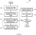
- Fig. 4 shows a method 300 for improving an endoscopic video according to a second exemplary embodiment.
- the method 300 is similar to the method 200, however the method 300 describes a non-rigid registration process to better compensate for optical distortions, tissue motion and point-of-view (POV) changes.
- Non-rigid registration generally refers to the application of a plurality of local transformations to an image to better correlate to a reference image (in this case the map image).
- a reference image in this case the map image.
- different areas of the images may be transformed independently to better capture changes in position between the areas.
- the map image is calculated according to steps 205-220 of the method 200.
- a scale space representation of the map image is calculated.
- a scale space representation generally refers to a representation of image data as a set of gradually smoothed images at different scales. For example, large-scale structures in the map image are emphasized while fine-scale structures are suppressed.
- the current image is captured and correlated to the map image, initially at low spatial frequencies.
- the current image is fused with the map image and enhanced according to steps 230-250 of the method 200.
- the image processing steps are repeated as new images are acquired.
- multiple independent regional transforms are developed by optimizing progressively smaller areas in progressively higher spatial frequency scales. Anatomy-relative motion may be estimated.
- a Structure from Motion (SFM) depth map is derived based on the non-linear transform.
- SFM generally refers to a mapping of three-dimensional structures from a succession of images captured by a moving POV, where the changing position and orientation (pose) of the imager is tracked. The depth map is continuously populated as new images are gathered.
- Elements of the aforementioned image processing methods 200, 300 may be used to further enhance displays in the following procedure-specific ways.
- Kidney stone treatments such as laser lithotripsy, may involve the fragmentation of a stone into many pieces of varying sizes, some of which would require further reduction, and some of which may be small enough to be retrieved or expelled naturally. Tracking stone particles in ureteroscopic video, inferring their size, and providing the physician with appropriate annotations may improve the speed, confidence and precision of these interventions.
- object sizing with respect to kidney stones may be sized and annotated in an endoscopic display such as, e.g., lesions, growths and other tissue features.
- Fig. 5 shows a method 400 for annotating an endoscopic video with object size information.
- the spatial transform and image map are generated and the preliminary set of images is captured and correlated, according to steps 205-215 of method 200.
- scope-relative objects are identified and segmented in the preliminary set of images.
- Scope-relative objects may include, e.g., a laser fiber for performing laser lithotripsy.
- the scope-relative object segmentation is implemented using predefined feature maps and constrained registration geometry.
- interesting objects are identified and segmented in the preliminary set of images.
- interesting objects may include, e.g., kidney stones.
- the interesting object segmentation is implemented using image features and blob-finding algorithms, as would be known by a person skilled in the art.
- the spatial transform is applied to the preliminary set of images and the super-resolution scene map is calculated excluding the identified scope-relative objects.
- a probabilistic depth map is derived according to steps 310-330 of method 300.
- the depth map is based off the independent regional transforms from the super-resolution map alignment and the implied camera pose transforms.
- the steps 310-330 include a continuous acquisition of images and a correlation/addition to the scene map to continuously improve the resolution of the scene map.
- the sizes of the previously identified interesting objects are estimated based on depth information and angular extent in a currently captured image.
- the interesting objects are annotated on the display of the currently captured image.
- the dimensions of the object may be rendered directly on the display.
- the objects may be annotated with brackets to show a boundary of the object.
- the brackets may be color-coded to show a size classification of the object by comparing the size estimate for the object with predefined treatment thresholds. For example, the brackets may be colored red when the size estimate indicates that the object requires further reduction, yellow when the size estimate indicates that the object is small enough for retrieval but too large for natural expulsion, and green when the size estimate indicates that the object may be passed naturally.
- the kidney stone may be annotated to indicate the smallest size of tube that the kidney stone can fit through.
- Fig. 6 shows an exemplary endoscopic video display where a kidney stone is bracketed.
- the frame-to-frame correlation or super-resolution map described above is used to build a statistical description of color and albedo from multiple samples at each segmented pixel.
- Machine learning with clustering algorithms and in vivo training data is used to create a composition map for projecting probability of membership within identified size groups.
- SFM techniques may be employed to inform a process for tracking an endoscope position during an endoscopic procedure.
- An SFM pose estimation for the endoscope, as determined from the endoscopic video, may be used in combination with position information from the EM tracker 108 and a segmented 3D reconstruction of the patient anatomy derived from previously acquired images to refine a position determination for the endoscope relative to a continuously deforming patient anatomy.
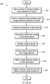
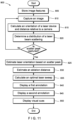
- Fig. 7 shows a method 500 for tracking the position of an endoscope relative to continuously deforming anatomy according to a first exemplary embodiment.
- a previously acquired 3D image volume of a target portion of anatomy is received and segmented.
- the 3D volume may be generated by previously captured CT images or images captured from a similar imaging medium.
- an SFM depth map is derived according to steps 305-330 of method 300. The depth map provides for anatomy-relative motion, a position estimate and a confidence level for the position estimates.
- a six degree-of-freedom (6DOF) position for the endoscope 102 is determined using the EM tracker 108 disposed near the tip of the endoscope 102.
- the 6DOF position from 515 and the images populating the SFM depth map from 510 are received substantially simultaneously and may be correlated.
- the position data from the EM tracker 108, the position data from the SFM map and the fixed volume of the anatomy are combined to estimate a minimal deformation of the anatomy sufficient to maintain the endoscope 102 within the surface boundaries of the anatomy.
- the resulting model assumes the scope 102 moves freely in the cavity until the position data indicates the scope 102 has contacted the segmented surface model.
- the anatomy is deformed to maintain the scope 102 position within the surface. For example, the anatomy displaces perpendicularly to the surface orientation.
- the combined model and the 6DOF endoscope position inform and improve the position estimation from the SFM depth map.
- the improved estimate prevents a runaway integration of noise.
- the processed images are shown on the display 114 with live annotation navigation feedback.
- the annotations may reflect positions of stones or particles from previous imaging. Additionally, by incorporating magnetic resonance (MR) tracking or optical tracking, the annotations may reflect paths that have already been navigated. The method is repeated continuously as images are acquired.
- MR magnetic resonance
- SFM techniques known in the art may be used rather than the SFM depth map derived in method 300.
- the SFM depth map from method 300 has a simpler geometry, with less non-linearity necessary to represent typical transformations, e.g., those resulting from camera motion.
- Conventional SFM algorithms also require geometric corrections for camera optics, but these corrections do not extend to assumptions about surface continuity and orientation. If these assumptions are valid, they sharply reduce the numerical complexity of the solution, which in turn improves performance and reduces errors.
- BPH benign prostate hyperplasia
- Knowledge of a position of the endoscope with respect to anatomical landmarks permits annotation of the endoscopic video, providing assistance to physicians in gauging distances and tracking an ongoing, multi-step intervention.
- Fig. 8 shows a method 600 for motion tracking in endoscopic video of a tubular structure, e.g., a urethra, according to a second exemplary embodiment.
- the method 600 may provide annotations including a current position relative to anatomical landmarks, desired positions associated with the intended intervention, and progress indicators relative to that intervention.
- a center of camera rotation and a direction of travel are mapped.
- the optical system is mapped to derive a physical angle between each image pixel and the direction of scope travel, from the POV of the scope. Mapping the optical system effectively defines a scalar, as in a latitude, for each pixel of the image, that specifies angular distance from a camera axis (the North Pole, in the latitude analogy, except that 0° would be at the pole, and 90° at the equator, rather than vice versa).
- the "pole" position in the image, at 0° is a point that remains directed at the same position when the camera is advanced or retracted exactly along the camera axis.
- a distance to each anatomic pixel is determined for each image based on the mapping steps 605-610. The distance is measured from a reference plane perpendicular to the scope 102 to the object imaged at the pixel.
- a deformed image is calculated in cylindrical space based on the determined distances and a rotation angle of the scope relative to its own axis.
- the deformed image is calculated relative to the vector from the point of scope rotation to a reference orientation feature in the scope video.
- An arbitrary feature is chosen to define the reference orientation (e. g., setting the 0° angle in Fig. 3 ), thus all subsequent mappings relate, directly or indirectly, to the reference orientation.
- image features are identified.
- Saliency metrics such as, e.g. Hessian determinants, are calculated in scale space from the deformed cylindrical image to identify the image features.
- a feature descriptor is built with image features related to any portion of interventional instruments within the field of view of the imager 104 being ignored.
- the feature descriptor is based on feature area pixel values in the cylindrical image.
- features are matched based on the descriptors.
- present algorithms tend to match an image to another image, however, in another embodiment, a global map is possible and potentially advantageous. Unlike prior methods, it is not necessary to perform scale and rotation invariant calculations. By making inferences about the geometry of the scene map, the geometry of the features should remain in consistent scale and orientation from one image to the next.
- the orientation of features is relative to the vector of the feature to a vanishing point of the extrapolated cylinder, such that if a nearby image is subsequently acquired with the scope rotated substantially along its axis, it will not affect this orientation angle, and the features can thereby be matched more directly.
- Fig. 9 shows an exemplary endoscopic video display where a current position relative to anatomic landmarks, a desired position associated with the intervention, and progress indicators relative to that intervention are shown.
- Fig. 10 shows a method 700 for motion tracking in endoscopic video of a tubular structure according to a second exemplary embodiment.
- the method 700 is similar to the method 600.
- a deformed image is calculated in cylindrical space according to steps 605-620 of method 600.
- a mask image is generated where pixels corresponding to instrument objects are ignored. If the current image is the first image, in 715, the mask image is saved as a map image. If the current image is a subsequent image, in 720, an optimal alignment with the map image is calculated based on a suitable alignment criterion. For example, the optimal alignment may be calculated based on a correlation such as a Hessian-determinant weighted correlation or an entropy-weighted correlation. In 725, the image is overlaid with the map based on the optimal alignment.
- an updated map image is calculated using a statistical combination of map position samples. For instance, the map image may be calculated using a median intensity of each color channel across all images where each pixel is defined.
- the relative position of the camera is inferred from the alignment with the map.
- One procedure for treating BPH is laser prostatectomy, which shrinks or removes prostate tissue by passing a laser over the prostate tissue.
- the procedure is characterized by a sweep range (i.e., an angular range the laser passes over) and a sweep speed (i.e. an angle change per unit time) for the laser.
- the sweep range, sweep speed, and sweep angle are determined by a power density of the laser, a distance from the tissue to the laser, a type of tissue, a laser frequency, and a fiber size.
- Annotations may be incorporated to an endoscopic video feed to facilitate laser treatment procedures through increased physician awareness and precision.
- Fig. 11 shows a method 800 for analyzing a cystoscopic video feed and providing annotations to inform a laser treatment procedure, e.g., GreenLight TM laser.
- image features are stored for a laser device.
- an image is captured.
- an orientation of the laser device and a distance relative to the camera are calculated if the imager has captured at least a portion of the laser device in the image.
- a distribution of a laser beam scattering is determined using color filtering.
- the laser orientation is estimated based on the scatter peak.
- the scatter peak is an intensity of reflected laser light, i.e. a bright spot that comes and goes with laser pulses, to infer a position in the anatomy directly in a path of the laser beam.
- an estimate of the laser orientation is determined.
- an ablation distance is estimated using a pre-calibrated map of a distance from the laser device to the scatter peak.
- the distance estimate may be refined using the distance calculation of step 815, if available.
- an optimal laser sweep rate is calculated based on the distance estimate and a laser power setting.
- a first annotation is provided on the display 114 showing the optimal sweep rate and demonstrating a relation between the optimal sweep rate and a detected sweep rate.
- a second annotation is provided on the display 114 showing an ablation depth.
- the displayed ablation depth may be relative to a distance to the capsule of the prostate.
- visual cues are provided to illustrate a proper rate and a relationship between the optimal and measured rate and sweep angle.
- Fig. 12 shows an exemplary endoscopic video display with prostate laser annotations shown, including power level relative to an inferred optimal range, apparent average depth to laser-targeted tissue, inferred sweep angle, and optimal sweep angle / sweep pace calculated from power and average depth.
- video annotations are provided comparing an apparent ablation depth at each sweep angle to a projected urethra-to-capsule distance measured in a previously acquired 3D image volume. Since the laser position is at a (former) urethral position, the distance to capsule measure in prior imaging provides a limit depth for the ablation procedure. A user may choose not to continue ablation at or beyond the capsule.
- angle and depth estimates are combined with laser angle and insertion depth measurements calculated from a 3D instrument tracking system.
- Differentiating different types of tissue in an endoscopic view may further facilitate a variety of endoscopic procedures.
- An amplification of even very slight fluctuations in aligned image frames may allow for correlation to cardiac phase measurements and highlight differences in tissue perfusion.
- an appropriately designed convolutional neural network may permit ad hoc tuning of filtering and amplification parameters to create a more flexible implementation than prior methods.
- the input volume of the CNN feeding the first convolutional layer would have a depth on the order of several seconds worth of video frames, times 3 for the red, green and blue color channels.
- the output layer would have the same length and width of the video frames, and a depth of 1, and represent the degree of cardiac correlation.
- the network would be trained on uncompressed in vivo endoscopic video with a cost function measuring the pixel's maximum time correlation to any of a series of predefined wavelets, scaled to match the specified cardiac interval.
- the resulting values, and maximum wavelet may drive amplification of the signals to derive differences in tissue perfusion and, relatedly, highlight differences in tissue makeup.
- Use of the CNN allows for the derivation in an unsupervised manner, whereas prior methods involve repeated user interventions to appropriately filter the raw image frames.
- a CNN may also be employed to produce images with low noise and enhanced detail.
- a deep Convolutional Neural Network may be applied directly to the pixel data from the endoscope.
- the output volume has the same depth as the input volume (presumably 3, for the red, green & blue channels) but has a width & height that are the same integer multiple of the input width and height (the super-resolution multiplier), and fractional striding of convolution layers.
- the error term for training the network is calculated by performing discrete Hartley transforms on the original pixels, as well as the output values, and minimizing the squared difference, except where the highest spatial frequency component of the input data is shifted beyond its Nyquist limit to correspond with the highest spatial frequency representable in the output data transform.
- the spatial frequency transform of the input data is performed on a multi-frame average image computed using correlation-based registration transforms, to aid in noise suppression.
- Various existing super-resolution or SFM methods may be combined with elements of the disclosed methods to improve the existing techniques.
- the known/calibratable optical geometry of the endoscope as discussed with respect to method 200, may be taken advantage of to improve existing super-resolution techniques.
- An image map generated based on the optical geometry of the endoscope may have 1 sample per pixel in each frame and the scope images may be transformed with known super-resolution techniques, such as "shift and fusion.”
- Other techniques for enhancing an image include applying various filters to the pixel data, such as band, median, or high frequency filters, and using edge detection for interesting objects through a cloudy or turbid environment. These further techniques may be used alone or in combination with the aforementioned embodiments.
Landscapes
- Engineering & Computer Science (AREA)
- Physics & Mathematics (AREA)
- Health & Medical Sciences (AREA)
- General Physics & Mathematics (AREA)
- Theoretical Computer Science (AREA)
- Life Sciences & Earth Sciences (AREA)
- Surgery (AREA)
- Computer Vision & Pattern Recognition (AREA)
- Molecular Biology (AREA)
- Biomedical Technology (AREA)
- Heart & Thoracic Surgery (AREA)
- Medical Informatics (AREA)
- Animal Behavior & Ethology (AREA)
- General Health & Medical Sciences (AREA)
- Public Health (AREA)
- Veterinary Medicine (AREA)
- Pathology (AREA)
- Nuclear Medicine, Radiotherapy & Molecular Imaging (AREA)
- Biophysics (AREA)
- Radiology & Medical Imaging (AREA)
- Optics & Photonics (AREA)
- Signal Processing (AREA)
- Human Computer Interaction (AREA)
- Multimedia (AREA)
- Gynecology & Obstetrics (AREA)
- Oral & Maxillofacial Surgery (AREA)
- Endoscopes (AREA)
- Image Processing (AREA)
- Closed-Circuit Television Systems (AREA)
Claims (7)
- Endoskopisches System (100), umfassend:einen endoskopischen Bildgeber (104), der eingerichtet ist, Einzelbilder einer Zielstelle in einem lebenden Körper zu erfassen;einen Prozessor, der eingerichtet ist:eine räumliche Transformation auf einen vorläufigen Satz von Einzelbildern anzuwenden, wobei die räumliche Transformation die Einzelbilder in zylindrische Koordinaten umwandelt;ein Kartenbild aus den räumlich transformierten Einzelbildern zu berechnen, wobei jede Pixelposition im Kartenbild durch einen Vektor fester Dimension definiert ist;ein aktuelles Einzelbild mit dem Kartenbild auszurichten und die räumliche Transformation auf das aktuelle Einzelbild anzuwenden;das räumlich transformierte aktuelle Einzelbild mit dem Kartenbild zu fusionieren, um ein fusioniertes Bild zu erzeugen; undeine inverse räumliche Transformation auf das fusionierte Bild anzuwenden, um ein verbessertes aktuelles Einzelbild mit einer größeren räumlichen Auflösung als das aktuelle Einzelbild zu erzeugen; undeine Anzeige (114), die eingerichtet ist, das verbesserte aktuelle Einzelbild anzuzeigen.
- Endoskopisches System (100) nach Anspruch 1, wobei die räumliche Transformation auf der Grundlage einer optischen Geometrie des endoskopischen Bildgebers (104) erzeugt wird.
- Endoskopisches System (100) nach einem der Ansprüche 1 bis 2, wobei das Kartenbild eine Auflösung hat, die um ein ganzzahliges Vielfaches größer ist als die Auflösung des endoskopischen Bildgebers (104).
- Endoskopisches System (100) nach einem der Ansprüche 1 bis 3, wobei das aktuelle Einzelbild mit dem Kartenbild auf der Grundlage einer Kreuzkorrelation ausgerichtet wird, bei der ein Grad der Ähnlichkeit zwischen dem aktuellen Einzelbild und dem Kartenbild gemessen wird.
- Endoskopisches System (100) nach einem der Ansprüche 1 bis 4, wobei der Prozessor ferner eingerichtet ist:
das Sichtfeld des verbesserten aktuellen Bildes im Vergleich zum aktuellen Einzelbild auf der Grundlage eines Bereichs des Kartenbildes zu erweitern, der das räumlich transformierte aktuelle Einzelbild umgibt. - Endoskopisches System (100) nach einem der Ansprüche 1 bis 5, wobei der Prozessor ferner eingerichtet ist:
das räumlich transformierte aktuelle Einzelbild zum Kartenbild hinzuzufügen. - Endoskopisches System (100) nach einem der Ansprüche 1 bis 6, wobei, wenn eine bestimmte Pixelposition im Kartenbild voll ist, ein ältestes Sample gelöscht wird, wenn ein neues Sample hinzugefügt wird.
Priority Applications (2)
| Application Number | Priority Date | Filing Date | Title |
|---|---|---|---|
| EP25181913.2A EP4592940A3 (de) | 2019-09-23 | 2020-08-27 | System zur endoskopischen videoverstärkung |
| EP24158246.9A EP4345733B1 (de) | 2019-09-23 | 2020-08-27 | System zur endoskopischen videoverstärkung |
Applications Claiming Priority (2)
| Application Number | Priority Date | Filing Date | Title |
|---|---|---|---|
| US201962904408P | 2019-09-23 | 2019-09-23 | |
| PCT/US2020/048202 WO2021061335A1 (en) | 2019-09-23 | 2020-08-27 | System and method for endoscopic video enhancement, quantitation and surgical guidance |
Related Child Applications (3)
| Application Number | Title | Priority Date | Filing Date |
|---|---|---|---|
| EP25181913.2A Division EP4592940A3 (de) | 2019-09-23 | 2020-08-27 | System zur endoskopischen videoverstärkung |
| EP24158246.9A Division EP4345733B1 (de) | 2019-09-23 | 2020-08-27 | System zur endoskopischen videoverstärkung |
| EP24158246.9A Division-Into EP4345733B1 (de) | 2019-09-23 | 2020-08-27 | System zur endoskopischen videoverstärkung |
Publications (2)
| Publication Number | Publication Date |
|---|---|
| EP4035120A1 EP4035120A1 (de) | 2022-08-03 |
| EP4035120B1 true EP4035120B1 (de) | 2024-03-27 |
Family
ID=72433040
Family Applications (3)
| Application Number | Title | Priority Date | Filing Date |
|---|---|---|---|
| EP24158246.9A Active EP4345733B1 (de) | 2019-09-23 | 2020-08-27 | System zur endoskopischen videoverstärkung |
| EP20768785.6A Active EP4035120B1 (de) | 2019-09-23 | 2020-08-27 | System zur verbesserung endoskopischer videos |
| EP25181913.2A Pending EP4592940A3 (de) | 2019-09-23 | 2020-08-27 | System zur endoskopischen videoverstärkung |
Family Applications Before (1)
| Application Number | Title | Priority Date | Filing Date |
|---|---|---|---|
| EP24158246.9A Active EP4345733B1 (de) | 2019-09-23 | 2020-08-27 | System zur endoskopischen videoverstärkung |
Family Applications After (1)
| Application Number | Title | Priority Date | Filing Date |
|---|---|---|---|
| EP25181913.2A Pending EP4592940A3 (de) | 2019-09-23 | 2020-08-27 | System zur endoskopischen videoverstärkung |
Country Status (8)
| Country | Link |
|---|---|
| US (4) | US11430097B2 (de) |
| EP (3) | EP4345733B1 (de) |
| JP (3) | JP7206438B2 (de) |
| KR (2) | KR102939120B1 (de) |
| CN (2) | CN114423355B (de) |
| AU (3) | AU2020352836B2 (de) |
| CA (2) | CA3147447C (de) |
| WO (1) | WO2021061335A1 (de) |
Families Citing this family (13)
| Publication number | Priority date | Publication date | Assignee | Title |
|---|---|---|---|---|
| EP4345733B1 (de) * | 2019-09-23 | 2025-07-16 | Boston Scientific Scimed, Inc. | System zur endoskopischen videoverstärkung |
| US11918178B2 (en) * | 2020-03-06 | 2024-03-05 | Verily Life Sciences Llc | Detecting deficient coverage in gastroenterological procedures |
| US11288771B2 (en) * | 2020-04-29 | 2022-03-29 | Adobe Inc. | Texture hallucination for large-scale image super-resolution |
| WO2022066569A1 (en) * | 2020-09-22 | 2022-03-31 | Boston Scientific Scimed, Inc. | Image processing systems and methods of using the same |
| TWI792381B (zh) * | 2021-03-25 | 2023-02-11 | 鈺立微電子股份有限公司 | 影像擷取裝置及其深度資訊計算方法 |
| US12087007B2 (en) | 2021-03-31 | 2024-09-10 | Auris Health, Inc. | Vision-based 6DOF camera pose estimation in bronchoscopy |
| CN113538522B (zh) * | 2021-08-12 | 2022-08-12 | 广东工业大学 | 一种用于腹腔镜微创手术的器械视觉跟踪方法 |
| US12505660B2 (en) * | 2021-12-29 | 2025-12-23 | Samsung Electronics Co., Ltd. | Image processing method and apparatus using convolutional neural network |
| CN119300767A (zh) * | 2022-06-01 | 2025-01-10 | 皇家飞利浦有限公司 | 引导介入成像设备 |
| CN115988151B (zh) * | 2022-12-29 | 2024-07-05 | 南京图格医疗科技有限公司 | 一种使用低像素时钟实时处理视频的方法及系统 |
| EP4669235A1 (de) * | 2023-02-20 | 2025-12-31 | The Chancellor, Masters and Scholars of The University of Oxford | Ureteroskopiebildgebungssystem und -verfahren |
| CN116211260B (zh) * | 2023-05-09 | 2023-07-21 | 西南医科大学附属医院 | 一种基于变焦扫描的肾结石形态三维成像系统及方法 |
| CN116958147B (zh) * | 2023-09-21 | 2023-12-22 | 青岛美迪康数字工程有限公司 | 基于深度图像特征的目标区域确定方法、装置和设备 |
Family Cites Families (44)
| Publication number | Priority date | Publication date | Assignee | Title |
|---|---|---|---|---|
| JPH10276980A (ja) | 1997-04-02 | 1998-10-20 | Olympus Optical Co Ltd | 内視鏡装置 |
| US6804406B1 (en) | 2000-08-30 | 2004-10-12 | Honeywell International Inc. | Electronic calibration for seamless tiled display using optical function generator |
| WO2002038323A1 (en) | 2000-11-13 | 2002-05-16 | Micmacmo Aps | Laser ablation |
| US7381183B2 (en) | 2003-04-21 | 2008-06-03 | Karl Storz Development Corp. | Method for capturing and displaying endoscopic maps |
| US7420675B2 (en) * | 2003-06-25 | 2008-09-02 | The University Of Akron | Multi-wavelength imaging system |
| US8036494B2 (en) * | 2004-04-15 | 2011-10-11 | Hewlett-Packard Development Company, L.P. | Enhancing image resolution |
| JP4018679B2 (ja) | 2004-08-24 | 2007-12-05 | ザイオソフト株式会社 | レンダリング処理方法、レンダリング処理プログラム、レンダリング処理装置 |
| US8248414B2 (en) * | 2006-09-18 | 2012-08-21 | Stryker Corporation | Multi-dimensional navigation of endoscopic video |
| US7813538B2 (en) * | 2007-04-17 | 2010-10-12 | University Of Washington | Shadowing pipe mosaicing algorithms with application to esophageal endoscopy |
| JP4563421B2 (ja) | 2007-05-28 | 2010-10-13 | ザイオソフト株式会社 | 画像処理方法及び画像処理プログラム |
| JP5235650B2 (ja) | 2008-12-26 | 2013-07-10 | Hoya株式会社 | 光走査型内視鏡装置、光走査型内視鏡、および光走査型内視鏡プロセッサ |
| US8348829B2 (en) * | 2008-12-26 | 2013-01-08 | Hoya Corporation | Scanning endoscope apparatus, scanning endoscope, and scanning endoscope processor |
| CN101616310B (zh) * | 2009-07-17 | 2011-05-11 | 清华大学 | 可变视角及分辨率的双目视觉系统目标图像稳定化方法 |
| JP2011139734A (ja) | 2010-01-05 | 2011-07-21 | Hoya Corp | 内視鏡装置 |
| JP6144914B2 (ja) | 2010-02-12 | 2017-06-07 | コーニンクレッカ フィリップス エヌ ヴェKoninklijke Philips N.V. | レーザ改良された3d表面再構成 |
| TWI428855B (zh) * | 2010-12-02 | 2014-03-01 | Chung Shan Inst Of Science | The Method of Restoring Three Dimensional Image of Capsule Endoscopy |
| CN108606773B (zh) | 2012-02-29 | 2020-08-11 | 普罗赛普特生物机器人公司 | 自动化图像引导的组织切除和处理 |
| CN102651127A (zh) * | 2012-04-01 | 2012-08-29 | 深圳市万兴软件有限公司 | 一种超分辨率重建的图像处理方法及系统 |
| US10231626B2 (en) * | 2013-03-15 | 2019-03-19 | The Regents Of The University Of California | Imaging system and method for fluorescence guided surgery |
| CN103198501B (zh) * | 2013-04-09 | 2016-12-07 | 上海理工大学 | 一种牙齿全景图像自动重构方法 |
| JP6265627B2 (ja) | 2013-05-23 | 2018-01-24 | オリンパス株式会社 | 内視鏡装置及び内視鏡装置の作動方法 |
| US9282985B2 (en) | 2013-11-11 | 2016-03-15 | Gyrus Acmi, Inc. | Aiming beam detection for safe laser lithotripsy |
| JP6235921B2 (ja) | 2014-02-07 | 2017-11-22 | 国立大学法人広島大学 | 内視鏡画像診断支援システム |
| JP6270537B2 (ja) | 2014-02-27 | 2018-01-31 | オリンパス株式会社 | 医療用システム |
| US9259231B2 (en) | 2014-05-11 | 2016-02-16 | Gyrus Acmi, Inc. | Computer aided image-based enhanced intracorporeal lithotripsy |
| EP3254234A4 (de) * | 2015-02-06 | 2018-07-11 | The University of Akron | System zur optischen bildgebung und verfahren dafür |
| WO2016171238A1 (ja) | 2015-04-23 | 2016-10-27 | オリンパス株式会社 | 外科処置装置 |
| CN107667380A (zh) * | 2015-06-05 | 2018-02-06 | 西门子公司 | 用于内窥镜和腹腔镜导航的同时场景解析和模型融合的方法和系统 |
| EP3103396B1 (de) * | 2015-06-10 | 2018-10-24 | Helmholtz Zentrum München Deutsches Forschungszentrum für Gesundheit und Umwelt GmbH | Vorrichtung und verfahren zur hybriden optoakustischen tomografie und ultraschallsonografie |
| CN107249427B (zh) | 2015-07-06 | 2019-05-07 | 奥林巴斯株式会社 | 医疗装置、医疗图像生成方法以及医疗图像生成程序 |
| CN105069748B (zh) * | 2015-07-16 | 2017-11-10 | 哈尔滨工业大学 | 一种基于微小卫星物方扫描技术获取高分辨率图像的方法 |
| US20170084036A1 (en) * | 2015-09-21 | 2017-03-23 | Siemens Aktiengesellschaft | Registration of video camera with medical imaging |
| US10154193B1 (en) * | 2016-03-08 | 2018-12-11 | National Technology & Engineering Solutions Of Sandia, Llc | Noncircular aperture imaging system |
| US20170281102A1 (en) * | 2016-03-31 | 2017-10-05 | Weng-Dah Ken | Non-contact angle measuring apparatus, mission critical inspection apparatus, non-invasive diagnosis/treatment apparatus, method for filtering matter wave from a composite particle beam, non-invasive measuring apparatus, apparatus for generating a virtual space-time lattice, and fine atomic clock |
| CN106060418A (zh) * | 2016-06-29 | 2016-10-26 | 深圳市优象计算技术有限公司 | 基于imu信息的宽动态图像融合方法 |
| US11751947B2 (en) * | 2017-05-30 | 2023-09-12 | Brainlab Ag | Soft tissue tracking using physiologic volume rendering |
| JP7290245B2 (ja) | 2017-06-09 | 2023-06-13 | 株式会社Okファイバーテクノロジー | 内視鏡システム、データ作成方法及び、内視鏡システムの作動方法 |
| JP7236689B2 (ja) | 2018-03-05 | 2023-03-10 | リオン株式会社 | 3次元形状データ作成システムの作動方法、及び3次元形状データ作成システム |
| CN108564652B (zh) * | 2018-03-12 | 2020-02-14 | 中国科学院自动化研究所 | 高效利用内存的高精度三维重建方法与系统及设备 |
| GB2589250B (en) * | 2018-06-15 | 2023-03-08 | Canon Kk | Medical image processing apparatus, medical image processing method and program |
| CN109342861B (zh) * | 2018-12-10 | 2020-12-22 | 哈尔滨工业大学 | 一种霍尔推力器周向辐条特性测量方法及系统 |
| US10987518B2 (en) * | 2019-01-03 | 2021-04-27 | Pacesetter, Inc. | Terminating pacemaker mediated tachycardia (PMT) in dual chamber leadless pacemaker systems |
| EP4345733B1 (de) * | 2019-09-23 | 2025-07-16 | Boston Scientific Scimed, Inc. | System zur endoskopischen videoverstärkung |
| WO2022056642A1 (en) * | 2020-09-18 | 2022-03-24 | Stryker European Operations Limited | Systems and methods for fluorescence visualization |
-
2020
- 2020-08-27 EP EP24158246.9A patent/EP4345733B1/de active Active
- 2020-08-27 AU AU2020352836A patent/AU2020352836B2/en active Active
- 2020-08-27 US US16/948,013 patent/US11430097B2/en active Active
- 2020-08-27 CA CA3147447A patent/CA3147447C/en active Active
- 2020-08-27 EP EP20768785.6A patent/EP4035120B1/de active Active
- 2020-08-27 CN CN202080066528.2A patent/CN114423355B/zh active Active
- 2020-08-27 WO PCT/US2020/048202 patent/WO2021061335A1/en not_active Ceased
- 2020-08-27 CA CA3232181A patent/CA3232181A1/en active Pending
- 2020-08-27 KR KR1020247002652A patent/KR102939120B1/ko active Active
- 2020-08-27 EP EP25181913.2A patent/EP4592940A3/de active Pending
- 2020-08-27 KR KR1020227009550A patent/KR102630074B1/ko active Active
- 2020-08-27 JP JP2022515013A patent/JP7206438B2/ja active Active
- 2020-08-27 CN CN202411306792.5A patent/CN119273555A/zh active Pending
-
2022
- 2022-07-27 US US17/815,445 patent/US11954834B2/en active Active
-
2023
- 2023-01-04 JP JP2023000266A patent/JP7513774B2/ja active Active
- 2023-08-10 AU AU2023214320A patent/AU2023214320B2/en active Active
-
2024
- 2024-02-23 US US18/585,477 patent/US12354240B2/en active Active
- 2024-06-27 JP JP2024103678A patent/JP7756202B2/ja active Active
-
2025
- 2025-01-31 AU AU2025200684A patent/AU2025200684A1/en active Pending
- 2025-06-11 US US19/234,683 patent/US20250307992A1/en active Pending
Also Published As
Similar Documents
| Publication | Publication Date | Title |
|---|---|---|
| EP4035120B1 (de) | System zur verbesserung endoskopischer videos | |
| US9646423B1 (en) | Systems and methods for providing augmented reality in minimally invasive surgery | |
| JP5795599B2 (ja) | 内視鏡手術のための画像統合ベースレジストレーション及びナビゲーション | |
| JP2013517031A5 (de) | ||
| US12349859B2 (en) | Two-phase instrument guidance for accurate endoscopic surgical procedures | |
| US20230215059A1 (en) | Three-dimensional model reconstruction | |
| Speidel et al. | Interventional imaging: vision |
Legal Events
| Date | Code | Title | Description |
|---|---|---|---|
| STAA | Information on the status of an ep patent application or granted ep patent |
Free format text: STATUS: UNKNOWN |
|
| STAA | Information on the status of an ep patent application or granted ep patent |
Free format text: STATUS: THE INTERNATIONAL PUBLICATION HAS BEEN MADE |
|
| PUAI | Public reference made under article 153(3) epc to a published international application that has entered the european phase |
Free format text: ORIGINAL CODE: 0009012 |
|
| STAA | Information on the status of an ep patent application or granted ep patent |
Free format text: STATUS: REQUEST FOR EXAMINATION WAS MADE |
|
| 17P | Request for examination filed |
Effective date: 20220106 |
|
| AK | Designated contracting states |
Kind code of ref document: A1 Designated state(s): AL AT BE BG CH CY CZ DE DK EE ES FI FR GB GR HR HU IE IS IT LI LT LU LV MC MK MT NL NO PL PT RO RS SE SI SK SM TR |
|
| DAV | Request for validation of the european patent (deleted) | ||
| DAX | Request for extension of the european patent (deleted) | ||
| GRAP | Despatch of communication of intention to grant a patent |
Free format text: ORIGINAL CODE: EPIDOSNIGR1 |
|
| STAA | Information on the status of an ep patent application or granted ep patent |
Free format text: STATUS: GRANT OF PATENT IS INTENDED |
|
| INTG | Intention to grant announced |
Effective date: 20231023 |
|
| RIN1 | Information on inventor provided before grant (corrected) |
Inventor name: HARRAH, TIMOTHY, PAUL Inventor name: RIKER, ROBERT, J. Inventor name: RAUNIYAR, NIRAJ, PRASAD |
|
| GRAS | Grant fee paid |
Free format text: ORIGINAL CODE: EPIDOSNIGR3 |
|
| GRAA | (expected) grant |
Free format text: ORIGINAL CODE: 0009210 |
|
| STAA | Information on the status of an ep patent application or granted ep patent |
Free format text: STATUS: THE PATENT HAS BEEN GRANTED |
|
| AK | Designated contracting states |
Kind code of ref document: B1 Designated state(s): AL AT BE BG CH CY CZ DE DK EE ES FI FR GB GR HR HU IE IS IT LI LT LU LV MC MK MT NL NO PL PT RO RS SE SI SK SM TR |
|
| REG | Reference to a national code |
Ref country code: GB Ref legal event code: FG4D |
|
| REG | Reference to a national code |
Ref country code: CH Ref legal event code: EP |
|
| REG | Reference to a national code |
Ref country code: DE Ref legal event code: R096 Ref document number: 602020028015 Country of ref document: DE |
|
| REG | Reference to a national code |
Ref country code: IE Ref legal event code: FG4D |
|
| PG25 | Lapsed in a contracting state [announced via postgrant information from national office to epo] |
Ref country code: LT Free format text: LAPSE BECAUSE OF FAILURE TO SUBMIT A TRANSLATION OF THE DESCRIPTION OR TO PAY THE FEE WITHIN THE PRESCRIBED TIME-LIMIT Effective date: 20240327 |
|
| REG | Reference to a national code |
Ref country code: LT Ref legal event code: MG9D |
|
| PG25 | Lapsed in a contracting state [announced via postgrant information from national office to epo] |
Ref country code: GR Free format text: LAPSE BECAUSE OF FAILURE TO SUBMIT A TRANSLATION OF THE DESCRIPTION OR TO PAY THE FEE WITHIN THE PRESCRIBED TIME-LIMIT Effective date: 20240628 |
|
| PG25 | Lapsed in a contracting state [announced via postgrant information from national office to epo] |
Ref country code: HR Free format text: LAPSE BECAUSE OF FAILURE TO SUBMIT A TRANSLATION OF THE DESCRIPTION OR TO PAY THE FEE WITHIN THE PRESCRIBED TIME-LIMIT Effective date: 20240327 Ref country code: RS Free format text: LAPSE BECAUSE OF FAILURE TO SUBMIT A TRANSLATION OF THE DESCRIPTION OR TO PAY THE FEE WITHIN THE PRESCRIBED TIME-LIMIT Effective date: 20240627 |
|
| PG25 | Lapsed in a contracting state [announced via postgrant information from national office to epo] |
Ref country code: RS Free format text: LAPSE BECAUSE OF FAILURE TO SUBMIT A TRANSLATION OF THE DESCRIPTION OR TO PAY THE FEE WITHIN THE PRESCRIBED TIME-LIMIT Effective date: 20240627 Ref country code: NO Free format text: LAPSE BECAUSE OF FAILURE TO SUBMIT A TRANSLATION OF THE DESCRIPTION OR TO PAY THE FEE WITHIN THE PRESCRIBED TIME-LIMIT Effective date: 20240627 Ref country code: LT Free format text: LAPSE BECAUSE OF FAILURE TO SUBMIT A TRANSLATION OF THE DESCRIPTION OR TO PAY THE FEE WITHIN THE PRESCRIBED TIME-LIMIT Effective date: 20240327 Ref country code: HR Free format text: LAPSE BECAUSE OF FAILURE TO SUBMIT A TRANSLATION OF THE DESCRIPTION OR TO PAY THE FEE WITHIN THE PRESCRIBED TIME-LIMIT Effective date: 20240327 Ref country code: GR Free format text: LAPSE BECAUSE OF FAILURE TO SUBMIT A TRANSLATION OF THE DESCRIPTION OR TO PAY THE FEE WITHIN THE PRESCRIBED TIME-LIMIT Effective date: 20240628 Ref country code: FI Free format text: LAPSE BECAUSE OF FAILURE TO SUBMIT A TRANSLATION OF THE DESCRIPTION OR TO PAY THE FEE WITHIN THE PRESCRIBED TIME-LIMIT Effective date: 20240327 Ref country code: BG Free format text: LAPSE BECAUSE OF FAILURE TO SUBMIT A TRANSLATION OF THE DESCRIPTION OR TO PAY THE FEE WITHIN THE PRESCRIBED TIME-LIMIT Effective date: 20240327 |
|
| REG | Reference to a national code |
Ref country code: NL Ref legal event code: FP |
|
| PG25 | Lapsed in a contracting state [announced via postgrant information from national office to epo] |
Ref country code: SE Free format text: LAPSE BECAUSE OF FAILURE TO SUBMIT A TRANSLATION OF THE DESCRIPTION OR TO PAY THE FEE WITHIN THE PRESCRIBED TIME-LIMIT Effective date: 20240327 Ref country code: LV Free format text: LAPSE BECAUSE OF FAILURE TO SUBMIT A TRANSLATION OF THE DESCRIPTION OR TO PAY THE FEE WITHIN THE PRESCRIBED TIME-LIMIT Effective date: 20240327 |
|
| REG | Reference to a national code |
Ref country code: AT Ref legal event code: MK05 Ref document number: 1670629 Country of ref document: AT Kind code of ref document: T Effective date: 20240327 |
|
| PG25 | Lapsed in a contracting state [announced via postgrant information from national office to epo] |
Ref country code: IS Free format text: LAPSE BECAUSE OF FAILURE TO SUBMIT A TRANSLATION OF THE DESCRIPTION OR TO PAY THE FEE WITHIN THE PRESCRIBED TIME-LIMIT Effective date: 20240727 |
|
| PG25 | Lapsed in a contracting state [announced via postgrant information from national office to epo] |
Ref country code: PT Free format text: LAPSE BECAUSE OF FAILURE TO SUBMIT A TRANSLATION OF THE DESCRIPTION OR TO PAY THE FEE WITHIN THE PRESCRIBED TIME-LIMIT Effective date: 20240729 Ref country code: SM Free format text: LAPSE BECAUSE OF FAILURE TO SUBMIT A TRANSLATION OF THE DESCRIPTION OR TO PAY THE FEE WITHIN THE PRESCRIBED TIME-LIMIT Effective date: 20240327 |
|
| PG25 | Lapsed in a contracting state [announced via postgrant information from national office to epo] |
Ref country code: ES Free format text: LAPSE BECAUSE OF FAILURE TO SUBMIT A TRANSLATION OF THE DESCRIPTION OR TO PAY THE FEE WITHIN THE PRESCRIBED TIME-LIMIT Effective date: 20240327 |
|
| PG25 | Lapsed in a contracting state [announced via postgrant information from national office to epo] |
Ref country code: CZ Free format text: LAPSE BECAUSE OF FAILURE TO SUBMIT A TRANSLATION OF THE DESCRIPTION OR TO PAY THE FEE WITHIN THE PRESCRIBED TIME-LIMIT Effective date: 20240327 Ref country code: EE Free format text: LAPSE BECAUSE OF FAILURE TO SUBMIT A TRANSLATION OF THE DESCRIPTION OR TO PAY THE FEE WITHIN THE PRESCRIBED TIME-LIMIT Effective date: 20240327 |
|
| PG25 | Lapsed in a contracting state [announced via postgrant information from national office to epo] |
Ref country code: AT Free format text: LAPSE BECAUSE OF FAILURE TO SUBMIT A TRANSLATION OF THE DESCRIPTION OR TO PAY THE FEE WITHIN THE PRESCRIBED TIME-LIMIT Effective date: 20240327 |
|
| PG25 | Lapsed in a contracting state [announced via postgrant information from national office to epo] |
Ref country code: PL Free format text: LAPSE BECAUSE OF FAILURE TO SUBMIT A TRANSLATION OF THE DESCRIPTION OR TO PAY THE FEE WITHIN THE PRESCRIBED TIME-LIMIT Effective date: 20240327 |
|
| PG25 | Lapsed in a contracting state [announced via postgrant information from national office to epo] |
Ref country code: SK Free format text: LAPSE BECAUSE OF FAILURE TO SUBMIT A TRANSLATION OF THE DESCRIPTION OR TO PAY THE FEE WITHIN THE PRESCRIBED TIME-LIMIT Effective date: 20240327 |
|
| PG25 | Lapsed in a contracting state [announced via postgrant information from national office to epo] |
Ref country code: SM Free format text: LAPSE BECAUSE OF FAILURE TO SUBMIT A TRANSLATION OF THE DESCRIPTION OR TO PAY THE FEE WITHIN THE PRESCRIBED TIME-LIMIT Effective date: 20240327 Ref country code: SK Free format text: LAPSE BECAUSE OF FAILURE TO SUBMIT A TRANSLATION OF THE DESCRIPTION OR TO PAY THE FEE WITHIN THE PRESCRIBED TIME-LIMIT Effective date: 20240327 Ref country code: RO Free format text: LAPSE BECAUSE OF FAILURE TO SUBMIT A TRANSLATION OF THE DESCRIPTION OR TO PAY THE FEE WITHIN THE PRESCRIBED TIME-LIMIT Effective date: 20240327 Ref country code: PT Free format text: LAPSE BECAUSE OF FAILURE TO SUBMIT A TRANSLATION OF THE DESCRIPTION OR TO PAY THE FEE WITHIN THE PRESCRIBED TIME-LIMIT Effective date: 20240729 Ref country code: PL Free format text: LAPSE BECAUSE OF FAILURE TO SUBMIT A TRANSLATION OF THE DESCRIPTION OR TO PAY THE FEE WITHIN THE PRESCRIBED TIME-LIMIT Effective date: 20240327 Ref country code: IS Free format text: LAPSE BECAUSE OF FAILURE TO SUBMIT A TRANSLATION OF THE DESCRIPTION OR TO PAY THE FEE WITHIN THE PRESCRIBED TIME-LIMIT Effective date: 20240727 Ref country code: ES Free format text: LAPSE BECAUSE OF FAILURE TO SUBMIT A TRANSLATION OF THE DESCRIPTION OR TO PAY THE FEE WITHIN THE PRESCRIBED TIME-LIMIT Effective date: 20240327 Ref country code: EE Free format text: LAPSE BECAUSE OF FAILURE TO SUBMIT A TRANSLATION OF THE DESCRIPTION OR TO PAY THE FEE WITHIN THE PRESCRIBED TIME-LIMIT Effective date: 20240327 Ref country code: CZ Free format text: LAPSE BECAUSE OF FAILURE TO SUBMIT A TRANSLATION OF THE DESCRIPTION OR TO PAY THE FEE WITHIN THE PRESCRIBED TIME-LIMIT Effective date: 20240327 Ref country code: AT Free format text: LAPSE BECAUSE OF FAILURE TO SUBMIT A TRANSLATION OF THE DESCRIPTION OR TO PAY THE FEE WITHIN THE PRESCRIBED TIME-LIMIT Effective date: 20240327 |
|
| PG25 | Lapsed in a contracting state [announced via postgrant information from national office to epo] |
Ref country code: IT Free format text: LAPSE BECAUSE OF FAILURE TO SUBMIT A TRANSLATION OF THE DESCRIPTION OR TO PAY THE FEE WITHIN THE PRESCRIBED TIME-LIMIT Effective date: 20240327 |
|
| PG25 | Lapsed in a contracting state [announced via postgrant information from national office to epo] |
Ref country code: IT Free format text: LAPSE BECAUSE OF FAILURE TO SUBMIT A TRANSLATION OF THE DESCRIPTION OR TO PAY THE FEE WITHIN THE PRESCRIBED TIME-LIMIT Effective date: 20240327 |
|
| REG | Reference to a national code |
Ref country code: DE Ref legal event code: R097 Ref document number: 602020028015 Country of ref document: DE |
|
| PG25 | Lapsed in a contracting state [announced via postgrant information from national office to epo] |
Ref country code: DK Free format text: LAPSE BECAUSE OF FAILURE TO SUBMIT A TRANSLATION OF THE DESCRIPTION OR TO PAY THE FEE WITHIN THE PRESCRIBED TIME-LIMIT Effective date: 20240327 |
|
| PG25 | Lapsed in a contracting state [announced via postgrant information from national office to epo] |
Ref country code: DK Free format text: LAPSE BECAUSE OF FAILURE TO SUBMIT A TRANSLATION OF THE DESCRIPTION OR TO PAY THE FEE WITHIN THE PRESCRIBED TIME-LIMIT Effective date: 20240327 |
|
| PLBE | No opposition filed within time limit |
Free format text: ORIGINAL CODE: 0009261 |
|
| STAA | Information on the status of an ep patent application or granted ep patent |
Free format text: STATUS: NO OPPOSITION FILED WITHIN TIME LIMIT |
|
| 26N | No opposition filed |
Effective date: 20250103 |
|
| REG | Reference to a national code |
Ref country code: CH Ref legal event code: PL |
|
| PG25 | Lapsed in a contracting state [announced via postgrant information from national office to epo] |
Ref country code: LU Free format text: LAPSE BECAUSE OF NON-PAYMENT OF DUE FEES Effective date: 20240827 |
|
| PG25 | Lapsed in a contracting state [announced via postgrant information from national office to epo] |
Ref country code: MC Free format text: LAPSE BECAUSE OF FAILURE TO SUBMIT A TRANSLATION OF THE DESCRIPTION OR TO PAY THE FEE WITHIN THE PRESCRIBED TIME-LIMIT Effective date: 20240327 Ref country code: CH Free format text: LAPSE BECAUSE OF NON-PAYMENT OF DUE FEES Effective date: 20240831 Ref country code: SI Free format text: LAPSE BECAUSE OF FAILURE TO SUBMIT A TRANSLATION OF THE DESCRIPTION OR TO PAY THE FEE WITHIN THE PRESCRIBED TIME-LIMIT Effective date: 20240327 |
|
| REG | Reference to a national code |
Ref country code: BE Ref legal event code: MM Effective date: 20240831 |
|
| PG25 | Lapsed in a contracting state [announced via postgrant information from national office to epo] |
Ref country code: BE Free format text: LAPSE BECAUSE OF NON-PAYMENT OF DUE FEES Effective date: 20240831 |
|
| PG25 | Lapsed in a contracting state [announced via postgrant information from national office to epo] |
Ref country code: FR Free format text: LAPSE BECAUSE OF NON-PAYMENT OF DUE FEES Effective date: 20240831 |
|
| PGFP | Annual fee paid to national office [announced via postgrant information from national office to epo] |
Ref country code: NL Payment date: 20250723 Year of fee payment: 6 |
|
| PGFP | Annual fee paid to national office [announced via postgrant information from national office to epo] |
Ref country code: DE Payment date: 20250724 Year of fee payment: 6 |
|
| PGFP | Annual fee paid to national office [announced via postgrant information from national office to epo] |
Ref country code: GB Payment date: 20250725 Year of fee payment: 6 |
|
| PGFP | Annual fee paid to national office [announced via postgrant information from national office to epo] |
Ref country code: IE Payment date: 20250725 Year of fee payment: 6 |
|
| PG25 | Lapsed in a contracting state [announced via postgrant information from national office to epo] |
Ref country code: CY Free format text: LAPSE BECAUSE OF FAILURE TO SUBMIT A TRANSLATION OF THE DESCRIPTION OR TO PAY THE FEE WITHIN THE PRESCRIBED TIME-LIMIT; INVALID AB INITIO Effective date: 20200827 |
|
| PG25 | Lapsed in a contracting state [announced via postgrant information from national office to epo] |
Ref country code: HU Free format text: LAPSE BECAUSE OF FAILURE TO SUBMIT A TRANSLATION OF THE DESCRIPTION OR TO PAY THE FEE WITHIN THE PRESCRIBED TIME-LIMIT; INVALID AB INITIO Effective date: 20200827 |