EP3936136A1 - Molécules d'arnsi spécifiques, composition et utilisation de ces molécules pour le traitement du cancer du sein triple négatif - Google Patents
Molécules d'arnsi spécifiques, composition et utilisation de ces molécules pour le traitement du cancer du sein triple négatif Download PDFInfo
- Publication number
- EP3936136A1 EP3936136A1 EP21181521.2A EP21181521A EP3936136A1 EP 3936136 A1 EP3936136 A1 EP 3936136A1 EP 21181521 A EP21181521 A EP 21181521A EP 3936136 A1 EP3936136 A1 EP 3936136A1
- Authority
- EP
- European Patent Office
- Prior art keywords
- sirna
- erbb
- seq
- spt3
- cells
- Prior art date
- Legal status (The legal status is an assumption and is not a legal conclusion. Google has not performed a legal analysis and makes no representation as to the accuracy of the status listed.)
- Pending
Links
Images


Classifications
-
- C—CHEMISTRY; METALLURGY
- C07—ORGANIC CHEMISTRY
- C07K—PEPTIDES
- C07K14/00—Peptides having more than 20 amino acids; Gastrins; Somatostatins; Melanotropins; Derivatives thereof
- C07K14/435—Peptides having more than 20 amino acids; Gastrins; Somatostatins; Melanotropins; Derivatives thereof from animals; from humans
- C07K14/705—Receptors; Cell surface antigens; Cell surface determinants
- C07K14/71—Receptors; Cell surface antigens; Cell surface determinants for growth factors; for growth regulators
-
- C—CHEMISTRY; METALLURGY
- C12—BIOCHEMISTRY; BEER; SPIRITS; WINE; VINEGAR; MICROBIOLOGY; ENZYMOLOGY; MUTATION OR GENETIC ENGINEERING
- C12N—MICROORGANISMS OR ENZYMES; COMPOSITIONS THEREOF; PROPAGATING, PRESERVING, OR MAINTAINING MICROORGANISMS; MUTATION OR GENETIC ENGINEERING; CULTURE MEDIA
- C12N15/00—Mutation or genetic engineering; DNA or RNA concerning genetic engineering, vectors, e.g. plasmids, or their isolation, preparation or purification; Use of hosts therefor
- C12N15/09—Recombinant DNA-technology
- C12N15/11—DNA or RNA fragments; Modified forms thereof; Non-coding nucleic acids having a biological activity
- C12N15/113—Non-coding nucleic acids modulating the expression of genes, e.g. antisense oligonucleotides; Antisense DNA or RNA; Triplex- forming oligonucleotides; Catalytic nucleic acids, e.g. ribozymes; Nucleic acids used in co-suppression or gene silencing
- C12N15/1135—Non-coding nucleic acids modulating the expression of genes, e.g. antisense oligonucleotides; Antisense DNA or RNA; Triplex- forming oligonucleotides; Catalytic nucleic acids, e.g. ribozymes; Nucleic acids used in co-suppression or gene silencing against oncogenes or tumor suppressor genes
-
- A—HUMAN NECESSITIES
- A61—MEDICAL OR VETERINARY SCIENCE; HYGIENE
- A61K—PREPARATIONS FOR MEDICAL, DENTAL OR TOILETRY PURPOSES
- A61K31/00—Medicinal preparations containing organic active ingredients
- A61K31/70—Carbohydrates; Sugars; Derivatives thereof
- A61K31/7088—Compounds having three or more nucleosides or nucleotides
- A61K31/713—Double-stranded nucleic acids or oligonucleotides
-
- A—HUMAN NECESSITIES
- A61—MEDICAL OR VETERINARY SCIENCE; HYGIENE
- A61K—PREPARATIONS FOR MEDICAL, DENTAL OR TOILETRY PURPOSES
- A61K45/00—Medicinal preparations containing active ingredients not provided for in groups A61K31/00 - A61K41/00
- A61K45/06—Mixtures of active ingredients without chemical characterisation, e.g. antiphlogistics and cardiaca
-
- A—HUMAN NECESSITIES
- A61—MEDICAL OR VETERINARY SCIENCE; HYGIENE
- A61K—PREPARATIONS FOR MEDICAL, DENTAL OR TOILETRY PURPOSES
- A61K9/00—Medicinal preparations characterised by special physical form
- A61K9/0012—Galenical forms characterised by the site of application
- A61K9/0019—Injectable compositions; Intramuscular, intravenous, arterial, subcutaneous administration; Compositions to be administered through the skin in an invasive manner
-
- A—HUMAN NECESSITIES
- A61—MEDICAL OR VETERINARY SCIENCE; HYGIENE
- A61P—SPECIFIC THERAPEUTIC ACTIVITY OF CHEMICAL COMPOUNDS OR MEDICINAL PREPARATIONS
- A61P35/00—Antineoplastic agents
-
- C—CHEMISTRY; METALLURGY
- C12—BIOCHEMISTRY; BEER; SPIRITS; WINE; VINEGAR; MICROBIOLOGY; ENZYMOLOGY; MUTATION OR GENETIC ENGINEERING
- C12N—MICROORGANISMS OR ENZYMES; COMPOSITIONS THEREOF; PROPAGATING, PRESERVING, OR MAINTAINING MICROORGANISMS; MUTATION OR GENETIC ENGINEERING; CULTURE MEDIA
- C12N15/00—Mutation or genetic engineering; DNA or RNA concerning genetic engineering, vectors, e.g. plasmids, or their isolation, preparation or purification; Use of hosts therefor
- C12N15/09—Recombinant DNA-technology
- C12N15/11—DNA or RNA fragments; Modified forms thereof; Non-coding nucleic acids having a biological activity
- C12N15/113—Non-coding nucleic acids modulating the expression of genes, e.g. antisense oligonucleotides; Antisense DNA or RNA; Triplex- forming oligonucleotides; Catalytic nucleic acids, e.g. ribozymes; Nucleic acids used in co-suppression or gene silencing
-
- C—CHEMISTRY; METALLURGY
- C12—BIOCHEMISTRY; BEER; SPIRITS; WINE; VINEGAR; MICROBIOLOGY; ENZYMOLOGY; MUTATION OR GENETIC ENGINEERING
- C12Q—MEASURING OR TESTING PROCESSES INVOLVING ENZYMES, NUCLEIC ACIDS OR MICROORGANISMS; COMPOSITIONS OR TEST PAPERS THEREFOR; PROCESSES OF PREPARING SUCH COMPOSITIONS; CONDITION-RESPONSIVE CONTROL IN MICROBIOLOGICAL OR ENZYMOLOGICAL PROCESSES
- C12Q1/00—Measuring or testing processes involving enzymes, nucleic acids or microorganisms; Compositions therefor; Processes of preparing such compositions
- C12Q1/68—Measuring or testing processes involving enzymes, nucleic acids or microorganisms; Compositions therefor; Processes of preparing such compositions involving nucleic acids
- C12Q1/6876—Nucleic acid products used in the analysis of nucleic acids, e.g. primers or probes
- C12Q1/6883—Nucleic acid products used in the analysis of nucleic acids, e.g. primers or probes for diseases caused by alterations of genetic material
- C12Q1/6886—Nucleic acid products used in the analysis of nucleic acids, e.g. primers or probes for diseases caused by alterations of genetic material for cancer
-
- C—CHEMISTRY; METALLURGY
- C12—BIOCHEMISTRY; BEER; SPIRITS; WINE; VINEGAR; MICROBIOLOGY; ENZYMOLOGY; MUTATION OR GENETIC ENGINEERING
- C12N—MICROORGANISMS OR ENZYMES; COMPOSITIONS THEREOF; PROPAGATING, PRESERVING, OR MAINTAINING MICROORGANISMS; MUTATION OR GENETIC ENGINEERING; CULTURE MEDIA
- C12N2310/00—Structure or type of the nucleic acid
- C12N2310/10—Type of nucleic acid
- C12N2310/14—Type of nucleic acid interfering N.A.
-
- C—CHEMISTRY; METALLURGY
- C12—BIOCHEMISTRY; BEER; SPIRITS; WINE; VINEGAR; MICROBIOLOGY; ENZYMOLOGY; MUTATION OR GENETIC ENGINEERING
- C12N—MICROORGANISMS OR ENZYMES; COMPOSITIONS THEREOF; PROPAGATING, PRESERVING, OR MAINTAINING MICROORGANISMS; MUTATION OR GENETIC ENGINEERING; CULTURE MEDIA
- C12N2310/00—Structure or type of the nucleic acid
- C12N2310/30—Chemical structure
- C12N2310/31—Chemical structure of the backbone
- C12N2310/315—Phosphorothioates
-
- C—CHEMISTRY; METALLURGY
- C12—BIOCHEMISTRY; BEER; SPIRITS; WINE; VINEGAR; MICROBIOLOGY; ENZYMOLOGY; MUTATION OR GENETIC ENGINEERING
- C12N—MICROORGANISMS OR ENZYMES; COMPOSITIONS THEREOF; PROPAGATING, PRESERVING, OR MAINTAINING MICROORGANISMS; MUTATION OR GENETIC ENGINEERING; CULTURE MEDIA
- C12N2310/00—Structure or type of the nucleic acid
- C12N2310/30—Chemical structure
- C12N2310/32—Chemical structure of the sugar
- C12N2310/321—2'-O-R Modification
-
- C—CHEMISTRY; METALLURGY
- C12—BIOCHEMISTRY; BEER; SPIRITS; WINE; VINEGAR; MICROBIOLOGY; ENZYMOLOGY; MUTATION OR GENETIC ENGINEERING
- C12N—MICROORGANISMS OR ENZYMES; COMPOSITIONS THEREOF; PROPAGATING, PRESERVING, OR MAINTAINING MICROORGANISMS; MUTATION OR GENETIC ENGINEERING; CULTURE MEDIA
- C12N2310/00—Structure or type of the nucleic acid
- C12N2310/30—Chemical structure
- C12N2310/35—Nature of the modification
- C12N2310/351—Conjugate
- C12N2310/3515—Lipophilic moiety, e.g. cholesterol
-
- C—CHEMISTRY; METALLURGY
- C12—BIOCHEMISTRY; BEER; SPIRITS; WINE; VINEGAR; MICROBIOLOGY; ENZYMOLOGY; MUTATION OR GENETIC ENGINEERING
- C12N—MICROORGANISMS OR ENZYMES; COMPOSITIONS THEREOF; PROPAGATING, PRESERVING, OR MAINTAINING MICROORGANISMS; MUTATION OR GENETIC ENGINEERING; CULTURE MEDIA
- C12N2320/00—Applications; Uses
- C12N2320/30—Special therapeutic applications
- C12N2320/31—Combination therapy
-
- C—CHEMISTRY; METALLURGY
- C12—BIOCHEMISTRY; BEER; SPIRITS; WINE; VINEGAR; MICROBIOLOGY; ENZYMOLOGY; MUTATION OR GENETIC ENGINEERING
- C12N—MICROORGANISMS OR ENZYMES; COMPOSITIONS THEREOF; PROPAGATING, PRESERVING, OR MAINTAINING MICROORGANISMS; MUTATION OR GENETIC ENGINEERING; CULTURE MEDIA
- C12N2320/00—Applications; Uses
- C12N2320/30—Special therapeutic applications
- C12N2320/32—Special delivery means, e.g. tissue-specific
-
- C—CHEMISTRY; METALLURGY
- C12—BIOCHEMISTRY; BEER; SPIRITS; WINE; VINEGAR; MICROBIOLOGY; ENZYMOLOGY; MUTATION OR GENETIC ENGINEERING
- C12N—MICROORGANISMS OR ENZYMES; COMPOSITIONS THEREOF; PROPAGATING, PRESERVING, OR MAINTAINING MICROORGANISMS; MUTATION OR GENETIC ENGINEERING; CULTURE MEDIA
- C12N2320/00—Applications; Uses
- C12N2320/30—Special therapeutic applications
- C12N2320/34—Allele or polymorphism specific uses
-
- C—CHEMISTRY; METALLURGY
- C12—BIOCHEMISTRY; BEER; SPIRITS; WINE; VINEGAR; MICROBIOLOGY; ENZYMOLOGY; MUTATION OR GENETIC ENGINEERING
- C12Q—MEASURING OR TESTING PROCESSES INVOLVING ENZYMES, NUCLEIC ACIDS OR MICROORGANISMS; COMPOSITIONS OR TEST PAPERS THEREFOR; PROCESSES OF PREPARING SUCH COMPOSITIONS; CONDITION-RESPONSIVE CONTROL IN MICROBIOLOGICAL OR ENZYMOLOGICAL PROCESSES
- C12Q1/00—Measuring or testing processes involving enzymes, nucleic acids or microorganisms; Compositions therefor; Processes of preparing such compositions
- C12Q1/68—Measuring or testing processes involving enzymes, nucleic acids or microorganisms; Compositions therefor; Processes of preparing such compositions involving nucleic acids
- C12Q1/6876—Nucleic acid products used in the analysis of nucleic acids, e.g. primers or probes
- C12Q1/6883—Nucleic acid products used in the analysis of nucleic acids, e.g. primers or probes for diseases caused by alterations of genetic material
-
- C—CHEMISTRY; METALLURGY
- C12—BIOCHEMISTRY; BEER; SPIRITS; WINE; VINEGAR; MICROBIOLOGY; ENZYMOLOGY; MUTATION OR GENETIC ENGINEERING
- C12Q—MEASURING OR TESTING PROCESSES INVOLVING ENZYMES, NUCLEIC ACIDS OR MICROORGANISMS; COMPOSITIONS OR TEST PAPERS THEREFOR; PROCESSES OF PREPARING SUCH COMPOSITIONS; CONDITION-RESPONSIVE CONTROL IN MICROBIOLOGICAL OR ENZYMOLOGICAL PROCESSES
- C12Q2600/00—Oligonucleotides characterized by their use
- C12Q2600/156—Polymorphic or mutational markers
-
- C—CHEMISTRY; METALLURGY
- C12—BIOCHEMISTRY; BEER; SPIRITS; WINE; VINEGAR; MICROBIOLOGY; ENZYMOLOGY; MUTATION OR GENETIC ENGINEERING
- C12Q—MEASURING OR TESTING PROCESSES INVOLVING ENZYMES, NUCLEIC ACIDS OR MICROORGANISMS; COMPOSITIONS OR TEST PAPERS THEREFOR; PROCESSES OF PREPARING SUCH COMPOSITIONS; CONDITION-RESPONSIVE CONTROL IN MICROBIOLOGICAL OR ENZYMOLOGICAL PROCESSES
- C12Q2600/00—Oligonucleotides characterized by their use
- C12Q2600/158—Expression markers
Definitions
- the present invention generally relates to the field of molecular biology and RNA interference (RNAi). More specifically, the methods of the present invention relates to specific double stranded RNA (dsRNA) molecules, referred to as small (or short) interfering RNAs (siRNAs), its composition and use thereof, as well as a method of treating cancer and methods of inhibiting cancer cell proliferation, particularly a method of treating breast cancer.
- dsRNA specific double stranded RNA
- siRNAs small (or short) interfering RNAs
- the method of the present invention is a method for inhibiting in vitro proliferation of triple negative breast cancer (TNBC) cells and for the blockade of in vivo grow of TN breast tumors.
- TNBC triple negative breast cancer
- the invention provides specific siRNA molecules, comprising nucleotide sequences as depicted in SEQ ID NO: 1 and SEQ ID NO: 2.
- siRNA molecules are suitable for the treatment of breast cancer, particularly, TNBC.
- Aspects of the invention also relate to methods for detecting a diagnostic marker in a sample of a subject suspected of having a TNBC tumor, the methods comprising providing a diagnostic sample of the subject, contacting the diagnostic sample of the subject with the siRNA molecules of the invention, and determining the presence of a diagnostic marker in said sample.
- Double stranded RNA has the capacity to inhibit protein expression, which represents an extraordinary therapy for a large number of human diseases.
- therapeutic dsRNA are emerging as a new class of drugs.
- RNA interference RNA interference
- mRNA homologous host messenger RNA
- siRNAs small interfering RNAs
- siRNA-based drug can target any mRNAs of interest, regardless of the cellular location of the translated proteins; iii) the sequence-specific basis of RNAi allows to target any mRNA and to discriminate against different alleles and alternatively spliced isoforms of protein-coding mRNAs ( Goldberg, M.S. et al. (2012) Pyruvate kinase M2-specific siRNA induces apoptosis and tumor regression.
- siRNAs are especially advantageous for therapeutic applications in heterogeneous diseases that lack common molecular signatures.
- triple-negative breast cancer refers to tumors that do not express clinically significant levels of the steroid hormone receptors estrogen (ER) and progesterone (PR), and lack overexpression at the cytoplasmic membrane of ErbB-2/HER2 (MErbB-2), a member of the ErbBs family of receptor tyrosine kinases (EGFR/ErbB-1, ErbB-2, ErbB-3, ErbB-4) or ERBB2 gene amplification.
- TNBC triple-negative breast cancer
- ER steroid hormone receptors
- PR progesterone
- EGFR/ErbB-1, ErbB-2, ErbB-3, ErbB-4 ERBB2 gene amplification.
- GE profile studies in TNBC cohorts revealed that this subtype is indeed a heterogeneous group and identified six different GE profile clusters: basal-like 1 (BL1), basal-like 2 (BL2), immunomodulatory (IM), mesenchymal (M), mesenchymal stem-like (MSL) and luminal androgen receptor (LAR) ( Lehmann, B.D. et al. (2011) Identification of human triple-negative breast cancer subtypes and preclinical models for selection of targeted therapies. JClinInvest 121, 2750-2767 ).
- BL1 basal-like 1
- BL2 basal-like 2
- IM immunomodulatory
- M mesenchymal
- MSL mesenchymal stem-like
- LAR luminal androgen receptor
- TNBC-4type BL1, BL2, M and LAR
- PloS one 11, e0157368 ; Prat, A. et al. (2013a) Molecular characterization of basal-like and non-basal-like triple-negative breast cancer. The oncologist 18, 123-133 ).
- TNBC transcriptomes identified four subtypes: LAR, M, basal-like immune suppressed (BLIS), and BL immune activated (BLIA) ( Burstein, M.D. et al. (2015) Comprehensive genomic analysis identifies novel subtypes and targets of triple-negative breast cancer. ClinCancer Res. 21, 1688-1698 ). Correlation of the transcriptional profiles of TN tumors to those of the intrinsic BC molecular subtypes (luminal A (LumA), luminal B (LumB), basal-like (BL), and ErbB-2-enriched (ErbB-2E)) ( Perou, C.M. et al. (2000) Molecular portraits of human breast tumours. Nature 406, 747-752 ; Prat, A.
- TNBCs segregated into the ErbB-2E subtype which is defined by MErbB-2 overexpression and/or ERBB2 gene amplification (Prat, A. et al. (2013a) op. cit.).
- TNBCs that segregated into the ErbB-2E subtype showed only five genes significantly downregulated as compared to ErbB-2E tumors, including ErbB-2 (Prat, A. et al. (2013a) op. cit.).
- MErbB-2 role as a potent inducer of BC growth and metastasis is well acknowledged ( Henderson, I.C. et al. (1998) The relationship between prognostic and predictive factors in the management of breast cancer. Breast Cancer ResTreat 52, 261-288 ; Ross, J.S. et al. (2009) The HER-2 receptor and breast cancer: ten years of targeted anti-HER-2 therapy and personalized medicine. Oncologist 14, 320-368 ; Slamon, D.J. et al. (1989) Studies of the HER-2/neu proto-oncogene in human breast and ovarian cancer. Science 244, 707-712 ).
- MErbB-2 migrates to the nucleus (NErbB-2) of BC cells where it binds promoters/enhancers of target genes and functions as a transcription factor (TF) or as a coactivator to promote BC growth, metastasis and resistance to anti-MErbB-2 therapies ( Beguelin, W. et al. (2010) Progesterone receptor induces ErbB-2 nuclear translocation to promote breast cancer growth via a novel transcriptional effect: ErbB-2 function as a coactivator of Stat3. MolCell Biol 30, 5456-5472 ; Cordo Russo, R.I. et al.
- ErbB-2 refers to the tyrosine kinase receptor ErbB-2 that belongs to the epidermal growth factor receptor family. ErbB-2 can be natural or synthetic (e.g., derived from PCR and/or recombinant DNA techniques). ErbB-2 can be from a mammal, such as a human. An exemplary wild-type ErbB-2 nucleic acid sequence is NCBI GenBank Accession No. NG_007503.1 (SEQ ID NO: 3).
- WT wild-type
- p165ErbB-2 an ErbB-2 isoform having a molecular weight (MW) of 165 kDa on sodium dodecyl sulfate polyacrylamide gel electrophoresis (SDS-PAGE) (herein also referred to as p165ErbB-2) in cell lines from the four TNBC subtypes identified at present.
- WT wild-type
- p165ErbB-2 sodium dodecyl sulfate polyacrylamide gel electrophoresis
- MDA-MB-453 cells luminal androgen receptor, LAR subtype express wild-type (WT) ErbB-2 (MW of 185 kDa, also referred to as p185ErbB-2).
- WT wild-type
- MDA-MB-468 cells (basal-like 1, BL1 subtype) express only p165ErbB-2.
- HCC-70 basal-like 2, BL2 subtype
- MDA-MB-231 cells (mesenchymal, M subtype) express both p185ErbB-2 (WTErbB-2) and p165ErbB-2.
- ErbB-2 protein variants are placed in a new and unanticipated scenario: the nucleus of TNBC.
- PCR polymerase chain reaction
- RNAi RNA interference
- NP_001276865 encoded by the ErbB-2 alternative transcript 3
- SpT3, NM_001289936 is the contributor to the low MW variant p165ErbB-2. This is the first report of the expression of the non-canonical ErbB-2 isoform c in any normal or malignant cell type.
- the present inventors have designed novel and specific siRNA molecules that target the abovementioned alternative transcript variant encoding isoform c of ErbB-2 and propose the use of said specific siRNAs for the treatment of breast cancer, in particular, triple negative breast cancer.
- the present invention provides a new alternative for treating cancer and methods of inhibiting cancer cell proliferation, more particularly, for the treatment of triple negative breast cancer, through specific siRNAs directed against the ErbB-2 isoform c.
- kits for gene silencing wherein said kit is comprised of at least two siRNA duplexes, wherein one strand of each duplex comprises at least eighteen contiguous bases that are complementary to a region of one target messenger RNA (mRNA).
- mRNA target messenger RNA
- the present invention provides specific siRNA molecules targeting ErbB-2 alternative transcript variant 3 (SpT3) which encodes the non-canonical ErbB-2 isoform c, including, but not restricted to those having the sequences depicted in SEQ ID NO: 1 and SEQ ID NO: 2.
- SpT3 ErbB-2 alternative transcript variant 3
- the present invention provides a specific siRNA molecule targeted to ErbB-2 alternative transcript variant 3 (SpT3, NM_001289936, SEQ ID NO: 32), which encodes the non-canonical isoform c (NP_001276865) of ErbB-2 having a MW of -165 kDa on SDS-PAGE, said siRNA molecule comprising a sequence as depicted in SEQ ID NO: 1 or in SEQ ID NO: 2 or any other sequence having a sequence identity greater than 90% between the siRNA and the portion of the target gene.
- the aforesaid siRNA molecule is composed by a duplex region and displays either no overhangs or at least one overhang, wherein each overhang has up to six or fewer nucleotides, wherein the duplex region comprises a sense strand and an antisense strand, wherein said sense strand and said antisense strand together form said duplex region and said duplex region is 18-30 base pairs in length and said antisense strand comprises a sequence that is complementary to the target RNA, SEQ ID NO: 32.
- the sense strand and the antisense strand each comprise one or more chemically-modified nucleotides; or the sense strand and the antisense strand each consists of chemically-modified nucleotides.
- a second object of the present invention is to provide a pharmaceutical composition comprising a siRNA molecule as disclosed herein, in particular an effective amount thereof, together with a pharmaceutically acceptable excipient or carrier.
- a pharmaceutical composition comprising an effective amount of a siRNA molecule as disclosed herein, in particular an effective amount thereof, further comprising an additional anti-cancer agent, and a pharmaceutically acceptable excipient or carrier.
- the present invention also provides the use of the siRNAs disclosed herein for the manufacture of a medicament for the treatment of breast cancer.
- the breast cancer is triple negative breast cancer.
- a method for the treatment of triple negative breast cancer comprising administration of one or more of the siRNA molecules according to the invention, or a composition comprising said siRNAs according to the invention and a pharmaceutically acceptable excipient or carrier, or a composition comprising said siRNAs, an additional anti-cancer agent, and a pharmaceutically acceptable excipient or carrier.
- SpT3 siRNA #2 SEQ ID NO: 2 is used.
- the present invention concerns a siRNA molecule according to the invention for use in a method for the treatment of triple negative breast cancer, comprising the administration of one or more of said siRNA,
- the present invention concerns a composition according to the present invention for use in a method for the treatment of triple negative breast cancer, comprising the administration of said composition.
- SpT3 siRNA #2 (SEQ ID NO: 2) is used.
- SpT3 siRNA#1 (SEQ ID NO: 1) is used.
- Another aspect of the present invention involves the administration of a pool of the two siRNAS designed by the inventors: SEQ ID NO: 1 and SEQ ID NO: 2 to increase the gene silencing activity.
- SEQ ID NO: 1 and SEQ ID NO: 2 may be used either alone or as a pool.
- kits for detecting a diagnostic marker in a sample of a subject suspected of having a TNBC tumor comprising providing a diagnostic sample of the subject, contacting the diagnostic sample of the subject with the siRNA molecules of the invention, and determining the presence of a diagnostic marker in said sample.
- the diagnostic marker is alternative transcript variant 3 (SpT3), which encodes isoform c of ErbB-2, having a MW of 165 kDa on SDS-PAGE (p165ErbB-2).
- said method is based on the ability of a siRNA molecular beacon (MB) to recognize a unique mRNA sequence at the 5' UTR and 5' coding region of SpT3, the transcript variant encoding ErbB-2 isoform c which displays a MW of 165 kDa (p165ErbB-2).
- MB siRNA molecular beacon
- the siRNA molecules of the invention having the sequences depicted in SEQ ID NO: 1 or SEQ ID NO: 2 are targeted to this unique region.
- SpT3 MB siRNA molecular beacon
- Yet another object of the present invention is directed to a binary molecular beacon consisting of SpT3 MB displaying SpT3 siRNA #1 of SEQ ID NO: 1 and SpT3 MB displaying SpT3 siRNA #2 of SEQ ID NO: 2.
- MBs may include also other specific siRNAs sequences targeting SpT3 as explained above.
- the method for detecting a diagnostic marker involves the use of a highly sensitive, branched fluorescence in situ hybridization (FISH) technology named RNAscope ® ( Wang, F. et al. (2012) RNAscope: a novel in situ RNA analysis platform for formalin-fixed, paraffin-embedded tissues. JMD 14, 22-29 ) for determining the presence of alternative ErbB-2 transcripts in breast cancer, in particular TNBC.
- FISH fluorescence in situ hybridization
- the target probes of the RNAscope ® assays are designed to specifically recognize a unique mRNA sequence at the 5' UTR and 5' coding region of SpT3, the transcript variant encoding ErbB-2 isoform c which displays a MW of 165 kDa (p165ErbB-2).
- RNAscope ® is well described into the following issued patents, in: U.S. Pat. Nos. 7,709,198 ; 8,604,182 ; 8,658,361 ; 8,951,726 ; 7,709,198 ; 8,658,361 ; and 7,709,198 .
- a kit for detection of ErbB-2 alternative transcript variant 3 comprising agents for conducting an RNA ISH assay including, but not restricted to, one or more target probe sets having the sequences depicted in SEQ ID NO: 1 and SEQ ID NO: 2, for detection of ErbB-2 alternative transcript variant 3 (SpT3) diagnostic marker.
- the aforementioned ISH assay is an RNAscope ® assay.”
- Further objects of the invention are a method for inhibiting in vitro proliferation of triple negative breast cancer (TNBC) cells, by using the aforesaid siRNA molecules; a method for the blockade of in vivo grow of TN breast tumors, comprising the administration to a patient of the aforesaid siRNA molecules.
- TNBC triple negative breast cancer
- FIGS 1A-1C NErbB-2 expression in TNBC patients.
- Low membrane ErbB-2 (MErbB-2) levels (1+ score) are not considered as membrane ErbB-2 overexpression in the clinical setting.
- Thick arrows indicate MErbB-2 and slim arrows, NErbB-2. Nuclei were stained with DAPI.
- NErbB-2 IF staining using the C-18 ErbB-2 polyclonal antibody (upper panel) or the ErbB-2 A-2 monoclonal antibody raised against amino acids 1180-1197 at the ErbB-2 C-terminus (lower panel).
- One representative tumor displaying 3+ NErbB-2 score is shown. Nuclei were stained with DAPI.
- (1C) NErbB-2 expression was detected with the C-18 antibody by IF, as described in Fig. 1A , or by immunohistochemistry (IHC). Shown are representative images of tumors displaying 3+ NErbB-2 score. Boxed areas in the IHC staining are shown in detail in the inset (400X).
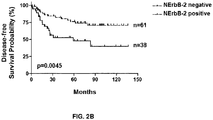
- NErbB-2 shows clinical relevance in TNBC. Relationship between NErbB-2 positivity and survival in terms of overall survival (2A), disease-free survival (2B), local relapse-free survival (2C) and distant metastasis-free survival (2D) probabilities (%), as assessed by a Kaplan-Meier analysis and log-rank test.
- the TNBC clinical study is based on the monitoring of 99 patients, 38 therefrom being NErbB-2-positive (full line) and 61 NErbB-2-negative (dotted line).
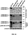
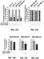
- FIGS 3A-3I ErbB-2 isoforms expression and activation in BC cells.
- MDA-MB-453 cells (LAR subtype) expressed full-length ErbB-2 (185 kDa, p185ErbB-2, WTErbB-2) comparable to the canonical ErbB-2 present in BT-474 cells, used as a control.
- MDA-MB-468 (BL1 subtype) showed an ErbB-2 variant of lower MW (-165 kDa, p165ErbB-2).
- HCC-70 and MDA-MB-231 cells presented both p185ErbB-2 (WTErbB-2) and p165ErbB-2. Additionally, the ErbB-2 truncated isoforms p95ErbB-2/CTFs (-90-115 kDa) produced by proteolytic cleavage and alternative initiation of translation were observed.
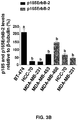
- 3B Signal intensities of p185ErbB-2 (black bars) and p165ErbB-2 (grey bars) in four independent WBs performed as in Fig. 3A , were analyzed by densitometry and normalized to ⁇ -tubulin protein bands.
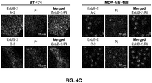
- FIGS 4A-4D ErbB-2 subcellular localization in BC cells.
- 4A ErbB-2 localization was studied by IF and confocal microscopy using the ErbB-2 C-18 antibody followed by incubation with an IgG-Alexa Fluor 488 secondary antibody. Thick arrows indicate the presence of MErbB-2 and slim ones show NErbB-2 presence in TNBC cell lines and in the control BT-474 cells upon heregulin (HRG) stimulation. Nuclei were stained with DAPI.
- 4B Percentage of nuclear ErbB-2 presence in confocal images from Fig. 4A .
- Integrated density (mean fluorescence intensity per unit area) of subcellular compartments was quantified in 50 cells from each cell line and was analyzed as percentages (mean ⁇ SD), relative to the total content (integrated density) of ErbB-2 in each cell, which was set to 100%.
- One-way ANOVA with Dunnett's multiple comparisons test was applied to determined significant differences between control and HRG- ⁇ 1-treated cells. For b vs a, and c vs a: P ⁇ 0.001.
- Figures 5A-5B N-glycosylation pattern of ErbB-2 in TNBC cells.
- 5A NetNGlyc 1.0 server (http://www.cbs.dtu.dk/services/NetNGlyc/) was used to predict N-glycosylation sites by analyzing the Asn-Xaa-Ser/Thr sequons defined as a pre-requisite for N-glycosylation ( Blom, N. et al. (2004) Prediction of post-translational glycosylation and phosphorylation of proteins from the amino acid sequence. Proteomics 4, 1633-1649 ).
- Each graph illustrates putative N-glycosylation sites across the protein chain of different ErbB-2 isoforms (https://www.ncbi.nlm.nih.gov/refseq/).
- X-axis represents protein length from N- to C-terminal.
- a position with a vertical grey line crossing the threshold (black horizontal line at 0.5) is predicted to be N-glycosylated.
- BT-474 and MDA-MB-468 cells were treated with tunicamycin (10 ⁇ g/ml) or DMSO as control for 15 h. ErbB-2 expression was then evaluated by WB with the C-18 antibody. MW calculation of protein bands was performed using the relative migration distance (Rf) method.
- FIGS 6A-6C Schematic representation of protein-coding ErbB-2 transcript variants annotated on RefSeq database (NCBI RefSeq, 2020 https://www.ncbi.nlm.nih.gov/refseq/).
- Consensus coding sequences (CCDSs) and untranslated regions (5' and 3' UTRs) are represented as lines and dashed lines, respectively. Numbers inside boxes and below lines indicate the sequence length in nucleotides (nt).
- the position of the translation start site (ATG) and the translation stop codon (TGA) are also shown.
- Variant 1 corresponds to the canonical sequence that encodes the ErbB-2 isoform a, p185ErbB-2 (WTErbB-2).
- Variant 2 differs in the 5' UTR, it lacks a portion of the 5' CCDS, initiating translation at a downstream start codon.
- the box indicating region from position 1 to position 558 nt shows the differential region of variant 2 compared to variant 1.
- Variant 3 (SpT3) has an alternate 5' UTR and 5' CCDS.
- the box indicating region from position 1 to position 607 nt indicates the differential region of variant 3 with respect to the canonical transcript.
- Variant 3 also presents a unique sequence of 51 nt (exon 5) spanning its 5' UTR and 5' CCDS (box indicating region from position 560 to position 610 nt).
- Variant 4 (SpT4) lacks the sequence that corresponds to exon 26 in the 3' CCDS of variant 1 (box indicating region from position 3421 to position 3673 nt) resulting in a translational frameshift. The box indicating region from position 3421 to position 4411 nt shows the differential 3' UTR and 3' CCDS region of variant 4.
- 6C Schematic representation of ErbB-2 ⁇ 16 spliced transcript compared with variant 1.
- FIG. 7 Schematic representation of ErbB-2 isoforms. The regions that are relevant to ErbB-2 function are depicted as grey boxes. Numbers below lines indicate the protein length in amino acids (aa). The extracellular portion is composed by 4 domains (I-IV): two leucine-rich (L) ligand-binding domains, I/LI and III/LII, and two cysteine-rich (C) domains, II/CI and IV/CII, which facilitate the formation of disulfide bonds. The position of the signal peptide (SP) in the amino-terminus is also shown. ErbB-2 contains a transmembrane domain (TM region) and a juxtamembrane domain, which bears the nuclear localization sequence (NLS).
- TM region transmembrane domain
- NLS nuclear localization sequence
- the intracellular portion includes the catalytic protein tyrosine kinase domain (PTKc domain) which contains the activation loop (A-loop), and a proline-rich transactivation domain near the carboxy-terminus.
- PTKc domain catalytic protein tyrosine kinase domain
- A-loop activation loop
- proline-rich transactivation domain near the carboxy-terminus.
- the major phosphorylation sites are indicated (grey dashed lines).
- FIG 8 Signal peptide prediction. Graphical output of SignalP 4.1 Server (http://www.cbs.dtu.dk/services/SignaIP/) for signal peptide (SP) prediction ( Nielsen, H. (2017) Predicting Secretory Proteins with SignaIP. Methods in molecular biology 1611, 59-73 ; Emanuelsson, O. et al. (2007) Locating proteins in the cell using TargetP, SignaIP and related tools. Nature protocols 2, 953-971 ).
- C-score raw cleavage site score, dark grey continuous line
- the C-score is trained to be high at the position immediately after the cleavage site (the first residue in the mature protein).
- S-score signal peptide score, light grey dashed line
- Y-score combined cleavage site score, dark grey dotted line
- Y-score combined cleavage site score, dark grey dotted line
- Figures 9A-9E Amplification of the entire coding region of ErbB-2 transcript variants by long range (LR)-PCR.
- LR-PCRs were performed with RANGER DNA polymerase using cDNA as template and two sets of primers: (9A, 9B, 9C) forward primer of SEQ ID NO: 4 and reverse primer of SEQ ID NO: 5, spanning the complete coding region of variants 1, 4 and ErbB-2 ⁇ 16 on one side (LR-PCR1), and on the other, (9D, 9E) forward primer of SEQ ID NO: 6 and reverse primer of SEQ ID NO: 5, spanning the complete coding region of variants 2 and 3 (LR-PCR2).
- Analysis of LR-PCR products on 0.7% agarose gels showed fragments ranging between ⁇ 4 to 5 kb long (indicated by arrows) corresponding to the size of expected products, which are indicated.
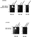
- FIGS 10A-10C Representative gel images for the nested PCR strategy.
- LR-PCR1 product was used as template for nested PCR with primers for SpT1
- LR-PCR2 product as template for nested PCR with primers for SpT2 and SpT3.
- 10A SpT1 expression was studied using primers spanning its differential region between exons 16 to 26 (forward primer of SEQ ID NO: 7 and reverse primer of SEQ ID NO: 8).
- Expression of SpT2 (10B) and SpT3 (10C) was assesed using variants specific primers spanning their differential region between exons 1 to 5.
- Primers for SpT2 are depicted as SEQ ID NOs: 9 and 10
- primers for SpT3 have SEQ ID NOs: 11 and 12.
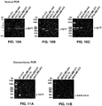
- FIGS 11A-11B Representative gel images for the conventional PCR strategy. Expression of SpT4 (11A) and of ErbB-2 ⁇ 16 (11B) was studied by conventional PCR using spliced-specific primers: for SpT4 primers of SEQ ID NOs: 13 and 14; and primers for ErbB-2 ⁇ 16 have SEQ ID NOs: 15 and 16.
- Figures 12A-12D Representative gel images of the competitive PCR strategy.
- Amplification products corresponding to competitors (variants with exon 26 or 16 inclusion) and targets (SpT4 or ErbB-2 ⁇ 16, respectively) were quantified by densitometry and plotted as % of total ErbB-2 mRNA levels.
- targets SpT4 or ErbB-2 ⁇ 16, respectively
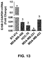
- FIG. 13 Total ErbB-2 mRNA levels assessed by Reverse Transcriptase (RT)-quantitative PCR (qPCR). Fold change was calculated by normalizing the absolute levels of ErbB-2 mRNA to those of GAPDH, used as an internal control, setting the value of BT-474 cells to 1. Data are presented as mean ⁇ S.D. For b vs a: P ⁇ 0.001. Primers for total ErbB-2 are depicted as SEQ ID NOs: 21 and 22, and primers for GAPDH have SEQ ID NOs: 25 and 26.
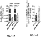
- FIGS 14A-14B SpT3 expression levels assessed by RT-qPCR.
- 14A Total ErbB-2 and SpT3 mRNA levels were measured by RT-qPCR. Fold change was calculated by normalizing the absolute levels of total ErbB-2 or SpT3 mRNA to those of GAPDH, used as an internal control, setting the value of BT-474 cells to 1. Data are presented as mean ⁇ S.D. For b vs a: P ⁇ 0.001.
- FIGS 15A-15G siRNAs targeting the common coding region of transcript variants 1 to 4, and of ErbB-2 ⁇ 16 are unable to silence p165ErbB-2 expression.
- 15A Schematic diagram of the target sequences of a pool of 4 siRNAs (ErbB-2 siRNA, siGENOME SMARTpool siRNAs for human ErbB-2 (2064) cat #M-0031126-04-0020, Dharmacon, Lafayette, CO, USA) designed to target the common coding region of all ErbB-2 variants analyzed in the present application (grey boxes indicating regions between nucleotides (nt): 1080-1098, 1558-1576, 1559-1577, and 2565-2583).
- CCDSs Consensus coding sequences
- UTRs untranslated regions
- Numbers below the lines indicate the sequence length of mRNA in nt. Shown are the position of the translation start site (ATG) and the translation stop codon (TGA).
- the pool of siRNAs targeting ErbB-2 have the sequences: GGACGAAUUCUGCACAAUG (SEQ ID NO: 27); GACGAAUUCUGCACAAUGG (SEQ ID NO: 28); CUACAACACAGACACGUUU (SEQ ID NO: 29) and AGACGAAGCAUACGUGAUG (SEQ ID NO: 30).
- BT-474 and TNBC cells were transfected with either Control siRNA (siGENOME Non-Targeting siRNA #5, cat #D-001210-05-20, Dharmacon, Lafayette, CO, USA), shown in the sequence listing as SEQ ID NO: 31 (UGGUUUACAUGUCGACUAA), or with the abovementioned pool of 4 siRNAs (ErbB-2 siRNA) at a final concentration of 100 nM.
- Protein extracts 50 ⁇ g were examined for ErbB-2 isoforms expression with the C-18 antibody.
- ⁇ -tubulin was used as a loading control.
- ErbB-2 siRNA pool had no effect on NErbB-2 levels in TNBC cells only expressing p165ErbB-2.
- Cell lines were transfected with control siRNA (SEQ ID NO: 31) (upper panels) or with ErbB-2 siRNA pool (SEQ ID NOs: 27-30) (lower panels) at a final concentration of 100 nM. Nuclei were stained with propidium iodide (PI). ErbB-2 levels were visualized by IF and confocal microscopy using the ErbB-2 C-18 antibody in BT-474 (16A) and TNBC cell lines: MDA-MB-468 (16B), HCC-70 (16C), MDA-MB-231 (16D) and MDA-MB-453 (16E).
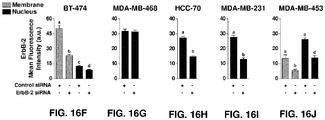
- FIGS 17A-17E ErbB-2 siRNA pool blocks proliferation in BC cells expressing only p185ErbB-2.
- BT-474 (17A), MDA-MB-468 (17B), HCC-70 (17C), MDA-MB-231 (17D) and MDA-MB-453 (17E) cells were transfected with Control siRNA (SEQ ID NO: 31) or with the ErbB-2 siRNA pool (SEQ ID NOs: 27-30) at a final concentration of 100 nM and proliferation was assessed by [ 3 H]-thymidine uptake. Data are presented as mean ⁇ S.D. of three independent experiments. For b vs a: P ⁇ 0.001.
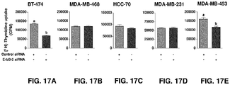
- FIGS 18A-18D siRNAs targeting variant 3 (SpT3) which encodes the non-canonical isoform c, silence p165ErbB-2 expression.
- SpT3 siRNA #1 SEQ ID NO: 1
- SpT3 siRNA #2 SEQ ID NO: 2
- SpT3 siRNAs have the sequences: GUGAGAUACUUCAAAGAUU (SEQ ID NO: 1) and CAAAGAUUCCAGAAGAUAU (SEQ ID NO: 2).
- Consensus coding sequences and untranslated regions (5' and 3' UTRs) are represented as full lines and dashed lines, respectively. Numbers below lines indicate the sequence length of mRNA in nucleotides (nt). The position of the translation start site (ATG) and the translation stop codon (TGA) are also shown.
- Target sequences of SpT3 siRNA #1 (SEQ ID NO: 1) and SpT3 siRNA #2 (SEQ ID NO: 2) are depicted as a dark grey box indicating region between nt 555-573, and as a black box indicating region between nt 566-584, respectively.
- BT-474 and TNBC cells were transfected with Control siRNA (SEQ ID NO: 31) or with SpT3 siRNA #2 (SEQ ID NO: 2) targeting SpT3, at a final concentration of 100 nM.
- Protein extracts (10 ⁇ g) were examined for ErbB-2 isoforms expression with the C-18 antibody.
- ⁇ -tubulin was used as a loading control.
- Signal intensities of (18C) p185ErbB-2 and (18D) p165ErbB-2 (ErbB-2 isoform c) were analyzed by densitometry from three independent WBs performed as in Fig. 18B . Fold change was calculated by normalizing the absolute levels of each ErbB-2 isoform to those of ⁇ -tubulin, setting the value of Control siRNA-transfected cells to 1.
- b vs a P ⁇ 0.001.
- SpT3 siRNA #2 reduces nuclear ErbB-2 levels in TNBC cells expressing only p165ErbB-2 (ErbB-2 isoform c) or both p165ErbB-2 (ErbB-2 isoform c) and p185ErbB-2.
- TNBC cell lines were transfected with Control siRNA (SEQ ID NO: 31) (upper panels) or with SpT3 siRNA #2 (SEQ ID NO: 2) (lower panels) at a final concentration of 100 nM. Nuclei were stained with DAPI.
- ErbB-2 levels were visualized by IF and confocal microscopy using the ErbB-2 C-18 antibody in MDA-MB-468 (19A), MDA-MB-231 (19B) and MDA-MB-453 cells (19C). Quantitative analysis of NErbB-2 expression in confocal images from Figs. 19A to 19C was performed. Fluorescence intensities from nuclear compartment (black bars) were quantified in 50 cells from MDA-MB-468 (19D), MDA-MB-231 (19E) and MDA-MB-453 (19F), and plotted (mean ⁇ S.D.). For b vs a: P ⁇ 0.001. Experiments in Figs. 19A to 19C and its quantification were repeated thrice with similar results.
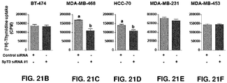
- FIGS 20A-20E SpT3 siRNA #2 inhibits in vitro proliferation of TNBC cells expressing only p165ErbB-2 (ErbB-2 isoform c) or both p165ErbB-2 (ErbB-2 isoform c) and p185ErbB-2.
- (20A) BT-474, (20B) MDA-MB-468, (20C) HCC-70, (20D) MDA-MB-231 and (20E) MDA-MB-453 cells were transfected with Control siRNA (SEQ ID NO: 31) or with SpT3 siRNA #2 (SEQ ID NO: 2) targeting SpT3, at a final concentration of 100 nM and proliferation was assessed by [ 3 H]-thymidine uptake. Data are presented as mean ⁇ S.D. of three independent experiments. For b vs a: P ⁇ 0.001.
- FIGS 21A-21F Effects of SpT3 siRNA #1.
- (21A) BT-474 and TNBC cells (MDA-MB-468, HCC-70, MDA-MB-231 and MDA-MB-453) were transfected with Control siRNA (SEQ ID NO: 31) or with SpT3 siRNA #1 (SEQ ID NO: 1) at a final concentration of 100 nM.
- Protein extracts 50 ⁇ g were examined for ErbB-2 isoforms expression with the C-18 antibody.
- ⁇ -tubulin was used as a loading control. Images shown are representative of three independent experiments.
- FIG 22 Effects of SpT3 siRNA #2 on tumor explants.
- Triple negative tumor explants established from MDA-MB-468 xenografts were cultured with medium containing SpT3 siRNA #2 (SEQ ID NO: 2) or Control siRNA (SEQ ID NO: 31) at a final concentration of 100 nM.
- WB analyses of p165ErbB-2 (ErbB-2 isoform c) and Erk5 were performed in tumor lysates after 24 hours of transfection. Numbers under each blot represent the corresponding densitometric quantification.
- Fold change of protein levels was calculated by normalizing the absolute levels of p165ErbB-2 (ErbB-2 isoform c) and Erk5 bands to those of ⁇ -tubulin, used as loading control, setting the value of control siRNA-transfected tumors to 1.
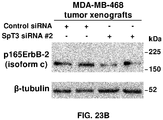
- FIG. 23A-23B Preclinical model of the blockade of p165ErbB-2 (ErbB-2 isoform c) expression with SpT3 siRNA #2.
- (23B) Mice were sacrificed two days after the last treatment and tumor lysates were analyzed by WB with the C-18 antibody. Shown are two representative tumors from each group.
- FIGS. 24A-24B Silencing of p165ErbB-2 (ErbB-2 isoform c) with SpT3 siRNA #2 in the MDA-MB-468 preclinical model reduces nuclear ErbB-2 expression.
- (24) IF and confocal microscopy of histological sections from MDA-MB-468 tumors excised at the end of the experiment. ErbB-2 was detected using the C-18 antibody, followed by incubation with an IgG-Alexa Fluor 488 secondary antibody. Nuclei were stained with DAPI. Representative images are shown.
- 24B Quantitative analysis of NErbB-2 expression in images from Fig. 24A .
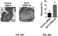
- FIGS 25A-25D Histopathological analysis.
- 25A Representative hematoxylin and eosin (H&E) staining of histological sections from MDA-MB-468 tumors excised at the end of the experiment. Mitotic figures are indicated with black arrows.
- 25B Tumor proliferation was quantified by mitotic figures count per high power field (HPF) in MDA-MB-468 histological sections. Data are presented as mean ⁇ S.D. For b vs a: P ⁇ 0.001.
- 25C Representative H&E staining of histological sections from MDA-MB-468 tumors showing areas with extensive necrosis (pale eosinophilic areas) after SpT3 siRNA #2 treatment.
- FIGS 26A-26D Preclinical toxicological analysis of animal blood samples.
- ALT Alanine transaminase
- AST aspartate aminotransferase
- 26C total bilirubin levels were determined for evaluating hepatotoxicity in SpT3 siRNA #2 injected mice.
- 26D Creatinine levels were also measured to assess SpT3 siRNA #2 effects on renal function. n.s., non-significat by unpaired Student's t test.
- nucleotide base is a heterocyclic pyrimidine or purine compound that is a component of a nucleotide, and includes the primary purine bases adenine (A) and guanine (G), and the primary pyrimidine bases cytosine (C), thymine (T), and uracil (U).
- a nucleobase may further be modified to include, without limitation, universal bases, hydrophobic bases, promiscuous bases, size-expanded bases, and fluorinated bases. (See. e.g., Modified Nucleosides in Biochemistry, Biotechnology and Medicine, Herdewijn, P. ed. Wiley-VCH, 2008 ). The synthesis of such modified nucleobases (including phosphoramidite compounds that include modified nucleobases) is known in the art.
- nucleoside refers to a molecule made up of a nucleotide base and its sugar.
- nucleotide refers to a nucleoside having a phosphate group on its 3' or 5' sugar hydroxyl group.
- oligonucleotide means a polymer of linked nucleotides each of which can be independently modified or unmodified.
- the polynucleotides can be “deoxyribonucleic acid” (DNA), “ribonucleic acid” (RNA), or derivatives or modified versions thereof.
- the polynucleotide may be single-stranded or double-stranded.
- deoxynucleotide refers to a nucleotide or polynucleotide lacking a hydroxyl group (OH group) at the 2' and/or 3' position of a sugar moiety. Instead, it has a hydrogen bonded to the 2' and/or 3' carbon.
- deoxynucleotide refers to the lack of an OH group at the 2' position of the sugar moiety, having instead a hydrogen bonded directly to the 2' carbon.
- deoxyribonucleotide and “DNA” refer to a nucleotide or polynucleotide comprising at least one sugar moiety that has an H, rather than an OH, at its 2' and/or 3' position.
- ribonucleotide and the phrase “ribonucleic acid” (RNA), refer to a modified or unmodified nucleotide or polynucleotide comprising at least one ribonucleotide unit.
- a ribonucleotide unit comprises an hydroxyl group attached to the 2' position of a ribosyl moiety that has a nitrogenous base attached in N-glycosidic linkage at the 1' position of a ribosyl moiety, and a moiety that either allows for linkage to another nucleotide or precludes linkage
- 3' end refers to the end of a nucleic acid that contains an unmodified hydroxyl group at the 3' carbon of its ribose ring.
- 5' end refers to the end of a nucleic acid that contains a phosphate group attached to the 5' carbon of its ribose ring.
- mRNA messenger RNA
- mRNA transcripts include, but are not limited to pre-mRNA transcript(s), transcript processing intermediates, mature mRNA(s) ready for translation and transcripts of the gene or genes, or nucleic acids derived from the mRNA transcript(s). Processing of pre-mRNA transcripts, along with the possible use of alternative promoters and alternative polyadenylation sites, allows a single gene to generate many different mature RNAs, by varying the pattern of splicing in a process known as alternative splicing. Alternative splicing can also introduce or remove regulatory elements to affect mRNA translation, localization or stability.
- alternatively spliced mRNAs are translated into alternative splice form proteins, here referred to as protein isoforms, that contain different amino acid sequences than the corresponding wild-type or canonical protein produced by normally spliced mRNA.
- RNAi agent and "RNA silencing agent” means a composition that contains an RNA or RNA-like (e.g., chemically modified RNA) oligonucleotide molecule that is capable of degrading or inhibiting (e.g., degrades or inhibits under appropriate conditions) translation of messenger RNA (mRNA) transcripts of a target mRNA in a sequence specific manner.
- RNAi agents may operate through the RNA interference mechanism (i.e., inducing RNA interference through interaction with the RNA interference pathway machinery (RNA-induced silencing complex or RISC) of mammalian cells), or by any alternative mechanism(s) or pathway(s). While it is believed that RNAi agents, as that term is used herein, operate primarily through the RNA interference mechanism, the disclosed RNAi agents are not bound by or limited to any particular pathway or mechanism of action.
- siRNAs small (or short) interfering RNA
- siRNAs consist of two RNA strands, an antisense (or guide) strand and a sense (or passenger) strand. These molecules display generally between 18-30 basepairs and contain varying degrees of complementarity to their target mRNA in the antisense strand.
- Some, but not all, siRNAs include structures with overhangs. Overhangs have been described to be advantageous and may be present on the 5' ends or on the 3' ends of either strand as they reduce recognition by RNAses. Some authors recommend including overhangs on both 3' ends of the molecules, whereas others consider one overhang to be sufficient.
- overhangs are said to further enhance resistance to nuclease (RNase) degradation.
- RNase nuclease
- tail refers to 3, 4, 5, 6, 7, 8, 9, 10, 11, 12, 13, 14, 15, 16, 17, 18, 19, 20 or more sequential nucleotides at the 3' end of one or both of the sense strand and the antisense strand that are single-stranded.
- RNA includes duplexes of two separate strands, as well as single strands that can form hairpin structures comprising a duplex region.
- antisense (or guide) strand refers to the strand of an siRNA duplex that contains some degree of complementarity to a target gene or mRNA and contains complementarity to the sense strand of the siRNA duplex.
- sense (or passenger) strand refers to the strand of an siRNA duplex that contains complementarity to the antisense strand of the siRNA duplex.
- duplex region refers to the region in two complementary or partially complementary polynucleotides (e.g., a sense strand or an antisense strand) that form base pairs with one another.
- a base pair is a unit consisting of two nucleobases connected by hydrogen bonds.
- DNA contains four bases: the two purines adenine (A) and guanine (G) and the two pyrimidines cytosine (C) and thymine (T).
- RNA thymine is replaced by uracil (U).
- Non-Watson-Crick base-pairing models display alternative hydrogen-bonding patterns; examples are Hoogsteen base pairs, which are A-T or C-G analogues.
- nucleotide or “nucleotide analog” or “altered nucleotide” or “modified nucleotide” refer to a non-standard nucleotide, including non-naturally occurring ribonucleotides or deoxyribonucleotides.
- nucleotide analogs may be modified to change chemical properties of the nucleotide, maintaining the ability of the nucleotide analog to perform its function.
- Nucleotide analogs include nucleotides having modifications in the chemical structure of the base, sugar and/or phosphate.
- the nucleobase moiety may be modified by the replacement or addition of one or more atoms or groups.
- a pyrimidine base may be modified at the 2, 3, 4, 5, and/or 6 position of the pyrimidine ring.
- the exocyclic amine of cytosine may also be modified.
- a purine base may be modified at the 1, 2, 3, 6, 7, or 8 position.
- the exocyclic amine of adenine may also be modified.
- Base modifications are known to those of ordinary skill in the art. Some examples include alkylated, halogenated, thiolated, aminated, amidated, or acetylated bases, or other heterocycles. Nucleobase modifications in small amounts (up to 10%) could reduce immune reactions and improve the thermodynamic siRNA profile.
- nucleobases substitutions Reviewed in Chernikov, I.V. et al. (2019) Current Development of siRNA Bioconjugates: From Research to the Clinic. Frontiers in pharmacology 10, 444 ) with various modified analogs (pseudouridine, 2'thiouridine, dihydrouridine, 2,4-difluorobenzene, 4-methylbenzimidazole, hypoxanthine, 7-deazaguanin, N2-alkyl-8-oxoguanine, N2-benzylguanine, and 2,6-diaminopurine) may be used to increase the thermal stability of the siRNA duplex.
- modified analogs pseudouridine, 2'thiouridine, dihydrouridine, 2,4-difluorobenzene, 4-methylbenzimidazole, hypoxanthine, 7-deazaguanin, N2-alkyl-8-oxoguanine, N2-benzylguanine, and 2,6-dia
- Non-limiting examples of modified bases are described in U.S. Pat. Nos. 10,011,836 ; 8,008,474 ; 7,816,512 and 7,977,471 , all of which are incorporated herein by reference.
- nucleotide analogs may also display modifications at the sugar moiety.
- Sugar moieties include natural, unmodified sugars, e.g., monosaccharide (such as pentose, e.g., ribose, deoxyribose), modified sugars and sugar analogs.
- modifications of sugar moiety include substitutions in the 2'-position, as the 2'OH group participates in the cleavage of RNA by endoribonucleases.
- modifications include but are nor limited to, the replacement of the 2'OH group by a group selected from H, OR, R, F, Cl, Br, I, SH, SR, NH2, NHR, NR2, COOR, or OR, wherein R is substituted or unsubstituted C1-C6 alkyl, alkenyl, alkynyl, aryl, etc, as described in U.S. Pat. No. 9,080,171 and U.S. Pat. Application No. 2019/0024082 , all of which are incorporated herein by reference.
- Nucleotide analogs with a modified structure of the furanose cycle such as derivatives containing 6-membered (hexitol (HNA), cyclohexenic (CeNA) and altritol (ANA) nucleic acids), and 7-membered rings (oxepanic nucleic acid (ONA)), bicyclic (locked nucleic acids (LNA), 2'-deoxymethanocarbanucleosides (MCs)), tricyclic (tricyclo-DNA (tc-DNA)), and acyclic (unlocked nucleic acid (UNA)) derivatives, can be used.
- HNA 6-membered
- CeNA cyclohexenic
- ANA altritol
- LNA locked nucleic acids
- MCs 2'-deoxymethanocarbanucleosides
- tc-DNA tricyclic
- UPA unlocked nucleic acid
- nucleotide analogs may also display nonnatural phosphodiester linkages such as methylphosphonates, phosphorothioates and peptides.
- nucleotide analogs may also comprise hydrophobic modifications at the base moiety.
- hydrophobic modifications are phenyl, indolyl, isobutyl, butyl, aminobenzyl, benzyl, thiophene, ethynyl, and imidazole.
- nucleotide also includes what are known in the art as universal bases. These nucleotide analogues lack hydrogen bonding sites and are generally hydrophobic aromatic base residues. Some examples are 3-nitropyrrole 5-nitroindole, or nebularine.
- nucleotide is also meant to include the nucleotide analogues N3' to P5' phosphoramidates. These compounds contain 3'-amino group replacing the ribosyl 3'-O-atom.
- nucleotide also includes those species that have a detectable label, such as for example a radioactive or fluorescent moiety, or mass label attached to the nucleotide.
- gene silencing or “knockdown” refers to a process by which the expression of a specific gene product is lessened or attenuated.
- shilenced or “knockdown”, when referring to expression of a given gene, mean that the expression of the gene, as measured by the level of RNA transcribed from the gene or the level of polypeptide, protein, alternative isoform of a given protein, e.g.
- ErbB-2, or protein subunit translated from the mRNA in a cell, group of cells, tissue, organ, or subject in which the gene is transcribed is reduced when the cell, group of cells, tissue, organ, or subject is treated with the siRNAis described herein as compared to a second cell, group of cells, tissue, organ, or subject that has not or have not been so treated.
- off-target silencing and off-target interference are defined as degradation of mRNA other than the intended target mRNA due to overlapping and/or partial homology with secondary mRNA messages.
- Such off-target effects are primarily mediated by the sequence-specific interaction between the siRNA seed regions (position 2-8 of either siRNA strand counting from the 5'-end) and complementary sequences in the 3' UTR of off-targets ( Bramsen, J.B. et al. (2010) A screen of chemical modifications identifies position-specific modification by UNA to most potently reduce siRNA off-target effects. Nucleic acids research 38, 5761-5773 ).
- siRNA score refers to a number determined by applying any of the design algorithms or formulas (e.g., siDESIGN Center, Dharmacon, Lafayette, CO, USA) to a given siRNA sequence.
- the present invention provides specific siRNA molecules for use to silence a particular mRNA (e.g., ErbB-2 alternative transcript variant 3, SpT3) which have been selected on the basis of this method.
- Target mRNA refers to a messenger RNA (mRNA) to which a given siRNA can be directed against.
- Target sequence refers to a contiguous portion of the nucleotide sequence of a target mRNA molecule formed during the transcription of a target gene, including mRNA that is a product of RNA processing of a primary transcription product.
- siRNA target can refer to the gene, mRNA, or protein against which an siRNA is directed.
- Non-targeting siRNA As used herein, “Non-targeting siRNA”, “Control siRNA” (siGENOME Non-Targeting siRNA#5 cat #D-001210-05-20, Dharmacon, Lafayette, CO, USA), refers to a highly functional, chemically synthesized negative control siRNA duplex for experiments in human, mouse, and rat cells.
- the control siRNA oligonucleotide used herein does not target any known mammalian gene. Neither the mRNA nor protein level of the experimental gene is affected by this control siRNA. Cell viability as well as cell phenotype of samples treated with the control siRNA remains comparable to those of untreated samples.
- control siRNA does not bind to ErbB-2 alternative transcript variant 3 (SpT3), which encodes isoform c of ErbB-2.
- SpT3 ErbB-2 alternative transcript variant 3
- the control siRNA used in the assays carried out in the present invention consists of the sequence UGGUUUACAUGUCGACUAA shown as SEQ ID NO: 31.
- an RNA silencing agent (or any portion thereof), e.g., the siRNA molecules of the present invention targeting ErbB-2 alternative transcript SpT3 which encodes ErbB-2 isoform c, as described herein, may be modified such that the activity of the RNA silencing agent is further improved.
- the RNA silencing agents described in the present invention may be modified with any of the modifications described herein. The modifications can, in part, serve to further enhance target discrimination, to enhance stability of the agent (e.g., to prevent degradation), to promote cellular uptake, to enhance the target efficiency, to improve efficacy in binding (e.g., to the targets), to improve patient tolerance to the agent, and/or to reduce toxicity.
- certain modifications may be incorporated to facilitate preferential selection of the antisense strand of the siRNA molecule by the cellular RNAi machinery, such that the antisense strand preferentially guides cleavage or translational repression of a target mRNA, and thus increasing the efficiency of target cleavage and silencing.
- Non-limiting examples of structural and chemical modifications used to achieve preferential strand selection include the development of internally destabilized duplexes, asymmetric duplexes and 2'OMe modifications of the two nucleotides at the 5' end of the sense strand.
- the siRNAs of the invention may be substituted with a destabilizing nucleotide.
- a destabilizing nucleotide Non-limiting examples include acyclic nucleotide residues which decrease the stability when incorporated into RNA duplexes.
- Unlocked nucleic acid (UNA) is reported to destabilize RNA-RNA interactions and can be strategically used in siRNA design to induce local thermodynamical destabilization (Bramsen, J.B. et al. (2010) op. cit. )
- the siRNAs of the invention may be altered according to asymmetry design rules.
- Asymmetric siRNA compounds have previously been developed. Non-limiting examples include: i) siRNAs with deletions at the sense and/or antisense strand, with a duplex region of 16 nucleotides (nt) ( Chu, C.Y. and Rana, T.M. (2008) Potent RNAi by short RNA triggers. Rna 14, 1714-1719 ); ii) asymmetric interfering RNA (aiRNA) compounds with a short sense strand (12-15 nt) and a regular length antisense strand (21 nt) ( Sun, X. et al.
- sd-rxRNA ® a new class of small, hydrophobic, asymmetric RNAi compounds, termed "self-delivering rxRNAs"
- sd-rxRNA ® are extensively modified siRNAs compounds, with a small duplex region of ⁇ 15 nt, and a fully phosphorothioated single-stranded tail.
- sd-rxRNAs are functional in vitro and in vivo without the aid of any delivery vehicle ( Byrne, M. et al. (2013) Novel hydrophobically modified asymmetric RNAi compounds (sd-rxRNA) demonstrate robust efficacy in the eye. J Ocul Pharmacol Ther 29, 855-864 ).
- modifications that increase or decrease sugar flexibility including locked nucleic acid (LNA) and unlocked nucleic acid (UNA), may be used to introduce chemical asymmetry into duplex siRNAs (Reviewed in Khvorova, A. and Watts, J.K. (2017) The chemical evolution of oligonucleotide therapies of clinical utility. Nature biotechnology 35, 238-248 ).
- LNA locked nucleic acid
- UPA unlocked nucleic acid
- siRNA molecules of the present invention may be modified at various locations in order to promote metabolic stability and enhance silencing activity.
- modifications include, but are not limited to: a) sugar modifications such as 2' ribo modifications, including particularly combinations of 2'-O-methyl (2'OMe), 2'-fluoro (2'F), 2'-deoxy and others; modifications that increase or decrease sugar flexibility, including locked nucleic acid (LNA) and unlocked nucleic acid (UNA); b) backbone modifications such as phosphorothioate internucleotide linkages (referred to as phosphorothioate modifications) at both ends of both strands of the siRNA duplex; and methylation of the 5' carbon to give (S)-5'-C-methyl-RNA; and c) 5'-phosphate stabilization of the siRNA guide strand by incorporation of phosphate analogs such as 5'E-vinyl phosphonate (5'-E-VP), 5'-methyl phosphonate, 5'-C-methyl
- immune stimulation may be beneficial for the treatment of cancer and siRNAs with both RNAi and immunostimulatory activity (isiRNAs)
- siRNAs with both RNAi and immunostimulatory activity
- isiRNAs RNAi and immunostimulatory activity
- 5'-triphosphate modifications known to activate the cytosolic antiviral helicase retinoic acid-induced protein I (Rig-I) and, therefore induce innate immunity, may be introduced to the siRNAs of the present invention (Poeck, H. et al. (2008) op.cit.).
- siRNA molecules of the present invention may be associated with a hydrophobic moiety for targeting and/or delivery of the molecule to a cell.
- the hydrophobic moiety is associated (covalently or noncovalently) with the nucleic acid molecule through a linker. Any linker known in the art may be used to associate the nucleic acid with the hydrophobic moiety.
- the linker is capable of being cleaved from the nucleic acidunder physiological conditions (e.g., acid labile linkers are cleaved in the acidic endosomal/lysosomal compartments, and disulfide linkers are cleaved at the reductive environment of cytosolic space).
- the linker is capable of being cleaved by an enzyme (e.g., an esterase or phosphodiesterase).
- an enzyme e.g., an esterase or phosphodiesterase.
- linkers employed for hydrophobic/lipid conjugation include: trans-4-hydroxyprolinol linker, disulfide linker, aminohexyl linker, triethyl glycol (TEG) linker and 2-aminobutyl-1-3-propanediol (C7) linker.
- the hydrophobic moiety is linked at various positions of the siRNA molecule. In a preferred embodiment, the hydrophobic moiety is linked to either the 3' or 5' end of the sense strand. In an exemplary embodiment, the hydrophobic moiety is selected from the group consisting of saturated fatty acids (e.g. docosanoic acid (DCA)), non-saturated fatty acids (e.g. docosahexaenoic acid (DHA, 22:6 n-3) and eicosapentaenoic acid (EPA, 20:5 n-3)), sterols (e.g. cholesterol (Chol) and lithocholic acid (LA)) and vitamins (e.g.
- DCA docosanoic acid
- DHA docosahexaenoic acid
- LA lithocholic acid
- RA retinoic acid
- TS ⁇ -tocopheryl succinate
- cationic dyes e.g., Cy3
- the siRNA molecules of the present invention contain 5'and/or 3'-end overhangs.
- the number and/or sequence of nucleotides overhang on one end of the polynucleotide may be the same or different from the other end of the polynucleotide.
- one or more of the overhang nucleotides may contain chemical modification(s), such as phosphorothioate or 2'OMe modification.
- the 3' and 5' end of the siRNA molecules of the present invention can be substantially protected from nucleases by modifying the 3' or 5' linkages (e.g., U.S. Pat. No. 5,849,902 and WO 98/13526 ).
- End-blocking groups or “exonuclease blocking groups” may also be used to protect the 5' and 3' end of the oligonucleotides.
- Such groups include modified nucleotides and non-nucleotide exonuclease resistant structures. Exemplary end-blocking groups are described in U.S. Pat. No. 9,080,171 .
- Oligonucleotides and oligonucleotide compositions of the present invention are contacted with (i.e., brought into contact with, also referred to herein as administered or delivered to) and taken up by one or more cells.
- the term "cells" includes eukaryotic cells, preferably vertebrate cells, and, more preferably, mammalian cells.
- the oligonucleotide compositions of the invention are contacted with human cells.
- siRNAs of the present invention can be delivered or administered to a cell (e.g., a cancer cell) in vitro, in vivo, or ex vivo.
- a cell e.g., a cancer cell
- novel siRNAs disclosed herein are suitable for any known delivery method, including intratumor or systemic routes.
- Delivery of the siRNAs of the present invention to an organelle, cell, tissue, and/or organism can be done by any method known to those skilled in the art.
- One exemplary means of delivering or introducing siRNAs into a cell is by transfection or transduction procedures.
- Transfection refers to the acquisition by a cell of siRNAs by incorporation of added siRNAs molecules. Transfection can occur by physical or chemical methods. Transduction refers to the process of transferring nucleic acid into a cell using a DNA or RNA virus.
- Such methods for delivering siRNAs to an organelle, cell, tissue, and/or organism include, but are not limited to, direct delivery of RNA such as by ex vivo transfection, by injection, including microinjection; by electroporation; by calcium phosphate precipitation; by using DEAE-dextran followed by polyethylene glycol; by direct sonic loading; by liposome mediated transfection and receptor-mediated transfection; by microprojectile bombardment; by agitation with silicon carbide fibers; by Agrobacterium-mediated transformation; by PEG-mediated transformation of protoplasts; by desiccation/inhibition-mediated RNA uptake, naked plasmid adsorption, and any combination of such methods.
- organelle(s), cell(s), tissue(s) or organism(s) may be stably or transiently transformed.
- a vector may be used in some embodiments as a carrier for the siRNA.
- the vector may comprise deoxyribonucleic acids (DNA) and/or ribonucleic acids (RNA).
- Vectors include plasmids, cosmids, viruses (bacteriophage, animal viruses, and plant viruses), and artificial chromosomes (e.g., YACs).
- plasmid vectors such as E. coli; phage vectors; and viral vectors such as adenoviral vectors, adeno-associated virus (AAV) vectors, retroviral vectors, vaccinia viruses, and Semliki Forest virus vectors.
- AAV adeno-associated virus
- an oligonucleotide is associated with a carrier or vehicle.
- the siRNAs of the present invention can be delivered or administered to a cell by suitable art recognized methods including but not limited to: a) lipid-based siRNA delivery, b) polymer-based siRNA delivery, c) bioconjugated siRNA delivery, d) inorganic nanoparticles, and e) extracellular vesicles.
- Lipid-based systems for delivering siRNAs embody varied lipid nanoparticles, including liposomes, micelles, emulsions, and solid lipid nanoparticles.
- lipid composition, drug-to-lipid ratio, particle size, and the manufacturing process should be optimized.
- Liposomal formulations containing the compounds (e.g., siRNAs), disclosed herein or compositions thereof may be lyophilized, to produce a lyophilizate, which may be reconstituted with a pharmaceutically acceptable carrier, such as water, to regenerate a liposomal suspension.
- a pharmaceutically acceptable carrier such as water
- Cationic liposomes are vesicular structures characterized by a phospholipid bilayer membrane and an inner aqueous medium. Multilamellar liposomes have multiple lipid layers separated by aqueous medium. They form spontaneously when phospholipids are suspended in an excess of aqueous solution. The diameters of the liposomes generally range from about 15 nm to about 5 microns.
- the components mainly include cationic lipids and neutral adjuvant lipids. Cationic lipids can be readily mixed and complexed with negatively charged siRNA molecules to form nanoparticles by electrostatic interaction. Diphenol phthalocyanine ethanolamine (DOPE) and cholesterol are commonly used as neutral auxiliary lipid molecules.
- DOPE Diphenol phthalocyanine ethanolamine
- cholesterol are commonly used as neutral auxiliary lipid molecules.
- the technology for forming liposomal suspensions is well known in the art.
- the compound or composition thereof is an aqueous-soluble composition
- using conventional liposome technology the same may be incorporated into lipid vesicles.
- the compound or composition will be substantially entrained within the hydrophilic center or core of the liposomes.
- the lipid layer employed may be of any conventional composition and may either contain cholesterol or may be cholesterol-free.
- the compound or composition of interest is water-insoluble, again employing conventional liposome formation technology, the composition may be substantially entrained within the hydrophobic lipid bilayer that forms the structure of the liposome. In either instance, the liposomes that are produced may be reduced in size, as through the use of standard sonication and homogenization techniques.
- cationic liposomes are stable nucleic acid lipid particles (SNALPs).
- SNALPs the siRNA is surrounded by a lipid bilayer containing a mixture of cationic lipids, fused lipids, cholesterol, and polyethylene glycol (PEG)-modified neutral lipids.
- PEG polyethylene glycol
- nucleic acids associated with the invention can be hydrophobically modified and can be encompassed within neutral nanotransporters (further description of neutral nanotransporters is incorporated in U.S. Pat. No. 10,138,485 ). Such particles enable quantitative oligonucleotide incorporation into noncharged lipid mixtures. The lack of toxic levels of cationic lipids in such neutral nanotransporter compositions is an important feature.
- the neutral nanotransporters compositions enable efficient loading of oligonucleotide into neutral fat formulation.
- the composition includes an oligonucleotide that is modified in a manner such that the hydrophobicity of the molecule is increased (for example a hydrophobic molecule is attached (covalently or no-covalently) to a hydrophobic molecule on the oligonucleotide terminus or a non-terminal nucleotide, base, sugar, or backbone), the modified oligonucleotide being mixed with a neutral fat formulation (for example containing at least 25% of cholesterol and 25% of 1,2-dioleoyl-sn-glycero-3-phosphatidylcholine (DOPC) or analogs thereof).
- DOPC 1,2-dioleoyl-sn-glycero-3-phosphatidylcholine
- a cargo molecule such as another lipid can also be included in the composition.
- lipid-like delivery molecules termed lipidoids may also be used to deliver the siRNAs of the invention ( Akinc, A. et al. (2008) A combinatorial library of lipid-like materials for delivery of RNAi therapeutics. Nature biotechnology 26, 561-569 ).
- a cationic polymer-based delivery system may be used for siRNA administration.
- the siRNA is condensed within different kinds of cationic polymers that form nanoparticles, and the surface of the nanoparticles is decorated with PEG and targeting moieties.
- Said molecules include, but are not limited to, natural polymer materials such as chitosan, beta-cyclodextrin, hyaluronic acid, and gelatin, and synthetic polymers such as poly-L-lysine (PLL), poly-L-glutamic acid (PGA), and polyethyleneimine (PEI).
- dendrimers may be used for siRNA administration.
- Dendrimers are synthetic, highly branched macromolecules with three-dimensional nanometric structure. Dendrimers have high surface charge density, which also uses electrostatic interactions to load siRNA drugs effectively. Cationic dendrimers have proven useful in masking the charge of siRNA long enough for in vivo delivery.
- dendrimers are polyamidoamine dendrimers (PAMAM), polylylamine dendrimers (poly-L-lysine dendrimers), and polypropylene imine dendrimers (polypropylenimine dendrimers), among others.
- PAMAM polyamidoamine dendrimers
- polylylamine dendrimers poly-L-lysine dendrimers
- polypropylene imine dendrimers polypropylenimine dendrimers
- the delivery of siRNAs can also be improved by targeting the siRNAs to a cellular receptor.
- the targeting moieties can be conjugated to the oligonucleotides or attached to a carrier group (i.e., poly(L-lysine) or liposomes) linked to the oligonucleotides. This method is well suited to cells that display specific receptor-mediated endocytosis.
- Non-limiting examples of targeting moieties linked to oligonucleotides include 6-phosphomannosylated proteins and nucleic-acid aptamers.
- Non-limiting examples of specific ligands linked to the polylysine component of polylysine-based delivery systems include transferrin-polylysine, adenovirus-polylysine, and influenza virus hemagglutinin HA-2 N-terminal fusogenic peptides-polylysine conjugates.
- targeting ligands may be included in the siRNA delivery vehicles, e.g., the use of fusion proteins made by an antibody and the cationic protein protamine ( De Paula, D. et al. (2007) Hydrophobization and bioconjugation for enhanced siRNA delivery and targeting. RNA 13, 431-456 ).
- a liposome may be complexed with a hemagglutinating virus (HVJ). This has been shown to facilitate fusion with the cell membrane and promote cell entry of liposome-encapsulated DNA.
- a liposome may be complexed or employed in conjunction with nuclear non-histone chromosomal proteins (HMG-1).
- HMG-1 nuclear non-histone chromosomal proteins
- a liposome may be complexed or employed in conjunction with both HVJ and HMG-1.
- a delivery vehicle may comprise a ligand and a liposome.
- siRNAs can also be conjugated with cell-penetrating peptides (CPPs), which can enhance the internalization of siRNAs into cells.
- CPPs cell-penetrating peptides
- the composition includes an oligonucleotide which is complementary to a target nucleic acid molecule encoding the protein, and a covalently attached CPP.
- CPPs are cationic or amphiphilic peptides, usually up to 30 amino acids.
- CPPs may be natural or synthetic and can cross the plasma membrane and the blood-brain barrier.
- Non-limiting examples of CPPs include: HIV TAT transcription factor, lactoferrin, Herpes VP22 protein, fibroblast growth factor 2, penetratin and transportan.
- the guide and/or passenger strands can be attached to a conjugate.
- the conjugate is hydrophobic.
- the hydrophobic molecule is or includes a lipophilic group. Conjugation with lipids may enhance siRNA uptake via receptor-mediated endocytosis or by an increased membrane permeability of the otherwise negatively charged siRNA. The presence of such conjugate can influence the ability of the siRNA molecule to be taken into a cell with or without a lipid transfection reagent.
- molecules associated with the invention are "self-delivering" RNA (sdRNA) including rxRNA ® (RXi Therapeutics).
- sdRNA self-delivering RNA
- rxRNA ® rxRNA ®
- self-delivery refers to the ability of a molecule to be delivered into a cell without the need for an additional delivery vehicle such as a transfection reagent. Further description of self-delivering siRNAs are incorporated by reference from U.S. Patent Application Nos. 2019/0024082 and 2016/0319278 and U.S. Patent No. 9,080,171 .
- the delivery of siRNAs can also be achieved using inorganic nanoparticles.
- inorganic nanoparticles include but are not limited to: gold nanoparticles (AuNPs), magnetic nanoparticles (MNPs), mesoporous silica nanoparticles (MSNPs), carbon nanotubes (CNTs) and quantum dots (QDs).
- siRNAs of the present invention can also be delivered by extracellular vesicles (EVs).
- EVs are bilayer membrane-coated vesicles, 30-100 nm in diameter, which are secreted by cells.
- EVs are "exosomes”.
- Methods generally used to load therapeutic siRNA into the exosomes include but are not limited to: (1) plasma membrane anchors are used to induce a high degree of protein oligomerization on the surface of exosomes; (2) a zipper-like sequence is added to the non-coding region of the mRNA to promote mRNA loading into the exosomes; (3) siRNA is over-expressed in the donor cell, which positively loads it into the exosomes; (4) exosomes are infected with an AAV containing therapeutic information; or (5) small molecule drugs or siRNA are loaded into exosomes via physical methods, including mixing (e.g., for curcumin) and electroporation.
- plasma membrane anchors are used to induce a high degree of protein oligomerization on the surface of exosomes
- a zipper-like sequence is added to the non-coding region of the mRNA to promote mRNA loading into the exosomes
- siRNA is over-expressed in the donor cell, which positively loads it into the exosomes
- hydrophobically modified siRNAs can efficiently be loaded into exosomes upon simple co-incubation without altering vesicle size distribution or intactness ( Didiot, M.C. et al. (2016) Exosome-mediated Delivery of Hydrophobically Modified siRNA for Huntingtin mRNA Silencing. Mol Ther 24, 1836-1847 ).
- a pharmaceutical composition comprising a therapeutically effective amount of one or more siRNA compounds as described herein, and a pharmaceutically acceptable carrier.
- a method of treating or managing breast cancer, particularly TNBC comprising administering to a patient in need of such treatment or management a therapeutically effective amount of an active agent, siRNA, or nucleic acid as described herein, or a pharmaceutical composition comprising said active agent, siRNA, or nucleic acid.
- administration refers to contacting cells with siRNAs of the present invention and can be performed in vitro, in vivo or ex vivo.
- an effective amount refers to the amount of a therapeutic active agent (e.g., siRNA) that produces the desired therapeutic result when administered or delivered to a subject by an appropriate dose and regimen.
- a therapeutic active agent e.g., siRNA
- pharmaceutically acceptable means that the active agent is suitable for administration or delivery to a subject to achieve the treatments described herein, without unduly deleterious side effects in light of the severity of the disease and necessity of the treatment.
- pharmaceutically acceptable carrier is intended to include any and all solvents, dispersion media, coatings, antibacterial and antifungal agents, isotonic and absorption delaying agents, and the like, compatible with pharmaceutical administration.
- the use of such media and agents for pharmaceutically active substances is well known in the art. Except insofar as any conventional media or agent is incompatible with the active compound, use thereof in the compositions is contemplated. Supplementary active compounds can also be incorporated into the compositions.
- the active agents and/or compositions thereof described herein may be formulated for administration or delivery together with a pharmaceutically acceptable excipient or carrier in accordance with known techniques in the art (See, e.g., Remington, The Science and Practice of Pharmacy (9.sup.th Ed. 1995 )).
- a pharmaceutically acceptable excipient or carrier in accordance with known techniques in the art (See, e.g., Remington, The Science and Practice of Pharmacy (9.sup.th Ed. 1995 )).
- the active compound(s) including the physiologically acceptable salts thereof
- the carrier must, of course, be acceptable in the sense of being compatible with any other ingredients in the formulation and must not be deleterious to the patient.
- the carrier may be a solid or a liquid, or both, and is preferably formulated with the compound(s) as a unit-dose formulation, for example, a tablet, which may contain from 0.01 or 0.5% to 95% or 99% by weight of the active compound.
- One or more active compounds may be incorporated in the formulations of the invention, which may be prepared by any of the well-known techniques of pharmacy comprising admixing the components, optionally including one or more accessory ingredients.
- parenteral administration which in some instances is preferred, includes administration by the following routes: intravenous; intramuscular; interstitially; intraarterially; subcutaneous; intrasynovial; intradermal; intracerebroventricular; intrathecal; transepithelial, including transdermal; pulmonary via inhalation; sublingual and buccal; topically, including dermal; vaginal; rectal; and nasal inhalation via insufflation.
- routes e.g., parenterally, orally, or intraperitoneally.
- Parenteral administration which in some instances is preferred, includes administration by the following routes: intravenous; intramuscular; interstitially; intraarterially; subcutaneous; intrasynovial; intradermal; intracerebroventricular; intrathecal; transepithelial, including transdermal; pulmonary via inhalation; sublingual and buccal; topically, including dermal; vaginal; rectal; and nasal inhalation via insufflation.
- the most suitable route in any given case
- Formulations of the present invention suitable for parenteral administration comprise sterile aqueous and non-aqueous injection solutions of the active compound, which preparations are preferably isotonic with the blood of the intended recipient. These preparations may contain anti-oxidants, buffers, bacteriostats and solutes that render the formulation isotonic with the blood of the intended recipient.
- Aqueous and non-aqueous sterile suspensions may include suspending agents and thickening agents.
- the formulations may be presented in unitary-dose or multi-dose containers, for example sealed ampoules and vials, and may be stored in a freeze-dried (lyophilized) form requiring only the addition of the sterile liquid carrier, for example, saline or waterfor-injection, immediately prior to use.
- Extemporaneous injection solutions and suspensions may be prepared from sterile powders, granules and tablets of the kind previously described.
- an injectable, stable, sterile composition comprising an active compound or composition in a unit dosage form in a sealed container.
- the compound or composition is provided in the form of a lyophilizate that is capable of being reconstituted with a suitable pharmaceutically acceptable carrier to form a liquid composition suitable for injection thereof into a subject.
- a sufficient amount of emulsifying agent that is physiologically acceptable may be employed in sufficient quantity to emulsify the compound or composition in an aqueous carrier.
- emulsifying agent is phosphatidyl choline.
- the pharmaceutical compositions may contain other additives, such as pH-adjusting additives.
- useful pH-adjusting agents include acids, such as hydrochloric acid, bases or buffers, such as sodium lactate, sodium acetate, sodium phosphate, sodium citrate, sodium borate, or sodium gluconate.
- the compositions may contain microbial preservatives.
- Useful microbial preservatives include methylparaben, propylparaben, and benzyl alcohol. The microbial preservative is typically employed when the formulation is placed in a vial designed for multidose use.
- the pharmaceutical compositions of the present invention may be lyophilized using techniques well known in the art.
- Oligonucleotides of the present invention may be incorporated into liposomes or liposomes modified with polyethylene glycol or admixed with cationic lipids for parenteral administration. Incorporation of additional substances into the liposome, for example, antibodies reactive against membrane proteins found on specific target cells, can help target the oligonucleotides to specific cell types.
- Liposomal formulations containing the compounds (e.g., siRNAs), disclosed herein or compositions thereof may be lyophilized, as disclosed above, to produce a lyophilizate, which may be reconstituted with a pharmaceutically acceptable carrier, such as water, to regenerate a liposomal suspension.
- a pharmaceutically acceptable carrier such as water
- Toxicity and therapeutic efficacy of such compounds can be determined by standard pharmaceutical procedures in cell cultures or experimental animals, e.g., for determining the LD50 (the dose lethal to 50% of the population) and the ED50 (the dose therapeutically effective in 50% of the population).
- the dose ratio between toxic and therapeutic effects is the therapeutic index and it can be expressed as the ratio LD50/ED50.
- Compounds that exhibit large therapeutic indices are particularly suitable. Although compounds that exhibit toxic side effects may be used, care should be taken to design a delivery system that targets such compounds to the site of affected tissue in order to minimize potential damage to uninfected cells and, thereby, reduce side effects.
- the data obtained from the cell culture assays and animal studies can be used in formulating a range of dosage for use in humans.
- the dosage of such compounds lies typically within a range of circulating concentrations that include the ED50 with little or no toxicity.
- the dosage may vary within this range depending upon the dosage form employed and the route of administration utilized.
- the therapeutically effective dose can be estimated initially from cell culture assays.
- a dose may be formulated in animal models to achieve a circulating plasma concentration range that includes the EC50 (i.e., the concentration of the test compound which achieves a half-maximal response) as determined in cell culture.
- Such information can be used to more accurately determine useful doses in humans.
- Levels in plasma may be measured, for example, by high performance liquid chromatography.
- the active agent of the present invention may optionally be administered in conjunction with other compounds and/or therapies useful in the treatment of cancer.
- siRNAs of the present invention may be administered in both the neoadjuvant and adjuvant settings of a patient's treatment.
- therapies include, but are not limited to, radiotherapeutic agents and factors; surgery; antibiotics such as doxorubicin, daunorubicin, mitomycin, actinomycin D, and bleomycin; chemotherapeutic agents such as cisplatin, VP16, adriamycin, verapamil, and podophyllotoxin; tumor necrosis factor; plant alkaloids such as taxol, vincristine, and vinblastine; and alkylating agents such as carmustine, melphalan, cyclophosphamide, chlorambucil, busulfan, and lomustine.
- radiotherapeutic agents and factors include, but are not limited to, radiotherapeutic agents and factors; surgery; antibiotics such as doxorubicin, daunorubicin, mitomycin, actinomycin D, and bleomycin; chemotherapeutic agents such as cisplatin, VP16, adriamycin, verapamil, and podophyl
- cancer therapies for TNBC which may be used in conjuntion to siRNAs disclosed herein include, but are not limited to, standard anthracycline- and/or taxanebased chemotherapy.
- Additional therapies for TNBC which may be used alone and/or with anthracycline and/or taxane chemotherapeutic regimens, include: a) chemotherapy treatments with carboplatin, capecitabine and cyclophosphamide; b) anti-androgen receptor (AR) therapies using bicalutamide or enzalutamide; c) treatment with the anti-PD-I1 antibodies nivolumab, pembrolizumab, atezolizumab, avelumab and durvalumab; d) endocrine therapy for estrogen receptor-beta-positive TNBC, using toremifene or anastrozole; e) immunotherapy with the PVX-410 multipeptide vaccine; treatment with: f) the anti-EGF-R antibody cetuximab;
- the other compounds and/or therapies may optionally be administered concurrently or sequentially.
- the active agents may be mixed prior to administration or delivery, or may be administered or delivered at the same point in time but at different anatomic sites and/or by using different routes of administration.
- the siRNA is delivered as a single-agent therapy to treat the TNBC.
- a "single-agent therapy,” as used herein, is one in which no other agent or therapy is utilized to treat the TNBC or to sensitize the cancer cell to the siRNA, i.e., the siRNA, is administered or delivered as a single therapeutic or agent to treat the TNBC.
- the siRNA is delivered as a single-agent therapy in the first-line therapeutic approach.
- first-line therapeutic approach “first-line therapy” and grammatical variations thereof, as used herein, refer to a therapeutic utilized in the initial treatment of a disease or disorder.
- the first-line therapeutic approach as used herein is not limited to single-agent therapies, but may also apply to combination therapies.
- the siRNAs are utilized as a first-line therapy for the initial treatment of cancer, wherein the siRNAs are delivered as single-agent therapy or as a combination therapy.
- the siRNAs are utilized as a therapeutic in the second-line therapeutic approach or in any subsequent therapeutic approach.
- the second-line therapeutic approach and any subsequent therapeutic approaches refer to therapeutic approaches after the initial therapeutic approach, i.e., the first-line therapeutic approach. These approaches may be the same as or different from the first-line therapeutic approach and may comprise a single-agent therapy or a combination therapy.
- the present invention also relates to vectors expressing the nucleic acids of the invention, and cells comprising such vectors or the nucleic acids.
- the cell may be a mammalian cell in vivo or in culture, such as a human cell.
- the invention further relates to compositions comprising the RNAi constructs, and a pharmaceutically acceptable carrier or diluent.
- Another aspect of the invention provides a method for inhibiting the expression of a target gene in a mammalian cell, comprising contacting the mammalian cell with any of the RNAi constructs. The method may be carried out in vitro, ex vivo, or in vivo.
- the target cells e.g., breast cancer cells
- siRNAs of the present invention are targeted to a neoplasm or neoplastic cells of epithelial origin.
- the neoplasm of epithelial origin is breast cancer.
- breast cancer is triple negative breast cancer (TNBC).
- Treatment is defined as the application or administration of a therapeutic agent (e.g., a RNAi agent or vector or transgene encoding same) to a patient, or application or administration of a therapeutic agent to an isolated tissue or cell line from a patient, who has the disease or disorder, a symptom of disease or disorder or a predisposition toward a disease or disorder, with the purpose to cure, heal, alleviate, relieve, alter, remedy, ameliorate, improve or affect the disease or disorder, the symptoms of the disease or disorder, or the predisposition toward disease.
- a therapeutic agent e.g., a RNAi agent or vector or transgene encoding same
- Treatment of cells, or contacting cells can be performed in vitro (e.g., by culturing the cell with the agent), in vivo (e.g., by administering the agent to a subject), or ex vivo (e.g., by administering the agent to cells removed from a subject).
- cells are isolated from an animal (e.g., a human), transformed (i.e., transduced or transfected in vitro) with a delivery vehicle containing the siRNA and then administered to a recipient.
- a delivery vehicle containing the siRNA i.e., a delivery vehicle containing the siRNA
- Procedures for removing cells from mammals are well known to those of ordinary skill in the art.
- tissue or (whole or parts of) organs may be removed, treated ex vivo and then returned to the patient.
- cells, tissue or organs may be cultured, bathed, perfused and the like under conditions for introducing the siRNAs of the invention into the desired cells.
- in vivo treatment cells of a subject are transformed in vivo with the siRNAs of the invention.
- the in vivo treatment may involve, but is not limited to, systemic intravenous treatment, local internal treatment such as by localized perfusion or topical treatment, and the like.
- the siRNAs are delivered to the cell or subject by injection.
- the injection e.g., needle injection
- the injection may comprise one or more injections and can be, for example, subcutaneous, intradermal, intramuscular, intervenous, intraperitoneal, intrathecal, and/or intratumor.
- Methods of injection are well known to those of ordinary skill in the art (e.g., injection of a composition comprising a saline solution).
- Further embodiments of the present invention include the introduction of the siRNAs by direct microinjection.
- Chemically modified siRNAs of the present invention can be administered by any known delivery method, including local (intratumoral) or systemic routes.
- the siRNAs are delivered directly to the neoplasm, for example, by injection using a needle and syringe. Injection into the neoplasm permits large quantities of the siRNAs to be delivered directly to the target cells while minimizing delivery to systemic sites. By direct injection into the neoplasm, an effective amount to promote RNA interference by the siRNAs is distributed throughout at least a substantial volume of the neoplasm. In some instances, delivery of the siRNAs requires a single injection into the neoplasm. In other instances, delivery of the siRNAs requires multiple injections into separate regions of the neoplasm such that the entire mass of the neoplasm is invested with an effective amount to promote RNA interference by the siRNAs. See U.S. Pat. Nos. 5,162,115 and 5,051,257 , each of which is incorporated herein by reference.
- the total dose, concentration, volume of the siRNAs delivered, and rate of delivery can be optimized for a given neoplasm type, size and architecture.
- the zone of RNA interference can be controlled by optimizing these parameters.
- the volume and concentration of the siRNAs delivered into the neoplasm must be sufficient to promote RNA interference throughout the tumor. Depending on the number of injections, and their placement with respect to neoplasm architecture, it can be useful to administer total siRNAs volumes less than the neoplasm volume, greater than the neoplasm volume, or approximately equal to the neoplasm volume.
- the siRNAs are delivered directly to the neoplasm using an implantable device.
- siRNAs injection into a neoplasm can be accompanied by ultrasound guidance.
- the siRNAs are administered systemically, for example, intravenously, intraarterially, intramuscularly, or subcutaneously.
- siRNA to brain tumors. Any appropriate delivery mechanism for delivering an siRNA to the brain or spinal cord can be applied. In some embodiments, delivery to the brain or spinal cord occurs by infusion, intrathecal delivery, parenchymal delivery, intravenous delivery or direct injection into the brain or spinal cord. In some embodiments, an siRNA is delivered to a specific region of the brain. An siRNA can be modified or formulated appropriately to pass the blood-brain barrier. In other embodiments, an siRNA is administered in such a way that it does not need to cross the blood-brain barrier.
- an "effective amount" of an oligonucleotide of the present invention is defined as an amount, at dosages and for periods of time necessary to achieve the desired result.
- an effective amount of an oligonucleotide may vary according to factors such as the type of cell, the oligonucleotide used, and for in vivo uses the disease state, age, sex, and weight of the individual, and the ability of the oligonucleotide to elicit a desired response in the individual.
- Establishment of therapeutic levels of oligonucleotides within the cell is dependent upon the rates of uptake and efflux or degradation. Decreasing the degree of degradation prolongs the intracellular half-life of the oligonucleotide.
- chemically-modified oligonucleotides e.g., with modification of the phosphate backbone, may require different dosing.
- oligonucleotide and number of doses administered will depend upon the data generated experimentally and in clinical trials. Several factors such as the desired effect, the delivery vehicle, disease indication, and the route of administration, will affect the dosage. Dosages can be readily determined by one of ordinary skill in the art and formulated into the subject pharmaceutical compositions. Preferably, the duration of treatment will extend at least through the course of the disease symptoms.
- the initial pharmaceutically effective amount of the active compound or composition administered parenterally may be in the range of about 0.1 to 50 mg/kg of patient body weight per day.
- the desired dosage can be delivered by a single bolus administration, by multiple bolus administrations, by continuous infusion administration, or by intermittent infusion administration of active compound, depending on the pattern of pharmacokinetic decay that the practitioner wishes to achieve.
- the active compound(s) is/are administered to the patient at one time or over a series of treatments. Depending on the type and severity of the disease, about 1 mg/kg to 15 mg/kg of active compound(s) is an initial candidate dosage for administration to the patient.
- a typical daily dosage might range from about 0.1, 0.5, 1, 10 or 100 micrograms/kg up to 100, 200 or 500 mg/kg, or more, including any intermediate values, depending on the factors mentioned above.
- a more particular dosage of the active compound will be in the range from about 0.05 mg/kg to about 10 mg/kg.
- one or more doses of about 0.5 mg/kg, 2.0 mg/kg, 4.0 mg/kg or 10 mg/kg (or any combination thereof) may be administered to the patient.
- Such doses may be administered intermittently, e.g. every week or every three weeks (e.g., such that the patient receives from about two to about twenty, e.g. about six doses of siRNA).
- An initial higher loading dose, followed by one or more lower doses may be administered.
- An exemplary dosing regimen comprises administering an initial loading dose of about 0.5 to 10 mg/kg, followed by a weekly maintenance dose of about 0.5 to 10 mg/kg of the active compound.
- an initial loading dose of about 0.5 to 10 mg/kg
- a weekly maintenance dose of about 0.5 to 10 mg/kg of the active compound.
- Other dosage regimens may be useful.
- the progress of this therapy can be monitored by conventional techniques and assays.
- Subjects treated by the methods of the present invention can also be administered one or more additional therapeutic agents.
- Chemotherapeutic agents may be administered by methods well known to the skilled practitioner, including systemically, direct injection into the cancer, or by localization at the site of the cancer by associating the desired chemotherapeutic agent with an appropriate slow release material or intra-arterial perfusing of the tumor.
- the preferred dose may be chosen by the practitioner based on the nature of the cancer to be treated, and other factors routinely considered in administering.
- Subjects may also be treated by radiation therapy, including, but not limited to, external beam radiotherapy, which may be at any suitable dose (e.g., 20 to 70 Gy or more per tumor, typically delivered over a fractionated schedule).
- external beam radiotherapy may be at any suitable dose (e.g., 20 to 70 Gy or more per tumor, typically delivered over a fractionated schedule).
- the diagnostic marker is alternative transcript variant 3 (SpT3), which encodes isoform c of ErbB-2, having a MW of 165 kDa on SDS-PAGE (p165ErbB-2).
- sample refers to a material known or suspected of expressing or containing one or more polynucleotide or polypeptide cancer markers.
- the diagnostic sample may be any cell, tissue or organ and may be removed by standard biopsy. Exemplary tissues for use in methods described herein include, but are not limited to, blood, bone, brain tissue, endometrial tissue, kidney tissue, stomach tissue, mammary tissue, mammary gland tissue, muscle tissue, nervous tissue and soft tissue.
- the diagnostic sample may also be a bodily fluid, including, but not limited to, cerebrospinal fluid, pericardial fluid, peritoneal fluid, saliva, serum, and urine.
- the diagnostic method disclosed herein is based on the ability of a siRNA molecular beacon (SpT3 MB) to recognize the unique mRNA sequence at the 5' UTR and 5' coding region of SpT3, the transcript variant encoding the isoform c of ErbB-2, which displays a MW of 165kDa (p165ErbB-2).
- SpT3 MB siRNA molecular beacon
- targeted siRNA molecules including, but not limited to, those of the present invention, comprising the sequences depicted in SEQ ID NO: 1 or SEQ ID NO: 2.
- a binary MB approach may also be used consisting of the simultaneous contact of the diagnostic sample with both the SpT3 MB displaying SEQ ID NO: 1 and the SpT3 MB displaying SEQ ID NO: 2.
- the SpT3 MB is designed to have a stem-loop structure, with the nucleotides in the stem complementary to each other forming a four to seven base pairs double stranded stem and the loop consisting of SEQ ID NO: 1 or SEQ ID NO: 2. Furthermore, conjugated to the 5' and 3' ends of this molecular beacon are a fluorophore and a quencher, respectively. Replacement of the organic fluorophores with other brighter signaling materials, such as quantum dots and fluorescent polymers is also considered. Chemical modifications to promote metabolic stability and improve target cell penetration may be introduced to the SpT3 MBs, including but not limited to 2'OMe-modified siRNAs.
- the diagnostic method disclosed in the present invention enables the detection of gene expression in fixed as well as viable cells, thus allowing identification of the expression of SpT3 in clinical samples.
- the diagnostic method disclosed herein concerns an RNA in situ hybridization (ISH) assay for determining the presence of alternative ErbB-2 transcripts (e.g., SpT3).
- ISH RNA in situ hybridization
- the aforementioned ISH assay is an RNAscope ® assay.
- RNAscope ® platform presents a unique probe design strategy that allows simultaneous signal amplification and background suppression to achieve single-molecule visualization while preserving tissue morphology.
- RNAscope ® is compatible with routine formalin-fixed, paraffin embedded (PE) tissue specimens and can use either conventional chromogenic dyes for bright-field microscopy or fluorescent dyes for multiplex analysis (Wang, F. et al. (2012) op. cit.).
- Target probes for the RNAscope ® assay displaying, but not limited to, the sequences depicted in SEQ ID NO: 1 or SEQ ID NO: 2 may be designed to specifically recognize a unique mRNA sequence at the 5' UTR and 5' coding region of SpT3, the transcript variant encoding ErbB-2 isoform c which displays a MW of 165 kDa (p165ErbB-2).
- SpT3 ErbB-2 alternative transcript variant 3
- a series of target probes to hybridize SpT3 may be designed according to methods described in published U.S. Patent Nos. 7,709,198 ; 8,604,182 ; 8,658,361 ; 8,951,726 ; 7,709,198 ; 8,658,361 and 7,709,198 .
- each target probe may contain an 18 to 25-base region complementary to the target mRNA (e.g., SpT3), a spacer sequence, and a 14-base tail sequence (conceptualized as Z).
- a pair of target probes (double Z), each possessing a different type of tail sequence may hybridize contiguously to a target region (-50 bases).
- the two tail sequences together form a 28-base hybridization site for the preamplifier, which contains 20 binding sites for the amplifier, which, in turn, contains 20 binding sites for the label probe.
- a 1 kb region on the RNA molecule could be targeted by 20 probe pairs; thus, sequential hybridizations with the preamplifier, amplifier, and label probe can theoretically yield up to 8000 labels for each target RNA molecule.
- the label probe can be either fluorescently labeled for direct visualization under an epifluorescent microscope or conjugated to an alkaline phosphatase or horseradish peroxidase (HRP) molecule for chromogenic reactions (Fast Red with alkaline phosphatase and 3,3'-diaminobenzidine (DAB) with HRP).
- HRP horseradish peroxidase
- the alkaline phosphatase or HRP-labeled probes have an added advantage, in that chromogen-stained slides can be viewed under a standard bright-field microscope similar to immunohistochemistry (IHC) procedures, making RNAscope ® assay results easier to read and archive in a clinical setting.
- the target probes for different ErbB-2 transcripts could have the same tail sequence recognized by the same signal amplification system, generating a pooled signal; alternatively, multiple signal amplification systems with different label probes can be used to detect each mRNA species, allowing for multiplex detection of multiple target RNAs.
- Hybridization chromogenic detection and estimation of mRNA copy number may be performed as described (Wang, F. et al. (2012), op. cit.).
- ErbB-2 isoform c encoded by SpT3, is the major contributor to the p165ErbB-2 protein isoform.
- ErbB-2 was placed in a new and unanticipated scenario, i.e., the nucleus of TNBC. Also, it was revealed that alternative ErbB-2 transcript 3 (SpT3), encoding ErbB-2 isoform c, is the major oncogenic driver of TNBC proliferation. The authors' clinical findings also identified nuclear ErbB-2 as a biomarker of poor clinical outcome in TNBC.
- TNBC needs to be further subclassified as either nuclear ErbB-2-positive or nuclear ErbB-2-negative.
- nuclear wild-type (WT) ErbB-2 p185ErbB-2, WTErbB-22
- WT nuclear wild-type
- p185ErbB-2, WTErbB-2 p185ErbB-2, WTErbB-2
- WTErbB-2 nuclear wild-type ErbB-2
- WTErbB-2 nuclear wild-type
- the nuclear ErbB-2 isoform c is postulated as a novel common biomarker and target of therapy in BL and M TNBC subtypes.
- Example 1 - NErbB-2 correlates with poor prognosis in TNBC
- Kaplan-Meier analysis revealed that TNBC patients bearing NErbB-2-positive tumors showed significantly shorter overall survival (OS) and disease-free survival (DFS) compared to patients whose tumors lacked NErbB-2 ( FIG. 2A and FIG. 2B ).
- OS overall survival
- DFS disease-free survival
- LRFS Local relapse-free survival
- DMFS distant metastasis-free survival
- FIG. 2C and FIG. 2D Univariate analysis revealed that NErbB-2 and higher clinical stage were associated with lower OS, DFS and DMFS (see Table 4 below). Higher lymph node status was also associated with shorter OS and DMFS.
- OS overall survival
- DFS disease-free survival
- DMFS distant metastasis-free survival
- the present inventors identified nuclear ErbB-2 as a biomarker of poor clinical outcome in TNBC.
- the present inventors explored the presence and activation state of ErbB-2 in TNBC cell lines from all four TNBC molecular subtypes (TNBC-4type). While ErbB-2 expression was detected in cell lysates from all lines, using the C-18 antibody, ErbB-2 variants of different molecular weight (MW) where found among subtypes. Comparable to control BT-474 cells, from the ErbB-2-enriched (ErbB-2E) intrinsic BC subtype, MDA-MB-453 cells (LAR subtype) express wild-type (WT) ErbB-2 (MW of 185 kDa, p185ErbB-2) ( FIG. 3A ).
- MDA-MB-468 cells (BL1 subtype) express only an isoform with a SDS-PAGE MW of 165 kDa (p165ErbB-2) ( FIG. 3A ).
- TNBC lines express significantly lower p185ErbB-2 levels than those in BT-474 cells ( FIG. 3A and FIG. 3B ).
- Levels of p165ErbB-2 isoform in MDA-MB-468, HCC-70 and MDA-MB-231 cells were also lower than those of p185ErbB-2 in BT-474 cells ( FIG. 3A and FIG. 3B ).
- Proteolytic cleavage and alternative initiation of translation of ErbB-2 result in carboxy-terminal fragments from 90 to 115 kDa in BC cells and tumors, collectively referred to as p95ErbB-2/CTFs variants ( Anido, J. et al. (2006) Biosynthesis of tumorigenic HER2 C-terminal fragments by alternative initiation of translation. EMBO J 25, 3234-3244 ), which are associated with nodal metastasis, resistance to anti-MErbB-2 and to endocrine therapies (Anido, J. et al. (2006) op. cit. ; Scaltriti, M. et al.
- NErbB-2 is phosphorylated at tyrosine (Tyr) 877 (Beguelin, W. et al. (2010) op. cit. ; Cordo Russo, R.I. et al. (2015) op. cit.), a site different from the autophosphorylation ones, located at the kinase domain ( Guo, W. et al.
- Beta 4 integrin amplifies ErbB2 signaling to promote mammary tumorigenesis.
- Cell 126, 489-502 Xu, W. et al. (2007) Loss of Hsp90 association upregulates Src-dependent ErbB2 activity. MolCell Biol 27, 220-228 ).
- This phosphorylation appears to be mandatory for ErbB-2 nuclear migration in said cells (Beguelin, W. et al. (2010) op. cit. ; Cordo Russo, R.I. et al. (2015) op. cit.).
- ErbB-2 Tyr877 phosphorylation was found in all TNBC cells at both p165 and p185 ErbB-2 ( FIG. 3I ).
- NErbB-2 presence in TNBC cells by immunofluorescence (IF) and confocal microscopy with the C-18 antibody.
- TNBC cells also display very low to moderate levels of MErbB-2 staining ( FIG. 4A and FIG. 4B ).
- Subcellular fractionation and immunoblotting studies, using C-18 antibody showed that the major ErbB-2 nuclear isoform reflects its abundance in each cell type ( FIG. 4D ).
- the extracellular domain of ErbB-2 displays 7 putative sites for N-glycosylation ( FIG. 5A ), 5 of which were validated in BC cells ( Frei, A.P. et al. (2012) Direct identification of ligand-receptor interactions on living cells and tissues. Nature biotechnology 30, 997-1001 ; Watanabe, M. et al. (2013) Improvement of mass spectrometry analysis of glycoproteins by MALDI-MS using 3-aminoquinoline/alpha-cyano-4-hydroxycinnamic acid. Analytical and bioanalytical chemistry 405, 4289-4293 ). This posttranslational modification alters the migration pattern of proteins on SDS-PAGE.
- SpTs spliced transcripts
- SpPs protein isoforms
- Alternative transcript variant 3 SpT3 is the longest one ( FIG. 6A ).
- Transcript variant 2 SpT2
- TSS transcriptional start site
- SpT3 differs from SpT3 in that it excludes the exon 5 by alternative splicing ( FIG. 6A ).
- the transcriptional unit of ERBB2 has another TSS, downstream of the one that generates SpT2 and SpT3, from which other ErbB-2 transcripts are generated, among them, SpT1 and SpT4 ( FIG. 6B ).
- SpT1 is considered the canonical transcript which translates into p185ErbB-2 WTErbB-2, herein referred as isoform a ( FIG. 7 ).
- SpT4 lacks the penultimate exon (26, considering the order of exons in SpT1), and uses an alternative 3' splice site in the last exon (27, according to the order in SpT1) ( FIG. 6B ).
- the protein encoded by SpT4 has a shorter C-terminus compared to isoform a (and FIG. 7 ).
- translation of SpT2 and SpT3 give rise to proteins (isoforms b and c, respectively) with the same C-termini as isoform a, but with shorter N-termini, lacking the signal peptide (SP) ( FIG. 7 and FIG. 8 ).
- SP signal peptide
- ErbB-2 ⁇ 16 an alternative-spliced ErbB-2 isoform (ErbB-2 ⁇ 16) was detected in BC and associated with high oncogenic and metastatic potential ( Castiglioni, F. et al. (2006) Role of exon-16-deleted HER2 in breast carcinomas. Endocr Relat Cancer 13, 221-232 ; Kwong, K.Y. and Hung, M.C. (1998) A novel splice variant of HER2 with increased transformation activity. Mol Carcinog 23, 62-68 ; Mitra, D. et al. (2009) An oncogenic isoform of HER2 associated with locally disseminated breast cancer and trastuzumab resistance. Molecular cancer therapeutics 8, 2152-2162 ; Turpin, J.
- ErbB2DeltaEx16 splice variant is a major oncogenic driver in breast cancer that promotes a pro-metastatic tumor microenvironment. Oncogene 35, 6053-6064 ).
- ErbB-2 ⁇ 16 lacks exon 16 (considering the order of exons in SpT1) ( FIG. 6C ). The exclusion of this exon results in an in-frame deletion of 16 amino acids (aa) at the juxtamembrane domain ( FIG. 7 ). There is no mRNA sequence of ErbB-2 ⁇ 16 logged to any publicly available databases.
- ErbB-2 ⁇ 16 protein runs slightly faster than p185ErbB-2 on SDS-PAGE, in a position comparable to that of p165ErbB-2, its MW was not disclosed. Therefore, the inventors also investigated whether ErbB-2 ⁇ 16 protein contributes to the p165ErbB-2 band detected by WB. ErbB-2 C- and N-terminal antibodies would recognize ErbB-2 ⁇ 16, which displays intact Cand N-terminus (see Table 6 below). TABLE 6. Anti-ErbB-2 antibodies used in this study. Target amino acids sequences (aa) corresponding to the amino- and carboxy-terminus of all ErbB-2 isoforms are indicated.
- Anti-ErbB-2 antibody ErbB-2 isoform ErbB-2 C-18 aa ErbB-2 A-2 aa ErbB-2 C-3 aa Isoform a 1248-1255 1180-1197 251-450 Isoform b 1218-1225 1150-1167 221-420 Isoform c 1233-1240 1165-1182 236-435 Isoform d - - 251-450 ErbB-2 ⁇ 16 1232-1239 1164-1181 251-450
- PCR polymerase chain reaction
- RNA from all cells was reverse transcribed. Due to the high sequence homology among ErbB-2 transcripts, the first strategy used by the present inventors was to use cDNA for long-range (LR)-PCR. Two sets of primers were used: one of them amplifies the entire coding region of transcripts 1, 4, and ErbB-2A16 (LR-PCR1: forward primer SEQ ID NO: 4; reverse primer SEQ ID NO: 5) while the other, amplifies the entire coding region of transcripts 2 and 3 (LR-PCR2: forward primer SEQ ID NO: 6; reverse primer SEQ ID NO: 5).
- Forward primer for LR-PCR1 (SEQ ID NO: 4) anneal to the common 5' end of SpT1, SpT4 and ErbB-2 ⁇ 16.
- Forward primer for LR-PCR2 (SEQ ID NO: 6) anneal to the common 5' end of SpT2 and SpT3.
- the same reverse primer (SEQ ID NO: 5), which anneals to the common 3' end of all five transcripts, was used for both LR-PCRs.
- each of the LR-PCR products were purified and used as templates for nested PCR with variant specific primers for SpTs 1, 2 or 3.
- the inventors designed forward and reverse primers annealing to exons 16 and 26, respectively.
- the inventors designed two set of primers spanning their differential region between exons 1 to 5 (additional exons upstream of the canonical exon 1).
- SpT1 was detected in all cell lines where expression of p185ErbB-2 (WT ErbB-2) was found, but not in TNBC MDA-MB-468 cells, which only express the p165ErbB-2 isoform ( FIG. 10A ).
- Direct Sanger sequencing in both directions of the PCR products confirmed SpT1 identity in BT-474 (SEQ IDs 34 and 35) and HCC-70 cells (SEQ IDs 36 and 37).
- SpT2 and SpT3 were present in all TNBC lines and in BT-474 cells ( FIGS. 10B and 10C ). Sequencing of the corresponding amplicons verified transcript identities.
- cDNAs from all cell lines were used for competitive PCR to quantify the relative abundance of SpT4 in comparison to those of variants that include exon 26 using primers that amplify the region between exons 25 and 27.
- All cell lines express SpT4 which represents 4 to 6% of the total ErbB-2 mRNA ( FIGS. 12A and 12C ).
- the inventors also performed competitive PCR to quantify the relative levels of ErbB-2 ⁇ 16 in comparison to those of variants that include exon 16 using primers spanning exons 15 and 17.
- ErbB-2 ⁇ 16 transcript accounts for 9 to 12% of total ErbB-2 mRNA ( FIGS. 12B and 12D ).
- the inventors studied the expression levels of SpT3 by reverse transcriptase (RT)-quantitative PCR (qPCR) in BT-474 and MDA-MB-468 cells. Consistent with their findings of lower total ErbB-2 message in TNBC as compared with ErbB-2E cells ( FIG. 13 ), SpT3 expression was decreased in MDA-MB-468 cells with respect to BT-474 ( FIG. 14A ). However, the ratio of SpT3/total ErbB-2 was higher in MDA-MB-468 cells ( FIG. 14B ).
- RT reverse transcriptase
- qPCR quantitative PCR
- Example 4 Nuclear ErbB-2 isoform c induces proliferation of TNBC of basal and mesenchymal subtypes
- ErbB-2 siRNA To further dissect the identity of the p165ErbB-2 protein, a pool of 4 different siRNAs (siGENOME SMARTpool Human ERBB2, Dharmacon, Lafayette, CO, USA), herein referred as ErbB-2 siRNA ( FIG.15A ) was used. These siRNAs target the common coding region of transcript variants 1 to 4, and ErbB-2 ⁇ 16. Unexpectedly, these series of siRNAs silenced only p185ErbB-2 but were ineffective to knockdown p165ErbB-2 ( FIGS. 15B-15D ). Levels of ErbB-2 mRNA were silenced in cells expressing p185ErbB-2 ( FIG. 15G ) or both p185 and p165ErbB-2 ( FIG. 15F ), but remained unaffected in cells which only express p165ErbB-2 ( FIG. 15E ).
- FIGS. 17A and 17E blocked in vitro proliferation of cells that only express p185ErbB-2 ( FIGS. 17A and 17E ) but were unable to inhibit proliferation of TNBC cells which express p165ErbB-2 ( FIG. 17B ) or both p165 and p185ErbB-2 ( FIGS. 17C and 17D ).
- SpTs 2 and 3 were identified as the main contributors to the p165ErbB-2 protein. However, since the secondary structure of said variants remains unknown, the use of total ErbB-2 siRNAs was possibly inappropriate to target them. As shown in FIGS. 6A-6C , SpT3 includes a specific exon (5, according to the order of exons in SpT3), spanning its 5' UTR and 5' coding region from nucleotides (nt) 560 to 610, which is neither present in any of the other transcripts here studied.
- At least two optimized siRNAs that have been selected according to the aforementioned algorithms are used to silence a gene, more preferably at least three and most preferably at least four.
- the siRNAs may be used individually or together in a pool or kit. Further, they may be applied to a cell simultaneously or separately. Preferably, the at least two siRNAs are applied simultaneously. Pools are particularly beneficial for many research applications. However, for therapeutics, it may be more desirable to employ a single siRNA.
- siDESIGN Center was used for identifying functional siRNAs against ErbB-2 alternative transcript variant 3 (SpT3) (SEQ ID NO: 32), which encodes the non-canonical isoform c (SEQ ID NO: 33).
- siRNA molecules include, but are not limited to those having the sequences depicted in SEQ ID NO: 1 and SEQ ID NO: 2.
- any other sequence selection algorithm or method may be used for optimizing siRNAs for specific targets (e.g., ERBB2 alternative transcript variant 3, NM_001289936, SEQ ID NO: 32).
- the first step of siDESIGN Center comprises selecting the target nucleotide sequence for evaluation by the siDESIGN algorithm.
- Target Gene Variants Table with the Accession number(s) for all known RefSeq variants associated with the target gene (e.g., ERBB2 human) was displayed. Specific variant(s) were selected in the Target Gene Variants Table (e.g., ERBB2 alternative transcript variant 3, NM_001289936) and targeted by resulting siRNA designs.
- target mRNA i.e., 5' UTR, open reading frame (ORF), 3' UTR
- BLAST analysis was also considered to further enhance target specificity.
- candidate siRNA design regions are also selected by the siDESIGN algorithm with other important criteria.
- Several of these key criteria include the exclusion of: i) motifs correlated with toxicity; ii) known miRNA seed region motifs and iii) known single nucleotide polymorphisms (SNPs).
- SNPs single nucleotide polymorphisms
- BLAST analysis revealed that both siRNAs were highly sequence specific (100% sequence identity between each siRNA and SpT3), targeting SpT3 mRNA at positions 555-573 (SpT3 siRNA #1, SEQ ID NO: 1) and 566-584 (SpT3 siRNA #2, SEQ ID NO: 2) ( FIG. 18A ).
- Example 5 - SpT3 siRNA #2 of the present invention abrogates tumor growth in TNBC
- the present inventors evaluated the effect of silencing in triple negative tumors the expression of p165ErbB-2 (isoform c) with the siRNA #2. They used an ex vivo methodology to culture MDA-MB-468 human tumor xenografts. This procedure is a modified version of a previously reported technique ( Centenera, M.M. et al. (2012) Evidence for efficacy of new Hsp90 inhibitors revealed by ex vivo culture of human prostate tumors. Clin Cancer Res 18, 3562-3570 ; Dean, J.L. et al. (2012) Therapeutic response to CDK4/6 inhibition in breast cancer defined by ex vivo analyses of human tumors.
- the inventors established a human TN breast tumor explant that allowed to study, on one hand, TNBC response to their invented siRNAs drugs and, on the other, to reveal in the preclinical setting the role of ErbB-2 isoform c as biomarker in TN tumors.
- TNBC tumor necrosis factor
- MDA-MB-468 cells 5 ⁇ 10 6 /mouse
- MDA-MB-468 tumors were established (volumes of 50 ⁇ 70 mm 3 ), they were surgically removed and dissected into approximately 1-mm 3 using a scalpel. Four tumor pieces were placed on a presoaked gelatin sponge (Johnson and Johnson) in 6-well plates containing 1 mL DMEM F12 with 10% FBS. Tumor explants were cultured at 37°C in a 5% CO 2 -enriched atmosphere for 6 hours.
- tumor explants were treated for 24 hours with SpT3 siRNA #2 (SEQ ID NO: 2), or Control siRNA (SEQ ID NO: 25) at a final concentration of 100 nM, by using DharmaFECT transfection reagent (Dharmacon) according to the manufacturer's instructions.
- DharmaFECT transfection reagent Dharmacon
- cultured tumor explants were lysed in ice-cold radioimmunoprecipitation assay buffer (10 mmol/L Tris-HCI, 150 mmol/L NaCl, 1 mmol/L EDTA, 1% Triton X-100) containing protease inhibitor cocktail (Sigma), using the Precellys24 tissue homogenizer (Bertin Technologies).
- Example 6 - SpT3 siRNA #2 of the present invention abrogates TNBC growth in vivo
- tumor xenografts were developed in nude mice using MDA-MB-468 cells. Mice received intratumoral injections of SpT3 siRNA #2 (SEQ ID NO: 2), or Control siRNA (SEQ ID NO: 31), thrice a week. Tumor volume was measured as a function of time (days) in the xenografts generated for this assay ( FIG. 23A and Table 8 below). TABLE 8. Tumor growth analysis of MDA-MB-468 xenografts treated with either SpT3 siRNA #2 (SEQ ID NO: 2), or Control siRNA (SEQ ID NO: 31). Mean tumor volume (mm 3 ) ⁇ S.D.
- ErbB-2 isoform c encoded by SpT3 and displaying a SDS-PAGE MW of 165 kDa, is located in the nucleus and drives in vitro and in vivo growth of BL and M TNBC subtypes. Accordingly, SpT3 siRNA #2 of the invention proves to be an excellent therapy for TNBC tumors.
- DFS disease-free survival
- DMFS distant metastasis-free survival
- LRFS local relapse-free survival
- DMFS and LRFS were defined as the time from BC diagnosis to the first recording of a distant metastasis or a local recurrence, respectively.
- Local relapse was defined as recurrences of BC occurring in the ipsilateral breast, regional lymph nodes, and skin from the breast.
- Distant relapse was defined as recurrences of BC occurring beyond the confines of the ipsilateral breast, chest wall, or regional lymph nodes.
- Sites of distant relapse included: brain (and central nervous system), liver, lung, bone, pleural/peritoneal, and supraclavicular nodes.
- the secondary endpoint was the overall survival (OS).
- Pre-treatment patient staging was classified according to the American Joint Committee on Cancer (AJCC) system ( Singletary, S.E.
- MDA-MB-468, HCC-70, MDA-MB-231, BT-474, SK-BR-3 and T47D cells were obtained from American Type Culture Collection (Manassas, VA, USA). MDA-MB-453 and HCC-1419 were a gift from DJ Slamon (University of California, Los Angeles, CA, USA). Luciferase-expressing MDA-MB-231 cells (MDA-MB-231-luc) were kindly provided by MC Hung (University of Texas, M.D. Anderson Cancer Center, Houston, TX, USA). BT-474, SK-BR-3 and T47D cells were cultured as described previously (Beguelin, W. et al. (2010) op. cit.
- MDA-MB-453 and HCC-1419 cells were cultured as described by DJ Slamon ( O'Brien, N.A. et al. (2010) Activated phosphoinositide 3-kinase/AKTsignaling confers resistance to trastuzumab but not lapatinib. MolCancer Ther 9, 1489-1502 ).
- MDA-MB-468, HCC-70 and MDA-MB-231 were maintained according to the supplier's instructions.
- MDA-MB-231-luc cells were cultured as described by MC Hung ( Xie, X. et al. (2012) Targeted expression of BikDD eliminates breast cancer with virtually no toxicity in noninvasive imaging models. Molecular cancer therapeutics 11, 1915-1924 ).
- rabbit polyclonal anti-ErbB-2 clone C-18 (sc-284, raised against the C-terminus), mouse monoclonal anti-ErbB-2 clone A-2 (sc-393712, raised against the C-terminus), mouse monoclonal anti-ErbB-2 clone C-3 (sc-377344, raised against the N-terminus), and rabbit polyclonal anti-Histone H3 clone C-16 (sc-8654-R), all from Santa Cruz Biotechnology; rabbit polyclonal anti-pErbB-2 Tyr877 (2241) from Cell Signaling Technology; mouse monoclonal anti- ⁇ -Tubulin clone T0198 (T0198) from Sigma-Aldrich; rabbit monoclonal anti-Erk5 clone EP791Y (ab40809) from Abcam; and HRP-conjugated secondary antibodies from Vector Laboratories.
- the antibodies used for immunofluorescence (IF) and confocal microscopy were anti-ErbB-2 C-18, anti-ErbB-2 A-2, anti-ErbB-2 C-3, and mouse monoclonal anti-green fluorescence protein (GFP) clone B-2 (SC-9996), all from Santa Cruz Biotechnology, and Alexa Fluor-conjugated secondary antibodies from Invitrogen.
- the antibodies used for immunohistochemistry (IHC) were anti-ErbB-2 C-18 and anti-ErbB-2 A-2, from Santa Cruz Biotechnology.
- Total lysates were obtained from cells growing in complete media or subjected to the different treatments. 10-50 ⁇ g of lysates were separated on a 6-12% sodium dodecyl sulfate-polyacrylamide gel electrophoresis (SDS-PAGE), transferred to a nitrocellulose membrane and blotted as described (Beguelin, W. et al. (2010) op. cit.) with the antibodies detailed in each experiment. Signal intensities of phospho-ErbB-2 bands were analyzed by densitometry using Image J software (National Institutes of Health) and normalized to total ErbB-2 protein bands. Experiments assessing total protein content were also repeated three to five times and signal intensities were normalized to ⁇ -tubulin bands, used as loading control.
- SDS-PAGE sodium dodecyl sulfate-polyacrylamide gel electrophoresis
- Subcellular protein fractionation kit for cultured cells (78840, Thermo Fisher Scientific Inc., Waltham, MA, USA) was performed in order to obtain cytoplasmic, membrane, and nuclear protein extracts, according to manufacturer's instructions.
- Molecular weight (MW) determination was performed as described ( Guan, Y. et al. (2015) An equation to estimate the difference between theoretically predicted and SDS PAGE-displayed molecular weights for an acidic peptide. Scientific reports 5, 13370 ). Briefly, for each western blot (WB), a MW standard protein ladder (Amersham ECL Rainbow Marker Full range, GE Healthcare) was loaded along with different protein samples.
- an Rf value defined as the migration distance of a protein through the gel divided by the migration distance of the dye front, was calculated for each standard protein.
- the Rf value for each protein sample was then obtained and used for the calculation of the SDS-PAGE-displayed MW of the ErbB-2 protein band.
- Unsupervised hierarchical clustering of BC cells was carried out on the quantification profiles of p185-, p165- and p95-ErbB-2 from WBs. Normalized signal intensities of the different ErbB-2 isoforms were log2 transformed and clustering analysis was performed using the R's heatmap.2 function. The heatmap and associated dendrogram were generated with the Euclidean distance and complete linkage clustering method without additional normalization. Color scale reflects the standardized expression of each isoform with red indicating highest expression and green indicating lowest expression.
- BT-474 and MDA-MB-468 cells were treated for 15 h with 10 ⁇ g/ml tunicamycin (Enzo Life Sciences, Inc.) or dimethyl sulfoxide (DMSO, Merck). Total cells lysates were processed as indicated for WB analysis using the ErbB-2 C-18 antibody. MW calculation was performed as indicated above.
- RT-qPCRs were performed in a StepOne Real Time PCR System (Applied Biosystems) with FastStart Universal SYBR Green Master mix (Roche) using the following cycling conditions: 40 cycles with 15 s of denaturing at 95°C and annealing/extension at 60°C for 1 min.
- Expression analysis of the canonical SpT1 and the transcript variants SpT2 and SpT3 was performed using a long-range PCR (LR-PCR) and a nested PCR approach.
- LR-PCR long-range PCR
- cDNAs from all cell lines were subjected to LR-PCR using two sets of primers, one of them amplifies the entire coding region of SpT1, SpT4, and ErbB-2 ⁇ 16 (LR-PCR1), and the other the entire coding region of SpT2 and SpT3 (LR-PCR2).
- Nested PCR was then performed using first-round LR-PCR products and variant specific primers spanning the differential regions of SpT1, SpT2 and SpT3.
- LR-PCRs were performed with a ready-to-use RANGER Mix 2X (BIOLINE), based on the high-fidelity hot-start RANGER DNA Polymerase. Briefly, after cDNA synthesis, 1 ⁇ l of RT-PCR product was amplified in a reaction mixture containing 25 ⁇ l of manufacturer's supplied buffer which includes RANGER DNA polymerase along with dNTPs and MgCl 2 , 0.3 ⁇ M of each primer, 5% DMSO and nuclease-free water up to 50 ⁇ l.
- LR-PCR reactions were carried out in a Veriti 96 Well Thermal Cycler (Applied Biosystems) under the following conditions: polymerase activation was at 95°C for 3 min followed by 35 cycles of amplification (98°C for 20 s, 55°C for 15 s, 72°C for 5 min) and a final extension step at 72°C for 5 min.
- the LR-PCR products were run on 0.7% agarose gels, visualized by staining with 0.5 ⁇ g/ml ethidium bromide (Invitrogen) and purified with the QIAquick gel extraction kit (QIAGEN) according to manufacturer's instructions.
- LR-PCR1 1 ⁇ l of purified LR-PCR1 product was subjected to nested PCR using RANGER DNA polymerase (BIOLINE) and specific primers as indicated above.
- RANGER DNA polymerase BIOLINE
- purified LR-PCR2 products were used for nested PCR with Taq DNA polymerase (Invitrogen) and variant-specific primers.
- This PCR mixture contained 1 ⁇ l of LR-PCR2 product, 200 ⁇ M dNTPs, 0.5 ⁇ M of each primer, 1.5 mM MgCl 2 , 0.05 U of Taq DNA polymerase in 10X PCR buffer and nuclease-free water up to 20 ⁇ l.
- Nested PCR reactions for SpT2 and SpT3 were also performed in a Veriti 96 Well Thermal Cycler (Applied Biosystems) under the following conditions: a denaturation step at 94°C for 2 min, followed by 30 cycles at 94°C for 30 s, 55°C for 30 s, 72 °C for 40 s, and a final extension step at 72°C for 5 min.
- Expression analysis of SpT4 and ErbB-2 ⁇ 16 was performed by conventional PCR using Taq DNA polymerase (Invitrogen) and cDNAs from all cell lines as templates. Cycling conditions were similar to those used in nested PCR for SpT2 and SpT3, setting the annealing temperature at 60°C.
- SpT3 siRNA #2 or Control siRNA were appropriately diluted in 25 ⁇ l serum free medium (DMEM/F12), and complexed with 1.5 ⁇ l DharmaFECT1 transfection reagent (Dharmacon) previously diluted in 25 ⁇ l DMEM/F12, according to manufacturer's instructions. Mice were sacrificed two days after the last treatment.
- Tumor volumes, growth rates and percentage of growth inhibition were calculated as described ( Proietti, C. et al. (2005) Progestins induce transcriptional activation of signal transducer and activator of transcription 3 (Stat3) via a Jak- and Src-dependent mechanism in breast cancer cells. MolCell Biol 25, 4826-4840 ).
- Stat3 signal transducer and activator of transcription 3
- MolCell Biol 25, 4826-4840 tumor pieces for molecular studies were stored at -80°C. Tumor pieces and whole organ specimens for histopathological examination were fixed in 10% formalin. Blood samples were also collected for performing hematology tests and evaluating serum biochemistry parameters.
- Hematoxylin and eosin (H&E) staining was performed on 5 ⁇ m slide sections of MDA-MB-468 tumors, and used for histopathological examination. Mitotic figure counts were performed in 10 consecutive high power fields (HPF, 400 X magnification) using a Leica DM500 light microscope (0.45 mm diameter of the HPF). The identification of well-defined mitotic figures was performed as previously described ( Baak, J.P. et al. (2005) Prospective multicenter validation of the independent prognostic value of the mitotic activity index in lymph node-negative breast cancer patients younger than 55 years. J Clin Oncol 23, 5993-6001 ; van Diest, P.J. et al.
- Necrotic areas were characterized by cell and nuclear swelling, pale eosinophilic cytoplasm, nuclear dissolution (karyolysis), nuclear fragments (karyorrhexis) and loss of cellular detail with shadows of tumor cells visible to variable extent. Some degree of nuclear condensation (pyknosis) may be present. Adjacent cellular debris and inflammation (neutrophils, macrophages, etc.) may also be present if cell membrane leakage or rupture has occurred.
- ErbB-2 was localized using either a rabbit polyclonal (C-18) or a mouse monoclonal (C-3 or A-2) ErbB-2 antibody (all from Santa Cruz Biotechnology). Secondary antibodies for ErbB-2 C-18 were goat anti-rabbit IgG-Alexa Fluor 488 or donkey anti-rabbit IgG-Alexa Fluor 546. For ErbB-2 C-3 or A-2 a goat anti-mouse IgG-Alexa 488 was used, all from Invitrogen. Negative controls were carried out using PBS instead of primary antibodies. When cells were transfected with hErbB-2 ⁇ NLS, green fluorescent protein (GFP) from this expression vector was visualized by direct fluorescence imaging.
- GFP green fluorescent protein
- cytoplasmic membrane compartment was defined as the difference between the image of the cell and a binary erosion (iterations: 5-25), and the nuclear compartment was defined according to the nuclear stain (DAPI (4',6-diamidino-2-phenylindole) or propidium iodide).
- An integrated density value (mean fluorescence intensity per unit area) was obtained for total ErbB-2 (tErbB-2), for membrane ErbB-2 (mErbB-2), and for nuclear ErbB-2 (nErbB-2).
- tErbB-2 total ErbB-2
- mErbB-2 membrane ErbB-2
- nErbB-2 nuclear ErbB-2
- NErbB-2 ratio of integrated density of nErbB-2/tErbB-2
- MErbB-2 mErbB-2/tErbB-2
- Membrane ErbB-2 (MErbB-2) expression levels detected by IF were semiquantified using the same scores as those used in IHC staining (see below). Nuclear ErbB-2 levels detected by IF were scored considering both the percentage of positive cells and staining intensity. A score of 0 represents faint or no staining in less than 10% of cells, 1+ weak nuclear staining in 10-25%, 2+ moderate staining in 26-50%, and 3+ strong staining in > 50% of cells. Scores of 2+ and 3+ were considered positive for NErbB-2 presence (Schillaci, R. et al. (2012) op. cit.).
- NErbB-2 was also evaluated by IHC with the ErbB-2 C-18 antibody as follows: heat-induced antigen retrieval was performed in 10 mM Tris, 1 mM EDTA pH 9 for 30 min. Slides were incubated with the ErbB-2 C-18 antibody (dilution 1:200) overnight at 4°C. Sections were subsequently incubated with the anti-rabbit EnVision+ System-HRP Labelled Polymer (K4003, Dako, Agilent) and developed using 3,3'-diaminobenzidine chromogen solution (Cell Marque, MilliporeSigma) according to manufacturer's protocol. The score was performed as detailed for IF.
- MErbB-2 expression was evaluated by IHC with c-erb-B2 clone A0485 (Dako), as we already described (Schillaci, R. et al. (2012) op. cit. ) .
- MErbB-2 was scored according to the American Society of Clinical Oncology/College of American Pathologists (ASCO/CAP) guidelines ( Wolff, A.C. et al. (2013) Recommendations for human epidermal growth factor receptor 2 testing in breast cancer: American Society of Clinical Oncology/College of American Pathologists clinical practice guideline update. JClinOncol 31, 3997-4013 ). Scores 2+ in which FISH confirmed ErbB-2 amplification and 3+ were considered positive for MErbB-2 overexpression.
- Estrogen (ER) and progesterone receptor (PR) were evaluated by IHC with clone 6F11 (Novocastra Laboratories) and clone hPRa2+hPRa3 (NeoMarkers), respectively and scored as described (Schillaci, R. et al. (2012) op. cit.).
- Nuclear epidermal growth factor receptor is a functional molecular target in triple-negative breast cancer. Molecular cancer therapeutics 13, 1356-1368 .
- RNA 13, 431-456 Hydrophobization and bioconjugation for enhanced siRNA delivery and targeting.
- Beta 4 integrin amplifies ErbB2 signaling to promote mammary tumorigenesis. Cell 126, 489-502 .
- Lapatinib a dual EGFR and HER2 tyrosine kinase inhibitor, downregulates thymidylate synthase by inhibiting the nuclear translocation of EGFR and HER2.
- PLoSOne 4, e5933 a dual EGFR and HER2 tyrosine kinase inhibitor
- Prat, A. et al. (2013a) Molecular characterization of basal-like and non-basal-like triple-negative breast cancer. The oncologist 18, 123-133 .
- RNAscope a novel in situ RNA analysis platform for formalinfixed, paraffin-embedded tissues. JMD 14, 22-29 .
Landscapes
- Health & Medical Sciences (AREA)
- Life Sciences & Earth Sciences (AREA)
- Chemical & Material Sciences (AREA)
- Genetics & Genomics (AREA)
- Engineering & Computer Science (AREA)
- Organic Chemistry (AREA)
- General Health & Medical Sciences (AREA)
- Molecular Biology (AREA)
- Zoology (AREA)
- Biomedical Technology (AREA)
- Wood Science & Technology (AREA)
- Biotechnology (AREA)
- General Engineering & Computer Science (AREA)
- Bioinformatics & Cheminformatics (AREA)
- Biochemistry (AREA)
- Medicinal Chemistry (AREA)
- Veterinary Medicine (AREA)
- Pharmacology & Pharmacy (AREA)
- Animal Behavior & Ethology (AREA)
- Public Health (AREA)
- Biophysics (AREA)
- Proteomics, Peptides & Aminoacids (AREA)
- Physics & Mathematics (AREA)
- Microbiology (AREA)
- Epidemiology (AREA)
- Immunology (AREA)
- Oncology (AREA)
- Plant Pathology (AREA)
- Analytical Chemistry (AREA)
- Pathology (AREA)
- Chemical Kinetics & Catalysis (AREA)
- Hospice & Palliative Care (AREA)
- Dermatology (AREA)
- General Chemical & Material Sciences (AREA)
- Nuclear Medicine, Radiotherapy & Molecular Imaging (AREA)
- Gastroenterology & Hepatology (AREA)
- Toxicology (AREA)
- Cell Biology (AREA)
- Pharmaceuticals Containing Other Organic And Inorganic Compounds (AREA)
- Medicines That Contain Protein Lipid Enzymes And Other Medicines (AREA)
Applications Claiming Priority (1)
| Application Number | Priority Date | Filing Date | Title |
|---|---|---|---|
| US16/910,481 US11459567B2 (en) | 2020-06-24 | 2020-06-24 | Specific siRNA molecules, composition and use thereof for the treatment of triple negative breast cancer |
Publications (1)
| Publication Number | Publication Date |
|---|---|
| EP3936136A1 true EP3936136A1 (fr) | 2022-01-12 |
Family
ID=76601130
Family Applications (1)
| Application Number | Title | Priority Date | Filing Date |
|---|---|---|---|
| EP21181521.2A Pending EP3936136A1 (fr) | 2020-06-24 | 2021-06-24 | Molécules d'arnsi spécifiques, composition et utilisation de ces molécules pour le traitement du cancer du sein triple négatif |
Country Status (2)
| Country | Link |
|---|---|
| US (1) | US11459567B2 (fr) |
| EP (1) | EP3936136A1 (fr) |
Citations (17)
| Publication number | Priority date | Publication date | Assignee | Title |
|---|---|---|---|---|
| US5051257A (en) | 1989-05-09 | 1991-09-24 | Pietronigro Dennis D | Antineoplastic solution and method for treating neoplasms |
| US5162115A (en) | 1989-05-09 | 1992-11-10 | Pietronigro Dennis D | Antineoplastic solution and method for treating neoplasms |
| WO1998013526A1 (fr) | 1996-09-26 | 1998-04-02 | Oligos Etc. Inc. | Oligonucleotides antisens chimeres a trois composants |
| EP1527176A2 (fr) | 2002-08-05 | 2005-05-04 | atugen AG | Nouvelles formes de molecules d'arn interferant |
| WO2008104978A2 (fr) | 2007-02-28 | 2008-09-04 | Quark Pharmaceuticals, Inc. | Nouvelles structures d'arnsi |
| US7709198B2 (en) | 2005-06-20 | 2010-05-04 | Advanced Cell Diagnostics, Inc. | Multiplex detection of nucleic acids |
| US7816512B2 (en) | 2002-11-14 | 2010-10-19 | Dharmacon, Inc. | siRNA targeting proto-oncogene MET |
| US7977471B2 (en) | 2002-11-14 | 2011-07-12 | Dharmacon, Inc. | siRNA targeting TNFα |
| US8008474B2 (en) | 2002-11-14 | 2011-08-30 | Dharmacon, Inc. | siRNA targeting KRAS |
| US8658361B2 (en) | 2010-10-21 | 2014-02-25 | Advanced Cell Diagnostics, Inc. | Ultra sensitive method for in situ detection of nucleic acids |
| US9080171B2 (en) | 2010-03-24 | 2015-07-14 | RXi Parmaceuticals Corporation | Reduced size self-delivering RNAi compounds |
| US20160319278A1 (en) | 2015-04-03 | 2016-11-03 | University Of Massachusetts | Fully stabilized asymmetric sirna |
| US10011836B2 (en) | 2002-11-14 | 2018-07-03 | Thermo Fisher Scientific Inc. | Methods and compositions for selecting siRNA of improved functionality |
| US10138485B2 (en) | 2008-09-22 | 2018-11-27 | Rxi Pharmaceuticals Corporation | Neutral nanotransporters |
| WO2019018841A2 (fr) * | 2017-07-21 | 2019-01-24 | The Regents Of The University Of Colorado, A Body Corporate | Compositions et procédés d'identification d'agents thérapeutiques immuno-oncologiques combinatoires |
| US20190024082A1 (en) | 2017-06-23 | 2019-01-24 | University Of Massachusetts | Two-tailed self-delivering sirna |
| WO2020096248A1 (fr) * | 2018-11-09 | 2020-05-14 | 주식회사 셀레믹스 | Procédé de fabrication et de détection pour une sonde destinée à détecter des mutations dans des cellules de tissu de cancer pulmonaire |
Family Cites Families (2)
| Publication number | Priority date | Publication date | Assignee | Title |
|---|---|---|---|---|
| US7148342B2 (en) * | 2002-07-24 | 2006-12-12 | The Trustees Of The University Of Pennyslvania | Compositions and methods for sirna inhibition of angiogenesis |
| US20170044540A1 (en) * | 2014-04-22 | 2017-02-16 | Mina Therapeutics Limited | Sarna compositions and methods of use |
-
2020
- 2020-06-24 US US16/910,481 patent/US11459567B2/en active Active
-
2021
- 2021-06-24 EP EP21181521.2A patent/EP3936136A1/fr active Pending
Patent Citations (20)
| Publication number | Priority date | Publication date | Assignee | Title |
|---|---|---|---|---|
| US5162115A (en) | 1989-05-09 | 1992-11-10 | Pietronigro Dennis D | Antineoplastic solution and method for treating neoplasms |
| US5051257A (en) | 1989-05-09 | 1991-09-24 | Pietronigro Dennis D | Antineoplastic solution and method for treating neoplasms |
| WO1998013526A1 (fr) | 1996-09-26 | 1998-04-02 | Oligos Etc. Inc. | Oligonucleotides antisens chimeres a trois composants |
| US5849902A (en) | 1996-09-26 | 1998-12-15 | Oligos Etc. Inc. | Three component chimeric antisense oligonucleotides |
| EP1527176A2 (fr) | 2002-08-05 | 2005-05-04 | atugen AG | Nouvelles formes de molecules d'arn interferant |
| US8008474B2 (en) | 2002-11-14 | 2011-08-30 | Dharmacon, Inc. | siRNA targeting KRAS |
| US10011836B2 (en) | 2002-11-14 | 2018-07-03 | Thermo Fisher Scientific Inc. | Methods and compositions for selecting siRNA of improved functionality |
| US7816512B2 (en) | 2002-11-14 | 2010-10-19 | Dharmacon, Inc. | siRNA targeting proto-oncogene MET |
| US7977471B2 (en) | 2002-11-14 | 2011-07-12 | Dharmacon, Inc. | siRNA targeting TNFα |
| US8604182B2 (en) | 2005-06-20 | 2013-12-10 | Advanced Cell Diagnostics, Inc. | Multiplex detection of nucleic acids |
| US7709198B2 (en) | 2005-06-20 | 2010-05-04 | Advanced Cell Diagnostics, Inc. | Multiplex detection of nucleic acids |
| US8951726B2 (en) | 2005-06-20 | 2015-02-10 | Advanced Cell Diagnostics, Inc. | Multiplex detection of nucleic acids |
| WO2008104978A2 (fr) | 2007-02-28 | 2008-09-04 | Quark Pharmaceuticals, Inc. | Nouvelles structures d'arnsi |
| US10138485B2 (en) | 2008-09-22 | 2018-11-27 | Rxi Pharmaceuticals Corporation | Neutral nanotransporters |
| US9080171B2 (en) | 2010-03-24 | 2015-07-14 | RXi Parmaceuticals Corporation | Reduced size self-delivering RNAi compounds |
| US8658361B2 (en) | 2010-10-21 | 2014-02-25 | Advanced Cell Diagnostics, Inc. | Ultra sensitive method for in situ detection of nucleic acids |
| US20160319278A1 (en) | 2015-04-03 | 2016-11-03 | University Of Massachusetts | Fully stabilized asymmetric sirna |
| US20190024082A1 (en) | 2017-06-23 | 2019-01-24 | University Of Massachusetts | Two-tailed self-delivering sirna |
| WO2019018841A2 (fr) * | 2017-07-21 | 2019-01-24 | The Regents Of The University Of Colorado, A Body Corporate | Compositions et procédés d'identification d'agents thérapeutiques immuno-oncologiques combinatoires |
| WO2020096248A1 (fr) * | 2018-11-09 | 2020-05-14 | 주식회사 셀레믹스 | Procédé de fabrication et de détection pour une sonde destinée à détecter des mutations dans des cellules de tissu de cancer pulmonaire |
Non-Patent Citations (94)
Also Published As
| Publication number | Publication date |
|---|---|
| US20210403914A1 (en) | 2021-12-30 |
| US11459567B2 (en) | 2022-10-04 |
Similar Documents
| Publication | Publication Date | Title |
|---|---|---|
| Liao et al. | miR-10b is overexpressed in hepatocellular carcinoma and promotes cell proliferation, migration and invasion through RhoC, uPAR and MMPs | |
| Zhong et al. | miR-30a suppresses cell migration and invasion through downregulation of PIK3CD in colorectal carcinoma | |
| Fang et al. | Targeting the Wnt/β-catenin pathway in human osteosarcoma cells | |
| Dews et al. | Augmentation of tumor angiogenesis by a Myc-activated microRNA cluster | |
| JP5931897B2 (ja) | マイクロrna−21、ミスマッチ修復および結腸直腸癌に関連する物質および方法 | |
| JPWO2014077354A1 (ja) | 抗癌治療に用いられる長鎖非コードrna | |
| JPWO2007144985A1 (ja) | Rpn2遺伝子発現抑制剤の用途 | |
| US20190276831A1 (en) | Lung cancer diagnostics and therapeutics with mir-660 | |
| JP2015501645A (ja) | 肺がんの腫瘍発生および化学療法耐性を低減するために有用なmiRNAならびに関連する組成物および方法 | |
| Tang et al. | MicroRNAs in breast cancer therapy | |
| WO2015133522A1 (fr) | Médicament contre le cancer colorectal, et méthode prognostique pour les patients atteints d'un cancer colorectal | |
| WO2017207623A1 (fr) | Miarn utilisés en tant que biomarqueurs et régulateurs de cellules souches cancéreuses | |
| US20160319285A1 (en) | Use of miR-494 to Modulate TRAIL-induced Apoptosis through BIM Down-regulation | |
| EP2760457A1 (fr) | Procédés et compositions pharmaceutiques pour le traitement du cancer | |
| JP5812491B2 (ja) | 腫瘍治療剤 | |
| EP3936136A1 (fr) | Molécules d'arnsi spécifiques, composition et utilisation de ces molécules pour le traitement du cancer du sein triple négatif | |
| Suzuki et al. | Diversity of epidermal growth factor receptor-mediated activation of downstream molecules in human lung carcinomas | |
| Jiang et al. | LOC102724163 promotes breast cancer cell proliferation and invasion by stimulating MUC19 expression | |
| US8198254B2 (en) | Annexin A9 (ANXA9) biomarker and therapeutic target in epithelial cancer | |
| Sonobe et al. | Long noncoding RNA TUG1 promotes cisplatin resistance in ovarian cancer via upregulation of DNA polymerase eta | |
| Takahashi et al. | Small interfering RNA-mediated silencing of the ribophorin II gene: advances in the treatment of malignant breast cancer | |
| Sexton | XPO1 as a Therapeutic Target in Gastric Cancer | |
| Lu et al. | Circular RNA Circ_0005962 Contributes to Lung Adenocarcinoma Cell Proliferation and Stem Cell Formation Through Sponging of miR-3611 and Modulating CYP24A1 Expression | |
| Peng et al. | Exosomal CircRNA-MANBA Mediates Hepatocellular Carcinoma Sorafenib Resistance via miR-1290/CD109/p-STAT3 Axis | |
| WO2020161614A1 (fr) | Méthodes de traitement du cancer du sein triple négatif à l'aide d'une protéine humaine erb b-2 mutant |
Legal Events
| Date | Code | Title | Description |
|---|---|---|---|
| PUAI | Public reference made under article 153(3) epc to a published international application that has entered the european phase |
Free format text: ORIGINAL CODE: 0009012 |
|
| STAA | Information on the status of an ep patent application or granted ep patent |
Free format text: STATUS: THE APPLICATION HAS BEEN PUBLISHED |
|
| AK | Designated contracting states |
Kind code of ref document: A1 Designated state(s): AL AT BE BG CH CY CZ DE DK EE ES FI FR GB GR HR HU IE IS IT LI LT LU LV MC MK MT NL NO PL PT RO RS SE SI SK SM TR |
|
| B565 | Issuance of search results under rule 164(2) epc |
Effective date: 20211209 |
|
| STAA | Information on the status of an ep patent application or granted ep patent |
Free format text: STATUS: REQUEST FOR EXAMINATION WAS MADE |
|
| 17P | Request for examination filed |
Effective date: 20220629 |
|
| RBV | Designated contracting states (corrected) |
Designated state(s): AL AT BE BG CH CY CZ DE DK EE ES FI FR GB GR HR HU IE IS IT LI LT LU LV MC MK MT NL NO PL PT RO RS SE SI SK SM TR |