EP3925976A1 - Protéines de liaison à nkg2d, cd16 et un antigène associé à une tumeur - Google Patents
Protéines de liaison à nkg2d, cd16 et un antigène associé à une tumeur Download PDFInfo
- Publication number
- EP3925976A1 EP3925976A1 EP21164073.5A EP21164073A EP3925976A1 EP 3925976 A1 EP3925976 A1 EP 3925976A1 EP 21164073 A EP21164073 A EP 21164073A EP 3925976 A1 EP3925976 A1 EP 3925976A1
- Authority
- EP
- European Patent Office
- Prior art keywords
- seq
- amino acid
- antigen
- acid sequence
- chain variable
- Prior art date
- Legal status (The legal status is an assumption and is not a legal conclusion. Google has not performed a legal analysis and makes no representation as to the accuracy of the status listed.)
- Withdrawn
Links
- 239000000427 antigen Substances 0.000 title claims abstract description 402
- 108091007433 antigens Proteins 0.000 title claims abstract description 402
- 102000036639 antigens Human genes 0.000 title claims abstract description 402
- 206010028980 Neoplasm Diseases 0.000 title claims abstract description 135
- 230000027455 binding Effects 0.000 title claims description 474
- 102000004169 proteins and genes Human genes 0.000 title claims description 244
- 108090000623 proteins and genes Proteins 0.000 title claims description 244
- 201000011510 cancer Diseases 0.000 claims abstract description 61
- 238000002560 therapeutic procedure Methods 0.000 claims abstract description 5
- 125000003275 alpha amino acid group Chemical group 0.000 claims description 357
- 210000004027 cell Anatomy 0.000 claims description 109
- 102100035360 Cerebellar degeneration-related antigen 1 Human genes 0.000 claims description 103
- 101100112922 Candida albicans CDR3 gene Proteins 0.000 claims description 100
- 102100035361 Cerebellar degeneration-related protein 2 Human genes 0.000 claims description 100
- 101000737793 Homo sapiens Cerebellar degeneration-related antigen 1 Proteins 0.000 claims description 100
- 101000737796 Homo sapiens Cerebellar degeneration-related protein 2 Proteins 0.000 claims description 100
- 108090000765 processed proteins & peptides Proteins 0.000 claims description 91
- 102000004196 processed proteins & peptides Human genes 0.000 claims description 85
- 229920001184 polypeptide Polymers 0.000 claims description 83
- 210000000822 natural killer cell Anatomy 0.000 claims description 77
- 108010066687 Epithelial Cell Adhesion Molecule Proteins 0.000 claims description 71
- 108010054477 Immunoglobulin Fab Fragments Proteins 0.000 claims description 58
- 102000001706 Immunoglobulin Fab Fragments Human genes 0.000 claims description 58
- 239000000203 mixture Substances 0.000 claims description 57
- 238000009472 formulation Methods 0.000 claims description 44
- 102100039496 Choline transporter-like protein 4 Human genes 0.000 claims description 29
- 101710148283 Choline transporter-like protein 4 Proteins 0.000 claims description 28
- 101000623901 Homo sapiens Mucin-16 Proteins 0.000 claims description 28
- 102100023123 Mucin-16 Human genes 0.000 claims description 28
- 238000000034 method Methods 0.000 claims description 26
- 206010009944 Colon cancer Diseases 0.000 claims description 16
- 206010058467 Lung neoplasm malignant Diseases 0.000 claims description 16
- 201000005202 lung cancer Diseases 0.000 claims description 16
- 208000020816 lung neoplasm Diseases 0.000 claims description 16
- 210000004881 tumor cell Anatomy 0.000 claims description 16
- 208000001333 Colorectal Neoplasms Diseases 0.000 claims description 15
- 239000012634 fragment Substances 0.000 claims description 15
- INZOTETZQBPBCE-NYLDSJSYSA-N 3-sialyl lewis Chemical compound O[C@H]1[C@H](O)[C@H](O)[C@H](C)O[C@H]1O[C@H]([C@H](O)CO)[C@@H]([C@@H](NC(C)=O)C=O)O[C@H]1[C@H](O)[C@@H](O[C@]2(O[C@H]([C@H](NC(C)=O)[C@@H](O)C2)[C@H](O)[C@H](O)CO)C(O)=O)[C@@H](O)[C@@H](CO)O1 INZOTETZQBPBCE-NYLDSJSYSA-N 0.000 claims description 14
- 206010006187 Breast cancer Diseases 0.000 claims description 14
- 208000026310 Breast neoplasm Diseases 0.000 claims description 14
- IPWKGIFRRBGCJO-IMJSIDKUSA-N Ala-Ser Chemical compound C[C@H]([NH3+])C(=O)N[C@@H](CO)C([O-])=O IPWKGIFRRBGCJO-IMJSIDKUSA-N 0.000 claims description 13
- 239000001608 potassium adipate Substances 0.000 claims description 13
- 206010033128 Ovarian cancer Diseases 0.000 claims description 12
- 206010061535 Ovarian neoplasm Diseases 0.000 claims description 12
- 206010060862 Prostate cancer Diseases 0.000 claims description 12
- 208000000236 Prostatic Neoplasms Diseases 0.000 claims description 12
- 150000001413 amino acids Chemical class 0.000 claims description 12
- 201000007270 liver cancer Diseases 0.000 claims description 12
- 208000014018 liver neoplasm Diseases 0.000 claims description 12
- LJPIRKICOISLKN-WHFBIAKZSA-N Gly-Ala-Ser Chemical compound NCC(=O)N[C@@H](C)C(=O)N[C@@H](CO)C(O)=O LJPIRKICOISLKN-WHFBIAKZSA-N 0.000 claims description 11
- 208000007097 Urinary Bladder Neoplasms Diseases 0.000 claims description 11
- 201000005112 urinary bladder cancer Diseases 0.000 claims description 11
- 206010005003 Bladder cancer Diseases 0.000 claims description 10
- 206010061902 Pancreatic neoplasm Diseases 0.000 claims description 10
- CFDVGUXRLQWLJX-QGTNPELVSA-N beta-D-Galp-(1->3)-[alpha-L-Fucp-(1->4)]-D-GlcpNAc Chemical compound O[C@H]1[C@H](O)[C@H](O)[C@H](C)O[C@H]1O[C@H]1[C@H](O[C@H]2[C@@H]([C@@H](O)[C@@H](O)[C@@H](CO)O2)O)[C@@H](NC(C)=O)C(O)O[C@@H]1CO CFDVGUXRLQWLJX-QGTNPELVSA-N 0.000 claims description 10
- 208000015486 malignant pancreatic neoplasm Diseases 0.000 claims description 10
- 150000007523 nucleic acids Chemical class 0.000 claims description 10
- 201000002528 pancreatic cancer Diseases 0.000 claims description 10
- 208000008443 pancreatic carcinoma Diseases 0.000 claims description 10
- HKZAAJSTFUZYTO-LURJTMIESA-N (2s)-2-[[2-[[2-[[2-[(2-aminoacetyl)amino]acetyl]amino]acetyl]amino]acetyl]amino]-3-hydroxypropanoic acid Chemical compound NCC(=O)NCC(=O)NCC(=O)NCC(=O)N[C@@H](CO)C(O)=O HKZAAJSTFUZYTO-LURJTMIESA-N 0.000 claims description 9
- 208000005718 Stomach Neoplasms Diseases 0.000 claims description 9
- 206010017758 gastric cancer Diseases 0.000 claims description 9
- 201000011549 stomach cancer Diseases 0.000 claims description 9
- 102100024364 Disintegrin and metalloproteinase domain-containing protein 8 Human genes 0.000 claims description 8
- 101710116123 Disintegrin and metalloproteinase domain-containing protein 8 Proteins 0.000 claims description 8
- 102100024361 Disintegrin and metalloproteinase domain-containing protein 9 Human genes 0.000 claims description 8
- 101710116121 Disintegrin and metalloproteinase domain-containing protein 9 Proteins 0.000 claims description 8
- 102100038437 Sodium-dependent phosphate transport protein 2B Human genes 0.000 claims description 8
- 108050003877 Sodium-dependent phosphate transport protein 2B Proteins 0.000 claims description 8
- 201000001441 melanoma Diseases 0.000 claims description 7
- 239000001601 sodium adipate Substances 0.000 claims description 7
- 208000000461 Esophageal Neoplasms Diseases 0.000 claims description 6
- 206010030155 Oesophageal carcinoma Diseases 0.000 claims description 6
- 108010003723 Single-Domain Antibodies Proteins 0.000 claims description 6
- 210000001744 T-lymphocyte Anatomy 0.000 claims description 6
- 201000004101 esophageal cancer Diseases 0.000 claims description 6
- 208000008839 Kidney Neoplasms Diseases 0.000 claims description 5
- 208000034578 Multiple myelomas Diseases 0.000 claims description 5
- 206010029260 Neuroblastoma Diseases 0.000 claims description 5
- 206010038389 Renal cancer Diseases 0.000 claims description 5
- 206010041067 Small cell lung cancer Diseases 0.000 claims description 5
- 201000010881 cervical cancer Diseases 0.000 claims description 5
- 208000006990 cholangiocarcinoma Diseases 0.000 claims description 5
- 201000010536 head and neck cancer Diseases 0.000 claims description 5
- 208000014829 head and neck neoplasm Diseases 0.000 claims description 5
- 201000010982 kidney cancer Diseases 0.000 claims description 5
- 208000003174 Brain Neoplasms Diseases 0.000 claims description 4
- 206010008342 Cervix carcinoma Diseases 0.000 claims description 4
- 241000282412 Homo Species 0.000 claims description 4
- 206010035226 Plasma cell myeloma Diseases 0.000 claims description 4
- 208000024770 Thyroid neoplasm Diseases 0.000 claims description 4
- 208000006105 Uterine Cervical Neoplasms Diseases 0.000 claims description 4
- 239000003937 drug carrier Substances 0.000 claims description 4
- 201000008968 osteosarcoma Diseases 0.000 claims description 4
- 208000000587 small cell lung carcinoma Diseases 0.000 claims description 4
- 201000002510 thyroid cancer Diseases 0.000 claims description 4
- 206010014733 Endometrial cancer Diseases 0.000 claims description 3
- 206010014759 Endometrial neoplasm Diseases 0.000 claims description 3
- 101600074231 Homo sapiens Sodium-dependent phosphate transport protein 2B (isoform 2) Proteins 0.000 claims description 3
- 102300065209 Sodium-dependent phosphate transport protein 2B isoform 2 Human genes 0.000 claims description 3
- 230000030833 cell death Effects 0.000 claims description 3
- 230000004069 differentiation Effects 0.000 claims description 3
- 230000002708 enhancing effect Effects 0.000 claims description 3
- 102000039446 nucleic acids Human genes 0.000 claims description 3
- 108020004707 nucleic acids Proteins 0.000 claims description 3
- 102000018651 Epithelial Cell Adhesion Molecule Human genes 0.000 claims 13
- 108091008324 binding proteins Proteins 0.000 abstract description 46
- 230000009870 specific binding Effects 0.000 abstract description 46
- 239000008194 pharmaceutical composition Substances 0.000 abstract description 13
- 108010001657 NK Cell Lectin-Like Receptor Subfamily K Proteins 0.000 abstract description 8
- 102000000812 NK Cell Lectin-Like Receptor Subfamily K Human genes 0.000 abstract description 8
- 238000011282 treatment Methods 0.000 abstract description 5
- 101000917858 Homo sapiens Low affinity immunoglobulin gamma Fc region receptor III-A Proteins 0.000 abstract 1
- 101000917839 Homo sapiens Low affinity immunoglobulin gamma Fc region receptor III-B Proteins 0.000 abstract 1
- 102100029185 Low affinity immunoglobulin gamma Fc region receptor III-B Human genes 0.000 abstract 1
- 102000023732 binding proteins Human genes 0.000 abstract 1
- 235000018102 proteins Nutrition 0.000 description 195
- 102100031940 Epithelial cell adhesion molecule Human genes 0.000 description 58
- 238000006467 substitution reaction Methods 0.000 description 55
- 230000037396 body weight Effects 0.000 description 47
- 102000014914 Carrier Proteins Human genes 0.000 description 45
- 230000035772 mutation Effects 0.000 description 27
- 235000001014 amino acid Nutrition 0.000 description 24
- 102000012804 EPCAM Human genes 0.000 description 21
- 101150084967 EPCAM gene Proteins 0.000 description 21
- 241000699666 Mus <mouse, genus> Species 0.000 description 21
- 101150057140 TACSTD1 gene Proteins 0.000 description 21
- -1 e-fabp Proteins 0.000 description 21
- 238000005734 heterodimerization reaction Methods 0.000 description 21
- 102100037850 Interferon gamma Human genes 0.000 description 20
- 108010074328 Interferon-gamma Proteins 0.000 description 20
- 102100035486 Nectin-4 Human genes 0.000 description 20
- 101710043865 Nectin-4 Proteins 0.000 description 20
- XLYOFNOQVPJJNP-UHFFFAOYSA-N water Substances O XLYOFNOQVPJJNP-UHFFFAOYSA-N 0.000 description 19
- 239000012669 liquid formulation Substances 0.000 description 16
- FAPWRFPIFSIZLT-UHFFFAOYSA-M Sodium chloride Chemical compound [Na+].[Cl-] FAPWRFPIFSIZLT-UHFFFAOYSA-M 0.000 description 15
- 102000005962 receptors Human genes 0.000 description 15
- 108020003175 receptors Proteins 0.000 description 15
- 101001023379 Homo sapiens Lysosome-associated membrane glycoprotein 1 Proteins 0.000 description 14
- 102100035133 Lysosome-associated membrane glycoprotein 1 Human genes 0.000 description 14
- 239000003112 inhibitor Substances 0.000 description 14
- 102000006496 Immunoglobulin Heavy Chains Human genes 0.000 description 13
- 108010019476 Immunoglobulin Heavy Chains Proteins 0.000 description 13
- 230000004913 activation Effects 0.000 description 13
- 150000003839 salts Chemical class 0.000 description 13
- 230000008685 targeting Effects 0.000 description 13
- 102000013463 Immunoglobulin Light Chains Human genes 0.000 description 12
- 108010065825 Immunoglobulin Light Chains Proteins 0.000 description 12
- HEMHJVSKTPXQMS-UHFFFAOYSA-M Sodium hydroxide Chemical compound [OH-].[Na+] HEMHJVSKTPXQMS-UHFFFAOYSA-M 0.000 description 12
- 239000002253 acid Substances 0.000 description 12
- 239000000243 solution Substances 0.000 description 12
- 229940024606 amino acid Drugs 0.000 description 11
- 239000000872 buffer Substances 0.000 description 11
- 150000001875 compounds Chemical class 0.000 description 11
- 230000001404 mediated effect Effects 0.000 description 11
- 239000013641 positive control Substances 0.000 description 11
- 239000002585 base Substances 0.000 description 10
- 238000000684 flow cytometry Methods 0.000 description 10
- 238000012216 screening Methods 0.000 description 10
- 229950011267 solitomab Drugs 0.000 description 10
- 229960002685 biotin Drugs 0.000 description 9
- 239000011616 biotin Substances 0.000 description 9
- 239000003795 chemical substances by application Substances 0.000 description 9
- 238000001990 intravenous administration Methods 0.000 description 9
- 210000003819 peripheral blood mononuclear cell Anatomy 0.000 description 9
- 238000002360 preparation method Methods 0.000 description 9
- 239000003755 preservative agent Substances 0.000 description 9
- FBPFZTCFMRRESA-KVTDHHQDSA-N D-Mannitol Chemical compound OC[C@@H](O)[C@@H](O)[C@H](O)[C@H](O)CO FBPFZTCFMRRESA-KVTDHHQDSA-N 0.000 description 8
- 229930195725 Mannitol Natural products 0.000 description 8
- 230000009089 cytolysis Effects 0.000 description 8
- 231100000135 cytotoxicity Toxicity 0.000 description 8
- 230000003013 cytotoxicity Effects 0.000 description 8
- 239000003814 drug Substances 0.000 description 8
- 239000000594 mannitol Substances 0.000 description 8
- 235000010355 mannitol Nutrition 0.000 description 8
- 230000002335 preservative effect Effects 0.000 description 8
- 239000008227 sterile water for injection Substances 0.000 description 8
- 101001109501 Homo sapiens NKG2-D type II integral membrane protein Proteins 0.000 description 7
- 230000006051 NK cell activation Effects 0.000 description 7
- 230000006240 deamidation Effects 0.000 description 7
- 230000000694 effects Effects 0.000 description 7
- 239000000833 heterodimer Substances 0.000 description 7
- 230000003993 interaction Effects 0.000 description 7
- 239000003446 ligand Substances 0.000 description 7
- 238000004519 manufacturing process Methods 0.000 description 7
- 235000010482 polyoxyethylene sorbitan monooleate Nutrition 0.000 description 7
- 229920000053 polysorbate 80 Polymers 0.000 description 7
- 230000001225 therapeutic effect Effects 0.000 description 7
- 201000009030 Carcinoma Diseases 0.000 description 6
- 101000581981 Homo sapiens Neural cell adhesion molecule 1 Proteins 0.000 description 6
- 108060003951 Immunoglobulin Proteins 0.000 description 6
- 101100404853 Mus musculus Klrk1 gene Proteins 0.000 description 6
- 102100027347 Neural cell adhesion molecule 1 Human genes 0.000 description 6
- 230000003213 activating effect Effects 0.000 description 6
- 239000008365 aqueous carrier Substances 0.000 description 6
- 238000003556 assay Methods 0.000 description 6
- 239000008228 bacteriostatic water for injection Substances 0.000 description 6
- KRKNYBCHXYNGOX-UHFFFAOYSA-N citric acid Chemical compound OC(=O)CC(O)(C(O)=O)CC(O)=O KRKNYBCHXYNGOX-UHFFFAOYSA-N 0.000 description 6
- 238000002648 combination therapy Methods 0.000 description 6
- 208000037265 diseases, disorders, signs and symptoms Diseases 0.000 description 6
- 229940126534 drug product Drugs 0.000 description 6
- 239000001963 growth medium Substances 0.000 description 6
- 102000018358 immunoglobulin Human genes 0.000 description 6
- 239000000825 pharmaceutical preparation Substances 0.000 description 6
- 229920005862 polyol Polymers 0.000 description 6
- 150000003077 polyols Chemical class 0.000 description 6
- KZNICNPSHKQLFF-UHFFFAOYSA-N succinimide Chemical compound O=C1CCC(=O)N1 KZNICNPSHKQLFF-UHFFFAOYSA-N 0.000 description 6
- 210000001519 tissue Anatomy 0.000 description 6
- QGZKDVFQNNGYKY-UHFFFAOYSA-N Ammonia Chemical compound N QGZKDVFQNNGYKY-UHFFFAOYSA-N 0.000 description 5
- 101710145634 Antigen 1 Proteins 0.000 description 5
- VEXZGXHMUGYJMC-UHFFFAOYSA-N Hydrochloric acid Chemical compound Cl VEXZGXHMUGYJMC-UHFFFAOYSA-N 0.000 description 5
- 108010002350 Interleukin-2 Proteins 0.000 description 5
- 102000000588 Interleukin-2 Human genes 0.000 description 5
- 229930006000 Sucrose Natural products 0.000 description 5
- CZMRCDWAGMRECN-UGDNZRGBSA-N Sucrose Chemical compound O[C@H]1[C@H](O)[C@@H](CO)O[C@@]1(CO)O[C@@H]1[C@H](O)[C@@H](O)[C@H](O)[C@@H](CO)O1 CZMRCDWAGMRECN-UGDNZRGBSA-N 0.000 description 5
- 150000007513 acids Chemical class 0.000 description 5
- KQNZDYYTLMIZCT-KQPMLPITSA-N brefeldin A Chemical compound O[C@@H]1\C=C\C(=O)O[C@@H](C)CCC\C=C\[C@@H]2C[C@H](O)C[C@H]21 KQNZDYYTLMIZCT-KQPMLPITSA-N 0.000 description 5
- 239000008366 buffered solution Substances 0.000 description 5
- 239000004067 bulking agent Substances 0.000 description 5
- 230000006378 damage Effects 0.000 description 5
- 239000003599 detergent Substances 0.000 description 5
- 201000010099 disease Diseases 0.000 description 5
- 239000013604 expression vector Substances 0.000 description 5
- 108020001507 fusion proteins Proteins 0.000 description 5
- 102000037865 fusion proteins Human genes 0.000 description 5
- 150000002270 gangliosides Chemical class 0.000 description 5
- 102000044042 human KLRK1 Human genes 0.000 description 5
- 238000011534 incubation Methods 0.000 description 5
- 239000000543 intermediate Substances 0.000 description 5
- 230000002147 killing effect Effects 0.000 description 5
- 239000002953 phosphate buffered saline Substances 0.000 description 5
- 239000000047 product Substances 0.000 description 5
- 239000011780 sodium chloride Substances 0.000 description 5
- 239000005720 sucrose Substances 0.000 description 5
- 239000004094 surface-active agent Substances 0.000 description 5
- 229940124597 therapeutic agent Drugs 0.000 description 5
- OOSZCNKVJAVHJI-UHFFFAOYSA-N 1-[(4-fluorophenyl)methyl]piperazine Chemical compound C1=CC(F)=CC=C1CN1CCNCC1 OOSZCNKVJAVHJI-UHFFFAOYSA-N 0.000 description 4
- OWEGMIWEEQEYGQ-UHFFFAOYSA-N 100676-05-9 Natural products OC1C(O)C(O)C(CO)OC1OCC1C(O)C(O)C(O)C(OC2C(OC(O)C(O)C2O)CO)O1 OWEGMIWEEQEYGQ-UHFFFAOYSA-N 0.000 description 4
- UAIUNKRWKOVEES-UHFFFAOYSA-N 3,3',5,5'-tetramethylbenzidine Chemical compound CC1=C(N)C(C)=CC(C=2C=C(C)C(N)=C(C)C=2)=C1 UAIUNKRWKOVEES-UHFFFAOYSA-N 0.000 description 4
- 108091003079 Bovine Serum Albumin Proteins 0.000 description 4
- DCXYFEDJOCDNAF-REOHCLBHSA-N L-asparagine Chemical compound OC(=O)[C@@H](N)CC(N)=O DCXYFEDJOCDNAF-REOHCLBHSA-N 0.000 description 4
- ROHFNLRQFUQHCH-YFKPBYRVSA-N L-leucine Chemical compound CC(C)C[C@H](N)C(O)=O ROHFNLRQFUQHCH-YFKPBYRVSA-N 0.000 description 4
- 206010025323 Lymphomas Diseases 0.000 description 4
- GUBGYTABKSRVRQ-PICCSMPSSA-N Maltose Natural products O[C@@H]1[C@@H](O)[C@H](O)[C@@H](CO)O[C@@H]1O[C@@H]1[C@@H](CO)OC(O)[C@H](O)[C@H]1O GUBGYTABKSRVRQ-PICCSMPSSA-N 0.000 description 4
- 229930191564 Monensin Natural products 0.000 description 4
- GAOZTHIDHYLHMS-UHFFFAOYSA-N Monensin A Natural products O1C(CC)(C2C(CC(O2)C2C(CC(C)C(O)(CO)O2)C)C)CCC1C(O1)(C)CCC21CC(O)C(C)C(C(C)C(OC)C(C)C(O)=O)O2 GAOZTHIDHYLHMS-UHFFFAOYSA-N 0.000 description 4
- 208000015914 Non-Hodgkin lymphomas Diseases 0.000 description 4
- 238000002835 absorbance Methods 0.000 description 4
- 239000011324 bead Substances 0.000 description 4
- GUBGYTABKSRVRQ-QUYVBRFLSA-N beta-maltose Chemical compound OC[C@H]1O[C@H](O[C@H]2[C@H](O)[C@@H](O)[C@H](O)O[C@@H]2CO)[C@H](O)[C@@H](O)[C@@H]1O GUBGYTABKSRVRQ-QUYVBRFLSA-N 0.000 description 4
- 229940098773 bovine serum albumin Drugs 0.000 description 4
- JUMGSHROWPPKFX-UHFFFAOYSA-N brefeldin-A Natural products CC1CCCC=CC2(C)CC(O)CC2(C)C(O)C=CC(=O)O1 JUMGSHROWPPKFX-UHFFFAOYSA-N 0.000 description 4
- 239000007853 buffer solution Substances 0.000 description 4
- 150000001720 carbohydrates Chemical class 0.000 description 4
- 239000000969 carrier Substances 0.000 description 4
- 230000001413 cellular effect Effects 0.000 description 4
- LOKCTEFSRHRXRJ-UHFFFAOYSA-I dipotassium trisodium dihydrogen phosphate hydrogen phosphate dichloride Chemical compound P(=O)(O)(O)[O-].[K+].P(=O)(O)([O-])[O-].[Na+].[Na+].[Cl-].[K+].[Cl-].[Na+] LOKCTEFSRHRXRJ-UHFFFAOYSA-I 0.000 description 4
- KDQPSPMLNJTZAL-UHFFFAOYSA-L disodium hydrogenphosphate dihydrate Chemical compound O.O.[Na+].[Na+].OP([O-])([O-])=O KDQPSPMLNJTZAL-UHFFFAOYSA-L 0.000 description 4
- 238000005516 engineering process Methods 0.000 description 4
- 238000002474 experimental method Methods 0.000 description 4
- 229960005358 monensin Drugs 0.000 description 4
- GAOZTHIDHYLHMS-KEOBGNEYSA-N monensin A Chemical compound C([C@@](O1)(C)[C@H]2CC[C@@](O2)(CC)[C@H]2[C@H](C[C@@H](O2)[C@@H]2[C@H](C[C@@H](C)[C@](O)(CO)O2)C)C)C[C@@]21C[C@H](O)[C@@H](C)[C@@H]([C@@H](C)[C@@H](OC)[C@H](C)C(O)=O)O2 GAOZTHIDHYLHMS-KEOBGNEYSA-N 0.000 description 4
- 230000009871 nonspecific binding Effects 0.000 description 4
- 239000000244 polyoxyethylene sorbitan monooleate Substances 0.000 description 4
- 229940068968 polysorbate 80 Drugs 0.000 description 4
- 239000001509 sodium citrate Substances 0.000 description 4
- NLJMYIDDQXHKNR-UHFFFAOYSA-K sodium citrate Chemical compound O.O.[Na+].[Na+].[Na+].[O-]C(=O)CC(O)(CC([O-])=O)C([O-])=O NLJMYIDDQXHKNR-UHFFFAOYSA-K 0.000 description 4
- 229940074545 sodium dihydrogen phosphate dihydrate Drugs 0.000 description 4
- 230000000638 stimulation Effects 0.000 description 4
- MZOFCQQQCNRIBI-VMXHOPILSA-N (3s)-4-[[(2s)-1-[[(2s)-1-[[(1s)-1-carboxy-2-hydroxyethyl]amino]-4-methyl-1-oxopentan-2-yl]amino]-5-(diaminomethylideneamino)-1-oxopentan-2-yl]amino]-3-[[2-[[(2s)-2,6-diaminohexanoyl]amino]acetyl]amino]-4-oxobutanoic acid Chemical compound OC[C@@H](C(O)=O)NC(=O)[C@H](CC(C)C)NC(=O)[C@H](CCCN=C(N)N)NC(=O)[C@H](CC(O)=O)NC(=O)CNC(=O)[C@@H](N)CCCCN MZOFCQQQCNRIBI-VMXHOPILSA-N 0.000 description 3
- NFGXHKASABOEEW-UHFFFAOYSA-N 1-methylethyl 11-methoxy-3,7,11-trimethyl-2,4-dodecadienoate Chemical compound COC(C)(C)CCCC(C)CC=CC(C)=CC(=O)OC(C)C NFGXHKASABOEEW-UHFFFAOYSA-N 0.000 description 3
- 208000031261 Acute myeloid leukaemia Diseases 0.000 description 3
- 101150013553 CD40 gene Proteins 0.000 description 3
- 108010021064 CTLA-4 Antigen Proteins 0.000 description 3
- 208000005623 Carcinogenesis Diseases 0.000 description 3
- 108010019670 Chimeric Antigen Receptors Proteins 0.000 description 3
- KRKNYBCHXYNGOX-UHFFFAOYSA-K Citrate Chemical compound [O-]C(=O)CC(O)(CC([O-])=O)C([O-])=O KRKNYBCHXYNGOX-UHFFFAOYSA-K 0.000 description 3
- 102000004127 Cytokines Human genes 0.000 description 3
- 108090000695 Cytokines Proteins 0.000 description 3
- 102100039498 Cytotoxic T-lymphocyte protein 4 Human genes 0.000 description 3
- 101800001224 Disintegrin Proteins 0.000 description 3
- 238000012286 ELISA Assay Methods 0.000 description 3
- WQZGKKKJIJFFOK-GASJEMHNSA-N Glucose Chemical compound OC[C@H]1OC(O)[C@H](O)[C@@H](O)[C@@H]1O WQZGKKKJIJFFOK-GASJEMHNSA-N 0.000 description 3
- DHMQDGOQFOQNFH-UHFFFAOYSA-N Glycine Chemical compound NCC(O)=O DHMQDGOQFOQNFH-UHFFFAOYSA-N 0.000 description 3
- 108010001336 Horseradish Peroxidase Proteins 0.000 description 3
- DGAQECJNVWCQMB-PUAWFVPOSA-M Ilexoside XXIX Chemical compound C[C@@H]1CC[C@@]2(CC[C@@]3(C(=CC[C@H]4[C@]3(CC[C@@H]5[C@@]4(CC[C@@H](C5(C)C)OS(=O)(=O)[O-])C)C)[C@@H]2[C@]1(C)O)C)C(=O)O[C@H]6[C@@H]([C@H]([C@@H]([C@H](O6)CO)O)O)O.[Na+] DGAQECJNVWCQMB-PUAWFVPOSA-M 0.000 description 3
- ROHFNLRQFUQHCH-UHFFFAOYSA-N Leucine Natural products CC(C)CC(N)C(O)=O ROHFNLRQFUQHCH-UHFFFAOYSA-N 0.000 description 3
- 102000005741 Metalloproteases Human genes 0.000 description 3
- 108010006035 Metalloproteases Proteins 0.000 description 3
- 208000033776 Myeloid Acute Leukemia Diseases 0.000 description 3
- 108091028043 Nucleic acid sequence Proteins 0.000 description 3
- 229910019142 PO4 Inorganic materials 0.000 description 3
- 102100040678 Programmed cell death protein 1 Human genes 0.000 description 3
- 101710089372 Programmed cell death protein 1 Proteins 0.000 description 3
- 206010042971 T-cell lymphoma Diseases 0.000 description 3
- 208000027585 T-cell non-Hodgkin lymphoma Diseases 0.000 description 3
- 108060008682 Tumor Necrosis Factor Proteins 0.000 description 3
- 102000000852 Tumor Necrosis Factor-alpha Human genes 0.000 description 3
- 102100040245 Tumor necrosis factor receptor superfamily member 5 Human genes 0.000 description 3
- 125000000539 amino acid group Chemical group 0.000 description 3
- 230000001580 bacterial effect Effects 0.000 description 3
- 230000015572 biosynthetic process Effects 0.000 description 3
- 230000036952 cancer formation Effects 0.000 description 3
- 235000014633 carbohydrates Nutrition 0.000 description 3
- 231100000504 carcinogenesis Toxicity 0.000 description 3
- 229960001231 choline Drugs 0.000 description 3
- OEYIOHPDSNJKLS-UHFFFAOYSA-N choline Chemical compound C[N+](C)(C)CCO OEYIOHPDSNJKLS-UHFFFAOYSA-N 0.000 description 3
- 230000000295 complement effect Effects 0.000 description 3
- 238000004132 cross linking Methods 0.000 description 3
- 238000012258 culturing Methods 0.000 description 3
- 238000002784 cytotoxicity assay Methods 0.000 description 3
- 231100000263 cytotoxicity test Toxicity 0.000 description 3
- 238000000432 density-gradient centrifugation Methods 0.000 description 3
- 238000013461 design Methods 0.000 description 3
- 150000002016 disaccharides Chemical class 0.000 description 3
- 238000012377 drug delivery Methods 0.000 description 3
- 238000004108 freeze drying Methods 0.000 description 3
- 230000002209 hydrophobic effect Effects 0.000 description 3
- 238000002955 isolation Methods 0.000 description 3
- 238000002372 labelling Methods 0.000 description 3
- 208000032839 leukemia Diseases 0.000 description 3
- 239000012931 lyophilized formulation Substances 0.000 description 3
- 238000002844 melting Methods 0.000 description 3
- 230000008018 melting Effects 0.000 description 3
- 231100000252 nontoxic Toxicity 0.000 description 3
- 230000003000 nontoxic effect Effects 0.000 description 3
- NBIIXXVUZAFLBC-UHFFFAOYSA-K phosphate Chemical compound [O-]P([O-])([O-])=O NBIIXXVUZAFLBC-UHFFFAOYSA-K 0.000 description 3
- 239000010452 phosphate Substances 0.000 description 3
- 229920000136 polysorbate Polymers 0.000 description 3
- 230000008569 process Effects 0.000 description 3
- 238000000746 purification Methods 0.000 description 3
- 208000000649 small cell carcinoma Diseases 0.000 description 3
- 239000011734 sodium Substances 0.000 description 3
- 229910052708 sodium Inorganic materials 0.000 description 3
- 230000002269 spontaneous effect Effects 0.000 description 3
- 206010041823 squamous cell carcinoma Diseases 0.000 description 3
- 239000003381 stabilizer Substances 0.000 description 3
- 229960002317 succinimide Drugs 0.000 description 3
- 239000000439 tumor marker Substances 0.000 description 3
- 238000005406 washing Methods 0.000 description 3
- 125000004178 (C1-C4) alkyl group Chemical group 0.000 description 2
- QTBSBXVTEAMEQO-UHFFFAOYSA-M Acetate Chemical compound CC([O-])=O QTBSBXVTEAMEQO-UHFFFAOYSA-M 0.000 description 2
- 241000251468 Actinopterygii Species 0.000 description 2
- DHMQDGOQFOQNFH-UHFFFAOYSA-M Aminoacetate Chemical compound NCC([O-])=O DHMQDGOQFOQNFH-UHFFFAOYSA-M 0.000 description 2
- NLXLAEXVIDQMFP-UHFFFAOYSA-N Ammonia chloride Chemical compound [NH4+].[Cl-] NLXLAEXVIDQMFP-UHFFFAOYSA-N 0.000 description 2
- 206010061424 Anal cancer Diseases 0.000 description 2
- 208000010839 B-cell chronic lymphocytic leukemia Diseases 0.000 description 2
- 208000003950 B-cell lymphoma Diseases 0.000 description 2
- 108010008629 CA-125 Antigen Proteins 0.000 description 2
- 101100506090 Caenorhabditis elegans hil-2 gene Proteins 0.000 description 2
- 101100355609 Caenorhabditis elegans rae-1 gene Proteins 0.000 description 2
- BVKZGUZCCUSVTD-UHFFFAOYSA-L Carbonate Chemical compound [O-]C([O-])=O BVKZGUZCCUSVTD-UHFFFAOYSA-L 0.000 description 2
- YASYEJJMZJALEJ-UHFFFAOYSA-N Citric acid monohydrate Chemical compound O.OC(=O)CC(O)(C(O)=O)CC(O)=O YASYEJJMZJALEJ-UHFFFAOYSA-N 0.000 description 2
- PTOAARAWEBMLNO-KVQBGUIXSA-N Cladribine Chemical compound C1=NC=2C(N)=NC(Cl)=NC=2N1[C@H]1C[C@H](O)[C@@H](CO)O1 PTOAARAWEBMLNO-KVQBGUIXSA-N 0.000 description 2
- FBPFZTCFMRRESA-FSIIMWSLSA-N D-Glucitol Natural products OC[C@H](O)[C@H](O)[C@@H](O)[C@H](O)CO FBPFZTCFMRRESA-FSIIMWSLSA-N 0.000 description 2
- FBPFZTCFMRRESA-JGWLITMVSA-N D-glucitol Chemical compound OC[C@H](O)[C@@H](O)[C@H](O)[C@H](O)CO FBPFZTCFMRRESA-JGWLITMVSA-N 0.000 description 2
- 108010009540 DNA (Cytosine-5-)-Methyltransferase 1 Proteins 0.000 description 2
- 102100036279 DNA (cytosine-5)-methyltransferase 1 Human genes 0.000 description 2
- AOJJSUZBOXZQNB-TZSSRYMLSA-N Doxorubicin Chemical compound O([C@H]1C[C@@](O)(CC=2C(O)=C3C(=O)C=4C=CC=C(C=4C(=O)C3=C(O)C=21)OC)C(=O)CO)[C@H]1C[C@H](N)[C@H](O)[C@H](C)O1 AOJJSUZBOXZQNB-TZSSRYMLSA-N 0.000 description 2
- 241000283073 Equus caballus Species 0.000 description 2
- 229910052693 Europium Inorganic materials 0.000 description 2
- 108010087819 Fc receptors Proteins 0.000 description 2
- 102000009109 Fc receptors Human genes 0.000 description 2
- 102000003886 Glycoproteins Human genes 0.000 description 2
- 108090000288 Glycoproteins Proteins 0.000 description 2
- 102000004457 Granulocyte-Macrophage Colony-Stimulating Factor Human genes 0.000 description 2
- 108010017213 Granulocyte-Macrophage Colony-Stimulating Factor Proteins 0.000 description 2
- 101001002657 Homo sapiens Interleukin-2 Proteins 0.000 description 2
- 229940076838 Immune checkpoint inhibitor Drugs 0.000 description 2
- 102000037984 Inhibitory immune checkpoint proteins Human genes 0.000 description 2
- 108091008026 Inhibitory immune checkpoint proteins Proteins 0.000 description 2
- 102000002698 KIR Receptors Human genes 0.000 description 2
- 108010043610 KIR Receptors Proteins 0.000 description 2
- CKLJMWTZIZZHCS-REOHCLBHSA-N L-aspartic acid Chemical compound OC(=O)[C@@H](N)CC(O)=O CKLJMWTZIZZHCS-REOHCLBHSA-N 0.000 description 2
- 208000031671 Large B-Cell Diffuse Lymphoma Diseases 0.000 description 2
- 208000031422 Lymphocytic Chronic B-Cell Leukemia Diseases 0.000 description 2
- 241000124008 Mammalia Species 0.000 description 2
- 108010063954 Mucins Proteins 0.000 description 2
- 102000015728 Mucins Human genes 0.000 description 2
- 208000002231 Muscle Neoplasms Diseases 0.000 description 2
- 102000002356 Nectin Human genes 0.000 description 2
- 108060005251 Nectin Proteins 0.000 description 2
- 208000027190 Peripheral T-cell lymphomas Diseases 0.000 description 2
- 102100037170 Phosphate carrier protein, mitochondrial Human genes 0.000 description 2
- 101710128683 Phosphate carrier protein, mitochondrial Proteins 0.000 description 2
- ZLMJMSJWJFRBEC-UHFFFAOYSA-N Potassium Chemical compound [K] ZLMJMSJWJFRBEC-UHFFFAOYSA-N 0.000 description 2
- RJKFOVLPORLFTN-LEKSSAKUSA-N Progesterone Chemical compound C1CC2=CC(=O)CC[C@]2(C)[C@@H]2[C@@H]1[C@@H]1CC[C@H](C(=O)C)[C@@]1(C)CC2 RJKFOVLPORLFTN-LEKSSAKUSA-N 0.000 description 2
- 101710100969 Receptor tyrosine-protein kinase erbB-3 Proteins 0.000 description 2
- 102100029986 Receptor tyrosine-protein kinase erbB-3 Human genes 0.000 description 2
- 102100029981 Receptor tyrosine-protein kinase erbB-4 Human genes 0.000 description 2
- 101710100963 Receptor tyrosine-protein kinase erbB-4 Proteins 0.000 description 2
- 208000015634 Rectal Neoplasms Diseases 0.000 description 2
- 241000283984 Rodentia Species 0.000 description 2
- 206010039491 Sarcoma Diseases 0.000 description 2
- 208000000453 Skin Neoplasms Diseases 0.000 description 2
- 108010042650 Solute Carrier Proteins Proteins 0.000 description 2
- 102000004589 Solute Carrier Proteins Human genes 0.000 description 2
- 208000031672 T-Cell Peripheral Lymphoma Diseases 0.000 description 2
- NKANXQFJJICGDU-QPLCGJKRSA-N Tamoxifen Chemical compound C=1C=CC=CC=1C(/CC)=C(C=1C=CC(OCCN(C)C)=CC=1)/C1=CC=CC=C1 NKANXQFJJICGDU-QPLCGJKRSA-N 0.000 description 2
- 102100023935 Transmembrane glycoprotein NMB Human genes 0.000 description 2
- 102100040013 UL16-binding protein 6 Human genes 0.000 description 2
- 101710173417 UL16-binding protein 6 Proteins 0.000 description 2
- 229950005186 abagovomab Drugs 0.000 description 2
- 239000004480 active ingredient Substances 0.000 description 2
- 208000009956 adenocarcinoma Diseases 0.000 description 2
- 230000002776 aggregation Effects 0.000 description 2
- 238000004220 aggregation Methods 0.000 description 2
- 150000001412 amines Chemical class 0.000 description 2
- 239000013011 aqueous formulation Substances 0.000 description 2
- 230000008901 benefit Effects 0.000 description 2
- WPYMKLBDIGXBTP-UHFFFAOYSA-N benzoic acid group Chemical group C(C1=CC=CC=C1)(=O)O WPYMKLBDIGXBTP-UHFFFAOYSA-N 0.000 description 2
- 210000004369 blood Anatomy 0.000 description 2
- 239000008280 blood Substances 0.000 description 2
- 201000006491 bone marrow cancer Diseases 0.000 description 2
- 239000006172 buffering agent Substances 0.000 description 2
- 238000002619 cancer immunotherapy Methods 0.000 description 2
- 230000006037 cell lysis Effects 0.000 description 2
- 230000005754 cellular signaling Effects 0.000 description 2
- 210000003169 central nervous system Anatomy 0.000 description 2
- 238000005119 centrifugation Methods 0.000 description 2
- 239000003153 chemical reaction reagent Substances 0.000 description 2
- 239000007979 citrate buffer Substances 0.000 description 2
- 229960004106 citric acid Drugs 0.000 description 2
- 229960002303 citric acid monohydrate Drugs 0.000 description 2
- 239000012228 culture supernatant Substances 0.000 description 2
- 125000000151 cysteine group Chemical group N[C@@H](CS)C(=O)* 0.000 description 2
- 238000002022 differential scanning fluorescence spectroscopy Methods 0.000 description 2
- 206010012818 diffuse large B-cell lymphoma Diseases 0.000 description 2
- 238000010790 dilution Methods 0.000 description 2
- 239000012895 dilution Substances 0.000 description 2
- 229940079593 drug Drugs 0.000 description 2
- 229920001971 elastomer Polymers 0.000 description 2
- 239000000839 emulsion Substances 0.000 description 2
- 210000002919 epithelial cell Anatomy 0.000 description 2
- OGPBJKLSAFTDLK-UHFFFAOYSA-N europium atom Chemical compound [Eu] OGPBJKLSAFTDLK-UHFFFAOYSA-N 0.000 description 2
- MKXKFYHWDHIYRV-UHFFFAOYSA-N flutamide Chemical compound CC(C)C(=O)NC1=CC=C([N+]([O-])=O)C(C(F)(F)F)=C1 MKXKFYHWDHIYRV-UHFFFAOYSA-N 0.000 description 2
- 208000005017 glioblastoma Diseases 0.000 description 2
- 230000030294 homotypic cell-cell adhesion Effects 0.000 description 2
- 230000007062 hydrolysis Effects 0.000 description 2
- 238000006460 hydrolysis reaction Methods 0.000 description 2
- 150000004679 hydroxides Chemical class 0.000 description 2
- 239000012274 immune-checkpoint protein inhibitor Substances 0.000 description 2
- 229940072221 immunoglobulins Drugs 0.000 description 2
- 239000007924 injection Substances 0.000 description 2
- 238000002347 injection Methods 0.000 description 2
- 230000003834 intracellular effect Effects 0.000 description 2
- 210000000265 leukocyte Anatomy 0.000 description 2
- 239000003550 marker Substances 0.000 description 2
- 208000020968 mature T-cell and NK-cell non-Hodgkin lymphoma Diseases 0.000 description 2
- 239000012528 membrane Substances 0.000 description 2
- 229910052751 metal Inorganic materials 0.000 description 2
- 239000002184 metal Substances 0.000 description 2
- 150000002739 metals Chemical class 0.000 description 2
- 238000013508 migration Methods 0.000 description 2
- 201000002077 muscle cancer Diseases 0.000 description 2
- 230000009826 neoplastic cell growth Effects 0.000 description 2
- 150000007524 organic acids Chemical class 0.000 description 2
- 210000005259 peripheral blood Anatomy 0.000 description 2
- 239000011886 peripheral blood Substances 0.000 description 2
- 239000008363 phosphate buffer Substances 0.000 description 2
- 229940068965 polysorbates Drugs 0.000 description 2
- 229910052700 potassium Inorganic materials 0.000 description 2
- 239000011591 potassium Substances 0.000 description 2
- 230000001737 promoting effect Effects 0.000 description 2
- 206010038038 rectal cancer Diseases 0.000 description 2
- 201000001275 rectum cancer Diseases 0.000 description 2
- 102200081893 rs137854510 Human genes 0.000 description 2
- 230000003248 secreting effect Effects 0.000 description 2
- 210000002966 serum Anatomy 0.000 description 2
- 230000011664 signaling Effects 0.000 description 2
- 201000000849 skin cancer Diseases 0.000 description 2
- 239000007787 solid Substances 0.000 description 2
- 239000000600 sorbitol Substances 0.000 description 2
- 238000010186 staining Methods 0.000 description 2
- KDYFGRWQOYBRFD-UHFFFAOYSA-L succinate(2-) Chemical compound [O-]C(=O)CCC([O-])=O KDYFGRWQOYBRFD-UHFFFAOYSA-L 0.000 description 2
- 230000002195 synergetic effect Effects 0.000 description 2
- 238000003786 synthesis reaction Methods 0.000 description 2
- VZCYOOQTPOCHFL-UHFFFAOYSA-N trans-butenedioic acid Natural products OC(=O)C=CC(O)=O VZCYOOQTPOCHFL-UHFFFAOYSA-N 0.000 description 2
- 238000001890 transfection Methods 0.000 description 2
- 108091007466 transmembrane glycoproteins Proteins 0.000 description 2
- 229940121358 tyrosine kinase inhibitor Drugs 0.000 description 2
- 239000005483 tyrosine kinase inhibitor Substances 0.000 description 2
- 241001430294 unidentified retrovirus Species 0.000 description 2
- 230000003442 weekly effect Effects 0.000 description 2
- HDTRYLNUVZCQOY-UHFFFAOYSA-N α-D-glucopyranosyl-α-D-glucopyranoside Natural products OC1C(O)C(O)C(CO)OC1OC1C(O)C(O)C(O)C(CO)O1 HDTRYLNUVZCQOY-UHFFFAOYSA-N 0.000 description 1
- LSPHULWDVZXLIL-UHFFFAOYSA-N (+/-)-Camphoric acid Chemical compound CC1(C)C(C(O)=O)CCC1(C)C(O)=O LSPHULWDVZXLIL-UHFFFAOYSA-N 0.000 description 1
- SSOORFWOBGFTHL-OTEJMHTDSA-N (4S)-5-[[(2S)-1-[[(2S)-1-[[(2S)-1-[[(2S)-1-[[(2S)-1-[[(2S)-1-[[(2S)-1-[[(2S)-6-amino-1-[[(2S)-1-[[(2S)-1-[[(2S)-1-[[(2S)-1-[[2-[(2S)-2-[[(2S)-1-[[(2S)-1-[[(2S)-1-[[(2S)-1-[[(2S)-1-[[(2S)-1-[[(2S)-6-amino-1-[[(2S)-1-[[(2S)-1-[[(2S,3S)-1-[[(2S)-1-[[(2S)-1-[[(2S)-6-amino-1-[[(2S)-1-[[(2S)-1-[[(2S)-1-[[(2S)-1-[[(2S)-1-[[(2S)-5-amino-1-[[(2S)-1-[[(2S)-1-[[(2S)-6-amino-1-[[(2S)-6-amino-1-[[(2S)-1-[[(2S)-1-[[(2S)-5-amino-1-[[(2S)-5-carbamimidamido-1-[[(2S)-5-carbamimidamido-1-[[(1S)-4-carbamimidamido-1-carboxybutyl]amino]-1-oxopentan-2-yl]amino]-1-oxopentan-2-yl]amino]-1,5-dioxopentan-2-yl]amino]-5-carbamimidamido-1-oxopentan-2-yl]amino]-5-carbamimidamido-1-oxopentan-2-yl]amino]-1-oxohexan-2-yl]amino]-1-oxohexan-2-yl]amino]-5-carbamimidamido-1-oxopentan-2-yl]amino]-4-methyl-1-oxopentan-2-yl]amino]-1,5-dioxopentan-2-yl]amino]-4-methyl-1-oxopentan-2-yl]amino]-3-hydroxy-1-oxopropan-2-yl]amino]-3-hydroxy-1-oxopropan-2-yl]amino]-3-hydroxy-1-oxopropan-2-yl]amino]-1-oxopropan-2-yl]amino]-1-oxohexan-2-yl]amino]-3-hydroxy-1-oxopropan-2-yl]amino]-1-oxo-3-phenylpropan-2-yl]amino]-3-methyl-1-oxopentan-2-yl]amino]-3-methyl-1-oxobutan-2-yl]amino]-5-carbamimidamido-1-oxopentan-2-yl]amino]-1-oxohexan-2-yl]amino]-3-methyl-1-oxobutan-2-yl]amino]-5-carbamimidamido-1-oxopentan-2-yl]amino]-3-methyl-1-oxobutan-2-yl]amino]-4-methyl-1-oxopentan-2-yl]amino]-1-oxopropan-2-yl]amino]-5-carbamimidamido-1-oxopentan-2-yl]carbamoyl]pyrrolidin-1-yl]-2-oxoethyl]amino]-3-(1H-indol-3-yl)-1-oxopropan-2-yl]amino]-4-methyl-1-oxopentan-2-yl]amino]-1-oxo-3-phenylpropan-2-yl]amino]-5-carbamimidamido-1-oxopentan-2-yl]amino]-1-oxohexan-2-yl]amino]-3-methyl-1-oxobutan-2-yl]amino]-5-carbamimidamido-1-oxopentan-2-yl]amino]-4-methyl-1-oxopentan-2-yl]amino]-1-oxo-3-phenylpropan-2-yl]amino]-3-(1H-imidazol-4-yl)-1-oxopropan-2-yl]amino]-3-methyl-1-oxobutan-2-yl]amino]-4-methyl-1-oxopentan-2-yl]amino]-4-[[(2S)-2-[[(2S)-2-[[(2S)-2,6-diaminohexanoyl]amino]-3-methylbutanoyl]amino]propanoyl]amino]-5-oxopentanoic acid Chemical compound CC[C@H](C)[C@H](NC(=O)[C@@H](NC(=O)[C@H](CCCNC(N)=N)NC(=O)[C@H](CCCCN)NC(=O)[C@@H](NC(=O)[C@H](CCCNC(N)=N)NC(=O)[C@@H](NC(=O)[C@H](CC(C)C)NC(=O)[C@H](C)NC(=O)[C@H](CCCNC(N)=N)NC(=O)[C@@H]1CCCN1C(=O)CNC(=O)[C@H](Cc1c[nH]c2ccccc12)NC(=O)[C@H](CC(C)C)NC(=O)[C@H](Cc1ccccc1)NC(=O)[C@H](CCCNC(N)=N)NC(=O)[C@H](CCCCN)NC(=O)[C@@H](NC(=O)[C@H](CCCNC(N)=N)NC(=O)[C@H](CC(C)C)NC(=O)[C@H](Cc1ccccc1)NC(=O)[C@H](Cc1c[nH]cn1)NC(=O)[C@@H](NC(=O)[C@H](CC(C)C)NC(=O)[C@H](CCC(O)=O)NC(=O)[C@H](C)NC(=O)[C@@H](NC(=O)[C@@H](N)CCCCN)C(C)C)C(C)C)C(C)C)C(C)C)C(C)C)C(C)C)C(=O)N[C@@H](Cc1ccccc1)C(=O)N[C@@H](CO)C(=O)N[C@@H](CCCCN)C(=O)N[C@@H](C)C(=O)N[C@@H](CO)C(=O)N[C@@H](CO)C(=O)N[C@@H](CO)C(=O)N[C@@H](CC(C)C)C(=O)N[C@@H](CCC(N)=O)C(=O)N[C@@H](CC(C)C)C(=O)N[C@@H](CCCNC(N)=N)C(=O)N[C@@H](CCCCN)C(=O)N[C@@H](CCCCN)C(=O)N[C@@H](CCCNC(N)=N)C(=O)N[C@@H](CCCNC(N)=N)C(=O)N[C@@H](CCC(N)=O)C(=O)N[C@@H](CCCNC(N)=N)C(=O)N[C@@H](CCCNC(N)=N)C(=O)N[C@@H](CCCNC(N)=N)C(O)=O SSOORFWOBGFTHL-OTEJMHTDSA-N 0.000 description 1
- FPVKHBSQESCIEP-UHFFFAOYSA-N (8S)-3-(2-deoxy-beta-D-erythro-pentofuranosyl)-3,6,7,8-tetrahydroimidazo[4,5-d][1,3]diazepin-8-ol Natural products C1C(O)C(CO)OC1N1C(NC=NCC2O)=C2N=C1 FPVKHBSQESCIEP-UHFFFAOYSA-N 0.000 description 1
- LKJPYSCBVHEWIU-KRWDZBQOSA-N (R)-bicalutamide Chemical compound C([C@@](O)(C)C(=O)NC=1C=C(C(C#N)=CC=1)C(F)(F)F)S(=O)(=O)C1=CC=C(F)C=C1 LKJPYSCBVHEWIU-KRWDZBQOSA-N 0.000 description 1
- DKZYXHCYPUVGAF-UHFFFAOYSA-N 1-[6-(3,5-dichloro-4-hydroxyphenyl)-4-[[4-[(dimethylamino)methyl]cyclohexyl]amino]-1,5-naphthyridin-3-yl]ethanone Chemical compound CN(C)CC1CCC(CC1)Nc1c(cnc2ccc(nc12)-c1cc(Cl)c(O)c(Cl)c1)C(C)=O DKZYXHCYPUVGAF-UHFFFAOYSA-N 0.000 description 1
- BOMZMNZEXMAQQW-UHFFFAOYSA-N 2,5,11-trimethyl-6h-pyrido[4,3-b]carbazol-2-ium-9-ol;acetate Chemical compound CC([O-])=O.C[N+]1=CC=C2C(C)=C(NC=3C4=CC(O)=CC=3)C4=C(C)C2=C1 BOMZMNZEXMAQQW-UHFFFAOYSA-N 0.000 description 1
- 229940080296 2-naphthalenesulfonate Drugs 0.000 description 1
- NDMPLJNOPCLANR-UHFFFAOYSA-N 3,4-dihydroxy-15-(4-hydroxy-18-methoxycarbonyl-5,18-seco-ibogamin-18-yl)-16-methoxy-1-methyl-6,7-didehydro-aspidospermidine-3-carboxylic acid methyl ester Natural products C1C(CC)(O)CC(CC2(C(=O)OC)C=3C(=CC4=C(C56C(C(C(O)C7(CC)C=CCN(C67)CC5)(O)C(=O)OC)N4C)C=3)OC)CN1CCC1=C2NC2=CC=CC=C12 NDMPLJNOPCLANR-UHFFFAOYSA-N 0.000 description 1
- ZRPLANDPDWYOMZ-UHFFFAOYSA-N 3-cyclopentylpropionic acid Chemical compound OC(=O)CCC1CCCC1 ZRPLANDPDWYOMZ-UHFFFAOYSA-N 0.000 description 1
- CLPFFLWZZBQMAO-UHFFFAOYSA-N 4-(5,6,7,8-tetrahydroimidazo[1,5-a]pyridin-5-yl)benzonitrile Chemical compound C1=CC(C#N)=CC=C1C1N2C=NC=C2CCC1 CLPFFLWZZBQMAO-UHFFFAOYSA-N 0.000 description 1
- ZHSKUOZOLHMKEA-UHFFFAOYSA-N 4-[5-[bis(2-chloroethyl)amino]-1-methylbenzimidazol-2-yl]butanoic acid;hydron;chloride Chemical compound Cl.ClCCN(CCCl)C1=CC=C2N(C)C(CCCC(O)=O)=NC2=C1 ZHSKUOZOLHMKEA-UHFFFAOYSA-N 0.000 description 1
- FHVDTGUDJYJELY-UHFFFAOYSA-N 6-{[2-carboxy-4,5-dihydroxy-6-(phosphanyloxy)oxan-3-yl]oxy}-4,5-dihydroxy-3-phosphanyloxane-2-carboxylic acid Chemical compound O1C(C(O)=O)C(P)C(O)C(O)C1OC1C(C(O)=O)OC(OP)C(O)C1O FHVDTGUDJYJELY-UHFFFAOYSA-N 0.000 description 1
- STQGQHZAVUOBTE-UHFFFAOYSA-N 7-Cyan-hept-2t-en-4,6-diinsaeure Natural products C1=2C(O)=C3C(=O)C=4C(OC)=CC=CC=4C(=O)C3=C(O)C=2CC(O)(C(C)=O)CC1OC1CC(N)C(O)C(C)O1 STQGQHZAVUOBTE-UHFFFAOYSA-N 0.000 description 1
- 108091022885 ADAM Proteins 0.000 description 1
- 102000029791 ADAM Human genes 0.000 description 1
- 102100031585 ADP-ribosyl cyclase/cyclic ADP-ribose hydrolase 1 Human genes 0.000 description 1
- 208000024893 Acute lymphoblastic leukemia Diseases 0.000 description 1
- 208000014697 Acute lymphocytic leukaemia Diseases 0.000 description 1
- 241000321096 Adenoides Species 0.000 description 1
- 208000003200 Adenoma Diseases 0.000 description 1
- 206010073478 Anaplastic large-cell lymphoma Diseases 0.000 description 1
- 229940122531 Anaplastic lymphoma kinase inhibitor Drugs 0.000 description 1
- 201000003076 Angiosarcoma Diseases 0.000 description 1
- 208000007860 Anus Neoplasms Diseases 0.000 description 1
- 239000004475 Arginine Substances 0.000 description 1
- DCXYFEDJOCDNAF-UHFFFAOYSA-N Asparagine Natural products OC(=O)C(N)CC(N)=O DCXYFEDJOCDNAF-UHFFFAOYSA-N 0.000 description 1
- 206010003571 Astrocytoma Diseases 0.000 description 1
- 208000036170 B-Cell Marginal Zone Lymphoma Diseases 0.000 description 1
- 102100024222 B-lymphocyte antigen CD19 Human genes 0.000 description 1
- 102100022005 B-lymphocyte antigen CD20 Human genes 0.000 description 1
- 229940124290 BCR-ABL tyrosine kinase inhibitor Drugs 0.000 description 1
- 208000032791 BCR-ABL1 positive chronic myelogenous leukemia Diseases 0.000 description 1
- 229940125814 BTK kinase inhibitor Drugs 0.000 description 1
- 206010004146 Basal cell carcinoma Diseases 0.000 description 1
- 206010004593 Bile duct cancer Diseases 0.000 description 1
- 206010005949 Bone cancer Diseases 0.000 description 1
- 208000018084 Bone neoplasm Diseases 0.000 description 1
- 241000283690 Bos taurus Species 0.000 description 1
- 208000011691 Burkitt lymphomas Diseases 0.000 description 1
- COVZYZSDYWQREU-UHFFFAOYSA-N Busulfan Chemical compound CS(=O)(=O)OCCCCOS(C)(=O)=O COVZYZSDYWQREU-UHFFFAOYSA-N 0.000 description 1
- FERIUCNNQQJTOY-UHFFFAOYSA-M Butyrate Chemical compound CCCC([O-])=O FERIUCNNQQJTOY-UHFFFAOYSA-M 0.000 description 1
- FERIUCNNQQJTOY-UHFFFAOYSA-N Butyric acid Natural products CCCC(O)=O FERIUCNNQQJTOY-UHFFFAOYSA-N 0.000 description 1
- 102000007269 CA-125 Antigen Human genes 0.000 description 1
- 102100024217 CAMPATH-1 antigen Human genes 0.000 description 1
- 102100038077 CD226 antigen Human genes 0.000 description 1
- 102100027207 CD27 antigen Human genes 0.000 description 1
- 102000017420 CD3 protein, epsilon/gamma/delta subunit Human genes 0.000 description 1
- 108050005493 CD3 protein, epsilon/gamma/delta subunit Proteins 0.000 description 1
- 108010065524 CD52 Antigen Proteins 0.000 description 1
- 102100025221 CD70 antigen Human genes 0.000 description 1
- 229940126074 CDK kinase inhibitor Drugs 0.000 description 1
- 239000012275 CTLA-4 inhibitor Substances 0.000 description 1
- 229940045513 CTLA4 antagonist Drugs 0.000 description 1
- 241000282832 Camelidae Species 0.000 description 1
- 241000282465 Canis Species 0.000 description 1
- SHHKQEUPHAENFK-UHFFFAOYSA-N Carboquone Chemical compound O=C1C(C)=C(N2CC2)C(=O)C(C(COC(N)=O)OC)=C1N1CC1 SHHKQEUPHAENFK-UHFFFAOYSA-N 0.000 description 1
- AOCCBINRVIKJHY-UHFFFAOYSA-N Carmofur Chemical compound CCCCCCNC(=O)N1C=C(F)C(=O)NC1=O AOCCBINRVIKJHY-UHFFFAOYSA-N 0.000 description 1
- 102100034744 Cell division cycle 7-related protein kinase Human genes 0.000 description 1
- 102000019034 Chemokines Human genes 0.000 description 1
- 108010012236 Chemokines Proteins 0.000 description 1
- 208000005243 Chondrosarcoma Diseases 0.000 description 1
- 208000010833 Chronic myeloid leukaemia Diseases 0.000 description 1
- 108010047041 Complementarity Determining Regions Proteins 0.000 description 1
- 108050006400 Cyclin Proteins 0.000 description 1
- 102000016736 Cyclin Human genes 0.000 description 1
- 102100034770 Cyclin-dependent kinase inhibitor 3 Human genes 0.000 description 1
- 201000005171 Cystadenoma Diseases 0.000 description 1
- UHDGCWIWMRVCDJ-CCXZUQQUSA-N Cytarabine Chemical compound O=C1N=C(N)C=CN1[C@H]1[C@@H](O)[C@H](O)[C@@H](CO)O1 UHDGCWIWMRVCDJ-CCXZUQQUSA-N 0.000 description 1
- RGHNJXZEOKUKBD-SQOUGZDYSA-M D-gluconate Chemical compound OC[C@@H](O)[C@@H](O)[C@H](O)[C@@H](O)C([O-])=O RGHNJXZEOKUKBD-SQOUGZDYSA-M 0.000 description 1
- 229940124087 DNA topoisomerase II inhibitor Drugs 0.000 description 1
- 229940126289 DNA-PK inhibitor Drugs 0.000 description 1
- 101100481408 Danio rerio tie2 gene Proteins 0.000 description 1
- 108010049207 Death Domain Receptors Proteins 0.000 description 1
- 102000009058 Death Domain Receptors Human genes 0.000 description 1
- FEWJPZIEWOKRBE-JCYAYHJZSA-N Dextrotartaric acid Chemical compound OC(=O)[C@H](O)[C@@H](O)C(O)=O FEWJPZIEWOKRBE-JCYAYHJZSA-N 0.000 description 1
- 208000002699 Digestive System Neoplasms Diseases 0.000 description 1
- 108010052167 Dihydroorotate Dehydrogenase Proteins 0.000 description 1
- 102100032823 Dihydroorotate dehydrogenase (quinone), mitochondrial Human genes 0.000 description 1
- 206010061825 Duodenal neoplasm Diseases 0.000 description 1
- KCXVZYZYPLLWCC-UHFFFAOYSA-N EDTA Chemical compound OC(=O)CN(CC(O)=O)CCN(CC(O)=O)CC(O)=O KCXVZYZYPLLWCC-UHFFFAOYSA-N 0.000 description 1
- 102000001301 EGF receptor Human genes 0.000 description 1
- 238000002965 ELISA Methods 0.000 description 1
- 208000005431 Endometrioid Carcinoma Diseases 0.000 description 1
- SAMRUMKYXPVKPA-VFKOLLTISA-N Enocitabine Chemical compound O=C1N=C(NC(=O)CCCCCCCCCCCCCCCCCCCCC)C=CN1[C@H]1[C@@H](O)[C@H](O)[C@@H](CO)O1 SAMRUMKYXPVKPA-VFKOLLTISA-N 0.000 description 1
- 208000002460 Enteropathy-Associated T-Cell Lymphoma Diseases 0.000 description 1
- 102100031983 Ephrin type-B receptor 4 Human genes 0.000 description 1
- OBMLHUPNRURLOK-XGRAFVIBSA-N Epitiostanol Chemical compound C1[C@@H]2S[C@@H]2C[C@]2(C)[C@H]3CC[C@](C)([C@H](CC4)O)[C@@H]4[C@@H]3CC[C@H]21 OBMLHUPNRURLOK-XGRAFVIBSA-N 0.000 description 1
- CTKXFMQHOOWWEB-UHFFFAOYSA-N Ethylene oxide/propylene oxide copolymer Chemical compound CCCOC(C)COCCO CTKXFMQHOOWWEB-UHFFFAOYSA-N 0.000 description 1
- 208000006168 Ewing Sarcoma Diseases 0.000 description 1
- 102000010834 Extracellular Matrix Proteins Human genes 0.000 description 1
- 108010037362 Extracellular Matrix Proteins Proteins 0.000 description 1
- 241000282324 Felis Species 0.000 description 1
- 241000282326 Felis catus Species 0.000 description 1
- 208000004057 Focal Nodular Hyperplasia Diseases 0.000 description 1
- VZCYOOQTPOCHFL-OWOJBTEDSA-N Fumaric acid Chemical compound OC(=O)\C=C\C(O)=O VZCYOOQTPOCHFL-OWOJBTEDSA-N 0.000 description 1
- 208000022072 Gallbladder Neoplasms Diseases 0.000 description 1
- 206010017993 Gastrointestinal neoplasms Diseases 0.000 description 1
- 208000032612 Glial tumor Diseases 0.000 description 1
- 206010018338 Glioma Diseases 0.000 description 1
- 206010018404 Glucagonoma Diseases 0.000 description 1
- 239000004471 Glycine Substances 0.000 description 1
- 108010017080 Granulocyte Colony-Stimulating Factor Proteins 0.000 description 1
- 102000004269 Granulocyte Colony-Stimulating Factor Human genes 0.000 description 1
- 102000001398 Granzyme Human genes 0.000 description 1
- 108060005986 Granzyme Proteins 0.000 description 1
- 208000002125 Hemangioendothelioma Diseases 0.000 description 1
- 208000001258 Hemangiosarcoma Diseases 0.000 description 1
- 206010019629 Hepatic adenoma Diseases 0.000 description 1
- 102100034458 Hepatitis A virus cellular receptor 2 Human genes 0.000 description 1
- 206010073073 Hepatobiliary cancer Diseases 0.000 description 1
- 229920000209 Hexadimethrine bromide Polymers 0.000 description 1
- 208000017604 Hodgkin disease Diseases 0.000 description 1
- 208000010747 Hodgkins lymphoma Diseases 0.000 description 1
- 101000777636 Homo sapiens ADP-ribosyl cyclase/cyclic ADP-ribose hydrolase 1 Proteins 0.000 description 1
- 101000980825 Homo sapiens B-lymphocyte antigen CD19 Proteins 0.000 description 1
- 101000897405 Homo sapiens B-lymphocyte antigen CD20 Proteins 0.000 description 1
- 101000884298 Homo sapiens CD226 antigen Proteins 0.000 description 1
- 101000914511 Homo sapiens CD27 antigen Proteins 0.000 description 1
- 101000934356 Homo sapiens CD70 antigen Proteins 0.000 description 1
- 101000945740 Homo sapiens Cell division cycle 7-related protein kinase Proteins 0.000 description 1
- 101000889282 Homo sapiens Choline transporter-like protein 4 Proteins 0.000 description 1
- 101000945639 Homo sapiens Cyclin-dependent kinase inhibitor 3 Proteins 0.000 description 1
- 101000851181 Homo sapiens Epidermal growth factor receptor Proteins 0.000 description 1
- 101001068133 Homo sapiens Hepatitis A virus cellular receptor 2 Proteins 0.000 description 1
- 101001034652 Homo sapiens Insulin-like growth factor 1 receptor Proteins 0.000 description 1
- 101001137987 Homo sapiens Lymphocyte activation gene 3 protein Proteins 0.000 description 1
- 101000991061 Homo sapiens MHC class I polypeptide-related sequence B Proteins 0.000 description 1
- 101001133056 Homo sapiens Mucin-1 Proteins 0.000 description 1
- 101000744394 Homo sapiens Oxidized purine nucleoside triphosphate hydrolase Proteins 0.000 description 1
- 101001117317 Homo sapiens Programmed cell death 1 ligand 1 Proteins 0.000 description 1
- 101001136592 Homo sapiens Prostate stem cell antigen Proteins 0.000 description 1
- 101000633784 Homo sapiens SLAM family member 7 Proteins 0.000 description 1
- 101000777293 Homo sapiens Serine/threonine-protein kinase Chk1 Proteins 0.000 description 1
- 101000934346 Homo sapiens T-cell surface antigen CD2 Proteins 0.000 description 1
- 101000610605 Homo sapiens Tumor necrosis factor receptor superfamily member 10A Proteins 0.000 description 1
- 101000610604 Homo sapiens Tumor necrosis factor receptor superfamily member 10B Proteins 0.000 description 1
- 101000801234 Homo sapiens Tumor necrosis factor receptor superfamily member 18 Proteins 0.000 description 1
- 101000679903 Homo sapiens Tumor necrosis factor receptor superfamily member 25 Proteins 0.000 description 1
- 101000851376 Homo sapiens Tumor necrosis factor receptor superfamily member 8 Proteins 0.000 description 1
- 101000851370 Homo sapiens Tumor necrosis factor receptor superfamily member 9 Proteins 0.000 description 1
- 101000621390 Homo sapiens Wee1-like protein kinase Proteins 0.000 description 1
- CPELXLSAUQHCOX-UHFFFAOYSA-N Hydrogen bromide Chemical compound Br CPELXLSAUQHCOX-UHFFFAOYSA-N 0.000 description 1
- 102000008394 Immunoglobulin Fragments Human genes 0.000 description 1
- 108010021625 Immunoglobulin Fragments Proteins 0.000 description 1
- 102100039688 Insulin-like growth factor 1 receptor Human genes 0.000 description 1
- 102000006992 Interferon-alpha Human genes 0.000 description 1
- 108010047761 Interferon-alpha Proteins 0.000 description 1
- 108090000467 Interferon-beta Proteins 0.000 description 1
- 102000003996 Interferon-beta Human genes 0.000 description 1
- 102000013462 Interleukin-12 Human genes 0.000 description 1
- 108010065805 Interleukin-12 Proteins 0.000 description 1
- 102000003812 Interleukin-15 Human genes 0.000 description 1
- 108090000172 Interleukin-15 Proteins 0.000 description 1
- SHGAZHPCJJPHSC-NUEINMDLSA-N Isotretinoin Chemical compound OC(=O)C=C(C)/C=C/C=C(C)C=CC1=C(C)CCCC1(C)C SHGAZHPCJJPHSC-NUEINMDLSA-N 0.000 description 1
- 229940122245 Janus kinase inhibitor Drugs 0.000 description 1
- 208000007766 Kaposi sarcoma Diseases 0.000 description 1
- QNAYBMKLOCPYGJ-REOHCLBHSA-N L-alanine Chemical compound C[C@H](N)C(O)=O QNAYBMKLOCPYGJ-REOHCLBHSA-N 0.000 description 1
- ZDXPYRJPNDTMRX-VKHMYHEASA-N L-glutamine Chemical compound OC(=O)[C@@H](N)CCC(N)=O ZDXPYRJPNDTMRX-VKHMYHEASA-N 0.000 description 1
- HNDVDQJCIGZPNO-YFKPBYRVSA-N L-histidine Chemical compound OC(=O)[C@@H](N)CC1=CN=CN1 HNDVDQJCIGZPNO-YFKPBYRVSA-N 0.000 description 1
- FBOZXECLQNJBKD-ZDUSSCGKSA-N L-methotrexate Chemical compound C=1N=C2N=C(N)N=C(N)C2=NC=1CN(C)C1=CC=C(C(=O)N[C@@H](CCC(O)=O)C(O)=O)C=C1 FBOZXECLQNJBKD-ZDUSSCGKSA-N 0.000 description 1
- COLNVLDHVKWLRT-QMMMGPOBSA-N L-phenylalanine Chemical compound OC(=O)[C@@H](N)CC1=CC=CC=C1 COLNVLDHVKWLRT-QMMMGPOBSA-N 0.000 description 1
- QIVBCDIJIAJPQS-VIFPVBQESA-N L-tryptophane Chemical compound C1=CC=C2C(C[C@H](N)C(O)=O)=CNC2=C1 QIVBCDIJIAJPQS-VIFPVBQESA-N 0.000 description 1
- OUYCCCASQSFEME-QMMMGPOBSA-N L-tyrosine Chemical compound OC(=O)[C@@H](N)CC1=CC=C(O)C=C1 OUYCCCASQSFEME-QMMMGPOBSA-N 0.000 description 1
- KZSNJWFQEVHDMF-BYPYZUCNSA-N L-valine Chemical compound CC(C)[C@H](N)C(O)=O KZSNJWFQEVHDMF-BYPYZUCNSA-N 0.000 description 1
- 102000017578 LAG3 Human genes 0.000 description 1
- JVTAAEKCZFNVCJ-UHFFFAOYSA-M Lactate Chemical compound CC(O)C([O-])=O JVTAAEKCZFNVCJ-UHFFFAOYSA-M 0.000 description 1
- 208000032004 Large-Cell Anaplastic Lymphoma Diseases 0.000 description 1
- 206010023825 Laryngeal cancer Diseases 0.000 description 1
- 208000018142 Leiomyosarcoma Diseases 0.000 description 1
- 206010024218 Lentigo maligna Diseases 0.000 description 1
- HLFSDGLLUJUHTE-SNVBAGLBSA-N Levamisole Chemical compound C1([C@H]2CN3CCSC3=N2)=CC=CC=C1 HLFSDGLLUJUHTE-SNVBAGLBSA-N 0.000 description 1
- 206010061523 Lip and/or oral cavity cancer Diseases 0.000 description 1
- 208000036241 Liver adenomatosis Diseases 0.000 description 1
- 201000003791 MALT lymphoma Diseases 0.000 description 1
- 229940124647 MEK inhibitor Drugs 0.000 description 1
- 229940124787 MELK inhibitor Drugs 0.000 description 1
- 102000043129 MHC class I family Human genes 0.000 description 1
- 108091054437 MHC class I family Proteins 0.000 description 1
- 102100030300 MHC class I polypeptide-related sequence B Human genes 0.000 description 1
- 241000282567 Macaca fascicularis Species 0.000 description 1
- 108010046938 Macrophage Colony-Stimulating Factor Proteins 0.000 description 1
- 102100028123 Macrophage colony-stimulating factor 1 Human genes 0.000 description 1
- FYYHWMGAXLPEAU-UHFFFAOYSA-N Magnesium Chemical compound [Mg] FYYHWMGAXLPEAU-UHFFFAOYSA-N 0.000 description 1
- 208000032271 Malignant tumor of penis Diseases 0.000 description 1
- 208000025205 Mantle-Cell Lymphoma Diseases 0.000 description 1
- 208000000172 Medulloblastoma Diseases 0.000 description 1
- 108010061593 Member 14 Tumor Necrosis Factor Receptors Proteins 0.000 description 1
- 208000015021 Meningeal Neoplasms Diseases 0.000 description 1
- IVDYZAAPOLNZKG-KWHRADDSSA-N Mepitiostane Chemical compound O([C@@H]1[C@]2(CC[C@@H]3[C@@]4(C)C[C@H]5S[C@H]5C[C@@H]4CC[C@H]3[C@@H]2CC1)C)C1(OC)CCCC1 IVDYZAAPOLNZKG-KWHRADDSSA-N 0.000 description 1
- 206010027476 Metastases Diseases 0.000 description 1
- 241001465754 Metazoa Species 0.000 description 1
- AFVFQIVMOAPDHO-UHFFFAOYSA-N Methanesulfonic acid Chemical compound CS(O)(=O)=O AFVFQIVMOAPDHO-UHFFFAOYSA-N 0.000 description 1
- VFKZTMPDYBFSTM-KVTDHHQDSA-N Mitobronitol Chemical compound BrC[C@@H](O)[C@@H](O)[C@H](O)[C@H](O)CBr VFKZTMPDYBFSTM-KVTDHHQDSA-N 0.000 description 1
- 229930192392 Mitomycin Natural products 0.000 description 1
- 208000003445 Mouth Neoplasms Diseases 0.000 description 1
- 102100034256 Mucin-1 Human genes 0.000 description 1
- 206010057269 Mucoepidermoid carcinoma Diseases 0.000 description 1
- 241001529936 Murinae Species 0.000 description 1
- 101100481410 Mus musculus Tek gene Proteins 0.000 description 1
- 241000699670 Mus sp. Species 0.000 description 1
- 102100038895 Myc proto-oncogene protein Human genes 0.000 description 1
- 101710135898 Myc proto-oncogene protein Proteins 0.000 description 1
- 208000033761 Myelogenous Chronic BCR-ABL Positive Leukemia Diseases 0.000 description 1
- NWIBSHFKIJFRCO-WUDYKRTCSA-N Mytomycin Chemical compound C1N2C(C(C(C)=C(N)C3=O)=O)=C3[C@@H](COC(N)=O)[C@@]2(OC)[C@@H]2[C@H]1N2 NWIBSHFKIJFRCO-WUDYKRTCSA-N 0.000 description 1
- OVRNDRQMDRJTHS-UHFFFAOYSA-N N-acelyl-D-glucosamine Natural products CC(=O)NC1C(O)OC(CO)C(O)C1O OVRNDRQMDRJTHS-UHFFFAOYSA-N 0.000 description 1
- OVRNDRQMDRJTHS-FMDGEEDCSA-N N-acetyl-beta-D-glucosamine Chemical compound CC(=O)N[C@H]1[C@H](O)O[C@H](CO)[C@@H](O)[C@@H]1O OVRNDRQMDRJTHS-FMDGEEDCSA-N 0.000 description 1
- PVNIIMVLHYAWGP-UHFFFAOYSA-N Niacin Chemical compound OC(=O)C1=CC=CN=C1 PVNIIMVLHYAWGP-UHFFFAOYSA-N 0.000 description 1
- 206010029461 Nodal marginal zone B-cell lymphomas Diseases 0.000 description 1
- 206010029488 Nodular melanoma Diseases 0.000 description 1
- 241000283973 Oryctolagus cuniculus Species 0.000 description 1
- MUBZPKHOEPUJKR-UHFFFAOYSA-N Oxalic acid Chemical compound OC(=O)C(O)=O MUBZPKHOEPUJKR-UHFFFAOYSA-N 0.000 description 1
- 102100039792 Oxidized purine nucleoside triphosphate hydrolase Human genes 0.000 description 1
- 239000012661 PARP inhibitor Substances 0.000 description 1
- 208000004091 Parotid Neoplasms Diseases 0.000 description 1
- 206010061336 Pelvic neoplasm Diseases 0.000 description 1
- 208000002471 Penile Neoplasms Diseases 0.000 description 1
- 206010034299 Penile cancer Diseases 0.000 description 1
- KHGNFPUMBJSZSM-UHFFFAOYSA-N Perforine Natural products COC1=C2CCC(O)C(CCC(C)(C)O)(OC)C2=NC2=C1C=CO2 KHGNFPUMBJSZSM-UHFFFAOYSA-N 0.000 description 1
- 241000009328 Perro Species 0.000 description 1
- 208000009565 Pharyngeal Neoplasms Diseases 0.000 description 1
- 208000007913 Pituitary Neoplasms Diseases 0.000 description 1
- 208000007452 Plasmacytoma Diseases 0.000 description 1
- 108010064218 Poly (ADP-Ribose) Polymerase-1 Proteins 0.000 description 1
- 229940121906 Poly ADP ribose polymerase inhibitor Drugs 0.000 description 1
- 102100023712 Poly [ADP-ribose] polymerase 1 Human genes 0.000 description 1
- 239000002202 Polyethylene glycol Substances 0.000 description 1
- 208000006664 Precursor Cell Lymphoblastic Leukemia-Lymphoma Diseases 0.000 description 1
- 208000009052 Precursor T-Cell Lymphoblastic Leukemia-Lymphoma Diseases 0.000 description 1
- HFVNWDWLWUCIHC-GUPDPFMOSA-N Prednimustine Chemical compound O=C([C@@]1(O)CC[C@H]2[C@H]3[C@@H]([C@]4(C=CC(=O)C=C4CC3)C)[C@@H](O)C[C@@]21C)COC(=O)CCCC1=CC=C(N(CCCl)CCCl)C=C1 HFVNWDWLWUCIHC-GUPDPFMOSA-N 0.000 description 1
- 102100024216 Programmed cell death 1 ligand 1 Human genes 0.000 description 1
- XBDQKXXYIPTUBI-UHFFFAOYSA-M Propionate Chemical compound CCC([O-])=O XBDQKXXYIPTUBI-UHFFFAOYSA-M 0.000 description 1
- 102100036735 Prostate stem cell antigen Human genes 0.000 description 1
- 229940079156 Proteasome inhibitor Drugs 0.000 description 1
- 206010051807 Pseudosarcoma Diseases 0.000 description 1
- 201000008183 Pulmonary blastoma Diseases 0.000 description 1
- 108020004511 Recombinant DNA Proteins 0.000 description 1
- 208000006265 Renal cell carcinoma Diseases 0.000 description 1
- 201000000582 Retinoblastoma Diseases 0.000 description 1
- BKRGVLQUQGGVSM-KBXCAEBGSA-N Revanil Chemical compound C1=CC(C=2[C@H](N(C)C[C@H](C=2)NC(=O)N(CC)CC)C2)=C3C2=CNC3=C1 BKRGVLQUQGGVSM-KBXCAEBGSA-N 0.000 description 1
- 102100029198 SLAM family member 7 Human genes 0.000 description 1
- 240000004808 Saccharomyces cerevisiae Species 0.000 description 1
- 206010070834 Sensitisation Diseases 0.000 description 1
- MTCFGRXMJLQNBG-UHFFFAOYSA-N Serine Natural products OCC(N)C(O)=O MTCFGRXMJLQNBG-UHFFFAOYSA-N 0.000 description 1
- 102100031081 Serine/threonine-protein kinase Chk1 Human genes 0.000 description 1
- 206010054184 Small intestine carcinoma Diseases 0.000 description 1
- OCOKWVBYZHBHLU-UHFFFAOYSA-N Sobuzoxane Chemical compound C1C(=O)N(COC(=O)OCC(C)C)C(=O)CN1CCN1CC(=O)N(COC(=O)OCC(C)C)C(=O)C1 OCOKWVBYZHBHLU-UHFFFAOYSA-N 0.000 description 1
- VMHLLURERBWHNL-UHFFFAOYSA-M Sodium acetate Chemical compound [Na+].CC([O-])=O VMHLLURERBWHNL-UHFFFAOYSA-M 0.000 description 1
- 208000032383 Soft tissue cancer Diseases 0.000 description 1
- 102000005157 Somatostatin Human genes 0.000 description 1
- 108010056088 Somatostatin Proteins 0.000 description 1
- 108010090804 Streptavidin Proteins 0.000 description 1
- 208000010502 Subcutaneous panniculitis-like T-cell lymphoma Diseases 0.000 description 1
- 206010042553 Superficial spreading melanoma stage unspecified Diseases 0.000 description 1
- 208000031673 T-Cell Cutaneous Lymphoma Diseases 0.000 description 1
- 208000029052 T-cell acute lymphoblastic leukemia Diseases 0.000 description 1
- 208000000389 T-cell leukemia Diseases 0.000 description 1
- 208000028530 T-cell lymphoblastic leukemia/lymphoma Diseases 0.000 description 1
- 102100025237 T-cell surface antigen CD2 Human genes 0.000 description 1
- 208000020982 T-lymphoblastic lymphoma Diseases 0.000 description 1
- 108010065917 TOR Serine-Threonine Kinases Proteins 0.000 description 1
- 102000013530 TOR Serine-Threonine Kinases Human genes 0.000 description 1
- 101150117918 Tacstd2 gene Proteins 0.000 description 1
- 208000024313 Testicular Neoplasms Diseases 0.000 description 1
- 206010057644 Testis cancer Diseases 0.000 description 1
- ZMZDMBWJUHKJPS-UHFFFAOYSA-M Thiocyanate anion Chemical compound [S-]C#N ZMZDMBWJUHKJPS-UHFFFAOYSA-M 0.000 description 1
- JLNMFGCJODTXDH-WEDXCCLWSA-N Thr-Lys-Gly Chemical compound [H]N[C@@H]([C@@H](C)O)C(=O)N[C@@H](CCCCN)C(=O)NCC(O)=O JLNMFGCJODTXDH-WEDXCCLWSA-N 0.000 description 1
- AYFVYJQAPQTCCC-UHFFFAOYSA-N Threonine Natural products CC(O)C(N)C(O)=O AYFVYJQAPQTCCC-UHFFFAOYSA-N 0.000 description 1
- 239000004473 Threonine Substances 0.000 description 1
- 108010078233 Thymalfasin Proteins 0.000 description 1
- IVTVGDXNLFLDRM-HNNXBMFYSA-N Tomudex Chemical compound C=1C=C2NC(C)=NC(=O)C2=CC=1CN(C)C1=CC=C(C(=O)N[C@@H](CCC(O)=O)C(O)=O)S1 IVTVGDXNLFLDRM-HNNXBMFYSA-N 0.000 description 1
- 206010062129 Tongue neoplasm Diseases 0.000 description 1
- 239000000317 Topoisomerase II Inhibitor Substances 0.000 description 1
- 101710150448 Transcriptional regulator Myc Proteins 0.000 description 1
- HDTRYLNUVZCQOY-WSWWMNSNSA-N Trehalose Natural products O[C@@H]1[C@@H](O)[C@@H](O)[C@@H](CO)O[C@@H]1O[C@@H]1[C@H](O)[C@@H](O)[C@@H](O)[C@@H](CO)O1 HDTRYLNUVZCQOY-WSWWMNSNSA-N 0.000 description 1
- QIVBCDIJIAJPQS-UHFFFAOYSA-N Tryptophan Natural products C1=CC=C2C(CC(N)C(O)=O)=CNC2=C1 QIVBCDIJIAJPQS-UHFFFAOYSA-N 0.000 description 1
- 102100040113 Tumor necrosis factor receptor superfamily member 10A Human genes 0.000 description 1
- 102100040112 Tumor necrosis factor receptor superfamily member 10B Human genes 0.000 description 1
- 102100028785 Tumor necrosis factor receptor superfamily member 14 Human genes 0.000 description 1
- 102100033728 Tumor necrosis factor receptor superfamily member 18 Human genes 0.000 description 1
- 102100022203 Tumor necrosis factor receptor superfamily member 25 Human genes 0.000 description 1
- 102100022153 Tumor necrosis factor receptor superfamily member 4 Human genes 0.000 description 1
- 101710165473 Tumor necrosis factor receptor superfamily member 4 Proteins 0.000 description 1
- 102100036857 Tumor necrosis factor receptor superfamily member 8 Human genes 0.000 description 1
- 102100036856 Tumor necrosis factor receptor superfamily member 9 Human genes 0.000 description 1
- 102100027212 Tumor-associated calcium signal transducer 2 Human genes 0.000 description 1
- 101150013568 US16 gene Proteins 0.000 description 1
- 208000023915 Ureteral Neoplasms Diseases 0.000 description 1
- 206010046392 Ureteric cancer Diseases 0.000 description 1
- 206010046431 Urethral cancer Diseases 0.000 description 1
- 206010046458 Urethral neoplasms Diseases 0.000 description 1
- 208000002495 Uterine Neoplasms Diseases 0.000 description 1
- 201000005969 Uveal melanoma Diseases 0.000 description 1
- 102000008790 VE-cadherin Human genes 0.000 description 1
- 229940124674 VEGF-R inhibitor Drugs 0.000 description 1
- 208000009311 VIPoma Diseases 0.000 description 1
- KZSNJWFQEVHDMF-UHFFFAOYSA-N Valine Natural products CC(C)C(N)C(O)=O KZSNJWFQEVHDMF-UHFFFAOYSA-N 0.000 description 1
- 108010000134 Vascular Cell Adhesion Molecule-1 Proteins 0.000 description 1
- 102000016549 Vascular Endothelial Growth Factor Receptor-2 Human genes 0.000 description 1
- 108010053099 Vascular Endothelial Growth Factor Receptor-2 Proteins 0.000 description 1
- 102100023543 Vascular cell adhesion protein 1 Human genes 0.000 description 1
- ZVNYJIZDIRKMBF-UHFFFAOYSA-N Vesnarinone Chemical compound C1=C(OC)C(OC)=CC=C1C(=O)N1CCN(C=2C=C3CCC(=O)NC3=CC=2)CC1 ZVNYJIZDIRKMBF-UHFFFAOYSA-N 0.000 description 1
- 241000700605 Viruses Species 0.000 description 1
- 229930003451 Vitamin B1 Natural products 0.000 description 1
- 208000004354 Vulvar Neoplasms Diseases 0.000 description 1
- 102100023037 Wee1-like protein kinase Human genes 0.000 description 1
- 208000008383 Wilms tumor Diseases 0.000 description 1
- 208000012018 Yolk sac tumor Diseases 0.000 description 1
- OIPILFWXSMYKGL-UHFFFAOYSA-N acetylcholine Chemical compound CC(=O)OCC[N+](C)(C)C OIPILFWXSMYKGL-UHFFFAOYSA-N 0.000 description 1
- 229960004373 acetylcholine Drugs 0.000 description 1
- 206010000583 acral lentiginous melanoma Diseases 0.000 description 1
- 208000009621 actinic keratosis Diseases 0.000 description 1
- 239000008186 active pharmaceutical agent Substances 0.000 description 1
- 239000013543 active substance Substances 0.000 description 1
- 201000011186 acute T cell leukemia Diseases 0.000 description 1
- 230000000996 additive effect Effects 0.000 description 1
- 229950009084 adecatumumab Drugs 0.000 description 1
- 210000002534 adenoid Anatomy 0.000 description 1
- 208000002517 adenoid cystic carcinoma Diseases 0.000 description 1
- 201000001256 adenosarcoma Diseases 0.000 description 1
- 201000008395 adenosquamous carcinoma Diseases 0.000 description 1
- 210000002867 adherens junction Anatomy 0.000 description 1
- 239000000853 adhesive Substances 0.000 description 1
- 230000001070 adhesive effect Effects 0.000 description 1
- WNLRTRBMVRJNCN-UHFFFAOYSA-L adipate(2-) Chemical compound [O-]C(=O)CCCCC([O-])=O WNLRTRBMVRJNCN-UHFFFAOYSA-L 0.000 description 1
- 239000002671 adjuvant Substances 0.000 description 1
- 230000002411 adverse Effects 0.000 description 1
- 238000001261 affinity purification Methods 0.000 description 1
- 239000000556 agonist Substances 0.000 description 1
- 235000004279 alanine Nutrition 0.000 description 1
- 229940072056 alginate Drugs 0.000 description 1
- 235000010443 alginic acid Nutrition 0.000 description 1
- 229920000615 alginic acid Polymers 0.000 description 1
- 229910052783 alkali metal Inorganic materials 0.000 description 1
- 150000001340 alkali metals Chemical class 0.000 description 1
- 229910052784 alkaline earth metal Inorganic materials 0.000 description 1
- 150000001342 alkaline earth metals Chemical class 0.000 description 1
- SHGAZHPCJJPHSC-YCNIQYBTSA-N all-trans-retinoic acid Chemical compound OC(=O)\C=C(/C)\C=C\C=C(/C)\C=C\C1=C(C)CCCC1(C)C SHGAZHPCJJPHSC-YCNIQYBTSA-N 0.000 description 1
- HDTRYLNUVZCQOY-LIZSDCNHSA-N alpha,alpha-trehalose Chemical compound O[C@@H]1[C@@H](O)[C@H](O)[C@@H](CO)O[C@@H]1O[C@@H]1[C@H](O)[C@@H](O)[C@H](O)[C@@H](CO)O1 HDTRYLNUVZCQOY-LIZSDCNHSA-N 0.000 description 1
- AWUCVROLDVIAJX-UHFFFAOYSA-N alpha-glycerophosphate Natural products OCC(O)COP(O)(O)=O AWUCVROLDVIAJX-UHFFFAOYSA-N 0.000 description 1
- 229910052782 aluminium Inorganic materials 0.000 description 1
- XAGFODPZIPBFFR-UHFFFAOYSA-N aluminium Chemical compound [Al] XAGFODPZIPBFFR-UHFFFAOYSA-N 0.000 description 1
- 239000004178 amaranth Substances 0.000 description 1
- ROBVIMPUHSLWNV-UHFFFAOYSA-N aminoglutethimide Chemical compound C=1C=C(N)C=CC=1C1(CC)CCC(=O)NC1=O ROBVIMPUHSLWNV-UHFFFAOYSA-N 0.000 description 1
- 229960003437 aminoglutethimide Drugs 0.000 description 1
- 229910021529 ammonia Inorganic materials 0.000 description 1
- 235000019270 ammonium chloride Nutrition 0.000 description 1
- XCPGHVQEEXUHNC-UHFFFAOYSA-N amsacrine Chemical compound COC1=CC(NS(C)(=O)=O)=CC=C1NC1=C(C=CC=C2)C2=NC2=CC=CC=C12 XCPGHVQEEXUHNC-UHFFFAOYSA-N 0.000 description 1
- 229960001220 amsacrine Drugs 0.000 description 1
- 201000007696 anal canal cancer Diseases 0.000 description 1
- 238000004458 analytical method Methods 0.000 description 1
- 230000033115 angiogenesis Effects 0.000 description 1
- 206010002449 angioimmunoblastic T-cell lymphoma Diseases 0.000 description 1
- 150000001450 anions Chemical class 0.000 description 1
- 239000005557 antagonist Substances 0.000 description 1
- 239000002246 antineoplastic agent Substances 0.000 description 1
- 201000011165 anus cancer Diseases 0.000 description 1
- 230000006907 apoptotic process Effects 0.000 description 1
- 238000013459 approach Methods 0.000 description 1
- 239000007864 aqueous solution Substances 0.000 description 1
- ODKSFYDXXFIFQN-UHFFFAOYSA-N arginine Natural products OC(=O)C(N)CCCNC(N)=N ODKSFYDXXFIFQN-UHFFFAOYSA-N 0.000 description 1
- 229960001230 asparagine Drugs 0.000 description 1
- 235000009582 asparagine Nutrition 0.000 description 1
- 229940009098 aspartate Drugs 0.000 description 1
- KLNFSAOEKUDMFA-UHFFFAOYSA-N azanide;2-hydroxyacetic acid;platinum(2+) Chemical compound [NH2-].[NH2-].[Pt+2].OCC(O)=O KLNFSAOEKUDMFA-UHFFFAOYSA-N 0.000 description 1
- VSRXQHXAPYXROS-UHFFFAOYSA-N azanide;cyclobutane-1,1-dicarboxylic acid;platinum(2+) Chemical compound [NH2-].[NH2-].[Pt+2].OC(=O)C1(C(O)=O)CCC1 VSRXQHXAPYXROS-UHFFFAOYSA-N 0.000 description 1
- 208000029336 bartholin gland carcinoma Diseases 0.000 description 1
- 230000033590 base-excision repair Effects 0.000 description 1
- 230000009286 beneficial effect Effects 0.000 description 1
- 229940077388 benzenesulfonate Drugs 0.000 description 1
- SRSXLGNVWSONIS-UHFFFAOYSA-M benzenesulfonate Chemical compound [O-]S(=O)(=O)C1=CC=CC=C1 SRSXLGNVWSONIS-UHFFFAOYSA-M 0.000 description 1
- SRSXLGNVWSONIS-UHFFFAOYSA-N benzenesulfonic acid Chemical compound OS(=O)(=O)C1=CC=CC=C1 SRSXLGNVWSONIS-UHFFFAOYSA-N 0.000 description 1
- 229940092714 benzenesulfonic acid Drugs 0.000 description 1
- 229940050390 benzoate Drugs 0.000 description 1
- 229960000997 bicalutamide Drugs 0.000 description 1
- 239000011230 binding agent Substances 0.000 description 1
- 230000033228 biological regulation Effects 0.000 description 1
- 230000000903 blocking effect Effects 0.000 description 1
- 210000001754 blood buffy coat Anatomy 0.000 description 1
- 230000036765 blood level Effects 0.000 description 1
- 210000004204 blood vessel Anatomy 0.000 description 1
- 210000001124 body fluid Anatomy 0.000 description 1
- 108010018828 cadherin 5 Proteins 0.000 description 1
- MIOPJNTWMNEORI-UHFFFAOYSA-N camphorsulfonic acid Chemical compound C1CC2(CS(O)(=O)=O)C(=O)CC1C2(C)C MIOPJNTWMNEORI-UHFFFAOYSA-N 0.000 description 1
- 229960004562 carboplatin Drugs 0.000 description 1
- 229960002115 carboquone Drugs 0.000 description 1
- 208000002458 carcinoid tumor Diseases 0.000 description 1
- 229960003261 carmofur Drugs 0.000 description 1
- 150000001768 cations Chemical class 0.000 description 1
- 238000000423 cell based assay Methods 0.000 description 1
- 230000024245 cell differentiation Effects 0.000 description 1
- 230000010261 cell growth Effects 0.000 description 1
- 230000012292 cell migration Effects 0.000 description 1
- 230000009087 cell motility Effects 0.000 description 1
- 230000004663 cell proliferation Effects 0.000 description 1
- 239000006285 cell suspension Substances 0.000 description 1
- 230000023549 cell-cell signaling Effects 0.000 description 1
- 229960003230 cetrorelix Drugs 0.000 description 1
- SBNPWPIBESPSIF-MHWMIDJBSA-N cetrorelix Chemical compound C([C@@H](C(=O)N[C@H](CCCNC(N)=O)C(=O)N[C@@H](CC(C)C)C(=O)N[C@@H](CCCNC(N)=N)C(=O)N1[C@@H](CCC1)C(=O)N[C@H](C)C(N)=O)NC(=O)[C@H](CO)NC(=O)[C@@H](CC=1C=NC=CC=1)NC(=O)[C@@H](CC=1C=CC(Cl)=CC=1)NC(=O)[C@@H](CC=1C=C2C=CC=CC2=CC=1)NC(C)=O)C1=CC=C(O)C=C1 SBNPWPIBESPSIF-MHWMIDJBSA-N 0.000 description 1
- 108700008462 cetrorelix Proteins 0.000 description 1
- 238000004587 chromatography analysis Methods 0.000 description 1
- 208000032852 chronic lymphocytic leukemia Diseases 0.000 description 1
- 229960002436 cladribine Drugs 0.000 description 1
- 238000005352 clarification Methods 0.000 description 1
- 208000009060 clear cell adenocarcinoma Diseases 0.000 description 1
- 208000029742 colonic neoplasm Diseases 0.000 description 1
- 239000002299 complementary DNA Substances 0.000 description 1
- 201000010918 connective tissue cancer Diseases 0.000 description 1
- 230000009260 cross reactivity Effects 0.000 description 1
- 239000013078 crystal Substances 0.000 description 1
- 201000007241 cutaneous T cell lymphoma Diseases 0.000 description 1
- 208000035250 cutaneous malignant susceptibility to 1 melanoma Diseases 0.000 description 1
- 239000002875 cyclin dependent kinase inhibitor Substances 0.000 description 1
- 229940043378 cyclin-dependent kinase inhibitor Drugs 0.000 description 1
- 229960000684 cytarabine Drugs 0.000 description 1
- 230000016396 cytokine production Effects 0.000 description 1
- 230000001461 cytolytic effect Effects 0.000 description 1
- 231100000433 cytotoxic Toxicity 0.000 description 1
- 210000001151 cytotoxic T lymphocyte Anatomy 0.000 description 1
- 229940127089 cytotoxic agent Drugs 0.000 description 1
- 239000002254 cytotoxic agent Substances 0.000 description 1
- 230000001472 cytotoxic effect Effects 0.000 description 1
- STQGQHZAVUOBTE-VGBVRHCVSA-N daunorubicin Chemical compound O([C@H]1C[C@@](O)(CC=2C(O)=C3C(=O)C=4C=CC=C(C=4C(=O)C3=C(O)C=21)OC)C(C)=O)[C@H]1C[C@H](N)[C@H](O)[C@H](C)O1 STQGQHZAVUOBTE-VGBVRHCVSA-N 0.000 description 1
- 229960000975 daunorubicin Drugs 0.000 description 1
- 230000009615 deamination Effects 0.000 description 1
- 238000006481 deamination reaction Methods 0.000 description 1
- 230000034994 death Effects 0.000 description 1
- 230000003247 decreasing effect Effects 0.000 description 1
- 108010017271 denileukin diftitox Proteins 0.000 description 1
- 229960002923 denileukin diftitox Drugs 0.000 description 1
- 238000011118 depth filtration Methods 0.000 description 1
- 238000011161 development Methods 0.000 description 1
- 230000018109 developmental process Effects 0.000 description 1
- 238000003745 diagnosis Methods 0.000 description 1
- 208000024558 digestive system cancer Diseases 0.000 description 1
- 239000003085 diluting agent Substances 0.000 description 1
- 208000035475 disorder Diseases 0.000 description 1
- 238000009826 distribution Methods 0.000 description 1
- MOTZDAYCYVMXPC-UHFFFAOYSA-N dodecyl hydrogen sulfate Chemical compound CCCCCCCCCCCCOS(O)(=O)=O MOTZDAYCYVMXPC-UHFFFAOYSA-N 0.000 description 1
- 229940043264 dodecyl sulfate Drugs 0.000 description 1
- 231100000673 dose–response relationship Toxicity 0.000 description 1
- ZWAOHEXOSAUJHY-ZIYNGMLESA-N doxifluridine Chemical compound O[C@@H]1[C@H](O)[C@@H](C)O[C@H]1N1C(=O)NC(=O)C(F)=C1 ZWAOHEXOSAUJHY-ZIYNGMLESA-N 0.000 description 1
- 229950005454 doxifluridine Drugs 0.000 description 1
- 229960004679 doxorubicin Drugs 0.000 description 1
- 239000013583 drug formulation Substances 0.000 description 1
- 229940088679 drug related substance Drugs 0.000 description 1
- 201000000312 duodenum cancer Diseases 0.000 description 1
- 239000012636 effector Substances 0.000 description 1
- 239000000806 elastomer Substances 0.000 description 1
- 229950000549 elliptinium acetate Drugs 0.000 description 1
- 210000000750 endocrine system Anatomy 0.000 description 1
- 208000001991 endodermal sinus tumor Diseases 0.000 description 1
- 201000003908 endometrial adenocarcinoma Diseases 0.000 description 1
- 201000006828 endometrial hyperplasia Diseases 0.000 description 1
- 201000000330 endometrial stromal sarcoma Diseases 0.000 description 1
- 208000028730 endometrioid adenocarcinoma Diseases 0.000 description 1
- 208000029179 endometrioid stromal sarcoma Diseases 0.000 description 1
- 210000002889 endothelial cell Anatomy 0.000 description 1
- 229950011487 enocitabine Drugs 0.000 description 1
- 210000003979 eosinophil Anatomy 0.000 description 1
- 102000052116 epidermal growth factor receptor activity proteins Human genes 0.000 description 1
- 108700015053 epidermal growth factor receptor activity proteins Proteins 0.000 description 1
- 229950002973 epitiostanol Drugs 0.000 description 1
- 210000003743 erythrocyte Anatomy 0.000 description 1
- CCIVGXIOQKPBKL-UHFFFAOYSA-M ethanesulfonate Chemical compound CCS([O-])(=O)=O CCIVGXIOQKPBKL-UHFFFAOYSA-M 0.000 description 1
- GOZRRIWDZQPGMN-UHFFFAOYSA-N ethyl 2-[5-(7h-purin-6-ylsulfanyl)pentanoylamino]acetate Chemical compound CCOC(=O)CNC(=O)CCCCSC1=NC=NC2=C1NC=N2 GOZRRIWDZQPGMN-UHFFFAOYSA-N 0.000 description 1
- 125000001495 ethyl group Chemical group [H]C([H])([H])C([H])([H])* 0.000 description 1
- VJJPUSNTGOMMGY-MRVIYFEKSA-N etoposide Chemical compound COC1=C(O)C(OC)=CC([C@@H]2C3=CC=4OCOC=4C=C3[C@@H](O[C@H]3[C@@H]([C@@H](O)[C@@H]4O[C@H](C)OC[C@H]4O3)O)[C@@H]3[C@@H]2C(OC3)=O)=C1 VJJPUSNTGOMMGY-MRVIYFEKSA-N 0.000 description 1
- 229960005420 etoposide Drugs 0.000 description 1
- HQMNCQVAMBCHCO-DJRRULDNSA-N etretinate Chemical compound CCOC(=O)\C=C(/C)\C=C\C=C(/C)\C=C\C1=C(C)C=C(OC)C(C)=C1C HQMNCQVAMBCHCO-DJRRULDNSA-N 0.000 description 1
- 229960002199 etretinate Drugs 0.000 description 1
- 230000005284 excitation Effects 0.000 description 1
- 210000002744 extracellular matrix Anatomy 0.000 description 1
- 208000024519 eye neoplasm Diseases 0.000 description 1
- 229950011548 fadrozole Drugs 0.000 description 1
- 230000002349 favourable effect Effects 0.000 description 1
- 238000000855 fermentation Methods 0.000 description 1
- 230000004151 fermentation Effects 0.000 description 1
- 238000001943 fluorescence-activated cell sorting Methods 0.000 description 1
- 229960002074 flutamide Drugs 0.000 description 1
- 210000000285 follicular dendritic cell Anatomy 0.000 description 1
- 201000003444 follicular lymphoma Diseases 0.000 description 1
- 229960004421 formestane Drugs 0.000 description 1
- OSVMTWJCGUFAOD-KZQROQTASA-N formestane Chemical compound O=C1CC[C@]2(C)[C@H]3CC[C@](C)(C(CC4)=O)[C@@H]4[C@@H]3CCC2=C1O OSVMTWJCGUFAOD-KZQROQTASA-N 0.000 description 1
- YAKWPXVTIGTRJH-UHFFFAOYSA-N fotemustine Chemical compound CCOP(=O)(OCC)C(C)NC(=O)N(CCCl)N=O YAKWPXVTIGTRJH-UHFFFAOYSA-N 0.000 description 1
- 229960004783 fotemustine Drugs 0.000 description 1
- 230000005714 functional activity Effects 0.000 description 1
- 229930182830 galactose Natural products 0.000 description 1
- 201000010175 gallbladder cancer Diseases 0.000 description 1
- 208000010749 gastric carcinoma Diseases 0.000 description 1
- 201000011555 gastric fundus cancer Diseases 0.000 description 1
- 208000015419 gastrin-producing neuroendocrine tumor Diseases 0.000 description 1
- 201000000052 gastrinoma Diseases 0.000 description 1
- 201000010231 gastrointestinal system cancer Diseases 0.000 description 1
- 238000002523 gelfiltration Methods 0.000 description 1
- SDUQYLNIPVEERB-QPPQHZFASA-N gemcitabine Chemical compound O=C1N=C(N)C=CN1[C@H]1C(F)(F)[C@H](O)[C@@H](CO)O1 SDUQYLNIPVEERB-QPPQHZFASA-N 0.000 description 1
- 229960005277 gemcitabine Drugs 0.000 description 1
- 210000004392 genitalia Anatomy 0.000 description 1
- 210000004907 gland Anatomy 0.000 description 1
- 229940050410 gluconate Drugs 0.000 description 1
- 150000004676 glycans Chemical class 0.000 description 1
- XLXSAKCOAKORKW-AQJXLSMYSA-N gonadorelin Chemical compound C([C@@H](C(=O)NCC(=O)N[C@@H](CC(C)C)C(=O)N[C@@H](CCCNC(N)=N)C(=O)N1[C@@H](CCC1)C(=O)NCC(N)=O)NC(=O)[C@H](CO)NC(=O)[C@H](CC=1C2=CC=CC=C2NC=1)NC(=O)[C@H](CC=1N=CNC=1)NC(=O)[C@H]1NC(=O)CC1)C1=CC=C(O)C=C1 XLXSAKCOAKORKW-AQJXLSMYSA-N 0.000 description 1
- 239000008187 granular material Substances 0.000 description 1
- 230000012010 growth Effects 0.000 description 1
- 201000009277 hairy cell leukemia Diseases 0.000 description 1
- 238000003306 harvesting Methods 0.000 description 1
- 230000005802 health problem Effects 0.000 description 1
- 201000010235 heart cancer Diseases 0.000 description 1
- 208000024348 heart neoplasm Diseases 0.000 description 1
- 201000011066 hemangioma Diseases 0.000 description 1
- 201000005787 hematologic cancer Diseases 0.000 description 1
- 208000024200 hematopoietic and lymphoid system neoplasm Diseases 0.000 description 1
- 206010073071 hepatocellular carcinoma Diseases 0.000 description 1
- 231100000844 hepatocellular carcinoma Toxicity 0.000 description 1
- MNWFXJYAOYHMED-UHFFFAOYSA-N heptanoic acid Chemical compound CCCCCCC(O)=O MNWFXJYAOYHMED-UHFFFAOYSA-N 0.000 description 1
- 229940022353 herceptin Drugs 0.000 description 1
- FUZZWVXGSFPDMH-UHFFFAOYSA-N hexanoic acid Chemical compound CCCCCC(O)=O FUZZWVXGSFPDMH-UHFFFAOYSA-N 0.000 description 1
- 210000003630 histaminocyte Anatomy 0.000 description 1
- HNDVDQJCIGZPNO-UHFFFAOYSA-N histidine Natural products OC(=O)C(N)CC1=CN=CN1 HNDVDQJCIGZPNO-UHFFFAOYSA-N 0.000 description 1
- 229940121372 histone deacetylase inhibitor Drugs 0.000 description 1
- 239000003276 histone deacetylase inhibitor Substances 0.000 description 1
- 230000013632 homeostatic process Effects 0.000 description 1
- 239000000710 homodimer Substances 0.000 description 1
- 238000000265 homogenisation Methods 0.000 description 1
- 210000005260 human cell Anatomy 0.000 description 1
- XMBWDFGMSWQBCA-UHFFFAOYSA-N hydrogen iodide Chemical compound I XMBWDFGMSWQBCA-UHFFFAOYSA-N 0.000 description 1
- ZMZDMBWJUHKJPS-UHFFFAOYSA-N hydrogen thiocyanate Natural products SC#N ZMZDMBWJUHKJPS-UHFFFAOYSA-N 0.000 description 1
- QAOWNCQODCNURD-UHFFFAOYSA-M hydrogensulfate Chemical compound OS([O-])(=O)=O QAOWNCQODCNURD-UHFFFAOYSA-M 0.000 description 1
- HOMGKSMUEGBAAB-UHFFFAOYSA-N ifosfamide Chemical compound ClCCNP1(=O)OCCCN1CCCl HOMGKSMUEGBAAB-UHFFFAOYSA-N 0.000 description 1
- 229960001101 ifosfamide Drugs 0.000 description 1
- 201000002316 ileum cancer Diseases 0.000 description 1
- 210000002865 immune cell Anatomy 0.000 description 1
- 230000028993 immune response Effects 0.000 description 1
- 210000000987 immune system Anatomy 0.000 description 1
- 238000011293 immunotherapeutic strategy Methods 0.000 description 1
- 238000009169 immunotherapy Methods 0.000 description 1
- 229950008097 improsulfan Drugs 0.000 description 1
- DBIGHPPNXATHOF-UHFFFAOYSA-N improsulfan Chemical compound CS(=O)(=O)OCCCNCCCOS(C)(=O)=O DBIGHPPNXATHOF-UHFFFAOYSA-N 0.000 description 1
- 230000001976 improved effect Effects 0.000 description 1
- 230000006872 improvement Effects 0.000 description 1
- 238000001727 in vivo Methods 0.000 description 1
- 238000010348 incorporation Methods 0.000 description 1
- 208000015181 infectious disease Diseases 0.000 description 1
- 230000002757 inflammatory effect Effects 0.000 description 1
- 238000001802 infusion Methods 0.000 description 1
- 230000002401 inhibitory effect Effects 0.000 description 1
- 108091008042 inhibitory receptors Proteins 0.000 description 1
- 210000005007 innate immune system Anatomy 0.000 description 1
- 206010022498 insulinoma Diseases 0.000 description 1
- 229960003130 interferon gamma Drugs 0.000 description 1
- 229960001388 interferon-beta Drugs 0.000 description 1
- 230000000968 intestinal effect Effects 0.000 description 1
- 238000001361 intraarterial administration Methods 0.000 description 1
- 238000010212 intracellular staining Methods 0.000 description 1
- 208000014899 intrahepatic bile duct cancer Diseases 0.000 description 1
- 238000007918 intramuscular administration Methods 0.000 description 1
- 238000007912 intraperitoneal administration Methods 0.000 description 1
- 238000007913 intrathecal administration Methods 0.000 description 1
- 238000004255 ion exchange chromatography Methods 0.000 description 1
- 229960005386 ipilimumab Drugs 0.000 description 1
- SUMDYPCJJOFFON-UHFFFAOYSA-N isethionic acid Chemical compound OCCS(O)(=O)=O SUMDYPCJJOFFON-UHFFFAOYSA-N 0.000 description 1
- 229960005280 isotretinoin Drugs 0.000 description 1
- 201000003856 jejunal cancer Diseases 0.000 description 1
- 201000009592 jejunal neoplasm Diseases 0.000 description 1
- FPCCSQOGAWCVBH-UHFFFAOYSA-N ketanserin Chemical compound C1=CC(F)=CC=C1C(=O)C1CCN(CCN2C(C3=CC=CC=C3NC2=O)=O)CC1 FPCCSQOGAWCVBH-UHFFFAOYSA-N 0.000 description 1
- 229960005417 ketanserin Drugs 0.000 description 1
- 208000003849 large cell carcinoma Diseases 0.000 description 1
- 201000011061 large intestine cancer Diseases 0.000 description 1
- 206010023841 laryngeal neoplasm Diseases 0.000 description 1
- 201000004962 larynx cancer Diseases 0.000 description 1
- 208000011080 lentigo maligna melanoma Diseases 0.000 description 1
- 230000003902 lesion Effects 0.000 description 1
- HPJKCIUCZWXJDR-UHFFFAOYSA-N letrozole Chemical compound C1=CC(C#N)=CC=C1C(N1N=CN=C1)C1=CC=C(C#N)C=C1 HPJKCIUCZWXJDR-UHFFFAOYSA-N 0.000 description 1
- 229960003881 letrozole Drugs 0.000 description 1
- 229960001614 levamisole Drugs 0.000 description 1
- 230000000670 limiting effect Effects 0.000 description 1
- 150000002632 lipids Chemical class 0.000 description 1
- 239000007788 liquid Substances 0.000 description 1
- 229960003587 lisuride Drugs 0.000 description 1
- 210000004072 lung Anatomy 0.000 description 1
- 201000000966 lung oat cell carcinoma Diseases 0.000 description 1
- 210000004698 lymphocyte Anatomy 0.000 description 1
- 201000007919 lymphoplasmacytic lymphoma Diseases 0.000 description 1
- 239000008176 lyophilized powder Substances 0.000 description 1
- 239000012139 lysis buffer Substances 0.000 description 1
- 229940124302 mTOR inhibitor Drugs 0.000 description 1
- 210000002540 macrophage Anatomy 0.000 description 1
- 229910052749 magnesium Inorganic materials 0.000 description 1
- 239000011777 magnesium Substances 0.000 description 1
- 201000005831 male reproductive organ cancer Diseases 0.000 description 1
- VZCYOOQTPOCHFL-UPHRSURJSA-N maleic acid Chemical compound OC(=O)\C=C/C(O)=O VZCYOOQTPOCHFL-UPHRSURJSA-N 0.000 description 1
- 208000030883 malignant astrocytoma Diseases 0.000 description 1
- 230000003211 malignant effect Effects 0.000 description 1
- 208000006178 malignant mesothelioma Diseases 0.000 description 1
- 208000022006 malignant tumor of meninges Diseases 0.000 description 1
- 208000016847 malignant urinary system neoplasm Diseases 0.000 description 1
- 210000004962 mammalian cell Anatomy 0.000 description 1
- 239000003628 mammalian target of rapamycin inhibitor Substances 0.000 description 1
- 201000007924 marginal zone B-cell lymphoma Diseases 0.000 description 1
- 208000021937 marginal zone lymphoma Diseases 0.000 description 1
- 239000011159 matrix material Substances 0.000 description 1
- 230000007246 mechanism Effects 0.000 description 1
- 201000008203 medulloepithelioma Diseases 0.000 description 1
- 229950009246 mepitiostane Drugs 0.000 description 1
- 239000002207 metabolite Substances 0.000 description 1
- 230000009401 metastasis Effects 0.000 description 1
- 208000011645 metastatic carcinoma Diseases 0.000 description 1
- 206010061289 metastatic neoplasm Diseases 0.000 description 1
- 229960000485 methotrexate Drugs 0.000 description 1
- 238000000386 microscopy Methods 0.000 description 1
- 210000000110 microvilli Anatomy 0.000 description 1
- 230000005012 migration Effects 0.000 description 1
- 150000007522 mineralic acids Chemical class 0.000 description 1
- 229960005485 mitobronitol Drugs 0.000 description 1
- 239000002829 mitogen activated protein kinase inhibitor Substances 0.000 description 1
- VFKZTMPDYBFSTM-GUCUJZIJSA-N mitolactol Chemical compound BrC[C@H](O)[C@@H](O)[C@@H](O)[C@H](O)CBr VFKZTMPDYBFSTM-GUCUJZIJSA-N 0.000 description 1
- 229950010913 mitolactol Drugs 0.000 description 1
- 229960004857 mitomycin Drugs 0.000 description 1
- 238000012434 mixed-mode chromatography Methods 0.000 description 1
- 150000002772 monosaccharides Chemical class 0.000 description 1
- 201000005962 mycosis fungoides Diseases 0.000 description 1
- YOHYSYJDKVYCJI-UHFFFAOYSA-N n-[3-[[6-[3-(trifluoromethyl)anilino]pyrimidin-4-yl]amino]phenyl]cyclopropanecarboxamide Chemical compound FC(F)(F)C1=CC=CC(NC=2N=CN=C(NC=3C=C(NC(=O)C4CC4)C=CC=3)C=2)=C1 YOHYSYJDKVYCJI-UHFFFAOYSA-N 0.000 description 1
- 229950006780 n-acetylglucosamine Drugs 0.000 description 1
- KVBGVZZKJNLNJU-UHFFFAOYSA-M naphthalene-2-sulfonate Chemical compound C1=CC=CC2=CC(S(=O)(=O)[O-])=CC=C21 KVBGVZZKJNLNJU-UHFFFAOYSA-M 0.000 description 1
- 229950007221 nedaplatin Drugs 0.000 description 1
- QZGIWPZCWHMVQL-UIYAJPBUSA-N neocarzinostatin chromophore Chemical compound O1[C@H](C)[C@H](O)[C@H](O)[C@@H](NC)[C@H]1O[C@@H]1C/2=C/C#C[C@H]3O[C@@]3([C@@H]3OC(=O)OC3)C#CC\2=C[C@H]1OC(=O)C1=C(O)C=CC2=C(C)C=C(OC)C=C12 QZGIWPZCWHMVQL-UIYAJPBUSA-N 0.000 description 1
- 201000008026 nephroblastoma Diseases 0.000 description 1
- 201000011682 nervous system cancer Diseases 0.000 description 1
- 230000001537 neural effect Effects 0.000 description 1
- 210000000440 neutrophil Anatomy 0.000 description 1
- 235000001968 nicotinic acid Nutrition 0.000 description 1
- 239000011664 nicotinic acid Substances 0.000 description 1
- VFEDRRNHLBGPNN-UHFFFAOYSA-N nimustine Chemical compound CC1=NC=C(CNC(=O)N(CCCl)N=O)C(N)=N1 VFEDRRNHLBGPNN-UHFFFAOYSA-N 0.000 description 1
- 229960001420 nimustine Drugs 0.000 description 1
- YMVWGSQGCWCDGW-UHFFFAOYSA-N nitracrine Chemical compound C1=CC([N+]([O-])=O)=C2C(NCCCN(C)C)=C(C=CC=C3)C3=NC2=C1 YMVWGSQGCWCDGW-UHFFFAOYSA-N 0.000 description 1
- 229950008607 nitracrine Drugs 0.000 description 1
- 229910000069 nitrogen hydride Inorganic materials 0.000 description 1
- 201000000032 nodular malignant melanoma Diseases 0.000 description 1
- 231100000065 noncytotoxic Toxicity 0.000 description 1
- 230000002020 noncytotoxic effect Effects 0.000 description 1
- 229920002113 octoxynol Polymers 0.000 description 1
- 201000008106 ocular cancer Diseases 0.000 description 1
- 231100000590 oncogenic Toxicity 0.000 description 1
- 230000002246 oncogenic effect Effects 0.000 description 1
- 201000005443 oral cavity cancer Diseases 0.000 description 1
- 201000000890 orbital cancer Diseases 0.000 description 1
- 235000005985 organic acids Nutrition 0.000 description 1
- 210000004798 organs belonging to the digestive system Anatomy 0.000 description 1
- 201000004228 ovarian endometrial cancer Diseases 0.000 description 1
- 229960005244 oxymetholone Drugs 0.000 description 1
- ICMWWNHDUZJFDW-UHFFFAOYSA-N oxymetholone Natural products C1CC2CC(=O)C(=CO)CC2(C)C2C1C1CCC(C)(O)C1(C)CC2 ICMWWNHDUZJFDW-UHFFFAOYSA-N 0.000 description 1
- ICMWWNHDUZJFDW-DHODBPELSA-N oxymetholone Chemical compound C([C@@H]1CC2)C(=O)\C(=C/O)C[C@]1(C)[C@@H]1[C@@H]2[C@@H]2CC[C@](C)(O)[C@@]2(C)CC1 ICMWWNHDUZJFDW-DHODBPELSA-N 0.000 description 1
- 208000021255 pancreatic insulinoma Diseases 0.000 description 1
- 201000005163 papillary serous adenocarcinoma Diseases 0.000 description 1
- 208000024641 papillary serous cystadenocarcinoma Diseases 0.000 description 1
- 208000003154 papilloma Diseases 0.000 description 1
- 201000001219 parotid gland cancer Diseases 0.000 description 1
- 230000007170 pathology Effects 0.000 description 1
- 230000037361 pathway Effects 0.000 description 1
- 229960002340 pentostatin Drugs 0.000 description 1
- FPVKHBSQESCIEP-JQCXWYLXSA-N pentostatin Chemical compound C1[C@H](O)[C@@H](CO)O[C@H]1N1C(N=CNC[C@H]2O)=C2N=C1 FPVKHBSQESCIEP-JQCXWYLXSA-N 0.000 description 1
- 229930192851 perforin Natural products 0.000 description 1
- 230000010412 perfusion Effects 0.000 description 1
- 230000002093 peripheral effect Effects 0.000 description 1
- JRKICGRDRMAZLK-UHFFFAOYSA-L peroxydisulfate Chemical compound [O-]S(=O)(=O)OOS([O-])(=O)=O JRKICGRDRMAZLK-UHFFFAOYSA-L 0.000 description 1
- 238000002823 phage display Methods 0.000 description 1
- 239000000546 pharmaceutical excipient Substances 0.000 description 1
- 230000002974 pharmacogenomic effect Effects 0.000 description 1
- 201000008006 pharynx cancer Diseases 0.000 description 1
- DYUMLJSJISTVPV-UHFFFAOYSA-N phenyl propanoate Chemical compound CCC(=O)OC1=CC=CC=C1 DYUMLJSJISTVPV-UHFFFAOYSA-N 0.000 description 1
- COLNVLDHVKWLRT-UHFFFAOYSA-N phenylalanine Natural products OC(=O)C(N)CC1=CC=CC=C1 COLNVLDHVKWLRT-UHFFFAOYSA-N 0.000 description 1
- 239000002935 phosphatidylinositol 3 kinase inhibitor Substances 0.000 description 1
- 229940043441 phosphoinositide 3-kinase inhibitor Drugs 0.000 description 1
- 230000035790 physiological processes and functions Effects 0.000 description 1
- 229940075930 picrate Drugs 0.000 description 1
- OXNIZHLAWKMVMX-UHFFFAOYSA-M picrate anion Chemical compound [O-]C1=C([N+]([O-])=O)C=C([N+]([O-])=O)C=C1[N+]([O-])=O OXNIZHLAWKMVMX-UHFFFAOYSA-M 0.000 description 1
- 229950010765 pivalate Drugs 0.000 description 1
- IUGYQRQAERSCNH-UHFFFAOYSA-N pivalic acid Chemical compound CC(C)(C)C(O)=O IUGYQRQAERSCNH-UHFFFAOYSA-N 0.000 description 1
- 229920001983 poloxamer Polymers 0.000 description 1
- 229920001993 poloxamer 188 Polymers 0.000 description 1
- 229940044519 poloxamer 188 Drugs 0.000 description 1
- 229920001223 polyethylene glycol Polymers 0.000 description 1
- 229950008882 polysorbate Drugs 0.000 description 1
- 239000011148 porous material Substances 0.000 description 1
- 235000015497 potassium bicarbonate Nutrition 0.000 description 1
- 229910000028 potassium bicarbonate Inorganic materials 0.000 description 1
- 239000011736 potassium bicarbonate Substances 0.000 description 1
- TYJJADVDDVDEDZ-UHFFFAOYSA-M potassium hydrogencarbonate Chemical compound [K+].OC([O-])=O TYJJADVDDVDEDZ-UHFFFAOYSA-M 0.000 description 1
- 229960004694 prednimustine Drugs 0.000 description 1
- 208000025638 primary cutaneous T-cell non-Hodgkin lymphoma Diseases 0.000 description 1
- 201000006037 primary mediastinal B-cell lymphoma Diseases 0.000 description 1
- 208000029340 primitive neuroectodermal tumor Diseases 0.000 description 1
- 238000012545 processing Methods 0.000 description 1
- 229960003387 progesterone Drugs 0.000 description 1
- 239000000186 progesterone Substances 0.000 description 1
- 229960003857 proglumide Drugs 0.000 description 1
- 230000035755 proliferation Effects 0.000 description 1
- 230000000069 prophylactic effect Effects 0.000 description 1
- 239000003207 proteasome inhibitor Substances 0.000 description 1
- 210000004203 pyloric antrum Anatomy 0.000 description 1
- 230000005855 radiation Effects 0.000 description 1
- 229960004432 raltitrexed Drugs 0.000 description 1
- BMKDZUISNHGIBY-UHFFFAOYSA-N razoxane Chemical compound C1C(=O)NC(=O)CN1C(C)CN1CC(=O)NC(=O)C1 BMKDZUISNHGIBY-UHFFFAOYSA-N 0.000 description 1
- 229960000460 razoxane Drugs 0.000 description 1
- 230000007115 recruitment Effects 0.000 description 1
- 230000002829 reductive effect Effects 0.000 description 1
- 238000007634 remodeling Methods 0.000 description 1
- 238000011160 research Methods 0.000 description 1
- 201000007048 respiratory system cancer Diseases 0.000 description 1
- 230000004044 response Effects 0.000 description 1
- 238000012552 review Methods 0.000 description 1
- 201000009410 rhabdomyosarcoma Diseases 0.000 description 1
- 201000007416 salivary gland adenoid cystic carcinoma Diseases 0.000 description 1
- 238000009738 saturating Methods 0.000 description 1
- 238000013341 scale-up Methods 0.000 description 1
- 230000035945 sensitivity Effects 0.000 description 1
- 230000008313 sensitization Effects 0.000 description 1
- 208000004548 serous cystadenocarcinoma Diseases 0.000 description 1
- 208000037968 sinus cancer Diseases 0.000 description 1
- 201000002314 small intestine cancer Diseases 0.000 description 1
- 201000000267 smooth muscle cancer Diseases 0.000 description 1
- 230000008410 smoothened signaling pathway Effects 0.000 description 1
- AWUCVROLDVIAJX-GSVOUGTGSA-N sn-glycerol 3-phosphate Chemical compound OC[C@@H](O)COP(O)(O)=O AWUCVROLDVIAJX-GSVOUGTGSA-N 0.000 description 1
- 229950010372 sobuzoxane Drugs 0.000 description 1
- 239000001632 sodium acetate Substances 0.000 description 1
- 235000017281 sodium acetate Nutrition 0.000 description 1
- 229960002668 sodium chloride Drugs 0.000 description 1
- 239000008354 sodium chloride injection Substances 0.000 description 1
- 229940074404 sodium succinate Drugs 0.000 description 1
- ZDQYSKICYIVCPN-UHFFFAOYSA-L sodium succinate (anhydrous) Chemical compound [Na+].[Na+].[O-]C(=O)CCC([O-])=O ZDQYSKICYIVCPN-UHFFFAOYSA-L 0.000 description 1
- 239000002904 solvent Substances 0.000 description 1
- NHXLMOGPVYXJNR-ATOGVRKGSA-N somatostatin Chemical compound C([C@H]1C(=O)N[C@H](C(N[C@@H](CO)C(=O)N[C@@H](CSSC[C@@H](C(=O)N[C@@H](CCCCN)C(=O)N[C@@H](CC(N)=O)C(=O)N[C@@H](CC=2C=CC=CC=2)C(=O)N[C@@H](CC=2C=CC=CC=2)C(=O)N[C@@H](CC=2C3=CC=CC=C3NC=2)C(=O)N[C@@H](CCCCN)C(=O)N[C@H](C(=O)N1)[C@@H](C)O)NC(=O)CNC(=O)[C@H](C)N)C(O)=O)=O)[C@H](O)C)C1=CC=CC=C1 NHXLMOGPVYXJNR-ATOGVRKGSA-N 0.000 description 1
- 229960000553 somatostatin Drugs 0.000 description 1
- 238000001179 sorption measurement Methods 0.000 description 1
- 241000894007 species Species 0.000 description 1
- 208000014618 spinal cord cancer Diseases 0.000 description 1
- 210000000952 spleen Anatomy 0.000 description 1
- 210000004989 spleen cell Anatomy 0.000 description 1
- 206010062113 splenic marginal zone lymphoma Diseases 0.000 description 1
- 230000006641 stabilisation Effects 0.000 description 1
- 238000011105 stabilization Methods 0.000 description 1
- 230000000087 stabilizing effect Effects 0.000 description 1
- 210000000130 stem cell Anatomy 0.000 description 1
- 230000001954 sterilising effect Effects 0.000 description 1
- 238000004659 sterilization and disinfection Methods 0.000 description 1
- 230000004936 stimulating effect Effects 0.000 description 1
- 238000003860 storage Methods 0.000 description 1
- 229960001052 streptozocin Drugs 0.000 description 1
- ZSJLQEPLLKMAKR-GKHCUFPYSA-N streptozocin Chemical compound O=NN(C)C(=O)N[C@H]1[C@@H](O)O[C@H](CO)[C@@H](O)[C@@H]1O ZSJLQEPLLKMAKR-GKHCUFPYSA-N 0.000 description 1
- 210000003699 striated muscle Anatomy 0.000 description 1
- 238000007920 subcutaneous administration Methods 0.000 description 1
- 239000000126 substance Substances 0.000 description 1
- 239000000758 substrate Substances 0.000 description 1
- 208000030457 superficial spreading melanoma Diseases 0.000 description 1
- 238000001356 surgical procedure Methods 0.000 description 1
- 208000024891 symptom Diseases 0.000 description 1
- 210000000225 synapse Anatomy 0.000 description 1
- 229960001603 tamoxifen Drugs 0.000 description 1
- 229940095064 tartrate Drugs 0.000 description 1
- 229960001674 tegafur Drugs 0.000 description 1
- WFWLQNSHRPWKFK-ZCFIWIBFSA-N tegafur Chemical compound O=C1NC(=O)C(F)=CN1[C@@H]1OCCC1 WFWLQNSHRPWKFK-ZCFIWIBFSA-N 0.000 description 1
- NRUKOCRGYNPUPR-QBPJDGROSA-N teniposide Chemical compound COC1=C(O)C(OC)=CC([C@@H]2C3=CC=4OCOC=4C=C3[C@@H](O[C@H]3[C@@H]([C@@H](O)[C@@H]4O[C@@H](OC[C@H]4O3)C=3SC=CC=3)O)[C@@H]3[C@@H]2C(OC3)=O)=C1 NRUKOCRGYNPUPR-QBPJDGROSA-N 0.000 description 1
- 229960001278 teniposide Drugs 0.000 description 1
- 201000003120 testicular cancer Diseases 0.000 description 1
- 238000010257 thawing Methods 0.000 description 1
- 238000011287 therapeutic dose Methods 0.000 description 1
- 230000004797 therapeutic response Effects 0.000 description 1
- 229960003495 thiamine Drugs 0.000 description 1
- DPJRMOMPQZCRJU-UHFFFAOYSA-M thiamine hydrochloride Chemical compound Cl.[Cl-].CC1=C(CCO)SC=[N+]1CC1=CN=C(C)N=C1N DPJRMOMPQZCRJU-UHFFFAOYSA-M 0.000 description 1
- 229960002363 thiamine pyrophosphate Drugs 0.000 description 1
- 235000008170 thiamine pyrophosphate Nutrition 0.000 description 1
- 239000011678 thiamine pyrophosphate Substances 0.000 description 1
- YXVCLPJQTZXJLH-UHFFFAOYSA-N thiamine(1+) diphosphate chloride Chemical compound [Cl-].CC1=C(CCOP(O)(=O)OP(O)(O)=O)SC=[N+]1CC1=CN=C(C)N=C1N YXVCLPJQTZXJLH-UHFFFAOYSA-N 0.000 description 1
- NZVYCXVTEHPMHE-ZSUJOUNUSA-N thymalfasin Chemical compound CC(=O)N[C@@H](CO)C(=O)N[C@@H](CC(O)=O)C(=O)N[C@@H](C)C(=O)N[C@@H](C)C(=O)N[C@@H](C(C)C)C(=O)N[C@@H](CC(O)=O)C(=O)N[C@@H]([C@@H](C)O)C(=O)N[C@@H](CO)C(=O)N[C@@H](CO)C(=O)N[C@@H](CCC(O)=O)C(=O)N[C@@H]([C@@H](C)CC)C(=O)N[C@@H]([C@@H](C)O)C(=O)N[C@@H]([C@@H](C)O)C(=O)N[C@@H](CCCCN)C(=O)N[C@@H](CC(O)=O)C(=O)N[C@@H](CC(C)C)C(=O)N[C@@H](CCCCN)C(=O)N[C@@H](CCC(O)=O)C(=O)N[C@@H](CCCCN)C(=O)N[C@@H](CCCCN)C(=O)N[C@@H](CCC(O)=O)C(=O)N[C@@H](C(C)C)C(=O)N[C@@H](C(C)C)C(=O)N[C@@H](CCC(O)=O)C(=O)N[C@@H](CCC(O)=O)C(=O)N[C@@H](C)C(=O)N[C@@H](CCC(O)=O)C(=O)N[C@@H](CC(N)=O)C(O)=O NZVYCXVTEHPMHE-ZSUJOUNUSA-N 0.000 description 1
- 229960004231 thymalfasin Drugs 0.000 description 1
- 208000008732 thymoma Diseases 0.000 description 1
- JOXIMZWYDAKGHI-UHFFFAOYSA-N toluene-4-sulfonic acid Chemical compound CC1=CC=C(S(O)(=O)=O)C=C1 JOXIMZWYDAKGHI-UHFFFAOYSA-N 0.000 description 1
- 201000006134 tongue cancer Diseases 0.000 description 1
- 239000012929 tonicity agent Substances 0.000 description 1
- 231100000331 toxic Toxicity 0.000 description 1
- 230000002588 toxic effect Effects 0.000 description 1
- 238000002054 transplantation Methods 0.000 description 1
- 229960001727 tretinoin Drugs 0.000 description 1
- 239000000107 tumor biomarker Substances 0.000 description 1
- 230000004614 tumor growth Effects 0.000 description 1
- 230000005909 tumor killing Effects 0.000 description 1
- 230000006433 tumor necrosis factor production Effects 0.000 description 1
- OUYCCCASQSFEME-UHFFFAOYSA-N tyrosine Natural products OC(=O)C(N)CC1=CC=C(O)C=C1 OUYCCCASQSFEME-UHFFFAOYSA-N 0.000 description 1
- 150000004917 tyrosine kinase inhibitor derivatives Chemical class 0.000 description 1
- ZDPHROOEEOARMN-UHFFFAOYSA-N undecanoic acid Chemical compound CCCCCCCCCCC(O)=O ZDPHROOEEOARMN-UHFFFAOYSA-N 0.000 description 1
- 208000010576 undifferentiated carcinoma Diseases 0.000 description 1
- 201000011294 ureter cancer Diseases 0.000 description 1
- 201000000360 urethra cancer Diseases 0.000 description 1
- 201000004435 urinary system cancer Diseases 0.000 description 1
- 206010046766 uterine cancer Diseases 0.000 description 1
- 201000009825 uterine corpus cancer Diseases 0.000 description 1
- 206010046885 vaginal cancer Diseases 0.000 description 1
- 208000013139 vaginal neoplasm Diseases 0.000 description 1
- 239000004474 valine Substances 0.000 description 1
- 239000013598 vector Substances 0.000 description 1
- 208000008662 verrucous carcinoma Diseases 0.000 description 1
- 229950005577 vesnarinone Drugs 0.000 description 1
- 229960004528 vincristine Drugs 0.000 description 1
- OGWKCGZFUXNPDA-XQKSVPLYSA-N vincristine Chemical compound C([N@]1C[C@@H](C[C@]2(C(=O)OC)C=3C(=CC4=C([C@]56[C@H]([C@@]([C@H](OC(C)=O)[C@]7(CC)C=CCN([C@H]67)CC5)(O)C(=O)OC)N4C=O)C=3)OC)C[C@@](C1)(O)CC)CC1=C2NC2=CC=CC=C12 OGWKCGZFUXNPDA-XQKSVPLYSA-N 0.000 description 1
- OGWKCGZFUXNPDA-UHFFFAOYSA-N vincristine Natural products C1C(CC)(O)CC(CC2(C(=O)OC)C=3C(=CC4=C(C56C(C(C(OC(C)=O)C7(CC)C=CCN(C67)CC5)(O)C(=O)OC)N4C=O)C=3)OC)CN1CCC1=C2NC2=CC=CC=C12 OGWKCGZFUXNPDA-UHFFFAOYSA-N 0.000 description 1
- 229960004355 vindesine Drugs 0.000 description 1
- UGGWPQSBPIFKDZ-KOTLKJBCSA-N vindesine Chemical compound C([C@@H](C[C@]1(C(=O)OC)C=2C(=CC3=C([C@]45[C@H]([C@@]([C@H](O)[C@]6(CC)C=CCN([C@H]56)CC4)(O)C(N)=O)N3C)C=2)OC)C[C@@](C2)(O)CC)N2CCC2=C1N=C1[C]2C=CC=C1 UGGWPQSBPIFKDZ-KOTLKJBCSA-N 0.000 description 1
- GBABOYUKABKIAF-GHYRFKGUSA-N vinorelbine Chemical compound C1N(CC=2C3=CC=CC=C3NC=22)CC(CC)=C[C@H]1C[C@]2(C(=O)OC)C1=CC([C@]23[C@H]([C@]([C@H](OC(C)=O)[C@]4(CC)C=CCN([C@H]34)CC2)(O)C(=O)OC)N2C)=C2C=C1OC GBABOYUKABKIAF-GHYRFKGUSA-N 0.000 description 1
- 229960002066 vinorelbine Drugs 0.000 description 1
- 239000011691 vitamin B1 Substances 0.000 description 1
- 235000010374 vitamin B1 Nutrition 0.000 description 1
- 201000005102 vulva cancer Diseases 0.000 description 1
- 239000008215 water for injection Substances 0.000 description 1
- 239000000080 wetting agent Substances 0.000 description 1
- 229950009268 zinostatin Drugs 0.000 description 1
Images
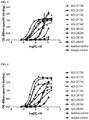

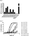
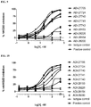


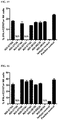
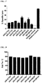
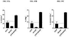
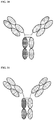
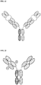


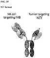

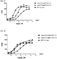
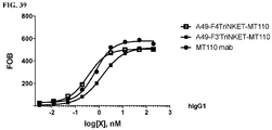
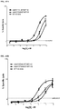

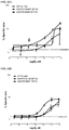
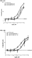
Classifications
-
- C—CHEMISTRY; METALLURGY
- C07—ORGANIC CHEMISTRY
- C07K—PEPTIDES
- C07K16/00—Immunoglobulins [IGs], e.g. monoclonal or polyclonal antibodies
- C07K16/18—Immunoglobulins [IGs], e.g. monoclonal or polyclonal antibodies against material from animals or humans
- C07K16/28—Immunoglobulins [IGs], e.g. monoclonal or polyclonal antibodies against material from animals or humans against receptors, cell surface antigens or cell surface determinants
- C07K16/2851—Immunoglobulins [IGs], e.g. monoclonal or polyclonal antibodies against material from animals or humans against receptors, cell surface antigens or cell surface determinants against the lectin superfamily, e.g. CD23, CD72
-
- A—HUMAN NECESSITIES
- A61—MEDICAL OR VETERINARY SCIENCE; HYGIENE
- A61P—SPECIFIC THERAPEUTIC ACTIVITY OF CHEMICAL COMPOUNDS OR MEDICINAL PREPARATIONS
- A61P35/00—Antineoplastic agents
-
- C—CHEMISTRY; METALLURGY
- C07—ORGANIC CHEMISTRY
- C07K—PEPTIDES
- C07K16/00—Immunoglobulins [IGs], e.g. monoclonal or polyclonal antibodies
- C07K16/18—Immunoglobulins [IGs], e.g. monoclonal or polyclonal antibodies against material from animals or humans
- C07K16/28—Immunoglobulins [IGs], e.g. monoclonal or polyclonal antibodies against material from animals or humans against receptors, cell surface antigens or cell surface determinants
- C07K16/2803—Immunoglobulins [IGs], e.g. monoclonal or polyclonal antibodies against material from animals or humans against receptors, cell surface antigens or cell surface determinants against the immunoglobulin superfamily
- C07K16/2821—Immunoglobulins [IGs], e.g. monoclonal or polyclonal antibodies against material from animals or humans against receptors, cell surface antigens or cell surface determinants against the immunoglobulin superfamily against ICAM molecules, e.g. CD50, CD54, CD102
-
- C—CHEMISTRY; METALLURGY
- C07—ORGANIC CHEMISTRY
- C07K—PEPTIDES
- C07K16/00—Immunoglobulins [IGs], e.g. monoclonal or polyclonal antibodies
- C07K16/18—Immunoglobulins [IGs], e.g. monoclonal or polyclonal antibodies against material from animals or humans
- C07K16/28—Immunoglobulins [IGs], e.g. monoclonal or polyclonal antibodies against material from animals or humans against receptors, cell surface antigens or cell surface determinants
- C07K16/2803—Immunoglobulins [IGs], e.g. monoclonal or polyclonal antibodies against material from animals or humans against receptors, cell surface antigens or cell surface determinants against the immunoglobulin superfamily
- C07K16/283—Immunoglobulins [IGs], e.g. monoclonal or polyclonal antibodies against material from animals or humans against receptors, cell surface antigens or cell surface determinants against the immunoglobulin superfamily against Fc-receptors, e.g. CD16, CD32, CD64
-
- C—CHEMISTRY; METALLURGY
- C07—ORGANIC CHEMISTRY
- C07K—PEPTIDES
- C07K16/00—Immunoglobulins [IGs], e.g. monoclonal or polyclonal antibodies
- C07K16/18—Immunoglobulins [IGs], e.g. monoclonal or polyclonal antibodies against material from animals or humans
- C07K16/28—Immunoglobulins [IGs], e.g. monoclonal or polyclonal antibodies against material from animals or humans against receptors, cell surface antigens or cell surface determinants
- C07K16/2896—Immunoglobulins [IGs], e.g. monoclonal or polyclonal antibodies against material from animals or humans against receptors, cell surface antigens or cell surface determinants against molecules with a "CD"-designation, not provided for elsewhere
-
- C—CHEMISTRY; METALLURGY
- C07—ORGANIC CHEMISTRY
- C07K—PEPTIDES
- C07K16/00—Immunoglobulins [IGs], e.g. monoclonal or polyclonal antibodies
- C07K16/18—Immunoglobulins [IGs], e.g. monoclonal or polyclonal antibodies against material from animals or humans
- C07K16/28—Immunoglobulins [IGs], e.g. monoclonal or polyclonal antibodies against material from animals or humans against receptors, cell surface antigens or cell surface determinants
- C07K16/30—Immunoglobulins [IGs], e.g. monoclonal or polyclonal antibodies against material from animals or humans against receptors, cell surface antigens or cell surface determinants from tumour cells
- C07K16/3076—Immunoglobulins [IGs], e.g. monoclonal or polyclonal antibodies against material from animals or humans against receptors, cell surface antigens or cell surface determinants from tumour cells against structure-related tumour-associated moieties
- C07K16/3084—Immunoglobulins [IGs], e.g. monoclonal or polyclonal antibodies against material from animals or humans against receptors, cell surface antigens or cell surface determinants from tumour cells against structure-related tumour-associated moieties against tumour-associated gangliosides
-
- A—HUMAN NECESSITIES
- A61—MEDICAL OR VETERINARY SCIENCE; HYGIENE
- A61K—PREPARATIONS FOR MEDICAL, DENTAL OR TOILETRY PURPOSES
- A61K39/00—Medicinal preparations containing antigens or antibodies
- A61K2039/505—Medicinal preparations containing antigens or antibodies comprising antibodies
-
- C—CHEMISTRY; METALLURGY
- C07—ORGANIC CHEMISTRY
- C07K—PEPTIDES
- C07K2317/00—Immunoglobulins specific features
- C07K2317/30—Immunoglobulins specific features characterized by aspects of specificity or valency
- C07K2317/31—Immunoglobulins specific features characterized by aspects of specificity or valency multispecific
-
- C—CHEMISTRY; METALLURGY
- C07—ORGANIC CHEMISTRY
- C07K—PEPTIDES
- C07K2317/00—Immunoglobulins specific features
- C07K2317/30—Immunoglobulins specific features characterized by aspects of specificity or valency
- C07K2317/33—Crossreactivity, e.g. for species or epitope, or lack of said crossreactivity
-
- C—CHEMISTRY; METALLURGY
- C07—ORGANIC CHEMISTRY
- C07K—PEPTIDES
- C07K2317/00—Immunoglobulins specific features
- C07K2317/50—Immunoglobulins specific features characterized by immunoglobulin fragments
- C07K2317/52—Constant or Fc region; Isotype
-
- C—CHEMISTRY; METALLURGY
- C07—ORGANIC CHEMISTRY
- C07K—PEPTIDES
- C07K2317/00—Immunoglobulins specific features
- C07K2317/50—Immunoglobulins specific features characterized by immunoglobulin fragments
- C07K2317/52—Constant or Fc region; Isotype
- C07K2317/522—CH1 domain
-
- C—CHEMISTRY; METALLURGY
- C07—ORGANIC CHEMISTRY
- C07K—PEPTIDES
- C07K2317/00—Immunoglobulins specific features
- C07K2317/50—Immunoglobulins specific features characterized by immunoglobulin fragments
- C07K2317/52—Constant or Fc region; Isotype
- C07K2317/53—Hinge
-
- C—CHEMISTRY; METALLURGY
- C07—ORGANIC CHEMISTRY
- C07K—PEPTIDES
- C07K2317/00—Immunoglobulins specific features
- C07K2317/50—Immunoglobulins specific features characterized by immunoglobulin fragments
- C07K2317/55—Fab or Fab'
-
- C—CHEMISTRY; METALLURGY
- C07—ORGANIC CHEMISTRY
- C07K—PEPTIDES
- C07K2317/00—Immunoglobulins specific features
- C07K2317/50—Immunoglobulins specific features characterized by immunoglobulin fragments
- C07K2317/56—Immunoglobulins specific features characterized by immunoglobulin fragments variable (Fv) region, i.e. VH and/or VL
- C07K2317/565—Complementarity determining region [CDR]
-
- C—CHEMISTRY; METALLURGY
- C07—ORGANIC CHEMISTRY
- C07K—PEPTIDES
- C07K2317/00—Immunoglobulins specific features
- C07K2317/50—Immunoglobulins specific features characterized by immunoglobulin fragments
- C07K2317/56—Immunoglobulins specific features characterized by immunoglobulin fragments variable (Fv) region, i.e. VH and/or VL
- C07K2317/569—Single domain, e.g. dAb, sdAb, VHH, VNAR or nanobody®
-
- C—CHEMISTRY; METALLURGY
- C07—ORGANIC CHEMISTRY
- C07K—PEPTIDES
- C07K2317/00—Immunoglobulins specific features
- C07K2317/60—Immunoglobulins specific features characterized by non-natural combinations of immunoglobulin fragments
- C07K2317/62—Immunoglobulins specific features characterized by non-natural combinations of immunoglobulin fragments comprising only variable region components
- C07K2317/622—Single chain antibody (scFv)
-
- C—CHEMISTRY; METALLURGY
- C07—ORGANIC CHEMISTRY
- C07K—PEPTIDES
- C07K2317/00—Immunoglobulins specific features
- C07K2317/70—Immunoglobulins specific features characterized by effect upon binding to a cell or to an antigen
- C07K2317/73—Inducing cell death, e.g. apoptosis, necrosis or inhibition of cell proliferation
-
- C—CHEMISTRY; METALLURGY
- C07—ORGANIC CHEMISTRY
- C07K—PEPTIDES
- C07K2317/00—Immunoglobulins specific features
- C07K2317/70—Immunoglobulins specific features characterized by effect upon binding to a cell or to an antigen
- C07K2317/75—Agonist effect on antigen
-
- C—CHEMISTRY; METALLURGY
- C07—ORGANIC CHEMISTRY
- C07K—PEPTIDES
- C07K2317/00—Immunoglobulins specific features
- C07K2317/70—Immunoglobulins specific features characterized by effect upon binding to a cell or to an antigen
- C07K2317/76—Antagonist effect on antigen, e.g. neutralization or inhibition of binding
-
- C—CHEMISTRY; METALLURGY
- C07—ORGANIC CHEMISTRY
- C07K—PEPTIDES
- C07K2317/00—Immunoglobulins specific features
- C07K2317/90—Immunoglobulins specific features characterized by (pharmaco)kinetic aspects or by stability of the immunoglobulin
- C07K2317/92—Affinity (KD), association rate (Ka), dissociation rate (Kd) or EC50 value
-
- C—CHEMISTRY; METALLURGY
- C07—ORGANIC CHEMISTRY
- C07K—PEPTIDES
- C07K2317/00—Immunoglobulins specific features
- C07K2317/90—Immunoglobulins specific features characterized by (pharmaco)kinetic aspects or by stability of the immunoglobulin
- C07K2317/94—Stability, e.g. half-life, pH, temperature or enzyme-resistance
-
- Y—GENERAL TAGGING OF NEW TECHNOLOGICAL DEVELOPMENTS; GENERAL TAGGING OF CROSS-SECTIONAL TECHNOLOGIES SPANNING OVER SEVERAL SECTIONS OF THE IPC; TECHNICAL SUBJECTS COVERED BY FORMER USPC CROSS-REFERENCE ART COLLECTIONS [XRACs] AND DIGESTS
- Y02—TECHNOLOGIES OR APPLICATIONS FOR MITIGATION OR ADAPTATION AGAINST CLIMATE CHANGE
- Y02A—TECHNOLOGIES FOR ADAPTATION TO CLIMATE CHANGE
- Y02A50/00—TECHNOLOGIES FOR ADAPTATION TO CLIMATE CHANGE in human health protection, e.g. against extreme weather
- Y02A50/30—Against vector-borne diseases, e.g. mosquito-borne, fly-borne, tick-borne or waterborne diseases whose impact is exacerbated by climate change
Definitions
- the invention relates to multi-specific binding proteins that bind to NKG2D, CD16, and a tumor-associated antigen.
- Cancer continues to be a significant health problem despite the substantial research efforts and scientific advances reported in the literature for treating this disease.
- Some of the most frequently diagnosed cancers include prostate cancer, breast cancer, lung cancer, and colorectal cancer.
- Prostate cancer is the most common form of cancer in men.
- Breast cancer remains a leading cause of death in women.
- Blood and bone marrow cancers are also frequently diagnosed cancer types, including multiple myelomas, leukemia, and lymphomas. Current treatment options for these cancers are not effective for all patients and/or can have substantial adverse side effects. Other types of cancer also remain challenging to treat using existing therapeutic options.
- Cancer immunotherapies are desirable because they are highly specific and can facilitate destruction of cancer cells using the patient's own immune system. Fusion proteins such as bi-specific T-cell engagers are cancer immunotherapies described in the literature that bind to tumor cells and T-cells to facilitate destruction of tumor cells. Antibodies that bind to certain tumor-associated antigens and to certain immune cells have been described in the literature. See, for example WO 2016/134371 and WO 2015/095412 .
- NK cells Natural killer cells are a component of the innate immune system and make up approximately 15% of circulating lymphocytes. NK cells infiltrate virtually all tissues and were originally characterized by their ability to kill tumor cells effectively without the need for prior sensitization. Activated NK cells kill target cells by means similar to cytotoxic T cells - i.e., via cytolytic granules that contain perforin and granzymes as well as via death receptor pathways. Activated NK cells also secrete inflammatory cytokines such as IFN-gamma and chemokines that promote the recruitment of other leukocytes to the target tissue.
- cytotoxic T cells i.e., via cytolytic granules that contain perforin and granzymes as well as via death receptor pathways.
- Activated NK cells also secrete inflammatory cytokines such as IFN-gamma and chemokines that promote the recruitment of other leukocytes to the target tissue.
- NK cells respond to signals through a variety of activating and inhibitory receptors on their surface. For example, when NK cells encounter healthy self-cells, their activity is inhibited through activation of the killer-cell immunoglobulin-like receptors (KIRs). Alternatively, when NK cells encounter foreign cells or cancer cells, they are activated via their activating receptors (e.g ., Natural killer group 2 member D (NKG2D), NCRs, DNAM1). NK cells are also activated by the constant region of some immunoglobulins through CD16 receptors on their surface. The overall sensitivity of NK cells to activation depends on the sum of stimulatory and inhibitory signals.
- KIRs killer-cell immunoglobulin-like receptors
- EpCAM Epithelial cell adhesion molecule
- EpCAM is a transmembrane glycoprotein mediating Ca 2+ -independent homotypic cell-cell adhesion in epithelia. EpCAM is also involved in cell signaling, migration, proliferation, and differentiation. Additionally, EpCAM has oncogenic potential via its capacity to upregulate c-myc, e-fabp, and cyclins A and E. Since EpCAM is expressed exclusively in epithelia and epithelial-derived neoplasms, EpCAM can be used as diagnostic marker for various cancers, such as head and neck cancer, ovarian cancer, bladder cancer, breast cancer, colorectal cancer, prostate cancer, gastric cancer, liver cancer, esophageal cancer, and lung cancer. It appears to play a role in tumorigenesis and metastasis of carcinomas, so it can also act as a potential prognostic marker and as a potential target for immunotherapeutic strategies.
- CA125 also known as mucin 16 is a member of the mucin family glycoproteins.
- CA-125 has found application as a tumor marker or biomarker that may be elevated in the blood of some patients with specific types of cancers, including ovarian cancer, endometrial cancer, and pancreatic cancer.
- CA-125 has been shown to play a role in advancing tumorigenesis and tumor proliferation by several different mechanisms, including suppressing the response of natural killer cells, and thereby protecting cancer cells from the immune response; and by enabling cell growth and promoting cell motility.
- NaPi2b Sodium-dependent phosphate transport protein 2b
- NaPi2b is involved in actively transporting phosphate into cells via Na+ co-transport.
- it is the main phosphate transport protein in the intestinal brush border membrane, and has a role in the synthesis of surfactant in lungs' alveoli.
- NaPi2b is also an antigen expressed in a variety of cancer, such as lung cancer, ovarian cancer, and thyroid cancer.
- Nectin4 is a member of the Nectin family, which is a family of cellular adhesion molecules involved in Ca 2+ -independent cellular adhesion. Nectins are ubiquitously expressed and have adhesive roles in a wide range of tissues such as the adherens junction of epithelia or the chemical synapse of the neuronal tissue. It is also a tumor associated antigen, and expressed in cancers such as bladder cancer, breast cancer, ovarian cancer, pancreatic cancer, colorectal cancer, and lung cancer.
- Gangliosides have been implicated in many physiological processes, including growth, differentiation, migration, and apoptosis through modulating both cell signaling processes and cell-to-cell and cell-to-matrix interactions.
- GM1 is a ganglioside
- Fucosyl-GM1 is a ganglioside with a unique structure in which the terminal galactose is ⁇ -1,2-fucosylated at the non-reducing end. It is expressed in very few normal tissues but occurs in a variety of cancers such as in small cell lung cancer, neuroblastoma, liver cancer. Consequently, fucosyl-GM1 has been considered to be a candidate as a tumor marker and target antigen in antibody immunotherapy small cell lung cancer, neuroblastoma, liver cancer.
- ADAM a disintegrin and metalloproteinase proteins have a predominant role in the protein ectodomain shedding of membrane-bound molecules. They have emerged as critical regulators of cell-cell signaling during development and homeostasis, and are believed to contribute to pathologies, such as cancer, where their regulation is altered.
- ADAM8 a member the ADAM family, is overexpressed in pancreatic cancer, breast cancer, lung cancer, and renal cancer.
- ADAM9 has been shown to cleave and release a number of molecules with important roles in tumorigenesis and angiogenesis, such as EGF, FGFR2iiib, Tie-2, Flk-1, EphB4, CD40, VCAM-1, and VE-cadherin.
- ADAM9 is overexpressed in renal cancer, breast cancer, lung cancer, liver cancer, pancreatic cancer, melanoma, cervical cancer, prostate cancer, osteosarcoma, and brain cancer.
- SLC44A4 also known as CTL4, is a member of the family of solute carrier proteins known as choline transporter-like proteins (CTL1-5). SLC44A4 has not been shown to be involved in choline transport, but it has been linked with acetylcholine synthesis and transport as well as uptake of thiamine pyrophosphate, the phosphorylated form of vitamin B1. SLC44A4 is normally expressed on the apical surface of secretory epithelial cells, but it is markedly upregulated in a variety of epithelial tumors, most notably pancreatic cancer, prostate cancer, and gastric cancer.
- CA19-9 is the common term for carbohydrate antigen sialyl Lewis a. It is overexpressed in cancer of the digestive organs such as pancreatic cancer, colorectal cancer, cholangiocarcinoma, and liver cancer. Therefore, it is the most frequently applied serum tumor marker for diagnosis of these above mentioned cancers.
- the invention provides multi-specific binding proteins that bind to a tumor-associated antigen (selected from any one of the antigens provided in Table 11) and to the NKG2D receptor and CD16 receptor on natural killer cells.
- a tumor-associated antigen selected from any one of the antigens provided in Table 11
- Such proteins can engage more than one kind of NK activating receptor, and may block the binding of natural ligands to NKG2D.
- the proteins can agonize NK cells in humans, and in other species such as rodents and cynomolgus monkeys.
- one aspect of the invention provides a protein that incorporates a first antigen-binding site that binds NKG2D; a second antigen-binding site that binds an antigen selected from EpCAM, Cancer Antigen 125 (CA125), sodium/phosphate cotransporter 2B (NaPi2b), Nectin cell adhesion molecule 4 (Nectin4), Fucosyl-GM1 (monosialotetrahexosylganglioside), disintegrin and metalloproteinase domain-containing protein 8 (ADAM8), disintegrin and metalloproteinase domain-containing protein 9 (ADAM9), solute carrier family 44 member 4 (SLC44A4), and sialylated Lewis a antigen (CA19-9); and an antibody Fc domain, a portion thereof sufficient to bind CD16, or a third antigen-binding site that binds CD16.
- an antigen selected from EpCAM, Cancer Antigen 125 (CA125), sodium/phosphate cotransport
- the antigen-binding sites may each incorporate an antibody heavy chain variable domain and an antibody light chain variable domain (e.g. arranged as in an antibody, or fused together to from an scFv), or one or more of the antigen-binding sites may be a single domain antibody, such as a V H H antibody like a camelid antibody or a V NAR antibody like those found in cartilaginous fish.
- the invention provides multi-specific binding proteins that bind the NKG2D receptor, CD16, and an antigen selected from EpCAM, Cancer Antigen 125 (CA125), sodium/phosphate cotransporter 2B (NaPi2b), Nectin cell adhesion molecule 4 (Nectin4), Fucosyl-GM1 (monosialotetrahexosylganglioside), disintegrin and metalloproteinase domain-containing protein 8 (ADAM8), disintegrin and metalloproteinase domain-containing protein 9 (ADAM9), solute carrier family 44 member 4 (SLC44A4), and sialylated Lewis a antigen (CA19-9).
- EpCAM EpCAM
- CA125 Cancer Antigen 125
- NaPi2b sodium/phosphate cotransporter 2B
- Nectin4 Nectin cell adhesion molecule 4
- Fucosyl-GM1 monosialotetrahexosylganglioside
- ADAM8 dis
- Some proteins of the present disclosure include an Fab fragment linked to the antibody Fc domain or a portion thereof sufficient to bind CD 16, or the third antigen-binding site that binds CD16.
- Some proteins of the present disclosure include an Fab fragment, wherein the heavy chain portion of the Fab fragment comprises a heavy chain variable domain and a CH1 domain, and wherein the heavy chain variable domain is linked to the CH1 domain.
- Some proteins of the present disclosure include an Fab fragment linked to the antibody Fc domain.
- the invention provides a protein comprising (a) a first antigen-binding site comprising an Fab fragment that binds NKG2D; (b) a second antigen-binding site comprising a single-chain variable fragment (scFv) that binds EpCAM; and (c) an antibody Fc domain or a portion thereof sufficient to bind CD 16, or a third antigen-binding site that binds CD16.
- the present invention provides a protein in which the first antigen-binding site that binds NKG2D is an Fab fragment, and the second antigen-binding site that binds a tumor-associated antigen EpCAM is an scFv.
- Certain proteins described in the present disclosure include an scFv-targeting EpCAM, comprising a heavy chain variable domain and a light chain variable domain, linked to an antibody Fc domain or a portion thereof sufficient to bind CD16, or the third antigen-binding site that binds CD16, via a hinge comprising Ala-Ser or Gly-Ala-Ser.
- Some proteins of the present disclosure includes an scFv-targeting EpCAM linked to an antibody Fc domain.
- Some proteins of the present disclosure includes a heavy chain variable domain of an scFv-targeting EpCAM, which forms a disulfide bridge with the light chain variable domain of the scFv.
- Some proteins of the present disclosure include an scFv-targeting EpCAM, in which a disulfide bridge is formed between C44 from the heavy chain variable domain and C100 from the light chain variable domain.
- Some proteins of the present disclosure include an scFv-targeting EpCAM linked to an antibody Fc domain, in which the light chain variable domain of the scFv is positioned at the N-terminus of the heavy chain variable domain of the scFv, and is linked to the heavy chain variable domain of the scFv via a flexible linker (GlyGlyGlyGlySer) 4 (G4S) 4 ) (SEQ ID NO:206), and the Fab fragment that binds NKG2D is linked to the antibody Fc domain.
- a flexible linker GlyGlyGlyGlyGlySer 4 (G4S) 4 )
- Some proteins of the present disclosure include an scFv-targeting EpCAM in which the heavy chain variable domain is positioned at the N-terminus or the C-terminus of the light chain variable domain of the scFv.
- Some proteins of the present disclosure include an scFv-targeting EpCAM in which the light chain variable domain is positioned at the N-terminus of the heavy chain variable domain of the scFv.
- a protein of the present disclosure provides a protein comprising (a) a first antigen-binding site comprising a single-chain variable fragment (scFv) that binds NKG2D; (b) a second antigen-binding site that binds EpCAM; and (c) an antibody Fc domain or a portion thereof sufficient to bind CD 16, or a third antigen-binding site that binds CD 16.
- a protein of the present disclosure further comprises an additional antigen-binding site that binds EpCAM.
- the second antigen-binding site of a protein described in the present disclosure is an Fab fragment that binds EpCAM.
- the second and the additional antigen-binding site of a protein described in the present disclosure are Fab fragments that bind EpCAM.
- the second and the additional antigen-binding site of a protein described in the present disclosure are scFvs that bind EpCAM.
- the heavy chain variable domain of the scFv that binds NKG2D is positioned at the N-terminus or the C-terminus of the light chain variable domain of the scFv.
- the light chain variable domain is positioned at the N-terminus of the heavy chain variable domain of the scFv that binds NKG2D.
- the scFv that binds to NKG2D is linked to the antibody Fc domain or a portion thereof sufficient to bind CD16, or a third antigen-binding site that binds CD16.
- the scFv that binds to NKG2D is linked to the antibody Fc domain or a portion thereof sufficient to bind CD16, or a third antigen-binding site that binds CD16 via a hinge comprising Ala-Ser (e.g., in a TriNKET that comprises an additional antigen-binding site that binds EpCAM, CA125, NaPi2b, Nectin4, Fucosyl-GM1, ADAM8, ADAM9, SLC44A4, or CA19-9) or Gly-Ala-Ser (e.g.
- the scFv that binds to NKG2D is linked to the C-terminus of the antibody Fc domain or a portion thereof sufficient to bind CD 16, or a third antigen-binding site that binds CD 16 via a flexible linker comprising G4S.
- the C-terminus of the antibody Fc domain is linked to the N-terminus of the light chain variable domain of the scFv that binds NKG2D.
- a disulfide bridge is formed between the heavy chain variable domain of the scFv and the light chain variable domain of the scFv. In certain embodiments, the disulfide bridge is formed between C44 from the heavy chain variable domain and C100 from the light chain variable domain.
- Some proteins of the present disclosure include a sequence selected from SEQ ID NO:210 and SEQ ID NO:211.
- Some proteins of the present disclosure include an scFv linked to an antibody Fc domain, wherein the scFv linked to the antibody Fc domain is represented by a sequence selected from SEQ ID NO:208 and SEQ ID NO:209.
- Some proteins of the present disclosure include a sequence of SEQ ID NO:205 and SEQ ID NO:213.
- Some proteins of the present disclosure include a sequence at least 90% identical to an amino acid sequence selected from SEQ ID NO:210 and SEQ ID NO:211.
- Some proteins of the present disclosure include a sequence at least 95% identical to an amino acid sequence selected from SEQ ID NO:210 and SEQ ID NO:211.
- Some proteins of the present disclosure include a sequence at least 99% identical to an amino acid sequence selected from SEQ ID NO:210 and SEQ ID NO:211.
- Some proteins of the present disclosure include an amino acid sequence of SEQ ID NO:203.
- Some proteins of the present disclosure include an amino acid sequence of SEQ ID NO:203 and SEQ ID NO:204.
- Some proteins of the present disclosure include an amino acid sequence at least 90% identical to an amino acid sequence of SEQ ID NO:203. Some proteins of the present disclosure include an amino acid sequence at least 95% identical to an amino acid sequence of SEQ ID NO:203. Some proteins of the present disclosure include an amino acid sequence at least 99% identical to an amino acid sequence of SEQ ID NO:203.
- the first antigen-binding site which binds to NKG2D, in some embodiments, can incorporate a heavy chain variable domain related to SEQ ID NO:1, such as by having an amino acid sequence at least 90% (e.g., 90%, 91%, 92%, 93%, 94%, 95%, 96%, 97%, 98%, 99%, or 100%) identical to SEQ ID NO:1, and/or incorporating amino acid sequences identical to the CDR1 (SEQ ID NO:105), CDR2 (SEQ ID NO:106), and CDR3 (SEQ ID NO: 107) sequences of SEQ ID NO:1.
- the heavy chain variable domain related to SEQ ID NO: 1 can be coupled with a variety of light chain variable domains to form an NKG2D binding site.
- the first antigen-binding site that incorporates a heavy chain variable domain related to SEQ ID NO: 1 can further incorporate a light chain variable domain selected from any one of the sequences related to SEQ ID NOs:2, 4, 6, 8, 10, 12, 14, 16, 18, 20, 22, 24, 26, 28, 30, 32, 34, 36, 38, and 40.
- the first antigen-binding site incorporates a heavy chain variable domain with amino acid sequences at least 90% (e.g., 90%, 91%, 92%, 93%, 94%, 95%, 96%, 97%, 98%, 99%, or 100%) identical to SEQ ID NO: 1 and a light chain variable domain with amino acid sequences at least 90% (e.g.
- the first antigen-binding site can incorporate a heavy chain variable domain related to SEQ ID NO:41 and a light chain variable domain related to SEQ ID NO:42.
- the heavy chain variable domain of the first antigen-binding site can be at least 90% (e.g., 90%, 91%, 92%, 93%, 94%, 95%, 96%, 97%, 98%, 99%, or 100%) identical to SEQ ID NO:41, and/or incorporate amino acid sequences identical to the CDR1 (SEQ ID NO:43), CDR2 (SEQ ID NO:44), and CDR3 (SEQ ID NO:45) sequences of SEQ ID NO:41.
- the light chain variable domain of the second antigen-binding site can be at least 90% (e.g., 90%, 91%, 92%, 93%, 94%, 95%, 96%, 97%, 98%, 99%, or 100%) identical to SEQ ID NO:42, and/or incorporate amino acid sequences identical to the CDR1 (SEQ ID NO:46), CDR2 (SEQ ID NO:47), and CDR3 (SEQ ID NO:48) sequences of SEQ ID NO:42.
- the first antigen-binding site can incorporate a heavy chain variable domain related to SEQ ID NO:49 and a light chain variable domain related to SEQ ID NO:50.
- the heavy chain variable domain of the first antigen-binding site can be at least 90% (e.g., 90%, 91%, 92%, 93%, 94%, 95%, 96%, 97%, 98%, 99%, or 100%) identical to SEQ ID NO:49, and/or incorporate amino acid sequences identical to the CDR1 (SEQ ID NO:51), CDR2 (SEQ ID NO:52), and CDR3 (SEQ ID NO:53) sequences of SEQ ID NO:49.
- the light chain variable domain of the second antigen-binding site can be at least 90% (e.g., 90%, 91%, 92%, 93%, 94%, 95%, 96%, 97%, 98%, 99%, or 100%) identical to SEQ ID NO:50, and/or incorporate amino acid sequences identical to the CDR1 (SEQ ID NO:54), CDR2 (SEQ ID NO:55), and CDR3 (SEQ ID NO:56) sequences of SEQ ID NO:50.
- the first antigen-binding site can incorporate a heavy chain variable domain related to SEQ ID NO:57 and a light chain variable domain related to SEQ ID NO:58, such as by having amino acid sequences at least 90% (e.g., 90%, 91%, 92%, 93%, 94%, 95%, 96%, 97%, 98%, 99%, or 100%) identical to SEQ ID NO:57 and at least 90% ( e.g., 90%, 91%, 92%, 93%, 94%, 95%, 96%, 97%, 98%, 99%, or 100%) identical to SEQ ID NO:58, respectively.
- 90%, 91%, 92%, 93%, 94%, 95%, 96%, 97%, 98%, 99%, or 100% identical to SEQ ID NO:58, respectively.
- the first antigen-binding site can incorporate a heavy chain variable domain related to SEQ ID NO:59 and a light chain variable domain related to SEQ ID NO:60,
- the heavy chain variable domain of the first antigen-binding site can be at least 90% (e.g., 90%, 91%, 92%, 93%, 94%, 95%, 96%, 97%, 98%, 99%, or 100%) identical to SEQ ID NO:59, and/or incorporate amino acid sequences identical to the CDR1 (SEQ ID NO: 109), CDR2 (SEQ ID NO: 110), and CDR3 (SEQ ID NO:111) sequences of SEQ ID NO:59.
- the light chain variable domain of the second antigen-binding site can be at least 90% (e.g., 90%, 91%, 92%, 93%, 94%, 95%, 96%, 97%, 98%, 99%, or 100%) identical to SEQ ID NO:60, and/or incorporate amino acid sequences identical to the CDR1 (SEQ ID NO: 112), CDR2 (SEQ ID NO:113), and CDR3 (SEQ ID NO:114) sequences of SEQ ID NO:60.
- the first antigen-binding site which binds to NKG2D, in some embodiments, can incorporate a heavy chain variable domain related to SEQ ID NO:61 and a light chain variable domain related to SEQ ID NO:62.
- the heavy chain variable domain of the first antigen-binding site can be at least 90% (e.g., 90%, 91%, 92%, 93%, 94%, 95%, 96%, 97%, 98%, 99%, or 100%) identical to SEQ ID NO:61, and/or incorporate amino acid sequences identical to the CDR1 (SEQ ID NO:63), CDR2 (SEQ ID NO:64), and CDR3 (SEQ ID NO:65) sequences of SEQ ID NO:61.
- the light chain variable domain of the second antigen-binding site can be at least 90% (e.g., 90%, 91%, 92%, 93%, 94%, 95%, 96%, 97%, 98%, 99%, or 100%) identical to SEQ ID NO:62, and/or incorporate amino acid sequences identical to the CDR1 (SEQ ID NO:66), CDR2 (SEQ ID NO:67), and CDR3 (SEQ ID NO:68) sequences of SEQ ID NO:62.
- the first antigen-binding site can incorporate a heavy chain variable domain related to SEQ ID NO:69 and a light chain variable domain related to SEQ ID NO:70.
- the heavy chain variable domain of the first antigen-binding site can be at least 90% (e.g., 90%, 91%, 92%, 93%, 94%, 95%, 96%, 97%, 98%, 99%, or 100%) identical to SEQ ID NO:69, and/or incorporate amino acid sequences identical to the CDR1 (SEQ ID NO:71), CDR2 (SEQ ID NO:72), and CDR3 (SEQ ID NO:73) sequences of SEQ ID NO:69.
- the light chain variable domain of the second antigen-binding site can be at least 90% (e.g., 90%, 91%, 92%, 93%, 94%, 95%, 96%, 97%, 98%, 99%, or 100%) identical to SEQ ID NO:70, and/or incorporate amino acid sequences identical to the CDR1 (SEQ ID NO:74), CDR2 (SEQ ID NO:75), and CDR3 (SEQ ID NO:76) sequences of SEQ ID NO:70.
- the first antigen-binding site can incorporate a heavy chain variable domain related to SEQ ID NO:77 and a light chain variable domain related to SEQ ID NO:78.
- the heavy chain variable domain of the first antigen-binding site can be at least 90% (e.g., 90%, 91%, 92%, 93%, 94%, 95%, 96%, 97%, 98%, 99%, or 100%) identical to SEQ ID NO:77, and/or incorporate amino acid sequences identical to the CDR1 (SEQ ID NO:79), CDR2 (SEQ ID NO:80), and CDR3 (SEQ ID NO:81) sequences of SEQ ID NO:77.
- the light chain variable domain of the second antigen-binding site can be at least 90% (e.g., 90%, 91%, 92%, 93%, 94%, 95%, 96%, 97%, 98%, 99%, or 100%) identical to SEQ ID NO:78, and/or incorporate amino acid sequences identical to the CDR1 (SEQ ID NO:82), CDR2 (SEQ ID NO:83), and CDR3 (SEQ ID NO:84) sequences of SEQ ID NO:78.
- the first antigen-binding site can incorporate a heavy chain variable domain related to SEQ ID NO:85 and a light chain variable domain related to SEQ ID NO:86.
- the heavy chain variable domain of the first antigen-binding site can be at least 90% (e.g., 90%, 91%, 92%, 93%, 94%, 95%, 96%, 97%, 98%, 99%, or 100%) identical to SEQ ID NO:85, and/or incorporate amino acid sequences identical to the CDR1 (SEQ ID NO:87), CDR2 (SEQ ID NO:88), and CDR3 (SEQ ID NO:89) sequences of SEQ ID NO:85.
- the light chain variable domain of the second antigen-binding site can be at least 90% (e.g., 90%, 91%, 92%, 93%, 94%, 95%, 96%, 97%, 98%, 99%, or 100%) identical to SEQ ID NO:86, and/or incorporate amino acid sequences identical to the CDR1 (SEQ ID NO:90), CDR2 (SEQ ID NO:91), and CDR3 (SEQ ID NO:92) sequences of SEQ ID NO:86.
- the first antigen-binding site can incorporate a heavy chain variable domain related to SEQ ID NO:93 and a light chain variable domain related to SEQ ID NO:94.
- the heavy chain variable domain of the first antigen-binding site can be at least 90% (e.g., 90%, 91%, 92%, 93%, 94%, 95%, 96%, 97%, 98%, 99%, or 100%) identical to SEQ ID NO:93, and/or incorporate amino acid sequences identical to the CDR1 (SEQ ID NO:95), CDR2 (SEQ ID NO:96), and CDR3 (SEQ ID NO:97) sequences of SEQ ID NO:93.
- the light chain variable domain of the second antigen-binding site can be at least 90% (e.g., 90%, 91%, 92%, 93%, 94%, 95%, 96%, 97%, 98%, 99%, or 100%) identical to SEQ ID NO:94, and/or incorporate amino acid sequences identical to the CDR1 (SEQ ID NO:98), CDR2 (SEQ ID NO:99), and CDR3 (SEQ ID NO:100) sequences of SEQ ID NO:94.
- the first antigen-binding site can incorporate a heavy chain variable domain related to SEQ ID NO:101 and a light chain variable domain related to SEQ ID NO:102, such as by having amino acid sequences at least 90% (e.g., 90%, 91%, 92%, 93%, 94%, 95%, 96%, 97%, 98%, 99%, or 100%) identical to SEQ ID NO:101 and at least 90% (e.g., 90%, 91%, 92%, 93%, 94%, 95%, 96%, 97%, 98%, 99%, or 100%) identical to SEQ ID NO:102, respectively.
- 90%, 91%, 92%, 93%, 94%, 95%, 96%, 97%, 98%, 99%, or 100% identical to SEQ ID NO:102, respectively.
- the first antigen-binding site can incorporate a heavy chain variable domain related to SEQ ID NO:103 and a light chain variable domain related to SEQ ID NO:104, such as by having amino acid sequences at least 90% (e.g., 90%, 91%, 92%, 93%, 94%, 95%, 96%, 97%, 98%, 99%, or 100%) identical to SEQ ID NO:103 and at least 90% (e.g., 90%, 91%, 92%, 93%, 94%, 95%, 96%, 97%, 98%, 99%, or 100%) identical to SEQ ID NO:104, respectively.
- 90%, 91%, 92%, 93%, 94%, 95%, 96%, 97%, 98%, 99%, or 100% identical to SEQ ID NO:104, respectively.
- the second antigen-binding site can bind to EpCAM and can incorporate a heavy chain variable domain related to SEQ ID NO:115 and a light chain variable domain related to SEQ ID NO:119.
- the heavy chain variable domain of the second antigen-binding site can be at least 90% (e.g., 90%, 91%, 92%, 93%, 94%, 95%, 96%, 97%, 98%, 99%, or 100%) identical to SEQ ID NO:115, and/or incorporate amino acid sequences identical to the CDR1 (SEQ ID NO: 116), CDR2 (SEQ ID NO:117), and CDR3 (SEQ ID NO: 118) sequences of SEQ ID NO:115.
- the light chain variable domain of the second antigen-binding site can be at least 90% (e.g., 90%, 91%, 92%, 93%, 94%, 95%, 96%, 97%, 98%, 99%, or 100%) identical to SEQ ID NO:119, and/or incorporate amino acid sequences identical to the CDR1 (SEQ ID NO:120), CDR2 (SEQ ID NO:121), and CDR3 (SEQ ID NO: 122) sequences of SEQ ID NO:119.
- the second antigen-binding site can bind to EpCAM and can incorporate a heavy chain variable domain related to SEQ ID NO:123 and a light chain variable domain related to SEQ ID NO:127.
- the heavy chain variable domain of the second antigen-binding site can be at least 90% (e.g., 90%, 91%, 92%, 93%, 94%, 95%, 96%, 97%, 98%, 99%, or 100%) identical to SEQ ID NO:123, and/or incorporate amino acid sequences identical to the CDR1 (SEQ ID NO: 124), CDR2 (SEQ ID NO:125), and CDR3 (SEQ ID NO: 126) sequences of SEQ ID NO:123.
- the light chain variable domain of the second antigen-binding site can be at least 90% (e.g., 90%, 91%, 92%, 93%, 94%, 95%, 96%, 97%, 98%, 99%, or 100%) identical to SEQ ID NO:127, and/or incorporate amino acid sequences identical to the CDR1 (SEQ ID NO:128), CDR2 (SEQ ID NO:129), and CDR3 (SEQ ID NO: 130) sequences of SEQ ID NO:127.
- the second antigen-binding site can bind to EpCAM and can incorporate a heavy chain variable domain related to SEQ ID NO:131 and a light chain variable domain related to SEQ ID NO:135.
- the heavy chain variable domain of the second antigen-binding site can be at least 90% (e.g., 90%, 91%, 92%, 93%, 94%, 95%, 96%, 97%, 98%, 99%, or 100%) identical to SEQ ID NO:131, and/or incorporate amino acid sequences identical to the CDR1 (SEQ ID NO: 132), CDR2 (SEQ ID NO:133), and CDR3 (SEQ ID NO: 134) sequences of SEQ ID NO:131.
- the light chain variable domain of the second antigen-binding site can be at least 90% (e.g., 90%, 91%, 92%, 93%, 94%, 95%, 96%, 97%, 98%, 99%, or 100%) identical to SEQ ID NO:135, and/or incorporate amino acid sequences identical to the CDR1 (SEQ ID NO:136), CDR2 (SEQ ID NO:137), and CDR3 (SEQ ID NO: 138) sequences of SEQ ID NO:135.
- the second antigen-binding site can bind to EpCAM and can incorporate a heavy chain variable domain related to SEQ ID NO:139 and a light chain variable domain related to SEQ ID NO:143.
- the heavy chain variable domain of the second antigen-binding site can be at least 90% (e.g., 90%, 91%, 92%, 93%, 94%, 95%, 96%, 97%, 98%, 99%, or 100%) identical to SEQ ID NO:139, and/or incorporate amino acid sequences identical to the CDR1 (SEQ ID NO: 140), CDR2 (SEQ ID NO:141), and CDR3 (SEQ ID NO: 142) sequences of SEQ ID NO:139.
- the light chain variable domain of the second antigen-binding site can be at least 90% (e.g., 90%, 91%, 92%, 93%, 94%, 95%, 96%, 97%, 98%, 99%, or 100%) identical to SEQ ID NO:143, and/or incorporate amino acid sequences identical to the CDR1 (SEQ ID NO:144), CDR2 (SEQ ID NO:145), and CDR3 (SEQ ID NO: 146) sequences of SEQ ID NO:143.
- the second antigen-binding site can bind to CA125 and can incorporate a heavy chain variable domain related to SEQ ID NO:155 and a light chain variable domain related to SEQ ID NO:159.
- the heavy chain variable domain of the second antigen-binding site can be at least 90% (e.g., 90%, 91%, 92%, 93%, 94%, 95%, 96%, 97%, 98%, 99%, or 100%) identical to SEQ ID NO: 155, and/or incorporate amino acid sequences identical to the CDR1 (SEQ ID NO:156), CDR2 (SEQ ID NO:157), and CDR3 (SEQ ID NO: 158) sequences of SEQ ID NO: 155.
- the light chain variable domain of the second antigen-binding site can be at least 90% (e.g., 90%, 91%, 92%, 93%, 94%, 95%, 96%, 97%, 98%, 99%, or 100%) identical to SEQ ID NO:159, and/or incorporate amino acid sequences identical to the CDR1 (SEQ ID NO: 160), CDR2 (SEQ ID NO: 161), and CDR3 (SEQ ID NO: 162) sequences of SEQ ID NO:159.
- the second antigen-binding site can bind to CA125 and can incorporate a heavy chain variable domain related to SEQ ID NO: 163 and a light chain variable domain related to SEQ ID NO:167.
- the heavy chain variable domain of the second antigen-binding site can be at least 90% (e.g., 90%, 91%, 92%, 93%, 94%, 95%, 96%, 97%, 98%, 99%, or 100%) identical to SEQ ID NO: 163, and/or incorporate amino acid sequences identical to the CDR1 (SEQ ID NO: 164), CDR2 (SEQ ID NO: 165), and CDR3 (SEQ ID NO: 166) sequences of SEQ ID NO: 163.
- the light chain variable domain of the second antigen-binding site can be at least 90% (e.g., 90%, 91%, 92%, 93%, 94%, 95%, 96%, 97%, 98%, 99%, or 100%) identical to SEQ ID NO: 167, and/or incorporate amino acid sequences identical to the CDR1 (SEQ ID NO: 168), CDR2 (SEQ ID NO: 169), and CDR3 (SEQ ID NO: 170) sequences of SEQ ID NO: 167.
- the second antigen-binding site can bind to NaPi2b and can incorporate a heavy chain variable domain related to SEQ ID NO: 171 and a light chain variable domain related to SEQ ID NO: 175.
- the heavy chain variable domain of the second antigen-binding site can be at least 90% (e.g., 90%, 91%, 92%, 93%, 94%, 95%, 96%, 97%, 98%, 99%, or 100%) identical to SEQ ID NO: 171, and/or incorporate amino acid sequences identical to the CDR1 (SEQ ID NO: 172), CDR2 (SEQ ID NO: 173), and CDR3 (SEQ ID NO: 174) sequences of SEQ ID NO: 171.
- the light chain variable domain of the second antigen-binding site can be at least 90% (e.g., 90%, 91%, 92%, 93%, 94%, 95%, 96%, 97%, 98%, 99%, or 100%) identical to SEQ ID NO: 175, and/or incorporate amino acid sequences identical to the CDR1 (SEQ ID NO: 176), CDR2 (SEQ ID NO: 177), and CDR3 (SEQ ID NO: 178) sequences of SEQ ID NO: 175.
- the second antigen-binding site can bind to Nectin4 and can incorporate a heavy chain variable domain related to SEQ ID NO: 179 and a light chain variable domain related to SEQ ID NO:183.
- the heavy chain variable domain of the second antigen-binding site can be at least 90% (e.g., 90%, 91%, 92%, 93%, 94%, 95%, 96%, 97%, 98%, 99%, or 100%) identical to SEQ ID NO: 179, and/or incorporate amino acid sequences identical to the CDR1 (SEQ ID NO: 180), CDR2 (SEQ ID NO: 181), and CDR3 (SEQ ID NO: 182) sequences of SEQ ID NO: 179.
- the light chain variable domain of the second antigen-binding site can be at least 90% (e.g., 90%, 91%, 92%, 93%, 94%, 95%, 96%, 97%, 98%, 99%, or 100%) identical to SEQ ID NO: 183, and/or incorporate amino acid sequences identical to the CDR1 (SEQ ID NO: 184), CDR2 (SEQ ID NO: 185), and CDR3 (SEQ ID NO: 186) sequences of SEQ ID NO: 183.
- the second antigen-binding site can bind to fucosyl-GM1 and can incorporate a heavy chain variable domain related to SEQ ID NO: 187 and a light chain variable domain related to SEQ ID NO: 191.
- the heavy chain variable domain of the second antigen-binding site can be at least 90% (e.g., 90%, 91%, 92%, 93%, 94%, 95%, 96%, 97%, 98%, 99%, or 100%) identical to SEQ ID NO: 187, and/or incorporate amino acid sequences identical to the CDR1 (SEQ ID NO: 188), CDR2 (SEQ ID NO: 189), and CDR3 (SEQ ID NO: 190) sequences of SEQ ID NO: 187.
- the light chain variable domain of the second antigen-binding site can be at least 90% (e.g., 90%, 91%, 92%, 93%, 94%, 95%, 96%, 97%, 98%, 99%, or 100%) identical to SEQ ID NO:191, and/or incorporate amino acid sequences identical to the CDR1 (SEQ ID NO: 192), CDR2 (SEQ ID NO: 193), and CDR3 (SEQ ID NO: 194) sequences of SEQ ID NO: 191.
- the second antigen-binding site can bind to SLC44A4 and can incorporate a heavy chain variable domain related to SEQ ID NO: 195 and a light chain variable domain related to SEQ ID NO:199.
- the heavy chain variable domain of the second antigen-binding site can be at least 90% (e.g., 90%, 91%, 92%, 93%, 94%, 95%, 96%, 97%, 98%, 99%, or 100%) identical to SEQ ID NO: 195, and/or incorporate amino acid sequences identical to the CDR1 (SEQ ID NO: 196), CDR2 (SEQ ID NO: 197), and CDR3 (SEQ ID NO: 198) sequences of SEQ ID NO: 195.
- the light chain variable domain of the second antigen-binding site can be at least 90% (e.g., 90%, 91%, 92%, 93%, 94%, 95%, 96%, 97%, 98%, 99%, or 100%) identical to SEQ ID NO: 199, and/or incorporate amino acid sequences identical to the CDR1 (SEQ ID NO:200), CDR2 (SEQ ID NO:201), and CDR3 (SEQ ID NO:202) sequences of SEQ ID NO:199.
- the second antigen binding site incorporates a light chain variable domain having an amino acid sequence identical to the amino acid sequence of the light chain variable domain present in the first antigen binding site.
- the protein incorporates a portion of an antibody Fc domain sufficient to bind CD16, wherein the antibody Fc domain comprises hinge and CH2 domains, and/or amino acid sequences at least 90% identical to amino acid sequence 234-332 of a human IgG antibody.
- Some proteins of the present disclosure bind to NKG2D with a K D of 10 nM or weaker affinity.
- Formulations containing one of these proteins; cells containing one or more nucleic acids expressing these proteins, and methods of enhancing tumor cell death using these proteins are also provided.
- Another aspect of the invention provides a method of treating cancer in a patient.
- the method comprises administering to a patient in need thereof a therapeutically effective amount of the multi-specific binding protein described herein.
- Exemplary cancers for treatment using the multi-specific binding proteins include, for example, head and neck cancer, ovarian cancer, bladder cancer, breast cancer, colorectal cancer, prostate cancer, gastric cancer, liver cancer, esophageal cancer, and lung cancer.
- the invention provides multi-specific binding proteins that bind EPCAM on a cancer cell and the NKG2D receptor and CD16 receptor on natural killer cells to activate the natural killer cells, pharmaceutical compositions comprising such multi-specific binding proteins, and therapeutic methods using such multi-specific proteins and pharmaceutical compositions, including for the treatment of cancer.
- multi-specific binding proteins that bind EPCAM on a cancer cell and the NKG2D receptor and CD16 receptor on natural killer cells to activate the natural killer cells
- pharmaceutical compositions comprising such multi-specific binding proteins
- therapeutic methods using such multi-specific proteins and pharmaceutical compositions including for the treatment of cancer.
- the term "antigen-binding site” refers to the part of the immunoglobulin molecule that participates in antigen binding.
- the antigen binding site is formed by amino acid residues of the N-terminal variable ("V") regions of the heavy ("H") and light (“L”) chains.
- V N-terminal variable
- H heavy
- L light
- Three highly divergent stretches within the V regions of the heavy and light chains are referred to as “hypervariable regions" which are interposed between more conserved flanking stretches known as “framework regions,” or "FR”.
- FR refers to amino acid sequences which are naturally found between and adjacent to hypervariable regions in immunoglobulins.
- the three hypervariable regions of a light chain and the three hypervariable regions of a heavy chain are disposed relative to each other in three dimensional space to form an antigen-binding surface.
- the antigen-binding surface is complementary to the three-dimensional surface of a bound antigen, and the three hypervariable regions of each of the heavy and light chains are referred to as "complementarity-determining regions," or "CDRs.”
- CDRs complementarity-determining regions
- the antigen-binding site is formed by a single antibody chain providing a "single domain antibody.”
- Antigen-binding sites can exist in an intact antibody, in an antigen-binding fragment of an antibody that retains the antigen-binding surface, or in a recombinant polypeptide such as an scFv, using a peptide linker to connect the heavy chain variable domain to the light chain variable domain in a single polypeptide.
- tumor associated antigen means any antigen including but not limited to a protein, glycoprotein, ganglioside, carbohydrate, lipid that is associated with cancer. Such antigen can be expressed on malignant cells or in the tumor microenvironment such as on tumor-associated blood vessels, extracellular matrix, mesenchymal stroma, or immune infiltrates.
- the terms "subject” and “patient” refer to an organism to be treated by the methods and compositions described herein. Such organisms preferably include, but are not limited to, mammals (e.g ., murines, simians, equines, bovines, porcines, canines, felines, and the like), and more preferably include humans.
- the term "effective amount” refers to the amount of a compound (e.g., a compound of the present invention) sufficient to effect beneficial or desired results.
- An effective amount can be administered in one or more administrations, applications or dosages and is not intended to be limited to a particular formulation or administration route.
- the term “treating” includes any effect, e.g., lessening, reducing, modulating, ameliorating or eliminating, that results in the improvement of the condition, disease, disorder, and the like, or ameliorating a symptom thereof.
- composition refers to the combination of an active agent with a carrier, inert or active, making the composition especially suitable for diagnostic or therapeutic use in vivo or ex vivo.
- the term "pharmaceutically acceptable carrier” refers to any of the standard pharmaceutical carriers, such as a phosphate buffered saline solution, water, emulsions ( e.g ., such as an oil/water or water/oil emulsions), and various types of wetting agents.
- the compositions also can include stabilizers and preservatives.
- stabilizers and adjuvants see e.g., Martin, Remington's Pharmaceutical Sciences, 15th Ed., Mack Publ. Co., Easton, PA [1975].
- the term "pharmaceutically acceptable salt” refers to any pharmaceutically acceptable salt (e.g., acid or base) of a compound of the present invention which, upon administration to a subject, is capable of providing a compound of this invention or an active metabolite or residue thereof.
- salts of the compounds of the present invention may be derived from inorganic or organic acids and bases.
- Exemplary acids include, but are not limited to, hydrochloric, hydrobromic, sulfuric, nitric, perchloric, fumaric, maleic, phosphoric, glycolic, lactic, salicylic, succinic, toluene-p-sulfonic, tartaric, acetic, citric, methanesulfonic, ethanesulfonic, formic, benzoic, malonic, naphthalene-2-sulfonic, benzenesulfonic acid, and the like.
- Other acids such as oxalic, while not in themselves pharmaceutically acceptable, may be employed in the preparation of salts useful as intermediates in obtaining the compounds of the invention and their pharmaceutically acceptable acid addition salts.
- Exemplary bases include, but are not limited to, alkali metal (e.g ., sodium) hydroxides, alkaline earth metal (e.g ., magnesium) hydroxides, ammonia, and compounds of formula NW 4 + , wherein W is C 1-4 alkyl, and the like.
- alkali metal e.g ., sodium
- alkaline earth metal e.g ., magnesium
- W is C 1-4 alkyl
- Exemplary salts include, but are not limited to: acetate, adipate, alginate, aspartate, benzoate, benzenesulfonate, bisulfate, butyrate, citrate, camphorate, camphorsulfonate, cyclopentanepropionate, digluconate, dodecylsulfate, ethanesulfonate, fumarate, flucoheptanoate, glycerophosphate, hemisulfate, heptanoate, hexanoate, hydrochloride, hydrobromide, hydroiodide, 2-hydroxyethanesulfonate, lactate, maleate, methanesulfonate, 2-naphthalenesulfonate, nicotinate, oxalate, palmoate, pectinate, persulfate, phenylpropionate, picrate, pivalate, propionate, succinate, tartrate, thiocyanate
- salts of the compounds of the present invention are contemplated as being pharmaceutically acceptable.
- salts of acids and bases that are non-pharmaceutically acceptable may also find use, for example, in the preparation or purification of a pharmaceutically acceptable compound.
- compositions are described as having, including, or comprising specific components, or where processes and methods are described as having, including, or comprising specific steps, it is contemplated that, additionally, there are compositions of the present invention that consist essentially of, or consist of, the recited components, and that there are processes and methods according to the present invention that consist essentially of, or consist of, the recited processing steps.
- compositions specifying a percentage are by weight unless otherwise specified. Further, if a variable is not accompanied by a definition, then the previous definition of the variable controls.
- the invention provides multi-specific binding proteins that bind to the NKG2D receptor and CD16 receptor on natural killer cells, and the tumor-associated antigen selected from any one of the antigens provided in Table 11.
- the multi-specific binding proteins are useful in the pharmaceutical compositions and therapeutic methods described herein. Binding of the multi-specific binding proteins to the NKG2D receptor and CD16 receptor on a natural killer cell enhances the activity of the natural killer cell toward destruction of tumor cells expressing the tumor-associated antigen selected from any one of the antigens provided in Table 11. Binding of the multi-specific binding proteins to tumor-associated antigen-expressing cells brings the cancer cells into proximity with the natural killer cell, which facilitates direct and indirect destruction of the cancer cells by the natural killer cell. Further description of some exemplary multi-specific binding proteins is provided below.
- the first component of the multi-specific binding proteins binds to NKG2D receptor-expressing cells, which can include but are not limited to NK cells, ⁇ T cells and CD8 + ⁇ T cells.
- NKG2D receptor-expressing cells can include but are not limited to NK cells, ⁇ T cells and CD8 + ⁇ T cells.
- the multi-specific binding proteins may block natural ligands, such as ULBP6 (UL16 binding protein 6) and MICA (Major Histocompatibility Complex Class I Chain-Related A), from binding to NKG2D and activating NKG2D receptors.
- ULBP6 UL16 binding protein 6
- MICA Major Histocompatibility Complex Class I Chain-Related A
- the second component of the multi-specific binding proteins binds a tumor-associated antigen selected from any one of the antigens provided in Table 11.
- the tumor-associated antigen-expressing cells which may be found in leukemias such as, for example, acute myeloid leukemia and T-cell leukemia.
- the third component for the multi-specific binding proteins binds to cells expressing CD16, an Fc receptor on the surface of leukocytes including natural killer cells, macrophages, neutrophils, eosinophils, mast cells, and follicular dendritic cells.
- the multi-specific binding proteins described herein can take various formats.
- one format is a heterodimeric, multi-specific antibody including a first immunoglobulin heavy chain, a first immunoglobulin light chain, a second immunoglobulin heavy chain and a second immunoglobulin light chain ( FIG. 1 ).
- the first immunoglobulin heavy chain includes a first Fc (hinge-CH2-CH3) domain, a first heavy chain variable domain and optionally a first CH1 heavy chain domain.
- the first immunoglobulin light chain includes a first light chain variable domain and a first light chain constant domain.
- the first immunoglobulin light chain, together with the first immunoglobulin heavy chain forms an antigen-binding site that binds NKG2D.
- the second immunoglobulin heavy chain comprises a second Fc (hinge-CH2-CH3) domain, a second heavy chain variable domain and optionally a second CH1 heavy chain domain.
- the second immunoglobulin light chain includes a second light chain variable domain and a second light chain constant domain.
- the second immunoglobulin light chain, together with the second immunoglobulin heavy chain forms an antigen-binding site that binds a tumor-associated antigen selected from any one of the antigens provided in Table 11.
- the first Fc domain and second Fc domain together are able to bind to CD16 ( FIG. 1 ).
- the first immunoglobulin light chain is identical to the second immunoglobulin light chain.
- the first immunoglobulin heavy chain includes a first Fc (hinge-CH2-CH3) domain fused via either a linker or an antibody hinge to a single-chain variable fragment (scFv) composed of a heavy chain variable domain and light chain variable domain which pair and bind NKG2D, or bind a tumor-associated antigen selected from any one of the antigens provided in Table 11.
- the second immunoglobulin heavy chain includes a second Fc (hinge-CH2-CH3) domain, a second heavy chain variable domain and optionally a CH1 heavy chain domain.
- the immunoglobulin light chain includes a light chain variable domain and a light chain constant domain.
- the second immunoglobulin heavy chain pairs with the immunoglobulin light chain and binds to NKG2D or binds a tumor-associated antigen selected from any one of the antigens provided in Table 11.
- the first Fc domain and the second Fc domain together are able to bind to CD16 ( FIG. 2 ).
- One or more additional binding motifs may be fused to the C-terminus of the constant region CH3 domain, optionally via a linker sequence.
- the antigen-binding motif is a single-chain or disulfide-stabilized variable region (scFv) forming a tetravalent or trivalent molecule.
- the multi-specific binding protein is in the Triomab form, which is a trifunctional, bispecific antibody that maintains an IgG-like shape.
- This chimera consists of two half antibodies, each with one light and one heavy chain, that originate from two parental antibodies.
- the multi-specific binding protein is the KiH Common Light Chain (LC) form, which involves the knobs-into-holes (KIHs) technology.
- the KIH involves engineering C H 3 domains to create either a "knob” or a "hole” in each heavy chain to promote heterodimerization.
- the concept behind the "Knobs-into-Holes (KiH)" Fc technology was to introduce a "knob” in one CH3 domain (CH3A) by substitution of a small residue with a bulky one (e.g ., T366W CH3A in EU numbering).
- a complementary "hole” surface was created on the other CH3 domain (CH3B) by replacing the closest neighboring residues to the knob with smaller ones (e.g., T366S/L368A/Y407V CH3B ).
- the "hole” mutation was optimized by structured-guided phage library screening ( Atwell S, Ridgway JB, Wells JA, Carter P., Stable heterodimers from remodeling the domain interface of a homodimer using a phage display library, J. Mol. Biol. (1997) 270(1):26-35 ).
- the multi-specific binding protein is in the dual-variable domain immunoglobulin (DVD-Ig TM ) form, which combines the target binding domains of two monoclonal antibodies via flexible naturally occurring linkers, and yields a tetravalent IgG-like molecule.
- DVD-Ig TM dual-variable domain immunoglobulin
- the multi-specific binding protein is in the Orthogonal Fab interface (Ortho-Fab) form.
- Ortho-Fab IgG approach Lewis SM, Wu X, Pustilnik A, Sereno A, Huang F, Rick HL, et al., Generation of bispecific IgG antibodies by structure-based design of an orthogonal Fab interface. Nat. Biotechnol. (2014) 32(2):191-8 )
- structure-based regional design introduces complementary mutations at the LC and HC VH-CH1 interface in only one Fab fragment, without any changes being made to the other Fab fragment.
- the multi-specific binding protein is in the 2-in-1 Ig format. In some embodiments, the multi-specific binding protein is in the ES form, which is a heterodimeric construct containing two different Fab fragments binding to targets 1 and target 2 fused to the Fc. Heterodimerization is ensured by electrostatic steering mutations in the Fc.
- the multi-specific binding protein is in the ⁇ -Body form, which is a heterodimeric construct with two different Fab fragments fused to Fc stabilized by heterodimerization mutations: Fab fragment1 targeting antigen 1 contains kappa LC, while second Fab fragment targeting antigen 2 contains lambda LC.
- FIG. 30A is an exemplary representation of one form of a ⁇ -Body;
- FIG. 30B is an exemplary representation of another ⁇ -Body.
- the multi-specific binding protein is in Fab Arm Exchange form (antibodies that exchange Fab arms by swapping a heavy chain and attached light chain (half-molecule) with a heavy-light chain pair from another molecule, which results in bispecific antibodies).
- the multi-specific binding protein is in the SEED Body form.
- the strand-exchange engineered domain (SEED) platform was designed to generate asymmetric and bispecific antibody-like molecules, a capability that expands therapeutic applications of natural antibodies.
- This protein engineered platform is based on exchanging structurally related sequences of immunoglobulin within the conserved CH3 domains.
- the SEED design allows efficient generation of AG/GA heterodimers, while disfavoring homodimerization of AG and GA SEED CH3 domains. ( Muda M. et al., Protein Eng. Des. Sel. (2011, 24(5):447-54 )).
- the multi-specific binding protein is in the LuZ-Y form, in which a leucine zipper is used to induce heterodimerization of two different HCs. ( Wranik, BJ. et al., J. Biol. Chem. (2012), 287:43331-9 ).
- the multi-specific binding protein is in the Cov-X-Body form.
- CovX-Bodies two different peptides are joined together using a branched azetidinone linker and fused to the scaffold antibody under mild conditions in a site-specific manner. Whereas the pharmacophores are responsible for functional activities, the antibody scaffold imparts long half-life and Ig-like distribution.
- the pharmacophores can be chemically optimized or replaced with other pharmacophores to generate optimized or unique bispecific antibodies. ( Doppalapudi VR et al., PNAS (2010), 107(52);22611-22616 ).
- the multi-specific binding protein is in an Oasc-Fab heterodimeric form that includes Fab fragment binding to target 1, and scFab binding to target 2 fused to Fc. Heterodimerization is ensured by mutations in the Fc.
- the multi-specific binding protein is in a DuetMab form, which is a heterodimeric construct containing two different Fab fragments binding to antigens 1 and 2, and Fc stabilized by heterodimerization mutations.
- Fab fragments 1 and 2 contain differential S-S bridges that ensure correct LC and HC pairing.
- the multi-specific binding protein is in a CrossmAb form, which is a heterodimeric construct with two different Fab fragments binding to targets 1 and 2, fused to Fc stabilized by heterodimerization.
- CL and CH1 domains and VH and VL domains are switched, e.g., CH1 is fused in-line with VL, while CL is fused in-line with VH
- the multi-specific binding protein is in a Fit-Ig form, which is a homodimeric construct where Fab fragment binding to antigen 2 is fused to the N terminus of HC of Fab fragment that binds to antigen 1.
- the construct contains wild-type Fc.
- Table 1 lists peptide sequences of heavy chain variable domains and light chain variable domains that, in combination, can bind to NKG2D.
- the NKG2D binding domains can vary in their binding affinity to NKG2D, nevertheless, they all activate human NKG2D and NK cells.
- a heavy chain variable domain represented by SEQ ID NO:101 can be paired with a light chain variable domain represented by SEQ ID NO:102 to form an antigen-binding site that can bind to NKG2D, as illustrated in US 9,273,136 .
- a heavy chain variable domain represented by SEQ ID NO:103 can be paired with a light chain variable domain represented by SEQ ID NO:104 to form an antigen-binding site that can bind to NKG2D, as illustrated in US 7,879,985 .
- Table 2 lists peptide sequences of heavy chain variable domains and light chain variable domains that, in combination, can bind to EpCAM.
- Table 2 Clones Heavy chain variable domain amino acid sequence
- Light chain variable domain amino acid sequence Oportuzumab CDR1 (SEQ ID NO:116) - GYTFTNY CDR2 (SEQ ID NO:117) - NTYTGE CDR3 (SEQ ID NO:118) - FAIKGDY CDR1(SEQ ID NO:120) - KSLLHSNGITYLY CDR2 (SEQ ID NO:121) - QMSNLAS CDR3 (SEQ ID NO:122) - AQNLEIPRT Adecatumumab CDR1 (SEQ ID NO:124) - GFTFSSY CDR2 (SEQ ID NO:125) - SYDGSN CDR3 (SEQ ID NO:126) - DMGWGSGWRPYYYYGMDV CDR1 (SEQ ID NO:128) - QSISSYLN
- novel antigen-binding sites that can bind to EpCAM can be identified by screening for binding to the amino acid sequence defined by SEQ ID NO:147.
- Antigen-binding sites that can bind to tumor associated antigen CA125 can be identified by screening for binding to the amino acid sequence defined by SEQ ID NO:148.
- Antigen-binding sites that can bind to tumor associated antigen NaPi2b can be identified by screening for binding to the amino acid sequence defined by SEQ ID NO:149.
- Antigen-binding sites that can bind to tumor associated antigen Nectin4 can be identified by screening for binding to the amino acid sequence defined by SEQ ID NO:150.
- Antigen-binding sites that can bind to tumor associated antigen Fucosyl-GM1 can be identified by screening for binding to monosialotetrahexosylganglioside.
- Antigen-binding sites that can bind to tumor associated antigen ADAM8 can be identified by screening for binding to the amino acid sequence defined by SEQ ID NO:151.
- Antigen-binding sites that can bind to tumor associated antigen ADAM9 can be identified by screening for binding to the amino acid sequence defined by SEQ ID NO:152.
- Antigen-binding sites that can bind to tumor associated antigen SLC44A4 can be identified by screening for binding to the amino acid sequence defined by SEQ ID NO:153.
- Antigen-binding sites that can bind to tumor associated antigen CA19-9 can be identified by screening for binding to the amino acid sequence defined by SEQ ID NO:154.
- Table 3 lists peptide sequences of heavy chain variable domains and light chain variable domains that, in combination, can bind to CA125 (abagovomab, sofituzumab), NaPi2b (lifastuzumab), Nectin4 (enfortumab), Fucosyl-GM1 (described in US Patent Application Publication No.: 20130142789, specific sequences are incorporated by reference herein), or SLC44A4 (described in International Application Publication No.: WO2010111018 , specific sequences are incorporated by reference herein).
- scFv linked to an antibody constant region that also includes mutations that enable heterodimerization of two polypeptide chains.
- the scFv containing a heavy chain variable domain (V H ) and a light chain variable domain (V L ) from an anti-NKG2D antibody is used in preparing a multispecific protein of the present disclosure.
- Each sequence represents V L -(G4S) 4 -V H -hinge (AS or GAS)-Fc containing heterodimerization mutations (underlined).
- V L and V H contain 100V L - 44V H S-S bridge (underlined), and can be from any tumor targeting or NKG2D binding antibody.
- the Ala-Ser (AS, bolded & underlined) is included at the elbow hinge region sequence to balance between flexibility and optimal geometry.
- an additional Gly can be added to the N-terminus of the AS sequence, generating a hinge having the sequence of Gly-Ala-Ser (GAS, bolded & underlined).
- GAS Gly-Ala-Ser
- Thr-Lys-Gly can be added to the AS sequence at the hinge.
- (G4S) 4 linker is underlined in the sequences listed in the paragraph below.
- a TriNKET of the present disclosure is NKG2D-binding-F4-TriNKET-EpCAM comprising a first polypeptide comprising the sequence of SEQ ID NO:203 (F4- EpCAMFc-AJchainB-NKG2D-binding scFv), and a second polypeptide comprising the sequence of SEQ ID NO:204 (Anti-EpCAM HC-hinge-Fc).
- the NKG2D-binding-F4-TriNKET-EpCAM also comprises two EpCAM-targeting light chains each comprising an anti-EpCAM V L - Constant domain comprising the sequence of SEQ ID NO:214. For example, in the structure of FIG.
- the NKG2D-binding-F4-TriNKET-EpCAM includes SEQ ID NO:203 and SEQ ID NO:214 forming one arm of the TriNKET, and SEQ ID NO:204 and SEQ ID NO:214 forming the second arm of the TriNKET.
- Each of the arms comprises an EpCAM-binding Fab fragment, which comprises a heavy chain portion comprising a heavy chain variable domain and a CH1 domain, in which the heavy chain variable domain is connected to the CH1 domain; and a light chain portion comprising a light chain variable domain and a light chain constant domain (SEQ ID NO:214).
- the CH1 domain is connected to the Fc domain, which is connected to an scFv-targeting NKG2D, forming a polypeptide comprising the sequence of SEQ ID NO:203.
- the CH1 domain is connected to the Fc domain, forming a polypeptide comprising the sequence of SEQ ID NO:204.
- F4-EpCAMFc-AJchainB-NKG2D-binding scFv comprises a EpCAM-targeting heavy chain variable domain (V H ) (SEQ ID NO: 139) and a CH1 domain connected to an Fc domain (hinge-CH2-CH3), which at the C-terminus of the Fc is linked to a single-chain variable fragment (scFv) that binds NKG2D.
- the Fc domain in SEQ ID NO:203 comprises a S354C substitution, which forms a disulfide bond with a Y349C substitution in another Fc domain (SEQ ID NO:204, described below).
- the Fc domain in SEQ ID NO:203 includes Q347R, D399V, and F405T substitutions.
- the scFv that binds NKG2D is represented by the amino acid sequence of SEQ ID NO:205, and includes a light chain variable domain (V L ) linked to an heavy chain variable domain (V H ) via a (G4S) 4 linker, GGGGSGGGGSGGGGSGGGGS (SEQ ID NO:206).
- V L and V H comprised within SEQ ID NO:205 are connected as V L -(G4S) 4 -V H ; V L and V H contain 100V L - 44V H S-S bridge (resulting from G100C and G44C substitutions, respectively) (cysteine residues are bold-italics-underlined).
- the C-terminus of the Fc domain is linked to the N-terminus of the scFv (SEQ ID NO:205) via a short SGSGGGGS linker (SEQ ID NO:207).
- Anti-EpCAM HC-hinge-Fc includes a EpCAM-targeting heavy chain variable domain and a CH1 domain connected to an Fc domain (hinge-CH2-CH3).
- the Fc domain in SEQ ID NO:204 includes a Y349C substitution, which forms a disulfide bond with an S354C substitution in the CH3 domain of the Fc linked to the NKG2D-binding scFv (SEQ ID NO:203).
- the Fc domain also includes K360E and K409W substitutions.
- Anti-EpCAM V L - Constant domain includes a EpCAM-targeting light chain portion comprising a light chain variable domain and a light chain constant domain.
- the Fc domain linked to the NKG2D-binding scFv fragment comprises the mutations of K360E and K409W
- the Fc domain linked to the EPCAM Fab fragment comprises matching mutations Q347R, D399V, and F405T for forming a heterodimer.
- the Fc domain linked to the NKG2D-binding scFv includes a Y349C substitution in the CH3 domain, which forms a disulfide bond with a S354C substitution on the Fc domain that is not linked to an NKG2D-binding scFv.
- TriNKET of the present disclosure is NKG2D-binding-F3'-TriNKET-EPCAM, sequences of which are described below (CDRs (Kabat numbering) are underlined).
- TriNKETs of the present disclosure are in the form A49-F3'-TriNKET-EPCAM, sequences of which are provided below (CDRs (Kabat numbering) are underlined).
- An A49-F3'-TriNKET-EPCAM includes a single-chain variable fragment (scFv) that binds EPCAM (SEQ ID NOs:208 and 209 are exemplary sequences of such EPCAM-binding scFv polypeptides), linked to an Fc domain via a hinge comprising Gly-Ala-Ser (for example, in SEQ ID NO:210 and SEQ ID NO:211); and an NKG2D-binding Fab fragment ("A49") including a heavy chain portion comprising an heavy chain variable domain (SEQ ID NO:85) and a CH1 domain, and a light chain portion comprising a light chain variable domain (SEQ ID NO:86) and a light chain constant domain, wherein the heavy chain variable domain is connected to the CH1 domain, and the CH1 domain is connected to the Fc domain.
- scFv single-chain variable fragment that binds EPCAM
- SEQ ID NOs:208 and 209 are exemplary sequences of such EPCAM-binding scF
- the Fc domain linked to the EpCAM-targeting Fab comprises Q347R, D399V, and F405T substitutions for forming a heterodimer with an Fab comprising K360E and K409W substitutions (see, e.g., SEQ ID NO:212 described below).
- An EPCAM-binding scFv of the present disclosure can include a heavy chain variable domain connected to a light chain variable domain with a (G4S) 4 linker (represented as V L (G4S) 4 V H or LH where V L is N-terminal to V H , and represented as V H (G4S) 4 V L or HL where V H is N-terminal to V L ).
- SEQ ID NOs:208 and 209 are exemplary sequences of such EPCAM-binding scFv polypeptides.
- V L and V H comprised within the scFv contain 100V L - 44V H S-S bridge (resulting from G100C and G44C substitutions, respectively) (cysteine residues are in bold-italics-underlined in the sequences below).
- G4S is the bolded-underlined sequence GGGGSGGGGSGGGGSGGGGS (SEQ ID NO:206) in SEQ ID NO:208 and SEQ ID NO:209.
- SEQ ID NO:210 and SEQ ID NO:211 represent two sequences of an EPCAM-binding scFv, which can be linked to an Fc domain via a hinge comprising Gly-Ala-Ser (bold-underlined).
- the Fc domain linked to the scFv includes Q347R, D399V, and F405T substitutions.
- SEQ ID NO:212 represents the heavy chain portion of a Fab fragment, which comprises an heavy chain variable domain (SEQ ID NO:85) of an NKG2D-binding site and a CH1 domain, connected to an Fc domain.
- the Fc domain in SEQ ID NO:212 includes a Y349C substitution in the CH3 domain, which forms a disulfide bond with a S354C substitution on the Fc linked to the EpCAM-binding scFv (e.g., SEQ ID NO:210 and SEQ ID NO:211).
- the Fc domain also includes K360E and K409W substitutions.
- SEQ ID NO:213 represents the light chain portion of a Fab fragment comprising a light chain variable domain (SEQ ID NO:86) of an NKG2D-binding site and a light chain constant domain.
- the Fc domain linked to the NKG2D-binding Fab fragment includes the mutations of Q347R, D399V, and F405T, and the Fc domain linked to the EPCAM scFv comprises matching mutations K360E and K409W for forming a heterodimer.
- the Fc domain linked to the NKG2D-binding Fab fragment includes a S354C substitution in the CH3 domain, which forms a disulfide bond with a Y349C substitution on the Fc linked to the EPCAM-binding scFv.
- CD16 binding is mediated by the hinge region and the CH2 domain.
- the interaction with CD16 is primarily focused on amino acid residues Asp 265 - Glu 269, Asn 297 - Thr 299, Ala 327 - Ile 332, Leu 234 - Ser 239, and carbohydrate residue N-acetyl-D-glucosamine in the CH2 domain (see, Sondermann et al., Nature, 406 (6793):267-273 ).
- mutations can be selected to enhance or reduce the binding affinity to CD 16, such as by using phage-displayed libraries or yeast surface-displayed cDNA libraries, or can be designed based on the known three-dimensional structure of the interaction.
- the assembly of heterodimeric antibody heavy chains can be accomplished by expressing two different antibody heavy chain sequences in the same cell, which may lead to the assembly of homodimers of each antibody heavy chain as well as assembly of heterodimers. Promoting the preferential assembly of heterodimers can be accomplished by incorporating different mutations in the CH3 domain of each antibody heavy chain constant region as shown in US13/494870 , US16/028850 , US11/533709 , US12/875015 , US13/289934 , US14/773418 , US12/811207 , US13/866756 , US14/647480 , and US14/830336 .
- mutations can be made in the CH3 domain based on human IgG1 and incorporating distinct pairs of amino acid substitutions within a first polypeptide and a second polypeptide that allow these two chains to selectively heterodimerize with each other.
- the positions of amino acid substitutions illustrated below are all numbered according to the EU index as in Kabat.
- an amino acid substitution in the first polypeptide replaces the original amino acid with a larger amino acid, selected from arginine (R), phenylalanine (F), tyrosine (Y) or tryptophan (W), and at least one amino acid substitution in the second polypeptide replaces the original amino acid(s) with a smaller amino acid(s), chosen from alanine (A), serine (S), threonine (T), or valine (V), such that the larger amino acid substitution (a protuberance) fits into the surface of the smaller amino acid substitutions (a cavity).
- one polypeptide can incorporate a T366W substitution, and the other can incorporate three substitutions including T366S, L368A, and Y407V.
- An antibody heavy chain variable domain of the invention can optionally be coupled to an amino acid sequence at least 90% identical to an antibody constant region, such as an IgG constant region including hinge, CH2 and CH3 domains with or without CH1 domain.
- an antibody constant region such as an IgG constant region including hinge, CH2 and CH3 domains with or without CH1 domain.
- the amino acid sequence of the constant region is at least 90% identical to a human antibody constant region, such as an human IgG1 constant region, an IgG2 constant region, IgG3 constant region, or IgG4 constant region.
- the amino acid sequence of the constant region is at least 90% identical to an antibody constant region from another mammal, such as rabbit, dog, cat, mouse, or horse.
- One or more mutations can be incorporated into the constant region as compared to human IgG1 constant region, for example at Q347, Y349, L351, S354, E356, E357, K360, Q362, S364, T366, L368, K370, N390, K392, T394, D399, S400, D401, F405, Y407, K409, T411 and/or K439.
- substitutions include, for example, Q347E, Q347R, Y349S, Y349K, Y349T, Y349D, Y349E, Y349C, T350V, L351K, L351D, L351Y, S354C, E356K, E357Q, E357L, E357W, K360E, K360W, Q362E, S364K, S364E, S364H, S364D, T366V, T366I, T366L, T366M, T366K, T366W, T366S, L368E, L368A, L368D, K370S, N390D, N390E, K392L, K392M, K392V, K392F, K392D, K392E, T394F, T394W, D399R, D399K, D399V, S400K,
- mutations that can be incorporated into the CH1 of a human IgG1 constant region may be at amino acid V125, F126, P127, T135, T139, A140, F170, P171, and/or V173.
- mutations that can be incorporated into the C ⁇ of a human IgG1 constant region may be at amino acid E123, F116, S176, V163, S174, and/or T164.
- amino acid substitutions could be selected from the following sets of substitutions shown in Table 4.
- Table 4 First Polypeptide Second Polypeptide Set 1 S364E/F405A Y349K/T394F Set 2 S364H/D401K Y349T/T411E Set 3 S364H/T394F Y349T/F405A Set 4 S364E/T394F Y349K/F405A Set 5 S364E/T411E Y349K/D401K Set 6 S364D/T394F Y349K/F405A Set 7 S364H/F405A Y349T/T394F Set 8 S364K/E357Q L368D/K370S Set 9 L368D/K370S S364K Set 10 L368E/K370S S364K Set 11 K360E/Q362E D401K Set 12 L368D/K370S S364K/E357L
- amino acid substitutions could be selected from the following sets of substitutions shown in Table 5.
- Table 5 First Polypeptide Second Polypeptide Set 1 K409W D399V/F405T Set 2 Y349S E357W Set 3 K360E Q347R Set 4 K360E/K409W Q347R/D399V/F405T Set 5 Q347E/K360E/K409W Q347R/D399V/F405T Set 6 Y349S/K409W E357W/D399V/F405T
- amino acid substitutions could be selected from the following set of substitutions shown in Table 6.
- Table 6 First Polypeptide Second Polypeptide Set 1 T366K/L351K L351D/L368E Set 2 T366K/L351K L351D/Y349E Set 3 T366K/L351K L351D/Y349D Set 4 T366K/L351K L351D/Y349E/L368E Set 5 T366K/L351K L351D/Y349D/L368E Set 6 E356K/D399K K392D/K409D
- At least one amino acid substitution in each polypeptide chain could be selected from Table 7.
- Table 7 First Polypeptide Second Polypeptide L351Y, D399R, D399K, S400K, S400R, Y407A, Y407I, Y407V T366V, T366I, T366L, T366M, N390D, N390E, K392L, K392M, K392V, K392F K392D, K392E, K409F, K409W, T411D and T411E
- At least one amino acid substitutions could be selected from the following set of substitutions in Table 8, where the position(s) indicated in the First Polypeptide column is replaced by any known negatively-charged amino acid, and the position(s) indicated in the Second Polypeptide Column is replaced by any known positively-charged amino acid.
- Table 8 First Polypeptide Second Polypeptide K392, K370, K409, or K439 D399, E356, or E357
- At least one amino acid substitutions could be selected from the following set of in Table 9, where the position(s) indicated in the First Polypeptide column is replaced by any known positively-charged amino acid, and the position(s) indicated in the Second Polypeptide Column is replaced by any known negatively-charged amino acid.
- amino acid substitutions could be selected from the following set in Table 10.
- Table 10 First Polypeptide Second Polypeptide T350V, L351Y, F405A, and Y407V T350V, T366L, K392L, and T394W
- the structural stability of a hetero-multimeric protein may be increased by introducing S354C on either of the first or second polypeptide chain, and Y349C on the opposing polypeptide chain, which forms an artificial disulfide bridge within the interface of the two polypeptides.
- the amino acid sequence of one polypeptide chain of the antibody constant region differs from the amino acid sequence of an IgG1 constant region at position T366, and wherein the amino acid sequence of the other polypeptide chain of the antibody constant region differs from the amino acid sequence of an IgG1 constant region at one or more positions selected from the group consisting of T366, L368 and Y407.
- the amino acid sequence of one polypeptide chain of the antibody constant region differs from the amino acid sequence of an IgG1 constant region at one or more positions selected from the group consisting of T366, L368 and Y407, and wherein the amino acid sequence of the other polypeptide chain of the antibody constant region differs from the amino acid sequence of an IgG1 constant region at position T366.
- the amino acid sequence of one polypeptide chain of the antibody constant region differs from the amino acid sequence of an IgG1 constant region at one or more positions selected from the group consisting of E357, K360, Q362, S364, L368, K370, T394, D401, F405, and T411 and wherein the amino acid sequence of the other polypeptide chain of the antibody constant region differs from the amino acid sequence of an IgG1 constant region at one or more positions selected from the group consisting of Y349, E357, S364, L368, K370, T394, D401, F405 and T411.
- the amino acid sequence of one polypeptide chain of the antibody constant region differs from the amino acid sequence of an IgG1 constant region at one or more positions selected from the group consisting of Y349, E357, S364, L368, K370, T394, D401, F405 and T411 and wherein the amino acid sequence of the other polypeptide chain of the antibody constant region differs from the amino acid sequence of an IgG1 constant region at one or more positions selected from the group consisting of E357, K360, Q362, S364, L368, K370, T394, D401, F405, and T411.
- the amino acid sequence of one polypeptide chain of the antibody constant region differs from the amino acid sequence of an IgG1 constant region at one or more positions selected from the group consisting of L351, D399, S400 and Y407 and wherein the amino acid sequence of the other polypeptide chain of the antibody constant region differs from the amino acid sequence of an IgG1 constant region at one or more positions selected from the group consisting of T366, N390, K392, K409 and T411.
- the amino acid sequence of one polypeptide chain of the antibody constant region differs from the amino acid sequence of an IgG1 constant region at one or more positions selected from the group consisting of T366, N390, K392, K409 and T411 and wherein the amino acid sequence of the other polypeptide chain of the antibody constant region differs from the amino acid sequence of an IgG1 constant region at one or more positions selected from the group consisting of L351, D399, S400 and Y407.
- the amino acid sequence of one polypeptide chain of the antibody constant region differs from the amino acid sequence of an IgG1 constant region at one or more positions selected from the group consisting of Q347, Y349, K360, and K409, and wherein the amino acid sequence of the other polypeptide chain of the antibody constant region differs from the amino acid sequence of an IgG1 constant region at one or more positions selected from the group consisting of Q347, E357, D399 and F405.
- the amino acid sequence of one polypeptide chain of the antibody constant region differs from the amino acid sequence of an IgG1 constant region at one or more positions selected from the group consisting of Q347, E357, D399 and F405, and wherein the amino acid sequence of the other polypeptide chain of the antibody constant region differs from the amino acid sequence of an IgG1 constant region at one or more positions selected from the group consisting of Y349, K360, Q347 and K409.
- the amino acid sequence of one polypeptide chain of the antibody constant region differs from the amino acid sequence of an IgG1 constant region at one or more positions selected from the group consisting of K370, K392, K409 and K439, and wherein the amino acid sequence of the other polypeptide chain of the antibody constant region differs from the amino acid sequence of an IgG1 constant region at one or more positions selected from the group consisting of D356, E357 and D399.
- the amino acid sequence of one polypeptide chain of the antibody constant region differs from the amino acid sequence of an IgG1 constant region at one or more positions selected from the group consisting of D356, E357 and D399, and wherein the amino acid sequence of the other polypeptide chain of the antibody constant region differs from the amino acid sequence of an IgG1 constant region at one or more positions selected from the group consisting of K370, K392, K409 and K439.
- the amino acid sequence of one polypeptide chain of the antibody constant region differs from the amino acid sequence of an IgG1 constant region at one or more positions selected from the group consisting of L351, E356, T366 and D399, and wherein the amino acid sequence of the other polypeptide chain of the antibody constant region differs from the amino acid sequence of an IgG1 constant region at one or more positions selected from the group consisting of Y349, L351, L368, K392 and K409.
- the amino acid sequence of one polypeptide chain of the antibody constant region differs from the amino acid sequence of an IgG1 constant region at one or more positions selected from the group consisting of Y349, L351, L368, K392 and K409, and wherein the amino acid sequence of the other polypeptide chain of the antibody constant region differs from the amino acid sequence of an IgG1 constant region at one or more positions selected from the group consisting of L351, E356, T366 and D399.
- the amino acid sequence of one polypeptide chain of the antibody constant region differs from the amino acid sequence of an IgG1 constant region by an S354C substitution and wherein the amino acid sequence of the other polypeptide chain of the antibody constant region differs from the amino acid sequence of an IgG1 constant region by a Y349C substitution.
- the amino acid sequence of one polypeptide chain of the antibody constant region differs from the amino acid sequence of an IgG1 constant region by a Y349C substitution and wherein the amino acid sequence of the other polypeptide chain of the antibody constant region differs from the amino acid sequence of an IgG1 constant region by an S354C substitution.
- the amino acid sequence of one polypeptide chain of the antibody constant region differs from the amino acid sequence of an IgG1 constant region by K360E and K409W substitutions and wherein the amino acid sequence of the other polypeptide chain of the antibody constant region differs from the amino acid sequence of an IgG1 constant region by O347R, D399V and F405T substitutions.
- the amino acid sequence of one polypeptide chain of the antibody constant region differs from the amino acid sequence of an IgG1 constant region by O347R, D399V and F405T substitutions and wherein the amino acid sequence of the other polypeptide chain of the antibody constant region differs from the amino acid sequence of an IgG1 constant region by K360E and K409W substitutions.
- the amino acid sequence of one polypeptide chain of the antibody constant region differs from the amino acid sequence of an IgG1 constant region by a T366W substitutions and wherein the amino acid sequence of the other polypeptide chain of the antibody constant region differs from the amino acid sequence of an IgG1 constant region by T366S, T368A, and Y407V substitutions.
- the amino acid sequence of one polypeptide chain of the antibody constant region differs from the amino acid sequence of an IgG1 constant region by T366S, T368A, and Y407V substitutions and wherein the amino acid sequence of the other polypeptide chain of the antibody constant region differs from the amino acid sequence of an IgG1 constant region by a T366W substitution.
- the amino acid sequence of one polypeptide chain of the antibody constant region differs from the amino acid sequence of an IgG1 constant region by T350V, L351Y, F405A, and Y407V substitutions and wherein the amino acid sequence of the other polypeptide chain of the antibody constant region differs from the amino acid sequence of an IgG1 constant region by T350V, T366L, K392L, and T394W substitutions.
- the amino acid sequence of one polypeptide chain of the antibody constant region differs from the amino acid sequence of an IgG1 constant region by T350V, T366L, K392L, and T394W substitutions and wherein the amino acid sequence of the other polypeptide chain of the antibody constant region differs from the amino acid sequence of an IgG1 constant region by T350V, L351Y, F405A, and Y407V substitutions.
- the multi-specific proteins described above can be made using recombinant DNA technology well known to a skilled person in the art.
- a first nucleic acid sequence encoding the first immunoglobulin heavy chain can be cloned into a first expression vector
- a second nucleic acid sequence encoding the second immunoglobulin heavy chain can be cloned into a second expression vector
- a third nucleic acid sequence encoding the immunoglobulin light chain can be cloned into a third expression vector
- the first, second, and third expression vectors can be stably transfected together into host cells to produce the multimeric proteins.
- Clones can be cultured under conditions suitable for bio-reactor scale-up and maintained expression of the multi-specific protein.
- the multispecific proteins can be isolated and purified using methods known in the art including centrifugation, depth filtration, cell lysis, homogenization, freeze-thawing, affinity purification, gel filtration, ion exchange chromatography, hydrophobic interaction exchange chromatography, and mixed-mode chromatography.
- the multi-specific proteins described herein include an NKG2D-binding site, a CD16-binding site, and a tumor-associated antigen selected from any one of the antigens provided in Table 11.
- the multi-specific proteins bind simultaneously to cells expressing NKG2D and/or CD16, such as NK cells, and to tumor cells expressing a tumor-associated antigen selected from any one of the antigens provided in Table 11. Binding of the multi-specific proteins to NK cells can enhance the activity of the NK cells toward destruction of the tumor cells.
- the multi-specific proteins bind to a tumor-associated antigen selected from any one of the antigens provided in Table 11 with a similar affinity to the corresponding monoclonal antibody (i.e., a monoclonal antibody containing the same a tumor-associated antigen-binding site as the one incorporated in the multi-specific proteins (selected from any one of the antigens provided in Table 11)).
- the multi-specific proteins are more effective in killing the tumor cells expressing a tumor-associated antigen selected from any one of the antigens provided in Table 11 than the corresponding monoclonal antibodies.
- the multi-specific proteins described herein which include an NKG2D-binding site and a binding site for a tumor-associated antigen selected from any one of the antigens provided in Table 11, activate primary human NK cells when co-culturing with cells expressing the tumor-associated antigen. NK cell activation is marked by the increase in CD107a degranulation and IFN- ⁇ cytokine production. Furthermore, compared to a corresponding monoclonal antibody for a tumor-associated antigen selected from any one of the antigens provided in Table 11, the multi-specific proteins may show superior activation of human NK cells in the presence of cells expressing the tumor-associated antigen.
- the multi-specific proteins described herein which include an NKG2D-binding site and a binding site for a tumor-associated antigen selected from any one of the antigens provided in Table 11, enhance the activity of rested and IL-2-activated human NK cells co-culturing with cells expressing the tumor-associated antigen.
- the multi-specific proteins offer an advantage in targeting tumor cells that express the tumor-associated antigen.
- the multi-specific binding proteins described herein may be more effective in reducing tumor growth and killing cancer cells.
- EpCAM-targeting F4-TriNKET killed target cells more effectively than the parental mAb targeting EpCAM.
- the F4-TriNKET also killed target cells more potently than F3'-TriNKET (e.g., NKG2D-binding-F3'-TriNKET-EpCAM), which may be a reflection of the stronger binding of F4-TriNKET to target cells.
- the invention provides methods for treating cancer using a multi-specific binding protein described herein and/or a pharmaceutical composition described herein.
- the methods may be used to treat a variety of cancers which express EPCAM by administering to a patient in need thereof a therapeutically effective amount of a multi-specific binding protein described herein.
- the therapeutic method can be characterized according to the cancer to be treated.
- the cancer is acute myeloid leukemia, multiple myeloma, diffuse large B cell lymphoma, thymoma, adenoid cystic carcinoma, gastrointestinal cancer, renal cancer, breast cancer, glioblastoma, lung cancer, ovarian cancer, brain cancer, prostate cancer, pancreatic cancer, or melanoma.
- the cancer is a solid tumor. In certain other embodiments, the cancer is colon cancer, bladder cancer, cervical cancer, endometrial cancer, esophageal cancer, leukemia, liver cancer, rectal cancer, stomach cancer, testicular cancer, or uterine cancer.
- the cancer is a vascularized tumor, squamous cell carcinoma, adenocarcinoma, small cell carcinoma, melanoma, glioma, neuroblastoma, sarcoma (e.g., an angiosarcoma or chondrosarcoma), larynx cancer, parotid cancer, bilary tract cancer, thyroid cancer, acral lentiginous melanoma, actinic keratoses, acute lymphocytic leukemia, acute myeloid leukemia, adenoid cycstic carcinoma, adenomas, adenosarcoma, adenosquamous carcinoma, anal canal cancer, anal cancer, anorectum cancer, astrocytic tumor, bartholin gland carcinoma, basal cell carcinoma, biliary cancer, bone cancer, bone marrow cancer, bronchial cancer, bronchial gland carcinoma, carcinoid, cholangiocarcino
- the cancer is non-Hodgkin's lymphoma, such as a B-cell lymphoma or a T-cell lymphoma.
- the non-Hodgkin's lymphoma is a B-cell lymphoma, such as a diffuse large B-cell lymphoma, primary mediastinal B-cell lymphoma, follicular lymphoma, small lymphocytic lymphoma, mantle cell lymphoma, marginal zone B-cell lymphoma, extranodal marginal zone B-cell lymphoma, nodal marginal zone B-cell lymphoma, splenic marginal zone B-cell lymphoma, Burkitt lymphoma, lymphoplasmacytic lymphoma, hairy cell leukemia, or primary central nervous system (CNS) lymphoma.
- B-cell lymphoma such as a diffuse large B-cell lymphoma, primary mediastinal B-cell lymphoma, follicular lymphom
- the non-Hodgkin's lymphoma is a T-cell lymphoma, such as a precursor T-lymphoblastic lymphoma, peripheral T-cell lymphoma, cutaneous T-cell lymphoma, angioimmunoblastic T-cell lymphoma, extranodal natural killer/T-cell lymphoma, enteropathy type T-cell lymphoma, subcutaneous panniculitis-like T-cell lymphoma, anaplastic large cell lymphoma, or peripheral T-cell lymphoma.
- T-cell lymphoma such as a precursor T-lymphoblastic lymphoma, peripheral T-cell lymphoma, cutaneous T-cell lymphoma, angioimmunoblastic T-cell lymphoma, extranodal natural killer/T-cell lymphoma, enteropathy type T-cell lymphoma, subcutaneous panniculitis-like T-cell lymphoma, anaplastic large cell lymphoma, or
- the cancer to be treated can be characterized according to the presence of a particular antigen expressed on the surface of the cancer cell.
- the cancer cell can express one or more of the following in addition to EpCAM: CD2, CD19, CD20, CD30, CD38, CD40, CD52, CD70, EGFR/ERBB1, IGF1R, HER3/ERBB3, HER4/ERBB4, MUC1, TROP2, cMET, SLAMF7, PSCA, MICA, MICB, TRAILR1, TRAILR2, MAGE-A3, B7.1, B7.2, CTLA4, and PD1.
- the cancer to be treated is selected from head and neck cancer, ovarian cancer, bladder cancer, breast cancer, colorectal cancer, prostate cancer, gastric cancer, liver cancer, esophageal cancer, and lung cancer.
- the cancer to be treated is selected from ovarian cancer, endometrial cancer, pancreatic cancer, lung cancer, thyroid cancer, bladder cancer, breast cancer, colorectal cancer, small cell lung cancer, neuroblastoma, liver cancer, renal cancer, melanoma, cervical cancer, prostate cancer, osteosarcoma, brain cancer, gastric cancer, cholangiocarcinoma.
- a multi-specific binding protein described herein can be used in combination with additional therapeutic agents to treat the cancer.
- Exemplary therapeutic agents that may be used as part of a combination therapy in treating cancer, include, for example, radiation, mitomycin, tretinoin, ribomustin, gemcitabine, vincristine, etoposide, cladribine, mitobronitol, methotrexate, doxorubicin, carboquone, pentostatin, nitracrine, zinostatin, cetrorelix, letrozole, raltitrexed, daunorubicin, fadrozole, fotemustine, thymalfasin, sobuzoxane, nedaplatin, cytarabine, bicalutamide, vinorelbine, vesnarinone, aminoglutethimide, amsacrine, proglumide, elliptinium acetate, ketanserin, doxifluridine, etretinate, isotretinoin, streptozoc
- immune checkpoint inhibitors include agents that inhibit one or more of (i) cytotoxic T lymphocyte-associated antigen 4 (CTLA4), (ii) programmed cell death protein 1 (PD1), (iii) PDL1, (iv) LAG3, (v) B7-H3, (vi) B7-H4, and (vii) TIM3.
- CTLA4 inhibitor ipilimumab has been approved by the United States Food and Drug Administration for treating melanoma.
- agents that may be used as part of a combination therapy in treating cancer are monoclonal antibody agents that target non-checkpoint targets (e.g., herceptin) and non-cytotoxic agents (e.g., tyrosine-kinase inhibitors).
- non-checkpoint targets e.g., herceptin
- non-cytotoxic agents e.g., tyrosine-kinase inhibitors
- anti-cancer agents include, for example: (i) an inhibitor selected from an ALK Inhibitor, an ATR Inhibitor, an A2A Antagonist, a Base Excision Repair Inhibitor, a Bcr-Abl Tyrosine Kinase Inhibitor, a Bruton's Tyrosine Kinase Inhibitor, a CDC7 Inhibitor, a CHK1 Inhibitor, a Cyclin-Dependent Kinase Inhibitor, a DNA-PK Inhibitor, an Inhibitor of both DNA-PK and mTOR, a DNMT1 Inhibitor, a DNMT1 Inhibitor plus 2-chloro-deoxyadenosine, an HDAC Inhibitor, a Hedgehog Signaling Pathway Inhibitor, an IDO Inhibitor, a JAK Inhibitor, a mTOR Inhibitor, a MEK Inhibitor, a MELK
- Proteins of the invention can also be used as an adjunct to surgical removal of the primary lesion.
- the amount of multi-specific binding protein and additional therapeutic agent and the relative timing of administration may be selected in order to achieve a desired combined therapeutic effect.
- the therapeutic agents in the combination, or a pharmaceutical composition or compositions comprising the therapeutic agents may be administered in any order such as, for example, sequentially, concurrently, together, simultaneously and the like.
- a multi-specific binding protein may be administered during a time when the additional therapeutic agent(s) exerts its prophylactic or therapeutic effect, or vice versa.
- compositions that contain a therapeutically effective amount of a protein described herein.
- the composition can be formulated for use in a variety of drug delivery systems.
- One or more physiologically acceptable excipients or carriers can also be included in the composition for proper formulation.
- Suitable formulations for use in the present disclosure are found in Remington's Pharmaceutical Sciences, Mack Publishing Company, Philadelphia, Pa., 17th ed., 1985 .
- Langer Science 249:1527-1533, 1990 ).
- compositions can contain a therapeutically effective amount of a multi-specific binding protein comprising an antigen (listed in Table 11) site.
- the intravenous drug delivery formulation of the present disclosure may be contained in a bag, a pen, or a syringe.
- the bag may be connected to a channel comprising a tube and/or a needle.
- the formulation may be a lyophilized formulation or a liquid formulation.
- the formulation may freeze-dried (lyophilized) and contained in about 12-60 vials.
- the formulation may be freeze-dried and 45 mg of the freeze-dried formulation may be contained in one vial.
- the about 40 mg - about 100 mg of freeze-dried formulation may be contained in one vial.
- freeze dried formulation from 12, 27, or 45 vials are combined to obtained a therapeutic dose of the protein in the intravenous drug formulation.
- the formulation may be a liquid formulation and stored as about 250 mg/vial to about 1000 mg/vial. In certain embodiments, the formulation may be a liquid formulation and stored as about 600 mg/vial. In certain embodiments, the formulation may be a liquid formulation and stored as about 250 mg/vial.
- the protein could exist in a liquid aqueous pharmaceutical formulation including a therapeutically effective amount of the protein in a buffered solution forming a formulation.
- compositions may be sterilized by conventional sterilization techniques, or may be sterile filtered.
- the resulting aqueous solutions may be packaged for use as-is, or lyophilized, the lyophilized preparation being combined with a sterile aqueous carrier prior to administration.
- the pH of the preparations typically will be between 3 and 11, more preferably between 5 and 9 or between 6 and 8, and most preferably between 7 and 8, such as 7 to 7.5.
- the resulting compositions in solid form may be packaged in multiple single dose units, each containing a fixed amount of the above-mentioned agent or agents.
- the composition in solid form can also be packaged in a container for a flexible quantity.
- the present disclosure provides a formulation with an extended shelf life including the protein of the present disclosure, in combination with mannitol, citric acid monohydrate, sodium citrate, disodium phosphate dihydrate, sodium dihydrogen phosphate dihydrate, sodium chloride, polysorbate 80, water, and sodium hydroxide.
- an aqueous formulation is prepared including the protein of the present disclosure in a pH-buffered solution.
- the buffer of this invention may have a pH ranging from about 4 to about 8, e.g., from about 4.5 to about 6.0, or from about 4.8 to about 5.5, or may have a pH of about 5.0 to about 5.2. Ranges intermediate to the above recited pH's are also intended to be part of this disclosure. For example, ranges of values using a combination of any of the above recited values as upper and/or lower limits are intended to be included. Examples of buffers that will control the pH within this range include acetate (e.g., sodium acetate), succinate (such as sodium succinate), gluconate, histidine, citrate and other organic acid buffers.
- the formulation includes a buffer system which contains citrate and phosphate to maintain the pH in a range of about 4 to about 8.
- the pH range may be from about 4.5 to about 6.0, or from about pH 4.8 to about 5.5, or in a pH range of about 5.0 to about 5.2.
- the buffer system includes citric acid monohydrate, sodium citrate, disodium phosphate dihydrate, and/or sodium dihydrogen phosphate dihydrate.
- the buffer system includes about 1.3 mg/mL of citric acid (e.g., 1.305 mg/mL), about 0.3 mg/mL of sodium citrate (e.g., 0.305 mg/mL), about 1.5 mg/mL of disodium phosphate dihydrate (e.g., 1.53 mg/mL), about 0.9 mg/mL of sodium dihydrogen phosphate dihydrate (e.g., 0.86), and about 6.2 mg/mL of sodium chloride (e.g., 6.165 mg/mL).
- citric acid e.g., 1.305 mg/mL
- sodium citrate e.g. 0.305 mg/mL
- 1.5 mg/mL of disodium phosphate dihydrate e.g., 1.53 mg/mL
- about 0.9 mg/mL of sodium dihydrogen phosphate dihydrate e.g., 0.86
- sodium chloride e.g., 6.165 mg/mL
- the buffer system includes 1-1.5 mg/mL of citric acid, 0.25 to 0.5 mg/mL of sodium citrate, 1.25 to 1.75 mg/mL of disodium phosphate dihydrate, 0.7 to 1.1 mg/mL of sodium dihydrogen phosphate dihydrate, and 6.0 to 6.4 mg/mL of sodium chloride.
- the pH of the formulation is adjusted with sodium hydroxide.
- a polyol which acts as a tonicifier and may stabilize the antibody, may also be included in the formulation.
- the polyol is added to the formulation in an amount which may vary with respect to the desired isotonicity of the formulation.
- the aqueous formulation may be isotonic.
- the amount of polyol added may also be altered with respect to the molecular weight of the polyol. For example, a lower amount of a monosaccharide (e.g., mannitol) may be added, compared to a disaccharide (such as trehalose).
- the polyol which may be used in the formulation as a tonicity agent is mannitol.
- the mannitol concentration may be about 5 to about 20 mg/mL. In certain embodiments, the concentration of mannitol may be about 7.5 to 15 mg/mL. In certain embodiments, the concentration of mannitol may be about 10-14 mg/mL. In certain embodiments, the concentration of mannitol may be about 12 mg/mL. In certain embodiments, the polyol sorbitol may be included in the formulation.
- a detergent or surfactant may also be added to the formulation.
- exemplary detergents include nonionic detergents such as polysorbates (e.g., polysorbates 20, 80 etc.) or poloxamers (e.g., poloxamer 188).
- the amount of detergent added is such that it reduces aggregation of the formulated antibody and/or minimizes the formation of particulates in the formulation and/or reduces adsorption.
- the formulation may include a surfactant which is a polysorbate.
- the formulation may contain the detergent polysorbate 80 or Tween 80.
- Tween 80 is a term used to describe polyoxyethylene (20) sorbitanmonooleate (see Fiedler, Lexikon der Hifsstoffe, Editio Cantor Verlag Aulendorf, 4th ed., 1996 ).
- the formulation may contain between about 0.1 mg/mL and about 10 mg/mL of polysorbate 80, or between about 0.5 mg/mL and about 5 mg/mL. In certain embodiments, about 0.1% polysorbate 80 may be added in the formulation.
- the protein product of the present disclosure is formulated as a liquid formulation.
- the liquid formulation may be presented at a 10 mg/mL concentration in either a USP / Ph Eur type I 50R vial closed with a rubber stopper and sealed with an aluminum crimp seal closure.
- the stopper may be made of elastomer complying with USP and Ph Eur.
- vials may be filled with 61.2 mL of the protein product solution in order to allow an extractable volume of 60 mL.
- the liquid formulation may be diluted with 0.9% saline solution.
- the liquid formulation of the disclosure may be prepared as a 10 mg/mL concentration solution in combination with a sugar at stabilizing levels.
- the liquid formulation may be prepared in an aqueous carrier.
- a stabilizer may be added in an amount no greater than that which may result in a viscosity undesirable or unsuitable for intravenous administration.
- the sugar may be disaccharides, e.g ., sucrose.
- the liquid formulation may also include one or more of a buffering agent, a surfactant, and a preservative.
- the pH of the liquid formulation may be set by addition of a pharmaceutically acceptable acid and/or base.
- the pharmaceutically acceptable acid may be hydrochloric acid.
- the base may be sodium hydroxide.
- deamidation is a common product variant of peptides and proteins that may occur during fermentation, harvest/cell clarification, purification, drug substance/drug product storage and during sample analysis.
- Deamidation is the loss of NH3 from a protein forming a succinimide intermediate that can undergo hydrolysis.
- the succinimide intermediate results in a 17 dalton mass decrease of the parent peptide.
- the subsequent hydrolysis results in an 18 dalton mass increase.
- Isolation of the succinimide intermediate is difficult due to instability under aqueous conditions. As such, deamidation is typically detectable as 1 dalton mass increase. Deamidation of an asparagine results in either aspartic or isoaspartic acid.
- the parameters affecting the rate of deamidation include pH, temperature, solvent dielectric constant, ionic strength, primary sequence, local polypeptide conformation and tertiary structure.
- the amino acid residues adjacent to Asn in the peptide chain affect deamidation rates. Gly and Ser following an Asn in protein sequences results in a higher susceptibility to deamidation.
- the liquid formulation of the present disclosure may be preserved under conditions of pH and humidity to prevent deamination of the protein product.
- the aqueous carrier of interest herein is one which is pharmaceutically acceptable (safe and non-toxic for administration to a human) and is useful for the preparation of a liquid formulation.
- Illustrative carriers include sterile water for injection (SWFI), bacteriostatic water for injection (BWFI), a pH buffered solution (e.g., phosphate-buffered saline), sterile saline solution, Ringer's solution or dextrose solution.
- a preservative may be optionally added to the formulations herein to reduce bacterial action.
- the addition of a preservative may, for example, facilitate the production of a multi-use (multiple-dose) formulation.
- Intravenous (IV) formulations may be the preferred administration route in particular instances, such as when a patient is in the hospital after transplantation receiving all drugs via the IV route.
- the liquid formulation is diluted with 0.9% Sodium Chloride solution before administration.
- the diluted drug product for injection is isotonic and suitable for administration by intravenous infusion.
- a salt or buffer components may be added in an amount of 10 mM - 200 mM.
- the salts and/or buffers are pharmaceutically acceptable and are derived from various known acids (inorganic and organic) with "base forming" metals or amines.
- the buffer may be phosphate buffer.
- the buffer may be glycinate, carbonate, citrate buffers, in which case, sodium, potassium or ammonium ions can serve as counterion.
- a preservative may be optionally added to the formulations herein to reduce bacterial action.
- the addition of a preservative may, for example, facilitate the production of a multi-use (multiple-dose) formulation.
- the aqueous carrier of interest herein is one which is pharmaceutically acceptable (safe and non-toxic for administration to a human) and is useful for the preparation of a liquid formulation.
- Illustrative carriers include sterile water for injection (SWFI), bacteriostatic water for injection (BWFI), a pH buffered solution (e.g., phosphate-buffered saline), sterile saline solution, Ringer's solution or dextrose solution.
- the protein of the present disclosure could exist in a lyophilized formulation including the proteins and a lyoprotectant.
- the lyoprotectant may be sugar, e.g., disaccharides.
- the lyoprotectant may be sucrose or maltose.
- the lyophilized formulation may also include one or more of a buffering agent, a surfactant, a bulking agent, and/or a preservative.
- the amount of sucrose or maltose useful for stabilization of the lyophilized drug product may be in a weight ratio of at least 1:2 protein to sucrose or maltose.
- the protein to sucrose or maltose weight ratio may be of from 1:2 to 1:5.
- the pH of the formulation, prior to lyophilization may be set by addition of a pharmaceutically acceptable acid and/or base.
- the pharmaceutically acceptable acid may be hydrochloric acid.
- the pharmaceutically acceptable base may be sodium hydroxide.
- the pH of the solution containing the protein of the present disclosure may be adjusted between 6 to 8.
- the pH range for the lyophilized drug product may be from 7 to 8.
- a salt or buffer components may be added in an amount of 10 mM - 200 mM.
- the salts and/or buffers are pharmaceutically acceptable and are derived from various known acids (inorganic and organic) with "base forming" metals or amines.
- the buffer may be phosphate buffer.
- the buffer may be glycinate, carbonate, citrate buffers, in which case, sodium, potassium or ammonium ions can serve as counterion.
- a “bulking agent” may be added.
- a “bulking agent” is a compound which adds mass to a lyophilized mixture and contributes to the physical structure of the lyophilized cake (e.g., facilitates the production of an essentially uniform lyophilized cake which maintains an open pore structure).
- Illustrative bulking agents include mannitol, glycine, polyethylene glycol and sorbitol. The lyophilized formulations of the present invention may contain such bulking agents.
- a preservative may be optionally added to the formulations herein to reduce bacterial action.
- the addition of a preservative may, for example, facilitate the production of a multi-use (multiple-dose) formulation.
- the lyophilized drug product may be constituted with an aqueous carrier.
- the aqueous carrier of interest herein is one which is pharmaceutically acceptable (e.g., safe and non-toxic for administration to a human) and is useful for the preparation of a liquid formulation, after lyophilization.
- Illustrative diluents include sterile water for injection (SWFI), bacteriostatic water for injection (BWFI), a pH buffered solution (e.g., phosphate-buffered saline), sterile saline solution, Ringer's solution or dextrose solution.
- the lyophilized drug product of the current disclosure is reconstituted with either Sterile Water for Injection, USP (SWFI) or 0.9% Sodium Chloride Injection, USP.
- SWFI Sterile Water for Injection
- USP 0.9% Sodium Chloride Injection
- the lyophilized protein product of the instant disclosure is constituted to about 4.5 mL water for injection and diluted with 0.9% saline solution (sodium chloride solution).
- Actual dosage levels of the active ingredients in the pharmaceutical compositions of this invention may be varied so as to obtain an amount of the active ingredient which is effective to achieve the desired therapeutic response for a particular patient, composition, and mode of administration, without being toxic to the patient.
- the specific dose can be a uniform dose for each patient, for example, 50-5000 mg of protein.
- a patient's dose can be tailored to the approximate body weight or surface area of the patient.
- Other factors in determining the appropriate dosage can include the disease or condition to be treated or prevented, the severity of the disease, the route of administration, and the age, sex and medical condition of the patient. Further refinement of the calculations necessary to determine the appropriate dosage for treatment is routinely made by those skilled in the art, especially in light of the dosage information and assays disclosed herein.
- the dosage can also be determined through the use of known assays for determining dosages used in conjunction with appropriate dose-response data. An individual patient's dosage can be adjusted as the progress of the disease is monitored.
- Blood levels of the targetable construct or complex in a patient can be measured to see if the dosage needs to be adjusted to reach or maintain an effective concentration.
- Pharmacogenomics may be used to determine which targetable constructs and/or complexes, and dosages thereof, are most likely to be effective for a given individual ( Schmitz et al., Clinica Chimica Acta 308: 43-53, 2001 ; Steimer et al., Clinica Chimica Acta 308: 33-41, 2001 ).
- dosages based on body weight are from about 0.01 ⁇ g to about 100 mg per kg of body weight, such as about 0.01 ⁇ g to about 100 mg/kg of body weight, about 0.01 ⁇ g to about 50 mg/kg of body weight, about 0.01 ⁇ g to about 10 mg/kg of body weight, about 0.01 ⁇ g to about 1 mg/kg of body weight, about 0.01 ⁇ g to about 100 ⁇ g/kg of body weight, about 0.01 ⁇ g to about 50 ⁇ g/kg of body weight, about 0.01 ⁇ g to about 10 ⁇ g/kg of body weight, about 0.01 ⁇ g to about 1 ⁇ g/kg of body weight, about 0.01 ⁇ g to about 0.1 ⁇ g/kg of body weight, about 0.1 ⁇ g to about 100 mg/kg of body weight, about 0.1 ⁇ g to about 50 mg/kg of body weight, about 0.1 ⁇ g to about 10 mg/kg of body weight, about 0.1 ⁇ g to about 1 mg/kg of body weight, about 0.1 ⁇ g to about
- Doses may be given once or more times daily, weekly, monthly or yearly, or even once every 2 to 20 years. Persons of ordinary skill in the art can easily estimate repetition rates for dosing based on measured residence times and concentrations of the targetable construct or complex in bodily fluids or tissues.
- Administration of the present invention could be intravenous, intraarterial, intraperitoneal, intramuscular, subcutaneous, intrapleural, intrathecal, intracavitary, by perfusion through a catheter or by direct intralesional injection. This may be administered once or more times daily, once or more times weekly, once or more times monthly, and once or more times annually.
- Example 1 NKG2D binding domains bind to NKG2D
- NKG2D-binding domains bind to purified recombinant NKG2D
- nucleic acid sequences of human, mouse, or cynomolgus NKG2D ectodomains were fused with nucleic acid sequences encoding human IgG1 Fc domains and introduced into mammalian cells to be expressed.
- NKG2D-Fc fusion proteins were adsorbed to wells of microplates. After blocking the wells with bovine serum albumin to prevent non-specific binding, NKG2D-binding domains were titrated and added to the wells pre-adsorbed with NKG2D-Fc fusion proteins.
- the isotype control showed minimal binding to recombinant NKG2D-Fc proteins, while the positive control bound strongest to the recombinant antigens.
- NKG2D-binding domains produced by all clones demonstrated binding across human, mouse, and cynomolgus recombinant NKG2D-Fc proteins, although with varying affinities from clone to clone.
- NKG2D-binding domains bind to cells expressing NKG2D
- EL4 mouse lymphoma cell lines were engineered to express human or mouse NKG2D-CD3 zeta signaling domain chimeric antigen receptors.
- An NKG2D-binding clone, an isotype control, or a positive control was used at a 100 nM concentration to stain extracellular NKG2D expressed on the EL4 cells.
- the antibody binding was detected using fluorophore-conjugated anti-human IgG secondary antibodies.
- Cells were analyzed by flow cytometry, and fold-over-background (FOB) was calculated using the mean fluorescence intensity (MFI) of NKG2D-expressing cells compared to parental EL4 cells.
- MFI mean fluorescence intensity
- NKG2D-binding domains produced by all clones bound to EL4 cells expressing human and mouse NKG2D.
- Positive control antibodies comprising heavy chain and light chain variable domains selected from SEQ ID NOs: 101-104, or anti-mouse NKG2D clones MI-6 and CX-5 available at eBioscience) gave the best FOB binding signal.
- the NKG2D-binding affinity for each clone was similar between cells expressing human NKG2D ( FIG. 6 ) and mouse ( FIG. 7 ) NKG2D.
- Example 2 NKG2D-binding domains block natural ligand binding to NKG2D
- Recombinant human NKG2D-Fc proteins were adsorbed to wells of a microplate, and the wells were blocked with bovine serum albumin to reduce non-specific binding.
- a saturating concentration of ULBP-6-His-biotin was added to the wells, followed by addition of the NKG2D-binding domain clones. After a 2-hour incubation, wells were washed and ULBP-6-His-biotin that remained bound to the NKG2D-Fc coated wells was detected by streptavidin-conjugated to horseradish peroxidase and TMB substrate. Absorbance was measured at 450 nM and corrected at 540 nM.
- NKG2D-binding domains were calculated from the percentage of ULBP-6-His-biotin that was blocked from binding to the NKG2D-Fc proteins in wells.
- the positive control antibody comprising heavy chain and light chain variable domains selected from SEQ ID NOs: 101-104
- various NKG2D-binding domains blocked ULBP-6 binding to NKG2D, while isotype control showed little competition with ULBP-6 ( FIG. 8 ).
- ULBP-6 sequence is represented by SEQ ID NO: 108
- Recombinant human MICA-Fc proteins were adsorbed to wells of a microplate, and the wells were blocked with bovine serum albumin to reduce non-specific binding.
- NKG2D-Fc-biotin was added to wells followed by NKG2D-binding domains. After incubation and washing, NKG2D-Fc-biotin that remained bound to MICA-Fc coated wells was detected using streptavidin-HRP and TMB substrate. Absorbance was measured at 450 nM and corrected at 540 nM.
- NKG2D-binding domains to the NKG2D-Fc proteins were calculated from the percentage of NKG2D-Fc-biotin that was blocked from binding to the MICA-Fc coated wells.
- the positive control antibody comprising heavy chain and light chain variable domains selected from SEQ ID NOs: 101-104
- various NKG2D-binding domains blocked MICA binding to NKG2D, while isotype control showed little competition with MICA ( FIG. 9 ).
- Recombinant mouse Rae-1delta-Fc (purchased from R&D Systems) was adsorbed to wells of a microplate, and the wells were blocked with bovine serum albumin to reduce non-specific binding.
- Mouse NKG2D-Fc-biotin was added to the wells followed by NKG2D-binding domains. After incubation and washing, NKG2D-Fc-biotin that remained bound to Rae-1delta-Fc coated wells was detected using streptavidin-HRP and TMB substrate. Absorbance was measured at 450 nM and corrected at 540 nM.
- NKG2D-binding domains to the NKG2D-Fc proteins were calculated from the percentage of NKG2D-Fc-biotin that was blocked from binding to the Rae-1delta-Fc coated wells.
- the positive control comprising heavy chain and light chain variable domains selected from SEQ ID NOs: 101-104, or anti-mouse NKG2D clones MI-6 and CX-5 available at eBioscience
- various NKG2D-binding domain clones blocked Rae-1delta binding to mouse NKG2D, while the isotype control antibody showed little competition with Rae-1delta ( FIG. 10 ).
- Nucleic acid sequences of human and mouse NKG2D were fused to nucleic acid sequences encoding a CD3 zeta signaling domain to obtain chimeric antigen receptor (CAR) constructs.
- the NKG2D-CAR constructs were then cloned into a retrovirus vector using Gibson assembly and transfected into expi293 cells for retrovirus production.
- EL4 cells were infected with viruses containing NKG2D-CAR together with 8 ⁇ g/mL polybrene. 24 hours after infection, the expression levels of NKG2D-CAR in the EL4 cells were analyzed by flow cytometry, and clones which express high levels of the NKG2D-CAR on the cell surface were selected.
- NKG2D-binding domains activate NKG2D
- Intracellular TNF- ⁇ production an indicator for NKG2D activation, was assayed by flow cytometry. The percentage of TNF- ⁇ positive cells was normalized to the cells treated with the positive control. All NKG2D-binding domains activated both human NKG2D ( FIG. 11 ) and mouse NKG2D ( FIG. 12 ).
- Example 4 NKG2D-binding domains activate NK cells
- PBMCs Peripheral blood mononuclear cells
- NK cells CD3 - CD56 +
- Isolated NK cells were then cultured in media containing 100 ng/mL IL-2 for 24-48 hours before they were transferred to the wells of a microplate to which the NKG2D-binding domains were adsorbed, and cultured in the media containing fluorophore-conjugated anti-CD107a antibody, brefeldin-A, and monensin.
- NK cells were assayed by flow cytometry using fluorophore-conjugated antibodies against CD3, CD56 and IFN- ⁇ .
- CD107a and IFN- ⁇ staining were analyzed in CD3 - CD56 + cells to assess NK cell activation.
- the increase in CD107a/IFN- ⁇ double-positive cells is indicative of better NK cell activation through engagement of two activating receptors rather than one receptor.
- NKG2D-binding domains and the positive control e.g., heavy chain variable domain represent by SEQ ID NO: 101 or SEQ ID NO: 103, and light chain variable domain represented by SEQ ID NO: 102 or SEQ ID NO: 104) showed a higher percentage of NK cells becoming CD107a + and IFN- ⁇ + than the isotype control ( FIG. 13 & FIG. 14 represent data from two independent experiments, each using a different donor's PBMC for NK cell preparation).
- Spleens were obtained from C57B1/6 mice and crushed through a 70 ⁇ m cell strainer to obtain single cell suspension.
- Cells were pelleted and resuspended in ACK lysis buffer (purchased from Thermo Fisher Scientific #A1049201; 155 mM ammonium chloride, 10 mM potassium bicarbonate, 0.01 mM EDTA) to remove red blood cells.
- the remaining cells were cultured with 100 ng/mL hIL-2 for 72 hours before being harvested and prepared for NK cell isolation.
- NK cells (CD3 - NK1.1 + ) were then isolated from spleen cells using a negative depletion technique with magnetic beads with typically >90% purity.
- NK cells were cultured in media containing 100 ng/mL mIL-15 for 48 hours before they were transferred to the wells of a microplate to which the NKG2D-binding domains were adsorbed, and cultured in the media containing fluorophore-conjugated anti-CD107a antibody, brefeldin-A, and monensin. Following culture in NKG2D-binding domain-coated wells, NK cells were assayed by flow cytometry using fluorophore-conjugated antibodies against CD3, NK1.1 and IFN- ⁇ . CD107a and IFN- ⁇ staining were analyzed in CD3 - NK1.1' cells to assess NK cell activation.
- FIG. 15 & FIG. 16 represent data from two independent experiments, each using a different mouse for NK cell preparation).
- Example 5 NKG2D-binding domains enable cytotoxicity of target tumor cells
- NK cells Human and mouse primary NK cell activation assays demonstrated increased cytotoxicity markers on NK cells after incubation with NKG2D-binding domains. To address whether this translates into increased tumor cell lysis, a cell-based assay was utilized where each NKG2D-binding domain was developed into a monospecific antibody. The Fc region was used as one targeting arm, while the Fab fragment regions (NKG2D-binding domain) acted as another targeting arm to activate NK cells. THP-1 cells, which are of human origin and express high levels of Fc receptors, were used as a tumor target and a Perkin Elmer DELFIA Cytotoxicity Kit was used.
- THP-1 cells were labeled with BATDA reagent, and resuspended at 10 5 /mL in culture media. Labeled THP-1 cells were then combined with NKG2D antibodies and isolated mouse NK cells in wells of a microtiter plate at 37 °C for 3 hours. After incubation, 20 ⁇ L of the culture supernatant was removed, mixed with 200 ⁇ L of Europium solution and incubated with shaking for 15 minutes in the dark. Fluorescence was measured over time by a PheraStar plate reader equipped with a time-resolved fluorescence module (Excitation 337 nM, Emission 620 nM) and specific lysis was calculated according to the kit instructions.
- NKG2D antibodies also increased specific lysis of THP-1 target cells, while isotype control antibody showed reduced specific lysis.
- the dotted line indicates specific lysis of THP-1 cells by mouse NK cells without antibody added ( FIG. 17 ).
- PBMCs Peripheral blood mononuclear cells
- NK cells were purified from PBMCs using negative magnetic beads (StemCell # 17955). NK cells were >90% CD3 - CD56 + as determined by flow cytometry. Cells were then expanded 48 hours in media containing 100 ng/mL hIL-2 (Peprotech #200-02) before use in activation assays.
- Antibodies were coated onto a 96-well flat-bottom plate at a concentration of 2 ⁇ g/mL (anti-CD 16, Biolegend # 302013) and 5 ⁇ g/mL (anti-NKG2D, R&D #MAB139) in 100 ⁇ L sterile PBS overnight at 4 °C followed by washing the wells thoroughly to remove excess antibody.
- IL-2-activated NK cells were resuspended at 5 ⁇ 10 5 cells/mL in culture media supplemented with 100 ng/mL human IL-2 (hIL2) and 1 ⁇ g/mL APC-conjugated anti-CD107a mAb (Biolegend # 328619).
- NK cells were labeled with anti-CD3 (Biolegend #300452) and anti-CD56 mAb (Biolegend # 318328), and subsequently fixed, permeabilized and labeled with anti-IFN- ⁇ mAb (Biolegend # 506507). NK cells were analyzed for expression of CD107a and IFN- ⁇ by flow cytometry after gating on live CD56 + CD3 - cells.
- FIG. 19A-19C To investigate the relative potency of receptor combination, crosslinking of NKG2D or CD16, and co-crosslinking of both receptors by plate-bound stimulation was performed. As shown in Figure 19 (FIGs. 19A-19C ), combined stimulation of CD16 and NKG2D resulted in highly elevated levels of CD107a (degranulation) ( FIG. 19A ) and/or IFN- ⁇ production ( FIG. 19B ). Dotted lines represent an additive effect of individual stimulations of each receptor.
- FIG. 19A demonstrates levels of CD107a;
- FIG. 19B demonstrates levels of IFN- ⁇ ;
- FIG. 19C demonstrates levels of CD107a and IFN- ⁇ .
- Data shown in FIGs. 19A-19C are representative of five independent experiments using five different healthy donors.
- Human cancer cell lines expressing EPCAM were used to assess tumor antigen binding of TriNKETs derived from EPCAM targeting clone MT110 in F4 and F3' formats.
- the human cell lines H747, HCC827 and HCT116 were used to assess binding of TriNKETs and mAb to cell expressed EPCAM.
- TriNKETs or mAb were diluted and incubated with the respective cells. Binding was detected using a fluorophore conjugated anti-human IgG secondary antibody. Cells were analyzed by flow cytometry, binding MFI to cell expressed EPCAM was normalized to human recombinant IgG1 stained controls to obtain fold over background values.
- FIG. 37 shows binding of trispecific binding proteins (TriNKETs) of the present disclosure (A49-F4-TriNKET-MT110 and A49-F3'-TriNKET-MT110) and parental monoclonal antibody (mAb) to EpCAM expressing H747 human colorectal cancer cells.
- FIG. 38 shows binding of trispecific binding proteins (TriNKETs) of the present disclosure (A49-F4-TriNKET-MT110 and A49-F3'-TriNKET-MT110) and parental monoclonal antibody (mAb) to EpCAM expressing HCC827 human lung cancer cells.
- FIG. 38 shows binding of trispecific binding proteins (TriNKETs) of the present disclosure (A49-F4-TriNKET-MT110 and A49-F3'-TriNKET-MT110) and parental monoclonal antibody (mAb) to EpCAM expressing HCC827 human lung cancer cells.
- TriNKETs trispecific binding proteins of the present disclosure (A49-F4-TriNKET-MT110 and A49-F3'-TriNKET-MT110) and parental monoclonal antibody (mAb) to EpCAM expressing HCT116 human colorectal cancer cells.
- mAb parental monoclonal antibody
- PBMCs peripheral blood buffy coats using density gradient centrifugation. Isolated PBMCs were washed and prepared for NK cell isolation. NK cells were isolated using a negative selection technique with magnetic beads. Purity of isolated NK cells achieved was typically greater than 90% CD3 - CD56 + . Isolated NK cells were incubated overnight without cytokine, and used the following day in cytotoxicity assays.
- Human cancer cell lines expressing a target of interest were harvested from culture, washed with HBS, and resuspended in growth media at 10 6 cells/mL for labeling with BATDA reagent (Perkin Elmer, AD0116). Manufacturer instructions were followed for labeling of the target cells. After labeling, cells were washed 3 times with HBS and resuspended at 0.5x10 5 cells/mL in culture media. To prepare the background wells, an aliquot of the labeled cells was put aside, and the cells were spun out of the media. 100 ⁇ L of the media was carefully added to wells in triplicate to avoid disturbing the pelleted cells. 100 ⁇ L of BATDA-labeled cells were added to each well of the 96-well plate.
- NK cells were harvested from culture, washed, and resuspended at 1.0x10 5 -2.0x10 6 cell/mL in culture media, depending on the desired effector to target cell ratio. 50 ⁇ L of NK cells were added to each well of the plate to provide a total of 200 ⁇ L culture volume. The plate was incubated at 37 °C with 5% CO 2 for 2-4 hours before developing the assay.
- FIG. 40A and FIG. 40B TriNKET-mediated cytotoxicity of rested human NK cells from two different healthy donors against H747 human cancer cells.
- FIG. 41A and FIG. 41B TriNKET-mediated cytotoxicity of rested human NK cells from two different healthy donors against HCC827 human cancer cells.
- FIG. 42A and FIG. 42B TriNKET-mediated cytotoxicity of rested human NK cells from two different healthy donors against MCF7 human cancer cells.
- FIG. 43A and FIG. 43B TriNKET-mediated cytotoxicity of rested human NK cells from two different healthy donors against HCT116 human cancer cells.
- EPCAM-targeting F4-TriNKET killed target cells more effectively than the parental mAb targeting EPCAM.
- F4-TriNKET also killed target cells more potently than F3'-TriNKET, which may be a reflection of the stronger binding of F4-TriNKET to target cells.
Applications Claiming Priority (4)
| Application Number | Priority Date | Filing Date | Title |
|---|---|---|---|
| US201762555110P | 2017-09-07 | 2017-09-07 | |
| US201762566824P | 2017-10-02 | 2017-10-02 | |
| PCT/US2018/050073 WO2019051308A1 (fr) | 2017-09-07 | 2018-09-07 | Protéines de liaison à nkg2d, cd16 et un antigène associé à une tumeur |
| EP18853095.0A EP3679071A4 (fr) | 2017-09-07 | 2018-09-07 | Protéines de liaison à nkg2d, cd16 et un antigène associé à une tumeur |
Related Parent Applications (2)
| Application Number | Title | Priority Date | Filing Date |
|---|---|---|---|
| EP18853095.0A Division EP3679071A4 (fr) | 2017-09-07 | 2018-09-07 | Protéines de liaison à nkg2d, cd16 et un antigène associé à une tumeur |
| EP18853095.0A Division-Into EP3679071A4 (fr) | 2017-09-07 | 2018-09-07 | Protéines de liaison à nkg2d, cd16 et un antigène associé à une tumeur |
Publications (1)
| Publication Number | Publication Date |
|---|---|
| EP3925976A1 true EP3925976A1 (fr) | 2021-12-22 |
Family
ID=65634405
Family Applications (2)
| Application Number | Title | Priority Date | Filing Date |
|---|---|---|---|
| EP21164073.5A Withdrawn EP3925976A1 (fr) | 2017-09-07 | 2018-09-07 | Protéines de liaison à nkg2d, cd16 et un antigène associé à une tumeur |
| EP18853095.0A Pending EP3679071A4 (fr) | 2017-09-07 | 2018-09-07 | Protéines de liaison à nkg2d, cd16 et un antigène associé à une tumeur |
Family Applications After (1)
| Application Number | Title | Priority Date | Filing Date |
|---|---|---|---|
| EP18853095.0A Pending EP3679071A4 (fr) | 2017-09-07 | 2018-09-07 | Protéines de liaison à nkg2d, cd16 et un antigène associé à une tumeur |
Country Status (13)
| Country | Link |
|---|---|
| US (3) | US20200277383A1 (fr) |
| EP (2) | EP3925976A1 (fr) |
| JP (3) | JP7431392B2 (fr) |
| KR (2) | KR20200044957A (fr) |
| CN (2) | CN113004417A (fr) |
| AU (2) | AU2018329937A1 (fr) |
| BR (1) | BR112020004489A2 (fr) |
| CA (2) | CA3112984A1 (fr) |
| IL (2) | IL273067A (fr) |
| MX (2) | MX2020002626A (fr) |
| RU (1) | RU2021110369A (fr) |
| SG (2) | SG10202102502VA (fr) |
| WO (1) | WO2019051308A1 (fr) |
Families Citing this family (13)
| Publication number | Priority date | Publication date | Assignee | Title |
|---|---|---|---|---|
| AU2018219887A1 (en) | 2017-02-08 | 2019-08-22 | Dragonfly Therapeutics, Inc. | Multi-specific binding proteins for activation of natural killer cells and therapeutic uses thereof to treat cancer |
| CA3235295A1 (fr) | 2017-02-20 | 2018-08-23 | Dragonfly Therapeutics, Inc. | Proteines se liant a her2, nkg2d et cd16 |
| CA3090236A1 (fr) * | 2018-02-08 | 2019-08-15 | Dragonfly Therapeutics, Inc. | Polytherapie de cancer impliquant des proteines de liaison multi-specifiques qui activent des cellules tueuses naturelles |
| PE20220278A1 (es) | 2018-02-08 | 2022-02-25 | Dragonfly Therapeutics Inc | Dominios variables de anticuerpos que se dirigen al receptor nkg2d |
| CN113286808A (zh) | 2018-10-23 | 2021-08-20 | 蜻蜓疗法股份有限公司 | 异二聚体Fc融合蛋白 |
| WO2021087245A1 (fr) * | 2019-10-30 | 2021-05-06 | The Texas A&M University System | Commutation de protéase pour thérapie par lymphocytes t à récepteur antigénique chimérique à double cible |
| JP2023522972A (ja) * | 2020-04-22 | 2023-06-01 | ドラゴンフライ セラピューティクス, インコーポレイテッド | ヘテロ二量体Fc融合タンパク質のための製剤、投薬量レジメン、及び製造工程 |
| EP4213888A1 (fr) * | 2020-09-17 | 2023-07-26 | Agensys, Inc. | Méthodes de traitement de cancers à l'aide de conjugués anticorps-médicament (adc) se liant à des protéines 191p4d12 |
| CN113563474B (zh) * | 2021-06-11 | 2023-05-02 | 广州医科大学附属第三医院(广州重症孕产妇救治中心、广州柔济医院) | EpCAM-CD16-NKG2D三特异性抗体及其应用 |
| AU2022328753A1 (en) * | 2021-08-20 | 2024-01-25 | Tallac Therapeutics, Inc. | Nectin-4 antibodies and conjugates |
| AU2022358500A1 (en) * | 2021-09-29 | 2024-04-04 | Dragonfly Therapeutics, Inc. | Proteins binding nkg2d, cd16 and baff-r |
| KR20230060546A (ko) * | 2021-10-22 | 2023-05-04 | 상트네어바이오사이언스 주식회사 | 두개의 Fc 도메인을 포함하는 항원 결합 단백질 및 이의 용도 |
| WO2024026253A1 (fr) * | 2022-07-25 | 2024-02-01 | Agensys, Inc. | Méthodes de traitement de patients atteints d'un cancer urothélial localement avancé ou métastatique avec des conjugués anticorps-médicament (cam) qui se lient à des protéines 191p4d12 en combinaison avec du pembrolizumab |
Citations (7)
| Publication number | Priority date | Publication date | Assignee | Title |
|---|---|---|---|---|
| WO2007002905A1 (fr) * | 2005-06-29 | 2007-01-04 | University Of Miami | Proteine de fusion anticorps-ligand de cellule immunitaire pour le traitement du cancer |
| WO2010111018A1 (fr) | 2009-03-06 | 2010-09-30 | Agensys, Inc. | Conjugues anticorps-medicament (adc) se liant aux proteines 24p4c12 |
| US20130142789A1 (en) | 2005-12-08 | 2013-06-06 | Medarex, Inc. | Human monoclonal antibodies to fucosyl-gm1 and methods for using anti-fucosyl-gm1 antibodies |
| WO2014124326A1 (fr) * | 2013-02-08 | 2014-08-14 | Stem Centrx, Inc. | Nouvelles constructions multispécifiques |
| WO2015009856A2 (fr) * | 2013-07-16 | 2015-01-22 | Genentech, Inc. | Procédés de traitement du cancer à l'aide d'antagonistes se liant à l'axe pd-1 et inhibiteurs de mek |
| WO2015095412A1 (fr) | 2013-12-19 | 2015-06-25 | Zhong Wang | Anticorps bispécifique comprenant deux fragments de liaison à l'antigène à domaine unique |
| WO2016134371A2 (fr) | 2015-02-20 | 2016-08-25 | Ohio State Innovation Foundation | Anticorps bivalent dirigé contre nkg2d et antigènes associés à une tumeur |
Family Cites Families (11)
| Publication number | Priority date | Publication date | Assignee | Title |
|---|---|---|---|---|
| JP2003111595A (ja) * | 2001-06-25 | 2003-04-15 | Kyogo Ito | 腫瘍抗原 |
| CN1812999A (zh) * | 2003-05-31 | 2006-08-02 | 麦克罗梅特股份公司 | 包含EpCAM特异构建体的药物组合物 |
| WO2010067487A1 (fr) | 2008-12-12 | 2010-06-17 | Oncotherapy Science, Inc. | Nectine-4 pour gènes cibles de thérapie contre le cancer et de diagnostic du cancer |
| WO2011003780A1 (fr) * | 2009-07-06 | 2011-01-13 | F. Hoffmann-La Roche Ag | Anticorps bispécifiques de liaison à la digoxygénine |
| TW202344270A (zh) | 2010-09-29 | 2023-11-16 | 美商艾澤西公司 | 與191p4d12蛋白結合之抗體藥物共軛物(adc) |
| CN107936121B (zh) * | 2011-05-16 | 2022-01-14 | 埃泰美德(香港)有限公司 | 多特异性fab融合蛋白及其使用方法 |
| JP6571527B2 (ja) | 2012-11-21 | 2019-09-04 | ウーハン ワイゼットワイ バイオファルマ カンパニー リミテッドWuhan Yzy Biopharma Co., Ltd. | 二重特異性抗体 |
| MX2016008098A (es) * | 2013-12-20 | 2017-01-11 | Hoffmann La Roche | Anticuerpos biespecificos del receptor 2 de factor de crecimiento ipidermico himano (her2) y metodo de uso. |
| WO2015095972A1 (fr) | 2013-12-23 | 2015-07-02 | The Centre For Drug Research And Development | Anticorps comportant des extensions polypeptidiques c-terminales à chaîne légère et leurs conjugués et procédés d'utilisation |
| JP2018503399A (ja) | 2015-01-14 | 2018-02-08 | コンパス セラピューティクス リミテッド ライアビリティ カンパニー | 多特異性免疫調節抗原結合構築物 |
| US10675357B2 (en) | 2015-09-09 | 2020-06-09 | INSERM (Institut National de la Santé et de la Recherche Médicale | Antibodies having specificity to nectin-4 and uses thereof |
-
2018
- 2018-09-07 WO PCT/US2018/050073 patent/WO2019051308A1/fr active Application Filing
- 2018-09-07 RU RU2021110369A patent/RU2021110369A/ru unknown
- 2018-09-07 CA CA3112984A patent/CA3112984A1/fr active Pending
- 2018-09-07 CN CN202110251101.6A patent/CN113004417A/zh active Pending
- 2018-09-07 EP EP21164073.5A patent/EP3925976A1/fr not_active Withdrawn
- 2018-09-07 MX MX2020002626A patent/MX2020002626A/es unknown
- 2018-09-07 EP EP18853095.0A patent/EP3679071A4/fr active Pending
- 2018-09-07 BR BR112020004489-0A patent/BR112020004489A2/pt unknown
- 2018-09-07 KR KR1020207009780A patent/KR20200044957A/ko not_active Application Discontinuation
- 2018-09-07 JP JP2020513796A patent/JP7431392B2/ja active Active
- 2018-09-07 KR KR1020217007213A patent/KR20210030503A/ko not_active Application Discontinuation
- 2018-09-07 SG SG10202102502VA patent/SG10202102502VA/en unknown
- 2018-09-07 SG SG11202001930QA patent/SG11202001930QA/en unknown
- 2018-09-07 US US16/644,585 patent/US20200277383A1/en not_active Abandoned
- 2018-09-07 CN CN201880071464.8A patent/CN111315782A/zh active Pending
- 2018-09-07 AU AU2018329937A patent/AU2018329937A1/en active Pending
- 2018-09-07 CA CA3074840A patent/CA3074840A1/fr active Pending
-
2020
- 2020-03-04 IL IL273067A patent/IL273067A/en unknown
- 2020-03-06 MX MX2021002970A patent/MX2021002970A/es unknown
-
2021
- 2021-03-02 US US17/190,155 patent/US20210292420A1/en not_active Abandoned
- 2021-03-08 IL IL281323A patent/IL281323A/en unknown
- 2021-03-09 JP JP2021037490A patent/JP2021098733A/ja active Pending
- 2021-03-09 AU AU2021201494A patent/AU2021201494A1/en active Pending
-
2023
- 2023-02-13 US US18/108,961 patent/US20230357409A1/en active Pending
- 2023-08-14 JP JP2023132099A patent/JP2023166409A/ja active Pending
Patent Citations (7)
| Publication number | Priority date | Publication date | Assignee | Title |
|---|---|---|---|---|
| WO2007002905A1 (fr) * | 2005-06-29 | 2007-01-04 | University Of Miami | Proteine de fusion anticorps-ligand de cellule immunitaire pour le traitement du cancer |
| US20130142789A1 (en) | 2005-12-08 | 2013-06-06 | Medarex, Inc. | Human monoclonal antibodies to fucosyl-gm1 and methods for using anti-fucosyl-gm1 antibodies |
| WO2010111018A1 (fr) | 2009-03-06 | 2010-09-30 | Agensys, Inc. | Conjugues anticorps-medicament (adc) se liant aux proteines 24p4c12 |
| WO2014124326A1 (fr) * | 2013-02-08 | 2014-08-14 | Stem Centrx, Inc. | Nouvelles constructions multispécifiques |
| WO2015009856A2 (fr) * | 2013-07-16 | 2015-01-22 | Genentech, Inc. | Procédés de traitement du cancer à l'aide d'antagonistes se liant à l'axe pd-1 et inhibiteurs de mek |
| WO2015095412A1 (fr) | 2013-12-19 | 2015-06-25 | Zhong Wang | Anticorps bispécifique comprenant deux fragments de liaison à l'antigène à domaine unique |
| WO2016134371A2 (fr) | 2015-02-20 | 2016-08-25 | Ohio State Innovation Foundation | Anticorps bivalent dirigé contre nkg2d et antigènes associés à une tumeur |
Non-Patent Citations (15)
| Title |
|---|
| ATWELL SRIDGWAY JBWELLS JACARTER P.: "Stable heterodimers from remodeling the domain interface of a homodimer using a phage display library", J. MOL. BIOL., vol. 270, no. 1, 1997, pages 26 - 35, XP002610109, DOI: 10.1006/jmbi.1997.1116 |
| DOPPALAPUDI VR ET AL., PNAS, vol. 107, no. 52, 2010, pages 22611 - 22616 |
| ELLIOTT JMULTSCH MLEE JTONG RTAKEDA KSPIESS C ET AL.: "Antiparallel conformation of knob and hole aglycosylated half-antibody homodimers is mediated by a CH2-CH3 hydrophobic interaction", J. MOL. BIOL., vol. 426, no. 9, 2014, pages 1947 - 57, XP028844614, DOI: 10.1016/j.jmb.2014.02.015 |
| FIEDLER: "Lexikon der Hifsstoffe", 1996 |
| LANGER, SCIENCE, vol. 249, 1990, pages 1527 - 1533 |
| LEWIS SMWU XPUSTILNIK ASERENO AHUANG FRICK HL ET AL.: "Generation of bispecific IgG antibodies by structure-based design of an orthogonal Fab interface", NAT. BIOTECHNOL., vol. 32, no. 2, 2014, pages 191 - 8, XP055240003, DOI: 10.1038/nbt.2797 |
| MARTIN: "Remington's Pharmaceutical Sciences", 1985, MACK PUBLISHING COMPANY |
| MILLER JEFFREY S ET AL: "Annual Review of Cancer Biology Natural Killer Cells in Cancer Immunotherapy", ANNU. REV. CANCER BIOL, vol. 8, no. 3, 7 November 2018 (2018-11-07), pages 3377 - 103, XP055860551, Retrieved from the Internet <URL:https://www.annualreviews.org/doi/pdf/10.1146/annurev-cancerbio-030518-055653> DOI: 10.1146/annurev-cancerbio- * |
| MIMOTO FKADONO SKATADA HIGAWA TKAMIKAWA THATTORI K: "Crystal structure of a novel asymmetrically engineered Fc variant with improved affinity for FcyRs", MOL. IMMUNOL., vol. 58, no. 1, 2014, pages 132 - 8, XP028548629, DOI: 10.1016/j.molimm.2013.11.017 |
| MUDA M. ET AL., PROTEIN ENG. DES. SEL., vol. 24, no. 5, 2011, pages 447 - 54 |
| NICOLE C. SMITS ET AL: "Designing multivalent proteins based on natural killer cell receptors and their ligands as immunotherapy for cancer", EXPERT OPINION ON BIOLOGICAL THERAPY, vol. 16, no. 9, 9 June 2016 (2016-06-09), pages 1105 - 1112, XP055537271, ISSN: 1471-2598, DOI: 10.1080/14712598.2016.1195364 * |
| SONDERMANN ET AL., NATURE, vol. 406, no. 6793, pages 267 - 273 |
| STEIMER ET AL., CLINICA CHIMICA ACTA, vol. 308, 2001, pages 33 - 41 |
| SZUN SZUN TAY ET AL: "TriKEs and BiKEs join CARs on the cancer immunotherapy highway", HUMAN VACCINES & IMMUNOTHERAPEUTICS, vol. 12, no. 11, 20 June 2016 (2016-06-20), US, pages 2790 - 2796, XP055440105, ISSN: 2164-5515, DOI: 10.1080/21645515.2016.1198455 * |
| WRANIK, BJ. ET AL., J. BIOL. CHEM., vol. 287, 2012, pages 43331 - 9 |
Also Published As
| Publication number | Publication date |
|---|---|
| KR20200044957A (ko) | 2020-04-29 |
| US20230357409A1 (en) | 2023-11-09 |
| AU2021201494A1 (en) | 2021-03-25 |
| RU2020112333A (ru) | 2021-10-08 |
| JP2023166409A (ja) | 2023-11-21 |
| MX2020002626A (es) | 2020-10-07 |
| EP3679071A1 (fr) | 2020-07-15 |
| CN111315782A (zh) | 2020-06-19 |
| US20200277383A1 (en) | 2020-09-03 |
| MX2021002970A (es) | 2021-05-12 |
| CA3112984A1 (fr) | 2019-03-14 |
| JP2021098733A (ja) | 2021-07-01 |
| WO2019051308A1 (fr) | 2019-03-14 |
| AU2018329937A1 (en) | 2020-03-19 |
| JP7431392B2 (ja) | 2024-02-15 |
| KR20210030503A (ko) | 2021-03-17 |
| IL281323A (en) | 2021-04-29 |
| SG10202102502VA (en) | 2021-04-29 |
| EP3679071A4 (fr) | 2021-06-09 |
| RU2021110369A (ru) | 2021-06-01 |
| CN113004417A (zh) | 2021-06-22 |
| CA3074840A1 (fr) | 2019-03-14 |
| JP2020533311A (ja) | 2020-11-19 |
| BR112020004489A2 (pt) | 2020-09-08 |
| IL273067A (en) | 2020-04-30 |
| SG11202001930QA (en) | 2020-04-29 |
| US20210292420A1 (en) | 2021-09-23 |
Similar Documents
| Publication | Publication Date | Title |
|---|---|---|
| US20210130474A1 (en) | Multispecific binding proteins targeting caix, ano1, mesothelin, trop2, or claudin-18.2 | |
| US20230357409A1 (en) | Proteins binding nkg2d, cd16 and nectin4 | |
| US11884732B2 (en) | Proteins binding HER2, NKG2D and CD16 | |
| EP3882270A2 (fr) | Protéines se liant à nkg2d, cd16 et à l'egfr, ccr4, ou pd-l1 | |
| US20190375838A1 (en) | Proteins binding bcma, nkg2d and cd16 | |
| US20200277384A1 (en) | Proteins binding nkg2d, cd16, and c-type lectin-like molecule-1 (cll-1) | |
| US20210130471A1 (en) | Proteins binding cd33, nkg2d and cd16 | |
| US20240018266A1 (en) | Proteins binding cd123, nkg2d and cd16 | |
| US20200157226A1 (en) | Proteins binding nkg2d, cd16 and a tumor-associated antigen | |
| US20200165344A1 (en) | Proteins binding nkg2d, cd16 and flt3 | |
| US20200024353A1 (en) | Proteins binding psma, nkg2d and cd16 | |
| US20200231700A1 (en) | Proteins binding gd2, nkg2d and cd16 |
Legal Events
| Date | Code | Title | Description |
|---|---|---|---|
| PUAI | Public reference made under article 153(3) epc to a published international application that has entered the european phase |
Free format text: ORIGINAL CODE: 0009012 |
|
| STAA | Information on the status of an ep patent application or granted ep patent |
Free format text: STATUS: THE APPLICATION HAS BEEN PUBLISHED |
|
| AC | Divisional application: reference to earlier application |
Ref document number: 3679071 Country of ref document: EP Kind code of ref document: P |
|
| AK | Designated contracting states |
Kind code of ref document: A1 Designated state(s): AL AT BE BG CH CY CZ DE DK EE ES FI FR GB GR HR HU IE IS IT LI LT LU LV MC MK MT NL NO PL PT RO RS SE SI SK SM TR |
|
| B565 | Issuance of search results under rule 164(2) epc |
Effective date: 20211122 |
|
| STAA | Information on the status of an ep patent application or granted ep patent |
Free format text: STATUS: REQUEST FOR EXAMINATION WAS MADE |
|
| 17P | Request for examination filed |
Effective date: 20220622 |
|
| RBV | Designated contracting states (corrected) |
Designated state(s): AL AT BE BG CH CY CZ DE DK EE ES FI FR GB GR HR HU IE IS IT LI LT LU LV MC MK MT NL NO PL PT RO RS SE SI SK SM TR |
|
| P01 | Opt-out of the competence of the unified patent court (upc) registered |
Effective date: 20230524 |
|
| STAA | Information on the status of an ep patent application or granted ep patent |
Free format text: STATUS: EXAMINATION IS IN PROGRESS |
|
| 17Q | First examination report despatched |
Effective date: 20230711 |
|
| STAA | Information on the status of an ep patent application or granted ep patent |
Free format text: STATUS: THE APPLICATION IS DEEMED TO BE WITHDRAWN |
|
| 18D | Application deemed to be withdrawn |
Effective date: 20231122 |