EP3875152B1 - Compositions et méthodes pour l'administration ciblée de cytokines - Google Patents
Compositions et méthodes pour l'administration ciblée de cytokines Download PDFInfo
- Publication number
- EP3875152B1 EP3875152B1 EP21155548.7A EP21155548A EP3875152B1 EP 3875152 B1 EP3875152 B1 EP 3875152B1 EP 21155548 A EP21155548 A EP 21155548A EP 3875152 B1 EP3875152 B1 EP 3875152B1
- Authority
- EP
- European Patent Office
- Prior art keywords
- omcp
- nkg2d
- cells
- cell
- cytokine
- Prior art date
- Legal status (The legal status is an assumption and is not a legal conclusion. Google has not performed a legal analysis and makes no representation as to the accuracy of the status listed.)
- Active
Links
- 239000000203 mixture Substances 0.000 title claims description 138
- 108090000695 Cytokines Proteins 0.000 title description 193
- 102000004127 Cytokines Human genes 0.000 title description 186
- 238000000034 method Methods 0.000 title description 58
- 238000012384 transportation and delivery Methods 0.000 title description 22
- 108010002350 Interleukin-2 Proteins 0.000 claims description 234
- 108090000623 proteins and genes Proteins 0.000 claims description 64
- 102000004169 proteins and genes Human genes 0.000 claims description 62
- 108090000765 processed proteins & peptides Proteins 0.000 claims description 51
- 230000035772 mutation Effects 0.000 claims description 39
- 102220596838 Non-structural maintenance of chromosomes element 1 homolog_R38A_mutation Human genes 0.000 claims description 31
- 125000003275 alpha amino acid group Chemical group 0.000 claims description 26
- 108700018351 Major Histocompatibility Complex Proteins 0.000 claims description 25
- 230000020382 suppression by virus of host antigen processing and presentation of peptide antigen via MHC class I Effects 0.000 claims description 24
- 108091006116 chimeric peptides Proteins 0.000 claims description 16
- 241000700629 Orthopoxvirus Species 0.000 claims description 9
- 101001109501 Homo sapiens NKG2-D type II integral membrane protein Proteins 0.000 description 276
- 102100022680 NKG2-D type II integral membrane protein Human genes 0.000 description 262
- 102000000588 Interleukin-2 Human genes 0.000 description 227
- 210000004027 cell Anatomy 0.000 description 185
- 210000000822 natural killer cell Anatomy 0.000 description 151
- 239000003446 ligand Substances 0.000 description 134
- 206010028980 Neoplasm Diseases 0.000 description 128
- 230000027455 binding Effects 0.000 description 120
- 241000699670 Mus sp. Species 0.000 description 104
- 210000001744 T-lymphocyte Anatomy 0.000 description 72
- 230000004913 activation Effects 0.000 description 70
- 125000005647 linker group Chemical group 0.000 description 66
- 235000018102 proteins Nutrition 0.000 description 54
- 102000005962 receptors Human genes 0.000 description 53
- 108020003175 receptors Proteins 0.000 description 53
- 229920001223 polyethylene glycol Polymers 0.000 description 50
- 239000002202 Polyethylene glycol Substances 0.000 description 48
- 238000011282 treatment Methods 0.000 description 47
- 210000002865 immune cell Anatomy 0.000 description 43
- 230000011664 signaling Effects 0.000 description 43
- 230000000694 effects Effects 0.000 description 40
- 241000282414 Homo sapiens Species 0.000 description 36
- 210000004698 lymphocyte Anatomy 0.000 description 35
- 210000004988 splenocyte Anatomy 0.000 description 34
- 241001465754 Metazoa Species 0.000 description 33
- 201000011510 cancer Diseases 0.000 description 31
- 238000001727 in vivo Methods 0.000 description 29
- 230000001404 mediated effect Effects 0.000 description 29
- FAPWRFPIFSIZLT-UHFFFAOYSA-M Sodium chloride Chemical compound [Na+].[Cl-] FAPWRFPIFSIZLT-UHFFFAOYSA-M 0.000 description 28
- 208000015181 infectious disease Diseases 0.000 description 27
- -1 IL14 Proteins 0.000 description 25
- 230000009089 cytolysis Effects 0.000 description 25
- 230000001965 increasing effect Effects 0.000 description 25
- 238000002474 experimental method Methods 0.000 description 24
- 238000000338 in vitro Methods 0.000 description 24
- 239000000427 antigen Substances 0.000 description 23
- 230000006870 function Effects 0.000 description 23
- 230000003993 interaction Effects 0.000 description 23
- 239000011780 sodium chloride Substances 0.000 description 23
- 230000001588 bifunctional effect Effects 0.000 description 22
- 102000003812 Interleukin-15 Human genes 0.000 description 21
- 108090000172 Interleukin-15 Proteins 0.000 description 21
- 108091007433 antigens Proteins 0.000 description 21
- 102000036639 antigens Human genes 0.000 description 21
- 238000009169 immunotherapy Methods 0.000 description 21
- 239000002502 liposome Substances 0.000 description 21
- 241000700626 Cowpox virus Species 0.000 description 20
- 102000043129 MHC class I family Human genes 0.000 description 20
- 108091054437 MHC class I family Proteins 0.000 description 20
- 210000001151 cytotoxic T lymphocyte Anatomy 0.000 description 20
- 230000001472 cytotoxic effect Effects 0.000 description 19
- 239000000546 pharmaceutical excipient Substances 0.000 description 19
- 150000003904 phospholipids Chemical class 0.000 description 19
- 230000026731 phosphorylation Effects 0.000 description 19
- 238000006366 phosphorylation reaction Methods 0.000 description 19
- 231100000765 toxin Toxicity 0.000 description 19
- 102000003810 Interleukin-18 Human genes 0.000 description 18
- 108090000171 Interleukin-18 Proteins 0.000 description 18
- 206010058467 Lung neoplasm malignant Diseases 0.000 description 18
- 108010029477 STAT5 Transcription Factor Proteins 0.000 description 18
- 102100024481 Signal transducer and activator of transcription 5A Human genes 0.000 description 18
- 241000700605 Viruses Species 0.000 description 18
- 231100000433 cytotoxic Toxicity 0.000 description 18
- 241001529936 Murinae Species 0.000 description 17
- 102100022682 NKG2-A/NKG2-B type II integral membrane protein Human genes 0.000 description 16
- 238000002347 injection Methods 0.000 description 16
- 239000007924 injection Substances 0.000 description 16
- 101000607318 Homo sapiens UL16-binding protein 3 Proteins 0.000 description 15
- 108010038453 Interleukin-2 Receptors Proteins 0.000 description 15
- 102000010789 Interleukin-2 Receptors Human genes 0.000 description 15
- 241000699666 Mus <mouse, genus> Species 0.000 description 15
- 102100040011 UL16-binding protein 3 Human genes 0.000 description 15
- 235000001014 amino acid Nutrition 0.000 description 15
- 210000003719 b-lymphocyte Anatomy 0.000 description 15
- 208000020816 lung neoplasm Diseases 0.000 description 15
- 210000002966 serum Anatomy 0.000 description 15
- 229940024606 amino acid Drugs 0.000 description 14
- 150000001413 amino acids Chemical class 0.000 description 14
- 239000012636 effector Substances 0.000 description 14
- 102000044042 human KLRK1 Human genes 0.000 description 14
- 201000005202 lung cancer Diseases 0.000 description 14
- 201000001441 melanoma Diseases 0.000 description 14
- 238000002560 therapeutic procedure Methods 0.000 description 14
- 238000013296 A/J mouse Methods 0.000 description 13
- 102000014150 Interferons Human genes 0.000 description 13
- 108010050904 Interferons Proteins 0.000 description 13
- 108020003285 Isocitrate lyase Proteins 0.000 description 13
- 102100030301 MHC class I polypeptide-related sequence A Human genes 0.000 description 13
- 230000008901 benefit Effects 0.000 description 13
- 230000007423 decrease Effects 0.000 description 13
- 239000000412 dendrimer Substances 0.000 description 13
- 229920000736 dendritic polymer Polymers 0.000 description 13
- 239000003814 drug Substances 0.000 description 13
- 238000000684 flow cytometry Methods 0.000 description 13
- 239000010445 mica Substances 0.000 description 13
- 229910052618 mica group Inorganic materials 0.000 description 13
- 210000004881 tumor cell Anatomy 0.000 description 13
- 101001109508 Homo sapiens NKG2-A/NKG2-B type II integral membrane protein Proteins 0.000 description 12
- 102100040678 Programmed cell death protein 1 Human genes 0.000 description 12
- 238000004458 analytical method Methods 0.000 description 12
- 210000002540 macrophage Anatomy 0.000 description 12
- 230000007246 mechanism Effects 0.000 description 12
- 230000008685 targeting Effects 0.000 description 12
- 239000003053 toxin Substances 0.000 description 12
- 108700012359 toxins Proteins 0.000 description 12
- PEDCQBHIVMGVHV-UHFFFAOYSA-N Glycerine Chemical compound OCC(O)CO PEDCQBHIVMGVHV-UHFFFAOYSA-N 0.000 description 11
- 241000710886 West Nile virus Species 0.000 description 11
- 230000003213 activating effect Effects 0.000 description 11
- 238000003556 assay Methods 0.000 description 11
- 210000001616 monocyte Anatomy 0.000 description 11
- 230000000770 proinflammatory effect Effects 0.000 description 11
- 210000003289 regulatory T cell Anatomy 0.000 description 11
- 239000000243 solution Substances 0.000 description 11
- 230000004580 weight loss Effects 0.000 description 11
- 102000003886 Glycoproteins Human genes 0.000 description 10
- 108090000288 Glycoproteins Proteins 0.000 description 10
- 241000282412 Homo Species 0.000 description 10
- 101000834898 Homo sapiens Alpha-synuclein Proteins 0.000 description 10
- 101000611936 Homo sapiens Programmed cell death protein 1 Proteins 0.000 description 10
- 101000652359 Homo sapiens Spermatogenesis-associated protein 2 Proteins 0.000 description 10
- 102100020881 Interleukin-1 alpha Human genes 0.000 description 10
- 230000028993 immune response Effects 0.000 description 10
- 239000004530 micro-emulsion Substances 0.000 description 10
- 239000003921 oil Substances 0.000 description 10
- 235000019198 oils Nutrition 0.000 description 10
- 230000004614 tumor growth Effects 0.000 description 10
- 230000003827 upregulation Effects 0.000 description 10
- XLYOFNOQVPJJNP-UHFFFAOYSA-N water Substances O XLYOFNOQVPJJNP-UHFFFAOYSA-N 0.000 description 10
- 210000001266 CD8-positive T-lymphocyte Anatomy 0.000 description 9
- 102100025137 Early activation antigen CD69 Human genes 0.000 description 9
- 102100029360 Hematopoietic cell signal transducer Human genes 0.000 description 9
- 101000934374 Homo sapiens Early activation antigen CD69 Proteins 0.000 description 9
- 101000990188 Homo sapiens Hematopoietic cell signal transducer Proteins 0.000 description 9
- 101000991061 Homo sapiens MHC class I polypeptide-related sequence B Proteins 0.000 description 9
- 101000971513 Homo sapiens Natural killer cells antigen CD94 Proteins 0.000 description 9
- 208000006552 Lewis Lung Carcinoma Diseases 0.000 description 9
- 206010025323 Lymphomas Diseases 0.000 description 9
- 102100030300 MHC class I polypeptide-related sequence B Human genes 0.000 description 9
- OKKJLVBELUTLKV-UHFFFAOYSA-N Methanol Chemical compound OC OKKJLVBELUTLKV-UHFFFAOYSA-N 0.000 description 9
- 230000006051 NK cell activation Effects 0.000 description 9
- 102100021462 Natural killer cells antigen CD94 Human genes 0.000 description 9
- 239000002253 acid Substances 0.000 description 9
- 125000000217 alkyl group Chemical group 0.000 description 9
- KRKNYBCHXYNGOX-UHFFFAOYSA-N citric acid Chemical compound OC(=O)CC(O)(C(O)=O)CC(O)=O KRKNYBCHXYNGOX-UHFFFAOYSA-N 0.000 description 9
- 201000010099 disease Diseases 0.000 description 9
- 208000037265 diseases, disorders, signs and symptoms Diseases 0.000 description 9
- 229940079593 drug Drugs 0.000 description 9
- 238000009472 formulation Methods 0.000 description 9
- 210000003630 histaminocyte Anatomy 0.000 description 9
- 230000002209 hydrophobic effect Effects 0.000 description 9
- 230000005847 immunogenicity Effects 0.000 description 9
- 230000002147 killing effect Effects 0.000 description 9
- 150000007523 nucleic acids Chemical class 0.000 description 9
- 239000002953 phosphate buffered saline Substances 0.000 description 9
- 102000004196 processed proteins & peptides Human genes 0.000 description 9
- 241000894007 species Species 0.000 description 9
- 210000000952 spleen Anatomy 0.000 description 9
- 210000002536 stromal cell Anatomy 0.000 description 9
- 230000003612 virological effect Effects 0.000 description 9
- VDABVNMGKGUPEY-UHFFFAOYSA-N 6-carboxyfluorescein succinimidyl ester Chemical compound C=1C(O)=CC=C2C=1OC1=CC(O)=CC=C1C2(C1=C2)OC(=O)C1=CC=C2C(=O)ON1C(=O)CCC1=O VDABVNMGKGUPEY-UHFFFAOYSA-N 0.000 description 8
- 208000023275 Autoimmune disease Diseases 0.000 description 8
- ZWZWYGMENQVNFU-UHFFFAOYSA-N Glycerophosphorylserin Natural products OC(=O)C(N)COP(O)(=O)OCC(O)CO ZWZWYGMENQVNFU-UHFFFAOYSA-N 0.000 description 8
- DHMQDGOQFOQNFH-UHFFFAOYSA-N Glycine Chemical compound NCC(O)=O DHMQDGOQFOQNFH-UHFFFAOYSA-N 0.000 description 8
- 241000700159 Rattus Species 0.000 description 8
- 208000036142 Viral infection Diseases 0.000 description 8
- 210000003651 basophil Anatomy 0.000 description 8
- 230000015572 biosynthetic process Effects 0.000 description 8
- 150000001720 carbohydrates Chemical class 0.000 description 8
- 239000013078 crystal Substances 0.000 description 8
- 230000003247 decreasing effect Effects 0.000 description 8
- 210000004443 dendritic cell Anatomy 0.000 description 8
- 210000003979 eosinophil Anatomy 0.000 description 8
- 125000000524 functional group Chemical group 0.000 description 8
- 108020001507 fusion proteins Proteins 0.000 description 8
- 102000037865 fusion proteins Human genes 0.000 description 8
- 230000003394 haemopoietic effect Effects 0.000 description 8
- 239000001257 hydrogen Substances 0.000 description 8
- 229910052739 hydrogen Inorganic materials 0.000 description 8
- 230000001900 immune effect Effects 0.000 description 8
- 210000000440 neutrophil Anatomy 0.000 description 8
- 230000005855 radiation Effects 0.000 description 8
- 230000002829 reductive effect Effects 0.000 description 8
- 239000002904 solvent Substances 0.000 description 8
- 230000003393 splenic effect Effects 0.000 description 8
- 230000001225 therapeutic effect Effects 0.000 description 8
- 230000009385 viral infection Effects 0.000 description 8
- 102000050627 Glucocorticoid-Induced TNFR-Related Human genes 0.000 description 7
- 108700002054 Glucocorticoid-Induced TNFR-Related Proteins 0.000 description 7
- 101001057504 Homo sapiens Interferon-stimulated gene 20 kDa protein Proteins 0.000 description 7
- 101001055144 Homo sapiens Interleukin-2 receptor subunit alpha Proteins 0.000 description 7
- 206010061218 Inflammation Diseases 0.000 description 7
- 102100026878 Interleukin-2 receptor subunit alpha Human genes 0.000 description 7
- 102100021457 Killer cell lectin-like receptor subfamily G member 1 Human genes 0.000 description 7
- 101710150988 Killer cell lectin-like receptor subfamily G member 1 Proteins 0.000 description 7
- 102000000812 NK Cell Lectin-Like Receptor Subfamily K Human genes 0.000 description 7
- 108010001657 NK Cell Lectin-Like Receptor Subfamily K Proteins 0.000 description 7
- 102100024216 Programmed cell death 1 ligand 1 Human genes 0.000 description 7
- 238000013459 approach Methods 0.000 description 7
- 235000014633 carbohydrates Nutrition 0.000 description 7
- 150000001875 compounds Chemical class 0.000 description 7
- 239000003431 cross linking reagent Substances 0.000 description 7
- 230000003013 cytotoxicity Effects 0.000 description 7
- 231100000135 cytotoxicity Toxicity 0.000 description 7
- 239000003085 diluting agent Substances 0.000 description 7
- 230000004927 fusion Effects 0.000 description 7
- 230000012010 growth Effects 0.000 description 7
- 230000004054 inflammatory process Effects 0.000 description 7
- 210000004072 lung Anatomy 0.000 description 7
- 102000039446 nucleic acids Human genes 0.000 description 7
- 108020004707 nucleic acids Proteins 0.000 description 7
- 239000008194 pharmaceutical composition Substances 0.000 description 7
- 238000011084 recovery Methods 0.000 description 7
- 230000000638 stimulation Effects 0.000 description 7
- 239000000126 substance Substances 0.000 description 7
- 239000004094 surface-active agent Substances 0.000 description 7
- 230000004083 survival effect Effects 0.000 description 7
- 230000009885 systemic effect Effects 0.000 description 7
- 238000012360 testing method Methods 0.000 description 7
- 206010073306 Exposure to radiation Diseases 0.000 description 6
- JZNWSCPGTDBMEW-UHFFFAOYSA-N Glycerophosphorylethanolamin Natural products NCCOP(O)(=O)OCC(O)CO JZNWSCPGTDBMEW-UHFFFAOYSA-N 0.000 description 6
- 108010017213 Granulocyte-Macrophage Colony-Stimulating Factor Proteins 0.000 description 6
- 102100039620 Granulocyte-macrophage colony-stimulating factor Human genes 0.000 description 6
- 101000960954 Homo sapiens Interleukin-18 Proteins 0.000 description 6
- 230000006978 adaptation Effects 0.000 description 6
- 230000004075 alteration Effects 0.000 description 6
- 239000000872 buffer Substances 0.000 description 6
- 238000004422 calculation algorithm Methods 0.000 description 6
- 230000008859 change Effects 0.000 description 6
- 239000003086 colorant Substances 0.000 description 6
- 230000034994 death Effects 0.000 description 6
- 210000003989 endothelium vascular Anatomy 0.000 description 6
- 238000005516 engineering process Methods 0.000 description 6
- 150000002148 esters Chemical class 0.000 description 6
- 150000004665 fatty acids Chemical class 0.000 description 6
- 102000043959 human IL18 Human genes 0.000 description 6
- 229940079322 interferon Drugs 0.000 description 6
- 150000002632 lipids Chemical group 0.000 description 6
- 230000036210 malignancy Effects 0.000 description 6
- 210000000056 organ Anatomy 0.000 description 6
- 244000052769 pathogen Species 0.000 description 6
- WTJKGGKOPKCXLL-RRHRGVEJSA-N phosphatidylcholine Chemical compound CCCCCCCCCCCCCCCC(=O)OC[C@H](COP([O-])(=O)OCC[N+](C)(C)C)OC(=O)CCCCCCCC=CCCCCCCCC WTJKGGKOPKCXLL-RRHRGVEJSA-N 0.000 description 6
- 150000008104 phosphatidylethanolamines Chemical class 0.000 description 6
- 229920001184 polypeptide Polymers 0.000 description 6
- 230000009257 reactivity Effects 0.000 description 6
- 230000004044 response Effects 0.000 description 6
- 239000013598 vector Substances 0.000 description 6
- 239000003981 vehicle Substances 0.000 description 6
- TZCPCKNHXULUIY-RGULYWFUSA-N 1,2-distearoyl-sn-glycero-3-phosphoserine Chemical compound CCCCCCCCCCCCCCCCCC(=O)OC[C@H](COP(O)(=O)OC[C@H](N)C(O)=O)OC(=O)CCCCCCCCCCCCCCCCC TZCPCKNHXULUIY-RGULYWFUSA-N 0.000 description 5
- 206010018338 Glioma Diseases 0.000 description 5
- 101001109503 Homo sapiens NKG2-C type II integral membrane protein Proteins 0.000 description 5
- 101001117317 Homo sapiens Programmed cell death 1 ligand 1 Proteins 0.000 description 5
- VEXZGXHMUGYJMC-UHFFFAOYSA-N Hydrochloric acid Chemical compound Cl VEXZGXHMUGYJMC-UHFFFAOYSA-N 0.000 description 5
- 102000015696 Interleukins Human genes 0.000 description 5
- 108010063738 Interleukins Proteins 0.000 description 5
- 101150074862 KLRC3 gene Proteins 0.000 description 5
- QNAYBMKLOCPYGJ-REOHCLBHSA-N L-alanine Chemical compound C[C@H](N)C(O)=O QNAYBMKLOCPYGJ-REOHCLBHSA-N 0.000 description 5
- 102100031413 L-dopachrome tautomerase Human genes 0.000 description 5
- 241000700627 Monkeypox virus Species 0.000 description 5
- 101000981253 Mus musculus GPI-linked NAD(P)(+)-arginine ADP-ribosyltransferase 1 Proteins 0.000 description 5
- 102100022683 NKG2-C type II integral membrane protein Human genes 0.000 description 5
- 102100022701 NKG2-E type II integral membrane protein Human genes 0.000 description 5
- 206010035226 Plasma cell myeloma Diseases 0.000 description 5
- 229920002472 Starch Polymers 0.000 description 5
- 108091008874 T cell receptors Proteins 0.000 description 5
- 102000016266 T-Cell Antigen Receptors Human genes 0.000 description 5
- 239000004480 active ingredient Substances 0.000 description 5
- 235000004279 alanine Nutrition 0.000 description 5
- 230000001668 ameliorated effect Effects 0.000 description 5
- 239000005557 antagonist Substances 0.000 description 5
- 230000003110 anti-inflammatory effect Effects 0.000 description 5
- VTYYLEPIZMXCLO-UHFFFAOYSA-L calcium carbonate Substances [Ca+2].[O-]C([O-])=O VTYYLEPIZMXCLO-UHFFFAOYSA-L 0.000 description 5
- 230000001413 cellular effect Effects 0.000 description 5
- 210000003169 central nervous system Anatomy 0.000 description 5
- 239000003795 chemical substances by application Substances 0.000 description 5
- 230000001684 chronic effect Effects 0.000 description 5
- 238000013270 controlled release Methods 0.000 description 5
- 238000004132 cross linking Methods 0.000 description 5
- 239000007884 disintegrant Substances 0.000 description 5
- 238000010494 dissociation reaction Methods 0.000 description 5
- 230000005593 dissociations Effects 0.000 description 5
- 108010051081 dopachrome isomerase Proteins 0.000 description 5
- 125000001495 ethyl group Chemical group [H]C([H])([H])C([H])([H])* 0.000 description 5
- 239000000796 flavoring agent Substances 0.000 description 5
- 239000012634 fragment Substances 0.000 description 5
- 235000011187 glycerol Nutrition 0.000 description 5
- 210000000987 immune system Anatomy 0.000 description 5
- 230000036039 immunity Effects 0.000 description 5
- 230000001976 improved effect Effects 0.000 description 5
- 230000000670 limiting effect Effects 0.000 description 5
- 230000007774 longterm Effects 0.000 description 5
- 239000012528 membrane Substances 0.000 description 5
- 230000015654 memory Effects 0.000 description 5
- 235000019813 microcrystalline cellulose Nutrition 0.000 description 5
- 239000008108 microcrystalline cellulose Substances 0.000 description 5
- 229940016286 microcrystalline cellulose Drugs 0.000 description 5
- 230000002093 peripheral effect Effects 0.000 description 5
- 230000035755 proliferation Effects 0.000 description 5
- 235000019698 starch Nutrition 0.000 description 5
- 238000006467 substitution reaction Methods 0.000 description 5
- 210000001519 tissue Anatomy 0.000 description 5
- MTCFGRXMJLQNBG-REOHCLBHSA-N (2S)-2-Amino-3-hydroxypropansäure Chemical compound OC[C@H](N)C(O)=O MTCFGRXMJLQNBG-REOHCLBHSA-N 0.000 description 4
- JVJGCCBAOOWGEO-RUTPOYCXSA-N (2s)-2-[[(2s)-2-[[(2s)-2-[[(2s)-2-[[(2s)-4-amino-2-[[(2s,3s)-2-[[(2s,3s)-2-[[(2s)-2-azaniumyl-3-hydroxypropanoyl]amino]-3-methylpentanoyl]amino]-3-methylpentanoyl]amino]-4-oxobutanoyl]amino]-3-phenylpropanoyl]amino]-4-carboxylatobutanoyl]amino]-6-azaniumy Chemical compound OC[C@H](N)C(=O)N[C@@H]([C@@H](C)CC)C(=O)N[C@@H]([C@@H](C)CC)C(=O)N[C@@H](CC(N)=O)C(=O)N[C@H](C(=O)N[C@@H](CCC(O)=O)C(=O)N[C@@H](CCCCN)C(=O)N[C@@H](CC(C)C)C(O)=O)CC1=CC=CC=C1 JVJGCCBAOOWGEO-RUTPOYCXSA-N 0.000 description 4
- 101710117290 Aldo-keto reductase family 1 member C4 Proteins 0.000 description 4
- 206010003445 Ascites Diseases 0.000 description 4
- CIWBSHSKHKDKBQ-JLAZNSOCSA-N Ascorbic acid Chemical compound OC[C@H](O)[C@H]1OC(=O)C(O)=C1O CIWBSHSKHKDKBQ-JLAZNSOCSA-N 0.000 description 4
- 206010003571 Astrocytoma Diseases 0.000 description 4
- IJGRMHOSHXDMSA-UHFFFAOYSA-N Atomic nitrogen Chemical compound N#N IJGRMHOSHXDMSA-UHFFFAOYSA-N 0.000 description 4
- 241000894006 Bacteria Species 0.000 description 4
- 229920002261 Corn starch Polymers 0.000 description 4
- IAZDPXIOMUYVGZ-UHFFFAOYSA-N Dimethylsulphoxide Chemical compound CS(C)=O IAZDPXIOMUYVGZ-UHFFFAOYSA-N 0.000 description 4
- LFQSCWFLJHTTHZ-UHFFFAOYSA-N Ethanol Chemical compound CCO LFQSCWFLJHTTHZ-UHFFFAOYSA-N 0.000 description 4
- 241000710831 Flavivirus Species 0.000 description 4
- 239000004471 Glycine Substances 0.000 description 4
- HVLSXIKZNLPZJJ-TXZCQADKSA-N HA peptide Chemical compound C([C@@H](C(=O)N[C@@H](CC(O)=O)C(=O)N[C@@H](C(C)C)C(=O)N1[C@@H](CCC1)C(=O)N[C@@H](CC(O)=O)C(=O)N[C@@H](CC=1C=CC(O)=CC=1)C(=O)N[C@@H](C)C(O)=O)NC(=O)[C@H]1N(CCC1)C(=O)[C@@H](N)CC=1C=CC(O)=CC=1)C1=CC=C(O)C=C1 HVLSXIKZNLPZJJ-TXZCQADKSA-N 0.000 description 4
- 101000959820 Homo sapiens Interferon alpha-1/13 Proteins 0.000 description 4
- 101001002657 Homo sapiens Interleukin-2 Proteins 0.000 description 4
- 101000607306 Homo sapiens UL16-binding protein 1 Proteins 0.000 description 4
- 101000607320 Homo sapiens UL16-binding protein 2 Proteins 0.000 description 4
- 206010062016 Immunosuppression Diseases 0.000 description 4
- 102100040019 Interferon alpha-1/13 Human genes 0.000 description 4
- 102100037850 Interferon gamma Human genes 0.000 description 4
- 108010074328 Interferon-gamma Proteins 0.000 description 4
- 102000003814 Interleukin-10 Human genes 0.000 description 4
- 108090000174 Interleukin-10 Proteins 0.000 description 4
- 102100030704 Interleukin-21 Human genes 0.000 description 4
- 102000004388 Interleukin-4 Human genes 0.000 description 4
- 108090000978 Interleukin-4 Proteins 0.000 description 4
- 108010002586 Interleukin-7 Proteins 0.000 description 4
- 102100021592 Interleukin-7 Human genes 0.000 description 4
- 101150069255 KLRC1 gene Proteins 0.000 description 4
- ROHFNLRQFUQHCH-YFKPBYRVSA-N L-leucine Chemical compound CC(C)C[C@H](N)C(O)=O ROHFNLRQFUQHCH-YFKPBYRVSA-N 0.000 description 4
- ROHFNLRQFUQHCH-UHFFFAOYSA-N Leucine Natural products CC(C)CC(N)C(O)=O ROHFNLRQFUQHCH-UHFFFAOYSA-N 0.000 description 4
- 101100404845 Macaca mulatta NKG2A gene Proteins 0.000 description 4
- 229920000168 Microcrystalline cellulose Polymers 0.000 description 4
- 229920000881 Modified starch Polymers 0.000 description 4
- 101100481579 Mus musculus Tlr11 gene Proteins 0.000 description 4
- 101100481580 Mus musculus Tlr12 gene Proteins 0.000 description 4
- 208000002454 Nasopharyngeal Carcinoma Diseases 0.000 description 4
- 241000283973 Oryctolagus cuniculus Species 0.000 description 4
- 102000004503 Perforin Human genes 0.000 description 4
- 108010056995 Perforin Proteins 0.000 description 4
- KHGNFPUMBJSZSM-UHFFFAOYSA-N Perforine Natural products COC1=C2CCC(O)C(CCC(C)(C)O)(OC)C2=NC2=C1C=CO2 KHGNFPUMBJSZSM-UHFFFAOYSA-N 0.000 description 4
- ISWSIDIOOBJBQZ-UHFFFAOYSA-N Phenol Chemical compound OC1=CC=CC=C1 ISWSIDIOOBJBQZ-UHFFFAOYSA-N 0.000 description 4
- 241000283984 Rodentia Species 0.000 description 4
- MTCFGRXMJLQNBG-UHFFFAOYSA-N Serine Natural products OCC(N)C(O)=O MTCFGRXMJLQNBG-UHFFFAOYSA-N 0.000 description 4
- 102100029215 Signaling lymphocytic activation molecule Human genes 0.000 description 4
- 101710163413 Signaling lymphocytic activation molecule Proteins 0.000 description 4
- PXIPVTKHYLBLMZ-UHFFFAOYSA-N Sodium azide Chemical compound [Na+].[N-]=[N+]=[N-] PXIPVTKHYLBLMZ-UHFFFAOYSA-N 0.000 description 4
- UIIMBOGNXHQVGW-UHFFFAOYSA-M Sodium bicarbonate Chemical compound [Na+].OC([O-])=O UIIMBOGNXHQVGW-UHFFFAOYSA-M 0.000 description 4
- 102000002689 Toll-like receptor Human genes 0.000 description 4
- 108020000411 Toll-like receptor Proteins 0.000 description 4
- 206010052779 Transplant rejections Diseases 0.000 description 4
- 108060008682 Tumor Necrosis Factor Proteins 0.000 description 4
- 102000000852 Tumor Necrosis Factor-alpha Human genes 0.000 description 4
- 102100040012 UL16-binding protein 1 Human genes 0.000 description 4
- 102100039989 UL16-binding protein 2 Human genes 0.000 description 4
- 108010067390 Viral Proteins Proteins 0.000 description 4
- QTBSBXVTEAMEQO-UHFFFAOYSA-N acetic acid Substances CC(O)=O QTBSBXVTEAMEQO-UHFFFAOYSA-N 0.000 description 4
- 230000001154 acute effect Effects 0.000 description 4
- AWUCVROLDVIAJX-UHFFFAOYSA-N alpha-glycerophosphate Natural products OCC(O)COP(O)(O)=O AWUCVROLDVIAJX-UHFFFAOYSA-N 0.000 description 4
- 239000007864 aqueous solution Substances 0.000 description 4
- 230000001363 autoimmune Effects 0.000 description 4
- 230000004888 barrier function Effects 0.000 description 4
- SQVRNKJHWKZAKO-UHFFFAOYSA-N beta-N-Acetyl-D-neuraminic acid Natural products CC(=O)NC1C(O)CC(O)(C(O)=O)OC1C(O)C(O)CO SQVRNKJHWKZAKO-UHFFFAOYSA-N 0.000 description 4
- 210000004369 blood Anatomy 0.000 description 4
- 239000008280 blood Substances 0.000 description 4
- 210000001185 bone marrow Anatomy 0.000 description 4
- 208000035269 cancer or benign tumor Diseases 0.000 description 4
- 125000003178 carboxy group Chemical group [H]OC(*)=O 0.000 description 4
- 230000003915 cell function Effects 0.000 description 4
- 210000000170 cell membrane Anatomy 0.000 description 4
- 229920002678 cellulose Polymers 0.000 description 4
- OSASVXMJTNOKOY-UHFFFAOYSA-N chlorobutanol Chemical compound CC(C)(O)C(Cl)(Cl)Cl OSASVXMJTNOKOY-UHFFFAOYSA-N 0.000 description 4
- 239000008120 corn starch Substances 0.000 description 4
- 238000013480 data collection Methods 0.000 description 4
- 230000001419 dependent effect Effects 0.000 description 4
- 238000011161 development Methods 0.000 description 4
- 230000018109 developmental process Effects 0.000 description 4
- 239000002270 dispersing agent Substances 0.000 description 4
- 230000009429 distress Effects 0.000 description 4
- 210000002889 endothelial cell Anatomy 0.000 description 4
- 238000011156 evaluation Methods 0.000 description 4
- 230000017188 evasion or tolerance of host immune response Effects 0.000 description 4
- 230000002349 favourable effect Effects 0.000 description 4
- 239000000945 filler Substances 0.000 description 4
- 235000013355 food flavoring agent Nutrition 0.000 description 4
- 125000005179 haloacetyl group Chemical group 0.000 description 4
- 230000002267 hypothalamic effect Effects 0.000 description 4
- 230000002163 immunogen Effects 0.000 description 4
- 230000001506 immunosuppresive effect Effects 0.000 description 4
- 229940047124 interferons Drugs 0.000 description 4
- 108010074108 interleukin-21 Proteins 0.000 description 4
- 229940047122 interleukins Drugs 0.000 description 4
- 238000007912 intraperitoneal administration Methods 0.000 description 4
- 238000001990 intravenous administration Methods 0.000 description 4
- 235000005772 leucine Nutrition 0.000 description 4
- 239000007788 liquid Substances 0.000 description 4
- 201000005296 lung carcinoma Diseases 0.000 description 4
- 238000004519 manufacturing process Methods 0.000 description 4
- 238000005259 measurement Methods 0.000 description 4
- 239000000693 micelle Substances 0.000 description 4
- 229910052757 nitrogen Inorganic materials 0.000 description 4
- 230000036961 partial effect Effects 0.000 description 4
- 108010042234 peptide SVYDFFVWL Proteins 0.000 description 4
- 229930192851 perforin Natural products 0.000 description 4
- 210000005105 peripheral blood lymphocyte Anatomy 0.000 description 4
- 150000003905 phosphatidylinositols Chemical class 0.000 description 4
- 238000002360 preparation method Methods 0.000 description 4
- 239000003755 preservative agent Substances 0.000 description 4
- 230000000284 resting effect Effects 0.000 description 4
- 230000002441 reversible effect Effects 0.000 description 4
- 238000009738 saturating Methods 0.000 description 4
- 235000004400 serine Nutrition 0.000 description 4
- SQVRNKJHWKZAKO-OQPLDHBCSA-N sialic acid Chemical compound CC(=O)N[C@@H]1[C@@H](O)C[C@@](O)(C(O)=O)OC1[C@H](O)[C@H](O)CO SQVRNKJHWKZAKO-OQPLDHBCSA-N 0.000 description 4
- 230000019491 signal transduction Effects 0.000 description 4
- 230000008838 symmetric binding Effects 0.000 description 4
- PORPENFLTBBHSG-MGBGTMOVSA-N 1,2-dihexadecanoyl-sn-glycerol-3-phosphate Chemical compound CCCCCCCCCCCCCCCC(=O)OC[C@H](COP(O)(O)=O)OC(=O)CCCCCCCCCCCCCCC PORPENFLTBBHSG-MGBGTMOVSA-N 0.000 description 3
- 238000011714 129 mouse Methods 0.000 description 3
- YXHLJMWYDTXDHS-IRFLANFNSA-N 7-aminoactinomycin D Chemical compound C[C@H]1OC(=O)[C@H](C(C)C)N(C)C(=O)CN(C)C(=O)[C@@H]2CCCN2C(=O)[C@@H](C(C)C)NC(=O)[C@H]1NC(=O)C1=C(N)C(=O)C(C)=C2OC(C(C)=C(N)C=C3C(=O)N[C@@H]4C(=O)N[C@@H](C(N5CCC[C@H]5C(=O)N(C)CC(=O)N(C)[C@@H](C(C)C)C(=O)O[C@@H]4C)=O)C(C)C)=C3N=C21 YXHLJMWYDTXDHS-IRFLANFNSA-N 0.000 description 3
- 108700012813 7-aminoactinomycin D Proteins 0.000 description 3
- 208000024893 Acute lymphoblastic leukemia Diseases 0.000 description 3
- 208000014697 Acute lymphocytic leukaemia Diseases 0.000 description 3
- 208000031261 Acute myeloid leukaemia Diseases 0.000 description 3
- 208000005623 Carcinogenesis Diseases 0.000 description 3
- 241000700199 Cavia porcellus Species 0.000 description 3
- 102000000844 Cell Surface Receptors Human genes 0.000 description 3
- 108010001857 Cell Surface Receptors Proteins 0.000 description 3
- FBPFZTCFMRRESA-FSIIMWSLSA-N D-Glucitol Natural products OC[C@H](O)[C@H](O)[C@@H](O)[C@H](O)CO FBPFZTCFMRRESA-FSIIMWSLSA-N 0.000 description 3
- FBPFZTCFMRRESA-KVTDHHQDSA-N D-Mannitol Chemical compound OC[C@@H](O)[C@@H](O)[C@H](O)[C@H](O)CO FBPFZTCFMRRESA-KVTDHHQDSA-N 0.000 description 3
- FBPFZTCFMRRESA-JGWLITMVSA-N D-glucitol Chemical compound OC[C@H](O)[C@@H](O)[C@H](O)[C@H](O)CO FBPFZTCFMRRESA-JGWLITMVSA-N 0.000 description 3
- 241000196324 Embryophyta Species 0.000 description 3
- 208000001382 Experimental Melanoma Diseases 0.000 description 3
- 208000002250 Hematologic Neoplasms Diseases 0.000 description 3
- 101001002469 Homo sapiens Interferon lambda-2 Proteins 0.000 description 3
- 101000998146 Homo sapiens Interleukin-17A Proteins 0.000 description 3
- 101000581981 Homo sapiens Neural cell adhesion molecule 1 Proteins 0.000 description 3
- 101100101727 Homo sapiens RAET1L gene Proteins 0.000 description 3
- 101001132524 Homo sapiens Retinoic acid early transcript 1E Proteins 0.000 description 3
- 101000607316 Homo sapiens UL-16 binding protein 5 Proteins 0.000 description 3
- 102100020989 Interferon lambda-2 Human genes 0.000 description 3
- 108010047761 Interferon-alpha Proteins 0.000 description 3
- 102000006992 Interferon-alpha Human genes 0.000 description 3
- 102100033461 Interleukin-17A Human genes 0.000 description 3
- 108010067003 Interleukin-33 Proteins 0.000 description 3
- 102000017761 Interleukin-33 Human genes 0.000 description 3
- 108010002335 Interleukin-9 Proteins 0.000 description 3
- 102000000585 Interleukin-9 Human genes 0.000 description 3
- 206010061252 Intraocular melanoma Diseases 0.000 description 3
- 101150018199 KLRC4 gene Proteins 0.000 description 3
- 229930195725 Mannitol Natural products 0.000 description 3
- 208000034578 Multiple myelomas Diseases 0.000 description 3
- 208000033776 Myeloid Acute Leukemia Diseases 0.000 description 3
- ZMXDDKWLCZADIW-UHFFFAOYSA-N N,N-Dimethylformamide Chemical compound CN(C)C=O ZMXDDKWLCZADIW-UHFFFAOYSA-N 0.000 description 3
- 108010001880 NK Cell Lectin-Like Receptor Subfamily C Proteins 0.000 description 3
- 102000000834 NK Cell Lectin-Like Receptor Subfamily C Human genes 0.000 description 3
- 102100022700 NKG2-F type II integral membrane protein Human genes 0.000 description 3
- 102100027347 Neural cell adhesion molecule 1 Human genes 0.000 description 3
- 208000015914 Non-Hodgkin lymphomas Diseases 0.000 description 3
- 108091028043 Nucleic acid sequence Proteins 0.000 description 3
- 229930040373 Paraformaldehyde Natural products 0.000 description 3
- 208000006664 Precursor Cell Lymphoblastic Leukemia-Lymphoma Diseases 0.000 description 3
- DNIAPMSPPWPWGF-UHFFFAOYSA-N Propylene glycol Chemical compound CC(O)CO DNIAPMSPPWPWGF-UHFFFAOYSA-N 0.000 description 3
- 108010001267 Protein Subunits Proteins 0.000 description 3
- 102000002067 Protein Subunits Human genes 0.000 description 3
- 208000006265 Renal cell carcinoma Diseases 0.000 description 3
- 102100033964 Retinoic acid early transcript 1E Human genes 0.000 description 3
- 108010039491 Ricin Proteins 0.000 description 3
- HEMHJVSKTPXQMS-UHFFFAOYSA-M Sodium hydroxide Chemical compound [OH-].[Na+] HEMHJVSKTPXQMS-UHFFFAOYSA-M 0.000 description 3
- 239000007983 Tris buffer Chemical class 0.000 description 3
- 102100040010 UL-16 binding protein 5 Human genes 0.000 description 3
- 101150042088 UL16 gene Proteins 0.000 description 3
- 102100040013 UL16-binding protein 6 Human genes 0.000 description 3
- 201000005969 Uveal melanoma Diseases 0.000 description 3
- 101150042678 VAV1 gene Proteins 0.000 description 3
- ATBOMIWRCZXYSZ-XZBBILGWSA-N [1-[2,3-dihydroxypropoxy(hydroxy)phosphoryl]oxy-3-hexadecanoyloxypropan-2-yl] (9e,12e)-octadeca-9,12-dienoate Chemical compound CCCCCCCCCCCCCCCC(=O)OCC(COP(O)(=O)OCC(O)CO)OC(=O)CCCCCCC\C=C\C\C=C\CCCCC ATBOMIWRCZXYSZ-XZBBILGWSA-N 0.000 description 3
- 230000001594 aberrant effect Effects 0.000 description 3
- 235000010443 alginic acid Nutrition 0.000 description 3
- 229920000615 alginic acid Polymers 0.000 description 3
- 125000000539 amino acid group Chemical group 0.000 description 3
- 210000000612 antigen-presenting cell Anatomy 0.000 description 3
- 239000011324 bead Substances 0.000 description 3
- 239000011230 binding agent Substances 0.000 description 3
- 230000000903 blocking effect Effects 0.000 description 3
- 239000006172 buffering agent Substances 0.000 description 3
- 229910000019 calcium carbonate Inorganic materials 0.000 description 3
- 230000036952 cancer formation Effects 0.000 description 3
- 150000001732 carboxylic acid derivatives Chemical class 0.000 description 3
- 231100000504 carcinogenesis Toxicity 0.000 description 3
- 230000020411 cell activation Effects 0.000 description 3
- 230000010261 cell growth Effects 0.000 description 3
- 235000010980 cellulose Nutrition 0.000 description 3
- 239000001913 cellulose Substances 0.000 description 3
- 230000002860 competitive effect Effects 0.000 description 3
- 230000000295 complement effect Effects 0.000 description 3
- 230000001268 conjugating effect Effects 0.000 description 3
- 238000012937 correction Methods 0.000 description 3
- 239000002537 cosmetic Substances 0.000 description 3
- 201000003740 cowpox Diseases 0.000 description 3
- 238000002425 crystallisation Methods 0.000 description 3
- 230000008025 crystallization Effects 0.000 description 3
- 238000002447 crystallographic data Methods 0.000 description 3
- 230000001461 cytolytic effect Effects 0.000 description 3
- 231100000599 cytotoxic agent Toxicity 0.000 description 3
- 239000002619 cytotoxin Substances 0.000 description 3
- 230000006378 damage Effects 0.000 description 3
- 238000010511 deprotection reaction Methods 0.000 description 3
- 238000010586 diagram Methods 0.000 description 3
- 235000014113 dietary fatty acids Nutrition 0.000 description 3
- 230000004069 differentiation Effects 0.000 description 3
- 238000009792 diffusion process Methods 0.000 description 3
- 231100000673 dose–response relationship Toxicity 0.000 description 3
- 238000013401 experimental design Methods 0.000 description 3
- 239000000194 fatty acid Substances 0.000 description 3
- 229930195729 fatty acid Natural products 0.000 description 3
- 201000005787 hematologic cancer Diseases 0.000 description 3
- 208000024200 hematopoietic and lymphoid system neoplasm Diseases 0.000 description 3
- 239000000710 homodimer Substances 0.000 description 3
- 210000005104 human peripheral blood lymphocyte Anatomy 0.000 description 3
- RAXXELZNTBOGNW-UHFFFAOYSA-N imidazole Natural products C1=CNC=N1 RAXXELZNTBOGNW-UHFFFAOYSA-N 0.000 description 3
- 230000007365 immunoregulation Effects 0.000 description 3
- 239000012678 infectious agent Substances 0.000 description 3
- 230000008595 infiltration Effects 0.000 description 3
- 238000001764 infiltration Methods 0.000 description 3
- 230000028709 inflammatory response Effects 0.000 description 3
- 238000007918 intramuscular administration Methods 0.000 description 3
- 238000002955 isolation Methods 0.000 description 3
- 239000008101 lactose Substances 0.000 description 3
- 229960001375 lactose Drugs 0.000 description 3
- 231100000518 lethal Toxicity 0.000 description 3
- 230000001665 lethal effect Effects 0.000 description 3
- 208000032839 leukemia Diseases 0.000 description 3
- 244000144972 livestock Species 0.000 description 3
- 239000000314 lubricant Substances 0.000 description 3
- 210000001165 lymph node Anatomy 0.000 description 3
- 210000000207 lymphocyte subset Anatomy 0.000 description 3
- KDXKERNSBIXSRK-UHFFFAOYSA-N lysine Chemical compound NCCCCC(N)C(O)=O KDXKERNSBIXSRK-UHFFFAOYSA-N 0.000 description 3
- ZLNQQNXFFQJAID-UHFFFAOYSA-L magnesium carbonate Chemical compound [Mg+2].[O-]C([O-])=O ZLNQQNXFFQJAID-UHFFFAOYSA-L 0.000 description 3
- 239000001095 magnesium carbonate Substances 0.000 description 3
- 229910000021 magnesium carbonate Inorganic materials 0.000 description 3
- 230000003211 malignant effect Effects 0.000 description 3
- 239000000594 mannitol Substances 0.000 description 3
- 235000010355 mannitol Nutrition 0.000 description 3
- 210000003071 memory t lymphocyte Anatomy 0.000 description 3
- 108020004999 messenger RNA Proteins 0.000 description 3
- 108091070501 miRNA Proteins 0.000 description 3
- 239000002679 microRNA Substances 0.000 description 3
- 208000005871 monkeypox Diseases 0.000 description 3
- 239000000178 monomer Substances 0.000 description 3
- OHDXDNUPVVYWOV-UHFFFAOYSA-N n-methyl-1-(2-naphthalen-1-ylsulfanylphenyl)methanamine Chemical compound CNCC1=CC=CC=C1SC1=CC=CC2=CC=CC=C12 OHDXDNUPVVYWOV-UHFFFAOYSA-N 0.000 description 3
- QIQXTHQIDYTFRH-UHFFFAOYSA-N octadecanoic acid Chemical compound CCCCCCCCCCCCCCCCCC(O)=O QIQXTHQIDYTFRH-UHFFFAOYSA-N 0.000 description 3
- 201000002575 ocular melanoma Diseases 0.000 description 3
- 229920002866 paraformaldehyde Polymers 0.000 description 3
- 230000001717 pathogenic effect Effects 0.000 description 3
- 230000006320 pegylation Effects 0.000 description 3
- 229920002451 polyvinyl alcohol Polymers 0.000 description 3
- 235000019422 polyvinyl alcohol Nutrition 0.000 description 3
- 235000013855 polyvinylpyrrolidone Nutrition 0.000 description 3
- 229920000036 polyvinylpyrrolidone Polymers 0.000 description 3
- 238000012545 processing Methods 0.000 description 3
- 230000002685 pulmonary effect Effects 0.000 description 3
- 230000001177 retroviral effect Effects 0.000 description 3
- 206010039073 rheumatoid arthritis Diseases 0.000 description 3
- 229920003109 sodium starch glycolate Polymers 0.000 description 3
- 239000008109 sodium starch glycolate Substances 0.000 description 3
- 229940079832 sodium starch glycolate Drugs 0.000 description 3
- 239000000600 sorbitol Substances 0.000 description 3
- 230000000087 stabilizing effect Effects 0.000 description 3
- 229940032147 starch Drugs 0.000 description 3
- 239000008107 starch Substances 0.000 description 3
- 238000007920 subcutaneous administration Methods 0.000 description 3
- 239000006228 supernatant Substances 0.000 description 3
- 239000000829 suppository Substances 0.000 description 3
- 201000008205 supratentorial primitive neuroectodermal tumor Diseases 0.000 description 3
- 208000008732 thymoma Diseases 0.000 description 3
- 231100000331 toxic Toxicity 0.000 description 3
- 230000002588 toxic effect Effects 0.000 description 3
- 230000001988 toxicity Effects 0.000 description 3
- 231100000419 toxicity Toxicity 0.000 description 3
- 238000012546 transfer Methods 0.000 description 3
- 230000014616 translation Effects 0.000 description 3
- 238000013519 translation Methods 0.000 description 3
- 241001529453 unidentified herpesvirus Species 0.000 description 3
- 238000011870 unpaired t-test Methods 0.000 description 3
- 238000002255 vaccination Methods 0.000 description 3
- 210000000239 visual pathway Anatomy 0.000 description 3
- 230000004400 visual pathway Effects 0.000 description 3
- YBJHBAHKTGYVGT-ZKWXMUAHSA-N (+)-Biotin Chemical compound N1C(=O)N[C@@H]2[C@H](CCCCC(=O)O)SC[C@@H]21 YBJHBAHKTGYVGT-ZKWXMUAHSA-N 0.000 description 2
- NKUZQMZWTZAPSN-UHFFFAOYSA-N (2,5-dioxopyrrolidin-1-yl) 2-bromoacetate Chemical compound BrCC(=O)ON1C(=O)CCC1=O NKUZQMZWTZAPSN-UHFFFAOYSA-N 0.000 description 2
- VRDGQQTWSGDXCU-UHFFFAOYSA-N (2,5-dioxopyrrolidin-1-yl) 2-iodoacetate Chemical compound ICC(=O)ON1C(=O)CCC1=O VRDGQQTWSGDXCU-UHFFFAOYSA-N 0.000 description 2
- LLXVXPPXELIDGQ-UHFFFAOYSA-N (2,5-dioxopyrrolidin-1-yl) 3-(2,5-dioxopyrrol-1-yl)benzoate Chemical compound C=1C=CC(N2C(C=CC2=O)=O)=CC=1C(=O)ON1C(=O)CCC1=O LLXVXPPXELIDGQ-UHFFFAOYSA-N 0.000 description 2
- VQZYZXLBKBUOHE-UHFFFAOYSA-N (2,5-dioxopyrrolidin-1-yl) 3-(pyridin-2-yldisulfanyl)butanoate Chemical compound C=1C=CC=NC=1SSC(C)CC(=O)ON1C(=O)CCC1=O VQZYZXLBKBUOHE-UHFFFAOYSA-N 0.000 description 2
- WGMMKWFUXPMTRW-UHFFFAOYSA-N (2,5-dioxopyrrolidin-1-yl) 3-[(2-bromoacetyl)amino]propanoate Chemical compound BrCC(=O)NCCC(=O)ON1C(=O)CCC1=O WGMMKWFUXPMTRW-UHFFFAOYSA-N 0.000 description 2
- PVGATNRYUYNBHO-UHFFFAOYSA-N (2,5-dioxopyrrolidin-1-yl) 4-(2,5-dioxopyrrol-1-yl)butanoate Chemical compound O=C1CCC(=O)N1OC(=O)CCCN1C(=O)C=CC1=O PVGATNRYUYNBHO-UHFFFAOYSA-N 0.000 description 2
- PMJWDPGOWBRILU-UHFFFAOYSA-N (2,5-dioxopyrrolidin-1-yl) 4-[4-(2,5-dioxopyrrol-1-yl)phenyl]butanoate Chemical compound O=C1CCC(=O)N1OC(=O)CCCC(C=C1)=CC=C1N1C(=O)C=CC1=O PMJWDPGOWBRILU-UHFFFAOYSA-N 0.000 description 2
- WCMOHMXWOOBVMZ-UHFFFAOYSA-N (2,5-dioxopyrrolidin-1-yl) 6-[3-(2,5-dioxopyrrol-1-yl)propanoylamino]hexanoate Chemical compound O=C1CCC(=O)N1OC(=O)CCCCCNC(=O)CCN1C(=O)C=CC1=O WCMOHMXWOOBVMZ-UHFFFAOYSA-N 0.000 description 2
- LNAZSHAWQACDHT-XIYTZBAFSA-N (2r,3r,4s,5r,6s)-4,5-dimethoxy-2-(methoxymethyl)-3-[(2s,3r,4s,5r,6r)-3,4,5-trimethoxy-6-(methoxymethyl)oxan-2-yl]oxy-6-[(2r,3r,4s,5r,6r)-4,5,6-trimethoxy-2-(methoxymethyl)oxan-3-yl]oxyoxane Chemical compound CO[C@@H]1[C@@H](OC)[C@H](OC)[C@@H](COC)O[C@H]1O[C@H]1[C@H](OC)[C@@H](OC)[C@H](O[C@H]2[C@@H]([C@@H](OC)[C@H](OC)O[C@@H]2COC)OC)O[C@@H]1COC LNAZSHAWQACDHT-XIYTZBAFSA-N 0.000 description 2
- GVJHHUAWPYXKBD-UHFFFAOYSA-N (±)-α-Tocopherol Chemical compound OC1=C(C)C(C)=C2OC(CCCC(C)CCCC(C)CCCC(C)C)(C)CCC2=C1C GVJHHUAWPYXKBD-UHFFFAOYSA-N 0.000 description 2
- SGNXVBOIDPPRJJ-PSASIEDQSA-N 1-[(1r,6r)-9-azabicyclo[4.2.1]non-4-en-5-yl]ethanone Chemical compound CC(=O)C1=CCC[C@@H]2CC[C@H]1N2 SGNXVBOIDPPRJJ-PSASIEDQSA-N 0.000 description 2
- PZEUTLIKVUEDLB-UHFFFAOYSA-N 2-[[[2-[[6-amino-2-[[2-[[6-amino-2-[[2-[[2-[[2-[[2-[[2-[2-[[1-[2-[[2-[[2-[[2-[[2-[[2-[[6-amino-2-[[2-[[2-[2-[[2-[[2-[(2-aminoacetyl)amino]-3-methylpentanoyl]amino]acetyl]amino]propanoylamino]-3-methylbutanoyl]amino]-4-methylpentanoyl]amino]hexanoyl]amino]-3-methylbutanoyl]amino]-4-methylpentanoyl]amino]-3-hydroxybutanoyl]amino]-3-methylbutanoyl]amino]acetyl]amino]-4-methylpentanoyl]pyrrolidine-2-carbonyl]amino]propanoylamino]-4-methylpentanoyl]amino]-3-methylpentanoyl]amino]-3-hydroxypropanoyl]amino]-3-(1H-indol-3-yl)propanoyl]amino]-3-methylpentanoyl]amino]hexanoyl]amino]-5-carbamimidamidopentanoyl]amino]hexanoyl]amino]-3-(carbamoylamino)propanoyl]-(3-amino-3-oxopropyl)carbamoyl]amino]pentanediamide Chemical compound CCC(C)C(NC(=O)CN)C(=O)NCC(=O)NC(C)C(=O)NC(C(C)C)C(=O)NC(CC(C)C)C(=O)NC(CCCCN)C(=O)NC(C(C)C)C(=O)NC(CC(C)C)C(=O)NC(C(C)O)C(=O)NC(C(C)C)C(=O)NCC(=O)NC(CC(C)C)C(=O)N1CCCC1C(=O)NC(C)C(=O)NC(CC(C)C)C(=O)NC(C(C)CC)C(=O)NC(CO)C(=O)NC(Cc1c[nH]c2ccccc12)C(=O)NC(C(C)CC)C(=O)NC(CCCCN)C(=O)NC(CCCNC(N)=N)C(=O)NC(CCCCN)C(=O)NC(CNC(N)=O)C(=O)N(CCC(N)=O)C(=O)NC(CCC(N)=O)C(N)=O PZEUTLIKVUEDLB-UHFFFAOYSA-N 0.000 description 2
- KUDUQBURMYMBIJ-UHFFFAOYSA-N 2-prop-2-enoyloxyethyl prop-2-enoate Chemical compound C=CC(=O)OCCOC(=O)C=C KUDUQBURMYMBIJ-UHFFFAOYSA-N 0.000 description 2
- 208000030507 AIDS Diseases 0.000 description 2
- QTBSBXVTEAMEQO-UHFFFAOYSA-M Acetate Chemical compound CC([O-])=O QTBSBXVTEAMEQO-UHFFFAOYSA-M 0.000 description 2
- 239000012099 Alexa Fluor family Substances 0.000 description 2
- GUBGYTABKSRVRQ-XLOQQCSPSA-N Alpha-Lactose Chemical compound O[C@@H]1[C@@H](O)[C@@H](O)[C@@H](CO)O[C@H]1O[C@@H]1[C@@H](CO)O[C@H](O)[C@H](O)[C@H]1O GUBGYTABKSRVRQ-XLOQQCSPSA-N 0.000 description 2
- SGNXVBOIDPPRJJ-UHFFFAOYSA-N Anatoxin a Natural products CC(=O)C1=CCCC2CCC1N2 SGNXVBOIDPPRJJ-UHFFFAOYSA-N 0.000 description 2
- 108090000672 Annexin A5 Proteins 0.000 description 2
- 102000004121 Annexin A5 Human genes 0.000 description 2
- 206010060971 Astrocytoma malignant Diseases 0.000 description 2
- 208000010839 B-cell chronic lymphocytic leukemia Diseases 0.000 description 2
- 210000002237 B-cell of pancreatic islet Anatomy 0.000 description 2
- 102100024222 B-lymphocyte antigen CD19 Human genes 0.000 description 2
- 208000032791 BCR-ABL1 positive chronic myelogenous leukemia Diseases 0.000 description 2
- 206010004593 Bile duct cancer Diseases 0.000 description 2
- 241000283690 Bos taurus Species 0.000 description 2
- 238000011735 C3H mouse Methods 0.000 description 2
- 238000011740 C57BL/6 mouse Methods 0.000 description 2
- 241000282472 Canis lupus familiaris Species 0.000 description 2
- 241000283707 Capra Species 0.000 description 2
- DYJIDGJHOLTGBO-ZETCQYMHSA-N Caramboxin Chemical compound COc1cc(O)c(C(O)=O)c(C[C@H](N)C(O)=O)c1 DYJIDGJHOLTGBO-ZETCQYMHSA-N 0.000 description 2
- 102000011727 Caspases Human genes 0.000 description 2
- 108010076667 Caspases Proteins 0.000 description 2
- 241000700198 Cavia Species 0.000 description 2
- 206010007953 Central nervous system lymphoma Diseases 0.000 description 2
- 241000867607 Chlorocebus sabaeus Species 0.000 description 2
- 208000015943 Coeliac disease Diseases 0.000 description 2
- 241001282194 Condylura cristata Species 0.000 description 2
- 108091035707 Consensus sequence Proteins 0.000 description 2
- 241000699802 Cricetulus griseus Species 0.000 description 2
- 241000192700 Cyanobacteria Species 0.000 description 2
- 101710112752 Cytotoxin Proteins 0.000 description 2
- SRBFZHDQGSBBOR-IOVATXLUSA-N D-xylopyranose Chemical compound O[C@@H]1COC(O)[C@H](O)[C@H]1O SRBFZHDQGSBBOR-IOVATXLUSA-N 0.000 description 2
- 108020004414 DNA Proteins 0.000 description 2
- 230000005778 DNA damage Effects 0.000 description 2
- 231100000277 DNA damage Toxicity 0.000 description 2
- 241000199914 Dinophyceae Species 0.000 description 2
- KCXVZYZYPLLWCC-UHFFFAOYSA-N EDTA Chemical compound OC(=O)CN(CC(O)=O)CCN(CC(O)=O)CC(O)=O KCXVZYZYPLLWCC-UHFFFAOYSA-N 0.000 description 2
- 102000010180 Endothelin receptor Human genes 0.000 description 2
- 108050001739 Endothelin receptor Proteins 0.000 description 2
- 206010014967 Ependymoma Diseases 0.000 description 2
- 102000003951 Erythropoietin Human genes 0.000 description 2
- 108090000394 Erythropoietin Proteins 0.000 description 2
- 239000001856 Ethyl cellulose Substances 0.000 description 2
- ZZSNKZQZMQGXPY-UHFFFAOYSA-N Ethyl cellulose Chemical compound CCOCC1OC(OC)C(OCC)C(OCC)C1OC1C(O)C(O)C(OC)C(CO)O1 ZZSNKZQZMQGXPY-UHFFFAOYSA-N 0.000 description 2
- 208000012468 Ewing sarcoma/peripheral primitive neuroectodermal tumor Diseases 0.000 description 2
- 102100031507 Fc receptor-like protein 5 Human genes 0.000 description 2
- 241000282326 Felis catus Species 0.000 description 2
- WSFSSNUMVMOOMR-UHFFFAOYSA-N Formaldehyde Chemical compound O=C WSFSSNUMVMOOMR-UHFFFAOYSA-N 0.000 description 2
- 241000233866 Fungi Species 0.000 description 2
- WQZGKKKJIJFFOK-GASJEMHNSA-N Glucose Natural products OC[C@H]1OC(O)[C@H](O)[C@@H](O)[C@@H]1O WQZGKKKJIJFFOK-GASJEMHNSA-N 0.000 description 2
- 108010017080 Granulocyte Colony-Stimulating Factor Proteins 0.000 description 2
- 102000004269 Granulocyte Colony-Stimulating Factor Human genes 0.000 description 2
- 241000711549 Hepacivirus C Species 0.000 description 2
- 241000700131 Heterocephalus glaber Species 0.000 description 2
- 108010093488 His-His-His-His-His-His Proteins 0.000 description 2
- 208000017604 Hodgkin disease Diseases 0.000 description 2
- 208000021519 Hodgkin lymphoma Diseases 0.000 description 2
- 208000010747 Hodgkins lymphoma Diseases 0.000 description 2
- 101000980825 Homo sapiens B-lymphocyte antigen CD19 Proteins 0.000 description 2
- 101100286700 Homo sapiens IL1F10 gene Proteins 0.000 description 2
- 101100341519 Homo sapiens ITGAX gene Proteins 0.000 description 2
- 101001078133 Homo sapiens Integrin alpha-2 Proteins 0.000 description 2
- 101000852870 Homo sapiens Interferon alpha/beta receptor 1 Proteins 0.000 description 2
- 101001001420 Homo sapiens Interferon gamma receptor 1 Proteins 0.000 description 2
- 101001002470 Homo sapiens Interferon lambda-1 Proteins 0.000 description 2
- 101001055157 Homo sapiens Interleukin-15 Proteins 0.000 description 2
- 101001033279 Homo sapiens Interleukin-3 Proteins 0.000 description 2
- 101000998122 Homo sapiens Interleukin-37 Proteins 0.000 description 2
- 101000763579 Homo sapiens Toll-like receptor 1 Proteins 0.000 description 2
- 101000763537 Homo sapiens Toll-like receptor 10 Proteins 0.000 description 2
- 101000831567 Homo sapiens Toll-like receptor 2 Proteins 0.000 description 2
- 101000831496 Homo sapiens Toll-like receptor 3 Proteins 0.000 description 2
- 101000669447 Homo sapiens Toll-like receptor 4 Proteins 0.000 description 2
- 101000669460 Homo sapiens Toll-like receptor 5 Proteins 0.000 description 2
- 101000669406 Homo sapiens Toll-like receptor 6 Proteins 0.000 description 2
- 101000669402 Homo sapiens Toll-like receptor 7 Proteins 0.000 description 2
- 101000800483 Homo sapiens Toll-like receptor 8 Proteins 0.000 description 2
- 241000701024 Human betaherpesvirus 5 Species 0.000 description 2
- UFHFLCQGNIYNRP-UHFFFAOYSA-N Hydrogen Chemical compound [H][H] UFHFLCQGNIYNRP-UHFFFAOYSA-N 0.000 description 2
- 241000282620 Hylobates sp. Species 0.000 description 2
- 102000026633 IL6 Human genes 0.000 description 2
- 102100025305 Integrin alpha-2 Human genes 0.000 description 2
- 102100022297 Integrin alpha-X Human genes 0.000 description 2
- 102100036714 Interferon alpha/beta receptor 1 Human genes 0.000 description 2
- 102100026720 Interferon beta Human genes 0.000 description 2
- 101710147309 Interferon epsilon Proteins 0.000 description 2
- 102100026688 Interferon epsilon Human genes 0.000 description 2
- 102100035678 Interferon gamma receptor 1 Human genes 0.000 description 2
- 102100022469 Interferon kappa Human genes 0.000 description 2
- 102100037971 Interferon lambda receptor 1 Human genes 0.000 description 2
- 102100020990 Interferon lambda-1 Human genes 0.000 description 2
- 108090000467 Interferon-beta Proteins 0.000 description 2
- 102100026015 Interleukin-1 family member 10 Human genes 0.000 description 2
- 102000003815 Interleukin-11 Human genes 0.000 description 2
- 108090000177 Interleukin-11 Proteins 0.000 description 2
- 108010065805 Interleukin-12 Proteins 0.000 description 2
- 102000013462 Interleukin-12 Human genes 0.000 description 2
- 108090000176 Interleukin-13 Proteins 0.000 description 2
- 102000003816 Interleukin-13 Human genes 0.000 description 2
- 101800003050 Interleukin-16 Proteins 0.000 description 2
- 102000049772 Interleukin-16 Human genes 0.000 description 2
- 102100035017 Interleukin-18-binding protein Human genes 0.000 description 2
- 102100036678 Interleukin-27 subunit alpha Human genes 0.000 description 2
- 102100039064 Interleukin-3 Human genes 0.000 description 2
- 102100033502 Interleukin-37 Human genes 0.000 description 2
- 102000000743 Interleukin-5 Human genes 0.000 description 2
- 108010002616 Interleukin-5 Proteins 0.000 description 2
- 108090001005 Interleukin-6 Proteins 0.000 description 2
- 208000008839 Kidney Neoplasms Diseases 0.000 description 2
- OUYCCCASQSFEME-QMMMGPOBSA-N L-tyrosine Chemical compound OC(=O)[C@@H](N)CC1=CC=C(O)C=C1 OUYCCCASQSFEME-QMMMGPOBSA-N 0.000 description 2
- GUBGYTABKSRVRQ-QKKXKWKRSA-N Lactose Natural products OC[C@H]1O[C@@H](O[C@H]2[C@H](O)[C@@H](O)C(O)O[C@@H]2CO)[C@H](O)[C@@H](O)[C@H]1O GUBGYTABKSRVRQ-QKKXKWKRSA-N 0.000 description 2
- 239000004472 Lysine Substances 0.000 description 2
- 102000042218 MIC family Human genes 0.000 description 2
- 108091077479 MIC family Proteins 0.000 description 2
- TWRXJAOTZQYOKJ-UHFFFAOYSA-L Magnesium chloride Chemical compound [Mg+2].[Cl-].[Cl-] TWRXJAOTZQYOKJ-UHFFFAOYSA-L 0.000 description 2
- PEEHTFAAVSWFBL-UHFFFAOYSA-N Maleimide Chemical compound O=C1NC(=O)C=C1 PEEHTFAAVSWFBL-UHFFFAOYSA-N 0.000 description 2
- 206010025557 Malignant fibrous histiocytoma of bone Diseases 0.000 description 2
- 108010000684 Matrix Metalloproteinases Proteins 0.000 description 2
- 102000002274 Matrix Metalloproteinases Human genes 0.000 description 2
- 208000000172 Medulloblastoma Diseases 0.000 description 2
- 241001327088 Microtus ochrogaster Species 0.000 description 2
- 208000003445 Mouth Neoplasms Diseases 0.000 description 2
- 101100506192 Mus musculus H60b gene Proteins 0.000 description 2
- 101100506193 Mus musculus H60c gene Proteins 0.000 description 2
- 101001132523 Mus musculus Retinoic acid early-inducible protein 1-epsilon Proteins 0.000 description 2
- 101100481581 Mus musculus Tlr13 gene Proteins 0.000 description 2
- SECXISVLQFMRJM-UHFFFAOYSA-N N-Methylpyrrolidone Chemical compound CN1CCCC1=O SECXISVLQFMRJM-UHFFFAOYSA-N 0.000 description 2
- 230000004988 N-glycosylation Effects 0.000 description 2
- 208000034176 Neoplasms, Germ Cell and Embryonal Diseases 0.000 description 2
- 108010058846 Ovalbumin Proteins 0.000 description 2
- 229910019142 PO4 Inorganic materials 0.000 description 2
- 241000282577 Pan troglodytes Species 0.000 description 2
- 206010061902 Pancreatic neoplasm Diseases 0.000 description 2
- 241001494479 Pecora Species 0.000 description 2
- 241000699692 Peromyscus maniculatus Species 0.000 description 2
- NBIIXXVUZAFLBC-UHFFFAOYSA-N Phosphoric acid Chemical compound OP(O)(O)=O NBIIXXVUZAFLBC-UHFFFAOYSA-N 0.000 description 2
- 239000004372 Polyvinyl alcohol Substances 0.000 description 2
- 101710094000 Programmed cell death 1 ligand 1 Proteins 0.000 description 2
- 101710089372 Programmed cell death protein 1 Proteins 0.000 description 2
- 206010038389 Renal cancer Diseases 0.000 description 2
- 201000000582 Retinoblastoma Diseases 0.000 description 2
- VYGQUTWHTHXGQB-FFHKNEKCSA-N Retinol Palmitate Chemical compound CCCCCCCCCCCCCCCC(=O)OC\C=C(/C)\C=C\C=C(/C)\C=C\C1=C(C)CCCC1(C)C VYGQUTWHTHXGQB-FFHKNEKCSA-N 0.000 description 2
- 206010039491 Sarcoma Diseases 0.000 description 2
- VYPSYNLAJGMNEJ-UHFFFAOYSA-N Silicium dioxide Chemical compound O=[Si]=O VYPSYNLAJGMNEJ-UHFFFAOYSA-N 0.000 description 2
- CDBYLPFSWZWCQE-UHFFFAOYSA-L Sodium Carbonate Chemical compound [Na+].[Na+].[O-]C([O-])=O CDBYLPFSWZWCQE-UHFFFAOYSA-L 0.000 description 2
- 241000143991 Sorex araneus Species 0.000 description 2
- 241000283925 Spermophilus Species 0.000 description 2
- CZMRCDWAGMRECN-UGDNZRGBSA-N Sucrose Chemical compound O[C@H]1[C@H](O)[C@@H](CO)O[C@@]1(CO)O[C@@H]1[C@H](O)[C@@H](O)[C@H](O)[C@@H](CO)O1 CZMRCDWAGMRECN-UGDNZRGBSA-N 0.000 description 2
- 229930006000 Sucrose Natural products 0.000 description 2
- BXFOFFBJRFZBQZ-QYWOHJEZSA-N T-2 toxin Chemical compound C([C@@]12[C@]3(C)[C@H](OC(C)=O)[C@@H](O)[C@H]1O[C@H]1[C@]3(COC(C)=O)C[C@@H](C(=C1)C)OC(=O)CC(C)C)O2 BXFOFFBJRFZBQZ-QYWOHJEZSA-N 0.000 description 2
- 206010042971 T-cell lymphoma Diseases 0.000 description 2
- WYURNTSHIVDZCO-UHFFFAOYSA-N Tetrahydrofuran Chemical compound C1CCOC1 WYURNTSHIVDZCO-UHFFFAOYSA-N 0.000 description 2
- 102000036693 Thrombopoietin Human genes 0.000 description 2
- 108010041111 Thrombopoietin Proteins 0.000 description 2
- 208000024770 Thyroid neoplasm Diseases 0.000 description 2
- 108010060818 Toll-Like Receptor 9 Proteins 0.000 description 2
- 102100027010 Toll-like receptor 1 Human genes 0.000 description 2
- 102100027009 Toll-like receptor 10 Human genes 0.000 description 2
- 102100024333 Toll-like receptor 2 Human genes 0.000 description 2
- 102100024324 Toll-like receptor 3 Human genes 0.000 description 2
- 102100039360 Toll-like receptor 4 Human genes 0.000 description 2
- 102100039357 Toll-like receptor 5 Human genes 0.000 description 2
- 102100039387 Toll-like receptor 6 Human genes 0.000 description 2
- 102100039390 Toll-like receptor 7 Human genes 0.000 description 2
- 102100033110 Toll-like receptor 8 Human genes 0.000 description 2
- 102100033117 Toll-like receptor 9 Human genes 0.000 description 2
- 206010067584 Type 1 diabetes mellitus Diseases 0.000 description 2
- 108091005956 Type II transmembrane proteins Proteins 0.000 description 2
- 208000033559 Waldenström macroglobulinemia Diseases 0.000 description 2
- 241000710772 Yellow fever virus Species 0.000 description 2
- 238000010521 absorption reaction Methods 0.000 description 2
- 238000009825 accumulation Methods 0.000 description 2
- DPXJVFZANSGRMM-UHFFFAOYSA-N acetic acid;2,3,4,5,6-pentahydroxyhexanal;sodium Chemical compound [Na].CC(O)=O.OCC(O)C(O)C(O)C(O)C=O DPXJVFZANSGRMM-UHFFFAOYSA-N 0.000 description 2
- 238000007792 addition Methods 0.000 description 2
- 239000002671 adjuvant Substances 0.000 description 2
- 230000002411 adverse Effects 0.000 description 2
- 239000000443 aerosol Substances 0.000 description 2
- HAXFWIACAGNFHA-UHFFFAOYSA-N aldrithiol Chemical compound C=1C=CC=NC=1SSC1=CC=CC=N1 HAXFWIACAGNFHA-UHFFFAOYSA-N 0.000 description 2
- 125000003277 amino group Chemical group 0.000 description 2
- 238000010171 animal model Methods 0.000 description 2
- 230000008485 antagonism Effects 0.000 description 2
- 239000003963 antioxidant agent Substances 0.000 description 2
- 235000006708 antioxidants Nutrition 0.000 description 2
- 230000006907 apoptotic process Effects 0.000 description 2
- 235000009582 asparagine Nutrition 0.000 description 2
- 230000003190 augmentative effect Effects 0.000 description 2
- 239000003659 bee venom Substances 0.000 description 2
- 239000000440 bentonite Substances 0.000 description 2
- 229910000278 bentonite Inorganic materials 0.000 description 2
- 235000012216 bentonite Nutrition 0.000 description 2
- SVPXDRXYRYOSEX-UHFFFAOYSA-N bentoquatam Chemical compound O.O=[Si]=O.O=[Al]O[Al]=O SVPXDRXYRYOSEX-UHFFFAOYSA-N 0.000 description 2
- WQZGKKKJIJFFOK-VFUOTHLCSA-N beta-D-glucose Chemical compound OC[C@H]1O[C@@H](O)[C@H](O)[C@@H](O)[C@@H]1O WQZGKKKJIJFFOK-VFUOTHLCSA-N 0.000 description 2
- 230000008827 biological function Effects 0.000 description 2
- 229960002685 biotin Drugs 0.000 description 2
- 239000011616 biotin Substances 0.000 description 2
- 230000036772 blood pressure Effects 0.000 description 2
- 210000004899 c-terminal region Anatomy 0.000 description 2
- 229910052792 caesium Inorganic materials 0.000 description 2
- TVFDJXOCXUVLDH-UHFFFAOYSA-N caesium atom Chemical compound [Cs] TVFDJXOCXUVLDH-UHFFFAOYSA-N 0.000 description 2
- XAAHAAMILDNBPS-UHFFFAOYSA-L calcium hydrogenphosphate dihydrate Chemical compound O.O.[Ca+2].OP([O-])([O-])=O XAAHAAMILDNBPS-UHFFFAOYSA-L 0.000 description 2
- OSGAYBCDTDRGGQ-UHFFFAOYSA-L calcium sulfate Chemical compound [Ca+2].[O-]S([O-])(=O)=O OSGAYBCDTDRGGQ-UHFFFAOYSA-L 0.000 description 2
- 229910052799 carbon Inorganic materials 0.000 description 2
- 239000001768 carboxy methyl cellulose Substances 0.000 description 2
- 208000002458 carcinoid tumor Diseases 0.000 description 2
- 125000002091 cationic group Chemical group 0.000 description 2
- 230000022534 cell killing Effects 0.000 description 2
- 230000006037 cell lysis Effects 0.000 description 2
- 238000003654 cell permeability assay Methods 0.000 description 2
- 230000004663 cell proliferation Effects 0.000 description 2
- 239000006285 cell suspension Substances 0.000 description 2
- 229920003086 cellulose ether Polymers 0.000 description 2
- 201000007335 cerebellar astrocytoma Diseases 0.000 description 2
- 208000030239 cerebral astrocytoma Diseases 0.000 description 2
- 230000002490 cerebral effect Effects 0.000 description 2
- 239000002738 chelating agent Substances 0.000 description 2
- 238000006243 chemical reaction Methods 0.000 description 2
- 239000003153 chemical reaction reagent Substances 0.000 description 2
- 229960004926 chlorobutanol Drugs 0.000 description 2
- HVYWMOMLDIMFJA-DPAQBDIFSA-N cholesterol Chemical compound C1C=C2C[C@@H](O)CC[C@]2(C)[C@@H]2[C@@H]1[C@@H]1CC[C@H]([C@H](C)CCCC(C)C)[C@@]1(C)CC2 HVYWMOMLDIMFJA-DPAQBDIFSA-N 0.000 description 2
- 238000004891 communication Methods 0.000 description 2
- 230000001276 controlling effect Effects 0.000 description 2
- 108700010903 cytomegalovirus proteins Proteins 0.000 description 2
- 230000002950 deficient Effects 0.000 description 2
- 230000000779 depleting effect Effects 0.000 description 2
- 238000013461 design Methods 0.000 description 2
- 206010012601 diabetes mellitus Diseases 0.000 description 2
- 235000019700 dicalcium phosphate Nutrition 0.000 description 2
- 229940095079 dicalcium phosphate anhydrous Drugs 0.000 description 2
- 239000000539 dimer Substances 0.000 description 2
- FRKBLBQTSTUKOV-UHFFFAOYSA-N diphosphatidyl glycerol Natural products OP(O)(=O)OCC(OP(O)(O)=O)COP(O)(O)=O FRKBLBQTSTUKOV-UHFFFAOYSA-N 0.000 description 2
- BFMYDTVEBKDAKJ-UHFFFAOYSA-L disodium;(2',7'-dibromo-3',6'-dioxido-3-oxospiro[2-benzofuran-1,9'-xanthene]-4'-yl)mercury;hydrate Chemical compound O.[Na+].[Na+].O1C(=O)C2=CC=CC=C2C21C1=CC(Br)=C([O-])C([Hg])=C1OC1=C2C=C(Br)C([O-])=C1 BFMYDTVEBKDAKJ-UHFFFAOYSA-L 0.000 description 2
- 239000002552 dosage form Substances 0.000 description 2
- 230000009977 dual effect Effects 0.000 description 2
- 230000004064 dysfunction Effects 0.000 description 2
- 210000003162 effector t lymphocyte Anatomy 0.000 description 2
- 229940105423 erythropoietin Drugs 0.000 description 2
- 235000019325 ethyl cellulose Nutrition 0.000 description 2
- 229920001249 ethyl cellulose Polymers 0.000 description 2
- 230000007717 exclusion Effects 0.000 description 2
- 239000012530 fluid Substances 0.000 description 2
- 235000013305 food Nutrition 0.000 description 2
- 235000012631 food intake Nutrition 0.000 description 2
- 235000003599 food sweetener Nutrition 0.000 description 2
- 238000013467 fragmentation Methods 0.000 description 2
- 238000006062 fragmentation reaction Methods 0.000 description 2
- 238000007710 freezing Methods 0.000 description 2
- 230000008014 freezing Effects 0.000 description 2
- 230000002496 gastric effect Effects 0.000 description 2
- 210000004602 germ cell Anatomy 0.000 description 2
- 230000013595 glycosylation Effects 0.000 description 2
- 238000006206 glycosylation reaction Methods 0.000 description 2
- 230000009033 hematopoietic malignancy Effects 0.000 description 2
- 210000003958 hematopoietic stem cell Anatomy 0.000 description 2
- ROBWXMBKZRGUGK-UHFFFAOYSA-N heptacosaethylene glycol Chemical compound OCCOCCOCCOCCOCCOCCOCCOCCOCCOCCOCCOCCOCCOCCOCCOCCOCCOCCOCCOCCOCCOCCOCCOCCOCCOCCOCCO ROBWXMBKZRGUGK-UHFFFAOYSA-N 0.000 description 2
- IPCSVZSSVZVIGE-UHFFFAOYSA-M hexadecanoate Chemical compound CCCCCCCCCCCCCCCC([O-])=O IPCSVZSSVZVIGE-UHFFFAOYSA-M 0.000 description 2
- 208000029824 high grade glioma Diseases 0.000 description 2
- 102000056003 human IL15 Human genes 0.000 description 2
- 102000055277 human IL2 Human genes 0.000 description 2
- VKOBVWXKNCXXDE-UHFFFAOYSA-N icosanoic acid Chemical compound CCCCCCCCCCCCCCCCCCCC(O)=O VKOBVWXKNCXXDE-UHFFFAOYSA-N 0.000 description 2
- 210000000428 immunological synapse Anatomy 0.000 description 2
- 230000003308 immunostimulating effect Effects 0.000 description 2
- 230000001771 impaired effect Effects 0.000 description 2
- 238000000126 in silico method Methods 0.000 description 2
- 238000010348 incorporation Methods 0.000 description 2
- 238000011534 incubation Methods 0.000 description 2
- 208000027866 inflammatory disease Diseases 0.000 description 2
- 238000001802 infusion Methods 0.000 description 2
- 230000002401 inhibitory effect Effects 0.000 description 2
- 230000005764 inhibitory process Effects 0.000 description 2
- 108010080375 interferon kappa Proteins 0.000 description 2
- 230000003834 intracellular effect Effects 0.000 description 2
- 230000037041 intracellular level Effects 0.000 description 2
- 239000007928 intraperitoneal injection Substances 0.000 description 2
- 238000007914 intraventricular administration Methods 0.000 description 2
- 201000010982 kidney cancer Diseases 0.000 description 2
- DTOSIQBPPRVQHS-PDBXOOCHSA-M linolenate Chemical compound CC\C=C/C\C=C/C\C=C/CCCCCCCC([O-])=O DTOSIQBPPRVQHS-PDBXOOCHSA-M 0.000 description 2
- 208000012987 lip and oral cavity carcinoma Diseases 0.000 description 2
- 210000004185 liver Anatomy 0.000 description 2
- 235000018977 lysine Nutrition 0.000 description 2
- 229920002521 macromolecule Polymers 0.000 description 2
- HQKMJHAJHXVSDF-UHFFFAOYSA-L magnesium stearate Chemical compound [Mg+2].CCCCCCCCCCCCCCCCCC([O-])=O.CCCCCCCCCCCCCCCCCC([O-])=O HQKMJHAJHXVSDF-UHFFFAOYSA-L 0.000 description 2
- 208000030883 malignant astrocytoma Diseases 0.000 description 2
- 201000011614 malignant glioma Diseases 0.000 description 2
- 208000015486 malignant pancreatic neoplasm Diseases 0.000 description 2
- 238000013507 mapping Methods 0.000 description 2
- 239000000463 material Substances 0.000 description 2
- 230000001394 metastastic effect Effects 0.000 description 2
- 206010061289 metastatic neoplasm Diseases 0.000 description 2
- 229930182817 methionine Natural products 0.000 description 2
- 229920000609 methyl cellulose Polymers 0.000 description 2
- 235000010981 methylcellulose Nutrition 0.000 description 2
- 239000001923 methylcellulose Substances 0.000 description 2
- LXCFILQKKLGQFO-UHFFFAOYSA-N methylparaben Chemical compound COC(=O)C1=CC=C(O)C=C1 LXCFILQKKLGQFO-UHFFFAOYSA-N 0.000 description 2
- 235000019426 modified starch Nutrition 0.000 description 2
- 201000005962 mycosis fungoides Diseases 0.000 description 2
- 208000025113 myeloid leukemia Diseases 0.000 description 2
- 201000000050 myeloid neoplasm Diseases 0.000 description 2
- DAZSWUUAFHBCGE-KRWDZBQOSA-N n-[(2s)-3-methyl-1-oxo-1-pyrrolidin-1-ylbutan-2-yl]-3-phenylpropanamide Chemical compound N([C@@H](C(C)C)C(=O)N1CCCC1)C(=O)CCC1=CC=CC=C1 DAZSWUUAFHBCGE-KRWDZBQOSA-N 0.000 description 2
- 208000018795 nasal cavity and paranasal sinus carcinoma Diseases 0.000 description 2
- 239000002773 nucleotide Substances 0.000 description 2
- 125000003729 nucleotide group Chemical group 0.000 description 2
- 239000002674 ointment Substances 0.000 description 2
- 229940092253 ovalbumin Drugs 0.000 description 2
- GSSMIHQEWAQUPM-AOLPDKKJSA-N ovalbumin peptide Chemical compound C([C@H](NC(=O)[C@H](C(C)C)NC(=O)[C@H](C)NC(=O)[C@H](CCC(N)=O)NC(=O)[C@H](CO)NC(=O)[C@@H](N)[C@@H](C)CC)C(=O)N[C@@H](C)C(=O)N[C@@H](C)C(=O)N[C@@H](CC=1NC=NC=1)C(=O)N[C@@H](C)C(=O)N[C@@H](CCC(O)=O)C(=O)N[C@@H]([C@@H](C)CC)C(=O)N[C@@H](CC(N)=O)C(=O)N[C@@H](CCC(O)=O)C(=O)N[C@@H](C)C(=O)NCC(=O)N[C@@H](CCCNC(N)=N)C(O)=O)C1=CN=CN1 GSSMIHQEWAQUPM-AOLPDKKJSA-N 0.000 description 2
- 238000012856 packing Methods 0.000 description 2
- SECPZKHBENQXJG-FPLPWBNLSA-N palmitoleic acid Chemical compound CCCCCC\C=C/CCCCCCCC(O)=O SECPZKHBENQXJG-FPLPWBNLSA-N 0.000 description 2
- 201000002528 pancreatic cancer Diseases 0.000 description 2
- 208000008443 pancreatic carcinoma Diseases 0.000 description 2
- 230000037361 pathway Effects 0.000 description 2
- 210000005259 peripheral blood Anatomy 0.000 description 2
- 239000011886 peripheral blood Substances 0.000 description 2
- 230000008823 permeabilization Effects 0.000 description 2
- 229960003742 phenol Drugs 0.000 description 2
- 235000021317 phosphate Nutrition 0.000 description 2
- 229920000058 polyacrylate Polymers 0.000 description 2
- 229920005862 polyol Polymers 0.000 description 2
- 150000003077 polyols Chemical class 0.000 description 2
- 239000001267 polyvinylpyrrolidone Substances 0.000 description 2
- OXCMYAYHXIHQOA-UHFFFAOYSA-N potassium;[2-butyl-5-chloro-3-[[4-[2-(1,2,4-triaza-3-azanidacyclopenta-1,4-dien-5-yl)phenyl]phenyl]methyl]imidazol-4-yl]methanol Chemical compound [K+].CCCCC1=NC(Cl)=C(CO)N1CC1=CC=C(C=2C(=CC=CC=2)C2=N[N-]N=N2)C=C1 OXCMYAYHXIHQOA-UHFFFAOYSA-N 0.000 description 2
- 229920001592 potato starch Polymers 0.000 description 2
- 230000002335 preservative effect Effects 0.000 description 2
- 208000016800 primary central nervous system lymphoma Diseases 0.000 description 2
- 230000008569 process Effects 0.000 description 2
- 239000000047 product Substances 0.000 description 2
- 230000002035 prolonged effect Effects 0.000 description 2
- 125000001436 propyl group Chemical group [H]C([*])([H])C([H])([H])C([H])([H])[H] 0.000 description 2
- 235000004252 protein component Nutrition 0.000 description 2
- 230000009467 reduction Effects 0.000 description 2
- 208000030859 renal pelvis/ureter urothelial carcinoma Diseases 0.000 description 2
- 238000011160 research Methods 0.000 description 2
- 238000012216 screening Methods 0.000 description 2
- 229910000077 silane Inorganic materials 0.000 description 2
- 229910000030 sodium bicarbonate Inorganic materials 0.000 description 2
- 235000017557 sodium bicarbonate Nutrition 0.000 description 2
- 235000019812 sodium carboxymethyl cellulose Nutrition 0.000 description 2
- 229920001027 sodium carboxymethylcellulose Polymers 0.000 description 2
- 239000001509 sodium citrate Substances 0.000 description 2
- NLJMYIDDQXHKNR-UHFFFAOYSA-K sodium citrate Chemical compound O.O.[Na+].[Na+].[Na+].[O-]C(=O)CC(O)(CC([O-])=O)C([O-])=O NLJMYIDDQXHKNR-UHFFFAOYSA-K 0.000 description 2
- 239000007787 solid Substances 0.000 description 2
- 239000007909 solid dosage form Substances 0.000 description 2
- 238000000527 sonication Methods 0.000 description 2
- 230000009870 specific binding Effects 0.000 description 2
- 238000010186 staining Methods 0.000 description 2
- 238000012289 standard assay Methods 0.000 description 2
- UCSJYZPVAKXKNQ-HZYVHMACSA-N streptomycin Chemical compound CN[C@H]1[C@H](O)[C@@H](O)[C@H](CO)O[C@H]1O[C@@H]1[C@](C=O)(O)[C@H](C)O[C@H]1O[C@@H]1[C@@H](NC(N)=N)[C@H](O)[C@@H](NC(N)=N)[C@H](O)[C@H]1O UCSJYZPVAKXKNQ-HZYVHMACSA-N 0.000 description 2
- 239000005720 sucrose Substances 0.000 description 2
- 230000001629 suppression Effects 0.000 description 2
- 239000003765 sweetening agent Substances 0.000 description 2
- 208000024891 symptom Diseases 0.000 description 2
- 238000003786 synthesis reaction Methods 0.000 description 2
- 238000007910 systemic administration Methods 0.000 description 2
- 239000003826 tablet Substances 0.000 description 2
- 239000000454 talc Substances 0.000 description 2
- 229910052623 talc Inorganic materials 0.000 description 2
- 239000006068 taste-masking agent Substances 0.000 description 2
- 229940124597 therapeutic agent Drugs 0.000 description 2
- 201000002510 thyroid cancer Diseases 0.000 description 2
- 230000032258 transport Effects 0.000 description 2
- 239000013638 trimer Substances 0.000 description 2
- LENZDBCJOHFCAS-UHFFFAOYSA-N tris Chemical compound OCC(N)(CO)CO LENZDBCJOHFCAS-UHFFFAOYSA-N 0.000 description 2
- 239000003656 tris buffered saline Chemical class 0.000 description 2
- DCXXMTOCNZCJGO-UHFFFAOYSA-N tristearoylglycerol Chemical compound CCCCCCCCCCCCCCCCCC(=O)OCC(OC(=O)CCCCCCCCCCCCCCCCC)COC(=O)CCCCCCCCCCCCCCCCC DCXXMTOCNZCJGO-UHFFFAOYSA-N 0.000 description 2
- 238000013042 tunel staining Methods 0.000 description 2
- OUYCCCASQSFEME-UHFFFAOYSA-N tyrosine Natural products OC(=O)C(N)CC1=CC=C(O)C=C1 OUYCCCASQSFEME-UHFFFAOYSA-N 0.000 description 2
- 235000002374 tyrosine Nutrition 0.000 description 2
- 208000018417 undifferentiated high grade pleomorphic sarcoma of bone Diseases 0.000 description 2
- 238000010200 validation analysis Methods 0.000 description 2
- 210000003556 vascular endothelial cell Anatomy 0.000 description 2
- 231100000611 venom Toxicity 0.000 description 2
- 238000001262 western blot Methods 0.000 description 2
- 229940051021 yellow-fever virus Drugs 0.000 description 2
- DGVVWUTYPXICAM-UHFFFAOYSA-N β‐Mercaptoethanol Chemical compound OCCS DGVVWUTYPXICAM-UHFFFAOYSA-N 0.000 description 2
- HDTRYLNUVZCQOY-UHFFFAOYSA-N α-D-glucopyranosyl-α-D-glucopyranoside Natural products OC1C(O)C(O)C(CO)OC1OC1C(O)C(O)C(O)C(CO)O1 HDTRYLNUVZCQOY-UHFFFAOYSA-N 0.000 description 1
- GXFZCDMWGMFGFL-KKXMJGKMSA-N (+)-Tubocurarine chloride hydrochloride Chemical compound [Cl-].[Cl-].C([C@H]1[N+](C)(C)CCC=2C=C(C(=C(OC3=CC=C(C=C3)C[C@H]3C=4C=C(C(=CC=4CC[NH+]3C)OC)O3)C=21)O)OC)C1=CC=C(O)C3=C1 GXFZCDMWGMFGFL-KKXMJGKMSA-N 0.000 description 1
- TYKASZBHFXBROF-UHFFFAOYSA-N (2,5-dioxopyrrolidin-1-yl) 2-(2,5-dioxopyrrol-1-yl)acetate Chemical compound O=C1CCC(=O)N1OC(=O)CN1C(=O)C=CC1=O TYKASZBHFXBROF-UHFFFAOYSA-N 0.000 description 1
- JWDFQMWEFLOOED-UHFFFAOYSA-N (2,5-dioxopyrrolidin-1-yl) 3-(pyridin-2-yldisulfanyl)propanoate Chemical compound O=C1CCC(=O)N1OC(=O)CCSSC1=CC=CC=N1 JWDFQMWEFLOOED-UHFFFAOYSA-N 0.000 description 1
- GKSPIZSKQWTXQG-UHFFFAOYSA-N (2,5-dioxopyrrolidin-1-yl) 4-[1-(pyridin-2-yldisulfanyl)ethyl]benzoate Chemical compound C=1C=C(C(=O)ON2C(CCC2=O)=O)C=CC=1C(C)SSC1=CC=CC=N1 GKSPIZSKQWTXQG-UHFFFAOYSA-N 0.000 description 1
- OJQSISYVGFJJBY-UHFFFAOYSA-N 1-(4-isocyanatophenyl)pyrrole-2,5-dione Chemical compound C1=CC(N=C=O)=CC=C1N1C(=O)C=CC1=O OJQSISYVGFJJBY-UHFFFAOYSA-N 0.000 description 1
- RTBFRGCFXZNCOE-UHFFFAOYSA-N 1-methylsulfonylpiperidin-4-one Chemical compound CS(=O)(=O)N1CCC(=O)CC1 RTBFRGCFXZNCOE-UHFFFAOYSA-N 0.000 description 1
- FPIPGXGPPPQFEQ-UHFFFAOYSA-N 13-cis retinol Natural products OCC=C(C)C=CC=C(C)C=CC1=C(C)CCCC1(C)C FPIPGXGPPPQFEQ-UHFFFAOYSA-N 0.000 description 1
- LDGWQMRUWMSZIU-LQDDAWAPSA-M 2,3-bis[(z)-octadec-9-enoxy]propyl-trimethylazanium;chloride Chemical compound [Cl-].CCCCCCCC\C=C/CCCCCCCCOCC(C[N+](C)(C)C)OCCCCCCCC\C=C/CCCCCCCC LDGWQMRUWMSZIU-LQDDAWAPSA-M 0.000 description 1
- CNDCQWGRLNGNNO-UHFFFAOYSA-N 2-(2-sulfanylethoxy)ethanethiol Chemical compound SCCOCCS CNDCQWGRLNGNNO-UHFFFAOYSA-N 0.000 description 1
- OWDQCSBZQVISPN-UHFFFAOYSA-N 2-[(2,5-dioxopyrrolidin-1-yl)amino]-4-(2-iodoacetyl)benzoic acid Chemical compound OC(=O)C1=CC=C(C(=O)CI)C=C1NN1C(=O)CCC1=O OWDQCSBZQVISPN-UHFFFAOYSA-N 0.000 description 1
- AOBIOSPNXBMOAT-UHFFFAOYSA-N 2-[2-(oxiran-2-ylmethoxy)ethoxymethyl]oxirane Chemical compound C1OC1COCCOCC1CO1 AOBIOSPNXBMOAT-UHFFFAOYSA-N 0.000 description 1
- BUMODEBRFGPXRM-UHFFFAOYSA-N 2-[2-[2-[2-[2-[2-[2-(2-azidoethoxy)ethoxy]ethoxy]ethoxy]ethoxy]ethoxy]ethoxy]ethanol Chemical compound OCCOCCOCCOCCOCCOCCOCCOCCN=[N+]=[N-] BUMODEBRFGPXRM-UHFFFAOYSA-N 0.000 description 1
- FMYBFLOWKQRBST-UHFFFAOYSA-N 2-[bis(carboxymethyl)amino]acetic acid;nickel Chemical compound [Ni].OC(=O)CN(CC(O)=O)CC(O)=O FMYBFLOWKQRBST-UHFFFAOYSA-N 0.000 description 1
- 125000000022 2-aminoethyl group Chemical group [H]C([*])([H])C([H])([H])N([H])[H] 0.000 description 1
- OZDAOHVKBFBBMZ-UHFFFAOYSA-N 2-aminopentanedioic acid;hydrate Chemical compound O.OC(=O)C(N)CCC(O)=O OZDAOHVKBFBBMZ-UHFFFAOYSA-N 0.000 description 1
- SKYJRDAORYSCAL-UHFFFAOYSA-N 3-[2-[2-[2-[2-[2-[2-[2-(2-sulfanylethoxy)ethoxy]ethoxy]ethoxy]ethoxy]ethoxy]ethoxy]ethoxy]propanoic acid Chemical compound OC(=O)CCOCCOCCOCCOCCOCCOCCOCCOCCS SKYJRDAORYSCAL-UHFFFAOYSA-N 0.000 description 1
- AGAYQLZJBWFZQV-UHFFFAOYSA-N 3-acetyloxolane-2,5-dione Chemical compound CC(=O)C1CC(=O)OC1=O AGAYQLZJBWFZQV-UHFFFAOYSA-N 0.000 description 1
- QXZGLTYKKZKGLN-UHFFFAOYSA-N 4-(2,5-dioxopyrrolidin-1-yl)oxy-4-oxobutanoic acid Chemical compound OC(=O)CCC(=O)ON1C(=O)CCC1=O QXZGLTYKKZKGLN-UHFFFAOYSA-N 0.000 description 1
- LQILVUYCDHSGEU-UHFFFAOYSA-N 4-[(2,5-dioxopyrrol-1-yl)methyl]cyclohexane-1-carboxylic acid Chemical compound C1CC(C(=O)O)CCC1CN1C(=O)C=CC1=O LQILVUYCDHSGEU-UHFFFAOYSA-N 0.000 description 1
- NWAGXLBTAPTCPR-UHFFFAOYSA-N 5-(2,5-dioxopyrrolidin-1-yl)oxy-5-oxopentanoic acid Chemical compound OC(=O)CCCC(=O)ON1C(=O)CCC1=O NWAGXLBTAPTCPR-UHFFFAOYSA-N 0.000 description 1
- UHIKHSATVJGWOI-YCVJPRETSA-N 5-[(3as,4s,6ar)-2-oxo-1,3,3a,4,6,6a-hexahydrothieno[3,4-d]imidazol-4-yl]-n-[2-[2-[2-[2-[2-[2-[2-[2-[2-(2-aminoethoxy)ethoxy]ethoxy]ethoxy]ethoxy]ethoxy]ethoxy]ethoxy]ethoxy]ethyl]pentanamide Chemical compound N1C(=O)N[C@@H]2[C@H](CCCCC(=O)NCCOCCOCCOCCOCCOCCOCCOCCOCCOCCN)SC[C@@H]21 UHIKHSATVJGWOI-YCVJPRETSA-N 0.000 description 1
- HBEDKBRARKFPIC-UHFFFAOYSA-N 6-(2,5-dioxopyrrol-1-yl)hexanoic acid;1-hydroxypyrrolidine-2,5-dione Chemical compound ON1C(=O)CCC1=O.OC(=O)CCCCCN1C(=O)C=CC1=O HBEDKBRARKFPIC-UHFFFAOYSA-N 0.000 description 1
- FHVDTGUDJYJELY-UHFFFAOYSA-N 6-{[2-carboxy-4,5-dihydroxy-6-(phosphanyloxy)oxan-3-yl]oxy}-4,5-dihydroxy-3-phosphanyloxane-2-carboxylic acid Chemical compound O1C(C(O)=O)C(P)C(O)C(O)C1OC1C(C(O)=O)OC(OP)C(O)C1O FHVDTGUDJYJELY-UHFFFAOYSA-N 0.000 description 1
- 208000002008 AIDS-Related Lymphoma Diseases 0.000 description 1
- RNIHAPSVIGPAFF-UHFFFAOYSA-N Acrylamide-acrylic acid resin Chemical compound NC(=O)C=C.OC(=O)C=C RNIHAPSVIGPAFF-UHFFFAOYSA-N 0.000 description 1
- 241000251468 Actinopterygii Species 0.000 description 1
- 102000007469 Actins Human genes 0.000 description 1
- 108010085238 Actins Proteins 0.000 description 1
- 208000010507 Adenocarcinoma of Lung Diseases 0.000 description 1
- 229920001817 Agar Polymers 0.000 description 1
- 235000001674 Agaricus brunnescens Nutrition 0.000 description 1
- 239000012103 Alexa Fluor 488 Substances 0.000 description 1
- 244000291564 Allium cepa Species 0.000 description 1
- 235000002732 Allium cepa var. cepa Nutrition 0.000 description 1
- WSVLPVUVIUVCRA-KPKNDVKVSA-N Alpha-lactose monohydrate Chemical compound O.O[C@@H]1[C@@H](O)[C@@H](O)[C@@H](CO)O[C@H]1O[C@@H]1[C@@H](CO)O[C@H](O)[C@H](O)[C@H]1O WSVLPVUVIUVCRA-KPKNDVKVSA-N 0.000 description 1
- 239000005995 Aluminium silicate Substances 0.000 description 1
- USFZMSVCRYTOJT-UHFFFAOYSA-N Ammonium acetate Chemical compound N.CC(O)=O USFZMSVCRYTOJT-UHFFFAOYSA-N 0.000 description 1
- 239000005695 Ammonium acetate Substances 0.000 description 1
- 108091093088 Amplicon Proteins 0.000 description 1
- 206010061424 Anal cancer Diseases 0.000 description 1
- 235000002198 Annona diversifolia Nutrition 0.000 description 1
- 241000269350 Anura Species 0.000 description 1
- 208000007860 Anus Neoplasms Diseases 0.000 description 1
- 241000256837 Apidae Species 0.000 description 1
- 241000907515 Apoi virus Species 0.000 description 1
- 206010073360 Appendix cancer Diseases 0.000 description 1
- YZXBAPSDXZZRGB-DOFZRALJSA-M Arachidonate Chemical compound CCCCC\C=C/C\C=C/C\C=C/C\C=C/CCCC([O-])=O YZXBAPSDXZZRGB-DOFZRALJSA-M 0.000 description 1
- 235000017060 Arachis glabrata Nutrition 0.000 description 1
- 244000105624 Arachis hypogaea Species 0.000 description 1
- 235000010777 Arachis hypogaea Nutrition 0.000 description 1
- 235000018262 Arachis monticola Nutrition 0.000 description 1
- 241000239290 Araneae Species 0.000 description 1
- 239000004475 Arginine Substances 0.000 description 1
- 241000907340 Aroa virus Species 0.000 description 1
- DCXYFEDJOCDNAF-UHFFFAOYSA-N Asparagine Natural products OC(=O)C(N)CC(N)=O DCXYFEDJOCDNAF-UHFFFAOYSA-N 0.000 description 1
- 241000416162 Astragalus gummifer Species 0.000 description 1
- 241000271566 Aves Species 0.000 description 1
- 208000003950 B-cell lymphoma Diseases 0.000 description 1
- 208000028564 B-cell non-Hodgkin lymphoma Diseases 0.000 description 1
- 208000035143 Bacterial infection Diseases 0.000 description 1
- 241000907523 Bagaza virus Species 0.000 description 1
- 241001536481 Banzi virus Species 0.000 description 1
- 206010004146 Basal cell carcinoma Diseases 0.000 description 1
- 241000271937 Bitis arietans Species 0.000 description 1
- 206010005003 Bladder cancer Diseases 0.000 description 1
- 206010005949 Bone cancer Diseases 0.000 description 1
- 208000018084 Bone neoplasm Diseases 0.000 description 1
- 239000004135 Bone phosphate Substances 0.000 description 1
- 108030001720 Bontoxilysin Proteins 0.000 description 1
- 241000907510 Bouboui virus Species 0.000 description 1
- 108091003079 Bovine Serum Albumin Proteins 0.000 description 1
- 208000003174 Brain Neoplasms Diseases 0.000 description 1
- 206010006143 Brain stem glioma Diseases 0.000 description 1
- 206010006187 Breast cancer Diseases 0.000 description 1
- 208000026310 Breast neoplasm Diseases 0.000 description 1
- 241000907516 Bukalasa bat virus Species 0.000 description 1
- 208000011691 Burkitt lymphomas Diseases 0.000 description 1
- 239000004255 Butylated hydroxyanisole Substances 0.000 description 1
- 239000004322 Butylated hydroxytoluene Substances 0.000 description 1
- NLZUEZXRPGMBCV-UHFFFAOYSA-N Butylhydroxytoluene Chemical compound CC1=CC(C(C)(C)C)=C(O)C(C(C)(C)C)=C1 NLZUEZXRPGMBCV-UHFFFAOYSA-N 0.000 description 1
- FERIUCNNQQJTOY-UHFFFAOYSA-M Butyrate Chemical compound CCCC([O-])=O FERIUCNNQQJTOY-UHFFFAOYSA-M 0.000 description 1
- FERIUCNNQQJTOY-UHFFFAOYSA-N Butyric acid Natural products CCCC(O)=O FERIUCNNQQJTOY-UHFFFAOYSA-N 0.000 description 1
- 108090000342 C-Type Lectins Proteins 0.000 description 1
- 102000003930 C-Type Lectins Human genes 0.000 description 1
- 101150091609 CD274 gene Proteins 0.000 description 1
- 241000907338 Cacipacore virus Species 0.000 description 1
- 101100355609 Caenorhabditis elegans rae-1 gene Proteins 0.000 description 1
- 241000288950 Callithrix jacchus Species 0.000 description 1
- 241000282421 Canidae Species 0.000 description 1
- 241000282465 Canis Species 0.000 description 1
- 206010007279 Carcinoid tumour of the gastrointestinal tract Diseases 0.000 description 1
- 206010007281 Carcinoid tumour of the stomach Diseases 0.000 description 1
- 241000907522 Carey Island virus Species 0.000 description 1
- 229920000623 Cellulose acetate phthalate Polymers 0.000 description 1
- 240000008886 Ceratonia siliqua Species 0.000 description 1
- 235000013912 Ceratonia siliqua Nutrition 0.000 description 1
- 241000282994 Cervidae Species 0.000 description 1
- 206010008342 Cervix carcinoma Diseases 0.000 description 1
- 102000019034 Chemokines Human genes 0.000 description 1
- 108010012236 Chemokines Proteins 0.000 description 1
- 101710164760 Chlorotoxin Proteins 0.000 description 1
- 241000251730 Chondrichthyes Species 0.000 description 1
- 241001111317 Chondrodendron tomentosum Species 0.000 description 1
- VYZAMTAEIAYCRO-UHFFFAOYSA-N Chromium Chemical compound [Cr] VYZAMTAEIAYCRO-UHFFFAOYSA-N 0.000 description 1
- 208000010833 Chronic myeloid leukaemia Diseases 0.000 description 1
- KRKNYBCHXYNGOX-UHFFFAOYSA-K Citrate Chemical compound [O-]C(=O)CC(O)(CC([O-])=O)C([O-])=O KRKNYBCHXYNGOX-UHFFFAOYSA-K 0.000 description 1
- 241001638933 Cochlicella barbara Species 0.000 description 1
- 108091026890 Coding region Proteins 0.000 description 1
- 108010035532 Collagen Proteins 0.000 description 1
- 102000008186 Collagen Human genes 0.000 description 1
- 108010048623 Collagen Receptors Proteins 0.000 description 1
- 102000029816 Collagenase Human genes 0.000 description 1
- 108060005980 Collagenase Proteins 0.000 description 1
- 206010009944 Colon cancer Diseases 0.000 description 1
- 102000007644 Colony-Stimulating Factors Human genes 0.000 description 1
- 108010071942 Colony-Stimulating Factors Proteins 0.000 description 1
- 208000035473 Communicable disease Diseases 0.000 description 1
- 108010047041 Complementarity Determining Regions Proteins 0.000 description 1
- 241000907509 Cowbone Ridge virus Species 0.000 description 1
- 241000699800 Cricetinae Species 0.000 description 1
- 241000271532 Crotalus Species 0.000 description 1
- 241000195493 Cryptophyta Species 0.000 description 1
- 241000242741 Cubozoa Species 0.000 description 1
- 239000008709 Curare Substances 0.000 description 1
- 244000007835 Cyamopsis tetragonoloba Species 0.000 description 1
- 241000701022 Cytomegalovirus Species 0.000 description 1
- ZZZCUOFIHGPKAK-UHFFFAOYSA-N D-erythro-ascorbic acid Natural products OCC1OC(=O)C(O)=C1O ZZZCUOFIHGPKAK-UHFFFAOYSA-N 0.000 description 1
- 241000907513 Dakar bat virus Species 0.000 description 1
- 206010011968 Decreased immune responsiveness Diseases 0.000 description 1
- 241000725619 Dengue virus Species 0.000 description 1
- 241000710829 Dengue virus group Species 0.000 description 1
- 102000007260 Deoxyribonuclease I Human genes 0.000 description 1
- 108010008532 Deoxyribonuclease I Proteins 0.000 description 1
- 208000008743 Desmoplastic Small Round Cell Tumor Diseases 0.000 description 1
- 206010064581 Desmoplastic small round cell tumour Diseases 0.000 description 1
- 229920002307 Dextran Polymers 0.000 description 1
- 101100015729 Drosophila melanogaster drk gene Proteins 0.000 description 1
- 238000002965 ELISA Methods 0.000 description 1
- 241000725630 Ectromelia virus Species 0.000 description 1
- 241000907511 Edge Hill virus Species 0.000 description 1
- 108010000912 Egg Proteins Proteins 0.000 description 1
- 102000002322 Egg Proteins Human genes 0.000 description 1
- 241000272060 Elapidae Species 0.000 description 1
- 206010014733 Endometrial cancer Diseases 0.000 description 1
- 206010014759 Endometrial neoplasm Diseases 0.000 description 1
- 241000907514 Entebbe bat virus Species 0.000 description 1
- 102000004190 Enzymes Human genes 0.000 description 1
- 108090000790 Enzymes Proteins 0.000 description 1
- 241000283086 Equidae Species 0.000 description 1
- 208000000461 Esophageal Neoplasms Diseases 0.000 description 1
- 241000206602 Eukaryota Species 0.000 description 1
- 208000006168 Ewing Sarcoma Diseases 0.000 description 1
- 208000017259 Extragonadal germ cell tumor Diseases 0.000 description 1
- 101710120217 Fc receptor-like protein 5 Proteins 0.000 description 1
- 101150032879 Fcrl5 gene Proteins 0.000 description 1
- 241000282324 Felis Species 0.000 description 1
- 241000710781 Flaviviridae Species 0.000 description 1
- 229930091371 Fructose Natural products 0.000 description 1
- 239000005715 Fructose Substances 0.000 description 1
- RFSUNEUAIZKAJO-ARQDHWQXSA-N Fructose Chemical compound OC[C@H]1O[C@](O)(CO)[C@@H](O)[C@@H]1O RFSUNEUAIZKAJO-ARQDHWQXSA-N 0.000 description 1
- 208000022072 Gallbladder Neoplasms Diseases 0.000 description 1
- 241000287828 Gallus gallus Species 0.000 description 1
- 101100232738 Gallus gallus IFNL3 gene Proteins 0.000 description 1
- 206010051066 Gastrointestinal stromal tumour Diseases 0.000 description 1
- 108010010803 Gelatin Proteins 0.000 description 1
- 208000021309 Germ cell tumor Diseases 0.000 description 1
- 208000032612 Glial tumor Diseases 0.000 description 1
- WHUUTDBJXJRKMK-UHFFFAOYSA-N Glutamic acid Natural products OC(=O)C(N)CCC(O)=O WHUUTDBJXJRKMK-UHFFFAOYSA-N 0.000 description 1
- XYZZKVRWGOWVGO-UHFFFAOYSA-N Glycerol-phosphate Chemical compound OP(O)(O)=O.OCC(O)CO XYZZKVRWGOWVGO-UHFFFAOYSA-N 0.000 description 1
- 235000010469 Glycine max Nutrition 0.000 description 1
- 244000068988 Glycine max Species 0.000 description 1
- 229920002907 Guar gum Polymers 0.000 description 1
- 241001529916 Hepatitis GB virus B Species 0.000 description 1
- 101000690301 Homo sapiens Aldo-keto reductase family 1 member C4 Proteins 0.000 description 1
- 101100407305 Homo sapiens CD274 gene Proteins 0.000 description 1
- 101000852865 Homo sapiens Interferon alpha/beta receptor 2 Proteins 0.000 description 1
- 101000599613 Homo sapiens Interferon lambda receptor 1 Proteins 0.000 description 1
- 101001002466 Homo sapiens Interferon lambda-3 Proteins 0.000 description 1
- 101001002634 Homo sapiens Interleukin-1 alpha Proteins 0.000 description 1
- 101001019591 Homo sapiens Interleukin-18-binding protein Proteins 0.000 description 1
- 101000960946 Homo sapiens Interleukin-19 Proteins 0.000 description 1
- 101001010591 Homo sapiens Interleukin-20 Proteins 0.000 description 1
- 101001010626 Homo sapiens Interleukin-22 Proteins 0.000 description 1
- 101000853009 Homo sapiens Interleukin-24 Proteins 0.000 description 1
- 101000853002 Homo sapiens Interleukin-25 Proteins 0.000 description 1
- 101000853000 Homo sapiens Interleukin-26 Proteins 0.000 description 1
- 101000852998 Homo sapiens Interleukin-27 subunit alpha Proteins 0.000 description 1
- 101001043821 Homo sapiens Interleukin-31 Proteins 0.000 description 1
- 101000998139 Homo sapiens Interleukin-32 Proteins 0.000 description 1
- 101001055222 Homo sapiens Interleukin-8 Proteins 0.000 description 1
- 101001023379 Homo sapiens Lysosome-associated membrane glycoprotein 1 Proteins 0.000 description 1
- 101001128431 Homo sapiens Myeloid-derived growth factor Proteins 0.000 description 1
- 101100519206 Homo sapiens PDCD1 gene Proteins 0.000 description 1
- 101001099381 Homo sapiens Peroxisomal biogenesis factor 19 Proteins 0.000 description 1
- 101001117312 Homo sapiens Programmed cell death 1 ligand 2 Proteins 0.000 description 1
- 101001116548 Homo sapiens Protein CBFA2T1 Proteins 0.000 description 1
- 101000809875 Homo sapiens TYRO protein tyrosine kinase-binding protein Proteins 0.000 description 1
- 101000997835 Homo sapiens Tyrosine-protein kinase JAK1 Proteins 0.000 description 1
- 101000934996 Homo sapiens Tyrosine-protein kinase JAK3 Proteins 0.000 description 1
- 108010084873 Human Immunodeficiency Virus nef Gene Products Proteins 0.000 description 1
- 241000700588 Human alphaherpesvirus 1 Species 0.000 description 1
- 241000701044 Human gammaherpesvirus 4 Species 0.000 description 1
- 241001502974 Human gammaherpesvirus 8 Species 0.000 description 1
- 241000725303 Human immunodeficiency virus Species 0.000 description 1
- 241000829111 Human polyomavirus 1 Species 0.000 description 1
- 229920002153 Hydroxypropyl cellulose Polymers 0.000 description 1
- 206010021042 Hypopharyngeal cancer Diseases 0.000 description 1
- 206010056305 Hypopharyngeal neoplasm Diseases 0.000 description 1
- 108091058560 IL8 Proteins 0.000 description 1
- DGAQECJNVWCQMB-PUAWFVPOSA-M Ilexoside XXIX Chemical compound C[C@@H]1CC[C@@]2(CC[C@@]3(C(=CC[C@H]4[C@]3(CC[C@@H]5[C@@]4(CC[C@@H](C5(C)C)OS(=O)(=O)[O-])C)C)[C@@H]2[C@]1(C)O)C)C(=O)O[C@H]6[C@@H]([C@H]([C@@H]([C@H](O6)CO)O)O)O.[Na+] DGAQECJNVWCQMB-PUAWFVPOSA-M 0.000 description 1
- 241000609530 Ilheus virus Species 0.000 description 1
- 102000037982 Immune checkpoint proteins Human genes 0.000 description 1
- 108091008036 Immune checkpoint proteins Proteins 0.000 description 1
- 108060003951 Immunoglobulin Proteins 0.000 description 1
- 108010021625 Immunoglobulin Fragments Proteins 0.000 description 1
- 102000008394 Immunoglobulin Fragments Human genes 0.000 description 1
- 102000000507 Integrin alpha2 Human genes 0.000 description 1
- 108010014726 Interferon Type I Proteins 0.000 description 1
- 102000002227 Interferon Type I Human genes 0.000 description 1
- 102100036718 Interferon alpha/beta receptor 2 Human genes 0.000 description 1
- 102100036157 Interferon gamma receptor 2 Human genes 0.000 description 1
- 101710145151 Interferon lambda receptor 1 Proteins 0.000 description 1
- 102100020992 Interferon lambda-3 Human genes 0.000 description 1
- 108010066719 Interleukin Receptor Common gamma Subunit Proteins 0.000 description 1
- 102000018682 Interleukin Receptor Common gamma Subunit Human genes 0.000 description 1
- 102100020788 Interleukin-10 receptor subunit beta Human genes 0.000 description 1
- 101710199214 Interleukin-10 receptor subunit beta Proteins 0.000 description 1
- 108010017535 Interleukin-15 Receptors Proteins 0.000 description 1
- 102000004556 Interleukin-15 Receptors Human genes 0.000 description 1
- 101710205006 Interleukin-18-binding protein Proteins 0.000 description 1
- 102100039879 Interleukin-19 Human genes 0.000 description 1
- 102100030692 Interleukin-20 Human genes 0.000 description 1
- 102100030703 Interleukin-22 Human genes 0.000 description 1
- 108010065637 Interleukin-23 Proteins 0.000 description 1
- 102000013264 Interleukin-23 Human genes 0.000 description 1
- 102100036671 Interleukin-24 Human genes 0.000 description 1
- 102100036680 Interleukin-25 Human genes 0.000 description 1
- 102100036679 Interleukin-26 Human genes 0.000 description 1
- 108010066979 Interleukin-27 Proteins 0.000 description 1
- 102100021596 Interleukin-31 Human genes 0.000 description 1
- 102100033501 Interleukin-32 Human genes 0.000 description 1
- 108091007973 Interleukin-36 Proteins 0.000 description 1
- 102000004890 Interleukin-8 Human genes 0.000 description 1
- 108090001007 Interleukin-8 Proteins 0.000 description 1
- 102100026236 Interleukin-8 Human genes 0.000 description 1
- 241000907506 Israel turkey meningoencephalomyelitis virus Species 0.000 description 1
- 230000004163 JAK-STAT signaling pathway Effects 0.000 description 1
- 241000710842 Japanese encephalitis virus Species 0.000 description 1
- 241000907342 Jugra virus Species 0.000 description 1
- 241000907512 Jutiapa virus Species 0.000 description 1
- 241000907327 Kadam virus Species 0.000 description 1
- 238000010824 Kaplan-Meier survival analysis Methods 0.000 description 1
- 208000007766 Kaposi sarcoma Diseases 0.000 description 1
- 241000907328 Kedougou virus Species 0.000 description 1
- 241000178323 Kokobera virus Species 0.000 description 1
- 241000907540 Kokobera virus group Species 0.000 description 1
- 241000178324 Koutango virus Species 0.000 description 1
- UJVHVMNGOZXSOZ-VKHMYHEASA-N L-BMAA Chemical compound CNC[C@H](N)C(O)=O UJVHVMNGOZXSOZ-VKHMYHEASA-N 0.000 description 1
- XUJNEKJLAYXESH-REOHCLBHSA-N L-Cysteine Chemical compound SC[C@H](N)C(O)=O XUJNEKJLAYXESH-REOHCLBHSA-N 0.000 description 1
- DCXYFEDJOCDNAF-REOHCLBHSA-N L-asparagine Chemical compound OC(=O)[C@@H](N)CC(N)=O DCXYFEDJOCDNAF-REOHCLBHSA-N 0.000 description 1
- CKLJMWTZIZZHCS-REOHCLBHSA-N L-aspartic acid Chemical compound OC(=O)[C@@H](N)CC(O)=O CKLJMWTZIZZHCS-REOHCLBHSA-N 0.000 description 1
- ZDXPYRJPNDTMRX-VKHMYHEASA-N L-glutamine Chemical compound OC(=O)[C@@H](N)CCC(N)=O ZDXPYRJPNDTMRX-VKHMYHEASA-N 0.000 description 1
- 229930182816 L-glutamine Natural products 0.000 description 1
- KDXKERNSBIXSRK-YFKPBYRVSA-N L-lysine Chemical compound NCCCC[C@H](N)C(O)=O KDXKERNSBIXSRK-YFKPBYRVSA-N 0.000 description 1
- COLNVLDHVKWLRT-QMMMGPOBSA-N L-phenylalanine Chemical compound OC(=O)[C@@H](N)CC1=CC=CC=C1 COLNVLDHVKWLRT-QMMMGPOBSA-N 0.000 description 1
- 241000282838 Lama Species 0.000 description 1
- 241000710770 Langat virus Species 0.000 description 1
- 206010023825 Laryngeal cancer Diseases 0.000 description 1
- 241000238866 Latrodectus mactans Species 0.000 description 1
- 241000270322 Lepidosauria Species 0.000 description 1
- OYHQOLUKZRVURQ-HZJYTTRNSA-N Linoleic acid Chemical compound CCCCC\C=C/C\C=C/CCCCCCCC(O)=O OYHQOLUKZRVURQ-HZJYTTRNSA-N 0.000 description 1
- 206010061523 Lip and/or oral cavity cancer Diseases 0.000 description 1
- 239000000232 Lipid Bilayer Substances 0.000 description 1
- 241000710769 Louping ill virus Species 0.000 description 1
- 241000238865 Loxosceles reclusa Species 0.000 description 1
- 208000031422 Lymphocytic Chronic B-Cell Leukemia Diseases 0.000 description 1
- 102000008072 Lymphokines Human genes 0.000 description 1
- 108010074338 Lymphokines Proteins 0.000 description 1
- 206010025312 Lymphoma AIDS related Diseases 0.000 description 1
- KISDGNGREAJPQR-UHFFFAOYSA-N Lyngbyatoxin A Natural products C1C(CO)NC(=O)C(C(C)C)N(C)C2=CC=C(C(C)(CCC=C(C)C)C=C)C3=C2C1=CN3 KISDGNGREAJPQR-UHFFFAOYSA-N 0.000 description 1
- 102100035133 Lysosome-associated membrane glycoprotein 1 Human genes 0.000 description 1
- 102000034655 MIF Human genes 0.000 description 1
- 108060004872 MIF Proteins 0.000 description 1
- 241000282553 Macaca Species 0.000 description 1
- 208000030070 Malignant epithelial tumor of ovary Diseases 0.000 description 1
- 206010073059 Malignant neoplasm of unknown primary site Diseases 0.000 description 1
- 206010064912 Malignant transformation Diseases 0.000 description 1
- 208000032271 Malignant tumor of penis Diseases 0.000 description 1
- 229920002774 Maltodextrin Polymers 0.000 description 1
- 239000005913 Maltodextrin Substances 0.000 description 1
- 240000003183 Manihot esculenta Species 0.000 description 1
- 235000016735 Manihot esculenta subsp esculenta Nutrition 0.000 description 1
- 208000002030 Merkel cell carcinoma Diseases 0.000 description 1
- 206010027406 Mesothelioma Diseases 0.000 description 1
- AFVFQIVMOAPDHO-UHFFFAOYSA-N Methanesulfonic acid Chemical compound CS(O)(=O)=O AFVFQIVMOAPDHO-UHFFFAOYSA-N 0.000 description 1
- 108010049746 Microcystins Proteins 0.000 description 1
- 241000907337 Modoc virus Species 0.000 description 1
- 102000013967 Monokines Human genes 0.000 description 1
- 108010050619 Monokines Proteins 0.000 description 1
- 241000907325 Montana myotis leukoencephalitis virus Species 0.000 description 1
- 241000701029 Murid betaherpesvirus 1 Species 0.000 description 1
- 241000710908 Murray Valley encephalitis virus Species 0.000 description 1
- 101100404853 Mus musculus Klrk1 gene Proteins 0.000 description 1
- 231100000678 Mycotoxin Toxicity 0.000 description 1
- 201000003793 Myelodysplastic syndrome Diseases 0.000 description 1
- 208000033761 Myelogenous Chronic BCR-ABL Positive Leukemia Diseases 0.000 description 1
- 208000014767 Myeloproliferative disease Diseases 0.000 description 1
- 201000007224 Myeloproliferative neoplasm Diseases 0.000 description 1
- 206010061306 Nasopharyngeal cancer Diseases 0.000 description 1
- 206010029260 Neuroblastoma Diseases 0.000 description 1
- 206010029266 Neuroendocrine carcinoma of the skin Diseases 0.000 description 1
- 108020004485 Nonsense Codon Proteins 0.000 description 1
- 241000907507 Ntaya virus Species 0.000 description 1
- 102220501005 Nuclear cap-binding protein subunit 2_Y23F_mutation Human genes 0.000 description 1
- 241000238413 Octopus Species 0.000 description 1
- 206010030155 Oesophageal carcinoma Diseases 0.000 description 1
- 102000015636 Oligopeptides Human genes 0.000 description 1
- 108010038807 Oligopeptides Proteins 0.000 description 1
- 241000725177 Omsk hemorrhagic fever virus Species 0.000 description 1
- 206010053159 Organ failure Diseases 0.000 description 1
- 206010031096 Oropharyngeal cancer Diseases 0.000 description 1
- 206010057444 Oropharyngeal neoplasm Diseases 0.000 description 1
- BPQQTUXANYXVAA-UHFFFAOYSA-N Orthosilicate Chemical compound [O-][Si]([O-])([O-])[O-] BPQQTUXANYXVAA-UHFFFAOYSA-N 0.000 description 1
- 208000007571 Ovarian Epithelial Carcinoma Diseases 0.000 description 1
- 206010033128 Ovarian cancer Diseases 0.000 description 1
- 206010061328 Ovarian epithelial cancer Diseases 0.000 description 1
- 206010033268 Ovarian low malignant potential tumour Diseases 0.000 description 1
- 206010061535 Ovarian neoplasm Diseases 0.000 description 1
- 101150087384 PDCD1 gene Proteins 0.000 description 1
- 229920001734 PEG propionaldehyde Polymers 0.000 description 1
- 241000006863 Palythoa Species 0.000 description 1
- 208000030852 Parasitic disease Diseases 0.000 description 1
- 208000000821 Parathyroid Neoplasms Diseases 0.000 description 1
- 241000682990 Pegivirus A Species 0.000 description 1
- 229930182555 Penicillin Natural products 0.000 description 1
- JGSARLDLIJGVTE-MBNYWOFBSA-N Penicillin G Chemical compound N([C@H]1[C@H]2SC([C@@H](N2C1=O)C(O)=O)(C)C)C(=O)CC1=CC=CC=C1 JGSARLDLIJGVTE-MBNYWOFBSA-N 0.000 description 1
- 208000002471 Penile Neoplasms Diseases 0.000 description 1
- 206010034299 Penile cancer Diseases 0.000 description 1
- 102100038883 Peroxisomal biogenesis factor 19 Human genes 0.000 description 1
- 208000009565 Pharyngeal Neoplasms Diseases 0.000 description 1
- 206010034811 Pharyngeal cancer Diseases 0.000 description 1
- 241000908523 Phnom Penh bat virus Species 0.000 description 1
- 108091000080 Phosphotransferase Proteins 0.000 description 1
- 206010035052 Pineal germinoma Diseases 0.000 description 1
- 206010050487 Pinealoblastoma Diseases 0.000 description 1
- 208000007641 Pinealoma Diseases 0.000 description 1
- 241000404883 Pisa Species 0.000 description 1
- 208000007913 Pituitary Neoplasms Diseases 0.000 description 1
- 201000005746 Pituitary adenoma Diseases 0.000 description 1
- 206010061538 Pituitary tumour benign Diseases 0.000 description 1
- 208000002151 Pleural effusion Diseases 0.000 description 1
- 201000008199 Pleuropulmonary blastoma Diseases 0.000 description 1
- 229920002562 Polyethylene Glycol 3350 Polymers 0.000 description 1
- 241001505332 Polyomavirus sp. Species 0.000 description 1
- 241000282405 Pongo abelii Species 0.000 description 1
- 108091000054 Prion Proteins 0.000 description 1
- 102000029797 Prion Human genes 0.000 description 1
- 102100024213 Programmed cell death 1 ligand 2 Human genes 0.000 description 1
- 206010060862 Prostate cancer Diseases 0.000 description 1
- 208000000236 Prostatic Neoplasms Diseases 0.000 description 1
- 108010076504 Protein Sorting Signals Proteins 0.000 description 1
- 241000288675 Pteropodidae Species 0.000 description 1
- 239000012980 RPMI-1640 medium Substances 0.000 description 1
- 101150066681 Rae1 gene Proteins 0.000 description 1
- 241000700157 Rattus norvegicus Species 0.000 description 1
- 208000015634 Rectal Neoplasms Diseases 0.000 description 1
- 240000000528 Ricinus communis Species 0.000 description 1
- 235000004443 Ricinus communis Nutrition 0.000 description 1
- 241000907520 Rio Bravo virus Species 0.000 description 1
- 241000907519 Saboya virus Species 0.000 description 1
- 241000907335 Sal Vieja virus Species 0.000 description 1
- 208000004337 Salivary Gland Neoplasms Diseases 0.000 description 1
- 206010061934 Salivary gland cancer Diseases 0.000 description 1
- 241000907336 San Perlita virus Species 0.000 description 1
- 241000239226 Scorpiones Species 0.000 description 1
- 206010070834 Sensitisation Diseases 0.000 description 1
- 241000178331 Sepik virus Species 0.000 description 1
- 206010040047 Sepsis Diseases 0.000 description 1
- 102220522775 Serine/arginine-rich splicing factor 2_F95D_mutation Human genes 0.000 description 1
- 241000270295 Serpentes Species 0.000 description 1
- 235000003434 Sesamum indicum Nutrition 0.000 description 1
- 244000040738 Sesamum orientale Species 0.000 description 1
- 208000009359 Sezary Syndrome Diseases 0.000 description 1
- BLRPTPMANUNPDV-UHFFFAOYSA-N Silane Chemical compound [SiH4] BLRPTPMANUNPDV-UHFFFAOYSA-N 0.000 description 1
- 108010003723 Single-Domain Antibodies Proteins 0.000 description 1
- 208000000453 Skin Neoplasms Diseases 0.000 description 1
- 206010041067 Small cell lung cancer Diseases 0.000 description 1
- UIIMBOGNXHQVGW-DEQYMQKBSA-M Sodium bicarbonate-14C Chemical compound [Na+].O[14C]([O-])=O UIIMBOGNXHQVGW-DEQYMQKBSA-M 0.000 description 1
- DWAQJAXMDSEUJJ-UHFFFAOYSA-M Sodium bisulfite Chemical compound [Na+].OS([O-])=O DWAQJAXMDSEUJJ-UHFFFAOYSA-M 0.000 description 1
- 208000021712 Soft tissue sarcoma Diseases 0.000 description 1
- 241000710888 St. Louis encephalitis virus Species 0.000 description 1
- 235000021355 Stearic acid Nutrition 0.000 description 1
- 240000001058 Sterculia urens Species 0.000 description 1
- 235000015125 Sterculia urens Nutrition 0.000 description 1
- 208000005718 Stomach Neoplasms Diseases 0.000 description 1
- 241000193996 Streptococcus pyogenes Species 0.000 description 1
- KDYFGRWQOYBRFD-UHFFFAOYSA-N Succinic acid Natural products OC(=O)CCC(O)=O KDYFGRWQOYBRFD-UHFFFAOYSA-N 0.000 description 1
- 241000282887 Suidae Species 0.000 description 1
- 239000012505 Superdex™ Substances 0.000 description 1
- 230000006052 T cell proliferation Effects 0.000 description 1
- 208000031673 T-Cell Cutaneous Lymphoma Diseases 0.000 description 1
- 208000027585 T-cell non-Hodgkin lymphoma Diseases 0.000 description 1
- 102100038717 TYRO protein tyrosine kinase-binding protein Human genes 0.000 description 1
- FEWJPZIEWOKRBE-UHFFFAOYSA-N Tartaric acid Natural products [H+].[H+].[O-]C(=O)C(O)C(O)C([O-])=O FEWJPZIEWOKRBE-UHFFFAOYSA-N 0.000 description 1
- 241000907504 Tembusu virus Species 0.000 description 1
- 208000024313 Testicular Neoplasms Diseases 0.000 description 1
- 206010057644 Testis cancer Diseases 0.000 description 1
- 108010055044 Tetanus Toxin Proteins 0.000 description 1
- 206010043515 Throat cancer Diseases 0.000 description 1
- 201000009365 Thymic carcinoma Diseases 0.000 description 1
- 241000710771 Tick-borne encephalitis virus Species 0.000 description 1
- 229920001615 Tragacanth Polymers 0.000 description 1
- 102000004887 Transforming Growth Factor beta Human genes 0.000 description 1
- 108090001012 Transforming Growth Factor beta Proteins 0.000 description 1
- 206010044407 Transitional cell cancer of the renal pelvis and ureter Diseases 0.000 description 1
- HDTRYLNUVZCQOY-WSWWMNSNSA-N Trehalose Natural products O[C@@H]1[C@@H](O)[C@@H](O)[C@@H](CO)O[C@@H]1O[C@@H]1[C@H](O)[C@@H](O)[C@@H](O)[C@@H](CO)O1 HDTRYLNUVZCQOY-WSWWMNSNSA-N 0.000 description 1
- 108010057266 Type A Botulinum Toxins Proteins 0.000 description 1
- 102100033438 Tyrosine-protein kinase JAK1 Human genes 0.000 description 1
- 102100025387 Tyrosine-protein kinase JAK3 Human genes 0.000 description 1
- 241000907508 Uganda S virus Species 0.000 description 1
- 206010046431 Urethral cancer Diseases 0.000 description 1
- 206010046458 Urethral neoplasms Diseases 0.000 description 1
- 208000007097 Urinary Bladder Neoplasms Diseases 0.000 description 1
- 241000282458 Ursus sp. Species 0.000 description 1
- 241000907517 Usutu virus Species 0.000 description 1
- 208000006105 Uterine Cervical Neoplasms Diseases 0.000 description 1
- 208000002495 Uterine Neoplasms Diseases 0.000 description 1
- 241000700618 Vaccinia virus Species 0.000 description 1
- 241000700647 Variola virus Species 0.000 description 1
- 241001416177 Vicugna pacos Species 0.000 description 1
- 241000271897 Viperidae Species 0.000 description 1
- 241000726445 Viroids Species 0.000 description 1
- FPIPGXGPPPQFEQ-BOOMUCAASA-N Vitamin A Natural products OC/C=C(/C)\C=C\C=C(\C)/C=C/C1=C(C)CCCC1(C)C FPIPGXGPPPQFEQ-BOOMUCAASA-N 0.000 description 1
- 229930003268 Vitamin C Natural products 0.000 description 1
- 229930003427 Vitamin E Natural products 0.000 description 1
- 206010047741 Vulval cancer Diseases 0.000 description 1
- 208000004354 Vulvar Neoplasms Diseases 0.000 description 1
- 241000366208 Wesselsbron virus Species 0.000 description 1
- 208000008383 Wilms tumor Diseases 0.000 description 1
- TVXBFESIOXBWNM-UHFFFAOYSA-N Xylitol Natural products OCCC(O)C(O)C(O)CCO TVXBFESIOXBWNM-UHFFFAOYSA-N 0.000 description 1
- 241000907334 Yaounde virus Species 0.000 description 1
- 241000907505 Yokose virus Species 0.000 description 1
- 241000907316 Zika virus Species 0.000 description 1
- 150000007513 acids Chemical class 0.000 description 1
- 230000009471 action Effects 0.000 description 1
- 230000003044 adaptive effect Effects 0.000 description 1
- 239000012082 adaptor molecule Substances 0.000 description 1
- 102000035181 adaptor proteins Human genes 0.000 description 1
- 108091005764 adaptor proteins Proteins 0.000 description 1
- 239000000654 additive Substances 0.000 description 1
- 208000020990 adrenal cortex carcinoma Diseases 0.000 description 1
- 208000007128 adrenocortical carcinoma Diseases 0.000 description 1
- 239000008272 agar Substances 0.000 description 1
- 239000000556 agonist Substances 0.000 description 1
- 230000001270 agonistic effect Effects 0.000 description 1
- 150000001298 alcohols Chemical class 0.000 description 1
- 229940072056 alginate Drugs 0.000 description 1
- 239000000783 alginic acid Substances 0.000 description 1
- 229960001126 alginic acid Drugs 0.000 description 1
- 150000004781 alginic acids Chemical class 0.000 description 1
- 150000001345 alkine derivatives Chemical class 0.000 description 1
- FPIPGXGPPPQFEQ-OVSJKPMPSA-N all-trans-retinol Chemical compound OC\C=C(/C)\C=C\C=C(/C)\C=C\C1=C(C)CCCC1(C)C FPIPGXGPPPQFEQ-OVSJKPMPSA-N 0.000 description 1
- HDTRYLNUVZCQOY-LIZSDCNHSA-N alpha,alpha-trehalose Chemical compound O[C@@H]1[C@@H](O)[C@H](O)[C@@H](CO)O[C@@H]1O[C@@H]1[C@H](O)[C@@H](O)[C@H](O)[C@@H](CO)O1 HDTRYLNUVZCQOY-LIZSDCNHSA-N 0.000 description 1
- HSFWRNGVRCDJHI-UHFFFAOYSA-N alpha-acetylene Natural products C#C HSFWRNGVRCDJHI-UHFFFAOYSA-N 0.000 description 1
- 229910000147 aluminium phosphate Inorganic materials 0.000 description 1
- 235000012211 aluminium silicate Nutrition 0.000 description 1
- 235000019257 ammonium acetate Nutrition 0.000 description 1
- 229940043376 ammonium acetate Drugs 0.000 description 1
- JFCQEDHGNNZCLN-UHFFFAOYSA-N anhydrous glutaric acid Natural products OC(=O)CCCC(O)=O JFCQEDHGNNZCLN-UHFFFAOYSA-N 0.000 description 1
- 210000004102 animal cell Anatomy 0.000 description 1
- 239000003242 anti bacterial agent Substances 0.000 description 1
- 230000001772 anti-angiogenic effect Effects 0.000 description 1
- 230000000844 anti-bacterial effect Effects 0.000 description 1
- 230000003466 anti-cipated effect Effects 0.000 description 1
- 230000005809 anti-tumor immunity Effects 0.000 description 1
- 239000003429 antifungal agent Substances 0.000 description 1
- 229940121375 antifungal agent Drugs 0.000 description 1
- 230000030741 antigen processing and presentation Effects 0.000 description 1
- 239000004599 antimicrobial Substances 0.000 description 1
- 230000003078 antioxidant effect Effects 0.000 description 1
- 201000011165 anus cancer Diseases 0.000 description 1
- 208000021780 appendiceal neoplasm Diseases 0.000 description 1
- 239000007900 aqueous suspension Substances 0.000 description 1
- PYMYPHUHKUWMLA-UHFFFAOYSA-N arabinose Natural products OCC(O)C(O)C(O)C=O PYMYPHUHKUWMLA-UHFFFAOYSA-N 0.000 description 1
- 229940114078 arachidonate Drugs 0.000 description 1
- YZXBAPSDXZZRGB-DOFZRALJSA-N arachidonic acid Chemical compound CCCCC\C=C/C\C=C/C\C=C/C\C=C/CCCC(O)=O YZXBAPSDXZZRGB-DOFZRALJSA-N 0.000 description 1
- ODKSFYDXXFIFQN-UHFFFAOYSA-N arginine Natural products OC(=O)C(N)CCCNC(N)=N ODKSFYDXXFIFQN-UHFFFAOYSA-N 0.000 description 1
- 235000010323 ascorbic acid Nutrition 0.000 description 1
- 239000011668 ascorbic acid Substances 0.000 description 1
- 229960005070 ascorbic acid Drugs 0.000 description 1
- 229960001230 asparagine Drugs 0.000 description 1
- 150000001508 asparagines Chemical class 0.000 description 1
- 235000003704 aspartic acid Nutrition 0.000 description 1
- 125000004429 atom Chemical group 0.000 description 1
- 230000002238 attenuated effect Effects 0.000 description 1
- 230000003416 augmentation Effects 0.000 description 1
- 230000005784 autoimmunity Effects 0.000 description 1
- 150000001540 azides Chemical class 0.000 description 1
- 230000001580 bacterial effect Effects 0.000 description 1
- 208000022362 bacterial infectious disease Diseases 0.000 description 1
- OGBUMNBNEWYMNJ-UHFFFAOYSA-N batilol Chemical class CCCCCCCCCCCCCCCCCCOCC(O)CO OGBUMNBNEWYMNJ-UHFFFAOYSA-N 0.000 description 1
- 230000006399 behavior Effects 0.000 description 1
- 229940116224 behenate Drugs 0.000 description 1
- UKMSUNONTOPOIO-UHFFFAOYSA-M behenate Chemical compound CCCCCCCCCCCCCCCCCCCCCC([O-])=O UKMSUNONTOPOIO-UHFFFAOYSA-M 0.000 description 1
- 102000012740 beta Adrenergic Receptors Human genes 0.000 description 1
- 108010079452 beta Adrenergic Receptors Proteins 0.000 description 1
- SRBFZHDQGSBBOR-UHFFFAOYSA-N beta-D-Pyranose-Lyxose Natural products OC1COC(O)C(O)C1O SRBFZHDQGSBBOR-UHFFFAOYSA-N 0.000 description 1
- OQFSQFPPLPISGP-UHFFFAOYSA-N beta-carboxyaspartic acid Natural products OC(=O)C(N)C(C(O)=O)C(O)=O OQFSQFPPLPISGP-UHFFFAOYSA-N 0.000 description 1
- 239000003012 bilayer membrane Substances 0.000 description 1
- 208000026900 bile duct neoplasm Diseases 0.000 description 1
- 230000004071 biological effect Effects 0.000 description 1
- 235000020958 biotin Nutrition 0.000 description 1
- OWMVSZAMULFTJU-UHFFFAOYSA-N bis-tris Chemical compound OCCN(CCO)C(CO)(CO)CO OWMVSZAMULFTJU-UHFFFAOYSA-N 0.000 description 1
- 210000000988 bone and bone Anatomy 0.000 description 1
- 201000008873 bone osteosarcoma Diseases 0.000 description 1
- 208000012172 borderline epithelial tumor of ovary Diseases 0.000 description 1
- 229940089093 botox Drugs 0.000 description 1
- 229940053031 botulinum toxin Drugs 0.000 description 1
- 210000004556 brain Anatomy 0.000 description 1
- 210000000133 brain stem Anatomy 0.000 description 1
- 201000002143 bronchus adenoma Diseases 0.000 description 1
- SNCZNSNPXMPCGN-UHFFFAOYSA-N butanediamide Chemical compound NC(=O)CCC(N)=O SNCZNSNPXMPCGN-UHFFFAOYSA-N 0.000 description 1
- 235000019282 butylated hydroxyanisole Nutrition 0.000 description 1
- 235000010354 butylated hydroxytoluene Nutrition 0.000 description 1
- 239000011575 calcium Substances 0.000 description 1
- 239000000378 calcium silicate Substances 0.000 description 1
- 229910052918 calcium silicate Inorganic materials 0.000 description 1
- OYACROKNLOSFPA-UHFFFAOYSA-N calcium;dioxido(oxo)silane Chemical compound [Ca+2].[O-][Si]([O-])=O OYACROKNLOSFPA-UHFFFAOYSA-N 0.000 description 1
- 238000004364 calculation method Methods 0.000 description 1
- 230000009702 cancer cell proliferation Effects 0.000 description 1
- 230000000711 cancerogenic effect Effects 0.000 description 1
- 239000007894 caplet Substances 0.000 description 1
- 239000002775 capsule Substances 0.000 description 1
- 239000000298 carbocyanine Substances 0.000 description 1
- 125000004432 carbon atom Chemical group C* 0.000 description 1
- 150000004649 carbonic acid derivatives Chemical class 0.000 description 1
- 230000015556 catabolic process Effects 0.000 description 1
- 150000001768 cations Chemical class 0.000 description 1
- 230000011712 cell development Effects 0.000 description 1
- 230000036755 cellular response Effects 0.000 description 1
- 230000005754 cellular signaling Effects 0.000 description 1
- 230000004637 cellular stress Effects 0.000 description 1
- 229940081734 cellulose acetate phthalate Drugs 0.000 description 1
- 201000010881 cervical cancer Diseases 0.000 description 1
- 201000008522 childhood cerebral astrocytoma Diseases 0.000 description 1
- 208000011654 childhood malignant neoplasm Diseases 0.000 description 1
- QPAKKWCQMHUHNI-GQIQPHNSSA-N chlorotoxin Chemical compound C([C@H]1C(=O)NCC(=O)N2CCC[C@H]2C(=O)N[C@@H](CCC(N)=O)C(=O)N[C@H]2CSSC[C@H]3C(=O)N[C@@H](CC(O)=O)C(=O)N[C@@H](CC(O)=O)C(=O)N[C@H]4CSSC[C@@H](C(N[C@@H](CCSC)C(=O)N5CCC[C@H]5C(=O)N[C@@H](CSSC[C@@H](C(=O)N1)NC(=O)[C@H](CCCCN)NC(=O)CNC(=O)[C@H](CCCNC(N)=N)NC(=O)CNC(=O)[C@H](CCCCN)NC(=O)CNC(=O)CNC(=O)[C@H](CSSC[C@H](NC(=O)[C@H](CC(C)C)NC2=O)C(=O)N[C@@H](CCCNC(N)=N)C(N)=O)NC4=O)C(=O)N[C@@H](CC=1C=CC=CC=1)C(=O)N[C@@H]([C@@H](C)O)C(=O)N[C@@H]([C@@H](C)O)C(=O)N[C@@H](CC(O)=O)C(=O)N[C@@H](CC=1N=CNC=1)C(=O)N[C@@H](CCC(N)=O)C(=O)N[C@@H](CCSC)C(=O)N[C@@H](C)C(=O)N[C@@H](CCCNC(N)=N)C(=O)N[C@@H](CCCCN)C(=O)N3)=O)NC(=O)[C@@H](N)CCSC)C1=CC=C(O)C=C1 QPAKKWCQMHUHNI-GQIQPHNSSA-N 0.000 description 1
- 229960005534 chlorotoxin Drugs 0.000 description 1
- 208000006990 cholangiocarcinoma Diseases 0.000 description 1
- 235000012000 cholesterol Nutrition 0.000 description 1
- WLNARFZDISHUGS-MIXBDBMTSA-N cholesteryl hemisuccinate Chemical compound C1C=C2C[C@@H](OC(=O)CCC(O)=O)CC[C@]2(C)[C@@H]2[C@@H]1[C@@H]1CC[C@H]([C@H](C)CCCC(C)C)[C@@]1(C)CC2 WLNARFZDISHUGS-MIXBDBMTSA-N 0.000 description 1
- 238000004587 chromatography analysis Methods 0.000 description 1
- 229910052804 chromium Inorganic materials 0.000 description 1
- 239000011651 chromium Substances 0.000 description 1
- 208000032852 chronic lymphocytic leukemia Diseases 0.000 description 1
- 229960004106 citric acid Drugs 0.000 description 1
- 235000015165 citric acid Nutrition 0.000 description 1
- 150000001860 citric acid derivatives Chemical class 0.000 description 1
- 238000003776 cleavage reaction Methods 0.000 description 1
- 238000003501 co-culture Methods 0.000 description 1
- 229920001436 collagen Polymers 0.000 description 1
- 229960002424 collagenase Drugs 0.000 description 1
- 208000029742 colonic neoplasm Diseases 0.000 description 1
- 229940047120 colony stimulating factors Drugs 0.000 description 1
- 239000002299 complementary DNA Substances 0.000 description 1
- 108050003126 conotoxin Proteins 0.000 description 1
- 108091036078 conserved sequence Proteins 0.000 description 1
- 238000010276 construction Methods 0.000 description 1
- 230000008878 coupling Effects 0.000 description 1
- 238000010168 coupling process Methods 0.000 description 1
- 238000005859 coupling reaction Methods 0.000 description 1
- 239000006071 cream Substances 0.000 description 1
- 238000002790 cross-validation Methods 0.000 description 1
- 201000007241 cutaneous T cell lymphoma Diseases 0.000 description 1
- 208000017763 cutaneous neuroendocrine carcinoma Diseases 0.000 description 1
- 235000018417 cysteine Nutrition 0.000 description 1
- XUJNEKJLAYXESH-UHFFFAOYSA-N cysteine Natural products SCC(N)C(O)=O XUJNEKJLAYXESH-UHFFFAOYSA-N 0.000 description 1
- 230000001086 cytosolic effect Effects 0.000 description 1
- 230000007402 cytotoxic response Effects 0.000 description 1
- 230000007123 defense Effects 0.000 description 1
- 230000007812 deficiency Effects 0.000 description 1
- 238000006731 degradation reaction Methods 0.000 description 1
- 230000002939 deleterious effect Effects 0.000 description 1
- 238000012217 deletion Methods 0.000 description 1
- 230000037430 deletion Effects 0.000 description 1
- 238000000586 desensitisation Methods 0.000 description 1
- 239000008121 dextrose Substances 0.000 description 1
- 238000003745 diagnosis Methods 0.000 description 1
- 150000004683 dihydrates Chemical class 0.000 description 1
- IJRJBQGVWNVZSA-UHFFFAOYSA-N dilC18(3)(1+) Chemical compound CC1(C)C2=CC=CC=C2N(CCCCCCCCCCCCCCCCCC)C1=CC=CC1=[N+](CCCCCCCCCCCCCCCCCC)C2=CC=CC=C2C1(C)C IJRJBQGVWNVZSA-UHFFFAOYSA-N 0.000 description 1
- 238000010790 dilution Methods 0.000 description 1
- 239000012895 dilution Substances 0.000 description 1
- LOKCTEFSRHRXRJ-UHFFFAOYSA-I dipotassium trisodium dihydrogen phosphate hydrogen phosphate dichloride Chemical compound P(=O)(O)(O)[O-].[K+].P(=O)(O)([O-])[O-].[Na+].[Na+].[Cl-].[K+].[Cl-].[Na+] LOKCTEFSRHRXRJ-UHFFFAOYSA-I 0.000 description 1
- 230000008034 disappearance Effects 0.000 description 1
- 125000002228 disulfide group Chemical group 0.000 description 1
- UKMSUNONTOPOIO-UHFFFAOYSA-N docosanoic acid Chemical compound CCCCCCCCCCCCCCCCCCCCCC(O)=O UKMSUNONTOPOIO-UHFFFAOYSA-N 0.000 description 1
- POULHZVOKOAJMA-UHFFFAOYSA-M dodecanoate Chemical compound CCCCCCCCCCCC([O-])=O POULHZVOKOAJMA-UHFFFAOYSA-M 0.000 description 1
- POULHZVOKOAJMA-UHFFFAOYSA-N dodecanoic acid Chemical compound CCCCCCCCCCCC(O)=O POULHZVOKOAJMA-UHFFFAOYSA-N 0.000 description 1
- 231100000294 dose-dependent toxicity Toxicity 0.000 description 1
- 231100000371 dose-limiting toxicity Toxicity 0.000 description 1
- 230000002222 downregulating effect Effects 0.000 description 1
- 239000003937 drug carrier Substances 0.000 description 1
- 238000012377 drug delivery Methods 0.000 description 1
- 241001493065 dsRNA viruses Species 0.000 description 1
- 239000000975 dye Substances 0.000 description 1
- 235000013399 edible fruits Nutrition 0.000 description 1
- 210000002969 egg yolk Anatomy 0.000 description 1
- 235000013345 egg yolk Nutrition 0.000 description 1
- 239000003995 emulsifying agent Substances 0.000 description 1
- 230000001804 emulsifying effect Effects 0.000 description 1
- 238000005538 encapsulation Methods 0.000 description 1
- 230000002357 endometrial effect Effects 0.000 description 1
- 239000002158 endotoxin Substances 0.000 description 1
- 230000002708 enhancing effect Effects 0.000 description 1
- 229940088598 enzyme Drugs 0.000 description 1
- 201000004101 esophageal cancer Diseases 0.000 description 1
- 230000034964 establishment of cell polarity Effects 0.000 description 1
- IWBOPFCKHIJFMS-UHFFFAOYSA-N ethylene glycol bis(2-aminoethyl) ether Chemical compound NCCOCCOCCN IWBOPFCKHIJFMS-UHFFFAOYSA-N 0.000 description 1
- DEFVIWRASFVYLL-UHFFFAOYSA-N ethylene glycol bis(2-aminoethyl)tetraacetic acid Chemical compound OC(=O)CN(CC(O)=O)CCOCCOCCN(CC(O)=O)CC(O)=O DEFVIWRASFVYLL-UHFFFAOYSA-N 0.000 description 1
- 125000002534 ethynyl group Chemical group [H]C#C* 0.000 description 1
- 238000001704 evaporation Methods 0.000 description 1
- 230000008020 evaporation Effects 0.000 description 1
- 210000001808 exosome Anatomy 0.000 description 1
- 239000000284 extract Substances 0.000 description 1
- 201000008819 extrahepatic bile duct carcinoma Diseases 0.000 description 1
- 208000024519 eye neoplasm Diseases 0.000 description 1
- 239000003925 fat Substances 0.000 description 1
- 239000012894 fetal calf serum Substances 0.000 description 1
- 210000002950 fibroblast Anatomy 0.000 description 1
- 238000001914 filtration Methods 0.000 description 1
- 235000019634 flavors Nutrition 0.000 description 1
- MHMNJMPURVTYEJ-UHFFFAOYSA-N fluorescein-5-isothiocyanate Chemical compound O1C(=O)C2=CC(N=C=S)=CC=C2C21C1=CC=C(O)C=C1OC1=CC(O)=CC=C21 MHMNJMPURVTYEJ-UHFFFAOYSA-N 0.000 description 1
- 238000001943 fluorescence-activated cell sorting Methods 0.000 description 1
- 230000037433 frameshift Effects 0.000 description 1
- 238000004108 freeze drying Methods 0.000 description 1
- 230000002538 fungal effect Effects 0.000 description 1
- 201000010175 gallbladder cancer Diseases 0.000 description 1
- WIGCFUFOHFEKBI-UHFFFAOYSA-N gamma-tocopherol Natural products CC(C)CCCC(C)CCCC(C)CCCC1CCC2C(C)C(O)C(C)C(C)C2O1 WIGCFUFOHFEKBI-UHFFFAOYSA-N 0.000 description 1
- 239000007789 gas Substances 0.000 description 1
- 206010017758 gastric cancer Diseases 0.000 description 1
- 201000011243 gastrointestinal stromal tumor Diseases 0.000 description 1
- 239000000499 gel Substances 0.000 description 1
- 238000001641 gel filtration chromatography Methods 0.000 description 1
- 239000008273 gelatin Substances 0.000 description 1
- 229920000159 gelatin Polymers 0.000 description 1
- 235000019322 gelatine Nutrition 0.000 description 1
- 235000011852 gelatine desserts Nutrition 0.000 description 1
- 201000007116 gestational trophoblastic neoplasm Diseases 0.000 description 1
- 239000008103 glucose Substances 0.000 description 1
- 235000013922 glutamic acid Nutrition 0.000 description 1
- 150000004676 glycans Chemical class 0.000 description 1
- 150000002327 glycerophospholipids Chemical class 0.000 description 1
- 239000008187 granular material Substances 0.000 description 1
- 210000003714 granulocyte Anatomy 0.000 description 1
- 101150098203 grb2 gene Proteins 0.000 description 1
- 239000003102 growth factor Substances 0.000 description 1
- 239000001963 growth medium Substances 0.000 description 1
- 235000010417 guar gum Nutrition 0.000 description 1
- 239000000665 guar gum Substances 0.000 description 1
- 229960002154 guar gum Drugs 0.000 description 1
- 201000009277 hairy cell leukemia Diseases 0.000 description 1
- 150000004820 halides Chemical class 0.000 description 1
- 238000003306 harvesting Methods 0.000 description 1
- 201000010536 head and neck cancer Diseases 0.000 description 1
- 208000014829 head and neck neoplasm Diseases 0.000 description 1
- 230000002440 hepatic effect Effects 0.000 description 1
- 108700012707 hepatitis C virus NS3 Proteins 0.000 description 1
- 230000003284 homeostatic effect Effects 0.000 description 1
- 230000009675 homeostatic proliferation Effects 0.000 description 1
- 230000005745 host immune response Effects 0.000 description 1
- 102000054751 human RUNX1T1 Human genes 0.000 description 1
- 210000004408 hybridoma Anatomy 0.000 description 1
- 230000036571 hydration Effects 0.000 description 1
- 238000006703 hydration reaction Methods 0.000 description 1
- 125000001183 hydrocarbyl group Chemical group 0.000 description 1
- XMBWDFGMSWQBCA-UHFFFAOYSA-N hydrogen iodide Chemical compound I XMBWDFGMSWQBCA-UHFFFAOYSA-N 0.000 description 1
- 239000001863 hydroxypropyl cellulose Substances 0.000 description 1
- 235000010977 hydroxypropyl cellulose Nutrition 0.000 description 1
- 239000001866 hydroxypropyl methyl cellulose Substances 0.000 description 1
- 235000010979 hydroxypropyl methyl cellulose Nutrition 0.000 description 1
- 229920003088 hydroxypropyl methyl cellulose Polymers 0.000 description 1
- UFVKGYZPFZQRLF-UHFFFAOYSA-N hydroxypropyl methyl cellulose Chemical compound OC1C(O)C(OC)OC(CO)C1OC1C(O)C(O)C(OC2C(C(O)C(OC3C(C(O)C(O)C(CO)O3)O)C(CO)O2)O)C(CO)O1 UFVKGYZPFZQRLF-UHFFFAOYSA-N 0.000 description 1
- 201000006866 hypopharynx cancer Diseases 0.000 description 1
- 238000003384 imaging method Methods 0.000 description 1
- 238000003119 immunoblot Methods 0.000 description 1
- 102000018358 immunoglobulin Human genes 0.000 description 1
- 230000016784 immunoglobulin production Effects 0.000 description 1
- 230000003259 immunoinhibitory effect Effects 0.000 description 1
- 238000001114 immunoprecipitation Methods 0.000 description 1
- 210000003000 inclusion body Anatomy 0.000 description 1
- 230000001939 inductive effect Effects 0.000 description 1
- 230000002458 infectious effect Effects 0.000 description 1
- 239000003112 inhibitor Substances 0.000 description 1
- 108091008042 inhibitory receptors Proteins 0.000 description 1
- 208000014674 injury Diseases 0.000 description 1
- 150000002484 inorganic compounds Chemical class 0.000 description 1
- 229910010272 inorganic material Inorganic materials 0.000 description 1
- 229910052500 inorganic mineral Inorganic materials 0.000 description 1
- 229910001867 inorganic solvent Inorganic materials 0.000 description 1
- 239000003049 inorganic solvent Substances 0.000 description 1
- 238000007689 inspection Methods 0.000 description 1
- 230000002452 interceptive effect Effects 0.000 description 1
- 108010085650 interferon gamma receptor Proteins 0.000 description 1
- 108010018844 interferon type III Proteins 0.000 description 1
- 108040006849 interleukin-2 receptor activity proteins Proteins 0.000 description 1
- 238000010253 intravenous injection Methods 0.000 description 1
- 210000004153 islets of langerhan Anatomy 0.000 description 1
- 239000012948 isocyanate Substances 0.000 description 1
- 150000002513 isocyanates Chemical class 0.000 description 1
- 238000005304 joining Methods 0.000 description 1
- NLYAJNPCOHFWQQ-UHFFFAOYSA-N kaolin Chemical compound O.O.O=[Al]O[Si](=O)O[Si](=O)O[Al]=O NLYAJNPCOHFWQQ-UHFFFAOYSA-N 0.000 description 1
- 210000000244 kidney pelvis Anatomy 0.000 description 1
- 238000012004 kinetic exclusion assay Methods 0.000 description 1
- 238000011813 knockout mouse model Methods 0.000 description 1
- 238000002372 labelling Methods 0.000 description 1
- 239000000832 lactitol Substances 0.000 description 1
- VQHSOMBJVWLPSR-JVCRWLNRSA-N lactitol Chemical compound OC[C@H](O)[C@@H](O)[C@@H]([C@H](O)CO)O[C@@H]1O[C@H](CO)[C@H](O)[C@H](O)[C@H]1O VQHSOMBJVWLPSR-JVCRWLNRSA-N 0.000 description 1
- 235000010448 lactitol Nutrition 0.000 description 1
- 229960003451 lactitol Drugs 0.000 description 1
- 229960001021 lactose monohydrate Drugs 0.000 description 1
- 206010023841 laryngeal neoplasm Diseases 0.000 description 1
- 229940070765 laurate Drugs 0.000 description 1
- 210000000265 leukocyte Anatomy 0.000 description 1
- 229940049918 linoleate Drugs 0.000 description 1
- 229940040452 linolenate Drugs 0.000 description 1
- 229920006008 lipopolysaccharide Polymers 0.000 description 1
- 201000007270 liver cancer Diseases 0.000 description 1
- 208000014018 liver neoplasm Diseases 0.000 description 1
- 230000005960 long-lasting response Effects 0.000 description 1
- 230000007787 long-term memory Effects 0.000 description 1
- 201000005249 lung adenocarcinoma Diseases 0.000 description 1
- 201000001037 lung lymphoma Diseases 0.000 description 1
- 210000004324 lymphatic system Anatomy 0.000 description 1
- 230000000527 lymphocytic effect Effects 0.000 description 1
- 210000005210 lymphoid organ Anatomy 0.000 description 1
- KISDGNGREAJPQR-OICBGKIFSA-N lyngbyatoxin A Chemical compound C1[C@@H](CO)NC(=O)[C@H](C(C)C)N(C)C2=CC=C([C@](C)(CCC=C(C)C)C=C)C3=C2C1=CN3 KISDGNGREAJPQR-OICBGKIFSA-N 0.000 description 1
- 230000002101 lytic effect Effects 0.000 description 1
- 201000000564 macroglobulinemia Diseases 0.000 description 1
- 229910001629 magnesium chloride Inorganic materials 0.000 description 1
- 239000000395 magnesium oxide Substances 0.000 description 1
- CPLXHLVBOLITMK-UHFFFAOYSA-N magnesium oxide Inorganic materials [Mg]=O CPLXHLVBOLITMK-UHFFFAOYSA-N 0.000 description 1
- 235000019359 magnesium stearate Nutrition 0.000 description 1
- AXZKOIWUVFPNLO-UHFFFAOYSA-N magnesium;oxygen(2-) Chemical compound [O-2].[Mg+2] AXZKOIWUVFPNLO-UHFFFAOYSA-N 0.000 description 1
- 230000036212 malign transformation Effects 0.000 description 1
- 208000026045 malignant tumor of parathyroid gland Diseases 0.000 description 1
- 229940035034 maltodextrin Drugs 0.000 description 1
- 210000004962 mammalian cell Anatomy 0.000 description 1
- 241001515942 marmosets Species 0.000 description 1
- 230000035800 maturation Effects 0.000 description 1
- 230000010534 mechanism of action Effects 0.000 description 1
- 210000000716 merkel cell Anatomy 0.000 description 1
- HEBKCHPVOIAQTA-UHFFFAOYSA-N meso ribitol Natural products OCC(O)C(O)C(O)CO HEBKCHPVOIAQTA-UHFFFAOYSA-N 0.000 description 1
- 208000037970 metastatic squamous neck cancer Diseases 0.000 description 1
- 239000004292 methyl p-hydroxybenzoate Substances 0.000 description 1
- 235000010270 methyl p-hydroxybenzoate Nutrition 0.000 description 1
- 229960002216 methylparaben Drugs 0.000 description 1
- 230000000813 microbial effect Effects 0.000 description 1
- 244000005700 microbiome Species 0.000 description 1
- 230000005012 migration Effects 0.000 description 1
- 238000013508 migration Methods 0.000 description 1
- 230000003278 mimic effect Effects 0.000 description 1
- 235000010755 mineral Nutrition 0.000 description 1
- 239000011707 mineral Substances 0.000 description 1
- 239000002480 mineral oil Substances 0.000 description 1
- 235000010446 mineral oil Nutrition 0.000 description 1
- 238000002156 mixing Methods 0.000 description 1
- 210000002864 mononuclear phagocyte Anatomy 0.000 description 1
- 238000010172 mouse model Methods 0.000 description 1
- 206010051747 multiple endocrine neoplasia Diseases 0.000 description 1
- 238000002703 mutagenesis Methods 0.000 description 1
- 231100000350 mutagenesis Toxicity 0.000 description 1
- 230000000869 mutational effect Effects 0.000 description 1
- 239000002636 mycotoxin Substances 0.000 description 1
- 208000017869 myelodysplastic/myeloproliferative disease Diseases 0.000 description 1
- 210000000066 myeloid cell Anatomy 0.000 description 1
- 210000004985 myeloid-derived suppressor cell Anatomy 0.000 description 1
- 229940105132 myristate Drugs 0.000 description 1
- 210000004296 naive t lymphocyte Anatomy 0.000 description 1
- 239000002105 nanoparticle Substances 0.000 description 1
- 239000007922 nasal spray Substances 0.000 description 1
- 201000011216 nasopharynx carcinoma Diseases 0.000 description 1
- 230000009826 neoplastic cell growth Effects 0.000 description 1
- 201000008026 nephroblastoma Diseases 0.000 description 1
- 238000007857 nested PCR Methods 0.000 description 1
- 239000002581 neurotoxin Substances 0.000 description 1
- 231100000618 neurotoxin Toxicity 0.000 description 1
- OKXGHXHZNCJMSV-UHFFFAOYSA-N nitro phenyl carbonate Chemical compound [O-][N+](=O)OC(=O)OC1=CC=CC=C1 OKXGHXHZNCJMSV-UHFFFAOYSA-N 0.000 description 1
- 208000002154 non-small cell lung carcinoma Diseases 0.000 description 1
- 231100000252 nontoxic Toxicity 0.000 description 1
- 230000003000 nontoxic effect Effects 0.000 description 1
- 238000010899 nucleation Methods 0.000 description 1
- OQCDKBAXFALNLD-UHFFFAOYSA-N octadecanoic acid Natural products CCCCCCCC(C)CCCCCCCCC(O)=O OQCDKBAXFALNLD-UHFFFAOYSA-N 0.000 description 1
- 229940049964 oleate Drugs 0.000 description 1
- ZQPPMHVWECSIRJ-KTKRTIGZSA-N oleic acid Chemical compound CCCCCCCC\C=C/CCCCCCCC(O)=O ZQPPMHVWECSIRJ-KTKRTIGZSA-N 0.000 description 1
- 229920001542 oligosaccharide Polymers 0.000 description 1
- 150000002482 oligosaccharides Chemical class 0.000 description 1
- 235000008390 olive oil Nutrition 0.000 description 1
- 239000004006 olive oil Substances 0.000 description 1
- 208000022982 optic pathway glioma Diseases 0.000 description 1
- 238000005457 optimization Methods 0.000 description 1
- 239000003960 organic solvent Substances 0.000 description 1
- 201000006958 oropharynx cancer Diseases 0.000 description 1
- 230000003204 osmotic effect Effects 0.000 description 1
- 201000008968 osteosarcoma Diseases 0.000 description 1
- 230000002611 ovarian Effects 0.000 description 1
- 208000021284 ovarian germ cell tumor Diseases 0.000 description 1
- 230000001590 oxidative effect Effects 0.000 description 1
- 150000002924 oxiranes Chemical class 0.000 description 1
- 239000003002 pH adjusting agent Substances 0.000 description 1
- 125000001312 palmitoyl group Chemical group O=C([*])C([H])([H])C([H])([H])C([H])([H])C([H])([H])C([H])([H])C([H])([H])C([H])([H])C([H])([H])C([H])([H])C([H])([H])C([H])([H])C([H])([H])C([H])([H])C([H])([H])C([H])([H])[H] 0.000 description 1
- 201000002530 pancreatic endocrine carcinoma Diseases 0.000 description 1
- 230000000803 paradoxical effect Effects 0.000 description 1
- 244000045947 parasite Species 0.000 description 1
- 238000007911 parenteral administration Methods 0.000 description 1
- 239000002245 particle Substances 0.000 description 1
- 235000020232 peanut Nutrition 0.000 description 1
- 239000008188 pellet Substances 0.000 description 1
- 229940049954 penicillin Drugs 0.000 description 1
- RCCYSVYHULFYHE-UHFFFAOYSA-N pentanediamide Chemical compound NC(=O)CCCC(N)=O RCCYSVYHULFYHE-UHFFFAOYSA-N 0.000 description 1
- 238000010647 peptide synthesis reaction Methods 0.000 description 1
- 238000011338 personalized therapy Methods 0.000 description 1
- 238000002823 phage display Methods 0.000 description 1
- 230000000144 pharmacologic effect Effects 0.000 description 1
- 239000012071 phase Substances 0.000 description 1
- WVDDGKGOMKODPV-ZQBYOMGUSA-N phenyl(114C)methanol Chemical compound O[14CH2]C1=CC=CC=C1 WVDDGKGOMKODPV-ZQBYOMGUSA-N 0.000 description 1
- COLNVLDHVKWLRT-UHFFFAOYSA-N phenylalanine Natural products OC(=O)C(N)CC1=CC=CC=C1 COLNVLDHVKWLRT-UHFFFAOYSA-N 0.000 description 1
- 208000028591 pheochromocytoma Diseases 0.000 description 1
- NBIIXXVUZAFLBC-UHFFFAOYSA-K phosphate Chemical compound [O-]P([O-])([O-])=O NBIIXXVUZAFLBC-UHFFFAOYSA-K 0.000 description 1
- 239000010452 phosphate Substances 0.000 description 1
- 150000003013 phosphoric acid derivatives Chemical class 0.000 description 1
- 102000020233 phosphotransferase Human genes 0.000 description 1
- DCWXELXMIBXGTH-UHFFFAOYSA-N phosphotyrosine Chemical compound OC(=O)C(N)CC1=CC=C(OP(O)(O)=O)C=C1 DCWXELXMIBXGTH-UHFFFAOYSA-N 0.000 description 1
- 229930000184 phytotoxin Natural products 0.000 description 1
- 239000006187 pill Substances 0.000 description 1
- QWYZFXLSWMXLDM-UHFFFAOYSA-M pinacyanol iodide Chemical compound [I-].C1=CC2=CC=CC=C2N(CC)C1=CC=CC1=CC=C(C=CC=C2)C2=[N+]1CC QWYZFXLSWMXLDM-UHFFFAOYSA-M 0.000 description 1
- 201000007315 pineal gland astrocytoma Diseases 0.000 description 1
- 201000004838 pineal region germinoma Diseases 0.000 description 1
- 201000003113 pineoblastoma Diseases 0.000 description 1
- 208000021310 pituitary gland adenoma Diseases 0.000 description 1
- 208000010626 plasma cell neoplasm Diseases 0.000 description 1
- 210000004180 plasmocyte Anatomy 0.000 description 1
- 210000004910 pleural fluid Anatomy 0.000 description 1
- 231100000572 poisoning Toxicity 0.000 description 1
- 230000000607 poisoning effect Effects 0.000 description 1
- 229920000889 poly(m-phenylene isophthalamide) Polymers 0.000 description 1
- 229920002401 polyacrylamide Polymers 0.000 description 1
- 229920000642 polymer Polymers 0.000 description 1
- 102000054765 polymorphisms of proteins Human genes 0.000 description 1
- 231100000683 possible toxicity Toxicity 0.000 description 1
- 230000003389 potentiating effect Effects 0.000 description 1
- 239000000843 powder Substances 0.000 description 1
- 238000004321 preservation Methods 0.000 description 1
- 230000002265 prevention Effects 0.000 description 1
- 150000003141 primary amines Chemical class 0.000 description 1
- 208000025638 primary cutaneous T-cell non-Hodgkin lymphoma Diseases 0.000 description 1
- 210000001948 pro-b lymphocyte Anatomy 0.000 description 1
- 208000023958 prostate neoplasm Diseases 0.000 description 1
- 125000006239 protecting group Chemical group 0.000 description 1
- 230000004845 protein aggregation Effects 0.000 description 1
- 230000006916 protein interaction Effects 0.000 description 1
- 238000000455 protein structure prediction Methods 0.000 description 1
- 230000004850 protein–protein interaction Effects 0.000 description 1
- 238000000746 purification Methods 0.000 description 1
- 238000011110 re-filtration Methods 0.000 description 1
- 230000008707 rearrangement Effects 0.000 description 1
- 230000033300 receptor internalization Effects 0.000 description 1
- 206010038038 rectal cancer Diseases 0.000 description 1
- 201000001275 rectum cancer Diseases 0.000 description 1
- 230000012121 regulation of immune response Effects 0.000 description 1
- 230000001105 regulatory effect Effects 0.000 description 1
- 208000015347 renal cell adenocarcinoma Diseases 0.000 description 1
- 238000009877 rendering Methods 0.000 description 1
- 230000008521 reorganization Effects 0.000 description 1
- 230000004043 responsiveness Effects 0.000 description 1
- 150000004492 retinoid derivatives Chemical class 0.000 description 1
- 235000019172 retinyl palmitate Nutrition 0.000 description 1
- 239000011769 retinyl palmitate Substances 0.000 description 1
- 229940108325 retinyl palmitate Drugs 0.000 description 1
- 201000009410 rhabdomyosarcoma Diseases 0.000 description 1
- 229940100486 rice starch Drugs 0.000 description 1
- 102220016814 rs149947445 Human genes 0.000 description 1
- 238000005070 sampling Methods 0.000 description 1
- 229920006395 saturated elastomer Polymers 0.000 description 1
- RPQXVSUAYFXFJA-HGRQIUPRSA-N saxitoxin Chemical compound NC(=O)OC[C@@H]1N=C(N)N2CCC(O)(O)[C@@]22N=C(N)N[C@@H]12 RPQXVSUAYFXFJA-HGRQIUPRSA-N 0.000 description 1
- RPQXVSUAYFXFJA-UHFFFAOYSA-N saxitoxin hydrate Natural products NC(=O)OCC1N=C(N)N2CCC(O)(O)C22NC(N)=NC12 RPQXVSUAYFXFJA-UHFFFAOYSA-N 0.000 description 1
- 230000007017 scission Effects 0.000 description 1
- 230000028327 secretion Effects 0.000 description 1
- 230000008313 sensitization Effects 0.000 description 1
- 238000000926 separation method Methods 0.000 description 1
- 238000002864 sequence alignment Methods 0.000 description 1
- 230000009919 sequestration Effects 0.000 description 1
- 239000000377 silicon dioxide Substances 0.000 description 1
- 210000003491 skin Anatomy 0.000 description 1
- 201000000849 skin cancer Diseases 0.000 description 1
- 201000008261 skin carcinoma Diseases 0.000 description 1
- 208000000587 small cell lung carcinoma Diseases 0.000 description 1
- 201000002314 small intestine cancer Diseases 0.000 description 1
- 150000003384 small molecules Chemical class 0.000 description 1
- 229940083538 smallpox vaccine Drugs 0.000 description 1
- 229910052708 sodium Inorganic materials 0.000 description 1
- 239000011734 sodium Substances 0.000 description 1
- 229910000029 sodium carbonate Inorganic materials 0.000 description 1
- 229960001790 sodium citrate Drugs 0.000 description 1
- 235000011083 sodium citrates Nutrition 0.000 description 1
- 235000010267 sodium hydrogen sulphite Nutrition 0.000 description 1
- 210000004872 soft tissue Anatomy 0.000 description 1
- 239000007790 solid phase Substances 0.000 description 1
- 229960002920 sorbitol Drugs 0.000 description 1
- 150000003408 sphingolipids Chemical class 0.000 description 1
- 210000000278 spinal cord Anatomy 0.000 description 1
- 239000007921 spray Substances 0.000 description 1
- 206010041823 squamous cell carcinoma Diseases 0.000 description 1
- 208000037969 squamous neck cancer Diseases 0.000 description 1
- 230000006641 stabilisation Effects 0.000 description 1
- 238000011105 stabilization Methods 0.000 description 1
- 239000003381 stabilizer Substances 0.000 description 1
- 238000010561 standard procedure Methods 0.000 description 1
- 239000008117 stearic acid Substances 0.000 description 1
- 125000003696 stearoyl group Chemical group O=C([*])C([H])([H])C([H])([H])C([H])([H])C([H])([H])C([H])([H])C([H])([H])C([H])([H])C([H])([H])C([H])([H])C([H])([H])C([H])([H])C([H])([H])C([H])([H])C([H])([H])C([H])([H])C([H])([H])C([H])([H])[H] 0.000 description 1
- 150000003431 steroids Chemical class 0.000 description 1
- 230000004936 stimulating effect Effects 0.000 description 1
- 210000002784 stomach Anatomy 0.000 description 1
- 201000011549 stomach cancer Diseases 0.000 description 1
- 229960005322 streptomycin Drugs 0.000 description 1
- 230000035882 stress Effects 0.000 description 1
- 238000010254 subcutaneous injection Methods 0.000 description 1
- 239000007929 subcutaneous injection Substances 0.000 description 1
- 238000007801 sublethal irradiation Methods 0.000 description 1
- 125000001424 substituent group Chemical group 0.000 description 1
- 239000001384 succinic acid Substances 0.000 description 1
- 150000005846 sugar alcohols Polymers 0.000 description 1
- 230000008093 supporting effect Effects 0.000 description 1
- 238000001356 surgical procedure Methods 0.000 description 1
- 239000000375 suspending agent Substances 0.000 description 1
- 239000000725 suspension Substances 0.000 description 1
- 235000020357 syrup Nutrition 0.000 description 1
- 239000006188 syrup Substances 0.000 description 1
- 238000012385 systemic delivery Methods 0.000 description 1
- 231100000057 systemic toxicity Toxicity 0.000 description 1
- 239000011975 tartaric acid Substances 0.000 description 1
- 235000002906 tartaric acid Nutrition 0.000 description 1
- 229950001699 teceleukin Drugs 0.000 description 1
- 201000003120 testicular cancer Diseases 0.000 description 1
- 229940118376 tetanus toxin Drugs 0.000 description 1
- QZZGJDVWLFXDLK-UHFFFAOYSA-M tetracosanoate Chemical compound CCCCCCCCCCCCCCCCCCCCCCCC([O-])=O QZZGJDVWLFXDLK-UHFFFAOYSA-M 0.000 description 1
- QZZGJDVWLFXDLK-UHFFFAOYSA-N tetracosanoic acid Chemical compound CCCCCCCCCCCCCCCCCCCCCCCC(O)=O QZZGJDVWLFXDLK-UHFFFAOYSA-N 0.000 description 1
- TUNFSRHWOTWDNC-UHFFFAOYSA-N tetradecanoic acid Chemical compound CCCCCCCCCCCCCC(O)=O TUNFSRHWOTWDNC-UHFFFAOYSA-N 0.000 description 1
- YLQBMQCUIZJEEH-UHFFFAOYSA-N tetrahydrofuran Natural products C=1C=COC=1 YLQBMQCUIZJEEH-UHFFFAOYSA-N 0.000 description 1
- CFMYXEVWODSLAX-QOZOJKKESA-N tetrodotoxin Chemical compound O([C@@]([C@H]1O)(O)O[C@H]2[C@@]3(O)CO)[C@H]3[C@@H](O)[C@]11[C@H]2[C@@H](O)N=C(N)N1 CFMYXEVWODSLAX-QOZOJKKESA-N 0.000 description 1
- 229950010357 tetrodotoxin Drugs 0.000 description 1
- CFMYXEVWODSLAX-UHFFFAOYSA-N tetrodotoxin Natural products C12C(O)NC(=N)NC2(C2O)C(O)C3C(CO)(O)C1OC2(O)O3 CFMYXEVWODSLAX-UHFFFAOYSA-N 0.000 description 1
- 238000010257 thawing Methods 0.000 description 1
- 230000004797 therapeutic response Effects 0.000 description 1
- RTKIYNMVFMVABJ-UHFFFAOYSA-L thimerosal Chemical compound [Na+].CC[Hg]SC1=CC=CC=C1C([O-])=O RTKIYNMVFMVABJ-UHFFFAOYSA-L 0.000 description 1
- 229940033663 thimerosal Drugs 0.000 description 1
- 125000003396 thiol group Chemical group [H]S* 0.000 description 1
- CNHYKKNIIGEXAY-UHFFFAOYSA-N thiolan-2-imine Chemical compound N=C1CCCS1 CNHYKKNIIGEXAY-UHFFFAOYSA-N 0.000 description 1
- 230000000451 tissue damage Effects 0.000 description 1
- 231100000827 tissue damage Toxicity 0.000 description 1
- 238000011200 topical administration Methods 0.000 description 1
- 230000000699 topical effect Effects 0.000 description 1
- 125000002088 tosyl group Chemical group [H]C1=C([H])C(=C([H])C([H])=C1C([H])([H])[H])S(*)(=O)=O 0.000 description 1
- 231100000816 toxic dose Toxicity 0.000 description 1
- 235000010487 tragacanth Nutrition 0.000 description 1
- 239000000196 tragacanth Substances 0.000 description 1
- 229940116362 tragacanth Drugs 0.000 description 1
- 230000002463 transducing effect Effects 0.000 description 1
- 238000010361 transduction Methods 0.000 description 1
- 230000026683 transduction Effects 0.000 description 1
- 238000001890 transfection Methods 0.000 description 1
- 230000001052 transient effect Effects 0.000 description 1
- 238000003146 transient transfection Methods 0.000 description 1
- 206010044412 transitional cell carcinoma Diseases 0.000 description 1
- 102000035160 transmembrane proteins Human genes 0.000 description 1
- 108091005703 transmembrane proteins Proteins 0.000 description 1
- 230000008733 trauma Effects 0.000 description 1
- 229940074410 trehalose Drugs 0.000 description 1
- 150000003626 triacylglycerols Chemical class 0.000 description 1
- QORWJWZARLRLPR-UHFFFAOYSA-H tricalcium bis(phosphate) Chemical compound [Ca+2].[Ca+2].[Ca+2].[O-]P([O-])([O-])=O.[O-]P([O-])([O-])=O QORWJWZARLRLPR-UHFFFAOYSA-H 0.000 description 1
- 208000029387 trophoblastic neoplasm Diseases 0.000 description 1
- 230000005909 tumor killing Effects 0.000 description 1
- 208000029729 tumor suppressor gene on chromosome 11 Diseases 0.000 description 1
- 210000003171 tumor-infiltrating lymphocyte Anatomy 0.000 description 1
- PSVXZQVXSXSQRO-UHFFFAOYSA-N undecaethylene glycol Chemical compound OCCOCCOCCOCCOCCOCCOCCOCCOCCOCCOCCO PSVXZQVXSXSQRO-UHFFFAOYSA-N 0.000 description 1
- 210000000626 ureter Anatomy 0.000 description 1
- 201000005112 urinary bladder cancer Diseases 0.000 description 1
- 206010046766 uterine cancer Diseases 0.000 description 1
- 208000037965 uterine sarcoma Diseases 0.000 description 1
- 229960005486 vaccine Drugs 0.000 description 1
- 206010046885 vaginal cancer Diseases 0.000 description 1
- 208000013139 vaginal neoplasm Diseases 0.000 description 1
- 230000002792 vascular Effects 0.000 description 1
- 235000013311 vegetables Nutrition 0.000 description 1
- 210000003462 vein Anatomy 0.000 description 1
- 239000002435 venom Substances 0.000 description 1
- 210000001048 venom Anatomy 0.000 description 1
- 230000035899 viability Effects 0.000 description 1
- 230000029812 viral genome replication Effects 0.000 description 1
- 210000001835 viscera Anatomy 0.000 description 1
- 235000019155 vitamin A Nutrition 0.000 description 1
- 239000011719 vitamin A Substances 0.000 description 1
- 235000019154 vitamin C Nutrition 0.000 description 1
- 239000011718 vitamin C Substances 0.000 description 1
- 235000019165 vitamin E Nutrition 0.000 description 1
- 239000011709 vitamin E Substances 0.000 description 1
- 229940046009 vitamin E Drugs 0.000 description 1
- 229940045997 vitamin a Drugs 0.000 description 1
- 201000005102 vulva cancer Diseases 0.000 description 1
- 230000003442 weekly effect Effects 0.000 description 1
- 239000002023 wood Substances 0.000 description 1
- 239000000811 xylitol Substances 0.000 description 1
- 235000010447 xylitol Nutrition 0.000 description 1
- HEBKCHPVOIAQTA-SCDXWVJYSA-N xylitol Chemical compound OC[C@H](O)[C@@H](O)[C@H](O)CO HEBKCHPVOIAQTA-SCDXWVJYSA-N 0.000 description 1
- 229960002675 xylitol Drugs 0.000 description 1
Images
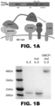
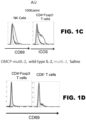
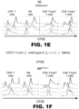


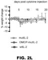
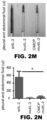
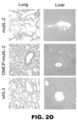
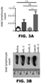

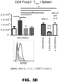
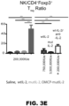
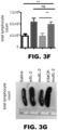


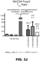
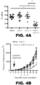

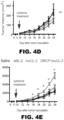
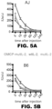

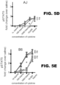
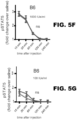
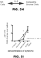
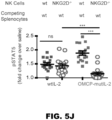
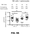
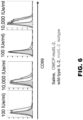

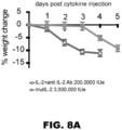
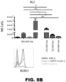
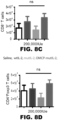


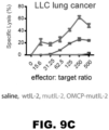
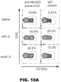

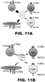

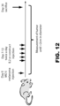

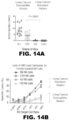
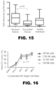


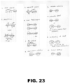


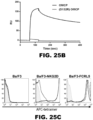

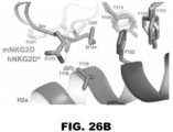


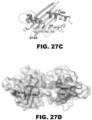
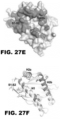


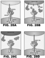
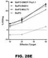


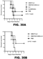
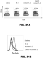
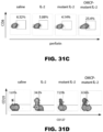


Classifications
-
- C—CHEMISTRY; METALLURGY
- C07—ORGANIC CHEMISTRY
- C07K—PEPTIDES
- C07K14/00—Peptides having more than 20 amino acids; Gastrins; Somatostatins; Melanotropins; Derivatives thereof
- C07K14/435—Peptides having more than 20 amino acids; Gastrins; Somatostatins; Melanotropins; Derivatives thereof from animals; from humans
- C07K14/52—Cytokines; Lymphokines; Interferons
- C07K14/54—Interleukins [IL]
- C07K14/55—IL-2
-
- A—HUMAN NECESSITIES
- A61—MEDICAL OR VETERINARY SCIENCE; HYGIENE
- A61K—PREPARATIONS FOR MEDICAL, DENTAL OR TOILETRY PURPOSES
- A61K47/00—Medicinal preparations characterised by the non-active ingredients used, e.g. carriers or inert additives; Targeting or modifying agents chemically bound to the active ingredient
- A61K47/50—Medicinal preparations characterised by the non-active ingredients used, e.g. carriers or inert additives; Targeting or modifying agents chemically bound to the active ingredient the non-active ingredient being chemically bound to the active ingredient, e.g. polymer-drug conjugates
- A61K47/51—Medicinal preparations characterised by the non-active ingredients used, e.g. carriers or inert additives; Targeting or modifying agents chemically bound to the active ingredient the non-active ingredient being chemically bound to the active ingredient, e.g. polymer-drug conjugates the non-active ingredient being a modifying agent
- A61K47/62—Medicinal preparations characterised by the non-active ingredients used, e.g. carriers or inert additives; Targeting or modifying agents chemically bound to the active ingredient the non-active ingredient being chemically bound to the active ingredient, e.g. polymer-drug conjugates the non-active ingredient being a modifying agent the modifying agent being a protein, peptide or polyamino acid
- A61K47/64—Drug-peptide, drug-protein or drug-polyamino acid conjugates, i.e. the modifying agent being a peptide, protein or polyamino acid which is covalently bonded or complexed to a therapeutically active agent
- A61K47/642—Drug-peptide, drug-protein or drug-polyamino acid conjugates, i.e. the modifying agent being a peptide, protein or polyamino acid which is covalently bonded or complexed to a therapeutically active agent the peptide or protein in the drug conjugate being a cytokine, e.g. IL2, chemokine, growth factors or interferons being the inactive part of the conjugate
-
- A—HUMAN NECESSITIES
- A61—MEDICAL OR VETERINARY SCIENCE; HYGIENE
- A61K—PREPARATIONS FOR MEDICAL, DENTAL OR TOILETRY PURPOSES
- A61K47/00—Medicinal preparations characterised by the non-active ingredients used, e.g. carriers or inert additives; Targeting or modifying agents chemically bound to the active ingredient
- A61K47/50—Medicinal preparations characterised by the non-active ingredients used, e.g. carriers or inert additives; Targeting or modifying agents chemically bound to the active ingredient the non-active ingredient being chemically bound to the active ingredient, e.g. polymer-drug conjugates
- A61K47/51—Medicinal preparations characterised by the non-active ingredients used, e.g. carriers or inert additives; Targeting or modifying agents chemically bound to the active ingredient the non-active ingredient being chemically bound to the active ingredient, e.g. polymer-drug conjugates the non-active ingredient being a modifying agent
- A61K47/62—Medicinal preparations characterised by the non-active ingredients used, e.g. carriers or inert additives; Targeting or modifying agents chemically bound to the active ingredient the non-active ingredient being chemically bound to the active ingredient, e.g. polymer-drug conjugates the non-active ingredient being a modifying agent the modifying agent being a protein, peptide or polyamino acid
- A61K47/65—Peptidic linkers, binders or spacers, e.g. peptidic enzyme-labile linkers
-
- A—HUMAN NECESSITIES
- A61—MEDICAL OR VETERINARY SCIENCE; HYGIENE
- A61P—SPECIFIC THERAPEUTIC ACTIVITY OF CHEMICAL COMPOUNDS OR MEDICINAL PREPARATIONS
- A61P31/00—Antiinfectives, i.e. antibiotics, antiseptics, chemotherapeutics
- A61P31/12—Antivirals
-
- A—HUMAN NECESSITIES
- A61—MEDICAL OR VETERINARY SCIENCE; HYGIENE
- A61P—SPECIFIC THERAPEUTIC ACTIVITY OF CHEMICAL COMPOUNDS OR MEDICINAL PREPARATIONS
- A61P35/00—Antineoplastic agents
-
- C—CHEMISTRY; METALLURGY
- C07—ORGANIC CHEMISTRY
- C07K—PEPTIDES
- C07K14/00—Peptides having more than 20 amino acids; Gastrins; Somatostatins; Melanotropins; Derivatives thereof
- C07K14/005—Peptides having more than 20 amino acids; Gastrins; Somatostatins; Melanotropins; Derivatives thereof from viruses
-
- A—HUMAN NECESSITIES
- A61—MEDICAL OR VETERINARY SCIENCE; HYGIENE
- A61K—PREPARATIONS FOR MEDICAL, DENTAL OR TOILETRY PURPOSES
- A61K38/00—Medicinal preparations containing peptides
-
- C—CHEMISTRY; METALLURGY
- C07—ORGANIC CHEMISTRY
- C07K—PEPTIDES
- C07K2299/00—Coordinates from 3D structures of peptides, e.g. proteins or enzymes
-
- C—CHEMISTRY; METALLURGY
- C07—ORGANIC CHEMISTRY
- C07K—PEPTIDES
- C07K2319/00—Fusion polypeptide
- C07K2319/20—Fusion polypeptide containing a tag with affinity for a non-protein ligand
-
- C—CHEMISTRY; METALLURGY
- C07—ORGANIC CHEMISTRY
- C07K—PEPTIDES
- C07K2319/00—Fusion polypeptide
- C07K2319/33—Fusion polypeptide fusions for targeting to specific cell types, e.g. tissue specific targeting, targeting of a bacterial subspecies
-
- C—CHEMISTRY; METALLURGY
- C12—BIOCHEMISTRY; BEER; SPIRITS; WINE; VINEGAR; MICROBIOLOGY; ENZYMOLOGY; MUTATION OR GENETIC ENGINEERING
- C12N—MICROORGANISMS OR ENZYMES; COMPOSITIONS THEREOF; PROPAGATING, PRESERVING, OR MAINTAINING MICROORGANISMS; MUTATION OR GENETIC ENGINEERING; CULTURE MEDIA
- C12N2710/00—MICROORGANISMS OR ENZYMES; COMPOSITIONS THEREOF; PROPAGATING, PRESERVING, OR MAINTAINING MICROORGANISMS; MUTATION OR GENETIC ENGINEERING; CULTURE MEDIA dsDNA viruses
- C12N2710/00011—Details
- C12N2710/24011—Poxviridae
- C12N2710/24111—Orthopoxvirus, e.g. vaccinia virus, variola
- C12N2710/24122—New viral proteins or individual genes, new structural or functional aspects of known viral proteins or genes
-
- Y—GENERAL TAGGING OF NEW TECHNOLOGICAL DEVELOPMENTS; GENERAL TAGGING OF CROSS-SECTIONAL TECHNOLOGIES SPANNING OVER SEVERAL SECTIONS OF THE IPC; TECHNICAL SUBJECTS COVERED BY FORMER USPC CROSS-REFERENCE ART COLLECTIONS [XRACs] AND DIGESTS
- Y02—TECHNOLOGIES OR APPLICATIONS FOR MITIGATION OR ADAPTATION AGAINST CLIMATE CHANGE
- Y02A—TECHNOLOGIES FOR ADAPTATION TO CLIMATE CHANGE
- Y02A50/00—TECHNOLOGIES FOR ADAPTATION TO CLIMATE CHANGE in human health protection, e.g. against extreme weather
- Y02A50/30—Against vector-borne diseases, e.g. mosquito-borne, fly-borne, tick-borne or waterborne diseases whose impact is exacerbated by climate change
Definitions
- compositions for targeted cytokine delivery may improve immunotherapy by limiting side effects associated with immunotherapy.
- IL2 interleukin 2
- CTLs cytotoxic lymphocytes
- NK natural killer cells
- CD8 + CTLs CD8 + T lymphocytes
- IL2R high affinity trimeric ⁇ IL2 receptor
- NK cells which express the lower affinity ⁇ chains of the IL2R at rest.
- NK cells do express the high affinity ⁇ chain of IL2R after activation and depend on this trimeric receptor for peak cytolytic capacity. Mutant forms of IL2 with decreased affinity for IL2R ⁇ have been described and offer a more favorable side effect profile. However, they also result in lower efficacy and decreased therapeutic potential due to decreased CTL activation.
- compositions described herein provide for delivery of cytokines to a defined cell via a ligand.
- a cytokine to a ligand which specifically binds to a protein on the target cell creates an "address" for delivery of the cytokine.
- IL2 is directly targeted to lymphocytes, such as natural killer (NK) cells and CD8+ cytotoxic T lymphocytes (CTLs), via the orthopoxvirus major histocompatibility complex class I-like protein (OMCP) ligand.
- lymphocytes such as natural killer (NK) cells and CD8+ cytotoxic T lymphocytes (CTLs)
- CTLs cytotoxic T lymphocytes
- OMCP orthopoxvirus major histocompatibility complex class I-like protein
- NKG2D ligands including but not limited to ULBP1, ULBP2, ULBP3, H60, Rae-1 ⁇ , Rae-1 ⁇ , Rae-1 ⁇ , Rae-1 ⁇ , MICA, MICB, h-HLA-A, could also be used instead of OMCP.
- ULBP1, ULBP2, ULBP3, H60 Rae-1 ⁇ , Rae-1 ⁇ , Rae-1 ⁇ , Rae-1 ⁇ , MICA, MICB, h-HLA-A
- the invention encompasses a composition comprising a cytokine linked to a ligand.
- the composition may further comprise a linker to connect the cytokine to the ligand.
- the cytokine, ligand and linker are described in greater detail below. It should be understood that any of the cytokines claimed can be linked to any of the ligands claimed in the absence or presence of any of the linkers described below.
- the invention provides a nucleic acid molecule encoding a cytokine, a ligand, and optionally a linker.
- cytokine is a small protein ( ⁇ 5-20 kDa) that is important in cell signaling. Cytokines are released by cells and affect the behavior of other cells and/or the cells that release the cytokine. Non-limiting examples of cytokines include chemokines, interferons, interleukins, lymphokines, tumor necrosis factor, monokines, and colony stimulating factors. Cytokines may be produced by a broad range of cells including, but not limited to, immune cells such as macrophages, B lymphocytes, T lymphocytes, mast cells and monocytes, endothelial cells, fibroblasts and stromal cells. A cytokine may be produced by more than one type of cell.
- Cytokines act through receptors and are especially important in the immune system, modulate the balance between humoral and cell-based immune responses, and regulate maturation, growth and responsiveness of cell populations. Cytokines are important in host responses to infection, immune responses, inflammation, trauma, sepsis, cancer and reproduction.
- a cytokine of the invention may be a naturally occurring cytokine or may be a mutated version of a naturally occurring cytokine. As used herein, "naturally occurring”, which may also be referred to as wild-type, includes allelic variances.
- a mutated version or "mutant" of a naturally occurring cytokine refers to specific mutations that have been made to the naturally occurring sequence to alter the function, activity and/or specificity of the cytokine.
- the mutations may enhance the function, activity and/or specificity of the cytokine. In another embodiment, the mutations may decrease the function, activity and/or specificity of the cytokine.
- the mutation may include deletions or additions of one or more amino acid residues of the cytokine.
- Cytokines may be classified based on structure. For example, cytokines may be classified into four types: the four- ⁇ -helix bundle family, the IL1 family, the IL17 family and the cysteine-knot cytokines. Members of the four- ⁇ -helix bundle family have three-dimensional structures with four bundles of ⁇ -helices. This family is further divided into three sub-families: the IL2 subfamily, the interferon (IFN) subfamily and the IL10 subfamily. The IL2 subfamily is the largest and comprises several non-immunological cytokines including, but not limited to, erythropoietin (EPO) and thrombopoietin (TPO).
- EPO erythropoietin
- TPO thrombopoietin
- a cytokine of the composition is a cytokine from the four- ⁇ -helix bundle family or a mutant thereof.
- a skilled artisan would be able to determine cytokines within the four- ⁇ -helix bundle family.
- a cytokine of the composition is an IL2 subfamily cytokine or a mutant thereof.
- Non-limiting examples of members of the IL2 subfamily include IL2, IL4, IL7, IL9, IL15 and IL21.
- a cytokine of the composition is IL2 or a mutant thereof.
- a cytokine of the composition is IL15 or a mutant thereof.
- sequence information for the full length human IL15 amino acid sequence can be found using, for example, the GenBank accession number CAG46777.1, AAI00962.1 or AAI00963.1.
- sequence information for the full length human IL15 mRNA sequence can be found using, for example, the GenBank accession number CR542007.1, KJ891469.1, NM_172175.2, NM_000585.4 or CR541980.1.
- GenBank accession number CR542007.1, KJ891469.1, NM_172175.2, NM_000585.4 or CR541980.1 A skilled artisan will appreciate that IL15 may be found in a variety of species and methods of identifying analogs or homologs of IL15 are known in the art as described in detail below.
- the IL1 family is a group of 11 cytokines, which plays a central role in the regulation of immune and inflammatory responses.
- the IL1 family of cytokines are proinflammatory cytokines that regulate and initiate inflammatory responses.
- Non-limiting examples of IL1 family cytokines include IL1 ⁇ , IL1 ⁇ , IL1Ra, IL18, IL36Ra, IL36 ⁇ , IL37, IL36 ⁇ , IL36 ⁇ , IL38, and IL33.
- IL1 family members have a similar gene structure. A skilled artisan would be able to determine cytokines within the IL1 family.
- a cytokine of the composition is IL18 or a mutant thereof.
- the sequence information for the full length human IL18 amino acid sequence can be found using, for example, the GenBank accession number CAG46771.1.
- the sequence information for the full length human IL18 mRNA sequence can be found using, for example, the GenBank accession number KR710147.1, CR542001.1, CR541973.1 or KJ897054.1.
- GenBank accession number KR710147.1, CR542001.1, CR541973.1 or KJ897054.1 A skilled artisan will appreciate that IL18 may be found in a variety of species and methods of identifying analogs or homologs of IL18 are known in the art as described in detail below.
- Interferons are named for their ability to "interfere” with viral replication by protecting cells from virus infection. IFNs also have other functions: they activate immune cells, such as natural killer cells and macrophages; they increase host defenses by up-regulating antigen presentation by virtue of increasing the expression of major histocompatibility complex (MHC) antigens. Based on the type of receptor through which they signal, human interferons have been classified into three major types: Type I IFN, Type II IFN, and Type III IFN.
- MHC major histocompatibility complex
- Type I IFNs bind to a specific cell surface receptor complex known as the IFN- ⁇ / ⁇ receptor (IFNAR) that consists of IFNAR1 and IFNAR2 chains.
- IFNAR IFN- ⁇ / ⁇ receptor
- type I interferons present in humans are IFN- ⁇ , IFN- ⁇ , IFN- ⁇ , IFN- ⁇ and IFN- ⁇ .
- a cytokine of the composition is a Type 1 IFN cytokine or a mutant thereof, including, but not limited to wild-type and mutant forms of IFN- ⁇ , IFN- ⁇ , IFN- ⁇ , IFN- ⁇ and IFN- ⁇ .
- Type II IFNs bind to IFNGR that consists of IFNGR1 and IFNGR2 chains.
- Non-limiting examples of type II interferons present in humans is IFN- ⁇ .
- a cytokine of the composition is a Type II IFN cytokine or a mutant thereof, including, but not limited to wild-type and mutant forms of IFN- ⁇ .
- Type III IFNs signal through a receptor complex consisting of IL10R2 (also called CRF2-4) and IFNLR1 (also called CRF2-12).
- Non-limiting examples of type III interferons include IFN- ⁇ 1, IFN- ⁇ 2 and IFN- ⁇ 3 (also called IL29, IL28A and IL28B respectively).
- a cytokine of the composition is a Type III IFN cytokine or a mutant thereof, including, but not limited to wild-type and mutant forms of IFN- ⁇ 1, IFN- ⁇ 2 and IFN- ⁇ 3.
- the cytokine is an interleukin or mutant thereof, as defined in the claims.
- the majority of interleukins are synthesized by helper CD4 T lymphocytes, as well as through monocytes, macrophages, and endothelial cells. Interleukins may promote the development and differentiation of T and B lymphocytes and hematopoietic cells.
- Non-limiting examples of interleukins include IL1, IL2, IL3, IL4, IL5, IL6, IL7, IL8 (CXCL8), IL9, IL10, IL11, IL12, IL13, IL14, IL15, IL16, IL17, IL18, IL19, IL20, IL21, IL22, IL23, IL24, IL25, IL26, IL27, IL28, IL29, IL30, IL31, IL32, IL33, IL35, or IL36.
- a cytokine of the composition is IL2 or a mutant thereof.
- IL2 is a lymphokine that induces the proliferation of responsive T cells. In addition, it acts on some B cells, via receptor-specific binding, as a growth factor and antibody production stimulant.
- the IL2 protein is secreted as a single glycosylated polypeptide, and cleavage of a signal sequence is required for its activity.
- the structure of IL2 comprises a bundle of 4 helices (termed A-D), flanked by 2 shorter helices and several poorly defined loops. Residues in helix A, and in the loop region between helices A and B, are important for receptor binding. Secondary structure analysis suggests similarity to IL4 and granulocyte-macrophage colony stimulating factor (GMCSF).
- GMCSF granulocyte-macrophage colony stimulating factor
- a cytokine of the composition is IL2 or a variant thereof.
- a variant may be a truncated or mutated IL2.
- the sequence information for the full length human IL2 amino acid sequence can be found using, for example, the GenBank accession number AAA59140.1 or AAH70338.1.
- the sequence information for the full length human IL2 mRNA sequence can be found using, for example, the GenBank accession number BC070338.1 or M22005.1.
- IL2 may be found in a variety of species. Non-limiting examples include mouse (AA116874.1), pig (NP_999026.1), cattle (AAQ10670.1), rat (EDM01295.1), rabbit (AAC23838.1), goat (AAQ10671.1), sheep (ABK41601.1), chicken (AAV35056.1), hamster (ERE88380.1), and dog (AAA68969.1). It is appreciated that the present invention is directed to analogs of IL2 in other organisms and is not limited to the human analog. Homologs can be found in other species by methods known in the art. For example, sequence similarity may be determined by conventional algorithms, which typically allow introduction of a small number of gaps in order to achieve the best fit.
- percent identity of two polypeptides or two nucleic acid sequences is determined using the algorithm of Karlin and Altschul (Proc. Natl. Acad. Sci. USA 87:2264-2268, 1993 ). Such an algorithm is incorporated into the BLASTN and BLASTX programs of Altschul et al. (J. Mol. Biol. 215:403-410, 1990 ). BLAST nucleotide searches may be performed with the BLASTN program to obtain nucleotide sequences homologous to a nucleic acid molecule of the invention. Equally, BLAST protein searches may be performed with the BLASTX program to obtain amino acid sequences that are homologous to a polypeptide of the invention.
- Gapped BLAST is utilized as described in Altschul et al. (Nucleic Acids Res. 25:3389-3402, 1997 ).
- the default parameters of the respective programs e.g., BLASTX and BLASTN
- BLASTX and BLASTN the default parameters of the respective programs. See www.ncbi.nlm.nih.gov for more details.
- a homolog will have a least 80, 81, 82, 83, 84, 85, 86, 87, 88, or 89% homology.
- the sequence may be at least 90, 91, 92, 93, 94, 95, 96, 97, 98, 99, or 100% homologous to IL2.
- a cytokine of the composition is a wildtype sequence of IL2 comprising the sequence set forth in SEQ ID NO:5 (APTSSSTKKTQLQLEHLLLDLQMILNGINNYKNPKLTRMLTFKFYMPKKATELKHLQCL EEELKPLEEVLNLAQSKNFHLRPRDLISNINVIVLELKGSETTFMCEYADETATIVEFLNR WITFCQSIISTLT) or SEQ ID NO:5 with at least one mutation selected from the group consisting of R38A, F42K, and C125S.
- a mutation is a mutation that causes IL2 to preferentially bind the receptor IL2 ⁇ .
- a mutation is a mutation that alters the function of IL2 such that IL2 has a decreased affinity for the IL2 receptor alpha (IL2R ⁇ ).
- the mutations selected from the group consisting of R38A, F42K and/or C125S are relative to SEQ ID NO:5.
- the C125S mutation may be included to reduce protein aggregation.
- a mutated version of IL2 comprises at least one mutation selected from the group consisting of R38A, F42K and C125S relative to SEQ ID NO:5.
- a mutated version of IL2 comprises the mutations R38A, F42K and C125S relative to SEQ ID NO:5.
- a cytokine of the composition is a mutated sequence of IL2 such as the sequence set forth in SEQ ID NO:6 (APTSSSTKKTQLQLEHLLLDLQMILNGINNYKNPKLTAMLTKKFYMPKKATELKHLQCL EEELKPLEEVLNLAQSKNFHLRPRDLISNINVIVLELKGSETTFMCEYADETATIVEFLNR WITFSQSIISTLT).
- a toxin is substituted for a cytokine.
- the term "toxin” means the toxic material or product of plants, animals, microorganisms (including, but not limited to, bacteria, viruses, fungi, rickettsiae or protozoa), or infectious substances, or a recombinant or synthesized molecule, whatever their origin and method of production.
- a toxin may be a small molecule, peptide, or protein that is capable of causing disease on contact with or absorption by body tissues interacting with biological macromolecules such as enzymes or cellular receptors.
- a toxin may be a "biotoxin" which is used to explicitly identify the toxin as from biological origin.
- Biotoxins may be further classified into fungal biotoxins, or short mycotoxins, microbial biotoxins, plant biotoxins, short phytotoxins and animal biotoxins.
- biotoxins include: cyanotoxins, produced by cyanobacteria, such as microcystins, nodularins, anatoxin-a, cylindrospermopsins, lyngbyatoxin-a, saxitoxin, lipopolysaccharides, aplysiatoxins, BMAA; dinotoxins, produced by dinoflagellates, such as saxitoxins and gonyautoxins; necrotoxins produced by, for example, the brown recluse or "fiddle back" spider, most rattlesnakes and vipers, the puff adder, Streptococcus pyogenes; neurotoxins, produced by, for example, the black widow spider, most scorpions, the box jellyfish, elapid snakes, the cone snail, the Blue-ringed octop
- a toxin is a cytotoxin.
- a cytotoxin is selected from the group consisting of ricin, apitoxin, and T-2 mycotoxin.
- a toxin is ricin.
- a cytokine or toxin of the invention may be PEGylated for improved systemic half-life and reduced dosage frequency.
- PEG may be added to a cytokine or toxin.
- a composition of the invention may comprise a cytokine or toxin comprising PEG.
- PEG may be selected from the group consisting of PEG-10K, PEG-20K and PEG-40K. Methods of conjugating PEG to a protein are standard in the art. For example, see Kolate et al, Journal of Controlled Release 2014; 192(28): 67-81 . Still further, a cytokine or toxin of the invention may be modified to remove T cell epitopes.
- T cell epitopes can be the cause of an immunogenicity issue upon administration of a composition to a subject. Through their presentation to T cells, they activate the process of anti-drug antibody development. Preclinical screening for T cell epitopes may be performed in silico, followed by in vitro and in vivo validation. T cell epitope-mapping tools such as EpiMatrix can be highly accurate predictors of immune response. Deliberate removal of T cell epitopes may reduce immunogenicity. Other means of improving the safety and efficacy of a composition of the invention by reducing their immunogenicity include humanization and PEGylation.
- a "ligand” is a protein that specifically binds to a receptor on a target cell and is not the corresponding binding partner to the cytokine linked to the ligand.
- a ligand may be from a eukaryote, a prokaryote or a virus.
- a ligand may be from a virus.
- the phrase "specifically binds” herein means ligands bind to the target protein with an affinity (K d ) in the range of at least 0.1 mM to 1 pM, or in the range of at least 0.1 pM to 200 nM, or in the range of at least 0.1 pM to 10 nM.
- a dissociation constant measures the propensity of a larger object to separate (dissociate) reversibly into smaller components.
- the dissociation constant is the inverse of the association constant.
- the dissociation constant may be used to describe the affinity between a ligand (L) and a target protein (P).
- K d ([P] ⁇ [L]) / [C], wherein C is a ligand-target protein complex and wherein [P], [L] and [C] represent molar concentrations of the protein, ligand and complex, respectively.
- a ligand may trigger a signal through its binding to a receptor on a target cell.
- a receptor is a protein molecule that may be embedded within the plasma membrane surface of a cell that receives chemical signals from outside the cell. When such chemical signals bind to a receptor, they cause some form of cellular/tissue response.
- a target cell is an immune cell. Accordingly, a ligand of the composition binds to a receptor expressed on immune cells.
- immune cells include macrophages, B lymphocytes, T lymphocytes, mast cells, monocytes, dendritic cells, eosinophils, natural killer cells, basophils, neutrophils.
- immune cells include, but are not limited to, macrophages, B lymphocytes, T lymphocytes, mast cells, monocytes, dendritic cells, eosinophils, natural killer cells, basophils, neutrophils.
- an immune cell is a natural killer cell or a T lymphocyte.
- receptors expressed on immune cells include major histocompatibility complex (MHC; e.g. MHCI, MHCII, and MHCIII), toll-like receptors (TLRs; e.g.
- a receptor on a target cell includes, but is not limited to, major histocompatibility complex (MHC; e.g. MHCI, MHCII, and MHCIII), toll-like receptors (TLRs; e.g. MHC; e.g. MHCI, MHCII, and MHCIII), toll-like receptors (TLRs; e.g. MHC; e.g. MHCI, MHCII, and MHCIII), toll-like receptors (TLRs; e.g.
- MHC major histocompatibility complex
- TLRs toll-like receptors
- the receptor on a target cell is a CD94/NKG2 family receptor.
- a ligand of the composition specifically binds to a receptor expressed on natural killer (NK) cells and CD8+ cytotoxic T lymphocytes (CTLs).
- CTLs cytotoxic T lymphocytes
- a ligand of the composition does not specifically bind to a receptor on vascular endothelial cells or regulatory T cells (T regs ).
- a receptor expressed on NK cells and CTLs may be a CD94/NKG2 family receptor or KLRG1.
- KLRG1 Insert cell lectin-like receptor subfamily G member 1
- CD94/NKG2 family receptors are a family of C-type lectin receptors which are expressed predominantly on the surface of NK cells and a subset of CD8+ T-lymphocyte. These receptors stimulate or inhibit cytotoxic activity of NK cells, therefore they are divided into activating and inhibitory receptors according to their function.
- CD94/NKG2 recognize MHC class I-related glycoproteins.
- CD94/NKG2 family includes seven members: NKG2A, NKG2B, NKG2C, NKG2D, NKG2E, NKG2F and NKG2H.
- a ligand of the invention specifically binds to a receptor selected from the group consisting of NKG2A, NKG2B, NKG2C, NKG2D, NKG2E, NKG2F and NKG2H.
- NKG2 receptors are transmembrane proteins type II which dimerize with CD94 molecule.
- CD94 contains a short cytoplasmic domain and it is responsible for signal transduction. Therefore NKG2 receptors form disulfide bonded heterodimers.
- NKG2D represents an exception, it is a homodimer.
- NKG2A and NKG2B receptors transmit inhibitory signal.
- NKG2C, NKG2E and NKG2H are activating receptors.
- NKG2D is activating receptor as well but it couples with adaptor protein DAP10 which carries signaling motif YINM (SEQ ID NO:34). Src or Jak kinases phosphorylate DAP10, which can then associate with p85 subunit of PI(3)K or adaptor molecule Grb2. This signaling triggers actin reorganization (cell polarization) and degranulation.
- NKG2F receptor function has not been clarified yet.
- a ligand of the composition specifically binds to the NKG2D receptor.
- NKG2D is an activating receptor found on NK cells and CD8+ T cells (both ⁇ and ⁇ ).
- the structure of NKG2D consists of two disulphide-linked type II transmembrane proteins with short intracellular domains incapable of transducing signals.
- the function of NKG2D on CD8+ T cells is to send co-stimulatory signals to activate them.
- a ligand that binds to NKG2D may be an anti-NKG2D antibody.
- An "anti-NKG2D” includes all antibodies that specifically bind an epitope within NKG2D.
- the term "antibody' includes the term "monoclonal antibody”.
- “Monoclonal antibody” refers to an antibody that is derived from a single copy or clone, including e.g., any eukaryotic, prokaryotic, or phage clone. Monoclonal antibodies can be produced using e.g., hybridoma techniques well known in the art, as well as recombinant technologies, phage display technologies, synthetic technologies or combinations of such technologies and other technologies readily known in the art. Further by “antibody” is meant a functional monoclonal antibody, or an immunologically effective fragment thereof; such as an Fab, Fab', or F(ab')2 fragment thereof.
- antibody As long as the protein retains the ability specifically to bind its intended target, it is included within the term “antibody.” Also included within the definition “antibody” for example are single chain forms, generally designated Fv, regions, of antibodies with this specificity. These scFvs are comprised of the heavy and light chain variable regions connected by a linker. Methods of making and using scFvs are known in the art. Additionally, included within the definition “antibody” are single-domain antibodies, generally designated sdAb, which is an antibody fragment consisting of a single monomeric variable antibody domain. A sdAb antibody may be derived from camelids (V H H fragments) or cartilaginous fishes (V NAR fragments).
- humanized antibody includes an anti-NKG2D antibody that is composed partially or fully of amino acid sequence sequences derived from a human antibody germline by altering the sequence of an antibody having non-human complementarity determining regions ("CDR").
- CDR complementarity determining regions
- the simplest such alteration may consist simply of substituting the constant region of a human antibody for the murine constant region, thus resulting in a human/murine chimera which may have sufficiently low immunogenicity to be acceptable for pharmaceutical use.
- the variable region of the antibody and even the CDR is also humanized by techniques that are by now well known in the art.
- an anti-NKG2D antibody is a Fab, Fab', or F(ab')2 fragment.
- ligands that bind to NKG2D share an MHC class I-related ⁇ 1 ⁇ 2 superdomain that constitutes the common site for interaction with NKG2D.
- MHC class I-related glycoproteins such as MIC family proteins (i.e., MICA, MICB), UL16-binding family proteins (i.e., ULBP1, ULBP2, ULPB3, ULBP4, ULBP5, ULBP6), retinoid acid early induce gene 1 (Rae1)-like proteins (i.e., Rae1 ⁇ , Rae1 ⁇ , Rael ⁇ , Rae1 ⁇ , Rae1 ⁇ ), members of the H60 protein family (i.e., H60a, H60b, H60c), h-HLA-A, as well as Mult1 in mice and orthopoxvirus major histocompatibility complex class I-like protein (OMCP).
- MICA MIC family proteins
- UL16-binding family proteins i.e., ULBP1, ULBP2,
- a ligand is a MHC class-I-related glycoprotein.
- a ligand of the invention is selected from the group consisting of MICA, MICB, ULBP1, ULBP2, ULBP3, ULBP4, ULBP5, ULBP6, Rae1 ⁇ , Rae1 ⁇ , Rae1 ⁇ , Rae1 ⁇ , Rae1 ⁇ , H60a, H60b, H60c, h-HLA-A, Mult1 and OMCP.
- a ligand is a UL16-binding family protein or a MIC family protein.
- a ligand is selected from the group consisting of ULBP1, ULBP2, ULBP3, ULBP4, ULBP5, and ULBP6.
- a ligand is ULBP3.
- a ligand is orthopoxvirus major histocompatibility complex class I-like protein (OMCP) selected from the group consisting of SEQ ID NOS: 7, 13 and 14. Described herein, but not claimed are variants, which may relate to a truncated or mutated OMCP that has about the same binding affinity of the full length OMCP.
- OMCP orthopoxvirus major histocompatibility complex class I-like protein
- a variant may be a truncated or mutated OMCP that has a slightly lower binding affinity relative to the binding affinity of the full length OMCP. In another embodiment, a variant is a truncated or mutated OMCP that has a slightly higher binding affinity relative to the binding affinity of the full length OMCP.
- Methods to determine binding affinity of a ligand to target protein are known in the art and described above.
- OMCP specifically binds to NKG2D with a binding affinity of about 0.1 to about 5 nM. For example, OMCP specially binds to human NKG2D with a binding affinity of about 0.2 nM and mouse NKG2D with a binding affinity of about 3 nM.
- OMCP or a variant thereof binds to human NKG2D with a binding affinity of about 1000 nM to about 0.1 nM. In certain embodiments, OMCP or a variant thereof binds to human NKG2D with a binding affinity of about 100 nM to about 0.1 nM, about 10 nM to about 0.1 nM, or about 1 nM to about 0.1 nM. In other embodiments, OMCP or a variant thereof binds to human NKG2D with a binding affinity of about 1000 nM to about 1 nM, or about 1000 nM to about 10 nM, or about 1000 nM to about 100 nM.
- OMCP or a variant thereof binds to human NKG2D with a binding affinity of about 100 nM to about 1 nM, or about 100 nM to 10 nM.
- OMCP or a variant thereof binds to human NKG2D with a binding affinity of about 1000 nM, about 500 nM, about 100 nM, about 50 nM, about 10 nM, about 9 nM, about 8 nM, about 7 nM, about 6 nM about 5 nM, about 4 nM, about 3 nM, about 2 nM, about 1 nM, about 0.9 nM, about 0.8 nM, about 0.7 nM, about 0.6 nM, about 0.5 nM, about 0.4 nM, about 0.3 nM, about 0.2 nM or about 0.1 nM.
- a variant is a truncated or mutated OMCP that has binding affinity for one or more NKG2 family receptors other than NKG2D.
- a variant is a truncated or mutated OMCP that has binding affinity for one or more NKG2 family receptors selected from the group consisting of NKG2A, NKG2B, NKG2C, NKG2E, NKG2F and NKG2H.
- Mutations to OMCP may be rationally selected via structure-based knowledge or mutations to OMCP may be identified via selection-based mutagenesis.
- mutations may be rationally selected to occur in the OMCP-NKG2D interface to either enhance or reduce binding affinity. Amino acids involved in binding at the OMCP-NKG2D interface are described in the Examples.
- OMCP consists of an MHCI-like ⁇ 1/ ⁇ 2 platform domain ( FIG. 20A ).
- the platform domain of OMCP has been trimmed to have only a six-stranded beta sheet with shorter flanking helices.
- the helix of the OMCP ⁇ 1 domain (H1) is continuous, while the helix of the ⁇ 2 domain is broken into two regions (H2a and H2b).
- the helices flank a six-stranded beta sheet and together form the characteristic platform that defines MHC proteins.
- the alpha helices of OMCP are close together and thus have no groove for binding peptides or other ligands like antigen-presenting MHC platform domains.
- a ligand of the invention comprises one or more of the ⁇ helices of a MHC class I-related glycoprotein. In other embodiments, a ligand of the invention consists of one or more of the ⁇ helices of a MHC class I-related glycoprotein. More specifically, a ligand of the invention comprises the ⁇ 1 domain (H1), ⁇ 2 domain (H2), H2a, H2b, or combinations thereof of a MHC class I-related glycoprotein.
- a ligand of the invention consists of the ⁇ 1 domain (H1), ⁇ 2 domain (H2), H2a, H2b, or combinations thereof of a MHC class I-related glycoprotein.
- a ligand of the invention comprises the ⁇ 2 domain (H2) of a MHC class I-related glycoprotein.
- a ligand of the invention consists of the ⁇ 2 domain (H2) of a MHC class I-related glycoprotein.
- a ligand of the invention comprises one or more of the ⁇ helices of OMCP.
- a ligand of the invention comprises the ⁇ 1 domain (H1), ⁇ 2 domain (H2), H2a, H2b, or combinations thereof of OMCP.
- a ligand of the invention comprises the ⁇ 2 domain (H2) of OMCP.
- a ligand of the invention consists of one or more of the ⁇ helices of OMCP.
- a ligand of the invention consists of the ⁇ 1 domain (H1), ⁇ 2 domain (H2), H2a, H2b, or combinations thereof of OMCP. In still another specific embodiment, a ligand of the invention consists of the ⁇ 2 domain (H2) of OMCP.
- OMCP is from an orthopoxvirus.
- OMCP is from a cowpox virus or a monkeypox virus.
- OMCP is from the Brighton Red strain of cowpoxvirus.
- homologs can be found in other species by methods known in the art. For example, sequence similarity may be determined by conventional algorithms, which typically allow introduction of a small number of gaps in order to achieve the best fit. In particular, "percent identity" of two polypeptides or two nucleic acid sequences is determined using the algorithm of Karlin and Altschul (Proc. Natl. Acad. Sci. USA 87:2264-2268, 1993 ). Such an algorithm is incorporated into the BLASTN and BLASTX programs of Altschul et al. (J. Mol. Biol. 215:403-410, 1990 ).
- BLAST nucleotide searches may be performed with the BLASTN program to obtain nucleotide sequences homologous to a nucleic acid molecule of the invention.
- BLAST protein searches may be performed with the BLASTX program to obtain amino acid sequences that are homologous to a polypeptide of the invention.
- Gapped BLAST is utilized as described in Altschul et al. (Nucleic Acids Res. 25:3389-3402, 1997 ).
- the default parameters of the respective programs e.g., BLASTX and BLASTN are employed. See www.ncbi.nlm.nih.gov for more details.
- a homolog will have a least 80, 81, 82, 83, 84, 85, 86, 87, 88, or 89% homology.
- the sequence may be at least 90, 91, 92, 93, 94, 95, 96, 97, 98, 99, or 100% homologous to OMCP.
- a skilled artisan will appreciate that structural homologs of OMCP may be found in other species or viruses.
- a structural homolog may be a protein that is structurally related but the sequence is a distal homolog.
- OMCP has low sequence identity for endogenous NKG2D ligands however it was discovered that OMCP would bind to NKG2D based on structural homology.
- Structural homologs can be found in other species by methods known in the art.
- protein structure prediction may be determined by various databases, such as Phyre and Phyre2. Such databases generate reliable protein models that may be used to determine structural homologs.
- the main results table in Phyre2 provides confidence estimates, images and links to the three-dimensional predicted models and information derived from either Structural Classification of Proteins database (SCOP) or the Protein Data Bank (PDB) depending on the source of the detected template. For each match a link takes the user to a detailed view of the alignment between the user sequence and the sequence of known three-dimensional structure. See www.sbg.bio.ic.ac.uklphyre2/ for more details. Generally, a structural homolog will have a least 50, 51, 52, 53, 54, 55, 56, 57, 58, or 59% confidence with OMCP.
- a structural homolog will have a least 60, 61, 62, 63, 64, 65, 66, 67, 68, or 69% confidence with OMCP.
- a structural homolog will have a least 70, 71, 72, 73, 74, 75, 76, 77, 78, or 79% confidence with OMCP.
- a structural homolog will have a least 80, 81, 82, 83, 64, 85, 86, 87, 88, or 89% confidence with OMCP.
- a structural homolog may have at least 90, 91, 92, 93, 94, 95, 96, 97, 98, 99, or 100% confidence with OMCP.
- the structural information for OMCP-human NKG2D may be found using the PDB ID: 4PDC.
- composition of the invention may comprise an OMCP peptide comprising the sequence set forth in SEQ ID NO:7 (HKLAFNFNLEINGSDTHSTVDVYLDDSQIITFDGKDIRPTIPFMIGDEIFLPFYKNVFSEF FSLFRRVPTSTPYEDLTYFYECDYTDNKSTFDQFYLYNGEEYTVKTQEATNKNMWLTT SEFRLKKWFDGEDCIMHLRSLVRKMEDSKRNT).
- composition of the invention may comprise an OMCP peptide comprising the sequence set forth in SEQ ID NO:13 (GHKLAFNFNLEINGSDTHSTVDVYLDDSQIITFDGKDIRPTIPFMIGDEIFLPFYKNVFSE FFSLFRRVPTSTPYEDLTYFYECDYTDNKSTFDQFYLYNGEEYTVKTQEATNKNMWLT TSEFRLKKWFDGEDCIMHLRSLVRKMEDSKR).
- composition of the invention may comprise an OMCP peptide comprising the sequence set forth in SEQ ID NO:14 (HKLVHYFNLKINGSDITNTADILLDNYPIMTFDGKDIYPSIAFMVGNKLFLDLYKNIFVEF FRLFRVSVSSQYEELEYYYSCDYTNNRPTIKQHYFYNGEEYTEIDRSKKATNKNSWLIT SGFRLQKWFDSEDCIIYLRSLVRRMEDSNK).
- a receptor expressed on immune cells may be PD1.
- PD1 also known as programmed cell death protein 1 and CD279 (cluster of differentiation 279), is a protein that in humans is encoded by the PDCD1 gene.
- PD1 is a cell surface receptor that belongs to the immunoglobulin superfamily and is expressed on T cells and pro-B cells.
- PD1 binds two ligands, PDL1 and PDL2.
- PD1 functioning as an immune checkpoint, plays an important role in down regulating the immune system by preventing the activation of T-cells.
- a ligand of the composition specifically binds to PD1.
- a ligand that specifically binds to PD1 may be an anti-PD1 antibody.
- an “anti-PD1” includes all antibodies that specifically bind an epitope within PD1.
- the term “antibody” is described above.
- a ligand that specifically binds to PD1 may be PDL1.
- PDL1 programmed death-ligand 1also known as cluster of differentiation 274 (CD274) or B7 homolog 1 (B7-H1), is a protein that in humans is encoded by the CD274 gene.
- PDL1 binds to its receptor, PD1, found on activated T cells, B cells, and myeloid cells, to modulate activation or inhibition.
- the affinity between PDL1 and PD1, as defined by the dissociation constant K d is 770nM.
- a ligand of the composition may be Glucocorticoid-induced TNFR-related (GITR) ligand (GITRL).
- GITR activation by GITRL influences the activity of effector and regulatory T cells, thus participating in the development of immune response against tumors and infectious agents, as well as in autoimmune and inflammatory diseases.
- GITR triggering stimulates T effector activity and inhibits Treg activity.
- GITR inhibition may ameliorate autoimmune/inflammatory diseases whereas GITR activation may treat viral, bacterial and parasitic infections, as well as boost immune responses against tumors.
- GITRL is a type II transmembrane protein expressed at high levels on antigen presenting cells (APC) and endothelial cells.
- a ligand of the invention is modified for improved systemic half-life and reduced dosage frequency.
- N-glycans may be added to a ligand. While the biological function is typically determined by the protein component, carbohydrate can play a role in molecular stability, solubility, in vivo activity, serum half-life, and immunogenicity.
- the sialic acid component of carbohydrate in particular, can extend the serum half-life of protein therapeutics. Accordingly, new N-linked glycosylation consensus sequences may be introduced into desirable positions in the peptide backbone to generate proteins with increased sialic acid containing carbohydrate, thereby increasing in vivo activity due to a longer serum half-life.
- PEG may be added to a ligand.
- Methods of conjugating PEG to a protein are standard in the art. For example, see Kolate et al, Journal of Controlled Release 2014; 192(28): 67-81 .
- a composition of the invention may comprise a ligand comprising PEG and/or one or more N-glycans.
- PEG is selected from the group consisting of PEG-10K, PEG-20K and PEG-40K.
- a ligand of the invention may be modified to remove T cell epitopes. T cell epitopes can be the cause of an immunogenicity issue upon administration of a composition to a subject.
- T cell epitope-mapping tools such as EpiMatrix can be highly accurate predictors of immune response. Deliberate removal of T cell epitopes may reduce immunogenicity. Other means of improving the safety and efficacy of a composition of the invention by reducing their immunogenicity include humanization and PEGylation.
- a composition of the invention further comprises a linker.
- the linker may be used to connect the cytokine to the ligand. It is to be understood that linking the cytokine to the ligand will not adversely affect the function of the cytokine or the ligand.
- Suitable linkers include amino acid chains and alkyl chains functionalized with reactive groups for coupling to both the cytokine and the ligand or combinations thereof.
- the linker may include amino acid side chains, referred to as a peptide linker.
- Amino acid residue linkers are usually at least one residue and can be 50 or more residues, but alone do not specifically bind to the target protein.
- a linker may be about 1 to about 10 amino acids.
- a linker may be about 10 to about 20 amino acids.
- a linker may be about 20 to about 30 amino acids.
- a linker may be about 30 to about 40 amino acids.
- a linker may be about 40 to about 50 amino acids.
- a linker may be more than 50 amino acids.
- a linker may be 1, 2, 3, 4, 5, 6, 7, 8, 9, 10, 11, 12, 13, 14, 15, 16, 17, 18, 19, 20, 21, 22, 23, 24, 25, 26, 27, 28, 29, 30, 31, 32, 33, 34, 35, 36, 37, 38, 39, 40, 41, 42, 43, 44, 45, 46, 47, 48, 49 or 50 amino acids.
- a linker is about 20 to about 30 amino acids. In another specific embodiment, a linker is about 26 amino acids.
- a linker may be (AAS) n , (AAAL) n , (G n S) n or (G 2 S) n , wherein A is alanine, S is serine, L is leucine, and G is glycine and wherein n is an integer from 1-20, or 1-10, or 3-10.
- n may be 1, 2, 3, 4, 5, 6, 7, 8, 9, 10, 11, 12, 13, 14, 15, 16, 17, 18, 19 or 20.
- a linker includes, but is not limited to, (AAS) n , (AAAL) n , (G n S) n or (G 2 S) n , wherein A is alanine, S is serine, L is leucine, and G is glycine and wherein n is an integer from 1-20, or 1-10, or 3-10.
- a linker may comprise one or more epitope tags. For instance, a linker may comprise 1, 2, 3, 4, 5, 6, 7 or 8 epitope tags. In a specific embodiment, a linker comprises 2 epitope tags.
- Non-limiting examples of epitope tags include FLAG tag (DYKDDDK epitope (SEQ ID NO:9)), HA tag (YPYDVPDYA epitope (SEQ ID NO:10)), His tag (6x-His or 8x-His), Myc tag (EQKLISEEDL epitope (SEQ ID NO:11)) and V5 tag (GKPIPNPLLGLDST epitope (SEQ ID NO:12)).
- a linker may comprise at least one tag selected from the group consisting of a FLAG tag and a His tag.
- a linker comprises a FLAG tag and a His tag.
- a linker comprises the sequence set forth in SEQ ID NO:8 (GSSGSSDYKDDDDKHHHHHHHHGSSGSS).
- an alkyl chain linking group may be coupled to the cytokine by reacting the terminal amino group or the terminal carboxyl group with a functional group on the alkyl chain, such as a carboxyl group or an activated ester. Subsequently the ligand is attached to the alkyl chain to complete the formation of the complex by reacting a second functional group on the alkyl chain with an appropriate group on the ligand.
- the second functional group on the alkyl chain is selected from substituents that are reactive with a functional group on the ligand while not being reactive with the cytokine.
- the second functional group of the alkyl chain linking group can be an amino group or vice versa.
- formation of the conjugate may require protection and deprotection of the functional groups present in order to avoid formation of undesired products. Protection and deprotection are accomplished using protecting groups, reagents, and protocols common in the art of organic synthesis. Particularly, protection and deprotection techniques employed in solid phase peptide synthesis may be used. It will be appreciated that linking groups may alternatively be coupled first to the ligand and then to the cytokine.
- An alternative chemical linking group to an alkyl chain is polyethylene glycol (PEG), which is functionalized in the same manner as the alkyl chain described above.
- PEG polyethylene glycol
- Such a linker may be referred to as a heterobifunctional PEG linker or a homobifunctional PEG linker.
- heterobifunctional PEG linkers include: O-(2-Aminoethyl)-O'-[2-(biotinylamino)ethyl]octaethylene glycol; O-(2-Aminoethyl)-O'-(2-carboxyethyl)polyethylene glycol hydrochloride M p 3000; O-(2-Aminoethyl)-O'-(2-carboxyethyl)polyethylene glycol 5,000 hydrochloride M p 5,000; O-(2-Aminoethyl)polyethylene glycol 3,000 Mp 3,000; O-(2-Aminoethyl)-O'-(2-(succinylamino)ethyl)polyethylene glycol hydrochloride M p 10,000; O-(2-Azidoethyl)heptaethylene glycol; O-[2-(Biotinylamino)ethyl]-O'-(2-carbox
- Non-limiting examples of homobifunctional PEG linkers include: MAL-PEG-MAL (Bifunctional Maleimide PEG Maleimide); OPSS-PEG-OPSS (OPSS: orthopyridyl disulfide; PDP-PEG-PDP); HS-PEG-SH (Bifunctional Thiol PEG Thiol); SG-PEG-SG (Bifunctional PEG Succinimidyl Glutarate NHS ester); SS-PEG-SS (Bifunctional PEG Succinimidyl Succinate NHS ester); GAS-PEG-GAS (Bifunctional PEG Succinimidyl ester NHS-PEG-NHS); SAS-PEG-SAS (Bifunctional PEG Succinimidyl ester NHS-PEG-NHS); Amine-PEG-Amine (Bifunctional PEG Amine NH2-PEG-NH2); AC-PEG-AC (Bifunctional Acrylate PEG Acrylate); ACA-PEG-ACA (Bi
- a linker of the invention may be modified for improved systemic half-life and reduced dosage frequency.
- N-glycans are added to a linker. While the biological function is typically determined by the protein component, carbohydrates can play a role in molecular stability, solubility, in vivo activity, serum half-life, and immunogenicity.
- the sialic acid component of carbohydrate in particular, can extend the serum half-life of protein therapeutics. Accordingly, new N-linked glycosylation consensus sequences may be introduced into desirable positions in the peptide backbone to generate proteins with increased sialic acid containing carbohydrate, thereby increasing in vivo activity due to a longer serum half-life.
- PEG is added to a linker.
- a composition of the invention comprises a ligand comprising PEG and/or one or more N-glycans.
- PEG is selected from the group consisting of PEG-10K, PEG-20K and PEG-40K.
- Another aspect of the invention involves cross-linking the peptides of the invention to improve their pharmacokinetic, immunogenic, diagnostic, and/or therapeutic attributes.
- Cross-linking involves joining two molecules by a covalent bond through a chemical reaction at suitable site(s) (e.g., primary amines, sulfhydryls) on the cytokine and ligand of the invention.
- site(s) e.g., primary amines, sulfhydryls
- the cytokine and ligand may be crosslinked together.
- the cross-linking agents may form a cleavable or non-cleavable linker between the cytokine and the ligand.
- Cross-linking agents that form non-cleavable linkers between the cytokine and the ligand may comprise a maleimido- or haloacetyl-based moiety.
- such non-cleavable linkers are said to be derived from maleimido- or haloacetyl-based moiety.
- Cross-linking agents comprising a maleimido-based moiety include N-succinimidyl 4-(maleimidomethyl)cyclohexanecarboxylate (SMCC), N-succinimidyl-4-(N-maleimidomethyl)-cyclohexane-1-carboxy-(6-amidocaproate), which is a "long chain" analog of SMCC (LC-SMCC), ⁇ -maleimidoundecanoic acid N-succinimidyl ester (KMUA), ⁇ -maleimidobutyric acid N-succinimidyl ester (GMBS), ⁇ -maleimidocaproic acid N-hydroxysuccinimide ester (EMCS), m-maleimidobenzoyl-N-hydroxysuccinimide ester (MBS), N-( ⁇ -maleimidoacetoxy)-succinimide ester [AMAS], succinimidyl-6-( ⁇ -maleimidopro
- cross-linking agents form non-cleavable linkers derived from maleimido-based moieties.
- Cross-linking agents comprising a haloacetyl-based moiety include N-succinimidyl-4-(iodoacetyl)-aminobenzoate (SIAB), N-succinimidyl iodoacetate (SIA), N-succinimidyl bromoacetate (SBA) and N-succinimidyl 3-(bromoacetamido)propionate (SBAP). These cross-linking agents form non-cleavable linkers derived from haloacetyl-based moieties.
- Cross-linking agents that form non-cleavable linkers between the cytokine and the ligand may comprise N-succinimidyl 3-(2-pyridyldithio)propionate, 4-succinimidyl-oxycarbonyl- ⁇ -methyl-alpha-(2-pyridyldithio)-toluene (SMPT), N-succinimidyl-3-(2-pyridyldithio)-butyrate (SDPB), 2-iminothiolane, or acetylsuccinic anhydride.
- SMPT 4-succinimidyl-oxycarbonyl- ⁇ -methyl-alpha-(2-pyridyldithio)-toluene
- SDPB N-succinimidyl-3-(2-pyridyldithio)-butyrate
- 2-iminothiolane or acetylsuccinic anhydride.
- composition comprising a chimeric peptide, the chimeric peptide comprising an orthopoxvirus major histocompatibility complex class 1-like protein (OMCP) peptide linked to an IL2 peptide, wherein the OMCP peptide comprises an amino acid sequence selected from the group consisting of SEQ ID NOS: 7, 13, and 14, and the IL2 peptide comprises an amino acid sequence of SEQ ID NO:5 or SEQ ID NO:5 with at least one mutation, wherein said at least one mutation is selected from the group consisting of R38A, F42K, and C125S.
- OMCP orthopoxvirus major histocompatibility complex class 1-like protein
- the invention encompasses a chimeric peptide comprising a cytokine peptide and a ligand peptide, wherein the cytokine peptide is not a binding partner of the ligand peptide.
- the invention encompasses a chimeric peptide comprising a cytokine peptide and a ligand peptide, wherein the ligand peptide binds to an NKG2D receptor.
- ligand peptide is used interchangeably with “ligand” and "cytokine peptide” is used interchangeably with "cytokine” for purposes of descriptions herein of various cytokines and ligands that are suitable for use in the present compositions and methods.
- the composition of the invention comprises a cytokine peptide that is in the IL2 subfamily.
- the cytokine peptide is an IL2 peptide comprising an amino acid sequence of SEQ ID NO:5 or SEQ ID NO:5 with at least one mutation, which mutation is selected from the group consisting of R38A, F42K and C125S.
- the cytokine peptide comprises the amino acid sequence set forth in SEQ ID NO:5 or SEQ ID NO:6.
- the ligand peptide is OMCP.
- the ligand peptide binds to a receptor expressed on NK cells and CD8+ CTLs.
- the ligand peptide binds to an NKG2D receptor.
- the ligand peptide comprises the amino acid sequence set forth in SEQ ID NO:7 or a portion thereof that is capable of binding to the NKG2D receptor.
- a chimeric peptide further comprises a linker peptide.
- a linker peptide comprises the amino acid sequence selected from the group consisting of (AAS) n , (AAAL) n , (G n S) n or (G 2 S) n , wherein A is alanine, S is serine, L is leucine, and G is glycine and wherein n is an integer from 1-20, or 1-10, or 3-10.
- a linker peptide comprises at least one tag selected from the group consisting of a FLAG tag and a His tag.
- a linker peptide is about 20 to about 30 amino acids.
- a linker peptide comprises the amino acid sequence set forth in SEQ ID NO:8.
- the invention also encompasses a nucleic acid molecule encoding a chimeric peptide as described herein. Additionally, the invention encompasses a pharmaceutical composition comprising a chimeric peptide as described herein. Pharmaceutical compositions are described in more detail in Section I(f) .
- FIG. 23 . 1 depicts a composition comprising ⁇ 2 domain (H2) of OMCP linked to a cytokine.
- H2 domain (H2) of OMCP linked to a cytokine 2. depicts a composition comprising OMCP linked to a cytokine, wherein the composition is pegylated.
- FIG. 7 depicts a composition comprising various NKG2D ligands linked to a cytokine. 7. depicts a composition comprising a mutated version of OMCP linked to a cytokine, wherein the OMCP may be mutated to have improved binding affinity or weaker binding affinity. 8. depicts a composition comprising a mutated version of OMCP linked to a cytokine, wherein the OMCP may be mutated to have binding affinity for other NKG2 receptors. 9. depicts a composition comprising a viral protein liked to a cytokine. For example, OMCP binds to NKG2D. Additionally, CPXV203 binds to MHCI. 10.
- OMCP linked to a mutated cytokine. It is understood that the OMCP sequence could be from various sources such as cowpox or monkeypox. Also, Fc-chimeras of OMCP and IL2, and variants thereof may be used.
- the composition comprises IL2 linked to OMCP, as set forth in the claims.
- the composition comprises IL2 linked to OMCP via a peptide linker.
- the composition comprises IL2 linked to OMCP via a peptide linker comprising about 20 to about 30 amino acids.
- the composition comprises IL2 linked to OMCP via a peptide linker comprising a FLAG tag and a His tag.
- the IL2 may be a mutated version of IL2 comprising the mutations R38A and F42K.
- the composition comprises IL2 linked to OMCP.
- the composition comprises IL2 linked to OMCP via a peptide linker.
- the composition comprises IL2 linked to OMCP via a peptide linker comprising about 20 to about 30 amino acids.
- the composition comprises IL2 linked to OMCP via a peptide linker comprising a FLAG tag and a His tag.
- the IL2 may be a mutated version of IL2 comprising the mutations R38A and F42K.
- the composition comprises the DNA sequence set forth in SEQ ID NO:1
- composition comprises the amino acid sequence set forth in SEQ ID NO:2
- the present disclosure also provides pharmaceutical compositions.
- the pharmaceutical composition comprises a composition of the invention which is detailed above, as an active ingredient and at least one pharmaceutically acceptable excipient.
- the pharmaceutically acceptable excipient may be a diluent, a binder, a filler, a buffering agent, a pH modifying agent, a disintegrant, a dispersant, a preservative, a lubricant, taste-masking agent, a flavoring agent, or a coloring agent.
- the amount and types of excipients utilized to form pharmaceutical compositions may be selected according to known principles of pharmaceutical science.
- the excipient may be a diluent.
- the diluent may be compressible (i.e., plastically deformable) or abrasively brittle.
- suitable compressible diluents include microcrystalline cellulose (MCC), cellulose derivatives, cellulose powder, cellulose esters (i.e., acetate and butyrate mixed esters), ethyl cellulose, methyl cellulose, hydroxypropyl cellulose, hydroxypropyl methylcellulose, sodium carboxymethylcellulose, corn starch, phosphated corn starch, pregelatinized corn starch, rice starch, potato starch, tapioca starch, starch-lactose, starch-calcium carbonate, sodium starch glycolate, glucose, fructose, lactose, lactose monohydrate, sucrose, xylose, lactitol, mannitol, malitol, sorbitol, xylitol, malto
- MCC
- the excipient may be a binder.
- Suitable binders include, but are not limited to, starches, pregelatinized starches, gelatin, polyvinylpyrrolidone, cellulose, methylcellulose, sodium carboxymethylcellulose, ethylcellulose, polyacrylamides, polyvinyloxoazolidone, polyvinylalcohols, C 12 -C 18 fatty acid alcohol, polyethylene glycol, polyols, saccharides, oligosaccharides, polypeptides, oligopeptides, and combinations thereof.
- the excipient may be a filler.
- suitable fillers include, but are not limited to, carbohydrates, inorganic compounds, and polyvinylpyrrolidone.
- the filler may be calcium sulfate, both di- and tri-basic, starch, calcium carbonate, magnesium carbonate, microcrystalline cellulose, dibasic calcium phosphate, magnesium carbonate, magnesium oxide, calcium silicate, talc, modified starches, lactose, sucrose, mannitol, or sorbitol.
- the excipient may be a buffering agent.
- suitable buffering agents include, but are not limited to, phosphates, carbonates, citrates, tris buffers, and buffered saline salts (e.g., Tris buffered saline or phosphate buffered saline).
- the excipient may be a pH modifier.
- the pH modifying agent may be sodium carbonate, sodium bicarbonate, sodium citrate, citric acid, or phosphoric acid.
- the excipient may be a disintegrant.
- the disintegrant may be non-effervescent or effervescent.
- Suitable examples of non-effervescent disintegrants include, but are not limited to, starches such as corn starch, potato starch, pregelatinized and modified starches thereof, sweeteners, clays, such as bentonite, micro-crystalline cellulose, alginates, sodium starch glycolate, gums such as agar, guar, locust bean, karaya, pecitin, and tragacanth.
- suitable effervescent disintegrants include sodium bicarbonate in combination with citric acid and sodium bicarbonate in combination with tartaric acid.
- the excipient may be a dispersant or dispersing enhancing agent.
- Suitable dispersants may include, but are not limited to, starch, alginic acid, polyvinylpyrrolidones, guar gum, kaolin, bentonite, purified wood cellulose, sodium starch glycolate, isoamorphous silicate, and microcrystalline cellulose.
- the excipient may be a preservative.
- suitable preservatives include antioxidants, such as BHA, BHT, vitamin A, vitamin C, vitamin E, or retinyl palmitate, citric acid, sodium citrate; chelators such as EDTA or EGTA; and antimicrobials, such as parabens, chlorobutanol, or phenol.
- the excipient may be a lubricant.
- suitable lubricants include minerals such as talc or silica; and fats such as vegetable stearin, magnesium stearate or stearic acid.
- the excipient may be a taste-masking agent.
- Taste-masking materials include cellulose ethers; polyethylene glycols; polyvinyl alcohol; polyvinyl alcohol and polyethylene glycol copolymers; monoglycerides or triglycerides; acrylic polymers; mixtures of acrylic polymers with cellulose ethers; cellulose acetate phthalate; and combinations thereof.
- the excipient may be a flavoring agent.
- Flavoring agents may be chosen from synthetic flavor oils and flavoring aromatics and/or natural oils, extracts from plants, leaves, flowers, fruits, and combinations thereof.
- the excipient may be a coloring agent.
- Suitable color additives include, but are not limited to, food, drug and cosmetic colors (FD&C), drug and cosmetic colors (D&C), or external drug and cosmetic colors (Ext. D&C).
- the weight fraction of the excipient or combination of excipients in the composition may be about 99% or less, about 97% or less, about 95% or less, about 90% or less, about 85% or less, about 80% or less, about 75% or less, about 70% or less, about 65% or less, about 60% or less, about 55% or less, about 50% or less, about 45% or less, about 40% or less, about 35% or less, about 30% or less, about 25% or less, about 20% or less, about 15% or less, about 10% or less, about 5% or less, about 2%, or about 1% or less of the total weight of the composition.
- compositions can be formulated into various dosage forms and administered by a number of different means that will deliver a therapeutically effective amount of the active ingredient.
- Such compositions can be administered orally, parenterally, or topically in dosage unit formulations containing conventional nontoxic pharmaceutically acceptable carriers, adjuvants, and vehicles as desired.
- Topical administration may also involve the use of transdermal administration such as transdermal patches or iontophoresis devices.
- parenteral as used herein includes subcutaneous, intravenous, intramuscular, or intrasternal injection, or infusion techniques.
- Formulation of drugs is discussed in, for example, Gennaro, A. R., Remington's Pharmaceutical Sciences, Mack Publishing Co., Easton, Pa. (18th ed, 1995 ), and Liberman, H. A. and Lachman, L., Eds., Pharmaceutical Dosage Forms, Marcel Dekker Inc., New York, N.Y. (1980 ).
- Solid dosage forms for oral administration include capsules, tablets, caplets, pills, powders, pellets, and granules.
- the active ingredient is ordinarily combined with one or more pharmaceutically acceptable excipients, examples of which are detailed above.
- Oral preparations may also be administered as aqueous suspensions, elixirs, or syrups.
- the active ingredient may be combined with various sweetening or flavoring agents, coloring agents, and, if so desired, emulsifying and/or suspending agents, as well as diluents such as water, ethanol, glycerin, and combinations thereof.
- the preparation may be an aqueous or an oil-based solution.
- Aqueous solutions may include a sterile diluent such as water, saline solution, a pharmaceutically acceptable polyol such as glycerol, propylene glycol, or other synthetic solvents; an antibacterial and/or antifungal agent such as benzyl alcohol, methyl paraben, chlorobutanol, phenol, thimerosal, and the like; an antioxidant such as ascorbic acid or sodium bisulfite; a chelating agent such as etheylenediaminetetraacetic acid; a buffer such as acetate, citrate, or phosphate; and/or an agent for the adjustment of tonicity such as sodium chloride, dextrose, or a polyalcohol such as mannitol or sorbitol.
- the pH of the aqueous solution may be adjusted with acids or
- transmucosal administration For topical (e.g., transdermal or transmucosal) administration, penetrants appropriate to the barrier to be permeated are generally included in the preparation.
- Transmucosal administration may be accomplished through the use of nasal sprays, aerosol sprays, tablets, or suppositories, and transdermal administration may be via ointments, salves, gels, patches, or creams as generally known in the art.
- a composition comprising a compound of the invention is encapsulated in a suitable vehicle to either aid in the delivery of the compound to target cells, to increase the stability of the composition, or to minimize potential toxicity of the composition.
- a suitable vehicle is suitable for delivering a composition of the present invention.
- suitable structured fluid delivery systems may include nanoparticles, liposomes, microemulsions, micelles, dendrimers and other phospholipid-containing systems. Methods of incorporating compositions into delivery vehicles are known in the art.
- a liposome delivery vehicle may be utilized.
- Liposomes depending upon the embodiment, are suitable for delivery of the compound of the invention in view of their structural and chemical properties.
- liposomes are spherical vesicles with a phospholipid bilayer membrane.
- the lipid bilayer of a liposome may fuse with other bilayers (e.g., the cell membrane), thus delivering the contents of the liposome to cells.
- the compound of the invention may be selectively delivered to a cell by encapsulation in a liposome that fuses with the targeted cell's membrane.
- Liposomes may be comprised of a variety of different types of phospholipids having varying hydrocarbon chain lengths.
- Phospholipids generally comprise two fatty acids linked through glycerol phosphate to one of a variety of polar groups. Suitable phospholipids include phosphatidic acid (PA), phosphatidylserine (PS), phosphatidylinositol (PI), phosphatidylglycerol (PG), diphosphatidylglycerol (DPG), phosphatidylcholine (PC), and phosphatidylethanolamine (PE).
- PA phosphatidic acid
- PS phosphatidylserine
- PI phosphatidylinositol
- PG phosphatidylglycerol
- DPG diphosphatidylglycerol
- PC phosphatidylcholine
- PE phosphatidylethanolamine
- the fatty acid chains comprising the phospholipids may
- Suitable fatty acid chains include (common name presented in parentheses) n-dodecanoate (laurate), n-tretradecanoate (myristate), n-hexadecanoate (palmitate), n-octadecanoate (stearate), n-eicosanoate (arachidate), n-docosanoate (behenate), n-tetracosanoate (lignocerate), cis-9-hexadecenoate (palmitoleate), cis-9-octadecanoate (oleate), cis,cis-9,12-octadecandienoate (linoleate), all cis-9, 12, 15-octadecatrienoate (linolenate), and all cis-5,8,11,14-eicosatetraenoate (arachidonate).
- the two fatty acid chains of a phospholipid may be identical or different.
- Acceptable phospholipids include dioleoyl PS, dioleoyl PC, distearoyl PS, distearoyl PC, dimyristoyl PS, dimyristoyl PC, dipalmitoyl PG, stearoyl, oleoyl PS, palmitoyl, linolenyl PS, and the like.
- the phospholipids may come from any natural source, and, as such, may comprise a mixture of phospholipids.
- egg yolk is rich in PC, PG, and PE
- soy beans contains PC, PE, PI, and PA
- animal brain or spinal cord is enriched in PS.
- Phospholipids may come from synthetic sources too. Mixtures of phospholipids having a varied ratio of individual phospholipids may be used. Mixtures of different phospholipids may result in liposome compositions having advantageous activity or stability of activity properties.
- phospholipids may be mixed, in optimal ratios with cationic lipids, such as N-(1-(2,3-dioleolyoxy)propyl)-N,N,N-trimethyl ammonium chloride, 1,1'-dioctadecyl-3,3,3',3'-tetramethylindocarbocyanine perchloarate, 3,3'-deheptyloxacarbocyanine iodide, 1,1'-dedodecyl-3,3,3',3'-tetramethylindocarbocyanine perchloarate, 1,1'-dioleyl-3,3,3',3'-tetramethylindo carbocyanine methanesulfonate, N-4-(delinoleylaminostyryl)-N-methylpyridinium iodide, or 1,1,-dilinoleyl-3,3,3',3'-tetramethylindocarbo
- Liposomes may optionally comprise sphingolipids, in which spingosine is the structural counterpart of glycerol and one of the one fatty acids of a phosphoglyceride, or cholesterol, a major component of animal cell membranes.
- Liposomes may optionally, contain pegylated lipids, which are lipids covalently linked to polymers of polyethylene glycol (PEG). PEGs may range in size from about 500 to about 10,000 daltons.
- Liposomes may further comprise a suitable solvent.
- the solvent may be an organic solvent or an inorganic solvent.
- Suitable solvents include, but are not limited to, dimethylsulfoxide (DMSO), methylpyrrolidone, N-methylpyrrolidone, acetronitrile, alcohols, dimethylformamide, tetrahydrofuran, or combinations thereof.
- Liposomes carrying the compound of the invention may be prepared by any known method of preparing liposomes for drug delivery, such as, for example, detailed in U.S. Pat. Nos. 4,241,046 , 4,394,448 , 4,529,561 , 4,755,388 , 4,828,837 , 4,925,661 , 4,954,345 , 4,957,735, 5,043,164 , 5,064,655 , 5,077,211 and 5,264,618 .
- liposomes may be prepared by sonicating lipids in an aqueous solution, solvent injection, lipid hydration, reverse evaporation, or freeze drying by repeated freezing and thawing.
- the liposomes are formed by sonication.
- the liposomes may be multilamellar, which have many layers like an onion, or unilamellar.
- the liposomes may be large or small. Continued high-shear sonication tends to form smaller unilamellar lipsomes.
- liposome formation may be varied. These parameters include, but are not limited to, temperature, pH, concentration of methionine compound, concentration and composition of lipid, concentration of multivalent cations, rate of mixing, presence of and concentration of solvent.
- a composition of the invention may be delivered to a cell as a microemulsion.
- Microemulsions are generally clear, thermodynamically stable solutions comprising an aqueous solution, a surfactant, and "oil.”
- the "oil” in this case, is the supercritical fluid phase.
- the surfactant rests at the oil-water interface. Any of a variety of surfactants are suitable for use in microemulsion formulations including those described herein or otherwise known in the art.
- the aqueous microdomains suitable for use in the invention generally will have characteristic structural dimensions from about 5 nm to about 100 nm. Aggregates of this size are poor scatterers of visible light and hence, these solutions are optically clear.
- microemulsions can and will have a multitude of different microscopic structures including sphere, rod, or disc shaped aggregates.
- the structure may be micelles, which are the simplest microemulsion structures that are generally spherical or cylindrical objects. Micelles are like drops of oil in water, and reverse micelles are like drops of water in oil.
- the microemulsion structure is the lamellae. It comprises consecutive layers of water and oil separated by layers of surfactant.
- the "oil" of microemulsions optimally comprises phospholipids. Any of the phospholipids detailed above for liposomes are suitable for embodiments directed to microemulsions.
- the composition of the invention may be encapsulated in a microemulsion by any method generally known in the art.
- a composition of the invention may be delivered in a dendritic macromolecule, or a dendrimer.
- a dendrimer is a branched tree-like molecule, in which each branch is an interlinked chain of molecules that divides into two new branches (molecules) after a certain length. This branching continues until the branches (molecules) become so densely packed that the canopy forms a globe.
- the properties of dendrimers are determined by the functional groups at their surface. For example, hydrophilic end groups, such as carboxyl groups, would typically make a water-soluble dendrimer. Alternatively, phospholipids may be incorporated in the surface of a dendrimer to facilitate absorption across the skin.
- any of the phospholipids detailed for use in liposome embodiments are suitable for use in dendrimer embodiments.
- Any method generally known in the art may be utilized to make dendrimers and to encapsulate compositions of the invention therein.
- dendrimers may be produced by an iterative sequence of reaction steps, in which each additional iteration leads to a higher order dendrimer. Consequently, they have a regular, highly branched 3D structure, with nearly uniform size and shape.
- the final size of a dendrimer is typically controlled by the number of iterative steps used during synthesis.
- a variety of dendrimer sizes are suitable for use in the invention. Generally, the size of dendrimers may range from about 1 nm to about 100 nm.
- a method to deliver a cytokine to a target cell comprises contacting a target cell with a composition comprising a cytokine linked to a ligand, wherein the ligand specifically binds to a receptor on the target cell. Additionally, the method comprises contacting a target cell with a composition comprising a chimeric peptide as described in Section I.
- a target cell may be any cell comprising a target receptor for which the ligand specifically binds to. The ligand and specific binding are described in Section I.
- a target cell may be an immune cell.
- Non-limiting example of immune cells include macrophages, B lymphocytes, T lymphocytes, mast cells, monocytes, dendritic cells, eosinophils, natural killer cells, basophils, neutrophils.
- an immune cell is selected from the group consisting of a macrophage, B lymphocyte, T lymphocyte, mast cell, monocyte, dendritic cell, eosinophil, natural killer cell, basophil, and neutrophil.
- a target cell is a natural killer (NK) cell and/or a CD8+ T cell.
- a target cell is a NKG2D-expressing cell.
- Non-limiting examples of NKG2D-expressing cell include natural killer (NK) cells and CD8+ T cells (both ⁇ and ⁇ ).
- Another method concerns activating immune cells.
- the method comprises contacting an immune cell with a composition comprising a cytokine linked to a ligand, wherein the ligand specifically binds to a receptor on the immune cell thereby activating the cell. Additionally, the method comprises contacting an immune cell with a composition comprising a chimeric peptide as described in Section I.
- immune cells include macrophages, B lymphocytes, T lymphocytes, mast cells, monocytes, dendritic cells, eosinophils, natural killer cells, basophils, neutrophils.
- an immune cell is selected from the group consisting of a macrophage, B lymphocyte, T lymphocyte, mast cell, monocyte, dendritic cell, eosinophil, natural killer cell, basophil, and neutrophil.
- an immune cell is a natural killer (NK) cell and/or a CD8+ T cell.
- a cytokine may be a proinflammatory cytokine.
- proinflammatory cytokine is a cytokine which promotes systemic inflammation. A skilled artisan would be able to determine those cytokines that are proinflammatory.
- a proinflammatory cytokine is IL1 ⁇ , IL1 ⁇ , IL2, IL3, IL6, IL7, IL9, IL12, IL15, IL17, IL18, IL21, IFN ⁇ , IFN ⁇ , TNF ⁇ , MIF, G-CSF, GM-CSF or mutants thereof.
- a proinflammatory cytokine is an IL1 family cytokine.
- an IL1 family cytokine is selected from the group consisting of IL1 ⁇ , IL1 ⁇ , IL1Ra, IL18, IL36Ra, IL36 ⁇ , IL37, IL36 ⁇ , IL36 ⁇ , IL38, IL33 and mutants thereof.
- a proinflammatory cytokine is selected from the group consisting of IL2, IL7, IL15, IL18, IL21 and mutants thereof.
- a proinflammatory cytokine is selected from the group consisting of IL2, IL15, IL18, and mutants thereof.
- a proinflammatory cytokine is IL2 or a mutant thereof.
- Activation of the immune cells may result in lysis of tumor cells. Accordingly, activation of immune cells may be measured by determining the amount of tumor cell lysis. In an embodiment, activation of the immune cells may result in about 10% to about 100% lysis of tumor cells. In another embodiment, activation of the immune cells may result in about 20% to about 80% lysis of tumor cells. In still another embodiment, activation of the immune cells may result in greater than 40% lysis of tumor cells. For example, activation of the immune cells may result in greater than 40%, greater than 45%, greater than 50%, greater than 55%, greater than 60%, greater than 65%, greater than 70%, greater than 75%, greater than 80%, greater than 85%, greater than 90%, greater than 95%, or greater than 99% lysis of tumor cells. The lysis of tumor cells may be measured using any standard assay (e.g., caspase assays, TUNEL and DNA fragmentation assays, cell permeability assays, and Annexin V assays).
- standard assay e.g.,
- Method for treating a tumor comprises identifying a subject with a tumor and administering to the subject a composition comprising a cytokine linked to a ligand, wherein the ligand specifically binds to a receptor on a target cell. Additionally, the method comprises administering to the subject a composition comprising a chimeric peptide as described in Section I. Specifically, the inventors have shown that delivering a cytokine to a target cell activates the cells bound by the composition, wherein the activated cells specifically lyse tumor cells thereby reducing the amount of cancer cells.
- a cytokine is a proinflammatory cytokine as described in the preceding paragraph.
- a composition as described may be used in treating, stabilizing and preventing cancer and associated diseases in a subject.
- treating, stabilizing, or preventing cancer is meant causing a reduction in the size of a tumor or in the number of cancer cells, slowing or preventing an increase in the size of a tumor or cancer cell proliferation, increasing the disease-free survival time between the disappearance of a tumor or other cancer and its reappearance, preventing an initial or subsequent occurrence of a tumor or other cancer, or reducing an adverse symptom associated with a tumor or other cancer.
- the inventors have shown that a composition of the invention activates natural killer (NK) cells bound by the composition, wherein the activated NK cells specifically lyse tumor cells thereby reducing the amount of tumor cells.
- NK natural killer
- the percent of tumor or cancerous cells surviving the treatment is at least 20, 30, 40, 50, 60, 70, 80, 90 or 100% lower than the initial number of tumor or cancerous cells, as measured using any standard assay (e.g., caspase assays, TUNEL and DNA fragmentation assays, cell permeability assays, and Annexin V assays).
- any standard assay e.g., caspase assays, TUNEL and DNA fragmentation assays, cell permeability assays, and Annexin V assays.
- the decrease in the number of tumor or cancerous cells induced by administration is at least 2, 5, 10, 15, 20, 25, 30, 35, 40, 45 or 50-fold greater than the decrease in the number of non-tumor or non-cancerous cells.
- the methods result in a decrease of 20, 30, 40, 50, 60, 50, 80, 90 or 100% in the size of a tumor or in the number of cancerous cells, as determined using standard methods.
- at least 20, 30, 40, 50, 60, 70, 80, 90, or 95% of the treated subjects have a complete remission in which all evidence of the tumor or cancer disappears.
- the tumor or cancer does not reappear or reappears after at least 1, 2, 3, 4, 5, 10, 15, or 20 years.
- the method comprises contacting an immune cell with a composition comprising a cytokine linked to a ligand, wherein the ligand specifically binds to a receptor on the immune cell thereby suppressing the cell. Additionally, the method comprises contacting an immune cells with a composition comprising a chimeric peptide as described in Section I.
- immune cells include macrophages, B lymphocytes, T lymphocytes, mast cells, monocytes, dendritic cells, eosinophils, natural killer cells, basophils, neutrophils.
- an immune cell is selected from the group consisting of a macrophage, B lymphocyte, T lymphocyte, mast cell, monocyte, dendritic cell, eosinophil, natural killer cell, basophil, and neutrophil.
- an immune cell is a natural killer (NK) cell and/or a CD8+ T cell.
- a cytokine may be an anti-inflammatory cytokine.
- anti-inflammatory cytokine is a cytokine that counteracts various aspects of inflammation, for example cell activation or the production of proinflammatory cytokines, and thus contributes to the control of the magnitude of the inflammatory response.
- an anti-inflammatory cytokine is IL4, IL5, IL10, IL11, IL13, IL16, IL35, IFN ⁇ , TGF ⁇ , G-CSF or a mutant thereof.
- an anti-inflammatory cytokine is IL10 or a mutant thereof.
- methods to kill immune cells comprises contacting an immune cell with a composition comprising a toxin linked to a ligand, wherein the ligand specifically binds to a receptor on the immune cell thereby killing the cell. Suppression or killing of the immune cells may result in treatment, stabilization and prevention of autoimmune diseases caused by overactive immune cells.
- NKG2D-expressing cells and/or aberrant expression of host NKG2DLs have been implicated in diabetes, celiac disease and rheumatoid arthritis.
- NK cells can recognize pancreatic beta cells and destroy them. The destruction of pancreatic beta cells may lead to type 1 diabetes.
- overactive immune cells are involved in transplant/graft rejection.
- a composition described herein may be used in treating, stabilizing and preventing an automimmune disease in a subject.
- the autoimmune disease is type 1 diabetes.
- the autoimmune disease is transplant or graft rejection.
- the autoimmune disease is rheumatoid arthritis.
- a composition comprising a cytokine linked to a ligand may specifically bind an immune cell that is then activated to target and lyse the infected host cell.
- the method comprises administering to the subject a composition comprising a chimeric peptide as described in Section I.
- infection includes the presence of pathogens in or on a subject, which, if its growth were inhibited, would result in a benefit to the subject.
- the term “infection” in addition to referring to the presence of pathogens also refers to normal flora which are not desirable.
- pathogen refers to an infectious agent that can produce disease.
- infectious agent include virus, bacterium, prion, fungus, viroid, or parasite that cause disease in a subject.
- an infection is caused by pathogens such as bacteria or viruses.
- the infection is an intracellular infection.
- the infection is a viral infection.
- the viral infection is caused by a flavivirus. Flavivirus is a genus of viruses in the family Flaviviridae.
- Non-limiting examples of flaviviruses include Gadget's Gully virus, Kadam virus, Kyasanur Forrest disease virus, Langat virus, Omsk hemorrhagic fever virus, Tick-borne encephalitis virus, Louping ill virus, Aroa virus, Dengue viruses 1-4, Kedougou virus, Cacipacore virus, Koutango virus, Murray Valley encephalitis virus, St.
- Louis encephalitis virus Usutu virus, West Nile virus, Yaounde virus, Kokobera virus group, Kokobera virus, Bagaza virus, Ilheus virus, Israel turkey meningoencephalomyelitis virus, Ntaya virus, Tembusu virus, Zika virus, Banzi virus, Bouboui virus, Edge Hill virus, Jugra virus, Saboya virus, Sepik virus, Kenya S virus, Wesselsbron virus, Yellow fever virus, Entebbe bat virus, Yokose virus, acea virus, Cowbone Ridge virus, Jutiapa virus, Modoc virus, Sal Vieja virus, San Perlita virus, Bukalasa bat virus, Carey Island virus, Dakar bat virus, Montana myotis leukoencephalitis virus, Phnom Penh bat virus, Rio Bravo virus, hepatitis C virus, e.g., hepatitis C virus genotypes 1-6, and GB virus A and B.
- hepatitis C virus e.g., hepati
- the flavivirus may be selected from the group consisting of West Nile virus, dengue virus, Japanese encephalitis virus, and yellow fever virus.
- the viral infection is caused by West Nile virus.
- a pathogen more specifically a virus, can induce the expression of proteins for which NKG2D binds.
- a composition comprising a cytokine linked to a ligand may specifically bind a NK cell that is then activated to target and lyse the infected host cell expressing NKG2D.
- a composition comprising a cytokine linked to a ligand may activate cytotoxic T lymphocytes that recognize infected cells via other mechanisms for targeted killing.
- Also described are methods to alleviate immunosuppression related to radiation exposure or lympotoxic substances comprising administering a composition comprising a cytokine linked to a ligand. Additionally, the method comprises administering a composition comprising a chimeric peptide as described in Section I. Additionally, a composition described may be used to raise CD4 counts in HIV positive subjects. For example, the composition may be used to activate immune cells which can help restore the immune system of the subject.
- compositions may be used to expand CD8+ memory cells.
- a pharmacologically effective amount of a composition of the invention may be administered to a subject.
- Administration is performed using standard effective techniques, including peripherally (i.e. not by administration into the central nervous system) or locally to the central nervous system.
- Peripheral administration includes but is not limited to intravenous, intraperitoneal, subcutaneous, pulmonary, transdermal, intramuscular, intranasal, buccal, sublingual, or suppository administration.
- Local administration, including directly into the central nervous system (CNS) includes but is not limited to via a lumbar, intraventricular or intraparenchymal catheter or using a surgically implanted controlled release formulation. Pheresis may be used to deliver a composition of the invention.
- a composition of the invention may be administered via an infusion (continuous or bolus).
- compositions for effective administration are deliberately designed to be appropriate for the selected mode of administration, and pharmaceutically acceptable excipients such as compatible dispersing agents, buffers, surfactants, preservatives, solubilizing agents, isotonicity agents, stabilizing agents and the like are used as appropriate.
- pharmaceutically acceptable excipients such as compatible dispersing agents, buffers, surfactants, preservatives, solubilizing agents, isotonicity agents, stabilizing agents and the like are used as appropriate.
- Suitable vehicles for such injections are straightforward.
- administration may also be effected through the mucosal membranes by means of nasal aerosols or suppositories.
- Suitable formulations for such modes of administration are well known and typically include surfactants that facilitate cross-membrane transfer.
- surfactants are often derived from steroids or are cationic lipids, such as N-[1-(2,3-dioleoyl)propyl]-N,N,N-trimethyl ammonium chloride (DOTMA) or various compounds such as cholesterol hemisuccinate, phosphatidyl glycerols and the like.
- DOTMA cationic lipids
- a therapeutically effective amount of a composition of the invention is administered to a subject.
- a "therapeutically effective amount” is an amount of the therapeutic composition sufficient to produce a measurable response (e.g., an immunostimulatory, an anti-angiogenic response, a cytotoxic response, tumor regression, immunoinhibitory, immunosuppression, infection reduction).
- a measurable response e.g., an immunostimulatory, an anti-angiogenic response, a cytotoxic response, tumor regression, immunoinhibitory, immunosuppression, infection reduction.
- Actual dosage levels of active ingredients in a therapeutic composition of the invention can be varied so as to administer an amount of the active compound(s) that is effective to achieve the desired therapeutic response for a particular subject.
- a minimal dose is administered, and dose is escalated in the absence of dose-limiting toxicity. Determination and adjustment of a therapeutically effective dose, as well as evaluation of when and how to make such adjustments, are known to those of ordinary skill in the art of medicine.
- a typical dose contains from about 10 IU/kg to about 1,000,000 IU/kg of a cytokine described herein. In an embodiment, a typical dose contains from about 10 IU/kg to about 100 IU/kg.
- a typical dose contains about 100 IU/kg to about 1,000 IU/kg. In still another embodiment, a typical dose contains about 1,000 IU/kg to about 10,000 IU/kg. In yet still another embodiment, a typical dose contains about 10,000 IU/kg to about 100,000 IU/kg. In a different embodiment, a typical dose contains about 100,000 IU/kg to about 1,000,000 IU/kg. In certain embodiments, a typical dose contains about 500,000 IU/kg to about 1,000,000 IU/kg. In other embodiments, a typical dose contains about 100,000 IU/kg to about 500,000 IU/kg. Alternatively, a typical dose contains about 50,000 IU/kg to about 100,000 IU/kg.
- a typical dose contains about 10,000 IU/kg to about 50,000 IU/kg. In still another embodiment, a typical dose contains about 5,000 IU/kg to about 10,000 IU/kg. In a specific embodiment, a typical dose contains about 5,000 IU/kg to about 200,000 IU/kg. In another specific embodiment, a typical dose contains about 5,000 IU/kg to about 500,000 IU/kg. In still another specific embodiment, a typical dose contains about 50,000 IU/kg to about 500,000 IU/kg. In still yet another specific embodiment, a typical dose contains about 250,000 IU/kg to about 750,000 IU/kg.
- the frequency of dosing may be once, twice, three times or more daily or once, twice, three times or more per week or per month, as needed as to effectively treat the symptoms or disease.
- the frequency of dosing may be once, twice or three times daily.
- a dose may be administered every 24 hours, every 12 hours, or every 8 hours.
- the frequency of dosing may be twice daily.
- Duration of treatment could range from a single dose administered on a one-time basis to a life-long course of therapeutic treatments.
- the duration of treatment can and will vary depending on the subject and the cancer or autoimmune disease or infection to be treated.
- the duration of treatment may be for 1 day, 2 days, 3 days, 4 days, 5 days, 6 days, or 7 days.
- the duration of treatment may be for 1 week, 2 weeks, 3 weeks, 4 weeks, 5 weeks or 6 weeks.
- the duration of treatment may be for 1 month, 2 months, 3 months, 4 months, 5 months, 6 months, 7 months, 8 months, 9 months, 10 months, 11 months, or 12 months.
- the duration of treatment may be for 1 year, 2 years, 3 years, 4 years, 5 years, or greater than 5 years. It is also contemplated that administration may be frequent for a period of time and then administration may be spaced out for a period of time. For example, duration of treatment may be 5 days, then no treatment for 9 days, then treatment for 5 days.
- Treatment could begin immediately, such as at the time of diagnosis, or treatment could begin following surgery. Treatment could begin in a hospital or clinic itself, or at a later time after discharge from the hospital or after being seen in an outpatient clinic.
- biodegradable films and matrices or osmotic mini-pumps, or delivery systems based on dextran beads, alginate, or collagen.
- a composition of the invention may be used to treat or recognize a tumor derived from a neoplasm or a cancer.
- the neoplasm may be malignant or benign, the cancer may be primary or metastatic; the neoplasm or cancer may be early stage or late stage.
- Non-limiting examples of neoplasms or cancers that may be treated include acute lymphoblastic leukemia, acute myeloid leukemia, adrenocortical carcinoma, AIDS-related cancers, AIDS-related lymphoma, anal cancer, appendix cancer, astrocytomas (childhood cerebellar or cerebral), basal cell carcinoma, bile duct cancer, bladder cancer, bone cancer, brainstem glioma, brain tumors (cerebellar astrocytoma, cerebral astrocytoma/malignant glioma, ependymoma, medulloblastoma, supratentorial primitive neuroectodermal tumors, visual pathway and hypothalamic gliomas), breast cancer, bronchial adenomas/carcinoids, Burkitt lymphoma, carcinoid tumors (childhood, gastrointestinal), carcinoma of unknown primary, central nervous system lymphoma (primary), cerebellar astrocytoma, cerebral
- the neoplasm or cancer may be selected from the group consisting of melanoma, renal cell carcinoma, lung cancer and blood cancer.
- a "blood cancer” is a cancer that affects the blood, bone marrow and lymphatic system.
- leukemia There are three main groups of blood cancer: leukemia, lymphoma and myeloma.
- the four broad classification of leukemia are: acute lymphocytic leukemia (ALL), acute myelogenous leukemia (AML), chronic lymphocytic leukemia (CLL) and chronic myelogenous leukemia (CML).
- Lymphomas are divided into two categories: Hodgkin lymphoma and non-Hodgkin lymphoma.
- non-Hodgkin lymphomas are B-cell lymphomas, and either grow quickly (high-grade) or slowly (low-grade). There are 14 types of B-cell non-Hodgkin lymphomas. The rest are T-cell lymphomas, named after a different cancerous white blood cell, or lymphocyte. Because myeloma frequently occurs at many sites in the bone marrow, it is often referred to as multiple myeloma.
- a suitable subject includes a human, a livestock animal, a companion animal, a lab animal, or a zoological animal.
- the subject may be a rodent, e.g. a mouse, a rat, a guinea pig, etc.
- the subject may be a livestock animal.
- suitable livestock animals may include pigs, cows, horses, goats, sheep, llamas and alpacas.
- the subject may be a companion animal.
- companion animals may include pets such as dogs, cats, rabbits, and birds.
- the subject may be a zoological animal.
- a "zoological animal” refers to an animal that may be found in a zoo. Such animals may include non-human primates, large cats, wolves, and bears.
- the animal is a laboratory animal.
- Non-limiting examples of a laboratory animal may include rodents, canines, felines, and non-human primates.
- the animal is a rodent.
- Non-limiting examples of rodents may include mice, rats, guinea pigs, etc.
- the subject is a human. Table A.
- the IL-2R ⁇ chain serves to capture IL-2 at the cell surface to facilitate subsequent binding to the signaling part of the receptor, namely the IL-2R ⁇ chains.
- IL-2R ⁇ is expressed on this population after initial activation, however, and is required for maximum cytotoxic lymphocyte expansion 2 .
- High dose IL-2 can induce the activation of all cytotoxic lymphocytes and is approved for treatment of several malignancies with an approximately 15% partial or complete tumor response 3-5 .
- T regs regulatory T cell
- IL-2R ⁇ IL-2R ⁇ and are thus preferentially activated by IL-2 over cytotoxic lymphocytes 8 .
- Lowering the IL-2 dose can ameliorate side effects but also decreases efficacy.
- Mutant forms of IL-2 such as those with substitutions of alanine for arginine at the 38 position (R38A) and/or lysine for phenylalanine at the 42 position (F42K), decrease the affinity of IL-2 for IL-2R ⁇ and thus eliminate many side effects 9 . Such IL-2 ⁇ mutants may also decrease the efficacy of immunotherapy 2 .
- a form of IL-2 that could preferentially activate cytotoxic lymphocytes in the absence of IL-2R ⁇ reactivity would be highly advantageous for clinical applications.
- NKG2D recognizes MHC class-I-related stress ligands expressed by malignant or virally-transformed cells 10 .
- NKG2D has the highest specificity for cytotoxic lymphocytes as it is constitutively expressed on both murine and human NK cells as well as activated CD8 + T cells 11 .
- tumors and virally infected cells utilize shed NKG2D ligands as a mechanism of immune evasion 12,13 .
- Orthopox major histocompatibility complex class I-like protein, or OMCP is an NKG2D ligand decoy shed by monkeypox and cowpox virus infected cells.
- OMCP binds to both human and murine NKG2D with the highest affinity of any known ligand we thought it might function as an ideal targeting vector to optimally deliver IL-2 to cytotoxic lymphocytes 14,15 .
- a fusion protein designed to deliver an IL-2R ⁇ mutant to NKG2D-expressing lymphocytes 15 .
- this construct overcomes decreased efficacy associated with mutations in the IL-2R ⁇ binding region while retaining a favorable safety profile.
- Systemic administration of this fusion protein improves immunotherapy against both solid and liquid tumors. Targeted delivery of IL-2 can thus be safely used to maximally activate NKG2D-expressing lymphocytes, such as NK cells, to optimize immunotherapy without systemic side effects.
- Example 1 NKG2D-targeted delivery of an IL-2 mutant preferentially activates cytotoxic lymphocytes in vitro.
- IL-2 fusion protein that would target cytotoxic lymphocytes directly via the NKG2D receptor.
- This fusion protein combines the high affinity NKG2D ligand OMCP with an IL-2 mutated to reduce IL-2R ⁇ reactivity.
- Our construct termed OMCP-mutIL-2, consists of the 152 residue OMCP protein fused to the N-terminus of the 133 amino acid R38A/F42K mutant form of human IL-2 (mutIL-2) via a flexible 30 residue linker ( FIG. 1A-B ). The construct was first assessed for its in vitro binding ability.
- FIG. 17 shows that the construct appears to only bind to NK cells which express NKG2D. The construct does not show binding to CD4+CD3+ T cells, CD8+CD3+ T cells, CD11C+CD11b- DCs, CD11c-CD11b+ Macs or CD19+CD3- B cells.
- CD69 expression was observed with OMCP-mut-IL-2, wtIL-2 or mutIL-2 ( FIG. 6 ).
- CD8 + and CD4 + Foxp3 - effector T cells demonstrated no upregulation of CD69 after 36 hours, even at highest doses of cytokines ( FIG. 1C-D and data not shown).
- FIG. 1E-F Longer exposure over a period of five days led to proliferation of both NK and CD8 + T cells exposed to wtIL-2 and OMCP-mutIL-2 ( FIG. 1E-F ).
- OMCP-mutIL-2 activated CD8 + T cells and NK cells equivalently to mutIL-2 in NKG2D -/- splenocytes, indicating that the increased activation was due to the effect of OMCP targeting upon NKG2D-bearing cells ( FIG. 1F ; FIG. 6 , right two graphs). Only incubation with wtIL-2 led to CD4 + Foxp3 + T regs and CD4 + Foxp3 - effector cell proliferation ( FIG. 1E-F ).
- OMCP-mutIL-2 results in preferential NK activation that is superior or equivalent to wtIL-2 in a dose-dependent manner.
- CD8 + T cells can also be activated but require prolonged exposure to higher doses of OMCP-mutIL-2.
- Example 2 Low-dose cytokine therapy offers a favorable safety profile.
- Dose-dependent toxicity can limit cytokine administration in vivo.
- To model human immunotherapy protocols we next treated A/J mice with wtIL-2 given as ten doses over a five day cycle 18 . While A/J mice tolerated 750,000 IUe of wtIL-2, significant mortality was evident at higher doses ( FIG. 2A-B ). Even after a 750,000 IUe dose mice demonstrated extreme distress, weight loss, decreased food consumption, ascites and hepatic inflammation ( FIG. 2A -E; FIG. 7A-C ). These side-effects mirror the capillary leak and distress associated with high dose IL-2 therapy in humans 7 .
- OMCP-mutlL-2 preferentially expands and activates NK cells in vivo compared to wtlL-2 or mutlL-2.
- mice received 200,000 IUe of cytokine or construct given as ten equal doses over five days. Splenic lymphocytes were evaluated flow cytometrically on day six. Both wtlL-2 and OMCP-mutIL-2 increased lymphocyte content and splenic size over saline-treated controls ( FIG. 3A-B ). OMCP-mutIL-2 led to a substantial expansion and activation of NK cells measured by cellularity and surface KLRG1 levels ( FIG. 3C ).
- NK cells comprised close to half of all splenic lymphocytes, paralleling or even surpassing the total lymphocyte counts of saline or mutIL-2-treated mice ( FIG. 3A vs. FIG. 3C ).
- NK expansion by 200,000 IUe of OMCP-mutIL-2 was superior to near toxic doses of wtIL-2 (750,000IU), high dose mutIL-2 (3,500,000 IUe), or wtlL-2 complexed to anti-IL-2 antibody (clone MAB602) 19 ( FIG. 3C ).
- mice could not tolerate the full 200,000 IUe of wtIL-2/anti-IL-2 antibody and injections had to be terminated at 160,000 or 180,000 IUe with requisite animal sacrifice due to animal distress and rapid weight loss ( FIG. 8A ).
- WtIL-2 led to a significant expansion of CD4 + Foxp3 + T regs , specifically the ICOS + subsets in A/J mice even when complexed to anti-IL-2 antibodies ( FIG. 3D ).
- the NK/T reg ratio which has been described as a predictive factor for success of immunotherapy 20 , was dramatically increased in OMCP-mutIL-2 treated mice compared to all other treatment conditions ( FIG. 3E ).
- OMCP-mutlL-2 preferentially expands and activates NK cells in human peripheral blood lymphocytes compared to wtlL-2 or mutlL-2.
- human peripheral blood lymphocytes were co-cultured for 36 hours in 100IUe of either wild-type IL2, R38A/F42K mutant form of IL-2 or OMCP-mutant IL-2.
- NK cells The cells were flow cytometrically analyzed and relative prevalence of CD56+CD3- NK cells compared between conditions. A relatively higher proportion of NK cells was evident in the OMCP-mutant IL-2 group ( FIG. 31A ). Perforin levels were higher in OMCP-mutant IL-2 treated NK cells (red) compared to saline (black), IL-2 (blue) or mutant IL-2 (green) treated ones ( FIG. 31B ).
- CD8+ T cells Similar to NK cells, higher intracellular levels of perforin were evident in CD8+ T cells treated with OMCP-mutant IL-2 compared to other conditions ( FIG. 31C ).
- Tregs When gating on CD4+Foxp3+CD45RA- T cells a relatively higher proportion of activated CD25+CD127- regulatory T cells was evident in IL-2 treated peripheral blood lymphocyte cultures compared to other conditions ( FIG. 31D ). Taken together this data suggests that OMCP-mutIL-2 preferentially expands and activates NK cells and CD8+ cells in human peripheral blood lymphocytes compared to wtIL-2 or mutIL-2. Importantly, OMCP-mutIL-2 does not activated regulatory T cells significantly relative to IL2.
- Example 5 Treatment with OMCP-mutlL-2 offers superior immunologic control of malignancies in vivo.
- NK cells can mediate natural cytotoxicity without prior sensitization. NK cells also form the primary barrier for expansion of select malignancies, such as lymphoma and lung cancer 16,17,21,22 .
- Antibody-IL-2 conjugates, or IL-2/anti-IL-2 antibody complexes demonstrate improved biologic activity over purified cytokine by extending the duration of serum half-life 23,24 .
- IL-2 increased serum half-life
- OMCP-mutIL-2 was the result of signaling through NKG2D as antibody-mediated crosslinking of this receptor can activate NK cells ( FIG. 10A ) 25 . While the addition of purified OMCP to mutIL-2 did not augment NK activation or expansion in vitro or in vivo (data not shown) we would not expect a monomeric ligand to crosslink NKG2D. We thus directly compared NK cell activation in the presence of 1000 IUe of OMCP-mutIL-2, mutIL-2 and mutIL-2 combined with equimolar concentration of pentamerized OMCP.
- OMCP-mutIL-2 failed to increase STAT5 phosphorylation over mutIL-2 alone.
- IL-2 ⁇ reactivity is important for peak IL-2 signaling in resting NK cells, and that NKG2D-binding may effectively substitute for IL-2R ⁇ -binding in IL-2-mediated signal transduction.
- Such data failed to explain the superior NK activation by OMCP-mutIL-2 in vivo or in bulk splenocyte cultures ( FIG. 1C-D , FIG. 3 ).
- IL-2 signaling results in the internalization of IL-2/IL-2R, with subsequent degradation of IL-2 and IL-2R ⁇ .
- the binding of OMCP-mutIL-2 to both the IL-2 receptor and NKG2D could thus lead to altered internalization and enhanced NK cell activation by prolonging IL-2 signaling.
- To test this we stimulated freshly isolated NK cells for 15 minutes, replaced the culture media with cytokine free media, and monitored STAT5 phosphorylation for four hours. Identical decay of phospho-STAT5 was evident for both wtIL-2 and OMCP-mutIL-2 ( FIG. 5F-G ). Thus altering duration of IL-2 signaling is not responsible for superior NK activation by OMCP-mutIL-2.
- OMCP-mutIL-2 may be the result of altered cytokine interaction with competing stromal cells ( FIG. 5H ). Indeed, in the presence of other splenocytes OMCP-mutIL-2 demonstrated a dose-dependent enhancement in NK STAT5 phosphorylation over wtIL-2 ( FIG. 5I ).
- IL-2R ⁇ expression by stromal cells and NKG2D expression by NK cells on IL-2 signal transduction To accomplish this we isolated splenic NK cells from either wild-type or NKG2D -/- B6 mice and combined them with wild-type splenocytes depleted of NK cells.
- NK cell depleted splenocytes were treated with saturating concentrations of IL-2R ⁇ -blocking antibody (clone 3C7) prior to recombining with wild-type NK cells.
- the cultures were then stimulated with 1000 IUe of either wtIL-2 or OMCP-mutIL-2 for 15 minutes.
- STAT5 phosphorylation was identical in NKG2D -/- or wild type NK cells in the presence of wtIL-2 ( FIG. 5J , left two columns).
- IL-2 therapy initially showed great promise, it has been limited by activation of T regs and toxic side effects associated with activation of vascular endothelium.
- Several strategies have been proposed to preferentially activate cytotoxic lymphocytes.
- One strategy has been to create mutants with increased affinity for IL-2R ⁇ to remove the preference for IL-2R ⁇ 26,27 .
- these IL-2 mutants retain wild type binding for IL-2R ⁇ , and would therefore still be recognized by T reg cells and vascular endothelium.
- Our results also suggest that competition with IL2-R ⁇ + -expressing cells limits bioavailability of wtIL-2 to cytotoxic lymphocytes.
- Another promising therapy involves anti-IL2 antibodies that sterically inhibit wtIL-2 binding to IL-2R ⁇ 1,28,29 .
- Such treatment can extend serum half-life 24 due to the Fc region of the antibody and potentially due to reduced competition for wtIL-2 from IL-2R ⁇ -expressing cells.
- Antibody-IL-2 fusion proteins have also been designed to target IL-2 to specific tumor antigens 30,31 . While offering the potential for personalized therapy such antibody-mediated delivery of IL-2 to the tumor depends on the expression of a known tumor associated antigen, a situation that often does not exist. This approach could potentially be further limited by tumor-mediated alteration of the targeted antigen.
- IL-2 mutants with reduced affinity for IL-2R ⁇ have been tested extensively. Compared to wtlL-2 these mutants can be administered in supratherapeutic doses without IL-2R ⁇ -mediated capillary leak or systemic toxicity 32 . While these mutants have excellent safety profiles, they activate cytotoxic lymphocytes poorly ( FIG. 5C-E ) 33 .
- Our approach combines several of the concepts above to target a safe form of IL-2 directly to cytotoxic lymphocytes, instead of tumors. This is accomplished by replacing the normal targeting of IL-2 to IL-2R ⁇ with NKG2D.
- OMCP has been described as an evolutionary antagonist of NKG2D 35 blockade of this immunoreceptor at the time of tumor therapy may be construed as counterproductive. Nevertheless, natural cytotoxicity and tumor clearance was augmented in OMCP-mutIL-2-treated mice even in the presence of established tumors. This suggests minimal or transient NKG2D receptor occupancy and preservation of function. Alternatively recent reports have demonstrated that shed NKG2D ligands may actually promote tumor immunity through reversal of NK desensitization imposed by chronic agonistic engagement 36 . While we did not detect NK activation or expansion by monomeric or even pentameric OMCP, it is possible that within the tumor bed such competitive antagonism plays a paradoxical role in NK activation.
- IL-2 may upregulate receptors necessary for NK migration and tumor infiltration. It is thus possible that anti-tumor immunity mediated by OMCP-mutIL-2 may depend on NK cells located outside the tumor bed and not subject to local tumor-specific tolerance or anergy. Furthermore, OMCP maybe the ideal "targeting vector" due to its high affinity and long half-life of binding to human NKG2D.
- NK cells from two separate strains of mice were activated by OMCP-mutIL-2 we did not detect global expansion of activation of CD8 + T cells by our construct. This is most likely due to the fact that NKG2D is expressed only on select subsets of CD8 + T cells, namely memory or activated cytotoxic lymphocytes. Based on the paucity of this cell population in mice raised in specific pathogen-free environment, OMCP-mutIL-2-mediated activation was limited in our system to NK cells. To this end we focused on immunotherapy for lung cancer and lymphoma, whose growth is regulated primarily by NK cells 16,17,22,37 . Nevertheless OMCP-mutIL-2 was able to expand CD8 + T cells when administered in high concentrations in vitro ( FIG. 1E-F ). Thus, it may be possible that NKG2D-targeted delivery of immunostimulatory cytokines may lead to the expansion and/or activation of antigen-specific CD8 + memory cells for long-term tumor immunity under normal immunologic conditions.
- Cytokine and Construct Generation The sequences encoding human IL-2 (1-133; C125S) and mutant IL2 (1-133; R38A, F42K, C125S) were cloned into the pFM1.2R 38 with an N-terminal FLAG/hexahistidine tag.
- the chimeric OMCP-mutIL-2 molecule comprises the full-length OMCP (1-152) coding sequence cloned in frame with a C-terminal FLAG/hexahistidine tag-mutant IL-2 (1-133; R38A, F42k, C125S) cloned into the pFM1.2R vector. Proteins were expressed by transient transfection into HEK293F (Life_Technologies).
- Supernatant was recovered at 72h and 144h post-transfection. Supernatants were supplemented with 5 mM imidazole and 0.02% sodium azide and purified by nickel-nitrilotriacetic acid (Ni-NTA) chromatography (Qiagen). Purified proteins were buffer exchanged into saline and flash frozen in liquid nitrogen. Equivalent in vitro and in vivo activity was documented for wild-type IL-2 generated in house and Teceleukin (TecinTM) available from the NCI repository (Frederick National Laboratory for Cancer Research). Thus for some experiments these two preparations of IL-2 were used interchangeably.
- Wild-type IL-2 has a specific activity of 15 ⁇ 10 6 IU/mg 39 .
- a 4.4 ⁇ M solution is equivalent to 1000 IU/ ⁇ l.
- all cytokines and construct were administered on a molar basis with 1 ⁇ l of 4.4 ⁇ M solution defined as 1000IU equivalents (IUe from here on).
- Such a system allows for equimolar comparison between IL-2, mutIL-2 and OMCP-mutIL-2 despite difference in molecular weight.
- mice A/J (8-12 weeks) and C57BL/6J (6-9 weeks) strains of mice were purchased from the Jackson Laboratory (Bar Harbor, Maine). NKG2D -/- mice on the B6 background were kindly provided by Wayne Yokoyama and bred in house (Howard Hughes Institute of Medicine at Washington University in St. Louis). Animals were housed in a barrier facility in air-filtered cages and allowed free access to food and water. For some experiments A/J mice were treated with depleting concentrations of anti-Asialo-GM1 (50 ⁇ l day -2; 25 ⁇ l day -1) or control rabbit IgG (Wako Chemical Company). Animal procedures were approved by the animal studies committee of Washington University School of Medicine, St. Louis, MO.
- Tissue harvest and in vitro cultures Single cell suspension of splenocytes were obtained by crushing whole spleens through 70 ⁇ m cell strainers prior to RBC lysis by ACK buffer (Lonza, Walkersville, MD) and re-filtration through a 40 ⁇ m filter. Lungs were digested for 90 minutes at 37°C in 1 mg/ml collagenase II (Fisher Scientific), and 5 U/ml DNase I (Sigma-Aldridge) prior to processing in an identical fashion to spleens.
- splenocytes from either A/J, B6, or NKG2D -/- mice were extracted in a sterile fashion and seeded in 12-well plates in complete media (RPMI 1640 supplemented with 10% FBS, 100 U/ml Penicillin and Streptomycin, 2 mM L-glutamine and 50 ⁇ M 2-Mercaptoethanol) at 5 million cells per ml per well.
- the cells were treated with increasing doses of human recombinant IL-2, mutIL-2, OMCP-mutIL-2, or OMCP for 36 hours as described in the manuscript.
- NK isolation experiments bulk splenocytes were labeled with CFSE and cultured in 1000 IUe/ml of cytokine for 5 days prior to flow cytometric analysis.
- NK isolation experiments bulk splenocytes were processed using either the NK cell isolation kit II or CD49b (DX5) positive magnetic bead selection (both from Miltenyi Biotech).
- NK cell isolation kit II CD49b (DX5) positive magnetic bead selection (both from Miltenyi Biotech).
- STAT5 phosphorylation experiments isolated NK cells were stimulated in increasing concentrations of IL-2 or construct at 100,000 cells/500 ⁇ l for 15 minutes.
- NK depleted stromal cells were labeled with CFSE (for identification after fixation and permeabilization) and recombined with NK depleted stromal cells.
- CFSE for identification after fixation and permeabilization
- NK depleted stromal cells were combined with wild-type splenocyte stromal cells.
- NK-depleted splenocytes from wild-type B6 mice were treated with saturating concentrations of anti-IL-2 ⁇ blocking antibody (clone 3C7) or isotype control (both from Biolegend) prior to recombining with NK cells.
- Antibodies were conjugated to either FITC, PE, PerCP-CyTM5.5, PE-Cyanine7, APC, APC-eFluor ® 780, eFluor ® 450, or Alexa Fluor ® 647.
- Phospho-STAT5 evaluation was performed by paraformaldehyde fixation, methanol permeabilization and staining with AlexaFluor488-conjugated Anti-Stat5 (pY694) (BD Pharmingen; clone 612599).
- pY694 AlexaFluor488-conjugated Anti-Stat5
- pY694 AlexaFluor488-conjugated Anti-Stat5
- mice received intraperitoneal injections of cytokines in 200 ⁇ l volume given as ten equal doses given twice a day over a period of five days. As described above all cytokines were normalized to IUe on a molar basis. For select experiments, the mice were then sacrificed on day 6 and organs were fixed in 10% buffered formalin for histological analyses. For other experiments splenocyte and lung lymphocyte populations were analyzed flow cytometrically. For all the in vivo cytokine treatment experiments, animals were weighed (daily or every other day) and expressed as % change from start of cytokine therapy.
- mutIL-2 or OMCP-mutIL-2 were labeled with Alexa Fluor ® 647 (LifeTechnologies Inc.) according to manufacturer instructions. Serum was collected at times specified and concentration of cytokine determined fluoroscopically according to a standard curve.
- LLC Lewis lung carcinoma
- mice were treated with anti-NK1.1 antibody (clone PK136) or mouse IgG isotype control (both from BioXcell) at 500 ⁇ g day -2, 250 ⁇ g day -1 and 250 ⁇ g weekly for the duration of the experiment.
- anti-NK1.1 antibody clone PK136
- mouse IgG isotype control both from BioXcell
- For lymphoma clearance experiments A/J mice were treated with ten doses of cytokine over a period of five days as described above and on day #6 injected intravenously with YAC-1 cells that were labeled with CFSE at 5 ⁇ 10 6 cells/mouse. Mice were sacrificed 4 hours later, lungs were digested and viability of YAC-1 determined by forward and side scatter analysis of CFSE + cells.
- MHCI MHC class I
- Many herpesviruses encode at least one protein that prevents the cell surface expression of MHCI [1,2].
- this immune evasion strategy renders the infected cell susceptible to NK cell-mediated lysis due to loss of inhibitory signals [3].
- Viral infection also leads to cell surface display of NKG2D ligands (NKG2DLs) recognized by the activating receptor NKG2D, further predisposing the infected cell towards NK cell-mediated lysis. Therefore, viruses that target MHCI expression often also sabotage NKG2D-mediated cell responses by targeting NKG2DLs on the infected cell [4-7].
- NKG2DLs are not normally expressed on the cell surface but can be induced by cellular stress [8]. The specific trigger for NKG2DL expression is not known but NKG2DLs are upregulated in response to several viral infections [9-12]. NKG2DLs comprise a large group of proteins all recognized by NKG2D, despite having low sequence identity. NKG2DLs include the MIC (A and B) and ULBP (1-6) families in humans as well as MULT1 and the RAE-1 ( ⁇ - ⁇ ) and H60 (a-c) families in mice [13]. The redundancy in NKG2DLs is likely due to a combination of tissue specific expression patterns of the ligands and the need to counter viral NKG2D evasion strategies [14].
- NKG2DLs have evolved mechanisms to inhibit the cell surface expression of NKG2DLs as a means of interfering with NKG2D surveillance of viral infection.
- This strategy is most apparent among ⁇ - and ⁇ -herpesviruses, in which four murine cytomegalovirus proteins (m138, m145, m152, m155) [15-18], two human cytomegalovirus proteins (UL16, UL142) [19,20] and one Kaposi's sarcoma-associated herpesvirus protein (K5) [21] have been demonstrated to block NKG2DL surface expression.
- m138, m145, m152, m155 two human cytomegalovirus proteins
- UL16, UL142 two human cytomegalovirus proteins
- K5 Kaposi's sarcoma-associated herpesvirus protein
- RNA viruses as hepatitis C virus NS3/4a and human immunodeficiency virus Nef proteins also block the expression of a subset of NKG2DLs [22,23]. Additionally, human cytomegalovirus, herpes simplex virus type 1 and Epstein-Barr virus each also encode at least one miRNA that prevents translation of MICB [24,25]. Similarly, JCV and BKV polyoma viruses target ULBP3 with miRNAs [26]. However, blocking NKG2DL expression on the infected cell is an imperfect evasion strategy, since no single viral protein or miRNA has been shown to block the expression of all NKG2DLs.
- cowpoxvirus Like several herpesviruses, cowpoxvirus (CPXV) also sabotages MHCI expression. CPXV expresses CPXV012 and CPXV203, two proteins that prevent TAP-mediated peptide transport and MHCI trafficking to the cell surface, respectively [27-34]. Ectromelia virus, a related orthopoxvirus, induces NKG2DL expression and NKG2D is critical for the control of ectomelia virus pathogensis [35]. Infection with another orthopoxvirus, monkeypox virus, leads to dramatic expansion of NK cells but impaired NK cell function [36]. Together this suggests that CPXV infected cells would be sensitive to NK cell-mediated lysis.
- CMXV Unlike herpesviruses, CPXV does not target NKG2DLs. Instead this virus targets NKG2D directly by encoding a competitive inhibitor of NKG2DLs, orthopoxvirus MHC class I-like protein (OMCP) [37,38].
- OMCP is a 152 residue protein that is secreted from infected cells and antagonizes the NKG2D-mediated killing of NKG2DL-expressing target cells [37].
- OMCP also plays an important role in vivo, with OMCP-null CPXV attenuated in mouse models of infection (M. Sun et al, personal communication). OMCP binds to murine NKG2D with an affinity equal or greater than all tested murine NKG2DLs, and to human NKG2D with an affinity ⁇ 5,000-fold higher than human NKG2DLs [37-40].
- NKG2DLs contain an MHCI-like platform domain composed of an eight-stranded beta sheet with two helices [43-47].
- the platform domain is subdivided into ⁇ 1 and ⁇ 2 domains, with each domain containing four beta strands and an alpha helix.
- the groove between the helices of the NKG2DL platform domain is closed and therefore NKG2DLs do not bind peptides.
- OMCP Like host NKG2DLs, OMCP also adopts an MHCI-like platform domain [38]. However, the platform domain of OMCP has been trimmed to have only a six-stranded beta sheet with shorter flanking helices. We termed the helix of the ⁇ 1 domain H1 and the discontinuous helix of the ⁇ 2 domain is termed H2a and H2b. The H2a and H2b helices of OMCP are also rearranged to be flatter against the beta sheet and to be splayed apart from each other. These differences in the OMCP structure were hypothesized to be important for the high affinity binding of OMCP to NKG2D. However, OMCP was still expected to bind to NKG2D in the same orientation as host NKG2DLs, i.e. with the alpha helices oriented diagonally within the symmetric NKG2D binding groove.
- OMCP adopts an MHCI-like platform domain [38].
- OMCP had several notable deviations in the putative NKG2D-binding site that were hypothesized to be important for the high affinity binding of OMCP to NKG2D.
- the electron density map was continuous and unambiguous throughout all chains of the structure, with the exception of Q108 in OMCP. This residue was situated in the center of the largest loop of OMCP and unambiguous density for this residue was also absent from the structure of OMCP alone [38].
- the structure of OMCP bound to NKG2D showed no major differences from our previous structure of OMCP alone, with an RMSD for all atoms of 0.8 ⁇ .
- NKG2D was also similar to previous NKG2D structures with RMSDs ranging from 0.5-0.9 ⁇ .
- the ⁇ 3- ⁇ 4 loop of NKG2D is the only region of either OMCP or NKG2D that displayed above-average B factors.
- Example 8 The interface between OMCP and NKG2D.
- OMCP was hypothesized to bind to the same surface of NKG2D used by host NKG2DLs because (i) OMCP competed with host NKG2DLs for NKG2D and (ii) mutations within the NKG2DL-binding pocket of NKG2D altered OMCP binding affinity [38].
- OMCP does bind NKG2D using the same concave binding pocket as host NKG2DLs ( FIG. 24A ).
- OMCP binds primarily using the discontinuous helices of its ⁇ 2 domain, H2a and H2b. The position of the H2a and H2b helices is such that every surface exposed side chain of both helices within the binding site directly contacts NKG2D ( FIG. 24B ). Only two contacts are found outside of H2a and H2b, Ile49 and Arg66. Both of these residues are within the ⁇ 1 domain but lie outside of the H1 helix.
- NKG2D A NKG2D subunit A
- Tyr199 OMCP residues
- Met184 The core residues of NKG2D A contact four OMCP residues and the most critical of these residues is Phe122. Phe122 makes multiple hydrophobic contacts with all three NKG2D A core residues, including pi-stacking with Tyr152.
- OMCP is the first NKG2D ligand not to utilize all six NKG2D core-binding residues, with only Met184 and Tyr152 of NKG2D subunit B (NKG2D B ) contacting OMCP.
- NKG2D B Met184 and Tyr152 each make a single hydrogen bond and hydrophobic contacts with OMCP residues.
- Two OMCP residues, Trp127 and Asp132 make contacts with both NKG2D protomers.
- Trp127 forms a hydrogen bond to Lys150 of NKG2D A and makes several hydrophobic contacts with Leu148 of NKG2D B , Lys150 and Ser151 of NKG2D A .
- OMCP Asp132 forms a hydrogen bond with Tyr152 of NKG2D B and a salt bridge with Lys150 of NKG2D A ( FIG. 25A ).
- L2 the 14-fold higher affinity of OMCP for human vs murine NKG2D was mapped to three amino acid substitutions in the ⁇ 5'- ⁇ 5 loop of NKG2D, abbreviated L2 [38].
- L2 in human NKG2D is bent towards the center of the concave binding cavity compared to L2 of murine NKG2D.
- OMCP mpx Eighteen OMCP variants have been described between different CPXV and MPXV strains [51]. In this study we have crystallized OMCP from the Brighton Red strain of CPXV which has >60% sequence identity with the highly conserved sequence of the other 17 OMCP variants, collectively termed OMCP mpx . Of the 12 OMCP contact residues observed, 9 are identical to OMCP mpx . Of the remaining contacts, all three are conservative hydrophobic substitutions (I49L, T118I and M135I) ( FIG. 27 ). OMCP mpx binds to NKG2D and the substitutions in the NKG2D contact residues are unlikely to grossly affect the affinity of OMCP mpx for NKG2D [37].
- Example 9 A novel NKG2D-binding adaptation.
- Host NKG2DLs have low sequence identity but overall similar structures, with MHCI-like platform domains binding diagonally across the symmetric binding groove created by the NKG2D homodimer [13,41,52].
- Host ligands contact one NKG2D half site with H1 and the S1-S2 loop, and contact the second NKG2D half site with H2b.
- OMCP binds the NKG2D binding groove in a novel orientation, rotating ⁇ 45° relative to host NKG2DLs ( FIG. 27 ). Instead of using H1 and S1-S2 loop like host ligands, OMCP has replaced these contacts with H2a. This rotation leads to the helices of OMCP being perpendicular to the NKG2D binding groove, instead of lying diagonally across it.
- H2a and H2b make the OMCP orientation possible.
- the ⁇ 2 helices of OMCP and host NKG2DLs are discontinuous, with the two shorter helices hinged relative to each other.
- the angle between H2a and H2b is ⁇ 90°, positioning H2a away from the NKG2D interface.
- OMCP has increased the hinge angle between the helices by ⁇ 20°, leading to a ⁇ 2 helix that is flatter relative to the beta sheet of OMCP.
- the flattening of the ⁇ 2 helix allows H2a and H2b to closely complement the concave binding groove of the NKG2D homodimer ( FIG. 24B ).
- the second unique feature of the ⁇ 2 helix is the separation of H2a and H2b relative to each other.
- This region also contains a translation that completely separates H2a and H2b into two distinct helices.
- This translation is critical for NKG2D binding because it allows each helix to be directly centered on the core binding sites of each NKG2D monomer ( FIG. 27 ).
- the symmetry between OMCP and NKG2D binding is in stark contrast to the canonical binding of an asymmetric host ligand to the symmetric NKG2D binding groove [52].
- the contact sites between NKG2D and host NKG2DLs are made up of two patches centered on the core binding sites of NKG2D and H1/S1-S2 loop and H2b of NKG2DLs [41].
- the interface of NKG2D with NKG2DLs is discontinuous, particularly in the center of the NKG2D binding groove ( FIG. 27 ).
- Due to the unique orientation of OMCP, H2a and H2b make continuous contacts along the entire NKG2D binding groove ( FIG. 27 ).
- the sidechains of OMCP Lys126, Trp127, Glu131 and Asp132 make contacts with residues in the center of the NKG2D binding groove and bridge the core binding sites on each NKG2D monomer ( FIG. 24B ).
- OMCP Trp127 is directed towards the center of the NKG2D dimer and makes hydrophobic contacts with residues on both NKG2D monomers, effectively closing any gaps in the binding interface.
- CPXV and MPXV-infected cells secrete OMCP, which can act as an NKG2D-antagonist [37].
- This immune evasion strategy is reminiscent of cancer induced-NKG2DL shedding.
- Some cancer cells proteolytically cleave NKG2DLs from the cell surface using matrix metalloproteinases (MMPs), simultaneously preventing NKG2D-bearing lymphocytes from targeting the cancer cell, as well as creating soluble NKG2DLs to inhibit NKG2D in trans.
- MMPs matrix metalloproteinases
- Cell-associated NKG2DLs trigger NKG2D effector functions ( FIG. 28A ), while cancer-induced, soluble NKG2DLs block NKG2D function ( FIG. 28B ) .
- OMCP is soluble and blocks NKG2D function in trans [37] ( FIG. 28C ). Unlike host NKG2DLs, OMCP binds NKG2D with a novel orientation. We therefore asked whether OMCP could serve as a NKG2D agonist in the context of the cell membrane, analogously to host NKG2D ligands. Since OMCP is a secreted protein, an artificially cell-associated OMCP was constructed by using a heterologous transmembrane domain from Thy1.1 [37] ( FIG. 28D ).
- OMCP-Thy1.1 construct expressing either the OMCP-Thy1.1 construct or host NKG2DLs.
- OMCP-Thy1.1-expressing target cells were killed equivalently to host NKG2DL-transduced target cells, indicating that despite its altered binding orientation, cell-associated OMCP was able to activate NKG2D signaling ( FIG. 28E ).
- OMCP must be secreted lest it active NKG2D-effector functions itself, despite potential loss of efficacy due to diffusion.
- NKG2D is monomorphic, this mechanism has the significant advantage of requiring a single protein to prevent NKG2D recognition of the infected cell.
- sequence-divergent host NKG2DLs and their associated polymorphisms are thought to be driven by selection from pathogen-encoded NKG2DL antagonists [14].
- viral NKG2L antagonists are under selective pressure from the diverse host NKG2DLs in a continual cycle of adaptation. Due to the need to recognize multiple NKG2DLs, NKG2D has a limited mutational space to adapt. The limited ability of NKG2D to mutate is yet another advantage of OMCP directly targeting NKG2D, instead of NKG2DLs.
- OMCP Similarly to OMCP, some cancer cells shed host NKG2DLs to create their own soluble NKG2D antagonists. However, this strategy has the additional benefit of removing host NKG2DL from the surface of cancer cells.
- CPXV and MPXV lack a known mechanism of blocking host NKG2DL surface expression. Secreted OMCP must then be able to compete efficiently against the high local concentration of multiple host NKG2DLs on the infected cell, as well as against diffusion away from the infected cell.
- One possible way to increase OMCP's ability to compete with host ligands would be to increase the avidity of OMCP by having multiple NKG2D-binding domains.
- OMCP binds human and murine NKG2D with half-lives of 348 and 54 seconds, respectively, compared to half-lives of 1.5-18 seconds for most NKG2DLs [38,44,56]. Indeed, the increased half-life for NKG2D allows OMCP to effectively antagonize NKG2D-mediated immunity in a murine infection model (M. Sun et al, personal communication).
- OMCP-NKG2D instead revealed a novel orientation for an NKG2D ligand in the NKG2D binding groove. Alterations within the ⁇ 2 domain helix allow OMCP to arrange its helices perpendicularly within the binding groove. This reorientation places the H2a and H2b helices directly in contact with the core binding sites of NKG2D and also forms the largest and most continuous binding interface with NKG2D. Because the forces (hydrogen bonds, van der Waals, hydrophobic interactions) that mediate protein-protein interactions are individually weak, a large, continuous interface with high shape complementary allows for a cumulatively strong interaction between proteins. This change in the binding orientation of OMCP reveals how the MHCI-like platform used by host ligands can be adapted by a pathogen to enhance NKG2D binding.
- NKG2DLs and OMCP have a similar MHCI-like platform, it is reasonable to wonder why no host ligand has evolved an analogous high-affinity interaction with NKG2D.
- the host immune response must be carefully calibrated to balance the need for protection against the threat of autoimmunity.
- the expression of NKG2DLs on the cell surface signals for effector functions, even a small amount of high affinity host ligand on the cell surface could trigger an immune response, and the resulting tissue damage could be deleterious for the host.
- NKG2D-expressing cells and/or aberrant expression of host NKG2DLs have been implicated in diabetes, celiac disease and rheumatoid arthritis [57-60]. Viruses are not constrained by autoimmune selective pressures. Therefore, CPXV and MPXV were free to evolve a viral NKG2DL with the highest possible affinity to maximize immune evasion potential.
- OMCP triggers NKG2D signaling when attached to a target cell membrane, despite the novel orientation of OMCP relative to host NKG2DLs.
- the interaction of host NKG2DLs with the dimeric NKG2D bears broad structural similarity to the interaction between MHC molecules with their cognate T cell receptors (TCRs).
- TCRs T cell receptors
- the NKG2DL/MHC lies diagonally across the surface created by the dimeric NKG2D/TCR.
- MHC-TCR complexes that, like OMCP-NKG2D, interact with unconventional orientations [61-65].
- OMCP-NKG2D and p3A1-H2-L d -42F3 have opposite signaling outcomes, despite having very similar orientations.
- TCR signaling requires co-receptor binding to either the ⁇ 2/ ⁇ 2 or ⁇ 3 domains of MHCII or MHCI, respectively.
- the failure of p3A1-H2-L d -42F3 to signal, and of other unconventional MHC-TCR complexes to form true immunological synapses, is potentially due to the inability of co-receptors to form correct quaternary structures for signaling [64,65,67].
- NKG2D Signaling by NKG2D is not known to require co-receptor stimulation and the majority of NKG2DLs lack the co-receptor binding ⁇ 2/ ⁇ 2 or ⁇ 3 domains of true MHC molecules. This difference in co-receptor dependency likely explains why OMCP (when attached via transmembrane) is still competent to stimulate NKG2D-signaling compared to MHC-TCR complexes with unconventional binding orientations. Further, it suggests that clustering of NKG2D on the cell surface is the major determinant of NKG2D-mediated activation.
- NKG2D-binding null mutant D132R A high throughput in vitro selection approach based on combinatorial cell surface display was utilized to identify NKG2D-binding null mutants.
- the sequence of OMCP was globally mutagenized using error-prone PCR, and the mutated amplicons were spliced to a signal-less Thy1.1 cDNA via overlap extension PCR.
- This library of mutated OMCPs fused to unmutated Thy1.1 was cloned into the pMXs-IRES-EGFP retroviral transfer vector (kind gift of Toshio Kitamura, University of Tokyo) to generate a molecular library for transduction into Ba/F3 cells.
- the transductants were then sorted for green fluorescence and anti-Thy1.1 expression to yield a cellular library whose members all had surface expression of OMCP, filtering out mutations giving frameshifts, premature stop codons, and folding-incompetent OMCP.
- This OMCP library was sorted for NKG2D binding using NKG2D-tetramers. Sorted cells were cloned by limiting dilution and analyzed. The retroviral cassettes of cells lacking or having reduced NKG2D-binding activity were amplified and sequenced. Utilizing this approach, we identified Asp132 as a critical residue for NKG2D binding.
- OMCP BR and human NKG2D expression constructs were previously described [38].
- the (D132R) OMCP BR protein was prepared identically to WT OMCP BR .
- (23D/95D) OMCP-NKG2D complex was reconstituted by oxidative co-refolding from purified inclusion bodies, as described previously [38].
- Refolded protein was slowly diluted 10-fold with water and captured on a 5 ml HiTrap Q HP column (GE Healthcare) using a Profinia instrument (Bio-Rad). The captured protein was washed with 50 mM Tris, pH 8.5, 20 mM NaCl and bulk eluted with 50 mM Tris, pH 8.5, 250 mM NaCl.
- the eluted protein was then concentrated and further purified by gel filtration chromatography on a Superdex S75 column (16/60; Amersham Biosciences). Fractions containing mono-dispersed OMCP-NKG2D complex ( ⁇ 50 KDa) were pooled and buffer exchanged into 25mM Ammonium acetate pH 7.4.
- Crystallization, data collection and processing Native protein crystals were grown by hanging drop vapor diffusion at 20°C by streak seeding into a well solution containing 15% PEG 3350, 0.2M MgCl 2 , 0.1 M Bis-Tris pH 6.75. Crystals were cryoprotected with well solution containing 15% glycerol before flash freezing directly in a liquid nitrogen bath. Diffraction data were collected at the Advanced Light Source synchrotron (beamline 4.2.2). Native (23D/95D) OMCP-hNKG2D crystal diffraction data were collected at 100 K and at a wavelength of 1.00004 ⁇ . Additional diffraction data statistics are summarized in Table 1 . Data processing with HKL2000 [68] showed the crystals belonged to the primitive monoclinic space group P2 1 (space group #4). The asymmetric unit of the crystal contained two copies of the (23D/95D) OMCP-hNKG2D complex.
- GenBank numbers for species used in Consurf analysis are: Humans (30749494), Borean orangutan (21902299), Chimpanzee (57113989), Gibbon (332232684), Macaque (355785888), Green Monkey (635063485), Common marmoset (380848799), Mouse (148667521), Brown rat (149049263), Guinea Pig (348569092), Ground squirrel (532114387), Deer mouse (589967905), Naked mole rat (512868733), Prairie vole (532053033), European Shrew (505834608), Star-nosed mole (507978716), Chinese hamster (537136230), and Cat (410963826).
- Atomic coordinates The atomic coordinates (accession code 4PDC) have been deposited in the Protein Data Bank, Research Collaboratory for Structural Bioinformatics (Rutgers University, New Brunswick, NJ).
- NK cell killing assays In vitro NK cell killing assays. Splenocytes from C57BL/6 mice were preactivated with 200 U/ml IL-2 for 24 hours and used as cytotoxic effectors against stably transduced Ba/F3 cell lines in standard killing assays. Target cells were carboxyfluorescein succinimidyl ester (CFSE) labeled and co-incubated with activated splenocytes at 37°C, 5% CO2 for 4 hours at effector:target ratios of 10:1, 20:1, and 40:1. Killing percentage was determined by incorporation of the dead cell exclusion dye 7-amino-actinomycin D (7AAD) in the CFSE+ target population as assessed by flow cytometry.
- CFSE carboxyfluorescein succinimidyl ester
- Percent specific lysis was calculated using the formula [(experimental dead % - background dead %) / (maximum release dead % - background dead %)] ⁇ 100.
- C57BL/6 mice were obtained from the National Cancer Institute (Charles River, MA). Mice were maintained under specific pathogen-free conditions and used between 8 and 12 weeks of age. Single cell suspensions of splenocytes used in killing assays were generated using standard protocols [81].
- Table 1 Data collection and refinement statistics OMCP BR -hNKG2D Data collection Space group P2 1 Cell dimensions a, b, c ( ⁇ ) 43.3, 101.1, 91.4 ⁇ , ⁇ , ⁇ (°) 90.0, 91.6, 90.0 Resolution ( ⁇ ) 50-2.0 (2.07-2.00) R sym 11.8 (48.5) I / ⁇ 14.5 (3.8) Completeness (%) 93.5 (91.5) Redundancy 6.2 (5.3) Refinement Resolution ( ⁇ ) 44-2.0 Total reflections 309693 Unique reflection 50139 R work 16.6% (21.0%) R free 21.4% (29.5%) Wilson B-factor 21.62 Protein residues 791 Water molecules 524 R.M.S.
- Example 11 Individuals with poorly functioning natural killer cells are more susceptible to malignancies.
- FIG. 14A shows that AJ and 129 are lung cancer susceptible strain of mice and B6 and C3H are lung cancer resistant strains of mice based on the larger tumor burden found in AJ and 129 mice.
- FIG. 14B shows that when NK cells from the various mouse strains were incubated with LM2 lung carcinoma cells at varying ratios, the NK cells freshly isolated from B6 and C3H mice (lung cancer resistant strains) resulted in significantly more lysis of LM2 lung carcinoma cells than the NK cells freshly isolated from AJ and 129 mice (lung cancer susceptible strains). Taken together these data show that strains of mice that are resistant to lung cancer have NK cells that more effectively lyse lung carcinoma cells. Further, susceptible strains have poorly functioning NK cells.
- FIG. 15 shows that a greater percentage of NK cells appear to produce TNF ⁇ in "resistant” patients versus “susceptible” patients. Further, it has been shown that tumors downregulate the lytic capacity of NK cells, even if they were highly functional before. 53 Thus, even individuals with highly functioning NK cells may benefit from therapy to enhance NK cell function.
- FIG. 16 shows that IL-2 activated NK cells from both resistant (B6 and C3H) and susceptible (AJ and 129) mouse strains can lyse LM2 lung cancer cells. Accordingly, mouse NK Cells that did not show significant lysis of cancer cells (NK cells from 129 & AJ strains) were much more effective at lysis when treated with IL-2. NK cells from cancer-resistant strains also showed increase % of specific lysis.
- Example 12 OMCP-mutIL2 mediated immunotherapy in vivo.
- Immunoregulation of malignancies involves an intricate interplay of multiple cellular components.
- CD4 + Foxp3 + T regs have been shown in multiple models to contribute to tumor-specific tolerance and facilitate tumor growth 4,16,17 .
- NK cells and CD8 + CTLs contribute to immunoregulation of multiple tumors, such as melanoma 12,18 .
- Other tumors, such as lung cancer are controlled almost exclusively by NK cells with little contribution by the adaptive immune system 19,20 (and unpublished data AS. Krupnick).
- OMCP-mutIL2 mediated immunotherapy we will rely on B16 melanoma expressing the model tumor antigen ovalbumin (MO5 tumor cell line) 21 .
- ovalbumin-expressing cell line also offers the advantage of studying the immune response to a the highly immunogenic peptide SIINFEKL (SEQ ID NO:4) in addition to naturally occurring tumor associated antigens such as tyrosinase-related protein 2 which generally expands T cells with low avidity 25,26 .
- mice will be injected subcutaneously with 1 ⁇ 10 6 MO5 melanoma cells.
- One week after tumor injection mice will be divided into 4 groups (10 mice per group) and treated with ten twice a day injections of either: wild type IL2 (group #1); mutIL2 (group #2); OMCP-mutIL2 (group #3) or; saline (group #4) ( FIG. 12 ).
- Tumor growth will be followed by daily measurements of diameter for 4 weeks, or until one of the groups develops tumors > 2cm in diameter. At that point mice in all groups will be sacrificed for analysis.
- lymphocyte infiltration of both the tumor and draining inguinal lymph node will be evaluated by flow cytometry.
- Antigen specificity will be determined by identifying CD8 + CTLs with T cell receptor specific for either the ovalbumin peptide SIINFEKL (SEQ ID NO:4) or melanoma specific tyrosinase-related protein 2 peptide SVYDFFVWL (SEQ ID NO:3) (both tetramers from Proimmune, Sarasota, Fl.). Tumor apoptosis will be quantitated by TUNEL staining.
- Example 13 CD8 + memory T cell generation after treatment with OMCP-mutIL2 fusion construct.
- CD8 + T cells primarily differentiate into short-lived CD44 hi CD62L low effector cells (ECs) with cytolytic potential. A portion of activated cells, however, differentiate to long-lived CD44 hi CD62L hi central memory T cells (CD8 + CMs) 29-31 .
- CD8 + CMs act as an antigen specific reservoir for cellular protection and upon restimulation differentiate into CD8 + ECs with cytolytic function. The durability of CD8 + CMs makes them an ideal target for ex vivo generation and adoptive transfer for long-term protection 31 .
- OMCP-mutIL2 creates a unique immunologic environment where CD4 + Foxp3 + T regs are maintained but not actively expanded ( FIG. 3 ). While NKG2D is not expressed on resting CD8 + T cells, it is induced on this population upon activation 35 .
- OMCP-mutIL2 results in CD8 + T cell proliferation at levels comparable to wild-type IL2 in NKG2D-sufficent mice ( FIG. 1E-F ).
- mice In order to test long-term memory formation after cytokine stimulation in vivo we will utilize a model of irradiated tumor cell vaccination and cytokine treatment. In order to accomplish this we will subcutaneously inject 1 ⁇ 10 7 lethally irradiated (10Gy) MO5 melanoma cells into C57BI/6 mice. The recipient mice will then be treated either regular IL2 (group #1), mutIL2 (group #2), OMCP-mutIL2 (group #3) or saline (group #4) in twice daily doses over a course of 5 days ( FIG. 13 ). The mice will be sacrificed at various time points ranging from one to three months post infection ( FIG. 13 ).
- Antigen-specific CD8 + CM formation will be assessed by phenotypic analysis of splenic, peripheral lymph node, lung, and liver-resident CD8 + CD44 hi CD62L hi CMs. Antigen specificity will be determined by MHC Class I staining for either the ovalbumin peptide, SIINFEKL (SEQ ID NO:4), or melanoma specific tyrosinase-related protein 2 peptide SVYDFFVWL (SEQ ID NO:3) (both from Proimmune, Sarasota, Fl.).
- mice from the four groups described above will not be sacrificed for phenotypic analysis and will be reinjected with live MO5 melanoma (1 ⁇ 10 6 cells/mouse subcutaneously). Melanoma growth will be assessed by serial measurement of tumor diameter. Contribution of CD8 + T cells to any immunologic protection will be assessed by CD8-specific antibody depletion in a portion of mice (clone YTS 169.4, BioXcell Inc., West Riverside, NH).
- Example 14 Mechanism of CTL activation by OMCP-mutlL2 fusion construct.
- OMCP-mutIL2 chimera A mechanistic understanding of the enhanced activation of effector cell function by the OMCP-mutIL2 chimera will be critical for optimizing this therapeutic agent.
- the interaction of the fusion protein with IL2R and NKG2D are likely to be dependent on several factors including the length of the linker peptide ( FIG. 1E-F ). Therefore, it is critical to understand the mechanism of OMCP-mutIL2 chimera mediated CTL activation in order to allow for optimization of the construct and design of future immunotherapy protocols.
- the two-domain chimeric protein could potentially increase the activation of NKG2D-expressing cells by three non-mutually exclusive mechanisms. First and foremost the OMCP-mutIL2 construct could increase the avidity of mutIL2 binding to targeted cells.
- Target cells will be serially diluted by a factor of 2 in 11 falcon tubes in media containing 0.05% NaN 3 .
- the 12th tube will contain just the media.
- OMCP-mutIL2 or mutIL2 alone will then be added to each tube of either wild-type or NKG2D -/- cells and the cells with cytokine will be rotated at 4 °C for 36 h.
- the cells were centrifuged at 2400 rpm for 4 min and the free construct present in the supernatant will be measured by an anti-IL2 ELISA.
- the equilibrium dissociation constant (Kd) will then be calculated 41 .
- This approach has the advantage of measuring the avidity of cell surface molecules at physiologic densities and obviates the need for labeling, which can artificially lower the affinity of antibodies for their antigens 42,43 .
- the two-domain structure of the fusion protein is likely to significantly increase the half-life of the protein on the surface of NKG2D + and IL2R + cells. Any increase in surface half-life likely affects both the internalization of the bound receptors and signaling intensity and duration.
- we will incubate each construct with the above mentioned cell types over a range of times and monitor the change in cell surface expression of IL2R ⁇ and NKG2D using flow cytometry as previously described 44 .
- IL2-IL2R engagement signals through JAK-STAT pathways, while NKG2D signals through DAP10/12 pathways.
- IL2R IL2R
- NKG2D-mediated signaling will be assessed by Western blot for phosphorylated JAK1 and JAK3 in freshly isolated CD4+Foxp3+ T regs , NK cells or CD8 + T cells incubated in vitro with the construct 46,47 .
- NKG2D-mediated signaling will be assessed by immunoprecipitation of DAP10 or DAP12 followed by Western blotting for phosphotyrosine as previously described 48,49 .
- Both IL2 and OMCP interact with their cognate receptors with high affinity; the fusion of the two proteins is anticipated to greatly enhance the avidity of the chimeric construct for cells expressing both IL2R and NKG2D.
- the tethering of the construct to two cell surface receptors may lead to reduced internalization and increased duration of signaling. Combined these two phenomena represent the most likely mechanism for increased proliferation of NK cells in vivo.
- the signaling via NKG2D relies upon receptor clustering 45 . Since the construct is soluble it is possible, though unlikely, that the chimera will cluster NKG2D and induce DAP10/12 signaling.
- Vav1 is a signal mediator downstream of DAP10 50 .
- Using a Vav1 knockout has the advantage of leaving NKG2D expression intact, in contrast to DAP knockouts 50 . This will remove the NKG2D signaling component while leaving the NKG2D-dependent targeting intact.
- a clearer understanding of the mechanism of action for OMCP-IL2 chimera dependent expansion will be crucial for further refinements of the therapeutic agent. Understanding these parameters will allow for testing of different construct designs, primarily in the length of the linker between OMCP and IL2, to calibrate the effects of the chimera.
- Example 15 In vivo immunotherapy with IL-2, R38A/F42K IL2 or OMCP targeted IL2 constructs.
- B6 mice will be injected subcutaneously with 1 ⁇ 10 6 cells of the poorly immunogenic B16 melanoma cell line.
- One week after tumor injection mice will be divided into 13 groups (5 mice per group) and treated with five daily injections of IL2, R38A/F42K IL2, OMCP fusion constructs or saline as described in FIG. 18 and Table 4 .
- Tumor growth will be followed by daily measurements of diameter for 4 weeks or until one of the groups develops tumors > 2cm in diameter.
- mice in all groups will be sacrificed for analysis.
- tumor growth lymphocyte infiltration of both the tumor and draining inguinal lymph node will be evaluated by flow cytometry.
- We will quantitate the total number and activation status of CD4 + Foxp3 + Tregs (expressed as % of tumor infiltrating lymphocytes and % ICOS + ).
- We will also evaluate NK cell number and activation as measured by IFN- ⁇ production and CD69 upregulation. Tumor apoptosis will be evaluated by TUNEL staining.
- Table 4 Experimental Design for Dosing for IL2, R38A/F42K IL2, OMCP-R38A/F42K IL2 or OMCP linked IL2 constructs Dose Cytokine LOW DOSE INTERMEDIATE DOSE HIGH DOSE IL2 Group 1 Group 2 Group 3 R38A / F42K IL2 Group 4 Group 5 Group 6 OMCP-wild-type IL2 Group 7 Group 8 Group 9 OMCP-R38A / F42K IL2 Group 10 Group 11 Group 12 Saline Group 13
- B6 mice will be infected with a sublethal dose of MCMV (5 ⁇ 10 4 ) particle forming units (PFUs) as previously described 29 .
- PFUs particle forming units
- Day 1 post infection the mice will be divided into 13 groups (5 mice per group) and treated with five daily injections of IL2, R38A/F42K IL2, OMCP fusion constructs or saline as described in FIG. 18 and Table 4.
- OMCP expressing IL2 constructs we will closely evaluate our data for confounding factors such as excessive CTL death due to extreme stimulation as well as possible sequestration of CTLs in systemic organs such as the liver and lungs. If our hypothesis is supported and NK cells are activated and tumor growth ameliorated after OMCP-construct treatment we would repeat these experiments after NK depletion (using anti-NK1.1 clone PK136, mouse anti-mouse depleting antibody) and CD8 depletion (clone YTS169, rat anti-mouse CD8 + T cell-depleting antibody) (both from BioXcell, West Lebanon, NH). Based on these results future work will focus on immunotherapy in primary carcinogenesis models.
- Sublethal radiation exposure is a constant risk to those involved in combat duty.
- sublethal irradiation results in immunologic damage due to selective death of lymphocyte subsets.
- CD8 + T cells and CD44 lo naive T cells are specifically sensitive to radiation-induced death while NK cell function significantly declines after irradiation.
- CD4 + 25 + T cells as well as CD44 hi memory-like T cells have a survival advantage after radiation. Both CD4 + 25 + T cells and CD8 + CD44 hi T cells can downregulate immune responses, explaining why even limited exposure to radiation can result in significant immunosuppression. Pharmacologic interventions to restore the immune system can alleviate morbidity and mortality of radiation poisoning.
- IL2 in alleviating radiation-induced changes has never been studied.
- the low affinity IL2 receptor is expressed on bone marrow-resident hematopoietic stem cells and committed NK progenitors.
- NK cells in turn, can secrete granulocyte-macrophage colony-stimulating factor (GM-CSF) upon stimulation, a cytokine that can assist with hematopoietic recovery.
- GM-CSF granulocyte-macrophage colony-stimulating factor
- mice with either sublethal 4.5 or lethal 7.5Gy from a cesium source.
- mice in both radiation doses will be randomly divided into 13 groups as described in Table 4 and treated for five days with low, intermediate or high dose IL2, R38A/F42K IL2 or OMCP expressing IL2 constructs ( FIG. 18 ).
- a portion of the mice will be injected with saline after irradiation (group 13) ( Table 4 ) and unirradiated untreated B6 mice will be included as a control as well (group 14).
- hematopoietic recovery will be monitored by flow cytometric analysis of peripheral blood obtained by superficial mandibular vein sampling. The sample will be analyzed for total number of NK cells, T cells, B cells, granulocytes, as well as monocytes and macrophages per ml of blood. Since 90% of untreated mice die 15-25 days after exposure to 7.5Gy, mice will be followed daily and survival curves in each treatment group will be compared by Kaplan-Meier analysis. Moribund mice in the 7.5Gy group will be carefully analyzed for cause of death evaluating the bone marrow, spleen and peripheral organs for both infection as well as hematopoietic failure by flow cytometry and tissue culture. Since mice in the sublethal 4.5Gy group are expected to survive long term, they will be sacrificed one month after exposure and peripheral lymphoid organs as well as bone marrow evaluated for hematopoietic recovery by flow cytometric analysis.
- mice Radiation related DNA damage results in malignant transformation. Hematopoietic malignancies are especially prominent after radiation exposure.
- IL2 or OMCP linked IL2 constructs In order to evaluate the ability of IL2 or OMCP linked IL2 constructs to facilitate in clearing hematopoietic malignancies after radiation exposure we will treat B6 mice with sublethal exposure to 4.5Gy from a cesium source. Two days after irradiation the mice will be injected with 10 3 RMA-S lymphoma cells i.p. and three days later treated for a five day course with low, intermediate or high dose IL2, R38A/F42K IL2 or OMCP expressing IL2 constructs (Table 4 , FIG. 19 ) . Unirradiated B6 mice will be included as a control (group 14) as well. The mice will be followed for survival.
- Example 17 OMCP-targeted delivery of IL15 enhances CD25 upregulation.
- Interleukin 15 is a cytokine with structural similarity to IL2. Like IL2, IL15 binds to and signals through a complex composed of IL2/IL15 receptor beta chain (CD122) and the common gamma chain (gamma-C, CD132). IL15 is secreted by mononuclear phagocytes (and some other cells) following infection by viruses. IL15 regulates T and natural killer (NK) cell activation and proliferation. Survival signals that maintain memory T cells in the absence of antigen are provided by IL15. This cytokine is also implicated in NK cell development. IL-15 belongs to the four ⁇ -helix bundle family of cytokine.
- OMCP was linked to the cytokine IL15 and its ability to active NK cells compared to IL15 alone was examined. NK cell activation was measured by CD25 upregulation. As demonstrated in FIG. 21 , higher levels of CD25 are evident when IL15 is delivered by OMCP vs naked cytokine alone in equimolar doses.
- Example 18 OMCP-targeted delivery of IL18 enhances NK cell activation.
- OMCP was linked to WT human IL18, WT murine IL18 or mutant human IL18 (which inhibits its interaction with IL18BP) and its ability to active NK cells was examined ( FIG. 32 ) .
- Peripheral blood lymphocytes were cultured for 48 hours in 4.4 ⁇ M of either wild-type IL18 (blue), OMCP-IL18 (red) or saline (black).
- Example 19 The D132R mutation in OMCP significantly decreases its NKG2D binding.
- mice were given OMCP-IL2, the binding null mutant of OMCP, OMCP(D132R)-IL2, IL2 alone, IL2(38R/42A) alone and PBS.
- OMCP(D132R)-IL2 and PBS all mice succumbed to infection by about day 11.
- IL2(38R/42A) and OMCP-IL2 resulted in about 40% of mice surviving beyond 21 days ( FIG. 30A ) .
Landscapes
- Health & Medical Sciences (AREA)
- Life Sciences & Earth Sciences (AREA)
- Chemical & Material Sciences (AREA)
- Organic Chemistry (AREA)
- Medicinal Chemistry (AREA)
- General Health & Medical Sciences (AREA)
- Proteomics, Peptides & Aminoacids (AREA)
- Molecular Biology (AREA)
- Bioinformatics & Cheminformatics (AREA)
- Engineering & Computer Science (AREA)
- Veterinary Medicine (AREA)
- Public Health (AREA)
- Biophysics (AREA)
- Biochemistry (AREA)
- Gastroenterology & Hepatology (AREA)
- Genetics & Genomics (AREA)
- Pharmacology & Pharmacy (AREA)
- Animal Behavior & Ethology (AREA)
- Virology (AREA)
- Epidemiology (AREA)
- Toxicology (AREA)
- Zoology (AREA)
- Chemical Kinetics & Catalysis (AREA)
- General Chemical & Material Sciences (AREA)
- Nuclear Medicine, Radiotherapy & Molecular Imaging (AREA)
- Communicable Diseases (AREA)
- Oncology (AREA)
- Medicines That Contain Protein Lipid Enzymes And Other Medicines (AREA)
- Peptides Or Proteins (AREA)
- Medical Preparation Storing Or Oral Administration Devices (AREA)
- Medicinal Preparation (AREA)
Claims (7)
- Composition comprenant un peptide chimérique, le peptide chimérique comprenant un peptide de protéine du complexe majeur d'histocompatibilité de type classe 1 d'orthopoxvirus (OMCP) lié à un peptide IL2, le peptide OMCP comprenant une séquence d'acides aminés choisie parmi le groupe constitué des SEQ ID NO : 7, 13 et 14, et le peptide IL2 comprenant une séquence d'acides aminés de SEQ ID NO : 5 ou SEQ ID NO : 5 avec au moins une mutation, au moins une mutation étant choisie parmi le groupe constitué de R38A, F42K et C125S.
- Composition de la revendication 1, dans laquelle le peptide IL2 comprend la séquence d'acides aminés de SEQ ID NO : 5.
- Composition de la revendication 2, dans laquelle le peptide OMCP comprend la séquence d'acides aminés de SEQ ID NO : 7.
- Composition de la revendication 1, dans laquelle le peptide IL2 comprend la séquence d'acides aminés de SEQ ID NO : 5 avec au moins une mutation choisie parmi le groupe constitué de R38A, F42K et C125S.
- Composition de la revendication 4, dans laquelle le peptide OMCP comprend la séquence d'acides aminés de SEQ ID NO : 7.
- Composition de l'une quelconque des revendications 1, 2 ou 4, dans laquelle le peptide OMCP comprend la séquence d'acides aminés de SEQ ID NO : 13.
- Composition de l'une quelconque des revendications 1, 2 ou 4, dans laquelle le peptide OMCP comprend la séquence d'acides aminés de SEQ ID NO : 14.
Applications Claiming Priority (4)
| Application Number | Priority Date | Filing Date | Title |
|---|---|---|---|
| US201462091898P | 2014-12-15 | 2014-12-15 | |
| US201562243829P | 2015-10-20 | 2015-10-20 | |
| PCT/US2015/065872 WO2016100375A2 (fr) | 2014-12-15 | 2015-12-15 | Compositions et méthodes pour l'administration ciblée de médicaments |
| EP15870897.4A EP3233192B1 (fr) | 2014-12-15 | 2015-12-15 | Compositions et méthodes pour l'administration ciblée de médicaments |
Related Parent Applications (2)
| Application Number | Title | Priority Date | Filing Date |
|---|---|---|---|
| EP15870897.4A Division-Into EP3233192B1 (fr) | 2014-12-15 | 2015-12-15 | Compositions et méthodes pour l'administration ciblée de médicaments |
| EP15870897.4A Division EP3233192B1 (fr) | 2014-12-15 | 2015-12-15 | Compositions et méthodes pour l'administration ciblée de médicaments |
Publications (2)
| Publication Number | Publication Date |
|---|---|
| EP3875152A1 EP3875152A1 (fr) | 2021-09-08 |
| EP3875152B1 true EP3875152B1 (fr) | 2024-04-10 |
Family
ID=56127836
Family Applications (2)
| Application Number | Title | Priority Date | Filing Date |
|---|---|---|---|
| EP21155548.7A Active EP3875152B1 (fr) | 2014-12-15 | 2015-12-15 | Compositions et méthodes pour l'administration ciblée de cytokines |
| EP15870897.4A Active EP3233192B1 (fr) | 2014-12-15 | 2015-12-15 | Compositions et méthodes pour l'administration ciblée de médicaments |
Family Applications After (1)
| Application Number | Title | Priority Date | Filing Date |
|---|---|---|---|
| EP15870897.4A Active EP3233192B1 (fr) | 2014-12-15 | 2015-12-15 | Compositions et méthodes pour l'administration ciblée de médicaments |
Country Status (9)
| Country | Link |
|---|---|
| US (3) | US10793613B2 (fr) |
| EP (2) | EP3875152B1 (fr) |
| DK (1) | DK3233192T3 (fr) |
| ES (1) | ES2881374T3 (fr) |
| HR (1) | HRP20211113T1 (fr) |
| HU (1) | HUE055460T2 (fr) |
| PL (1) | PL3233192T3 (fr) |
| PT (1) | PT3233192T (fr) |
| WO (1) | WO2016100375A2 (fr) |
Families Citing this family (13)
| Publication number | Priority date | Publication date | Assignee | Title |
|---|---|---|---|---|
| EP3875152B1 (fr) | 2014-12-15 | 2024-04-10 | Washington University | Compositions et méthodes pour l'administration ciblée de cytokines |
| JP7220458B2 (ja) * | 2016-02-05 | 2023-02-10 | ワシントン・ユニバーシティ | 標的サイトカインデリバリーのための組成物および方法 |
| EP3596108A4 (fr) | 2017-03-15 | 2020-12-23 | Pandion Operations, Inc. | Immunotolérance ciblée |
| JP2020521452A (ja) | 2017-05-24 | 2020-07-27 | パンディオン・セラピューティクス・インコーポレイテッド | 標的化免疫寛容 |
| BR122021015266B1 (pt) | 2017-08-03 | 2023-01-24 | Amgen Inc. | Conjugado compreendendo muteína de il-21 e anticorpo anti-pd, kit e composição farmacêutica |
| US10946068B2 (en) | 2017-12-06 | 2021-03-16 | Pandion Operations, Inc. | IL-2 muteins and uses thereof |
| US10174092B1 (en) | 2017-12-06 | 2019-01-08 | Pandion Therapeutics, Inc. | IL-2 muteins |
| JP2019122373A (ja) | 2018-01-12 | 2019-07-25 | アムジエン・インコーポレーテツド | 抗pd−1抗体及び治療方法 |
| RU2020128440A (ru) | 2018-02-28 | 2022-03-28 | Пфайзер Инк. | Варианты il-15 и их применения |
| CN114080397A (zh) | 2019-05-17 | 2022-02-22 | Xencor股份有限公司 | Il-7-fc融合蛋白 |
| AU2020279240A1 (en) | 2019-05-20 | 2021-12-23 | Pandion Operations, Inc. | MAdCAM targeted immunotolerance |
| EP4107187A1 (fr) | 2020-02-21 | 2022-12-28 | Pandion Operations, Inc. | Immunotolérance ciblée sur un tissu avec un effecteur cd39 |
| CA3176515A1 (fr) * | 2020-04-22 | 2021-10-28 | Merck Sharp & Dohme Llc | Conjugues interleukine-2 humain polarises pour le dimere beta gammac de recepteur interleukine-2 et conjugues a un polymere non peptidique soluble dans l'eau |
Family Cites Families (34)
| Publication number | Priority date | Publication date | Assignee | Title |
|---|---|---|---|---|
| US4394448A (en) | 1978-02-24 | 1983-07-19 | Szoka Jr Francis C | Method of inserting DNA into living cells |
| US4529561A (en) | 1978-03-24 | 1985-07-16 | The Regents Of The University Of California | Method for producing liposomes in selected size range |
| US4241046A (en) | 1978-11-30 | 1980-12-23 | Papahadjopoulos Demetrios P | Method of encapsulating biologically active materials in lipid vesicles |
| US4925661A (en) | 1984-04-19 | 1990-05-15 | Leaf Huang | Target-specific cytotoxic liposomes |
| US4957735A (en) | 1984-06-12 | 1990-09-18 | The University Of Tennessee Research Corporation | Target-sensitive immunoliposomes- preparation and characterization |
| US4755388A (en) | 1984-11-09 | 1988-07-05 | The Regents Of The University Of California | Liposome-encapsulated 5-fluoropyrimidines and methods for their use |
| DE3542773A1 (de) | 1985-12-04 | 1987-06-11 | Roehm Pharma Gmbh | Hautwirksame pharmaka mit liposomen als wirkstofftraeger |
| US4828837A (en) | 1987-03-30 | 1989-05-09 | Liposome Technology, Inc. | Non-crystalline minoxidil composition, its production and application |
| US5077211A (en) | 1988-07-06 | 1991-12-31 | Applied Genetics, Inc. | Purification and administration of dna repair enzymes |
| US5043164A (en) | 1989-01-17 | 1991-08-27 | The University Of Tennessee Research Corporation | Blood-stable, cholesterol-free liposomes |
| US5064655A (en) | 1989-02-24 | 1991-11-12 | Liposome Technology, Inc. | Liposome gel composition and method |
| US5264618A (en) | 1990-04-19 | 1993-11-23 | Vical, Inc. | Cationic lipids for intracellular delivery of biologically active molecules |
| US6303573B1 (en) | 1999-06-07 | 2001-10-16 | The Burnham Institute | Heart homing peptides and methods of using same |
| GB9916703D0 (en) | 1999-07-16 | 1999-09-15 | Alcami Antonio | Viral protein binding compositions and methods |
| EP1214089A1 (fr) | 1999-09-20 | 2002-06-19 | The John P. Robarts Research Institute | Utilisations therapeutiques de polypeptide m3 |
| US6962696B1 (en) | 1999-10-04 | 2005-11-08 | Vion Pharmaceuticals Inc. | Compositions and methods for tumor-targeted delivery of effector molecules |
| US20060183161A1 (en) | 2000-07-17 | 2006-08-17 | Martin Nicklin | IL-ILI gene and polypeptide products |
| CA2456470A1 (fr) | 2001-08-13 | 2003-02-27 | University Of Southern California | Mutants d'interleukine-2 a toxicite reduite |
| AU2002357784B2 (en) * | 2001-12-04 | 2008-07-31 | Merck Patent Gmbh | Immunocytokines with modulated selectivity |
| US9447185B2 (en) | 2005-10-14 | 2016-09-20 | Innate Pharma, S.A. | Compositions and methods for treating proliferative disorders |
| CN110698561A (zh) | 2007-12-14 | 2020-01-17 | 诺沃—诺迪斯克有限公司 | 抗人nkg2d抗体及其用途 |
| WO2010017103A2 (fr) | 2008-08-04 | 2010-02-11 | The United States Of America, As Represented By The Secretary, Department Of Health And Human Servic | Anticorps monoclonaux anti-nkg2d humain entièrement humains |
| EP2396034A4 (fr) | 2009-02-10 | 2012-11-21 | Shenogen Pharma Group Ltd | Anticorps et méthodes de traitement de maladies associées à un récepteur d' strogène |
| EP2673294B1 (fr) | 2011-02-10 | 2016-04-27 | Roche Glycart AG | Polypeptides d'interleukine-2 mutants |
| US11492383B2 (en) * | 2011-06-24 | 2022-11-08 | Stephen D. Gillies | Light chain immunoglobulin fusion proteins and methods of use thereof |
| JP6639228B2 (ja) | 2012-03-29 | 2020-02-05 | アルター・バイオサイエンス・コーポレーション | 新生物を治療するための方法 |
| US9546203B2 (en) * | 2013-03-14 | 2017-01-17 | Amgen Inc. | Aglycosylated Fc-containing polypeptides with cysteine substitutions |
| SG10201710638UA (en) | 2013-04-12 | 2018-01-30 | Saverio Lafrancesca | Improving organs for transplantation |
| WO2014201308A1 (fr) | 2013-06-12 | 2014-12-18 | Washington University | Vecteurs adénoviraux ciblant les cellules endothéliales et procédés et utilisations associés |
| AU2014413902A1 (en) | 2014-12-08 | 2017-06-15 | The University Of Queensland | Methods and compositions for treating or preventing flavivirus infections |
| EP3875152B1 (fr) | 2014-12-15 | 2024-04-10 | Washington University | Compositions et méthodes pour l'administration ciblée de cytokines |
| JP7220458B2 (ja) | 2016-02-05 | 2023-02-10 | ワシントン・ユニバーシティ | 標的サイトカインデリバリーのための組成物および方法 |
| CN115073581A (zh) * | 2016-05-04 | 2022-09-20 | 美国安进公司 | 用于扩增t调节性细胞的白细胞介素-2突变蛋白 |
| US11351227B2 (en) | 2017-04-27 | 2022-06-07 | Washington University | Chemokine decoy receptors of rodent gammaherpesviruses and uses thereof |
-
2015
- 2015-12-15 EP EP21155548.7A patent/EP3875152B1/fr active Active
- 2015-12-15 ES ES15870897T patent/ES2881374T3/es active Active
- 2015-12-15 EP EP15870897.4A patent/EP3233192B1/fr active Active
- 2015-12-15 PT PT158708974T patent/PT3233192T/pt unknown
- 2015-12-15 WO PCT/US2015/065872 patent/WO2016100375A2/fr active Application Filing
- 2015-12-15 HU HUE15870897A patent/HUE055460T2/hu unknown
- 2015-12-15 DK DK15870897.4T patent/DK3233192T3/da active
- 2015-12-15 PL PL15870897T patent/PL3233192T3/pl unknown
- 2015-12-15 US US15/536,580 patent/US10793613B2/en active Active
-
2020
- 2020-08-18 US US16/996,265 patent/US11713342B2/en active Active
-
2021
- 2021-07-12 HR HRP20211113TT patent/HRP20211113T1/hr unknown
-
2023
- 2023-06-12 US US18/333,172 patent/US20240092854A1/en active Pending
Also Published As
| Publication number | Publication date |
|---|---|
| HRP20211113T1 (hr) | 2021-10-15 |
| EP3233192B1 (fr) | 2021-04-14 |
| ES2881374T3 (es) | 2021-11-29 |
| WO2016100375A3 (fr) | 2016-08-18 |
| US20210032306A1 (en) | 2021-02-04 |
| DK3233192T3 (da) | 2021-07-19 |
| US11713342B2 (en) | 2023-08-01 |
| US20190092831A1 (en) | 2019-03-28 |
| PL3233192T3 (pl) | 2021-11-02 |
| EP3233192A2 (fr) | 2017-10-25 |
| EP3233192A4 (fr) | 2018-04-04 |
| US20240092854A1 (en) | 2024-03-21 |
| EP3875152A1 (fr) | 2021-09-08 |
| HUE055460T2 (hu) | 2021-11-29 |
| US10793613B2 (en) | 2020-10-06 |
| PT3233192T (pt) | 2021-07-19 |
| WO2016100375A2 (fr) | 2016-06-23 |
Similar Documents
| Publication | Publication Date | Title |
|---|---|---|
| US11897929B2 (en) | Compositions and methods for targeted cytokine delivery | |
| US11713342B2 (en) | Compositions and methods for targeted cytokine delivery | |
| EP4223771A1 (fr) | Criblage et utilisation antitumorale du récepteur des lymphocytes t spécifique d'une mutation kras | |
| JP2023157959A (ja) | インターロイキン-18バリアントとその利用法 | |
| KR20200023165A (ko) | 단일 도메인 항체에 기반한 bcma 키메라 항원 수용체 및 응용 | |
| US20230002450A1 (en) | Bispecific Fusion Protein Using Orthopoxvirus Major Histocompatibility Complex (MHC) Class I-Like Protein (OMCP) and Tumor-Specific Binding Partner | |
| KR102011789B1 (ko) | 키메라 항원 수용체 및 키메라 항원 수용체가 발현된 t 세포 | |
| US10344073B2 (en) | Cell compositions and methods for cancer therapy | |
| KR20220048988A (ko) | 대식세포 특이적 인게이저 조성물 및 이의 사용 방법 | |
| WO2018132516A1 (fr) | Polythérapie pour le traitment de tumeurs avec une protéine de fusion fc de liaison à l'intégrine et un modulateur immunitaire | |
| KR20230117120A (ko) | 인터류킨-18 변이체 및 사용 방법 | |
| CN114957481A (zh) | 针对cd19和cd22的双靶点star | |
| WO2023230616A2 (fr) | Combinaison d'un conjugué de médicament à petites molécules et de lymphocytes cytotoxiques exprimant car et procédés d'utilisation | |
| WO2022214653A1 (fr) | Nouvel échafaudage pour molécules bifonctionnelles présentant des propriétés améliorées |
Legal Events
| Date | Code | Title | Description |
|---|---|---|---|
| PUAI | Public reference made under article 153(3) epc to a published international application that has entered the european phase |
Free format text: ORIGINAL CODE: 0009012 |
|
| STAA | Information on the status of an ep patent application or granted ep patent |
Free format text: STATUS: THE APPLICATION HAS BEEN PUBLISHED |
|
| AC | Divisional application: reference to earlier application |
Ref document number: 3233192 Country of ref document: EP Kind code of ref document: P |
|
| AK | Designated contracting states |
Kind code of ref document: A1 Designated state(s): AL AT BE BG CH CY CZ DE DK EE ES FI FR GB GR HR HU IE IS IT LI LT LU LV MC MK MT NL NO PL PT RO RS SE SI SK SM TR |
|
| STAA | Information on the status of an ep patent application or granted ep patent |
Free format text: STATUS: REQUEST FOR EXAMINATION WAS MADE |
|
| 17P | Request for examination filed |
Effective date: 20220307 |
|
| RBV | Designated contracting states (corrected) |
Designated state(s): AL AT BE BG CH CY CZ DE DK EE ES FI FR GB GR HR HU IE IS IT LI LT LU LV MC MK MT NL NO PL PT RO RS SE SI SK SM TR |
|
| REG | Reference to a national code |
Ref country code: HK Ref legal event code: DE Ref document number: 40060902 Country of ref document: HK |
|
| STAA | Information on the status of an ep patent application or granted ep patent |
Free format text: STATUS: EXAMINATION IS IN PROGRESS |
|
| 17Q | First examination report despatched |
Effective date: 20220707 |
|
| P01 | Opt-out of the competence of the unified patent court (upc) registered |
Effective date: 20230607 |
|
| GRAP | Despatch of communication of intention to grant a patent |
Free format text: ORIGINAL CODE: EPIDOSNIGR1 |
|
| STAA | Information on the status of an ep patent application or granted ep patent |
Free format text: STATUS: GRANT OF PATENT IS INTENDED |
|
| RIC1 | Information provided on ipc code assigned before grant |
Ipc: C07K 14/005 20060101ALN20231107BHEP Ipc: A61K 38/00 20060101ALN20231107BHEP Ipc: C07K 14/55 20060101ALN20231107BHEP Ipc: C07K 14/705 20060101ALI20231107BHEP Ipc: A61P 31/12 20060101ALI20231107BHEP Ipc: A61P 35/00 20060101AFI20231107BHEP |
|
| INTG | Intention to grant announced |
Effective date: 20231121 |
|
| GRAS | Grant fee paid |
Free format text: ORIGINAL CODE: EPIDOSNIGR3 |
|
| GRAA | (expected) grant |
Free format text: ORIGINAL CODE: 0009210 |
|
| STAA | Information on the status of an ep patent application or granted ep patent |
Free format text: STATUS: THE PATENT HAS BEEN GRANTED |
|
| AC | Divisional application: reference to earlier application |
Ref document number: 3233192 Country of ref document: EP Kind code of ref document: P |
|
| AK | Designated contracting states |
Kind code of ref document: B1 Designated state(s): AL AT BE BG CH CY CZ DE DK EE ES FI FR GB GR HR HU IE IS IT LI LT LU LV MC MK MT NL NO PL PT RO RS SE SI SK SM TR |
|
| REG | Reference to a national code |
Ref country code: GB Ref legal event code: FG4D |
|
| REG | Reference to a national code |
Ref country code: CH Ref legal event code: EP |
|
| REG | Reference to a national code |
Ref country code: DE Ref legal event code: R096 Ref document number: 602015088317 Country of ref document: DE |
|
| REG | Reference to a national code |
Ref country code: IE Ref legal event code: FG4D |