EP3867355B1 - Gebärmutterhalskrebsorganoide - Google Patents
Gebärmutterhalskrebsorganoide Download PDFInfo
- Publication number
- EP3867355B1 EP3867355B1 EP19783337.9A EP19783337A EP3867355B1 EP 3867355 B1 EP3867355 B1 EP 3867355B1 EP 19783337 A EP19783337 A EP 19783337A EP 3867355 B1 EP3867355 B1 EP 3867355B1
- Authority
- EP
- European Patent Office
- Prior art keywords
- cells
- organoids
- wnt
- organoid
- stem cells
- Prior art date
- Legal status (The legal status is an assumption and is not a legal conclusion. Google has not performed a legal analysis and makes no representation as to the accuracy of the status listed.)
- Active
Links
Images











Classifications
-
- C—CHEMISTRY; METALLURGY
- C12—BIOCHEMISTRY; BEER; SPIRITS; WINE; VINEGAR; MICROBIOLOGY; ENZYMOLOGY; MUTATION OR GENETIC ENGINEERING
- C12N—MICROORGANISMS OR ENZYMES; COMPOSITIONS THEREOF; PROPAGATING, PRESERVING, OR MAINTAINING MICROORGANISMS; MUTATION OR GENETIC ENGINEERING; CULTURE MEDIA
- C12N5/00—Undifferentiated human, animal or plant cells, e.g. cell lines; Tissues; Cultivation or maintenance thereof; Culture media therefor
- C12N5/06—Animal cells or tissues; Human cells or tissues
- C12N5/0602—Vertebrate cells
- C12N5/0681—Cells of the genital tract; Non-germinal cells from gonads
- C12N5/0682—Cells of the female genital tract, e.g. endometrium; Non-germinal cells from ovaries, e.g. ovarian follicle cells
-
- G—PHYSICS
- G01—MEASURING; TESTING
- G01N—INVESTIGATING OR ANALYSING MATERIALS BY DETERMINING THEIR CHEMICAL OR PHYSICAL PROPERTIES
- G01N33/00—Investigating or analysing materials by specific methods not covered by groups G01N1/00 - G01N31/00
- G01N33/48—Biological material, e.g. blood, urine; Haemocytometers
- G01N33/50—Chemical analysis of biological material, e.g. blood, urine; Testing involving biospecific ligand binding methods; Immunological testing
- G01N33/5005—Chemical analysis of biological material, e.g. blood, urine; Testing involving biospecific ligand binding methods; Immunological testing involving human or animal cells
- G01N33/5008—Chemical analysis of biological material, e.g. blood, urine; Testing involving biospecific ligand binding methods; Immunological testing involving human or animal cells for testing or evaluating the effect of chemical or biological compounds, e.g. drugs, cosmetics
- G01N33/5011—Chemical analysis of biological material, e.g. blood, urine; Testing involving biospecific ligand binding methods; Immunological testing involving human or animal cells for testing or evaluating the effect of chemical or biological compounds, e.g. drugs, cosmetics for testing antineoplastic activity
-
- G—PHYSICS
- G01—MEASURING; TESTING
- G01N—INVESTIGATING OR ANALYSING MATERIALS BY DETERMINING THEIR CHEMICAL OR PHYSICAL PROPERTIES
- G01N33/00—Investigating or analysing materials by specific methods not covered by groups G01N1/00 - G01N31/00
- G01N33/48—Biological material, e.g. blood, urine; Haemocytometers
- G01N33/50—Chemical analysis of biological material, e.g. blood, urine; Testing involving biospecific ligand binding methods; Immunological testing
- G01N33/5005—Chemical analysis of biological material, e.g. blood, urine; Testing involving biospecific ligand binding methods; Immunological testing involving human or animal cells
- G01N33/5008—Chemical analysis of biological material, e.g. blood, urine; Testing involving biospecific ligand binding methods; Immunological testing involving human or animal cells for testing or evaluating the effect of chemical or biological compounds, e.g. drugs, cosmetics
- G01N33/5082—Supracellular entities, e.g. tissue, organisms
-
- G—PHYSICS
- G01—MEASURING; TESTING
- G01N—INVESTIGATING OR ANALYSING MATERIALS BY DETERMINING THEIR CHEMICAL OR PHYSICAL PROPERTIES
- G01N33/00—Investigating or analysing materials by specific methods not covered by groups G01N1/00 - G01N31/00
- G01N33/48—Biological material, e.g. blood, urine; Haemocytometers
- G01N33/50—Chemical analysis of biological material, e.g. blood, urine; Testing involving biospecific ligand binding methods; Immunological testing
- G01N33/53—Immunoassay; Biospecific binding assay; Materials therefor
- G01N33/575—Immunoassay; Biospecific binding assay; Materials therefor for cancer
- G01N33/5755—Immunoassay; Biospecific binding assay; Materials therefor for cancer of the uterine cervix, uterine corpus or endometrium
-
- C—CHEMISTRY; METALLURGY
- C12—BIOCHEMISTRY; BEER; SPIRITS; WINE; VINEGAR; MICROBIOLOGY; ENZYMOLOGY; MUTATION OR GENETIC ENGINEERING
- C12N—MICROORGANISMS OR ENZYMES; COMPOSITIONS THEREOF; PROPAGATING, PRESERVING, OR MAINTAINING MICROORGANISMS; MUTATION OR GENETIC ENGINEERING; CULTURE MEDIA
- C12N2501/00—Active agents used in cell culture processes, e.g. differentation
- C12N2501/01—Modulators of cAMP or cGMP, e.g. non-hydrolysable analogs, phosphodiesterase inhibitors, cholera toxin
-
- C—CHEMISTRY; METALLURGY
- C12—BIOCHEMISTRY; BEER; SPIRITS; WINE; VINEGAR; MICROBIOLOGY; ENZYMOLOGY; MUTATION OR GENETIC ENGINEERING
- C12N—MICROORGANISMS OR ENZYMES; COMPOSITIONS THEREOF; PROPAGATING, PRESERVING, OR MAINTAINING MICROORGANISMS; MUTATION OR GENETIC ENGINEERING; CULTURE MEDIA
- C12N2501/00—Active agents used in cell culture processes, e.g. differentation
- C12N2501/40—Regulators of development
- C12N2501/415—Wnt; Frizzeled
-
- C—CHEMISTRY; METALLURGY
- C12—BIOCHEMISTRY; BEER; SPIRITS; WINE; VINEGAR; MICROBIOLOGY; ENZYMOLOGY; MUTATION OR GENETIC ENGINEERING
- C12N—MICROORGANISMS OR ENZYMES; COMPOSITIONS THEREOF; PROPAGATING, PRESERVING, OR MAINTAINING MICROORGANISMS; MUTATION OR GENETIC ENGINEERING; CULTURE MEDIA
- C12N2513/00—3D culture
Definitions
- the present invention relates to a method for the production of a cervix epithelial cell organoid culture.
- an organoid culture of cervix epithelial cells, and a biobank comprising a plurality of different organoid cultures thereof may be generated.
- a culture medium suitable for the long-term culture of epithelial stem cells is described.
- the use of the organoid culture in the biobank for medical applications, which is not part of the invention is described, e.g. for the field of diagnostics and therapy and the fields of drug screening and immunotherapy, e.g. neo-epitope detection for personalized immune intervention.
- Cervical cancer predominantly occurs at the transition zone (TZ), where the stratified squamous epithelium of the ectocervix and the simple columnar epithelium of the endocervix meet (1, 2). It is one of the most common and deadly cancers in women and occurs as two histologically distinct types: adenocarcinomas (ADC) and squamous cell carcinomas (SCC) (3). Both ADC and SCC are proposed to originate from a common precursor stem cell population with a unique immunophenotype that is localized exclusively at the TZ. This precursor cell population is proposed to consist either of residual embryonic cytokeratin 7 (KRT7) positive cells (4), p63+/KRT7+/KRT5+ transitional basal cells (5), or embryonic reserve cells (6).
- KRT7 residual embryonic cytokeratin 7
- RNA-ISH single molecule RNA in situ hybridization
- a mouse model of squamous metaplasia we show that endo- and ectocervical epithelia are derived from two separate lineage-specific stem cells that meet at the TZ.
- bioinformatics analysis as well as immunohistochemistry of cancer tissue, we find that the transcriptional signatures of these lineages correspond to those of SCC and ADC, indicating that these two histologically distinct cancer types also arise from the two different lineages rather than from a common precursor.
- a first aspect of the invention relates to a method for the production of a cervix epithelial cell organoid culture, said method comprising the steps:
- stem cells from the cervix may be used as a starting material for the culture. These stem cells may be obtained from samples of dissociated cells, from cervical tissues or cells from biopsies of healthy cervix tissue.
- the stem cells may be derived from any mammal species, e.g. human or other primates or rodents such as mouse or rat. More particularly, cervix stem cells derived from human subjects, e.g. human patients, are used as a starting material.
- endocervical tissue two discrete types of stem cells are present. Depending on the culture conditions endocervical or ectocervical organoids may be obtained. In ectocervical tissue, a single type of stem cells is present from which ectocervical organoids may be obtained.
- the cultivation is carried out in a cell culture medium which is capable of supporting growth and/or propagation of ectocervical or endocervical organoids as described below.
- a cell culture medium which is capable of supporting growth and/or propagation of ectocervical or endocervical organoids as described below.
- Any cell culture system may be used which are supportive of maintenance of stem cells such as 3D cultures and feeder based cultures.
- the method for the production of an organoid comprising columnar endocervical epithelium cells, which are positive for the cellular markers KRT7 and/or KRT8, comprises the step of cultivating human endocervical stem cells in a Wnt proficient medium.
- the method for the production of an organoid comprising squamous stratified ectocervical epithelial cells, which are positive for the cellular marker KRT5
- a cervix epithelial cell organoid culture is obtained, which can be stably propagated, e.g. a squamous stratified ectocervical epithelial organoid cell culture or a columnar endocervical epithelial organoid cell culture.
- the human columnar endocervical epithelium organoid cell culture obtained according to the method of the present invention is stably propagated for at least six months in a suitable cell culture medium that is a Wnt proficient cell culture medium, which comprises an activator of Wnt signaling.
- the human squamous stratified ectocervical epithelial organoid cell culture obtained according to the method of the present invention is stably propagated for at least six months in a suitable cell culture medium that is a Wnt deficient cell culture medium, which contains a cAMP signaling activator and which does not contain exogenously added activators of Wnt signaling.
- the culture is defined by the presence of squamous stratified ectocervical epithelial cells or columnar endocervical epithelial cells in the form of organoids which may have an average size of about 100-500 ⁇ m.
- organoids which may have an average size of about 100-500 ⁇ m.
- the respective organoids were found to exhibit a characteristic morphology and/or to express characteristic cellular markers.
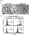
- Ectocervical cell culture organoids e.g. human or mouse ectocervical organoids have a morphology recapitulating the in vivo tissue architecture characterized by the presence of stratified epithelial layers and by the presence of the cellular marker E-cadherin.
- the outer layer of the organoids may consist of basal cells which are positive for the cellular marker p63. These cells may differentiate into para-basal cells with reduced levels of p63 and characteristic p63 negative cells facing the lumen. Only basal cells are positive for the proliferation marker Ki67.
- Ectocervical stem cells including stem cells from ectocervical organoid cultures were found to have a high upregulation of the Wnt antagonists Dickkopf Wnt signaling pathway inhibitor 1 (DKK1), DKK3 and KREMEN2 (DKK receptor). Further, ectocervical organoids were found to be negative for the Wnt target gene LGR5. Further, human ectocervical stem cells were found to have high expression levels of the Notch ligand, ⁇ -like ligand 3 (DLL3) and manic fringe (MFNG). Additional upregulated genes of ectocervical stem cells are described in the Example section.
- DKK1 Dickkopf Wnt signaling pathway inhibitor 1
- DKK3 DKK3
- KREMEN2 DKK receptor
- ectocervical organoids were found to be negative for the Wnt target gene LGR5.
- human ectocervical stem cells were found to have high expression levels of the Notch ligand, ⁇ -like ligand 3
- Endocervical cell culture organoids were found to consist of a large lumen with a simple columnar epithelium characterized by the presence of the cellular marker E-cadherin. A sporadic distribution of Ki67 positive proliferating cells was found. In endocervical organoids, Wnt-related genes are upregulated while Wnt antagonists and Notch-related genes are upregulated in ectocervical organoids.
- both stem cells and differentiated cells of ectocervical squamous organoids show high expression of the cellular marker KRT5 whereas this marker is absent from endocervical columnar organoids.
- endocervical organoids show high expression of KRT7 and KRT8.
- Endocervical stem cells or ectocervical stem cells may be cultivated into a respective organoid cell culture using appropriate culture media as disclosed herein.
- the organoid culture is a stable culture, i.e. it can be propagated for a time of at least six months or longer in a suitable cell culture medium as described herein.
- the organoid culture may be subjected to freezing/thawing procedures, and expansion, e.g. into a multi-well format, suitable for high throughput, setups and drug testing.
- cells from the organoid culture may be subjected to genetic modification, e.g., by genome editing.
- a biobank comprising a plurality of different organoid cultures is produced.
- the biobank may comprise the organoid cultures in the form of individual propagating organoid cultures and/or frozen organoid cultures.
- the cell culture medium may be selected from any suitable eukaryotic cell culture medium comprising salts, vitamins and trace elements.
- the cell culture medium for each organoid culture has specific niche requirements.
- a preferred culture medium comprises epidermal growth factor (EGF), e.g. in an amount of 5-100 ng/ml such as about 30 ng/ml, nicotinamide and an inhibitor of tissue growth factor ⁇ (TGF- ⁇ ) signaling, e.g. TGF- ⁇ R kinase inhibitor IV, noggin, e.g. about 100 ng/ml mouse noggin, and a ROCK inhibitor such as Y-27632, e.g. in an amount of about 10 ⁇ M.
- EGF epidermal growth factor
- TGF- ⁇ tissue growth factor ⁇
- TGF- ⁇ R kinase inhibitor IV e.g. TGF- ⁇ R kinase inhibitor IV
- noggin e.g. about 100 ng/ml mouse noggin
- ROCK inhibitor such as Y-27632
- a Wnt deficient medium i.e. a medium without exogenously added activators of Wnt signaling such as Wnt3a and/or RSPOL.
- the medium preferably comprises an activator of cAMP signaling such as forskolin.
- a suitable medium may comprise EGF, e.g. in an amount of 1-50 ng/ml such as about 10 ng/ml, fibroblast growth factor 10 (FGF-10), e.g. in an amount of 20-250 ng/ml such as about 100 ng/ml, noggin, e.g.
- TGF- ⁇ signaling e.g. TGF- ⁇ R kinase inhibitor IV.
- a Wnt proficient medium i.e. a medium comprising an activator of Wnt signaling such as Wnt3a and/or RSPO1.
- This medium is essentially free or free from a cAMP signaling activator such as forskolin.
- a suitable medium may comprise EGF, e.g. in an amount of 1-50 ng/ml such as about 10 ng/ml, fibroblast growth factor 10 (FGF-10), e.g. in an amount of 20-250 ng/ml such as about 100 ng/ml, noggin, e.g.
- the medium may contain Wnt3a and RSPO1, e.g. from a conditioned medium.
- the cell culture medium of the organoid culture may also comprise a suitable 3D cell culture matrix supporting the growth of organoids such as a basement membrane - like matrix, e.g. a Matrigel ® , BME ® or EHS ® matrix.
- a suitable matrix such as Matrigel ® and providing a suitable composition of niche factors in the medium, stem cells from the primary isolate will expand and differentiate into compact structure, i.e. epithelial organoids, which can grow indefinitely.
- the organoids which are not part of the present invention, can be used as models for improving diagnosis and/or therapy of cervical disorders, particularly cervical disorders involving pathological cell growth including malignant or benign cell growth, more particularly selected from cervical cancer, precancerous conditions, cervical neoplastic lesions or cervical metaplasias.
- the cervical disorders may be cervical squamous cell carcinoma or precursor conditions thereof such as precancerous conditions, neoplastic lesions or metaplasias resulting from cervical squamous cells.
- the cervical disorders may be cervical adenocarcinoma or precursor conditions thereof such as precancerous conditions, neoplastic lesions or metaplasias resulting from cervical glandular cells including adenomas.
- the organoids may be derived from primary human stem cells, they are excellent tools for use in personalized medicine, e.g. for developing therapeutic protocols individually adapted to the stem cell donor.
- the use may comprise determining the efficacy of a therapeutic compound in the treatment of cervical disorders as described above, in particular cervical adenocarcinoma or cervical squamous cell carcinoma, a cervical precancerous condition, a cervical neoplastic lesion or a cervical metaplasia, e.g.
- a therapeutic compound suitable in the treatment of cervical adenocarcinoma, cervical squamous cell carcinoma, a cervical precancerous condition, a cervical neoplastic lesion or a cervical metaplasia e.g. for drug screening.
- cervix epithelial cell organoid culture and/or the biobank as a test system for immunotherapy such as cancer immunotherapy, e.g. for testing the efficacy of immune cells, e.g. T cells in immunotherapy, e.g. the testing of immune cells, e.g. T cells obtained from a patient to be treated or obtained from a heterologous source, e.g. by determining antigen-specific immune responses or for identifying novel neo-antigens and/or neo-epitopes. These neo-antigens or neo-epitopes are useful for cancer vaccine design, particularly for personalized immune interventions.
- immunotherapy e.g. for testing the efficacy of immune cells, e.g. T cells in immunotherapy, e.g. the testing of immune cells, e.g. T cells obtained from a patient to be treated or obtained from a heterologous source, e.g. by determining antigen-specific immune responses or for identifying
- Still a further aspect, not part of the present invention, is based on the finding that the stromal compartment of the cervix provides specific growth factors or inhibitors which influence the outgrowth of specific cell lineages by activating or inhibiting Wnt signaling.
- stromal factors such as DKK2, DKK3, Axin2, RSpondin1, RSpondin2 or RSpondin3 are useful targets for the diagnosis and/or therapy of cervical disorders, as described above.
- a further aspect, not part of the present invention relates to a method of diagnosing a cervical disorder comprising the determination of the presence and/or amount of a stromal factor such as DKK2, DKK3, Axin2, RSpondin1, RSpondin2 or RSpondin3 in a sample from a subject, e.g. in a cervix tissue sample and/or a body fluid sample, and optionally comparing the determined amount with a reference amount which may be derived from a healthy subject or a group of healthy subjects.
- the method may involve determination of a stromal factor on the protein level by methods known in the art, e.g. by immunologic methods using appropriate reagents, e.g. antibodies or antibody fragments specifically directed against said stromal factor.
- the method may involve determination of a stromal factor on the level of nucleic acid molecules, e.g. on the transcript level, by methods known in the art, e.g. by nucleic acid amplification and/or hybridization using appropriate reagents, e.g. primers and/or probes specific for said nucleic acid molecule.
- This aspect not part of the invention, also relates to a kit comprising at least one reagent for the determination of a stromal factor as indicated above and the use of the kit for the diagnosis of a cervical disorder as described above.
- a further aspect, not part of the invention, relates to compounds for the therapy of a cervical disorder as described above.
- This aspect relates to the use of (i) a stromal factor such as DKK2, DKK3, Axin2, RSpondin1, RSpondin2 or RSpondin3 or a nucleic acid molecule encoding said stromal factor, or of (ii) an inhibitor of such a stromal factor or an inhibitor of a nucleic acid molecule coding therefor as an active agent for the modulation of pathological cervical cell growth.
- the active agent may be administered in a therapeutically effective amount to a subject in need thereof.
- the therapeutic goal of this administration is the provision of reduction or elimination of the pathological cell growth by modulating, i.e. stimulating or inhibiting, Wnt signaling depending on whether the pathological cervical cell growth is caused by, associated with, or accompanied by pathologically decreased or increased Wnt signaling.
- a further aspect, not part of the present invention also relates to a pharmaceutical composition
- a pharmaceutical composition comprising an active agent selected from (i) a stromal factor such as DKK2, DKK3, Axin2, RSpondin1, RSpondin2 or RSpondin3 or a nucleic acid molecule encoding said stromal factor or (ii) an inhibitor of such a stromal factor or an inhibitor of a nucleic acid molecule coding therefor, and pharmaceutically acceptable carrier.
- an active agent selected from (i) a stromal factor such as DKK2, DKK3, Axin2, RSpondin1, RSpondin2 or RSpondin3 or a nucleic acid molecule encoding said stromal factor or (ii) an inhibitor of such a stromal factor or an inhibitor of a nucleic acid molecule coding therefor, and pharmaceutically acceptable carrier.
- a stromal factor may be administered by means known in the art for the administration of therapeutic polypeptides.
- a recombinant stromal factor is used.
- a nucleic acid molecule encoding the stromal factor may be administered by means known in the art for the administration of therapeutic nucleic acid molecules.
- the nucleic acid molecule may be present as such, or located on a viral or non-viral vector in operative linkage with expression control elements allowing expression in the subject to be treated.
- Exemplary inhibitors may be selected from antibodies including monoclonal, chimeric, humanized or human antibodies or antigen-binding fragments of such antibodies specifically directed against said stromal factor.
- An antibody may be administered by means known in the art for the administration of therapeutic antibodies.
- a recombinant stromal factor is administered.
- Further exemplary inhibitors may be selected from inhibitory nucleic acid molecules including antisense molecules and inhibitory RNA molecules such as siRNA molecules specifically directed against a transcript of said stromal factor.
- An inhibitory nucleic acid molecule may be administered by means known in the art for the administration of such agents.
- the inhibitory nucleic acid molecule is administered in suitable vesicles, e.g. cationic vesicles such as liposomes, and/or conjugated to a heterologous moiety, e.g. selected from fatty acids, lipids, and saccharides.
- mouse-anti-p63 (Abcam, # ab375), rabbit-anti-p63 (Abcam, # ab53039), mouse-anti-E-Cadherin (BD Biosciences, # 610181), rabbit-anti-Ki67 (Abcam, # ab16667), mouse/rat-anti-Ki67-FITC (eBioscience, # 11-5698), mouse-anti-KRT5 (Sigma, # C-7785), rabbit-anti-KRT5 (Abcam, # ab52635), rabbit-anti-cytokeratin 5-Alexa488 (Abcam, # ab193894), mouse-anti-KRT7 (Santa Cruz, # sc-23876), rabbit-anti-cytokeratin 7 (Abcam, # ab181598), rabbit-anti-cytokeratin 7-Alexa555 (Abcam, # ab209601), rabbit-anti-CSTA (Cystatin A) (Sigma # HPA001031), rabbit-anti-AGR2 (Proteintech, # 12
- Cervical cells were cultured in ADF medium (Invitrogen, # 12634) supplemented with 12 mM HEPES (Invitrogen, # 15630-056), 1% GlutaMax (Invitrogen, # 35050-038), 1% B27 (Invitrogen, # 17504-044), 1% N2 (Invitrogen, # 17502048), 50 ng/ml mouse epidermal growth factor (EGF) (Invitrogen, # PMG8043), 100 ng/ml mouse noggin (Peprotech, # 250-38-100), 100 ng/ml human fibroblast growth factor (FGF)-10 (Peprotech, # 100-26-25), 1.25 mM N-acetyl-L-cysteine (Sigma, # A9165-5G), 10 mM nicotinamide, (Sigma, # N0636), 2 ⁇ M TGF- ⁇ R Kinase Inhibitor IV (Calbiochem, # 616454), 10 ⁇ M
- the medium consisted of ADF, 12 mM HEPES and 1% GlutaMax, supplemented with 1% B27, 1% N2, 0.5 ⁇ g/ml hydrocortisone (Sigma, # H0888-1G), 10 ng/ml human EGF (Invitrogen, # PHG0311), 100 ng/ml human noggin (Peprotech; # 120-10C), 100 ng/ml human FGF-10 (Peprotech, # 100-26-25), 1.25 mM N-acetyl-L-cysteine, 10 mM nicotinamide, 2 ⁇ M TGF- ⁇ R kinase Inhibitor IV, 10 ⁇ M ROCK inhibitor (Y-27632), 10 ⁇ M forskolin (Sigma, F6886) and 1% penicillin/streptomycin.
- the medium consisted of ADF, 12 mM HEPES, 1% GlutaMax, supplemented with 1% B27, 1% N2, 10 ng/ml human EGF, 100 ng/ml human noggin, 100 ng/ml human FGF-10, 1.25 mM N-acetyl-L-cysteine, 10 mM nicotinamide, 2 mM TGF- ⁇ R Kinase Inhibitor IV and 10 ⁇ M ROCK inhibitor (Y-27632) with 25% Wnt3A- and 25% R-spondin1 conditioned medium.
- Minced tissue was incubated in 0.5 mg/ml collagenase type II (Calbiochem, # 234155) for 2.5 h at 37°C in a shaker incubator. Tissue and dissociated cells were pelleted by centrifugation (5 min at 1000 g, 4°C), supernatant discarded, cells resuspended in TrypLE express (Gibco, # 12604021) and incubated for 15 min at 37°C in a shaker incubator. After dissociation the cell and tissue pellet was resuspended in ADF (Invitrogen) medium and passed through a 40 ⁇ m cell strainer (BD Falc, # 352340) to separate the single dissociated cells from tissue pieces. Cells were pelleted by centrifugation (5 min at 1000xg, 40°C), resuspended in either human ecto- or endocervical or mouse cervical medium and cultured either directly as organoids or in 2D.
- ADF Invitrogen
- Human epithelial stem cells isolated from the tissue were resuspended in either ecto- or endo cervical medium and plated in collagen-coated tissue culture flasks. Cells were incubated at 37°C, 5% CO 2 in a humidified incubator. Once they reached 70-80% confluence, cells were detached using TrypLE Express, and centrifuged at 1000xg for 5 min at 40°C. The cells were then used for culturing organoids or for maintenance of 2D stem cells. 2D stem cells were maintained by seeding the 2D cells from P1 into tissue culture flasks containing lethally irradiated J2-3T3 fibroblast feeder cells in ecto- or endocervical medium.
- Matrigel Cells isolated from tissue or the stem cells grown in 2D culture were mixed with 50 ⁇ l of ice cold Matrigel (BD, # 356231) at a density of 20,000 cells, and the Matrigel droplet was placed in a pre-warmed 24-well plate and allowed to polymerize for 10 min at 37°C. The Matrigel droplet was then overlaid with 500 ⁇ l of pre-warmed human ecto- or endocervical medium. Cultures were kept at 37°C, 5% CO 2 in a humidified incubator for 2-3 weeks and medium replaced every four days. For passaging the organoids, Matrigel was first dissolved by adding 1 ml of ice cold ADF and pipetting up and down 5 times.
- BD ice cold Matrigel
- Organoids were collected in a 15 ml Falcon tube and a further 4 ml of ice cold ADF medium was added and organoids resuspended well to completely dissolve the Matrigel, followed by centrifugation at 300xg for 5 mins at 4°C. Medium was discarded and the ectocervical and mouse organoids were incubated with 1 ml of TrypLE Express for 30 min at 37°C followed by mechanical fragmentation using a fire-polished glass Pasteur pipette by vigorous pipetting (8-10 times) to generate single cells. The single cells were then seeded at a 1:10 ratio back into Matrigel for expanding and culturing.
- Endocervical organoids were subjected to mechanical fragmentation as described above after centrifugation to generate fragments that were seeded back into Matrigel at 1:5 ratio.
- Matrigel was allowed to polymerize for 10 min at 37°C, overlaid with pre-warmed medium and cultured as described above.
- Stem cells were counted and a defined number resuspended in 50 ⁇ l of Matrigel to generate organoids as described above. Between 2-3 weeks after plating images were taken of the whole well and the number and area of organoids formed were determined using ImageJ to calculate organoid forming efficiency.
- Organoids were washed with cold PBS five times to remove Matrigel before fixing with 4% paraformaldehyde for 1 h at room temperature (RT) followed by washing with PBS twice. Organoids were then subjected to dehydration in an ascending ethanol series followed by isopropanol and acetone for 20 min each. The dehydrated organoids were paraffin embedded and 5 ⁇ m sections cut on a Microm HM 315 microtome. Mouse and human tissues were extensively washed with PBS and fixed using 4% PFA overnight at RT. Samples were subjected to dehydration in an ascending ethanol series followed by isopropanol and xylene (60 mins each) followed by paraffinization using a Leica TP1020 tissue processor.
- the tissue was embedded and 5 ⁇ m sections cut on a microtome.
- paraffin sections were deparaffinized and rehydrated, followed by treatment with antigen retrieval solution (Dako, # S1699). Sections were blocked using blocking buffer (1% BSA and 2% FCS in PBS) for 1 h at RT. Primary antibodies were diluted in blocking buffer and incubated for 90 min at RT followed by five PSB washes before 1 h incubation with secondary antibodies diluted in blocking buffer along with Hoechst or Draq5. Sections were washed with PBS five times and mounted using Mowiol. Images were acquired with a Leica TCS SP8 confocal microscope.
- Fresh epithelial isolates were grown on collagen-coated coverslips in 2D and fixed with 4% paraformaldehyde for 30 min at RT. Cells were permeabilized and blocked with 0.5% Triton X-100 and 1% BSA in PBS. Primary antibodies were diluted in 1% BSA in PBS and incubated for 1 h at RT followed by three washes in PSBT (0.1% Tween 20 in PBS), followed by 1 h incubation with secondary antibodies diluted in 1% BSA in PBS along with Hoechst or Draq5. Coverslips were washed three times with PBST and once with PBS and mounted using Mowiol. Images were acquired on a Leica TCS SP8 confocal microscope. Images were processed with Adobe Photoshop; 3D reconstruction was done with the Velocity 6.3 software package (Perkin Elmer).
- Matrigel was removed from the organoids by extensive washing with ice cold PBS prior to fixation (4x 45 min) and allowed to settle by gravity to maintain the 3D structure. Organoids were then fixed using pre-warmed (37 °C) 3.7% PFA for 1 h at RT followed by three PBST washes. Permeabilization and blocking was performed overnight at 40 °C using 5% donkey serum, 1% FCS, 0.05% Tween20, 2% Triton X-100, 0.02% sodium azide in PBS.
- Organoids were incubated with primary antibodies diluted in blocking buffer (5% donkey serum, 1% FCS, 0.25% Triton X-100, 0.02% sodium azide in PBS) at 4°C for 3-5 days followed by three PBST washes for 45 min each at RT. Next, organoids were incubated with secondary antibodies diluted in blocking buffer for two days at 4°C followed by one PBST wash for 45 min and three washes with PBS containing 5% glycerol for 45 min each.
- blocking buffer 5% donkey serum, 1% FCS, 0.25% Triton X-100, 0.02% sodium azide in PBS
- Organoids were then carefully transferred to an ibidi ⁇ -slide (# 81822) together with some PBS and glycerol solution and Z stack images were acquired with a confocal microscope and image processing and 3D reconstructions were done using Velocity 6.3 software.
- RNA-ISH Single-molecule RNA in situ hybridization
- RNA in situ labelling For single molecule RNA in situ labelling, paraffin embedded 10 ⁇ m tissue sections were used with RNAscope 2.5 HD Red Reagent kit (Advanced Cell Diagnostics). Hybridizations were performed according to the manufacture's protocol. In each experiment, positive (PPIB) and negative (DapB) control probes were used as per the manufacturer's guidelines. Tiled bright field images were obtained with Axio Scan.Z1 tissue imager (Zeiss). Images were further processed with Zen 2.3 (Blue edition) image analysis software and further compiled using Adobe illustrator.
- sample sizes were selected based on available samples.
- Microarray experiments were performed as single-color hybridizations on custom whole genome human 8x60k Agilent arrays (Design ID 048908) and Agilent Feature Extraction software was used to obtain probe intensities.
- the extracted single-color raw data files were background corrected, quantile normalized and further analyzed for differential gene expression using R ( 48 ) and the associated BioConductor package LIMMA ( 49 ) (Supplementary Information Table 2).
- Microarray gene expression comparisons between groups were performed using unpaired tests for all human comparisons. R was also used for all statistical analyses unless stated otherwise. Mann-Whitney-U test was used for comparisons of gene expression in SCJ marker genes with a threshold of p ⁇ 0.05.
- Microarray data have been deposited in the Gene Expression Omnibus (GEO; wwv.ncbi.nlm.nih.gov/geo/) of the National Center for Biotechnology Information and can be accessed with the GEO accession number GSE87076. These data form part of the present disclosure.
- GEO Gene Expression Omnibus
- the signature of differentially expressed genes between ectocervical 2D/EO vs DO organoids was selected from all genes with a false discovery rate (FDR) ⁇ 0.05 and log2 fold change ⁇ -1.5 or > 1.5 in any of the two comparisons (2D vs. DO or EO vs. DO) and the largest absolute fold change from both comparisons and possible replicate probes was taken for each gene.
- FDR false discovery rate
- Raw data from different microarray data sets obtained from adult tissue stem cells (SC) cultured on feeder cells and corresponding differentiated cells from air liquid interface (ALI), Matrigel or self-assembly sphere (SAS) were downloaded from GEO (GSE57584, GSE66115, GSE69453, GSE65013, GSE32606, GSE69429, GSE49292) and normalized together using method 'RMA-sketch' with Affymetrix Power Tools.
- GSEA Gene Set Enrichment Analysis
- Aggregated features including clustering results based on DNA methylation, mRNA and microRNA expression was obtained from the Cervical and Endocervical Cancer (CESC) project Firehose site of TCGA ( 53 ). For the details on the majority vote see Fig. 13 .
- CESC Cervical and Endocervical Cancer
- a squamous vs columnar organoid signature was defined based on the fold changes between ectocervical squamous and endocervical columnar differentiated organoids for 2,834 genes with FDR ⁇ 0.05 and absolute log2 fold change > 1, selecting the probe with lowest p-value for each gene.
- Co-Sq Score Spearman correlation coefficients
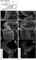
- transition zone (TZ) maintenance we carried out a detailed analysis of marker profiles in human and mouse cervical epithelium. Strikingly, our comprehensive, unbiased analysis including the entire endo- and ectocervix regions failed to detect any specific cell type-restricted exclusively to the TZ in contrast to the prevailing concept. Instead, we observed two distinct epithelial lineages, with KRT5 expressed throughout the squamous stratified epithelium and KRT7/KRT8 expressed throughout the columnar epithelium.
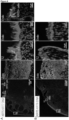
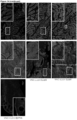
- RNA-ISH confirmed that KRT8 and KRT5 expression was restricted to the columnar epithelium and basal/parabasal cells of squamous epithelium, respectively ( Fig. 1 E-F , and Fig. 7 A-B ).
- KRT5CreErt2/Rosa26-tdTomato and KRT8CreErt2/Rosa26-tdTomato mice for genetic lineage tracing we confirmed the presence of two distinct epithelial lineages in the cervix ( Fig. 1 H-I and J ).
- Cre was induced by tamoxifen injection 4 weeks after birth.
- KRT5+ cells exclusively populated the stratified epithelium, including all differentiated cells, while KRT8+ cells exclusively generated endocervical epithelium.
- EGF epidermal growth factor
- FGF-10 fibroblast growth factor 10
- TGF- ⁇ transforming growth factor beta
- BMP bone morphogenetic protein
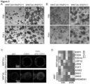
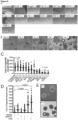
- the outer layer consists of p63+ basal cells, which differentiate into parabasal cells with p63 staining fading out towards the lumen. Also, typical of ectocervical tissue, only basal cells express the proliferation marker Ki67 ( Fig. 8G ).
- organoids consisting of a simple columnar epithelial layer when cultured in the presence of Wnt proficient medium containing Wnt3a and RSPO1 ( Fig. 2B-C ). These organoids faithfully resemble the in vivo endocervical epithelium, are KRT7+/KRT5- and exhibit sporadic Ki67 staining ( Fig.9A and 8I ). Their self-renewal capacity in culture can be maintained for more than seven months ( Fig. 9B ). Further, transcriptional profiling of organoids derived from human ecto- and endocervix revealed distinct keratin expression patterns ( Fig. 8J ).
- Wnt agonists and antagonists in the underlying stroma defines the borders between the two epithelial types.
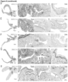
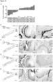
- Both the Wnt agonist RSPO1 and its downstream target Axin2 are highly expressed in the lamina basement (stroma) beneath the columnar epithelium and RSPO3 in the muscularis of the endocervix ( Fig. 2E , 9D-F ).
- the Wnt antagonist DKK2 is specifically expressed in stroma proximal to the basal cells of the ectocervical squamous epithelium, which express high levels of DKK3 ( Fig. 2F-H , 9G-H ).
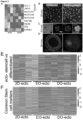
- Fig. 11A Early organoids consist of 8-16 cells that are undifferentiated and positive for Ki67 and p63 ( Fig. 3C and Fig. 11B ). Mature organoids consist of several stratified differentiated layers with more than two-thirds of cells differentiated and less than one-third of cells proliferating ( Fig. 3D and Fig. 11B ). Gene expression patterns of cells from 2D cultures and early organoids show high similarity and display a distinct set of differentially expressed genes compared to mature organoids ( Fig. 3E ). Recent studies reported that stem cells from diverse tissue types show similar transcriptional signatures compared to the large divergence observed in the ensuing differentiated tissues (14).
- DLL3 Delta-Like Ligand 3
- MFNG Manic Fringe
- Ectocervical stem cells also showed highly upregulated expression of the WNT antagonists DKK1, DKK3 and the DKK receptor KREMEN2 ( Fig. 3G ).
- GSEA gene set enrichment analysis
- squamous organoids including the characteristic basal and parabasal p63+ cells ( FIG. 4A ).
- endocervical organoids derived from single cells remained columnar even when transferred to Wnt deficient medium, thus excluding the possibility that columnar cells transdifferentiate to the squamous lineage ( Fig. 4B ).
- primary ectocervical cells grown in 2D with either Wnt proficient or deficient medium give rise only to stratified organoids in Wnt deficient medium ( Fig. 4C ).
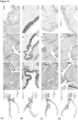
- HOXA11 has previously been associated with cervix development (23) and deregulation of HOXB2, HOXB4, and HOXB13 have been implicated in cervical carcinogenesis (24).
- Fig. 12A we analyzed the pattern of HOX gene expression in the ecto- vs. endocervical organoids ( Fig. 12A ). Strikingly, we observed substantial differences between the two cultures, supporting the notion that the two tissue types represent different biological lineages in the cervix.
- ADC cervical adenocarcinoma
- SCC squamous cell carcinoma
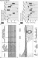
- cancer samples classified as columnar-like were mainly KRT5low, KRT7high and p63low, while samples in the squamous-like and undetermined group were mainly KRT5high and p63high with mixed KRT7 status ( Fig. 5A ), suggesting that the undetermined group could consist of SCCs within or outgrown into columnar endocervix, leading to the presence of contaminating endocervical columnar KRT7+ cells in the samples.
- SCJ squamocolumnar junction
- the TZs of the mucosal epithelium constitute critical zones of enhanced disposition to infections and carcinogenesis (27-31). Revealing the principles of cellular regulation and homeostasis of these tissue regions is key to understanding the impact of intrinsic and extrinsic disturbances, as well as for prospective and therapeutic disease prevention.
- Wnt signaling has been shown to be indispensable for the maintenance and homeostasis of adult stem cells in several mammalian tissues (13, 32). However, here we show that in the cervix, Wnt signaling stimulated by the underlying stroma drives the columnar lineage while imposing quiescence of squamous lineage-specific stem cells that exist in the same milieu.
- mice fed with vitamin A deficient diet develop cervical metaplasia throughout the endocervix. Since metaplasia is a pre-neoplastic condition and a major risk factor for carcinogenesis we conclude that with nutritional supplementation with vitamin A we not only can prevent metaplasia but also cancer development.
Landscapes
- Health & Medical Sciences (AREA)
- Engineering & Computer Science (AREA)
- Life Sciences & Earth Sciences (AREA)
- Biomedical Technology (AREA)
- Chemical & Material Sciences (AREA)
- Immunology (AREA)
- Biotechnology (AREA)
- Cell Biology (AREA)
- Hematology (AREA)
- Bioinformatics & Cheminformatics (AREA)
- Molecular Biology (AREA)
- Urology & Nephrology (AREA)
- Microbiology (AREA)
- Biochemistry (AREA)
- General Health & Medical Sciences (AREA)
- Zoology (AREA)
- Organic Chemistry (AREA)
- Wood Science & Technology (AREA)
- Genetics & Genomics (AREA)
- Physics & Mathematics (AREA)
- Pathology (AREA)
- General Physics & Mathematics (AREA)
- Analytical Chemistry (AREA)
- Medicinal Chemistry (AREA)
- Food Science & Technology (AREA)
- Toxicology (AREA)
- Tropical Medicine & Parasitology (AREA)
- Reproductive Health (AREA)
- General Engineering & Computer Science (AREA)
- Measuring Or Testing Involving Enzymes Or Micro-Organisms (AREA)
- Micro-Organisms Or Cultivation Processes Thereof (AREA)
Claims (4)
- Verfahren für die Herstellung einer Zervixepithel-Zellorganoidkultur, wobei das Verfahren die Schritte umfasst:(a) Kultivieren von Zervix-Stammzellen in einem geeigneten Zellkulturmedium unter Bedingungen, unter welchen ein Organoid gebildet wird, und(b) gegebenenfalls Erhalten eines Organoids aus dem Kultivierungsschritt (a),(i) wobei die Zervix-Stammzellen endozervikale Stammzellen sind, z. B. humane endozervikale Stammzellen, und wobei das Zellkulturmedium ein Wnt-kompetentes Medium ist und wobei das Organoid säulenförmige endozervikale Epithelzellen umfasst, welche für die zellulären Marker KRT7 und/oder KRT8 positiv sind, oder(ii) wobei die Zervix-Stammzellen ektozervikale Stammzellen sind, z. B. humane ektozervikale Stammzellen, und wobei das Kulturmedium ein Wnt-defizientes Medium ist, welches einen cAMP-Signalweg-Agonisten wie Forskolin enthält, und wobei das Organoid mehrschichtige ektozervikale Plattenepithelzellen umfasst, welche positiv für den Marker KRT5 sind.
- Verfahren nach Anspruch 1, wobei die Zervixepithel-Zellorganoidkultur eine humane, mehrschichtige, ektozervikale Plattenepithelzellkultur ist, welche mindestens sechs Monate lang stabil in einem geeigneten Zellkulturmedium vermehrt wird, welches ein Wnt-defizientes Zellkulturmedium ist, welches einen cAMP-Signalgebungs-Aktivator enthält und welches keine exogen hinzugefügten Aktivatoren der Wnt-Signalgebung enthält.
- Verfahren nach Anspruch 1, wobei die Zervixepithel-Zellorganoidkultur eine humane säulenförmige endozervikale Epithel-Zellkultur ist, welche für mindestens sechs Monate in einem geeigneten Zellkulturmedium stabil vermehrt wird, welches ein Wnt-kompetentes Zellkulturmedium ist, welches einen Aktivator der Wnt-Signalgebung umfasst.
- Verfahren nach Anspruch 1, 2 oder 3, wobei eine Biobank, umfassend mehrere verschiedene Organoidkulturen, hergestellt wird.
Applications Claiming Priority (2)
| Application Number | Priority Date | Filing Date | Title |
|---|---|---|---|
| EP18200408.5A EP3640324A1 (de) | 2018-10-15 | 2018-10-15 | Gebärmutterhalskrebsorganoide |
| PCT/EP2019/077575 WO2020078840A1 (en) | 2018-10-15 | 2019-10-11 | Cervical cancer organoids |
Publications (3)
| Publication Number | Publication Date |
|---|---|
| EP3867355A1 EP3867355A1 (de) | 2021-08-25 |
| EP3867355B1 true EP3867355B1 (de) | 2025-01-22 |
| EP3867355C0 EP3867355C0 (de) | 2025-01-22 |
Family
ID=63857824
Family Applications (2)
| Application Number | Title | Priority Date | Filing Date |
|---|---|---|---|
| EP18200408.5A Withdrawn EP3640324A1 (de) | 2018-10-15 | 2018-10-15 | Gebärmutterhalskrebsorganoide |
| EP19783337.9A Active EP3867355B1 (de) | 2018-10-15 | 2019-10-11 | Gebärmutterhalskrebsorganoide |
Family Applications Before (1)
| Application Number | Title | Priority Date | Filing Date |
|---|---|---|---|
| EP18200408.5A Withdrawn EP3640324A1 (de) | 2018-10-15 | 2018-10-15 | Gebärmutterhalskrebsorganoide |
Country Status (3)
| Country | Link |
|---|---|
| US (1) | US20210355450A1 (de) |
| EP (2) | EP3640324A1 (de) |
| WO (1) | WO2020078840A1 (de) |
Families Citing this family (5)
| Publication number | Priority date | Publication date | Assignee | Title |
|---|---|---|---|---|
| CN112080474A (zh) * | 2020-09-21 | 2020-12-15 | 上海交通大学 | 一种体外建立肿瘤类器官的方法 |
| CN114790446B (zh) * | 2022-03-24 | 2022-11-08 | 中山大学附属第一医院 | 一种宫颈腺癌类器官培养基及其构建方法 |
| CN114854670B (zh) * | 2022-07-01 | 2024-10-29 | 武汉大学 | 一种人类宫颈疣类器官模型的构建方法以及该模型在开发先导化合物中的应用 |
| CN116716236B (zh) * | 2023-06-29 | 2024-10-29 | 重庆大学附属肿瘤医院 | 一种宫颈癌类器官的构建方法及应用 |
| CN118272309A (zh) * | 2023-07-14 | 2024-07-02 | 北京嘉士腾医学检验实验室有限公司 | 一种高效的消化道肿瘤类器官培养方法 |
-
2018
- 2018-10-15 EP EP18200408.5A patent/EP3640324A1/de not_active Withdrawn
-
2019
- 2019-10-11 WO PCT/EP2019/077575 patent/WO2020078840A1/en not_active Ceased
- 2019-10-11 US US17/285,148 patent/US20210355450A1/en active Pending
- 2019-10-11 EP EP19783337.9A patent/EP3867355B1/de active Active
Non-Patent Citations (13)
| Title |
|---|
| "Posters", ARCHIVES OF GYNECOLOGY AND OBSTETRICS, SPRINGER BERLIN HEIDELBERG, BERLIN/HEIDELBERG, vol. 282, no. Suppl 1, 10 September 2010 (2010-09-10), pages 63 - 270, XP037076757, ISSN: 0932-0067, [retrieved on 20100910], DOI: 10.1007/S00404-010-1634-7 * |
| ANDREAS SCHMID ET AL: "Modulation of Wnt signaling is essential for the differentiation of ciliated epithelial cells in human airways", FEBS LETTERS, vol. 591, no. 21, 10 October 2017 (2017-10-10), NL, pages 3493 - 3506, XP055542009, ISSN: 0014-5793, DOI: 10.1002/1873-3468.12851 * |
| ANONYMOUS: "Wissenschaftlicher Bericht 2014-2015 - zentrum für infektionsforschung - Universität Würzburg", 1 January 2015 (2015-01-01), XP055540734, Retrieved from the Internet <URL:https://www.uni-wuerzburg.de/fileadmin/87030000/ZINFreport_2014-15/ZINFReport_2014-15_beta.pdf> [retrieved on 20190111] * |
| AYEHUNIE S ET AL: "Organotypic human vaginal-ectocervical tissue model for irritation studies of spermicides, microbicides, and feminine-care products", TOXICOLOGY IN VITRO, ELSEVIER SCIENCE, GB, vol. 20, no. 5, 1 August 2006 (2006-08-01), pages 689 - 698, XP024966229, ISSN: 0887-2333, [retrieved on 20060801], DOI: 10.1016/J.TIV.2005.10.002 * |
| B. E. HJELM ET AL: "Development and Characterization of a Three-Dimensional Organotypic Human Vaginal Epithelial Cell Model", BIOLOGY OF REPRODUCTION, vol. 82, no. 3, 9 December 2009 (2009-12-09), pages 617 - 627, XP055106951, ISSN: 0006-3363, DOI: 10.1095/biolreprod.109.080408 * |
| CANTERO JON ET AL: "Maximizing the immunological output of the cervicovaginal explant model", JOURNAL OF IMMUNOLOGICAL METHODS, ELSEVIER SCIENCE PUBLISHERS B.V.,AMSTERDAM, NL, vol. 460, 9 June 2018 (2018-06-09), pages 26 - 35, XP085428607, ISSN: 0022-1759, DOI: 10.1016/J.JIM.2018.06.005 * |
| CHUMDURI CINDRILLA ET AL: "Opposing Wnt signals regulate cervical squamocolumnar homeostasis and emergence of metaplasia", NATURE CELL BIOLOGY, NATURE PUBLISHING GROUP UK, LONDON, vol. 23, no. 2, 18 January 2021 (2021-01-18), pages 184 - 197, XP037819856, ISSN: 1465-7392, [retrieved on 20210118], DOI: 10.1038/S41556-020-00619-0 * |
| CHUMDURI CINDRILLA ET AL: "Opposing Wnt signals regulate cervical squamocolumnar homeostasis and emergence of metaplasia", NATURE CELL BIOLOGY, vol. 23, no. 2, February 2021 (2021-02-01), pages 184 - 197, XP037365970, ISSN: 1465-7392, DOI: 10.1038/S41556-020-00619-0 * |
| COLLINS K B ET AL: "Development of an in vitro organ culture model to study transmission of HIV-1 in the female genital tract", NATURE MEDICINE, NATURE PUBLISHING GROUP US, NEW YORK, vol. 6, no. 4, 1 April 2000 (2000-04-01), pages 475 - 479, XP002401732, ISSN: 1078-8956 * |
| GURUMURTHY RAJENDRA KUMAR ET AL: "Patient-derived and mouse endo-ectocervical organoid generation, genetic manipulation and applications to model infection", NATURE PROTOCOLS, NATURE PUBLISHING GROUP, GB, vol. 17, no. 7, 11 May 2022 (2022-05-11), pages 1658 - 1690, XP037897759, ISSN: 1754-2189, [retrieved on 20220511], DOI: 10.1038/S41596-022-00695-6 * |
| MING JIANG ET AL: "Transitional basal cells at the squamous?columnar junction generate Barrett?s oesophagus", NATURE, vol. 550, no. 7677, 12 October 2017 (2017-10-12), London, pages 529 - 533, XP055542061, ISSN: 0028-0836, DOI: 10.1038/nature24269 * |
| PETER L. VILLA ET AL: "Human Cervical Keratinocyte-Derived Monolayer and Organoid Cultures for Disease Modelling and Drug Screening", BIORXIV, 20 July 2018 (2018-07-20), XP055540972, Retrieved from the Internet <URL:https://www.biorxiv.org/content/biorxiv/early/2018/07/20/373456.full.pdf> DOI: 10.1101/373456 * |
| SARTINI SARA ET AL: "Cervical organoids go viral", CELL STEM CELL, ELSEVIER, CELL PRESS, AMSTERDAM, NL, vol. 28, no. 8, 5 August 2021 (2021-08-05), pages 1337 - 1338, XP086722070, ISSN: 1934-5909, [retrieved on 20210805], DOI: 10.1016/J.STEM.2021.07.007 * |
Also Published As
| Publication number | Publication date |
|---|---|
| US20210355450A1 (en) | 2021-11-18 |
| EP3867355C0 (de) | 2025-01-22 |
| EP3867355A1 (de) | 2021-08-25 |
| WO2020078840A1 (en) | 2020-04-23 |
| EP3640324A1 (de) | 2020-04-22 |
Similar Documents
| Publication | Publication Date | Title |
|---|---|---|
| Chumduri et al. | Opposing Wnt signals regulate cervical squamocolumnar homeostasis and emergence of metaplasia | |
| US20210355450A1 (en) | Cervical cancer organoids | |
| Qi et al. | Targeting the Wnt-regulatory protein CTNNBIP1 by microRNA-214 enhances the stemness and self-renewal of cancer stem-like cells in lung adenocarcinomas | |
| Shinriki et al. | Loss of CYLD promotes cell invasion via ALK5 stabilization in oral squamous cell carcinoma | |
| Gao et al. | RETRACTED ARTICLE: Long noncoding RNA MAGI1-IT1 promoted invasion and metastasis of epithelial ovarian cancer via the miR-200a/ZEB axis | |
| Bao et al. | Hyperactivated Wnt-β-catenin signaling in the absence of sFRP1 and sFRP5 disrupts trophoblast differentiation through repression of Ascl2 | |
| Liu et al. | MicroRNA-494-dependent WDHDI inhibition suppresses epithelial-mesenchymal transition, tumor growth and metastasis in cholangiocarcinoma | |
| EP3006941A1 (de) | Verfahren zum screening eines krebsmetastasenhemmers anhand der kultur von zellen oder kugelförmig aggregierten zellen mit regulierung von lysyl-trna-synthetase zur expression oder nichtexpression | |
| Chumduri et al. | Transition of Wnt signaling microenvironment delineates the squamo-columnar junction and emergence of squamous metaplasia of the cervix | |
| Wang et al. | Role of Cks1 amplification and overexpression in breast cancer | |
| CN115837079A (zh) | Igf2bp1高表达在食管癌检测和治疗中的应用 | |
| He et al. | Lentiviral vector-mediated down-regulation of Notch1 in endometrial stem cells results in proliferation and migration in endometriosis | |
| CN103667441A (zh) | 一种Hsa-miR-145-5p试剂盒及其成熟体模拟物的应用 | |
| CN107312855B (zh) | 一种与喉鳞癌相关的基因及其应用 | |
| CN108220443A (zh) | Cgref1作为标志物在肾透明细胞癌诊疗中的应用 | |
| CN116103403B (zh) | 一种用于卵巢癌诊断和预后判断的生物标志物及其应用 | |
| CN110951880B (zh) | 检测下咽癌lncRNA标志物的试剂在制备诊断下咽癌产品中的应用 | |
| Dou et al. | Endothelial RNF20 suppresses endothelial-to-mesenchymal transition and safeguards physiological angiocrine signaling to prevent congenital heart disease | |
| CN107881240B (zh) | 骨肉瘤的诊治标志物 | |
| Pan et al. | A Fibroblast State Choreographs an Epithelial YAP-dependent Regenerative Program Essential to (Pre) malignancy via ECM-mediated Mechanotransduction | |
| CN110229896A (zh) | Steap1在卵巢癌诊断、治疗和预后中的应用 | |
| Li et al. | Interleukin-6, Tumor Necrosis Factor-α Expression Levels in Prostate Cancer Tissues and Their Effects on Epithelial Interstitial Transformation, Migration and Invasion in Prostate Cancer Cells | |
| Wang et al. | CENP-A promotes cervical cancer progression through p53 pathway | |
| Maenhoudt et al. | Organoid models of female reproductive tissues to study biology and disease | |
| CN107385081A (zh) | 一种与肾癌相关的基因及其应用 |
Legal Events
| Date | Code | Title | Description |
|---|---|---|---|
| STAA | Information on the status of an ep patent application or granted ep patent |
Free format text: STATUS: UNKNOWN |
|
| STAA | Information on the status of an ep patent application or granted ep patent |
Free format text: STATUS: THE INTERNATIONAL PUBLICATION HAS BEEN MADE |
|
| PUAI | Public reference made under article 153(3) epc to a published international application that has entered the european phase |
Free format text: ORIGINAL CODE: 0009012 |
|
| STAA | Information on the status of an ep patent application or granted ep patent |
Free format text: STATUS: REQUEST FOR EXAMINATION WAS MADE |
|
| 17P | Request for examination filed |
Effective date: 20210427 |
|
| AK | Designated contracting states |
Kind code of ref document: A1 Designated state(s): AL AT BE BG CH CY CZ DE DK EE ES FI FR GB GR HR HU IE IS IT LI LT LU LV MC MK MT NL NO PL PT RO RS SE SI SK SM TR |
|
| DAV | Request for validation of the european patent (deleted) | ||
| DAX | Request for extension of the european patent (deleted) | ||
| STAA | Information on the status of an ep patent application or granted ep patent |
Free format text: STATUS: EXAMINATION IS IN PROGRESS |
|
| 17Q | First examination report despatched |
Effective date: 20220502 |
|
| GRAP | Despatch of communication of intention to grant a patent |
Free format text: ORIGINAL CODE: EPIDOSNIGR1 |
|
| STAA | Information on the status of an ep patent application or granted ep patent |
Free format text: STATUS: GRANT OF PATENT IS INTENDED |
|
| INTG | Intention to grant announced |
Effective date: 20240813 |
|
| GRAS | Grant fee paid |
Free format text: ORIGINAL CODE: EPIDOSNIGR3 |
|
| GRAA | (expected) grant |
Free format text: ORIGINAL CODE: 0009210 |
|
| STAA | Information on the status of an ep patent application or granted ep patent |
Free format text: STATUS: THE PATENT HAS BEEN GRANTED |
|
| AK | Designated contracting states |
Kind code of ref document: B1 Designated state(s): AL AT BE BG CH CY CZ DE DK EE ES FI FR GB GR HR HU IE IS IT LI LT LU LV MC MK MT NL NO PL PT RO RS SE SI SK SM TR |
|
| REG | Reference to a national code |
Ref country code: GB Ref legal event code: FG4D |
|
| REG | Reference to a national code |
Ref country code: CH Ref legal event code: EP |
|
| REG | Reference to a national code |
Ref country code: IE Ref legal event code: FG4D |
|
| REG | Reference to a national code |
Ref country code: DE Ref legal event code: R096 Ref document number: 602019065138 Country of ref document: DE |
|
| U01 | Request for unitary effect filed |
Effective date: 20250217 |
|
| U07 | Unitary effect registered |
Designated state(s): AT BE BG DE DK EE FI FR IT LT LU LV MT NL PT RO SE SI Effective date: 20250221 |
|
| PG25 | Lapsed in a contracting state [announced via postgrant information from national office to epo] |
Ref country code: RS Free format text: LAPSE BECAUSE OF FAILURE TO SUBMIT A TRANSLATION OF THE DESCRIPTION OR TO PAY THE FEE WITHIN THE PRESCRIBED TIME-LIMIT Effective date: 20250422 |
|
| PG25 | Lapsed in a contracting state [announced via postgrant information from national office to epo] |
Ref country code: PL Free format text: LAPSE BECAUSE OF FAILURE TO SUBMIT A TRANSLATION OF THE DESCRIPTION OR TO PAY THE FEE WITHIN THE PRESCRIBED TIME-LIMIT Effective date: 20250122 |
|
| PG25 | Lapsed in a contracting state [announced via postgrant information from national office to epo] |
Ref country code: ES Free format text: LAPSE BECAUSE OF FAILURE TO SUBMIT A TRANSLATION OF THE DESCRIPTION OR TO PAY THE FEE WITHIN THE PRESCRIBED TIME-LIMIT Effective date: 20250122 |
|
| PG25 | Lapsed in a contracting state [announced via postgrant information from national office to epo] |
Ref country code: NO Free format text: LAPSE BECAUSE OF FAILURE TO SUBMIT A TRANSLATION OF THE DESCRIPTION OR TO PAY THE FEE WITHIN THE PRESCRIBED TIME-LIMIT Effective date: 20250422 Ref country code: IS Free format text: LAPSE BECAUSE OF FAILURE TO SUBMIT A TRANSLATION OF THE DESCRIPTION OR TO PAY THE FEE WITHIN THE PRESCRIBED TIME-LIMIT Effective date: 20250522 |
|
| PG25 | Lapsed in a contracting state [announced via postgrant information from national office to epo] |
Ref country code: HR Free format text: LAPSE BECAUSE OF FAILURE TO SUBMIT A TRANSLATION OF THE DESCRIPTION OR TO PAY THE FEE WITHIN THE PRESCRIBED TIME-LIMIT Effective date: 20250122 |
|
| PG25 | Lapsed in a contracting state [announced via postgrant information from national office to epo] |
Ref country code: GR Free format text: LAPSE BECAUSE OF FAILURE TO SUBMIT A TRANSLATION OF THE DESCRIPTION OR TO PAY THE FEE WITHIN THE PRESCRIBED TIME-LIMIT Effective date: 20250423 |
|
| PG25 | Lapsed in a contracting state [announced via postgrant information from national office to epo] |
Ref country code: SM Free format text: LAPSE BECAUSE OF FAILURE TO SUBMIT A TRANSLATION OF THE DESCRIPTION OR TO PAY THE FEE WITHIN THE PRESCRIBED TIME-LIMIT Effective date: 20250122 |
|
| PG25 | Lapsed in a contracting state [announced via postgrant information from national office to epo] |
Ref country code: CZ Free format text: LAPSE BECAUSE OF FAILURE TO SUBMIT A TRANSLATION OF THE DESCRIPTION OR TO PAY THE FEE WITHIN THE PRESCRIBED TIME-LIMIT Effective date: 20250122 |
|
| PG25 | Lapsed in a contracting state [announced via postgrant information from national office to epo] |
Ref country code: SK Free format text: LAPSE BECAUSE OF FAILURE TO SUBMIT A TRANSLATION OF THE DESCRIPTION OR TO PAY THE FEE WITHIN THE PRESCRIBED TIME-LIMIT Effective date: 20250122 |
|
| U20 | Renewal fee for the european patent with unitary effect paid |
Year of fee payment: 7 Effective date: 20250929 |
|
| PLBE | No opposition filed within time limit |
Free format text: ORIGINAL CODE: 0009261 |
|
| STAA | Information on the status of an ep patent application or granted ep patent |
Free format text: STATUS: NO OPPOSITION FILED WITHIN TIME LIMIT |
|
| 26N | No opposition filed |
Effective date: 20251023 |
|
| PGFP | Annual fee paid to national office [announced via postgrant information from national office to epo] |
Ref country code: GB Payment date: 20251022 Year of fee payment: 7 |