EP3848060A1 - Apparatus, method and system for selectively affecting and/or killing a virus - Google Patents
Apparatus, method and system for selectively affecting and/or killing a virus Download PDFInfo
- Publication number
- EP3848060A1 EP3848060A1 EP21160522.5A EP21160522A EP3848060A1 EP 3848060 A1 EP3848060 A1 EP 3848060A1 EP 21160522 A EP21160522 A EP 21160522A EP 3848060 A1 EP3848060 A1 EP 3848060A1
- Authority
- EP
- European Patent Office
- Prior art keywords
- radiation
- exemplary
- skin
- lamp
- wavelength
- Prior art date
- Legal status (The legal status is an assumption and is not a legal conclusion. Google has not performed a legal analysis and makes no representation as to the accuracy of the status listed.)
- Pending
Links
- 241000700605 Viruses Species 0.000 title claims abstract description 61
- 238000000034 method Methods 0.000 title claims abstract description 56
- 230000002147 killing effect Effects 0.000 title description 28
- 230000005855 radiation Effects 0.000 claims abstract description 155
- 241000894006 Bacteria Species 0.000 claims description 40
- 239000000443 aerosol Substances 0.000 claims description 40
- 230000006378 damage Effects 0.000 claims description 17
- SBBCVCNRQAYGFO-UHFFFAOYSA-N [Br].[Kr] Chemical compound [Br].[Kr] SBBCVCNRQAYGFO-UHFFFAOYSA-N 0.000 claims description 11
- CRCGQDIFUPCYPU-UHFFFAOYSA-N [Cl].[Kr] Chemical compound [Cl].[Kr] CRCGQDIFUPCYPU-UHFFFAOYSA-N 0.000 claims description 9
- 210000004087 cornea Anatomy 0.000 claims description 7
- 239000000126 substance Substances 0.000 claims description 7
- 230000002265 prevention Effects 0.000 claims description 6
- 230000002070 germicidal effect Effects 0.000 description 59
- 210000003491 skin Anatomy 0.000 description 58
- 206010052428 Wound Diseases 0.000 description 51
- 208000027418 Wounds and injury Diseases 0.000 description 51
- 206010041925 Staphylococcal infections Diseases 0.000 description 47
- 208000015688 methicillin-resistant staphylococcus aureus infectious disease Diseases 0.000 description 47
- 210000005260 human cell Anatomy 0.000 description 36
- 241000699670 Mus sp. Species 0.000 description 34
- 239000002609 medium Substances 0.000 description 24
- 241000282898 Sus scrofa Species 0.000 description 22
- 238000003556 assay Methods 0.000 description 20
- 241000699666 Mus <mouse, genus> Species 0.000 description 19
- 230000004083 survival effect Effects 0.000 description 19
- 210000004027 cell Anatomy 0.000 description 18
- 210000001519 tissue Anatomy 0.000 description 17
- 241000712461 unidentified influenza virus Species 0.000 description 16
- 230000001580 bacterial effect Effects 0.000 description 13
- 230000000694 effects Effects 0.000 description 12
- 208000015181 infectious disease Diseases 0.000 description 12
- 239000000523 sample Substances 0.000 description 12
- 238000002474 experimental method Methods 0.000 description 11
- 239000000758 substrate Substances 0.000 description 11
- 238000009281 ultraviolet germicidal irradiation Methods 0.000 description 11
- 241000282887 Suidae Species 0.000 description 10
- 238000010586 diagram Methods 0.000 description 10
- 238000012545 processing Methods 0.000 description 10
- 238000009826 distribution Methods 0.000 description 9
- 239000013641 positive control Substances 0.000 description 9
- 241000197306 H1N1 subtype Species 0.000 description 8
- 230000008901 benefit Effects 0.000 description 8
- 230000005540 biological transmission Effects 0.000 description 8
- 206010022000 influenza Diseases 0.000 description 8
- 238000011804 SKH1 hairless mouse Methods 0.000 description 7
- 238000001727 in vivo Methods 0.000 description 7
- 239000013642 negative control Substances 0.000 description 7
- 230000000699 topical effect Effects 0.000 description 7
- 230000003612 virological effect Effects 0.000 description 7
- CQVWXNBVRLKXPE-UHFFFAOYSA-N 2-octyl cyanoacrylate Chemical compound CCCCCCC(C)OC(=O)C(=C)C#N CQVWXNBVRLKXPE-UHFFFAOYSA-N 0.000 description 6
- 229920001651 Cyanoacrylate Polymers 0.000 description 6
- 239000012591 Dulbecco’s Phosphate Buffered Saline Substances 0.000 description 6
- 241001465754 Metazoa Species 0.000 description 6
- 208000002847 Surgical Wound Diseases 0.000 description 6
- 206010048038 Wound infection Diseases 0.000 description 6
- 239000004599 antimicrobial Substances 0.000 description 6
- UPUOLJWYFICKJI-UHFFFAOYSA-N cyclobutane;pyrimidine Chemical class C1CCC1.C1=CN=CN=C1 UPUOLJWYFICKJI-UHFFFAOYSA-N 0.000 description 6
- 210000002950 fibroblast Anatomy 0.000 description 6
- 238000003384 imaging method Methods 0.000 description 6
- 230000002055 immunohistochemical effect Effects 0.000 description 6
- 230000002779 inactivation Effects 0.000 description 6
- 238000001228 spectrum Methods 0.000 description 6
- 238000001356 surgical procedure Methods 0.000 description 6
- 206010015150 Erythema Diseases 0.000 description 5
- 206010073306 Exposure to radiation Diseases 0.000 description 5
- 206010069767 H1N1 influenza Diseases 0.000 description 5
- VYPSYNLAJGMNEJ-UHFFFAOYSA-N Silicium dioxide Chemical compound O=[Si]=O VYPSYNLAJGMNEJ-UHFFFAOYSA-N 0.000 description 5
- 238000013459 approach Methods 0.000 description 5
- 238000001574 biopsy Methods 0.000 description 5
- 238000000338 in vitro Methods 0.000 description 5
- 230000006698 induction Effects 0.000 description 5
- 230000003902 lesion Effects 0.000 description 5
- 238000004519 manufacturing process Methods 0.000 description 5
- QSHDDOUJBYECFT-UHFFFAOYSA-N mercury Chemical compound [Hg] QSHDDOUJBYECFT-UHFFFAOYSA-N 0.000 description 5
- 229910052753 mercury Inorganic materials 0.000 description 5
- 210000004940 nucleus Anatomy 0.000 description 5
- 230000001954 sterilising effect Effects 0.000 description 5
- 239000000725 suspension Substances 0.000 description 5
- 201000010740 swine influenza Diseases 0.000 description 5
- 238000012360 testing method Methods 0.000 description 5
- OZFAFGSSMRRTDW-UHFFFAOYSA-N (2,4-dichlorophenyl) benzenesulfonate Chemical compound ClC1=CC(Cl)=CC=C1OS(=O)(=O)C1=CC=CC=C1 OZFAFGSSMRRTDW-UHFFFAOYSA-N 0.000 description 4
- 108020004414 DNA Proteins 0.000 description 4
- 241000282412 Homo Species 0.000 description 4
- 206010061218 Inflammation Diseases 0.000 description 4
- 230000000845 anti-microbial effect Effects 0.000 description 4
- 230000004054 inflammatory process Effects 0.000 description 4
- 238000005259 measurement Methods 0.000 description 4
- 239000006199 nebulizer Substances 0.000 description 4
- 230000000149 penetrating effect Effects 0.000 description 4
- DKGDFQNNQSTBHA-UHFFFAOYSA-N pyrimidine;1h-pyrimidin-2-one Chemical compound C1=CN=CN=C1.O=C1N=CC=CN1 DKGDFQNNQSTBHA-UHFFFAOYSA-N 0.000 description 4
- 230000009467 reduction Effects 0.000 description 4
- 238000005070 sampling Methods 0.000 description 4
- 230000003595 spectral effect Effects 0.000 description 4
- 238000004659 sterilization and disinfection Methods 0.000 description 4
- 238000003860 storage Methods 0.000 description 4
- 210000000434 stratum corneum Anatomy 0.000 description 4
- CPKVUHPKYQGHMW-UHFFFAOYSA-N 1-ethenylpyrrolidin-2-one;molecular iodine Chemical compound II.C=CN1CCCC1=O CPKVUHPKYQGHMW-UHFFFAOYSA-N 0.000 description 3
- OYPRJOBELJOOCE-UHFFFAOYSA-N Calcium Chemical compound [Ca] OYPRJOBELJOOCE-UHFFFAOYSA-N 0.000 description 3
- LFQSCWFLJHTTHZ-UHFFFAOYSA-N Ethanol Chemical compound CCO LFQSCWFLJHTTHZ-UHFFFAOYSA-N 0.000 description 3
- 239000012981 Hank's balanced salt solution Substances 0.000 description 3
- FYYHWMGAXLPEAU-UHFFFAOYSA-N Magnesium Chemical compound [Mg] FYYHWMGAXLPEAU-UHFFFAOYSA-N 0.000 description 3
- RJQXTJLFIWVMTO-TYNCELHUSA-N Methicillin Chemical compound COC1=CC=CC(OC)=C1C(=O)N[C@@H]1C(=O)N2[C@@H](C(O)=O)C(C)(C)S[C@@H]21 RJQXTJLFIWVMTO-TYNCELHUSA-N 0.000 description 3
- 108091093078 Pyrimidine dimer Proteins 0.000 description 3
- 206010072170 Skin wound Diseases 0.000 description 3
- 241000191967 Staphylococcus aureus Species 0.000 description 3
- 208000031650 Surgical Wound Infection Diseases 0.000 description 3
- 230000004075 alteration Effects 0.000 description 3
- 230000000844 anti-bacterial effect Effects 0.000 description 3
- 238000004166 bioassay Methods 0.000 description 3
- 239000011575 calcium Substances 0.000 description 3
- 229910052791 calcium Inorganic materials 0.000 description 3
- 230000003532 cataractogenesis Effects 0.000 description 3
- 210000000805 cytoplasm Anatomy 0.000 description 3
- 230000007812 deficiency Effects 0.000 description 3
- 238000013461 design Methods 0.000 description 3
- 210000001339 epidermal cell Anatomy 0.000 description 3
- 210000002615 epidermis Anatomy 0.000 description 3
- 230000036541 health Effects 0.000 description 3
- 238000005286 illumination Methods 0.000 description 3
- 230000000415 inactivating effect Effects 0.000 description 3
- 230000001678 irradiating effect Effects 0.000 description 3
- 239000010410 layer Substances 0.000 description 3
- 239000007788 liquid Substances 0.000 description 3
- 239000011777 magnesium Substances 0.000 description 3
- 229910052749 magnesium Inorganic materials 0.000 description 3
- 229960003085 meticillin Drugs 0.000 description 3
- 239000000203 mixture Substances 0.000 description 3
- 230000007170 pathology Effects 0.000 description 3
- 230000037361 pathway Effects 0.000 description 3
- 230000001681 protective effect Effects 0.000 description 3
- 230000004044 response Effects 0.000 description 3
- 239000000243 solution Substances 0.000 description 3
- 238000011282 treatment Methods 0.000 description 3
- XLYOFNOQVPJJNP-UHFFFAOYSA-N water Substances O XLYOFNOQVPJJNP-UHFFFAOYSA-N 0.000 description 3
- 208000035742 Air-borne transmission Diseases 0.000 description 2
- XKRFYHLGVUSROY-UHFFFAOYSA-N Argon Chemical compound [Ar] XKRFYHLGVUSROY-UHFFFAOYSA-N 0.000 description 2
- 108091003079 Bovine Serum Albumin Proteins 0.000 description 2
- 208000002177 Cataract Diseases 0.000 description 2
- 206010059866 Drug resistance Diseases 0.000 description 2
- 201000011001 Ebola Hemorrhagic Fever Diseases 0.000 description 2
- 206010016654 Fibrosis Diseases 0.000 description 2
- WZUVPPKBWHMQCE-UHFFFAOYSA-N Haematoxylin Chemical compound C12=CC(O)=C(O)C=C2CC2(O)C1C1=CC=C(O)C(O)=C1OC2 WZUVPPKBWHMQCE-UHFFFAOYSA-N 0.000 description 2
- 241000712431 Influenza A virus Species 0.000 description 2
- 229920000153 Povidone-iodine Polymers 0.000 description 2
- 201000003176 Severe Acute Respiratory Syndrome Diseases 0.000 description 2
- 230000005856 abnormality Effects 0.000 description 2
- 231100000569 acute exposure Toxicity 0.000 description 2
- 230000005557 airborne transmission Effects 0.000 description 2
- 238000000540 analysis of variance Methods 0.000 description 2
- 238000004458 analytical method Methods 0.000 description 2
- 239000003242 anti bacterial agent Substances 0.000 description 2
- 229940088710 antibiotic agent Drugs 0.000 description 2
- 230000006907 apoptotic process Effects 0.000 description 2
- 239000012620 biological material Substances 0.000 description 2
- 230000015572 biosynthetic process Effects 0.000 description 2
- 230000000903 blocking effect Effects 0.000 description 2
- 229940098773 bovine serum albumin Drugs 0.000 description 2
- 239000000872 buffer Substances 0.000 description 2
- 238000004364 calculation method Methods 0.000 description 2
- 230000000711 cancerogenic effect Effects 0.000 description 2
- 231100000315 carcinogenic Toxicity 0.000 description 2
- 210000003855 cell nucleus Anatomy 0.000 description 2
- 238000004140 cleaning Methods 0.000 description 2
- 230000001427 coherent effect Effects 0.000 description 2
- 238000004891 communication Methods 0.000 description 2
- 230000000254 damaging effect Effects 0.000 description 2
- 230000034994 death Effects 0.000 description 2
- 231100000517 death Toxicity 0.000 description 2
- 230000001419 dependent effect Effects 0.000 description 2
- 238000011161 development Methods 0.000 description 2
- 230000018109 developmental process Effects 0.000 description 2
- 229940079593 drug Drugs 0.000 description 2
- 239000003814 drug Substances 0.000 description 2
- 231100000321 erythema Toxicity 0.000 description 2
- 239000000835 fiber Substances 0.000 description 2
- 230000004761 fibrosis Effects 0.000 description 2
- 239000007789 gas Substances 0.000 description 2
- 231100000206 health hazard Toxicity 0.000 description 2
- 239000012678 infectious agent Substances 0.000 description 2
- 230000002458 infectious effect Effects 0.000 description 2
- 238000007689 inspection Methods 0.000 description 2
- 239000006194 liquid suspension Substances 0.000 description 2
- 230000001404 mediated effect Effects 0.000 description 2
- 238000012544 monitoring process Methods 0.000 description 2
- 239000002245 particle Substances 0.000 description 2
- 230000035515 penetration Effects 0.000 description 2
- 230000008832 photodamage Effects 0.000 description 2
- 229960001621 povidone-iodine Drugs 0.000 description 2
- 108090000623 proteins and genes Proteins 0.000 description 2
- 102000004169 proteins and genes Human genes 0.000 description 2
- 239000010453 quartz Substances 0.000 description 2
- 230000035945 sensitivity Effects 0.000 description 2
- 238000013207 serial dilution Methods 0.000 description 2
- 210000005166 vasculature Anatomy 0.000 description 2
- 230000003442 weekly effect Effects 0.000 description 2
- 206010002091 Anaesthesia Diseases 0.000 description 1
- 101100468593 Arabidopsis thaliana RH32 gene Proteins 0.000 description 1
- 241000282465 Canis Species 0.000 description 1
- 102000003952 Caspase 3 Human genes 0.000 description 1
- 108090000397 Caspase 3 Proteins 0.000 description 1
- 102000008186 Collagen Human genes 0.000 description 1
- 108010035532 Collagen Proteins 0.000 description 1
- 206010011224 Cough Diseases 0.000 description 1
- 208000001490 Dengue Diseases 0.000 description 1
- 206010012310 Dengue fever Diseases 0.000 description 1
- YZCKVEUIGOORGS-OUBTZVSYSA-N Deuterium Chemical compound [2H] YZCKVEUIGOORGS-OUBTZVSYSA-N 0.000 description 1
- 241001115402 Ebolavirus Species 0.000 description 1
- 102000016942 Elastin Human genes 0.000 description 1
- 108010014258 Elastin Proteins 0.000 description 1
- 231100000948 EpiDerm Skin Irritation Test Toxicity 0.000 description 1
- PIWKPBJCKXDKJR-UHFFFAOYSA-N Isoflurane Chemical compound FC(F)OC(Cl)C(F)(F)F PIWKPBJCKXDKJR-UHFFFAOYSA-N 0.000 description 1
- 208000025370 Middle East respiratory syndrome Diseases 0.000 description 1
- 241000127282 Middle East respiratory syndrome-related coronavirus Species 0.000 description 1
- 206010067482 No adverse event Diseases 0.000 description 1
- 208000022873 Ocular disease Diseases 0.000 description 1
- 206010030113 Oedema Diseases 0.000 description 1
- CBENFWSGALASAD-UHFFFAOYSA-N Ozone Chemical compound [O-][O+]=O CBENFWSGALASAD-UHFFFAOYSA-N 0.000 description 1
- 241000315672 SARS coronavirus Species 0.000 description 1
- 206010040047 Sepsis Diseases 0.000 description 1
- 208000000453 Skin Neoplasms Diseases 0.000 description 1
- 206010058679 Skin oedema Diseases 0.000 description 1
- 238000012288 TUNEL assay Methods 0.000 description 1
- 238000002835 absorbance Methods 0.000 description 1
- 238000010521 absorption reaction Methods 0.000 description 1
- 230000009471 action Effects 0.000 description 1
- 238000000516 activation analysis Methods 0.000 description 1
- 239000000853 adhesive Substances 0.000 description 1
- 230000001070 adhesive effect Effects 0.000 description 1
- 230000037005 anaesthesia Effects 0.000 description 1
- 230000000840 anti-viral effect Effects 0.000 description 1
- 210000001742 aqueous humor Anatomy 0.000 description 1
- 229910052786 argon Inorganic materials 0.000 description 1
- QVGXLLKOCUKJST-UHFFFAOYSA-N atomic oxygen Chemical compound [O] QVGXLLKOCUKJST-UHFFFAOYSA-N 0.000 description 1
- 239000003899 bactericide agent Substances 0.000 description 1
- 229940064804 betadine Drugs 0.000 description 1
- -1 betadine) Chemical compound 0.000 description 1
- 230000003115 biocidal effect Effects 0.000 description 1
- 239000007844 bleaching agent Substances 0.000 description 1
- 230000022534 cell killing Effects 0.000 description 1
- 230000001413 cellular effect Effects 0.000 description 1
- 230000005025 clonogenic survival Effects 0.000 description 1
- 229920001436 collagen Polymers 0.000 description 1
- 238000010276 construction Methods 0.000 description 1
- 238000011109 contamination Methods 0.000 description 1
- 238000012258 culturing Methods 0.000 description 1
- 238000013480 data collection Methods 0.000 description 1
- 230000007423 decrease Effects 0.000 description 1
- 230000003247 decreasing effect Effects 0.000 description 1
- 208000025729 dengue disease Diseases 0.000 description 1
- 210000004207 dermis Anatomy 0.000 description 1
- 229910052805 deuterium Inorganic materials 0.000 description 1
- 235000005911 diet Nutrition 0.000 description 1
- 230000037213 diet Effects 0.000 description 1
- 238000010790 dilution Methods 0.000 description 1
- 239000012895 dilution Substances 0.000 description 1
- 231100000673 dose–response relationship Toxicity 0.000 description 1
- 238000001035 drying Methods 0.000 description 1
- 229920002549 elastin Polymers 0.000 description 1
- 238000000295 emission spectrum Methods 0.000 description 1
- 230000003511 endothelial effect Effects 0.000 description 1
- 238000003366 endpoint assay Methods 0.000 description 1
- YQGOJNYOYNNSMM-UHFFFAOYSA-N eosin Chemical compound [Na+].OC(=O)C1=CC=CC=C1C1=C2C=C(Br)C(=O)C(Br)=C2OC2=C(Br)C(O)=C(Br)C=C21 YQGOJNYOYNNSMM-UHFFFAOYSA-N 0.000 description 1
- 210000000744 eyelid Anatomy 0.000 description 1
- 230000004438 eyesight Effects 0.000 description 1
- 238000001914 filtration Methods 0.000 description 1
- 239000012530 fluid Substances 0.000 description 1
- 239000005350 fused silica glass Substances 0.000 description 1
- 239000011521 glass Substances 0.000 description 1
- 231100001261 hazardous Toxicity 0.000 description 1
- 210000003128 head Anatomy 0.000 description 1
- 210000003630 histaminocyte Anatomy 0.000 description 1
- 238000010191 image analysis Methods 0.000 description 1
- 238000002991 immunohistochemical analysis Methods 0.000 description 1
- 238000003364 immunohistochemistry Methods 0.000 description 1
- 238000011065 in-situ storage Methods 0.000 description 1
- 230000008595 infiltration Effects 0.000 description 1
- 238000001764 infiltration Methods 0.000 description 1
- 210000004969 inflammatory cell Anatomy 0.000 description 1
- 230000028709 inflammatory response Effects 0.000 description 1
- 208000037797 influenza A Diseases 0.000 description 1
- 230000003993 interaction Effects 0.000 description 1
- 238000011835 investigation Methods 0.000 description 1
- 229960002725 isoflurane Drugs 0.000 description 1
- 210000003734 kidney Anatomy 0.000 description 1
- 210000001985 kidney epithelial cell Anatomy 0.000 description 1
- 238000007477 logistic regression Methods 0.000 description 1
- 230000007774 longterm Effects 0.000 description 1
- 210000002540 macrophage Anatomy 0.000 description 1
- 210000004962 mammalian cell Anatomy 0.000 description 1
- 239000000463 material Substances 0.000 description 1
- 230000000813 microbial effect Effects 0.000 description 1
- 238000000386 microscopy Methods 0.000 description 1
- 230000004048 modification Effects 0.000 description 1
- 238000012986 modification Methods 0.000 description 1
- 230000000877 morphologic effect Effects 0.000 description 1
- 238000010172 mouse model Methods 0.000 description 1
- 238000000491 multivariate analysis Methods 0.000 description 1
- 231100000350 mutagenesis Toxicity 0.000 description 1
- 238000002703 mutagenesis Methods 0.000 description 1
- 231100000219 mutagenic Toxicity 0.000 description 1
- 230000003505 mutagenic effect Effects 0.000 description 1
- 238000011580 nude mouse model Methods 0.000 description 1
- 231100001035 ocular change Toxicity 0.000 description 1
- 230000008397 ocular pathology Effects 0.000 description 1
- 238000005457 optimization Methods 0.000 description 1
- 230000004792 oxidative damage Effects 0.000 description 1
- 230000001590 oxidative effect Effects 0.000 description 1
- 239000001301 oxygen Substances 0.000 description 1
- 229910052760 oxygen Inorganic materials 0.000 description 1
- 239000012188 paraffin wax Substances 0.000 description 1
- 244000052769 pathogen Species 0.000 description 1
- 230000001717 pathogenic effect Effects 0.000 description 1
- 230000000144 pharmacologic effect Effects 0.000 description 1
- 230000035790 physiological processes and functions Effects 0.000 description 1
- 238000013310 pig model Methods 0.000 description 1
- 229920003023 plastic Polymers 0.000 description 1
- 239000004033 plastic Substances 0.000 description 1
- 230000010287 polarization Effects 0.000 description 1
- 230000002980 postoperative effect Effects 0.000 description 1
- 230000003334 potential effect Effects 0.000 description 1
- 230000008569 process Effects 0.000 description 1
- 230000004224 protection Effects 0.000 description 1
- 238000007388 punch biopsy Methods 0.000 description 1
- 150000003254 radicals Chemical class 0.000 description 1
- 238000001959 radiotherapy Methods 0.000 description 1
- 230000002829 reductive effect Effects 0.000 description 1
- 238000002644 respiratory therapy Methods 0.000 description 1
- 229920006395 saturated elastomer Polymers 0.000 description 1
- 239000000377 silicon dioxide Substances 0.000 description 1
- 201000000849 skin cancer Diseases 0.000 description 1
- 210000001626 skin fibroblast Anatomy 0.000 description 1
- 229910001220 stainless steel Inorganic materials 0.000 description 1
- 239000010935 stainless steel Substances 0.000 description 1
- 238000007619 statistical method Methods 0.000 description 1
- 238000007920 subcutaneous administration Methods 0.000 description 1
- 230000009897 systematic effect Effects 0.000 description 1
- 231100000331 toxic Toxicity 0.000 description 1
- 230000002588 toxic effect Effects 0.000 description 1
- 230000036572 transepidermal water loss Effects 0.000 description 1
- 230000014599 transmission of virus Effects 0.000 description 1
- 239000012588 trypsin Substances 0.000 description 1
- 239000001974 tryptic soy broth Substances 0.000 description 1
- 108010050327 trypticase-soy broth Proteins 0.000 description 1
- 201000008827 tuberculosis Diseases 0.000 description 1
- 238000002211 ultraviolet spectrum Methods 0.000 description 1
- 229960005486 vaccine Drugs 0.000 description 1
- 230000004304 visual acuity Effects 0.000 description 1
- 230000000007 visual effect Effects 0.000 description 1
- 230000004382 visual function Effects 0.000 description 1
- 238000005303 weighing Methods 0.000 description 1
Images


Classifications
-
- A—HUMAN NECESSITIES
- A61—MEDICAL OR VETERINARY SCIENCE; HYGIENE
- A61N—ELECTROTHERAPY; MAGNETOTHERAPY; RADIATION THERAPY; ULTRASOUND THERAPY
- A61N5/00—Radiation therapy
- A61N5/06—Radiation therapy using light
- A61N5/0613—Apparatus adapted for a specific treatment
- A61N5/0624—Apparatus adapted for a specific treatment for eliminating microbes, germs, bacteria on or in the body
-
- A—HUMAN NECESSITIES
- A61—MEDICAL OR VETERINARY SCIENCE; HYGIENE
- A61L—METHODS OR APPARATUS FOR STERILISING MATERIALS OR OBJECTS IN GENERAL; DISINFECTION, STERILISATION OR DEODORISATION OF AIR; CHEMICAL ASPECTS OF BANDAGES, DRESSINGS, ABSORBENT PADS OR SURGICAL ARTICLES; MATERIALS FOR BANDAGES, DRESSINGS, ABSORBENT PADS OR SURGICAL ARTICLES
- A61L2/00—Methods or apparatus for disinfecting or sterilising materials or objects other than foodstuffs or contact lenses; Accessories therefor
- A61L2/0005—Methods or apparatus for disinfecting or sterilising materials or objects other than foodstuffs or contact lenses; Accessories therefor for pharmaceuticals, biologicals or living parts
- A61L2/0011—Methods or apparatus for disinfecting or sterilising materials or objects other than foodstuffs or contact lenses; Accessories therefor for pharmaceuticals, biologicals or living parts using physical methods
- A61L2/0029—Radiation
- A61L2/0047—Ultraviolet radiation
-
- A—HUMAN NECESSITIES
- A61—MEDICAL OR VETERINARY SCIENCE; HYGIENE
- A61L—METHODS OR APPARATUS FOR STERILISING MATERIALS OR OBJECTS IN GENERAL; DISINFECTION, STERILISATION OR DEODORISATION OF AIR; CHEMICAL ASPECTS OF BANDAGES, DRESSINGS, ABSORBENT PADS OR SURGICAL ARTICLES; MATERIALS FOR BANDAGES, DRESSINGS, ABSORBENT PADS OR SURGICAL ARTICLES
- A61L2/00—Methods or apparatus for disinfecting or sterilising materials or objects other than foodstuffs or contact lenses; Accessories therefor
- A61L2/02—Methods or apparatus for disinfecting or sterilising materials or objects other than foodstuffs or contact lenses; Accessories therefor using physical phenomena
- A61L2/08—Radiation
- A61L2/10—Ultra-violet radiation
-
- A—HUMAN NECESSITIES
- A61—MEDICAL OR VETERINARY SCIENCE; HYGIENE
- A61L—METHODS OR APPARATUS FOR STERILISING MATERIALS OR OBJECTS IN GENERAL; DISINFECTION, STERILISATION OR DEODORISATION OF AIR; CHEMICAL ASPECTS OF BANDAGES, DRESSINGS, ABSORBENT PADS OR SURGICAL ARTICLES; MATERIALS FOR BANDAGES, DRESSINGS, ABSORBENT PADS OR SURGICAL ARTICLES
- A61L2/00—Methods or apparatus for disinfecting or sterilising materials or objects other than foodstuffs or contact lenses; Accessories therefor
- A61L2/26—Accessories or devices or components used for biocidal treatment
-
- A—HUMAN NECESSITIES
- A61—MEDICAL OR VETERINARY SCIENCE; HYGIENE
- A61L—METHODS OR APPARATUS FOR STERILISING MATERIALS OR OBJECTS IN GENERAL; DISINFECTION, STERILISATION OR DEODORISATION OF AIR; CHEMICAL ASPECTS OF BANDAGES, DRESSINGS, ABSORBENT PADS OR SURGICAL ARTICLES; MATERIALS FOR BANDAGES, DRESSINGS, ABSORBENT PADS OR SURGICAL ARTICLES
- A61L2202/00—Aspects relating to methods or apparatus for disinfecting or sterilising materials or objects
- A61L2202/10—Apparatus features
- A61L2202/11—Apparatus for generating biocidal substances, e.g. vaporisers, UV lamps
-
- A—HUMAN NECESSITIES
- A61—MEDICAL OR VETERINARY SCIENCE; HYGIENE
- A61N—ELECTROTHERAPY; MAGNETOTHERAPY; RADIATION THERAPY; ULTRASOUND THERAPY
- A61N5/00—Radiation therapy
- A61N5/06—Radiation therapy using light
- A61N2005/065—Light sources therefor
- A61N2005/0654—Lamps
-
- A—HUMAN NECESSITIES
- A61—MEDICAL OR VETERINARY SCIENCE; HYGIENE
- A61N—ELECTROTHERAPY; MAGNETOTHERAPY; RADIATION THERAPY; ULTRASOUND THERAPY
- A61N5/00—Radiation therapy
- A61N5/06—Radiation therapy using light
- A61N2005/065—Light sources therefor
- A61N2005/0656—Chemical light sources
-
- A—HUMAN NECESSITIES
- A61—MEDICAL OR VETERINARY SCIENCE; HYGIENE
- A61N—ELECTROTHERAPY; MAGNETOTHERAPY; RADIATION THERAPY; ULTRASOUND THERAPY
- A61N5/00—Radiation therapy
- A61N5/06—Radiation therapy using light
- A61N2005/0658—Radiation therapy using light characterised by the wavelength of light used
- A61N2005/0661—Radiation therapy using light characterised by the wavelength of light used ultraviolet
-
- A—HUMAN NECESSITIES
- A61—MEDICAL OR VETERINARY SCIENCE; HYGIENE
- A61N—ELECTROTHERAPY; MAGNETOTHERAPY; RADIATION THERAPY; ULTRASOUND THERAPY
- A61N5/00—Radiation therapy
- A61N5/06—Radiation therapy using light
- A61N2005/0664—Details
- A61N2005/0667—Filters
Definitions
- Exemplary embodiments of the present disclosure relate to selectively affecting and/or killing a virus, and more specifically to exemplary apparatuses, methods and systems which can use an ultraviolet radiation to selectively affect and/or kill a virus while not harming human cells.
- exemplary embodiments of the exemplary apparatuses, methods and systems can be provided that can address at least some of such deficiencies of present systems and methods for killing viruses.
- exemplary embodiments of the exemplary apparatuses, methods and systems can use an ultraviolet ("UV”) radiation to selectively affect and/or kill bacteria or viruses while not harming human cells.
- UV ultraviolet
- a UV irradiator for example, an excilamp
- an excilamp can be provided which can affect and/or kill bacteria or viruses, without being harmful to human cells.
- the exemplary system, method and apparatus takes into consideration the fact that bacteria and viruses are typically physically much smaller than human cells, and thus, an appropriately chosen UV wavelength (e.g ., around 207 nm to 222 nm) preferably penetrates and kills bacteria and viruses, but preferably would not be able to penetrate into the biologically sensitive nucleus of human cells.
- Irradiating a wound with this exemplary tailored UV radiation can therefore provide the advantages of UV bacterial and viral sterilization, while being safe for a patient and staff, and preferably not requiring protective clothing/hoods/eye shields, or the like.
- the room air, or surfaces e.g ., walls, floors, ceiling, countertops, furniture, fixtures, etc.
- this exemplary UV lamp in hospital environments.
- exemplary UV lamps that can emit at a single wavelength, in contrast to standard mercury UV lamps which typically emit over a wide range of wavelengths.
- the exemplary lamps can include UV radiation emitted from an excited molecule complex (e.g ., an exciplex, such as either krypton-bromine or krypton-chlorine), called excilamps, and can be modified in accordance with certain exemplary embodiments of the present disclosure to produce UV radiation having a single wavelength, thus, facilitating modifying the UV radiation to have enough energy to penetrate and kill bacteria and viruses, but not enough range to penetrate to the nucleus of human cells.
- This can be performed based on certain exemplary embodiments, for example, using one or more modulators, wavelength-effecting masks, etc.
- a UV radiation at approximately 207 nm to about 222 nm can be provided, for example, that can differentially damage and/or kill methicillin-resistant Staphylococcus aureus ("MRSA"), relative to human cells.
- MRSA methicillin-resistant Staphylococcus aureus
- a conventional germicidal UV lamp can be approximately equally efficient at killing MRSA and human cells, by contrast, the exemplary 207 to 222 nm UV wavelength from excilamps can be approximately 5,000 times more efficient at killing MRSA relative to killing human cells.
- an apparatus and method for generating a radiation(s) can be provided.
- the exemplary apparatus and/or method can selectively kill and/or affect bacteria and/or virus(es) on a surface, or in an aerosol.
- a radiation source first arrangement configured to generate radiation(s) having one or more wavelengths provided in a range of about 190 nanometers ("nm") to about 230 nm, and second arrangement(s) configured to substantially prevent the radiation(s) from having any wavelength that can be outside of the range can be provided.
- the radiation can be configured to selectively affect or destroy the bacteria and/or virus(es) on a surface or in an aerosol, while substantially avoiding harm to cells of the body.
- the radiation source can include, for example, an excilamp, such as a krypton-bromine lamp or a krypton-chlorine lamp. Additionally, the radiation source first arrangement can be further configured to generate the radiation(s) having a single wavelength provided in the range, and the second arrangement(s) can be further configured to prevent the radiation from having any wavelength other than the single wavelength.
- the single wavelength can be about 207 nm, and/or about 222 nm. Further, the second arrangement(s) can include a chemical filter or a dielectric filter.
- the single wavelength can be 200 nm, 201 nm, 202 nm, 203 nm, 204 nm, 205 nm, 206 nm, 208 nm, 209 nm, 210 nm, 211 nm, 212 nm, 213 nm or 214 nm.
- the single wavelength can be 215 nm, 216 nm, 217 nm, 218 nm, 219 nm, 220 nm, 221 nm, 223 nm, 224 nm, 225 nm, 226 nm, 227 nm, 228 nm, 229 nm or 230 nm.
- the wavelengths can include a range of about 190-194 nm, 195-199 nm, 200-204 nm, 205-209 nm, 210-214 nm, 215-218 nm, 219-223 nm or 224-230 nm.
- the surface can include an animate surface, which can include skin of a person(s) a cornea of a person(s) or mucous of a person(s).
- the surface can also include an inanimate surface, which can include a fomite surface(s).
- systems and methods can be provided for generating radiation(s). For example, for example, using a radiation source first arrangement or another arrangement, it can be possible to generate the radiation(s) having one or more wavelengths provided in a range of about 190 nanometers ("nm") to about 230 nm. Further, it can be possible to, using second arrangement(s) and/or the same arrangement, to substantially prevent the radiation(s) from having any wavelength that can be outside of the range.
- the radiation(s) can be configured to selectively affect or destroy the bacteria and/or the virus(es) on a surface, while substantially avoiding harming to any of cells of the body.
- the radiation source can include an excilamp, a krypton-bromine lamp and/or a krypton-chlorine lamp.
- the radiation source first arrangement can be further configured to generate the radiation(s) having a single wavelength provided in the range, and the second arrangement(s) can be further configured to prevent the radiation(s) from having any wavelength other than the single wavelength.
- the single wavelength can be about 206 nm, 207 nm, and/or 222 nm.
- the second arrangement(s) can include a chemical filter and/or a dielectric filter.
- UV radiations of different wavelengths can have different abilities to penetrate into cells. Typically, the higher the wavelength, the more penetrating the radiation, and the lower the wavelength, the less penetrating the radiation.
- UV radiation with a low wavelength of about 200 nm while able to pass through water quite efficiently, can be heavily absorbed in the outer part of a human cell (e.g., the cytoplasm, see, for example, an exemplary diagram in Figure 2 ), and may not have enough energy to reach the biologically sensitive cell nucleus.
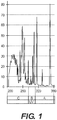
- Figure 1 shows a graph of an exemplary spectrum of UV wavelengths generated by a typical mercury UV lamp.
- the limited penetrating ability of approximately 200 nm UV radiation can be used for killing bacteria or viruses, as shown in the exemplary diagram of Figure 2 , because bacteria or viruses are typically physically far smaller than human cells.
- a typical bacterial cell is less than about 1 micrometer (" ⁇ m") in diameter, and a typical virus can vary from about 20nm to about 400nm, whereas human cells are typically about 10 to 30 ⁇ m across, depending on their type and location.
- the exemplary system, method and computer-accessible medium can be used to minimize airborne and surface-based transmissions of common viruses, such as H1N1, SARS-CoV and MERS-CoV, and of extremely dangerous viruses, including Dengue and Ebola, without harming human cells.
- common viruses such as H1N1, SARS-CoV and MERS-CoV
- extremely dangerous viruses including Dengue and Ebola
- Figure 2 shows a diagram of a typical human cell nucleus having a spherical geometry 202 or a flattened geometry 204, illustrating the penetration into a human cell of UV radiation with a wavelength of around 200 nm.
- effectively no UV radiation of this wavelength preferably reaches the cell nucleus 202 and 204, which contains the radiation-sensitive DNA.
- UV radiation of this wavelength would typically not be harmful to human cells or to humans.
- there can be a biological reason why UV with a wavelength around 200 nm will typically not be harmful to humans.
- UV radiation can be very efficiently absorbed by oxygen, producing ozone and oxidative damage.
- UV radiation can be very efficient at producing oxidative DNA base damage.
- a 200 nm wavelength UV radiation can be in a narrow UV "safety window".
- viruses are typically physically much smaller in size than human cells, UV radiation with a wavelength around 200 nm can penetrate through, and therefore kill, viruses.
- UV excilamps or one or more UV lasers or other coherent light sources, which, in contrast to standard UV lamps, can produce UV radiation at a specific wavelength -for example, around 200 nm.
- UV radiation around such exemplary wavelength e.g ., a single wavelength or in a range of certain wavelengths as described herein
- the exemplary excilamp can utilize certain exemplary concepts which were developed at the Institute of High Current Electronics ("IHCE"). ( See, e.g., Reference 11). Additional exemplary excilamps that can be utilized with the exemplary embodiments of the present disclosure may be available from Heraeus Noblelight in Germany.
- a filter 304 can be used to filter any UV radiation emitted from a side of lamp 302.
- exemplary embodiments of the present disclosure can use, for example, a krypton-bromine lamp (e.g., an excilamp), which can produce UV radiation at about 207 nm, or a krypton-chlorine lamp ( see, e.g., Figure 3 ), which can produce UV radiation at about 222 nm.
- a krypton-bromine lamp e.g., an excilamp
- a krypton-chlorine lamp see, e.g., Figure 3
- the exemplary spectra of these lamps are shown in the graph of Figure 4 .
- a spectral distribution 402 was produced by a krypton-bromine lamp
- spectral distribution 404 was produced by a krypton-chlorine lamp.
- certain exemplary features can be included (e.g., spectrum filtering elements such as multilayer dielectric filters or chemical filters) to remove unwanted wavelengths, or those wavelengths that can be outside of the preferable range of wavelengths.
- spectrum filtering elements such as multilayer dielectric filters or chemical filters
- absorption and/or reflective elements can be provided between the lamp and the irradiated surface to filter unwanted wavelengths, such as, for example, a band-pass filter, a long-wavelength blocking filter.
- the absorptive material can be fluorescent, such that it emits visible light when it absorbs UV radiation to provide an indication that the lamp is operating.
- other gases can be added to the lamp to suppress unwanted wavelengths. For example, adding argon to the krypton-bromine lamp can suppress generation of the 228 nm UV radiation.
- the typical power density output of the air-cooled excilamps can be about 7.5 to about 20 mW/cm 2 , although higher power density can be obtained in a water-cooled system. At about 20 mW/cm 2 , only a few seconds of exposure, or even only 1 second of exposure, can deliver about 20 mJ/cm 2 , which can be a typical bactericidal dose.
- Exemplary embodiments of the present disclosure can provide an excilamp, emitting about a 207 nm or about a 222 nm single wavelength UV radiation, to differentially kill bacteria while sparing adjacent human cells.
- the wavelength(s) of the UV radiation can be in the range of about 190 nm to about 230 nm, or in the range of about 200 nm to about 230 nm.
- Exemplary experiments implementing embodiments of the present disclosure can include: an in-vitro ( e.g., laboratory) 3-D human skin system ( see, e.g., References 49 and 98), a nude mouse model for in-vivo safety standards, and/or an in-vitro wound infection model. ( See, e.g., Reference 99).
- an exemplary test bench was developed for gathering, for example, exemplary preliminary sterilization results from exemplary UV radiation sources.
- the exemplary test bench can include: (i) a light-tight box, (ii) a shutter control, (iii) a filter holder and (iv) adjustable exposure parameters for time, distance and wavelength (e.g., 207 nm KrBr excilamp, 222 nm, KrCl excilamp, and 254 nm standard germicidal lamp).
- exemplary custom filters can be designed to eliminate higher-wavelength components in the excilamp emission spectra to provide optimal single-wavelength exposure.
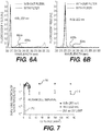
- a UV spectrometer and deuterium lamp can be used to validate the filter effectiveness, as shown, for example, the graphs shown in Figures 6A and 6B , which illustrate the normalized spectra comparing excilamp emission (e.g., elements 602a and 602b) with filtered excilamp emission (e.g., elements 604a and 604b) for both KrBr and KrCl excilamps.
- This exemplary test bench facilitated, for example, a generation of biological findings of filtered excilamp exposure to both bacteria and healthy human cells, which are described below.
- the exemplary biological testing experience has provided details regarding exemplary parameters for developing filtered KrBr and KrCl excilamps into optimal devices for clinical applications.
- the exemplary experiments investigated, for example, whether UV radiation from exemplary filtered excilamps can be effective at killing bacteria while sparing normal human cells.
- human fibroblasts were, for example, exposed to about 3 mJ/cm 2 from a standard germicidal UV lamp (e.g ., about 254 nm), and their survival was less than about 10 -4 .
- a standard germicidal UV lamp e.g ., about 254 nm
- their survival was less than about 10 -4 .
- fluences as high as 150 mJ/cm 2 from the exemplary filtered KrBr or KrCl excilamp ( e.g. , about 207 and about 222 nm, respectively)
- their survival was in the range from about 1 to about 10 -1 . ( See, e.g., graph shown in Figure 7 ).
- Figure 7 shows an exemplary graph indicating a clonogenic survival of normal human skin fibroblasts (e.g ., AG1522) exposed to UV radiation from exemplary filtered KrBr ( e.g., about 207 nm, element 705) or KrCl ( e.g., about 222 nm, element 710) excilamps, or from a conventional germicidal lamp ( e.g ., about 254 nm, element 715).
- KrBr e.g., about 207 nm, element 705
- KrCl e.g., about 222 nm, element 710
- MRSA methicillin resistant Staphylococcus aureus
- MRSA can be the cause of about 25% of surgical site infection, and can be associated with approximately 20,000 deaths per year in the United States; mostly healthcare related.
- MRSA and antibiotic-susceptible S. aureus are typically equally susceptible to UV radiation from conventional germicidal lamps. ( See, e.g., Reference 2).
- the exemplary results are shown, for example, in the chart of Figure 8 , which shows that at an excilamp fluence of about 100 mJ/cm 2 , a MRSA survival level of 10 -4 can be achieved.
- Figure 8 shows an exemplary graph of MRSA (e.g ., strain US300) inactivation after exposure to UV radiation from the exemplary filtered KrBr excilamp ( e.g ., about 207nm, element 805) or a KrCl excilamp ( e.g ., about 222 nm, element 810).
- MRSA e.g ., strain US300
- the exemplary filtered excilamp UV radiation at about 207 nm and at about 222 nm can differentially effect and/or kill MRSA relative to the human cells.
- the survival level of human cells can be, for example, in the range of about 0.1 to 1, while the survival level of MRSA can be in the range of about 10 -4 .
- Such exemplary findings are in considerable contrast to the situation for convention germicidal UV lamps ("GUVL”), which can be roughly equally efficient at killing bacteria and human cells.
- GUIL germicidal UV lamps
- the human cell survival from the GUVL can be about 0.3 x 10 -4 , a human cell survival advantage of 0.3.
- the human cell survival by the exemplary filtered excilamps can be in the range of about 0.1 to 1, a human cell survival advantage in the range of 5,000.
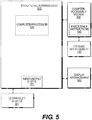
- FIG. 5 shows an exemplary block diagram of an exemplary embodiment of a system according to the present disclosure.
- exemplary procedures in accordance with the present disclosure described herein can be performed by or controlled using a UV generation source 580 and/or hardware processing arrangement and/or a computing arrangement 510, separately and in conjunction with one another.
- exemplary processing/computing arrangement 510 can be, for example, entirely or a part of, or include, but not limited to, a computer/processor 520 that can include, for example, one or more microprocessors, and use instructions stored on a computer-accessible medium (e.g ., RAM, ROM, hard drive, or other storage device).
- a computer-accessible medium e.g ., RAM, ROM, hard drive, or other storage device.
- a computer-accessible medium 530 (e.g., as described herein above, a storage device such as a hard disk, floppy disk, memory stick, CD-ROM, RAM, ROM, etc., or a collection thereof) can be provided ( e.g., in communication with the processing arrangement 510).
- the computer-accessible medium 530 can contain executable instructions 540 thereon.
- a storage arrangement 550 can be provided separately from the computer-accessible medium 530, which can provide the instructions to the processing arrangement 510 so as to configure the processing arrangement to execute certain exemplary procedures, processes and methods, as described herein above, for example.
- the exemplary processing arrangement 510 can be provided with or include an input/output arrangement 570, which can include, for example, a wired network, a wireless network, the internet, an intranet, a data collection probe, a sensor, etc.
- the exemplary processing arrangement 510 can be in communication with an exemplary display arrangement 560, which, according to certain exemplary embodiments of the present disclosure, can be a touch-screen configured for inputting information to the processing arrangement in addition to outputting information from the processing arrangement, for example.
- the exemplary display 560 and/or a storage arrangement 550 can be used to display and/or store data in a user-accessible format and/or user-readable format.
- SKH-1 hairless mice and pigs can be exposed to this UV wavelength radiation, and a variety of biological damage endpoints can be assessed.
- Positive control can be the same-dose exposure as a conventional 254 nm germicidal UV lamp.
- Negative controls can receive no UV radiation exposure.
- the endpoints can be physiological endpoints ( e.g ., skin edema and erythema), epidermal immunohistochemical and molecular endpoints, as well as cataractogenesis.
- the efficacy of 207 nm radiation can be assessed with the goal of using it to prevent SSI by continuous exposure of the wound during surgery.
- a liquid suspension containing live MRSA can be applied to the skin on the backs of SKH-1 hairless mice and of pigs, followed by wound induction and suturing.
- One set of wound sites can be treated with topical antibiotics (e.g., positive control), another set can remain untreated ( e.g., negative control), and a third set can be exposed to 207 nm radiation.
- Staged inspections of wounds for infection can be undertaken using objective wound assessment criteria.
- Far-UVC light e.g., light of about 190nm to about 230nm, or light of about 200 nm to about 230 nm
- UV radiation emitted in the same or similar range, by, for example, a KrCl excilamp ( e.g ., at about 222 nm), or a laser light source ( e.g., at about 222 nm), can be similarly effective at killing and/or damaging viruses.
- the antiviral efficacy of 222 nm UV radiation can be assessed as compared with 254 nm UV radiation from a conventional germicidal lamp, for the H1N1 influenza virus.
- Fluence-dependent virus inactivation determinations can be done for influenza viruses, Ebola, SARS and/or MERS on surfaces, such as fomite surfaces, which are surfaces capable of carrying infectious organisms, and subsequently for influenza virus in aerosols, using the exemplary bench top aerosol UV irradiation chamber.
- UV radiation is a well-established highly-efficient anti-microbial modality, effective both against bacteria and viruses.
- UV sterilization it is generally not practical to use UV sterilization in scenarios where people can be present, because it can be a human health hazard, being both carcinogenic and cataractogenic.
- the exemplary system, method and computer-accessible medium can include a far- UVC light source (e.g., at approximately 207 nm) from, for example a KrBr excimer lamp, a laser light source or a coherent light source (e.g., a light source having the same phase, the same polarization and/or the same direction), that has the anti-microbial advantages - both for bacteria and viruses - compared to conventional UV germicidal lamps, and without the corresponding human safety hazards.
- a far- UVC light source e.g., at approximately 207 nm
- a KrBr excimer lamp e.g., a laser light source or a coherent light source (e.g., a light source having the same phase, the same polarization and/or the same direction)
- a coherent light source e.g., a light source having the same phase, the same polarization and/or the same direction
- UV-mediated bacterial killing can be independent of drug resistance (see, e.g., References 2 and 3), using the exemplary system, method, and computer-accessible medium, according to an exemplary embodiment of the present disclosure.
- UV radiation at a wavelength of around 200 nm can be very strongly absorbed by proteins (e.g ., particularly through the peptide bond) and other biomolecules ( see, e.g., References 4 and 5), so its ability to penetrate biological material can be very limited.
- proteins e.g ., particularly through the peptide bond
- other biomolecules see, e.g., References 4 and 5
- the intensity of about 207-nm UV radiation can be reduced by half in only about 0.3 ⁇ m of tissue, compared with about 3 ⁇ m at 250 nm and much longer distances for higher UV radiation wavelengths.
- This phenomenon is shown in the graph of Figure 9 .
- about 207 nm UV radiation may only be minimally absorbed in pure water. ( See, e.g., Reference 8).
- the very short range in biological material of about 207-nm UV radiation means that, while it can penetrate and kill both bacteria and viruses on surfaces or in aerosols (e.g ., bacteria, viruses, and aerosols are all typically less than 1 ⁇ m in diameter), it cannot penetrate through either the human stratum corneum (e.g ., the outer dead-cell skin layer, which in humans can be 5 -20 ⁇ m thick ( see, e.g., Reference 9)), nor the ocular cornea, nor even the cytoplasm of individual human cells ( e.g., most human cells range in diameter from 10-25 ⁇ m ( see, e.g., Reference 10), and the layer thickness of the cytoplasm about the nucleus can be between about 1 micron to about 4.5 microns as shown in Figure 2 ).
- the human stratum corneum e.g ., the outer dead-cell skin layer, which in humans can be 5 -20 ⁇ m thick ( see, e.g., Reference 9)
- the ocular cornea nor
- the exemplary system, method and computer-accessible medium can use excimer lamps, often called excilamps that primarily emit a single UV wavelength from a specific excited molecule complex.
- Excilamps that emit in the wavelength region of interest have recently become commercially available, and contain, for instance, a krypton-bromine mixture, which can produce high- intensity far-UVC radiation at about 207 nm.
- Excilamps can be small, rugged, inexpensive, sufficiently intense, and long- lived ( e.g., approximately 10,000 hours).
- These excilamps can also emit a low level of higher wavelength UV radiation, which would be unacceptable for the exemplary applications.
- UV radiation filters e.g., bandpass filters
- UV radiation filters e.g., bandpass filters
- SSI Between about 0.5% and about 10% of all clean surgeries in the United States, corresponding to about 275,000 patients per year, can result in SSI.
- Patients who develop SSI can be about 60% more likely to spend time in an ICU, can be 5 times as likely to be readmitted, have a mortality rate twice that of non-infected patients, have an average of 7 days additional length of hospital stay ( see, e.g., Reference 18), and have roughly double the total healthcare costs compared with patients without SSI. ( See, e.g., Reference 19).
- the annual number of deaths in the United States attributed to SSI has been estimated at about 8,200 ( see, e.g., Reference 16), with annual patient hospital costs between about $3 billion and about $10 billion. ( See, e.g., Reference 20).
- 207 nm UV radiation can be as efficient as conventional germicidal lamps for inactivating MRSA, but can be far safer in regard to human exposure.
- a continuous low- fluence-rate exposure of about 207-nm UV radiation onto the surgical wound area during the entire surgical procedure can be a safe approach to killing bacteria, as they are alighted onto the wound area, and before they penetrated into the interior of the wound - again potentially with no adverse effects on patient or staff.
- UV radiation directly addresses the issue of drug resistance because UV radiation can generally be equi-effective at inactivating drug- resistant bacteria compared with wild-type strains ( see, e.g., References 2 and 3) - and in fact all exemplary studies have been performed with MRSA - a drug resistant bacterial strain.
- 207-nm excimer lamps in a surgical setting can be used.
- the exemplary system, method and computer-accessible medium, according to an exemplary embodiment of the present disclosure can be incorporated into a standard overhead surgical illumination system.
- a possible second UV radiation source, to ensure a level of redundancy from inadvertent shielding, can be incorporated into a surgeon's headlight illumination system, with the UV radiation transmitted to the headlight via fiber optics. ( See, e.g., Reference 28).
- UVGI germicidal UV lamp located in the upper part of the room - upper-room UV germicidal irradiation
- louvers facilitate the UVGI systems to meet the recommended limits for germicidal UV exposure, they achieve this by blocking more than 95% of the UV radiation exiting the UVGI fixture, resulting in decreased effectiveness.
- a further consideration includes reports of accidental germicidal UVC exposure after incorrect UVGI usage. ( See, e.g., Reference 37).
- the exemplary system, method and computer-accessible medium can replace standard germicidal lamps within UVGI fixtures.
- about 222 nm UV radiation can be at least as effective, and potentially more effective, at killing influenza virus, as compared with conventional germicidal UV lamps, but it would not be subject to the human safety concerns. This would open up widespread use of the exemplary system, method and computer-accessible medium in hospitals and communal settings.
- the exemplary antimicrobial system/method can use, for example, on about 207 nm or on about 222 nm single-wavelength UVC radiation ( e.g ., or any source with the wavelengths in between) which can kill bacteria or viruses without damaging mammalian cells or tissues. ( See, e.g., Reference 1).
- the exemplary system, method and computer-accessible medium can differ significantly from using conventional mercury-based germicidal lamps which emit a dominant bactericidal wavelength at about 254 nm, and which can be hazardous to humans. ( See, e.g., References 26 and 41-44).
- the exemplary system, method and computer-accessible medium can include the production of UV wavelengths around 207 nm or around 222 nm, which can be equally toxic to bacteria/viruses as compared with conventional UV germicidal lamps, but can be far safer in terms of human exposure.
- Excimer lamps e.g ., excilamps
- can be an efficient source of near monoenergetic UV radiation see, e.g., Reference 11
- an appropriate gas mixture e.g., in the exemplary case Kr + Br
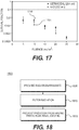
- Figure 10 shows measured spectra emitted from the exemplary KrBr excilamp. ( See, e.g., Reference 1).
- Excilamps can emit significant fluences of higher wavelength light ( see, e.g., graph shown in Figure 10 ), and these high-wavelengths can be more penetrating, which can result in significant biological damage.
- a customized bandpass filter can be used to remove all but the dominant wavelength emission ( see, e.g., Figure 10 inset graph).
- the filtered excilamp, and a shutter can be integrated into a portable apparatus with a user-friendly stand. All the exemplary studies reported here were performed with these exemplary filtered excilamps.
- a typical geometry can produce a uniform power density within about a 580 mm-diameter circular field at about 1-m lamp distance, with a power density of about 0.1 mW/cm 2 .
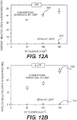
- the exemplary experiments were performed in-vitro using human skin AG1522 fibroblasts, and with MRSA bacteria. Both the fibroblasts and the MRSA were irradiated unshielded on a surface. Cell survival was compared with a conventional germicidal lamp vs. the exemplary filtered 207 nm lamp. As shown in the graphs of Figures 11A and 11B , a 207 nm UV exposure produces far less cell killing in human cells than a conventional germicidal lamp (e.g., Figure 11A , curve 1105 illustrating the MRSA kills of a germicidal lamp and curve 1110 illustrating the human cell kills for the germicidal lamp).
- 207-nm UV radiation kills MRSA almost as efficiently as a conventional germicidal lamp (e.g., Figure 11B , curve 1115 illustrating MRSA kills for the exemplary system, method and computer-accessible medium and curve 1120 illustrating human cell kills for the exemplary system, method and computer-accessible medium).
- a conventional germicidal lamp e.g., Figure 11B , curve 1115 illustrating MRSA kills for the exemplary system, method and computer-accessible medium and curve 1120 illustrating human cell kills for the exemplary system, method and computer-accessible medium.
- the exemplary system, method, and computer-accessible medium produces about 1,000-fold less killing in human cells compared to a conventional germicidal lamp. ( See, e.g., Reference 1).
- the exemplary on-surface in-vitro safety studies were extended by using a full 3-D human skin model (e.g ., EpiDerm, MatTek Corp), which recapitulates the human stratum corneum, epidermis and dermis).
- the skin model was irradiated from the top with UV radiation from a standard germicidal lamp and with about a 207 nm UV-wavelength.
- Immunohistological performed assays for common premutagenic skin photoproducts associated with UV exposure e.g ., cyclobutane pyrimidine dimers ("CPD") and pyrimidine-pyrimidone 6-4 photoproducts (e.g ., 6-4 PP).
- CPD cyclobutane pyrimidine dimers
- pyrimidine-pyrimidone 6-4 photoproducts e.g ., 6-4 PP
- the results are shown in the graph of Figure 4 .
- 207 nm UV radiation produced essentially none of the photoproducts which
- the typical thickness of the SKH-1 hairless mouse stratum corneum can be about 5 ⁇ m. ( See, e.g., Reference 50). Thus, it can be a useful conservative model for human skin, which has a typical range of stratum corneum thicknesses from about 5 to 20 ⁇ m. ( See, e.g., Reference 9).
- mice were sacrificed, and dorsal skin sections were prepared for analysis.
- the epidermal thickness was assessed, as well as induction of cyclobutane pyrimidine dimers ("CPD”), and induction of pyrimidine-pyrimidone 6-4 photoproducts ( e.g ., 6-4 PP).
- CPD cyclobutane pyrimidine dimers
- pyrimidine-pyrimidone 6-4 photoproducts e.g ., 6-4 PP
- Figure 12A shows an exemplary graph illustrating the effect of a conventional germicidal UV lamp 1205 and an exemplary filtered 207 nm UV lamp 1210 on the production of cyclobutane pyrimidine dimer in human skin model.
- Figure 12B shows an exemplary graph illustrating the effect of a conventional germicidal UV lamp 1205 and an exemplary filtered 207 nm UV lamp 1210 on the production of pyrimidine-pyrimidone 6-4 photoproducts ( e.g ., 6-4 PP) in human skin model.
- pyrimidine-pyrimidone 6-4 photoproducts e.g ., 6-4 PP
- Figure 13A ( e.g., top row) shows cross-sectional images of H&E stained skin samples from the three mouse groups.
- the epidermal layer thickness of the dorsal skin of the mice exposed to 150 mJ/cm 2 UV generated by the about 207 nm or the about 222 nm lamp was not statistically different from controls.
- the same fluence generated by the about 254 nm conventional germicidal lamp resulted in a 2.7 ⁇ 0.4 fold increase in epidermal thickness.
- Figure 13A shows typical cross-sectional images of skin samples from the three groups comparing pre-mutagenic photoproduct lesions CPD (e.g., middle row, dark stained cells) and 6-4 PP ( e.g., bottom row, dark stained cells).
- CPD e.g., middle row, dark stained cells
- 6-4 PP e.g., bottom row, dark stained cells
- Figure 13B shows an exemplary chart illustrating a percent of epidermal cells with premutagenic lesions for particular UV wavelengths for CPD (e.g., Sham 1305, 254nm 1310 and 207nm 1315) and 6-4PP (e.g., Sham 1320, 254nm 1325 and 207nm 1330).
- CPD e.g., Sham 1305, 254nm 1310 and 207nm 1315)
- 6-4PP e.g., Sham 1320, 254nm 1325 and 207nm 1330.
- MRSA was spread over the appropriate dorsal area of three pigs at different MRSA concentrations (e.g ., 105 - 107 cfu/ml). 12 superficial wounds were generated at each concentration, and the animals were observed for 7 days, with the goal of finding the minimum MRSA concentration to produce a 90% wound infection rate. Biopsy samples were also taken from all wounds at 7 days to confirm the source of the infection, and assayed using serial dilution.
- S
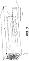
- a bench top aerosol exposure chamber was designed and constructed, which is shown in the image in Figure 14 .
- the Aerosol Generation Module 1405 has saturated/desiccated air and collision-nebulizer inputs, and has a series of internal baffles for droplet distribution. Temperature and humidity meters can monitor the conditions in the aerosol generation chamber, after which the aerosols can be flowed through the UV-exposure module which has a 300 x 275 mm silica Quartz Window 1410 on the UV irradiator side, and a quartz port on the far side to monitor UV irradiance with a UVC radiation meter.
- the aerosols can be in the UV field for times depending on the flow rate, which in turn can determine the UV radiation dose, and which can also be adjusted by moving the lamp nearer or further from the window.
- a particle sizer can measure size distribution of the aerosols in the UV irradiation volume.
- the aerosols can be drawn through output ports to two BioSamplers in the Sampling Module 1415.
- Mice 1505 can be placed individually in Compartments 1510 with size 60 mm ("W"), 125 mm ("L”) and 80 mm ("H") in specially-designed mouse-irradiation boxes, where the Mice 1505 can be housed before ( e.g., 48 hours acclimatization time), during, and after UV exposures, while being given water and Purina Laboratory Chow 5001 diet ad libitum.
- a metal-mesh top on the mouse-irradiation box can facilitate UV radiation transmission from the exemplary 207-nm KrBr excilamp or from a 254-nm germicidal lamp.
- Various conditions can include, for example: (i) sham exposure, (ii) 207-nm KrBr lamp at either 50 or 150 mJ/cm 2 , and (iii) germicidal UV lamp at either 50 or 150 mJ/cm 2 .
- a 207-nm UV excilamp emission characteristics in-situ can be measured prior to mouse exposures using a UVtechnik Micro Puck UV dosimeter and a Photon Control UVC spectrometer.
- a total of 245 SKH-1 hairless mice (e.g., 7 weeks old; strain code: 477; Charles River Labs) can be used to determine the effects from long-term UV radiation exposures.
- the total number of mice can be separated into two groups for 8 hours/day irradiations during 1 day and 1 month.
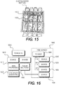
- a 70 mice per exposure-duration group can be used (e.g., element 1605), with 105 additional mice ( e.g., 1640) used for a time series ( e.g., 1660) of immunohistochemical and molecular endpoints following the 1-day exposure
- Each group of 70 mice can be divided into two subgroups where 35 mice ( e.g., 1625) can be harvested for assays (e.g., Tissue Section 1660) following the exposures and the other 35 mice ( e.g., 1610) can undergo live assays of skin properties ( e.g., 1615) and then can be maintained for an additional 9 months for eye studies ( e.g., 1620).
- Each group of 35 mice can contain mice representing each of the exemplary exposure conditions: (i) sham exposure, (ii) 207-nm KrBr lamp at either 50 or 150 mJ/cm 2 and (iii) germicidal UV lamp at either 50 or 150 mJ/cm 2 .
- the exemplary porcine model can be exposed to various exposure conditions, which can include, for example: (i) sham exposure, (ii) 207-nm KrBr lamp at either 50 or 150 mJ/cm 2 and, (iii) germicidal UV lamp at either 50 or 150 mJ/cm 2 .
- UV radiation exposure times can be in the range of about 20 minutes to 1 hour.
- Two pigs, one male and one female, can be used and all exposure conditions can be delivered to each animal, because the dorsal surface area on a pig can be sufficient for multiple acute exposure conditions and can provide multiple tissue samples. Each pig can be anesthetized during the exposures and each exposure condition can be delivered to a predetermined dorsal region of the animal. Following the acute exposures, skin properties can be recorded and tissue samples can be collected via skin punch for biological assays at 0, 24, 48 and 72 hours, to parallel the mouse skin irradiation experiment.
- the exemplary live assays can focus on skin properties. ( See, e.g., References 58 and 59). Skin erythema can be assessed by comparing skin redness measurements ( see, e.g., Reference 60) before and post-irradiation using an exemplary Konica-Minolta handheld colorimeter (e.g ., Chroma Meter CR-410T) currently being used to quantitate human skin erythema in radiotherapy patients. Skin trans-epidermal water loss can also be measured using a ServoMed Evaporimeter EP-2. All mice used in the live assays can be maintained for an additional 9 months for the exemplary mouse eye studies described below.
- Skin erythema can be assessed by comparing skin redness measurements ( see, e.g., Reference 60) before and post-irradiation using an exemplary Konica-Minolta handheld colorimeter (e.g ., Chroma Meter CR-410T) currently being used to quantitate human skin erythem
- Dorsal skin tissue sections were harvested from sacrificed mice and from live pig for skin imaging, immunohistochemical and molecular endpoints.
- tissue-section assays were harvested from mice and from pig skin punches. Confocal and multi-photon microscopy procedures were used along with advanced image analysis techniques (e.g ., Velocity, Metamorph), available at the Advanced Imaging Core in the Skin Disease Research Center at Columbia University, to examine microscopic features of the fixed skin and to measure skin-layer thicknesses.
- the exemplary skin-imaging endpoint assays were applied on tissue sections harvested from mice immediately following the exposures.
- the mice were sacrificed at 72 hours, a time point reported for maximal edema following UVB exposure. ( See, e.g., Reference 58).
- tissue sections were assayed using the Tissue Culture & Histology Core at the Skin Disease Research Center, Columbia University Medical Center.
- a time series e.g., 1660 was used following the exemplary 1-day (e.g., 8-h) exposure with samples harvested at 0 hours ( e.g., 1645), 24 hours ( e.g., 1650), 48 hours ( e.g., 1655) and 72 hours ( e.g., 1635) (note: 72-h samples acquired from mice used in the skin-imaging endpoint - see above).
- fixed skin tissue sections were stained with hematoxylin and eosin for histological analysis.
- DNA photodamage were detected by immunohistochemical analysis of cyclobutane pyrimidine dimers and 6, 4-photoproducts in fixed tissues. These procedures illustrate inflammatory cell infiltration of either lymphoid or myeloid origin. Inflammatory responses were further investigated using markers for mast cells and macrophages. Similarly, apoptosis were assessed with the TUNEL assay and/or the immunohistochemistry-based Caspase-3 activation analysis. Alteration in cutaneous vasculature as well as early onset of fibrosis was investigated. UV-induced alterations in cutaneous vasculature were examined through endothelial markers such as CD 31, whereas markers for collagen and elastin fibers formation were used to observe potential early onset of fibrosis.
- Ultraviolet irradiation of the eye can be associated with a variety of ocular disorders including eyelid and conjunctival abnormalities, corneal pathologies and cataract.
- the severity and type of pathology can be related to both dose and UV wavelength.
- Pathologies can arise from direct action of UV radiation, for example, cyclobutane-pyrimidine dimer formation leading to mutagenesis (see, e.g., References 5, 65 and 66), or indirectly by free radical mediated photochemical interactions with intraocular fluids and sub-cellular components. ( See, e.g., References 67-69).
- endpoints can be measured morphologically by weekly slit lamp examination of the anterior segment. While it can be unlikely that UV radiation of this wavelength can result in cataract due to the approximately 295 nm UV cutoff of the cornea ( see, e.g., References 72 and 73), to rule out UV-induced lens changes, dilated slit lamp exams can be performed periodically. Potential corneal, conjunctival and lens changes can be scored as to severity using generally accepted subjective criteria ( see, e.g., References 64, 74 and 75) and slit lamp photodocumentation.
- UV-induced anterior segment changes can also be analyzed histologically in selected animals sacrificed at weekly intervals following irradiation. Paraffin fixed and stained horizontal sections of the eye and orbit can be prepared and analyzed for abnormalities. ( See, e.g ., References 64, 76 and 77).
- VOS Virtual Optomotor System
- Acuity can be reliably quantitated by varying the spatial frequency of a displayed variable vertical sine wave grating until an optomotor (e.g., head turning) response was no longer elicited by the subject animal.
- the advantage of this approach can be that it can permit direct measurement of visual function rather than more subjective estimates of the effect of ocular changes on acuity. In all cases, data can be analyzed to determine baseline increases in prevalence, incidence or rate of progression of irradiation specific ocular pathologies.
- ANOVA Analysis of variance
- the proposed sample size of 140 mice divided into 5 groups, with repeated measurements at 4 time points, can warrant the use of multivariate analysis of variance ("MANOVA").
- MANOVA multivariate analysis of variance
- a liquid suspension containing live MRSA was applied to the skin on the back of the SKH-1 hairless mouse and the pigs, followed by wound induction and suturing.
- One set of wounds was treated with topical antibiotics (e.g ., positive control), another set was untreated (e.g ., negative control), and a third set was exposed to 207 nm radiation. Staged inspections of wounds for infection were undertaken using objective wound assessment criteria.
- 207 nm radiation can be effective in killing bacteria while potentially being much safer than conventional germicidal lamps for human exposure.
- the exemplary application to minimizing SSI rates can involve 207-nm irradiation of the wound during surgery to inactivate airborne bacteria as they can alight onto the wound.
- the exemplary proposed in-vivo studies using 207 nm irradiation of hairless mouse and pig models can be designed to provide a first assessment of the efficacy of the exemplary system, method and computer-accessible medium for killing bacteria and/or viruses, which can be introduced onto the skin surface, in the context of surgical wounds.
- the exemplary endpoints can be prevention of wound infections.
- mice SKH-1 hairless mice were anesthetized with isoflurane, and after cleaning with alcohol and povidone-iodine solution, a 20 mm x 20 mm area were marked on the dorsal skin. The MRSA solution at the previously determined concentration was applied to the marked area and allowed to dry. A subset of the mice was exposed to the 207 nm lamp to a total fluence of 50 mJ/cm 2 or 150 mJ/cm 2 , with the remaining mice serving as positive controls. Mice not inoculated with MRSA, but treated with the lamp, were run in parallel to serve as negative controls. A 10 mm incision was made within the 20 mm x 20 mm region of each mouse through the skin and epidermis. Incisions were closed using wound clips.
- mice and thus 72 wounds, were used, the same as planned for the pig skin studies described below (e.g., 24 mice receiving either 50 or 150 mJ/cm 2 ; 24 mice receiving topical antibiotic; 24 mice receiving no treatment). Power calculations are described below.
- mice were housed individually in custom designed boxes ( see, e.g., Figure 7 ) and were monitored daily for infection of the wound ( e.g ., as seen by erythema and purulent drainage) for up to 7 days.
- Mice with infected wounds were immediately euthanized and the wounds processed, as described below.
- mice were euthanized with CO 2 and cervical dislocation.
- Infected wounds were further assayed for inflammation and bacteria culture.
- a 2 mm punch biopsy section of each wound was processed for bacterial culture while the remainder was fixed in 10% NBS for assessed of inflammation. The degree of inflammation was graded on a scale of 0 to 3 ( see, e.g., Reference 81), while biopsy sections were assayed for bacteria titers using the CFU assay.
- Pig skin offers an excellent model from the perspective of dermatology and wound investigation. ( See, e.g., Reference 56).
- a multitude of morphologic, anatomic, immunohistochemical, dermatologic and pharmacologic studies have demonstrated that pig skin has important similarities in morphology, cellular composition and immunoreactivity to human skin. ( See, e.g., References 82 and 85).
- Phase I was the design optimization phase, to assess the appropriate concentration of MRSA for the exemplary Phase II studies, and to optimize the exemplary assay protocols.
- Phase I studies which do not involve UV irradiation, after the wound infection procedure, tissue biopsy samples were suspended in trypticase soy broth, sonicated, and serially diluted on TSA plates. MRSA colony counts were determined after incubating the plates at 37°C for 36 hours.
- the exemplary studies can use pathogen free domestic pigs weighing approximately 22-25 kg and involved, for example:
- the MRSA concentration was, for example, 10 7 cfu/ml.
- Each pig was optically masked to define the region were be exposed to 207-nm UV radiation. While each pig received only one single 207 nm fluence, the individual 207 nm fluences applied to all the pigs, and ranged from about 50 to about 150 mJ/cm 2 .
- Positive control wounds received povidone iodine (e.g., betadine), a well characterized bactericidal agent ( see, e.g., References 86 and 87), and negative controls involved neither UV radiation nor topical antimicrobial agent.
- a total of 12 wounds per pig, 60 mm long, and separated by a minimum of 25 mm were used.
- 4 wounds were in the 207-nm UV-irradiated region, 4 wounds were subject to the topical antimicrobial agent and 4 wounds acted as controls.
- the wounds were closed using absorbable sutures and then individually covered with Dermabond. Wounds were closed using 4-0 Biosyn.
- Every wound was visually monitored daily for signs of infection. If an individual wound was visually determined to be infected, the Dermabond was removed, the infected wound swabbed with a culture stick, and the sample transferred to standard tubed media for culture, after which Dermabond was reapplied to the wound. At the end of the 7 day observation period, the animals were humanely sacrificed and all wounds were both swabbed and biopsied to measure bacterial concentration.
- the exemplary studies utilized a frozen suspension of H1N1 influenza viruses (e.g ., A/PR/8/34 H1N1; ATCC VR-95, Manassas, VA).
- the virus suspension was thawed and divided into single-use aliquots to be refrozen and stored at -80°C until needed.
- An optimized plaque assay was used to measure virus titer before and after UV irradiation.
- the exemplary protocols were optimized for the surface based viral inactivation studies.
- 50 ⁇ L of influenza virus suspension was seeded from a titer of approximately 109 focus forming units / ml, onto 25 mm x 75 mm substrates that simulates typical surfaces found within workspaces and operating rooms: stainless steel, glass and plastics.
- the deposited liquid evaporated during a drying time of approximately 20 min, depending on ambient conditions.
- the exemplary control-seeded substrates were continually exposed to ambient conditions inside a biological safety cabinet.
- the remaining seeded substrates (e.g ., along with a clean substrate used as a negative control) were placed in the exemplary 207 nm or 222 nm exposure chamber, designed and built in-house at the exemplary instrument shop. All virus work was performed in a BSL-2 biological safety cabinet.
- Seeded substrates were divided into two sets for UV radiation treatment; one set was treated using a KrBr excilamp (e.g ., 207 nm) and the other set received comparable exposures using conventional germicidal lamps to provide positive controls. Groups of 3 seeded substrates were removed from the exposure chamber after each 207 nm exposure. Exposure doses ranged from between about 50 and about 150 mJ/cm 2 . The control-seeded substrates were sham irradiated. Immediately after the last 3 seeded substrates were removed from the exposure chamber; each seeded substrate was washed with DPBS++ ( e.g., DPBS [IX] + Mg++ and Ca++) using the following procedure.
- DPBS++ e.g., DPBS [IX] + Mg++ and Ca++
- a clearly-marked portion of the seeded substrate where viruses was deposited can be washed 10 times with a single 900- ⁇ L volume of DPBS++, using a pipette to remove all residue.
- the virus plaque assay was performed on the DPBS++ volume that was used to wash each seeded substrate.
- Viral infectivity was assessed with the standard plaque assay (see, e.g., Reference 91), which was optimized.
- influenza A virus e.g., H1N1; A/PR/8/34
- confluent Madin-Darby Canine Kidney Epithelial cells e.g., MDCK; ATCC CCL-314
- the virus was aspirated and the cells overlaid with 0.6% Avicel ( see, e.g., Reference 92) in medium ( e.g ., 2x MEM/BSA) containing TPCK-trypsin to final concentration of 2.0 ⁇ g/ml. Plates were incubated at 37°C for at least 3 days.
- Viral infectivity was expressed as plaque-forming units, PFU / ml.
- the aerosol studies were performed in a BSL-2 cabinet using the exemplary bench top UV aerosol exposure chamber. ( See, e.g., Figure 6 there).
- exemplary preliminary aerosol studies with the exemplary aerosol chamber 86.9% were sized between 0.3 and 0.5 ⁇ m, 10.9% between 0.5 and 0.7 ⁇ m, 1.9 % between 0.7 and 1.0 ⁇ m and 0.3% greater than 1 ⁇ m. These size distributions were changed by changing the input air/aerosol ratio.
- Influenza aerosols were generated by adding 0.075 ml of undiluted influenza virus and 75 ml of buffer (e.g ., Dulbecco's phosphate-buffered saline with calcium and magnesium containing 0.1% bovine serum albumin) into a high-output extended aerosol respiratory therapy ("HEART") nebulizer (e.g ., Westmed, Arlington, AZ) pressurized at 69 kPa. To achieve a particular relative humidity, the nebulizer output was mixed with proportions of dry and humidified air in a 7.5-liter chamber prior to delivery to the aerosol exposure chamber. Relative humidity (“RH”) and temperature in the chamber were measured prior to the exposure section using an Omega RH32 temperature and relative humidity meter (e.g., Omega Engineering Inc., Stamford, CT).
- RH relative humidity
- temperature in the chamber were measured prior to the exposure section using an Omega RH32 temperature and relative humidity meter (e.g., Omega Engineering Inc., Stamford, CT).
- the exemplary KrBr 207-nm excilamp exposures were used to deliver UV radiation sterilization treatments and a standard germicidal UV radiation lamp for positive controls.
- the UVC irradiance was monitored using a UVC radiation meter detecting UVC radiation transmitted through a fused quartz port opposing the UVC radiation entrance port at the exposure section of the chamber.
- Exposure doses ranged up to about 150 mJ/cm 2 , which spans dose levels used in previous studies using standard germicidal lamps to inactivate influenza virus. ( See, e.g., Reference 29).
- the UVC radiation dose was computed by multiplying the UVC irradiance by the exposure time.
- the exposure time was computed by dividing the volume of the chamber by the airflow rate. Based on the exemplary design dimensions and the exemplary planned airflow rate, an exposure time of approximately 8 seconds was expected.
- Air can be drawn through the chamber by a pump at 25 liters/minute through a manifold attached to 2 SKC Biosamplers (e.g ., SKC Inc., Eighty Four, PA), each operating at 12.5 liters/minute.
- SKC Biosamplers e.g ., SKC Inc., Eighty Four, PA
- Each Biosampler contains 20 ml of virus buffer (e.g ., Dulbecco's phosphate-buffered saline with calcium and magnesium containing 0.1% bovine serum albumin).
- a HEPA filter was placed after the samplers to remove fugitive aerosols before the airstream enters the pump. When sampling was not in progress, the aerosol-laden airstream running through the chamber were bypassed around the samplers, and the 25-liter/minute flow was directed to the HEPA filter.
- the nebulizer was run for about 20 minutes before sampling to ensure that concentrations within the chamber have stabilized.
- Samples were collected by passing the entire chamber airflow through the Biosamplers for a period of about 15 minutes.
- Sample sets were collected that consist of samples with: (i) the 207-nm KrBr excilamp on, (ii) the 254-nm germicidal lamp on ( e.g., for positive controls), and (iii) with the UV radiation off ( e.g., for negative controls).
- Triplicate sample sets were collected for combinations of the UVC radiation dose (e.g., ranging up to about 150 mJ/cm 2 ) and RH ( e.g., about 25%, about 50% and about 75%).
- BioSamplers were removed from the chamber, the volume of collection liquid was measured, and virus collection fluid was stored at 4°C for a maximum of 3 hours prior to performing the infectivity assay.
- the BioSamplers was decontaminated with about 10% bleach, rinsed with about 70% ethanol and dried before reusing.
- HBSS ++ Hank's Balanced Salt Solution with calcium and magnesium
- the exemplary results indicate that the exemplary KrCl excilamp having a wavelength of about 222 nm ( e.g., element 1705) can be as effective as a conventional germicidal UV lamp at 254 nm ( e.g., element 1710) for killing Influenza A virus ( e.g., H1N1; A/PR/8/34).
- FFU fluorescent focus units
- the KrCl excilamp at about 222 nm can be as effective as a conventional germicidal UV lamp at 254 nm, the KrCl lamp at 222 nm does not damage the surrounding tissue like the conventional germicidal UV lamp does.
- Figure 18 shows an exemplary flow diagram of an exemplary method for selectively killing or affecting a virus according to an exemplary embodiment of the present disclosure.
- radiation can be provided having one or more wavelengths that can be configured to selectively harm or damage the virus on a surface or in an aerosol.
- a particularly sized volume of air having the virus therein can be irradiated using a radiation having one or more wavelengths that can be in a range of between about 200 nm and about 230 nm.
- a filter can be provided such that, at procedure 1815, the radiation can be substantially prevented from having any wavelength that can be substantially harmful to cells of the body (e.g., a wavelength that is outside the range of between about 200 nm and about 230 nm).
- exemplary procedures described herein can be stored on any computer accessible medium, including a hard drive, RAM, ROM, removable disks, CD-ROM, memory sticks, etc., and executed by a processing arrangement and/or computing arrangement which can be and/or include a hardware processors, microprocessor, mini, macro, mainframe, etc., including a plurality and/or combination thereof.
- a processing arrangement and/or computing arrangement which can be and/or include a hardware processors, microprocessor, mini, macro, mainframe, etc., including a plurality and/or combination thereof.
- certain terms used in the present disclosure including the specification, drawings and claims thereof, can be used synonymously in certain instances, including, but not limited to, for example, data and information. It should be understood that, while these words, and/or other words that can be synonymous to one another, can be used synonymously herein, that there can be instances when such words can be intended to not be used synonymously.
- an apparatus for generating at least one radiation comprising:
- the at least one radiation is further configured to selectively affect or destroy at least one bacteria on the surface or in the aerosol.
- the radiation source includes an excilamp.
- the excilamp includes at least one of a krypton-bromine lamp or a krypton-chlorine lamp.
- the radiation source first arrangement is further configured to generate the at least one radiation having a single wavelength
- the at least one second arrangement is further configured to prevent the at least one radiation from having any wavelength other than the single wavelength
- the single wavelength is about 207 nm.
- the single wavelength is about 222 nm.
- the at least one second arrangement includes at least one of a chemical filter or a dielectric.
- the one or more wavelengths have a range of about 207 nm to about 222 nm.
- the one or more wavelengths have a range of about (i) 190-194 nm, (ii) 195-199 nm, (iii) 200-204 nm, (iv) 205-209 nm, (v) 210-214 nm, (vi) 215-218 nm, (vii) 219-223 nm, or (viii) 224-230 nm.
- the single wavelength is at least one of (i) about 201 nm, (ii) about 202 nm, (iii) about 203 nm, (iv) about 204 nm, (v) about 205 nm, (vi) about 206 nm, (vii) about 208 nm, (viii) about 209 nm, (ix) about 210 nm, (x) about 211 nm, (xi) about 212 nm, (xii) about 213 nm, or (xiii) about 214 nm.
- the single wavelength is at least one of (i) about 215 nm, (ii) about 216 nm, (iii) about 217 nm, (iv) about 218 nm, (v) about 219 nm, (vi) about 220 nm, (vii) about 221 nm, (viii) about 223 nm, (ix) about 224 nm, (x) about 225 nm, (xi) about 226 nm, (xii) about 227 nm, (xiii) about 228 nm, (xix) about 229 nm, or (xx) about 230 nm.
- the surface includes an animate surface.
- the animate surface includes at least one of (i) skin of at least one person, (ii) a cornea of the at least one person or (iii) mucous of the at least one person.
- the surface includes an inanimate surface.
- the inanimate surface includes at least one fomite surface.
- a method for selectively killing or affecting at least one virus comprising:
- the at least one radiation is further configured to selectively effect or destroy at least one bacteria on the surface or in the aerosol.
- the at least one radiation is directed by an excilamp.
- the excilamp includes at least one of a krypton-bromine lamp or a krypton-chlorine lamp.
- the at least one radiation is provided having a single wavelength, and wherein the prevention prevents the at least one radiation from having any wavelength other than the single wavelength.
- the single wavelength is about 207 nm.
- the single wavelength is about 222 nm.
- the causing procedure includes utilizing at least one of a chemical filter or a dielectric.
- the surface includes an animate surface.
- the animate surface includes at least one of (i) skin of at least one person, (ii) a cornea of the at least one person, or (iii) mucous of the at least one person.
- the surface includes an inanimate surface.
- the inanimate surface includes at least one fomite surface.
- a method for selectively killing or affecting at least one virus comprising:
- the at least one radiation is provided having a single wavelength, and wherein the prevention prevents the at least one radiation from having any wavelength other than the single wavelength.
- the single wavelength is about 207 nm.
- the single wavelength is about 222 nm.
- embodiment 35 which includes the subject matter of embodiment 31, further comprising selectively effecting or destroying at least one bacteria in the particularly sized volume of air.
- the irradiating procedure is performed by an excilamp.
- the excilamp includes at least one of a krypton-bromine lamp or a krypton-chlorine lamp.
- the causing procedure includes utilizing at least one of a chemical filter or a dielectric.
- the surface includes an animate surface.
- the animate surface includes at least one of (i) skin of at least one person, (ii) a cornea of the at least one person, or (iii) mucous of the at least one person.
- the surface includes an inanimate surface.
- the inanimate surface includes at least one fomite surface.
Abstract
Description
- This application relates to, and claims priority from,
U.S. Provisional Application No. 62/170,203, filed on June 3, 2015 U.S. Provisional Application No. 61/450,038, filed on March 7, 2011 PCT/US2012/027963 filed March 7, 2012 , andU.S. Patent Application No. 14/021,631, filed on September 9, 2013 . - Exemplary embodiments of the present disclosure relate to selectively affecting and/or killing a virus, and more specifically to exemplary apparatuses, methods and systems which can use an ultraviolet radiation to selectively affect and/or kill a virus while not harming human cells.
- There may be a need to address at least some of the deficiencies of existing conventional systems and methods for killing viruses, which can overcome deficiencies in present systems.
- Accordingly, exemplary embodiments of the exemplary apparatuses, methods and systems can be provided that can address at least some of such deficiencies of present systems and methods for killing viruses. For example, exemplary embodiments of the exemplary apparatuses, methods and systems can use an ultraviolet ("UV") radiation to selectively affect and/or kill bacteria or viruses while not harming human cells.
- In particular, in certain exemplary embodiments of the present disclosure, a UV irradiator, for example, an excilamp, can be provided which can affect and/or kill bacteria or viruses, without being harmful to human cells. The exemplary system, method and apparatus takes into consideration the fact that bacteria and viruses are typically physically much smaller than human cells, and thus, an appropriately chosen UV wavelength (e.g., around 207 nm to 222 nm) preferably penetrates and kills bacteria and viruses, but preferably would not be able to penetrate into the biologically sensitive nucleus of human cells. Irradiating a wound with this exemplary tailored UV radiation, for example, can therefore provide the advantages of UV bacterial and viral sterilization, while being safe for a patient and staff, and preferably not requiring protective clothing/hoods/eye shields, or the like. According to another exemplary embodiment of the present disclosure, the room air, or surfaces (e.g., walls, floors, ceiling, countertops, furniture, fixtures, etc.) can be exposed to this exemplary UV lamp in hospital environments.
- According to further exemplary embodiments of the present disclosure, it can be possible to provide exemplary UV lamps that can emit at a single wavelength, in contrast to standard mercury UV lamps which typically emit over a wide range of wavelengths. The exemplary lamps can include UV radiation emitted from an excited molecule complex (e.g., an exciplex, such as either krypton-bromine or krypton-chlorine), called excilamps, and can be modified in accordance with certain exemplary embodiments of the present disclosure to produce UV radiation having a single wavelength, thus, facilitating modifying the UV radiation to have enough energy to penetrate and kill bacteria and viruses, but not enough range to penetrate to the nucleus of human cells. This can be performed based on certain exemplary embodiments, for example, using one or more modulators, wavelength-effecting masks, etc.
- An exemplary excilamp wound irradiation can facilitate a practical and inexpensive approach to significantly reducing viral transmissions through airborne or surface contact. According to certain exemplary embodiments of the present disclosure, a UV radiation at approximately 207 nm to about 222 nm can be provided, for example, that can differentially damage and/or kill methicillin-resistant Staphylococcus aureus ("MRSA"), relative to human cells. Although a conventional germicidal UV lamp can be approximately equally efficient at killing MRSA and human cells, by contrast, the exemplary 207 to 222 nm UV wavelength from excilamps can be approximately 5,000 times more efficient at killing MRSA relative to killing human cells.
- According to certain exemplary embodiments of the present disclosure, an apparatus and method for generating a radiation(s) can be provided. According to certain exemplary embodiments, the exemplary apparatus and/or method can selectively kill and/or affect bacteria and/or virus(es) on a surface, or in an aerosol. For example, a radiation source first arrangement configured to generate radiation(s) having one or more wavelengths provided in a range of about 190 nanometers ("nm") to about 230 nm, and second arrangement(s) configured to substantially prevent the radiation(s) from having any wavelength that can be outside of the range can be provided. The radiation can be configured to selectively affect or destroy the bacteria and/or virus(es) on a surface or in an aerosol, while substantially avoiding harm to cells of the body. The radiation source can include, for example, an excilamp, such as a krypton-bromine lamp or a krypton-chlorine lamp. Additionally, the radiation source first arrangement can be further configured to generate the radiation(s) having a single wavelength provided in the range, and the second arrangement(s) can be further configured to prevent the radiation from having any wavelength other than the single wavelength. The single wavelength can be about 207 nm, and/or about 222 nm. Further, the second arrangement(s) can include a chemical filter or a dielectric filter.
- In some exemplary embodiments according to the present disclosure, the single wavelength can be 200 nm, 201 nm, 202 nm, 203 nm, 204 nm, 205 nm, 206 nm, 208 nm, 209 nm, 210 nm, 211 nm, 212 nm, 213 nm or 214 nm. In certain exemplary embodiments of the present disclosure, the single wavelength can be 215 nm, 216 nm, 217 nm, 218 nm, 219 nm, 220 nm, 221 nm, 223 nm, 224 nm, 225 nm, 226 nm, 227 nm, 228 nm, 229 nm or 230 nm. The wavelengths can include a range of about 190-194 nm, 195-199 nm, 200-204 nm, 205-209 nm, 210-214 nm, 215-218 nm, 219-223 nm or 224-230 nm. The virus can have a susceptibility parameter of Z=0.42 m2/J.
- The surface can include an animate surface, which can include skin of a person(s) a cornea of a person(s) or mucous of a person(s). The surface can also include an inanimate surface, which can include a fomite surface(s).
- According to yet another exemplary embodiment, systems and methods can be provided for generating radiation(s). For example, for example, using a radiation source first arrangement or another arrangement, it can be possible to generate the radiation(s) having one or more wavelengths provided in a range of about 190 nanometers ("nm") to about 230 nm. Further, it can be possible to, using second arrangement(s) and/or the same arrangement, to substantially prevent the radiation(s) from having any wavelength that can be outside of the range.
- The radiation(s) can be configured to selectively affect or destroy the bacteria and/or the virus(es) on a surface, while substantially avoiding harming to any of cells of the body. The radiation source can include an excilamp, a krypton-bromine lamp and/or a krypton-chlorine lamp. The radiation source first arrangement can be further configured to generate the radiation(s) having a single wavelength provided in the range, and the second arrangement(s) can be further configured to prevent the radiation(s) from having any wavelength other than the single wavelength. The single wavelength can be about 206 nm, 207 nm, and/or 222 nm. The second arrangement(s) can include a chemical filter and/or a dielectric filter.
- These and other objects, features and advantages of the present disclosure will become apparent upon reading the following detailed description of embodiments of the present disclosure in conjunction with the appended claims.
- Further objects, features and advantages of the present disclosure will become apparent from the following detailed description taken in conjunction with the accompanying Figures showing illustrative embodiments of the present disclosure, in which:
-
Figure 1 is an exemplary graph of an exemplary spectrum of UV wavelengths generated by a typical mercury UV lamp; -
Figure 2 is an exemplary illustration of an exemplary penetration of low wavelength UV radiation with respect to human cells and bacteria in accordance with an exemplary embodiment of the present disclosure; -
Figure 3 is an exemplary illustration of an exemplary excilamp which can provide the UV radiation at a single wavelength, or in a particular range of wavelengths, in accordance with an exemplary embodiment of the present disclosure; -
Figure 4 is an exemplary graph of the exemplary spectral distributions of the UV radiation generated by excilamps in accordance with certain exemplary embodiments of the present disclosure; -
Figure 5 is an exemplary block diagram of an apparatus according to particular exemplary embodiments of the present disclosure; -
Figures 6A and 6B are exemplary spectral graphs of exemplary excilamps according to certain exemplary embodiments of the present disclosure; -
Figure 7 is an exemplary graph of human cell survival with respect to UV fluence, according to certain exemplary embodiments of the present disclosure; -
Figure 8 is an exemplary graph of MRSA survival with respect to an excilamp fluence according to certain exemplary embodiments of the present disclosure; -
Figure 9 is an exemplary graph illustrating mean wavelength-dependent UV absorbance coefficients, averaged over measurements for 8 common proteins, according to an exemplary embodiment of the present disclosure; -
Figure 10 is an exemplary graph illustrating measured non-filtered and filtered UV spectra for the exemplary 207 nm KrBr excimer lamp according to an exemplary embodiment of the present disclosure; -
Figure 11A is an exemplary graph illustrating a comparison of killing or otherwise affecting of MRSA cells and AG1522 normal human fibroblast cells using a conventional germicidal UV lamp; -
Figure 11B is an exemplary graph illustrating a comparison of killing or otherwise affecting of MRSA cells and AG1522 normal human fibroblast cells using the exemplary 207 nm excimer lamp according to an exemplary embodiment of the present disclosure; -
Figure 12A is an exemplary graph illustrating the effect of a conventional germicidal UV lamp and an exemplary filtered 207 nm UV lamp on the production of cyclobutane pyrimidine dimer in human skin model; -
Figure 12B is an exemplary graph illustrating the effect of a conventional germicidal UV lamp and an exemplary filtered 207 nm UV lamp on the production of pyrimidine-pyrimidone 6-4 photoproducts (e.g., 6-4 PP) in human skin model; -
Figure 13A is a set of exemplary cross-sectional images from the exemplary in-vivo safety preliminary studies in hairless mouse skin, comparing the effects of 207 nm UV exposure with the same fluence of 254 nm conventional germicidal lamp exposure; -
Figure 13B is an exemplary chart illustrating a percent of epidermal cells with premutagenic lesions for particular UV wavelengths; -
Figure 14 is an exemplary photograph of an exemplary aerosol UV exposure chamber, inside a BSL-2 cabinet, according to an exemplary embodiment of the present disclosure; -
Figure 15 is an exemplary photograph of a mouse-irradiation box for UV exposures according to an exemplary embodiment of the present disclosure; -
Figure 16 is an exemplary flow diagram illustrating mouse distribution for biological assays according to an exemplary embodiment of the present disclosure; -
Figure 17 is an exemplary graph illustrating exemplary H1N1 survival results from a conventional germicidal lamp compared to the H1N1 survival results from the exemplary system, method and computer-accessible medium according to an exemplary embodiment of the present disclosure; and -
Figure 18 is an exemplary flow diagram of an exemplary method for selectively killing or affecting a virus according to an exemplary embodiment of the present disclosure. - Throughout the drawings, the same reference numerals and characters, unless otherwise stated, are used to denote like features, elements, components or portions of the illustrated embodiments. Moreover, while the present disclosure will now be described in detail with reference to the figures, it is done so in connection with the illustrative embodiments and is not limited by the particular embodiments illustrated in the figures and the accompanying claims.
- Ultra violet ("UV") radiations of different wavelengths can have different abilities to penetrate into cells. Typically, the higher the wavelength, the more penetrating the radiation, and the lower the wavelength, the less penetrating the radiation. For example, UV radiation with a low wavelength of about 200 nm, while able to pass through water quite efficiently, can be heavily absorbed in the outer part of a human cell (e.g., the cytoplasm, see, for example, an exemplary diagram in
Figure 2 ), and may not have enough energy to reach the biologically sensitive cell nucleus.Figure 1 shows a graph of an exemplary spectrum of UV wavelengths generated by a typical mercury UV lamp. - The limited penetrating ability of approximately 200 nm UV radiation can be used for killing bacteria or viruses, as shown in the exemplary diagram of
Figure 2 , because bacteria or viruses are typically physically far smaller than human cells. Specifically, a typical bacterial cell is less than about 1 micrometer ("µm") in diameter, and a typical virus can vary from about 20nm to about 400nm, whereas human cells are typically about 10 to 30 µm across, depending on their type and location. Thus, the exemplary system, method and computer-accessible medium, according to an exemplary embodiment of the present disclosure, can be used to minimize airborne and surface-based transmissions of common viruses, such as H1N1, SARS-CoV and MERS-CoV, and of extremely dangerous viruses, including Dengue and Ebola, without harming human cells. - In particular,
Figure 2 shows a diagram of a typical human cell nucleus having aspherical geometry 202 or a flattenedgeometry 204, illustrating the penetration into a human cell of UV radiation with a wavelength of around 200 nm. As shown inFigure 2 , effectively no UV radiation of this wavelength preferably reaches thecell nucleus - According to exemplary embodiments of the present disclosure, it can be possible to utilize one or more UV excilamps, or one or more UV lasers or other coherent light sources, which, in contrast to standard UV lamps, can produce UV radiation at a specific wavelength -for example, around 200 nm. UV radiation around such exemplary wavelength (e.g., a single wavelength or in a range of certain wavelengths as described herein) can penetrate and kill bacteria, but preferably would not penetrate into the nucleus of human cells, and thus, can be expected to be safe for both patient and staff.
- The exemplary excilamp can utilize certain exemplary concepts which were developed at the Institute of High Current Electronics ("IHCE"). (See, e.g., Reference 11). Additional exemplary excilamps that can be utilized with the exemplary embodiments of the present disclosure may be available from Heraeus Noblelight in Germany. The IHCE lamps, an exemplary embodiment of
such lamp 302 is shown in the diagram ofFigure 3 , can be small, rugged, cost approximately $1,000, and can be made to produce a variety of single wavelength UV radiations. Additionally, afilter 304 can be used to filter any UV radiation emitted from a side oflamp 302. Based on the considerations above, exemplary embodiments of the present disclosure can use, for example, a krypton-bromine lamp (e.g., an excilamp), which can produce UV radiation at about 207 nm, or a krypton-chlorine lamp (see, e.g.,Figure 3 ), which can produce UV radiation at about 222 nm. The exemplary spectra of these lamps are shown in the graph ofFigure 4 . As shown therein, aspectral distribution 402 was produced by a krypton-bromine lamp, andspectral distribution 404 was produced by a krypton-chlorine lamp. Additionally, according to further exemplary embodiments of the present disclosure, certain exemplary features can be included (e.g., spectrum filtering elements such as multilayer dielectric filters or chemical filters) to remove unwanted wavelengths, or those wavelengths that can be outside of the preferable range of wavelengths. For example, absorption and/or reflective elements can be provided between the lamp and the irradiated surface to filter unwanted wavelengths, such as, for example, a band-pass filter, a long-wavelength blocking filter. In one exemplary embodiment, the absorptive material can be fluorescent, such that it emits visible light when it absorbs UV radiation to provide an indication that the lamp is operating. Alternatively, or in addition, other gases can be added to the lamp to suppress unwanted wavelengths. For example, adding argon to the krypton-bromine lamp can suppress generation of the 228 nm UV radiation. - The typical power density output of the air-cooled excilamps can be about 7.5 to about 20 mW/cm2, although higher power density can be obtained in a water-cooled system. At about 20 mW/cm2, only a few seconds of exposure, or even only 1 second of exposure, can deliver about 20 mJ/cm2, which can be a typical bactericidal dose.
- Exemplary embodiments of the present disclosure can provide an excilamp, emitting about a 207 nm or about a 222 nm single wavelength UV radiation, to differentially kill bacteria while sparing adjacent human cells. Further, the wavelength(s) of the UV radiation, according to further exemplary embodiments of the present disclosure, can be in the range of about 190 nm to about 230 nm, or in the range of about 200 nm to about 230 nm. Exemplary experiments implementing embodiments of the present disclosure can include: an in-vitro (e.g., laboratory) 3-D human skin system (see, e.g., References 49 and 98), a nude mouse model for in-vivo safety standards, and/or an in-vitro wound infection model. (See, e.g., Reference 99).
- In an exemplary experiment implementing certain exemplary embodiments of the present disclosure, an exemplary test bench was developed for gathering, for example, exemplary preliminary sterilization results from exemplary UV radiation sources. For example, the exemplary test bench can include: (i) a light-tight box, (ii) a shutter control, (iii) a filter holder and (iv) adjustable exposure parameters for time, distance and wavelength (e.g., 207 nm KrBr excilamp, 222 nm, KrCl excilamp, and 254 nm standard germicidal lamp). Additionally, exemplary custom filters can be designed to eliminate higher-wavelength components in the excilamp emission spectra to provide optimal single-wavelength exposure. A UV spectrometer and deuterium lamp (e.g., for equipment calibration) can be used to validate the filter effectiveness, as shown, for example, the graphs shown in
Figures 6A and 6B , which illustrate the normalized spectra comparing excilamp emission (e.g.,elements elements - Described below are certain exemplary experiments implementing certain exemplary embodiments of the present disclosure. The exemplary experiments investigated, for example, whether UV radiation from exemplary filtered excilamps can be effective at killing bacteria while sparing normal human cells.
- In the exemplary experiment, human fibroblasts were, for example, exposed to about 3 mJ/cm2 from a standard germicidal UV lamp (e.g., about 254 nm), and their survival was less than about 10-4. By contrast, when they were exposed to fluences as high as 150 mJ/cm2 from the exemplary filtered KrBr or KrCl excilamp (e.g., about 207 and about 222 nm, respectively), their survival was in the range from about 1 to about 10-1. (See, e.g., graph shown in
Figure 7 ). Indeed,Figure 7 shows an exemplary graph indicating a clonogenic survival of normal human skin fibroblasts (e.g., AG1522) exposed to UV radiation from exemplary filtered KrBr (e.g., about 207 nm, element 705) or KrCl (e.g., about 222 nm, element 710) excilamps, or from a conventional germicidal lamp (e.g., about 254 nm, element 715). - In the exemplary experiment, bactericidal killing efficacy of the exemplary excilamps was tested, for example, on methicillin resistant Staphylococcus aureus ("MRSA"). MRSA can be the cause of about 25% of surgical site infection, and can be associated with approximately 20,000 deaths per year in the United States; mostly healthcare related. MRSA and antibiotic-susceptible S. aureus are typically equally susceptible to UV radiation from conventional germicidal lamps. (See, e.g., Reference 2). The exemplary results are shown, for example, in the chart of
Figure 8 , which shows that at an excilamp fluence of about 100 mJ/cm2, a MRSA survival level of 10-4 can be achieved. For example,Figure 8 shows an exemplary graph of MRSA (e.g., strain US300) inactivation after exposure to UV radiation from the exemplary filtered KrBr excilamp (e.g., about 207nm, element 805) or a KrCl excilamp (e.g., about 222 nm, element 810). - Comparing the exemplary results in
Figures 7 and8 , the exemplary filtered excilamp UV radiation at about 207 nm and at about 222 nm can differentially effect and/or kill MRSA relative to the human cells. For example, at exemplary filtered excilamp fluences of about 100 mJ/cm2, the survival level of human cells can be, for example, in the range of about 0.1 to 1, while the survival level of MRSA can be in the range of about 10-4. Such exemplary findings are in considerable contrast to the situation for convention germicidal UV lamps ("GUVL"), which can be roughly equally efficient at killing bacteria and human cells. For example, for a conventional germicidal UV lamp, at a UV fluence for which a GUVL can produce a bacterial survival of 10-4, the human cell survival from the GUVL can be about 0.3 x 10⁻⁴, a human cell survival advantage of 0.3. With the exemplary excilamp at about 207 or about 222 nm, at a UV fluence for which the exemplary 207 or 222 nm filtered excilamp can produce a bacterial survival of 10-4, the human cell survival by the exemplary filtered excilamps can be in the range of about 0.1 to 1, a human cell survival advantage in the range of 5,000. -
Figure 5 shows an exemplary block diagram of an exemplary embodiment of a system according to the present disclosure. For example, exemplary procedures in accordance with the present disclosure described herein can be performed by or controlled using aUV generation source 580 and/or hardware processing arrangement and/or acomputing arrangement 510, separately and in conjunction with one another. Such exemplary processing/computing arrangement 510 can be, for example, entirely or a part of, or include, but not limited to, a computer/processor 520 that can include, for example, one or more microprocessors, and use instructions stored on a computer-accessible medium (e.g., RAM, ROM, hard drive, or other storage device). - As shown in
Figure 5 , for example, a computer-accessible medium 530 (e.g., as described herein above, a storage device such as a hard disk, floppy disk, memory stick, CD-ROM, RAM, ROM, etc., or a collection thereof) can be provided (e.g., in communication with the processing arrangement 510). The computer-accessible medium 530 can containexecutable instructions 540 thereon. In addition or alternatively, astorage arrangement 550 can be provided separately from the computer-accessible medium 530, which can provide the instructions to theprocessing arrangement 510 so as to configure the processing arrangement to execute certain exemplary procedures, processes and methods, as described herein above, for example. - Further, the
exemplary processing arrangement 510 can be provided with or include an input/output arrangement 570, which can include, for example, a wired network, a wireless network, the internet, an intranet, a data collection probe, a sensor, etc. As shown inFigure 5 , theexemplary processing arrangement 510 can be in communication with anexemplary display arrangement 560, which, according to certain exemplary embodiments of the present disclosure, can be a touch-screen configured for inputting information to the processing arrangement in addition to outputting information from the processing arrangement, for example. Further, theexemplary display 560 and/or astorage arrangement 550 can be used to display and/or store data in a user-accessible format and/or user-readable format. - To determine the 207 nm UV radiation safety in vivo, SKH-1 hairless mice and pigs can be exposed to this UV wavelength radiation, and a variety of biological damage endpoints can be assessed. Positive control can be the same-dose exposure as a conventional 254 nm germicidal UV lamp. Negative controls can receive no UV radiation exposure. The endpoints can be physiological endpoints (e.g., skin edema and erythema), epidermal immunohistochemical and molecular endpoints, as well as cataractogenesis.
- The efficacy of 207 nm radiation (e.g., light) can be assessed with the goal of using it to prevent SSI by continuous exposure of the wound during surgery. For example, a liquid suspension containing live MRSA can be applied to the skin on the backs of SKH-1 hairless mice and of pigs, followed by wound induction and suturing. One set of wound sites can be treated with topical antibiotics (e.g., positive control), another set can remain untreated (e.g., negative control), and a third set can be exposed to 207 nm radiation. Staged inspections of wounds for infection can be undertaken using objective wound assessment criteria.
- Far-UVC light (e.g., light of about 190nm to about 230nm, or light of about 200 nm to about 230 nm) can kill or otherwise damage bacteria as efficiently as a conventional germicidal lamps. (See, e.g., Reference 94). In addition, UV radiation emitted in the same or similar range, by, for example, a KrCl excilamp (e.g., at about 222 nm), or a laser light source (e.g., at about 222 nm), can be similarly effective at killing and/or damaging viruses.
- The antiviral efficacy of 222 nm UV radiation can be assessed as compared with 254 nm UV radiation from a conventional germicidal lamp, for the H1N1 influenza virus. Fluence-dependent virus inactivation determinations can be done for influenza viruses, Ebola, SARS and/or MERS on surfaces, such as fomite surfaces, which are surfaces capable of carrying infectious organisms, and subsequently for influenza virus in aerosols, using the exemplary bench top aerosol UV irradiation chamber.
- UV radiation is a well-established highly-efficient anti-microbial modality, effective both against bacteria and viruses. However it is generally not practical to use UV sterilization in scenarios where people can be present, because it can be a human health hazard, being both carcinogenic and cataractogenic. Based on biophysical principles, the exemplary system, method and computer-accessible medium can include a far- UVC light source (e.g., at approximately 207 nm) from, for example a KrBr excimer lamp, a laser light source or a coherent light source (e.g., a light source having the same phase, the same polarization and/or the same direction), that has the anti-microbial advantages - both for bacteria and viruses - compared to conventional UV germicidal lamps, and without the corresponding human safety hazards.
- There can potentially be significant applications both for bacterial control, and also for preventing surface and airborne spread of a variety of viruses (e.g., on animate surfaces or inanimate surfaces, including fomite surfaces which are surfaces capable of carrying infectious organisms). One exemplary advantage can be that the UV-mediated bacterial killing can be independent of drug resistance (see, e.g., References 2 and 3), using the exemplary system, method, and computer-accessible medium, according to an exemplary embodiment of the present disclosure.
- The biophysical considerations on which the exemplary system, method and computer-accessible medium can be based can be as follows. For example, UV radiation at a wavelength of around 200 nm can be very strongly absorbed by proteins (e.g., particularly through the peptide bond) and other biomolecules (see, e.g., References 4 and 5), so its ability to penetrate biological material can be very limited. Thus, for example, the intensity of about 207-nm UV radiation can be reduced by half in only about 0.3 µm of tissue, compared with about 3 µm at 250 nm and much longer distances for higher UV radiation wavelengths. (See, e.g., References 6 and 7). This phenomenon is shown in the graph of
Figure 9 . In contrast, about 207 nm UV radiation may only be minimally absorbed in pure water. (See, e.g., Reference 8). - The very short range in biological material of about 207-nm UV radiation means that, while it can penetrate and kill both bacteria and viruses on surfaces or in aerosols (e.g., bacteria, viruses, and aerosols are all typically less than 1 µm in diameter), it cannot penetrate through either the human stratum corneum (e.g., the outer dead-cell skin layer, which in humans can be 5 -20 µm thick (see, e.g., Reference 9)), nor the ocular cornea, nor even the cytoplasm of individual human cells (e.g., most human cells range in diameter from 10-25 µm (see, e.g., Reference 10), and the layer thickness of the cytoplasm about the nucleus can be between about 1 micron to about 4.5 microns as shown in
Figure 2 ). - The exemplary system, method and computer-accessible medium, according to an exemplary embodiment of the present disclosure, can use excimer lamps, often called excilamps that primarily emit a single UV wavelength from a specific excited molecule complex. (See, e.g., Reference 11 and 12). Excilamps that emit in the wavelength region of interest, have recently become commercially available, and contain, for instance, a krypton-bromine mixture, which can produce high- intensity far-UVC radiation at about 207 nm. (See, e.g., Reference 11). Excilamps can be small, rugged, inexpensive, sufficiently intense, and long- lived (e.g., approximately 10,000 hours). (See, e.g., Reference 11, 13 and 14). These excilamps can also emit a low level of higher wavelength UV radiation, which would be unacceptable for the exemplary applications. However, UV radiation filters (e.g., bandpass filters) can be used to eliminate harmful wavelength.
- Between about 0.5% and about 10% of all clean surgeries in the United States, corresponding to about 275,000 patients per year, can result in SSI. (See, e.g., References 15-17). Patients who develop SSI can be about 60% more likely to spend time in an ICU, can be 5 times as likely to be readmitted, have a mortality rate twice that of non-infected patients, have an average of 7 days additional length of hospital stay (see, e.g., Reference 18), and have roughly double the total healthcare costs compared with patients without SSI. (See, e.g., Reference 19). The annual number of deaths in the United States attributed to SSI has been estimated at about 8,200 (see, e.g., Reference 16), with annual patient hospital costs between about $3 billion and about $10 billion. (See, e.g., Reference 20).
- There can be potentially two different bacterial pathways to surgical site infection: (i) first internal, largely through the bloodstream, and (ii) second external, largely through airborne transmission. The relative importance of these two pathways can depend on the type of surgery in question, but current evidence suggests that the majority of SSI result from bacteria alighting directly onto the surgical wound from the air. Evidence for the dominance of an airborne route comes from correlations between the density of airborne bacteria and postoperative sepsis rates. (See, e.g., References 21 and 22). Evidence for the significance of airborne bacteria alighting directly on the surgical wound comes, for example, from studies of conventional UV lamps specifically directed over the surgical site (see, e.g., Reference 23), and also wound-directed filtered airflow studies. (See, e.g., Reference 24).
- Studies of surgical wound irradiation with conventional germicidal UV lamps have shown significant promise, with UV fluences, or doses, corresponding to about 4 to about 5 logs of methicillin-resistant Staphylococcus aureus ("MRSA") cell kill, resulting in significant decreases in SSI rates. For example, over a nineteen-year period following 5,980 joint replacements, the SSI rate without UV lighting, and with laminar airflow, was about 1.8%, and the infection rate with UV lighting was about 0.6%, a three-fold reduction (e.g., p<0.0001). (See, e.g., Reference 23). The downside of this approach, however, as mentioned above, can be that conventional germicidal UV lamps can be a health hazard to both patient and surgical staff, necessitating the use of cumbersome protective clothing, hoods and eye shields for the surgical staff and the patient (see, e.g., References 25 and 26). Thus, there has been limited widespread use of germicidal UV lamps for wound sterilizing during surgery.
- However, about 207 nm UV radiation can be as efficient as conventional germicidal lamps for inactivating MRSA, but can be far safer in regard to human exposure. Thus a continuous low- fluence-rate exposure of about 207-nm UV radiation onto the surgical wound area during the entire surgical procedure can be a safe approach to killing bacteria, as they are alighted onto the wound area, and before they penetrated into the interior of the wound - again potentially with no adverse effects on patient or staff.
- Considerable resources have been devoted to minimizing SSI rates, with only moderate success, but one fundamental unresolved issue can be that of drug-resistant bacteria such as MRSA. (See, e.g., Reference 27). The use of UV radiation directly addresses the issue of drug resistance because UV radiation can generally be equi-effective at inactivating drug- resistant bacteria compared with wild-type strains (see, e.g., References 2 and 3) - and in fact all exemplary studies have been performed with MRSA - a drug resistant bacterial strain.
- In practice, several 207-nm excimer lamps in a surgical setting can be used. The exemplary system, method and computer-accessible medium, according to an exemplary embodiment of the present disclosure, can be incorporated into a standard overhead surgical illumination system. A possible second UV radiation source, to ensure a level of redundancy from inadvertent shielding, can be incorporated into a surgeon's headlight illumination system, with the UV radiation transmitted to the headlight via fiber optics. (See, e.g., Reference 28).
- In the United States, influenza results in about 3 million hospital days, and about $10 billion in health care costs - with the affected populations largely the elderly and the very young. (See, e.g., Reference 29). Influenza can spread rapidly, and the development of vaccines can take months to accomplish. There can also be significant concern about the pandemic spread of virulent strains of influenza such as H5N1. Thus, effective methodologies to prevent the transmission of influenza can be urgently needed. While it can be unlikely to be the only route, airborne transmission of influenza via small aerosols can generally be considered the dominant person-to-person transmission pathway, based on laboratory (see, e.g., References 30 and 31) and epidemiological studies. (See, e.g., References 32 and 33).
- One exemplary approach that has been considered, and has shown considerable promise, can be irradiation of circulating room air with a germicidal UV lamp located in the upper part of the room - upper-room UV germicidal irradiation ("UVGI"). (See, e.g., Reference 34). While clearly showing promise, due to their carcinogenic and cataractogenic potential, the large-scale use of upper-room UV germicidal lamps has not been widely adopted. Specifically, in order to minimize UV radiation reaching individuals in the lower part of the room, modern UVGI fixtures use louvers to collimate the UV beam away from the lower room. (See, e.g., Reference 35). However, while the louvers facilitate the UVGI systems to meet the recommended limits for germicidal UV exposure, they achieve this by blocking more than 95% of the UV radiation exiting the UVGI fixture, resulting in decreased effectiveness. (See, e.g., Reference 36). A further consideration includes reports of accidental germicidal UVC exposure after incorrect UVGI usage. (See, e.g., Reference 37).
- Thus, the exemplary system, method and computer-accessible medium, according to an exemplary embodiment of the present disclosure, can replace standard germicidal lamps within UVGI fixtures. Based on the exemplary data with H1N1 influenza virus, about 222 nm UV radiation can be at least as effective, and potentially more effective, at killing influenza virus, as compared with conventional germicidal UV lamps, but it would not be subject to the human safety concerns. This would open up widespread use of the exemplary system, method and computer-accessible medium in hospitals and communal settings.
- There can be many other scenarios in which airborne microbial transmission can be a concern, which have the potential to be effectively addressed using the exemplary system, method and computer-accessible medium. One bacterial example can be minimizing tuberculosis transmission, and the use of upper room UV germicidal irradiation has been shown to have significant potential in this regard (see, e.g., Reference 38), but with the safety caveats discussed above. With regard to viruses, use of the exemplary system, method and computer-accessible medium in airliners, hospitals, and other community settings can help limit pandemics such as SARS. A recent potential application can be illumination in the personal protective equipment ("PPE") removal room used by health care providers potentially exposed to infectious agents such as Ebola virus - which are known to be sensitive to UV. (See, e.g., Reference 39). It has been widely suggested that removal / doffing of PPE can be a weak link in the systematic protection of health care providers exposed to infectious agents. (See, e.g., Reference 40).
- The exemplary antimicrobial system/method can use, for example, on about 207 nm or on about 222 nm single-wavelength UVC radiation (e.g., or any source with the wavelengths in between) which can kill bacteria or viruses without damaging mammalian cells or tissues. (See, e.g., Reference 1). The exemplary system, method and computer-accessible medium can differ significantly from using conventional mercury-based germicidal lamps which emit a dominant bactericidal wavelength at about 254 nm, and which can be hazardous to humans. (See, e.g., References 26 and 41-44).
- Thus, the exemplary system, method and computer-accessible medium, according to an exemplary embodiment of the present disclosure, can include the production of UV wavelengths around 207 nm or around 222 nm, which can be equally toxic to bacteria/viruses as compared with conventional UV germicidal lamps, but can be far safer in terms of human exposure.
- Excimer lamps (e.g., excilamps) can be an efficient source of near monoenergetic UV radiation (see, e.g., Reference 11), and with an appropriate gas mixture (e.g., in the exemplary case Kr + Br) produce a near monoenergetic 207 nm UV radiation source.
Figure 10 shows measured spectra emitted from the exemplary KrBr excilamp. (See, e.g., Reference 1). Excilamps, however, can emit significant fluences of higher wavelength light (see, e.g., graph shown inFigure 10 ), and these high-wavelengths can be more penetrating, which can result in significant biological damage. Therefore, a customized bandpass filter can be used to remove all but the dominant wavelength emission (see, e.g.,Figure 10 inset graph). The filtered excilamp, and a shutter, can be integrated into a portable apparatus with a user-friendly stand. All the exemplary studies reported here were performed with these exemplary filtered excilamps. A typical geometry can produce a uniform power density within about a 580 mm-diameter circular field at about 1-m lamp distance, with a power density of about 0.1 mW/cm2. - The exemplary experiments were performed in-vitro using human skin AG1522 fibroblasts, and with MRSA bacteria. Both the fibroblasts and the MRSA were irradiated unshielded on a surface. Cell survival was compared with a conventional germicidal lamp vs. the exemplary filtered 207 nm lamp. As shown in the graphs of
Figures 11A and 11B , a 207 nm UV exposure produces far less cell killing in human cells than a conventional germicidal lamp (e.g.,Figure 11A ,curve 1105 illustrating the MRSA kills of a germicidal lamp andcurve 1110 illustrating the human cell kills for the germicidal lamp). At relevant fluences, 207-nm UV radiation kills MRSA almost as efficiently as a conventional germicidal lamp (e.g.,Figure 11B ,curve 1115 illustrating MRSA kills for the exemplary system, method and computer-accessible medium andcurve 1120 illustrating human cell kills for the exemplary system, method and computer-accessible medium). For example, for the same level of MRSA killing, the exemplary system, method, and computer-accessible medium produces about 1,000-fold less killing in human cells compared to a conventional germicidal lamp. (See, e.g., Reference 1). - The exemplary on-surface in-vitro safety studies were extended by using a full 3-D human skin model (e.g., EpiDerm, MatTek Corp), which recapitulates the human stratum corneum, epidermis and dermis). The skin model was irradiated from the top with UV radiation from a standard germicidal lamp and with about a 207 nm UV-wavelength. Immunohistological performed assays for common premutagenic skin photoproducts associated with UV exposure (e.g., cyclobutane pyrimidine dimers ("CPD") and pyrimidine-pyrimidone 6-4 photoproducts (e.g., 6-4 PP). The results are shown in the graph of
Figure 4 . In contrast to the results using a standard germicidal UV lamp, 207 nm UV radiation produced essentially none of the photoproducts which can be associated with UV-related skin cancer. - The typical thickness of the SKH-1 hairless mouse stratum corneum can be about 5 µm. (See, e.g., Reference 50). Thus, it can be a useful conservative model for human skin, which has a typical range of stratum corneum thicknesses from about 5 to 20 µm. (See, e.g., Reference 9).
- Irradiation details are given below, where more extensive safety studies using the exemplary system, method, and computer-accessible medium are shown. Here, a group of 4 SKH-1 hairless mice was exposed to excilamp at a fluence of 150 mJ/cm2, a second group of four 4 mice was exposed to the same UV fluence from a standard UV germicidal lamp, and a third group of 4 mice received no (e.g., sham) UV radiation exposure.
- At 48 hours post-exposure, the mice were sacrificed, and dorsal skin sections were prepared for analysis. The epidermal thickness was assessed, as well as induction of cyclobutane pyrimidine dimers ("CPD"), and induction of pyrimidine-pyrimidone 6-4 photoproducts (e.g., 6-4 PP).
-
Figure 12A shows an exemplary graph illustrating the effect of a conventionalgermicidal UV lamp 1205 and an exemplary filtered 207nm UV lamp 1210 on the production of cyclobutane pyrimidine dimer in human skin model.Figure 12B shows an exemplary graph illustrating the effect of a conventionalgermicidal UV lamp 1205 and an exemplary filtered 207nm UV lamp 1210 on the production of pyrimidine-pyrimidone 6-4 photoproducts (e.g., 6-4 PP) in human skin model. -
Figure 13A (e.g., top row) shows cross-sectional images of H&E stained skin samples from the three mouse groups. The epidermal layer thickness of the dorsal skin of the mice exposed to 150 mJ/cm2 UV generated by the about 207 nm or the about 222 nm lamp was not statistically different from controls. By contrast, the same fluence generated by the about 254 nm conventional germicidal lamp resulted in a 2.7±0.4 fold increase in epidermal thickness. -
Figure 13A (e.g., middle and lower rows) shows typical cross-sectional images of skin samples from the three groups comparing pre-mutagenic photoproduct lesions CPD (e.g., middle row, dark stained cells) and 6-4 PP (e.g., bottom row, dark stained cells). As expected, and as shown inFigure 13A , exposure to 150 mJ/cm2 from the 254- nm conventional germicidal lamp resulted in a dramatic increase versus controls in the percentage of these lesions in epidermal cells, whereas the tissue exposed to the same fluence of 207 nm UV radiation showed no statistically significance increase of these epidermal lesions relative to the controls.Figure 13B shows an exemplary chart illustrating a percent of epidermal cells with premutagenic lesions for particular UV wavelengths for CPD (e.g.,Sham 1305,254nm 1310 and 207nm 1315) and 6-4PP (e.g.,Sham 1320,254nm 1325 and 207nm 1330). - Preliminary results from pig experiments designed to assess the appropriate initial concentration of MRSA were generated. In the exemplary experiments which did not involve UV irradiation, MRSA was spread over the appropriate dorsal area of three pigs at different MRSA concentrations (e.g., 105 - 107 cfu/ml). 12 superficial wounds were generated at each concentration, and the animals were observed for 7 days, with the goal of finding the minimum MRSA concentration to produce a 90% wound infection rate. Biopsy samples were also taken from all wounds at 7 days to confirm the source of the infection, and assayed using serial dilution. Based on the wound infection numbers, 107 cfu/ml was chosen as the initial MRSA concentration (e.g., 11/12 wounds infected), and for this concentration, the results from the biopsy samples averaged 1.5±1.0 x 10⁶ MRSA cfu/tissue sample. The bacterial colonies appeared to be pure MRSA.
- A standard plaque assay was optimized, and used to measure the fractional survival of H1N1 influenza virus after UV exposure. After the virus was irradiated on a surface, the exemplary fractional survival ("S") results were fitted, both for the 207 nm UV radiation exposure and for the conventional germicidal lamp exposure, to the standard (see, e.g., Reference 29) exponential model, S= exp(-Z), where there can be a UV fluence and Z can be the so called "susceptibility" parameter. From a conventional germicidal lamp plaque forming unit ("PFU") data, a susceptibility parameter value of Z=0.32 m2/J was derived; a range that has been previously used. (See, e.g., Reference 29). From the 207 nm data, a susceptibility parameter value of Z=0.42 m2/J was derived, suggesting that 207 nm UV radiation can be even more effective than conventional germicidal lamps for inactivating H1N1 influenza virus.
- A bench top aerosol exposure chamber was designed and constructed, which is shown in the image in
Figure 14 . TheAerosol Generation Module 1405 has saturated/desiccated air and collision-nebulizer inputs, and has a series of internal baffles for droplet distribution. Temperature and humidity meters can monitor the conditions in the aerosol generation chamber, after which the aerosols can be flowed through the UV-exposure module which has a 300 x 275 mm silica Quartz Window 1410 on the UV irradiator side, and a quartz port on the far side to monitor UV irradiance with a UVC radiation meter. The aerosols can be in the UV field for times depending on the flow rate, which in turn can determine the UV radiation dose, and which can also be adjusted by moving the lamp nearer or further from the window. A particle sizer can measure size distribution of the aerosols in the UV irradiation volume. The aerosols can be drawn through output ports to two BioSamplers in theSampling Module 1415. - 86.9% of the aerosol was sized between 0.3 and 0.5 µm, 10.9% between 0.5 and 0.7 µm, 1.9 % between 0.7 and 1.0 µm, and 0.3% greater than 1 µm. These size distributions (e.g., at 37.9% relative humidity and 24.6 C) can be changed by changing the relative humidity, and can represent the appropriate aerosol size range for from human exhaled breath and coughs. (See, e.g., Reference 53 and, 54).
- As shown in the exemplary image provided in
Figure 15 ,Mice 1505 can be placed individually inCompartments 1510 withsize 60 mm ("W"), 125 mm ("L") and 80 mm ("H") in specially-designed mouse-irradiation boxes, where theMice 1505 can be housed before (e.g., 48 hours acclimatization time), during, and after UV exposures, while being given water and Purina Laboratory Chow 5001 diet ad libitum. A metal-mesh top on the mouse-irradiation box can facilitate UV radiation transmission from the exemplary 207-nm KrBr excilamp or from a 254-nm germicidal lamp. - Various conditions can be used, which can include, for example: (i) sham exposure, (ii) 207-nm KrBr lamp at either 50 or 150 mJ/cm2, and (iii) germicidal UV lamp at either 50 or 150 mJ/cm2. A 207-nm UV excilamp emission characteristics in-situ can be measured prior to mouse exposures using a UV Technik Micro Puck UV dosimeter and a Photon Control UVC spectrometer.
- A total of 245 SKH-1 hairless mice (e.g., 7 weeks old; strain code: 477; Charles River Labs) can be used to determine the effects from long-term UV radiation exposures. The total number of mice can be separated into two groups for 8 hours/day irradiations during 1 day and 1 month. As shown in the diagram of
Figure 16 , a 70 mice per exposure-duration group can be used (e.g., element 1605), with 105 additional mice (e.g., 1640) used for a time series (e.g., 1660) of immunohistochemical and molecular endpoints following the 1-day exposure - Each group of 70 mice (e.g., 1605) can be divided into two subgroups where 35 mice (e.g., 1625) can be harvested for assays (e.g., Tissue Section 1660) following the exposures and the other 35 mice (e.g., 1610) can undergo live assays of skin properties (e.g., 1615) and then can be maintained for an additional 9 months for eye studies (e.g., 1620). Each group of 35 mice can contain mice representing each of the exemplary exposure conditions: (i) sham exposure, (ii) 207-nm KrBr lamp at either 50 or 150 mJ/cm2 and (iii) germicidal UV lamp at either 50 or 150 mJ/cm2.
- The exemplary porcine model can be exposed to various exposure conditions, which can include, for example: (i) sham exposure, (ii) 207-nm KrBr lamp at either 50 or 150 mJ/cm2 and, (iii) germicidal UV lamp at either 50 or 150 mJ/cm2. UV radiation exposure times can be in the range of about 20 minutes to 1 hour. Two pigs, one male and one female, can be used and all exposure conditions can be delivered to each animal, because the dorsal surface area on a pig can be sufficient for multiple acute exposure conditions and can provide multiple tissue samples. Each pig can be anesthetized during the exposures and each exposure condition can be delivered to a predetermined dorsal region of the animal. Following the acute exposures, skin properties can be recorded and tissue samples can be collected via skin punch for biological assays at 0, 24, 48 and 72 hours, to parallel the mouse skin irradiation experiment.
- The exemplary live assays can focus on skin properties. (See, e.g., References 58 and 59). Skin erythema can be assessed by comparing skin redness measurements (see, e.g., Reference 60) before and post-irradiation using an exemplary Konica-Minolta handheld colorimeter (e.g., Chroma Meter CR-410T) currently being used to quantitate human skin erythema in radiotherapy patients. Skin trans-epidermal water loss can also be measured using a ServoMed Evaporimeter EP-2. All mice used in the live assays can be maintained for an additional 9 months for the exemplary mouse eye studies described below.
- Dorsal skin tissue sections were harvested from sacrificed mice and from live pig for skin imaging, immunohistochemical and molecular endpoints. For the exemplary skin-imaging endpoint, tissue-section assays were harvested from mice and from pig skin punches. Confocal and multi-photon microscopy procedures were used along with advanced image analysis techniques (e.g., Velocity, Metamorph), available at the Advanced Imaging Core in the Skin Disease Research Center at Columbia University, to examine microscopic features of the fixed skin and to measure skin-layer thicknesses. For the 1 month exposures, the exemplary skin-imaging endpoint assays were applied on tissue sections harvested from mice immediately following the exposures. For the exemplary 1-day (e.g., 8-h) exposure duration, the mice were sacrificed at 72 hours, a time point reported for maximal edema following UVB exposure. (See, e.g., Reference 58).
- For the exemplary immunohistochemical and molecular endpoints, tissue sections were assayed using the Tissue Culture & Histology Core at the Skin Disease Research Center, Columbia University Medical Center. A time series (e.g., 1660) was used following the exemplary 1-day (e.g., 8-h) exposure with samples harvested at 0 hours (e.g., 1645), 24 hours (e.g., 1650), 48 hours (e.g., 1655) and 72 hours (e.g., 1635) (note: 72-h samples acquired from mice used in the skin-imaging endpoint - see above). Specifically, fixed skin tissue sections were stained with hematoxylin and eosin for histological analysis. The induction of DNA photodamage, inflammation and apoptosis was examined. DNA photodamage were detected by immunohistochemical analysis of cyclobutane pyrimidine dimers and 6, 4-photoproducts in fixed tissues. These procedures illustrate inflammatory cell infiltration of either lymphoid or myeloid origin. Inflammatory responses were further investigated using markers for mast cells and macrophages. Similarly, apoptosis were assessed with the TUNEL assay and/or the immunohistochemistry-based Caspase-3 activation analysis. Alteration in cutaneous vasculature as well as early onset of fibrosis was investigated. UV-induced alterations in cutaneous vasculature were examined through endothelial markers such as CD 31, whereas markers for collagen and elastin fibers formation were used to observe potential early onset of fibrosis.
- Ultraviolet irradiation of the eye can be associated with a variety of ocular disorders including eyelid and conjunctival abnormalities, corneal pathologies and cataract. (See, e.g., References 61-64). The severity and type of pathology can be related to both dose and UV wavelength. Pathologies can arise from direct action of UV radiation, for example, cyclobutane-pyrimidine dimer formation leading to mutagenesis (see, e.g., References 5, 65 and 66), or indirectly by free radical mediated photochemical interactions with intraocular fluids and sub-cellular components. (See, e.g., References 67-69).
- These endpoints can be measured morphologically by weekly slit lamp examination of the anterior segment. While it can be unlikely that UV radiation of this wavelength can result in cataract due to the approximately 295 nm UV cutoff of the cornea (see, e.g., References 72 and 73), to rule out UV-induced lens changes, dilated slit lamp exams can be performed periodically. Potential corneal, conjunctival and lens changes can be scored as to severity using generally accepted subjective criteria (see, e.g., References 64, 74 and 75) and slit lamp photodocumentation.
- UV-induced anterior segment changes can also be analyzed histologically in selected animals sacrificed at weekly intervals following irradiation. Paraffin fixed and stained horizontal sections of the eye and orbit can be prepared and analyzed for abnormalities. (See, e.g., References 64, 76 and 77).
- To determine the potential effects of irradiation on visual disability, Virtual Optomotor System ("VOS") contrast sensitivity testing can be employed. (See, e.g., Reference 78). VOS can be a simple, precise and rapid method of quantifying mouse vision that permits reliable tracking of both onset and progression of decrements in visual acuity and contrast sensitivity. (See, e.g., Reference 79). Acuity can be reliably quantitated by varying the spatial frequency of a displayed variable vertical sine wave grating until an optomotor (e.g., head turning) response was no longer elicited by the subject animal. The advantage of this approach can be that it can permit direct measurement of visual function rather than more subjective estimates of the effect of ocular changes on acuity. In all cases, data can be analyzed to determine baseline increases in prevalence, incidence or rate of progression of irradiation specific ocular pathologies.
- The same, or similar, statistical analysis can be applied to the mouse studies and the pig studies - although smaller pig numbers can be used than mice, because multiple regions of the pig dorsal skin can be subject to different exposure conditions, whereas each mouse can be exposed to only one exposure condition.
- Analysis of variance ("ANOVA") can be used to analyze the data from the series of experiments performed to address this aim, using statistical criteria of about 80% power (e.g., beta=0.2) and about 95% significance (2∗alpha=0.05). For example, for the live assays of skin properties, skin imaging and eye studies, the sample size of 70 mice divided into 5 groups were sufficient to detect effect sizes of 0.4 or more for the response variables.
- For the time series of immunohistochemical and molecular endpoints, the proposed sample size of 140 mice divided into 5 groups, with repeated measurements at 4 time points, can warrant the use of multivariate analysis of variance ("MANOVA"). This sample size can be sufficient to detect effect sizes of about 0.2 or more for the response variables.
- A liquid suspension containing live MRSA was applied to the skin on the back of the SKH-1 hairless mouse and the pigs, followed by wound induction and suturing. One set of wounds was treated with topical antibiotics (e.g., positive control), another set was untreated (e.g., negative control), and a third set was exposed to 207 nm radiation. Staged inspections of wounds for infection were undertaken using objective wound assessment criteria.
- 207 nm radiation can be effective in killing bacteria while potentially being much safer than conventional germicidal lamps for human exposure. The exemplary application to minimizing SSI rates can involve 207-nm irradiation of the wound during surgery to inactivate airborne bacteria as they can alight onto the wound. The exemplary proposed in-vivo studies using 207 nm irradiation of hairless mouse and pig models can be designed to provide a first assessment of the efficacy of the exemplary system, method and computer-accessible medium for killing bacteria and/or viruses, which can be introduced onto the skin surface, in the context of surgical wounds. The exemplary endpoints can be prevention of wound infections.
- The efficacy of 207 nm UV radiation in preventing infections at predetermined concentrations of MRSA can be determined. SKH-1 hairless mice were anesthetized with isoflurane, and after cleaning with alcohol and povidone-iodine solution, a 20 mm x 20 mm area were marked on the dorsal skin. The MRSA solution at the previously determined concentration was applied to the marked area and allowed to dry. A subset of the mice was exposed to the 207 nm lamp to a total fluence of 50 mJ/cm2 or 150 mJ/cm2, with the remaining mice serving as positive controls. Mice not inoculated with MRSA, but treated with the lamp, were run in parallel to serve as negative controls. A 10 mm incision was made within the 20 mm x 20 mm region of each mouse through the skin and epidermis. Incisions were closed using wound clips.
- 72 mice, and thus 72 wounds, were used, the same as planned for the pig skin studies described below (e.g., 24 mice receiving either 50 or 150 mJ/cm2; 24 mice receiving topical antibiotic; 24 mice receiving no treatment). Power calculations are described below.
- The mice were housed individually in custom designed boxes (see, e.g.,
Figure 7 ) and were monitored daily for infection of the wound (e.g., as seen by erythema and purulent drainage) for up to 7 days. Mice with infected wounds were immediately euthanized and the wounds processed, as described below. At day 7, mice were euthanized with CO2 and cervical dislocation. Infected wounds were further assayed for inflammation and bacteria culture. A 2 mm punch biopsy section of each wound was processed for bacterial culture while the remainder was fixed in 10% NBS for assessed of inflammation. The degree of inflammation was graded on a scale of 0 to 3 (see, e.g., Reference 81), while biopsy sections were assayed for bacteria titers using the CFU assay. - Pig skin offers an excellent model from the perspective of dermatology and wound investigation. (See, e.g., Reference 56). A multitude of morphologic, anatomic, immunohistochemical, dermatologic and pharmacologic studies have demonstrated that pig skin has important similarities in morphology, cellular composition and immunoreactivity to human skin. (See, e.g., References 82 and 85).
- The exemplary pig-skin wound studies were divided into two phases. Phase I was the design optimization phase, to assess the appropriate concentration of MRSA for the exemplary Phase II studies, and to optimize the exemplary assay protocols. In these exemplary Phase I studies, which do not involve UV irradiation, after the wound infection procedure, tissue biopsy samples were suspended in trypticase soy broth, sonicated, and serially diluted on TSA plates. MRSA colony counts were determined after incubating the plates at 37°C for 36 hours. From the Phase I trial using an initial 107 cfu/ml for contamination, the exemplary results from 12 biopsy samples averaged about 1.5±1.0 x 10⁶ MRSA cfu/tissue sample, indicating that 107 cfu/ml was an appropriate initial MRSA concentration for the exemplary first UV studies.
- Phase II studies, involved superficial (e.g., skin / subcutaneous) wounds in 6 pigs. A range of 207-nm fluences was used, and a follow-up time of 7 days used to assess wound infection rates.
- The exemplary studies can use pathogen free domestic pigs weighing approximately 22-25 kg and involved, for example:
- 1) Shaving, cleaning and preparing skin on the pig's back;
- 2) Topical application of a solution containing appropriate concentrations of MRSA bacteria onto the skin;
- 3) Exposing the skin bearing the MRSA to 207 nm UV radiation or, as a positive control, to a standard topical antimicrobial agent;
- 4) Creating a series of superficial wounds on the pig skin, under anesthesia;
- 5) Closing and individually covering the wounds with Dermabond liquid skin adhesive;
- 6) Visually monitoring the wounds daily for infection over the observation period;
- 7) If an individual wound can be visually determined to be infected, the Dermabond can be removed, the infected wound swabbed with a culture stick, and the sample transferred to standard tubed media for culturing, after which Dermabond can be reapplied to the wound;
- 8) At the end of the observation period, after humane sacrifice, all wounds were swabbed and biopsied to measure bacterial concentration.
- The MRSA concentration was, for example, 107 cfu/ml. Each pig was optically masked to define the region were be exposed to 207-nm UV radiation. While each pig received only one single 207 nm fluence, the individual 207 nm fluences applied to all the pigs, and ranged from about 50 to about 150 mJ/cm2. Positive control wounds received povidone iodine (e.g., betadine), a well characterized bactericidal agent (see, e.g., References 86 and 87), and negative controls involved neither UV radiation nor topical antimicrobial agent.
- A total of 12 wounds per pig, 60 mm long, and separated by a minimum of 25 mm were used. In each pig, 4 wounds were in the 207-nm UV-irradiated region, 4 wounds were subject to the topical antimicrobial agent and 4 wounds acted as controls. The wounds were closed using absorbable sutures and then individually covered with Dermabond. Wounds were closed using 4-0 Biosyn.
- Every wound was visually monitored daily for signs of infection. If an individual wound was visually determined to be infected, the Dermabond was removed, the infected wound swabbed with a culture stick, and the sample transferred to standard tubed media for culture, after which Dermabond was reapplied to the wound. At the end of the 7 day observation period, the animals were humanely sacrificed and all wounds were both swabbed and biopsied to measure bacterial concentration.
- Power calculations apply to both the mouse and the pig studies, as the same number of wounds (see, e.g., Reference 72) can be planned for both. A logistic regression was used to model the relationship between infection control probability and UV dose. There were 24 control incisions (e.g., receiving zero UV radiation dose), 12 incisions with the low UV radiation dose (e.g., 50 mJ/cm2), and 12 with the high UV radiation dose (e.g., 150 mJ/cm2). Using criteria of 80% power (e.g., beta=0.2) and 95% significance (e.g., 2∗alpha=0.05), these sample sizes were sufficient to detect a dose response which can extend from about 10% wound control probability at zero UV dose up to about 99% wound control probability at the high UV dose.
- The exemplary studies utilized a frozen suspension of H1N1 influenza viruses (e.g., A/PR/8/34 H1N1; ATCC VR-95, Manassas, VA). The virus suspension was thawed and divided into single-use aliquots to be refrozen and stored at -80°C until needed. An optimized plaque assay was used to measure virus titer before and after UV irradiation.
- The exemplary protocols were optimized for the surface based viral inactivation studies. In the exemplary
surface virus study 50 µL of influenza virus suspension was seeded from a titer of approximately 109 focus forming units / ml, onto 25 mm x 75 mm substrates that simulates typical surfaces found within workspaces and operating rooms: stainless steel, glass and plastics. The deposited liquid evaporated during a drying time of approximately 20 min, depending on ambient conditions. The exemplary control-seeded substrates were continually exposed to ambient conditions inside a biological safety cabinet. The remaining seeded substrates (e.g., along with a clean substrate used as a negative control) were placed in the exemplary 207 nm or 222 nm exposure chamber, designed and built in-house at the exemplary instrument shop. All virus work was performed in a BSL-2 biological safety cabinet. - Seeded substrates were divided into two sets for UV radiation treatment; one set was treated using a KrBr excilamp (e.g., 207 nm) and the other set received comparable exposures using conventional germicidal lamps to provide positive controls. Groups of 3 seeded substrates were removed from the exposure chamber after each 207 nm exposure. Exposure doses ranged from between about 50 and about 150 mJ/cm2. The control-seeded substrates were sham irradiated. Immediately after the last 3 seeded substrates were removed from the exposure chamber; each seeded substrate was washed with DPBS++ (e.g., DPBS [IX] + Mg++ and Ca++) using the following procedure. A clearly-marked portion of the seeded substrate where viruses was deposited can be washed 10 times with a single 900-µL volume of DPBS++, using a pipette to remove all residue. The virus plaque assay was performed on the DPBS++ volume that was used to wash each seeded substrate.
- Viral infectivity was assessed with the standard plaque assay (see, e.g., Reference 91), which was optimized. Immediately after exposure of influenza A virus (e.g., H1N1; A/PR/8/34) to UV radiation, confluent Madin-Darby Canine Kidney Epithelial cells (e.g., MDCK; ATCC CCL-34) was incubated for 1.5 hours with serial dilutions of the virus. The virus was aspirated and the cells overlaid with 0.6% Avicel (see, e.g., Reference 92) in medium (e.g., 2x MEM/BSA) containing TPCK-trypsin to final concentration of 2.0 µg/ml. Plates were incubated at 37°C for at least 3 days. Viral infectivity was expressed as plaque-forming units, PFU / ml.
- While there can be numerous modes of influenza transmission, the spread of influenza virus via aerosols can be a key route (see, e.g., Reference 93); with the majority of expiratory aerosols in the submicron size range. (See, e.g., Reference 53). While distinct physiological processes can be responsible for aerosols with specific size distribution modes, the majority of particles for all activities can be produced in one or more modes with diameters below approximately 0.8 µm. (See, e.g., Reference 54).
- The aerosol studies were performed in a BSL-2 cabinet using the exemplary bench top UV aerosol exposure chamber. (See, e.g.,
Figure 6 there). In the exemplary preliminary aerosol studies with the exemplary aerosol chamber, 86.9% were sized between 0.3 and 0.5 µm, 10.9% between 0.5 and 0.7 µm, 1.9 % between 0.7 and 1.0 µm and 0.3% greater than 1 µm. These size distributions were changed by changing the input air/aerosol ratio. - Influenza aerosols were generated by adding 0.075 ml of undiluted influenza virus and 75 ml of buffer (e.g., Dulbecco's phosphate-buffered saline with calcium and magnesium containing 0.1% bovine serum albumin) into a high-output extended aerosol respiratory therapy ("HEART") nebulizer (e.g., Westmed, Tucson, AZ) pressurized at 69 kPa. To achieve a particular relative humidity, the nebulizer output was mixed with proportions of dry and humidified air in a 7.5-liter chamber prior to delivery to the aerosol exposure chamber. Relative humidity ("RH") and temperature in the chamber were measured prior to the exposure section using an Omega RH32 temperature and relative humidity meter (e.g., Omega Engineering Inc., Stamford, CT).
- The exemplary KrBr 207-nm excilamp exposures were used to deliver UV radiation sterilization treatments and a standard germicidal UV radiation lamp for positive controls. During exposures, the UVC irradiance was monitored using a UVC radiation meter detecting UVC radiation transmitted through a fused quartz port opposing the UVC radiation entrance port at the exposure section of the chamber. Exposure doses ranged up to about 150 mJ/cm2, which spans dose levels used in previous studies using standard germicidal lamps to inactivate influenza virus. (See, e.g., Reference 29). The UVC radiation dose was computed by multiplying the UVC irradiance by the exposure time. The exposure time was computed by dividing the volume of the chamber by the airflow rate. Based on the exemplary design dimensions and the exemplary planned airflow rate, an exposure time of approximately 8 seconds was expected.
- Air can be drawn through the chamber by a pump at 25 liters/minute through a manifold attached to 2 SKC Biosamplers (e.g., SKC Inc., Eighty Four, PA), each operating at 12.5 liters/minute. Each Biosampler contains 20 ml of virus buffer (e.g., Dulbecco's phosphate-buffered saline with calcium and magnesium containing 0.1% bovine serum albumin). A HEPA filter was placed after the samplers to remove fugitive aerosols before the airstream enters the pump. When sampling was not in progress, the aerosol-laden airstream running through the chamber were bypassed around the samplers, and the 25-liter/minute flow was directed to the HEPA filter.
- The nebulizer was run for about 20 minutes before sampling to ensure that concentrations within the chamber have stabilized. Samples were collected by passing the entire chamber airflow through the Biosamplers for a period of about 15 minutes. Sample sets were collected that consist of samples with: (i) the 207-nm KrBr excilamp on, (ii) the 254-nm germicidal lamp on (e.g., for positive controls), and (iii) with the UV radiation off (e.g., for negative controls). Triplicate sample sets were collected for combinations of the UVC radiation dose (e.g., ranging up to about 150 mJ/cm2) and RH (e.g., about 25%, about 50% and about 75%). After each sampling, the BioSamplers were removed from the chamber, the volume of collection liquid was measured, and virus collection fluid was stored at 4°C for a maximum of 3 hours prior to performing the infectivity assay. The BioSamplers was decontaminated with about 10% bleach, rinsed with about 70% ethanol and dried before reusing.
- For example, 100 µl of influenza A viral suspension (e.g., H1N1; A/PR/8/34) in phenol-free Hank's Balanced Salt Solution with calcium and magnesium (e.g., "HBSS++") was spread onto a 30-mm Petri dishes and immediately exposed to either a UVC light generated by a KrCl lamp (e.g., at about 222 nm) or by a conventional mercury germicidal lamp at 254 nm. 900 µl of HBSS++ was then used to collect the exposed viral suspension, and was serially diluted for infectivity assay on confluent Madin-Darby canine kidney ("MDCK") cells. Cells were infected with the virus for about 45 minutes. The cells were then washed and incubated overnight. A fluorescent-focus reduction assay (see, e.g., Reference 95) was performed to assess the viral infectivity. The number of cells showing fluorescent foci (e.g., fluorescent focus units ("FFU")) was computed based on dilution factors, and the ratio of FFU per sample relative to control calculated. As shown in
Figure 17 , the exemplary results indicate that the exemplary KrCl excilamp having a wavelength of about 222 nm (e.g., element 1705) can be as effective as a conventional germicidal UV lamp at 254 nm (e.g., element 1710) for killing Influenza A virus (e.g., H1N1; A/PR/8/34). Thus, while the KrCl excilamp at about 222 nm can be as effective as a conventional germicidal UV lamp at 254 nm, the KrCl lamp at 222 nm does not damage the surrounding tissue like the conventional germicidal UV lamp does. -
Figure 18 shows an exemplary flow diagram of an exemplary method for selectively killing or affecting a virus according to an exemplary embodiment of the present disclosure. For example, atprocedure 1805, radiation can be provided having one or more wavelengths that can be configured to selectively harm or damage the virus on a surface or in an aerosol. Alternatively, or in addition, atprocedure 1805, a particularly sized volume of air having the virus therein can be irradiated using a radiation having one or more wavelengths that can be in a range of between about 200 nm and about 230 nm. Atprocedure 1810, a filter can be provided such that, atprocedure 1815, the radiation can be substantially prevented from having any wavelength that can be substantially harmful to cells of the body (e.g., a wavelength that is outside the range of between about 200 nm and about 230 nm). - The foregoing merely illustrates the principles of the disclosure. Various modifications and alterations to the described embodiments will be apparent to those skilled in the art in view of the teachings herein. It will thus be appreciated that those skilled in the art will be able to devise numerous systems, arrangements, and procedures which, although not explicitly shown or described herein, embody the principles of the disclosure and can be thus within the scope of the disclosure. It should be understood that the exemplary procedures described herein can be stored on any computer accessible medium, including a hard drive, RAM, ROM, removable disks, CD-ROM, memory sticks, etc., and executed by a processing arrangement and/or computing arrangement which can be and/or include a hardware processors, microprocessor, mini, macro, mainframe, etc., including a plurality and/or combination thereof. In addition, certain terms used in the present disclosure, including the specification, drawings and claims thereof, can be used synonymously in certain instances, including, but not limited to, for example, data and information. It should be understood that, while these words, and/or other words that can be synonymous to one another, can be used synonymously herein, that there can be instances when such words can be intended to not be used synonymously.
- The following references are hereby incorporated by reference in their entirety.
- 1. Buonanno M, Randers-Pehrson G, Bigelow AW, Trivedi S, Lowy FD, Spotnitz HM, Hammer SM, Brenner DJ. 207-nm UV Light - A Promising Tool for Safe Low-Cost Reduction of Surgical Site Infections. I: In Vitro Studies. PLoS ONE, DOI: 101371/journalpone0076968. 2013 PMCID: PMC3797730.
- 2. Conner-Kerr TA, Sullivan PK, Gaillard J, Franklin ME, Jones RM. The effects of ultraviolet radiation on antibiotic-resistant bacteria in vitro. Ostomy Wound Manage. 1998;44:50-6.
- 3. Rao BK, Kumar P, Rao S, Gurung B. Bactericidal effect of ultraviolet C (UVC), direct and filtered through transparent plastic, on gram-positive cocci: an in vitro study. Ostomy/wound management. 2011;57:46-52.
- 4. Goldfarb AR, Saidel LJ. Ultraviolet absorption spectra of proteins. Science. 1951;114:156-7.
- 5. Setlow J. The molecular basis of biological effects of ultraviolet radiation and photoreactivation. In: M, E, A, H, editors. Current topics in radiation research. Amsterdam: North Holland; 1966. p. 195-248.
- 6. Coohill TP. Virus-cell interactions as probes for vacuum-ultraviolet radiation damage and repair. Photochemistry and photobiology. 1986;44:359-63.
- 7. Green H, Boll J, Parrish JA, Kochevar IE, Oseroff AR. Cytotoxicity and mutagenicity of low intensity, 248 and 193 nm excimer laser radiation in mammalian cells. Cancer Res. 1987;47:410-3.
- 8. Quickenden TI, Irvin JA. The ultraviolet absorption spectrum of liquid water. The Journal of Chemical Physics. 1980;72:4416-28.
- 9. Russell LM, Wiedersberg S, Delgado-Charro MB. The determination of stratum corneum thickness: an alternative approach. Eur J Pharm Biopharm. 2008;69:861-70 PMCID: PMC2577912.
- 10. Metzler DE, Metzler CM. Biochemistry: The Chemical Reactions of Living Cells. 2nd ed. San Diego: Academic Press; 2001.
- 11. Sosnin EA, Oppenlander T, Tarasenko VF. Applications of capacitive and barrier discharge excilamps in photoscience. J Photochem Photobiol C: Photochem Rev. 2006;7:145-63.
- 12. Volkova GA, Kirillova NN, Pavlovskaya EN, Yakovleva AV. Vacuum-ultraviolet lamps with a barrier discharge in inert gases. J Appl Spectrosc. 1984;41:1194-7.
- 13. Hitzschke L, Vollkommer F. Product families based on dielectric barrier discharges. In: Bergman, RS, editor. Proceedings of the Ninth International Symposium on the Science & Technology of Light Sources (LS:9). Ithaca, NY: Cornell University Press; 2001. p. 411-21.
- 14. Vollkommer F, Hitzschke L. Dielectric barrier discharge. In: Babucke, G, editor. Proceedings of the Eighth International Symposium on the Science & Technology of Light Sources (LS:8). Greifswald, Germany: Greifswald: Inst. Low Temp. Phys.; 1998. p. 51-60.
- 15. Haley VB, Van Antwerpen C, Tserenpuntsag B, Gase KA, Hazamy P, Doughty D, Tsivitis M, Stricof RL. Use of administrative data in efficient auditing of hospital-acquired surgical site infections, New York State 2009-2010. Infect Control Hosp Epidemiol. 2012;33:565-71.
- 16. Klevens RM, Edwards JR, Richards CL, Jr., Horan TC, Gaynes RP, Pollock DA, Cardo DM. Estimating health care-associated infections and deaths in U.S. hospitals, 2002. Public Health Rep. 2007;122:160-6 PMCID: PMC1820440.
- 17. Rosenthal VD, Bijie H, Maki DG, Mehta Y, Apisarnthanarak A, Medeiros EA, Leblebicioglu H, Fisher D, Alvarez-Moreno C, Khader IA, Del Rocio Gonzalez Martinez M, Cuellar LE, Navoa-Ng JA, Abouqal R, Guanche Garcell H, Mitrev Z, Pirez Garcia MC, Hamdi A, Duenas L, Cancel E, Gurskis V, Rasslan O, Ahmed A, Kanj SS, Ugalde OC, Mapp T, Raka L, Yuet Meng C, Thu le TA, Ghazal S, Gikas A, Narvaez LP, Mejia N, Hadjieva N, Gamar Elanbya MO, Guzman Siritt ME, Jayatilleke K. International Nosocomial Infection Control Consortium (INICC) report, data summary of 36 countries, for 2004-2009. Am J Infect Control. 2012;40:396-407.
- 18. Kirkland KB, Briggs JP, Trivette SL, Wilkinson WE, Sexton DJ. The impact of surgical-site infections in the 1990s: attributable mortality, excess length of hospitalization, and extra costs. Infect Control Hosp Epidemiol. 1999;20:725-30.
- 19. Broex EC, van Asselt AD, Bruggeman CA, van Tiel FH. Surgical site infections: how high are the costs? J Hosp Infect. 2009;72:193-201.
- 20. Scott RD. The Direct Medical Costs of Healthcare-Associated Infections in U.S. Hospitals and the Benefits of Prevention. Atlanta, GA: Centers for Disease Control and Prevention. www.cdc.gov/hai/pdfs/hai/scott_costpaper.pdf 2009 1/1/13.
- 21. Gosden PE, MacGowan AP, Bannister GC. Importance of air quality and related factors in the prevention of infection in orthopaedic implant surgery. The Journal of hospital infection. 1998;39:173-80.
- 22. Lidwell OM, Lowbury EJ, Whyte W, Blowers R, Stanley SJ, Lowe D. Airborne contamination of wounds in joint replacement operations: the relationship to sepsis rates. Journal of Hospital Infection. 1983;4:111-31.
- 23. Ritter MA, Olberding EM, Malinzak RA. Ultraviolet lighting during orthopaedic surgery and the rate of infection. The Journal of bone and joint surgery American volume. 2007;89:1935-40.
- 24. Stocks GW, O'Connor DP, Self SD, Marcek GA, Thompson BL. Directed air flow to reduce airborne particulate and bacterial contamination in the surgical field during total hip arthroplasty. The Journal of arthroplasty. 2011;26:771-6.
- 25. Lorian V, Zak O, Suter J, Bruecher C. Staphylococci, in vitro and in vivo. Diagnostic microbiology and infectious disease. 1985;3:433-44.
- 26. Mitchell DL, Nairn RS. The (6-4) photoproduct and human skin cancer. Photo-dermatology. 1988;5:61-4.
- 27. Fry DE, Barie PS. The changing face of Staphylococcus aureus: a continuing surgical challenge. Surgical infections. 2011;12:191-203.
- 28. Miller J, Yu XB, Yu PK, Cringle SJ, Yu DY. Development of a fiber-optic laser delivery system capable of delivering 213 and 266 nm pulsed Nd:YAG laser radiation for tissue ablation in a fluid environment. Applied optics. 2011;50:876-85.
- 29. McDevitt JJ, Rudnick SN, Radonovich LJ. Aerosol Susceptibility of Influenza Virus to UV-C Light. Applied and Environmental Microbiology. 2012;78:1666-9 PMCID: PMC3298127.
- 30. Alford RH, Kasel JA, Gerone PJ, Knight V. Human influenza resulting from aerosol inhalation. Proceedings of the Society for Experimental Biology and Medicine Society for Experimental Biology and Medicine (New York, NY). 1966;122:800-4.
- 31. Lowen AC, Mubareka S, Tumpey TM, Garcia-Sastre A, Palese P. The guinea pig as a transmission model for human influenza viruses. Proc Natl Acad Sci USA. 2006;103:9988-92 PMCID: PMC1502566.
- 32. McLean RL. Discussion after paper: the mechanism of spread of Asian influenza. Am Rev Respir Dis. 1961;83:36-8.
- 33. Moser MR, Bender TR, Margolis HS, Noble GR, Kendal AP, Ritter DG. An outbreak of influenza aboard a commercial airliner. American journal of epidemiology. 1979;110:1-6.
- 34. Nardell E, Vincent R, Sliney DH. Upper-room ultraviolet germicidal irradiation (UVGI) for air disinfection: a symposium in print. Photochemistry and photobiology. 2013;89:764-9.
- 35. Brickner PW, Vincent RL, First M, Nardell E, Murray M, Kaufman W. The application of ultraviolet germicidal irradiation to control transmission of airborne disease: bioterrorism countermeasure. Public Health Rep. 2003;118:99-114 PMCID: PMC1497517.
- 36. Rudnick SN, First MW, Sears T, Vincent RL, Brickner PW, Ngai PY, Zhang J, Levin RE, Chin K, Rahn RO, Miller SL, Nardell EA. Spatial distribution of fluence rate from upper-room ultraviolet germicidal irradiation: Experimental validation of a computer-aided design tool. HVAC&R Research. 2012;18:774-94.
- 37. Brickner PW, Vincent RL. Ultraviolet germicidal irradiation safety concerns: a lesson from the Tuberculosis Ultraviolet Shelter Study: Murphy's Law affirmed. Photochemistry and photobiology. 2013;89:819-21.
- 38. Escombe AR, Moore DA, Gilman RH, Navincopa M, Ticona E, Mitchell B, Noakes C, Martinez C, Sheen P, Ramirez R, Quino W, Gonzalez A, Friedland JS, Evans CA. Upper-room ultraviolet light and negative air ionization to prevent tuberculosis transmission. PLoS medicine. 2009;6:e43 PMCID: PMC2656548.
- 39. Sagripanti JL, Lytle CD. Sensitivity to ultraviolet radiation of Lassa, vaccinia, and Ebola viruses dried on surfaces. Arch Virol. 2011;156:489-94.
- 40. Williams CK, Carnahan H. Development and validation of tools for assessing use of personal protective equipment in health care. Am J Infect Control. 2013;41:28-32.
- 41. Koch-Paiz CA, Amundson SA, Bittner ML, Meltzer PS, Fornace AJ, Jr. Functional genomics of UV radiation responses in human cells. Mutat Res. 2004;549:65-78.
- 42. Pfeifer GP, Besaratinia A. UV wavelength-dependent DNA damage and human non-melanoma and melanoma skin cancer. Photochemical & photobiological sciences : Official journal of the European Photochemistry Association and the European Society for Photobiology. 2012;11:90-7 PMCID: PMC3289542.
- 43. Pfeifer GP, You YH, Besaratinia A. Mutations induced by ultraviolet light. Mutat Res. 2005;571: 19-31.
- 44. Pfeiffer P, Goedecke W, Obe G. Mechanisms of DNA double-strand break repair and their potential to induce chromosomal aberrations. Mutagenesis. 2000;15:289-302.
- 45. Avdeev SM, Velichevskaya KY, Sosnin EA, Tarsenko VF, Lavret'eva LV. Analysis of germicidal action of UV radiation of excimer and exciplex lamps. Light Eng. 2008;16:32-8.
- 46. Matafonova GG, Batoev VB, Astakhova SA, Gomez M, Christofi N. Efficiency of KrCl excilamp (222 nm) for inactivation of bacteria in suspension. Lett Appl Microbiol. 2008;47:508-13.
- 47. Sosnin EA, Avdeev SM, Kuznetzova EA, Lavrent'eva LV. A bacterial barrier-discharge KrBr Excilamp. Instr Experiment Tech. 2005;48:663-6.
- 48. Wang D, Oppenlander T, El-Din MG, Bolton JR. Comparison of the disinfection effects of vacuum-UV (VUV) and UV light on Bacillus subtilis spores in aqueous suspensions at 172, 222 and 254 nm. Photochem Photobiol. 2010;86:176-81.
- 49. Belyakov OV, Mitchell SA, Parikh D, Randers-Pehrson G, Marino SA, Amundson SA, Geard CR, Brenner DJ. Biological effects in unirradiated human tissue induced by radiation damage up to 1 mm away. Proc Natl Acad Sci USA. 2005;102:14203-8 PMCID: PMC1202386.
- 50. Ma T, Hara M, Sougrat R, Verbavatz J-M, Verkman AS. Impaired stratum corneum hydration in mice lacking epidermal water channel aquaporin-3. Journal of Biological Chemistry. 2002;277:17147-53.
- 51. Balish AL, Katz JM, Klimov AI. Influenza: Propagation, Quantification, and Storage. Current Protocols in Microbiology: John Wiley & Sons, Inc.; 2005.
- 52. Lai KM, Burge HA, First MW. Size and UV germicidal irradiation susceptibility of Serratia marcescens when aerosolized from different suspending media. Appl. Environ. Microbiol. 2004;70:2021-7 PMCID: PMC383042.
- 53. Papineni RS, Rosenthal FS. The size distribution of droplets in the exhaled breath of healthy human subjects. Journal of aerosol medicine : the official journal of the International Society for Aerosols in Medicine. 1997;10:105-16.
- 54. Morawska L, Johnson GR, Ristovski ZD, Hargreaves M, Mengersen K, Corbett S, Chao CYH, Li Y, Katoshevski D. Size distribution and sites of origin of droplets expelled from the human respiratory tract during expiratory activities. Journal of Aerosol Science. 2009;40:256-69.
- 55. Smirnova OA, Hu S, Cucinotta FA. Dynamics of acutely irradiated skin epidermal epithelium in swine: modeling studies. Health Phys. 2014;107:47-59.
- 56. Sullivan TP, Eaglstein WH, Davis SC, Mertz P. The pig as a model for human wound healing. Wound repair and regeneration : official publication of the Wound Healing Society [and] the European Tissue Repair Society. 2001;9:66-76.
- 57. Swindle MM, editor. Swine in the Laboratory: Surgery, Anesthesia, Imaging, and Experimental Techniques. 2nd ed. Boca Raton: CRC Press; 2007.
- 58. Athar M, An KP, Tang X, Morel KD, Kim AL, Kopelovich L, Bickers DR. Photoprotective effects of sulindac against ultraviolet B-induced phototoxicity in the skin of SKH-1 hairless mice. Toxicology and Applied Pharmacology. 2004;195:370-8.
- 59. Hung CF, Chen WY, Aljuffali IA, Shih HC, Fang JY. The risk of hydroquinone and sunscreen over- absorption via photodamaged skin is not greater in senescent skin as compared to young skin: Nude mouse as an animal model. International journal of pharmaceutics. 2014.
- 60. Russell NS, Knaken H, Bruinvis IAD, Hart AAM, Begg AC, Lebesque JV. Quantification of patient to patient variation of skin erythema developing as a response to radiotherapy. Radiotherapy and Oncology. 1994;30:213-21.
- 61. Coroneo M. Ultraviolet radiation and the anterior eye. Eye & contact lens. 2011;37:214-24.
- 62. Friedlaender MH. Ultraviolet radiation and the external eye. International ophthalmology clinics. 2005;45:49-54.
- 63. Hayashi LC, Yano E. Ultraviolet Radiation and Cataract-A Review. Asia-Pacific Journal of Public Health. 1998;10:57-63.
- 64. Vangsted P. Alterations to eye structures in hairless mice by long-term ultraviolet irradiation. A histopathological study. Acta ophthalmologica. 1985;63:199-206.
- 65. Kleiman NJ, Wang RR, Spector A. Ultraviolet light induced DNA damage and repair in bovine lens epithelial cells. Current eye research. 1990;9:1185-93.
- 66. Mallet JD, Rochette PJ. Wavelength-dependent ultraviolet induction of cyclobutane pyrimidine dimers in the human cornea. Photochemical & photobiological sciences : Official journal of the European Photochemistry Association and the European Society for Photobiology. 2013;12:1310-8.
- 67. Roberts JE. Ocular phototoxicity. Journal of photochemistry and photobiology B, Biology. 2001;64:136-43.
- 68. Wang F, Gao Q, Hu L, Gao N, Ge T, Yu J, Liu Y. Risk of eye damage from the wavelength-dependent biologically effective UVB spectrum irradiances. PLoS One. 2012;7:e52259 PMCID: PMC3527526.
- 69. Zigman S. Ocular light damage. Photochemistry and photobiology. 1993;57:1060-8.
- 70. Cejkova J, Lojda Z. The damaging effect of UV rays below 320 nm on the rabbit anterior eye segment. II. Enzyme histochemical changes and plasmin activity after prolonged irradiation. Acta histochemica. 1995;97:183-8.
- 71. Gallar J, Garcia de la Rubia P, Gonzalez GG, Belmonte C. Irritation of the anterior segment of the eye by ultraviolet radiation: influence of nerve blockade and calcium antagonists. Current eye research. 1995;14:827-35.
- 72. Boettner EA, Wolter JR. Transmission of the Ocular Media. Investigative Ophthalmology & Visual Science. 1962;1:776-83.
- 73. Henriksson JT, Bergmanson JP, Walsh JE. Ultraviolet radiation transmittance of the mouse eye and its individual media components. Experimental eye research. 2010;90:382-7.
- 74. Galichanin K, Lofgren S, Bergmanson J, Soderberg P. Evolution of damage in the lens after in vivo close to threshold exposure to UV-B radiation: cytomorphological study of apoptosis. Experimental eye research. 2010;91:369-77.
- 75. Wegener AR. In vivo studies on the effect of UV-radiation on the eye lens in animals. Documenta ophthalmologica Advances in ophthalmology. 1994;88:221-32.
- 76. Mesa R, Bassnett S. UV-B-Induced DNA Damage and Repair in the Mouse Lens. Investigative Ophthalmology & Visual Science. 2013;54:6789-97 PMCID: PMC3799563.
- 77. Smith R, Zabeleta A, John S. General and Special Histopathology. Systematic Evaluation of the Mouse Eye: Anatomy, Pathology, and Biomethods: CRC Press; 2001. p. 266-72.
- 78. Prusky GT, Alam NM, Beekman S, Douglas RM. Rapid Quantification of Adult and Developing Mouse Spatial Vision Using a Virtual Optomotor System. Investigative Ophthalmology & Visual Science. 2004;45:4611-6.
- 79. Douglas RM, Alam NM, Silver BD, McGill TJ, Tschetter WW, Prusky GT. Independent visual threshold measurements in the two eyes of freely moving rats and mice using a virtual-reality optokinetic system. Visual neuroscience. 2005;22:677-84.
- 80. Lu CW, Lin TY, Shieh JS, Wang MJ, Chiu KM. Antimicrobial effect of continuous lidocaine infusion in a Staphylococcus aureus-induced wound infection in a mouse model. Annals of plastic surgery. 2014;73:598-601.
- 81. Sams VG, Lawson CM, Coan P, Bemis D, Newkirk K, Karlstad M, Norwood J, Barlow P, Goldman MH, Daley BJ. Effect of local anesthetic on microorganisms in a murine model of surgical site infection. J Trauma Acute Care Surg. 2012;73:441-5; discussion 5-6.
- 82. Fries RB, Wallace WA, Roy S, Kuppusamy P, Bergdall V, Gordillo GM, Melvin WS, Sen CK. Dermal excisional wound healing in pigs following treatment with topically applied pure oxygen. Mutat Res. 2005;579:172-81.
- 83. Meyer W, Schwarz R, Neurand K. The skin of domestic mammals as a model for the human skin, with special reference to the domestic pig. Current problems in dermatology. 1978;7:39-52.
- 84. Morris GM, Hopewell JW. Epidermal cell kinetics of the pig. Cell & tissue kinetics. 1990;23:271-82.
- 85. Wollina U, Berger U, Mahrle G. Immunohistochemistry of porcine skin. Acta histochemica. 1991;90:87-91.
- 86. Larson E. Guideline for use of topical antimicrobial agents. American journal of infection control. 1988;16:253-66.
- 87. Haley CE, Marling-Cason M, Smith JW, Luby JP, Mackowiak PA. Bactericidal activity of antiseptics against methicillin-resistant Staphylococcus aureus. J Clin Microbiol. 1985;21:991-2 PMCID: PMC271835.
- 88. Hirsch T, Spielmann M, Zuhaili B, Koehler T, Fossum M, Steinau HU, Yao F, Steinstraesser L, Onderdonk AB, Eriksson E. Enhanced susceptibility to infections in a diabetic wound healing model. BMC surgery. 2008;8:5 PMCID: PMC2276479.
- 89. Darnell ME, Taylor DR. Evaluation of inactivation methods for severe acute respiratory syndrome coronavirus in noncellular blood products. Transfusion. 2006;46: 1770-7.
- 90. Rudnick SN, McDevitt JJ, First MW, Spengler JD. Inactivating influenza viruses on surfaces using hydrogen peroxide or triethylene glycol at low vapor concentrations. Am J Infect Control. 2009;37:813-9.
- 91. Dulbecco R. Production of Plaques in Monolayer Tissue Cultures by Single Particles of an Animal Virus. Proc Natl Acad Sci USA. 1952;38:747-52 PMCID: PMC1063645.
- 92. Matrosovich M, Matrosovich T, Garten W, Klenk HD. New low-viscosity overlay medium for viral plaque assays. Virol J. 2006;3:63 PMCID: PMC1564390.
- 93. Tellier R. Aerosol transmission of influenza A virus: a review of new studies. Journal of the Royal Society, Interface / the Royal Society. 2009;6 Suppl 6:S783-90 PMCID: PMC2843947.
- 94.Buonanno M, Randers-Pehrson G, Bigelow AW, Trivedi S, Lowy FD, Spotnitz HM, et al. 207-nm UV Light - A Promising Tool for Safe Low-Cost Reduction of Surgical Site Infections. I: In Vitro Studies. PLoS ONE, DOI: 101371/journalpone0076968. 2013.
- 95. Hartshorn KL, White MR, Tecle T, Tornoe I, Sorensen GL, Crouch EC, et al. Reduced influenza viral neutralizing activity of natural human trimers of surfactant protein D. Respiratory research. 2007;8:9.
- 96. Kvam E, Tyrrell RM. Induction of oxidative DNA base damage in human skin cells by UV and near visible radiation. Carcinogenesis 1997;18(12):2379-84.
- 97. Pattison DI, Davies MJ. Actions of ultraviolet light on cellular structures. EXS 2006(96):131-57.
- 98. Su Y, Meador JA, Geard CR, Balajee AS. Analysis of ionizing radiation-induced DNA damage and repair in threedimensional human skin model system. Exp Dermatol 2010;19(8):e16-22).
- 99. Giannini GT, Boothby JT, Sabelman EE. Infected wound model development of an in vitro biomaterial-protected wound infection model to study microbial activity and antimicrobial treatment through microdialysis. Adv Skin Wound Care 2010;23(8):358-64).
- According to
embodiment 1, there is provided an apparatus for generating at least one radiation, comprising: - a radiation source first arrangement configured to generate the at least one radiation having one or more wavelengths that are configured to selectively harm or damage at least one virus at least one of on a surface or in an aerosol; and
- at least one filter second arrangement configured to substantially prevent the at least one radiation from having any wavelength that is substantially harmful to cells of the body range.
- According to
embodiment 2, which includes the subject matter ofembodiment 1, the at least one radiation is further configured to selectively affect or destroy at least one bacteria on the surface or in the aerosol. - According to
embodiment 3, which includes the subject matter ofembodiment 1, the radiation source includes an excilamp. - According to
embodiment 4, which includes the subject matter ofembodiment 3, the excilamp includes at least one of a krypton-bromine lamp or a krypton-chlorine lamp. - According to
embodiment 5, which includes the subject matter ofembodiment 1, the radiation source first arrangement is further configured to generate the at least one radiation having a single wavelength, and wherein the at least one second arrangement is further configured to prevent the at least one radiation from having any wavelength other than the single wavelength. - According to embodiment 6, which includes the subject matter of
embodiment 5, the single wavelength is about 207 nm. - According to embodiment 7, which includes the subject matter of
embodiment 5, the single wavelength is about 222 nm. - According to embodiment 8, which includes the subject matter of
embodiment 1, the at least one second arrangement includes at least one of a chemical filter or a dielectric. - According to
embodiment 9, which includes the subject matter ofembodiment 1, the one or more wavelengths have a range of about 207 nm to about 222 nm. - According to
embodiment 10, which includes the subject matter ofembodiment 1, the one or more wavelengths have a range of about (i) 190-194 nm, (ii) 195-199 nm, (iii) 200-204 nm, (iv) 205-209 nm, (v) 210-214 nm, (vi) 215-218 nm, (vii) 219-223 nm, or (viii) 224-230 nm. - According to embodiment 11, which includes the subject matter of
embodiment 5, the single wavelength is at least one of (i) about 201 nm, (ii) about 202 nm, (iii) about 203 nm, (iv) about 204 nm, (v) about 205 nm, (vi) about 206 nm, (vii) about 208 nm, (viii) about 209 nm, (ix) about 210 nm, (x) about 211 nm, (xi) about 212 nm, (xii) about 213 nm, or (xiii) about 214 nm. - According to embodiment 12, which includes the subject matter of
embodiment 5, the single wavelength is at least one of (i) about 215 nm, (ii) about 216 nm, (iii) about 217 nm, (iv) about 218 nm, (v) about 219 nm, (vi) about 220 nm, (vii) about 221 nm, (viii) about 223 nm, (ix) about 224 nm, (x) about 225 nm, (xi) about 226 nm, (xii) about 227 nm, (xiii) about 228 nm, (xix) about 229 nm, or (xx) about 230 nm. - According to embodiment 13, which includes the subject matter of
embodiment 1, the virus has a susceptibility parameter of Z=0.42 m2/J. - According to embodiment 14, which includes the subject matter of
embodiment 1, the surface includes an animate surface. - According to
embodiment 15, which includes the subject matter of embodiment 14, the animate surface includes at least one of (i) skin of at least one person, (ii) a cornea of the at least one person or (iii) mucous of the at least one person. - According to embodiment 16, which includes the subject matter of
embodiment 1, the surface includes an inanimate surface. - According to embodiment 17, which includes the subject matter of embodiment 16, the inanimate surface includes at least one fomite surface.
- According to embodiment 18, there is provided a method for selectively killing or affecting at least one virus, comprising:
- providing at least one radiation having one or more wavelengths that are configured to selectively harm or damage the at least one virus at least one of on a surface or in an aerosol; and
- causing a substantial prevention of the at least one radiation from having any wavelength that is substantially harmful to cells of the body range.
- According to embodiment 19, which includes the subject matter of embodiment 18, the at least one radiation is further configured to selectively effect or destroy at least one bacteria on the surface or in the aerosol.
- According to
embodiment 20, which includes the subject matter of embodiment 18, the at least one radiation is directed by an excilamp. - According to embodiment 21, which includes the subject matter of
embodiment 20, the excilamp includes at least one of a krypton-bromine lamp or a krypton-chlorine lamp. - According to embodiment 22, which includes the subject matter of embodiment 18, the at least one radiation is provided having a single wavelength, and wherein the prevention prevents the at least one radiation from having any wavelength other than the single wavelength.
- According to embodiment 23, which includes the subject matter of embodiment 22, the single wavelength is about 207 nm.
- According to
embodiment 24, which includes the subject matter of embodiment 22, the single wavelength is about 222 nm. - According to
embodiment 25, which includes the subject matter of embodiment 18, the causing procedure includes utilizing at least one of a chemical filter or a dielectric. - According to embodiment 26, which includes the subject matter of embodiment 18, the virus has a susceptibility parameter of Z=0.42 m2/J.
- According to embodiment 27, which includes the subject matter of embodiment 18, the surface includes an animate surface.
- According to embodiment 28, which includes the subject matter of embodiment 27, the animate surface includes at least one of (i) skin of at least one person, (ii) a cornea of the at least one person, or (iii) mucous of the at least one person.
- According to embodiment 29, which includes the subject matter of embodiment 18, the surface includes an inanimate surface.
- According to
embodiment 30, which includes the subject matter of embodiment 29, the inanimate surface includes at least one fomite surface. - According to embodiment 31, there is provided a method for selectively killing or affecting at least one virus, comprising:
- irradiating a particularly sized volume of air having the at least one virus therein, using at least one radiation having at least one wavelength that is in a range of between about 200 nanometers (nm) and about 230 nm;
- causing a substantial prevention of the at least one radiation from having any wavelength that is outside of the range.
- According to embodiment 32, which includes the subject matter of embodiment 31, the at least one radiation is provided having a single wavelength, and wherein the prevention prevents the at least one radiation from having any wavelength other than the single wavelength.
- According to embodiment 33, which includes the subject matter of embodiment 32, the single wavelength is about 207 nm.
- According to embodiment 34, which includes the subject matter of embodiment 32, the single wavelength is about 222 nm.
- According to
embodiment 35, which includes the subject matter of embodiment 31, further comprising selectively effecting or destroying at least one bacteria in the particularly sized volume of air. - According to embodiment 36, which includes the subject matter of embodiment 31, the irradiating procedure is performed by an excilamp.
- According to embodiment 37, which includes the subject matter of embodiment 36, the excilamp includes at least one of a krypton-bromine lamp or a krypton-chlorine lamp.
- According to embodiment 38, which includes the subject matter of embodiment 31, the causing procedure includes utilizing at least one of a chemical filter or a dielectric.
- According to embodiment 39, which includes the subject matter of embodiment 31, the virus has a susceptibility parameter of Z=0.42 m2/J.
- According to
embodiment 40, which includes the subject matter of embodiment 31, the surface includes an animate surface. - According to embodiment 41, which includes the subject matter of
embodiment 40, the animate surface includes at least one of (i) skin of at least one person, (ii) a cornea of the at least one person, or (iii) mucous of the at least one person. - According to embodiment 42, which includes the subject matter of embodiment 31, the surface includes an inanimate surface.
- According to embodiment 43, which includes the subject matter of embodiment 42, the inanimate surface includes at least one fomite surface.
Claims (15)
- An apparatus for generating at least one radiation, comprising:a radiation source first arrangement configured to generate the at least one radiation having one or more wavelengths that are configured to selectively harm or damage at least one virus in an environment; andat least one filter second arrangement configured to substantially prevent the at least one radiation from having all wavelengths of UVC range that is substantially harmful to cells of the body which is outside of the range of about 200 nanometers, nm, to about 230 nm from passing.
- The apparatus of claim 1, wherein the at least one radiation is further configured to selectively affect or destroy at least one bacteria on the surface or in the aerosol.
- The apparatus of claim 1, wherein the radiation source first arrangement includes an excilamp.
- The apparatus of claim 3, wherein the excilamp includes at least one of a krypton-bromine lamp or a krypton-chlorine lamp.
- The apparatus of claim 1, wherein the radiation source first arrangement is further configured to generate the at least one radiation having a single wavelength, and wherein the at least one second arrangement is further configured to prevent the at least one radiation from having any wavelength other than the single wavelength.
- The apparatus of claim 5, wherein the single wavelength is about 207 nm or about 222 nm.
- The apparatus of claim 1, wherein the at least one second arrangement includes at least one of a chemical filter or a dielectric.
- The apparatus of claim 1, wherein the one or more wavelengths have a range of about 207 nm to about 222 nm.
- The apparatus of claim 1, wherein the one or more wavelengths have a range of about (i) 190-194 nm, (ii) 195-199 nm, (iii) 200-204 nm, (iv) 205-209 nm, (v) 210-214 nm, (vi) 215-218 nm, (vii) 219-223 nm, or (viii) 224-230 nm.
- The apparatus of claim 5, wherein the single wavelength is at least one of about 201 nm, about 202 nm, about 203 nm, about 204 nm, about 205 nm, about 206 nm, about 208 nm, about 209 nm, about 210 nm, about 211 nm, about 212 nm, about 213 nm, or about 214 nm, about 215 nm, about 216 nm, about 217 nm, about 218 nm, about 219 nm, about 220 nm, about 221 nm, about 223 nm, about 224 nm, about 225 nm, about 226 nm, about 227 nm, about 228 nm, about 229 nm, or about 230 nm.
- The apparatus of claim 1, wherein the virus has a susceptibility parameter of Z=0.42 m2/J.
- The apparatus of claim 1, wherein the radiation source first arrangement is configured to generate the at least one radiation on a surface, and wherein the surface includes an animate surface or an inanimate surface.
- The apparatus of claim 12, wherein the animate surface includes at least one of (i) skin of at least one person, (ii) a cornea of the at least one person or (iii) mucous of the at least one person.
- The apparatus of claim 12, wherein the inanimate surface includes at least one fomite surface.
- A method for operating an apparatus for generating at least one radiation, comprising:operating a radiation source first arrangement configured to generate the at least one radiation having one or more wavelengths that are configured to selectively harm or damage the at least one virus in an environment; andproviding at least one second arrangement configured to cause a substantial prevention of the at least one radiation from having all wavelengths in UVC range that is substantially harmful to cells of the body which are outside of the range of about 200 nanometers, nm, to about 230 nm from passing.
Applications Claiming Priority (3)
| Application Number | Priority Date | Filing Date | Title |
|---|---|---|---|
| US201562170203P | 2015-06-03 | 2015-06-03 | |
| EP16804508.6A EP3302328A4 (en) | 2015-06-03 | 2016-06-03 | Apparatus, method and system for selectively affecting and/or killing a virus |
| PCT/US2016/035680 WO2016196904A1 (en) | 2015-06-03 | 2016-06-03 | Apparatus, method and system for selectively affecting and/or killing a virus |
Related Parent Applications (1)
| Application Number | Title | Priority Date | Filing Date |
|---|---|---|---|
| EP16804508.6A Division EP3302328A4 (en) | 2015-06-03 | 2016-06-03 | Apparatus, method and system for selectively affecting and/or killing a virus |
Publications (1)
| Publication Number | Publication Date |
|---|---|
| EP3848060A1 true EP3848060A1 (en) | 2021-07-14 |
Family
ID=57442275
Family Applications (3)
| Application Number | Title | Priority Date | Filing Date |
|---|---|---|---|
| EP21160522.5A Pending EP3848060A1 (en) | 2015-06-03 | 2016-06-03 | Apparatus, method and system for selectively affecting and/or killing a virus |
| EP21160520.9A Pending EP3848059A1 (en) | 2015-06-03 | 2016-06-03 | Apparatus, method and system for selectively affecting and/or killing a virus |
| EP16804508.6A Pending EP3302328A4 (en) | 2015-06-03 | 2016-06-03 | Apparatus, method and system for selectively affecting and/or killing a virus |
Family Applications After (2)
| Application Number | Title | Priority Date | Filing Date |
|---|---|---|---|
| EP21160520.9A Pending EP3848059A1 (en) | 2015-06-03 | 2016-06-03 | Apparatus, method and system for selectively affecting and/or killing a virus |
| EP16804508.6A Pending EP3302328A4 (en) | 2015-06-03 | 2016-06-03 | Apparatus, method and system for selectively affecting and/or killing a virus |
Country Status (5)
| Country | Link |
|---|---|
| US (1) | US11167051B2 (en) |
| EP (3) | EP3848060A1 (en) |
| JP (3) | JP6847053B2 (en) |
| CN (4) | CN111888514A (en) |
| WO (1) | WO2016196904A1 (en) |
Families Citing this family (70)
| Publication number | Priority date | Publication date | Assignee | Title |
|---|---|---|---|---|
| US20180169279A1 (en) * | 2011-03-07 | 2018-06-21 | The Trustees Of Columbia University In The City Of New York | Apparatus, method and system for selectively affecting and/or killing a virus |
| DE102016225596A1 (en) * | 2016-12-20 | 2018-07-05 | Ernst-Moritz-Arndt-Universität Greifswald | Method and device for cleaning a medical device |
| US10910210B2 (en) * | 2017-01-10 | 2021-02-02 | Ushio Denki Kabushiki Kaisha | Ultraviolet sterilizer |
| JP6493703B1 (en) * | 2017-09-28 | 2019-04-03 | ウシオ電機株式会社 | Sterilization method, sterilizer |
| US11071799B2 (en) | 2018-02-20 | 2021-07-27 | Freestyle Partners, LLC | Portable and disposable UV device |
| EP4151241A1 (en) | 2018-02-20 | 2023-03-22 | Freestyle Partners, LLC | Handheld far-uvc device |
| US11020501B1 (en) | 2018-02-20 | 2021-06-01 | Freestyle Partners, LLC | Handheld ultraviolet irradiation device having distance measurement system |
| JP7119534B2 (en) * | 2018-04-24 | 2022-08-17 | ウシオ電機株式会社 | Dry sterilization device and dry sterilization method |
| US11131473B2 (en) * | 2019-07-12 | 2021-09-28 | Johnson Controls Tyco IP Holdings LLP | HVAC system design and operational tool for building infection control |
| US11761660B2 (en) | 2019-01-30 | 2023-09-19 | Johnson Controls Tyco IP Holdings LLP | Building control system with feedback and feedforward total energy flow compensation |
| JP7182078B2 (en) * | 2019-03-29 | 2022-12-02 | ウシオ電機株式会社 | Sterilization state detection method, sterilization method, sterilization state detection device, sterilization device |
| CN110251841B (en) * | 2019-06-13 | 2021-05-04 | 中国科学院苏州生物医学工程技术研究所 | Wavelength configuration method for light treatment, light source configuration method and light source |
| DE102019209305A1 (en) * | 2019-06-26 | 2020-12-31 | Fraunhofer-Gesellschaft zur Förderung der angewandten Forschung e.V. | Device and method for disinfecting skin and uses thereof |
| US11274842B2 (en) | 2019-07-12 | 2022-03-15 | Johnson Controls Tyco IP Holdings LLP | Systems and methods for optimizing ventilation, filtration, and conditioning schemes for buildings |
| WO2021025064A1 (en) * | 2019-08-05 | 2021-02-11 | ウシオ電機株式会社 | Light irradiation device |
| TW202123977A (en) * | 2019-10-07 | 2021-07-01 | 日商牛尾電機股份有限公司 | Ultraviolet ray radiation device |
| RU203095U1 (en) * | 2019-11-18 | 2021-03-22 | Общество с ограниченной ответственностью "Плазменные технологии" | EXCIMER LAMP FOR DERMATOLOGY |
| CN115038468A (en) * | 2020-01-21 | 2022-09-09 | 自由风格合伙有限公司 | Portable and disposable ultraviolet device with visible light and shaping optics |
| CN114867497B (en) * | 2020-02-18 | 2024-02-13 | 优志旺电机株式会社 | Antibacterial method |
| US11247067B2 (en) | 2020-03-11 | 2022-02-15 | Chine, Llc | Apparatus and methods for phototherapy |
| CN113398292B (en) * | 2020-03-17 | 2023-08-11 | 优志旺电机株式会社 | Inactivation device and inactivation method |
| CN115515651A (en) * | 2020-04-01 | 2022-12-23 | 自由风格合伙有限公司 | System and method for safe irradiation of pathogens |
| WO2021211723A1 (en) * | 2020-04-15 | 2021-10-21 | P Tech, Llc | Systems and methods for detection, treatment, prevention, and protection |
| US11116858B1 (en) | 2020-05-01 | 2021-09-14 | Uv Innovators, Llc | Ultraviolet (UV) light emission device employing visible light for target distance guidance, and related methods of use, particularly suited for decontamination |
| SE545068C2 (en) * | 2020-05-07 | 2023-03-21 | Uvivo Ab | Apparatus and method for uvc sterilization of solid objects or radiation surfaces |
| TR202007546A2 (en) * | 2020-05-14 | 2020-06-22 | Innowayrg Arastirma Gelistirme Ve Danismanlik Hizmetleri Sanayi Ve Ticaret Anonim Sirketi | AN OPERATING ROOM, INTENSIVE CARE OR STERILE ENVIRONMENT SYSTEM SUITABLE FOR WORKING UNDER UVC LIGHT |
| JP2021186629A (en) * | 2020-05-30 | 2021-12-13 | 龍郎 四尾 | Ultraviolet sterilization gate |
| US20210379215A1 (en) * | 2020-06-03 | 2021-12-09 | Brian Kelleher | Methods and apparatus using far-ultraviolet light to inactivate pathogens |
| JP7193019B2 (en) * | 2020-07-13 | 2022-12-20 | ウシオ電機株式会社 | Inactivation device and method |
| CH717989A1 (en) * | 2020-10-23 | 2022-04-29 | Jk Holding Gmbh | Device and method for reducing or eliminating pathogens. |
| CH717662A1 (en) * | 2020-07-15 | 2022-01-31 | Jk Holding Gmbh | Device for physical relaxation of a human being, use of a light source and method for disinfecting the device. |
| BR112023000017A2 (en) * | 2020-07-15 | 2023-01-31 | Jk Holding Gmbh | DEVICE AND METHOD TO REDUCE OR ELIMINATE PATHOGENS |
| JP6949394B1 (en) * | 2020-07-16 | 2021-10-13 | 株式会社吉田製作所 | Medical equipment |
| WO2022029643A1 (en) * | 2020-08-04 | 2022-02-10 | Cimmino Domenico | Disinfectant portal structure |
| JP7099500B2 (en) | 2020-08-25 | 2022-07-12 | ウシオ電機株式会社 | Inactivating device and inactivating method |
| JP6948605B1 (en) * | 2020-08-05 | 2021-10-13 | ウシオ電機株式会社 | Bacterial or virus inactivating method, bacterial or virus inactivating device |
| JP6977853B1 (en) * | 2020-11-25 | 2021-12-08 | ウシオ電機株式会社 | Bacterial or virus inactivating device |
| US11752228B2 (en) * | 2020-08-24 | 2023-09-12 | Lumenlabs Llc | Highly efficient UV C bulb with multifaceted filter |
| US20220059338A1 (en) | 2020-08-24 | 2022-02-24 | Sportsbeams Lighting, Inc. | Cartridge based uv c sterilization system |
| TW202209968A (en) * | 2020-09-01 | 2022-03-16 | 日商牛尾電機股份有限公司 | Lighting device having function of inactivating bacteria or viruses |
| JP6947261B1 (en) | 2020-09-01 | 2021-10-13 | ウシオ電機株式会社 | Ultraviolet irradiation device |
| JP7095786B2 (en) * | 2020-09-29 | 2022-07-05 | ウシオ電機株式会社 | Inactivating device |
| JP7099595B2 (en) * | 2020-09-29 | 2022-07-12 | ウシオ電機株式会社 | Inactivating device |
| JP6947271B1 (en) * | 2020-09-29 | 2021-10-13 | ウシオ電機株式会社 | Inactivating device |
| JP6897853B1 (en) * | 2020-09-29 | 2021-07-07 | ウシオ電機株式会社 | Inactivating device and inactivating method |
| USD928341S1 (en) | 2020-10-09 | 2021-08-17 | Freestyle Partners, LLC | Handheld ultraviolet sterilization device |
| KR102427239B1 (en) * | 2020-10-22 | 2022-08-22 | (주)아덴하이진 | sterilization device using UV light source harmless to human body |
| CH717988A1 (en) * | 2020-10-23 | 2022-04-29 | Jk Holding Gmbh | Access control device with physical disinfection. |
| JP6973603B1 (en) * | 2020-11-02 | 2021-12-01 | ウシオ電機株式会社 | UV irradiation device and UV irradiation method |
| JP6908172B1 (en) * | 2020-11-20 | 2021-07-21 | ウシオ電機株式会社 | Inactivation method and inactivation system |
| EP4032553A1 (en) * | 2020-11-25 | 2022-07-27 | Ushio Denki Kabushiki Kaisha | Microbe and virus inactivation device |
| JP6977899B1 (en) * | 2020-12-01 | 2021-12-08 | ウシオ電機株式会社 | UV irradiation device and UV irradiation method |
| JP6977898B1 (en) * | 2020-12-01 | 2021-12-08 | ウシオ電機株式会社 | UV irradiation device and UV irradiation method |
| JP6977903B1 (en) * | 2020-12-01 | 2021-12-08 | ウシオ電機株式会社 | Inactivating device and inactivating method |
| WO2022118529A1 (en) * | 2020-12-02 | 2022-06-09 | 日本国土開発株式会社 | Disinfection system |
| JP7293295B2 (en) * | 2020-12-03 | 2023-06-19 | ショット アクチエンゲゼルシャフト | Method for removing methicillin-resistant Staphylococcus aureus |
| JP7136238B2 (en) | 2021-02-02 | 2022-09-13 | ウシオ電機株式会社 | Inactivation device and method |
| CN116868034A (en) * | 2021-02-26 | 2023-10-10 | 富士胶片株式会社 | Ultraviolet sensing member, microcapsule, method for producing microcapsule, dispersion for forming ultraviolet sensing layer, and ultraviolet sensing kit |
| WO2022181321A1 (en) * | 2021-02-26 | 2022-09-01 | 富士フイルム株式会社 | Uv radiation sensitive member, microcapsule, microcapsule production method, liquid dispersion for forming uv radiation sensitive layer, and uv radiation sensitive kit |
| WO2022181285A1 (en) * | 2021-02-26 | 2022-09-01 | 富士フイルム株式会社 | Uv radiation sensitive member, microcapsule, microcapsule production method, liquid dispersion for forming uv radiation sensitive layer, and uv radiation sensitive kit |
| WO2022181288A1 (en) * | 2021-02-26 | 2022-09-01 | 富士フイルム株式会社 | Uv radiation sensitive member and uv radiation sensitive kit |
| EP4299691A1 (en) * | 2021-02-26 | 2024-01-03 | FUJIFILM Corporation | Uv radiation inspection tool, uv radiation inspection kit, and uv radiation inspection method |
| CN112827273B (en) * | 2021-03-02 | 2024-04-12 | 中铁建设集团机电安装有限公司 | Water spray disinfection and purification air device |
| WO2022190672A1 (en) * | 2021-03-09 | 2022-09-15 | ウシオ電機株式会社 | Apparatus for inactivating bacteria or viruses, and system for inactivating bacteria or viruses |
| WO2022192120A1 (en) * | 2021-03-10 | 2022-09-15 | Knoll, Inc. | Work space disenfection apparatus and method |
| EP4317361A1 (en) * | 2021-03-22 | 2024-02-07 | FUJIFILM Corporation | Test tool and testing method |
| US11313726B1 (en) | 2021-03-23 | 2022-04-26 | Lumenlabs Llc | Safe UV-C dosimeter |
| CN117043558A (en) * | 2021-03-31 | 2023-11-10 | 富士胶片株式会社 | Ultraviolet ray sensing component and ultraviolet ray sensing kit |
| WO2022268720A1 (en) | 2021-06-24 | 2022-12-29 | Signify Holding B.V. | In-situ and real-time generation and administration of viral vaccines using uv light inactivation |
| CN117545514A (en) * | 2021-06-29 | 2024-02-09 | 3M创新有限公司 | Apparatus, system and method of sterilizing a multi-layered article comprising an absorbing layer and an ultraviolet mirror |
Citations (3)
| Publication number | Priority date | Publication date | Assignee | Title |
|---|---|---|---|---|
| US20100028201A1 (en) * | 2005-01-31 | 2010-02-04 | Neister S Edward | Method and apparatus for sterilizing and disinfecting air and surfaces and protecting a zone from external microbial contamination |
| WO2012122210A1 (en) * | 2011-03-07 | 2012-09-13 | The Trustees Of Columbia University In The City Of New York | Apparatus, method, and system for selectively affecting and/or killing bacteria |
| CA2837770A1 (en) * | 2013-12-30 | 2015-06-30 | Quang Qt To | Dry hand sanitizer |
Family Cites Families (8)
| Publication number | Priority date | Publication date | Assignee | Title |
|---|---|---|---|---|
| WO2001005441A1 (en) | 1999-07-19 | 2001-01-25 | Mitsui Engineering & Shipbuilding Co., Ltd. | Process and apparatus for purification of oxygen-containing gas |
| US20030023284A1 (en) * | 2001-02-20 | 2003-01-30 | Vladimir Gartstein | Method and apparatus for the in-vivo treatment of pathogens |
| US7372044B2 (en) * | 2006-05-17 | 2008-05-13 | Andrew Ross | UV sterilization of user interface fomites |
| CN101731494B (en) * | 2008-11-10 | 2012-06-27 | 上海嘉里食品工业有限公司 | Method, device and system for removing aflatoxin |
| US20110054574A1 (en) * | 2009-08-26 | 2011-03-03 | Perry Felix | Ultraviolet sterilizer for surgery |
| US9165756B2 (en) * | 2011-06-08 | 2015-10-20 | Xenex Disinfection Services, Llc | Ultraviolet discharge lamp apparatuses with one or more reflectors |
| US10022434B2 (en) * | 2013-03-15 | 2018-07-17 | The Trustees Of The University Of Pennsylvania | Influenza nucleic acid molecules and vaccines made therefrom |
| CN203252950U (en) * | 2013-04-03 | 2013-10-30 | 江苏瑞丰科技实业有限公司 | High-efficiency comprehensive sterilizing machine |
-
2016
- 2016-06-03 CN CN202010801078.9A patent/CN111888514A/en active Pending
- 2016-06-03 EP EP21160522.5A patent/EP3848060A1/en active Pending
- 2016-06-03 CN CN202111192960.9A patent/CN114010812A/en active Pending
- 2016-06-03 EP EP21160520.9A patent/EP3848059A1/en active Pending
- 2016-06-03 CN CN201680032187.0A patent/CN107613895B/en active Active
- 2016-06-03 EP EP16804508.6A patent/EP3302328A4/en active Pending
- 2016-06-03 WO PCT/US2016/035680 patent/WO2016196904A1/en active Application Filing
- 2016-06-03 JP JP2017561799A patent/JP6847053B2/en active Active
- 2016-06-03 CN CN202210332661.9A patent/CN114768105A/en active Pending
-
2020
- 2020-03-17 US US16/821,781 patent/US11167051B2/en active Active
-
2021
- 2021-03-01 JP JP2021031648A patent/JP7227286B2/en active Active
- 2021-03-01 JP JP2021031538A patent/JP7183321B2/en active Active
Patent Citations (3)
| Publication number | Priority date | Publication date | Assignee | Title |
|---|---|---|---|---|
| US20100028201A1 (en) * | 2005-01-31 | 2010-02-04 | Neister S Edward | Method and apparatus for sterilizing and disinfecting air and surfaces and protecting a zone from external microbial contamination |
| WO2012122210A1 (en) * | 2011-03-07 | 2012-09-13 | The Trustees Of Columbia University In The City Of New York | Apparatus, method, and system for selectively affecting and/or killing bacteria |
| CA2837770A1 (en) * | 2013-12-30 | 2015-06-30 | Quang Qt To | Dry hand sanitizer |
Non-Patent Citations (98)
| Title |
|---|
| ALFORD RHKASEL JAGERONE PJKNIGHT V: "Human influenza resulting from aerosol inhalation", PROCEEDINGS OF THE SOCIETY FOR EXPERIMENTAL BIOLOGY AND MEDICINE SOCIETY FOR EXPERIMENTAL BIOLOGY AND MEDICINE (NEW YORK, NY, vol. 122, 1966, pages 800 - 4 |
| ATHAR MAN KPTANG XMOREL KDKIM ALKOPELOVICH LBICKERS DR: "Photoprotective effects of sulindac against ultraviolet B-induced phototoxicity in the skin of SKH-1 hairless mice", TOXICOLOGY AND APPLIED PHARMACOLOGY, vol. 195, 2004, pages 370 - 8 |
| AVDEEV SMVELICHEVSKAYA KYSOSNIN EATARSENKO VFLAVRET'EVA LV: "Analysis of germicidal action of UV radiation of excimer and exciplex lamps", LIGHT ENG, vol. 16, 2008, pages 32 - 8 |
| BALISH ALKATZ JMKLIMOV AI: "Current Protocols in Microbiology", 2005, JOHN WILEY & SONS, INC., article "Influenza: Propagation, Quantification, and Storage" |
| BELYAKOV OVMITCHELL SAPARIKH DRANDERS-PEHRSON GMARINO SAAMUNDSON SAGEARD CRBRENNER DJ: "Biological effects in unirradiated human tissue induced by radiation damage up to 1 mm away", PROC NATL ACAD SCI USA., vol. 102, 2005, pages 14203 - 8 |
| BOETTNER EAWOLTER JR: "Transmission of the Ocular Media", INVESTIGATIVE OPHTHALMOLOGY & VISUAL SCIENCE, vol. 1, 1962, pages 776 - 83 |
| BRICKNER PWVINCENT RL: "Ultraviolet germicidal irradiation safety concerns: a lesson from the Tuberculosis Ultraviolet Shelter Study: Murphy's Law affirmed", PHOTOCHEMISTRY AND PHOTOBIOLOGY, vol. 89, 2013, pages 819 - 21 |
| BRICKNER PWVINCENT RLFIRST MNARDELL EMURRAY MKAUFMAN W: "The application of ultraviolet germicidal irradiation to control transmission of airborne disease: bioterrorism countermeasure", PUBLIC HEALTH REP, vol. 118, 2003, pages 99 - 114 |
| BROEX ECVAN ASSELT ADBRUGGEMAN CAVAN TIEL FH: "Surgical site infections: how high are the costs?", J HOSP INFECT., vol. 72, 2009, pages 193 - 201, XP026168698, DOI: 10.1016/j.jhin.2009.03.020 |
| BUONANNO MRANDERS-PEHRSON GBIGELOW AWTRIVEDI SLOWY FDSPOTNITZ HM ET AL.: "207-nm UV Light - A Promising Tool for Safe Low-Cost Reduction of Surgical Site Infections", IN VITRO STUDIES. PLOS ONE, 2013 |
| BUONANNO MRANDERS-PEHRSON GBIGELOW AWTRIVEDI SLOWY FDSPOTNITZ HMHAMMER SMBRENNER DJ: "207-nm UV Light - A Promising Tool for Safe Low-Cost Reduction of Surgical Site Infections", PLOS ONE, DOI: 101371/JOURNALPONE0076968, 2013 |
| CEJKOVA JLOJDA Z: "The damaging effect of UV rays below 320 nm on the rabbit anterior eye segment. II. Enzyme histochemical changes and plasmin activity after prolonged irradiation", ACTA HISTOCHEMICA, vol. 97, 1995, pages 183 - 8 |
| CONNER-KERR TASULLIVAN PKGAILLARD JFRANKLIN MEJONES RM: "The effects of ultraviolet radiation on antibiotic-resistant bacteria in vitro", OSTOMY WOUND MANAGE, vol. 44, 1998, pages 50 - 6 |
| COOHILL TP: "Virus-cell interactions as probes for vacuum-ultraviolet radiation damage and repair", PHOTOCHEMISTRY AND PHOTOBIOLOGY, vol. 44, 1986, pages 359 - 63 |
| CORONEO M: "Ultraviolet radiation and the anterior eye", EYE & CONTACT LENS, vol. 37, 2011, pages 214 - 24 |
| DARNELL METAYLOR DR: "Evaluation of inactivation methods for severe acute respiratory syndrome coronavirus in noncellular blood products", TRANSFUSION, vol. 46, 2006, pages 1770 - 7 |
| DOUGLAS RMALAM NMSILVER BDMCGILL TJTSCHETTER WWPRUSKY GT: "Independent visual threshold measurements in the two eyes of freely moving rats and mice using a virtual-reality optokinetic system", VISUAL NEUROSCIENCE, vol. 22, 2005, pages 677 - 84, XP009171082, DOI: 10.1017/S0952523805225166 |
| DULBECCO R: "Production of Plaques in Monolayer Tissue Cultures by Single Particles of an Animal Virus", PROC NATL ACAD SCI USA., vol. 38, 1952, pages 747 - 52 |
| ESCOMBE ARMOORE DAGILMAN RHNAVINCOPA MTICONA EMITCHELL BNOAKES CMARTINEZ CSHEEN PRAMIREZ R: "Upper-room ultraviolet light and negative air ionization to prevent tuberculosis transmission", PLOS MEDICINE, vol. 6, 2009, pages e43 |
| FRIEDLAENDER MH: "Ultraviolet radiation and the external eye", INTERNATIONAL OPHTHALMOLOGY CLINICS, vol. 45, 2005, pages 49 - 54 |
| FRIES RBWALLACE WAROY SKUPPUSAMY PBERGDALL VGORDILLO GMMELVIN WSSEN CK: "Dermal excisional wound healing in pigs following treatment with topically applied pure oxygen", MUTAT RES, vol. 579, 2005, pages 172 - 81, XP005150418, DOI: 10.1016/j.mrfmmm.2005.02.023 |
| FRY DEBARIE PS: "The changing face of Staphylococcus aureus: a continuing surgical challenge", SURGICAL INFECTIONS, vol. 12, 2011, pages 191 - 203 |
| GALICHANIN KLOFGREN SBERGMANSON JSODERBERG P: "Evolution of damage in the lens after in vivo close to threshold exposure to UV-B radiation: cytomorphological study of apoptosis", EXPERIMENTAL EYE RESEARCH, vol. 91, 2010, pages 369 - 77 |
| GALLAR JGARCIA DE LA RUBIA PGONZALEZ GGBELMONTE C: "Irritation of the anterior segment of the eye by ultraviolet radiation: influence of nerve blockade and calcium antagonists", CURRENT EYE RESEARCH, vol. 14, 1995, pages 827 - 35 |
| GIANNINI GTBOOTHBY JTSABELMAN EE: "Infected wound model development of an in vitro biomaterial-protected wound infection model to study microbial activity and antimicrobial treatment through microdialysis", ADV SKIN WOUND CARE, vol. 23, no. 8, 2010, pages 358 - 64 |
| GOLDFARB ARSAIDEL LJ: "Ultraviolet absorption spectra of proteins", SCIENCE, vol. 114, 1951, pages 156 - 7 |
| GOSDEN PEMACGOWAN APBANNISTER GC: "Importance of air quality and related factors in the prevention of infection in orthopaedic implant surgery", THE JOURNAL OF HOSPITAL INFECTION, vol. 39, 1998, pages 173 - 80 |
| GREEN HBOLL JPARRISH JAKOCHEVAR IEOSEROFF AR: "Cytotoxicity and mutagenicity of low intensity, 248 and 193 nm excimer laser radiation in mammalian cells", CANCER RES, vol. 47, 1987, pages 410 - 3 |
| HALEY CEMARLING-CASON MSMITH JWLUBY JPMACKOWIAK PA: "Bactericidal activity of antiseptics against methicillin-resistant Staphylococcus aureus", J CLIN MICROBIOL, vol. 21, 1985, pages 991 - 2, XP002548038 |
| HALEY VBVAN ANTWERPEN CTSERENPUNTSAG BGASE KAHAZAMY PDOUGHTY DTSIVITIS MSTRICOF RL: "Use of administrative data in efficient auditing of hospital-acquired surgical site infections, New York State 2009-2010", INFECT CONTROL HOSP EPIDEMIOL, vol. 33, 2012, pages 565 - 71 |
| HARTSHORN KLWHITE MRTECLE TTORNOE ISORENSEN GLCROUCH EC ET AL.: "Reduced influenza viral neutralizing activity of natural human trimers of surfactant protein D", RESPIRATORY RESEARCH, vol. 8, 2007, pages 9, XP021021527, DOI: 10.1186/1465-9921-8-9 |
| HAYASHI LCYANO E: "Ultraviolet Radiation and Cataract—A Review", ASIA-PACIFIC JOURNAL OF PUBLIC HEALTH, vol. 10, 1998, pages 57 - 63 |
| HENRIKSSON JTBERGMANSON JPWALSH JE: "Ultraviolet radiation transmittance of the mouse eye and its individual media components", EXPERIMENTAL EYE RESEARCH, vol. 90, 2010, pages 382 - 7 |
| HIRSCH TSPIELMANN MZUHAILI BKOEHLER TFOSSUM MSTEINAU HUYAO FSTEINSTRAESSER LONDERDONK ABERIKSSON E: "Enhanced susceptibility to infections in a diabetic wound healing model", BMC SURGERY, vol. 8, 2008, pages 5, XP021035621 |
| HITZSCHKE LVOLLKOMMER F: "Proceedings of the Ninth International Symposium on the Science & Technology of Light Sources (LS:9", 2001, CORNELL UNIVERSITY PRESS, article "Product families based on dielectric barrier discharges", pages: 411 - 21 |
| HUNG CFCHEN WYALJUFFALI IASHIH HCFANG JY: "The risk of hydroquinone and sunscreen over- absorption via photodamaged skin is not greater in senescent skin as compared to young skin: Nude mouse as an animal model", INTERNATIONAL JOURNAL OF PHARMACEUTICS, 2014 |
| KIRKLAND KBBRIGGS JPTRIVETTE SLWILKINSON WESEXTON DJ: "The impact of surgical-site infections in the 1990s: attributable mortality, excess length of hospitalization, and extra costs", INFECT CONTROL HOSP EPIDEMIOL, vol. 20, 1999, pages 725 - 30 |
| KLEIMAN NJWANG RRSPECTOR A: "Ultraviolet light induced DNA damage and repair in bovine lens epithelial cells", CURRENT EYE RESEARCH, vol. 9, 1990, pages 1185 - 93 |
| KLEVENS RMEDWARDS JRRICHARDS CL, JR.HORAN TCGAYNES RPPOLLOCK DACARDO DM: "Estimating health care-associated infections and deaths in U.S. hospitals, 2002", PUBLIC HEALTH REP, vol. 122, 2007, pages 160 - 6 |
| KOCH-PAIZ CAAMUNDSON SABITTNER MLMELTZER PSFORNACE AJ, JR: "Functional genomics of UV radiation responses in human cells", MUTAT RES, vol. 549, 2004, pages 65 - 78 |
| KVAM ETYRRELL RM: "Induction of oxidative DNA base damage in human skin cells by UV and near visible radiation", CARCINOGENESIS, vol. 18, no. 12, 1997, pages 2379 - 84 |
| LAI KMBURGE HAFIRST MW: "Size and UV germicidal irradiation susceptibility of Serratia marcescens when aerosolized from different suspending media", APPL. ENVIRON. MICROBIOL., vol. 70, 2004, pages 2021 - 7 |
| LARSON E: "Guideline for use of topical antimicrobial agents", AMERICAN JOURNAL OF INFECTION CONTROL, vol. 16, 1988, pages 253 - 66, XP027115788 |
| LIDWELL OMLOWBURY EJWHYTE WBLOWERS RSTANLEY SJLOWE D: "Airborne contamination of wounds in joint replacement operations: the relationship to sepsis rates", JOURNAL OF HOSPITAL INFECTION, vol. 4, 1983, pages 111 - 3 1 |
| LORIAN VZAK OSUTER JBRUECHER C: "Staphylococci, in vitro and in vivo", DIAGNOSTIC MICROBIOLOGY AND INFECTIOUS DISEASE, vol. 3, 1985, pages 433 - 44, XP023939153, DOI: 10.1016/0732-8893(85)90082-3 |
| LOWEN ACMUBAREKA STUMPEY TMGARCIA-SASTRE APALESE P: "The guinea pig as a transmission model for human influenza viruses", PROC NATL ACAD SCI USA., vol. 103, 2006, pages 9988 - 92 |
| LU CWLIN TYSHIEH JSWANG MJCHIU KM: "Antimicrobial effect of continuous lidocaine infusion in a Staphylococcus aureus-induced wound infection in a mouse model", ANNALS OF PLASTIC SURGERY, vol. 73, 2014, pages 598 - 601 |
| MA THARA MSOUGRAT RVERBAVATZ J-MVERKMAN AS: "Impaired stratum corneum hydration in mice lacking epidermal water channel aquaporin-3", JOURNAL OF BIOLOGICAL CHEMISTRY, vol. 277, 2002, pages 17147 - 53 |
| MALLET JDROCHETTE PJ.: "Wavelength-dependent ultraviolet induction of cyclobutane pyrimidine dimers in the human cornea. Photochemical & photobiological sciences", OFFICIAL JOURNAL OF THE EUROPEAN PHOTOCHEMISTRY ASSOCIATION AND THE EUROPEAN SOCIETY FOR PHOTOBIOLOGY, vol. 12, 2013, pages 1310 - 8 |
| MATAFONOVA GGBATOEV VBASTAKHOVA SAGOMEZ MCHRISTOFI N: "Efficiency of KrCl excilamp (222 nm) for inactivation of bacteria in suspension", LETT APPL MICROBIOL, vol. 47, 2008, pages 508 - 13, XP055128118, DOI: 10.1111/j.1472-765X.2008.02461.x |
| MATROSOVICH MMATROSOVICH TGARTEN WKLENK HD: "New low-viscosity overlay medium for viral plaque assays", VIROL J., vol. 3, 2006, pages 63, XP021019369, DOI: 10.1186/1743-422X-3-63 |
| MCDEVITT JJRUDNICK SNRADONOVICH LJ: "Aerosol Susceptibility of Influenza Virus to UV-C Light", APPLIED AND ENVIRONMENTAL MICROBIOLOGY, vol. 78, 2012, pages 1666 - 9 |
| MCLEAN RL: "Discussion after paper: the mechanism of spread of Asian influenza", AM REV RESPIR DIS, vol. 83, 1961, pages 36 - 8 |
| MESA RBASSNETT S: "UV-B-Induced DNA Damage and Repair in the Mouse Lens", INVESTIGATIVE OPHTHALMOLOGY & VISUAL SCIENCE, vol. 54, 2013, pages 6789 - 97 |
| MEYER WSCHWARZ RNEURAND K: "The skin of domestic mammals as a model for the human skin, with special reference to the domestic pig", CURRENT PROBLEMS IN DERMATOLOGY, vol. 7, 1978, pages 39 - 52, XP009033716 |
| MILLER JYU XBYU PKCRINGLE SJYU DY: "Development of a fiber-optic laser delivery system capable of delivering 213 and 266 nm pulsed Nd:YAG laser radiation for tissue ablation in a fluid environment", APPLIED OPTICS, vol. 50, 2011, pages 876 - 85, XP001560503, DOI: 10.1364/AO.50.000876 |
| MITCHELL DLNAIRN RS: "The (6-4) photoproduct and human skin cancer", PHOTO-DERMATOLOGY, vol. 5, 1988, pages 61 - 4 |
| MORAWSKA LJOHNSON GRRISTOVSKI ZDHARGREAVES MMENGERSEN KCORBETT SCHAO CYHLI YKATOSHEVSKI D: "Size distribution and sites of origin of droplets expelled from the human respiratory tract during expiratory activities", JOURNAL OF AEROSOL SCIENCE, vol. 40, 2009, pages 256 - 69, XP025947079, DOI: 10.1016/j.jaerosci.2008.11.002 |
| MORRIS GMHOPEWELL JW: "Epidermal cell kinetics of the pig", CELL & TISSUE KINETICS, vol. 23, 1990, pages 271 - 82 |
| MOSER MRBENDER TRMARGOLIS HSNOBLE GRKENDAL APRITTER DG: "An outbreak of influenza aboard a commercial airliner", AMERICAN JOURNAL OF EPIDEMIOLOGY, vol. 110, 1979, pages 1 - 6 |
| NARDELL EVINCENT RSLINEY DH: "Upper-room ultraviolet germicidal irradiation (UVGI) for air disinfection: a symposium in print", PHOTOCHEMISTRY AND PHOTOBIOLOGY, vol. 89, 2013, pages 764 - 9 |
| PAPINENI RSROSENTHAL FS: "The size distribution of droplets in the exhaled breath of healthy human subjects. Journal of aerosol medicine", THE OFFICIAL JOURNAL OF THE INTERNATIONAL SOCIETY FOR AEROSOLS IN MEDICINE, vol. 10, 1997, pages 105 - 16 |
| PATTISON DIDAVIES MJ: "Actions of ultraviolet light on cellular structures", EXS, no. 96, 2006, pages 131 - 57 |
| PFEIFER GPBESARATINIA A: "UV wavelength-dependent DNA damage and human non-melanoma and melanoma skin cancer. Photochemical & photobiological sciences", OFFICIAL JOURNAL OF THE EUROPEAN PHOTOCHEMISTRY ASSOCIATION AND THE EUROPEAN SOCIETY FOR PHOTOBIOLOGY, vol. 11, 2012, pages 90 - 7 |
| PFEIFER GPYOU YHBESARATINIA A: "Mutations induced by ultraviolet light", MUTAT RES., vol. 571, 2005, pages 19 - 31, XP004770303, DOI: 10.1016/j.mrfmmm.2004.06.057 |
| PFEIFFER PGOEDECKE WOBE G: "Mechanisms of DNA double-strand break repair and their potential to induce chromosomal aberrations", MUTAGENESIS, vol. 15, 2000, pages 289 - 302, XP055312581, DOI: 10.1093/mutage/15.4.289 |
| PRUSKY GTALAM NMBEEKMAN SDOUGLAS RM: "Rapid Quantification of Adult and Developing Mouse Spatial Vision Using a Virtual Optomotor System", INVESTIGATIVE OPHTHALMOLOGY & VISUAL SCIENCE, vol. 45, 2004, pages 4611 - 6 |
| QUICKENDEN TIIRVIN JA: "The ultraviolet absorption spectrum of liquid water", THE JOURNAL OF CHEMICAL PHYSICS, vol. 72, 1980, pages 4416 - 28 |
| RAO BKKUMAR PRAO SGURUNG B: "Bactericidal effect of ultraviolet C (UVC), direct and filtered through transparent plastic, on gram-positive cocci: an in vitro study", OSTOMY/WOUND MANAGEMENT, vol. 57, 2011, pages 46 - 52 |
| RITTER MAOLBERDING EMMALINZAK RA: "Ultraviolet lighting during orthopaedic surgery and the rate of infection", THE JOURNAL OF BONE AND JOINT SURGERY AMERICAN VOLUME, vol. 89, 2007, pages 1935 - 40 |
| ROBERTS JE: "Ocular phototoxicity. Journal of photochemistry and photobiology B", BIOLOGY, vol. 64, 2001, pages 136 - 43 |
| ROSENTHAL VDBIJIE HMAKI DGMEHTA YAPISARNTHANARAK AMEDEIROS EALEBLEBICIOGLU HFISHER DALVAREZ-MORENO CKHADER IA: "International Nosocomial Infection Control Consortium (INICC) report, data summary of 36 countries, for 2004-2009", AM J INFECT CONTROL, vol. 40, 2012, pages 396 - 407, XP028923853, DOI: 10.1016/j.ajic.2011.05.020 |
| RUDNICK SNFIRST MWSEARS TVINCENT RLBRICKNER PWNGAI PYZHANG JLEVIN RECHIN KRAHN RO: "Spatial distribution of fluence rate from upper-room ultraviolet germicidal irradiation: Experimental validation of a computer-aided design tool", HVAC&R RESEARCH, vol. 18, 2012, pages 774 - 94 |
| RUDNICK SNMCDEVITT JJFIRST MWSPENGLER JD: "Inactivating influenza viruses on surfaces using hydrogen peroxide or triethylene glycol at low vapor concentrations", AM J INFECT CONTROL, vol. 37, 2009, pages 813 - 9, XP026792858 |
| RUSSELL LMWIEDERSBERG SDELGADO-CHARRO MB: "The determination of stratum corneum thickness: an alternative approach", EUR J PHARM BIOPHARM, vol. 69, 2008, pages 861 - 70, XP023519546, DOI: 10.1016/j.ejpb.2008.02.002 |
| RUSSELL NSKNAKEN HBRUINVIS IADHART AAMBEGG ACLEBESQUE JV: "Quantification of patient to patient variation of skin erythema developing as a response to radiotherapy", RADIOTHERAPY AND ONCOLOGY, vol. 30, 1994, pages 213 - 21, XP008125259, DOI: 10.1016/0167-8140(94)90460-X |
| SAGRIPANTI JLLYTLE CD: "Sensitivity to ultraviolet radiation of Lassa, vaccinia, and Ebola viruses dried on surfaces", ARCH VIROL, vol. 156, 2011, pages 489 - 94, XP019886027, DOI: 10.1007/s00705-010-0847-1 |
| SAMS VGLAWSON CMCOAN PBEMIS DNEWKIRK KKARLSTAD MNORWOOD JBARLOW PGOLDMAN MHDALEY BJ: "Effect of local anesthetic on microorganisms in a murine model of surgical site infection", J TRAUMA ACUTE CARE SURG, vol. 73, 2012, pages 441 - 5 |
| SETLOW J: "Current topics in radiation research", 1966, article "The molecular basis of biological effects of ultraviolet radiation and photoreactivation", pages: 195 - 248 |
| SMIRNOVA OAHU SCUCINOTTA FA: "Dynamics of acutely irradiated skin epidermal epithelium in swine: modeling studies", HEALTH PHYS, vol. 107, 2014, pages 47 - 59 |
| SMITH RZABELETA AJOHN S: "Systematic Evaluation of the Mouse Eye: Anatomy, Pathology, and Biomethods", 2001, CRC PRESS, article "General and Special Histopathology", pages: 266 - 72 |
| SOSNIN E A ET AL: "A Bactericidal Barrier-Discharge KrBr Excilamp", INSTRUMENTS AND EXPERIMENTAL TECHNIQUES, KLUWER ACADEMIC PUBLISHERS-PLENUM PUBLISHERS, NE, vol. 48, no. 5, 1 September 2005 (2005-09-01), pages 663 - 666, XP019297178, ISSN: 1608-3180 * |
| SOSNIN E A ET AL: "Applications of capacitive and barrier discharge excilamps in photoscience", JOURNAL OF PHOTOCHEMISTRY AND PHOTOBIOLOGY C: PHOTOCHEMISTRY REVIEWS, ELSEVIER, AMSTERDAM, NL, vol. 7, no. 4, 1 December 2006 (2006-12-01), pages 145 - 163, XP028042393, ISSN: 1389-5567, [retrieved on 20061201], DOI: 10.1016/J.JPHOTOCHEMREV.2006.12.002 * |
| SOSNIN EAAVDEEV SMKUZNETZOVA EALAVRENT'EVA LV: "A bacterial barrier-discharge KrBr Excilamp", INSTR EXPERIMENT TECH, vol. 48, 2005, pages 663 - 6, XP019297178 |
| SOSNIN EAOPPENLANDER TTARASENKO VF: "Applications of capacitive and barrier discharge excilamps in photoscience", J PHOTOCHEM PHOTOBIOL C: PHOTOCHEM REV., vol. 7, 2006, pages 145 - 63, XP025183023, DOI: 10.1016/j.jphotochemrev.2006.12.002 |
| STOCKS GWO'CONNOR DPSELF SDMARCEK GATHOMPSON BL: "Directed air flow to reduce airborne particulate and bacterial contamination in the surgical field during total hip arthroplasty", THE JOURNAL OF ARTHROPLASTY, vol. 26, 2011, pages 771 - 6, XP028381424, DOI: 10.1016/j.arth.2010.07.001 |
| SU YMEADOR JAGEARD CRBALAJEE AS: "Analysis of ionizing radiation-induced DNA damage and repair in threedimensional human skin model system", EXP DERMATOL, vol. 19, no. 8, 2010, pages e16 - 22 |
| SULLIVAN TPEAGLSTEIN WHDAVIS SCMERTZ P: "The pig as a model for human wound healing. Wound repair and regeneration", OFFICIAL PUBLICATION OF THE WOUND HEALING SOCIETY [AND] THE EUROPEAN TISSUE REPAIR SOCIETY, vol. 9, 2001, pages 66 - 76 |
| TELLIER R: "Aerosol transmission of influenza A virus: a review of new studies", JOURNAL OF THE ROYAL SOCIETY, INTERFACE / THE ROYAL SOCIETY, vol. 6, 2009, pages 783 - 90 |
| VANGSTED P: "Alterations to eye structures in hairless mice by long-term ultraviolet irradiation", A HISTOPATHOLOGICAL STUDY. ACTA OPHTHALMOLOGICA, vol. 63, 1985, pages 199 - 206 |
| VOLKOVA GAKIRILLOVA NNPAVLOVSKAYA ENYAKOVLEVA AV: "Vacuum-ultraviolet lamps with a barrier discharge in inert gases", J APPL SPECTROSC, vol. 41, 1984, pages 1194 - 7, XP002082372, DOI: 10.1007/BF00659516 |
| VOLLKOMMER FHITZSCHKE L: "Proceedings of the Eighth International Symposium on the Science & Technology of Light Sources (LS:8", 1998, INST. LOW TEMP. PHYS., article "Dielectric barrier discharge", pages: 51 - 60 |
| WANG DOPPENLANDER TEL-DIN MGBOLTON JR: "Comparison of the disinfection effects of vacuum-UV (VUV) and UV light on Bacillus subtilis spores in aqueous suspensions at 172, 222 and 254 nm", PHOTOCHEM PHOTOBIOL, vol. 86, 2010, pages 176 - 81 |
| WANG FGAO QHU LGAO NGE TYU JLIU Y: "Risk of eye damage from the wavelength-dependent biologically effective UVB spectrum irradiances", PLOS ONE, vol. 7, 2012, pages e52259 |
| WEGENER AR: "In vivo studies on the effect of UV-radiation on the eye lens in animals", DOCUMENTA OPHTHALMOLOGICA ADVANCES IN OPHTHALMOLOGY, vol. 88, 1994, pages 221 - 32 |
| WILLIAMS CKCARNAHAN H: "Development and validation of tools for assessing use of personal protective equipment in health care", AM J INFECT CONTROL, vol. 41, 2013, pages 28 - 32 |
| WOLLINA UBERGER UMAHRLE G: "Immunohistochemistry of porcine skin", ACTA HISTOCHEMICA, vol. 90, 1991, pages 87 - 91 |
| ZIGMAN S: "Ocular light damage", PHOTOCHEMISTRY AND PHOTOBIOLOGY, vol. 57, 1993, pages 1060 - 8 |
Also Published As
| Publication number | Publication date |
|---|---|
| CN107613895A (en) | 2018-01-19 |
| CN114768105A (en) | 2022-07-22 |
| EP3848059A1 (en) | 2021-07-14 |
| CN114010812A (en) | 2022-02-08 |
| JP2021090829A (en) | 2021-06-17 |
| CN107613895B (en) | 2022-04-26 |
| EP3302328A1 (en) | 2018-04-11 |
| US20200215215A1 (en) | 2020-07-09 |
| JP2018517488A (en) | 2018-07-05 |
| EP3302328A4 (en) | 2018-06-20 |
| JP6847053B2 (en) | 2021-03-24 |
| JP2021090828A (en) | 2021-06-17 |
| WO2016196904A1 (en) | 2016-12-08 |
| JP7183321B2 (en) | 2022-12-05 |
| JP7227286B2 (en) | 2023-02-21 |
| US11167051B2 (en) | 2021-11-09 |
| CN111888514A (en) | 2020-11-06 |
Similar Documents
| Publication | Publication Date | Title |
|---|---|---|
| US11007291B2 (en) | Apparatus, method and system for selectively affecting and/or killing a virus | |
| US11167051B2 (en) | Apparatus, method and system for selectively affecting and/or killing a virus | |
| US11918828B2 (en) | Apparatus, method, and system for selectively effecting and/or killing bacteria |
Legal Events
| Date | Code | Title | Description |
|---|---|---|---|
| PUAI | Public reference made under article 153(3) epc to a published international application that has entered the european phase |
Free format text: ORIGINAL CODE: 0009012 |
|
| STAA | Information on the status of an ep patent application or granted ep patent |
Free format text: STATUS: THE APPLICATION HAS BEEN PUBLISHED |
|
| AC | Divisional application: reference to earlier application |
Ref document number: 3302328 Country of ref document: EP Kind code of ref document: P |
|
| AK | Designated contracting states |
Kind code of ref document: A1 Designated state(s): AL AT BE BG CH CY CZ DE DK EE ES FI FR GB GR HR HU IE IS IT LI LT LU LV MC MK MT NL NO PL PT RO RS SE SI SK SM TR |
|
| STAA | Information on the status of an ep patent application or granted ep patent |
Free format text: STATUS: REQUEST FOR EXAMINATION WAS MADE |
|
| 17P | Request for examination filed |
Effective date: 20220114 |
|
| RBV | Designated contracting states (corrected) |
Designated state(s): AL AT BE BG CH CY CZ DE DK EE ES FI FR GB GR HR HU IE IS IT LI LT LU LV MC MK MT NL NO PL PT RO RS SE SI SK SM TR |
|
| STAA | Information on the status of an ep patent application or granted ep patent |
Free format text: STATUS: EXAMINATION IS IN PROGRESS |
|
| P01 | Opt-out of the competence of the unified patent court (upc) registered |
Effective date: 20230314 |
|
| 17Q | First examination report despatched |
Effective date: 20230517 |