EP3815744A1 - Appareil de radiothérapie pour animal - Google Patents
Appareil de radiothérapie pour animal Download PDFInfo
- Publication number
- EP3815744A1 EP3815744A1 EP19825088.8A EP19825088A EP3815744A1 EP 3815744 A1 EP3815744 A1 EP 3815744A1 EP 19825088 A EP19825088 A EP 19825088A EP 3815744 A1 EP3815744 A1 EP 3815744A1
- Authority
- EP
- European Patent Office
- Prior art keywords
- radiation
- radiotherapy apparatus
- irradiation
- animal
- treatment
- Prior art date
- Legal status (The legal status is an assumption and is not a legal conclusion. Google has not performed a legal analysis and makes no representation as to the accuracy of the status listed.)
- Granted
Links
Images


Classifications
-
- A—HUMAN NECESSITIES
- A61—MEDICAL OR VETERINARY SCIENCE; HYGIENE
- A61N—ELECTROTHERAPY; MAGNETOTHERAPY; RADIATION THERAPY; ULTRASOUND THERAPY
- A61N5/00—Radiation therapy
- A61N5/10—X-ray therapy; Gamma-ray therapy; Particle-irradiation therapy
- A61N5/1048—Monitoring, verifying, controlling systems and methods
- A61N5/1049—Monitoring, verifying, controlling systems and methods for verifying the position of the patient with respect to the radiation beam
-
- A—HUMAN NECESSITIES
- A61—MEDICAL OR VETERINARY SCIENCE; HYGIENE
- A61N—ELECTROTHERAPY; MAGNETOTHERAPY; RADIATION THERAPY; ULTRASOUND THERAPY
- A61N5/00—Radiation therapy
- A61N5/10—X-ray therapy; Gamma-ray therapy; Particle-irradiation therapy
- A61N5/1001—X-ray therapy; Gamma-ray therapy; Particle-irradiation therapy using radiation sources introduced into or applied onto the body; brachytherapy
-
- A—HUMAN NECESSITIES
- A61—MEDICAL OR VETERINARY SCIENCE; HYGIENE
- A61D—VETERINARY INSTRUMENTS, IMPLEMENTS, TOOLS, OR METHODS
- A61D7/00—Devices or methods for introducing solid, liquid, or gaseous remedies or other materials into or onto the bodies of animals
-
- A—HUMAN NECESSITIES
- A61—MEDICAL OR VETERINARY SCIENCE; HYGIENE
- A61N—ELECTROTHERAPY; MAGNETOTHERAPY; RADIATION THERAPY; ULTRASOUND THERAPY
- A61N5/00—Radiation therapy
- A61N5/10—X-ray therapy; Gamma-ray therapy; Particle-irradiation therapy
- A61N5/1042—X-ray therapy; Gamma-ray therapy; Particle-irradiation therapy with spatial modulation of the radiation beam within the treatment head
- A61N5/1045—X-ray therapy; Gamma-ray therapy; Particle-irradiation therapy with spatial modulation of the radiation beam within the treatment head using a multi-leaf collimator, e.g. for intensity modulated radiation therapy or IMRT
-
- A—HUMAN NECESSITIES
- A61—MEDICAL OR VETERINARY SCIENCE; HYGIENE
- A61N—ELECTROTHERAPY; MAGNETOTHERAPY; RADIATION THERAPY; ULTRASOUND THERAPY
- A61N5/00—Radiation therapy
- A61N5/10—X-ray therapy; Gamma-ray therapy; Particle-irradiation therapy
- A61N5/1048—Monitoring, verifying, controlling systems and methods
- A61N5/1064—Monitoring, verifying, controlling systems and methods for adjusting radiation treatment in response to monitoring
- A61N5/1065—Beam adjustment
-
- A—HUMAN NECESSITIES
- A61—MEDICAL OR VETERINARY SCIENCE; HYGIENE
- A61N—ELECTROTHERAPY; MAGNETOTHERAPY; RADIATION THERAPY; ULTRASOUND THERAPY
- A61N5/00—Radiation therapy
- A61N5/10—X-ray therapy; Gamma-ray therapy; Particle-irradiation therapy
- A61N5/1077—Beam delivery systems
-
- A—HUMAN NECESSITIES
- A61—MEDICAL OR VETERINARY SCIENCE; HYGIENE
- A61N—ELECTROTHERAPY; MAGNETOTHERAPY; RADIATION THERAPY; ULTRASOUND THERAPY
- A61N5/00—Radiation therapy
- A61N5/10—X-ray therapy; Gamma-ray therapy; Particle-irradiation therapy
- A61N5/1001—X-ray therapy; Gamma-ray therapy; Particle-irradiation therapy using radiation sources introduced into or applied onto the body; brachytherapy
- A61N2005/1019—Sources therefor
- A61N2005/1022—Generators, e.g. X-ray tubes
-
- A—HUMAN NECESSITIES
- A61—MEDICAL OR VETERINARY SCIENCE; HYGIENE
- A61N—ELECTROTHERAPY; MAGNETOTHERAPY; RADIATION THERAPY; ULTRASOUND THERAPY
- A61N5/00—Radiation therapy
- A61N5/10—X-ray therapy; Gamma-ray therapy; Particle-irradiation therapy
- A61N5/1048—Monitoring, verifying, controlling systems and methods
- A61N5/1049—Monitoring, verifying, controlling systems and methods for verifying the position of the patient with respect to the radiation beam
- A61N2005/1054—Monitoring, verifying, controlling systems and methods for verifying the position of the patient with respect to the radiation beam using a portal imaging system
-
- A—HUMAN NECESSITIES
- A61—MEDICAL OR VETERINARY SCIENCE; HYGIENE
- A61N—ELECTROTHERAPY; MAGNETOTHERAPY; RADIATION THERAPY; ULTRASOUND THERAPY
- A61N5/00—Radiation therapy
- A61N5/10—X-ray therapy; Gamma-ray therapy; Particle-irradiation therapy
- A61N2005/1085—X-ray therapy; Gamma-ray therapy; Particle-irradiation therapy characterised by the type of particles applied to the patient
- A61N2005/1089—Electrons
-
- A—HUMAN NECESSITIES
- A61—MEDICAL OR VETERINARY SCIENCE; HYGIENE
- A61N—ELECTROTHERAPY; MAGNETOTHERAPY; RADIATION THERAPY; ULTRASOUND THERAPY
- A61N5/00—Radiation therapy
- A61N5/10—X-ray therapy; Gamma-ray therapy; Particle-irradiation therapy
- A61N2005/1092—Details
- A61N2005/1094—Shielding, protecting against radiation
Definitions
- Embodiments of the present disclosure relate to a radiotherapy apparatus for an animal, and more particularly, relate to a radiotherapy apparatus having radiation in an optimized MeV energy spectrum for treatment of an animal.
- the radiotherapy refers to a method of retarding or stopping growth of malignant tissue or eliminating the malignant tissue by damaging or destroying target tissue by using high-energy waves such as X-rays or gamma rays, or high-energy particles such as electron rays or proton rays.
- radiotherapy apparatuses for radiotherapy targeting humans In general, studies on radiotherapy apparatuses for radiotherapy targeting humans have been conducted.
- the radiotherapy for humans refers to a therapy killing tumors, cancer cells, and the like by using high-energy radiation.
- radiotherapy for animals targets medium-sized animals, and animal cancer frequently occurs in epidermis in a current clinical practice.
- radiotherapy apparatuses for humans are used for radiotherapy for animals.
- a radiotherapy apparatus having high energy has efficiency in treatment of cancer located at an internal location as energy becomes higher, but requires a high radiation shielding level and is inefficient in the size of the apparatus and economic aspects for ensuring and operating a space.
- a radioactive isotope treatment apparatus such as Co-60, which uses gamma rays generated from radioactive isotopes has been most popularly used before the advent of a radiotherapy apparatus based on a linear accelerator, but has been decreasingly used due to a problem regarding safety management and security for an isotope source.
- a radiotherapy apparatus that has lower energy than a radiotherapy apparatus for a human, has energy with a small uncertain penumbra area, and ensures economical efficiency is required.
- An aspect of the present disclosure provides a radiotherapy apparatus for an animal.
- the technical problems to be solved by the present disclosure are not limited to the aforementioned problems, and any other technical problems may be inferred from the following embodiments.
- a radiotherapy apparatus for an animal includes a treatment part including an accommodation space in which the animal is placed, an irradiation part including an electron generator and a linear accelerator that is coupled to one side of the electron generator and is disposed in a direction perpendicular to the treatment part and that emits radiation toward the treatment part, and an image acquisition part that is located at a preset interval from the treatment part along an irradiation direction of the radiation and that obtains an image of an irradiation area when the radiation is applied, and the radiation has an output of 1MeV to 2MeV so as to be applied to a diseased part located within a predetermined distance range from epidermis of the animal.
- the irradiation part may further include a lamp that measures the irradiation area when the irradiation is applied and an ion chamber that measures an output of the radiation, and the lamp and the ion chamber may be located on the same plane.
- the irradiation part may further include a first collimator and a second collimator that adjust the irradiation area of the radiation, and the second collimator may be a pin-hole collimator.
- the treatment part may be movable in a horizontal direction and a vertical direction with respect to a parallel surface of the ground.
- the image acquisition part may further include a reflecting mirror, and an angle between a parallel surface of the ground and the reflecting mirror may be 45° or less.
- the predetermined distance range may be a range of 10 cm to 20 cm.
- the radiotherapy apparatus may further include a beam stopper located to be spaced apart from the image acquisition part along the irradiation direction of the radiation to interrupt leakage of the radiation through any one of the treatment part or the image acquisition part.
- the present disclosure may provide a radiotherapy apparatus for an animal.
- the radiotherapy apparatus according to the present disclosure may be a radiotherapy apparatus for an animal that includes a treatment part including an accommodation space in which the animal is placed, an irradiation part including an electron generator and a linear accelerator that is coupled to one side of the electron generator and is disposed in a direction perpendicular to the treatment part and that emits radiation toward the treatment part, and an image acquisition part that is located at a preset interval from the treatment part along an irradiation direction of the radiation and that obtains an image of an irradiation area when the radiation is applied, in which the radiation has an output of 1MeV to 2MeV so as to be applied to a diseased part located within a predetermined distance range from epidermis of the animal.
- the radiotherapy apparatus uses a low-energy spectrum and therefore has a structure in which vertical arrangement is made due to a short length and the ion chamber and the lamp included are located on the same plane. Thus, the size and volume of the radiotherapy apparatus may be minimized.
- the radiotherapy apparatus adjusts the angle of the reflecting mirror included in the image acquisition part, thereby minimizing the height of the apparatus.
- a radiotherapy apparatus for an animal that includes a treatment part including an accommodation space in which the animal is placed, an irradiation part including an electron generator and a linear accelerator that is coupled to one side of the electron generator and is disposed in a direction perpendicular to the treatment part and that emits radiation toward the treatment part, and an image acquisition part that is located at a preset interval from the treatment part along an irradiation direction of the radiation and that obtains an image of an irradiation area when the radiation is applied, in which the radiation has an output of 1MeV to 2MeV so as to be applied to a diseased part located within a predetermined distance range from epidermis of the animal.
- a portion when a portion includes a component, it may mean that the portion does not exclude another component unless specifically described to the contrary, but may further include another component.
- the terms "unit” and “module” described in the specification indicate a unit for processing at least one function or operation, which may be implemented by hardware, software or a combination thereof.
- connecting lines, or connectors shown in various figures are intended to represent functional relationships and/or physical or logical couplings between various elements. It should be noted that many alternative or additional functional relationships, physical connections, or logical connections may be present in a practical device.
- FIG. 1 is a view illustrating a radiotherapy apparatus according to an embodiment.
- the radiotherapy apparatus for an animal 100 may include an irradiation part 110, a treatment part 120, an image acquisition part 130, and a beam stopper 140. A more specific description thereabout will be given below.
- Radiotherapy is a therapy for treating a tumor by intensively applying high-dose radiation to the tumor.
- a treatment technology for concentrating radiation on a tumor while minimizing damage to a surrounding normal organ, a precise radiotherapy apparatus, and various image identification devices are necessarily required for successful radiotherapy.
- radiotherapy apparatus for a human body
- available energy is greater than required energy so that a surrounding organ may be affected
- the size of the radiotherapy apparatus may also be much larger than necessary, which may be inefficient. Accordingly, for more efficient radiotherapy for an animal, it is necessary to appropriately adjust the magnitude of energy and the size of a treatment apparatus.
- the treatment part 120 may form an accommodation space in which an animal is placed for treatment of the animal.
- the accommodation space at the top of the treatment part 120 may have a quadrangular shape.
- the accommodation space may have various sizes and shapes depending on the shape of the animal requiring treatment, and the like.
- the treatment part 120 may include a fixing part configured to fix the animal.
- Radiotherapy is a therapy for treating a tumor by intensively applying high-dose radiation to the tumor, and therefore for successful radiotherapy, it is necessary to concentrate the radiation on the tumor while minimizing damage to a surrounding normal organ. Accordingly, the fixing part may restrict a movement of the animal to enable the radiation to be concentrated on a desired place.
- the treatment part 120 may include a movement device for a movement of the treatment part 120.
- a movement of the treatment part 120 may be required for successful radiotherapy on an accurate part.
- the movement device of the treatment part 120 may be able to move while having an angle change between the treatment part 120 and a parallel surface of the ground.
- the treatment part 120 may be capable of both rotary motion and translational motion in a horizontal direction and a vertical direction with respect to the parallel surface of the ground.
- the movement device may be configured to move the treatment part 120 in a desired direction with respect to radiation generated from the irradiation part 110.
- the irradiation part 110 may emit X-rays, gamma rays, high-energy electrons, high-energy protons, or other high-energy fine particles. Furthermore, the irradiation part 110 may include any one of an X-ray generation device, a radioactive isotope source, or a linear accelerator. Alternatively, the irradiation part 110 may receive and emit a high-energy particle beam generated by accelerating particles in a particle accelerator provided outside the radiotherapy apparatus 100. For example, the irradiation part 110 may be implemented with a collimator. When the collimator is used, the irradiation part 110 is able to internally change the form of a beam, thereby enabling more efficient radiation energy transfer.
- the irradiation part 110 may include an electron generator and a linear accelerator.
- the linear accelerator may be coupled to one side of the electron generator and may be disposed in a direction perpendicular to the treatment part 120.
- the linear accelerator is disposed in the direction perpendicular to the treatment part 120, it specifically means that the linear accelerator may be disposed perpendicular to a plane extending from a flat surface of the treatment part 120 when the treatment part 120 is disposed parallel to the ground.
- the linear accelerator may be an X-band type accelerator that can be implemented to be more compact than a general linear accelerator.
- the linear accelerator may contribute to compactness of the radiotherapy apparatus.
- a linear accelerator used in an existing radiotherapy apparatus has a length of about 1 m and is disposed parallel to the surface of the ground.
- the linear accelerator of the present disclosure has a length of about 10 cm and is disposed in the direction perpendicular to the treatment part 120 as described above. Accordingly, the linear accelerator of the present disclosure may contribute to compactness of the radiotherapy apparatus.
- a gantry may be formed on one side surface of the irradiation part 110.
- the gantry may rotate 180° in a forward direction or a backward direction. That is, the gantry is formed such that the irradiation part 110 and the image acquisition part 130 are rotatable.
- the image acquisition part 130 may be located at a preset interval from the treatment part 120 along an irradiation direction of radiation and may obtain an image of an irradiation area when the radiation is applied.
- the image acquisition part 130 may be a kind of image sensor which obtains an image by detecting radiation and converting the detected radiation to an electrical signal.
- the image acquisition part 130 may be equipment for obtaining an image and may allow the position of a diseased part of the animal and an irradiation position of the treatment apparatus to be in alignment with each other before radiotherapy.
- a video-based electronic portable image device may be required to determine whether the position of a diseased part is correct or not, before radiation is applied to a subject. Specifically, during radiotherapy using radiation, to identify the position of the diseased part, the video-based electronic portable image device may obtain an image by detecting the radiation passing through the subject and converting the detected radiation to an electrical signal. Accordingly, the radiation may be accurately applied to the position of the diseased part.
- the video-based electronic portable image device may further include a reflecting mirror. Radiation emitted from the irradiation part 110 may be reflected through the reflecting mirror after passing through the animal on the treatment part 120. The reflected radiation may be changed to visible light by a scintillator, and the converted visible light may be detected through a camera, an optical sensor, or the like, and a user may monitor the visible light through display equipment.
- a conventional video-based electronic portable image device has a drawback in that the entire device size increases due to the position of a reflecting mirror that is fixed at an angle of 45° to minimize distortion of an image.
- the existing video-based electronic portable image device is not suitable to be included in the radiotherapy apparatus of the present disclosure. Accordingly, a contribution to compactness of the radiotherapy apparatus may be made by constructing a compact video-based image acquisition device by decreasing the height of the image acquisition part 130 by setting the angle of the reflecting mirror, which is included in the image acquisition part 130 of the present disclosure, to 45° or less with respect to a parallel surface of the ground.
- an image obtained by this method has a problem in that distortion of the image may occur.
- a distorted image may need to be corrected by using a separate image correction program.
- the beam stopper 140 may interrupt leakage of radiation. Specifically, when radiation emitted by the irradiation part 110 passes through any one of the treatment part 120 or the image acquisition part 130, it is necessary to prevent leakage of the radiation.
- the beam stopper 140 may be located to be spaced apart from the image acquisition part 130 along the irradiation direction of the radiation.
- the radiation is emitted from the irradiation part 110 toward the treatment part 120, and the image acquisition part 130 is located along the irradiation direction.
- the irradiation part 110, the treatment part 120, the image acquisition part 130, and the beam stopper 140 may be located in said order.
- the radiotherapy apparatus 100 may further include an error correction device (not illustrated).
- the error correction device (not illustrated) may include a motor, an actuator, or the like.
- the error correction device (not illustrated) may be installed in at least one of the irradiation part 110 or the image acquisition part 130 and may be formed to be movable with respect to the X-axis, the Y-axis, or the Z-axis.
- the radiotherapy apparatus may include the error correction device (not illustrated) to correct a position error, thereby improving the accuracy of the radiotherapy apparatus.
- the irradiation part 110 may include a first collimator, an ion chamber, a lamp, a second collimator, and the like.
- FIG. 2 is a view specifically illustrating the radiotherapy apparatus according to an embodiment.
- the irradiation part 110 of the present disclosure may include the linear accelerator 310, the first collimator 320, the ion chamber 331, the lamp 332, the second collimator 340, and the like.
- a specific description of the linear accelerator 310 is the same as the description of the above-described components and therefore will be omitted.
- only components related to this embodiment are illustrated in the radiotherapy apparatus of FIG. 2 . Accordingly, it will be understood by those skilled in the art related to this embodiment that other general purpose components other than the components illustrated in FIG. 2 may be further included in the radiotherapy apparatus.
- the ion chamber 331 may measure the output dose of a radiation beam. Specifically, when radiation enters the ion chamber, gas molecules in the ion chamber are excited to form ion pairs of a positive ion and a negative ion. At this time, current or voltage may be detected by applying an appropriate electric field to electrodes and collecting ions on the electrodes. Furthermore, the lamp 332 may be used to identify an irradiation area of radiation applied.
- an ion chamber and a lamp used in the existing radiotherapy apparatus are disposed in a vertical structure, and therefore a large space for installation is required.
- the ion chamber 331 and the lamp 332 included in the radiotherapy apparatus may be implemented on the same plane. Due to this, the ion chamber 331 and the lamp 332 may contribute to compactness of the radiotherapy apparatus.
- the irradiation part 110 of the present disclosure may further include the first collimator 320, the second collimator 340, and the like.
- the collimators may be multi-leaf collimators.
- the multi-leaf collimators are connected to the irradiation part 110 that applies radiation for treatment to a part to be treated and are used to allow the radiation to be applied to only the part to be treated.
- the first collimator 320 may determine the radiation range of a radiation beam emitted.
- the second collimator 340 may freely set the magnitude of the radiation beam.
- a pin-hole collimator having a driving method that is the same as the principle of a camera aperture may be used.
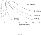
- FIG. 3 is a view illustrating an effect of a low-energy spectrum according to an embodiment.
- FIG. 3 illustrates dose deposition versus depth for energies of radiations.
- the doses decrease depending on the depths to which the radiations penetrate.
- the energy of radiation emitted becomes higher, a higher dose of radiation is delivered to an internal location, and thus efficiency in treatment is improved.
- the radiotherapy apparatus can be made compact, and there is an advantage in terms of economic efficiency. Considering that animal cancer frequently occurs in epidermis in a current clinical practice, it is necessary to select an appropriate energy spectrum.
- a 60-Co (1.25MeV) radiotherapy apparatus using gamma rays may require lower operating costs and a lower shielding level than a high-energy radiotherapy apparatus.
- economic costs for safety management and security are increasing due to an increase in the risk of terrorism against an isotope source, and when a dose rate is reduced due to the half-life of an isotope, there may be replacement costs according to that.
- the 60-Co (1.25MeV) radiotherapy apparatus may have a problem in that a penumbra exists, as compared with a radiotherapy apparatus based on a linear accelerator.
- a KeV unit low-energy radiotherapy apparatus using an X-ray tube method has a maximum dose point on epidermis and thus has efficiency in treating a surface, but may have a problem in that efficiency decreases when a tumor located at an internal location of a medium-sized animal is treated with radiation. Furthermore, radiation of the KeV unit has a larger penumbra area than radiation of the MeV unit, and therefore there may be a problem in that an uncertain area exists. Specifically, in a case of the KeV unit low-energy radiotherapy apparatus, it is unclear whether energy is transferred to a depth of 10 cm or more, and therefore there is a problem in that it is necessarily required to verify the dose for an irradiation surface.
- a radiotherapy apparatus having energy between 1MeV and 2MeV has critical significance in treatment of a medium-sized animal.
- the radiotherapy apparatus may allow the radiation to be applied not only to the epidermis of an animal but also to a diseased part located within a predetermined distance range from the epidermis.
- the predetermined distance may range from 10 cm to 20 cm.
- a penumbra area does not appear within an appropriate thickness range of 10 cm to 20 cm in the treatment of a medium-sized animal, and a treatment apparatus may be appropriately configured in terms of economy and space, as compared with a high-energy radiotherapy apparatus.
Landscapes
- Health & Medical Sciences (AREA)
- Engineering & Computer Science (AREA)
- Biomedical Technology (AREA)
- Life Sciences & Earth Sciences (AREA)
- Veterinary Medicine (AREA)
- Public Health (AREA)
- Animal Behavior & Ethology (AREA)
- General Health & Medical Sciences (AREA)
- Radiology & Medical Imaging (AREA)
- Nuclear Medicine, Radiotherapy & Molecular Imaging (AREA)
- Pathology (AREA)
- Wood Science & Technology (AREA)
- Zoology (AREA)
- Radiation-Therapy Devices (AREA)
Applications Claiming Priority (2)
| Application Number | Priority Date | Filing Date | Title |
|---|---|---|---|
| KR1020180074185A KR102068326B1 (ko) | 2018-06-27 | 2018-06-27 | 동물용 방사선 치료기 |
| PCT/KR2019/004607 WO2020004794A1 (fr) | 2018-06-27 | 2019-04-17 | Appareil de radiothérapie pour animal |
Publications (3)
| Publication Number | Publication Date |
|---|---|
| EP3815744A1 true EP3815744A1 (fr) | 2021-05-05 |
| EP3815744A4 EP3815744A4 (fr) | 2021-08-11 |
| EP3815744B1 EP3815744B1 (fr) | 2025-07-23 |
Family
ID=68985044
Family Applications (1)
| Application Number | Title | Priority Date | Filing Date |
|---|---|---|---|
| EP19825088.8A Active EP3815744B1 (fr) | 2018-06-27 | 2019-04-17 | Appareil de radiothérapie pour animal |
Country Status (5)
| Country | Link |
|---|---|
| US (1) | US11724125B2 (fr) |
| EP (1) | EP3815744B1 (fr) |
| KR (1) | KR102068326B1 (fr) |
| CN (1) | CN112334188A (fr) |
| WO (1) | WO2020004794A1 (fr) |
Families Citing this family (4)
| Publication number | Priority date | Publication date | Assignee | Title |
|---|---|---|---|---|
| US12285182B2 (en) | 2018-10-10 | 2025-04-29 | Innova Vascular, Inc. | Devices and methods for removing an embolus |
| KR102784314B1 (ko) | 2020-01-06 | 2025-03-21 | 주식회사 엘지에너지솔루션 | 안전성이 향상된 배터리 팩 및 이를 포함하는 이차전지 |
| CN115068833B (zh) * | 2021-03-15 | 2024-02-06 | 湖南华创医疗科技有限公司 | 用于束流阻挡器的定位装置和放射治疗系统 |
| WO2024010313A1 (fr) | 2022-07-04 | 2024-01-11 | 주식회사 이노비오젠 | Appareil de thérapie pour animaux combinant la radiothérapie et la thérapie thermique magnétique |
Family Cites Families (13)
| Publication number | Priority date | Publication date | Assignee | Title |
|---|---|---|---|---|
| US4726046A (en) * | 1985-11-05 | 1988-02-16 | Varian Associates, Inc. | X-ray and electron radiotherapy clinical treatment machine |
| US6353655B1 (en) * | 2000-08-23 | 2002-03-05 | Siemens Medical Solutions, Inc. | System and method for calculating fluence contributions from a source plane |
| KR20020073955A (ko) * | 2001-03-17 | 2002-09-28 | 주식회사 티아이티씨 | 의료 영상 검출장치 및 방법 |
| JP2003038475A (ja) | 2001-08-01 | 2003-02-12 | Kawasaki Heavy Ind Ltd | X線診断装置およびx線治療装置 |
| EP2305350A1 (fr) | 2001-08-24 | 2011-04-06 | Mitsubishi Heavy Industries, Ltd. | Appareil de traitement par rayonnement |
| US20090080602A1 (en) * | 2006-08-03 | 2009-03-26 | Kenneth Brooks | Dedicated breast radiation imaging/therapy system |
| KR100949141B1 (ko) | 2007-11-06 | 2010-03-25 | 원광대학교산학협력단 | X-선 튜브 광원에서의 특성방사선 획득 장치 |
| US8077830B2 (en) * | 2009-09-28 | 2011-12-13 | Varian Medical Systems, Inc. | Beam filter positioning device |
| CN103582455B (zh) * | 2011-02-14 | 2016-12-28 | 罗切斯特大学 | 基于锥形束乳房ct图像的计算机辅助检测和诊断的方法和装置 |
| US20130158382A1 (en) * | 2011-12-15 | 2013-06-20 | David Chao | Medical Treatment System With Non-Coplanar Capability |
| KR101465650B1 (ko) * | 2012-10-30 | 2014-11-27 | 성균관대학교산학협력단 | 더블 헤드 방식의 광 치료장치 |
| US10183181B2 (en) | 2015-07-22 | 2019-01-22 | Viewray Technologies, Inc. | Ion chamber for radiation measurement |
| PL3423154T3 (pl) | 2016-03-01 | 2021-11-02 | Intraop Medical Corporation | System napromieniania wiązką elektronową o niskiej energii, który generuje wiązki elektronowe z precyzyjnie kontrolowaną i regulowlaną głębokością penetracji, przydatny w zastosowaniach terapeutycznych |
-
2018
- 2018-06-27 KR KR1020180074185A patent/KR102068326B1/ko active Active
-
2019
- 2019-04-17 EP EP19825088.8A patent/EP3815744B1/fr active Active
- 2019-04-17 US US17/255,553 patent/US11724125B2/en active Active
- 2019-04-17 CN CN201980043508.0A patent/CN112334188A/zh active Pending
- 2019-04-17 WO PCT/KR2019/004607 patent/WO2020004794A1/fr not_active Ceased
Also Published As
| Publication number | Publication date |
|---|---|
| US11724125B2 (en) | 2023-08-15 |
| EP3815744A4 (fr) | 2021-08-11 |
| US20210268311A1 (en) | 2021-09-02 |
| KR20200001345A (ko) | 2020-01-06 |
| WO2020004794A1 (fr) | 2020-01-02 |
| CN112334188A (zh) | 2021-02-05 |
| KR102068326B1 (ko) | 2020-01-20 |
| EP3815744B1 (fr) | 2025-07-23 |
Similar Documents
| Publication | Publication Date | Title |
|---|---|---|
| US11724125B2 (en) | Radiotherapy apparatus for animal | |
| US20150352373A1 (en) | An apparatus to deliver conformal radiotherapy using external beam cobalt 60 | |
| KR102117680B1 (ko) | 방사선 치료기 및 방사선 치료기의 정도 관리 방법 | |
| WO2020101858A1 (fr) | Source de neutrons pour thérapie par capture de neutrons | |
| KR20200111270A (ko) | 암 치료를 위한 치료 전자 방사장치 | |
| KR101678681B1 (ko) | 방사선 치료기 및 방사선 치료기의 정도 관리 방법 | |
| Loi et al. | Neutron production from a mobile linear accelerator operating in electron mode for intraoperative radiation therapy | |
| US11944845B2 (en) | Asymmetric dual-mode ionization systems and methods | |
| Ha et al. | 6 MV X-band linear accelerator for stereotactic body radiation therapy | |
| CN108014428A (zh) | 一种磁共振图像引导的放射治疗系统 | |
| KR102080162B1 (ko) | 방사선 치료기 및 방사선 치료기의 정도 관리 방법 | |
| Fathallah | Commissioning of 360° rotational single room ProBeam Compact™(Varian Medical) pencil beam scanning proton therapy system | |
| Kim et al. | O-arm mounted X-band linear accelerator system for radiotherapy | |
| US20110309242A1 (en) | Radiation-activated Fiducial Markers for Organ Tracking | |
| EP3870283A1 (fr) | Source de neutrons pour thérapie par capture de neutrons | |
| JP4402851B2 (ja) | 分離型スノートを有する粒子線治療装置 | |
| Ng et al. | Problems and Solutions in Medical Physics: Radiotherapy Physics | |
| Boonzaier | A multi-institutional quantitative survey of multi-leaf collimator accuracy using a digital picket fence test with sub-millimetre detection capabilities. | |
| Klimpki | Pre-report on the dissertation Development of a treatment verification system for continuous scanning in proton therapy | |
| Uwais | Patient-Specific Quality Assurance For Volumetric Modulated Arc Therapy (Vmat) Using Fluence Map Analysis Generated From Log Data | |
| Lim et al. | Design of a radiotherapy machine using the 6 MeV C-band standing-wave accelerator | |
| da Silva | Analysis of the use of dosimetrically equivalent linear accelerators for Intensity Modulated Radiotherapy treatments | |
| Whitmore | Very High Energy Electron (VHEE) Radiotherapy: Focused Dose Delivery and Image Guidance | |
| Ahmed | Dosimetric Measurements in the Medical Linear Accelerator at (RICK) In Khartoum State | |
| Mazal | Proton beams in radiotherapy |
Legal Events
| Date | Code | Title | Description |
|---|---|---|---|
| STAA | Information on the status of an ep patent application or granted ep patent |
Free format text: STATUS: THE INTERNATIONAL PUBLICATION HAS BEEN MADE |
|
| PUAI | Public reference made under article 153(3) epc to a published international application that has entered the european phase |
Free format text: ORIGINAL CODE: 0009012 |
|
| STAA | Information on the status of an ep patent application or granted ep patent |
Free format text: STATUS: REQUEST FOR EXAMINATION WAS MADE |
|
| 17P | Request for examination filed |
Effective date: 20201224 |
|
| AK | Designated contracting states |
Kind code of ref document: A1 Designated state(s): AL AT BE BG CH CY CZ DE DK EE ES FI FR GB GR HR HU IE IS IT LI LT LU LV MC MK MT NL NO PL PT RO RS SE SI SK SM TR |
|
| A4 | Supplementary search report drawn up and despatched |
Effective date: 20210713 |
|
| RIC1 | Information provided on ipc code assigned before grant |
Ipc: A61N 5/10 20060101AFI20210707BHEP Ipc: A61D 7/00 20060101ALI20210707BHEP |
|
| DAV | Request for validation of the european patent (deleted) | ||
| DAX | Request for extension of the european patent (deleted) | ||
| STAA | Information on the status of an ep patent application or granted ep patent |
Free format text: STATUS: EXAMINATION IS IN PROGRESS |
|
| 17Q | First examination report despatched |
Effective date: 20230623 |
|
| GRAP | Despatch of communication of intention to grant a patent |
Free format text: ORIGINAL CODE: EPIDOSNIGR1 |
|
| STAA | Information on the status of an ep patent application or granted ep patent |
Free format text: STATUS: GRANT OF PATENT IS INTENDED |
|
| INTG | Intention to grant announced |
Effective date: 20250415 |
|
| GRAS | Grant fee paid |
Free format text: ORIGINAL CODE: EPIDOSNIGR3 |
|
| GRAA | (expected) grant |
Free format text: ORIGINAL CODE: 0009210 |
|
| STAA | Information on the status of an ep patent application or granted ep patent |
Free format text: STATUS: THE PATENT HAS BEEN GRANTED |
|
| AK | Designated contracting states |
Kind code of ref document: B1 Designated state(s): AL AT BE BG CH CY CZ DE DK EE ES FI FR GB GR HR HU IE IS IT LI LT LU LV MC MK MT NL NO PL PT RO RS SE SI SK SM TR |
|
| REG | Reference to a national code |
Ref country code: GB Ref legal event code: FG4D |
|
| REG | Reference to a national code |
Ref country code: CH Ref legal event code: EP |
|
| REG | Reference to a national code |
Ref country code: IE Ref legal event code: FG4D |
|
| REG | Reference to a national code |
Ref country code: DE Ref legal event code: R096 Ref document number: 602019072984 Country of ref document: DE |
|
| REG | Reference to a national code |
Ref country code: NL Ref legal event code: MP Effective date: 20250723 |
|
| PG25 | Lapsed in a contracting state [announced via postgrant information from national office to epo] |
Ref country code: PT Free format text: LAPSE BECAUSE OF FAILURE TO SUBMIT A TRANSLATION OF THE DESCRIPTION OR TO PAY THE FEE WITHIN THE PRESCRIBED TIME-LIMIT Effective date: 20251124 |
|
| PG25 | Lapsed in a contracting state [announced via postgrant information from national office to epo] |
Ref country code: NL Free format text: LAPSE BECAUSE OF FAILURE TO SUBMIT A TRANSLATION OF THE DESCRIPTION OR TO PAY THE FEE WITHIN THE PRESCRIBED TIME-LIMIT Effective date: 20250723 |
|
| REG | Reference to a national code |
Ref country code: AT Ref legal event code: MK05 Ref document number: 1815817 Country of ref document: AT Kind code of ref document: T Effective date: 20250723 |
|
| PG25 | Lapsed in a contracting state [announced via postgrant information from national office to epo] |
Ref country code: IS Free format text: LAPSE BECAUSE OF FAILURE TO SUBMIT A TRANSLATION OF THE DESCRIPTION OR TO PAY THE FEE WITHIN THE PRESCRIBED TIME-LIMIT Effective date: 20251123 |
|
| PG25 | Lapsed in a contracting state [announced via postgrant information from national office to epo] |
Ref country code: NO Free format text: LAPSE BECAUSE OF FAILURE TO SUBMIT A TRANSLATION OF THE DESCRIPTION OR TO PAY THE FEE WITHIN THE PRESCRIBED TIME-LIMIT Effective date: 20251023 |
|
| REG | Reference to a national code |
Ref country code: LT Ref legal event code: MG9D |
|
| PG25 | Lapsed in a contracting state [announced via postgrant information from national office to epo] |
Ref country code: AT Free format text: LAPSE BECAUSE OF FAILURE TO SUBMIT A TRANSLATION OF THE DESCRIPTION OR TO PAY THE FEE WITHIN THE PRESCRIBED TIME-LIMIT Effective date: 20250723 |
|
| PG25 | Lapsed in a contracting state [announced via postgrant information from national office to epo] |
Ref country code: FI Free format text: LAPSE BECAUSE OF FAILURE TO SUBMIT A TRANSLATION OF THE DESCRIPTION OR TO PAY THE FEE WITHIN THE PRESCRIBED TIME-LIMIT Effective date: 20250723 |
|
| PG25 | Lapsed in a contracting state [announced via postgrant information from national office to epo] |
Ref country code: HR Free format text: LAPSE BECAUSE OF FAILURE TO SUBMIT A TRANSLATION OF THE DESCRIPTION OR TO PAY THE FEE WITHIN THE PRESCRIBED TIME-LIMIT Effective date: 20250723 |
|
| PG25 | Lapsed in a contracting state [announced via postgrant information from national office to epo] |
Ref country code: GR Free format text: LAPSE BECAUSE OF FAILURE TO SUBMIT A TRANSLATION OF THE DESCRIPTION OR TO PAY THE FEE WITHIN THE PRESCRIBED TIME-LIMIT Effective date: 20251024 |
|
| PG25 | Lapsed in a contracting state [announced via postgrant information from national office to epo] |
Ref country code: SE Free format text: LAPSE BECAUSE OF FAILURE TO SUBMIT A TRANSLATION OF THE DESCRIPTION OR TO PAY THE FEE WITHIN THE PRESCRIBED TIME-LIMIT Effective date: 20250723 |
|
| PG25 | Lapsed in a contracting state [announced via postgrant information from national office to epo] |
Ref country code: LV Free format text: LAPSE BECAUSE OF FAILURE TO SUBMIT A TRANSLATION OF THE DESCRIPTION OR TO PAY THE FEE WITHIN THE PRESCRIBED TIME-LIMIT Effective date: 20250723 |
|
| PG25 | Lapsed in a contracting state [announced via postgrant information from national office to epo] |
Ref country code: BG Free format text: LAPSE BECAUSE OF FAILURE TO SUBMIT A TRANSLATION OF THE DESCRIPTION OR TO PAY THE FEE WITHIN THE PRESCRIBED TIME-LIMIT Effective date: 20250723 Ref country code: PL Free format text: LAPSE BECAUSE OF FAILURE TO SUBMIT A TRANSLATION OF THE DESCRIPTION OR TO PAY THE FEE WITHIN THE PRESCRIBED TIME-LIMIT Effective date: 20250723 |
|
| PG25 | Lapsed in a contracting state [announced via postgrant information from national office to epo] |
Ref country code: RS Free format text: LAPSE BECAUSE OF FAILURE TO SUBMIT A TRANSLATION OF THE DESCRIPTION OR TO PAY THE FEE WITHIN THE PRESCRIBED TIME-LIMIT Effective date: 20251023 |
|
| PG25 | Lapsed in a contracting state [announced via postgrant information from national office to epo] |
Ref country code: ES Free format text: LAPSE BECAUSE OF FAILURE TO SUBMIT A TRANSLATION OF THE DESCRIPTION OR TO PAY THE FEE WITHIN THE PRESCRIBED TIME-LIMIT Effective date: 20250723 |
|
| PG25 | Lapsed in a contracting state [announced via postgrant information from national office to epo] |
Ref country code: SM Free format text: LAPSE BECAUSE OF FAILURE TO SUBMIT A TRANSLATION OF THE DESCRIPTION OR TO PAY THE FEE WITHIN THE PRESCRIBED TIME-LIMIT Effective date: 20250723 |
|
| PG25 | Lapsed in a contracting state [announced via postgrant information from national office to epo] |
Ref country code: DK Free format text: LAPSE BECAUSE OF FAILURE TO SUBMIT A TRANSLATION OF THE DESCRIPTION OR TO PAY THE FEE WITHIN THE PRESCRIBED TIME-LIMIT Effective date: 20250723 |
|
| PG25 | Lapsed in a contracting state [announced via postgrant information from national office to epo] |
Ref country code: IT Free format text: LAPSE BECAUSE OF FAILURE TO SUBMIT A TRANSLATION OF THE DESCRIPTION OR TO PAY THE FEE WITHIN THE PRESCRIBED TIME-LIMIT Effective date: 20250723 |
|
| PG25 | Lapsed in a contracting state [announced via postgrant information from national office to epo] |
Ref country code: CZ Free format text: LAPSE BECAUSE OF FAILURE TO SUBMIT A TRANSLATION OF THE DESCRIPTION OR TO PAY THE FEE WITHIN THE PRESCRIBED TIME-LIMIT Effective date: 20250723 |
|
| PG25 | Lapsed in a contracting state [announced via postgrant information from national office to epo] |
Ref country code: SK Free format text: LAPSE BECAUSE OF FAILURE TO SUBMIT A TRANSLATION OF THE DESCRIPTION OR TO PAY THE FEE WITHIN THE PRESCRIBED TIME-LIMIT Effective date: 20250723 Ref country code: EE Free format text: LAPSE BECAUSE OF FAILURE TO SUBMIT A TRANSLATION OF THE DESCRIPTION OR TO PAY THE FEE WITHIN THE PRESCRIBED TIME-LIMIT Effective date: 20250723 |