EP3805261A1 - Hla-g transfected cells and their use in treating diabetes - Google Patents
Hla-g transfected cells and their use in treating diabetes Download PDFInfo
- Publication number
- EP3805261A1 EP3805261A1 EP20196775.9A EP20196775A EP3805261A1 EP 3805261 A1 EP3805261 A1 EP 3805261A1 EP 20196775 A EP20196775 A EP 20196775A EP 3805261 A1 EP3805261 A1 EP 3805261A1
- Authority
- EP
- European Patent Office
- Prior art keywords
- hla
- cell
- genetically modified
- cells
- ehla
- Prior art date
- Legal status (The legal status is an assumption and is not a legal conclusion. Google has not performed a legal analysis and makes no representation as to the accuracy of the status listed.)
- Withdrawn
Links
- 206010012601 diabetes mellitus Diseases 0.000 title claims description 11
- 210000004027 cell Anatomy 0.000 claims abstract description 351
- 150000007523 nucleic acids Chemical class 0.000 claims abstract description 87
- 238000000034 method Methods 0.000 claims abstract description 55
- 230000002829 reductive effect Effects 0.000 claims abstract description 45
- 230000005847 immunogenicity Effects 0.000 claims abstract description 43
- 230000001506 immunosuppresive effect Effects 0.000 claims abstract description 36
- 108020004707 nucleic acids Proteins 0.000 claims abstract description 35
- 102000039446 nucleic acids Human genes 0.000 claims abstract description 35
- 206010062016 Immunosuppression Diseases 0.000 claims abstract description 31
- 238000012239 gene modification Methods 0.000 claims abstract description 21
- 230000005017 genetic modification Effects 0.000 claims abstract description 21
- 235000013617 genetically modified food Nutrition 0.000 claims abstract description 21
- 239000000203 mixture Substances 0.000 claims abstract description 18
- 230000001413 cellular effect Effects 0.000 claims abstract description 11
- 210000004962 mammalian cell Anatomy 0.000 claims description 105
- 241000282414 Homo sapiens Species 0.000 claims description 83
- 108091028043 Nucleic acid sequence Proteins 0.000 claims description 54
- 102000010292 Peptide Elongation Factor 1 Human genes 0.000 claims description 41
- 108010077524 Peptide Elongation Factor 1 Proteins 0.000 claims description 41
- 210000000130 stem cell Anatomy 0.000 claims description 39
- 239000013604 expression vector Substances 0.000 claims description 38
- 108090000623 proteins and genes Proteins 0.000 claims description 38
- 102000004169 proteins and genes Human genes 0.000 claims description 32
- 239000013598 vector Substances 0.000 claims description 30
- 210000001671 embryonic stem cell Anatomy 0.000 claims description 26
- 108020005345 3' Untranslated Regions Proteins 0.000 claims description 24
- 241000699670 Mus sp. Species 0.000 claims description 23
- 210000003819 peripheral blood mononuclear cell Anatomy 0.000 claims description 23
- 231100000135 cytotoxicity Toxicity 0.000 claims description 21
- 230000003013 cytotoxicity Effects 0.000 claims description 21
- 108091036066 Three prime untranslated region Proteins 0.000 claims description 18
- 210000004263 induced pluripotent stem cell Anatomy 0.000 claims description 14
- 238000000338 in vitro Methods 0.000 claims description 13
- 239000013612 plasmid Substances 0.000 claims description 11
- 210000001778 pluripotent stem cell Anatomy 0.000 claims description 11
- 102100028972 HLA class I histocompatibility antigen, A alpha chain Human genes 0.000 claims description 8
- 108010075704 HLA-A Antigens Proteins 0.000 claims description 8
- 108010010378 HLA-DP Antigens Proteins 0.000 claims description 8
- 102000015789 HLA-DP Antigens Human genes 0.000 claims description 8
- 102100028976 HLA class I histocompatibility antigen, B alpha chain Human genes 0.000 claims description 7
- 102100028971 HLA class I histocompatibility antigen, C alpha chain Human genes 0.000 claims description 7
- 108010058607 HLA-B Antigens Proteins 0.000 claims description 7
- 108010052199 HLA-C Antigens Proteins 0.000 claims description 7
- 108010062347 HLA-DQ Antigens Proteins 0.000 claims description 7
- 108010058597 HLA-DR Antigens Proteins 0.000 claims description 7
- 102000006354 HLA-DR Antigens Human genes 0.000 claims description 7
- 125000003729 nucleotide group Chemical group 0.000 claims description 7
- 230000009467 reduction Effects 0.000 claims description 7
- 210000000822 natural killer cell Anatomy 0.000 claims description 6
- 239000002773 nucleotide Substances 0.000 claims description 6
- 108091023045 Untranslated Region Proteins 0.000 claims description 5
- 210000005260 human cell Anatomy 0.000 claims description 4
- 210000004153 islets of langerhan Anatomy 0.000 claims description 4
- 230000004663 cell proliferation Effects 0.000 claims description 3
- 210000000265 leukocyte Anatomy 0.000 claims description 3
- 230000001177 retroviral effect Effects 0.000 claims description 3
- 210000003014 totipotent stem cell Anatomy 0.000 claims description 3
- 230000005740 tumor formation Effects 0.000 claims description 3
- 102100028967 HLA class I histocompatibility antigen, alpha chain G Human genes 0.000 abstract description 190
- 108010024164 HLA-G Antigens Proteins 0.000 abstract description 190
- 230000017423 tissue regeneration Effects 0.000 abstract description 13
- 238000002560 therapeutic procedure Methods 0.000 abstract description 12
- 238000002659 cell therapy Methods 0.000 abstract description 6
- 230000014509 gene expression Effects 0.000 description 109
- 108700019146 Transgenes Proteins 0.000 description 60
- 239000005090 green fluorescent protein Substances 0.000 description 57
- 230000035772 mutation Effects 0.000 description 38
- 210000001519 tissue Anatomy 0.000 description 30
- 210000002950 fibroblast Anatomy 0.000 description 28
- 230000004069 differentiation Effects 0.000 description 26
- 108010043121 Green Fluorescent Proteins Proteins 0.000 description 25
- 102000004144 Green Fluorescent Proteins Human genes 0.000 description 25
- 206010028980 Neoplasm Diseases 0.000 description 23
- 125000003275 alpha amino acid group Chemical group 0.000 description 22
- 210000004271 bone marrow stromal cell Anatomy 0.000 description 20
- 230000002500 effect on skin Effects 0.000 description 19
- 230000027455 binding Effects 0.000 description 15
- 208000037265 diseases, disorders, signs and symptoms Diseases 0.000 description 15
- 102100040445 Keratin, type I cytoskeletal 14 Human genes 0.000 description 14
- 230000000694 effects Effects 0.000 description 14
- 238000000684 flow cytometry Methods 0.000 description 14
- 101710183391 Keratin, type I cytoskeletal 14 Proteins 0.000 description 13
- 239000003550 marker Substances 0.000 description 13
- 239000002609 medium Substances 0.000 description 13
- 210000003491 skin Anatomy 0.000 description 12
- 150000001413 amino acids Chemical class 0.000 description 11
- 210000003958 hematopoietic stem cell Anatomy 0.000 description 11
- 108090000765 processed proteins & peptides Proteins 0.000 description 11
- 201000010099 disease Diseases 0.000 description 10
- 238000002474 experimental method Methods 0.000 description 10
- 230000014759 maintenance of location Effects 0.000 description 10
- 238000002054 transplantation Methods 0.000 description 10
- 238000011282 treatment Methods 0.000 description 10
- 108700011259 MicroRNAs Proteins 0.000 description 9
- 238000002784 cytotoxicity assay Methods 0.000 description 9
- 231100000263 cytotoxicity test Toxicity 0.000 description 9
- 108010048367 enhanced green fluorescent protein Proteins 0.000 description 9
- 230000030279 gene silencing Effects 0.000 description 9
- 238000001727 in vivo Methods 0.000 description 9
- 210000002510 keratinocyte Anatomy 0.000 description 9
- 210000002901 mesenchymal stem cell Anatomy 0.000 description 9
- 239000002679 microRNA Substances 0.000 description 9
- 230000037361 pathway Effects 0.000 description 9
- 229920001184 polypeptide Polymers 0.000 description 9
- 102000004196 processed proteins & peptides Human genes 0.000 description 9
- 238000004064 recycling Methods 0.000 description 9
- 230000002269 spontaneous effect Effects 0.000 description 9
- 230000009261 transgenic effect Effects 0.000 description 9
- 230000003612 virological effect Effects 0.000 description 9
- 241000699802 Cricetulus griseus Species 0.000 description 8
- 101000935043 Homo sapiens Integrin beta-1 Proteins 0.000 description 8
- 102100025304 Integrin beta-1 Human genes 0.000 description 8
- 102000011755 Phosphoglycerate Kinase Human genes 0.000 description 8
- 101001099217 Thermotoga maritima (strain ATCC 43589 / DSM 3109 / JCM 10099 / NBRC 100826 / MSB8) Triosephosphate isomerase Proteins 0.000 description 8
- 241000700605 Viruses Species 0.000 description 8
- 230000009089 cytolysis Effects 0.000 description 8
- 239000001963 growth medium Substances 0.000 description 8
- 210000001178 neural stem cell Anatomy 0.000 description 8
- RXWNCPJZOCPEPQ-NVWDDTSBSA-N puromycin Chemical compound C1=CC(OC)=CC=C1C[C@H](N)C(=O)N[C@H]1[C@@H](O)[C@H](N2C3=NC=NC(=C3N=C2)N(C)C)O[C@@H]1CO RXWNCPJZOCPEPQ-NVWDDTSBSA-N 0.000 description 8
- 238000001890 transfection Methods 0.000 description 8
- 108700028369 Alleles Proteins 0.000 description 7
- 241001465754 Metazoa Species 0.000 description 7
- 102000008579 Transposases Human genes 0.000 description 7
- 108010020764 Transposases Proteins 0.000 description 7
- 210000004369 blood Anatomy 0.000 description 7
- 239000008280 blood Substances 0.000 description 7
- 230000000875 corresponding effect Effects 0.000 description 7
- 230000037430 deletion Effects 0.000 description 7
- 238000012217 deletion Methods 0.000 description 7
- 239000012636 effector Substances 0.000 description 7
- 238000010166 immunofluorescence Methods 0.000 description 7
- 238000003780 insertion Methods 0.000 description 7
- 230000037431 insertion Effects 0.000 description 7
- 230000002147 killing effect Effects 0.000 description 7
- 108020004999 messenger RNA Proteins 0.000 description 7
- 230000004048 modification Effects 0.000 description 7
- 238000012986 modification Methods 0.000 description 7
- 210000002569 neuron Anatomy 0.000 description 7
- 210000002237 B-cell of pancreatic islet Anatomy 0.000 description 6
- 101150024418 HLA-G gene Proteins 0.000 description 6
- 101000937544 Homo sapiens Beta-2-microglobulin Proteins 0.000 description 6
- 108060001084 Luciferase Proteins 0.000 description 6
- 239000005089 Luciferase Substances 0.000 description 6
- 102100027881 Tumor protein 63 Human genes 0.000 description 6
- 230000000735 allogeneic effect Effects 0.000 description 6
- 239000000427 antigen Substances 0.000 description 6
- 108091007433 antigens Proteins 0.000 description 6
- 102000036639 antigens Human genes 0.000 description 6
- 238000013459 approach Methods 0.000 description 6
- 238000003556 assay Methods 0.000 description 6
- 201000011510 cancer Diseases 0.000 description 6
- 238000001516 cell proliferation assay Methods 0.000 description 6
- 102000047279 human B2M Human genes 0.000 description 6
- 238000009396 hybridization Methods 0.000 description 6
- 239000012212 insulator Substances 0.000 description 6
- 230000002085 persistent effect Effects 0.000 description 6
- 230000035755 proliferation Effects 0.000 description 6
- 241000894007 species Species 0.000 description 6
- 239000000758 substrate Substances 0.000 description 6
- 102100027377 HBS1-like protein Human genes 0.000 description 5
- 101001009070 Homo sapiens HBS1-like protein Proteins 0.000 description 5
- 241000699666 Mus <mouse, genus> Species 0.000 description 5
- 208000012902 Nervous system disease Diseases 0.000 description 5
- 208000018737 Parkinson disease Diseases 0.000 description 5
- 210000001130 astrocyte Anatomy 0.000 description 5
- 230000015572 biosynthetic process Effects 0.000 description 5
- 239000003795 chemical substances by application Substances 0.000 description 5
- 208000035475 disorder Diseases 0.000 description 5
- 210000002242 embryoid body Anatomy 0.000 description 5
- 238000011577 humanized mouse model Methods 0.000 description 5
- 210000000987 immune system Anatomy 0.000 description 5
- 210000002894 multi-fate stem cell Anatomy 0.000 description 5
- 230000008569 process Effects 0.000 description 5
- 210000002966 serum Anatomy 0.000 description 5
- 241001430294 unidentified retrovirus Species 0.000 description 5
- FWBHETKCLVMNFS-UHFFFAOYSA-N 4',6-Diamino-2-phenylindol Chemical compound C1=CC(C(=N)N)=CC=C1C1=CC2=CC=C(C(N)=N)C=C2N1 FWBHETKCLVMNFS-UHFFFAOYSA-N 0.000 description 4
- 208000024827 Alzheimer disease Diseases 0.000 description 4
- 101710087047 Cytoskeleton-associated protein 4 Proteins 0.000 description 4
- 102000004190 Enzymes Human genes 0.000 description 4
- 108090000790 Enzymes Proteins 0.000 description 4
- LFQSCWFLJHTTHZ-UHFFFAOYSA-N Ethanol Chemical compound CCO LFQSCWFLJHTTHZ-UHFFFAOYSA-N 0.000 description 4
- 108010010803 Gelatin Proteins 0.000 description 4
- WZUVPPKBWHMQCE-UHFFFAOYSA-N Haematoxylin Chemical compound C12=CC(O)=C(O)C=C2CC2(O)C1C1=CC=C(O)C(O)=C1OC2 WZUVPPKBWHMQCE-UHFFFAOYSA-N 0.000 description 4
- 101000994365 Homo sapiens Integrin alpha-6 Proteins 0.000 description 4
- 101000738771 Homo sapiens Receptor-type tyrosine-protein phosphatase C Proteins 0.000 description 4
- 102100032816 Integrin alpha-6 Human genes 0.000 description 4
- 102100037422 Receptor-type tyrosine-protein phosphatase C Human genes 0.000 description 4
- 101710140697 Tumor protein 63 Proteins 0.000 description 4
- 208000027418 Wounds and injury Diseases 0.000 description 4
- 230000008901 benefit Effects 0.000 description 4
- 210000004413 cardiac myocyte Anatomy 0.000 description 4
- 108010007093 dispase Proteins 0.000 description 4
- 238000001943 fluorescence-activated cell sorting Methods 0.000 description 4
- 239000008273 gelatin Substances 0.000 description 4
- 229920000159 gelatin Polymers 0.000 description 4
- 235000019322 gelatine Nutrition 0.000 description 4
- 235000011852 gelatine desserts Nutrition 0.000 description 4
- 230000035876 healing Effects 0.000 description 4
- 210000003494 hepatocyte Anatomy 0.000 description 4
- 230000006058 immune tolerance Effects 0.000 description 4
- 230000006872 improvement Effects 0.000 description 4
- 208000015181 infectious disease Diseases 0.000 description 4
- 238000002347 injection Methods 0.000 description 4
- 239000007924 injection Substances 0.000 description 4
- CDAISMWEOUEBRE-UHFFFAOYSA-N inositol Chemical compound OC1C(O)C(O)C(O)C(O)C1O CDAISMWEOUEBRE-UHFFFAOYSA-N 0.000 description 4
- 239000007758 minimum essential medium Substances 0.000 description 4
- 201000006417 multiple sclerosis Diseases 0.000 description 4
- 210000000663 muscle cell Anatomy 0.000 description 4
- 210000004248 oligodendroglia Anatomy 0.000 description 4
- 229950010131 puromycin Drugs 0.000 description 4
- 230000008439 repair process Effects 0.000 description 4
- 238000003757 reverse transcription PCR Methods 0.000 description 4
- 239000000523 sample Substances 0.000 description 4
- 230000008685 targeting Effects 0.000 description 4
- 241000282693 Cercopithecidae Species 0.000 description 3
- 108091035707 Consensus sequence Proteins 0.000 description 3
- KCXVZYZYPLLWCC-UHFFFAOYSA-N EDTA Chemical compound OC(=O)CN(CC(O)=O)CCN(CC(O)=O)CC(O)=O KCXVZYZYPLLWCC-UHFFFAOYSA-N 0.000 description 3
- 241000713800 Feline immunodeficiency virus Species 0.000 description 3
- 102100024785 Fibroblast growth factor 2 Human genes 0.000 description 3
- 108090000379 Fibroblast growth factor 2 Proteins 0.000 description 3
- 208000009329 Graft vs Host Disease Diseases 0.000 description 3
- 102100028970 HLA class I histocompatibility antigen, alpha chain E Human genes 0.000 description 3
- 101000986085 Homo sapiens HLA class I histocompatibility antigen, alpha chain E Proteins 0.000 description 3
- FDJKUWYYUZCUJX-AJKRCSPLSA-N N-glycoloyl-beta-neuraminic acid Chemical compound OC[C@@H](O)[C@@H](O)[C@@H]1O[C@](O)(C(O)=O)C[C@H](O)[C@H]1NC(=O)CO FDJKUWYYUZCUJX-AJKRCSPLSA-N 0.000 description 3
- FDJKUWYYUZCUJX-UHFFFAOYSA-N N-glycolyl-beta-neuraminic acid Natural products OCC(O)C(O)C1OC(O)(C(O)=O)CC(O)C1NC(=O)CO FDJKUWYYUZCUJX-UHFFFAOYSA-N 0.000 description 3
- 241000713311 Simian immunodeficiency virus Species 0.000 description 3
- 206010043276 Teratoma Diseases 0.000 description 3
- 238000004458 analytical method Methods 0.000 description 3
- 239000003153 chemical reaction reagent Substances 0.000 description 3
- 230000001143 conditioned effect Effects 0.000 description 3
- 238000012258 culturing Methods 0.000 description 3
- 231100000433 cytotoxic Toxicity 0.000 description 3
- 230000001472 cytotoxic effect Effects 0.000 description 3
- 239000003814 drug Substances 0.000 description 3
- 238000001476 gene delivery Methods 0.000 description 3
- 208000024908 graft versus host disease Diseases 0.000 description 3
- 238000003384 imaging method Methods 0.000 description 3
- 102000006639 indoleamine 2,3-dioxygenase Human genes 0.000 description 3
- 108020004201 indoleamine 2,3-dioxygenase Proteins 0.000 description 3
- 230000010354 integration Effects 0.000 description 3
- 230000000670 limiting effect Effects 0.000 description 3
- 208000019423 liver disease Diseases 0.000 description 3
- 230000004770 neurodegeneration Effects 0.000 description 3
- 208000015122 neurodegenerative disease Diseases 0.000 description 3
- 238000007747 plating Methods 0.000 description 3
- 230000004224 protection Effects 0.000 description 3
- 230000001172 regenerating effect Effects 0.000 description 3
- 238000011069 regeneration method Methods 0.000 description 3
- 239000012679 serum free medium Substances 0.000 description 3
- 239000000243 solution Substances 0.000 description 3
- 208000020431 spinal cord injury Diseases 0.000 description 3
- 238000003153 stable transfection Methods 0.000 description 3
- 238000010186 staining Methods 0.000 description 3
- 238000006467 substitution reaction Methods 0.000 description 3
- 230000004083 survival effect Effects 0.000 description 3
- 210000000115 thoracic cavity Anatomy 0.000 description 3
- 230000017105 transposition Effects 0.000 description 3
- 230000004614 tumor growth Effects 0.000 description 3
- DGVVWUTYPXICAM-UHFFFAOYSA-N β‐Mercaptoethanol Chemical compound OCCS DGVVWUTYPXICAM-UHFFFAOYSA-N 0.000 description 3
- HJCMDXDYPOUFDY-WHFBIAKZSA-N Ala-Gln Chemical compound C[C@H](N)C(=O)N[C@H](C(O)=O)CCC(N)=O HJCMDXDYPOUFDY-WHFBIAKZSA-N 0.000 description 2
- 208000023275 Autoimmune disease Diseases 0.000 description 2
- 102100024505 Bone morphogenetic protein 4 Human genes 0.000 description 2
- 241000283707 Capra Species 0.000 description 2
- 208000024172 Cardiovascular disease Diseases 0.000 description 2
- 108091026890 Coding region Proteins 0.000 description 2
- 108010035532 Collagen Proteins 0.000 description 2
- 102000008186 Collagen Human genes 0.000 description 2
- 206010011878 Deafness Diseases 0.000 description 2
- 239000006144 Dulbecco’s modified Eagle's medium Substances 0.000 description 2
- 206010015548 Euthanasia Diseases 0.000 description 2
- 108090000331 Firefly luciferases Proteins 0.000 description 2
- 241000710198 Foot-and-mouth disease virus Species 0.000 description 2
- 101001035782 Gallus gallus Hemoglobin subunit beta Proteins 0.000 description 2
- 101000762379 Homo sapiens Bone morphogenetic protein 4 Proteins 0.000 description 2
- 101000868279 Homo sapiens Leukocyte surface antigen CD47 Proteins 0.000 description 2
- 101001098352 Homo sapiens OX-2 membrane glycoprotein Proteins 0.000 description 2
- 241000713772 Human immunodeficiency virus 1 Species 0.000 description 2
- 239000007760 Iscove's Modified Dulbecco's Medium Substances 0.000 description 2
- KFZMGEQAYNKOFK-UHFFFAOYSA-N Isopropanol Chemical compound CC(C)O KFZMGEQAYNKOFK-UHFFFAOYSA-N 0.000 description 2
- 108010025815 Kanamycin Kinase Proteins 0.000 description 2
- 241000713666 Lentivirus Species 0.000 description 2
- 102100032913 Leukocyte surface antigen CD47 Human genes 0.000 description 2
- 241001529936 Murinae Species 0.000 description 2
- 241000714177 Murine leukemia virus Species 0.000 description 2
- NWIBSHFKIJFRCO-WUDYKRTCSA-N Mytomycin Chemical compound C1N2C(C(C(C)=C(N)C3=O)=O)=C3[C@@H](COC(N)=O)[C@@]2(OC)[C@@H]2[C@H]1N2 NWIBSHFKIJFRCO-WUDYKRTCSA-N 0.000 description 2
- 208000025966 Neurological disease Diseases 0.000 description 2
- 102100037589 OX-2 membrane glycoprotein Human genes 0.000 description 2
- 108700026244 Open Reading Frames Proteins 0.000 description 2
- YHIPILPTUVMWQT-UHFFFAOYSA-N Oplophorus luciferin Chemical compound C1=CC(O)=CC=C1CC(C(N1C=C(N2)C=3C=CC(O)=CC=3)=O)=NC1=C2CC1=CC=CC=C1 YHIPILPTUVMWQT-UHFFFAOYSA-N 0.000 description 2
- 208000001132 Osteoporosis Diseases 0.000 description 2
- 229930040373 Paraformaldehyde Natural products 0.000 description 2
- 241000700159 Rattus Species 0.000 description 2
- 108010052090 Renilla Luciferases Proteins 0.000 description 2
- 208000028990 Skin injury Diseases 0.000 description 2
- 208000006011 Stroke Diseases 0.000 description 2
- 241000282898 Sus scrofa Species 0.000 description 2
- 208000025865 Ulcer Diseases 0.000 description 2
- 230000001154 acute effect Effects 0.000 description 2
- 210000000577 adipose tissue Anatomy 0.000 description 2
- 230000032683 aging Effects 0.000 description 2
- SHGAZHPCJJPHSC-YCNIQYBTSA-N all-trans-retinoic acid Chemical compound OC(=O)\C=C(/C)\C=C\C=C(/C)\C=C\C1=C(C)CCCC1(C)C SHGAZHPCJJPHSC-YCNIQYBTSA-N 0.000 description 2
- 125000000539 amino acid group Chemical group 0.000 description 2
- 206010003246 arthritis Diseases 0.000 description 2
- 208000027115 auditory system disease Diseases 0.000 description 2
- 230000000747 cardiac effect Effects 0.000 description 2
- 238000004113 cell culture Methods 0.000 description 2
- 206010008118 cerebral infarction Diseases 0.000 description 2
- 208000026106 cerebrovascular disease Diseases 0.000 description 2
- 230000008859 change Effects 0.000 description 2
- 238000010367 cloning Methods 0.000 description 2
- 229920001436 collagen Polymers 0.000 description 2
- 239000003636 conditioned culture medium Substances 0.000 description 2
- 230000003750 conditioning effect Effects 0.000 description 2
- 230000006378 damage Effects 0.000 description 2
- 231100000895 deafness Toxicity 0.000 description 2
- 230000007547 defect Effects 0.000 description 2
- 210000002986 dental sac Anatomy 0.000 description 2
- 238000013461 design Methods 0.000 description 2
- 238000001514 detection method Methods 0.000 description 2
- 238000010790 dilution Methods 0.000 description 2
- 239000012895 dilution Substances 0.000 description 2
- 238000010494 dissociation reaction Methods 0.000 description 2
- 230000005593 dissociations Effects 0.000 description 2
- 229940079593 drug Drugs 0.000 description 2
- 238000005516 engineering process Methods 0.000 description 2
- YQGOJNYOYNNSMM-UHFFFAOYSA-N eosin Chemical compound [Na+].OC(=O)C1=CC=CC=C1C1=C2C=C(Br)C(=O)C(Br)=C2OC2=C(Br)C(O)=C(Br)C=C21 YQGOJNYOYNNSMM-UHFFFAOYSA-N 0.000 description 2
- 239000003797 essential amino acid Substances 0.000 description 2
- 235000020776 essential amino acid Nutrition 0.000 description 2
- 208000030533 eye disease Diseases 0.000 description 2
- 238000002073 fluorescence micrograph Methods 0.000 description 2
- 238000000799 fluorescence microscopy Methods 0.000 description 2
- 102000034287 fluorescent proteins Human genes 0.000 description 2
- 108091006047 fluorescent proteins Proteins 0.000 description 2
- OVBPIULPVIDEAO-LBPRGKRZSA-N folic acid Chemical compound C=1N=C2NC(N)=NC(=O)C2=NC=1CNC1=CC=C(C(=O)N[C@@H](CCC(O)=O)C(O)=O)C=C1 OVBPIULPVIDEAO-LBPRGKRZSA-N 0.000 description 2
- 208000024693 gingival disease Diseases 0.000 description 2
- 230000003394 haemopoietic effect Effects 0.000 description 2
- 210000000442 hair follicle cell Anatomy 0.000 description 2
- 230000036541 health Effects 0.000 description 2
- 208000016354 hearing loss disease Diseases 0.000 description 2
- 208000019622 heart disease Diseases 0.000 description 2
- 230000002440 hepatic effect Effects 0.000 description 2
- 206010021093 hypospadias Diseases 0.000 description 2
- 238000011534 incubation Methods 0.000 description 2
- 208000014674 injury Diseases 0.000 description 2
- CDAISMWEOUEBRE-GPIVLXJGSA-N inositol Chemical compound O[C@H]1[C@H](O)[C@@H](O)[C@H](O)[C@H](O)[C@@H]1O CDAISMWEOUEBRE-GPIVLXJGSA-N 0.000 description 2
- 210000003292 kidney cell Anatomy 0.000 description 2
- 208000017169 kidney disease Diseases 0.000 description 2
- KWGKDLIKAYFUFQ-UHFFFAOYSA-M lithium chloride Chemical compound [Li+].[Cl-] KWGKDLIKAYFUFQ-UHFFFAOYSA-M 0.000 description 2
- 208000002780 macular degeneration Diseases 0.000 description 2
- 238000002826 magnetic-activated cell sorting Methods 0.000 description 2
- 108010082117 matrigel Proteins 0.000 description 2
- 230000001404 mediated effect Effects 0.000 description 2
- 230000001394 metastastic effect Effects 0.000 description 2
- 206010061289 metastatic neoplasm Diseases 0.000 description 2
- 238000000386 microscopy Methods 0.000 description 2
- 230000001537 neural effect Effects 0.000 description 2
- 210000000963 osteoblast Anatomy 0.000 description 2
- 229920002866 paraformaldehyde Polymers 0.000 description 2
- 239000008194 pharmaceutical composition Substances 0.000 description 2
- 238000002135 phase contrast microscopy Methods 0.000 description 2
- 239000000047 product Substances 0.000 description 2
- 230000002062 proliferating effect Effects 0.000 description 2
- 230000001105 regulatory effect Effects 0.000 description 2
- 210000000844 retinal pigment epithelial cell Anatomy 0.000 description 2
- 229930002330 retinoic acid Natural products 0.000 description 2
- 230000036560 skin regeneration Effects 0.000 description 2
- 238000012360 testing method Methods 0.000 description 2
- 230000000699 topical effect Effects 0.000 description 2
- 238000010361 transduction Methods 0.000 description 2
- 230000026683 transduction Effects 0.000 description 2
- 231100000397 ulcer Toxicity 0.000 description 2
- 238000011144 upstream manufacturing Methods 0.000 description 2
- 108091032973 (ribonucleotides)n+m Proteins 0.000 description 1
- KSXTUUUQYQYKCR-LQDDAWAPSA-M 2,3-bis[[(z)-octadec-9-enoyl]oxy]propyl-trimethylazanium;chloride Chemical compound [Cl-].CCCCCCCC\C=C/CCCCCCCC(=O)OCC(C[N+](C)(C)C)OC(=O)CCCCCCC\C=C/CCCCCCCC KSXTUUUQYQYKCR-LQDDAWAPSA-M 0.000 description 1
- 108020003589 5' Untranslated Regions Proteins 0.000 description 1
- 102000007469 Actins Human genes 0.000 description 1
- 108010085238 Actins Proteins 0.000 description 1
- 208000024893 Acute lymphoblastic leukemia Diseases 0.000 description 1
- 208000014697 Acute lymphocytic leukaemia Diseases 0.000 description 1
- 208000031261 Acute myeloid leukaemia Diseases 0.000 description 1
- 241000059559 Agriotes sordidus Species 0.000 description 1
- 102100027211 Albumin Human genes 0.000 description 1
- 108010088751 Albumins Proteins 0.000 description 1
- 206010002091 Anaesthesia Diseases 0.000 description 1
- 241000713826 Avian leukosis virus Species 0.000 description 1
- 208000032791 BCR-ABL1 positive chronic myelogenous leukemia Diseases 0.000 description 1
- 102000012304 Bestrophin Human genes 0.000 description 1
- 108050002823 Bestrophin Proteins 0.000 description 1
- 108050003623 Bestrophin-1 Proteins 0.000 description 1
- 101710164563 Beta-catenin-like protein 1 Proteins 0.000 description 1
- 108010045123 Blasticidin-S deaminase Proteins 0.000 description 1
- 208000019838 Blood disease Diseases 0.000 description 1
- 241000714266 Bovine leukemia virus Species 0.000 description 1
- 241000282994 Cervidae Species 0.000 description 1
- 108010077544 Chromatin Proteins 0.000 description 1
- 108020004705 Codon Proteins 0.000 description 1
- 208000014526 Conduction disease Diseases 0.000 description 1
- 208000032170 Congenital Abnormalities Diseases 0.000 description 1
- IGXWBGJHJZYPQS-SSDOTTSWSA-N D-Luciferin Chemical compound OC(=O)[C@H]1CSC(C=2SC3=CC=C(O)C=C3N=2)=N1 IGXWBGJHJZYPQS-SSDOTTSWSA-N 0.000 description 1
- 238000000116 DAPI staining Methods 0.000 description 1
- 108020004414 DNA Proteins 0.000 description 1
- 102000012410 DNA Ligases Human genes 0.000 description 1
- 108010061982 DNA Ligases Proteins 0.000 description 1
- 241000702421 Dependoparvovirus Species 0.000 description 1
- 108090000204 Dipeptidase 1 Proteins 0.000 description 1
- 108010044266 Dopamine Plasma Membrane Transport Proteins Proteins 0.000 description 1
- YQYJSBFKSSDGFO-UHFFFAOYSA-N Epihygromycin Natural products OC1C(O)C(C(=O)C)OC1OC(C(=C1)O)=CC=C1C=C(C)C(=O)NC1C(O)C(O)C2OCOC2C1O YQYJSBFKSSDGFO-UHFFFAOYSA-N 0.000 description 1
- 241000588724 Escherichia coli Species 0.000 description 1
- 206010016654 Fibrosis Diseases 0.000 description 1
- 238000012413 Fluorescence activated cell sorting analysis Methods 0.000 description 1
- 108700039691 Genetic Promoter Regions Proteins 0.000 description 1
- 241000713813 Gibbon ape leukemia virus Species 0.000 description 1
- 102000053171 Glial Fibrillary Acidic Human genes 0.000 description 1
- 101710193519 Glial fibrillary acidic protein Proteins 0.000 description 1
- 206010019663 Hepatic failure Diseases 0.000 description 1
- SQUHHTBVTRBESD-UHFFFAOYSA-N Hexa-Ac-myo-Inositol Natural products CC(=O)OC1C(OC(C)=O)C(OC(C)=O)C(OC(C)=O)C(OC(C)=O)C1OC(C)=O SQUHHTBVTRBESD-UHFFFAOYSA-N 0.000 description 1
- 208000017604 Hodgkin disease Diseases 0.000 description 1
- 208000010747 Hodgkins lymphoma Diseases 0.000 description 1
- 101000756632 Homo sapiens Actin, cytoplasmic 1 Proteins 0.000 description 1
- 101001111338 Homo sapiens Neurofilament heavy polypeptide Proteins 0.000 description 1
- 241000725303 Human immunodeficiency virus Species 0.000 description 1
- 102000004157 Hydrolases Human genes 0.000 description 1
- 108090000604 Hydrolases Proteins 0.000 description 1
- 102100034343 Integrase Human genes 0.000 description 1
- 108010002350 Interleukin-2 Proteins 0.000 description 1
- 206010048858 Ischaemic cardiomyopathy Diseases 0.000 description 1
- 108010066321 Keratin-14 Proteins 0.000 description 1
- ROHFNLRQFUQHCH-YFKPBYRVSA-N L-leucine Chemical compound CC(C)C[C@H](N)C(O)=O ROHFNLRQFUQHCH-YFKPBYRVSA-N 0.000 description 1
- HXEACLLIILLPRG-YFKPBYRVSA-N L-pipecolic acid Chemical compound [O-]C(=O)[C@@H]1CCCC[NH2+]1 HXEACLLIILLPRG-YFKPBYRVSA-N 0.000 description 1
- 102100023981 Lamina-associated polypeptide 2, isoform alpha Human genes 0.000 description 1
- 108091026898 Leader sequence (mRNA) Proteins 0.000 description 1
- ROHFNLRQFUQHCH-UHFFFAOYSA-N Leucine Natural products CC(C)CC(N)C(O)=O ROHFNLRQFUQHCH-UHFFFAOYSA-N 0.000 description 1
- 101710097668 Leucine aminopeptidase 2 Proteins 0.000 description 1
- 101500022510 Lithobates catesbeianus GnRH-associated peptide 2 Proteins 0.000 description 1
- 239000004472 Lysine Substances 0.000 description 1
- 241000713821 Mason-Pfizer monkey virus Species 0.000 description 1
- 206010027476 Metastases Diseases 0.000 description 1
- 241000713862 Moloney murine sarcoma virus Species 0.000 description 1
- 208000034578 Multiple myelomas Diseases 0.000 description 1
- 208000033776 Myeloid Acute Leukemia Diseases 0.000 description 1
- OVBPIULPVIDEAO-UHFFFAOYSA-N N-Pteroyl-L-glutaminsaeure Natural products C=1N=C2NC(N)=NC(=O)C2=NC=1CNC1=CC=C(C(=O)NC(CCC(O)=O)C(O)=O)C=C1 OVBPIULPVIDEAO-UHFFFAOYSA-N 0.000 description 1
- 229930193140 Neomycin Natural products 0.000 description 1
- 102100024007 Neurofilament heavy polypeptide Human genes 0.000 description 1
- 102100038553 Neurogenin-3 Human genes 0.000 description 1
- 101710096141 Neurogenin-3 Proteins 0.000 description 1
- 208000015914 Non-Hodgkin lymphomas Diseases 0.000 description 1
- 238000012408 PCR amplification Methods 0.000 description 1
- 241000282577 Pan troglodytes Species 0.000 description 1
- 102000012288 Phosphopyruvate Hydratase Human genes 0.000 description 1
- 108010022181 Phosphopyruvate Hydratase Proteins 0.000 description 1
- 206010035226 Plasma cell myeloma Diseases 0.000 description 1
- 102100037935 Polyubiquitin-C Human genes 0.000 description 1
- 208000006664 Precursor Cell Lymphoblastic Leukemia-Lymphoma Diseases 0.000 description 1
- 108010076504 Protein Sorting Signals Proteins 0.000 description 1
- 108010092799 RNA-directed DNA polymerase Proteins 0.000 description 1
- 101100247004 Rattus norvegicus Qsox1 gene Proteins 0.000 description 1
- 241000712909 Reticuloendotheliosis virus Species 0.000 description 1
- 241000714474 Rous sarcoma virus Species 0.000 description 1
- 101150086694 SLC22A3 gene Proteins 0.000 description 1
- 238000012300 Sequence Analysis Methods 0.000 description 1
- 102100033928 Sodium-dependent dopamine transporter Human genes 0.000 description 1
- 208000032383 Soft tissue cancer Diseases 0.000 description 1
- 208000020339 Spinal injury Diseases 0.000 description 1
- 238000000692 Student's t-test Methods 0.000 description 1
- 102000001435 Synapsin Human genes 0.000 description 1
- 108050009621 Synapsin Proteins 0.000 description 1
- 210000001744 T-lymphocyte Anatomy 0.000 description 1
- 108010056354 Ubiquitin C Proteins 0.000 description 1
- 108010051583 Ventricular Myosins Proteins 0.000 description 1
- 108700005077 Viral Genes Proteins 0.000 description 1
- 239000002253 acid Substances 0.000 description 1
- 230000006978 adaptation Effects 0.000 description 1
- 230000002411 adverse Effects 0.000 description 1
- VREFGVBLTWBCJP-UHFFFAOYSA-N alprazolam Chemical compound C12=CC(Cl)=CC=C2N2C(C)=NN=C2CN=C1C1=CC=CC=C1 VREFGVBLTWBCJP-UHFFFAOYSA-N 0.000 description 1
- 238000003277 amino acid sequence analysis Methods 0.000 description 1
- AVKUERGKIZMTKX-NJBDSQKTSA-N ampicillin Chemical compound C1([C@@H](N)C(=O)N[C@H]2[C@H]3SC([C@@H](N3C2=O)C(O)=O)(C)C)=CC=CC=C1 AVKUERGKIZMTKX-NJBDSQKTSA-N 0.000 description 1
- 229960000723 ampicillin Drugs 0.000 description 1
- 230000037005 anaesthesia Effects 0.000 description 1
- 238000010171 animal model Methods 0.000 description 1
- 230000000890 antigenic effect Effects 0.000 description 1
- 230000006907 apoptotic process Effects 0.000 description 1
- 238000003491 array Methods 0.000 description 1
- 102000015736 beta 2-Microglobulin Human genes 0.000 description 1
- 108010081355 beta 2-Microglobulin Proteins 0.000 description 1
- SQVRNKJHWKZAKO-UHFFFAOYSA-N beta-N-Acetyl-D-neuraminic acid Natural products CC(=O)NC1C(O)CC(O)(C(O)=O)OC1C(O)C(O)CO SQVRNKJHWKZAKO-UHFFFAOYSA-N 0.000 description 1
- 102000006635 beta-lactamase Human genes 0.000 description 1
- 230000003115 biocidal effect Effects 0.000 description 1
- 229930189065 blasticidin Natural products 0.000 description 1
- 230000008499 blood brain barrier function Effects 0.000 description 1
- 210000000601 blood cell Anatomy 0.000 description 1
- 210000001218 blood-brain barrier Anatomy 0.000 description 1
- 210000004556 brain Anatomy 0.000 description 1
- 230000015556 catabolic process Effects 0.000 description 1
- 239000006285 cell suspension Substances 0.000 description 1
- 238000003570 cell viability assay Methods 0.000 description 1
- 208000015114 central nervous system disease Diseases 0.000 description 1
- 238000005119 centrifugation Methods 0.000 description 1
- 238000006243 chemical reaction Methods 0.000 description 1
- 230000000973 chemotherapeutic effect Effects 0.000 description 1
- 210000003483 chromatin Anatomy 0.000 description 1
- 230000007882 cirrhosis Effects 0.000 description 1
- 208000019425 cirrhosis of liver Diseases 0.000 description 1
- 238000003776 cleavage reaction Methods 0.000 description 1
- 238000003501 co-culture Methods 0.000 description 1
- 238000007398 colorimetric assay Methods 0.000 description 1
- 239000002299 complementary DNA Substances 0.000 description 1
- 238000003271 compound fluorescence assay Methods 0.000 description 1
- 230000001010 compromised effect Effects 0.000 description 1
- 238000012937 correction Methods 0.000 description 1
- 230000002596 correlated effect Effects 0.000 description 1
- 238000005202 decontamination Methods 0.000 description 1
- 230000003588 decontaminative effect Effects 0.000 description 1
- 230000003247 decreasing effect Effects 0.000 description 1
- 230000003412 degenerative effect Effects 0.000 description 1
- 230000001419 dependent effect Effects 0.000 description 1
- 238000009795 derivation Methods 0.000 description 1
- 230000003292 diminished effect Effects 0.000 description 1
- 210000005064 dopaminergic neuron Anatomy 0.000 description 1
- 230000009977 dual effect Effects 0.000 description 1
- 210000003027 ear inner Anatomy 0.000 description 1
- 210000002308 embryonic cell Anatomy 0.000 description 1
- 210000002472 endoplasmic reticulum Anatomy 0.000 description 1
- 210000002304 esc Anatomy 0.000 description 1
- ZMMJGEGLRURXTF-UHFFFAOYSA-N ethidium bromide Chemical compound [Br-].C12=CC(N)=CC=C2C2=CC=C(N)C=C2[N+](CC)=C1C1=CC=CC=C1 ZMMJGEGLRURXTF-UHFFFAOYSA-N 0.000 description 1
- 229960005542 ethidium bromide Drugs 0.000 description 1
- 230000017188 evasion or tolerance of host immune response Effects 0.000 description 1
- 230000005284 excitation Effects 0.000 description 1
- 239000012530 fluid Substances 0.000 description 1
- MHMNJMPURVTYEJ-UHFFFAOYSA-N fluorescein-5-isothiocyanate Chemical compound O1C(=O)C2=CC(N=C=S)=CC=C2C21C1=CC=C(O)C=C1OC1=CC(O)=CC=C21 MHMNJMPURVTYEJ-UHFFFAOYSA-N 0.000 description 1
- 229960000304 folic acid Drugs 0.000 description 1
- 235000019152 folic acid Nutrition 0.000 description 1
- 239000011724 folic acid Substances 0.000 description 1
- 229960003692 gamma aminobutyric acid Drugs 0.000 description 1
- BTCSSZJGUNDROE-UHFFFAOYSA-N gamma-aminobutyric acid Chemical compound NCCCC(O)=O BTCSSZJGUNDROE-UHFFFAOYSA-N 0.000 description 1
- 238000001502 gel electrophoresis Methods 0.000 description 1
- 238000012226 gene silencing method Methods 0.000 description 1
- 238000001415 gene therapy Methods 0.000 description 1
- 239000011521 glass Substances 0.000 description 1
- 210000005046 glial fibrillary acidic protein Anatomy 0.000 description 1
- 208000014951 hematologic disease Diseases 0.000 description 1
- 208000018706 hematopoietic system disease Diseases 0.000 description 1
- 210000003897 hepatic stem cell Anatomy 0.000 description 1
- 208000006454 hepatitis Diseases 0.000 description 1
- 231100000283 hepatitis Toxicity 0.000 description 1
- 230000006801 homologous recombination Effects 0.000 description 1
- 238000002744 homologous recombination Methods 0.000 description 1
- 210000005104 human peripheral blood lymphocyte Anatomy 0.000 description 1
- 108010002685 hygromycin-B kinase Proteins 0.000 description 1
- 210000002865 immune cell Anatomy 0.000 description 1
- 230000037451 immune surveillance Effects 0.000 description 1
- 208000026278 immune system disease Diseases 0.000 description 1
- 230000036039 immunity Effects 0.000 description 1
- 238000010820 immunofluorescence microscopy Methods 0.000 description 1
- 230000002163 immunogen Effects 0.000 description 1
- 238000003364 immunohistochemistry Methods 0.000 description 1
- 230000002480 immunoprotective effect Effects 0.000 description 1
- 229960003444 immunosuppressant agent Drugs 0.000 description 1
- 230000001861 immunosuppressant effect Effects 0.000 description 1
- 239000003018 immunosuppressive agent Substances 0.000 description 1
- 238000000099 in vitro assay Methods 0.000 description 1
- 238000011503 in vivo imaging Methods 0.000 description 1
- 230000008595 infiltration Effects 0.000 description 1
- 238000001764 infiltration Methods 0.000 description 1
- 239000003112 inhibitor Substances 0.000 description 1
- 229960000367 inositol Drugs 0.000 description 1
- 238000001361 intraarterial administration Methods 0.000 description 1
- 230000003834 intracellular effect Effects 0.000 description 1
- 238000007918 intramuscular administration Methods 0.000 description 1
- 238000007912 intraperitoneal administration Methods 0.000 description 1
- 238000001990 intravenous administration Methods 0.000 description 1
- 238000010253 intravenous injection Methods 0.000 description 1
- 238000002955 isolation Methods 0.000 description 1
- HXEACLLIILLPRG-RXMQYKEDSA-N l-pipecolic acid Natural products OC(=O)[C@H]1CCCCN1 HXEACLLIILLPRG-RXMQYKEDSA-N 0.000 description 1
- 230000003902 lesion Effects 0.000 description 1
- 208000032839 leukemia Diseases 0.000 description 1
- 238000012417 linear regression Methods 0.000 description 1
- 150000002632 lipids Chemical class 0.000 description 1
- 210000004185 liver Anatomy 0.000 description 1
- 208000007903 liver failure Diseases 0.000 description 1
- 231100000835 liver failure Toxicity 0.000 description 1
- 230000004807 localization Effects 0.000 description 1
- 230000007774 longterm Effects 0.000 description 1
- 238000003670 luciferase enzyme activity assay Methods 0.000 description 1
- 210000004072 lung Anatomy 0.000 description 1
- 210000005075 mammary gland Anatomy 0.000 description 1
- 239000011159 matrix material Substances 0.000 description 1
- 230000035800 maturation Effects 0.000 description 1
- 230000007246 mechanism Effects 0.000 description 1
- 230000009401 metastasis Effects 0.000 description 1
- 238000001000 micrograph Methods 0.000 description 1
- 230000005012 migration Effects 0.000 description 1
- 238000013508 migration Methods 0.000 description 1
- 210000003470 mitochondria Anatomy 0.000 description 1
- 229960004857 mitomycin Drugs 0.000 description 1
- 238000010369 molecular cloning Methods 0.000 description 1
- 210000002161 motor neuron Anatomy 0.000 description 1
- 238000010172 mouse model Methods 0.000 description 1
- 210000003205 muscle Anatomy 0.000 description 1
- VMGAPWLDMVPYIA-HIDZBRGKSA-N n'-amino-n-iminomethanimidamide Chemical compound N\N=C\N=N VMGAPWLDMVPYIA-HIDZBRGKSA-N 0.000 description 1
- 230000031942 natural killer cell mediated cytotoxicity Effects 0.000 description 1
- 239000013642 negative control Substances 0.000 description 1
- 229960004927 neomycin Drugs 0.000 description 1
- 230000000926 neurological effect Effects 0.000 description 1
- 230000007935 neutral effect Effects 0.000 description 1
- 230000003472 neutralizing effect Effects 0.000 description 1
- 230000009871 nonspecific binding Effects 0.000 description 1
- 238000001543 one-way ANOVA Methods 0.000 description 1
- 210000000496 pancreas Anatomy 0.000 description 1
- 239000012188 paraffin wax Substances 0.000 description 1
- 239000013600 plasmid vector Substances 0.000 description 1
- 230000008488 polyadenylation Effects 0.000 description 1
- 108091033319 polynucleotide Chemical group 0.000 description 1
- 102000040430 polynucleotide Human genes 0.000 description 1
- 239000002157 polynucleotide Chemical group 0.000 description 1
- 239000013641 positive control Substances 0.000 description 1
- 238000010149 post-hoc-test Methods 0.000 description 1
- 230000001323 posttranslational effect Effects 0.000 description 1
- 238000002360 preparation method Methods 0.000 description 1
- 101710082686 probable leucine aminopeptidase 2 Proteins 0.000 description 1
- 238000012545 processing Methods 0.000 description 1
- 210000004129 prosencephalon Anatomy 0.000 description 1
- 108010045647 puromycin N-acetyltransferase Proteins 0.000 description 1
- 230000005855 radiation Effects 0.000 description 1
- 108010054624 red fluorescent protein Proteins 0.000 description 1
- 230000008929 regeneration Effects 0.000 description 1
- 230000003014 reinforcing effect Effects 0.000 description 1
- BOLDJAUMGUJJKM-LSDHHAIUSA-N renifolin D Natural products CC(=C)[C@@H]1Cc2c(O)c(O)ccc2[C@H]1CC(=O)c3ccc(O)cc3O BOLDJAUMGUJJKM-LSDHHAIUSA-N 0.000 description 1
- 230000001718 repressive effect Effects 0.000 description 1
- 238000011160 research Methods 0.000 description 1
- 210000003583 retinal pigment epithelium Anatomy 0.000 description 1
- 238000012552 review Methods 0.000 description 1
- 210000003705 ribosome Anatomy 0.000 description 1
- 239000011435 rock Substances 0.000 description 1
- 102200007391 rs794726878 Human genes 0.000 description 1
- 238000005070 sampling Methods 0.000 description 1
- 230000007017 scission Effects 0.000 description 1
- 230000028327 secretion Effects 0.000 description 1
- SQVRNKJHWKZAKO-OQPLDHBCSA-N sialic acid Chemical compound CC(=O)N[C@@H]1[C@@H](O)C[C@@](O)(C(O)=O)OC1[C@H](O)[C@H](O)CO SQVRNKJHWKZAKO-OQPLDHBCSA-N 0.000 description 1
- 238000001228 spectrum Methods 0.000 description 1
- 210000000278 spinal cord Anatomy 0.000 description 1
- 230000007480 spreading Effects 0.000 description 1
- 230000010473 stable expression Effects 0.000 description 1
- 238000009168 stem cell therapy Methods 0.000 description 1
- 238000007920 subcutaneous administration Methods 0.000 description 1
- 239000006228 supernatant Substances 0.000 description 1
- 230000000153 supplemental effect Effects 0.000 description 1
- 230000001629 suppression Effects 0.000 description 1
- 230000001225 therapeutic effect Effects 0.000 description 1
- 238000003151 transfection method Methods 0.000 description 1
- 239000012096 transfection reagent Substances 0.000 description 1
- 238000012546 transfer Methods 0.000 description 1
- 230000009466 transformation Effects 0.000 description 1
- 230000001052 transient effect Effects 0.000 description 1
- 238000007492 two-way ANOVA Methods 0.000 description 1
- 238000005199 ultracentrifugation Methods 0.000 description 1
- 241000701161 unidentified adenovirus Species 0.000 description 1
- 210000003462 vein Anatomy 0.000 description 1
- 238000011179 visual inspection Methods 0.000 description 1
- 230000003442 weekly effect Effects 0.000 description 1
Images


Classifications
-
- C—CHEMISTRY; METALLURGY
- C12—BIOCHEMISTRY; BEER; SPIRITS; WINE; VINEGAR; MICROBIOLOGY; ENZYMOLOGY; MUTATION OR GENETIC ENGINEERING
- C12N—MICROORGANISMS OR ENZYMES; COMPOSITIONS THEREOF; PROPAGATING, PRESERVING, OR MAINTAINING MICROORGANISMS; MUTATION OR GENETIC ENGINEERING; CULTURE MEDIA
- C12N15/00—Mutation or genetic engineering; DNA or RNA concerning genetic engineering, vectors, e.g. plasmids, or their isolation, preparation or purification; Use of hosts therefor
- C12N15/09—Recombinant DNA-technology
- C12N15/63—Introduction of foreign genetic material using vectors; Vectors; Use of hosts therefor; Regulation of expression
- C12N15/79—Vectors or expression systems specially adapted for eukaryotic hosts
- C12N15/85—Vectors or expression systems specially adapted for eukaryotic hosts for animal cells
-
- G—PHYSICS
- G01—MEASURING; TESTING
- G01N—INVESTIGATING OR ANALYSING MATERIALS BY DETERMINING THEIR CHEMICAL OR PHYSICAL PROPERTIES
- G01N33/00—Investigating or analysing materials by specific methods not covered by groups G01N1/00 - G01N31/00
- G01N33/48—Biological material, e.g. blood, urine; Haemocytometers
- G01N33/50—Chemical analysis of biological material, e.g. blood, urine; Testing involving biospecific ligand binding methods; Immunological testing
- G01N33/53—Immunoassay; Biospecific binding assay; Materials therefor
- G01N33/569—Immunoassay; Biospecific binding assay; Materials therefor for microorganisms, e.g. protozoa, bacteria, viruses
- G01N33/56966—Animal cells
- G01N33/56977—HLA or MHC typing
-
- A—HUMAN NECESSITIES
- A01—AGRICULTURE; FORESTRY; ANIMAL HUSBANDRY; HUNTING; TRAPPING; FISHING
- A01K—ANIMAL HUSBANDRY; AVICULTURE; APICULTURE; PISCICULTURE; FISHING; REARING OR BREEDING ANIMALS, NOT OTHERWISE PROVIDED FOR; NEW BREEDS OF ANIMALS
- A01K67/00—Rearing or breeding animals, not otherwise provided for; New or modified breeds of animals
- A01K67/027—New or modified breeds of vertebrates
- A01K67/0271—Chimeric vertebrates, e.g. comprising exogenous cells
-
- A—HUMAN NECESSITIES
- A61—MEDICAL OR VETERINARY SCIENCE; HYGIENE
- A61K—PREPARATIONS FOR MEDICAL, DENTAL OR TOILETRY PURPOSES
- A61K35/00—Medicinal preparations containing materials or reaction products thereof with undetermined constitution
- A61K35/12—Materials from mammals; Compositions comprising non-specified tissues or cells; Compositions comprising non-embryonic stem cells; Genetically modified cells
- A61K35/28—Bone marrow; Haematopoietic stem cells; Mesenchymal stem cells of any origin, e.g. adipose-derived stem cells
-
- A—HUMAN NECESSITIES
- A61—MEDICAL OR VETERINARY SCIENCE; HYGIENE
- A61K—PREPARATIONS FOR MEDICAL, DENTAL OR TOILETRY PURPOSES
- A61K35/00—Medicinal preparations containing materials or reaction products thereof with undetermined constitution
- A61K35/12—Materials from mammals; Compositions comprising non-specified tissues or cells; Compositions comprising non-embryonic stem cells; Genetically modified cells
- A61K35/33—Fibroblasts
-
- A—HUMAN NECESSITIES
- A61—MEDICAL OR VETERINARY SCIENCE; HYGIENE
- A61K—PREPARATIONS FOR MEDICAL, DENTAL OR TOILETRY PURPOSES
- A61K35/00—Medicinal preparations containing materials or reaction products thereof with undetermined constitution
- A61K35/12—Materials from mammals; Compositions comprising non-specified tissues or cells; Compositions comprising non-embryonic stem cells; Genetically modified cells
- A61K35/37—Digestive system
- A61K35/407—Liver; Hepatocytes
-
- A—HUMAN NECESSITIES
- A61—MEDICAL OR VETERINARY SCIENCE; HYGIENE
- A61K—PREPARATIONS FOR MEDICAL, DENTAL OR TOILETRY PURPOSES
- A61K35/00—Medicinal preparations containing materials or reaction products thereof with undetermined constitution
- A61K35/12—Materials from mammals; Compositions comprising non-specified tissues or cells; Compositions comprising non-embryonic stem cells; Genetically modified cells
- A61K35/44—Vessels; Vascular smooth muscle cells; Endothelial cells; Endothelial progenitor cells
-
- A—HUMAN NECESSITIES
- A61—MEDICAL OR VETERINARY SCIENCE; HYGIENE
- A61P—SPECIFIC THERAPEUTIC ACTIVITY OF CHEMICAL COMPOUNDS OR MEDICINAL PREPARATIONS
- A61P17/00—Drugs for dermatological disorders
-
- A—HUMAN NECESSITIES
- A61—MEDICAL OR VETERINARY SCIENCE; HYGIENE
- A61P—SPECIFIC THERAPEUTIC ACTIVITY OF CHEMICAL COMPOUNDS OR MEDICINAL PREPARATIONS
- A61P3/00—Drugs for disorders of the metabolism
- A61P3/08—Drugs for disorders of the metabolism for glucose homeostasis
- A61P3/10—Drugs for disorders of the metabolism for glucose homeostasis for hyperglycaemia, e.g. antidiabetics
-
- A—HUMAN NECESSITIES
- A61—MEDICAL OR VETERINARY SCIENCE; HYGIENE
- A61P—SPECIFIC THERAPEUTIC ACTIVITY OF CHEMICAL COMPOUNDS OR MEDICINAL PREPARATIONS
- A61P37/00—Drugs for immunological or allergic disorders
- A61P37/02—Immunomodulators
- A61P37/06—Immunosuppressants, e.g. drugs for graft rejection
-
- A—HUMAN NECESSITIES
- A61—MEDICAL OR VETERINARY SCIENCE; HYGIENE
- A61P—SPECIFIC THERAPEUTIC ACTIVITY OF CHEMICAL COMPOUNDS OR MEDICINAL PREPARATIONS
- A61P43/00—Drugs for specific purposes, not provided for in groups A61P1/00-A61P41/00
-
- C—CHEMISTRY; METALLURGY
- C07—ORGANIC CHEMISTRY
- C07K—PEPTIDES
- C07K14/00—Peptides having more than 20 amino acids; Gastrins; Somatostatins; Melanotropins; Derivatives thereof
- C07K14/435—Peptides having more than 20 amino acids; Gastrins; Somatostatins; Melanotropins; Derivatives thereof from animals; from humans
- C07K14/705—Receptors; Cell surface antigens; Cell surface determinants
- C07K14/70503—Immunoglobulin superfamily
- C07K14/70539—MHC-molecules, e.g. HLA-molecules
-
- C—CHEMISTRY; METALLURGY
- C07—ORGANIC CHEMISTRY
- C07K—PEPTIDES
- C07K16/00—Immunoglobulins [IGs], e.g. monoclonal or polyclonal antibodies
- C07K16/18—Immunoglobulins [IGs], e.g. monoclonal or polyclonal antibodies against material from animals or humans
- C07K16/28—Immunoglobulins [IGs], e.g. monoclonal or polyclonal antibodies against material from animals or humans against receptors, cell surface antigens or cell surface determinants
-
- C—CHEMISTRY; METALLURGY
- C12—BIOCHEMISTRY; BEER; SPIRITS; WINE; VINEGAR; MICROBIOLOGY; ENZYMOLOGY; MUTATION OR GENETIC ENGINEERING
- C12N—MICROORGANISMS OR ENZYMES; COMPOSITIONS THEREOF; PROPAGATING, PRESERVING, OR MAINTAINING MICROORGANISMS; MUTATION OR GENETIC ENGINEERING; CULTURE MEDIA
- C12N5/00—Undifferentiated human, animal or plant cells, e.g. cell lines; Tissues; Cultivation or maintenance thereof; Culture media therefor
- C12N5/06—Animal cells or tissues; Human cells or tissues
- C12N5/0602—Vertebrate cells
- C12N5/0603—Embryonic cells ; Embryoid bodies
-
- G—PHYSICS
- G01—MEASURING; TESTING
- G01N—INVESTIGATING OR ANALYSING MATERIALS BY DETERMINING THEIR CHEMICAL OR PHYSICAL PROPERTIES
- G01N33/00—Investigating or analysing materials by specific methods not covered by groups G01N1/00 - G01N31/00
- G01N33/48—Biological material, e.g. blood, urine; Haemocytometers
- G01N33/50—Chemical analysis of biological material, e.g. blood, urine; Testing involving biospecific ligand binding methods; Immunological testing
- G01N33/53—Immunoassay; Biospecific binding assay; Materials therefor
- G01N33/566—Immunoassay; Biospecific binding assay; Materials therefor using specific carrier or receptor proteins as ligand binding reagents where possible specific carrier or receptor proteins are classified with their target compounds
-
- G—PHYSICS
- G01—MEASURING; TESTING
- G01N—INVESTIGATING OR ANALYSING MATERIALS BY DETERMINING THEIR CHEMICAL OR PHYSICAL PROPERTIES
- G01N33/00—Investigating or analysing materials by specific methods not covered by groups G01N1/00 - G01N31/00
- G01N33/48—Biological material, e.g. blood, urine; Haemocytometers
- G01N33/50—Chemical analysis of biological material, e.g. blood, urine; Testing involving biospecific ligand binding methods; Immunological testing
- G01N33/68—Chemical analysis of biological material, e.g. blood, urine; Testing involving biospecific ligand binding methods; Immunological testing involving proteins, peptides or amino acids
- G01N33/6893—Chemical analysis of biological material, e.g. blood, urine; Testing involving biospecific ligand binding methods; Immunological testing involving proteins, peptides or amino acids related to diseases not provided for elsewhere
-
- A—HUMAN NECESSITIES
- A01—AGRICULTURE; FORESTRY; ANIMAL HUSBANDRY; HUNTING; TRAPPING; FISHING
- A01K—ANIMAL HUSBANDRY; AVICULTURE; APICULTURE; PISCICULTURE; FISHING; REARING OR BREEDING ANIMALS, NOT OTHERWISE PROVIDED FOR; NEW BREEDS OF ANIMALS
- A01K2207/00—Modified animals
- A01K2207/12—Animals modified by administration of exogenous cells
-
- A—HUMAN NECESSITIES
- A01—AGRICULTURE; FORESTRY; ANIMAL HUSBANDRY; HUNTING; TRAPPING; FISHING
- A01K—ANIMAL HUSBANDRY; AVICULTURE; APICULTURE; PISCICULTURE; FISHING; REARING OR BREEDING ANIMALS, NOT OTHERWISE PROVIDED FOR; NEW BREEDS OF ANIMALS
- A01K2227/00—Animals characterised by species
- A01K2227/10—Mammal
- A01K2227/105—Murine
-
- A—HUMAN NECESSITIES
- A01—AGRICULTURE; FORESTRY; ANIMAL HUSBANDRY; HUNTING; TRAPPING; FISHING
- A01K—ANIMAL HUSBANDRY; AVICULTURE; APICULTURE; PISCICULTURE; FISHING; REARING OR BREEDING ANIMALS, NOT OTHERWISE PROVIDED FOR; NEW BREEDS OF ANIMALS
- A01K2267/00—Animals characterised by purpose
- A01K2267/02—Animal zootechnically ameliorated
- A01K2267/025—Animal producing cells or organs for transplantation
-
- C—CHEMISTRY; METALLURGY
- C12—BIOCHEMISTRY; BEER; SPIRITS; WINE; VINEGAR; MICROBIOLOGY; ENZYMOLOGY; MUTATION OR GENETIC ENGINEERING
- C12N—MICROORGANISMS OR ENZYMES; COMPOSITIONS THEREOF; PROPAGATING, PRESERVING, OR MAINTAINING MICROORGANISMS; MUTATION OR GENETIC ENGINEERING; CULTURE MEDIA
- C12N2510/00—Genetically modified cells
-
- G—PHYSICS
- G01—MEASURING; TESTING
- G01N—INVESTIGATING OR ANALYSING MATERIALS BY DETERMINING THEIR CHEMICAL OR PHYSICAL PROPERTIES
- G01N2333/00—Assays involving biological materials from specific organisms or of a specific nature
- G01N2333/435—Assays involving biological materials from specific organisms or of a specific nature from animals; from humans
- G01N2333/705—Assays involving receptors, cell surface antigens or cell surface determinants
- G01N2333/71—Assays involving receptors, cell surface antigens or cell surface determinants for growth factors; for growth regulators
-
- G—PHYSICS
- G01—MEASURING; TESTING
- G01N—INVESTIGATING OR ANALYSING MATERIALS BY DETERMINING THEIR CHEMICAL OR PHYSICAL PROPERTIES
- G01N2800/00—Detection or diagnosis of diseases
- G01N2800/24—Immunology or allergic disorders
- G01N2800/245—Transplantation related diseases, e.g. graft versus host disease
Definitions
- Regenerative medicine in the form of cell transplantation is one of the most promising therapeutic approaches for the treatment of intractable medical conditions such as diabetes, heart disease, and neurodegenerative diseases.
- a major hurdle towards implementing cell transplantation in the clinic is immune rejection of donor cells, especially when these are derived from a foreign host. While it is possible to address immune rejection, in part, by administering immunosuppressant drugs, these entail severe adverse side effects.
- immunosuppressant drugs these entail severe adverse side effects.
- compositions and methods for cell transplantation therapy based on the long-term forced expression of at least an exogenous HLA-G protein in donor cells to be transplanted into a subject in need of such donor cells.
- the invention provides data that shows that cells (whether pluripotent or differentiated) modified to express exogenous HLA-G in the manner described herein have reduced immunogenicity and/or increased immunosuppression.
- the reduced immunogenicity and/or increased suppression abilities provided by the HLA-G genetic modification are stable and persist over long periods of time, including through processes of differentiation.
- HLA-G modified cells of the invention can serve as universal donor cells or tissue (i.e ., reducing or eliminating the requirement for matching the type of classical human leukocyte antigen (HLA) class I and class II molecules between donor cells and the recipient) for numerous injuries, diseases, or disorders.
- HLA human leukocyte antigen
- the genetically modified mammalian cell comprises: (a) an exogenous nucleic acid (e.g., an expression vector) comprising a nucleic acid sequence encoding an HLA-G protein having an amino acid sequence at least 85% identical to human HLA-G, and comprising one or more amino acid mutations that reduce retention of HLA-G in the endoplasmic reticulum-golgi recycling pathway, and/or (b) a 3' UTR (untranslated region) sequence that does not contain microRNA binding sites such as SEQ ID NO:3 or a sequence that does not comprise SEQ ID NO:4; and (ii) the encoded HLA-G protein is expressed by the genetically modified mammalian cell for at least seven weeks
- the invention provides a genetically modified mammalian cell that has reduced immunogenicity and/or improved immunosuppression as compared to the mammalian cell without said genetic modification, wherein: (i) the genetically modified mammalian cell comprises an exogenous nucleic acid comprising: (a) a nucleic acid sequence (such as SEQ ID NO:2) encoding an HLA-G protein having an amino acid sequence at least 95% identical to consensus wild-type human HLA-G (such as SEQ ID NO: 1), and comprising one or more amino acid mutations that reduce retention of HLA-G in the endoplasmic reticulum-golgi recycling pathway; and (b) a 3' untranslated region (UTR) (such as SEQ ID NO:3) that is at least 85% identical to the 3' untranslated region sequence of the consensus wild-type human HLA-G gene and does not comprise SEQ ID NO:4; and (ii) the encoded HLA-G protein is expressed by the genetically modified mammalian
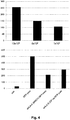
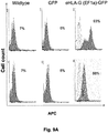
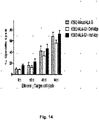
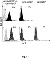
- a genetically modified cell has reduced immunogenicity and/or improved immunosuppression if it shows: (1) a reduction of NK-92 cytotoxicity of the genetically modified cell as compared to the mammalian cell without said genetic modification, (2) a reduction of in vitro peripheral blood mononuclear cell proliferation of the genetically modified cell as compared to the mammalian cell without said genetic modification, and/or (3) an increase in the size and weight of tumor formation by the genetically modified cell as compared to the mammalian cell without said genetic modification in humanized NSG mice.
- the genetically modified mammalian cell does not have matches ( i.e ., same allele(s)) in one or more HLA antigens as compared to the allogeneic recipient, wherein the HLA antigens are selected from the group consisting of HLA-A, HLA-B, HLA-C, HLA-DP, HLA-DQ, and HLA-DR.
- the genetically modified mammalian cell only has 1, 2, 3, 4, or 5 matches in one or more HLA antigens as compared to the allogeneic recipient, wherein the HLA antigens are selected from the group consisting of HLA-A, HLA-B, HLA-C, HLA-DP, HLA-DQ, and HLA-DR.
- the genetically modified mammalian cell has no matches in with the allogeneic recipient with respect to HLA-A, HLA-B, HLA-C, HLA-DP, HLA-DQ, and HLA-DR.
- the genetically modified cell comprises an HLA-G transgene without one or more amino acid mutations that reduce retention of HLA-G in the endoplasmic reticulum-golgi recycling pathway (i.e., an HLA-G wild-type consensus sequence such as SEQ ID NO: 1), but has an HLA-G transgene that comprises a 3' UTR (untranslated region) sequence that does not contain microRNA binding sites such as SEQ ID NO:3, or a sequence that does not comprise SEQ ID NO:4.
- the one or more mutations that reduce retention of HLA-G in the endoplasmic reticulum-golgi recycling pathway include a di-Lysine (KK) motif mutation.
- the KK motif mutation incudes a K334A mutation, a K335A mutation, or both mutations.
- the exogenous nucleic acid to be expressed in the genetically modified cell includes a 3' UTR sequence that does not contain SEQ ID NO.4. In one embodiment, where the 3' UTR sequence of the exogenous nucleic acid does not include SEQ ID NO:4, the nucleic acid sequence contains SEQ ID NO:3.
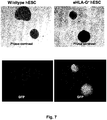
- the expressed HLA-G is present on the cell surface of the genetically modified mammalian cell.
- the genetically modified mammalian cell is a human cell, a mouse cell, a rat cell, a monkey cell, or a pig cell.
- the genetically modified mammalian cell is a stem cell, a progenitor cell, or a cell obtained by directed differentiation of the stem cell or the progenitor cell.
- the genetically modified mammalian cell is a cell that was already differentiated (whether naturally or in vitro) prior to introduction of an exogenous HLA-G transgene.
- the genetically modified mammalian cell is a stem cell (e.g., a pluripotent stem cell).
- the stem cell is an embryonic stem cell, an induced pluripotent stem cell, or a totipotent stem cell.
- the genetically modified mammalian cell is an embryonic stem cell. In another embodiment, the genetically modified mammalian cell is an induced pluripotent stem cell. In a further embodiment, the genetically modified mammalian cell is not of an immune system cell type. In another embodiment, the genetically modified mammalian cell is a cell obtained by in vitro differentiation of a stem cell or a progenitor cell wherein the stem cell or progenitor cell is genetically modified and then differentiated in vitro.
- the genetically modified cell is a fully differentiated cell, an epidermal progenitor cell, a pancreatic progenitor cell, a hematopoietic stem cell, a cell obtained by differentiation of the pluripotent stem cell, a keratinocyte, a fibroblast, a mesenchymal stem cell, a cardiomyocyte, a neural stem cell, a neuron, an astrocyte, or a pancreatic ⁇ cell progenitor.
- the expression vector is a transposon vector or a retroviral vector. In some embodiments, where the exogenous nucleic acid is an expression vector, the expression vector is a targeting vector, and the genetically modified mammalian cell was obtained by homologous recombination of the targeting vector. In some embodiments, the expression vector may further include a nucleic acid sequence encoding a reporter protein such as green fluorescent protein (GFP).
- GFP green fluorescent protein
- the exogenous nucleic acid also includes a nucleic acid sequence that (i) is at least 85% identical to the 3' untranslated region sequence of the human, HLA-G gene; and (ii) comprises at least one mutation that inhibits binding of a cognate microRNA to the mutated site within an mRNA comprising the mutated binding site within its 3' untranslated region.
- a nucleic acid sequence comprises SEQ ID NO:3.
- an artificial tissue is provided that contains the genetically modified cell.
- an isolated nucleic acid that includes (i) a first nucleic acid sequence that encodes an amino acid sequence at least 85% identical to human, HLA-G; and (ii) a second nucleic acid sequence that is at least 85% identical the 3' untranslated region sequence of the human HLA-G gene and operably linked to the first nucleic acid sequence, where the amino acid sequence comprises a mutation that reduces retention of HLA-G in the endoplasmic reticulum-golgi recycling pathway, and the second nucleic acid sequence comprises at least one mutation that inhibits binding of a cognate microRNA to an mRNA comprising the mutated binding site within its 3' untranslated region.
- the isolated nucleic acid the 3' untranslated region sequence does not comprise SEQ ID NO:4. In one embodiment, where the 3' untranslated region sequence does not comprise SEQ ID NO:4, the 3' untranslated region sequence comprises SEQ ID NO:3
- a mammalian expression vector that includes the isolated nucleic acid and a promoter operably linked to the first nucleic acid sequence, wherein the promoter is not silenced in a stem cell.
- the promoter contains the nucleic acid sequence of the Chinese hamster EF-1 ⁇ (CHEF-1 ⁇ ) promoter or human EF-1 ⁇ promoter.
- the CHEF-1 ⁇ promoter comprises SEQ ID NO:7.
- the promoter used to drive expression of an HLA-G transgene is a tissue or cell type-selective promoter.
- the mammalian expression vector includes comprising a third nucleic acid sequence encoding a reporter protein.
- the mammalian expression vector is a transposon vector.
- a genetically modified mammalian cell is provided that contains the mammalian expression vector.
- an isolated nucleic acid comprises: (i) a first nucleic acid sequence that encodes an amino acid sequence at least 95% identical to human HLA-G, wherein the amino acid sequence comprises a mutation that reduces retention of HLA-G in the endoplasmic reticulum-golgi recycling pathway; and (ii) a second nucleic acid sequence that is at least 95% identical the 3' untranslated region sequence of the human HLA-G gene and operably linked to the first nucleic acid sequence, wherein the second nucleic acid sequence comprises at least one mutation that inhibits binding of a cognate microRNA to an mRNA comprising the mutated binding site within its 3' untranslated region.
- the first nucleic acid sequence encodes an amino acid sequence of SEQ ID NO:2.
- the second nucleic acid sequence does not comprise SEQ ID NO:4.
- the second nucleic acid sequence comprises SEQ ID NO:3.
- a mammalian expression vector is provided that comprises said first and second nucleic acid sequences, and further comprises a promoter operably linked to the first nucleic acid sequence, wherein the promoter is not silenced in a stem cell or a in a cell generated by differentiation of the stem cell.
- a promoter can comprise the nucleic acid sequence of the Chinese hamster EF-1 ⁇ promoter.
- the mammalian expression vector further comprises a nucleic acid sequence encoding a reporter protein.
- the mammalian expression vector is a transposon vector.
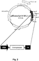
- the mammalian expression vector comprises all of the elements shown in Figure 1 .
- a mammalian expression vector comprises: (a) a Chinese hamster EF-1 ⁇ promoter, (b) a nucleic acid sequence that is operably linked to the promoter and that encodes human HLA-G with an amino acid sequence of SEQ ID NO:2, and (c) a 3'UTR sequence comprising SEQ ID NO:3.
- a genetically modified mammalian cell is provided that comprises such an expression vector.
- HLA-G modified mammalian cells are administered to a subject suffering from any of a number of conditions including, but not limited to cardiovascular disease, eye disease (e.g., macular degeneration), auditory disease, (e.g., deafness), diabetes, neurodegenerative disease, Alzheimer's Disease, Parkinson's Disease, multiple sclerosis, osteoporosis, liver disease, kidney disease, autoimmune disease, arthritis, gum disease, a dental condition, or a proliferative disorder (e.g., a cancer).
- an acute health condition e.g., stroke, spinal cord injury, burn, or a wound.
- the subject is suffering from loss of tissue such as lipatrophy or aging-related losses in collagen.
- the subject suffers from a non-healing ulcer, or is need for an agent to assist in closure of defects like hypospadias and epispadias.
- the subject is need for a permanent or temporary skin graft for would healing or for skin substitutes.
- the invention provides a universal method of cellular or tissue repair or regeneration to a subject in need thereof, the method comprising injecting or grafting to the subject a cellular or tissue composition comprising a population of eHLA-G modified cells, wherein the subject has at least one mismatched classical HLA class I or HLA class II molecule as compared to the population of eHLA-G modified cells, and wherein the population of eHLA-G modified cells exhibits reduced immunogenicity and/or improved immunosuppression as compared to cells of the same-type without the eHLA-G modification.
- the reduced immunogenicity and/or improved immunosuppression can be determined, for example, by comparing the eHLA-G modified cell to a control cell of the same type without the eHLA-G modification in an NK-92 cytotoxicity assay, a humanized NSG tumor growth assay, and/or a PBMC proliferation assay.
- the population of genetically modified cells comprises a population of eHLA-G genetically modified human dermal fibroblasts.
- the population of genetically modified cells comprises a population of eHLA-G genetically modified human epidermal progenitors.
- the population of genetically modified cells comprises a population of eHLA-G genetically modified human mesenchymal stem cells.
- the population of genetically modified cells comprises a population of eHLA-G genetically modified human embryonic stem cells. In another embodiment, the population of genetically modified cells comprises a population of cell differentiated in vitro from eHLA-G genetically modified human embryonic stem cells. In other embodiments, the population of genetically modified cells are not rejected by the subject's immune system for at least 2, 4, 6, 8, 10, 12, 14, 16, 18, 20, 24, 36, 48, or 52 weeks.
- the invention provides a method for regenerating skin to a subject in need thereof, the method comprising injecting a population of eHLA-G modified dermal fibroblasts and/or eHLA-G modified embryonic epidermal progenitors to a site of skin injury on the subject, wherein the subject has at least one mismatched classical HLA class I or HLA class II molecule as compared to the population of eHLA-G modified dermal fibroblasts and/or eHLA-G modified embryonic epidermal progenitors.
- a cell therapy method comprises administering to a subject in need thereof a population of genetically modified mammalian cells comprising an exogenous human ⁇ 2-microglobulin ( ⁇ 2m) molecule and an eHLA-G transgene of the invention.
- the present disclosure features genetically modified mammalian cells that express exogenous HLA-G persistently (HLA-G modified cells), as well as nucleic acid compositions to generate such modified mammalian cells.
- HLA-G modified cells genetically modified mammalian cells that express exogenous HLA-G persistently
- nucleic acid compositions to generate such modified mammalian cells.
- the eHLA-G genetic modifications described herein provide the cells with characteristics of reduced immunogenicity and/or improved immunosuppression, such that these cells have the promise of being universal or improved donor cells for transplants, cellular and tissue regeneration or reconstruction, and other therapies.
- compositions I. Compositions :
- HLA-G modified cells a wide range of mammalian cell types that express exogenous HLA-G (HLA-G modified cells) can be generated.
- Such cell types include, but are not limited to, totipotent cells, embryonic stem cells (e.g., human embryonic stem cells), induced pluripotent stem cells (e.g., human induced pluripotent stem cells), multipotent stem cells, epidermal progenitor cells, mesenchymal stem cells, pancreatic ⁇ cell progenitors, pancreatic ⁇ cells, cardiac progenitors, cardiomyocytes, hepatic progenitors, hepatocytes, muscle cell progenitors, muscle cells, kidney cells, osteoblasts, hematopoietic progenitors, dental follicle cells, hair follicle cells, retinal pigment epithelial cells, neural stem cells, neurons, astrocytes, oligodendrocytes, inner ear cells, and fibroblasts (including human dermal fibroblast (
- the HLA-G modified cells are not cells having an immune system cell type.
- mammalian cells can be derived one of several species including, e.g., human, mouse, rat, monkey, or pig.
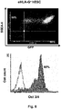
- any cell-type can be transfected with the constructs described herein and then tested for HLA-G expression and how such expression can impart reduced immunogenicity and/or improved immunosuppression to the modified cell.
- a genetically modified pluripotent stem cell line such as a human embryonic stem cell line, or a human induced pluripotent stem cell line, or any cell line that has multipotent traits including mesenchymal stem cells and immune system progenitor cells, that expresses HLA-G is generated and then subjected to directed differentiation to obtain a cell population that expresses HLA-G and that is substantially enriched for a desired cell type.
- the substantially enriched cell population includes at least about 2% to about 100% of the desired cell type, e.g., at least about 3%, 4%, 5%, 7%, 8%, 10%, 20%, 22%, 25%, 35%, 40%, 45%, 50%, 60%, 70%, 75%, 80%, 85%, or another percentage of the desired cell type from at least about 2% to about 100%.
- Methods for enriching cells of a desired cell type are known in the art. See, e.g., U.S. Patent Application No. 12/532,512
- genetically modified mammalian pluripotent or multipotent stem cell lines e.g., human embryonic stem cells or human induced pluripotent stem cells, etc.
- the cell line is genetically modified by stable transfection with one or more nucleic acid expression vectors (e.g., a plasmid vector or a minicircle vector) that include an expression cassette for expression of an HLA-G protein as described herein, a selection marker, and optionally a reporter protein.
- nucleic acid expression vectors e.g., a plasmid vector or a minicircle vector
- suitable selection markers encoded by such vectors include proteins that confer resistance to a selection agent.
- proteins and their corresponding selection agents include, without limitation, puromycin N-acetyltransferase (puromycin), hygromycin phosphotransferase (hygromycin), blasticidin-S-deaminase (blasticidin), and neomycin phosphotransferase (neomycin).
- Selection with the appropriate selection agent may last for at least about 3 to about 14 days, e.g., about 4, 5, 6, 7, 8, 9, 10, 12, 13, or another period from about 3 to about 14 days until resistant colonies are apparent.
- fluorescent reporter proteins include, but are not limited to, EGFP and its variants such as YFP, Cyan, and dEGFPs; DS-Red, monomeric Orange, the far-red fluorescent protein "Katushka” ( Shcherbo et al (2007), Nat Methods, 4:741-746 ), or variants of any of the foregoing.
- the reporter is an enzyme that converts a substrate that, in the process, yields a detectable signal, e.g., a fluorescent or luminescent signal in the presence of a fluorogenic or luminogenic substrate, respectively.
- the selection marker enzyme comprises the amino acid sequence of a luciferase, e.g., a firefly luciferase, click beetle luciferase, or Renilla luciferase.
- Luciferase activity can be detected by providing an appropriate luminogenic substrate, e.g., firefly luciferin for firefly luciferase or coelenterazine for Renilla luciferase.
- Luciferase activity in the presence of an appropriate substrate can be quantified by luminometry to assay total luciferase activity of whole cell populations in culture dish wells, or, alternatively, luciferase activity of individual cells or colonies can be detected by use of a microscope in combination with a photon counting camera. Details of luciferase assays, including high-throughput methods, are disclosed in, e.g., U.S. Pat. Nos. 5,650,135 , 5,744,320 , and 6,982,431 .
- the reporter enzyme comprises the amino acid sequence of a modified beta-lactamase, the expression of which can be detected and quantified in living cells by a ratiometric fluorescence assay for breakdown of fluorogenic .beta.-lactam substrates as described in, e.g., U.S. Pat. Nos. 5,741,657 , 6,031,094 , and U.S. Patent Publication No. 20070184513 . See also Qureshi (2007), Biotechniques, 42(1):91-96 for a review.
- Other suitable reporter enzymes include, but are not limited to, the Halo-Tag® hydrolase (Promega, Madison, Wis., as described in, e.g., U.S. Pat. No. 7,238,842 and Patent Publication Nos. 20080026407 and 20080145882 ) and beta.-galactosidase.
- the genetically modified mammalian cells are also genetically modified to express a human ⁇ 2 microglobulin (GenBank Accession No. AY187687.1). Without wishing to be bound by theory, it is believed that expression of human ⁇ 2 microglobulin will enhance surface expression of transgenic HLA-G in the genetically modified mammalian cells.
- the nucleic acid expression vector is a transposon vector that includes transposition elements that facilitate integration of the transposition of the transposon vector into a host genome when introduced into a host cell in the presence of a cognate transposase (e.g., the piggyBAC transposase), as described in, e.g., U.S. Patent Application No. 12/728,943 .
- Transposon expression vectors e.g., PiggyBac vectors
- transposase expression vectors are commercially available from, e.g., System Biosciences (Mountain View, CA).
- the expression vector may be a targeting vector, which allows site-specific integration of the eHLA-G transgene in the host cell genome.
- targeting vectors is routine in the art as exemplified by U.S. Patent No. 5,464,764 .
- genetically modified cells e.g., differentiated, multipotent, pluripotent, or totipotent stem cell lines
- a recombinant virus examples include, but are not limited to, retroviruses (including lentiviruses); adenoviruses; and adeno-associated viruses.
- the recombinant retrovirus is murine moloney leukemia virus (MMLV), but other recombinant retroviruses may also be used, e.g., Avian Leukosis Virus, Bovine Leukemia Virus, Murine Leukemia Virus (MLV), Mink-Cell focus-Inducing Virus, Murine Sarcoma Virus, Reticuloendotheliosis virus, Gibbon Ape Leukemia Virus, Mason Pfizer Monkey Virus, or Rous Sarcoma Virus, see, e.g., U.S. Pat. No. 6,333,195 .
- MMLV murine moloney leukemia virus
- retroviruses may also be used, e.g., Avian Leukosis Virus, Bovine Leukemia Virus, Murine Leukemia Virus (MLV), Mink-Cell focus-Inducing Virus, Murine Sarcoma Virus, Reticuloendotheliosis virus, Gibbon Ap
- the recombinant retrovirus is a lentivirus (e.g., Human Immunodeficiency Virus-1 (HIV-1); Simian Immunodeficiency Virus (SIV); or Feline Immunodeficiency Virus (FIV)), See, e.g., Johnston et al., (1999), Journal of Virology, 73(6):4991-5000 (FIV); Negre et al., (2002), Current Topics in Microbiology and Immunology, 261:53-74 (SIV); Naldini et al., (1996), Science, 272:263-267 (HIV).
- HIV-1 Human Immunodeficiency Virus-1
- SIV Simian Immunodeficiency Virus
- FMV Feline Immunodeficiency Virus
- the recombinant retrovirus may comprise a viral polypeptide (e.g., retroviral env) to aid entry into the target cell.
- a viral polypeptide e.g., retroviral env
- the viral polypeptide may be an amphotropic viral polypeptide, e.g., amphotropic env, that aids entry into cells derived from multiple species, including cells outside of the original host species. See, e.g., id.
- the viral polypeptide may be a xenotropic viral polypeptide that aids entry into cells outside of the original host species. See, e.g., id.
- the viral polypeptide is an ecotropic viral polypeptide, e.g., ecotropic env, that aids entry into cells of the original host species. See, e.g., id.
- Viral transduction of cells may be accomplished by any method known in the art. e.g., Palsson, B., et al., (1995), WO95/10619 ; Morling, F. J. et al., (1995), Gene Therapy, 2:504-508 ; Gopp et al., (2006), Methods Enzymol, 420:64-81 .
- the infection may be accomplished by spin-infection or "spinoculation" methods that involve subjecting the cells to centrifugation during the period closely following the addition of virus to the cells.
- virus may be concentrated prior to the infection, e.g., by ultracentrifugation.
- the multiplicity of infection (m.o.i.) used to transduce the cells to be genetically modified can range from about 1 m.o.i. to about 50 m.o.i., e.g., about 1 m.o.i., about 5 m.o.i., about 7.5; m.o.i., about 10 m.o.i., about 15 m.o.i., about 20 m.o.i., about 30 m.o.i., about 40 m.o.i., or about 50 m.o.i.;
- pluripotent stem cells e.g., human embryonic stem cells or human induced pluripotent stem cells
- directed differentiation are known in the art, as described in, e.g., U.S. Patent Nos. 7,955,849 , 7,763,466 , 7,264,968 ; and U.S. Patent Application Application Nos. 12/179,462 , and 12/ 187,543.
- human embryonic epidermal progenitors are derived from a human pluripotent stem cell line, e.g., human embryonic stem cells or human induced pluripotent stem cells as follows.
- Pluripotent stem cells are maintained in Embryonic Stem Cell (ESC) growth medium containing DMEM/F12 (1:1) supplemented with 20% knockout serum replacement, 0.1 mM MEM non-essential amino acids, 1 mM GlutaMax, 0.1 mM ⁇ -mercaptoethanol (Sigma).
- ESC growth medium is conditioned by plating mitotically inactivated mouse embryonic fibroblasts (MEFs) (CF-1, ATCC) at a density of 5x10 4 cells/cm 2 and incubating for 18-24 hours. After conditioning, 4 ng/ml bFGF is added and complete conditioned medium is sterile filtered.
- MEFs mouse embryonic fibroblasts
- hESCs are subcultured every 5-6 days (at a 1:3 or 1:4 split) on Matrigel® (BD Biosciences)-coated plates using 1 mg/ml Dispase to remove cell colonies.
- hEEPs Differentiation of pluripotent stem cells into K14 + /p63 + hEEPs, by first culturing the pluripotent stem cells in 6-well plates for 4 days in ESC growth medium and then changing over to 2 ml/well of differentiation medium, comprised of unconditioned hESC growth medium containing 1 ⁇ M all-trans retinoic acid (Sigma) and 25 ng/ml BMP4.
- DSFM defined keratinocyte serum-free medium
- hEEPs To enhance purity of isolated hEEPs, cells were sorted using magnetic activated cell sorting (MASC) with CD29 antibodies. About 92 percent of CD29 MASC sorted hEEP cell culture cells differentiated from eHLA-G(EF-1 ⁇ )-GFP modified hESCs were positive for K14, a specific keratinocyte marker.
- MSC magnetic activated cell sorting
- immunogenicity may be reduced by at least about 5% to about 95%, relative to a corresponding cell type that does not express exogenous HLA-G, e.g., about 6%, 7%, 10%, 12%, 15%, 20%, 30%, 40%, 50%, 65%, 70%, 80%, 85%, 90%, or another percent reduced immunogenicity relative to cells of the same cell type that do not express exogenous HLA-G.
- HLA-G modified mammalian cells e.g., human embryonic stem cells, or differentiated cells from HLA-G modified embryonic stem cells, or cells that are already fully differentiated prior to modification, etc.
- unmodified mammalian cells are cultured in the presence of an allogenic natural killer cell line (e.g., NK-92) and then cytotoxicity to the HLA-G modified versus unmodified cells by induced by the NK-92 cells is determined by any of a number of standard cell viability assays.
- an allogenic natural killer cell line e.g., NK-92
- the isolated nucleic acids used to generated HLA-modified mammalian cells, as described herein, contain an enhanced HLA-G "eHLA-G" transgene that drives increased cell surface expression and/or secretion of HLA-G relative to cell surface expression driven by a wildtype HLA-G transgene.
- eHLA-G enhanced HLA-G
- Such a transgene typically includes at least three distinct components: a promoter and 5' untranslated region (5' UTR) sequence; a coding sequence; and a 3' untranslated region (3' UTR) sequence.
- the promoter to be used to drive eHLA-G transgene expression is one capable of driving expression of the eHLA-G transgene in a cell type of interest for a period of at least about seven weeks to about 50 weeks, e.g., 8 weeks, 9 weeks, 10 weeks, 12 weeks, 15 weeks, 20 weeks, 25 weeks, 30 weeks, 35 weeks, 40 weeks, 42 weeks, 45 weeks, 47 weeks, 48 weeks, or another period from at least about seven weeks to about 50 weeks.
- Promoters capable of driving expression for such extend periods of time are effective in evading silencing that occurs in a number of cell types including, e.g., stem cells such as embryonic stem cells, induced pluripotent stem cells, or mesenchymal stem cells.
- Suitable, silencing-resistant promoters include, but are not limited to, the Chinese hamster elongation factor-1 alpha (CHEF-1 ⁇ ) promoter (see Running Deer et al (2004), Biotechnol. Prog., 20:880-889 ; and GenBank Accession No. AY188393.1), the M-U3/R-variant promoter of the Murine Stem Cell Virus (MSCV) promoter (as described in Swindle et al (2004), J Biol Chem, 279:34-41 ), the phosphoglycerate kinase (PGK) promoter, the human ⁇ -actin promoter, and the ubiquitin C promoter.
- CHEF-1 ⁇ Chinese hamster elongation factor-1 alpha
- MSCV Murine Stem Cell Virus
- PGK phosphoglycerate kinase
- the promoter used to drive expression of an eHLA-G transgene is expressed in one or more desired cell types at a level that is higher than in other cell types.
- a promoter that is active within or even selective for that particular cell type may be advantageous to select a promoter that is active within or even selective for that particular cell type.
- the expression level may be about two fold to 100 fold higher in the desired cell type compared to another cell type, e.g., about 3 fold, 4 fold, 5 fold, 10 fold, 20 fold, 25 fold, 30 fold, 40 fold, 50 fold, 70 fold, 80 fold, 90 fold, or another fold higher level of expression in the desired cell type compared to another cell type.
- tissue and/or cell type-selective promoters include, but are not limited to, the promoters for: Neuron-Specific Enolase (neuronal), Synapsin (neuronal), CamKII (forebrain neurons), HB9 (motor neurons), and Dopamine Transporter (dopaminergic neurons); Glial Fibrillary Acidic Protein (astrocytes); Albumin (liver); ⁇ -Myosin Heavy Chain ( ⁇ MHC-cardiomyocytes); Neurogenin 3 and Pancreas-Duodenum Homeobox 1 (pancreas); Keratin 14 (skin); and Bestrophin1 (retinal pigment epithelium);
- Neuron-Specific Enolase neuron-Specific Enolase
- Synapsin neuroneuronal
- CamKII forebrain neurons
- HB9 motor neurons
- Dopamine Transporter dopaminergic neurons
- Glial Fibrillary Acidic Protein astrocytes
- Albumin liver
- the eHLA-G transgene sequence encodes an HLA-G protein that contains at least one to about ten point mutations relative to the human (GenBank No. NP_002118.1), or chimpanzee consensus sequence, e.g., 2, 3, 4, 5, 6, 7, 8, 9, or 10 point mutations relative to the above-mentioned HLA-G protein consensus sequences.
- the human consensus wildtype sequence (SEQ ID NO:1) is shown below:
- the at least one to about ten point mutations increase the level of expression of the HLA-G protein on the cell surface of the expressing host cell by reducing retention of HLA-G in the endoplasmic reticulum during processing and maturation of the protein.
- Such mutations include, for example, an HLA-G "KK" motif mutation. See, e.g., Park et al (2001), Immunity, 15:213-224 .
- the KK motif mutation includes a K334A mutation, a K335A mutation, or both substitution mutations.
- the substitution may be made with a different aliphatic amino (e.g., leucine) acid or another type of type of amino acid that is non-basic.
- the encoded HLA-G protein has the amino acid sequence of (SEQ ID NO:2) , in which K334A and K335A substitutions, relative to the wildtype sequence, have been introduced (underlined):
- an eHLA-G transgene encodes an HLA-G protein the amino acid sequence of which is at least 75% to 100% identical to that of SEQ ID NO:2 , e.g., 77%, 80%, 82%, 85%, 87%, 88%, 90%, 92%, 95%, 96%, 97%, 98%, 99%, or another percent identical to the amino acid sequence of SEQ ID NO:2 .
- the eHLA-G transgene disclosed herein also includes a 3' untranslated (3' UTR) region containing a number of regulatory elements affecting the expression/translational efficiency of HLA-G transcripts.
- the eHLA-G transgene 3' UTR sequence is a nucleic acid sequence that includes a nucleic acid sequence that is at least 75% identical to the sequence of (SEQ ID NO:3) , e.g., at least 77%, 80%, 82%, 85%, 87%, 88%, 90%, 92%, 94%, 95%, 96%, 97%, or another percent identical to the sequence of HLA-G 3' UTR (SEQ ID NO:3) :
- Such sequences contain mutations (underlined) that decrease binding of two microRNA binding sites that result in increased expression of the trangene-encoded HLA-G.
- These sequences include a deletion of a 14 base pair sequence present in exon 8 of some HLA-G alleles. This 14 base pair sequence is shown below as SEQ ID NO NO:4 : SEQ ID NO:4 14-bp insertion sequence in HLA 3' UTR ATTTGTTCATGCCT
- SEQ ID NO:5 shown below, corresponds to SEQ ID NO:3 with the insertion of this 14 base pair sequence (lower case):
- HLA-G alleles having a 3' UTR yield a more stable HLA-G mRNA transcript. See Rouseau et al (2003), Human Immunology, 64:1005-1010 .
- expression of the allele with a 14 base pair deletion appeared to be more immunoprotective than the allele that includes the 14 pair insertion. See Fig. 13 .
- the effect of the 14 bp deletion on expression levels of HLA-G may be cell type-specific, e.g., the 14 bp deletion apparently enhances expression of eHLA-G in human cells, including at least hESCs, hEEPs, and human dermal fibroblastcells.
- the nucleic acid sequence containing the eHLA-G transgene also includes insulators sequences that flank the eHLA-G expression cassette. Insulator sequences mitigate genomic position effects that could spuriously affect expression of an integrated exogenous expression cassette.
- the insulator sequences to be used contain the chicken ⁇ -globin HS4 core insulator sequence.
- the nucleic acid containing the eHLA-G transgene will also include a selection marker as described herein.
- the isolated nucleic acid containing the eHLA-G transgene may further contain a reporter protein as described herein.
- generation of an eHLA-G-modified cell line is generated by the use of a transposon vector that comprises an eHLA-G expression cassette, but does not contain expression cassettes for a selection marker or a reporter protein.
- the vector is introduced to the cells to be modified along with a transposase expression vector, followed by limiting dilution cloning. As the population of cells transfected is very efficiently modified, the need to use selection markers or reporter proteins can be avoided. This is an important consideration, especially for cells to be used in the context of cell therapy in human patients.
- the percentage of cells that is successfully modified by transposon-based stable transfection methods can range from about 0.5% to about 50%, e.g., 1%, 2%, 3%, 5%, 7%, 8%, 15%, 20%, 22%, 30%, 40%, or another percentage from about 0.5% to about 50% of the cells transfected.
- a polycistronic expression cassette may incorporate one or more internal ribosomal entry site (IRES) sequences between open reading frames incorporated into the polycistronic expression cassette.
- IRES sequences and their use are known in the art as exemplified in, e.g., Martinez-Salas (1999), Curr Opin Biotechnol, 10(5):458-464 .
- multiple open reading frames may be linked to each other by an intervening 2A peptide sequence of the foot-and-mouth disease virus (F2A) or 2A-like sequences from other viruses.
- sequence comparison algorithm test and reference sequences typically are input into a computer, subsequence coordinates are designated, if necessary, and sequence algorithm program parameters are designated. The sequence comparison algorithm then calculates the percent sequence identity for the test sequence(s) relative to the reference sequence, based on the designated program parameters.
- sequence algorithm program parameters are designated.
- Examples of such programs include the MATCH-BOX, MULTAIN, GCG, FASTA, and ROBUST programs for amino acid sequence analysis, and the SIM, GAP, NAP, LAP2, GAP2, and PIPMAKER programs for nucleotide sequences.
- Preferred software analysis programs for both amino acid and polynucleotide sequence analysis include the ALIGN, CLUSTALW (e.g., version 1.6 and later versions thereof), and BLAST programs (e.g., BLAST 2.1, BL2SEQ, and later versions thereof).
- Identity (sometimes referred to as “overall identity”-in contrast to "local identity,” which is discussed further herein) with respect to amino acid or nucleotide sequences refers to the percentage of amino acid residues or nucleotide bases, respectively, that are identical in the two amino acid or nucleotide sequences when two such amino acid sequences or two such nucleotide sequences are optimally aligned with one another. If, in the optimal alignment, a position in a first sequence is occupied by the same amino acid residue or nucleotide residue as the corresponding position in the second corresponding amino acid or nucleotide sequence, the sequences exhibit identity with respect to that residue position.
- nucleic acids that that hybridize specifically under low, medium, or high stringency conditions to a probe of at least 100 nucleotides from a nucleic acid encoding the amino acid sequence of SEQ ID NO:2 or to the nucleic acid sequence of SEQ ID NOS:3 OR 5 .
- Low stringency hybridization conditions include, e.g., hybridization with a 100 nucleotide probe of about 40% to about 70% GC content; at 42 °C in 2X SSC and 0.1% SDS.
- Medium stringency hybridization conditions include, e.g., at 50 °C in 0.5X SSC and 0.1% SDS.
- High stringency hybridization conditions include, e.g., hybridization with the above-mentioned probe at 65 °C. in 0.2X SSC and 0.1% SDS. Under these conditions, as the hybridization temperature is elevated, a nucleic acid with a higher sequence homology is obtained.
- compositions comprising eHLA-G Genetically Modified Cells
- compositions comprising or generated using one or more HLA-G modified mammalian cell types.
- eHLA-G modified hESCs displayed stable and persistent HLA-G expression, even through directed differentiation into hEEPs.
- the stable and persistent HLA-G expression provided the genetically modified cells with reduced immunogenicity and/or improved immunosuppression.
- eHLA-G modified human dermal fibroblasts which are fully differentiated cells, also have stable and persistent HLA-G expression that provides reduced immunogenicity and/or improved immunosuppression.
- the eHLA-G constructs described herein can be used to generate universal donor cells of any type, whether from directed differentiation of a genetically modified pluripotent or multipotent cell, or from genetic modification of a fully differentiated cell.
- a topical composition for skin regeneration or repair comprises a genetically modified dermal fibroblast cell comprising an eHLA-G transgene as described herein.
- a pharmaceutical composition for injection is provided that comprises a genetically modified dermal fibroblast cell comprising an eHLA-G transgene as described herein.
- a skin graft composition is provided that comprises a genetically modified dermal fibroblast cell comprising an eHLA-G transgene as described herein.
- a permanent skin graft composition is provided that comprises a genetically modified embryonic epidermal progenitor cell comprising an eHLA-G transgene as described herein.
- biocompatible synthetic scaffolds for artificial tissue and methods for their generation are described in the art and may be used with the HLA-G modified cells described herein to produce an artificial tissue having reduced immunogenicity and/or improved immunosuppression as compared to tissues containing cells that do not express exogenous HLA-G. See, e.g., U.S. Patent No. 7,960,166 entitled "Microfabricated compositions and processes for engineering tissues containing multiple cell types.
- HLA-G human leukocyte antigen
- HLA-G modified cells that stably express eHLA-G (and optionally in addition, exogenous human ⁇ 2 microglobulin), as described herein, may be used as for therapy.
- the therapy may be directed at treating the cause of the disease; or alternatively, the therapy may be to treat the effects of the disease or condition.
- the genetically modified cells may be transferred to, or close to, an injured site in a subject; or the cells can be introduced to the subject in a manner allowing the cells to migrate, or home, to the injured site.
- the transferred cells may advantageously replace the damaged or injured cells and allow improvement in the overall condition of the subject.
- the transferred cells may stimulate tissue regeneration or repair, including skin regeneration or skin repair.
- HLA-G modified mammalian cells are administered to a subject suffering from any of a number of conditions including, but not limited to cardiovascular disease, eye disease (e.g., macular degeneration), auditory disease, (e.g., deafness), diabetes, neurodegenerative disease, Alzheimer's Disease, Parkinson's Disease, multiple sclerosis, osteoporosis, liver disease, kidney disease, autoimmune disease, arthritis, gum disease, a dental condition, or a proliferative disorder (e.g., a cancer).
- an acute health condition e.g., stroke, spinal cord injury, burn, or a wound.
- the subject is suffering from loss of tissue such as lipatrophy or aging-related losses in collagen.
- the subject suffers from a non-healing ulcer, or is need for an agent to assist in closure of defects like hypospadias and epispadias.
- the subject is need for a permanent or temporary skin graft for would healing or for skin substitutes.
- the invention provides a universal method of cellular or tissue grafting to a subject in need thereof, the method comprising injecting or grafting to the subject a cellular or tissue composition comprising a population of eHLA-G modified cells, wherein the subject has at least one mismatched classical HLA class I or HLA class II molecule as compared to the population of eHLA-G modified cells, and wherein the population of eHLA-G modified cells exhibits reduced immunogenicity and/or improved immunosuppression as compared to cells of the same-type without the eHLA-G modification.
- the reduced immunogenicity and/or improved immunosuppression can be determined, for example, by comparing the eHLA-G modified cell to a control cell of the same type without the eHLA-G modification in an NK-92 cytotoxicity assay, a humanized NSG tumor growth assay, and/or a PBMC proliferation assay.
- the invention provides a method for regenerating skin to a subject in need thereof, the method comprising injecting a population of eHLA-G modified dermal fibroblasts and/or eHLA-G modified embryonic epidermal progenitors to a site of skin injury on the subject, wherein the subject has at least one mismatched classical HLA class I or HLA class II molecule as compared to the population of eHLA-G modified dermal fibroblasts and/or eHLA-G modified embryonic epidermal progenitors.
- HLA-G modified cell types to be administered to a subject in need thereof include, but are not limited to, epidermal progenitor cells, mesenchymal stem cells, pancreatic ⁇ cell progenitors, pancreatic ⁇ cells, cardiac progenitors, cardiomyocytes, hepatic progenitors, hepatocytes, muscle cell progenitors, muscle cells, kidney cells, osteoblasts, hematopoietic progenitors, dental follicle cells, hair follicle cells, retinal pigment epithelial cells, neural stem cells, neurons, astrocytes, oligodendrocytes, or any combination thereof.
- mammalian cells can be derived one of several species including, e.g., human, mouse, rat, monkey, or pig.
- the therapy may be directed at treating the cause of the disease; or alternatively, the therapy may be to treat the effects of the disease or condition.
- the HLA-G modified cells may be transferred to, or close to, an injured site in a subject; or the cells can be introduced to the subject in a manner allowing the cells to migrate, or home, to the injured site.
- the transferred cells may advantageously replace the damaged or injured cells and allow improvement in the overall condition of the subject. In some instances, the transferred cells may stimulate tissue regeneration or repair.
- the transferred cells may be cells differentiated from HLA-G modified pluripotent (or totipotent) stem cells.
- the transferred cells also may be multipotent stem cells differentiated from pluripotent, HLA-G modified cells.
- the number of administrations of treatment to a subject may vary. Introducing the HLA-G modified and/or differentiated cells into the subject may be a one-time event; but in certain situations, such treatment may elicit improvement for a limited period of time and require an on-going series of repeated treatments. In other situations, multiple administrations of the cells may be required before an effect is observed. As will be appreciated by those of ordinary skill in the art, the exact treatment protocols will depend upon the disease or condition, and the stage of the disease and parameters of the individual subject being treated.
- the HLA-G modified cells may be introduced to the subject via any of the following routes: parenteral, intravenous, intraarterial, intramuscular, subcutaneous, transdermal, intratracheal, intraperitoneal, or into spinal fluid.
- the HLA-G modified cells may be differentiated into cells and then transferred to subjects suffering from a wide range of diseases or disorders.
- Pancreatic islet cells may be transplanted into a subject suffering from diabetes (e.g., diabetes mellitus, type 1), see e.g., Burns et al., (2006) Curr. Stem Cell Res. Ther., 2:255-266 .
- diabetes e.g., diabetes mellitus, type 1
- pancreatic beta cells derived from HLA-G modified cells are transplanted into a subject suffering from diabetes (e.g., diabetes mellitus, type 1).
- hepatic cells or hepatic stem cells derived from HLA-G modified cells are transplanted into a subject suffering from a liver disease, e.g., hepatitis, cirrhosis, or liver failure.
- a liver disease e.g., hepatitis, cirrhosis, or liver failure.
- Degenerative heart diseases such as ischemic cardiomyopathy, conduction disease, and congenital defects could benefit from stem cell therapies. See, e.g., Janssens et al., (2006), Lancet, 367:113-121 .
- Hematopoietic cells or hematopoietic stem cells (HSCs) derived from HLA-G modified cells may be transplanted into a subject suffering from cancer of the blood, or other blood or immune disorder.
- cancers of the blood that are potentially treated by hematopoietic cells or HSCs include: acute lymphoblastic leukemia, acute myeloblastic leukemia, chronic myelogenous leukemia (CML), Hodgkin's disease, multiple myeloma, and non-Hodgkin's lymphoma.
- CML chronic myelogenous leukemia
- Hodgkin's disease multiple myeloma
- non-Hodgkin's lymphoma Often, a subject suffering from such disease must undergo radiation and/or chemotherapeutic treatment in order to kill rapidly dividing blood cells.
- Introducing HSCs derived from HLA-G modified cells to these subjects may help to repopulate depleted reservoirs of cells.
- HLA-G modified cells may be differentiated into neural stem cells or neurons and then transplanted to an injured site to treat a neurological condition, e.g., Alzheimer's disease, Parkinson's disease, multiple sclerosis, cerebral infarction, spinal cord injury, or other central nervous system disorder, see, e.g., Morizane et al., (2008), Cell Tissue Res., 331(1):323-326 ; Coutts and Keirstead (2008), Exp. Neurol., 209(2):368-377 ; Goswami and Rao (2007), Drugs, 10(10):713-719 .
- a neurological condition e.g., Alzheimer's disease, Parkinson's disease, multiple sclerosis, cerebral infarction, spinal cord injury, or other central nervous system disorder, see, e.g., Morizane et al., (2008), Cell Tissue Res., 331(1):323-326 ; Coutts and Keirstead (2008), Exp. Neurol., 20
- the HLA-G modified cells may be differentiated into dopamine-acting neurons and then transplanted into the striate body of a subject with Parkinson's disease.
- neural stem cells may be differentiated into oligodendrocytes or progenitors of oligodendrocytes, which are then transferred to a subject suffering from MS.
- a successful approach may be to introduce neural stem cells to the subject.
- the HLA-G modified cells may be differentiated into neural stem cells followed by transplantation into the injured site.
- the HLA-G modified cells may also be engineered to respond to cues that can target their migration into lesions for brain and spinal cord repair, e.g., Chen et al., (2007), Stem Cell Rev., 3(4):280-288 .
- the HLA-G modified cells to be in cell therapy methods also express a reporter protein as described herein.
- the reporter protein to be used is one that facilitates in vivo detection (e.g., imaging) of the introduced cells.
- the cells may express a far-red emitting fluorescent protein such as Katushka, whose long excitation and emission wavelengths are well suited to imaging in tissues.
- Katushka is commercially available under the tradename "TurboFP635" (Evrogen, Moscow, Russia).
- MSCs were serially passaged and monitored for changes in the expression levels of the selected genes. By passage 3, native HLA-G expression was absent in MSCs, although other candidate markers remained unchanged. HLA-G expression was not altered in Jeg-3 cells. MSCs were then isolated based on expression of cell surface HLA-G using FACS and found that between 0.5-3% of MSCs expressed HLA-G at the cell surface. To assess the ability of HLA-G + MSCs to escape donor rejection in vivo , 1-3 x10 5 cells were injected into the tail veins of immunocompetent mice.
- HLA-G+ MSCs and Jeg-3 cells
- HSCs and Jurkat cells both HLA-G -
- Table 2 shows that 1 week post-transplantation, HLA-G+ MSCs (and Jeg-3 cells) demonstrated a 24-and 270-fold survival advantage over HSCs and Jurkat cells (both HLA-G - ), respectively.
- the advantage of HLA-G + MSCs increased to 27-and 311-fold.
- HLA-G - MSCs survived at the same rate as HSCs, suggesting that the HLA-G + subpopulation of MSCs may exhibit enhanced tolerizing effects relative to unsorted MSCs in an in vivo setting.
- HLA-G + HF human fibroblast
- K562 human fibroblast
- NK-mediated cytotoxicity was reduced by 75% in HLA-G + HF and virtually eliminated in HLA-G + K562 cells. This is consistent with the observed protection of HLA-G + Jeg-3, which was reversed by incubation with the neutralizing anti-HLA-G (87G) antibody but not an isotype control.
- ESC growth medium contains DMEM/F12 (1:1) supplemented with 20% knockout serum replacement, 0.1 mM MEM non-essential amino acids, 1 mM GlutaMax, 0.1 mM ⁇ -mercaptoethanol (Sigma).
- ESC growth medium was conditioned by plating mitotically inactivated mouse embryonic fibroblasts (MEFs) (CF-1, ATCC) at a density of 5x10 4 cells/cm 2 and incubating for 18-24 hours. After conditioning, 4 ng/ml bFGF was added and complete conditioned medium was sterile filtered.
- MEFs mitotically inactivated mouse embryonic fibroblasts
- hESCs was subcultivated every 5-6 days (1:3 or 1:4 split) on Matrigel-coated plates using 1 mg/ml Dispase to remove cell colonies.
- K14 + /p63 + hEEPs were generated according to the method of Metallo et al supra. Briefly, hESCs were cultured in 6-well plates for 4 days and then treated with 2 ml/well of differentiation medium, comprised of unconditioned hESC growth medium containing 1 ⁇ M all-trans retinoic acid (Sigma) and 25 ng/ml BMP4.
- PCR amplification was performed in the Hybaid Omnigene thermal cycler (Bio-rad, Hercules, CA) using specific primers of the genes of interest as shown in Fig. 12 .
- PCR conditions consisted of 35 cycles at 94 °C for 30 s, 65 °C for 1 min, and 72 °C for 1 min with a final extension at 72 °C for 10 min.
- Ten ⁇ l of each PCR product was detected by ethidium bromide gel electrophoresis.
- HLA-G transgene in differentiated hEEPs was confirmed by flow cytometry. Both HLA-G total expression and surface expression was robust for differentiated eHLA-G(EF-1a)-GFP-hEEPs (greater than 90% of cells) as compared to control cells with no exogenous HLA-G (GFP only hEEPs) and wild-type hEEPs (see Figure 17 ).Only clones that yield a similar differentiation potential as wildtype cells were selected for further study.
- a novel HLA-G construct was designed by combining multiple modifications: 1) mutation of HLA-G's ER retrieval motif (K334A/K335A); and 2) mutation of HLA-G's 3' UTR microRNA binding sites. Since viral gene delivery systems remain a serious regulatory challenge, the PiggyBac system was used, a transposon-based, nonviral approach that was recently shown to achieve a 90% transfection efficiency in H1 hESCs ( Lacoste et al (2009), Cell Stem Cell, 5:332-342 .). This system requires a donor plasmid containing the transposon ( Fig. 1A ) and a helper plasmid expressing the transposase ( Fig. 2 ).
- the ePiggyBac codon humanized transposase cDNA was custom synthesized (GeneArt) and then cloned in pBluescript (Stratagene) downstream of a PGK promoter and upstream of an SV40 polyadenylation signal sequence (pA).
- pA polyadenylation signal sequence
- multiple promoters were compared including the M-U3/R promoter, MSCV promoter, and the Chinese hamster EF1 ⁇ (CHEF-1 ⁇ ) promoter, the sequence of which is provided below as SEQ ID NO:6 .
- T53C/C136T mutant 5' terminal repeat (TR) of 313 bp and 3' TR of 235 bp was custom synthesized and cloned upstream and downstream, respectively, of the following expression cassette: eHLA-G, a 250 bp chicken ⁇ -globin HS4 core insulator ( Recillas-Targa et al (2002), PNAS USA, 99:6883-6888 ), EGFP, and pA.
- HS4 was used to prevent spreading of repressive chromatin into the integrated construct.
- EGFP expression was driven by the phosphoglycerate kinase (PGK) promoter (see Fig. 1A ).
- PGK phosphoglycerate kinase
- the sequence of the HS4 element is shown below as SEQ ID NO:7 . (SEQ ID NO:7) one embodiment of the HSF element
- the empty vector donor plasmid (HLA-G - ) was identical to the eHLA-G donor plasmid except the eHLA-G construct is excluded ( Fig. 1B ).
- hESCs Prior to gene transfer, hESCs were treated for 1 hr with 10 ⁇ M Y-27632, a ROCK inhibitor shown to substantially reduce dissociation-induced apoptosis and increases cloning efficiency ( Watanabe et al (2007), Nat Biotechnol, 25:681-686 .
- hESCs were dissociated in 0.25% trypsin-EDTA at 37°C for five minutes, washed in conditioned mTeSR medium plus Y-27632, and resuspended in nucleofection solution L (Amaxa). 3 ⁇ g of helper and 6 ⁇ g of transposon donor plasmids were added per 1.5 x 10 5 cells, and nucleofection was performed with program setting B-016. hESCs were then plated in CM plus Y-27632 at 2 x 10 5 cells per 6 cm dish for clonal selection. After 24 hours, the culture medium was changed to CM alone, then change daily thereafter.
- Clones with the highest dual expression of tdT/eHLA-G were selected using fluorescence microscopy rather than via antibiotic resistance or flow cytometry since transgene silencing is frequent in hESCs and only a fraction of single transgenic cells gives rise to a marked cell line ( Braam et al (2008), Nat Methods, 5:389-392 ).
- eHLA-G transfection efficiency was determined by plating transfected cells in CM plus Y-27632 at 2 x 10 3 cells per 6 cm dish for 24 hours, then changed to CM alone. Medium was then changed daily for seven days, and colonies were evaluated by live cell staining and immunofluorescence microscopy, as discussed in Example 5. For each clone, three high power fields were counted and the percentage of reporter protein + /eHLA-G + hESCs was calculated. Results of this experiment are shown in Figs 3-4 . The eHLA-G insertion site was determined using a plasmid rescue strategy as described in Lacoste et al supra.
- genomic DNA was isolated from transgenic hESC clones and digested with BamHI/BglII/NotI, self-ligated at low concentration with T4 DNA ligase overnight at 16°C, precipitated with 100% isopropanol, and washed with 70% ethanol before transformation in DH10B E. coli and selected on ampicillin.
- eHLA-G copy number was determined using SplinkTA PCR. Standard G-banding was performed every 20 passages to assess karyotype stability. eHLA-G and reporter protein gene silencing were assessed every 10 passages using flow cytometry. Prior to dissociation and analysis, hESCs were treated for one hour with 10 ⁇ M Y-27632.
- Figs. 8 and 9 at passage 16, essentially no silencing of EGFP or eHLA-G expression was observed. Pluripotency was assessed every 20 passages by immunocytochemical detection of the following: 1) pluripotency markers Oct3/4, SSEA-4, Sox2 and Nanog ( Figs.
- Live cell staining was used to detect cell surface HLA class la, HLA-E, eHLA-G, and HLA class II expression in transgenic versus wildtype hESCs and hEEPs. Briefly, cells were harvested and washed in cold PBS, stained with the corresponding 1° mAb in PBS containing 10% goat serum and 3% BSA for 60 min at 4°C, washed, fixed with 1% paraformaldehyde for 10 min, and subsequently stained with a goat anti-mouse IgG conjugated with FITC for 30 minutes at 4°C. Control aliquots w stained with an isotype-matched IgG to evaluate nonspecific binding to target cells.
- Each antibody (MEMG/9 for HLA-G, MEM-E/08 for HLA-E, Bu8 for HLA class la, and HKB1 (Abbiotec, San Diego, CA) for HLA class II) was first tested at several dilutions in order to determine the optimal conditions for achieving specific-only binding. After staining, cells were smeared on a glass slide, allowed to air dry, and then mounted with anti-fade media containing DAPI (Vector Laboratories). Slides were observed immediately under a Leica CTR6500 fluorescence microscope.
- HLA-A,B,C; HLA-E, HLA-DP, DQ, DR, and ⁇ -microglobulin were similar in wildtype and eHLA-G modified human ES cells, whereas an approximately seven-fold higher level of HLA-G expression was observed in the eHLA-G modified human ES cells.
- eHLA-G-expressing hESCs were cultured and differentiated into hEEPs as described in Example 2.
- NK-92 cells CL-2407, American Type Culture Collection, Manassas, VA
- ⁇ -MEM Minimum Essential Medium Alpha Medium
- FBS 12.5% FBS
- horse serum 12.5%
- 0.2 mM inositol 0.1 mM ⁇ -mercaptoethanol
- 0.02 mM folic acid 0.02 mM folic acid
- 100 IU/ml recombinant IL-2 (Sigma) at 37 °C in a 5% CO 2 humidified incubator.
- Cytotoxicity was performed using a CytoTox96 Non-Radioactive Cytotoxicity Assay Kit (Promega, Madison, WI) as the protocol instructed. Briefly, effector cells were mixed with 5 ⁇ 10 3 target cells at various NK-92 (Effector or "E") to hESCs or hEEPs (target or “T”) E:T cell ratios in U-bottom 96 well plates (Costar, Cambridge, MA). After 4 h at 37 °C in a humidified 5% CO 2 incubator, 50 ⁇ l of the supernatant was collected to determine the LDH release. Target cell spontaneous release and maximal release of LDH and the effector cell spontaneous release of LDH were determined by incubating these cells in medium alone.
- NK cytotoxicity experiments were also conducted on hEEPs differentiated from hESCs. As shown in Figure 19 , killing of eHLA(EF-1 ⁇ )-GFP-hEEPs differentiated from eHLA(EF-1 ⁇ )-GFP-hESCs was reduced well more than 100% (about 3-fold) as compared to control hEEPs. This data shows that eHLA(EF-1 ⁇ )-GFP transgene is stable and persistent throughout the process of differentiation, and that HLA-G expression is able to impart immunosuppressive and/or reduced immunogenicity characteristics in differentiated cells.
- GVHD graft-versus-host-disease
- NSG mice females at six weeks of age were purchased from Jackson Laboratory and handled in accordance with the guidelines of the Institutional Animal Care and Use Committee and the recommendations in the Guide for the Care and Use of Laboratory Animals (Institute of Laboratory Animal Resources, National Research Council, National Academy of Sciences). Functional humanized NSG mice were generated by intravenous injection of about 20 x 10 6 human PBMCs into NSG mice according to ( Pearson et al (2008), Curr Protoc Immunol, Ch. 15:Unit15.21 ; and King et al supra ).
- Engraftment was verified at about four weeks by collecting blood from the retroorbital venous plexus of anesthetized mice using EDTA-coated capillary tubes (Drummond Scientific) and EDTA-treated 1.5 ml tubes (Eppendorf). Cells were then processed for human CD45 positivity by FACS analysis according to King et al supra. Levels of human CD45 + cells reaching 0.1% in the blood at four weeks is considered a successful engraftment and allows allorejection studies.
- the hESC and hEEP culture systems that are typically used expose cells to immunogenic animal derivatives such as the sialic acid Neu5Gc ( Martin et al (2005), Nat Med, 11:228-232 ).
- immunogenic animal derivatives such as the sialic acid Neu5Gc ( Martin et al (2005), Nat Med, 11:228-232 ).
- a decontamination step can be added that was recently shown to significantly reduce Neu5Gc levels ( Heiskanen et al (2007), Stem Cells, 25:197-202 ).
- hESCs can be decontaminated of Neu5Gc using two approaches: 1) culturing in TeSR1 media supplemented with bFGF, LiCl, GABA, and pipecolic acid on human matrix coated plates according to the method of Ludwig et al (2006), Nat Biotechnol, 24:185-187 , and 2) KSR replacement by heat-inactivated blood group AB Rh - human serum ( Heiskanen et al (2007), Stem Cells, 25:197-202 .
- hESCs cultured under both methods can be assessed by incubation with anti-Neu5Gc mAb and analyzed by flow cytometry.
- the second approach may be adopted, as the method of Ludwig et al may require hESC adaptation.
- hEEPs are cultured in DFSM that lacks animal derivatives per its manufacturer.
- the objective of performing allografts with HLA-G modified cells is to assess whether HLA-G expression reduces immunogenicity as indicated by an increase in tumor size.
- Prior to transplantation the intended injection site was shaved to facilitate clinical observation. Animal guidelines including appropriate use of anesthesia were followed at all times. 5 x 10 6 transgenic eHLA-G + and HLA-G - hESCs were resuspended in 100 ⁇ l of the appropriate media containing india ink to mark the injection site. This facilitates histological assessment should insufficient fluorescence be observed.
- Cells were injected subcutaneously into the thoracic mammary fat pad of five 3-month old Hu-PBL-NSG mice. The results of these allografts are shown in Figures 20 and 21 .
- the "G0" hESCs are the control ESCs that do not contain an eHLA-G transgene, but rather only GFP.
- "mG1(#1)” and “mG1(#2)” refer to two different eHLA-G(EF-1 ⁇ )-GFP nucleofected hESC clones.
- the G0, mG1(#1), and mG1(#2) tumors, as shown in Figure 20 were measured and weighed.
- the G0 hESCs formed a tumor with a volume of 126.9 cubic millimeters and a weight of 32 milligrams.
- the mG1(#1) hESCs formed a tumor with a volume of 748.4 cubic millimeters and a weight of 318 milligrams.
- the mG1(#2) hESCs formed a tumor with a volume of 1116.7 cubic millimeters and a weight of 675 milligrams.
- Figure 21 shows the averaged results of tumors from hESC allografts onto five humanized NSG mice.
- the data shows that HLA-G nucleofected hESCs ("mG1") formed much larger (more than 3-fold by volume) and heavier (more than 2-fold by weight) tumors than wild-type hESCs ("G0") transplanted into humanized NSG mice.
- mG1 HLA-G nucleofected hESCs
- G0 wild-type hESCs
- allografts in humanized mice can be monitored using a Spectrum In Vivo Imaging System (Caliper, Mountain View, CA) with a 620 nm emission filter and 2.5 second exposure time. At wavelengths of 620 nm or greater, the autofluorescence experienced with GFP is eliminated and signal can be detected as deep as 2.5 cm beneath the surface of the skin ( Shaner et al (2004), Nat Biotechnol, 22:1567-1572 ). Mice are longitudinally imaged and weighed on a weekly basis. If no signal is observed during the four week period, mice are sacrificed and the injected thoracic mammary fat pad is assessed histologically using fluorescence microscopy to detect the fluorescent reporter protein used.
- PBMCs human ⁇ 2 microglobulin
- hESCs hESCs
- hEEPs human ⁇ 2 microglobulin
- CD45 PBMCs
- mice The impact of eHLA-G impact on hEEP tumorigenicity is assessed by injecting transgenic G + and non-transgenic G - -hEEPs using a similar protocol as discussed in Example 8 except that cells are transplanted into non-humanized NSG mice. A total of 14 mice are monitored using longitudinal fluorescent live animal imaging and weight assessment on a monthly basis for up to the earlier of nine months or meeting euthanasia criteria. Mice are then sacrificed and the injected thoracic mammary gland harvested in 4% neutral-buffered paraformaldehyde. Fixed tissue is transferred to 70% ethanol and embedded in paraffin. Sections are stained with hematoxylin and eosin and processed for fluorescent reporter protein microscopy. If the tissue sections are found to be reporter protein-negative, they will are stained using mAbs specific to human ⁇ 2 microglobulin and CD45, with the latter serving to ensure the presence of non-PBMCs at the injection site.
- the percentage increase in alloproliferation in vitro will be regressed against the PBMC-matched pair used in the in vivo phase (output is the level of reporter protein fluorescence at four weeks post-transplantation).
- a successful outcome here is a high R 2 value and negative slope coefficient, suggesting that eHLA-G expression leads to increased engraftment.
- Such an interpretation is contingent on a high correlation between eHLA-G expression and reporter protein positivity in the clones selected for use in the in vitro and in vivo studies.
- the eHLA-G(EF-1 ⁇ )-GFP transgene and control constructs were transfected into human newborn dermal fibroblasts (a cell-type that is already fully differentiated) using nucleofection.
- Human newborn dermal fibroblast cells (HFD) were purchased from ATCC and cultured in Iscove's Modified Dulbecco's Medium (IMDM) (ATCC) supplemented with 10% FBS and 1% PS (Invitrogen). When the cells reached 80% confluence, cells were harvested by incubating with 0.25% Trypsin-EDTA (Invitrogen) for 3 minute at 37°C.
- Stable GFP-positive cells were maintained in 500 ng/ml puromycin and HLA-G and GFP expression were detected with flow cytometry.
- Stable transfectants were maintained in culture for use in peripheral blood mononuclear cell (PBMC) proliferation assays and NK-92 cytotoxicity assays as described below.
- PBMC peripheral blood mononuclear cell
- PBMC proliferation assays Human dermal fibroblasts stably transfected with the eHLA-G(EF-1 ⁇ )-GFP transgene ("HFD-m1-GFP” cells) or GFP-alone control construct ("HFD-G0-GFP” cells) were assessed for their ability to inhibit PBMC proliferation. HFD-GO-GFP and -mG1-GFP cells were inactivated with Mitomycin C (10 ⁇ g/ml for 2.5 h) and seeded at 3.0x10 3 /well in 96-well plate and allowed to adhere for 24 h. 1x10 5 PBMC in the presence of 6 ⁇ g/ml PHA were added in triplicates in the corresponding HFD cells.
- MTT HFD-GO-GFP and -mG1-GFP cells alone were included for MTT-OD correction.
- MTT is a pale yellow substrate reagent that is cleaved by living cells to yield a dark blue formazan product. This process requires active mitochondria, and even freshly dead cells do not cleave significant amounts of MTT. MTT therefore provides a colorimetric assay that can be used for either proliferation or cytotoxicity assays.
- PBMC without PHA and PBMC with 6 ⁇ g/ml of PHA were included as controls.
- % PBMC proliferation [(OD570 of HFD/PBMC/PHA - OD570 of HFD)/ (OD570 of PBMC/PHA)] ⁇ 10.
- the HFD-mG1-GFP clone "mG1-R1" suppressed PBMC proliferation greater than controls and other clones, indicating that exogenous HLA-G expression can provide immunosuppression and/or reduced immunogenicity for differentiated cells, such as fibroblasts.
- HFD-mG1-GFP clones “mG1-R1” and “mG1-#1” suppressed NK-92 cytotoxicity greater than controls and other clones, indicating that exogenous HLA-G expression can provide immunosuppression and/or decreased immunogenicity for differentiated cells, such as fibroblasts.
- the invention further includes the subject matter of the claims of WO 2014/022423 from which this application is derived, the content of which is reproduced below as numbered paragraphs.
Landscapes
- Health & Medical Sciences (AREA)
- Life Sciences & Earth Sciences (AREA)
- Engineering & Computer Science (AREA)
- Chemical & Material Sciences (AREA)
- Immunology (AREA)
- Biomedical Technology (AREA)
- Cell Biology (AREA)
- Zoology (AREA)
- General Health & Medical Sciences (AREA)
- Biotechnology (AREA)
- Medicinal Chemistry (AREA)
- Organic Chemistry (AREA)
- Animal Behavior & Ethology (AREA)
- Developmental Biology & Embryology (AREA)
- Genetics & Genomics (AREA)
- Veterinary Medicine (AREA)
- Pharmacology & Pharmacy (AREA)
- Public Health (AREA)
- Bioinformatics & Cheminformatics (AREA)
- Virology (AREA)
- Molecular Biology (AREA)
- Biochemistry (AREA)
- Epidemiology (AREA)
- Hematology (AREA)
- Wood Science & Technology (AREA)
- Microbiology (AREA)
- Urology & Nephrology (AREA)
- Environmental Sciences (AREA)
- General Engineering & Computer Science (AREA)
- Biophysics (AREA)
- Proteomics, Peptides & Aminoacids (AREA)
- Physics & Mathematics (AREA)
- Chemical Kinetics & Catalysis (AREA)
- General Chemical & Material Sciences (AREA)
- Nuclear Medicine, Radiotherapy & Molecular Imaging (AREA)
- General Physics & Mathematics (AREA)
- Food Science & Technology (AREA)
- Pathology (AREA)
- Analytical Chemistry (AREA)
- Biodiversity & Conservation Biology (AREA)
Priority Applications (1)
| Application Number | Priority Date | Filing Date | Title |
|---|---|---|---|
| EP21209423.9A EP4015530A1 (en) | 2012-07-31 | 2013-07-30 | Hla-g transfected cells and their use in treating diabetes |
Applications Claiming Priority (4)
| Application Number | Priority Date | Filing Date | Title |
|---|---|---|---|
| US201261677739P | 2012-07-31 | 2012-07-31 | |
| EP13825511.2A EP2880054B1 (en) | 2012-07-31 | 2013-07-30 | Hla g-modified cells and methods |
| PCT/US2013/052767 WO2014022423A2 (en) | 2012-07-31 | 2013-07-30 | Hla g-modified cells and methods |
| EP18211100.5A EP3483178B1 (en) | 2012-07-31 | 2013-07-30 | Methods to produce hla-g-modified cells |
Related Parent Applications (3)
| Application Number | Title | Priority Date | Filing Date |
|---|---|---|---|
| EP18211100.5A Division-Into EP3483178B1 (en) | 2012-07-31 | 2013-07-30 | Methods to produce hla-g-modified cells |
| EP18211100.5A Division EP3483178B1 (en) | 2012-07-31 | 2013-07-30 | Methods to produce hla-g-modified cells |
| EP13825511.2A Division EP2880054B1 (en) | 2012-07-31 | 2013-07-30 | Hla g-modified cells and methods |
Related Child Applications (1)
| Application Number | Title | Priority Date | Filing Date |
|---|---|---|---|
| EP21209423.9A Division EP4015530A1 (en) | 2012-07-31 | 2013-07-30 | Hla-g transfected cells and their use in treating diabetes |
Publications (1)
| Publication Number | Publication Date |
|---|---|
| EP3805261A1 true EP3805261A1 (en) | 2021-04-14 |
Family
ID=50028654
Family Applications (4)
| Application Number | Title | Priority Date | Filing Date |
|---|---|---|---|
| EP20196775.9A Withdrawn EP3805261A1 (en) | 2012-07-31 | 2013-07-30 | Hla-g transfected cells and their use in treating diabetes |
| EP18211100.5A Active EP3483178B1 (en) | 2012-07-31 | 2013-07-30 | Methods to produce hla-g-modified cells |
| EP13825511.2A Active EP2880054B1 (en) | 2012-07-31 | 2013-07-30 | Hla g-modified cells and methods |
| EP21209423.9A Pending EP4015530A1 (en) | 2012-07-31 | 2013-07-30 | Hla-g transfected cells and their use in treating diabetes |
Family Applications After (3)
| Application Number | Title | Priority Date | Filing Date |
|---|---|---|---|
| EP18211100.5A Active EP3483178B1 (en) | 2012-07-31 | 2013-07-30 | Methods to produce hla-g-modified cells |
| EP13825511.2A Active EP2880054B1 (en) | 2012-07-31 | 2013-07-30 | Hla g-modified cells and methods |
| EP21209423.9A Pending EP4015530A1 (en) | 2012-07-31 | 2013-07-30 | Hla-g transfected cells and their use in treating diabetes |
Country Status (10)
| Country | Link |
|---|---|
| US (4) | US9714280B2 (zh) |
| EP (4) | EP3805261A1 (zh) |
| JP (4) | JP6297559B2 (zh) |
| KR (4) | KR102366081B1 (zh) |
| CN (2) | CN105612176B (zh) |
| AU (2) | AU2013296564C1 (zh) |
| CA (1) | CA2882028C (zh) |
| ES (2) | ES2716577T3 (zh) |
| SG (1) | SG11201500799YA (zh) |
| WO (1) | WO2014022423A2 (zh) |
Families Citing this family (21)
| Publication number | Priority date | Publication date | Assignee | Title |
|---|---|---|---|---|
| WO2012145384A1 (en) | 2011-04-20 | 2012-10-26 | University Of Washington Through Its Center For Commercialization | Beta-2 microglobulin-deficient cells |
| CN105612176B (zh) * | 2012-07-31 | 2021-01-19 | 阿格克斯治疗有限公司 | Hla g修饰的细胞及方法 |
| EP3670660A1 (en) | 2014-04-09 | 2020-06-24 | Dna Twopointo Inc. | Enhanced nucleic acid constructs for eukaryotic gene expression |
| US10344285B2 (en) | 2014-04-09 | 2019-07-09 | Dna2.0, Inc. | DNA vectors, transposons and transposases for eukaryotic genome modification |
| JP6836990B2 (ja) * | 2014-11-26 | 2021-03-03 | アクセラレイテッド・バイオサイエンシズ・コーポレーション | 誘導肝細胞およびそれらの使用 |
| WO2016094679A1 (en) | 2014-12-10 | 2016-06-16 | Regents Of The University Of Minnesota | Genetically modified cells, tissues, and organs for treating disease |
| EP3359671B1 (en) | 2015-10-08 | 2021-08-18 | Dna Twopointo Inc. | Dna vectors, transposons and transposases for eukaryotic genome modification |
| AU2017285224B2 (en) * | 2016-06-14 | 2023-05-18 | Regents Of The University Of Minnesota | Genetically modified cells, tissues, and organs for treating disease |
| US11866480B2 (en) | 2016-07-26 | 2024-01-09 | The University Of North Carolina At Chapel Hill | Vector-mediated immune tolerance in the eye |
| CN110268050A (zh) * | 2017-01-10 | 2019-09-20 | 综合医院公司 | 经修饰的t细胞和它们的使用方法 |
| EP3638258A4 (en) * | 2017-06-12 | 2021-09-08 | Sinai Health System | ALLOGRAFT TOLERANCE WITHOUT REQUIRING SYSTEMIC IMMUNE SUPPRESSION |
| EP3731876A4 (en) * | 2017-12-28 | 2022-04-06 | Gritstone bio, Inc. | ANTIGEN-BINDING PROTEINS DIRECTED AGAINST COMMON ANTIGENS |
| US10883084B2 (en) | 2018-10-05 | 2021-01-05 | Xenotherapeutics, Inc. | Personalized cells, tissues, and organs for transplantation from a humanized, bespoke, designated-pathogen free, (non-human) donor and methods and products relating to same |
| TW202035682A (zh) | 2018-12-14 | 2020-10-01 | 比利時商普羅米修亞生物科技股份有限公司 | 表現hla-g之肝先驅細胞及取得包含該等細胞之此等細胞組成物之方法與其用途 |
| JP7313898B2 (ja) | 2019-05-13 | 2023-07-25 | キヤノン株式会社 | ポリゴンミラー、画像形成装置、光偏光器および光走査装置 |
| US20210246426A1 (en) * | 2020-02-10 | 2021-08-12 | Rubius Therapeutics, Inc. | Engineered Erythroid Cells Including HLA-G Polypeptides and Methods of Use Thereof |
| EP4159846A1 (en) * | 2020-05-26 | 2023-04-05 | Healios K.K. | Hypoimmunogenic cells |
| WO2022046675A1 (en) * | 2020-08-23 | 2022-03-03 | Applied Stemcell, Inc. | Hla-f-modified cells and methods |
| EP4251741A1 (en) | 2020-11-30 | 2023-10-04 | CRISPR Therapeutics AG | Gene-edited natural killer cells |
| CN113234664B (zh) * | 2021-05-11 | 2024-05-10 | 澳门大学 | 一种胰腺祖细胞的制备方法及其应用 |
| AU2022371442A1 (en) * | 2021-10-21 | 2024-04-18 | Vertex Pharmaceuticals Incorporated | Hypoimmune cells |
Citations (22)
| Publication number | Priority date | Publication date | Assignee | Title |
|---|---|---|---|---|
| WO1995010619A2 (en) | 1993-10-08 | 1995-04-20 | The University Of Michigan | Methods of increasing rates of infection by directing motion of vectors |
| US5449614A (en) | 1988-09-01 | 1995-09-12 | Whitehead Institue For Biomedical Research | Recombinant retroviruses with amphotropic and ecotropic host ranges |
| US5464764A (en) | 1989-08-22 | 1995-11-07 | University Of Utah Research Foundation | Positive-negative selection methods and vectors |
| US5650135A (en) | 1994-07-01 | 1997-07-22 | The Board Of Trustees Of The Leland Stanford Junior University | Non-invasive localization of a light-emitting conjugate in a mammal |
| US5741657A (en) | 1995-03-20 | 1998-04-21 | The Regents Of The University Of California | Fluorogenic substrates for β-lactamase and methods of use |
| US5744320A (en) | 1995-06-07 | 1998-04-28 | Promega Corporation | Quenching reagents and assays for enzyme-mediated luminescence |
| US6031094A (en) | 1998-07-23 | 2000-02-29 | The Regents Of The University Of California | Beta-lactam substrates and uses thereof |
| US6200806B1 (en) | 1995-01-20 | 2001-03-13 | Wisconsin Alumni Research Foundation | Primate embryonic stem cells |
| US6333195B1 (en) | 1994-05-09 | 2001-12-25 | Chiron Corporation | Crossless retroviral vectors |
| US6982431B2 (en) | 1998-08-31 | 2006-01-03 | Molecular Devices Corporation | Sample analysis systems |
| US7217569B2 (en) | 2000-03-09 | 2007-05-15 | James A Thomson | Clonal cultures of primate embryonic stem cells |
| US7238842B2 (en) | 2003-01-31 | 2007-07-03 | Promega Corporation | Covalent tethering of functional groups to proteins |
| US7264968B2 (en) | 1993-10-14 | 2007-09-04 | President And Fellows Of Harvard College | Method of inducing and maintaining neuronal cells |
| US7332332B2 (en) | 2001-04-23 | 2008-02-19 | Amaxa Ag | Buffer solution for electroporation and a method comprising the use of the same |
| US20080145882A1 (en) | 2006-10-30 | 2008-06-19 | Promega Corporation | Mutant hydrolase proteins with enhanced kinetics and functional expression |
| WO2008121894A2 (en) * | 2007-03-30 | 2008-10-09 | Escape Therapeutics, Inc. | Endogenous expression of hla-g and/or hla-e by mesenchymal cells |
| US7763466B2 (en) | 2002-05-17 | 2010-07-27 | Mount Sinai School Of Medicine Of New York University | Mesoderm and definitive endoderm cell populations |
| US7960166B2 (en) | 2003-05-21 | 2011-06-14 | The General Hospital Corporation | Microfabricated compositions and processes for engineering tissues containing multiple cell types |
| US8048999B2 (en) | 2005-12-13 | 2011-11-01 | Kyoto University | Nuclear reprogramming factor |
| US8048675B1 (en) | 2010-05-12 | 2011-11-01 | Ipierian, Inc. | Integration-free human induced pluripotent stem cells from blood |
| US8058065B2 (en) | 2005-12-13 | 2011-11-15 | Kyoto University | Oct3/4, Klf4, c-Myc and Sox2 produce induced pluripotent stem cells |
| WO2014022423A2 (en) | 2012-07-31 | 2014-02-06 | Hantash Basil M | Hla g-modified cells and methods |
Family Cites Families (8)
| Publication number | Priority date | Publication date | Assignee | Title |
|---|---|---|---|---|
| ES2354160T3 (es) * | 2003-10-21 | 2011-03-10 | Merck Serono Sa | Secuencia de adn mínima que actúa como un aislante de cromatina, y su uso en la expresión de proteínas. |
| US8673634B2 (en) | 2003-11-13 | 2014-03-18 | Massachusetts Eye & Ear Infirmary | Method for the treatment of hearing loss |
| EP1809750B1 (en) * | 2004-11-08 | 2012-03-21 | ChromaGenics B.V. | Selection of host cells expressing protein at high levels |
| US20070036773A1 (en) * | 2005-08-09 | 2007-02-15 | City Of Hope | Generation and application of universal T cells for B-ALL |
| US8287854B2 (en) * | 2005-10-21 | 2012-10-16 | Cellresearch Corporation Pte Ltd | Skin equivalents derived from umbilical cord mesenchymal stem/progenitor cells and umbilical cord epithelial stem/progenitor cells |
| US20090156532A1 (en) * | 2006-01-24 | 2009-06-18 | The University Of Chicago | SNP BINDING SITE FOR microRNAs IN HLA-G |
| WO2007091078A2 (en) * | 2006-02-10 | 2007-08-16 | Axordia Limited | Polypeptide conjugate comprising hla-g and uses thereof, stem cells transfected with hla-g |
| US8592211B2 (en) | 2009-03-20 | 2013-11-26 | The Rockefeller University | Enhanced PiggyBac transposon and methods for transposon mutagenesis |
-
2013
- 2013-07-30 CN CN201380051169.3A patent/CN105612176B/zh active Active
- 2013-07-30 WO PCT/US2013/052767 patent/WO2014022423A2/en active Application Filing
- 2013-07-30 KR KR1020207033804A patent/KR102366081B1/ko active IP Right Grant
- 2013-07-30 CN CN202110063720.2A patent/CN113151179A/zh active Pending
- 2013-07-30 JP JP2015525519A patent/JP6297559B2/ja active Active
- 2013-07-30 KR KR1020187034048A patent/KR102055438B1/ko active IP Right Grant
- 2013-07-30 ES ES13825511T patent/ES2716577T3/es active Active
- 2013-07-30 AU AU2013296564A patent/AU2013296564C1/en active Active
- 2013-07-30 KR KR1020197036122A patent/KR102184947B1/ko active IP Right Grant
- 2013-07-30 EP EP20196775.9A patent/EP3805261A1/en not_active Withdrawn
- 2013-07-30 CA CA2882028A patent/CA2882028C/en active Active
- 2013-07-30 SG SG11201500799YA patent/SG11201500799YA/en unknown
- 2013-07-30 EP EP18211100.5A patent/EP3483178B1/en active Active
- 2013-07-30 EP EP13825511.2A patent/EP2880054B1/en active Active
- 2013-07-30 ES ES18211100T patent/ES2837487T3/es active Active
- 2013-07-30 EP EP21209423.9A patent/EP4015530A1/en active Pending
- 2013-07-30 KR KR1020157005165A patent/KR101923632B1/ko active IP Right Grant
-
2015
- 2015-01-28 US US14/608,004 patent/US9714280B2/en active Active
-
2017
- 2017-06-16 US US15/625,964 patent/US10502738B2/en active Active
-
2018
- 2018-02-21 JP JP2018028816A patent/JP6502544B2/ja active Active
- 2018-08-23 AU AU2018220087A patent/AU2018220087B2/en active Active
-
2019
- 2019-03-20 JP JP2019052699A patent/JP7294838B2/ja active Active
- 2019-10-22 US US16/659,918 patent/US11977073B2/en active Active
-
2020
- 2020-03-27 US US16/832,321 patent/US20200309776A1/en active Pending
-
2021
- 2021-04-14 JP JP2021068195A patent/JP7379410B2/ja active Active
Patent Citations (28)
| Publication number | Priority date | Publication date | Assignee | Title |
|---|---|---|---|---|
| US5449614A (en) | 1988-09-01 | 1995-09-12 | Whitehead Institue For Biomedical Research | Recombinant retroviruses with amphotropic and ecotropic host ranges |
| US5464764A (en) | 1989-08-22 | 1995-11-07 | University Of Utah Research Foundation | Positive-negative selection methods and vectors |
| WO1995010619A2 (en) | 1993-10-08 | 1995-04-20 | The University Of Michigan | Methods of increasing rates of infection by directing motion of vectors |
| US7264968B2 (en) | 1993-10-14 | 2007-09-04 | President And Fellows Of Harvard College | Method of inducing and maintaining neuronal cells |
| US6333195B1 (en) | 1994-05-09 | 2001-12-25 | Chiron Corporation | Crossless retroviral vectors |
| US5650135A (en) | 1994-07-01 | 1997-07-22 | The Board Of Trustees Of The Leland Stanford Junior University | Non-invasive localization of a light-emitting conjugate in a mammal |
| US6200806B1 (en) | 1995-01-20 | 2001-03-13 | Wisconsin Alumni Research Foundation | Primate embryonic stem cells |
| US5741657A (en) | 1995-03-20 | 1998-04-21 | The Regents Of The University Of California | Fluorogenic substrates for β-lactamase and methods of use |
| US20070184513A1 (en) | 1995-03-20 | 2007-08-09 | The Regents Of The University Of California | Substrates for beta-lactamase and uses thereof |
| US5744320A (en) | 1995-06-07 | 1998-04-28 | Promega Corporation | Quenching reagents and assays for enzyme-mediated luminescence |
| US6031094A (en) | 1998-07-23 | 2000-02-29 | The Regents Of The University Of California | Beta-lactam substrates and uses thereof |
| US6982431B2 (en) | 1998-08-31 | 2006-01-03 | Molecular Devices Corporation | Sample analysis systems |
| US7217569B2 (en) | 2000-03-09 | 2007-05-15 | James A Thomson | Clonal cultures of primate embryonic stem cells |
| US8003389B2 (en) | 2001-04-23 | 2011-08-23 | Lonza Cologne Gmbh | Buffer solution for electroporation and a method comprising the use of the same |
| US8039259B2 (en) | 2001-04-23 | 2011-10-18 | Lonza Cologne Gmbh | Buffer solution for electroporation and a method comprising the use of the same |
| US7332332B2 (en) | 2001-04-23 | 2008-02-19 | Amaxa Ag | Buffer solution for electroporation and a method comprising the use of the same |
| US8192990B2 (en) | 2001-04-23 | 2012-06-05 | Lonza Cologne Gmbh | Buffer solution for electroporation and a method comprising the use of the same |
| US7763466B2 (en) | 2002-05-17 | 2010-07-27 | Mount Sinai School Of Medicine Of New York University | Mesoderm and definitive endoderm cell populations |
| US7955849B2 (en) | 2002-05-17 | 2011-06-07 | Mount Sinai School Of Medicine | Method of enriching a mammalian cell population for mesoderm cells |
| US20080026407A1 (en) | 2003-01-31 | 2008-01-31 | Promega Corporation | Covalent tethering of functional groups to proteins |
| US7238842B2 (en) | 2003-01-31 | 2007-07-03 | Promega Corporation | Covalent tethering of functional groups to proteins |
| US7960166B2 (en) | 2003-05-21 | 2011-06-14 | The General Hospital Corporation | Microfabricated compositions and processes for engineering tissues containing multiple cell types |
| US8048999B2 (en) | 2005-12-13 | 2011-11-01 | Kyoto University | Nuclear reprogramming factor |
| US8058065B2 (en) | 2005-12-13 | 2011-11-15 | Kyoto University | Oct3/4, Klf4, c-Myc and Sox2 produce induced pluripotent stem cells |
| US20080145882A1 (en) | 2006-10-30 | 2008-06-19 | Promega Corporation | Mutant hydrolase proteins with enhanced kinetics and functional expression |
| WO2008121894A2 (en) * | 2007-03-30 | 2008-10-09 | Escape Therapeutics, Inc. | Endogenous expression of hla-g and/or hla-e by mesenchymal cells |
| US8048675B1 (en) | 2010-05-12 | 2011-11-01 | Ipierian, Inc. | Integration-free human induced pluripotent stem cells from blood |
| WO2014022423A2 (en) | 2012-07-31 | 2014-02-06 | Hantash Basil M | Hla g-modified cells and methods |
Non-Patent Citations (37)
| Title |
|---|
| "GenBank", Database accession no. AY188393.1 |
| BOYOUN PARK ET AL: "The Truncated Cytoplasmic Tail of HLA-G Serves a Quality-Control Function in Post-ER Compartments", IMMUNITY, vol. 15, no. 2, 1 August 2001 (2001-08-01), pages 213 - 224, XP055191391, ISSN: 1074-7613, DOI: 10.1016/S1074-7613(01)00179-0 * |
| BRAAM ET AL., NAT METHODS, vol. 5, 2008, pages 389 - 392 |
| BURNS ET AL., CURR. STEM CELL RES. THER., vol. 2, 2006, pages 255 - 266 |
| CHEN ET AL., STEM CELL REV., vol. 3, no. 4, 2007, pages 280 - 288 |
| CHRISTOPHER BURNS ET AL: "Diabetes Mellitus: A Potential Target for Stem Cell Therapy", CURRENT STEM CELL RESEARCH & THERAPY, vol. 1, no. 2, 1 May 2006 (2006-05-01), AE, pages 255 - 266, XP055771277, ISSN: 1574-888X, DOI: 10.2174/157488806776956832 * |
| COUTTSKEIRSTEAD, EXP. NEUROL., vol. 209, no. 2, 2008, pages 368 - 377 |
| GOPP ET AL., METHODS ENZYMOL, vol. 420, 2006, pages 64 - 81 |
| GOSWAMIRAO, DRUGS, vol. 10, no. 10, 2007, pages 713 - 719 |
| HEISKANEN ET AL., STEM CELLS, vol. 25, 2007, pages 1707 - 1712 |
| JANSSENS ET AL., LANCET, vol. 367, 2006, pages 113 - 121 |
| JOHNSTON ET AL., JOURNAL OF VIROLOGY, vol. 73, no. 6, 1999, pages 4991 - 5000 |
| KING ET AL., CLIN IMMUNOL, vol. 126, 2008, pages 303 - 314 |
| LACOSTE ET AL., CELL STEM CELL, vol. 5, 2009, pages 332 - 342 |
| LUDWIG ET AL., NAT BIOTECHNOL, vol. 24, 2006, pages 185 - 187 |
| MARTIN ET AL., NAT MED, vol. 11, 2005, pages 228 - 232 |
| MARTINEZ-SALAS, CURR OPIN BIOTECHNOL, vol. 10, no. 5, 1999, pages 458 - 464 |
| METALLO ET AL., METHODS MOL BIOL, vol. 585, 2010, pages 83 - 92 |
| MOHSEN KHOSRAVI MAHARLOOEI ET AL: "Iranian Journal of Medical Hypotheses and Ideas Treatment of Type 1 Diabetes Mellitus by Increasing Human Leukocyte Antigen-G Expression with Polymeric Nanoparticles Using Gene Therapy", 9 December 2009 (2009-12-09), XP055771489, Retrieved from the Internet <URL:https://ijmhi.tums.ac.ir/index.php/ijmhi/article/viewFile/69/69> [retrieved on 20210202] * |
| MORIZANE ET AL., CELL TISSUE RES., vol. 331, no. 1, 2008, pages 323 - 326 |
| MORLING, F. J. ET AL., GENE THERAPY, vol. 2, 1995, pages 504 - 508 |
| NALDINI ET AL., SCIENCE, vol. 272, 1996, pages 263 - 267 |
| NEGRE ET AL., CURRENT TOPICS IN MICROBIOLOGY AND IMMUNOLOGY, vol. 261, 2002, pages 53 - 74 |
| PARK ET AL., IMMUNITY, vol. 15, 2001, pages 213 - 224 |
| PEARSON ET AL.: "Curr Protoc Immunol", 2008 |
| QURESHI, BIOTECHNIQUES, vol. 42, no. 1, 2007, pages 91 - 96 |
| RECILLAS-TARGA ET AL., PNAS USA, vol. 99, 2002, pages 6883 - 6888 |
| RITA ANZALONE ET AL: "Wharton's Jelly Mesenchymal Stem Cells as Candidates for Beta Cells Regeneration: Extending the Differentiative and Immunomodulatory Benefits of Adult Mesenchymal Stem Cells for the Treatment of Type 1 Diabetes", STEM CELL REVIEWS AND REPORTS, HUMANA PRESS INC, NEW YORK, vol. 7, no. 2, 23 October 2010 (2010-10-23), pages 342 - 363, XP019895299, ISSN: 1558-6804, DOI: 10.1007/S12015-010-9196-4 * |
| ROUSEAU ET AL., HUMAN IMMUNOLOGY, vol. 64, 2003, pages 1005 - 1010 |
| ROUSSEAU PHILIPPE ET AL: "The 14 bp deletion-insertion polymorphism in the 3' UT region of the HLA-G gene influences HLA-G mRNA stability.", HUMAN IMMUNOLOGY, vol. 64, no. 11, November 2003 (2003-11-01), pages 1005 - 1010, XP002753903, ISSN: 0198-8859 * |
| RUNNING DEER ET AL., BIOTECHNOL. PROG., vol. 20, 2004, pages 880 - 889 |
| SHCHERBO ET AL., NAT METHODS, vol. 4, 2007, pages 741 - 746 |
| SWINDLE ET AL., J BIOL CHEM, vol. 279, 2004, pages 34 - 41 |
| SYMCZAK ET AL., NAT BIOTECHNOL, vol. 22, 2004, pages 1567 - 1572 |
| TROMPETER, J IMMUNOL. METHODS, vol. 274, no. 1-2, 2003, pages 245 - 256 |
| WATANABE ET AL., NAT BIOTECHNOL, vol. 25, 2007, pages 681 - 686 |
| ZHAO ET AL., TISSUE ENG PART A, vol. 16, no. 2, 2010, pages 725 - 733 |
Also Published As
Similar Documents
| Publication | Publication Date | Title |
|---|---|---|
| US11977073B2 (en) | HLA G-modified cells and methods | |
| US20230272429A1 (en) | Modification of blood type antigens | |
| EP4090349A2 (en) | Transplanted cell protection via inhibition of polymorphonuclear cells |
Legal Events
| Date | Code | Title | Description |
|---|---|---|---|
| PUAI | Public reference made under article 153(3) epc to a published international application that has entered the european phase |
Free format text: ORIGINAL CODE: 0009012 |
|
| STAA | Information on the status of an ep patent application or granted ep patent |
Free format text: STATUS: THE APPLICATION HAS BEEN PUBLISHED |
|
| AC | Divisional application: reference to earlier application |
Ref document number: 3483178 Country of ref document: EP Kind code of ref document: P Ref document number: 2880054 Country of ref document: EP Kind code of ref document: P |
|
| AK | Designated contracting states |
Kind code of ref document: A1 Designated state(s): AL AT BE BG CH CY CZ DE DK EE ES FI FR GB GR HR HU IE IS IT LI LT LU LV MC MK MT NL NO PL PT RO RS SE SI SK SM TR |
|
| STAA | Information on the status of an ep patent application or granted ep patent |
Free format text: STATUS: REQUEST FOR EXAMINATION WAS MADE |
|
| 17P | Request for examination filed |
Effective date: 20211012 |
|
| RBV | Designated contracting states (corrected) |
Designated state(s): AL AT BE BG CH CY CZ DE DK EE ES FI FR GB GR HR HU IE IS IT LI LT LU LV MC MK MT NL NO PL PT RO RS SE SI SK SM TR |
|
| STAA | Information on the status of an ep patent application or granted ep patent |
Free format text: STATUS: THE APPLICATION HAS BEEN WITHDRAWN |
|
| 18W | Application withdrawn |
Effective date: 20220203 |