EP3800269B1 - Methods for detecting multiple nucleic acids in a sample - Google Patents
Methods for detecting multiple nucleic acids in a sample Download PDFInfo
- Publication number
- EP3800269B1 EP3800269B1 EP20204162.0A EP20204162A EP3800269B1 EP 3800269 B1 EP3800269 B1 EP 3800269B1 EP 20204162 A EP20204162 A EP 20204162A EP 3800269 B1 EP3800269 B1 EP 3800269B1
- Authority
- EP
- European Patent Office
- Prior art keywords
- nucleic acid
- amount
- reporter
- compound
- binding member
- Prior art date
- Legal status (The legal status is an assumption and is not a legal conclusion. Google has not performed a legal analysis and makes no representation as to the accuracy of the status listed.)
- Active
Links
Images


Classifications
-
- C—CHEMISTRY; METALLURGY
- C12—BIOCHEMISTRY; BEER; SPIRITS; WINE; VINEGAR; MICROBIOLOGY; ENZYMOLOGY; MUTATION OR GENETIC ENGINEERING
- C12Q—MEASURING OR TESTING PROCESSES INVOLVING ENZYMES, NUCLEIC ACIDS OR MICROORGANISMS; COMPOSITIONS OR TEST PAPERS THEREFOR; PROCESSES OF PREPARING SUCH COMPOSITIONS; CONDITION-RESPONSIVE CONTROL IN MICROBIOLOGICAL OR ENZYMOLOGICAL PROCESSES
- C12Q1/00—Measuring or testing processes involving enzymes, nucleic acids or microorganisms; Compositions therefor; Processes of preparing such compositions
- C12Q1/68—Measuring or testing processes involving enzymes, nucleic acids or microorganisms; Compositions therefor; Processes of preparing such compositions involving nucleic acids
- C12Q1/6813—Hybridisation assays
- C12Q1/6816—Hybridisation assays characterised by the detection means
- C12Q1/6818—Hybridisation assays characterised by the detection means involving interaction of two or more labels, e.g. resonant energy transfer
-
- B—PERFORMING OPERATIONS; TRANSPORTING
- B01—PHYSICAL OR CHEMICAL PROCESSES OR APPARATUS IN GENERAL
- B01L—CHEMICAL OR PHYSICAL LABORATORY APPARATUS FOR GENERAL USE
- B01L3/00—Containers or dishes for laboratory use, e.g. laboratory glassware; Droppers
- B01L3/50—Containers for the purpose of retaining a material to be analysed, e.g. test tubes
- B01L3/502—Containers for the purpose of retaining a material to be analysed, e.g. test tubes with fluid transport, e.g. in multi-compartment structures
- B01L3/5027—Containers for the purpose of retaining a material to be analysed, e.g. test tubes with fluid transport, e.g. in multi-compartment structures by integrated microfluidic structures, i.e. dimensions of channels and chambers are such that surface tension forces are important, e.g. lab-on-a-chip
- B01L3/502715—Containers for the purpose of retaining a material to be analysed, e.g. test tubes with fluid transport, e.g. in multi-compartment structures by integrated microfluidic structures, i.e. dimensions of channels and chambers are such that surface tension forces are important, e.g. lab-on-a-chip characterised by interfacing components, e.g. fluidic, electrical, optical or mechanical interfaces
-
- C—CHEMISTRY; METALLURGY
- C12—BIOCHEMISTRY; BEER; SPIRITS; WINE; VINEGAR; MICROBIOLOGY; ENZYMOLOGY; MUTATION OR GENETIC ENGINEERING
- C12Q—MEASURING OR TESTING PROCESSES INVOLVING ENZYMES, NUCLEIC ACIDS OR MICROORGANISMS; COMPOSITIONS OR TEST PAPERS THEREFOR; PROCESSES OF PREPARING SUCH COMPOSITIONS; CONDITION-RESPONSIVE CONTROL IN MICROBIOLOGICAL OR ENZYMOLOGICAL PROCESSES
- C12Q1/00—Measuring or testing processes involving enzymes, nucleic acids or microorganisms; Compositions therefor; Processes of preparing such compositions
- C12Q1/68—Measuring or testing processes involving enzymes, nucleic acids or microorganisms; Compositions therefor; Processes of preparing such compositions involving nucleic acids
- C12Q1/6844—Nucleic acid amplification reactions
-
- G—PHYSICS
- G01—MEASURING; TESTING
- G01N—INVESTIGATING OR ANALYSING MATERIALS BY DETERMINING THEIR CHEMICAL OR PHYSICAL PROPERTIES
- G01N33/00—Investigating or analysing materials by specific methods not covered by groups G01N1/00 - G01N31/00
- G01N33/48—Biological material, e.g. blood, urine; Haemocytometers
- G01N33/50—Chemical analysis of biological material, e.g. blood, urine; Testing involving biospecific ligand binding methods; Immunological testing
- G01N33/52—Use of compounds or compositions for colorimetric, spectrophotometric or fluorometric investigation, e.g. use of reagent paper and including single- and multilayer analytical elements
-
- G—PHYSICS
- G01—MEASURING; TESTING
- G01N—INVESTIGATING OR ANALYSING MATERIALS BY DETERMINING THEIR CHEMICAL OR PHYSICAL PROPERTIES
- G01N33/00—Investigating or analysing materials by specific methods not covered by groups G01N1/00 - G01N31/00
- G01N33/48—Biological material, e.g. blood, urine; Haemocytometers
- G01N33/50—Chemical analysis of biological material, e.g. blood, urine; Testing involving biospecific ligand binding methods; Immunological testing
- G01N33/53—Immunoassay; Biospecific binding assay; Materials therefor
- G01N33/536—Immunoassay; Biospecific binding assay; Materials therefor with immune complex formed in liquid phase
- G01N33/542—Immunoassay; Biospecific binding assay; Materials therefor with immune complex formed in liquid phase with steric inhibition or signal modification, e.g. fluorescent quenching
-
- B—PERFORMING OPERATIONS; TRANSPORTING
- B01—PHYSICAL OR CHEMICAL PROCESSES OR APPARATUS IN GENERAL
- B01L—CHEMICAL OR PHYSICAL LABORATORY APPARATUS FOR GENERAL USE
- B01L2300/00—Additional constructional details
- B01L2300/08—Geometry, shape and general structure
- B01L2300/0809—Geometry, shape and general structure rectangular shaped
- B01L2300/0819—Microarrays; Biochips
-
- B—PERFORMING OPERATIONS; TRANSPORTING
- B01—PHYSICAL OR CHEMICAL PROCESSES OR APPARATUS IN GENERAL
- B01L—CHEMICAL OR PHYSICAL LABORATORY APPARATUS FOR GENERAL USE
- B01L2400/00—Moving or stopping fluids
- B01L2400/04—Moving fluids with specific forces or mechanical means
- B01L2400/0475—Moving fluids with specific forces or mechanical means specific mechanical means and fluid pressure
- B01L2400/0481—Moving fluids with specific forces or mechanical means specific mechanical means and fluid pressure squeezing of channels or chambers
-
- G—PHYSICS
- G01—MEASURING; TESTING
- G01N—INVESTIGATING OR ANALYSING MATERIALS BY DETERMINING THEIR CHEMICAL OR PHYSICAL PROPERTIES
- G01N21/00—Investigating or analysing materials by the use of optical means, i.e. using sub-millimetre waves, infrared, visible or ultraviolet light
- G01N21/62—Systems in which the material investigated is excited whereby it emits light or causes a change in wavelength of the incident light
- G01N21/63—Systems in which the material investigated is excited whereby it emits light or causes a change in wavelength of the incident light optically excited
- G01N21/64—Fluorescence; Phosphorescence
- G01N21/645—Specially adapted constructive features of fluorimeters
Definitions
- the present invention relates to methods for determining the presence and/or amount of multiple nucleic acids in a sample.
- Real-time PCR enables continuous monitoring of labels during the generation of PCR products.
- available methods utilize either labeled probes or DNA intercalating dye to monitor the amplification of PCR products.
- single oligonucleotide probes containing a fluorophore and a quencher placed e.g. 10-30 bases apart may be used.
- Such a TaqMan probe can be hydrolyzed during the amplification due to the 5'-3' double strand-specific exonuclease activity of a Taq polymerase that separates the fluorophore from the quencher, resulting in fluorescence increase.
- Real-time PCR instruments are typically used which are equipped with fluorescence detectors and software capable of estimating the cycle threshold (Ct), which is the cycle at which fluorescence is greater than background fluorescence, for positive reactions.
- Ct cycle threshold
- an internal amplification control is a non-target DNA sequence present in the very same sample, which is co-amplified simultaneously with the target sequence.
- a negative response could mean that there was no target sequence present in the reaction.
- the reaction was inhibited, due to malfunction of thermal cycler, incorrect PCR mixture, poor DNA polymerase activity, or not least the presence of inhibitory substances in the sample matrix.
- inhibitors may still be present from clinical samples (e.g. hemoglobin), environmental samples (e.g. humic and fulvic acids), and chemicals employed during nucleic acid extraction (e.g. ethanol detergents, or chaotropic agents). It is desirable to differentiate a true negative result from a false negative when PCR is affected by amplification inhibitors.
- the reliability of diagnostic assays can be increased by the inclusion of an internal control nucleic acid that can indicate the presence and impact of PCR inhibitors.
- An internal positive control is usually amplified simultaneously in the presence of a target sequence using a labeled fluorophore that emits light at a different wavelength than the fluorophore used for the target sequence assay, with the two fluorophores detected in different channels by the real-time PCR instrument.
- the commonly used internal controls for PCR are plasmids that contain a sequence similar to that of the assay target except for probe region.
- a limited number of internal positive control molecules may be added to individual assay target and co-amplified with target nucleic acid. Thus, an internal positive control signal is evidence that the amplification reaction proceeded sufficiently to generate a positive signal from very small quantities of target nucleic acid.
- WO 02/052030 A2 (Cepheid ) relates to compositions and methods for performing an amplification reaction of nucleic acids with internal controls that test the integrity of all aspects of the amplification reaction.
- Ullrich et al., PLOS one 7 (2012), e35438, 1-13 describes competitive reporter monitored amplification (CMA) quantification of molecular targets by real time monitoring of competitive reporter hybridization.
- WO 96/15270 A1 Perkin Elmers Corporation ) related to self-quenching fluorescence oligonucleotide probes.
- EP 1 493 822 A1 (Laborbecker ) describes a detection method of nucleic acids via amplification.
- nucleic acids e.g. target nucleic acids and internal control nucleic acids
- the present invention relates to assays, e.g. assays for multiple analytes in a sample.
- the application provides a method of determining the presence and/or amount of nucleic acids in a sample, comprising
- the method may further comprising determining a value indicative for the presence and/or amount of second nucleic acid based on the value indicative for the presence and/or amount of reporter compound captured on the binding member.
- the first and/or second detectable label of the FRET compound is a fluorescent label.
- both the first detectable label and the second detectable label emit in a wavelength range of from 300 nm to 850 nm, e.g. 450 nm to 750 nm or 550 to 650 nm.
- both the first detectable label and the second detectable label are identical.
- the value indicative for the presence and/or amount of first nucleic acid captured by FRET compounds may be determined in a complexed state of the FRET compounds.
- the first nucleic acid may be an analyte, e.g. a first analyte.
- the second nucleic acid may be selected from the group consisting of an analyte, e.g. a second analyte, and a control nucleic acid.
- the control nucleic acid may be selected from the group consisting of a positive amplification control and a negative amplification control.
- the first nucleic acid may be present in an amount of less than 100 copies per sample.
- the second nucleic acid may be present in an amount of 100 copies or more per sample.
- steps of (i) forming complexes each comprising a first nucleic acid and a FRET compound, (ii) forming complexes of a subset of the amount of reporter compound with at least a subset of the amount of second nucleic acid and (iii) capturing a remaining subset of the amount of reporter compound not in complex with a second nucleic acid on the binding member may be performed concomitantly.
- the method further comprises subjecting the first and/or second nucleic acid to amplification.
- amplification of the first and second nucleic acid is initiated prior to the step of forming complexes of a subset of the amount of reporter compound with at least a subset of the amount of second nucleic acid.
- the steps of (i) forming complexes each comprising a first nucleic acid and a FRET compound, (ii) forming complexes of a subset of the amount of reporter compound with at least a subset of the amount of second nucleic acid, (iii) capturing a remaining subset of the amount of reporter compound not in complex with a second nucleic acid on the binding member, and (iv) subjecting the first and/or second nucleic acid to amplification may be performed concomitantly.
- the binding member may comprise one or more different reporter specific capture compounds being capable of capturing a reporter compound on the binding member.
- the reporter specific capture compounds may be oligonucleotides.
- the different reporter specific capture compounds e.g. oligonucleotides, can be arranged on different locations with respect to the binding member.
- the reporter compounds may be captured on the binding member by forming complexes with the reporter specific capture compounds.
- At least a part of an interaction site of the reporter compound being capable of forming a complex with a second nucleic acid is also capable of forming a complex with a reporter specific capture compound.
- the reporter specific capture compounds and the second nucleic acid may compete for forming a complex with the reporter compound.
- the value indicative for the presence and/or amount of first nucleic acid captured by FRET compounds and/or the value indicative for the presence and/or amount of reporter compound captured on the binding member is determined in a reaction chamber comprising the binding member and a cover element.
- the cover element may be configured to be at least partially deformable such that the reaction chamber has a relaxed state interior volume and a compressed state interior volume.
- the value indicative for the presence and/or amount of first nucleic acid captured by FRET compounds is determined in the reaction chamber having a relaxed state interior volume. Further, the value indicative for the presence and/or amount of reporter compound captured on the binding member can be determined in the reaction chamber having a compressed state interior volume. For example, the value indicative for the presence and/or amount of first nucleic acid captured by FRET compounds is determined in the reaction chamber having a relaxed state interior volume and the value indicative for the presence and/or amount of reporter compound captured on the binding member is determined in the reaction chamber having a compressed state interior volume.
- the value indicative for the presence and/or amount of first nucleic acid captured by FRET compounds and the value indicative for the presence and/or amount of reporter compound captured on the binding member can be determined consecutively or concomitantly.
- the reporter compounds are oligonucleotides.
- the method may further comprise subjecting the first and/or second nucleic acids to reverse transcription prior to subjecting them to amplification.
- the method of determining the presence and/or amount of nucleic acids in a sample comprises:
- the amplification comprises a step of denaturing double stranded nucleic acids, wherein the double stranded nucleic acids comprise complexes of reporter compounds with second nucleic acids, complexes of reporter compounds with reporter specific capture compounds, double strands of reporter compounds and double strands of first and/or second nucleic acids.
- the amplification may comprise a step of annealing primer compounds to first and/or second nucleic acids, wherein optionally the annealing step is performed concomitantly with the step of forming complexes of a subset of the amount of reporter compound with at least a subset of the amount of second nucleic acid and/or the step of capturing a remaining subset of the amount of reporter compound not in complex with a second nucleic acid on the binding member.
- the amplification is an isothermal amplification method.
- the isothermal amplification method is an isothermal NEAR.
- the method of determining the presence and/or amount of nucleic acids in a sample may comprise:
- the amplification is a cyclic amplification
- the cyclic amplification optionally is a PCR
- performing the PCR optionally comprises using a polymerase having exonuclease activity.
- the value indicative for the presence and/or amount of reporter compound captured on the binding member is determined after at least one cycle of the cyclic amplification, and optionally after each cycle of the cyclic amplification.
- the value indicative for the presence and/or amount of first and/or second nucleic acid may then be determined each time after determining the value indicative for the presence and/or amount of reporter compound captured on the binding member.
- the method of determining the presence and/or amount of nucleic acids in a sample comprises:
- the value indicative for the presence and/or amount of a second nucleic acid may be determined based on a calibration curve correlating the value indicative for the presence and/or amount of reporter compound with the value indicative for the presence and/or amount of second nucleic acid.
- the method of determining the presence and/or amount of nucleic acids in a sample comprises:
- the method of determining the presence and/or amount of nucleic acids in a sample comprises:
- the method of determining the presence and/or amount of nucleic acids in a sample comprises:
- the method of determining the presence and/or amount of nucleic acids in a sample comprises:
- Analysis of biological samples may include determining whether one or more nucleic acids (for instance, a DNA, RNA, mRNA, or rRNA) are present in the sample. For example, one may analyze a sample to determine whether a nucleic acid indicative of the presence of a particular pathogen is present.
- nucleic acids for instance, a DNA, RNA, mRNA, or rRNA
- the application provides a method of determining the presence and/or amount of multiple nucleic acids in a sample comprising
- the first nucleic acid may be detected via determining a value indicative for the presence and/or amount of first nucleic acid captured by FRET compounds in solution
- the second nucleic acid e.g. a control nucleic acid
- the second nucleic acid can be detected via determining a value indicative for the presence and/or amount of reporter compound captured on the binding member. This may avoid the use of different chromophores for the detection of multiple nucleic acids in a solution.
- controls not associated with amplification such as enzyme controls, and thereby avoid the synthesis of a corresponding mimic amplicon.
- nucleic acid or “target nucleic acid” or “target analyte”, as used herein, denotes any nucleic acid molecule that can be detected by using the method (e.g. target nucleic acids that are capable of forming complexes with a FRET compound, or target nucleic acids or control nucleic acids that are capable of forming complexes with a reporter compound; see below).
- nucleic acid molecules include naturally occurring nucleic acids such as deoxyribonucleic acid (DNA) or ribonucleic acid (RNA) as well as artificially designed nucleic acids, e.g., nucleic acid analogs such as inter alia peptide nucleic acids (PNA) or locked nucleic acids (LNA), that are chemically synthesized or generated by means of recombinant gene technology (see, for example, Sambrook, J. et al. (1989) Molecular, Cloning: A Laboratory Manual, 2nd ed., Cold Spring Harbor Laboratory Press, Cold Spring Harbor, NY ).
- DNA deoxyribonucleic acid
- RNA ribonucleic acid
- nucleic acid analogs such as inter alia peptide nucleic acids (PNA) or locked nucleic acids (LNA)
- PNA peptide nucleic acids
- LNA locked nucleic acids
- nucleic acids include DNA sequences such as genomic DNA or cDNA molecules as well as RNA sequences such as hnRNA, mRNA or rRNA molecules or the reverse complement nucleic acid sequences thereof.
- Such nucleic acids can be of any length and can be either single-stranded or double-stranded molecules.
- target nucleic acids are 10 to 10000 nucleotides in length, e.g., 20 to 2000 nucleotides, 30 to 1000 nucleotides or 50 to 500 nucleotides.
- nucleotide is to be understood as referring to both ribonucleotides and deoxyribonucleotides (i.e. RNA and DNA molecules).
- the target analyte or target nucleic acid may be a nucleic acid associated with bacterial or viral infections.
- a nucleic acid associated with bacterial infections may denote any nucleic acid molecule of bacterial origin (e.g. whose nucleotide sequence is identical or complementary to a corresponding sequence within the bacterium genome) that is present in a liquid sample to be analyzed that has been infected by one or more bacterium species.
- bacteria that cause bacterial infections include inter alia Mycobacterium tuberculosis, Streptococcus, Pseudomonas, Shigella, Campylobacter , Staphylococcus aureus, Escherichia coli, and Salmonella.
- the target nucleic acid or target analyte to be detected is associated with infections caused by Mycobacterium tuberculosis.
- a nucleic acid associated with viral infections may denote any nucleic acid molecule of viral origin (e.g. whose nucleotide sequence is identical or complementary to a corresponding sequence within the virus genome) that is present in a liquid sample to be analyzed that has been infected by one or more virus species.
- the viruses infecting the host, from which the liquid sample is obtained may be any DNA virus (i.e. a virus having a DNA genome) or RNA virus (i.e. a virus having a RNA genome) (reviewed, e.g., in: Büchen-Osmond, C. (2003). Taxonomy and Classification of Viruses. In: Manual of Clinical Microbiology, 8th ed., vol. 2, p.
- DNA viruses include inter alia the families of Papovaviridae (e.g. papillomavirus), Adenoviridae (e.g. adenovirus), and Herpesviridae (e.g. Epstein-Barr virus, cytomegalovirus).
- RNA viruses include inter alia the families of Picornaviridae (e.g. poliovirus, rhinovirus) Flaviviridae (e.g. hepatitis C virus), Filoviridae (e.g. Marburg virus, ebolavirus), and Retroviridae (e.g. human immunodeficiency virus (HIV)).
- Picornaviridae e.g. poliovirus, rhinovirus
- Flaviviridae e.g. hepatitis C virus
- Filoviridae e.g. Marburg virus, ebolavirus
- Retroviridae e.g. human immunodeficiency virus (HIV)
- target nucleic acids to be detected are associated with infections caused by members of the Retroviridae, particularly they are associated with HIV infections.
- HIV refers to both the HIV-1 and HIV-2 species and to any subtypes derived thereof.
- nucleic acids associated with viral infections does not only refer to nucleic acids originating from free and from cell-associated viruses but also to pro-viral DNA molecules being integrated into the host's genome, reverse transcribed viral DNA molecules (i.e. the "intermediates” of viral replication), and transcripts derived from pro-viral DNA (i.e. RNA molecules obtained by transcription of the host DNA genome).
- the target nucleic acids are not subjected in isolated form to the method disclosed herein but in form of a sample that is supposed to comprise one or more species of target nucleic acids.
- the first nucleic acid as used herein is a target analyte or target nucleic acid as described above.
- the second nucleic acid may be a target analyte as described above.
- the second nucleic acid may be a control nucleic acid.
- Control nucleic acids may include positive and negative amplification controls and are described in more detail below.
- the second nucleic acid typically differs from the first nucleic acid, e.g., is a different type of nucleic acid such as a molecule having a different nucleotide sequence and/or a molecule descending from a different origin (e.g., a nucleic acid derived from a different pathogen infecting a host cell).
- the first nucleic acid may also be referred to as a first target analyte and the second nucleic acid may also be referred to as a second target analyte.
- the first target analyte is associated with HIV infections and the second target analyte is associated with HCV infections.
- amplicons of the first and/or second nucleic acid e.g. amplicons of a target analyte and/or a control nucleic acid
- amplicons of the first and/or second nucleic acid have a length of 10 to 300 nucleotides, for example 15 to 250 nucleotides, 30 to 200 nucleotides or 50 to 100 nucleotides.
- a "second nucleic acid" as used herein may include more than one nucleic acid type such as one or more target analytes differing from the first target analyte (e.g. a second, third, fourth, fifth, etc. target analyte) and one or more control nucleic acids (e.g. positive and negative amplification control nucleic acids).
- one or more target analytes differing from the first target analyte e.g. a second, third, fourth, fifth, etc. target analyte
- control nucleic acids e.g. positive and negative amplification control nucleic acids
- sample refers to any liquid, which is to be analyzed by using the methods described herein, and which is supposed to comprise one or more species of target nucleic acids to be detected.
- a sample may comprise purified nucleic acid preparations dissolved in water or a suitable buffer (e.g. Tris/EDTA) as well as various biological samples.
- liquid samples that can be analyzed using the methods described herein include inter alia organic and inorganic chemical solutions, drinking water, sewage, human and non-human body fluids such as whole blood, plasma, serum, urine, sputum, salvia or cerebrospinal fluid, cellular extracts from animals, plants or tissue cultures, prokaryotic and eukaryotic cell suspensions, phage preparations and the like.
- the liquid sample that can be analyzed using the methods described herein is a human sputum sample or a human urine sample.
- the liquid sample that can be analyzed using the methods described herein is a human whole blood sample or a human plasma sample.
- whole blood refers to blood with all its constituents.
- whole blood comprises both blood cells such as erythrocytes, leukocytes, and thrombocytes, and blood plasma in which the blood cells are suspended.
- the sample may further comprise one or more additional agents such as diluents, solvents or buffers that may result from an optional purification and/or processing of the sample prior to subjecting it to the methods described herein.
- the sample analyzed is an untreated sample such as an untreated whole blood sample.
- untreated is to be understood that after collecting the sample (e.g., by blood withdrawal from a patient) and before subjecting it to the methods described herein in a test device as described below no further sample processing (e.g., fractionation methods, drying/reconstitution, and the like) occurs.
- the volume of the fluid sample to be analyzed may be in the range of 1 ⁇ l to 30 ml, such as 5 ⁇ l to 20 ml or 10 ⁇ l to 10 ml.
- the volume of a urine or sputum sample to be analyzed may be in the range of 1 ml to 30 ml, typically in the range of 1 ml to 25 ml or 1 ml to 20 ml or 1 ml to 15 ml or 1 ml to 10 ml.
- the volume of urine or sputum sample is in the range of 5 ml to 15 ml such as about 10 ml.
- sample volumes exceeding 30 ml are within the scope of the disclosure as well.
- the volume of a whole blood or plasma sample to be analyzed may be in the range of 1 ⁇ l to 50 ⁇ l, typically in the range of 1 ⁇ l to 45 ⁇ l or 1 ⁇ l to 40 ⁇ l or 1 ⁇ l to 30 ⁇ l or 1 ⁇ l to 25 ⁇ l or 1 ⁇ l to 20 ⁇ l or 1 ⁇ l to 15 ⁇ l.
- the volume of the fluid sample such as a human whole blood sample is in the range of 1 ⁇ l to 10 ⁇ l.
- sample volumes exceeding 50 ⁇ l are within the scope of the disclosure as well.
- reporter molecule or “reporter compound”, as used herein, may denote any molecule that is capable of forming complexes with one or more second nucleic acids and that can be captured on a support member, e.g. a binding member, wherein the forming of complexes with the second nucleic acids inhibits the capturing of the reporter compound on the support member, e.g. the binding member.
- capture member refers to any interaction between a reporter compound and a second nucleic acid.
- the term may denote the binding of the molecules to each other that may be accomplished via a common or different binding regions comprised in the reporter molecule that mediate the interaction with the target (such as via Watson-Crick base pairing between complementary nucleotide sequences).
- the interaction is reversible.
- the term "being captured on a support member” or “being captured on the binding member” also denotes any direct or indirect (for example, via capture molecules; see below) interaction of a reporter molecule with a given binding member. This interaction is generally reversible as well.
- the reporter compounds may be nucleic acid molecules (i.e. RNA or DNA molecules as described above) having a length of 10 to 100 nucleotides, for example 15 to 50 nucleotides, 15 to 40 nucleotides or 20 to 30 nucleotides.
- the reporter molecules are single-stranded nucleic acid molecules or oligonucleotides.
- the reporter compound is configured such that the binding of such a reporter compound to a second nucleic acid to be detected inhibits the capturing of the reporter compound on the binding member.
- the nucleic acid reporter molecules may comprise at least one specific binding region (herein also referred to as "interaction site") that is not only capable of interacting with the second nucleic acid (e.g., by binding to an at least partially complementary sequence region of the second nucleic acid, thus allowing, e.g., Watson-Crick base-pairing between the reporter molecule and the second nucleic acid to be detected), but also of being captured on the binding member.
- the specific binding region comprised in the reporter compound is at least 12 nucleotides in length, e.g. at least 15 nucleotides, at least 18 nucleotides or at least 22 nucleotides.
- the nucleotide sequence of the binding portion of the reporter compound is complementary to the corresponding nucleotide sequence of the second nucleic acid.
- One or more species of reporter molecules may be employed, e.g. depending on the number of different types of nucleic acids, e.g. second nucleic acids, which are to be determined.
- the term "one or more species” denotes one or more different types of reporter compounds such as one or more nucleic acid molecules having different nucleotide sequences.
- a “binding member” as used herein may refer to any solid matrix on which reporter molecules, can be captured.
- matrices comprise inter alia synthetic particles such as magnetic beads (e.g., paramagnetic polystyrol beads, also known as Dynabeads ® ) and latex beads.
- a "binding member”, as used herein, may e.g. refer to any solid matrix, on which the reporter compounds can be captured either directly (e.g., via an anchor group comprised in the reporter compound) or in an indirect manner via one or more species of reporter specific capture compounds capable of capturing a reporter compound to the binding member by covalent or non-covalent interactions.
- binding members that can be used comprise inter alia the substrates of array elements (e.g., microscope slides, wafers or ceramic materials).
- reporter specific capture compound or "reporter specific capture molecule”, as used herein, denotes any compound or molecule being e.g. attached to or immobilized on the binding member that shows a specific binding behavior and/or a characteristic reactivity, which makes it suitable for the formation of complexes with a reporter compound (i.e. the binding to the reporter compound).
- Nucleic acids or oligonucleotides are typically used as reporter specific capture molecules.
- nucleic acids that can be used as reporter specific capture molecules include naturally occurring nucleic acids such as deoxyribonucleic acid (DNA) or ribonucleic acid (RNA) as well as nucleic acid analogs such as inter alia peptide nucleic acids (PNA) or locked nucleic acids (LNA).
- Naturally occurring nucleic acids include DNA sequences such as genomic DNA or cDNA molecules as well as RNA sequences such as hnRNA, mRNA or rRNA molecules or the reverse complement nucleic acid sequences thereof.
- Such nucleic acids can be of any length and can be either single-stranded or double-stranded molecules.
- reporter specific capture compounds are single-stranded oligonucleotides having a length of 10 to 100 nucleotides, e.g. of 15 to 50 nucleotides or 20 to 30 nucleotides.
- the reporter specific capture compounds may comprise at least one specific sequence region (i.e. the binding region), which is configured to bind a reporter compound, for example, to interact with a complementary sequence region of a reporter molecule via base-pairing between the reporter specific capture molecules and the nucleic acid to be detected.
- the specific binding region is at least 12 nucleotides in length, e.g. at least 15 nucleotides, at least 18 nucleotides or at least 22 nucleotides.
- the nucleotide sequence of the binding region of the reporter specific capture compound is complementary to the corresponding nucleotide sequence of the reporter compound.
- At least a part of an interaction site of the reporter compound being capable of forming a complex with a second nucleic acid is also capable of forming a complex with a reporter specific capture compound.
- the reporter specific capture molecules and the second nucleic acids compete for forming a complex with the reporter compound, e.g., the respective binding regions comprised in the reporter specific capture molecules and the second nucleic acids recognize the same or at least similar corresponding sequence(s) of a reporter molecule.
- the term "similar sequences”, as used herein, denotes sequences that differ only in one or more single nucleotide mismatches (i.e.
- the respective binding regions comprised in the reporter specific capture molecules and the second nucleic acids may be at least partially identical.
- the term "partially identical”, as used herein, denotes sequences differing only in one or more single nucleotides, as described above, or sequences having overlapping binding sites, i.e. sequences sharing a common nucleotide sequence but differ in at least one other part of the sequence region.
- the respective binding regions comprised in the reporter specific capture molecules and the target nucleic acids recognize different, non-overlapping (e.g., adjacent) sequences of a reporter molecule but binding of either the reporter specific capture molecule or the target nucleic acid to the reporter molecule sterically interferes with the binding of the other one.
- the chemical equilibrium between the steps of forming of complexes of reporter compound and second nucleic acid on the one hand and capturing of reporter compound on the binding member (e.g. by forming complexes with a reporter specific capture molecule) on the other hand may be influenced by varying the degree of similarity and/or partial identity of the sequences of the reporter specific capture molecule (with respect to the reporter compound sequences) and the reporter compound (with respect to the second nucleic acid), respectively.
- the reporter specific capture molecule sequences may be selected such that the binding region with respect to the reporter compound sequence is shorter or longer than that of the binding region of the reporter compound sequence with respect to the second nucleic acid sequence. In this way, the binding affinity of the reporter compound with respect to the second nucleic acid compared to that of the reporter compound with respect to the reporter specific capture molecule may be increased or decreased.
- One or more species of reporter specific capture molecules may be employed.
- the term "one or more species” denotes one or more different types of reporter specific capture molecules such as one or more nucleic acid molecules having different nucleotide sequences. More than one species of reporter specific capture molecule concomitantly used are also referred to as "library”. Such libraries comprise at least two but may also comprise many more different molecules, e.g. at least 10 different species, at least 20 different species, at least 50 different species and so forth.
- the different reporter specific capture compounds or libraries may also be arranged on different locations with respect to the binding member. For example, they may be present in form of arrays or any other spatial arrangement.
- array refers to a defined spatial arrangement (layout) of capture molecules such as reporter specific capture molecules on a binding member, also referred to as “substrate”, wherein the position of each species of molecule within the array is determined separately.
- the microarray comprises defined sites or predetermined regions, i.e. so-called “array elements” or “spots”, which may be arranged in a particular pattern, wherein each array element typically comprises only one species of reporter specific capture molecules.
- the arrangement of the reporter specific capture molecules on the binding member can be generated by means of covalent or non-covalent interactions.
- FRET efficiency may be measured and used to identify interactions between a first nucleic acid and a FRET compound as used herein. There are several ways of measuring the FRET efficiency by monitoring changes in the fluorescence emitted by the FRET donor or the FRET acceptor, which are well known by the skilled person.
- FRET Förster resonance energy transfer
- FRET fluorescence resonance energy transfer
- RET resonance energy transfer
- EET electronic energy transfer
- a donor chromophore also referred to herein simply as donor or chromophore, initially in its electronic excited state, may transfer energy to an acceptor chromophore, also referred to herein as acceptor or quencher, through nonradiative dipole-dipole coupling.
- Measurements of FRET efficiency can be used to determine if (donor) chromophore and quencher are within a certain distance from each other. When both the donor and acceptor chromophore are fluorescent, the term "fluorescence resonance energy transfer" is often used.
- a FRET compound as used herein may refer to a compound capable of forming a complex with a first nucleic acid wherein the FRET compound comprises a donor chromophore (e.g. a fluorescent label) which may be excited by a light source.
- the FRET compound may comprise both a donor chromophore and an acceptor chromophore or, in other words, a chromophore and a quencher.
- a first detectable label as used herein may refer to the donor chromophore or chromophore of a FRET compound.
- the first detectable label or donor chromophore of the FRET compound is a fluorescent label or fluorophore.
- a complexed state of a FRET compound refers to a state where the FRET compound is in complex with a first nucleic acid.
- An uncomplexed state of a FRET compound refers to a state where the FRET compound is not in complex with a first nucleic acid.
- a first detectable characteristic of a FRET compound as used herein may refer to a state wherein emission of the chromophore, e.g. a fluorophore, is quenched.
- a second detectable characteristic may refer to a state where emission of the chromophore, e.g. the fluorophore, is present. Accordingly, the value indicative for the presence and/or amount of first nucleic acid captured by FRET compounds in solution may be determined in a complexed state of the FRET compounds.
- a first detectable characteristic as used herein may refer to a state where emission of a chromophore, e.g. a fluorophore, is present.
- a second detectable characteristic may refer to a state where emission of the chromophore, e.g. the fluorophore, is quenched.
- emission of the chromophore may be quenched because the donor chromophore and acceptor chromophore are in proximity, such as within a distance of 1 to 10 nm, which allows FRET between the donor chromophore and acceptor chromophore, when the FRET compound is in an uncomplexed state, i.e. not in complex with the first nucleic acid.
- emission of the chromophore e.g. a fluorophore
- the fluorophore may be present because the donor chromophore and acceptor chromophore are not within a distance which would allow FRET between the donor chromophore and acceptor chromophore when the FRET compound is in a complexed state, i.e. in complex with the first nucleic acid.
- emission of the chromophore may be quenched because the donor chromophore and acceptor chromophore are in proximity, such as within a distance of 1 to 10 nm, which allows FRET between the donor chromophore and acceptor chromophore, when the FRET compound is in a complexed state, i.e. in complex with the first nucleic acid.
- emission of the chromophore e.g.
- the fluorophore may be present because the donor chromophore and acceptor chromophore are not within a distance which allows FRET between the donor chromophore and acceptor chromophore when the FRET compound is in a uncomplexed state, i.e. not in complex with the first nucleic acid.
- the FRET compound comprises a LightCycler probe system.
- the LightCycler probe system comprises a FRET donor compound and a FRET acceptor compound.
- both the FRET donor compound and the FRET acceptor compound are oligonucleotides.
- the FRET donor compound and the FRET acceptor compound may bind to the first nucleic acid at locations which are in proximity sufficient for FRET.
- a LightCycler probe system consists of a pair of single-stranded fluorescent labeled oligonucleotide probes.
- One oligonucleotide probe may be labeled at the 3'end with a donor fluorophore dye and the other oligonucleotide probe may be labeled at its 5' end with an acceptor fluorophore dyes.
- the free 3' hydroxyl group of the second probe may be blocked with a phosphate group to prevent DNA polymerase extension.
- PCR primers and LightCycler probes may hybridize to their specific target regions, bringing the donor dye into close proximity to the acceptor dye.
- energy is transferred by fluorescence resonance energy transfer from the donor to the acceptor dye. No energy transfer should occur when the two probes are free-floating, separated from each other in the solution. Hybridization of the probes is measured by a decrease in the donor fluorescence signal or the increase of in acceptor fluorescence signal.
- the FRET compound may be a molecular beacon.
- Molecular beacons may include oligonucleotide hybridization probes that can report the presence of a first nucleic acid in a solution.
- Molecular beacons are also often referred to as molecular beacon probes.
- molecular beacons are hairpinshaped molecules with an internally quenched chromophore, e.g. a fluorophore whose emission, e.g. fluorescence is restored when they bind to a target nucleic acid sequence.
- a typical molecular beacon may be between 20 to 30 nucleotides long, such as 20, 21, 22, 23, 24, 25, 26, 27, 28, 20 or 30 nucleotides, e.g. 25 nucleotides.
- the middle nucleotides such as the middle 15 nucleotides are typically complementary to the target nucleic acid and do not base pair with one another, while the nucleotide at each terminus, e.g. the 5 nucleotides at each terminus, are complementary to each other rather than to the target nucleic acid.
- a typical molecular beacon structure can be divided into four parts comprising a loop which is typically an 18-30 base pair region of the molecular beacon that is complementary to the target sequence, a stem (or beacon stem) which is formed by the attachment, to both termini of the loop, of two short, e.g. 5-7 nucleotide residues, oligonucleotides that are complementary to each other, wherein at the 5' end of the molecular beacon a fluorescent dye is covalently attached, and a 3' quencher which is non-fluorescent and may be covalently attached to the 3' end of the molecular beacon.
- a fluorescent dye is covalently attached, and a 5' quencher which is non-fluorescent may be covalently attached to the 5' end of the molecular beacon.
- a 5' quencher which is non-fluorescent may be covalently attached to the 5' end of the molecular beacon.
- the quencher resides in proximity to the fluorophore, which results in quenching the fluorescent emission of the latter.
- the event of hybridization occurs.
- the duplex formed between the first nucleic acid and the loop maybe more stable than that of the stem, because the former duplex involves more base pairs.
- the molecular beacon probe undergoes a conformational change that forces the stem hybrid to dissociate and the fluorophore and the quencher are separated from each other restoring fluorescence.
- illumination of the hybrid with light results in the fluorescent emission. The presence of the emission reports that the event of hybridization has occurred and hence the first nucleic acid sequence is present in the test sample.
- an FRET compound may be selected from loop tag probes, strand displacement probes or TaqMan probes.
- FRET FRET
- 335, 3-16 describe in detail guidelines that can be followed in choosing the appropriate fluorophore quencher combinations for different types of fluorescent hybridization probes and spectrofluorometric thermal cyclers and also describe exemplary fluorophore labels and quencher labels for fluorescent hybridization probes.
- the amount of FRET compound is typically present in excess compared to a species of reporter compound, e.g. in 5- to 100-fold excess or 10- to 80-fold excess or 20- to 50-fold excess or 30- to 40-fold excess.
- the first nucleic acid may be present in the sample in a low copy number. In some embodiments, the first nucleic acid may be present in an amount of less than 100 copies per sample. For example, the first nucleic acid compound may be present in an amount of 80 copies or less per sample, or 70 copies or less per sample, or 60 copies or less per sample, or 50 copies or less per sample, or 40 copies or less per sample, or 20 copies or less per sample, or 10 copies or less per sample.
- the second nucleic acid may be present in an amount of 100 copies or more per sample, e.g. in an amount of 200 copies or more per sample, or at least 300 copies per sample, or at least 400 copies per sample, or at least 500 copies per sample, or at least 600 copies per sample, or at least 700 copies per sample, or at least 800 copies per sample, or at least 900 copies per sample, or at least 1000, at least 2000, at least 5000 or at least 10000 copies per sample.
- determining a value indicative for the presence and/or amount of reporter compound captured on the binding member may refer to the detection/determination of parameters such as electrical conductivity, redox potential, optical absorption, fluorescence intensity or bioluminescence that allow for qualitative and/or quantitative measurements of the reporter compounds captured (or re-captured) on the binding member. Only one of these parameters may be determined but it is also possible to determine more than one parameter (e.g., electrical conductivity and the intensity of a fluorescence signal caused by a suitable label), either concomitantly or consecutively.
- parameters such as electrical conductivity, redox potential, optical absorption, fluorescence intensity or bioluminescence that allow for qualitative and/or quantitative measurements of the reporter compounds captured (or re-captured) on the binding member. Only one of these parameters may be determined but it is also possible to determine more than one parameter (e.g., electrical conductivity and the intensity of a fluorescence signal caused by a suitable label), either concomitantly or consecutively.
- the reporter compounds may be labeled with one or more detectable labels, also referred to herein as the "second detectable label".
- detectable label refers to any compound or moiety that comprises one or more appropriate chemical substances or enzymes, which directly or indirectly generate a detectable compound or signal in a chemical, physical or enzymatic reaction. Such a label may thus be necessary for or will facilitate detection of the reporter compound of interest by being capable of forming interactions with said reporter compound.
- the term is to be understood to include both detectable labels as such (also referred to as “markers”) as well as any compounds coupled to one or more such detectable markers.
- moieties interfering with the generation of a detectable signal by a label may also belong to the detectable labels.
- the detectable labels may be incorporated or attached to the target nucleic acids, e.g., in form of modified and/or labelled ribonucleotides, deoxynucleotides or dideoxynucleotides.
- Detectable markers or labels that may be used include any compound, which directly or indirectly generates a detectable compound or signal in a chemical, physical or enzymatic reaction. Labeling can be achieved by methods well known in the art (see, for example, Sambrook, J. et al., supra ; and Lottspeich, F., and Zorbas H., supra ).
- the labels can be selected inter alia from fluorescent labels, enzyme labels, colored labels, chromogenic labels, luminescent labels, radioactive labels, haptens, biotin, metal complexes, metals, and colloidal gold. All these types of labels are well established in the art.
- An example of a physical reaction that is mediated by such labels is the emission of fluorescence or phosphorescence upon irradiation or excitation or the emission of X-rays when using a radioactive label.
- Alkaline phosphatase, horseradish peroxidase, ⁇ -galactosidase, and ⁇ -lactamase are examples of enzyme labels, which catalyze the formation of chromogenic reaction products.
- the second detectable label is a fluorescent label.
- fluorescent labels are well established in the art and commercially available from different suppliers (see, for example, The Handbook - A Guide to Fluorescent Probes and Labeling Technologies, 10th ed. (2006), Molecular Probes, Invitrogen Corporation, Carlsbad, CA, USA ).
- both the first detectable label of the FRET compound and the second detectable label of the reporter compound are fluorescent labels.
- both the first detectable label and the second detectable label may emit in a wavelength range of from 300 nm to 850 nm, e.g. 450 nm to 750 nm or 550 to 650 nm.
- the first detectable label and the second detectable label emit in the same wavelength, e.g. are identical, e.g. are the same fluorophore such as Cy5 ® .
- the amount of FRET compound is typically present in excess compared to a species of reporter compound, e.g. in 5- to 100-fold excess or 10- to 80-fold excess or 20- to 50-fold excess or 30- to 40-fold excess.
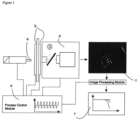
- a detection system For detecting such labels, a detection system may be used which is suitable for both determining values indicative for the presence and/or amount of first nucleic acids captured by FRET compounds and for the presence and/or amount of reporter compound captured on the binding member, e.g. on a microarray.
- the detection system may be adapted for holding a test device as described below. Typically, the detection system is positioned opposite to the binding member.
- the selection of a suitable detection system depends on several parameters such as the type of labels used for detection or the kind of analysis performed.
- Various optical detection systems are well established in the art. A general description of detection systems that can be used with the method can be found, e.g., in Lottspeich, F., and Zorbas H., supra.
- detection systems are based on the comparison of the fluorescence intensities of spectrally excited nucleic acids labeled with fluorophores.
- Fluorescence is the capacity of particular molecules to emit their own light when excited by light of a particular wavelength resulting in a characteristic absorption and emission behavior.
- quantitative detection of fluorescence signals is performed by means of modified methods of fluorescence microscopy (for review see, e.g., Lichtman, J.W., and Conchello, J.A. (2005) Nature Methods 2, 910-919 ; Zimmermann, T. (2005) Adv. Biochem. Eng. Biotechnol. 95, 245-265 ).
- the signals resulting from light absorption and light emission, respectively, may be separated by one or more filters and/or dichroites and imaged on suitable detectors.
- Data analysis is performed by means of digital image processing.
- Image processing may be achieved with several software packages well known in the art (such as Mathematical Digital Image Processing, EIKONA, or Image-PRO). Suitable software for such purposes is the Iconoclust software (Clondiag Chip Technologies GmbH, Jena, Germany).
- Suitable detection systems may be based on "classical" methods for measuring a fluorescent signal such as epifluorescence or darkfield fluorescence microscopy (reviewed, e.g., in: Lakowicz, J.R. (1999) Principles of Fluorescence Spectroscopy, 2nd ed., Plenum Publishing Corp., NY ).
- Another optical detection system that may be used is confocal fluorescence microscopy, wherein the object is illuminated in the focal plane of the lens via a point light source.
- the point light source, object and point light detector may be located on optically conjugated planes. Examples of such confocal systems are described in detail, for example, in Diaspro, A. (2002) Confocal and 2-photonmicroscopy: Foundations, Applications and Advances, Wiley-Liss, Hobroken, NJ .
- the fluorescence-optical system is usually a fluorescence microscope without an autofocus, for example a fluorescence microscope having a fixed focus.
- fluorescence detection methods include inter alia total internal fluorescence microscopy (see, e.g., Axelrod, D. (1999) Surface fluorescence microscopy with evanescent illumination, in: Lacey, A. (ed.) Light Microscopy in Biology, Oxford University Press, New York, 399-423 ), fluorescence lifetime imaging microscopy (see, for example, Dowling, K. et al. (1999) J. Mod. Optics 46, 199-209 ), fluorescence resonance energy transfer (FRET; see, for example, Periasamy, A. (2001) J. Biomed.
- FRET fluorescence resonance energy transfer
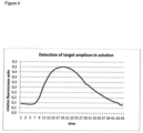
- the method further comprises determining a value indicative for the presence and/or amount of the second nucleic acid based on the value indicative for the presence and/or amount of reporter compound captured on the binding member.
- the presence and/or amount of the one or more second nucleic acids present in a particular sample may be calculated based on the difference between the presence and/or amount of reporter compound being present prior to the forming of second nucleic acid/reporter molecule complexes and the amount of reporter compound being captured on the binding member after said complex formation.
- the reporter compound may comprise one or more detectable labels as described above.
- the reporter compound may comprise two detectable labels.
- the detectable labels are fluorescent labels as described above.
- the detectable labels may be incorporated or attached to the reporter molecules, e.g., in form of modified and/or labeled ribonucleotides, deoxynucleotides or dideoxynucleotides.
- a test device used for performing the method may further comprise a detection system suitable for determining values indicative for the presence and/or amount of reporter compound captured on the binding member.
- a detection system suitable for determining values indicative for the presence and/or amount of target nucleic acids captured on a binding member as described above may be used.
- the detection/determination of a value indicative for the presence and/or amount of the first and/or second nucleic acids may be performed only once or more than once during the assay performed. If more than one detection step during a single assay is performed, the mean value of the results obtained may be calculated in some embodiments.
- the data obtained in one or more cycles of detection may be analyzed and mathematically processed using appropriate computer software known by persons skilled in the art in order to determine inter alia the presence, the length or the sequence of one or more first or second nucleic acids and/or to calculate its/their amount.
- determining the value indicative for the presence and/or amount of reporter compound captured on the binding member comprises time-dependent monitoring of the indicative value, e.g. the repeated performing of the determination/detection step and monitoring the course of the indicative value over time. Additionally or alternatively, determining the value indicative for the presence and/or amount of FRET compound comprises time-dependent monitoring of the indicative value, e.g. the repeated performing of the determination/detection step and monitoring the course of the indicative value over time.
- time-dependent monitoring of the value indicative for the presence and/or amount of reporter compound captured on the binding member and/or of the value indicative for the presence and/or amount of FRET compound may comprise performing the determination/detection step every second, every 2 s, every 3, every 5, every 10, every 20, every 30, every 60, every 90, or every 120 s.
- time-dependent monitoring of the value indicative for the presence and/or amount of reporter compound captured on the binding member and/or of the value indicative for the presence and/or amount of FRET compound may comprise performing the determination/detection step every cycle, every second cycle, every third cycle, every fourth cycle, or every fifth cycle.
- the value indicative for the presence and/or amount of second nucleic acid is determined based on a calibration curve correlating the value indicative for the presence and/or amount of reporter compound with the value indicative for the presence and/or amount of second nucleic acid.
- the method further comprises releasing the remaining subset of the amount of reporter compound from the binding member after the steps of forming complexes of a subset of the amount of reporter compound with at least a subset of the amount of second nucleic acid, capturing a remaining subset of the amount of reporter compound not in complex with a second nucleic acid on the binding member, and determining the value indicative for the presence and/or amount of reporter compound captured on the binding member.
- releasing denotes the detachment or unbinding of the reporter molecules from the binding member.
- nucleic acid reporter molecules are bound to the binding member by reporter specific nucleic acid capture molecules via complementary base-pairing, by increasing the temperature in the reaction chamber, in which the assay is performed, thus resulting in nucleic acid strand separation (i.e. denaturation).
- the steps of releasing, forming complexes, capturing, and determining are repeated N additional times, where N is an integer greater than or equal to 1.
- N is an integer greater than or equal to 1.
- the method is performed in a cyclic manner.
- the integer N is ⁇ 5, ⁇ 10 or ⁇ 20.
- the step of forming complexes of a subset of the amount of reporter compound with at least a subset of the amount of second nucleic acid and the step of capturing a remaining subset of the amount of reporter compound not in complex with a second nucleic acid on the binding member may be performed concomitantly.
- the steps of (i) forming complexes each comprising a first nucleic acid and a FRET compound, (ii) forming complexes of a subset of the amount of reporter compound with at least a subset of the amount of second nucleic acid and (iii) capturing a remaining subset of the amount of reporter compound not in complex with a second nucleic acid on the binding member may be performed concomitantly.
- the method further comprises subjecting the first and second nucleic acids to amplification, that is, to increase their amount present in the sample before subjecting the same to the further analysis in order to facilitate further detection.
- Amplification of the first and second nucleic acid may be initiated prior to the step of forming complexes of a subset of the amount of reporter compound with at least a subset of the amount of second nucleic acid.
- the multiple nucleic acids may be subjected to amplification while allowing reporter compounds to form a complex with a second nucleic acid, and reporter compounds not in complex with a second nucleic acid to be re-captured on the binding member.
- the steps of (i) forming complexes each comprising a first nucleic acid and a FRET compound, (ii) forming complexes of a subset of the amount of reporter compound with at least a subset of the amount of second nucleic acid, (iii) capturing a remaining subset of the amount of reporter compound not in complex with a second nucleic acid on the binding member, and (iv) subjecting the first and/or second nucleic acid to amplification are performed concomitantly.
- nucleic acid amplification is achieved by means of an isothermal amplification method.
- Amplifying nucleic acids in isothermal conditions makes it possible to avoid the use of a thermocycling apparatus.
- isothermal nucleic acid amplification methods known to the skilled person, including transcription-mediated amplification, nucleic acid sequence-based amplification, signal-mediated amplification of RNA technology, strand displacement amplification, rolling circle amplification, loop-mediated isothermal amplification, isothermal multiple displacement amplification, helicase-dependent amplification, single primer isothermal amplification, and circular helicase-dependent amplification.
- Isothermal nucleic acid amplification technologies are reviewed for example in P. Gill and A. Ghaemi, Nucleosides Nucleotides Nucleic Acids, 27(3): 224-243 (2008 ).
- NEAR nicking enzyme amplification reaction
- LAMP loop-mediated isothermal amplification
- SDA strand displacement amplification
- a strand-displacing DNA polymerase typically Bst DNA Polymerase, Large Fragment or Klenow Fragment (3'-5' exo-)
- Bst DNA Polymerase Large Fragment or Klenow Fragment (3'-5' exo-)
- the nicking site is regenerated with each polymerase displacement step, resulting in exponential amplification.
- HDA helicase-dependent amplification
- FRET compounds as described above such as molecular beacons are used for determining the presence and/or amount of first nucleic acid and reporter compounds as described above are used for determining the presence and/or amount of second nucleic acid during the isothermal amplification.
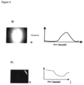
- the presence and/or amount of first nucleic acid may be determined by means of a FRET compound such as a molecular beacon which specifically binds to the first nucleic acid and which generates a signal in solution as described above depending on the amount of amplified first nucleic acid present in solution.
- the presence and/or amount of second nucleic acid may be determined by detecting a signal generated on the surface of the binding member such as a microarray by a labeled reporter compound which specifically binds both to the second nucleic acid and to a reporter specific capture molecule attached to the surface of the binding member, e.g. a microarray.
- nucleic acid amplification is achieved by means of a cyclic amplification.
- the cyclic amplification may comprise any number of amplification cycles that is equal or greater than two.
- cyclic amplification reaction comprises at least 10 or at least 20 cycles.
- An exemplary cyclic amplification is a polymerase chain reaction (PCR).
- PCR is an established standard method in molecular biology that is described in detail, e.g., in Sambrook et al., supra ; and in Ausubel, F.M. et al., supra.
- PCR is used for the amplification of double-stranded DNA molecules by employing a thermostable DNA polymerase.
- the DNA polymerase used in the amplification has exonuclease activity, particularly 5'->3' exonuclease activity.
- DNA polymerases include inter alia Taq DNA polymerase or Tth DNA polymerase (which are commercially available from multiple providers).
- Taq DNA polymerase or Tth DNA polymerase (which are commercially available from multiple providers).
- Tth DNA polymerase which are commercially available from multiple providers.
- the DNA polymerase may nucleolytically attack the labeled 5'-termini of reporter molecules that are bound to the second nucleic acids resulting in a progressive degradation of such reporter molecules.
- the amount of reporter compound that is captured on the binding member additionally decreases during the amplification reaction.
- the DNA polymerase employed may also exhibit 3'->5' exonuclease activity ("proofreading activity") for removing an incorrect nucleotide that has been added to the nascent DNA strand at a particular sequence position.
- proofreading activity 3'->5' exonuclease activity
- DNA polymerases having both exonuclease activities include inter alia Pwo DNA polymerase, and Pfu DNA polymerase (both enzymes are also commercially available from various suppliers).
- DNA polymerases having a 5' ⁇ 3' exonuclease activity are typically used if a degradation of a reporter compound is desirable in order to generate and/or enhance a detectable signal.
- FRET compounds as described above such as TaqMan probes are used for determining the presence and/or amount of first nucleic acid and reporter compounds as described above are used for determining the presence and/or amount of second nucleic acid during the cyclic amplification.
- the presence and/or amount of first nucleic acid may be determined by means of a FRET compound such as a TaqMan probe which specifically binds to the first nucleic acid and which generates a signal in solution as described above depending on the amount of amplified first nucleic acid present in solution.
- the presence and/or amount of second nucleic acid may be determined by detecting a signal generated on the surface of the binding member such as a microarray by a labeled reporter compound which specifically binds both to the second nucleic acid and to a reporter specific capture molecule attached to the surface of the binding member, e.g. a microarray.
- the method may further comprise subjecting the first and/or second nucleic acid to reverse transcription as described above prior to subjecting them to amplification.
- Reverse transcription is another standard method in molecular biology and also described, e.g., in Sambrook et al., supra ; and in Ausubel, F.M. et al., supra.
- amplification controls may be used to detect inhibitors of amplification.
- the second nucleic acid may be an internal control nucleic acid selected from the group consisting of an internal positive amplification control and an internal negative amplification control.
- control nucleic acids may be added to the sample, e.g. a body fluid sample as described above.
- the source of internal amplification controls may be plasmid DNA carrying the cloned internal amplification control sequence or purified PCR products.
- the internal control nucleic acid is an artificial nucleic acid which, under usual conditions, is naturally not present in a body fluid sample to be analysed.
- the internal control nucleic acid is armored RNA which can be reverse transcribed before amplification.
- An internal positive amplification control nucleic acid or competitive internal amplification control nucleic acid or internal positive control may be designed from partial or full sequences of target analyte.
- the first nucleic acid and the internal positive amplification control nucleic acid may have identical primer-binding sites.
- the internal positive amplification control nucleic acid may have a sequence corresponding to a partial sequence of the first nucleic acid. Sharing the same primer-binding site may allow for similar or essentially the same amplification conditions.
- corresponding amplicon primers may be provided in excess.
- reporter compound binding sites on the binding member may be specific for the internal positive amplification control nucleic acid to allow discrimination between the internal positive amplification control nucleic acid and optionally other control nucleic acids on the binding member.
- An internal negative amplification control nucleic acid or non-competitive internal amplification control nucleic acid may be a nucleic acid essentially without sequence homology to a target analyte or an internal positive amplification control nucleic acid.
- the internal negative amplification control nucleic acid may have a sequence which does not comprise a primer binding site for amplicon primers which are used to amplify the target analyte or first nucleic acid or internal positive amplification control nucleic acid.
- reporter compound binding sites on the binding member may be specific for the internal negative amplification control nucleic acid to allow discrimination between the negative amplification control nucleic acid, the internal positive amplification control nucleic acid and other control nucleic acids on the binding member.
- the signal on reporter compound binding sites specific for reporter compounds capable for forming complexes with the internal negative amplification control nucleic acid is expected to remain essentially constant during the process.
- the signal on reporter compound binding sites specific for reporter compounds capable for forming complexes with the internal negative amplification control nucleic acid may need to remain constant with no reported Ct value, otherwise the test may be considered as not valid.
- controls may also be used in the methods described herein.
- controls may be selected from a positive hybridization control, a processing-positive control, a processing-negative control, and a reagent control.
- a positive hybridization control may be a reporter compound which is not capable of forming complexes with any of the first and second nucleic acid.
- a processing positive control may be a negative sample spiked with a sufficient amount of target analyte and processed throughout the entire protocol.
- a processing negative control may refer to a negative sample spiked with a sufficient amount of closely related, but non-target analyte processed throughout the entire protocol.
- a non-template control may refer to a control containing all reagents, but no target or internal amplification control nucleic acids.
- a test device may be used for performing the method which may comprise one or more temperature control units and/or temperature regulating units for controlling and/or regulating the temperature within the structure or reaction chamber.
- a temperature control unit and/or temperature regulating unit may comprise one or more separate heating and/or cooling elements, which may directly contact a reaction chamber of the test device.
- the one or more heating and/or cooling elements are made of a heat conductive material. Examples of such heat conductive materials include inter alia silicon, ceramic materials like aluminium oxide ceramics, and/or metals like high-grade steel, aluminium, copper, or brass.
- An exemplary detailed description of a temperature control unit and/or temperature regulating suitable for performing the methods described herein can also be found in WO 01/02094 , whose relevant contents are herewith explicitly referred to.
- Measuring the temperature in the reaction chamber can be performed by various methods well established in the art, e.g. by using integrated resistance sensors, semiconductor sensors, light waveguide sensors, polychromatic dyes or liquid crystals. Furthermore, the temperature in the reaction chamber can be determined by using an integrated temperature sensor in the chamber body, a pyrometer or an infrared sensor, or by measuring the temperature-dependent alteration of parameters such as the refraction index at the surface on which detection takes place or the pH value of the sample, for example by measuring the colour alteration of a pH-sensitive indicator.
- a channel and/or a value connected with the reaction chamber may be heat-sealed or welded. Plastics channels or valves may be heat-sealed by contacting a hot pin with the channel or valve so that the plastics are melted and the channel or valve is locked.
- the step of providing the first and/or second nucleic acids may comprise releasing the first and/or second nucleic acids from biological material comprised in the sample.
- the sample may be heated in order to destroy cellular membranes and/or viral capsids (e.g., by employing a temperature control unit and/or temperature regulating unit as described above).
- this releasing step comprises contacting the fluid sample with a lysing reagent, for example a reagent comprising one or more detergents which disintegrate the cellular membranes and/or viral capsids.
- a lysing reagent for example a reagent comprising one or more detergents which disintegrate the cellular membranes and/or viral capsids.
- Such lysing reagents are well known in the art (see, for example, Sambrook, J. et al. (1989) Molecular, Cloning: A Laboratory Manual, 2nd ed., Cold Spring Harbor Laboratory Press, Cold Spring Harbor, NY ) and commercially available
- the method may further comprise separating the first and/or second nucleic acids from concomitant material.
- the value indicative for the presence and/or amount of first nucleic acid captured by FRET compounds and the value indicative for the presence and/or amount of reporter compound captured on the binding member may be determined consecutively or concomitantly.
- test device typically capable of performing most and for example all steps of the methods described herein.
- Exemplary test devices, reaction chambers and corresponding methods are also described in WO 2005/108604 , WO 2008/055915 , WO 2008/135564 and T. Ullrich et al., PloS ONE, 7, 4, e35438 (2012 ), whose relevant contents are herewith explicitly referred to.
- the methods described herein may be performed in a test device enabling determination of values indicative for the presence and/or amount of reporter compounds captured on the binding member during amplification by removing background signals from the solution. In one embodiment, this removal of solution from the reaction chamber of a test device is reversible.
- a test device comprising a reaction chamber as described below may be used for the amplification and detection.
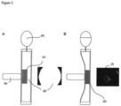
- the value indicative for the presence and/or amount of first nucleic acid captured by FRET compounds in solution and/or the value indicative for the presence and/or amount of reporter compound captured on the binding member may be determined in a reaction chamber comprising the binding member and a cover element.
- the binding member may be arranged on the cover element or on a surface opposite to the cover element.
- the cover element may be configured to be at least partially deformable such that the reaction chamber has a relaxed state interior volume and a compressed state interior volume.
- the value indicative for the presence and/or amount of first nucleic acid captured by FRET compounds in solution and/or the value indicative for the presence and/or amount of reporter compound captured on the binding member may be determined in a reaction chamber comprising a first surface, a second surface and the binding member capable of capturing the reporter compound, wherein the distance between the first and the second surface can be varied such that the reaction chamber has a relaxed state interior volume and a compressed state interior volume.
- the value indicative for the presence and/or amount of first nucleic acid captured by FRET compounds is determined in the reaction chamber having a relaxed state interior volume. Further, the value indicative for the presence and/or amount of reporter compound captured on the binding member may be determined in the reaction chamber having a compressed state interior volume.
- determining a value indicative for the presence and/or amount of the captured reporter compounds may be performed with an actuator actuated to deform the flexible cover element.
- the cover element may be deformed in such a way that the volume of the reaction chamber is reduced.
- the volume of the reaction chamber may be increased again after determining a value indicative for the presence and/or amount of the captured reporter compounds.
- an actuator may compress the reaction chamber to reduce the distance between the flexible cover element and the substrate thereby removing liquid comprising material which has not bound to the binding member from the detection zone.
- the binding member is provided in the reaction chamber and the steps of forming complexes each comprising a first nucleic acid and a FRET compound; forming complexes of a subset of the amount of reporter compound with a second nucleic acid; and capturing a remaining subset of the amount of reporter compound not in complex with a second nucleic acid on the binding member are performed in the reaction chamber. Further, subjecting first and/or second nucleic acids to amplification and/or (re-)capturing the reporter compounds with respect to the binding member may also be performed in the reaction chamber.
- Providing the first and/or second nucleic acids may be performed spatially separated from the steps of forming complexes each comprising a first nucleic acid and a FRET compound, forming complexes of a subset of the amount of reporter compound with a second nucleic acid; subjecting the first and/or second nucleic acids to amplification, capturing a remaining subset of the amount of reporter compound not in complex with a second nucleic acid on the binding member, determining a value indicative for the presence and/or amount of first nucleic acid captured by FRET compounds, and determining a value indicative for the presence and/or amount of reporter compound captured on the binding member.
- the methods described herein may e.g. be performed in a test device comprising a reaction chamber adapted for accommodating liquids, wherein the reaction chamber comprises at least one binding member and is e.g. in fluid communication with a microfluidic network.
- the method may be performed in a test device comprising a rigid substrate, a flexible cover element at least partially covering the substrate, a structure or reaction chamber formed in the substrate, adapted for accommodating liquids and comprising at least one binding member adapted for capturing the reporter compounds and for determining a value indicative for the presence and/or amount of reporter compound, optionally a microfluidic network interconnecting at least the reaction chamber with further structures, e.g. structures for providing the first and/or second nucleic acids.
- the test device may be placed with respect to or into a detector system.
- a detector system may comprise an actuator unit adapted for pressing the flexible cover element against the rigid substrate.
- the mechanical module of such a test device may contain a plunger or actuator (a) that is used to reversibly squeeze and release a reaction chamber of the test device.
- the temperature control module may comprise two peltier elements with an aperture in the center (b).
- Fluorescence images of the binding member (c), e.g. an array, may be acquired by an optical detection module (d). Synchronized simultaneous performance of all components may be controlled by the process control module (e), which also controls the thermal regime of the amplification reaction.
- the hybridization pattern is analyzed by the image analysis module. Results may be visualized as a plot of intensity on each individual probe of the binding member versus reaction time.
- the uncompressed reaction chamber of the test device may be filled with a reaction mix containing a high concentration of fluorescently labeled reporter probes. Therefore, imaging of the hybridization pattern on the binding member, e.g. a capture probe array, against the background is not feasible (a).
- pressure by means of a plunger or actuator (b) may be applied and the binding member (c) is pressed tightly against the top side of the reaction chamber, thus displacing the fluorescent liquid (d). Displaced liquid may be pressed into an integrated pneumatic spring (e).
- image acquisition the reaction chamber is allowed to relax and the liquid sample can return into the relaxed reaction chamber.
- the sample may contain competitive reporter compounds and molecular beacons which change the fluorescence signal in the sample as soon as an amplicon specific for the donor chromophore is formed.
- the binding member may comprise a microarray comprising reporter-specific capture compounds which are suitable for forming complexes with the reporter compounds and generate a fluorescence signal on the microarray.
- the reporter compounds are further specific for a sequence in the amplification product, e.g. a mimic fragment which is used as an internal positive amplification control or as an internal negative amplification control.
- Data analysis is made separately with the images resulting from the solution and the images resulting from the microarray (see Figure 4 ).
- the fluorescence intensity in solution increases due to the formation of complexes of the molecular beacons with the synthesized amplification product of the first nucleic acid.
- Shown in Figure 4a ) is a characteristic curve for a NEAR amplification.
- Figure 4b shows the decrease of the fluorescence signal due to the binding of the reporter compounds to the synthesized amplification product of the second nucleic acid in solution.
- the methods described herein may be used in immunoassays and point-of-care nucleic acid tests.
- Tuberculose DNA was amplified by using an isothermal NEAR in a test device as described in Figures 1 and 2 comprising a compressible reaction chamber and an array as the binding member capable of capturing reporter compounds.
- the sample included primers (reverse primer: Tb_DR_R25c 5'-AGACTCCATATGGAGTCTCATCTTTCCGTCCCC-3', forward primer: Tb_DR_F16 5'-CGACTCCATATGGAGTCGTCGTCAGACCCAAAA-3'); molecular beacon MB_P2_7 (5'-TCGGGGCAGACCCAAAACCCCGA-3'); reporter compound Anti_Tb_Target_CMA (5'-CCCAAAACCCCGAGAGG-3'); enzymes (nicking enzyme and strand displacement enzyme) and a target DNA.
- the amplification was conducted without target DNA, i.e. without template control. The amplification was conducted at 56°C for 600 seconds. Images were acquired every 15 seconds. Results are shown in Figures 5a ) and 5b
- IPC internal positive control
- PLC positive hybridization control
- Tuberculose DNA (target DNA) and internal positive control DNA (IPC) were amplified by using an isothermal NEAR in a test device as described in Figures 1 and 2 comprising a compressible reaction chamber and an array as the binding member capable of capturing reporter compounds.
- the templates for target analyte and IPC had the same sequences in the primer binding sites, but differed in the sequences of the binding sites for the reporter compounds and the FRET compounds (here a molecular beacon), respectively. Both amplicons could therefore be amplified by the same primers and could be detected independently from each other by different probes.
- the amplification reaction mix included primers (reverse primer TB_DR_25c, forward primer TbDR_F16), molecular beacon MB_PS_7, IPC reporter compound and IPC. Further, a reporter compound which is not capable of forming complexes with the IPC amplicon was added as a positive hybridization control (PHC: 5'-ccgactactacgggacgctggga-3') in order to show that the change in signal intensity of the IPC reporter is specific for the presence of the IPC amplicon. This PHC control therefore differs from the IPC reporter in its hybridization kinetics.
- the target DNA is initially present in a 5-fold excess compared to the target DNA the target DNA can be detected in the same assay with the internal positive control DNA (detection of target analyte captured by FRET compound in solution and detection of internal positive control reporter on the array). Due to the absence of a competitor in solution the signal intensity course of PHC has second order kinetics.
Landscapes
- Chemical & Material Sciences (AREA)
- Health & Medical Sciences (AREA)
- Life Sciences & Earth Sciences (AREA)
- Engineering & Computer Science (AREA)
- Immunology (AREA)
- Organic Chemistry (AREA)
- Molecular Biology (AREA)
- Zoology (AREA)
- Wood Science & Technology (AREA)
- Proteomics, Peptides & Aminoacids (AREA)
- General Health & Medical Sciences (AREA)
- Analytical Chemistry (AREA)
- Hematology (AREA)
- Biochemistry (AREA)
- Biotechnology (AREA)
- Microbiology (AREA)
- Physics & Mathematics (AREA)
- Urology & Nephrology (AREA)
- Biomedical Technology (AREA)
- Bioinformatics & Cheminformatics (AREA)
- General Engineering & Computer Science (AREA)
- Genetics & Genomics (AREA)
- Biophysics (AREA)
- Chemical Kinetics & Catalysis (AREA)
- Cell Biology (AREA)
- Food Science & Technology (AREA)
- Medicinal Chemistry (AREA)
- General Physics & Mathematics (AREA)
- Pathology (AREA)
- Dispersion Chemistry (AREA)
- Clinical Laboratory Science (AREA)
- Measuring Or Testing Involving Enzymes Or Micro-Organisms (AREA)
Priority Applications (1)
| Application Number | Priority Date | Filing Date | Title |
|---|---|---|---|
| EP24164295.8A EP4400600A3 (en) | 2014-02-21 | 2015-02-23 | Methods for detecting multiple nucleic acids in a sample |
Applications Claiming Priority (3)
| Application Number | Priority Date | Filing Date | Title |
|---|---|---|---|
| GBGB1403076.1A GB201403076D0 (en) | 2014-02-21 | 2014-02-21 | Methods for detecting multiple nucleic acids in a sample |
| EP15707584.7A EP3108008B1 (en) | 2014-02-21 | 2015-02-23 | Methods for detecting multiple nucleic acids in a sample |
| PCT/EP2015/053759 WO2015124779A1 (en) | 2014-02-21 | 2015-02-23 | Methods for detecting multiple nucleic acids in a sample |
Related Parent Applications (1)
| Application Number | Title | Priority Date | Filing Date |
|---|---|---|---|
| EP15707584.7A Division EP3108008B1 (en) | 2014-02-21 | 2015-02-23 | Methods for detecting multiple nucleic acids in a sample |
Related Child Applications (1)
| Application Number | Title | Priority Date | Filing Date |
|---|---|---|---|
| EP24164295.8A Division EP4400600A3 (en) | 2014-02-21 | 2015-02-23 | Methods for detecting multiple nucleic acids in a sample |
Publications (2)
| Publication Number | Publication Date |
|---|---|
| EP3800269A1 EP3800269A1 (en) | 2021-04-07 |
| EP3800269B1 true EP3800269B1 (en) | 2024-03-20 |
Family
ID=50482582
Family Applications (3)
| Application Number | Title | Priority Date | Filing Date |
|---|---|---|---|
| EP15707584.7A Active EP3108008B1 (en) | 2014-02-21 | 2015-02-23 | Methods for detecting multiple nucleic acids in a sample |
| EP24164295.8A Pending EP4400600A3 (en) | 2014-02-21 | 2015-02-23 | Methods for detecting multiple nucleic acids in a sample |
| EP20204162.0A Active EP3800269B1 (en) | 2014-02-21 | 2015-02-23 | Methods for detecting multiple nucleic acids in a sample |
Family Applications Before (2)
| Application Number | Title | Priority Date | Filing Date |
|---|---|---|---|
| EP15707584.7A Active EP3108008B1 (en) | 2014-02-21 | 2015-02-23 | Methods for detecting multiple nucleic acids in a sample |
| EP24164295.8A Pending EP4400600A3 (en) | 2014-02-21 | 2015-02-23 | Methods for detecting multiple nucleic acids in a sample |
Country Status (8)
| Country | Link |
|---|---|
| US (2) | US10246737B2 (cg-RX-API-DMAC7.html) |
| EP (3) | EP3108008B1 (cg-RX-API-DMAC7.html) |
| JP (2) | JP6727137B2 (cg-RX-API-DMAC7.html) |
| CN (1) | CN106687598B (cg-RX-API-DMAC7.html) |
| CA (1) | CA2937832C (cg-RX-API-DMAC7.html) |
| GB (1) | GB201403076D0 (cg-RX-API-DMAC7.html) |
| HK (1) | HK1231512A1 (cg-RX-API-DMAC7.html) |
| WO (1) | WO2015124779A1 (cg-RX-API-DMAC7.html) |
Families Citing this family (4)
| Publication number | Priority date | Publication date | Assignee | Title |
|---|---|---|---|---|
| GB201403076D0 (en) | 2014-02-21 | 2014-04-09 | ALERE TECHNOLOGIES GmbH | Methods for detecting multiple nucleic acids in a sample |
| GB201713251D0 (en) * | 2017-08-18 | 2017-10-04 | Lgc Genomics Ltd | Methods and kits for detection of nucleic acid molecules |
| CN110672569A (zh) * | 2019-09-30 | 2020-01-10 | 中国科学院宁波工业技术研究院慈溪生物医学工程研究所 | 一种dna或rna检测体系、检测方法和它们的应用 |
| CN112553376B (zh) * | 2020-12-23 | 2023-06-02 | 四川大学 | 引物组、试剂盒、区分不同ibv疫苗毒株的多重pcr检测方法及其应用 |
Family Cites Families (12)
| Publication number | Priority date | Publication date | Assignee | Title |
|---|---|---|---|---|
| US5538848A (en) * | 1994-11-16 | 1996-07-23 | Applied Biosystems Division, Perkin-Elmer Corp. | Method for detecting nucleic acid amplification using self-quenching fluorescence probe |
| EP1192007B1 (de) | 1999-07-02 | 2004-04-21 | Clondiag Chip Technologies GmbH | Microchip-matrix-vorrichtung zur vervielfältigung und charakterisierung von nukleinsäuren |
| US6312929B1 (en) * | 2000-12-22 | 2001-11-06 | Cepheid | Compositions and methods enabling a totally internally controlled amplification reaction |
| AU2002237982A1 (en) * | 2001-01-30 | 2002-08-12 | Vertex Pharmaceuticals Incorporated | A quantitative assay for nucleic acids |
| WO2003008622A2 (en) * | 2001-07-15 | 2003-01-30 | Keck Graduate Institute | Exponential nucleic acid amplification using nicking endonucleases |
| US7226732B2 (en) * | 2001-07-16 | 2007-06-05 | Cepheid | Methods, apparatus, and computer programs for verifying the integrity of a probe |
| EP1493822A1 (de) | 2003-07-02 | 2005-01-05 | Labor Becker, Olgemoeller & Kollegen GbR | Detektionsverfahren für Nucleinsäuren mit interner Amplifikationskontrolle |
| DE102004022263A1 (de) | 2004-05-06 | 2005-12-15 | Clondiag Chip Technologies Gmbh | Vorrichtung und Verfahren zum Nachweis von molekularen Wechselwirkungen |
| JP5335684B2 (ja) * | 2006-11-06 | 2013-11-06 | クロンデイアグ・ゲーエムベーハー | 結合要素を用いたアッセイのための装置及び方法 |
| EP2099930B1 (en) * | 2006-12-13 | 2015-02-18 | Luminex Corporation | Systems and methods for multiplex analysis of pcr in real time |
| US8633013B2 (en) | 2007-05-03 | 2014-01-21 | Clondiag Gmbh | Assays |
| GB201403076D0 (en) | 2014-02-21 | 2014-04-09 | ALERE TECHNOLOGIES GmbH | Methods for detecting multiple nucleic acids in a sample |
-
2014
- 2014-02-21 GB GBGB1403076.1A patent/GB201403076D0/en not_active Ceased
-
2015
- 2015-02-23 EP EP15707584.7A patent/EP3108008B1/en active Active
- 2015-02-23 HK HK17105017.9A patent/HK1231512A1/zh unknown
- 2015-02-23 US US15/116,185 patent/US10246737B2/en active Active
- 2015-02-23 WO PCT/EP2015/053759 patent/WO2015124779A1/en not_active Ceased
- 2015-02-23 CA CA2937832A patent/CA2937832C/en active Active
- 2015-02-23 EP EP24164295.8A patent/EP4400600A3/en active Pending
- 2015-02-23 CN CN201580008133.6A patent/CN106687598B/zh active Active
- 2015-02-23 JP JP2016570186A patent/JP6727137B2/ja active Active
- 2015-02-23 EP EP20204162.0A patent/EP3800269B1/en active Active
-
2019
- 2019-04-01 US US16/371,268 patent/US10787700B2/en active Active
-
2020
- 2020-03-17 JP JP2020046081A patent/JP2020114225A/ja active Pending
Also Published As
| Publication number | Publication date |
|---|---|
| US10787700B2 (en) | 2020-09-29 |
| EP4400600A3 (en) | 2024-10-23 |
| JP6727137B2 (ja) | 2020-07-22 |
| US20190376125A1 (en) | 2019-12-12 |
| EP4400600A2 (en) | 2024-07-17 |
| GB201403076D0 (en) | 2014-04-09 |
| EP3800269A1 (en) | 2021-04-07 |
| CA2937832C (en) | 2023-10-03 |
| JP2017506914A (ja) | 2017-03-16 |
| EP3108008A1 (en) | 2016-12-28 |
| WO2015124779A1 (en) | 2015-08-27 |
| CA2937832A1 (en) | 2015-08-27 |
| US10246737B2 (en) | 2019-04-02 |
| CN106687598A (zh) | 2017-05-17 |
| CN106687598B (zh) | 2022-10-11 |
| EP3108008B1 (en) | 2020-10-28 |
| US20170081710A1 (en) | 2017-03-23 |
| JP2020114225A (ja) | 2020-07-30 |
| HK1231512A1 (zh) | 2017-12-22 |
Similar Documents
| Publication | Publication Date | Title |
|---|---|---|
| CA2766351C (en) | Chimeric primers with hairpin conformations and methods of using same | |
| US20100041056A1 (en) | Temperature controlled nucleic-acid detection method suitable for practice in a closed-system | |
| US10787700B2 (en) | Methods for detecting multiple nucleic acids in a sample using reporter compounds and binding members thereof | |
| JP2012529266A (ja) | 反応カートリッジにおける多重核酸配列の検出 | |
| KR102814480B1 (ko) | 표적 핵산을 검출하기 위한 루프형 프라이머 및 루프-드-루프 방법 | |
| KR20140071968A (ko) | Pcr에 의한 미생물 dna 검출 방법, 이를 위한 시스템 및 조성물 | |
| CN105385684B (zh) | 用于为核酸扩增作对照的寡核苷酸 | |
| US20140080726A1 (en) | Enhanced method for probe based detection of nucleic acids | |
| HK40113575A (en) | Methods for detecting multiple nucleic acids in a sample | |
| HK40050543A (en) | Methods for detecting multiple nucleic acids in a sample | |
| De Falco et al. | Helicase-dependent isothermal amplification turns biosensing into modern diagnostic technologies | |
| AU2016296594B2 (en) | Methods and compositions for catalytic assays | |
| EP4276196A1 (en) | Decoy-oligonucleotides in nucleic acid detection methods | |
| JP7198225B2 (ja) | 多重核酸増幅測定法 | |
| HK1254039B (zh) | 用於催化性测定的方法和组合物 |
Legal Events
| Date | Code | Title | Description |
|---|---|---|---|
| PUAI | Public reference made under article 153(3) epc to a published international application that has entered the european phase |
Free format text: ORIGINAL CODE: 0009012 |
|
| STAA | Information on the status of an ep patent application or granted ep patent |
Free format text: STATUS: THE APPLICATION HAS BEEN PUBLISHED |
|
| AC | Divisional application: reference to earlier application |
Ref document number: 3108008 Country of ref document: EP Kind code of ref document: P |
|
| AK | Designated contracting states |
Kind code of ref document: A1 Designated state(s): AL AT BE BG CH CY CZ DE DK EE ES FI FR GB GR HR HU IE IS IT LI LT LU LV MC MK MT NL NO PL PT RO RS SE SI SK SM TR |
|
| STAA | Information on the status of an ep patent application or granted ep patent |
Free format text: STATUS: REQUEST FOR EXAMINATION WAS MADE |
|
| 17P | Request for examination filed |
Effective date: 20211006 |
|
| RBV | Designated contracting states (corrected) |
Designated state(s): AL AT BE BG CH CY CZ DE DK EE ES FI FR GB GR HR HU IE IS IT LI LT LU LV MC MK MT NL NO PL PT RO RS SE SI SK SM TR |
|
| REG | Reference to a national code |
Ref country code: HK Ref legal event code: DE Ref document number: 40050543 Country of ref document: HK |
|
| STAA | Information on the status of an ep patent application or granted ep patent |
Free format text: STATUS: EXAMINATION IS IN PROGRESS |
|
| 17Q | First examination report despatched |
Effective date: 20220824 |
|
| RAP3 | Party data changed (applicant data changed or rights of an application transferred) |
Owner name: ABBOTT RAPID DIAGNOSTICS JENA GMBH |
|
| P01 | Opt-out of the competence of the unified patent court (upc) registered |
Effective date: 20230602 |
|
| GRAP | Despatch of communication of intention to grant a patent |
Free format text: ORIGINAL CODE: EPIDOSNIGR1 |
|
| STAA | Information on the status of an ep patent application or granted ep patent |
Free format text: STATUS: GRANT OF PATENT IS INTENDED |
|
| INTG | Intention to grant announced |
Effective date: 20230926 |
|
| GRAS | Grant fee paid |
Free format text: ORIGINAL CODE: EPIDOSNIGR3 |
|
| GRAA | (expected) grant |
Free format text: ORIGINAL CODE: 0009210 |
|
| STAA | Information on the status of an ep patent application or granted ep patent |
Free format text: STATUS: THE PATENT HAS BEEN GRANTED |
|
| AC | Divisional application: reference to earlier application |
Ref document number: 3108008 Country of ref document: EP Kind code of ref document: P |
|
| AK | Designated contracting states |
Kind code of ref document: B1 Designated state(s): AL AT BE BG CH CY CZ DE DK EE ES FI FR GB GR HR HU IE IS IT LI LT LU LV MC MK MT NL NO PL PT RO RS SE SI SK SM TR |
|
| REG | Reference to a national code |
Ref country code: GB Ref legal event code: FG4D |
|
| REG | Reference to a national code |
Ref country code: CH Ref legal event code: EP |
|
| REG | Reference to a national code |
Ref country code: IE Ref legal event code: FG4D |
|
| REG | Reference to a national code |
Ref country code: DE Ref legal event code: R096 Ref document number: 602015088030 Country of ref document: DE |
|
| REG | Reference to a national code |
Ref country code: NL Ref legal event code: FP |
|
| PG25 | Lapsed in a contracting state [announced via postgrant information from national office to epo] |
Ref country code: LT Free format text: LAPSE BECAUSE OF FAILURE TO SUBMIT A TRANSLATION OF THE DESCRIPTION OR TO PAY THE FEE WITHIN THE PRESCRIBED TIME-LIMIT Effective date: 20240320 |
|
| REG | Reference to a national code |
Ref country code: LT Ref legal event code: MG9D |
|
| PG25 | Lapsed in a contracting state [announced via postgrant information from national office to epo] |
Ref country code: GR Free format text: LAPSE BECAUSE OF FAILURE TO SUBMIT A TRANSLATION OF THE DESCRIPTION OR TO PAY THE FEE WITHIN THE PRESCRIBED TIME-LIMIT Effective date: 20240621 |
|
| PG25 | Lapsed in a contracting state [announced via postgrant information from national office to epo] |
Ref country code: HR Free format text: LAPSE BECAUSE OF FAILURE TO SUBMIT A TRANSLATION OF THE DESCRIPTION OR TO PAY THE FEE WITHIN THE PRESCRIBED TIME-LIMIT Effective date: 20240320 Ref country code: RS Free format text: LAPSE BECAUSE OF FAILURE TO SUBMIT A TRANSLATION OF THE DESCRIPTION OR TO PAY THE FEE WITHIN THE PRESCRIBED TIME-LIMIT Effective date: 20240620 |
|
| PG25 | Lapsed in a contracting state [announced via postgrant information from national office to epo] |
Ref country code: RS Free format text: LAPSE BECAUSE OF FAILURE TO SUBMIT A TRANSLATION OF THE DESCRIPTION OR TO PAY THE FEE WITHIN THE PRESCRIBED TIME-LIMIT Effective date: 20240620 Ref country code: NO Free format text: LAPSE BECAUSE OF FAILURE TO SUBMIT A TRANSLATION OF THE DESCRIPTION OR TO PAY THE FEE WITHIN THE PRESCRIBED TIME-LIMIT Effective date: 20240620 Ref country code: LT Free format text: LAPSE BECAUSE OF FAILURE TO SUBMIT A TRANSLATION OF THE DESCRIPTION OR TO PAY THE FEE WITHIN THE PRESCRIBED TIME-LIMIT Effective date: 20240320 Ref country code: HR Free format text: LAPSE BECAUSE OF FAILURE TO SUBMIT A TRANSLATION OF THE DESCRIPTION OR TO PAY THE FEE WITHIN THE PRESCRIBED TIME-LIMIT Effective date: 20240320 Ref country code: GR Free format text: LAPSE BECAUSE OF FAILURE TO SUBMIT A TRANSLATION OF THE DESCRIPTION OR TO PAY THE FEE WITHIN THE PRESCRIBED TIME-LIMIT Effective date: 20240621 Ref country code: FI Free format text: LAPSE BECAUSE OF FAILURE TO SUBMIT A TRANSLATION OF THE DESCRIPTION OR TO PAY THE FEE WITHIN THE PRESCRIBED TIME-LIMIT Effective date: 20240320 Ref country code: BG Free format text: LAPSE BECAUSE OF FAILURE TO SUBMIT A TRANSLATION OF THE DESCRIPTION OR TO PAY THE FEE WITHIN THE PRESCRIBED TIME-LIMIT Effective date: 20240320 |
|
| REG | Reference to a national code |
Ref country code: AT Ref legal event code: MK05 Ref document number: 1667860 Country of ref document: AT Kind code of ref document: T Effective date: 20240320 |
|
| PG25 | Lapsed in a contracting state [announced via postgrant information from national office to epo] |
Ref country code: SE Free format text: LAPSE BECAUSE OF FAILURE TO SUBMIT A TRANSLATION OF THE DESCRIPTION OR TO PAY THE FEE WITHIN THE PRESCRIBED TIME-LIMIT Effective date: 20240320 Ref country code: LV Free format text: LAPSE BECAUSE OF FAILURE TO SUBMIT A TRANSLATION OF THE DESCRIPTION OR TO PAY THE FEE WITHIN THE PRESCRIBED TIME-LIMIT Effective date: 20240320 |
|
| PG25 | Lapsed in a contracting state [announced via postgrant information from national office to epo] |
Ref country code: IS Free format text: LAPSE BECAUSE OF FAILURE TO SUBMIT A TRANSLATION OF THE DESCRIPTION OR TO PAY THE FEE WITHIN THE PRESCRIBED TIME-LIMIT Effective date: 20240720 |
|
| PG25 | Lapsed in a contracting state [announced via postgrant information from national office to epo] |
Ref country code: PT Free format text: LAPSE BECAUSE OF FAILURE TO SUBMIT A TRANSLATION OF THE DESCRIPTION OR TO PAY THE FEE WITHIN THE PRESCRIBED TIME-LIMIT Effective date: 20240722 Ref country code: SM Free format text: LAPSE BECAUSE OF FAILURE TO SUBMIT A TRANSLATION OF THE DESCRIPTION OR TO PAY THE FEE WITHIN THE PRESCRIBED TIME-LIMIT Effective date: 20240320 |
|
| PG25 | Lapsed in a contracting state [announced via postgrant information from national office to epo] |
Ref country code: ES Free format text: LAPSE BECAUSE OF FAILURE TO SUBMIT A TRANSLATION OF THE DESCRIPTION OR TO PAY THE FEE WITHIN THE PRESCRIBED TIME-LIMIT Effective date: 20240320 |
|
| PG25 | Lapsed in a contracting state [announced via postgrant information from national office to epo] |
Ref country code: EE Free format text: LAPSE BECAUSE OF FAILURE TO SUBMIT A TRANSLATION OF THE DESCRIPTION OR TO PAY THE FEE WITHIN THE PRESCRIBED TIME-LIMIT Effective date: 20240320 Ref country code: CZ Free format text: LAPSE BECAUSE OF FAILURE TO SUBMIT A TRANSLATION OF THE DESCRIPTION OR TO PAY THE FEE WITHIN THE PRESCRIBED TIME-LIMIT Effective date: 20240320 |
|
| PG25 | Lapsed in a contracting state [announced via postgrant information from national office to epo] |
Ref country code: AT Free format text: LAPSE BECAUSE OF FAILURE TO SUBMIT A TRANSLATION OF THE DESCRIPTION OR TO PAY THE FEE WITHIN THE PRESCRIBED TIME-LIMIT Effective date: 20240320 |
|
| PG25 | Lapsed in a contracting state [announced via postgrant information from national office to epo] |
Ref country code: PL Free format text: LAPSE BECAUSE OF FAILURE TO SUBMIT A TRANSLATION OF THE DESCRIPTION OR TO PAY THE FEE WITHIN THE PRESCRIBED TIME-LIMIT Effective date: 20240320 |
|
| PG25 | Lapsed in a contracting state [announced via postgrant information from national office to epo] |
Ref country code: SK Free format text: LAPSE BECAUSE OF FAILURE TO SUBMIT A TRANSLATION OF THE DESCRIPTION OR TO PAY THE FEE WITHIN THE PRESCRIBED TIME-LIMIT Effective date: 20240320 |
|
| PG25 | Lapsed in a contracting state [announced via postgrant information from national office to epo] |
Ref country code: SM Free format text: LAPSE BECAUSE OF FAILURE TO SUBMIT A TRANSLATION OF THE DESCRIPTION OR TO PAY THE FEE WITHIN THE PRESCRIBED TIME-LIMIT Effective date: 20240320 Ref country code: SK Free format text: LAPSE BECAUSE OF FAILURE TO SUBMIT A TRANSLATION OF THE DESCRIPTION OR TO PAY THE FEE WITHIN THE PRESCRIBED TIME-LIMIT Effective date: 20240320 Ref country code: RO Free format text: LAPSE BECAUSE OF FAILURE TO SUBMIT A TRANSLATION OF THE DESCRIPTION OR TO PAY THE FEE WITHIN THE PRESCRIBED TIME-LIMIT Effective date: 20240320 Ref country code: PT Free format text: LAPSE BECAUSE OF FAILURE TO SUBMIT A TRANSLATION OF THE DESCRIPTION OR TO PAY THE FEE WITHIN THE PRESCRIBED TIME-LIMIT Effective date: 20240722 Ref country code: PL Free format text: LAPSE BECAUSE OF FAILURE TO SUBMIT A TRANSLATION OF THE DESCRIPTION OR TO PAY THE FEE WITHIN THE PRESCRIBED TIME-LIMIT Effective date: 20240320 Ref country code: IS Free format text: LAPSE BECAUSE OF FAILURE TO SUBMIT A TRANSLATION OF THE DESCRIPTION OR TO PAY THE FEE WITHIN THE PRESCRIBED TIME-LIMIT Effective date: 20240720 Ref country code: ES Free format text: LAPSE BECAUSE OF FAILURE TO SUBMIT A TRANSLATION OF THE DESCRIPTION OR TO PAY THE FEE WITHIN THE PRESCRIBED TIME-LIMIT Effective date: 20240320 Ref country code: EE Free format text: LAPSE BECAUSE OF FAILURE TO SUBMIT A TRANSLATION OF THE DESCRIPTION OR TO PAY THE FEE WITHIN THE PRESCRIBED TIME-LIMIT Effective date: 20240320 Ref country code: CZ Free format text: LAPSE BECAUSE OF FAILURE TO SUBMIT A TRANSLATION OF THE DESCRIPTION OR TO PAY THE FEE WITHIN THE PRESCRIBED TIME-LIMIT Effective date: 20240320 Ref country code: AT Free format text: LAPSE BECAUSE OF FAILURE TO SUBMIT A TRANSLATION OF THE DESCRIPTION OR TO PAY THE FEE WITHIN THE PRESCRIBED TIME-LIMIT Effective date: 20240320 |
|
| PG25 | Lapsed in a contracting state [announced via postgrant information from national office to epo] |
Ref country code: IT Free format text: LAPSE BECAUSE OF FAILURE TO SUBMIT A TRANSLATION OF THE DESCRIPTION OR TO PAY THE FEE WITHIN THE PRESCRIBED TIME-LIMIT Effective date: 20240320 |
|
| REG | Reference to a national code |
Ref country code: DE Ref legal event code: R097 Ref document number: 602015088030 Country of ref document: DE |
|
| PG25 | Lapsed in a contracting state [announced via postgrant information from national office to epo] |
Ref country code: IT Free format text: LAPSE BECAUSE OF FAILURE TO SUBMIT A TRANSLATION OF THE DESCRIPTION OR TO PAY THE FEE WITHIN THE PRESCRIBED TIME-LIMIT Effective date: 20240320 |
|
| PG25 | Lapsed in a contracting state [announced via postgrant information from national office to epo] |
Ref country code: DK Free format text: LAPSE BECAUSE OF FAILURE TO SUBMIT A TRANSLATION OF THE DESCRIPTION OR TO PAY THE FEE WITHIN THE PRESCRIBED TIME-LIMIT Effective date: 20240320 |
|
| PLBE | No opposition filed within time limit |
Free format text: ORIGINAL CODE: 0009261 |
|
| STAA | Information on the status of an ep patent application or granted ep patent |
Free format text: STATUS: NO OPPOSITION FILED WITHIN TIME LIMIT |
|
| PG25 | Lapsed in a contracting state [announced via postgrant information from national office to epo] |
Ref country code: DK Free format text: LAPSE BECAUSE OF FAILURE TO SUBMIT A TRANSLATION OF THE DESCRIPTION OR TO PAY THE FEE WITHIN THE PRESCRIBED TIME-LIMIT Effective date: 20240320 |
|
| PGFP | Annual fee paid to national office [announced via postgrant information from national office to epo] |
Ref country code: NL Payment date: 20250114 Year of fee payment: 11 |
|
| 26N | No opposition filed |
Effective date: 20241223 |
|
| PGFP | Annual fee paid to national office [announced via postgrant information from national office to epo] |
Ref country code: DE Payment date: 20250109 Year of fee payment: 11 |
|
| PG25 | Lapsed in a contracting state [announced via postgrant information from national office to epo] |
Ref country code: SI Free format text: LAPSE BECAUSE OF FAILURE TO SUBMIT A TRANSLATION OF THE DESCRIPTION OR TO PAY THE FEE WITHIN THE PRESCRIBED TIME-LIMIT Effective date: 20240320 |
|
| PGFP | Annual fee paid to national office [announced via postgrant information from national office to epo] |
Ref country code: FR Payment date: 20250110 Year of fee payment: 11 |
|
| PGFP | Annual fee paid to national office [announced via postgrant information from national office to epo] |
Ref country code: GB Payment date: 20250109 Year of fee payment: 11 |
|
| PG25 | Lapsed in a contracting state [announced via postgrant information from national office to epo] |
Ref country code: MC Free format text: LAPSE BECAUSE OF FAILURE TO SUBMIT A TRANSLATION OF THE DESCRIPTION OR TO PAY THE FEE WITHIN THE PRESCRIBED TIME-LIMIT Effective date: 20240320 |
|
| REG | Reference to a national code |
Ref country code: CH Ref legal event code: PL |
|
| PG25 | Lapsed in a contracting state [announced via postgrant information from national office to epo] |
Ref country code: LU Free format text: LAPSE BECAUSE OF NON-PAYMENT OF DUE FEES Effective date: 20250223 |
|
| PG25 | Lapsed in a contracting state [announced via postgrant information from national office to epo] |
Ref country code: CH Free format text: LAPSE BECAUSE OF NON-PAYMENT OF DUE FEES Effective date: 20250228 |
|
| REG | Reference to a national code |
Ref country code: BE Ref legal event code: MM Effective date: 20250228 |