EP3744397B1 - In-vitro-fokussierte ultraschallbehandlungsvorrichtung für beckenerkrankung - Google Patents
In-vitro-fokussierte ultraschallbehandlungsvorrichtung für beckenerkrankung Download PDFInfo
- Publication number
- EP3744397B1 EP3744397B1 EP18901604.1A EP18901604A EP3744397B1 EP 3744397 B1 EP3744397 B1 EP 3744397B1 EP 18901604 A EP18901604 A EP 18901604A EP 3744397 B1 EP3744397 B1 EP 3744397B1
- Authority
- EP
- European Patent Office
- Prior art keywords
- emitting surface
- sound emitting
- notch
- treatment device
- pelvic
- Prior art date
- Legal status (The legal status is an assumption and is not a legal conclusion. Google has not performed a legal analysis and makes no representation as to the accuracy of the status listed.)
- Active
Links
Images


Classifications
-
- A—HUMAN NECESSITIES
- A61—MEDICAL OR VETERINARY SCIENCE; HYGIENE
- A61N—ELECTROTHERAPY; MAGNETOTHERAPY; RADIATION THERAPY; ULTRASOUND THERAPY
- A61N7/00—Ultrasound therapy
-
- A—HUMAN NECESSITIES
- A61—MEDICAL OR VETERINARY SCIENCE; HYGIENE
- A61B—DIAGNOSIS; SURGERY; IDENTIFICATION
- A61B8/00—Diagnosis using ultrasonic, sonic or infrasonic waves
- A61B8/42—Details of probe positioning or probe attachment to the patient
- A61B8/4272—Details of probe positioning or probe attachment to the patient involving the acoustic interface between the transducer and the tissue
- A61B8/4281—Details of probe positioning or probe attachment to the patient involving the acoustic interface between the transducer and the tissue characterised by sound-transmitting media or devices for coupling the transducer to the tissue
-
- A—HUMAN NECESSITIES
- A61—MEDICAL OR VETERINARY SCIENCE; HYGIENE
- A61B—DIAGNOSIS; SURGERY; IDENTIFICATION
- A61B8/00—Diagnosis using ultrasonic, sonic or infrasonic waves
- A61B8/44—Constructional features of the ultrasonic, sonic or infrasonic diagnostic device
- A61B8/4477—Constructional features of the ultrasonic, sonic or infrasonic diagnostic device using several separate ultrasound transducers or probes
-
- A—HUMAN NECESSITIES
- A61—MEDICAL OR VETERINARY SCIENCE; HYGIENE
- A61G—TRANSPORT, PERSONAL CONVEYANCES, OR ACCOMMODATION SPECIALLY ADAPTED FOR PATIENTS OR DISABLED PERSONS; OPERATING TABLES OR CHAIRS; CHAIRS FOR DENTISTRY; FUNERAL DEVICES
- A61G13/00—Operating tables; Auxiliary appliances therefor
- A61G13/009—Physiotherapeutic tables, beds or platforms; Chiropractic or osteopathic tables
-
- A—HUMAN NECESSITIES
- A61—MEDICAL OR VETERINARY SCIENCE; HYGIENE
- A61N—ELECTROTHERAPY; MAGNETOTHERAPY; RADIATION THERAPY; ULTRASOUND THERAPY
- A61N7/00—Ultrasound therapy
- A61N7/02—Localised ultrasound hyperthermia
-
- A—HUMAN NECESSITIES
- A61—MEDICAL OR VETERINARY SCIENCE; HYGIENE
- A61N—ELECTROTHERAPY; MAGNETOTHERAPY; RADIATION THERAPY; ULTRASOUND THERAPY
- A61N7/00—Ultrasound therapy
- A61N7/02—Localised ultrasound hyperthermia
- A61N7/022—Localised ultrasound hyperthermia intracavitary
-
- G—PHYSICS
- G10—MUSICAL INSTRUMENTS; ACOUSTICS
- G10K—SOUND-PRODUCING DEVICES; METHODS OR DEVICES FOR PROTECTING AGAINST, OR FOR DAMPING, NOISE OR OTHER ACOUSTIC WAVES IN GENERAL; ACOUSTICS NOT OTHERWISE PROVIDED FOR
- G10K11/00—Methods or devices for transmitting, conducting or directing sound in general; Methods or devices for protecting against, or for damping, noise or other acoustic waves in general
- G10K11/18—Methods or devices for transmitting, conducting or directing sound
- G10K11/26—Sound-focusing or directing, e.g. scanning
- G10K11/28—Sound-focusing or directing, e.g. scanning using reflection, e.g. parabolic reflectors
-
- A—HUMAN NECESSITIES
- A61—MEDICAL OR VETERINARY SCIENCE; HYGIENE
- A61B—DIAGNOSIS; SURGERY; IDENTIFICATION
- A61B90/00—Instruments, implements or accessories specially adapted for surgery or diagnosis and not covered by any of the groups A61B1/00 - A61B50/00, e.g. for luxation treatment or for protecting wound edges
- A61B90/36—Image-producing devices or illumination devices not otherwise provided for
- A61B90/37—Surgical systems with images on a monitor during operation
- A61B2090/378—Surgical systems with images on a monitor during operation using ultrasound
-
- A—HUMAN NECESSITIES
- A61—MEDICAL OR VETERINARY SCIENCE; HYGIENE
- A61N—ELECTROTHERAPY; MAGNETOTHERAPY; RADIATION THERAPY; ULTRASOUND THERAPY
- A61N7/00—Ultrasound therapy
- A61N2007/0004—Applications of ultrasound therapy
-
- A—HUMAN NECESSITIES
- A61—MEDICAL OR VETERINARY SCIENCE; HYGIENE
- A61N—ELECTROTHERAPY; MAGNETOTHERAPY; RADIATION THERAPY; ULTRASOUND THERAPY
- A61N7/00—Ultrasound therapy
- A61N2007/0043—Ultrasound therapy intra-cavitary
-
- A—HUMAN NECESSITIES
- A61—MEDICAL OR VETERINARY SCIENCE; HYGIENE
- A61N—ELECTROTHERAPY; MAGNETOTHERAPY; RADIATION THERAPY; ULTRASOUND THERAPY
- A61N7/00—Ultrasound therapy
- A61N2007/0052—Ultrasound therapy using the same transducer for therapy and imaging
-
- A—HUMAN NECESSITIES
- A61—MEDICAL OR VETERINARY SCIENCE; HYGIENE
- A61N—ELECTROTHERAPY; MAGNETOTHERAPY; RADIATION THERAPY; ULTRASOUND THERAPY
- A61N7/00—Ultrasound therapy
- A61N2007/0056—Beam shaping elements
-
- A—HUMAN NECESSITIES
- A61—MEDICAL OR VETERINARY SCIENCE; HYGIENE
- A61N—ELECTROTHERAPY; MAGNETOTHERAPY; RADIATION THERAPY; ULTRASOUND THERAPY
- A61N7/00—Ultrasound therapy
- A61N2007/0056—Beam shaping elements
- A61N2007/0065—Concave transducers
-
- A—HUMAN NECESSITIES
- A61—MEDICAL OR VETERINARY SCIENCE; HYGIENE
- A61N—ELECTROTHERAPY; MAGNETOTHERAPY; RADIATION THERAPY; ULTRASOUND THERAPY
- A61N7/00—Ultrasound therapy
- A61N2007/0056—Beam shaping elements
- A61N2007/0069—Reflectors
-
- A—HUMAN NECESSITIES
- A61—MEDICAL OR VETERINARY SCIENCE; HYGIENE
- A61N—ELECTROTHERAPY; MAGNETOTHERAPY; RADIATION THERAPY; ULTRASOUND THERAPY
- A61N7/00—Ultrasound therapy
- A61N2007/0082—Scanning transducers
-
- A—HUMAN NECESSITIES
- A61—MEDICAL OR VETERINARY SCIENCE; HYGIENE
- A61N—ELECTROTHERAPY; MAGNETOTHERAPY; RADIATION THERAPY; ULTRASOUND THERAPY
- A61N7/00—Ultrasound therapy
- A61N2007/0086—Beam steering
- A61N2007/0091—Beam steering with moving parts, e.g. transducers, lenses, reflectors
Definitions
- the present disclosure belongs to the field of high intensity focused ultrasound treatment technology, and particularly relates to an extracorporeal focused ultrasound treatment device for a pelvic disease.
- High Intensity Focused Ultrasound (HIFU) technology has been widely used to treat benign and malignant tumors such as liver cancer, breast cancer, kidney cancer, bone tumor, uterine fibroid, etc.
- ultrasound is focused at a lesion site in a human body, and high energy density mechanical energy in the focal region is converted into heat energy to cause coagulative necrosis (also called ultrasound thermal ablation) of diseased tissues; meanwhile, because the ultrasonic energy density on the beam path is low, it can be guaranteed that influence on normal tissues around the diseased tissues and on the beam path is little or acceptable.
- Most of existing focused ultrasonic transducers for extracorporeal high intensity focused ultrasound treatment have a sound emitting surface in the shape of a spherical cap, and ultrasound emitted from the existing focused ultrasonic transducer is a traveling wave.
- the focal region formed by the existing ultrasound transducer has a shape similar to a cigar or a spindle, its length in the direction of the sound axis is relatively large and generally exceeds 10 mm, and its dimensions in the other two short axes range from 2 mm to 3 mm (taking the ultrasound frequency of 1MHz as an example), so that the focal region has a relatively large size, which affects the focusing of energy, and is unfavorable for ensuring the safety of treatment.
- ultrasound emitted by the existing ultrasonic transducer may be scattered or reflected by non-uniform tissues such as bones, organs containing air, and the like, making the ultrasound propagate in a seriously nonlinear manner, which in turn damages tissues in the beam path, causes an unpredictable deviation and distortion of the focal region, and influences the positioning of the focal region.
- prostate hyperplasia and prostate cancer are common diseases for adult men, and the incidence of prostate hyperplasia among men aged 40 years to 79 years in China is about 50%, and the incidence of prostate hyperplasia among men aged over 80 years is 80%.
- the prostate is located in the pelvic cavity, and there are a lot of non-uniform tissues such as bones, organs containing air, and the like around the prostate, so that ultrasound emitted from the outside of a body can hardly be focused at the prostate accurately through the non-uniform tissues.
- an ultrasonic transducer needs to be introduced into a body through the urethra or rectum, which causes discomfort to patients and easily causes damage to the urethra or rectum, and because the ultrasonic transducer has a limited size, low energy and difficulty in movement, the effect, efficiency and integrity of the treatment are poor.
- the existing focal region of ultrasound is cigar-shaped, it is difficult to accurately limit the focal region to a required position, and when one part of the focal region is positioned at a diseased tissue, other part of the focal region is very likely to exceed the diseased tissue and positioned at a normal tissue and may cause damage to the normal tissue, so that the treatment safety is reduced.
- CN 103 520 844 A describes an extracorporeal focused ultrasound treatment device for a pelvic disease.
- the present disclosure at least partially solves the problems of poor treatment effect, efficiency and safety of the existing focused ultrasound treatment device for prostate diseases, and provides an extracorporeal focused ultrasound treatment device for pelvic diseases, which has high treatment efficiency, good effect and good safety.
- an extracorporeal focused ultrasound treatment device for pelvic diseases which includes an ultrasonic transducer and a treatment couch, wherein the ultrasonic transducer includes a sound emitting surface and a sound generation unit that is configured to generate an ultrasonic wave;
- the sound emitting surface is a spherical surface having a first notch, a second notch and a third notch, a sphere corresponding to the spherical surface has a diameter in a range of 400 mm to 800 mm, one great circle of the sphere is a main great circle, the first notch and the second notch are respectively positioned at two intersections of the spherical surface and a diameter perpendicular to the main great circle, and the third notch connects the first notch with the second notch; within distances of 100 mm to 200mm from the main great circle respectively at both sides of the main great circle, a cross-section of the sound emitting surface parallel to the main
- the treatment couch is configured for a human body to lie in a lithotomy position, and when the human body lies in the lithotomy position on the treatment couch, a pelvic cavity of the human body is positioned at the center of the sphere corresponding to the sound emitting surface with two legs of the human body respectively sticking out of the sound emitting surface through the first notch and the second notch, and an upper part of the human body sticking out of the sound emitting surface through the third notch.
- an edge of the first notch and an edge of the second notch are in a first plane and a second plane, respectively.
- the first plane and the second plane are both parallel to the main great circle.
- a distance between the first plane and the second plane is in a range of 200 mm to 400 mm.
- the diameter of the sphere corresponding to the sound emitting surface is in a range of 420 mm to 600 mm; and within distances of 100 mm to 150 mm from the main great circle respectively at both sides of the main great circle, the central angle corresponding to the arc in the cross-section of the sound emitting surface parallel to the main great circle is larger than 180 degrees and smaller than 300 degrees.
- each cross-section of the sound emitting surface parallel to the main great circle is in a shape of an arc, and the central angle corresponding to the arc is larger than 200 degrees and smaller than 260 degrees.
- the opening of the arc in each cross-section of the sound emitting surface parallel to the main great circle is oriented in a same direction, and the central angle corresponding to the arc is equal.
- the sound emitting surface is symmetric with respect to the main great circle.
- ultrasound emitted from a first region of the sound emitting surface enters the pelvic cavity through abdomen of the human body.
- ultrasound emitted from a second region of the sound emitting surface enters the pelvic cavity through an area between coccyx and pubic symphysis of the human body.
- the extracorporeal focused ultrasound treatment device for pelvic diseases further includes:
- the treatment couch and the ultrasonic transducer are separated structures; and the extracorporeal focused ultrasound treatment device for pelvic diseases further includes a movement unit configured to cause the treatment couch to be close to or far away from the ultrasonic transducer.
- the extracorporeal focused ultrasound treatment device for pelvic diseases further includes: a medium containing unit configured to keep a sound transmission medium between a surface of the human body and the sound emitting surface.
- the extracorporeal focused ultrasound treatment device for pelvic diseases further includes: a driving unit configured to drive the ultrasonic transducer to move relative to the treatment couch.
- the extracorporeal focused ultrasound treatment device for pelvic diseases further includes: an imaging unit configured to form an image of the pelvic cavity.
- the ultrasound generated by the sound generation unit has a frequency in a range of 0.4 MHz to 1.5 MHz.
- an acoustical power of the ultrasound generated by the sound generation unit is in a range of 0W to 1200W.
- the acoustical power of the ultrasound generated by the sound generating unit is in a range of 0W to 800W.
- the extracorporeal focused ultrasound treatment device for pelvic diseases adopts a specific C-shaped ultrasonic transducer, and the focal region of the ultrasonic transducer has a shape close to a sphere, a small size and high energy density, so that the device has good treatment effect, high efficiency, little influence on normal tissues and good safety; moreover, non-uniform tissues such as bones and the like have little influence on the focusing effect of the ultrasound generated by the ultrasonic transducer, and in the meanwhile, the human body lies on his/her back on the treatment couch in a specific position such that the pelvic cavity is positioned near the focal region of the ultrasonic transducer, so as to allow the ultrasound to enter the human body with maximized beam path.
- the extracorporeal focused ultrasound treatment device for pelvic diseases can treat diseases of organs in the pelvic cavity by way of externally focusing ultrasonic waves, so that the size of the ultrasonic emitting surface (i.e., the sound emitting surface) of the ultrasonic transducer can be larger, and under the condition that the ultrasonic energy emitted per unit area is the same, the area of the acoustic window for ultrasound to enter the human body can be larger, and the energy density obtained at the focal region is higher.
- the treatment effect is improved, the treatment efficiency is improved, the treatment comfort is improved, the operation convenience is improved, the harm to the human body is reduced, and the treatment safety is improved.
- the extracorporeal focused ultrasound treatment device for pelvic diseases is suitable for treating diseases of organs in a pelvic cavity, such as prostate cancer, prostate hyperplasia, hysteromyoma, adenomyosis, cervical cancer, ovarian cancer, rectal cancer, colon cancer and the like, and is particularly suitable for treating prostate diseases.
- the present embodiment provides an extracorporeal focused ultrasound treatment device for pelvic diseases.
- the extracorporeal focused ultrasound treatment device for pelvic diseases adopts an ultrasonic transducer 1 in a specific form, and when a human body lies in a lithotomy position such that a pelvic cavity enters the ultrasonic transducer 1, ultrasound emitted by the ultrasonic transducer 1 can be focused at a specific position in the pelvic cavity of the human body to treat a disease of an organ in the pelvic cavity, such as prostate cancer, prostate hyperplasia, hysteromyoma, adenomyosis, cervical cancer, ovarian cancer, rectal cancer, colon cancer or the like, and the extracorporeal focused ultrasound treatment device for pelvic diseases is particularly suitable for treating prostate diseases.
- a disease of an organ in the pelvic cavity such as prostate cancer, prostate hyperplasia, hysteromyoma, adenomyosis, cervical cancer, ovarian cancer, rectal cancer, colon cancer or the like
- the extracorporeal focused ultrasound treatment device for pelvic diseases is particularly suitable
- the extracorporeal focused ultrasound treatment device for pelvic diseases of the embodiment includes an ultrasonic transducer 1 and a treatment couch 2.
- the ultrasonic transducer 1 includes a sound emitting surface 3 and a sound generation unit that is configured to generate an ultrasonic wave;
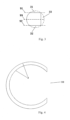
- the sound emitting surface 3 is a spherical surface having a first notch 31, a second notch 32 and a third notch 33, a sphere corresponding to the spherical surface has a diameter in a range of 400 mm to 800 mm, one great circle of the sphere is taken as a main great circle 99, the first notch 31 and the second notch 32 are respectively positioned at two intersections of the spherical surface and a diameter perpendicular to the main great circle 99, and the third notch 33 connects the first notch 31 with the second notch 32; within distances of 100 mm to 200 mm from the main great circle 99 respectively at both sides of the main great circle 99, a cross-section of the sound emitting surface 3 parallel to the main great circle 99 is in a shape of an arc, an opening of the arc corresponds to the third notch 33, and
- the treatment couch 2 is configured for a human body to lie in a lithotomy position, and when the human body lies in the lithotomy position on the treatment couch 2, the center of the sphere corresponding to the sound emitting surface 3 is positioned in a pelvic cavity of the human body, two legs respectively stick out of the sound emitting surface 3 through the first notch 31 and the second notch 32, and an upper part of the body sticks out of the sound emitting surface 3 through the third notch 33.
- the extracorporeal focused ultrasound treatment device for pelvic diseases of the embodiment has an ultrasonic transducer 1, and the ultrasonic transducer 1 has a sound generation unit, which is a device capable of generating ultrasound.
- the material of the sound generation unit may include piezoelectric ceramics, 1-3 type piezoelectric composite material, or the like.
- the shape, number, position, and other parameters of the sound generation unit may be designed such that the sound generation unit can emit ultrasound from all positions of the sound emitting surface 3, and the ultrasound emitted at each position propagates along the normal direction of the sound emitting surface 3 at the position, and the ultrasound can be finally focused (including directly focused or focused after being reflected) at a required position.
- the sound emitting surface 3 may be an acoustically transparent surface with a predetermined shape, and the sound generation unit (e.g., a piezoelectric array element 13) may be disposed behind the sound emitting surface 3; alternatively, the sound emitting surface 3 may be the emitting surface of the sound generation unit itself.
- the sound generation unit e.g., a piezoelectric array element 13
- the sound generation unit may also take different forms.
- the sound generation unit may be a plurality of piezoelectric array elements 13 (e.g., rectangular piezoelectric ceramic plates) disposed at different positions of the sound emitting surface 3, that is, the plurality of piezoelectric array elements 13 are spliced together to form the sound emitting surface 3; alternatively, the sound generation unit may also have the same shape as the sound emitting surface 3 (e.g., the sound generation unit is a specially shaped piezoelectric ceramic plate).
- the ultrasonic transducer 1 may further include, in addition to the sound emitting surface 3 and the sound generation unit, a driving circuit for the sound generation unit, a casing (e.g., the casing of the sound generation unit may include a housing 11, an upper cover 12, a lower cover, an end cover 14, etc.) for enclosing the driving circuit and the sound generation unit, and other components, which will not be described in detail herein.
- a driving circuit for the sound generation unit e.g., the casing of the sound generation unit may include a housing 11, an upper cover 12, a lower cover, an end cover 14, etc.
- the sound emitting surface 3 of the ultrasonic transducer 1 of the present embodiment is equivalent to a spherical surface lacking three portions, and the spherical surface may have a diameter in the range of 400 mm to 800 mm, preferably in the range of 420 mm to 600 mm.
- two portions (the first notch 31 and the second notch 32) missing from the sound emitting surface 3 are portions of the spherical surface at both ends of one diameter, and a great circle (i.e., a plane passing through the spherical center) perpendicular to the diameter is the main great circle 99.
- the third portion (third notch 33) missing from the sound emitting surface 3 is a portion laterally connecting the first notch 31 with the second notch 32.
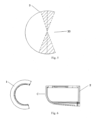
- a cross-section of the sound emitting surface 3 parallel to the main great circle 99 is in the shape of an arc, the central angle corresponding to the arc is greater than 180 degrees and less than 300 degrees, and preferably, greater than 200 degrees and less than 260 degrees, and the opening of the arc corresponds to the third notch 33. That is, at least within a certain distance from the main great circle 99, the portion of the spherical surface cut off by the third notch 33 has a limited range and the central angle corresponding to the remaining portion is within the above range.
- the sound emitting surface 3 has the capability of reflecting ultrasound, and at at least part of positions, the third notch 33 only cuts off a spherical surface smaller than half spherical surface. Therefore, as shown in Fig. 5 , ultrasound emitted from a part of the arc at an angle exceeding the central angle of 180 degrees is reflected by an opposite part of the sound emitting surface 3, and the part of the arc at the angle exceeding the central angle of 180 degrees may also reflect ultrasound emitted from the opposite part of the sound emitting surface 3, so that ultrasound can return in partial region (the region filled with oblique lines in Fig.
- the ultrasound generated by the ultrasound transducer 1 of the present embodiment is actually in the form of a combination of a traveling wave with a standing wave, and thus its propagation and focusing will change.
- the ultrasonic transducer 1 can compress the major axis of the original cigar-shaped focal region, so that the focal region has a shape closer to a spherical shape and has a smaller size, the energy density is improved, the treatment effect and efficiency are improved, the damage to normal tissues is reduced, and the safety is improved.
- the ultrasonic transducer 1 can also reduce the adverse effects of non-uniformity of tissues and bone tissues and the like on the focusing of ultrasound when the ultrasound propagates in a human body, and reduce deviation and distortion of the focal region, which facilitates accurate positioning of the focal region.
- edges of the first notch 31 and the second notch 32 are located in a first plane 91 and a second plane 92, respectively.
- the first plane 91 and the second plane 92 are both parallel to the main great circle 99.
- the first notch 31 and the second notch 32 are spherical caps cut off by planes.
- the first notch 31 and the second notch 32 are spherical caps cut off by two parallel planes, that is, the bottom surfaces of the two cut-off spherical caps are parallel to each other.
- the spherical surface excluding the first notch 31 and the second notch 32 is equivalent to a structure formed by butting the bottom surfaces of two spherical segments.
- the bottom surfaces of the two spherical segments are the main great circle 99, and the two spherical segments may have different heights.
- the sound emitting surface 3 in this form has a shape similar to a spherical segment, and is regular and simple in structure.
- first notch 31 and second notch 32 are cut off by planes that are not parallel to each other, or by curved surfaces that are not planar.
- the distance between the first plane 91 and the second plane 92 ranges from 200 mm to 400 mm. In an embodiment, the distance between the first plane 91 and the second plane 92 ranges from 200 mm to 300 mm.
- the distance between the first notch 31 and the second notch 32 is preferably in the above range (of course, the diameter of the sphere corresponding to the sound emitting surface 3 should be larger than the distance).
- Such sound emitting surface 3 has a sufficient area to generate ultrasound suitable for treatment and a size that is not too large, and can allow legs of the human body to stick out.
- the distance between the first plane 91 and the main great circle 99 is equal to the distance between the second plane 92 and the main great circle 99.
- the first notch 31 and the second notch 32 are preferably obtained by cutting with two planes that have a same distance to the center of the sphere, so that the two notches have a same size and are symmetrically distributed, which facilitates symmetry of the focal region and placement of the legs of the human body.
- first notch 31 and the second notch 32 have different distances to the center of the sphere, or have different shapes.
- any cross-section of the sound emitting surface 3 parallel to the main great circle 99 is in the shape of an arc, and the central angle corresponding to the arc is greater than 180 degrees and less than 300 degrees.
- the sound emitting surface 3 is arc-shaped in a cross-section parallel to the main great circle 99 at least in the vicinity of the main great circle 99.
- any cross-section of the sound emitting surface 3 parallel to the main great circle 99 may be in the shape of the arc, thereby ensuring that the sound emitting surface 3 can generate a standing wave at each position in the vertical direction.
- the arcs of the sound emitting surface 3 in any cross-sections thereof parallel to the main great circle 99 have openings orientated in a same direction, and correspond to central angles that are equal.
- the third notch 33 is orientated in the same direction, and corresponds to a same central angle. That is, the third notch 33 is preferably obtained by cutting with a plane perpendicular to the main great circle 99.
- the sound emitting surface 3 is shaped like the letter "C" as viewed in a direction perpendicular to the main great circle 99.
- the sound emitting surface 3 is symmetrical with respect to the main great circle 99.
- the sound emitting surface 3 is preferably symmetrical with respect to the main great circle 99, that is, parts of the sound emitting surface 3 respectively on both sides of the main great circle 99 are preferably of the same form, so that the sound field and focal region formed by the sound emitting surface are also symmetrical with respect to the main great circle 99, and are more regular and easy to control.
- the size and the central angle of the ultrasonic transducer 1 need to meet certain requirements, and the above limitation on the parameters of the sound emitting surface 3 just enables the ultrasonic transducer 1 to be adapted to the human body.
- the treatment couch 2 should have a chair, a leg support, etc., which will not be described in detail herein.
- the ultrasound transducer 1 cannot be suspended, and a corresponding housing, a supporting structure, a driving circuit, etc. should be provided, but for simplicity, these structures are not shown in the drawings.
- the extracorporeal focused ultrasound treatment device for pelvic diseases can treat diseases of organs in the pelvic cavity by way of externally focusing ultrasonic waves, so that the size of the ultrasonic emitting surface (i.e., the sound emitting surface 3) of the ultrasonic transducer can be larger, and under the condition that the ultrasonic energy emitted per unit area is the same, the area of the acoustic window for ultrasound to enter the human body can be larger, and the energy density obtained at the focal region is higher.
- the treatment effect is improved, the treatment efficiency is improved, the treatment comfort is improved, the operation convenience is improved, the harm to the human body is reduced, and the treatment safety is improved.
- the treatment couch 2 and the ultrasonic transducer 1 may be separated structures; the extracorporeal focused ultrasound treatment device for pelvic diseases also includes a movement unit configured to make the treatment couch 2 and the ultrasonic transducer 1 closer to or farther away from each other.
- the treatment couch 2 and the ultrasonic transducer 1 are preferably separated, and the treatment couch 2 may come closer to or farther away from the ultrasonic transducer 1 through the movement unit (e.g., a wheel, a rail, etc.).
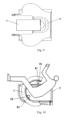
- the human body can lie on the treatment couch 2 in a lithotomy position when the treatment couch 2 is away from the ultrasonic transducer 1, and then the treatment couch 2 is caused to come close to the ultrasonic transducer 1 to make the perineum enter the sound emitting surface 3 through the third notch 33, thereby obtaining the structure shown in Figs. 10 and 11 .
- ultrasound emitted from a first region 35 of the sound emitting surface 3 enters the pelvic cavity through the abdomen of the human body; when the human body lies on the treatment couch 2 in the lithotomy position, ultrasound emitted from a second region 36 of the sound emitting surface 3 enters the pelvic cavity through area between the coccyx and pubic symphysis of the human body.
- the sound emitting surface 3 of the ultrasonic transducer 1 preferably has at least a first region 35 and a second region 36, and ultrasonic waves emitted from the two regions may respectively pass through the abdomen and the area between the coccyx and pubic symphysis to enter the pelvic cavity, so as to maximize the beam path.
- the sound emitting surface 3 should also have a region between the first region 35 and the second region 36, and since the central angle between the first region 35 and the second region 36 is usually less than 150 degrees, as shown in Fig. 10 , the sound emitting surface 3 actually should have a portion exceeding the first region 35 and the second region 36, such as a portion corresponding to the sacrum.
- ultrasonic waves emitted from all positions of the sound emitting surface 3 can form a better sound field together.
- the extracorporeal focused ultrasound treatment device for pelvic diseases further includes an imaging unit configured to form an image of the pelvic cavity.
- the extracorporeal focused ultrasound treatment device for pelvic diseases may also include an imaging unit (e.g., B-mode ultrasound, CT, MRI or the combination thereof) for forming an image of the pelvic cavity, so that a lesion is positioned before treatment and an image of an area around the treated part is formed in real time during treatment, so as to evaluate the treatment effect at any time and adjust the treatment plan.
- an imaging unit e.g., B-mode ultrasound, CT, MRI or the combination thereof
- the extracorporeal focused ultrasound treatment device for pelvic diseases may include:
- B-mode ultrasound can be used to form an image of the pelvic cavity for monitoring, and since the B-mode ultrasound also achieves imaging by using ultrasound, it is also blocked by bones, so that the B-mode ultrasonic probes should also be disposed in the first region 35 and the second region 36 as shown in Fig. 10 , so as to avoid bones to obtain images at these positions, to ensure clarity of the images, and to minimize the influence of the B-mode ultrasonic probe on the therapeutic ultrasound.
- a first B-mode ultrasonic probe 41 is disposed in the first region 35 and emits, through the abdomen, ultrasound for imaging, while a second B-mode ultrasonic probe 42 is disposed at a specific position in the second region 36, i.e., emits, through the perineum (rather than the anus, etc.,) ultrasound for imaging.
- the angle between the first B-mode ultrasonic probe 41 and the vertical direction is usually about 30 degrees, and the angle between the second B-mode ultrasonic probe 42 and the vertical direction is about 80 degrees.
- the B-mode ultrasonic probes may be arranged at corresponding positions of the sound emitting surface 3 and perform imaging in a non-contact manner; alternatively, as shown in Fig. 10 , the B-mode ultrasonic probes may protrude from the sound emitting surface 3 and may be retractable, so that one or two of the B-mode ultrasonic probes may be selected to extend out and contact with the human body as required for imaging.
- the B-mode ultrasonic probes are disposed on the sound emitting surface 3 (i.e., on the ultrasonic transducer 1), so that when the ultrasonic transducer 1 moves, the B-mode ultrasonic probes will move together with the ultrasonic transducer 1, and thus the B-mode ultrasonic probes aim at the optimal imaging positions at any time.
- the extracorporeal focused ultrasound treatment device for pelvic diseases further includes a driving unit for driving the ultrasonic transducer 1 to move relative to the treatment couch 2.
- a driving unit may be provided to drive the ultrasonic transducer 1 to move, and then to drive the focal region to move.
- the movement driven by the driving unit may include translations in three axial directions perpendicular to one another, and such movement may also cause the focal region to translate; alternatively, the movement may include rotating the ultrasound transducer 1 around different axial directions, so as to cause the ultrasound to enter the human body from different directions.
- the extracorporeal focused ultrasound treatment device for pelvic diseases further includes a medium containing unit for keeping a sound transmission medium between a surface of the human body and the sound emitting surface 3.
- a sound transmission medium such as deaerated water may be provided between the sound emitting surface 3 of the ultrasound transducer 1 and the human body, and for this reason, a medium containing unit capable of holding a sound transmission medium (e.g., deaerated water) is preferably provided to cause the space between the sound emitting surface 3 of the ultrasound transducer 1 of the present embodiments and the surface of the human body through which ultrasound is to pass to be filled with the sound transmission medium, and the medium containing unit may be in the form of a water basin or the like, and will not be described in detail herein.
- a medium containing unit capable of holding a sound transmission medium
- the ultrasound generated by the sound generation unit has a frequency in the range of 0.4 MHz to 1.5 MHz.
- the ultrasound generated by the sound generation unit has an acoustical power in the range of 0W to 1200W. In an embodiment, the acoustical power of the ultrasound generated by the sound generation unit ranges from 0W to 800W.
- the parameters of the ultrasound emitted by the ultrasonic transducer 1 are preferably in the above ranges to achieve good treatment effect.
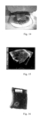
- the extracorporeal focused ultrasound treatment device for pelvic diseases of the embodiments emits ultrasonic waves at an acoustical power of 200W toward deaerated water, so as to cavitate water in the focal region, and a photograph of the cavitated region taken from the first notch 31 is shown in Fig. 12 .
- the cavitated region i.e., the focal region
- the focal region has a shape close to a circle, a size of 1.8 mm * 1.2 mm, and a length-width ratio of 3: 2, which indicates that, compared with the conventional focused ultrasound transducer with only traveling waves, the extracorporeal focused ultrasound treatment device for pelvic diseases of the embodiments has a focused ultrasound transducer having a focal region whose major axis is significantly compressed, whose shape changes from a cigar shape to an approximately spherical shape, and which has a reduced size, an increased energy density, and a more regular shape.
- the extracorporeal focused ultrasound treatment device for pelvic diseases of the embodiments When the extracorporeal focused ultrasound treatment device for pelvic diseases of the embodiments is used to treat an exvivo bovine liver with ultrasound irradiation at an acoustical power of 400W for 2 seconds, as shown in Fig. 13 , a target area with a depth of 80 mm is obviously damaged in a short time, and the damaged part is in a fusiform shape, and has a clear boundary, a size of 4.4 mm * 1.5 mm, and a length-width ratio of less than 3:1, which is lower than the length-width ratio (generally greater than 5: 1) of the damaged part caused by a conventional focused ultrasound transducer. This also indicates that the focal region of the extracorporeal focused ultrasound treatment device for pelvic diseases of the present embodiments has a more regular shape.
- a pelvic bone is placed at a preset position at an inner side of the ultrasonic transducer 1 of the extracorporeal focused ultrasound treatment device for pelvic diseases so as to simulate the position of the pelvic bone of a human body, and then an exvivo bovine muscle tissue is placed in the pelvic bone, as shown in Fig. 15 .
- the focal region is positioned at a position equivalent to the position having a distance of 10 mm from the rectum of the human body, and a treatment process for the prostate is simulated, the ultrasonic power is 400 W, the target area has a depth of 55 mm, and the ultrasonic irradiation time is 2 seconds * 5 times.
- the ultrasonic power is 400 W
- the target area has a depth of 55 mm
- the ultrasonic irradiation time is 2 seconds * 5 times.
- the bovine muscle tissue subjected to ultrasonic irradiation has an obviously damaged target area with a clear boundary, no damage is caused to the envelope, and no damage is caused to the interface between the simulated rectum and prostate.
- This shows that the ultrasound emitted by extracorporeal focused ultrasound treatment device for pelvic diseases of the embodiments is less affected by non-uniform tissues such as bones in a human body, can still maximize an beam path when being applied in an actual human body environment, forms a focal region with small size, excellent shape and accurate position, achieves good treatment effect and efficiency, and avoids damage to normal tissues.
Landscapes
- Health & Medical Sciences (AREA)
- Life Sciences & Earth Sciences (AREA)
- Engineering & Computer Science (AREA)
- Biomedical Technology (AREA)
- Veterinary Medicine (AREA)
- Public Health (AREA)
- General Health & Medical Sciences (AREA)
- Animal Behavior & Ethology (AREA)
- Radiology & Medical Imaging (AREA)
- Nuclear Medicine, Radiotherapy & Molecular Imaging (AREA)
- Physics & Mathematics (AREA)
- Biophysics (AREA)
- Molecular Biology (AREA)
- Acoustics & Sound (AREA)
- Pathology (AREA)
- Heart & Thoracic Surgery (AREA)
- Medical Informatics (AREA)
- Surgery (AREA)
- Orthopedic Medicine & Surgery (AREA)
- Physical Education & Sports Medicine (AREA)
- Rehabilitation Therapy (AREA)
- Multimedia (AREA)
- Gynecology & Obstetrics (AREA)
- Surgical Instruments (AREA)
- Thermotherapy And Cooling Therapy Devices (AREA)
Claims (15)
- Extrakorporal fokussierte Ultraschallbehandlungsvorrichtung für eine Beckenerkrankung, die einen Ultraschallwandler (1) und eine Behandlungsliege (2) umfasst, wobeider Ultraschallwandler eine Schallemissionsfläche (3) und eine Schallerzeugungseinheit (05) umfasst, die konfiguriert ist, um eine Ultraschallwelle zu erzeugen; wobei die Schallemissionsfläche eine kugelförmige Oberfläche mit einem ersten Einschnitt (31), einem zweiten Einschnitt (32) und einem dritten Einschnitt (33) ist,eine Kugel, die der kugelförmige Oberfläche entspricht, einen Durchmesser in einem Bereich von 400 mm bis 800 mm aufweist, wobei ein großer Kreis der Kugel ein großer Hauptkreis (99) ist,der erste Einschnitt und der zweite Einschnitt jeweils an zwei Schnittpunkten der sphärischen Oberfläche und eines Durchmessers, der senkrecht zu dem großen Hauptkreis ist, positioniert sind, und wobei der dritte Einschnitt den ersten Einschnitt mit dem zweiten Einschnitt verbindet; wobei sich innerhalb von Abständen von 100 mm bis 200 mm von dem großen Hauptkreis jeweils an beiden Seiten des großen Hauptkreises ein Querschnitt der Schallemissionsfläche parallel zum großen Hauptkreis in einer Form eines Bogens befindet, wobei eine Öffnung des Bogens dem dritten Einschnitt entspricht, und ein Mittelpunktswinkel, der dem Bogen entspricht, größer als 180 Grad und kleiner als 300 Grad ist; und die Schallemissionsfläche in der Lage ist, Ultraschall zu reflektieren, und wobei eine Ultraschallwelle, die von der Schallerzeugungseinheit erzeugt wird, auf einen Mittelpunkt der Kugel, die der Schallemissionsfläche entspricht, fokussiert wird; unddie Behandlungsliege so konfiguriert ist, dass ein menschlicher Körper in einer Steinschnittlage liegt, und so konfiguriert ist, dass sich dann, wenn der menschliche Körper auf der Behandlungsliege in der Steinschnittlage liegt, eine Beckenhöhle des menschlichen Körpers in der Mitte der Kugel, die der Schallemissionsfläche entspricht, mit zwei Beinen des Menschen befindet, bzw. so konfiguriert ist, dass eine Beckenhöhle aus der Schallemissionsfläche durch den ersten Einschnitt und den zweiten Einschnitt hervorsteht, und wobei ein oberer Teil des menschlichen Körpers durch den dritten Einschnitt aus der Schallemissionsfläche hervorsteht.
- Extrakorporal fokussierte Ultraschallbehandlungsvorrichtung für eine Beckenerkrankung nach Anspruch 1, wobei eine Kante des ersten Einschnitts und eine Kante des zweiten Einschnitts sich in einer ersten bzw. in einer zweiten Ebene befinden.
- Extrakorporal fokussierte Ultraschallbehandlungsvorrichtung für eine Beckenerkrankung nach Anspruch 2, wobei die erste Ebene und die zweite Ebene beide parallel zu dem großen Hauptkreis verlaufen.
- Extrakorporal fokussierte Ultraschallbehandlungsvorrichtung für eine Beckenerkrankung nach Anspruch 3, wobei ein Abstand zwischen der ersten Ebene und der zweiten Ebene in einem Bereich von 200 mm bis 400 mm liegt.
- Extrakorporal fokussierte Ultraschallbehandlungsvorrichtung für eine Beckenerkrankung nach Anspruch 3, wobei der Abstand zwischen der ersten Ebene und dem großen Hauptkreis gleich einem Abstand zwischen der zweiten Ebene und dem großen Hauptkreis ist.
- Extrakorporal fokussierte Ultraschallbehandlungsvorrichtung für eine Beckenerkrankung nach Anspruch 1, wobeider Durchmesser der Kugel, die der Schallemissionsfläche entspricht, in einem Bereich von 420 mm bis 600 mm liegt; undinnerhalb von Abständen von 100 mm bis 150 mm von dem großen Hauptkreis jeweils an beiden Seiten des großen Hauptkreises der Mittelpunktswinkel, der dem Bogen in dem Querschnitt der Schallemissionsfläche parallel zum großen Hauptkreis entspricht, größer als 180 Grad und kleiner als 300 Grad ist.
- Extrakorporal fokussierte Ultraschallbehandlungsvorrichtung für eine Beckenerkrankung nach Anspruch 1, wobei jeder Querschnitt der Schallemissionsfläche parallel zum Hauptgroßkreis die Form eines Bogens aufweist, und wobei der Mittelpunktswinkel, der dem Bogen entspricht, größer als 200 Grad und kleiner als 260 Grad ist.
- Extrakorporal fokussierte Ultraschallbehandlungsvorrichtung für eine Beckenerkrankung nach Anspruch 7, wobei die Öffnung des Bogens in jedem Querschnitt der Schallemissionsfläche parallel zu dem Hauptgroßkreis in einer selben Richtung orientiert ist, und wobei der Mittelpunktswinkel, der dem Bogen entspricht, gleich ist.
- Extrakorporal fokussierte Ultraschallbehandlungsvorrichtung für eine Beckenerkrankung nach Anspruch 1, wobei die Schallemissionsfläche symmetrisch in Bezug auf den großen Hauptkreis ist.
- Extrakorporal fokussierte Ultraschallbehandlungsvorrichtung für eine Beckenerkrankung nach Anspruch 1, wobei dann, wenn der menschliche Körper auf der Behandlungsliege in der Steinschnittlage liegt, der Ultraschall, der von einem ersten Bereich der Schallemissionsfläche emittiert wird, durch den Bauch des menschlichen Körpers in die Beckenhöhle eintritt; und
wobei dann, wenn der menschliche Körper auf der Behandlungsliege in der Steinschnittlage liegt, der Ultraschall, der von einem zweiten Bereich der Schallemissionsfläche emittiert wird, durch einen Bereich zwischen dem Steißbein und der Schambeinsymphyse des menschlichen Körpers in die Beckenhöhle eintritt. - Extrakorporal fokussierte Ultraschallbehandlungsvorrichtung für eine Beckenerkrankung nach Anspruch 10, die ferner umfasst:eine erste B-Mode-Ultraschallsonde, die konfiguriert ist, um ein Ultraschallbild von dem ersten Bereich der Schallemissionsfläche zu der Beckenhöhle durch den Bauch des menschlichen Körpers zu senden, um ein Bild der Beckenhöhle zu bilden; und/odereine zweite B-Mode-Ultraschallsonde, die konfiguriert ist, um ein Ultraschallbild von dem zweiten Bereich der Schallemissionsfläche zu der Beckenhöhle durch das Perineum des menschlichen Körpers zu senden, um ein Bild der Beckenhöhle zu bilden.
- Extrakorporal fokussierte Ultraschallbehandlungsvorrichtung für eine Beckenerkrankung nach Anspruch 1, wobeidie Behandlungsliege und der Ultraschallwandler getrennte Strukturen sind; unddie extrakorporale fokussierte Ultraschallbehandlungsvorrichtung für eine Beckenerkrankung ferner eine Bewegungseinheit umfasst, die konfiguriert ist, um die Behandlungsliege zu veranlassen, näher an den Ultraschallwandler heranzukommen oder sich weiter von diesem zu entfernen.
- Extrakorporal fokussierte Ultraschallbehandlungsvorrichtung für eine Beckenerkrankung nach Anspruch 1, die ferner umfasst:
ein Medium, das eine Einheit enthält, die konfiguriert ist, um ein Schhallübertragungsmedium zwischen einer Oberfläche des menschlichen Körpers und der Schallemissionsfläche zu halten. - Extrakorporal fokussierte Ultraschallbehandlungsvorrichtung für eine Beckenerkrankung nach Anspruch 1, die ferner umfasst:
eine Antriebseinheit, die konfiguriert ist, um den Ultraschallwandler anzutreiben, um sich relativ zur Behandlungsliege zu bewegen. - Extrakorporal fokussierte Ultraschallbehandlungsvorrichtung für eine Beckenerkrankung nach Anspruch 1, wobeider Ultraschall, der von der Schallerzeugungseinheit erzeugt worden ist, eine Frequenz in einem Bereich von 0,4 MHz bis 1,5 MHz aufweist;der Ultraschall, der von der Schallerzeugungseinheit erzeugt worden ist, eine akustische Leistung in einem Bereich von 0 W bis 1.200 W aufweist.
Applications Claiming Priority (2)
| Application Number | Priority Date | Filing Date | Title |
|---|---|---|---|
| CN201810059205.5A CN110064135B (zh) | 2018-01-22 | 2018-01-22 | 盆腔疾病体外聚焦超声治疗设备 |
| PCT/CN2018/104623 WO2019140928A1 (zh) | 2018-01-22 | 2018-09-07 | 盆腔疾病体外聚焦超声治疗设备 |
Publications (3)
| Publication Number | Publication Date |
|---|---|
| EP3744397A1 EP3744397A1 (de) | 2020-12-02 |
| EP3744397A4 EP3744397A4 (de) | 2021-10-20 |
| EP3744397B1 true EP3744397B1 (de) | 2024-12-18 |
Family
ID=67301931
Family Applications (1)
| Application Number | Title | Priority Date | Filing Date |
|---|---|---|---|
| EP18901604.1A Active EP3744397B1 (de) | 2018-01-22 | 2018-09-07 | In-vitro-fokussierte ultraschallbehandlungsvorrichtung für beckenerkrankung |
Country Status (10)
| Country | Link |
|---|---|
| US (1) | US11998764B2 (de) |
| EP (1) | EP3744397B1 (de) |
| JP (1) | JP7012861B2 (de) |
| KR (1) | KR102525435B1 (de) |
| CN (1) | CN110064135B (de) |
| CA (1) | CA3088816C (de) |
| ES (1) | ES3005837T3 (de) |
| RU (1) | RU2741721C1 (de) |
| SG (1) | SG11202006840VA (de) |
| WO (1) | WO2019140928A1 (de) |
Families Citing this family (4)
| Publication number | Priority date | Publication date | Assignee | Title |
|---|---|---|---|---|
| JP2022510654A (ja) | 2018-11-28 | 2022-01-27 | ヒストソニックス,インコーポレーテッド | 組織破砕システムおよび方法 |
| CN113117265B (zh) * | 2019-12-30 | 2023-03-28 | 重庆融海超声医学工程研究中心有限公司 | 检测装置 |
| JP2023530477A (ja) | 2020-06-18 | 2023-07-18 | ヒストソニックス,インコーポレーテッド | 組織破砕音響/患者結合システムおよび方法 |
| CN119318780B (zh) * | 2024-12-13 | 2025-04-01 | 北京渐健医疗科技有限公司 | 基于低强度聚焦超声技术的智能医疗机器人 |
Citations (1)
| Publication number | Priority date | Publication date | Assignee | Title |
|---|---|---|---|---|
| EP2524651B1 (de) * | 2010-04-02 | 2015-10-07 | Chongqing Haifu Medical Technology Co., Ltd. | Ultraschallwandler |
Family Cites Families (22)
| Publication number | Priority date | Publication date | Assignee | Title |
|---|---|---|---|---|
| US5131392A (en) * | 1990-02-13 | 1992-07-21 | Brigham & Women's Hospital | Use of magnetic field of magnetic resonance imaging devices as the source of the magnetic field of electromagnetic transducers |
| DE4238645C1 (de) * | 1992-11-16 | 1994-05-05 | Siemens Ag | Therapeutischer Ultraschall-Applikator für den Urogenitalbereich |
| JPH07194611A (ja) * | 1994-01-10 | 1995-08-01 | Toshiba Corp | 超音波治療装置 |
| JPH08252261A (ja) * | 1995-03-17 | 1996-10-01 | Toshiba Corp | 超音波治療装置 |
| CN2503917Y (zh) * | 2001-11-05 | 2002-08-07 | 北京源德生物医学工程股份有限公司 | 用于体外高能聚焦超声波治疗机的坐位架 |
| CN1169588C (zh) * | 2001-11-05 | 2004-10-06 | 北京源德生物医学工程股份有限公司 | 体外高能聚焦超声波治疗机 |
| CN1657114A (zh) * | 2004-02-17 | 2005-08-24 | 崔景彦 | 电子磁疗前列腺治疗仪 |
| US8409099B2 (en) | 2004-08-26 | 2013-04-02 | Insightec Ltd. | Focused ultrasound system for surrounding a body tissue mass and treatment method |
| CN1966109A (zh) | 2005-11-18 | 2007-05-23 | 重庆融海超声医学工程研究中心有限公司 | 一种超声波治疗头及含有该超声波治疗头的超声波治疗系统 |
| CN2889239Y (zh) * | 2005-12-19 | 2007-04-18 | 重庆海扶(Hifu)技术有限公司 | 能变换体位的治疗床及含该治疗床的高强度聚焦超声治疗系统 |
| FR2923612B1 (fr) | 2007-11-12 | 2011-05-06 | Super Sonic Imagine | Dispositif d'insonification comprenant un reseau tridimensionnel d'emetteurs disposes en spirale apte a generer un faisceau d'ondes focalisees de grande intensite |
| CN102917756B (zh) * | 2010-05-27 | 2016-12-21 | 皇家飞利浦电子股份有限公司 | 用于选择性地生成超声波和热的超声换能器 |
| FR2973250B1 (fr) | 2011-03-29 | 2015-01-02 | Edap Tms France | Sonde de therapie pour le traitement de tissus par l'intermediaire d'ondes ultrasonores focalisees croisees |
| CN103520844B (zh) * | 2012-07-03 | 2016-07-13 | 重庆海扶医疗科技股份有限公司 | 聚焦超声治疗装置 |
| CN103143125B (zh) * | 2013-03-25 | 2015-12-23 | 广州多浦乐电子科技有限公司 | 一种高强度聚焦超声治疗仪 |
| KR101467511B1 (ko) * | 2013-07-24 | 2014-12-02 | 알피니언메디칼시스템 주식회사 | 고강도 집속 초음파 치료용 보조기구 |
| EP2886159A1 (de) * | 2013-12-23 | 2015-06-24 | Theraclion SA | Verfahren zum Betreiben einer Vorrichtung zur Behandlung eines Gewebes und Vorrichtung zur Behandlung eines Gewebes |
| US10639108B2 (en) * | 2015-10-30 | 2020-05-05 | Auris Health, Inc. | Process for percutaneous operations |
| CN109788934B (zh) * | 2016-09-23 | 2022-04-26 | 三星麦迪森株式会社 | 妇产科诊断设备以及使用其的妇产科诊断方法 |
| KR101801900B1 (ko) * | 2016-12-30 | 2017-11-28 | 알피니언메디칼시스템 주식회사 | 차폐부를 가지는 hifu 장치와 hifu 장치의 영상 잡음 제거 및 영상용 트랜스듀서 보호방법 |
| KR102168246B1 (ko) * | 2017-02-28 | 2020-10-20 | 주식회사 하이로닉 | 고강도 집속 초음파 시술 장치 |
| CN208911311U (zh) * | 2018-01-22 | 2019-05-31 | 重庆海扶医疗科技股份有限公司 | 盆腔疾病体外聚焦超声治疗设备 |
-
2018
- 2018-01-22 CN CN201810059205.5A patent/CN110064135B/zh active Active
- 2018-09-07 RU RU2020127321A patent/RU2741721C1/ru active
- 2018-09-07 ES ES18901604T patent/ES3005837T3/es active Active
- 2018-09-07 JP JP2020540467A patent/JP7012861B2/ja active Active
- 2018-09-07 KR KR1020207024086A patent/KR102525435B1/ko active Active
- 2018-09-07 SG SG11202006840VA patent/SG11202006840VA/en unknown
- 2018-09-07 CA CA3088816A patent/CA3088816C/en active Active
- 2018-09-07 EP EP18901604.1A patent/EP3744397B1/de active Active
- 2018-09-07 WO PCT/CN2018/104623 patent/WO2019140928A1/zh not_active Ceased
- 2018-09-07 US US16/963,280 patent/US11998764B2/en active Active
Patent Citations (1)
| Publication number | Priority date | Publication date | Assignee | Title |
|---|---|---|---|---|
| EP2524651B1 (de) * | 2010-04-02 | 2015-10-07 | Chongqing Haifu Medical Technology Co., Ltd. | Ultraschallwandler |
Also Published As
| Publication number | Publication date |
|---|---|
| US20210361976A1 (en) | 2021-11-25 |
| SG11202006840VA (en) | 2020-08-28 |
| ES3005837T3 (en) | 2025-03-17 |
| US11998764B2 (en) | 2024-06-04 |
| EP3744397A1 (de) | 2020-12-02 |
| RU2741721C1 (ru) | 2021-01-28 |
| KR20200111745A (ko) | 2020-09-29 |
| KR102525435B1 (ko) | 2023-04-26 |
| CN110064135B (zh) | 2024-06-21 |
| JP2021511156A (ja) | 2021-05-06 |
| EP3744397A4 (de) | 2021-10-20 |
| CN110064135A (zh) | 2019-07-30 |
| CA3088816A1 (en) | 2019-07-25 |
| JP7012861B2 (ja) | 2022-01-28 |
| CA3088816C (en) | 2023-06-27 |
| WO2019140928A1 (zh) | 2019-07-25 |
Similar Documents
| Publication | Publication Date | Title |
|---|---|---|
| EP3744396B1 (de) | Ultraschallwandler und fokussierte ultraschallbehandlungsvorrichtung | |
| EP3744397B1 (de) | In-vitro-fokussierte ultraschallbehandlungsvorrichtung für beckenerkrankung | |
| WO2008025190A1 (en) | A high intensity focused ultrasound therapeutic system guided by an imaging device guided | |
| JPH06197907A (ja) | 治療用超音波アプリケータ | |
| CN102580261A (zh) | 一种适用于浅表肿瘤治疗的聚焦超声换能器装置 | |
| Brentnall et al. | A new high intensity focused ultrasound applicator for surgical applications | |
| CN208911311U (zh) | 盆腔疾病体外聚焦超声治疗设备 | |
| CN209422796U (zh) | 聚焦超声治疗系统 | |
| CN209060387U (zh) | 超声换能器、聚焦超声治疗设备 | |
| HK40011575A (en) | Extracorporeal focused ultrasoound therapeutic device for pelvic diseases | |
| HK40011575B (zh) | 盆腔疾病体外聚焦超声治疗设备 | |
| TWI651109B (zh) | 治療型超音波裝置及其用途 | |
| HK40011576A (en) | Ultrasonic transducer and focused ultrasound therapeutic device | |
| HK40011576B (zh) | 超声换能器、聚焦超声治疗设备 | |
| CN111801054A (zh) | 治疗型超声波装置及其用途 | |
| Zhou et al. | Producing uniform lesion pattern in HIFU ablation | |
| Zhou et al. | Comparison of pathway in high intensity focused ultrasound (HIFU) lesion production | |
| JP2004167034A (ja) | 超音波照射装置 | |
| CN113101551A (zh) | 用于乳腺肿瘤的超声治疗探头及包括其的装置 | |
| Lobstein-Adams | Development of a medical imaging-based technology for cancer treatment | |
| CN111821588A (zh) | 超声治疗设备 | |
| Jeong et al. | Extended necrosis by using dual-curved therapeutic transducer for noninvasive HIFU surgery | |
| Song et al. | Electronically steerable large-scale ultrasound phased-array for noninvasive transcranial therapy |
Legal Events
| Date | Code | Title | Description |
|---|---|---|---|
| STAA | Information on the status of an ep patent application or granted ep patent |
Free format text: STATUS: THE INTERNATIONAL PUBLICATION HAS BEEN MADE |
|
| PUAI | Public reference made under article 153(3) epc to a published international application that has entered the european phase |
Free format text: ORIGINAL CODE: 0009012 |
|
| STAA | Information on the status of an ep patent application or granted ep patent |
Free format text: STATUS: REQUEST FOR EXAMINATION WAS MADE |
|
| 17P | Request for examination filed |
Effective date: 20200721 |
|
| AK | Designated contracting states |
Kind code of ref document: A1 Designated state(s): AL AT BE BG CH CY CZ DE DK EE ES FI FR GB GR HR HU IE IS IT LI LT LU LV MC MK MT NL NO PL PT RO RS SE SI SK SM TR |
|
| AX | Request for extension of the european patent |
Extension state: BA ME |
|
| DAV | Request for validation of the european patent (deleted) | ||
| DAX | Request for extension of the european patent (deleted) | ||
| A4 | Supplementary search report drawn up and despatched |
Effective date: 20210916 |
|
| RIC1 | Information provided on ipc code assigned before grant |
Ipc: A61N 7/00 20060101ALN20210910BHEP Ipc: A61N 7/02 20060101AFI20210910BHEP |
|
| RAP3 | Party data changed (applicant data changed or rights of an application transferred) |
Owner name: CHONGQING HAIFU MEDICAL TECHNOLOGY CO., LTD. |
|
| RIC1 | Information provided on ipc code assigned before grant |
Ipc: A61N 7/00 20060101ALN20240725BHEP Ipc: A61N 7/02 20060101AFI20240725BHEP |
|
| GRAP | Despatch of communication of intention to grant a patent |
Free format text: ORIGINAL CODE: EPIDOSNIGR1 |
|
| STAA | Information on the status of an ep patent application or granted ep patent |
Free format text: STATUS: GRANT OF PATENT IS INTENDED |
|
| INTG | Intention to grant announced |
Effective date: 20240903 |
|
| GRAS | Grant fee paid |
Free format text: ORIGINAL CODE: EPIDOSNIGR3 |
|
| GRAA | (expected) grant |
Free format text: ORIGINAL CODE: 0009210 |
|
| STAA | Information on the status of an ep patent application or granted ep patent |
Free format text: STATUS: THE PATENT HAS BEEN GRANTED |
|
| AK | Designated contracting states |
Kind code of ref document: B1 Designated state(s): AL AT BE BG CH CY CZ DE DK EE ES FI FR GB GR HR HU IE IS IT LI LT LU LV MC MK MT NL NO PL PT RO RS SE SI SK SM TR |
|
| REG | Reference to a national code |
Ref country code: CH Ref legal event code: EP |
|
| REG | Reference to a national code |
Ref country code: DE Ref legal event code: R096 Ref document number: 602018077827 Country of ref document: DE |
|
| REG | Reference to a national code |
Ref country code: IE Ref legal event code: FG4D |
|
| REG | Reference to a national code |
Ref country code: ES Ref legal event code: FG2A Ref document number: 3005837 Country of ref document: ES Kind code of ref document: T3 Effective date: 20250317 |
|
| REG | Reference to a national code |
Ref country code: LT Ref legal event code: MG9D |
|
| PG25 | Lapsed in a contracting state [announced via postgrant information from national office to epo] |
Ref country code: HR Free format text: LAPSE BECAUSE OF FAILURE TO SUBMIT A TRANSLATION OF THE DESCRIPTION OR TO PAY THE FEE WITHIN THE PRESCRIBED TIME-LIMIT Effective date: 20241218 |
|
| PG25 | Lapsed in a contracting state [announced via postgrant information from national office to epo] |
Ref country code: FI Free format text: LAPSE BECAUSE OF FAILURE TO SUBMIT A TRANSLATION OF THE DESCRIPTION OR TO PAY THE FEE WITHIN THE PRESCRIBED TIME-LIMIT Effective date: 20241218 |
|
| PG25 | Lapsed in a contracting state [announced via postgrant information from national office to epo] |
Ref country code: BG Free format text: LAPSE BECAUSE OF FAILURE TO SUBMIT A TRANSLATION OF THE DESCRIPTION OR TO PAY THE FEE WITHIN THE PRESCRIBED TIME-LIMIT Effective date: 20241218 |
|
| PG25 | Lapsed in a contracting state [announced via postgrant information from national office to epo] |
Ref country code: NO Free format text: LAPSE BECAUSE OF FAILURE TO SUBMIT A TRANSLATION OF THE DESCRIPTION OR TO PAY THE FEE WITHIN THE PRESCRIBED TIME-LIMIT Effective date: 20250318 |
|
| PG25 | Lapsed in a contracting state [announced via postgrant information from national office to epo] |
Ref country code: LV Free format text: LAPSE BECAUSE OF FAILURE TO SUBMIT A TRANSLATION OF THE DESCRIPTION OR TO PAY THE FEE WITHIN THE PRESCRIBED TIME-LIMIT Effective date: 20241218 Ref country code: GR Free format text: LAPSE BECAUSE OF FAILURE TO SUBMIT A TRANSLATION OF THE DESCRIPTION OR TO PAY THE FEE WITHIN THE PRESCRIBED TIME-LIMIT Effective date: 20250319 |
|
| PG25 | Lapsed in a contracting state [announced via postgrant information from national office to epo] |
Ref country code: RS Free format text: LAPSE BECAUSE OF FAILURE TO SUBMIT A TRANSLATION OF THE DESCRIPTION OR TO PAY THE FEE WITHIN THE PRESCRIBED TIME-LIMIT Effective date: 20250318 |
|
| PG25 | Lapsed in a contracting state [announced via postgrant information from national office to epo] |
Ref country code: NL Free format text: LAPSE BECAUSE OF FAILURE TO SUBMIT A TRANSLATION OF THE DESCRIPTION OR TO PAY THE FEE WITHIN THE PRESCRIBED TIME-LIMIT Effective date: 20241218 |
|
| REG | Reference to a national code |
Ref country code: AT Ref legal event code: MK05 Ref document number: 1751780 Country of ref document: AT Kind code of ref document: T Effective date: 20241218 |
|
| PG25 | Lapsed in a contracting state [announced via postgrant information from national office to epo] |
Ref country code: SM Free format text: LAPSE BECAUSE OF FAILURE TO SUBMIT A TRANSLATION OF THE DESCRIPTION OR TO PAY THE FEE WITHIN THE PRESCRIBED TIME-LIMIT Effective date: 20241218 |
|
| PG25 | Lapsed in a contracting state [announced via postgrant information from national office to epo] |
Ref country code: PL Free format text: LAPSE BECAUSE OF FAILURE TO SUBMIT A TRANSLATION OF THE DESCRIPTION OR TO PAY THE FEE WITHIN THE PRESCRIBED TIME-LIMIT Effective date: 20241218 |
|
| PG25 | Lapsed in a contracting state [announced via postgrant information from national office to epo] |
Ref country code: IS Free format text: LAPSE BECAUSE OF FAILURE TO SUBMIT A TRANSLATION OF THE DESCRIPTION OR TO PAY THE FEE WITHIN THE PRESCRIBED TIME-LIMIT Effective date: 20250418 |
|
| PG25 | Lapsed in a contracting state [announced via postgrant information from national office to epo] |
Ref country code: PT Free format text: LAPSE BECAUSE OF FAILURE TO SUBMIT A TRANSLATION OF THE DESCRIPTION OR TO PAY THE FEE WITHIN THE PRESCRIBED TIME-LIMIT Effective date: 20250421 |
|
| PG25 | Lapsed in a contracting state [announced via postgrant information from national office to epo] |
Ref country code: EE Free format text: LAPSE BECAUSE OF FAILURE TO SUBMIT A TRANSLATION OF THE DESCRIPTION OR TO PAY THE FEE WITHIN THE PRESCRIBED TIME-LIMIT Effective date: 20241218 |
|
| PG25 | Lapsed in a contracting state [announced via postgrant information from national office to epo] |
Ref country code: AT Free format text: LAPSE BECAUSE OF FAILURE TO SUBMIT A TRANSLATION OF THE DESCRIPTION OR TO PAY THE FEE WITHIN THE PRESCRIBED TIME-LIMIT Effective date: 20241218 Ref country code: RO Free format text: LAPSE BECAUSE OF FAILURE TO SUBMIT A TRANSLATION OF THE DESCRIPTION OR TO PAY THE FEE WITHIN THE PRESCRIBED TIME-LIMIT Effective date: 20241218 |
|
| PG25 | Lapsed in a contracting state [announced via postgrant information from national office to epo] |
Ref country code: SK Free format text: LAPSE BECAUSE OF FAILURE TO SUBMIT A TRANSLATION OF THE DESCRIPTION OR TO PAY THE FEE WITHIN THE PRESCRIBED TIME-LIMIT Effective date: 20241218 |
|
| PG25 | Lapsed in a contracting state [announced via postgrant information from national office to epo] |
Ref country code: CZ Free format text: LAPSE BECAUSE OF FAILURE TO SUBMIT A TRANSLATION OF THE DESCRIPTION OR TO PAY THE FEE WITHIN THE PRESCRIBED TIME-LIMIT Effective date: 20241218 |
|
| PG25 | Lapsed in a contracting state [announced via postgrant information from national office to epo] |
Ref country code: IT Free format text: LAPSE BECAUSE OF FAILURE TO SUBMIT A TRANSLATION OF THE DESCRIPTION OR TO PAY THE FEE WITHIN THE PRESCRIBED TIME-LIMIT Effective date: 20241218 |
|
| PG25 | Lapsed in a contracting state [announced via postgrant information from national office to epo] |
Ref country code: SE Free format text: LAPSE BECAUSE OF FAILURE TO SUBMIT A TRANSLATION OF THE DESCRIPTION OR TO PAY THE FEE WITHIN THE PRESCRIBED TIME-LIMIT Effective date: 20241218 |
|
| REG | Reference to a national code |
Ref country code: DE Ref legal event code: R097 Ref document number: 602018077827 Country of ref document: DE |
|
| PG25 | Lapsed in a contracting state [announced via postgrant information from national office to epo] |
Ref country code: DK Free format text: LAPSE BECAUSE OF FAILURE TO SUBMIT A TRANSLATION OF THE DESCRIPTION OR TO PAY THE FEE WITHIN THE PRESCRIBED TIME-LIMIT Effective date: 20241218 |
|
| PGFP | Annual fee paid to national office [announced via postgrant information from national office to epo] |
Ref country code: DE Payment date: 20250922 Year of fee payment: 8 |
|
| PGFP | Annual fee paid to national office [announced via postgrant information from national office to epo] |
Ref country code: GB Payment date: 20250923 Year of fee payment: 8 |
|
| PLBE | No opposition filed within time limit |
Free format text: ORIGINAL CODE: 0009261 |
|
| STAA | Information on the status of an ep patent application or granted ep patent |
Free format text: STATUS: NO OPPOSITION FILED WITHIN TIME LIMIT |
|
| 26N | No opposition filed |
Effective date: 20250919 |
|
| PGFP | Annual fee paid to national office [announced via postgrant information from national office to epo] |
Ref country code: ES Payment date: 20251024 Year of fee payment: 8 |