EP3725222B1 - System und beatmungsgerät zur echtzeit-bestimmung einer lokalen beanspruchung einer lunge während der künstlichen beatmung - Google Patents
System und beatmungsgerät zur echtzeit-bestimmung einer lokalen beanspruchung einer lunge während der künstlichen beatmung Download PDFInfo
- Publication number
- EP3725222B1 EP3725222B1 EP20000151.9A EP20000151A EP3725222B1 EP 3725222 B1 EP3725222 B1 EP 3725222B1 EP 20000151 A EP20000151 A EP 20000151A EP 3725222 B1 EP3725222 B1 EP 3725222B1
- Authority
- EP
- European Patent Office
- Prior art keywords
- electrical impedance
- ventilator
- value
- eit
- specific
- Prior art date
- Legal status (The legal status is an assumption and is not a legal conclusion. Google has not performed a legal analysis and makes no representation as to the accuracy of the status listed.)
- Active
Links
- 210000004072 lung Anatomy 0.000 title claims description 120
- 230000029058 respiratory gaseous exchange Effects 0.000 title claims description 50
- 238000009423 ventilation Methods 0.000 claims description 52
- 238000002593 electrical impedance tomography Methods 0.000 claims description 30
- 230000000241 respiratory effect Effects 0.000 claims description 24
- 230000008859 change Effects 0.000 claims description 11
- 210000000038 chest Anatomy 0.000 claims description 7
- 230000001419 dependent effect Effects 0.000 claims description 4
- 238000000034 method Methods 0.000 description 30
- 239000007789 gas Substances 0.000 description 22
- 238000005259 measurement Methods 0.000 description 16
- QVGXLLKOCUKJST-UHFFFAOYSA-N atomic oxygen Chemical compound [O] QVGXLLKOCUKJST-UHFFFAOYSA-N 0.000 description 10
- 229910052760 oxygen Inorganic materials 0.000 description 9
- 239000001301 oxygen Substances 0.000 description 9
- CURLTUGMZLYLDI-UHFFFAOYSA-N Carbon dioxide Chemical compound O=C=O CURLTUGMZLYLDI-UHFFFAOYSA-N 0.000 description 8
- 230000008569 process Effects 0.000 description 6
- 238000010606 normalization Methods 0.000 description 5
- 229910002092 carbon dioxide Inorganic materials 0.000 description 4
- 239000001569 carbon dioxide Substances 0.000 description 4
- 230000001276 controlling effect Effects 0.000 description 4
- 230000000694 effects Effects 0.000 description 4
- 238000002847 impedance measurement Methods 0.000 description 4
- 238000003325 tomography Methods 0.000 description 4
- 239000000853 adhesive Substances 0.000 description 3
- 230000001070 adhesive effect Effects 0.000 description 3
- 239000012790 adhesive layer Substances 0.000 description 3
- 238000004364 calculation method Methods 0.000 description 3
- 238000004891 communication Methods 0.000 description 3
- 230000008878 coupling Effects 0.000 description 3
- 238000010168 coupling process Methods 0.000 description 3
- 238000005859 coupling reaction Methods 0.000 description 3
- 230000007423 decrease Effects 0.000 description 3
- 238000003384 imaging method Methods 0.000 description 3
- 239000010410 layer Substances 0.000 description 3
- 210000003281 pleural cavity Anatomy 0.000 description 3
- 238000005516 engineering process Methods 0.000 description 2
- 239000004753 textile Substances 0.000 description 2
- 208000007123 Pulmonary Atelectasis Diseases 0.000 description 1
- 210000004712 air sac Anatomy 0.000 description 1
- 238000004458 analytical method Methods 0.000 description 1
- 230000005540 biological transmission Effects 0.000 description 1
- 230000037396 body weight Effects 0.000 description 1
- 238000011161 development Methods 0.000 description 1
- 230000018109 developmental process Effects 0.000 description 1
- 210000003238 esophagus Anatomy 0.000 description 1
- 230000006870 function Effects 0.000 description 1
- 230000005484 gravity Effects 0.000 description 1
- 230000003434 inspiratory effect Effects 0.000 description 1
- 238000005399 mechanical ventilation Methods 0.000 description 1
- 238000012545 processing Methods 0.000 description 1
- 230000001105 regulatory effect Effects 0.000 description 1
- 230000004044 response Effects 0.000 description 1
- 238000010321 sleep therapy Methods 0.000 description 1
- 230000002123 temporal effect Effects 0.000 description 1
- 238000002560 therapeutic procedure Methods 0.000 description 1
- 238000011144 upstream manufacturing Methods 0.000 description 1
- 238000012800 visualization Methods 0.000 description 1
Images


Classifications
-
- A—HUMAN NECESSITIES
- A61—MEDICAL OR VETERINARY SCIENCE; HYGIENE
- A61B—DIAGNOSIS; SURGERY; IDENTIFICATION
- A61B5/00—Measuring for diagnostic purposes; Identification of persons
- A61B5/05—Detecting, measuring or recording for diagnosis by means of electric currents or magnetic fields; Measuring using microwaves or radio waves
- A61B5/053—Measuring electrical impedance or conductance of a portion of the body
- A61B5/0536—Impedance imaging, e.g. by tomography
-
- A—HUMAN NECESSITIES
- A61—MEDICAL OR VETERINARY SCIENCE; HYGIENE
- A61M—DEVICES FOR INTRODUCING MEDIA INTO, OR ONTO, THE BODY; DEVICES FOR TRANSDUCING BODY MEDIA OR FOR TAKING MEDIA FROM THE BODY; DEVICES FOR PRODUCING OR ENDING SLEEP OR STUPOR
- A61M16/00—Devices for influencing the respiratory system of patients by gas treatment, e.g. ventilators; Tracheal tubes
-
- A—HUMAN NECESSITIES
- A61—MEDICAL OR VETERINARY SCIENCE; HYGIENE
- A61B—DIAGNOSIS; SURGERY; IDENTIFICATION
- A61B5/00—Measuring for diagnostic purposes; Identification of persons
- A61B5/0033—Features or image-related aspects of imaging apparatus, e.g. for MRI, optical tomography or impedance tomography apparatus; Arrangements of imaging apparatus in a room
- A61B5/0036—Features or image-related aspects of imaging apparatus, e.g. for MRI, optical tomography or impedance tomography apparatus; Arrangements of imaging apparatus in a room including treatment, e.g., using an implantable medical device, ablating, ventilating
-
- A—HUMAN NECESSITIES
- A61—MEDICAL OR VETERINARY SCIENCE; HYGIENE
- A61B—DIAGNOSIS; SURGERY; IDENTIFICATION
- A61B5/00—Measuring for diagnostic purposes; Identification of persons
- A61B5/0033—Features or image-related aspects of imaging apparatus, e.g. for MRI, optical tomography or impedance tomography apparatus; Arrangements of imaging apparatus in a room
- A61B5/004—Features or image-related aspects of imaging apparatus, e.g. for MRI, optical tomography or impedance tomography apparatus; Arrangements of imaging apparatus in a room adapted for image acquisition of a particular organ or body part
-
- A—HUMAN NECESSITIES
- A61—MEDICAL OR VETERINARY SCIENCE; HYGIENE
- A61B—DIAGNOSIS; SURGERY; IDENTIFICATION
- A61B5/00—Measuring for diagnostic purposes; Identification of persons
- A61B5/08—Measuring devices for evaluating the respiratory organs
- A61B5/0803—Recording apparatus specially adapted therefor
-
- A61B5/0809—
-
- A—HUMAN NECESSITIES
- A61—MEDICAL OR VETERINARY SCIENCE; HYGIENE
- A61B—DIAGNOSIS; SURGERY; IDENTIFICATION
- A61B5/00—Measuring for diagnostic purposes; Identification of persons
- A61B5/08—Measuring devices for evaluating the respiratory organs
- A61B5/085—Measuring impedance of respiratory organs or lung elasticity
-
- A—HUMAN NECESSITIES
- A61—MEDICAL OR VETERINARY SCIENCE; HYGIENE
- A61B—DIAGNOSIS; SURGERY; IDENTIFICATION
- A61B5/00—Measuring for diagnostic purposes; Identification of persons
- A61B5/68—Arrangements of detecting, measuring or recording means, e.g. sensors, in relation to patient
- A61B5/6801—Arrangements of detecting, measuring or recording means, e.g. sensors, in relation to patient specially adapted to be attached to or worn on the body surface
- A61B5/6802—Sensor mounted on worn items
- A61B5/6803—Head-worn items, e.g. helmets, masks, headphones or goggles
-
- A—HUMAN NECESSITIES
- A61—MEDICAL OR VETERINARY SCIENCE; HYGIENE
- A61B—DIAGNOSIS; SURGERY; IDENTIFICATION
- A61B5/00—Measuring for diagnostic purposes; Identification of persons
- A61B5/74—Details of notification to user or communication with user or patient ; user input means
- A61B5/742—Details of notification to user or communication with user or patient ; user input means using visual displays
- A61B5/743—Displaying an image simultaneously with additional graphical information, e.g. symbols, charts, function plots
-
- A—HUMAN NECESSITIES
- A61—MEDICAL OR VETERINARY SCIENCE; HYGIENE
- A61M—DEVICES FOR INTRODUCING MEDIA INTO, OR ONTO, THE BODY; DEVICES FOR TRANSDUCING BODY MEDIA OR FOR TAKING MEDIA FROM THE BODY; DEVICES FOR PRODUCING OR ENDING SLEEP OR STUPOR
- A61M16/00—Devices for influencing the respiratory system of patients by gas treatment, e.g. ventilators; Tracheal tubes
- A61M16/0057—Pumps therefor
-
- A—HUMAN NECESSITIES
- A61—MEDICAL OR VETERINARY SCIENCE; HYGIENE
- A61M—DEVICES FOR INTRODUCING MEDIA INTO, OR ONTO, THE BODY; DEVICES FOR TRANSDUCING BODY MEDIA OR FOR TAKING MEDIA FROM THE BODY; DEVICES FOR PRODUCING OR ENDING SLEEP OR STUPOR
- A61M16/00—Devices for influencing the respiratory system of patients by gas treatment, e.g. ventilators; Tracheal tubes
- A61M16/021—Devices for influencing the respiratory system of patients by gas treatment, e.g. ventilators; Tracheal tubes operated by electrical means
- A61M16/022—Control means therefor
- A61M16/024—Control means therefor including calculation means, e.g. using a processor
-
- A—HUMAN NECESSITIES
- A61—MEDICAL OR VETERINARY SCIENCE; HYGIENE
- A61M—DEVICES FOR INTRODUCING MEDIA INTO, OR ONTO, THE BODY; DEVICES FOR TRANSDUCING BODY MEDIA OR FOR TAKING MEDIA FROM THE BODY; DEVICES FOR PRODUCING OR ENDING SLEEP OR STUPOR
- A61M16/00—Devices for influencing the respiratory system of patients by gas treatment, e.g. ventilators; Tracheal tubes
- A61M16/10—Preparation of respiratory gases or vapours
- A61M16/1005—Preparation of respiratory gases or vapours with O2 features or with parameter measurement
-
- A—HUMAN NECESSITIES
- A61—MEDICAL OR VETERINARY SCIENCE; HYGIENE
- A61B—DIAGNOSIS; SURGERY; IDENTIFICATION
- A61B5/00—Measuring for diagnostic purposes; Identification of persons
- A61B5/08—Measuring devices for evaluating the respiratory organs
- A61B5/0816—Measuring devices for examining respiratory frequency
-
- A—HUMAN NECESSITIES
- A61—MEDICAL OR VETERINARY SCIENCE; HYGIENE
- A61M—DEVICES FOR INTRODUCING MEDIA INTO, OR ONTO, THE BODY; DEVICES FOR TRANSDUCING BODY MEDIA OR FOR TAKING MEDIA FROM THE BODY; DEVICES FOR PRODUCING OR ENDING SLEEP OR STUPOR
- A61M16/00—Devices for influencing the respiratory system of patients by gas treatment, e.g. ventilators; Tracheal tubes
- A61M16/0057—Pumps therefor
- A61M16/0066—Blowers or centrifugal pumps
-
- A—HUMAN NECESSITIES
- A61—MEDICAL OR VETERINARY SCIENCE; HYGIENE
- A61M—DEVICES FOR INTRODUCING MEDIA INTO, OR ONTO, THE BODY; DEVICES FOR TRANSDUCING BODY MEDIA OR FOR TAKING MEDIA FROM THE BODY; DEVICES FOR PRODUCING OR ENDING SLEEP OR STUPOR
- A61M16/00—Devices for influencing the respiratory system of patients by gas treatment, e.g. ventilators; Tracheal tubes
- A61M16/06—Respiratory or anaesthetic masks
- A61M16/0683—Holding devices therefor
-
- A—HUMAN NECESSITIES
- A61—MEDICAL OR VETERINARY SCIENCE; HYGIENE
- A61M—DEVICES FOR INTRODUCING MEDIA INTO, OR ONTO, THE BODY; DEVICES FOR TRANSDUCING BODY MEDIA OR FOR TAKING MEDIA FROM THE BODY; DEVICES FOR PRODUCING OR ENDING SLEEP OR STUPOR
- A61M16/00—Devices for influencing the respiratory system of patients by gas treatment, e.g. ventilators; Tracheal tubes
- A61M16/0003—Accessories therefor, e.g. sensors, vibrators, negative pressure
- A61M2016/0027—Accessories therefor, e.g. sensors, vibrators, negative pressure pressure meter
-
- A—HUMAN NECESSITIES
- A61—MEDICAL OR VETERINARY SCIENCE; HYGIENE
- A61M—DEVICES FOR INTRODUCING MEDIA INTO, OR ONTO, THE BODY; DEVICES FOR TRANSDUCING BODY MEDIA OR FOR TAKING MEDIA FROM THE BODY; DEVICES FOR PRODUCING OR ENDING SLEEP OR STUPOR
- A61M16/00—Devices for influencing the respiratory system of patients by gas treatment, e.g. ventilators; Tracheal tubes
- A61M16/0003—Accessories therefor, e.g. sensors, vibrators, negative pressure
- A61M2016/003—Accessories therefor, e.g. sensors, vibrators, negative pressure with a flowmeter
- A61M2016/0033—Accessories therefor, e.g. sensors, vibrators, negative pressure with a flowmeter electrical
-
- A—HUMAN NECESSITIES
- A61—MEDICAL OR VETERINARY SCIENCE; HYGIENE
- A61M—DEVICES FOR INTRODUCING MEDIA INTO, OR ONTO, THE BODY; DEVICES FOR TRANSDUCING BODY MEDIA OR FOR TAKING MEDIA FROM THE BODY; DEVICES FOR PRODUCING OR ENDING SLEEP OR STUPOR
- A61M2202/00—Special media to be introduced, removed or treated
- A61M2202/02—Gases
- A61M2202/0208—Oxygen
-
- A—HUMAN NECESSITIES
- A61—MEDICAL OR VETERINARY SCIENCE; HYGIENE
- A61M—DEVICES FOR INTRODUCING MEDIA INTO, OR ONTO, THE BODY; DEVICES FOR TRANSDUCING BODY MEDIA OR FOR TAKING MEDIA FROM THE BODY; DEVICES FOR PRODUCING OR ENDING SLEEP OR STUPOR
- A61M2205/00—General characteristics of the apparatus
- A61M2205/33—Controlling, regulating or measuring
- A61M2205/3331—Pressure; Flow
- A61M2205/3334—Measuring or controlling the flow rate
-
- A—HUMAN NECESSITIES
- A61—MEDICAL OR VETERINARY SCIENCE; HYGIENE
- A61M—DEVICES FOR INTRODUCING MEDIA INTO, OR ONTO, THE BODY; DEVICES FOR TRANSDUCING BODY MEDIA OR FOR TAKING MEDIA FROM THE BODY; DEVICES FOR PRODUCING OR ENDING SLEEP OR STUPOR
- A61M2205/00—General characteristics of the apparatus
- A61M2205/33—Controlling, regulating or measuring
- A61M2205/3379—Masses, volumes, levels of fluids in reservoirs, flow rates
-
- A—HUMAN NECESSITIES
- A61—MEDICAL OR VETERINARY SCIENCE; HYGIENE
- A61M—DEVICES FOR INTRODUCING MEDIA INTO, OR ONTO, THE BODY; DEVICES FOR TRANSDUCING BODY MEDIA OR FOR TAKING MEDIA FROM THE BODY; DEVICES FOR PRODUCING OR ENDING SLEEP OR STUPOR
- A61M2205/00—General characteristics of the apparatus
- A61M2205/35—Communication
- A61M2205/3546—Range
- A61M2205/3569—Range sublocal, e.g. between console and disposable
-
- A—HUMAN NECESSITIES
- A61—MEDICAL OR VETERINARY SCIENCE; HYGIENE
- A61M—DEVICES FOR INTRODUCING MEDIA INTO, OR ONTO, THE BODY; DEVICES FOR TRANSDUCING BODY MEDIA OR FOR TAKING MEDIA FROM THE BODY; DEVICES FOR PRODUCING OR ENDING SLEEP OR STUPOR
- A61M2205/00—General characteristics of the apparatus
- A61M2205/35—Communication
- A61M2205/3576—Communication with non implanted data transmission devices, e.g. using external transmitter or receiver
- A61M2205/3592—Communication with non implanted data transmission devices, e.g. using external transmitter or receiver using telemetric means, e.g. radio or optical transmission
-
- A—HUMAN NECESSITIES
- A61—MEDICAL OR VETERINARY SCIENCE; HYGIENE
- A61M—DEVICES FOR INTRODUCING MEDIA INTO, OR ONTO, THE BODY; DEVICES FOR TRANSDUCING BODY MEDIA OR FOR TAKING MEDIA FROM THE BODY; DEVICES FOR PRODUCING OR ENDING SLEEP OR STUPOR
- A61M2205/00—General characteristics of the apparatus
- A61M2205/50—General characteristics of the apparatus with microprocessors or computers
- A61M2205/502—User interfaces, e.g. screens or keyboards
-
- A—HUMAN NECESSITIES
- A61—MEDICAL OR VETERINARY SCIENCE; HYGIENE
- A61M—DEVICES FOR INTRODUCING MEDIA INTO, OR ONTO, THE BODY; DEVICES FOR TRANSDUCING BODY MEDIA OR FOR TAKING MEDIA FROM THE BODY; DEVICES FOR PRODUCING OR ENDING SLEEP OR STUPOR
- A61M2205/00—General characteristics of the apparatus
- A61M2205/50—General characteristics of the apparatus with microprocessors or computers
- A61M2205/52—General characteristics of the apparatus with microprocessors or computers with memories providing a history of measured variating parameters of apparatus or patient
-
- A—HUMAN NECESSITIES
- A61—MEDICAL OR VETERINARY SCIENCE; HYGIENE
- A61M—DEVICES FOR INTRODUCING MEDIA INTO, OR ONTO, THE BODY; DEVICES FOR TRANSDUCING BODY MEDIA OR FOR TAKING MEDIA FROM THE BODY; DEVICES FOR PRODUCING OR ENDING SLEEP OR STUPOR
- A61M2209/00—Ancillary equipment
- A61M2209/08—Supports for equipment
- A61M2209/088—Supports for equipment on the body
-
- A—HUMAN NECESSITIES
- A61—MEDICAL OR VETERINARY SCIENCE; HYGIENE
- A61M—DEVICES FOR INTRODUCING MEDIA INTO, OR ONTO, THE BODY; DEVICES FOR TRANSDUCING BODY MEDIA OR FOR TAKING MEDIA FROM THE BODY; DEVICES FOR PRODUCING OR ENDING SLEEP OR STUPOR
- A61M2230/00—Measuring parameters of the user
- A61M2230/04—Heartbeat characteristics, e.g. ECG, blood pressure modulation
-
- A—HUMAN NECESSITIES
- A61—MEDICAL OR VETERINARY SCIENCE; HYGIENE
- A61M—DEVICES FOR INTRODUCING MEDIA INTO, OR ONTO, THE BODY; DEVICES FOR TRANSDUCING BODY MEDIA OR FOR TAKING MEDIA FROM THE BODY; DEVICES FOR PRODUCING OR ENDING SLEEP OR STUPOR
- A61M2230/00—Measuring parameters of the user
- A61M2230/08—Other bio-electrical signals
- A61M2230/10—Electroencephalographic signals
-
- A—HUMAN NECESSITIES
- A61—MEDICAL OR VETERINARY SCIENCE; HYGIENE
- A61M—DEVICES FOR INTRODUCING MEDIA INTO, OR ONTO, THE BODY; DEVICES FOR TRANSDUCING BODY MEDIA OR FOR TAKING MEDIA FROM THE BODY; DEVICES FOR PRODUCING OR ENDING SLEEP OR STUPOR
- A61M2230/00—Measuring parameters of the user
- A61M2230/08—Other bio-electrical signals
- A61M2230/14—Electro-oculogram [EOG]
-
- A—HUMAN NECESSITIES
- A61—MEDICAL OR VETERINARY SCIENCE; HYGIENE
- A61M—DEVICES FOR INTRODUCING MEDIA INTO, OR ONTO, THE BODY; DEVICES FOR TRANSDUCING BODY MEDIA OR FOR TAKING MEDIA FROM THE BODY; DEVICES FOR PRODUCING OR ENDING SLEEP OR STUPOR
- A61M2230/00—Measuring parameters of the user
- A61M2230/20—Blood composition characteristics
- A61M2230/202—Blood composition characteristics partial carbon oxide pressure, e.g. partial dioxide pressure (P-CO2)
-
- A—HUMAN NECESSITIES
- A61—MEDICAL OR VETERINARY SCIENCE; HYGIENE
- A61M—DEVICES FOR INTRODUCING MEDIA INTO, OR ONTO, THE BODY; DEVICES FOR TRANSDUCING BODY MEDIA OR FOR TAKING MEDIA FROM THE BODY; DEVICES FOR PRODUCING OR ENDING SLEEP OR STUPOR
- A61M2230/00—Measuring parameters of the user
- A61M2230/20—Blood composition characteristics
- A61M2230/205—Blood composition characteristics partial oxygen pressure (P-O2)
-
- A—HUMAN NECESSITIES
- A61—MEDICAL OR VETERINARY SCIENCE; HYGIENE
- A61M—DEVICES FOR INTRODUCING MEDIA INTO, OR ONTO, THE BODY; DEVICES FOR TRANSDUCING BODY MEDIA OR FOR TAKING MEDIA FROM THE BODY; DEVICES FOR PRODUCING OR ENDING SLEEP OR STUPOR
- A61M2230/00—Measuring parameters of the user
- A61M2230/40—Respiratory characteristics
- A61M2230/43—Composition of exhalation
- A61M2230/432—Composition of exhalation partial CO2 pressure (P-CO2)
-
- A—HUMAN NECESSITIES
- A61—MEDICAL OR VETERINARY SCIENCE; HYGIENE
- A61M—DEVICES FOR INTRODUCING MEDIA INTO, OR ONTO, THE BODY; DEVICES FOR TRANSDUCING BODY MEDIA OR FOR TAKING MEDIA FROM THE BODY; DEVICES FOR PRODUCING OR ENDING SLEEP OR STUPOR
- A61M2230/00—Measuring parameters of the user
- A61M2230/40—Respiratory characteristics
- A61M2230/43—Composition of exhalation
- A61M2230/435—Composition of exhalation partial O2 pressure (P-O2)
-
- A—HUMAN NECESSITIES
- A61—MEDICAL OR VETERINARY SCIENCE; HYGIENE
- A61M—DEVICES FOR INTRODUCING MEDIA INTO, OR ONTO, THE BODY; DEVICES FOR TRANSDUCING BODY MEDIA OR FOR TAKING MEDIA FROM THE BODY; DEVICES FOR PRODUCING OR ENDING SLEEP OR STUPOR
- A61M2230/00—Measuring parameters of the user
- A61M2230/40—Respiratory characteristics
- A61M2230/46—Resistance or compliance of the lungs
-
- A—HUMAN NECESSITIES
- A61—MEDICAL OR VETERINARY SCIENCE; HYGIENE
- A61M—DEVICES FOR INTRODUCING MEDIA INTO, OR ONTO, THE BODY; DEVICES FOR TRANSDUCING BODY MEDIA OR FOR TAKING MEDIA FROM THE BODY; DEVICES FOR PRODUCING OR ENDING SLEEP OR STUPOR
- A61M2230/00—Measuring parameters of the user
- A61M2230/60—Muscle strain, i.e. measured on the user
-
- A—HUMAN NECESSITIES
- A61—MEDICAL OR VETERINARY SCIENCE; HYGIENE
- A61M—DEVICES FOR INTRODUCING MEDIA INTO, OR ONTO, THE BODY; DEVICES FOR TRANSDUCING BODY MEDIA OR FOR TAKING MEDIA FROM THE BODY; DEVICES FOR PRODUCING OR ENDING SLEEP OR STUPOR
- A61M2230/00—Measuring parameters of the user
- A61M2230/65—Impedance, e.g. conductivity, capacity
-
- G—PHYSICS
- G06—COMPUTING; CALCULATING OR COUNTING
- G06T—IMAGE DATA PROCESSING OR GENERATION, IN GENERAL
- G06T2207/00—Indexing scheme for image analysis or image enhancement
- G06T2207/30—Subject of image; Context of image processing
- G06T2207/30004—Biomedical image processing
- G06T2207/30061—Lung
Definitions
- the present invention relates to a system for real-time determination of a local strain on a lung during artificial respiration. Furthermore, the invention relates to a system for the automated setting of a value specified by a ventilator, in particular a pressure, preferably a positive end-expiratory pressure (PEEP), comprising an aforementioned system for real-time determination of a local load on a lung during artificial ventilation, and also a Respirator comprising an aforesaid system for the automated setting of a value specified by a respirator.
- PEEP positive end-expiratory pressure
- a method for determining local stress on a lung during artificial respiration and a method for automatically setting a value specified by a respirator are not in accordance with the invention and are used for illustration.
- the invention relates to a corresponding ventilator.
- PEEP - positive end-expiratory pressure
- the respiration device applies at least one predetermined overpressure, the PEEP, to the airway permanently—that is, both during the inspiration phase and during the expiration phase.
- the PEEP is therefore still present after the end of the expiratory phase.
- the problem with such a determination of the load on the lungs is, on the one hand, that the functional residual capacity is difficult to determine, particularly during artificial respiration of a patient.
- such an above-mentioned determination of the strain on the lungs only provides a global value for the strain on the lungs. No local resolution is possible as to which areas of the lungs are more and which are less stressed. A conclusion about locally overstretched lung areas or locally collapsed sections of the lung is not possible.
- EIT electro-impedance tomography
- EIT is an imaging technique that can be used to non-invasively draw conclusions about the condition of a patient's lungs. For this purpose, use is made of the fact that the change in an impedance signal obtained by means of EIT, which is influenced by the patient's breathing movements, is greater in areas in which the lung has not yet collapsed than in areas with collapsed alveoli.
- EIT provides a method that allows information about the condition of the lungs to be obtained in real time, in particular about the opening and closing of alveoli or an overexpansion of alveoli. EIT can be performed at the patient's bedside and provides spatially differentiated information regarding different areas of the lungs. Details of the EIT are also on the document DE 10 2013 203 177 A1 referred.
- the object of the present invention is to provide a system for real-time determination of a local load on a lung during artificial respiration, which system can display the local load on the lungs in a non-invasive, fast and reliable manner.
- a further object is to provide a system for the automated setting of a value specified by a ventilator, which can control the artificial ventilation of the lungs in a fast and reliable manner.
- a further object is to provide a ventilator which can reliably perform artificial ventilation of the lungs, reducing the risk of overexpansion or collapse of regions of the lungs.
- a further object is to provide a method for determining a local strain on a lung during artificial respiration, by means of which the local strain on the lungs can be determined in a non-invasive, rapid and reliable manner.
- a further object is to provide a method for the automated setting of a value specified by a ventilator, by means of which the artificial ventilation of the lungs can be controlled in a reliable manner.
- a system for real-time determination of a local strain on a lung during artificial respiration according to claim 1 by a system for automatically setting a value specified by a ventilator according to claim 4 and by a ventilator according to claim 5 .
- a method for determining local stress on a lung during artificial respiration and a method for automatically setting a value specified by a respirator are not in accordance with the invention and are used for illustration.
- a system for real-time determination of a local strain on a lung during artificial respiration includes a ventilator, the ventilator being part of the system; wherein a device for electro-impedance tomography (EIT) is provided, which is set up to detect an electrical impedance distribution along at least one two-dimensional section through a human thorax; wherein a device for assigning the recorded electrical impedance distribution is provided, which is set up for this purpose is to subdivide the detected electrical impedance distribution into a plurality of EIT pixels at different points in time during the artificial respiration and to assign a specific value of the electrical impedance to a specific EIT pixel at a specific point in time, a device for determining a local strain value being provided , which is set up for this; to determine at least an end-inspiratory electrical impedance (ZINSP) of the specific EIT pixel and an end-expiratory electrical impedance (ZEXSP) of
- the impedance values recorded can be used as reference values for a volume present in a specific EIT pixel, a local determination of an analog value for the tidal volume in the specific EIT pixel can be undertaken for individual regions of the lung. If this reference impedance value representing the tidal volume is now related to the reference impedance value representing the volume remaining in the same EIT pixel, a value can be obtained which reflects the strain in this specific area of the lung. So instead of the global determination of the stress or the "strain" according to the prior art, a local resolution can be achieved and the local stress or the local "strain" can be determined.
- the end-expiratory electrical impedance (Z EXSP ) is subtracted from the end-inspiratory electrical impedance (Z INSP ) to determine the local tidal volume reference value ( ⁇ Z) of the specific EIT pixel.
- the end-inspiratory electrical impedance (Z INSP ) and the end-expiratory electrical impedance (Z EXSP ) are determined using the present system. From this, the difference is determined as a representative for the local tidal volume in the specific lung area. Finally, by relating this reference again to the end-expiratory electrical impedance (Z EXSP ), which represents the volume remaining in the specific area, a value representing the stress on the lung area is achieved.
- the end-expiratory electrical impedance corresponds to the final value of the electrical impedance at the end of expiration in the specific EIT pixel. It can also be called EELI for short.
- This value which represents the stress on the specific area of the lungs, is referred to here as the "preliminary stress value”.
- this provisional stress value is normalized using the present system. Such normalization means that a quotient is formed from the provisional stress value and a reference stress value (STR REF ). In this way, a relative stress value is finally obtained, which represents the local stress on the lungs.
- the standardization achieves comparability, in that the specific, provisional stress value determined in real time is still based on a reference stress value is obtained, which represents the condition of the "open lung".
- "Open lung” means that the lung is neither collapsed nor overstretched.
- the concretely determined, provisional stress value is therefore normalized by relating it to a value that represents the lungs in an approximately normal state. In this way, a relative stress value is obtained that represents the local stress on the lungs.
- the reference stress value (STR REF ) can be a quotient of the local tidal volume reference value ( ⁇ Z) and the end-expiratory electrical impedance (Z EXSP ) of the specific EIT pixel at a given positive end-expiratory pressure ( PEEP) act.
- the device for determining a local stress value for forming the relative stress value (STR RELATIVE ) of the specific EIT pixel can be designed in such a way that the preliminary stress value (STR VORL ) is based on a quotient of the local tidal volume reference value ( ⁇ Z) and the final expiratory electrical impedance (Z EXSP ) of the specific EIT pixel at a given positive end-expiratory pressure (PEEP).
- This predefined positive end-expiratory pressure (PEEP) can be a so-called “open lung PEEP” or “OL-PEEP” for short.
- the quotient of the local tidal volume reference value ( ⁇ Z) and the end-expiratory electrical impedance (Z EXSP ) can be used as a reference for the normalization described, for example with a positive end-expiratory pressure of 15 cmH 2 O. Alternatively, another positive end-expiratory pressure can also serve as a reference value.
- a system for the automated setting of a value specified by a ventilator in particular a pressure, preferably a positive end-expiratory pressure (PEEP), is proposed, comprising an above-described system, a control device being provided which is set up to adjust the value given by the ventilator based on the formed relative stress values (STR RELATIVE ).
- a pressure preferably a positive end-expiratory pressure (PEEP)
- STR RELATIVE formed relative stress values
- a ventilator comprising such a system for the automated setting of a value specified by a ventilator.
- the invention is also characterized in that the device for electro-impedance tomography (EIT) is coupled to the ventilator or is part of the ventilator.
- EIT electro-impedance tomography
- the artificial ventilation of a patient can be improved in that the knowledge gained about the local load on individual lung areas can be used to adjust ventilation parameters.
- the PEEP can be adjusted in particular if it is determined that individual areas of the lung are exposed to excessive stress. This can be detected, for example, by the fact that the relative stress value is high.
- the change in the relative stress value STR RELATIVE
- Another ventilation parameter to be set can be the tidal volume, for example.
- a control device can also be provided which automatically adjust the PEEP or other values such as the tidal volume or other values, so that a desired, for example particularly low, relative stress value (STR RELATIVE ) is present at all times.
- a method for determining a local stress on a lung during artificial respiration is proposed, in particular for implementation in a system according to one of claims 1 to 2.
- the following steps are carried out: detecting an electrical impedance distribution along at least one two-dimensional section through a human thorax using a device for electro-impedance tomography (EIT); dividing the detected electrical impedance distribution at different times during artificial respiration into a plurality of EIT pixels; assigning a specific electrical impedance value to a specific EIT pixel at a specific time; determining at least an end-inspiratory electrical impedance (Z INSP ) of the specific EIT pixel and an end-expiratory electrical impedance (Z EXSP ) of the specific EIT pixel; Forming a local tidal volume reference value ( ⁇ Z) of the specific EIT pixel as the difference between the end-inspiratory electrical impedance (Z INSP ) and the end-
- EIT electro-impedance tomography
- the reference stress value (STR REF ) as a quotient of the local tidal volume reference value ( ⁇ Z) and the end-expiratory electrical impedance (Z EXSP ) of the specific EIT pixel at a predetermined positive end-expiratory pressure (PEEP).
- a relevant lung area can first be identified. This step can further increase the efficiency of the method. In this way, the area in which the lung tissue to be observed is present in the patient can be precisely recorded by means of a preceding step.
- the EIT can be optimized accordingly, since the impedance measurements are only of importance with regard to the areas in which lung tissue is actually present.
- reference impedance measurements can first be carried out.
- the electrical impedance can be determined during artificial respiration under increased PEEP.
- PEEP power-to-emitter-to-emitter-to-emitter-to-emitter-to-emitter-to-emitter-to-emitter-to-emitter-to-emitter-to-emitter-to-emitter-to-emitter-to-emitter tissue that may have collapsed fills with air again and unfolds, and accordingly also shows a deflection in the impedance measurement. All areas that show a significant impedance difference (for example in the form of the local tidal volume reference value ⁇ Z) in an impedance measurement under increased PEEP, for example above a predetermined threshold value, can thus be identified as a relevant lung area.
- ⁇ Z the local tidal volume reference value
- a number of measurements can particularly preferably be carried out at different PEEP values and the pixels which show a previously mentioned significant impedance difference can be marked as relevant lung area in the different measurements.
- previously overstretched air sacs can be ventilated, especially in the upper parts of the lungs, which are independent of gravity, while the lowermost lung areas continue to collapse.
- These lowermost lung areas for example, only show ventilation at a higher PEEP, which is expressed in a relevant, significant impedance difference.
- a variety of different PEEP values should be set when identifying the relevant lung area and those pixels should be identified as lung pixels that show tidal ventilation on at least one of the PEEP levels.
- a device for identifying a relevant area of the lungs can also be provided, which is set up to identify a relevant area of the lungs.
- a method for the automated setting of a value specified by a ventilator, in particular a pressure, preferably a positive end-expiratory pressure (PEEP), is proposed, the following steps being carried out: determining a local strain on a lung during artificial respiration by means of a method for determining a local strain on a lung during artificial respiration according to any one of claims 5 to 7; and adjusting the ventilator commanded value based on the established relative stress values (STR RELATIVE ).
- a pressure preferably a positive end-expiratory pressure
- the previously described value in the form of the relative stress value can be used to adapt the parameters for artificial respiration until the stress or "strain", i.e. an increased relative stress value (STR RELAIIV ), (again) sets a desired low value.
- the proposed method can thus ensure in a reliable manner that overexpansion, but also collapse, of individual lung areas is avoided.
- PEEP can be reduced if the measured relative stress value is too high, above a threshold value.
- the present invention also makes it possible to express the overstretching or the "strain" for the entire lung in values.
- the total of all pixel values of the lung area can serve as a basis for this.
- the average overstretching value or the average "Strain” of all pixels in the lung area can be determined by summing up the "strain values" of all pixels and dividing this sum by the number of pixels in the lung area.
- different ventilation settings can be compared objectively using a simple numerical value.
- the PEEP or the tidal volume can be specifically set on the basis of this simple value.
- the problem of identifying lung stresses when the PEEP is low can be advantageously solved.
- the remaining residual lung tissue is reactively overstretched.
- the user of the system can be encouraged to apply more PEEP in response to increase the lung volume available for ventilation (EELI).
- a ventilator 1 according to the invention is shown, which can be a home ventilator or a sleep therapy device or a clinical ventilator 1 used here.
- the ventilator 1 is designed to carry out the method.
- the ventilator 1 is, for example, part of a system 10 which includes an EIT device 3 .
- the ventilator 1 comprises at least one controllable respiratory gas source 100 and a programmable control device 21 (regulating device) and at least one sensor device 2 for determining the pressure and/or flow of the respiratory gas, with the control device 21 using the respiratory gas source to specify a first predetermined ventilation pattern 45 (in terms of pressure, flow, volume, frequency).
- the ventilator also includes at least two other sensor devices 3 , 30 , 40 that are spatially separate from the ventilator 1 , for example, and that communicate with the control device 21 of the ventilator 1 .
- a sensor 3 is designed as an EIT (or EI) device and has a large number of individual sensor devices 3, 3', 3" ...
- each sensor device is designed to generate electrical potentials and/or to non-invasively determine the electrical impedance (EI ) of a body section and designed to determine and transmit the sensor readings to the control unit.
- the control device 21 evaluates the sensor readings of the EI sensor device 3 to determine the current ventilation 44 or the "strain" (stress) of the lungs during the Ventilation with a first ventilation pattern 45.
- the control device 21 also controls the respiratory gas source, for example, to specify a second ventilation pattern 55, which differs from the first ventilation pattern 45 in terms of pressure and/or flow and/or volume and/or frequency, with the control device 21 evaluates the sensor readings of the EI sensor device 3 to determine the resulting ventilation 54 or the "strain" of the lungs during ventilation with the second ventilation pattern 55, with the control device 21 comparing the instantaneous ventilation 44 or the "strain" with the resulting ventilation 54 or the the resulting "strain” is compared to determine the more suitable ventilation pattern. For example, the control device adjusts at least one pressure, for example the PEEP or a ventilation pattern, in such a way that the "strain" is reduced.
- a second ventilation pattern 55 which differs from the first ventilation pattern 45 in terms of pressure and/or flow and/or volume and/or frequency
- the "strain" is reduced by increasing the EPAP or the PEEP.
- the ventilator 1 comprises a respiratory gas source 100 with, for example, a blower device and/or a valve device 101 for generating a respiratory gas flow for ventilation:
- a control device 21 is provided here for controlling the respiratory gas source 100 and for acquiring and processing sensor data.
- the ventilator 1 is operated and set via a user interface 61 with operating elements and a display device 11 (display).
- the ventilator 1 has a respiratory interface 102 in order to supply the respiratory gas flow to a patient for ventilation.
- the breathing interface 102 shown here is a breathing mask 105 embodied as a nasal mask or full mask, for example, or a tube.
- a hood 106 is provided to fix the breathing mask 105 in place.
- the breathing interface 102 can also be designed, for example, as a full-face mask, as a nasal cannula, as a tube or as a laryngeal mask.
- At least one connecting hose 109 is provided for connecting the breathing interface 102 to the breathing gas source 100 and is connected to the breathing gas source 100 via a coupling device 112 to carry air.
- the connecting hose 109 is connected to the breathing interface 102 via a coupling element 107 .
- an exhalation element 108 which comprises a valve or is designed as such.
- the exhalation element 108 is provided, for example, to prevent breathing back into the ventilator 1 while the user is exhaling.
- the control device 21 is equipped with a sensor device 2, 3, 30, 40, not shown in detail. connected, which has one or more sensors for detecting device parameters and/or patient parameters and/or other variables characteristic of ventilation.
- the control device 21 can be designed as a central control device, for example in the ventilator, which processes all sensor values.
- the control device 21 can be implemented in the form of a number of decentralized control devices, for example one in the ventilator and (each) one which is assigned to one (or the) sensor(s). for the ventilation characteristic sizes.
- the control device 21 includes a pressure sensor 2, not shown in detail here, which determines the pressure conditions of the respiratory gas.
- the pressure sensor is operatively connected to the breathing gas, for example via a pressure measuring tube 110 to the breathing interface 102 .
- the pressure-measuring hose 110 is connected to the control device 21 via an inlet connector 111 .
- the control device 21 also includes a flow sensor 2, not shown in detail here, which determines the flow conditions of the breathing gas.
- the control device 21 is used here to control the blower device and/or the valve device 101.
- the control device 21 provides a necessary minimum pressure and compensates for pressure fluctuations that are caused by the user's breathing activity.
- the control device 21 also detects the current pressure in the breathing mask 105 and adjusts the output of the breathing gas source accordingly until a desired breathing pressure is present.
- the device parameters required for setting the breathing gas source 100 and the device configuration and/or device software are stored in a memory device 31 .
- the control device 21 can also regulate the oxygen content in the breathing gas by appropriately controlling the blower device and/or the valve device 101, the valve device having or connecting at least one oxygen source (compressed gas cylinder or hospital line).
- the admixture of oxygen to the respiratory gas flow takes place, for example, downstream or upstream of the blower device.
- the valve means is an oxygen mixing valve.
- the oxygen is admixed to the respiratory gas flow, for example, by a valve unit that guides and regulates the respiratory gas flow and the oxygen flow.
- the control device 21 can also be designed to record patient parameters recorded by sensors.
- the control device 21 can be equipped with sensors for measuring the respiratory excursion, for measuring an oxygen supply and/or a carbon dioxide supply and/or be equipped to measure an EEG, an EMG, an EOG or an EKG activity.
- the system 10 has at least two sensor devices 3, 30, 40 that are spatially separate from the ventilator 1 and that communicate with the control unit 21, with a sensor 3 having a large number of individual sensor devices 3, 3', 3"... and each Sensor device for generating electrical potentials is designed 13 for the non-invasive determination of the electrical impedance EI of a body section.
- the sensor devices 3 are designed, for example, as adhesive patches, with an outer layer facing away from the skin and an adhesive layer facing the skin, or combined as a belt that holds the sensor devices.
- the ventilator also includes at least one further sensor device 30 which is designed for the non-invasive determination of a carbon dioxide (CO2) value 30 .
- the sensor device 30 is designed, for example, as a clamp sensor or as an adhesive patch with an outer layer facing away from the skin and an adhesive layer facing the skin.
- the ventilation device 10 comprises at least one further sensor device 40 which is designed for the non-invasive determination of an oxygen (O2) value 40 .
- the sensor device 30 is designed, for example, as a clamp sensor or as an adhesive patch with an outer layer facing away from the skin and an adhesive layer facing the skin.
- the (CO2) value 30 and/or the (O2) value (40) can be determined invasively or non-invasively in the tissue of the patient and/or non-invasively in the respiratory gas flow.
- the sensors 3,30,40 include, for example, energy and transmission means for determining and wirelessly transmitting the sensor readings.
- the sensors 3, 30, 40 include, for example, communication means 35, 45 for transmitting the sensor readings to the controller 21.
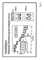
- FIG. 2 shows a schematic representation of the EIT measurement under ventilation.
- the ventilator also includes, for example, at least one additional sensor device 3 for carrying out the EIT measurement.
- the sensor device has, for example, 32 high-resolution sensors 3, 3'... in a textile belt, not shown.
- a not shown, integrated position sensor determines the position of the patient.
- the sensors are interconnected 4 by communication or line means 4 and arranged to receive signals sequentially from all directions.
- the control unit 21 can therefore spatially assign the sensor measurement values to the EIT measurement 3.
- the local tissue resistances can thus be determined from different directions and then converted into moving images.
- the control unit 21 processes the sensor measurement values of the EIT measurement 3 in such a way that a specific electrical impedance (EI) is determined for individual lung sections LA1, LA2.
- the sensor device 3 is connected to the control unit 21 via wired or wireless communication means 35 .
- the control unit 21 stores 11 and processes the sensor readings for use by the ventilator.
- the memory device 31 can be part of the control unit 21 or a separate module.
- the control unit 21 processes the sensor measurement values for use by the ventilator in such a way that, for example, a visualization of the patient's thorax P with the two lungs L is shown on the display of the ventilator 10 .
- the control unit 21 processes the sensor measurement values of the EIT measurement 3 in such a way that a specific electrical impedance (EI) of the lung L is determined for individual lung sections LA1, LA2.
- the specific electrical impedance (EI) of the lung sections LA is processed for display on the display in such a way that lung sections with a high electrical impedance L2 are represented graphically differently than those lung sections with a low electrical impedance L1.
- the control unit 21 temporally processes the sensor measurement values of the EIT measurement 3 in such a way that a specific electrical impedance EI of the lung L is determined for individual lung sections LA1, LA2 for each breath and is prepared for display on the display.
- the sensor device 3 (together with the controller and the memory) is also set up and designed to form, for example, an EIT sum signal 44', which is a measure of the ventilation of the lungs or lung sections.
- the sensor device 3 is also set up and configured (together with the controller and the memory) to determine a frequency of the EIT change frequency 44". The sensor device 3 can therefore determine the instantaneous ventilation 44 in summary.
- the EIT sum signal 44' is understood to mean that the impedance or impedance change distribution in the sectional plane determined by the sensors or the volume spanned by the sensors does not necessarily have to be shown graphically, for example on a display, for the implementation of the method, but at least the calculated result values of the impedance distribution or impedance change distribution (for the implementation of the method in the device according to the invention are available.
- the EIT change frequency 44" can be determined from the respiratory phase-dependent change in the impedance values of the sensors.
- the impedance values of the sensors fluctuate depending on the respiratory phase with inspiration and expiration.
- the EIT change frequency 44" therefore represents an alternative or supplementary measure by which the (frequency der) to determine ventilation of the lungs.
- the sensors are integrated into a textile, breathable and stretchable electrode belt together with a position sensor.
- the sensor 3 and the control unit are set up to generate an image rate of, for example, up to 50 per second. Lung areas LA are visualized.
- patient-related inputs are possible, such as body height, body weight, gender, chest circumference.
- the EIT data determined and processed in this way enable a differentiated analysis and representation of the electrical impedance EI of the lungs with temporal and spatial resolution.
- strain VT / FRC
- strain The calculation of the local strain (strain) of this novel EIT-based method (which as such does not fall under the claims) is carried out at the pixel level by determining the tidal volume of each pixel from its respective impedance change (delta Z) during the respiratory cycle and then inserting this into the in relation to its respective end-expiratory volume, which corresponds to its impedance value at the end of expiration (EELI).
- the pixel strain at each PEEP level was related to the individual pixel strain at the reference PEEP of 15 cmH2O, achieving a normalization that made it possible to create comparable relative strain images, which were the basis for the Calculation of numerical values of the total strain for the respective PEEP level.
- the respective pixel values were added up and normalized with regard to the number of all pixels in the lung ROI of each pig.
- EIT images were generated from the aforementioned strain images, which represented the mean relative strain of all subjects at the respective PEEP level.
- the measurement and control technology of the ventilator is networked with electrical impedance tomography (EIT).
- EIT electrical impedance tomography
Landscapes
- Health & Medical Sciences (AREA)
- Life Sciences & Earth Sciences (AREA)
- Animal Behavior & Ethology (AREA)
- Veterinary Medicine (AREA)
- Public Health (AREA)
- General Health & Medical Sciences (AREA)
- Engineering & Computer Science (AREA)
- Biomedical Technology (AREA)
- Heart & Thoracic Surgery (AREA)
- Pulmonology (AREA)
- Biophysics (AREA)
- Molecular Biology (AREA)
- Surgery (AREA)
- Medical Informatics (AREA)
- Pathology (AREA)
- Physics & Mathematics (AREA)
- Nuclear Medicine, Radiotherapy & Molecular Imaging (AREA)
- Radiology & Medical Imaging (AREA)
- Emergency Medicine (AREA)
- Anesthesiology (AREA)
- Hematology (AREA)
- Physiology (AREA)
- Measurement Of The Respiration, Hearing Ability, Form, And Blood Characteristics Of Living Organisms (AREA)
Applications Claiming Priority (1)
| Application Number | Priority Date | Filing Date | Title |
|---|---|---|---|
| DE102019109932 | 2019-04-15 |
Publications (2)
| Publication Number | Publication Date |
|---|---|
| EP3725222A1 EP3725222A1 (de) | 2020-10-21 |
| EP3725222B1 true EP3725222B1 (de) | 2023-07-12 |
Family
ID=70285372
Family Applications (1)
| Application Number | Title | Priority Date | Filing Date |
|---|---|---|---|
| EP20000151.9A Active EP3725222B1 (de) | 2019-04-15 | 2020-04-14 | System und beatmungsgerät zur echtzeit-bestimmung einer lokalen beanspruchung einer lunge während der künstlichen beatmung |
Country Status (4)
| Country | Link |
|---|---|
| US (1) | US11696699B2 (zh) |
| EP (1) | EP3725222B1 (zh) |
| CN (1) | CN111821549B (zh) |
| DE (1) | DE102020002279A1 (zh) |
Families Citing this family (9)
| Publication number | Priority date | Publication date | Assignee | Title |
|---|---|---|---|---|
| DE102020120900A1 (de) * | 2020-08-07 | 2022-02-10 | Drägerwerk AG & Co. KGaA | Vorrichtung und Verfahren zur Messung der Lungen-Compliance |
| EP4011424A1 (de) * | 2020-12-07 | 2022-06-15 | Löwenstein Medical Technology S.A. | System zur beatmung von lebewesen |
| EP4059416A1 (en) | 2021-03-18 | 2022-09-21 | SenTec AG | Regional strain |
| IT202100010775A1 (it) * | 2021-04-28 | 2022-10-28 | Roni Shabat | Sistemi e metodi per la determinazione dell'impedenza polmonare |
| CN114024522B (zh) * | 2021-12-20 | 2023-02-17 | 广州国家实验室 | 模拟肺部呼吸的电阻网络、装置和方法 |
| WO2024003413A1 (en) * | 2022-07-01 | 2024-01-04 | Academisch Ziekenhuis Maastricht | Method for controlling spontaneously triggered mechanical ventilation of a patient |
| CN115721535B (zh) * | 2022-09-24 | 2024-10-01 | 首都医科大学附属北京康复医院(北京工人疗养院) | 用于气道廓清的数据处理方法及相关系统 |
| CN116803342B (zh) * | 2023-07-20 | 2024-03-29 | 中国人民解放军空军军医大学 | 用于抗荷训练的阻抗数据处理方法及相关训练系统 |
| CN117898699A (zh) * | 2024-01-10 | 2024-04-19 | 中国人民解放军空军军医大学 | 一种基于eit引导肺康复的方法、系统及设备 |
Family Cites Families (6)
| Publication number | Priority date | Publication date | Assignee | Title |
|---|---|---|---|---|
| DE102005022896B3 (de) * | 2005-05-18 | 2006-05-11 | Dräger Medical AG & Co. KG | Vorrichtung und Verfahren zur Steuerung eines Beatmungsgerätes |
| BR102012028367B1 (pt) * | 2012-11-06 | 2022-03-22 | Timpel S.A | Método e aparato para gerar e exibir uma representação simplificada de informações obtidas através de tomografia por impedância elétrica |
| DE102013203177A1 (de) * | 2013-02-26 | 2014-08-28 | Hamilton Medical Ag | System zur automatisierten Einstellung eines durch eine Beatmungseinrichtung vorgegebenen Drucks |
| DE102015006902B3 (de) | 2015-06-04 | 2016-06-30 | Drägerwerk AG & Co. KGaA | Vorrichtung zur Verarbeitung und Visualisierung von Daten eines Elektro-lmpedanz-Tomographie-Gerätes zu einer Ermittlung und Visualisierung von regionalen Verzögerungen der Ventilation in der Lunge |
| DE102017007224A1 (de) * | 2017-08-02 | 2019-02-07 | Drägerwerk AG & Co. KGaA | Vorrichtung und Verfahren zu einer Bestimmung von Differenzkennzahlen auf Basis von EIT-Daten |
| DE102021000312A1 (de) * | 2020-02-07 | 2021-08-12 | Löwenstein Medical Technology S.A. | Verfahren und Vorrichtung zur Bestimmung einer regionalen Compliance einer Lunge bei Spontanatmung |
-
2020
- 2020-04-11 DE DE102020002279.0A patent/DE102020002279A1/de active Pending
- 2020-04-14 EP EP20000151.9A patent/EP3725222B1/de active Active
- 2020-04-14 US US16/847,752 patent/US11696699B2/en active Active
- 2020-04-15 CN CN202010296977.8A patent/CN111821549B/zh active Active
Also Published As
| Publication number | Publication date |
|---|---|
| EP3725222A1 (de) | 2020-10-21 |
| CN111821549B (zh) | 2024-10-15 |
| US11696699B2 (en) | 2023-07-11 |
| US20200324067A1 (en) | 2020-10-15 |
| CN111821549A (zh) | 2020-10-27 |
| DE102020002279A1 (de) | 2020-10-15 |
Similar Documents
| Publication | Publication Date | Title |
|---|---|---|
| EP3725222B1 (de) | System und beatmungsgerät zur echtzeit-bestimmung einer lokalen beanspruchung einer lunge während der künstlichen beatmung | |
| EP2961315B2 (de) | System zur automatisierten einstellung eines durch eine beatmungseinrichtung vorgegebenen drucks | |
| DE102007062214C5 (de) | Verfahren zum automatischen Steuern eines Beatmungssystems sowie zugehörige Beatmungsvorrichtung | |
| DE69231157T2 (de) | Automatisches system zum erzeugen eines kontinuierlichen positiven atemwegsdruck | |
| DE69736808T2 (de) | Determination einer leckluftströmung | |
| EP1226841B1 (de) | Verfahren und Vorrichtung zur Schlafüberwachung | |
| DE69922511T2 (de) | Respiratorisches unterstützungssystem mit detektion der atmung unter aufnahme neuroelektrischer signale | |
| EP1148907B1 (de) | Nichtinvasives verfahren zur optimierung der beatmung atelektatischer lungen | |
| DE102006018198A1 (de) | Verfahren und Vorrichtung zur Lungenventilation | |
| DE112004001957T5 (de) | System und Verfahren zur Zwerchfellstimulation | |
| DE102016011161A1 (de) | Vorrichtung zur Verarbeitung und Visualisierung von Daten eines Elektro-lmpedanz-Tomographie-Gerätes zu einer Ermittlung und Visualisierung von regionalen Eigenschaften der Ventilation der Lunge | |
| EP3653248A2 (de) | Beatmungsgeräte sowie steuerverfahren für beatmungsgeräte | |
| EP3862040B1 (de) | Beatmungsgerät zur künstlichen beatmung eines patienten | |
| EP2265179A1 (de) | Vorrichtung zur beurteilung der belastung des kreislaufs einer person während einer atemunterstützung mittels eines beatmungsgeräts | |
| WO2021063601A1 (de) | Verfahren und vorrichtung zum ermitteln eines respiratorischen und / oder eines kardiogenen signals | |
| DE102015014106A1 (de) | Vorrichtung zur druckunterstützenden oder druckkontrollierten Beatmung eines Patienten mit eingeschränkter Spontanatmung | |
| DE102007052472A1 (de) | Verfahren zum Betreiben einer Beatmungs- und/oder Anästhesievorrichtung im APRV-Modus unter Berücksichtigung der Impedanz und/oder der Impedanzänderung | |
| EP3698713B1 (de) | System zur erfassung von atemanstrengungen eines patienten | |
| EP3957241B1 (de) | Anordnung und computerprogrammprodukt zur ermittlung der regionalen lungen-compliance | |
| EP3964253B1 (de) | Computerprogramm und vorrichtung zur automatischen festlegung der sollfrequenz eines beatmungsgeräts | |
| DE102015011390A1 (de) | Vorrichtung, Verfahren und Computerprogramm zur Beatmung eines Patienten | |
| DE102022101845A1 (de) | Beatmungsvorrichtung, Verfahren, Computerprogramm und Vorrichtung zum Ermitteln eines Maßes für einen intrinsischen endexspiratorischen Druck | |
| DE102020001440A1 (de) | Beatmungsvorrichtung | |
| CH719307A2 (de) | Verfahren und System mit einer Messvorrichtung und einer Analysevorrichtung zu einer Verarbeitung von Daten. | |
| EP4437947A1 (de) | Vorrichtung und verfahren zur nicht invasiven bestimmung eines verschlussdrucks in der lunge eines patienten |
Legal Events
| Date | Code | Title | Description |
|---|---|---|---|
| PUAI | Public reference made under article 153(3) epc to a published international application that has entered the european phase |
Free format text: ORIGINAL CODE: 0009012 |
|
| STAA | Information on the status of an ep patent application or granted ep patent |
Free format text: STATUS: THE APPLICATION HAS BEEN PUBLISHED |
|
| AK | Designated contracting states |
Kind code of ref document: A1 Designated state(s): AL AT BE BG CH CY CZ DE DK EE ES FI FR GB GR HR HU IE IS IT LI LT LU LV MC MK MT NL NO PL PT RO RS SE SI SK SM TR |
|
| AX | Request for extension of the european patent |
Extension state: BA ME |
|
| STAA | Information on the status of an ep patent application or granted ep patent |
Free format text: STATUS: REQUEST FOR EXAMINATION WAS MADE |
|
| 17P | Request for examination filed |
Effective date: 20210414 |
|
| RBV | Designated contracting states (corrected) |
Designated state(s): AL AT BE BG CH CY CZ DE DK EE ES FI FR GB GR HR HU IE IS IT LI LT LU LV MC MK MT NL NO PL PT RO RS SE SI SK SM TR |
|
| STAA | Information on the status of an ep patent application or granted ep patent |
Free format text: STATUS: EXAMINATION IS IN PROGRESS |
|
| 17Q | First examination report despatched |
Effective date: 20211011 |
|
| 17Q | First examination report despatched |
Effective date: 20211022 |
|
| REG | Reference to a national code |
Ref country code: DE Ref legal event code: R079 Ref document number: 502020004078 Country of ref document: DE Free format text: PREVIOUS MAIN CLASS: A61B0005053000 Ipc: A61B0005053600 Ref country code: DE |
|
| GRAP | Despatch of communication of intention to grant a patent |
Free format text: ORIGINAL CODE: EPIDOSNIGR1 |
|
| STAA | Information on the status of an ep patent application or granted ep patent |
Free format text: STATUS: GRANT OF PATENT IS INTENDED |
|
| RIC1 | Information provided on ipc code assigned before grant |
Ipc: A61B 5/08 19680901ALI20230109BHEP Ipc: A61B 5/085 19900101ALI20230109BHEP Ipc: A61B 5/00 19680901ALI20230109BHEP Ipc: A61M 16/00 19680901ALI20230109BHEP Ipc: A61B 5/0536 20210101AFI20230109BHEP |
|
| INTG | Intention to grant announced |
Effective date: 20230131 |
|
| GRAS | Grant fee paid |
Free format text: ORIGINAL CODE: EPIDOSNIGR3 |
|
| GRAA | (expected) grant |
Free format text: ORIGINAL CODE: 0009210 |
|
| STAA | Information on the status of an ep patent application or granted ep patent |
Free format text: STATUS: THE PATENT HAS BEEN GRANTED |
|
| P01 | Opt-out of the competence of the unified patent court (upc) registered |
Effective date: 20230523 |
|
| AK | Designated contracting states |
Kind code of ref document: B1 Designated state(s): AL AT BE BG CH CY CZ DE DK EE ES FI FR GB GR HR HU IE IS IT LI LT LU LV MC MK MT NL NO PL PT RO RS SE SI SK SM TR |
|
| REG | Reference to a national code |
Ref country code: CH Ref legal event code: EP |
|
| REG | Reference to a national code |
Ref country code: IE Ref legal event code: FG4D Free format text: LANGUAGE OF EP DOCUMENT: GERMAN |
|
| REG | Reference to a national code |
Ref country code: DE Ref legal event code: R096 Ref document number: 502020004078 Country of ref document: DE |
|
| REG | Reference to a national code |
Ref country code: NL Ref legal event code: FP |
|
| REG | Reference to a national code |
Ref country code: LT Ref legal event code: MG9D |
|
| PG25 | Lapsed in a contracting state [announced via postgrant information from national office to epo] |
Ref country code: GR Free format text: LAPSE BECAUSE OF FAILURE TO SUBMIT A TRANSLATION OF THE DESCRIPTION OR TO PAY THE FEE WITHIN THE PRESCRIBED TIME-LIMIT Effective date: 20231013 |
|
| PG25 | Lapsed in a contracting state [announced via postgrant information from national office to epo] |
Ref country code: ES Free format text: LAPSE BECAUSE OF FAILURE TO SUBMIT A TRANSLATION OF THE DESCRIPTION OR TO PAY THE FEE WITHIN THE PRESCRIBED TIME-LIMIT Effective date: 20230712 |
|
| PG25 | Lapsed in a contracting state [announced via postgrant information from national office to epo] |
Ref country code: IS Free format text: LAPSE BECAUSE OF FAILURE TO SUBMIT A TRANSLATION OF THE DESCRIPTION OR TO PAY THE FEE WITHIN THE PRESCRIBED TIME-LIMIT Effective date: 20231112 |
|
| PG25 | Lapsed in a contracting state [announced via postgrant information from national office to epo] |
Ref country code: SE Free format text: LAPSE BECAUSE OF FAILURE TO SUBMIT A TRANSLATION OF THE DESCRIPTION OR TO PAY THE FEE WITHIN THE PRESCRIBED TIME-LIMIT Effective date: 20230712 Ref country code: RS Free format text: LAPSE BECAUSE OF FAILURE TO SUBMIT A TRANSLATION OF THE DESCRIPTION OR TO PAY THE FEE WITHIN THE PRESCRIBED TIME-LIMIT Effective date: 20230712 Ref country code: PT Free format text: LAPSE BECAUSE OF FAILURE TO SUBMIT A TRANSLATION OF THE DESCRIPTION OR TO PAY THE FEE WITHIN THE PRESCRIBED TIME-LIMIT Effective date: 20231113 Ref country code: NO Free format text: LAPSE BECAUSE OF FAILURE TO SUBMIT A TRANSLATION OF THE DESCRIPTION OR TO PAY THE FEE WITHIN THE PRESCRIBED TIME-LIMIT Effective date: 20231012 Ref country code: LV Free format text: LAPSE BECAUSE OF FAILURE TO SUBMIT A TRANSLATION OF THE DESCRIPTION OR TO PAY THE FEE WITHIN THE PRESCRIBED TIME-LIMIT Effective date: 20230712 Ref country code: LT Free format text: LAPSE BECAUSE OF FAILURE TO SUBMIT A TRANSLATION OF THE DESCRIPTION OR TO PAY THE FEE WITHIN THE PRESCRIBED TIME-LIMIT Effective date: 20230712 Ref country code: IS Free format text: LAPSE BECAUSE OF FAILURE TO SUBMIT A TRANSLATION OF THE DESCRIPTION OR TO PAY THE FEE WITHIN THE PRESCRIBED TIME-LIMIT Effective date: 20231112 Ref country code: HR Free format text: LAPSE BECAUSE OF FAILURE TO SUBMIT A TRANSLATION OF THE DESCRIPTION OR TO PAY THE FEE WITHIN THE PRESCRIBED TIME-LIMIT Effective date: 20230712 Ref country code: GR Free format text: LAPSE BECAUSE OF FAILURE TO SUBMIT A TRANSLATION OF THE DESCRIPTION OR TO PAY THE FEE WITHIN THE PRESCRIBED TIME-LIMIT Effective date: 20231013 Ref country code: FI Free format text: LAPSE BECAUSE OF FAILURE TO SUBMIT A TRANSLATION OF THE DESCRIPTION OR TO PAY THE FEE WITHIN THE PRESCRIBED TIME-LIMIT Effective date: 20230712 Ref country code: ES Free format text: LAPSE BECAUSE OF FAILURE TO SUBMIT A TRANSLATION OF THE DESCRIPTION OR TO PAY THE FEE WITHIN THE PRESCRIBED TIME-LIMIT Effective date: 20230712 |
|
| PG25 | Lapsed in a contracting state [announced via postgrant information from national office to epo] |
Ref country code: PL Free format text: LAPSE BECAUSE OF FAILURE TO SUBMIT A TRANSLATION OF THE DESCRIPTION OR TO PAY THE FEE WITHIN THE PRESCRIBED TIME-LIMIT Effective date: 20230712 |
|
| REG | Reference to a national code |
Ref country code: DE Ref legal event code: R097 Ref document number: 502020004078 Country of ref document: DE |
|
| PG25 | Lapsed in a contracting state [announced via postgrant information from national office to epo] |
Ref country code: SM Free format text: LAPSE BECAUSE OF FAILURE TO SUBMIT A TRANSLATION OF THE DESCRIPTION OR TO PAY THE FEE WITHIN THE PRESCRIBED TIME-LIMIT Effective date: 20230712 Ref country code: RO Free format text: LAPSE BECAUSE OF FAILURE TO SUBMIT A TRANSLATION OF THE DESCRIPTION OR TO PAY THE FEE WITHIN THE PRESCRIBED TIME-LIMIT Effective date: 20230712 Ref country code: EE Free format text: LAPSE BECAUSE OF FAILURE TO SUBMIT A TRANSLATION OF THE DESCRIPTION OR TO PAY THE FEE WITHIN THE PRESCRIBED TIME-LIMIT Effective date: 20230712 Ref country code: DK Free format text: LAPSE BECAUSE OF FAILURE TO SUBMIT A TRANSLATION OF THE DESCRIPTION OR TO PAY THE FEE WITHIN THE PRESCRIBED TIME-LIMIT Effective date: 20230712 Ref country code: CZ Free format text: LAPSE BECAUSE OF FAILURE TO SUBMIT A TRANSLATION OF THE DESCRIPTION OR TO PAY THE FEE WITHIN THE PRESCRIBED TIME-LIMIT Effective date: 20230712 Ref country code: SK Free format text: LAPSE BECAUSE OF FAILURE TO SUBMIT A TRANSLATION OF THE DESCRIPTION OR TO PAY THE FEE WITHIN THE PRESCRIBED TIME-LIMIT Effective date: 20230712 |
|
| PLBE | No opposition filed within time limit |
Free format text: ORIGINAL CODE: 0009261 |
|
| STAA | Information on the status of an ep patent application or granted ep patent |
Free format text: STATUS: NO OPPOSITION FILED WITHIN TIME LIMIT |
|
| PGFP | Annual fee paid to national office [announced via postgrant information from national office to epo] |
Ref country code: NL Payment date: 20240422 Year of fee payment: 5 |
|
| PG25 | Lapsed in a contracting state [announced via postgrant information from national office to epo] |
Ref country code: IT Free format text: LAPSE BECAUSE OF FAILURE TO SUBMIT A TRANSLATION OF THE DESCRIPTION OR TO PAY THE FEE WITHIN THE PRESCRIBED TIME-LIMIT Effective date: 20230712 |
|
| 26N | No opposition filed |
Effective date: 20240415 |
|
| PGFP | Annual fee paid to national office [announced via postgrant information from national office to epo] |
Ref country code: GB Payment date: 20240423 Year of fee payment: 5 |
|
| PGFP | Annual fee paid to national office [announced via postgrant information from national office to epo] |
Ref country code: DE Payment date: 20240418 Year of fee payment: 5 |
|
| PGFP | Annual fee paid to national office [announced via postgrant information from national office to epo] |
Ref country code: CH Payment date: 20240501 Year of fee payment: 5 |
|
| PG25 | Lapsed in a contracting state [announced via postgrant information from national office to epo] |
Ref country code: SI Free format text: LAPSE BECAUSE OF FAILURE TO SUBMIT A TRANSLATION OF THE DESCRIPTION OR TO PAY THE FEE WITHIN THE PRESCRIBED TIME-LIMIT Effective date: 20230712 |
|
| PGFP | Annual fee paid to national office [announced via postgrant information from national office to epo] |
Ref country code: FR Payment date: 20240422 Year of fee payment: 5 |