EP3697331B1 - Cathéter d'ablation à double stimulation et détection optique-électrique - Google Patents
Cathéter d'ablation à double stimulation et détection optique-électrique Download PDFInfo
- Publication number
- EP3697331B1 EP3697331B1 EP18792776.9A EP18792776A EP3697331B1 EP 3697331 B1 EP3697331 B1 EP 3697331B1 EP 18792776 A EP18792776 A EP 18792776A EP 3697331 B1 EP3697331 B1 EP 3697331B1
- Authority
- EP
- European Patent Office
- Prior art keywords
- distal tip
- tip electrode
- optical
- elongate body
- distal
- Prior art date
- Legal status (The legal status is an assumption and is not a legal conclusion. Google has not performed a legal analysis and makes no representation as to the accuracy of the status listed.)
- Active
Links
Images


Classifications
-
- A—HUMAN NECESSITIES
- A61—MEDICAL OR VETERINARY SCIENCE; HYGIENE
- A61B—DIAGNOSIS; SURGERY; IDENTIFICATION
- A61B18/00—Surgical instruments, devices or methods for transferring non-mechanical forms of energy to or from the body
- A61B18/18—Surgical instruments, devices or methods for transferring non-mechanical forms of energy to or from the body by applying electromagnetic radiation, e.g. microwaves
- A61B18/20—Surgical instruments, devices or methods for transferring non-mechanical forms of energy to or from the body by applying electromagnetic radiation, e.g. microwaves using laser
- A61B18/22—Surgical instruments, devices or methods for transferring non-mechanical forms of energy to or from the body by applying electromagnetic radiation, e.g. microwaves using laser the beam being directed along or through a flexible conduit, e.g. an optical fibre; Couplings or hand-pieces therefor
- A61B18/24—Surgical instruments, devices or methods for transferring non-mechanical forms of energy to or from the body by applying electromagnetic radiation, e.g. microwaves using laser the beam being directed along or through a flexible conduit, e.g. an optical fibre; Couplings or hand-pieces therefor with a catheter
-
- A—HUMAN NECESSITIES
- A61—MEDICAL OR VETERINARY SCIENCE; HYGIENE
- A61B—DIAGNOSIS; SURGERY; IDENTIFICATION
- A61B18/00—Surgical instruments, devices or methods for transferring non-mechanical forms of energy to or from the body
- A61B18/04—Surgical instruments, devices or methods for transferring non-mechanical forms of energy to or from the body by heating
- A61B18/12—Surgical instruments, devices or methods for transferring non-mechanical forms of energy to or from the body by heating by passing a current through the tissue to be heated, e.g. high-frequency current
- A61B18/14—Probes or electrodes therefor
- A61B18/1492—Probes or electrodes therefor having a flexible, catheter-like structure, e.g. for heart ablation
-
- A—HUMAN NECESSITIES
- A61—MEDICAL OR VETERINARY SCIENCE; HYGIENE
- A61B—DIAGNOSIS; SURGERY; IDENTIFICATION
- A61B90/00—Instruments, implements or accessories specially adapted for surgery or diagnosis and not covered by any of the groups A61B1/00 - A61B50/00, e.g. for luxation treatment or for protecting wound edges
- A61B90/30—Devices for illuminating a surgical field, the devices having an interrelation with other surgical devices or with a surgical procedure
-
- A—HUMAN NECESSITIES
- A61—MEDICAL OR VETERINARY SCIENCE; HYGIENE
- A61B—DIAGNOSIS; SURGERY; IDENTIFICATION
- A61B1/00—Instruments for performing medical examinations of the interior of cavities or tubes of the body by visual or photographical inspection, e.g. endoscopes; Illuminating arrangements therefor
- A61B1/00064—Constructional details of the endoscope body
- A61B1/00071—Insertion part of the endoscope body
- A61B1/0008—Insertion part of the endoscope body characterised by distal tip features
- A61B1/00087—Tools
-
- A—HUMAN NECESSITIES
- A61—MEDICAL OR VETERINARY SCIENCE; HYGIENE
- A61B—DIAGNOSIS; SURGERY; IDENTIFICATION
- A61B18/00—Surgical instruments, devices or methods for transferring non-mechanical forms of energy to or from the body
- A61B2018/00053—Mechanical features of the instrument of device
- A61B2018/00059—Material properties
-
- A—HUMAN NECESSITIES
- A61—MEDICAL OR VETERINARY SCIENCE; HYGIENE
- A61B—DIAGNOSIS; SURGERY; IDENTIFICATION
- A61B18/00—Surgical instruments, devices or methods for transferring non-mechanical forms of energy to or from the body
- A61B2018/00571—Surgical instruments, devices or methods for transferring non-mechanical forms of energy to or from the body for achieving a particular surgical effect
- A61B2018/00577—Ablation
-
- A—HUMAN NECESSITIES
- A61—MEDICAL OR VETERINARY SCIENCE; HYGIENE
- A61B—DIAGNOSIS; SURGERY; IDENTIFICATION
- A61B18/00—Surgical instruments, devices or methods for transferring non-mechanical forms of energy to or from the body
- A61B2018/00636—Sensing and controlling the application of energy
- A61B2018/00773—Sensed parameters
- A61B2018/00791—Temperature
-
- A—HUMAN NECESSITIES
- A61—MEDICAL OR VETERINARY SCIENCE; HYGIENE
- A61B—DIAGNOSIS; SURGERY; IDENTIFICATION
- A61B18/00—Surgical instruments, devices or methods for transferring non-mechanical forms of energy to or from the body
- A61B2018/00636—Sensing and controlling the application of energy
- A61B2018/00773—Sensed parameters
- A61B2018/00839—Bioelectrical parameters, e.g. ECG, EEG
-
- A—HUMAN NECESSITIES
- A61—MEDICAL OR VETERINARY SCIENCE; HYGIENE
- A61B—DIAGNOSIS; SURGERY; IDENTIFICATION
- A61B18/00—Surgical instruments, devices or methods for transferring non-mechanical forms of energy to or from the body
- A61B2018/00982—Surgical instruments, devices or methods for transferring non-mechanical forms of energy to or from the body combined with or comprising means for visual or photographic inspections inside the body, e.g. endoscopes
-
- A—HUMAN NECESSITIES
- A61—MEDICAL OR VETERINARY SCIENCE; HYGIENE
- A61B—DIAGNOSIS; SURGERY; IDENTIFICATION
- A61B18/00—Surgical instruments, devices or methods for transferring non-mechanical forms of energy to or from the body
- A61B18/04—Surgical instruments, devices or methods for transferring non-mechanical forms of energy to or from the body by heating
- A61B18/12—Surgical instruments, devices or methods for transferring non-mechanical forms of energy to or from the body by heating by passing a current through the tissue to be heated, e.g. high-frequency current
- A61B18/14—Probes or electrodes therefor
- A61B18/16—Indifferent or passive electrodes for grounding
- A61B2018/162—Indifferent or passive electrodes for grounding located on the probe body
-
- A—HUMAN NECESSITIES
- A61—MEDICAL OR VETERINARY SCIENCE; HYGIENE
- A61B—DIAGNOSIS; SURGERY; IDENTIFICATION
- A61B90/00—Instruments, implements or accessories specially adapted for surgery or diagnosis and not covered by any of the groups A61B1/00 - A61B50/00, e.g. for luxation treatment or for protecting wound edges
- A61B90/30—Devices for illuminating a surgical field, the devices having an interrelation with other surgical devices or with a surgical procedure
- A61B2090/306—Devices for illuminating a surgical field, the devices having an interrelation with other surgical devices or with a surgical procedure using optical fibres
-
- A—HUMAN NECESSITIES
- A61—MEDICAL OR VETERINARY SCIENCE; HYGIENE
- A61B—DIAGNOSIS; SURGERY; IDENTIFICATION
- A61B90/00—Instruments, implements or accessories specially adapted for surgery or diagnosis and not covered by any of the groups A61B1/00 - A61B50/00, e.g. for luxation treatment or for protecting wound edges
- A61B90/36—Image-producing devices or illumination devices not otherwise provided for
- A61B90/37—Surgical systems with images on a monitor during operation
- A61B2090/373—Surgical systems with images on a monitor during operation using light, e.g. by using optical scanners
-
- A—HUMAN NECESSITIES
- A61—MEDICAL OR VETERINARY SCIENCE; HYGIENE
- A61B—DIAGNOSIS; SURGERY; IDENTIFICATION
- A61B2218/00—Details of surgical instruments, devices or methods for transferring non-mechanical forms of energy to or from the body
- A61B2218/001—Details of surgical instruments, devices or methods for transferring non-mechanical forms of energy to or from the body having means for irrigation and/or aspiration of substances to and/or from the surgical site
- A61B2218/002—Irrigation
Definitions
- the present invention relates to a system for optically evaluating and ablating tissue.
- Minimally-invasive medical devices and procedures are desirable for treating a variety of cardiovascular maladies, such as cardiac arrhythmias including atrial fibrillation, and other irregularities in the transmission of electrical impulses through the heart.
- cardiac arrhythmias including atrial fibrillation, and other irregularities in the transmission of electrical impulses through the heart.
- One such example of a minimally invasive therapy involves the treatment of cardiac arrhythmias or irregular heartbeats in which physicians employ specialized cardiac assessment and treatment devices, such as mapping catheters and ablation catheters, to gain access to, diagnose, and treat interior regions of a patient's body.
- Such devices may include energized electrodes or other ablation assemblies to create lesions or other anatomical effects that disrupt or block electrical pathways through the targeted tissue.
- a specific area of cardiac tissue having aberrant electrically conductive pathways is typically initially identified for subsequent treatment. Additionally, the treated tissue may be analyzed to determine whether the tissue has been successfully ablated (i.e. that the aberrant electrical conductivity through the tissue has been destroyed). This localization or identification can include first using a medical device such as a mapping catheter to obtain a baseline electrophysiological map of electrical activity in selected tissue. Therefore, characterization of an area of tissue before, during, and/or after a treatment procedure is important for determining treatment efficacy.
- a medical device such as a mapping catheter to obtain a baseline electrophysiological map of electrical activity in selected tissue. Therefore, characterization of an area of tissue before, during, and/or after a treatment procedure is important for determining treatment efficacy.
- mapping catheter such as a mapping catheter to obtain a baseline electrophysiological map of electrical activity in selected tissue. Therefore, characterization of an area of tissue before, during, and/or after a treatment procedure is important for determining treatment efficacy.
- Currently known devices are based primary
- the present invention is defined by claim 1 and provides a system for optically evaluating and treating or ablating tissue. Specifically, the system allows for the optical and/or electrical evaluation of tissue at the same location(s) at which ablation or treatment or ablation energy is delivered. This allows for a more accurate evaluation of lesion formation and tissue condition before, during, and/or after a treatment or ablation procedure.
- the system for performing a medical procedure includes an elongate body including a proximal portion, a distal portion having a distal end, and a longitudinal axis, and a distal tip electrode at the elongate body distal end, the tip electrode being composed of an optically transparent material and having a continuous layer of an optically transparent and electrically conductive material.
- the distal tip electrode is composed of an optically transparent first material and the continuous layer is composed of an optically transparent and electrically conductive second material, the second material being different than the first material.
- the distal tip electrode is composed of at least one of a transparent polymer, ceramic, sapphire, synthetic sapphire, yttria, yttria-alumina garnet, zinc selenide, germanium, cadmium telluride, barium fluoride, caesium iodide, magnesium fluoride, thallium bromoiodide, zinc sulphide, calcium fluoride, quartz, diamond, ruby, synthetic ruby, crystal, glass, and fused silica.
- a transparent polymer ceramic, sapphire, synthetic sapphire, yttria, yttria-alumina garnet, zinc selenide, germanium, cadmium telluride, barium fluoride, caesium iodide, magnesium fluoride, thallium bromoiodide, zinc sulphide, calcium fluoride, quartz, diamond, ruby, synthetic ruby, crystal, glass, and fused silica.
- the second material is at least one of graphene and conductive diamond.
- the device further comprises at least one optical fiber within the elongate body, the at least one optical fiber being in optical communication with the distal tip electrode.
- the distal tip electrode includes a proximal face and a curved distal surface, the proximal face lying in a plane that is orthogonal to the elongate body longitudinal axis.
- the distal tip electrode includes an optical fiber channel having an opening, the optical fiber opening being on the distal tip electrode proximal face.
- the optical fiber channel extends within the distal tip electrode from the distal tip electrode proximal face to a location that is a distance from the distal tip electrode curved distal surface.
- the optical fiber channel is sized and configured to accept at least a portion of the at least one optical fiber.
- the device further includes at least one optical window within the elongate body distal portion, the at least one optical window being composed of an optically transparent material.
- the device further includes at least one lateral electrode on the elongate body distal portion, each of the at least one lateral electrode including at least one conductive area, each of the at least one conductive area being composed of an electrically conductive and optically transparent material.
- the plurality of conductive areas are on a flexible overlay, the elongate body having an outer surface, the flexible overlay being applied to the elongate body outer surface.
- the flexible overlay is at least partially composed of an optically transparent material such that the plurality of conductive areas and flexible overlay are optically transparent, the flexible overlay being applied to the elongate body such that at least some of the plurality of conductive areas are positioned over at least one of the at least one optical window.
- the at least one optical fiber is a first at least one optical fiber
- the device further comprising a second at least one optical fiber, each of the second at least one optical fiber being in optical communication with one of the at least one optical window.
- the at least one lateral electrode is a length of conductive material wound around at least a portion of the elongate body.
- the distal tip electrode includes a plurality of apertures, the plurality of apertures configured to be in fluid communication with a fluid source.
- a system for evaluating and ablating an area of tissue includes: a medical device including: an elongate body including a proximal portion, a distal portion having a distal end, and a longitudinal axis; a distal tip electrode at the elongate body distal end, the distal tip electrode being optically transparent and electrically conductive; a first plurality of optical fibers within the elongate body and in optical communication with the distal tip electrode; at least one optical window within the elongate body distal portion, the at least one optical window being composed of a transparent material; a second plurality of optical fibers within the elongate body and in optical communication with the at least one optical window; at least one lateral electrode on the elongate body distal portion, the at least one lateral electrode being a plurality of electrically conductive and optically transparent graphene areas, the plurality of electrically conductive and optically transparent graphene areas being aligned with the at least one optical window such that the plurality of electrical
- the system is configured to deliver treatment energy from the at least one lateral electrode to an area of tissue and to emit and receive light through the at least one lateral electrode and the at least one optical window to and from the same area of tissue.
- the processing circuitry is configured to: emit light from the light emitter through at least one of the distal tip electrode and the at least one lateral electrode through at least one of the first plurality of optical fibers and the second plurality of optical fibers to the area of tissue; receive light fluoresced from the area of tissue through at least one of the distal tip electrode and the at least one lateral electrode through at least one of the first plurality of optical fibers and the second plurality of optical fibers; and determine whether the area of tissue has been ablated based on the received light.
- a system for evaluating and ablating an area of tissue includes: an elongate body including a proximal portion, a distal portion having a distal end, and a longitudinal axis; a distal tip electrode at the elongate body distal end, the distal tip electrode being optically transparent and electrically conductive; a first plurality of optical fibers within the elongate body and in optical communication with the distal tip electrode; a plurality of optical windows within the elongate body distal portion, the at least one optical window being composed of an optically transparent material; a second plurality of optical fibers within the elongate body, each of the second plurality of optical fibers being in optical communication with at least one of the plurality of optical windows; a flexible overlay applied to at least a portion of the elongate body distal portion, the flexible overlay having: at least one layer composed of a flexible and optically transparent material; a plurality of electrically conductive and optically transparent graphene areas on the at least one layer
- the devices, systems, and methods described herein may be used to not only treat or ablate tissue, but also to optically and electrically evaluate the tissue, such as evaluating lesion formation and identifying tissue structures.
- relational terms such as “first,” “second,” “top” and “bottom,” and the like, may be used solely to distinguish one entity or element from another entity or element without necessarily requiring or implying any physical or logical relationship or order between such entities or elements.
- the terminology used herein is for the purpose of describing particular embodiments only and is not intended to be limiting of the concepts described herein.
- the singular forms “a”, “an” and “the” are intended to include the plural forms as well, unless the context clearly indicates otherwise.
- the joining term, "in communication with” and the like may be used to indicate electrical or data communication, which may be accomplished by physical contact, induction, electromagnetic radiation, radio signaling, infrared signaling or optical signaling, for example.
- electrical or data communication may be accomplished by physical contact, induction, electromagnetic radiation, radio signaling, infrared signaling or optical signaling, for example.
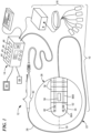
- the system 10 may generally include a treatment device 12 and a control unit 14.
- the system 10 may optionally include a navigation system 16, an imaging system such as near infrared (not shown) for obtaining images of anatomical features within a patient, and/or other system components for performing a particular medical procedure.
- a navigation system 16 an imaging system such as near infrared (not shown) for obtaining images of anatomical features within a patient, and/or other system components for performing a particular medical procedure.
- the treatment device 12 may be a treatment and mapping device.
- the device 12 may include an elongate body 22 passable through a patient's vasculature and/or proximate to a tissue region for diagnosis or treatment.
- the device 12 may be a catheter that is deliverable to the tissue region via a sheath or intravascular introducer (not shown) or a device that can access the pericardial space.
- the elongate body 22 may define a proximal portion 26, a distal portion 28, and a longitudinal axis 30, and may further include one or more lumens disposed within the elongate body 22 thereby providing mechanical, electrical, and/or fluid communication between the elongate body proximal portion 26 and the elongate distal portion 28.
- the device 12 may further include one or more treatment elements at, coupled to, or on the elongate body distal portion 28 for energetic, therapeutic, and/or investigatory interaction between the medical device 12 and a treatment site or region.
- the treatment element(s) may deliver, for example, radiofrequency energy, electroporation energy, laser energy, or other energetic transfer with a tissue area in proximity to the treatment element(s), including cardiac tissue, as discussed in more detail below.
- the treatment element(s) may include an optically transparent and electrically and/or thermally conductive distal tip electrode 34 and, optionally, one or more electrically and/or thermally conductive electrodes 36 (which may be referred to herein as lateral electrodes 36) along a length of the elongate body distal portion 28 and/or one or more optically transparent optical windows 38 along a length of the elongate body distal portion 28.
- the terms "transparent,” “substantially transparent,” and “optically transparent” refer to materials that are completely transparent as the term is understood in the art, or are at least sufficiently transparent to allow for optical imaging as discussed herein, even if the materials are not completely transparent.
- the electrically and/or thermally conductive electrode(s) 36 and optical window(s) 38 may be located in or on the lateral wall of the catheter body 22, as opposed to the distalmost end of the elongate body 22.
- the lateral electrode(s) 36 may be positioned over or aligned with the optical window(s) 38 to create a transparent area for optically or visually evaluating, energetically treating, and/or recording signals from tissue (thus, in some embodiments, the lateral electrode(s) 36 may be electrically and/or thermally conductive and optically transparent).
- the distal tip electrode 34 and, optionally, each of the optical windows 38 may be in optical communication with one or more optical fibers, a light emitter or source 44, and a light receiver 45.
- the light emitter 44 is a laser emitter.
- the distal tip electrode 34 may be in communication with an energy source 46, such as a radiofrequency (RF) and/or high-voltage energy source, and, optionally, each of the optical windows 38 may be in communication with a laser energy source 48 for delivering laser energy to an area of tissue.
- the device 12 may include one or more additional types of treatment element, such as expandable elements (for example, balloons or baskets), ultrasound transducers, electrodes of other configurations than those shown, thermal treatment electrodes in communication with a cryogenic fluid source, or the like.
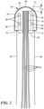
- the distal tip electrode 34 of FIG. 1 is shown in more detail, unattached from the elongate body 22.
- the distal tip electrode 34 is optically transparent and electrically and/or thermally conductive.
- the distal tip electrode may be composed of one or more transparent materials such as transparent polymers, ceramics, sapphire, synthetic sapphire, yttria, yttria-alumina garnet, zinc selenide, germanium, cadmium telluride, barium fluoride, caesium iodide, magnesium fluoride, thallium bromoiodide, zinc sulphide, calcium fluoride, quartz, diamond, ruby, synthetic ruby, crystal, glass, fused silica, or the like.
- transparent polymers such as transparent polymers, ceramics, sapphire, synthetic sapphire, yttria, yttria-alumina garnet, zinc selenide, germanium, cadmium telluride, barium fluoride, caesium iodide, magnesium fluoride, thallium bromoiodide, zinc sulphide, calcium fluoride, quartz, diamond, ruby, synthetic ruby, crystal, glass, fused silica, or the like
- the distal tip electrode 34 may be coated with or otherwise include a layer 55 of one or more transparent and electrically and/or thermally conductive materials such as graphene, conductive diamond, or the like.
- a graphene film may be grown directly on a sapphire distal tip electrode 34 using chemical vapor deposition.
- the layer 55 is a continuous layer that coats the entirety of, or at least substantially the entirety of, the portion of distal tip electrode 34 that is exposed from the elongate body 22 (as shown in FIG. 2 ), as opposed to being deposited, applied, or grown in a non-continuous pattern that leaves uncoated portions of the distal tip electrode 34.
- the continuous layer 55 may define the outer surface of the distal tip electrode 34.
- the distal tip electrode 34 includes a head portion 54 and a neck portion 56, and the head portion 54 is coated with a layer of graphene or conductive diamond.
- the distal tip electrode 34 does not include a neck portion.
- At least the distal tip electrode head portion 54, and optionally the distal tip electrode neck portion 56, may be composed of one or more transparent materials, and the distal tip electrode neck portion 56 may be composed of the same or different materials than the distal tip electrode head portion 54.
- at least the distal tip electrode head portion 54 may include the layer 55 of transparent and electrically and/or thermally conductive material(s).
- the distal tip electrode 34 may be configured to be coupled to the elongate body distal portion 28, for example, at a distal end 52 (a distalmost end face) of the elongate body distal portion 28.
- the distal tip electrode neck portion 56 has an outer diameter that is sized to fit within a portion of the elongate body 22, such as within a central lumen or other space (for example, the outer diameter of the neck portion 56 is less than an inner diameter of an elongate body lumen).
- the distal tip electrode neck portion 56 and/or other area of the distal tip electrode 34 may be coupled or affixed to the elongate body 22, such as by chemical adhesives or bonding, thermal welding, friction fit, using one or more mechanical connecting elements, or the like.
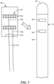
- the distal tip electrode 34 may be configured to be coupled to one or more electrical wires 60 and to one or more optical fibers 62 (a plurality of optical fibers 62 may be referred to herein as a fiber optic bundle). Further, the distal tip electrode 34 may be configured to be coupled to the elongate body 22 such that the electrical wire(s) 60 and optical fiber(s) 62 may be located within the elongate body 22 when the device is assembled.
- the distal tip electrode head portion 54 includes a distal surface 66 that may be curved and a proximal face 68 opposite the distal surface 66.
- the head portion proximal face 68 may have an outer diameter that is greater than the outer diameter of the distal tip electrode neck portion 56.
- the distal tip electrode neck portion 56 may also include a first end where the neck portion 56 joins the head portion 54 and a second end opposite the first end, the neck portion second end having a proximal face 70.
- the neck portion proximal face 70 may include one or more wire attachment elements 72, such as metalized rings or conductive pads (as shown in FIG. 3 ). Alternatively, one or more wire attachment elements 72 may be affixed to the head portion proximal face 68 where the proximal face 68 extends beyond the neck portion 56 (for example, as shown in FIG. 4 ).
- each of the one or more electrical wires 60 may be coupled to a corresponding wire attachment element 72, such that electrical signals from the distal tip electrode 34 may be received and transmitted through the electrical wire(s) 60 at the wire attachment elements 72.
- each of the head portion proximal face 68 and the neck portion proximal face 70 may be planar or at least substantially planar and may each lie in a plane that is orthogonal or at least substantially orthogonal to the elongate body longitudinal axis 30.
- the curved distal surface 66 of the distal tip electrode 34 may have a convex shape that is curved away from the head portion proximal face 68.
- the portion of the distal tip electrode head portion 54 between the proximal face 68 and the distal surface 66 may have a cylindrical shape. However, it will be understood that the distal tip electrode 34 may have other shapes or configurations than those described herein.
- the elongate body 22 may include a conduit that extends from the elongate body distal portion 28 to the elongate body proximal portion 26 for each electrical wire 60. Alternatively, more than one electrical wire 60, and in some embodiments all of the electrical wires 60 (if the device includes a plurality of electrical wires 60), may be located within the same elongate body lumen.
- the electrical wire(s) 60 may extend in a distal-to-proximal direction along the longitudinal axis 30 within the elongate body 22 toward the control unit 14.
- the distal tip electrode 34 further includes one or more optical fiber channels 74 that are sized to receive at least a portion of one or more optical fibers 62.
- the distal tip electrode 34 includes one optical fiber channel 74 that extends at least within the head portion 54 of the distal tip electrode 34 from the head portion proximal face 68 to a location within the distal tip electrode head portion 54 that is a distance D OC from the head portion distal surface 66.
- each optical fiber channel 74 may be a blind channel with one opening, the opening being on the head portion proximal face.
- the optical fiber channel 74 may have a longitudinal axis that is at least substantially coaxial with the elongate body longitudinal axis 30. If the distal tip electrode 34 includes a plurality of optical fiber channels 74, each optical fiber channel 74 may have a longitudinal axis that is at least substantially parallel to the elongate body longitudinal axis 30, and at least one optical fiber channel 74 may optionally have a longitudinal axis that is at least substantially coaxial with the elongate body longitudinal axis 30.
- the distal tip electrode 34 may be manufactured such that the head portion 54 and neck portion 56 are integrated into a single piece composed of the same material.
- the distal tip electrode neck portion 56 may be composed of different material(s) than the material(s) from which the distal tip electrode head portion 54 is composed.
- the optical fiber channel 74 may extend from the neck portion proximal face 70, through the distal tip electrode neck portion 56, and through the distal tip electrode head portion 54 to the location within the distal tip electrode head portion 54 that is a distance D OC from the head portion distal surface 66, which the opening of the optical fiber channel 74 being located on the neck portion proximal face 70.
- the distal tip electrode neck portion 56 may have a cylindrical configuration (not shown) that surrounds the opening of the optical fiber channel 74 located on the head portion proximal face 66.
- the optical fiber channel 74 may have any cross-sectional shape, although a circular cross section is shown in FIGS. 3 and 4 . Further, the optical fiber channel 74 may have an inner diameter that is sized to accept at least a portion of a distal portion 78 of at least one optical fiber 62.
- the distal tip electrode 34 may include one optical fiber channel 74 that is sized to accept at least a portion of distal portions 78 of a plurality of optical fibers 62.
- the distal tip electrode 34 may include a plurality of optical fiber channels 74, each of which being sized to accept at least a portion of a distal portion 78 of at least one optical fiber 62 (not shown).
- the optical fiber channel(s) 74 may be sized such that the optical fiber distal portions(s) 78 are held within the distal tip electrode 34 by a friction fit, or the optical fiber distal portion(s) 78 may be affixed within the optical fiber channel(s) 74, such as potted in position by adhesives.
- the optical fiber(s) 62 may extend within the elongate body 22 in a distal-to-proximal direction along the longitudinal axis 30 from the distal tip electrode 34 toward the control unit 14.
- the distal tip electrode 34 may optionally include one or more irrigation apertures 82 within the distal tip electrode head portion 54, and each irrigation aperture 82 may be in fluid communication with a fluid source 84 through an irrigation lumen 86.
- Each irrigation lumen 86 may include a first portion 86A that extends through the distal tip electrode 34 and a second portion 86B that extends through the elongate body 22, and the first portion 86A and the second portion 86B may be in fluid communication with each other when the device is assembled, and fluidly isolated from other parts of the device 12, such that the first 86A and second 86B portions of each irrigation lumen 86 are joined to form a continuous irrigation lumen 86 extending from the irrigation aperture 82 to the fluid source 84.
- a single irrigation lumen 86 may be directly coupled to the fluid source 84 and may branch downstream of the fluid source 84 into a plurality of irrigation lumens 86.
- the irrigation aperture(s) 82 may be used to deliver a fluid to the distal tip electrode 34 and environment external to the distal tip electrode 34, such as to cool the distal tip electrode 34 and/or a tissue treatment site during an ablation procedure.
- the fluid source may contain a biocompatible fluid such as saline with normal, hypotonic, or hypertonic NaCl concentrations.
- the fluid source may provide genetic materials that are intended to be transfected into tissues through electroporative processes following pulsed energy deliveries.
- the fluid source may provide nitrates to counteract blood vessel spasms that might otherwise occur.
- the fluid source may contain adjuvants such as calcium to enhance cell death from electroporation energy deliveries..
- the medical device 12 may include a handle 90 coupled to the elongate body proximal portion 26.
- the handle 90 may include circuitry for identification and/or use in controlling of the medical device 12 or another component of the system 10. Additionally, the handle 90 may also include connectors that are mateable to the control unit 14 to establish communication between the medical device 12 and one or more components or portions of the control unit 14.
- the handle 90 may also include one or more actuation or control features that allow a user to control, deflect, steer, or otherwise manipulate a distal portion of the medical device 12 from the proximal portion of the medical device 12.
- the handle 90 may include one or more components such as a lever or knob for manipulating the elongate body 22 and/or additional components of the medical device 12.
- the system 10 may include one or more sensors to monitor the operating parameters throughout the system, including for example, pressure, temperature, flow rates, volume, power delivery, impedance, or the like in the control unit 14 and/or the medical device 12, in addition to monitoring, recording or otherwise conveying measurements or conditions within the medical device 12 or the ambient environment at the distal portion of the medical device 12.
- the sensor(s) may be in communication with the control unit 14 for initiating or triggering one or more alerts or therapeutic delivery modifications during operation of the medical device 12.
- One or more valves, controllers, or the like may be in communication with the sensor(s) to provide for the controlled dispersion or circulation of fluid through the lumens/fluid paths of the medical device 12.
- Such valves, controllers, or the like may be located in a portion of the medical device 12 and/or in the control unit 14.
- control unit 14 may include any system components that are not part of the medical device 12 itself, other than components of the navigation system 16 and the imaging system (if included), regardless of whether the component is physically located within or external to the control unit 14.
- the navigation system 16 may be a standalone system in communication with the control unit 14 or may be contained within or integrated with the control unit 14, even though it is shown as being physically separated from the control unit in FIG. 1 .
- the control unit 14 may include one or more components for the delivery of one or more energy modalities for which the system is used, including therapeutic waveform generators, or laser energy.
- the control 14 unit may include an energy source 46 as a treatment or diagnostic mechanism in communication with the treatment element(s) of the device 12.
- the energy source 46 may be an RF generator having a plurality of output channels and operable in one or more modes of operation (for example, unipolar mode and/or bipolar mode) and or may be configured to deliver electrical energy, such as high-voltage energy.
- the control unit 14 also includes a laser energy source 48 for delivering laser energy to target tissue through one or more optical windows 38.
- the control unit 14 may also include a light emitter 44 and light receiver 45 that are in optical communication with and configured to transmit and receive, respectively, light through the optical fiber(s) 62.
- the light energy is high-intensity laser light to enable greater penetration into the target tissue.
- the control unit 14 may further include processing circuitry 92 that may include one or more controllers, processors, light sources, spectrometers, polarizers, optical filters, multiplexers, and/or software modules configured to execute instructions or algorithms to provide for the automated operation and performance of the features, sequences, calculations, or procedures described herein and/or required for a given medical procedure.
- the processing circuitry may include a processor and a memory.
- the memory may be in electrical communication with the processor and have instructions that, when executed by the processor, configure the processor to receive, process, or otherwise use signals from the device 12.
- the control unit 14 may include one or more user input devices, controllers, and displays 94 for collecting and conveying information from and to the user.
- the system (for example, the control unit 14) includes a charge coupled device (CCD camera) that receives light transmitted through the optical fiber(s) 62 and creates a digital image of the interrogated tissue.
- CCD camera is, or includes, the light receiver 45.
- the device 12 may include one or more treatment elements, such as a distal tip electrode 34 and, optionally, one or more electrically and/or thermally conductive areas on a location of the elongate body 22 other than at the distalmost tip of the elongate body distal portion 28.
- these electrically and/or thermally conductive areas may be referred to herein for simplicity as lateral electrodes 36.
- the lateral electrodes 36 are shown in FIG. 1 as being configured like band electrodes; however, the lateral electrodes 36 may have any suitable shape and/or configuration, and non-limiting examples of their manufacture are described in more detail below.
- the distal tip electrode 34 may be configured and coupled to the elongate body 22 as shown and described in FIGS. 1-4 .
- the elongate body distal portion 28 may include one or more optical windows 38 in the lateral walls of the elongate body 22 (that is, along a length of the elongate body 22 rather than at the distalmost end face).
- the optical window(s) 38 may each be an area of optically transparent material that is set into, integrated with, or otherwise incorporated into the elongate body distal portion 28.
- the elongate body 22 is composed of an opaque, flexible, biocompatible material (for example, polyether ether ketone (PEEK) or similar), whereas each optical window 38 is composed of a transparent biocompatible material (such as transparent ceramic, sapphire, or the like) that is inset or incorporated into an aperture within the elongate body 22 (as shown in FIGS. 5 and 6 ).
- PEEK polyether ether ketone
- each optical window 38 is composed of a transparent biocompatible material (such as transparent ceramic, sapphire, or the like) that is inset or incorporated into an aperture within the elongate body 22 (as shown in FIGS. 5 and 6 ).
- the lateral electrode(s) 36 may be printed, deposited, or affixed onto, or otherwise incorporated onto or into, a flexible overlay 98 that can be applied, affixed, adhered, or otherwise attached to an outer surface of the elongate body 22.
- the device 12 may include more than one flexible overlay 98.
- the flexible overlay 98 is applied to the elongate body distal portion 28, with at least a portion of the flexible overlay 98 being located on an outer surface of at least one optical window 38.
- the flexible overlay 98 may be composed of one or more layers of transparent, flexible, and biocompatible material(s), such as polyimide. The flexible overlay 98 is shown in FIG.
- the flexible overlay 98 may have any suitable shape or configuration.
- the flexible overlay 98 may have a serpentine, linear, helical, polygonal, or other shape.
- the flexible overlay 98 may be sized and configured to at least partially encircle a circumference of the elongate body distal portion 28.
- the flexible overlay 98 has a helical shape that is configured to be wound around a portion of the elongate body 22 at least once to form one or more bands.
- the flexible overlay 98 may include more than one layer of material. In one embodiment, the entirety of the flexible overlay 98 may be transparent. In another embodiment, at least those portions of the flexible overlay 98 that are configured and arranged to be affixed over one or more optical windows 38 in the elongate body 22 may be transparent. In one embodiment, the flexible overlay 98 includes a layer configured to be in contact with the elongate body 22, an outer or encapsulation layer 99, and one or more intermediate layers.
- the lateral electrode(s) 36 may each include one or more conductive areas 100 of graphene, conductive diamond, or other thermally and electrically conductive and optically transparent material.
- the conductive areas may be composed of graphene, as this material may be applied in so thin a layer that it does not change the mechanical properties of the substrate (here, the elongate body 22). Further, graphene may have unlimited fatigue by itself and may serve as an electromagnetic interference (EMI) barrier coating while still allowing for the transmission of light.
- EMI electromagnetic interference
- the encapsulation layer 99 may include one or more holes 102, each of which corresponding to a conductive area 100 when the flexible overlay 98 is assembled.
- a non-limiting example of an encapsulation layer 99 having a plurality of holes 102 is shown in FIG. 9 , and these holes 102 may correspond to the conductive areas shown in FIGS. 5 and 6 .
- Each conductive area 100 may be of any suitable size and configuration, but in one non-limiting example, each may be approximately 300 ⁇ m 2 .
- each optical window 38 may have a diameter of between approximately 100 ⁇ m and approximately 1000 ⁇ m, or have any other appropriate size for collecting adequate amounts of light energy, such that a plurality of conductive areas 100 may be aligned with each optical window 38.
- the conductive areas 100 associated with each hole 102 in the encapsulation layer 99 may be in contact with a conductive contact pad 104 (for example, the contact pad may be composed of gold, copper, or other electrically conductive material), either directly or via a conductive pathway.
- a conductive contact pad 104 for example, the contact pad may be composed of gold, copper, or other electrically conductive material
- graphene or other transparent conductive material may be deposited on a layer of the flexible overlay 98 to form the conductive areas 100 that will correspond to the encapsulation layer holes 102.
- graphene or other conductive material (which may or may not be transparent) may be deposited on the same layer of the flexible overlay 98 as the conductive areas 100 in, for example, thin lines between the electrode openings 100 and the conductive contact pad 104 to form conductive pathways or traces 106.
- each lateral electrode 36 may comprise a plurality of conductive areas 100, which may be overlaid on the optical windows 98 when the flexible overlay 98 is affixed to the elongate body 22.
- the transparent conductive areas 100 may not only allow light transmission through the optical windows 38, but may also be used to deliver RF, pulsed high voltage ablation energy, or other treatment or ablation energy and/or to record optical and/or electrical signals from tissue.
- the conductive areas may be used to record signals, and the optical windows 38 may be able to receive light transmission, from the tissue at the precise tissue location(s) to which energy is delivered.
- the delivery of energy and receipt of light transmission may be conducted simultaneously or in sequence.
- the encapsulation layer may completely or partially encase, cover, or enclose the conductive pathway(s) 106 and the conductive contact pad(s) 104.
- One or more wires or traces 110 may be connected to (for example, soldered to) or in electrical communication with each conductive contact pad 104.
- the wire(s) or trace(s) 110 may run along the outside of the elongate body 22 and/or transverse or pass into the elongate body 22.
- the wire(s) 100 may be deposited trace(s) composed of a conductive, high-current-carrying material such as copper and coated with a layer of graphene, which may protect the copper from corrosion. Such traces may then serve as ablation energy delivery electrodes as well.
- the conductive areas 100 may be arranged to create any desired lateral electrode 36 pattern.
- the conductive areas 100 may be arranged around at least a portion of a circumference of the elongate body 22 to provide lateral electrodes 36 that are at least substantially similar to band electrodes.
- the conductive areas 100 may be arranged in one or more linear patterns that are each configured to extend along the elongate body 22 and be at least substantially parallel to the elongate body longitudinal axis 30.
- the conductive areas 100 and any connecting wire(s) or trace(s) 106, 110 may be applied directly to the elongate body 22, and in particular over the optical window(s) 38, to provide the ability to deliver treatment energy to tissue and/or record signals from tissue without the need to create and apply a flexible overlay 98.
- the device 12 may not include a flexible overlay 98 with conductive areas 100. Instead, laser energy may be delivered to tissue at a target treatment location through the optical windows 38 without the need for conductive areas 100.
- the device 12 may include a flexible overly 98 affixed to the elongate body 22 such that one or more conductive areas 100 are over one or more optical windows 98. In this configuration, laser energy may be delivered through the optical windows 98 and the conductive areas 100 may be used to record signals from tissue such as intracardiac electrograms. Thus, the conductive areas 100 may be used to record signals from the tissue at the precise tissue location(s) to which laser energy is delivered.
- the delivery of energy and receipt of light transmission may be conducted simultaneously or in sequence.
- laser energy may be pulsed and tissue interrogation light may be delivered between laser energy deliveries.
- the tissue interrogation light may evoke the fluorescence and/or reflectance response to be measured, and this, in turn, may allow the system 10 and/or the user to monitor the progress of the laser ablation procedure.
- the laser pulse deliveries could then be halted when an end-point or other indication of sufficient ablation is detected.
- this technique may also be used for RF or pulsed electric field ablation by irreversible electroporation.
- the device may further include one or more optical fibers 62A in optical communication with the optical windows 38 in the elongate body 22.
- These optical fibers are indicated with reference number 62A in FIG. 10 for illustration, but all optical fibers may be referred to herein with reference number 62 for simplicity.
- the elongate body 22 may include a plurality of optical windows 38 and, optionally, a plurality of lateral electrodes 36.
- the optical fibers 62 that are in optical communication with the optical windows 38 may enable tissue sensing from lateral surface of the device.
- Each lateral electrode 36 may be in electrical communication with the control unit 14 and may be used to deliver energy from the energy source 46 to an area of tissue and/or to record electrical signals from the area of tissue. Additionally or alternatively, laser energy from the laser energy source 48, for example, emitting 790 nm light, may be delivered to an area of tissue through the optical window(s) 38 in the elongate body 22. For example, electrical, RF, pulsed RF, electroporation, and/or laser energy may be delivered by the device 12 to treat or ablate an area of tissue.
- the distal tip electrode 34 and/or lateral electrode(s) 36 may be positioned so that they are in contact with or proximate an area of tissue and can deliver light and/or energy to the tissue and receive signals (for example, optical signals) from the tissue.
- each optical fiber 62 may have a distal portion 78 that is at least partially located within or otherwise connected to the distal tip electrode 34 (as shown and described in FIG. 2 ) and a proximal end (not shown) that is in optical communication with the light emitter 44. Light may be transmitted through each optical fiber 62 from the light source 44 and out of the distal tip electrode 34. The light may then be reflected back into the optical fiber 62 and transmitted back to the light receiver 45.
- the light receiver 45 may convert this light energy into an electrical signal, which it may then transmit to the control unit processing circuitry 92.
- a diagnostic signal could be sent prior to delivering the ablative laser light to ensure that the ablation window was directed into targeted tissue and not directed into open blood or a layer of blood that might otherwise be carbonized when laser light is delivered.
- the optical fiber(s) 62 may be used to detect fluorescence that is naturally emitted by the reduced form of nicotinamide adenine dinucleotide (NADH) and/or nicotinamide adenine dinucleotide phosphate (NADPH), both of which being coenzymes that are involved in cellular metabolism and the transfer of electrons from one reaction to another.
- NADH nicotinamide adenine dinucleotide
- NADPH nicotinamide adenine dinucleotide phosphate
- the optical fiber(s) 62 will receive little or no fluorescence from ablated tissue when that tissue is interrogated with ultraviolet light (that is, when ultraviolet light is delivered from the light emitter 44 through the optical fiber(s) 62 to the tissue).
- ultraviolet light that is, when ultraviolet light is delivered from the light emitter 44 through the optical fiber(s) 62 to the tissue.
- NADH absorbs light at a wavelength of approximately 340 nm ( ⁇ 30 nm) and emits fluorescence at a wavelength of approximately 460 nm ( ⁇ 50 nm).
- Ultraviolet light is electromagnetic radiation with a wavelength of between approximately 10 nm and 400 nm; however, it will be understood that other wavelengths may be emitted by the light emitter 44, reflected or fluoresced from the tissue, and/or received by the light receiver 45.
- the optical fiber(s) 62 may be used to identify the type of tissue or tissue structure being interrogated by the device 12. Different tissues have different optical properties that may be detected by the device 12. For example, fatty tissue, pulmonary vein tissue, the fossa ovalis, cardiac muscle, and blood all have difference reflectance values that can be used by the system 10 to identify a type of tissue with which the electrode(s) 34, 36 are in contact.
- an exemplary medical device that includes an optical distal tip electrode 34 and one or more lateral electrodes without optical window(s) 38 or other transparencies. These electrodes may be referred to herein as non-optical lateral electrodes 118.
- the device 12 shown in FIG. 11 may be used to collect light energy or other optical signals from tissue through the distal tip electrode 34, whereas the distal tip electrode 34 and the non-optical lateral electrodes 118 may both be used to deliver treatment or ablation energy to the tissue.
- the non-optical lateral electrodes 118 may be wires that are wound at least once around a portion of the elongate body distal portion 28 (as shown in FIG. 11 ).
- each non-optical electrode 118 may be composed of a single piece or strand of conductive material, such as copper, tantalum, or gold, that has an electrode portion 120 that is wrapped around a circumference of the elongate body distal portion 28 at least once (for example, to form a band-electrode-like conductive region) and a wire portion 122 that extends from the electrode portion 120 proximally to the handle 90.
- the electrode portion 120 may be electrically and thermally conductive, whereas the wire portion 122 may have an electrically and thermally insulative coating.
- the non-optical lateral electrodes 118 may be band electrodes and may be connected to an electrode wire. However, it will be understood that the non-optical lateral electrodes 118 may have any suitable size, configuration, or shape.
- the device 12 may be navigated through the patient's vasculature to a target treatment location within the patient's heart.
- a target treatment location within the patient's heart.
- light may be emitted from the light emitter 44 through one or more optical fibers 62 toward tissue at the target treatment location ("interrogated tissue").
- tissue at the target treatment location
- optical fibers 62 that are in optical communication with the distal tip electrode 34 and one or more optical windows 38. That light may then be reflected from the interrogated tissue through the distal tip electrode 34 and/or the optical window(s) 38, transmitted back through the optical fiber(s) 62, received by the light receiver 45, converted to electrical signals, and then transmitted to the control unit processing circuitry 92.
- the processing circuitry 92 may determine whether the device 12 is in a correct position to treat the tissue target tissue location. For example, reflectance values may be used to determine whether the device is in contact with atrial wall tissue, pulmonary vein tissue, or is not in contact with tissue and is instead surrounded by blood. Additionally, the optical fiber(s) 62 may be used to detect fluorescence from the tissue at the target treatment location to establish a pre-treatment or baseline fluorescence value.
- energy may be delivered from the device to treat or ablate tissue at the target treatment location.
- RF energy may be delivered by the distal tip electrode 34
- RF energy may be delivered by one or more lateral electrodes 36
- laser energy may be delivered through the optical window(s) 38.
- the optical fiber(s) 62 may be used to detect fluorescence form the tissue at the target treatment location, and the processing circuitry 92 may compare this in-treatment and/or post-treatment fluorescence to the pre-treatment or baseline fluorescence. The comparison may indicate whether the tissue has been sufficiently ablated. If the tissue has been sufficiently ablated, the user may end the treatment procedure.
- this method may be repeated until optical characteristics of the tissue indicate that sufficient ablation has occurred.
- the comparison may also be used to determine an extent of lesion formation. For example, a plurality of comparisons in a plurality of locations may be made between the pre-treatment or threshold fluorescence and in-treatment and/or post-treatment fluorescence to identify a boundary of an area sufficiently ablated tissue and, therefore, to identify the size of the lesion.
- the distal tip electrode 34 and/or the conductive material of the lateral electrodes 36 may be used to record electrical signals from the tissue, including intracardiac electrograms. These signals may be used by the processing circuitry 92 to determine if such signals are of local origin or produced from far field depolarizations.
Landscapes
- Health & Medical Sciences (AREA)
- Surgery (AREA)
- Life Sciences & Earth Sciences (AREA)
- Engineering & Computer Science (AREA)
- Heart & Thoracic Surgery (AREA)
- Animal Behavior & Ethology (AREA)
- Veterinary Medicine (AREA)
- Public Health (AREA)
- General Health & Medical Sciences (AREA)
- Biomedical Technology (AREA)
- Nuclear Medicine, Radiotherapy & Molecular Imaging (AREA)
- Medical Informatics (AREA)
- Molecular Biology (AREA)
- Physics & Mathematics (AREA)
- Otolaryngology (AREA)
- Cardiology (AREA)
- Plasma & Fusion (AREA)
- Oral & Maxillofacial Surgery (AREA)
- Pathology (AREA)
- Optics & Photonics (AREA)
- Electromagnetism (AREA)
- Surgical Instruments (AREA)
- Laser Surgery Devices (AREA)
Claims (13)
- Système médical comprenant un dispositif médical, le dispositif médical comportant :un corps allongé (22) comportant une partie proximale (26), une partie distale (28) ayant une extrémité distale (52), et un axe longitudinal (30) ;une électrode de pointe distale (34) au niveau de l'extrémité distale (52) de corps allongé, l'électrode de pointe (52) étant composée d'un matériau optiquement transparent et ayant une couche continue d'un matériau optiquement transparent et électroconducteur,dans lequel le dispositif médical comporte en outre au moins une fenêtre optique (38) à l'intérieur de la partie distale (28) de corps allongé, l'au moins une fenêtre optique (38) étant composée d'un matériau optiquement transparent,caractérisé en ce que le dispositif médical comporte en outre au moins une électrode latérale (36) sur la partie distale (28) de corps allongé, chacune de l'au moins une électrode latérale (36) comportant au moins une zone conductrice (100), chacune de l'au moins une zone conductrice (100) étant composée d'un matériau électroconducteur et optiquement transparent, chacune de l'au moins une zone conductrice (100) étant sur un revêtement souple qui est appliqué à une surface externe du corps allongé (22), le revêtement souple étant au moins partiellement composé d'un matériau optiquement transparent de telle sorte que la pluralité de zones conductrices (100) et le revêtement souple soient optiquement transparents, le revêtement souple étant appliqué au corps allongé (22) de telle sorte qu'au moins certaines de la pluralité de zones conductrices (100) soient positionnées au-dessus d'au moins l'une de l'au moins une fenêtre optique (38).
- Système médical selon la revendication 1, dans lequel l'électrode de pointe distale (34) est composée d'un premier matériau optiquement transparent et la couche continue est composée d'un second matériau optiquement transparent et électroconducteur, le second matériau étant différent du premier matériau.
- Système médical selon la revendication 1 ou la revendication 2, dans lequel l'électrode de pointe distale (34) est composée d'au moins l'un parmi un polymère transparent, une céramique, un saphir, un saphir synthétique, un oxyde d'yttrium, un grenat d'oxyde d'yttrium-alumine, un séléniure de zinc, un germanium, un tellurure de cadmium, un fluorure de baryum, un iodure de césium, un fluorure de magnésium, un bromoiodure de thallium, un sulfure de zinc, un fluorure de calcium, un quartz, un diamant, un rubis, un rubis synthétique, un cristal, du verre et une silice fondue.
- Système médical selon la revendication 2 ou la revendication 3, dans lequel le second matériau est au moins l'un parmi le graphène et un diamant conducteur.
- Système médical selon l'une quelconque des revendications 1 à 4, dans lequel le dispositif médical comporte en outre au moins une fibre optique (62) à l'intérieur du corps allongé (22), l'au moins une fibre optique (62) étant en communication optique avec l'électrode de pointe distale (34).
- Système médical selon l'une quelconque des revendications 1 à 5, dans lequel l'électrode de pointe distale (34) comporte une face proximale et une surface distale incurvée, la face proximale reposant dans un plan qui est orthogonal à l'axe longitudinal de corps allongé.
- Système médical selon l'une quelconque des revendications 1 à 6, dans lequel l'électrode de pointe distale (34) comporte un canal de fibre optique (74) ayant une ouverture, l'ouverture de fibre optique étant sur la face proximale d'électrode de pointe distale.
- Système médical selon la revendication 7, dans lequel le canal de fibre optique (74) s'étend à l'intérieur de l'électrode de pointe distale (34) de la face proximale d'électrode de pointe distale jusqu'à un emplacement qui est à une distance à partir de la surface distale incurvée de l'électrode de pointe distale et est dimensionné et configuré pour accepter au moins une partie de l'au moins une fibre optique (62).
- Système médical selon l'une quelconque des revendications 5 à 8, dans lequel l'au moins une fibre optique (62) est une première au moins une fibre optique, le dispositif comprenant en outre une seconde au moins une fibre optique, chacune de la seconde au moins une fibre optique étant en communication optique avec l'une de l'au moins une fenêtre optique (38).
- Système médical selon l'une quelconque des revendications 1 à 9, dans lequel l'au moins une électrode latérale est une longueur de matériau conducteur enroulée autour d'au moins une partie du corps allongé.
- Système médical selon l'une quelconque des revendications 1 à 10, dans lequel l'électrode de pointe distale comporte une pluralité d'orifices, la pluralité d'orifices étant configurée pour être en communication fluidique avec une source de fluide.
- Système médical selon l'une quelconque des revendications 1 à 11, comprenant en outre une unité de commande (14), l'unité de commande (14) comportant :un émetteur de lumière (44) en communication optique avec chacune de la première pluralité de fibres optiques (62) et de la seconde pluralité de fibres optiques ;un récepteur de lumière (45) en communication optique avec chacune de la première pluralité de fibres optiques et de la seconde pluralité de fibres optiques ;une source d'énergie d'ablation (46) en communication avec l'électrode de pointe distale(34) et l'au moins une électrode latérale ; etdes circuits de traitement (92) en communication avec la première pluralité de fibres optiques, la seconde pluralité de fibres optiques, l'électrode de pointe distale (34) et l'au moins une électrode latérale (36).
- Système médical selon la revendication 12, dans lequel les circuits de traitement (92) sont configurés pour :émettre de la lumière provenant de l'émetteur de lumière à travers au moins l'une de l'électrode de pointe distale et de l'au moins une électrode latérale à travers au moins l'une de la première pluralité de fibres optiques et de la seconde pluralité de fibres optiques vers une zone de tissu ; etrecevoir une lumière fluorescente à partir de la zone de tissu à travers au moins l'une de l'électrode de pointe distale et de l'au moins une électrode latérale à travers au moins l'une de la première pluralité de fibres optiques et de la seconde pluralité de fibres optiques.
Applications Claiming Priority (2)
| Application Number | Priority Date | Filing Date | Title |
|---|---|---|---|
| US15/786,229 US10939959B2 (en) | 2017-10-17 | 2017-10-17 | Ablation catheter with dual optical-electrical stimulation and sensing |
| PCT/US2018/054192 WO2019079036A1 (fr) | 2017-10-17 | 2018-10-03 | Cathéter d'ablation à double stimulation et détection optique-électrique |
Publications (2)
| Publication Number | Publication Date |
|---|---|
| EP3697331A1 EP3697331A1 (fr) | 2020-08-26 |
| EP3697331B1 true EP3697331B1 (fr) | 2023-05-24 |
Family
ID=63963569
Family Applications (1)
| Application Number | Title | Priority Date | Filing Date |
|---|---|---|---|
| EP18792776.9A Active EP3697331B1 (fr) | 2017-10-17 | 2018-10-03 | Cathéter d'ablation à double stimulation et détection optique-électrique |
Country Status (4)
| Country | Link |
|---|---|
| US (2) | US10939959B2 (fr) |
| EP (1) | EP3697331B1 (fr) |
| CN (1) | CN111225628B (fr) |
| WO (1) | WO2019079036A1 (fr) |
Families Citing this family (20)
| Publication number | Priority date | Publication date | Assignee | Title |
|---|---|---|---|---|
| US20220233815A1 (en) * | 2019-06-06 | 2022-07-28 | Vascomed Gmbh | Catheter configured to measure a force acting on the catheter |
| CN111685866A (zh) * | 2019-08-07 | 2020-09-22 | 上海明石医疗科技有限公司 | 一种射频消融电极 |
| CN115103647A (zh) * | 2020-01-08 | 2022-09-23 | 460医学股份有限公司 | 用于消融损伤的光学询问的系统和方法 |
| US11931098B2 (en) * | 2020-02-19 | 2024-03-19 | Boston Scientific Medical Device Limited | System and method for carrying out a medical procedure |
| US11474310B2 (en) | 2020-02-28 | 2022-10-18 | Bard Access Systems, Inc. | Optical connection systems and methods thereof |
| EP4110176B1 (fr) | 2020-02-28 | 2024-11-20 | Bard Access Systems, Inc. | Cathéter à capacités de détection de forme optique |
| EP4114258A1 (fr) | 2020-03-03 | 2023-01-11 | Bard Access Systems, Inc. | Système et procédé de détection de forme optique et de conduction de signal électrique |
| US11931179B2 (en) | 2020-03-30 | 2024-03-19 | Bard Access Systems, Inc. | Optical and electrical diagnostic systems and methods thereof |
| CN116056651A (zh) * | 2020-07-28 | 2023-05-02 | 460医学股份有限公司 | 用于损伤形成和评估的系统和方法 |
| EP4178476B1 (fr) * | 2020-09-25 | 2025-09-03 | Boston Scientific Scimed, Inc. | Dispositifs médicaux et procédés associés |
| CN114344514A (zh) | 2020-10-13 | 2022-04-15 | 巴德阿克塞斯系统股份有限公司 | 用于光纤连接器的消毒罩及其方法 |
| WO2022109045A1 (fr) | 2020-11-18 | 2022-05-27 | Bard Access Systems, Inc. | Supports de stylets à fibres optiques et procédés associés |
| CN217041033U (zh) | 2020-11-24 | 2022-07-26 | 巴德阿克塞斯系统股份有限公司 | 用于将医疗器械插入患者体内的医疗器械系统 |
| CN115813547A (zh) | 2021-09-16 | 2023-03-21 | 巴德阿克塞斯系统股份有限公司 | 连接部件和执行医疗操作的方法 |
| US12318149B2 (en) | 2022-03-08 | 2025-06-03 | Bard Access Systems, Inc. | Medical shape sensing devices and systems |
| US12426956B2 (en) | 2022-03-16 | 2025-09-30 | Bard Access Systems, Inc. | Medical system and method for monitoring medical device insertion and illumination patterns |
| US12089815B2 (en) | 2022-03-17 | 2024-09-17 | Bard Access Systems, Inc. | Fiber optic medical systems and devices with atraumatic tip |
| US20230320663A1 (en) * | 2022-04-11 | 2023-10-12 | Bard Access Systems, Inc. | Fiber Optic Medical Systems and Devices with Electrical Tip |
| WO2024166326A1 (fr) * | 2023-02-09 | 2024-08-15 | オリンパスメディカルシステムズ株式会社 | Appareil médical, système médical, procédé pour faire fonctionner un appareil médical et programme pour faire fonctionner un appareil médical |
| WO2025117508A1 (fr) * | 2023-11-28 | 2025-06-05 | Boston Scientific Scimed, Inc. | Dispositifs médicaux assistés par photo ou image pour délivrer de l'énergie et/ou un fluide à un tissu, et procédés associés |
Family Cites Families (22)
| Publication number | Priority date | Publication date | Assignee | Title |
|---|---|---|---|---|
| US5991650A (en) | 1993-10-15 | 1999-11-23 | Ep Technologies, Inc. | Surface coatings for catheters, direct contacting diagnostic and therapeutic devices |
| US5989245A (en) * | 1994-03-21 | 1999-11-23 | Prescott; Marvin A. | Method and apparatus for therapeutic laser treatment |
| US5848969A (en) | 1996-10-28 | 1998-12-15 | Ep Technologies, Inc. | Systems and methods for visualizing interior tissue regions using expandable imaging structures |
| US5904651A (en) | 1996-10-28 | 1999-05-18 | Ep Technologies, Inc. | Systems and methods for visualizing tissue during diagnostic or therapeutic procedures |
| US5722403A (en) | 1996-10-28 | 1998-03-03 | Ep Technologies, Inc. | Systems and methods using a porous electrode for ablating and visualizing interior tissue regions |
| US7442193B2 (en) * | 2003-11-20 | 2008-10-28 | Covidien Ag | Electrically conductive/insulative over-shoe for tissue fusion |
| US7527625B2 (en) * | 2004-08-04 | 2009-05-05 | Olympus Corporation | Transparent electrode for the radiofrequency ablation of tissue |
| US7942873B2 (en) * | 2005-03-25 | 2011-05-17 | Angiodynamics, Inc. | Cavity ablation apparatus and method |
| US8123745B2 (en) * | 2007-06-29 | 2012-02-28 | Biosense Webster, Inc. | Ablation catheter with optically transparent, electrically conductive tip |
| US8500730B2 (en) * | 2007-11-16 | 2013-08-06 | Biosense Webster, Inc. | Catheter with omni-directional optical tip having isolated optical paths |
| US8513531B2 (en) * | 2009-07-15 | 2013-08-20 | The Board Of Trustees Of The University Of Arkansas | Electrodynamic arrays having nanomaterial electrodes |
| US8874232B2 (en) * | 2009-11-30 | 2014-10-28 | Boston Scientific Neuromodulation Corporation | Electrode array having concentric split ring electrodes and methods of making the same |
| US9907607B2 (en) * | 2009-12-30 | 2018-03-06 | St. Jude Medical, Atrial Fibrillation Division, Inc. | Guide and flexible sleeve for use with catheters |
| US20140171806A1 (en) * | 2012-12-17 | 2014-06-19 | Biosense Webster (Israel), Ltd. | Optical lesion assessment |
| US8986292B2 (en) * | 2011-04-13 | 2015-03-24 | St. Jude Medical, Inc. | Optical feedback RF ablator and ablator tip |
| WO2013123020A1 (fr) * | 2012-02-14 | 2013-08-22 | St. Jude Medical, Atrial Fibrillation Division, Inc. | Cathéter d'ablation à système de distribution d'énergie optique pour une réponse de tissu photo-acoustique |
| US9370311B2 (en) * | 2012-08-17 | 2016-06-21 | Medtronic Ablation Frontiers Llc | Electrophysiology catheter design |
| US9078667B2 (en) * | 2012-12-11 | 2015-07-14 | St. Jude Medical, Atrial Fibrillation Division, Inc. | Catheter having reduced force concentration at tissue contact site |
| JP2014190991A (ja) * | 2013-03-26 | 2014-10-06 | Sony Corp | 電気泳動素子、表示装置および電子機器 |
| US20150157405A1 (en) * | 2013-12-05 | 2015-06-11 | Biosense Webster (Israel) Ltd. | Needle catheter utilizing optical spectroscopy for tumor identification and ablation |
| WO2016196845A1 (fr) | 2015-06-02 | 2016-12-08 | GI Scientific, LLC | Dispositif de manipulation de matière à revêtement conducteur |
| US10548467B2 (en) * | 2015-06-02 | 2020-02-04 | GI Scientific, LLC | Conductive optical element |
-
2017
- 2017-10-17 US US15/786,229 patent/US10939959B2/en active Active
-
2018
- 2018-10-03 WO PCT/US2018/054192 patent/WO2019079036A1/fr not_active Ceased
- 2018-10-03 CN CN201880067985.6A patent/CN111225628B/zh active Active
- 2018-10-03 EP EP18792776.9A patent/EP3697331B1/fr active Active
-
2021
- 2021-02-01 US US17/163,787 patent/US11744642B2/en active Active
Also Published As
| Publication number | Publication date |
|---|---|
| US20190110838A1 (en) | 2019-04-18 |
| EP3697331A1 (fr) | 2020-08-26 |
| US10939959B2 (en) | 2021-03-09 |
| CN111225628A (zh) | 2020-06-02 |
| US11744642B2 (en) | 2023-09-05 |
| WO2019079036A1 (fr) | 2019-04-25 |
| US20210145515A1 (en) | 2021-05-20 |
| CN111225628B (zh) | 2024-06-04 |
Similar Documents
| Publication | Publication Date | Title |
|---|---|---|
| US11744642B2 (en) | Ablation catheter with dual optical-electrical stimulation and sensing | |
| US12383736B2 (en) | Catheter systems | |
| CN106852708B (zh) | 具有基于光的接触传感器的消融导管 | |
| EP2136702B1 (fr) | Catheter electrophysiologique a haute resolution | |
| CA2588402C (fr) | Procede d'evaluation en temps reel de l'ablation de tissus | |
| EP2837350B1 (fr) | dispositif pour évaluation de lésion optique | |
| US20140058375A1 (en) | High resolution map and ablate catheter | |
| JP2015100709A (ja) | 温度センサ及び光ファイバアレイを備えた灌注カテーテル先端部 | |
| US20160081555A1 (en) | Multi-range optical sensing |
Legal Events
| Date | Code | Title | Description |
|---|---|---|---|
| STAA | Information on the status of an ep patent application or granted ep patent |
Free format text: STATUS: UNKNOWN |
|
| STAA | Information on the status of an ep patent application or granted ep patent |
Free format text: STATUS: THE INTERNATIONAL PUBLICATION HAS BEEN MADE |
|
| PUAI | Public reference made under article 153(3) epc to a published international application that has entered the european phase |
Free format text: ORIGINAL CODE: 0009012 |
|
| STAA | Information on the status of an ep patent application or granted ep patent |
Free format text: STATUS: REQUEST FOR EXAMINATION WAS MADE |
|
| 17P | Request for examination filed |
Effective date: 20200427 |
|
| AK | Designated contracting states |
Kind code of ref document: A1 Designated state(s): AL AT BE BG CH CY CZ DE DK EE ES FI FR GB GR HR HU IE IS IT LI LT LU LV MC MK MT NL NO PL PT RO RS SE SI SK SM TR |
|
| AX | Request for extension of the european patent |
Extension state: BA ME |
|
| DAV | Request for validation of the european patent (deleted) | ||
| DAX | Request for extension of the european patent (deleted) | ||
| GRAP | Despatch of communication of intention to grant a patent |
Free format text: ORIGINAL CODE: EPIDOSNIGR1 |
|
| STAA | Information on the status of an ep patent application or granted ep patent |
Free format text: STATUS: GRANT OF PATENT IS INTENDED |
|
| RIC1 | Information provided on ipc code assigned before grant |
Ipc: A61B 18/16 20060101ALN20221212BHEP Ipc: A61B 18/00 20060101ALN20221212BHEP Ipc: A61B 90/00 20160101ALN20221212BHEP Ipc: A61B 1/00 20060101ALN20221212BHEP Ipc: A61B 90/30 20160101ALI20221212BHEP Ipc: A61B 18/14 20060101AFI20221212BHEP |
|
| INTG | Intention to grant announced |
Effective date: 20230109 |
|
| RIN1 | Information on inventor provided before grant (corrected) |
Inventor name: STEWART, MARK T. Inventor name: LASKE, TIMOTHY G. Inventor name: MARTINEZ, GONZALO |
|
| GRAS | Grant fee paid |
Free format text: ORIGINAL CODE: EPIDOSNIGR3 |
|
| GRAA | (expected) grant |
Free format text: ORIGINAL CODE: 0009210 |
|
| STAA | Information on the status of an ep patent application or granted ep patent |
Free format text: STATUS: THE PATENT HAS BEEN GRANTED |
|
| AK | Designated contracting states |
Kind code of ref document: B1 Designated state(s): AL AT BE BG CH CY CZ DE DK EE ES FI FR GB GR HR HU IE IS IT LI LT LU LV MC MK MT NL NO PL PT RO RS SE SI SK SM TR |
|
| REG | Reference to a national code |
Ref country code: GB Ref legal event code: FG4D |
|
| REG | Reference to a national code |
Ref country code: CH Ref legal event code: EP |
|
| REG | Reference to a national code |
Ref country code: DE Ref legal event code: R096 Ref document number: 602018050231 Country of ref document: DE |
|
| REG | Reference to a national code |
Ref country code: AT Ref legal event code: REF Ref document number: 1569081 Country of ref document: AT Kind code of ref document: T Effective date: 20230615 |
|
| REG | Reference to a national code |
Ref country code: IE Ref legal event code: FG4D |
|
| REG | Reference to a national code |
Ref country code: LT Ref legal event code: MG9D |
|
| REG | Reference to a national code |
Ref country code: NL Ref legal event code: MP Effective date: 20230524 |
|
| REG | Reference to a national code |
Ref country code: AT Ref legal event code: MK05 Ref document number: 1569081 Country of ref document: AT Kind code of ref document: T Effective date: 20230524 |
|
| PG25 | Lapsed in a contracting state [announced via postgrant information from national office to epo] |
Ref country code: SE Free format text: LAPSE BECAUSE OF FAILURE TO SUBMIT A TRANSLATION OF THE DESCRIPTION OR TO PAY THE FEE WITHIN THE PRESCRIBED TIME-LIMIT Effective date: 20230524 Ref country code: PT Free format text: LAPSE BECAUSE OF FAILURE TO SUBMIT A TRANSLATION OF THE DESCRIPTION OR TO PAY THE FEE WITHIN THE PRESCRIBED TIME-LIMIT Effective date: 20230925 Ref country code: NO Free format text: LAPSE BECAUSE OF FAILURE TO SUBMIT A TRANSLATION OF THE DESCRIPTION OR TO PAY THE FEE WITHIN THE PRESCRIBED TIME-LIMIT Effective date: 20230824 Ref country code: NL Free format text: LAPSE BECAUSE OF FAILURE TO SUBMIT A TRANSLATION OF THE DESCRIPTION OR TO PAY THE FEE WITHIN THE PRESCRIBED TIME-LIMIT Effective date: 20230524 Ref country code: ES Free format text: LAPSE BECAUSE OF FAILURE TO SUBMIT A TRANSLATION OF THE DESCRIPTION OR TO PAY THE FEE WITHIN THE PRESCRIBED TIME-LIMIT Effective date: 20230524 Ref country code: AT Free format text: LAPSE BECAUSE OF FAILURE TO SUBMIT A TRANSLATION OF THE DESCRIPTION OR TO PAY THE FEE WITHIN THE PRESCRIBED TIME-LIMIT Effective date: 20230524 |
|
| PG25 | Lapsed in a contracting state [announced via postgrant information from national office to epo] |
Ref country code: RS Free format text: LAPSE BECAUSE OF FAILURE TO SUBMIT A TRANSLATION OF THE DESCRIPTION OR TO PAY THE FEE WITHIN THE PRESCRIBED TIME-LIMIT Effective date: 20230524 Ref country code: PL Free format text: LAPSE BECAUSE OF FAILURE TO SUBMIT A TRANSLATION OF THE DESCRIPTION OR TO PAY THE FEE WITHIN THE PRESCRIBED TIME-LIMIT Effective date: 20230524 Ref country code: LV Free format text: LAPSE BECAUSE OF FAILURE TO SUBMIT A TRANSLATION OF THE DESCRIPTION OR TO PAY THE FEE WITHIN THE PRESCRIBED TIME-LIMIT Effective date: 20230524 Ref country code: LT Free format text: LAPSE BECAUSE OF FAILURE TO SUBMIT A TRANSLATION OF THE DESCRIPTION OR TO PAY THE FEE WITHIN THE PRESCRIBED TIME-LIMIT Effective date: 20230524 Ref country code: IS Free format text: LAPSE BECAUSE OF FAILURE TO SUBMIT A TRANSLATION OF THE DESCRIPTION OR TO PAY THE FEE WITHIN THE PRESCRIBED TIME-LIMIT Effective date: 20230924 Ref country code: HR Free format text: LAPSE BECAUSE OF FAILURE TO SUBMIT A TRANSLATION OF THE DESCRIPTION OR TO PAY THE FEE WITHIN THE PRESCRIBED TIME-LIMIT Effective date: 20230524 Ref country code: GR Free format text: LAPSE BECAUSE OF FAILURE TO SUBMIT A TRANSLATION OF THE DESCRIPTION OR TO PAY THE FEE WITHIN THE PRESCRIBED TIME-LIMIT Effective date: 20230825 |
|
| PG25 | Lapsed in a contracting state [announced via postgrant information from national office to epo] |
Ref country code: FI Free format text: LAPSE BECAUSE OF FAILURE TO SUBMIT A TRANSLATION OF THE DESCRIPTION OR TO PAY THE FEE WITHIN THE PRESCRIBED TIME-LIMIT Effective date: 20230524 |
|
| PG25 | Lapsed in a contracting state [announced via postgrant information from national office to epo] |
Ref country code: SK Free format text: LAPSE BECAUSE OF FAILURE TO SUBMIT A TRANSLATION OF THE DESCRIPTION OR TO PAY THE FEE WITHIN THE PRESCRIBED TIME-LIMIT Effective date: 20230524 |
|
| PG25 | Lapsed in a contracting state [announced via postgrant information from national office to epo] |
Ref country code: SM Free format text: LAPSE BECAUSE OF FAILURE TO SUBMIT A TRANSLATION OF THE DESCRIPTION OR TO PAY THE FEE WITHIN THE PRESCRIBED TIME-LIMIT Effective date: 20230524 Ref country code: SK Free format text: LAPSE BECAUSE OF FAILURE TO SUBMIT A TRANSLATION OF THE DESCRIPTION OR TO PAY THE FEE WITHIN THE PRESCRIBED TIME-LIMIT Effective date: 20230524 Ref country code: RO Free format text: LAPSE BECAUSE OF FAILURE TO SUBMIT A TRANSLATION OF THE DESCRIPTION OR TO PAY THE FEE WITHIN THE PRESCRIBED TIME-LIMIT Effective date: 20230524 Ref country code: EE Free format text: LAPSE BECAUSE OF FAILURE TO SUBMIT A TRANSLATION OF THE DESCRIPTION OR TO PAY THE FEE WITHIN THE PRESCRIBED TIME-LIMIT Effective date: 20230524 Ref country code: DK Free format text: LAPSE BECAUSE OF FAILURE TO SUBMIT A TRANSLATION OF THE DESCRIPTION OR TO PAY THE FEE WITHIN THE PRESCRIBED TIME-LIMIT Effective date: 20230524 Ref country code: CZ Free format text: LAPSE BECAUSE OF FAILURE TO SUBMIT A TRANSLATION OF THE DESCRIPTION OR TO PAY THE FEE WITHIN THE PRESCRIBED TIME-LIMIT Effective date: 20230524 |
|
| REG | Reference to a national code |
Ref country code: DE Ref legal event code: R097 Ref document number: 602018050231 Country of ref document: DE |
|
| PLBE | No opposition filed within time limit |
Free format text: ORIGINAL CODE: 0009261 |
|
| STAA | Information on the status of an ep patent application or granted ep patent |
Free format text: STATUS: NO OPPOSITION FILED WITHIN TIME LIMIT |
|
| 26N | No opposition filed |
Effective date: 20240227 |
|
| PG25 | Lapsed in a contracting state [announced via postgrant information from national office to epo] |
Ref country code: SI Free format text: LAPSE BECAUSE OF FAILURE TO SUBMIT A TRANSLATION OF THE DESCRIPTION OR TO PAY THE FEE WITHIN THE PRESCRIBED TIME-LIMIT Effective date: 20230524 |
|
| PG25 | Lapsed in a contracting state [announced via postgrant information from national office to epo] |
Ref country code: SI Free format text: LAPSE BECAUSE OF FAILURE TO SUBMIT A TRANSLATION OF THE DESCRIPTION OR TO PAY THE FEE WITHIN THE PRESCRIBED TIME-LIMIT Effective date: 20230524 Ref country code: IT Free format text: LAPSE BECAUSE OF FAILURE TO SUBMIT A TRANSLATION OF THE DESCRIPTION OR TO PAY THE FEE WITHIN THE PRESCRIBED TIME-LIMIT Effective date: 20230524 Ref country code: MC Free format text: LAPSE BECAUSE OF FAILURE TO SUBMIT A TRANSLATION OF THE DESCRIPTION OR TO PAY THE FEE WITHIN THE PRESCRIBED TIME-LIMIT Effective date: 20230524 |
|
| REG | Reference to a national code |
Ref country code: CH Ref legal event code: PL |
|
| REG | Reference to a national code |
Ref country code: BE Ref legal event code: MM Effective date: 20231031 |
|
| PG25 | Lapsed in a contracting state [announced via postgrant information from national office to epo] |
Ref country code: LU Free format text: LAPSE BECAUSE OF NON-PAYMENT OF DUE FEES Effective date: 20231003 |
|
| GBPC | Gb: european patent ceased through non-payment of renewal fee |
Effective date: 20231003 |
|
| PG25 | Lapsed in a contracting state [announced via postgrant information from national office to epo] |
Ref country code: LU Free format text: LAPSE BECAUSE OF NON-PAYMENT OF DUE FEES Effective date: 20231003 |
|
| PG25 | Lapsed in a contracting state [announced via postgrant information from national office to epo] |
Ref country code: GB Free format text: LAPSE BECAUSE OF NON-PAYMENT OF DUE FEES Effective date: 20231003 |
|
| PG25 | Lapsed in a contracting state [announced via postgrant information from national office to epo] |
Ref country code: CH Free format text: LAPSE BECAUSE OF NON-PAYMENT OF DUE FEES Effective date: 20231031 |
|
| PG25 | Lapsed in a contracting state [announced via postgrant information from national office to epo] |
Ref country code: GB Free format text: LAPSE BECAUSE OF NON-PAYMENT OF DUE FEES Effective date: 20231003 Ref country code: CH Free format text: LAPSE BECAUSE OF NON-PAYMENT OF DUE FEES Effective date: 20231031 |
|
| PG25 | Lapsed in a contracting state [announced via postgrant information from national office to epo] |
Ref country code: BE Free format text: LAPSE BECAUSE OF NON-PAYMENT OF DUE FEES Effective date: 20231031 |
|
| PG25 | Lapsed in a contracting state [announced via postgrant information from national office to epo] |
Ref country code: IE Free format text: LAPSE BECAUSE OF NON-PAYMENT OF DUE FEES Effective date: 20231003 |
|
| PG25 | Lapsed in a contracting state [announced via postgrant information from national office to epo] |
Ref country code: IE Free format text: LAPSE BECAUSE OF NON-PAYMENT OF DUE FEES Effective date: 20231003 |
|
| PG25 | Lapsed in a contracting state [announced via postgrant information from national office to epo] |
Ref country code: BG Free format text: LAPSE BECAUSE OF FAILURE TO SUBMIT A TRANSLATION OF THE DESCRIPTION OR TO PAY THE FEE WITHIN THE PRESCRIBED TIME-LIMIT Effective date: 20230524 |
|
| PG25 | Lapsed in a contracting state [announced via postgrant information from national office to epo] |
Ref country code: BG Free format text: LAPSE BECAUSE OF FAILURE TO SUBMIT A TRANSLATION OF THE DESCRIPTION OR TO PAY THE FEE WITHIN THE PRESCRIBED TIME-LIMIT Effective date: 20230524 |
|
| PG25 | Lapsed in a contracting state [announced via postgrant information from national office to epo] |
Ref country code: CY Free format text: LAPSE BECAUSE OF FAILURE TO SUBMIT A TRANSLATION OF THE DESCRIPTION OR TO PAY THE FEE WITHIN THE PRESCRIBED TIME-LIMIT; INVALID AB INITIO Effective date: 20181003 |
|
| PG25 | Lapsed in a contracting state [announced via postgrant information from national office to epo] |
Ref country code: HU Free format text: LAPSE BECAUSE OF FAILURE TO SUBMIT A TRANSLATION OF THE DESCRIPTION OR TO PAY THE FEE WITHIN THE PRESCRIBED TIME-LIMIT; INVALID AB INITIO Effective date: 20181003 |
|
| PGFP | Annual fee paid to national office [announced via postgrant information from national office to epo] |
Ref country code: FR Payment date: 20250924 Year of fee payment: 8 |
|
| PG25 | Lapsed in a contracting state [announced via postgrant information from national office to epo] |
Ref country code: TR Free format text: LAPSE BECAUSE OF FAILURE TO SUBMIT A TRANSLATION OF THE DESCRIPTION OR TO PAY THE FEE WITHIN THE PRESCRIBED TIME-LIMIT Effective date: 20230524 |
|
| PGFP | Annual fee paid to national office [announced via postgrant information from national office to epo] |
Ref country code: DE Payment date: 20250923 Year of fee payment: 8 |