EP3674406A1 - Isolation d'acides nucléiques - Google Patents
Isolation d'acides nucléiques Download PDFInfo
- Publication number
- EP3674406A1 EP3674406A1 EP19179423.9A EP19179423A EP3674406A1 EP 3674406 A1 EP3674406 A1 EP 3674406A1 EP 19179423 A EP19179423 A EP 19179423A EP 3674406 A1 EP3674406 A1 EP 3674406A1
- Authority
- EP
- European Patent Office
- Prior art keywords
- nucleic acid
- sample
- lysate
- composition
- liquefaction
- Prior art date
- Legal status (The legal status is an assumption and is not a legal conclusion. Google has not performed a legal analysis and makes no representation as to the accuracy of the status listed.)
- Withdrawn
Links
Images


Classifications
-
- C—CHEMISTRY; METALLURGY
- C12—BIOCHEMISTRY; BEER; SPIRITS; WINE; VINEGAR; MICROBIOLOGY; ENZYMOLOGY; MUTATION OR GENETIC ENGINEERING
- C12Q—MEASURING OR TESTING PROCESSES INVOLVING ENZYMES, NUCLEIC ACIDS OR MICROORGANISMS; COMPOSITIONS OR TEST PAPERS THEREFOR; PROCESSES OF PREPARING SUCH COMPOSITIONS; CONDITION-RESPONSIVE CONTROL IN MICROBIOLOGICAL OR ENZYMOLOGICAL PROCESSES
- C12Q1/00—Measuring or testing processes involving enzymes, nucleic acids or microorganisms; Compositions therefor; Processes of preparing such compositions
- C12Q1/68—Measuring or testing processes involving enzymes, nucleic acids or microorganisms; Compositions therefor; Processes of preparing such compositions involving nucleic acids
- C12Q1/6806—Preparing nucleic acids for analysis, e.g. for polymerase chain reaction [PCR] assay
-
- C—CHEMISTRY; METALLURGY
- C12—BIOCHEMISTRY; BEER; SPIRITS; WINE; VINEGAR; MICROBIOLOGY; ENZYMOLOGY; MUTATION OR GENETIC ENGINEERING
- C12Q—MEASURING OR TESTING PROCESSES INVOLVING ENZYMES, NUCLEIC ACIDS OR MICROORGANISMS; COMPOSITIONS OR TEST PAPERS THEREFOR; PROCESSES OF PREPARING SUCH COMPOSITIONS; CONDITION-RESPONSIVE CONTROL IN MICROBIOLOGICAL OR ENZYMOLOGICAL PROCESSES
- C12Q1/00—Measuring or testing processes involving enzymes, nucleic acids or microorganisms; Compositions therefor; Processes of preparing such compositions
- C12Q1/68—Measuring or testing processes involving enzymes, nucleic acids or microorganisms; Compositions therefor; Processes of preparing such compositions involving nucleic acids
- C12Q1/6844—Nucleic acid amplification reactions
- C12Q1/686—Polymerase chain reaction [PCR]
Definitions
- the present invention relates to improved compositions for and methods of processing and analysing samples.
- the invention relates to compositions, methods and kits for liberating nucleic acid from a biological sample allowing direct downstream processing of the nucleic acid. These compositions, methods and kits are useful in diagnosing, staging or otherwise characterizing various diseases.
- Microfluidic systems and combined microfluidic-microfluidic systems are attractive for diagnostics and allow for resource-limited settings because they use entire analytic protocols including sample pre-treatment, sample/reagent manipulation, separation, reaction, and detection integrated into a single platform.
- Current methods for lysing cells are based on mechanical lysis, thermal lysis, chemical lysis or electrical lysis. Once cells or samples have been lysed, or the nucleic acid is freed from the sample, microfluidic sensing systems require the nucleic acid to be purified or concentrated before delivery to a sensor.
- a wide range of nucleic acid extraction methods is available, each applying different types of chemistry and optimized for particular sample types. Because of their complex nature, most of the existing extraction methods are not appropriate for incorporation in microfluidic platforms or result in a significant loss of nucleic acids during the extraction step.
- Fresh tissue specimens, fixed and embedded samples and fine needle aspirate biopsies (FNA) are a valuable source of material for obtaining both molecular as well as clinical information since they often come from human specimens collected for examination of the histology of biopsies for the detection of disease.
- Tissue that is treated with a fixative which prepares the sample for a variety of (immune-) histochemistry procedures, undergoes a variety of cross-linking modifications between nucleic acids and amino acids ( Chaw Y.F.M. et al. Biochemistry 1980, 19: 5525-5531 ; Metz B. et al. J. Biol. Chem 2004.
- nucleic acid extraction from fixed and embedded sample slices requires the additional step of removal of the embedding material.
- FFPE formalin fixation and paraffin embedding
- Nucleic acid preparation from tissue slice samples typically requires a proteinase step, most often incubation with a heat-stable protease in the presence of surfactants, to release the nucleic acid and degrade inhibitors that can interfere with downstream nucleic acid analysis.
- the amount of nucleic acid released is oftentimes minute because very little actual tissue is present in the slice and, in the case of an FFPE tissue slice, nucleic acids are frequently degraded.
- the nucleic acid most often needs to be concentrated before delivery to a downstream sensor in automated systems.
- Non-toxic solutions for deparaffinization have been explored and improvements on nucleic acid recovery methods applicable on FFPE samples have been made available at the lab-scale level (e.g. WAXFREETM Kit from Trimgen, ExpressArt FFPE Clear RNAready Kit from Amplification Technologies, BiOsticTM FFPE Tissue Isolation Kit from Mo Bio Laboratories, and QuickExtractTM FFPE DNA Extraction Kit from Epicentre).
- WAXFREETM Kit from Trimgen
- ExpressArt FFPE Clear RNAready Kit from Amplification Technologies
- BiOsticTM FFPE Tissue Isolation Kit from Mo Bio Laboratories
- QuickExtractTM FFPE DNA Extraction Kit from Epicentre
- WO2012/075133 provides methods for in situ nucleic acid isolation from samples embedded in a hydrophobic matrix such as paraffin or a paraffin-blend.
- An emulsified lysate is hereto generated in the presence of a thermostable protease, and an additive selected from alkylene glycol, a poly-alkylene glycerol, or a block copolymer having an average molecular weight of 76 to 2900 Da, or a salt.
- Different additives are used for emulsifying the sample, including PEG200, PEG400, PEG1000, Brij30, Brij35P, Brij56 and Brij 76.
- the emulsified lysate is obtained in the presence of a mild chaotrope (e.g. urea or formamide) and heating.
- a mild chaotrope e.g. urea or formamide
- the method eliminates the need of physical separation of paraffin and the use of organic solvents such as xylene in a deparaffinization step.
- subsequent extraction of the nucleic acids from the emulsified lysate remains required for further downstream applications such as e.g. nucleic acid quantification by polymerase chain reaction, and such method might not be compatible with microfluidic systems
- Integrating a nucleic acid extraction protocol into a microfluidic platform requires a great effort to optimize yield and minimize nucleic acid loss. Furthermore, extraction is also a time-consuming step in the sample preparation procedure. In addition, extraction introduces a size bias (loss of smaller fragments) in the eluted nucleic acids, which is especially problematic when isolating nucleic acids from FFPE samples, which contain degraded nucleic acids. Therefore, a method that is uniformly applicable for obtaining nucleic acids from a broad range of biological samples, including FFPE samples in a condition allowing automated microfluidic system processing and direct downstream analysis would provide a great advantage compared to existing methods. In particular, FFPE samples in a condition allowing automated microfluidic system processing and direct downstream analysis, without the risk of losing certain nucleic acid fragments and introducing a length and purity bias, would provide a great advantage compared to existing methods.
- the present invention provides for nucleic acids liberated from biological samples in an environment that interfaces with downstream applications such as amplification processes.
- Compositions and methods described herein eliminate the requirement of separate nucleic acid extraction steps prior to downstream nucleic acid analysis.
- the sample preparation processes and compositions enable automated processing and are particularly suitable for implementation into microfluidic nucleic acid diagnostic systems.
- the present invention overcomes shortcomings of the conventional art and may achieve other advantages not contemplated by the conventional processes.
- the present invention provides for nucleic acids liberated from biological samples in an environment that interfaces with downstream applications such as amplification processes within a microfluidic system.
- Compositions and methods described herein eliminate the requirement of separate nucleic acid extraction steps and eliminate the need for nucleic acid extraction steps, reduce potential bias and eliminate the need for diluting the liberated nucleic acids prior to downstream nucleic acid analysis.
- it is an aspect of the invention to provide a method for releasing nucleic acid from a biological sample enabling direct nucleic acid analysis in a microfluidic system comprising the step of
- Methods of the invention include combinations of inventive methods and compositions working together to enhance the sensitivity and accuracy of nucleic acid determination.
- the nucleic acid is analysed directly in the lysate within the microfluidic system.
- the methods of the present invention are applicable on fresh tissue samples and/or fresh frozen tissue samples and/or fixed tissue samples and/or embedded tissue samples.
- the biological sample is a biopsy sample, a fixed sample, a wax-embedded sample and/or a FFPE sample.
- the composition for use in the methods of the invention has liquefying properties.
- the method is applicable for liquefying and/or dissolving wax from a wax containing biological sample.
- composition for use in the methods of the present invention had properties similar to the essential properties of the composition presently described.
- compositions for releasing nucleic acid from a biological sampleenabling direct nucleic acid analysisin a microfluidic system which compositions comprises a surfactant compatible with downstream nucleic acid analysis systems.
- compositions for releasing nucleic acid from a biological sample to form a lysate enabling nucleic acid analysis directly in the lysate within a microfluidic system are provided, which compositions comprises a surfactant compatible with downstream nucleic acid analysis systems.
- the composition when contacted with a sample will provide a lysate, which lysate in its undiluted formis compatible with downstream nucleic acid analysis systems.
- the lysate in its undiluted form or minor diluted form is compatible with downstream nucleic acid analysis systems.
- the compositions have emulsifying properties and comprise a non-ionic surfactant compatible with downstream nucleic acid analysis.
- the non-ionic surfactant is a C13(iso-tridecyl) fatty alcohol PEG ether or an oleyl alcohol PEG ether.
- the surfactant is Oleth®-8.
- Oleth®-8 corresponds to (Z)-3,6,9,12,15,18,21,24-Octaoxadotetracont-33-en-1-ol ( CAS number 27040-03-5 ).
- the compositions include at least a non-ionic surfactant, a thermo stable protease and a pH-buffering agent and are particularly useful to generate an emulsified lysate when brought in contact under heating with a wax containing sample, which emulsified lysate in its undiluted form is compatible with and can be processed directly by microfluidic systems for nucleic acid analyses.
- a wax containing sample which emulsified lysate in its undiluted form is compatible with and can be processed directly by microfluidic systems for nucleic acid analyses.
- the emulsified lysate in its undiluted form or minor diluted form is compatible with and can be processed directly by microfluidic systems for nucleic acid analyses.
- kit for obtaining a nucleic acid from a sample that can be processed directly by a microfluidic analyzer comprises at least a composition of the present invention.
- nucleic acid extraction from biological tissue samples commonly use two separate steps: a/ digestion of the tissue followed by b/ purification of the nucleic acids.
- Techniques for nucleic acid extraction from wax containing samples commonly use three separate steps: a/ de-waxing; followed by b/digestion of the tissue; and c/ purification of the nucleic acids.
- these methods are most frequently time consuming and/or not directly transferrable into fully integrated diagnostic systems most often because the nucleic acid extraction requires complicating reagents (e.g. ethanol) and substepssuch as centrifugation of the sample, or incompatibility with plastics (xylene) or fluidics (for example due to foam formation in the channels).
- Methods and compositions provided herein allow now for the direct analysis of nucleic acids from biological samples, including wax containing samples, without requiring prior purification of the nucleic acid from the sample.
- the invention hereto provides compositions for releasing nucleic acids from various biological samples, including wax containing samples.
- the compositions find their application in methods for releasing nucleic acid from a sample enabling direct nucleic acid analysis in a microfluidic system, and in methods for analysing the nucleic acid released from a sample in a microfluidic system.
- the methods comprise the step of contacting a biological sample with a composition under conditions to provide a lysate allowing the release of nucleic acid from the sample, which lysate is compatible with microfluidic systems designed for downstream nucleic acid analysis.
- the lysate is a liquid sample and may be a simple lysate, or alternatively may be the result from an incubation with an enzyme, such as a protease.
- an enzyme such as a protease.
- lysate means “lysate”, “liquid sample” or “digest” unless stated otherwise.
- the lysate is ready for direct nucleic acid analysis without requiring further purification of the released nucleic acid from the lysate.
- the nucleic acid can be analysed directly in the lysate.
- Direct nucleic acid analysis refers to an analysis of nucleic acid released in a lysate without requiring purification of the nucleic acid from detergents, proteins, salts and reagents used during the lysis step.
- the method is uniformly applicable for obtaining nucleic acids from a broad range of biological samples in a condition allowing automated microfluidic system processing and direct downstream analysis without the risk of losing certain nucleic acid fragments and introducing a length and purity bias. For instance no ethanol precipitation, phenol-chloroform extraction or mini-column purification is required. It is expected that the genetic information is representative when no purification steps are used.
- the nucleic acid analysis in particular the nucleic acid amplification, may in some instances require a minor diluted form of the lysate for reasons of e.g. diluting potent inhibitors of the amplification enzymes, diluting substances that destabilize the enzymes, ...
- the nucleic acid analysis, in particular the nucleic acid amplification may or may not require the addition of substances for performing an amplification of the nucleic acid, and accordingly may result in a minor dilution of the initial lysate.
- the substances required for further downstream processing of the sample may be provided in a dried format and may be dissolved directly in the lysate.
- Minor diluted form refers to diluting the nucleic acid lysate with nucleic acid lysate amplification substances anywhere in the range of undiluted to a 2-fold dilution.
- the invention provides for a method for releasing nucleic acid from a biological sample enabling direct nucleic acid analysis in a microfluidic system comprising the steps of
- the invention provides for a method for releasing nucleic acids contained in a biological sample, the method comprising the step of:
- the invention provides for a method for releasing nucleic acids contained in a biological sample, the method comprising the step of:
- the invention provides for a method for analysing nucleic acid released from a biological sample in a microfluidic system, which method incorporates the steps of:
- the invention provides for a method for analysing nucleic acid released from a biological sample in a microfluidic system, which method incorporates the steps of:
- Nucleic acid refers to a polymer of ribonucleosides or deoxyribonucleosides comprising phosphodiester linkages between nucleotide subunits.
- Nucleic acids include, but are not limited to, genomic DNA, cDNA, hnRNA, mRNA, rRNA, tRNA, microRNA, fragmented nucleic acid, nucleic acid obtained from subcellular organelles such as mitochondria, and nucleic acid obtained from microorganisms or viruses that may be present on or in a sample.
- the nucleic acid can be double-stranded or single-stranded, circular or linear.
- the nucleic acid is released from a biological sample.
- the nucleic acid is composed of DNA and RNA, and he RNA is preferably total RNA.
- sample or biological sample is intended to include a variety of biological sources that contain nucleic acid and/or cellular material.
- the nucleic acid and/or cellular material are from cells being tested to determine whether one or more particular markers are present.
- Samples included are samples from cultures of cells, eukaryotic microorganisms or diagnostic samples such as a body fluid, body fluid precipitate, lavage specimen, fine needle aspirate, biopsy sample, tissue sample, cancer cells, cells from a patient, cells from a tissue or in vitro cultured cells from an individual being tested and/or treated for disease or infection, or forensic samples.
- Non-limited examples of body fluid samples include whole blood, bone marrow, cerebral spinal fluid, peritoneal fluid, pleural fluid, lymph fluid, serum, plasma, urine, chyle, stool, ejaculate, sputum, nipple aspirate, saliva, swabs specimen, wash or lavage fluid and/or brush specimens.
- the sample is a fresh sample, a fresh frozen sample, a fine needle aspirate, a sample that has been treated for preservation and may contain cross-linking of reactive sites due to fixation treatment, a wax-contacted or wax-embedded sample, or an FFPE sample in the form of an FFPE slice.
- Fresh-frozen samples are samples which have been hardened and embedded in a cryo-solidifiable medium, such as a OCT-compound.
- Fine needle aspirates as used herein include but are not limited to cells that following centrifugation have been wax-embedded, with or without prior fixation treatment.
- “Wax” refers to a composition used in the histochemical art for embedding biological samples for histochemical or other analyses, usually consisting of a complex mixture of higher hydrocarbons often including esters or higher fatty acids and higher glycols, and may be mineral, natural or synthetic in origin.
- Paraffin is an example of a wax most commonly used in the histochemical field.
- the term “paraffin” is used synonymously with “alkane”, indicating hydrocarbons with the general formula CnH2n+2.
- the term “paraffin” includes paraffin wax and paraffin blend type embedding media.
- Paraffin wax refers to a mixture of alkanes that falls within the 20 ⁇ n ⁇ 40 range. Paraffin blends include further materials that may enhance properties of the paraffin in embedding procedures.
- FFPE samples formalin-fixed paraffin-embedded samples
- FFPE formalin-fixed paraffin-embedded samples
- the methods of the invention are successfully practiced on melanoma samples.
- the sample is a biological sample from an individual being interrogated for a biological state such as a health condition, a disease or an infection.
- the sample is from an individual diagnosed for a biological state but interrogated for prognosis or therapeutic intervention such as treatment selection or treatment outcome.
- the biological state is a disease and involves a neoplasia disorder, in particular a tumor or a cancer.
- a biological state, disease, infection or a response to therapeutic intervention can be assessed with use of markers.
- Marker is a characteristic that is objectively measured an evaluated and refers to a cellular component specific to a particular biological state.
- the marker may be a nucleic acid or a protein component or parts thereof. Preferably, it is a nucleic acid, DNA or RNA.
- markers are intended to include but are not limited to, translocations, microsatellites, alleles, mutations, single nucleotide polymorphisms (SNPs), insertions, deletions, splice variants, transposons, microRNA's, expression profiles, etc. associated with a disease or infection.
- DNA is used to identify SNPs, insertions, deletions or translocations.
- RNA is used to identify expression levels. Expression levels can eventually be linked to SNPs or other genetic variations.
- the example section shows that the methods of the invention were successfully used for detecting the presence of the BRAF gene. Specific assays exist to detect the presence of mutated variants of this gene (e.g.
- the markers are markers applicable for diagnosis or prognosis of cancer or disease, for the prediction of cancer or disease treatment outcome, for the selection of patients suitable for treatment, for the selection of the treatment regimens to be used, or for selecting treatment regimen change.
- the markers include nucleic acid modifications associated with a disease, preferably mutations, SNPs, insertions, deletions or translocations.
- a sample is contacted with a composition to provide a lysate in which nucleic acid is released, which composition is optimized for use in microfluidic analysers.
- the composition is transportable through a microfluidic system.
- such contacting step may thus be implemented in the microfluidic system itself or alternatively may require manual pipetting by a researcher prior to microfluidic system analysis.
- the microfluidic system may accept the sample and process the sample using the methods of the invention prior to analysis.
- the system will accept and analyse the lysate prepared beforehand.
- contacting is meant coming together, exposing, incubating, or mixing of the sample and the composition.
- Methods of the invention include combinations of inventive physical (heat, HiFu, ...) and biochemical (enzymes, salts, reducing agents, ...) methods and compositions working together to enhance the sensitivity and accuracy of nucleic acid determination.
- subjecting the composition to heating and HIFU gives improved emulsifying capacity compared to a composition subjected to heating in combination with stirring or shaking.
- heating temperatures to around 60°C e.g. 60 °C ⁇ 1°C; 60 °C ⁇ 2°C, 60 °C ⁇ 3°C, 60 °C ⁇ 4°C, 60 °C ⁇ 5°C
- the temperature is raised stepwise from room temperature to 60°C, followed by HIFU treatment.
- the HIFU power does not exceed 2.25 W.
- Releasing refers to liberating, obtaining and/or reversal of cross-linking.
- protease activity and pH-buffering may be required from the composition.
- Releasing may require from the composition potential precipitating activity of components other than nucleic acid present in the investigated sample and removal/dissolving of fixative.
- Releasing may require conditions such as heating or High-Intensity Focused Ultrasound (HIFU).
- HIFU High-Intensity Focused Ultrasound
- Nucleic acid obtained from FFPE samples typically contains nucleotide-to-nucleotide and nucleotide-to-protein cross-links, base modifications and other chemical modifications that affect the integrity of the nucleic acid.
- microfluidic system refers to systems dealing with the behaviour, precise control and manipulation of fluids that are geometrically constrained to a small, typically sub-millimeter, scale. Small volume fluids are moved, mixed, separated or otherwise processed at micro scale requiring small size and low energy consumption.
- Microfluidic systems include structures such as micro pneumatic systems, i.e. microsystems for the handling of off-chip fluids (pressure sources, liquid pumps, micro valves, etc%) and microfluidic structures for the on-chip handling of micro, nano- and picoliter volumes (microfluidic channels, etc).
- Microfluidic systems aim to integrate assay operations such as detection, as well as sample pre-treatment and sample preparation on one system.
- Devices and methods for conducting microfluidic analysis may incorporate biochips based on a DNA microarray or protein microarray, and/or devices for conducting thermo cycling (e.g. PCR, LCR, and others) and/or devices for sequencing.
- the microfluidic system will incorporate microfabricated analysis systems, requiring manipulation of the sample and liquefaction buffer outside the system.
- the microfluidic system will integrate the steps for providing a lysate as described in the methods of present invention, and be a fully integrated system that completes an assay from sample-in to result-out.
- the microfluidic system may thus be an integrated microsystem simultaneously implementing nucleic acid preparation and release as well as marker analysis including target amplification and detection.
- the method of the present invention comprises the steps of: contacting the biological sample with a composition for converting at least part of the sample into a lysate containing said nucleic acids within a microfluidic system, said lysate being directly transportable through the microfluidic system and analyzing the nucleic acid directly in the lysate within the microfluidic system. Accordingly, the lysate produced according to the methods of the invention should thus be directly transportable through a microfluidic system.
- Suitable microsystems have been described in EP1896180 , EP1904234 and EP2419705 and are accordingly incorporated in certain embodiments describing the present invention.
- cartridge-based systems containing one or more reaction chambers and one or more fluid chambers are used. Some of the fluid chambers may hold fluid which is used for producing lysate from the sample. Other chambers may hold fluids such as washing fluids and amplification solution.
- the reaction chambers are used to perform the different steps of the detection such as washing, lysis, and amplification.
- all reagents required for performing an assay are pre-positioned within the microfluidic device so that the device is a self-contained disposable apparatus for performing nucleic acid assays.
- Suitable means include biochips based on a DNA microarray or protein microarray, and/or devices for conducting thermo cycling (e.g. PCR, LCR, and others) and/or means for sequencing.
- the microfluidic system will incorporate means for performing thermo cycling, preferably polymerase chain reaction (PCR) or reverse transcription polymerase chain reaction (RT-PCR).
- PCR methods are well known in the art and rely on thermal cycling, consisting of cycles of repeated heating and cooling of the reaction for nucleic acid melting and enzymatic replication of the nucleic acid.
- amplification reactions typically employ target nucleic acid and reaction components such as a heat-stable DNA polymerase (for instance Taq polymerase), nucleotides and oligonucleotides (for instance primers, probes, blockers,...) required for initiation of nucleic acid synthesis.
- the microsystem will apply thermo cycling using reagents in dried-down form present in the microfluidic device.
- the sample will be treated as described in the present invention to form a lysate and reagents pre-positioned in the microfluidic device are reconstituted at the point of testing by the lysate. Accordingly, the lysate allows for downstream nucleic acid analysis directly in the lysate.
- micro pneumatic controllers are used to direct the lysate and the reagents as required for completing the assay. Assays may include end-point or real time detection, both methods are well known in the art.
- compositions for releasing nucleic acid from a biological sample enabling direct nucleic acid analysis in a microfluidic system which compositions comprise a surfactant compatible with downstream nucleic acid analysis systems.
- the composition when contacted with a sample will provide a lysate, which lysate in its undiluted form allows for downstream nucleic acid analysis directly in the lysate, and which lysate is compatible with downstream nucleic acid analysis systems.
- the lysate should be directly transportable through a microfluidic system.
- the composition has emulsifying activity and includes at least a surfactant, preferably a non-ionic surfactant.
- the lysate is in an undiluted form or in a minor diluted form.
- An “emulsion” is a mixture of two or more liquids that are normally immiscible (nonmixable or unblendable).
- An emulsifier also known as an “emulgent” is a substance that stabilizes an emulsion by increasing its kinetic stability.
- One class of emulsifiers is known as "surface active substances", or surfactants.
- Surfactant refers to a compound that lowers the surface tension of a liquid, the interfacial tension between two liquids, or that between a liquid and a solid. These surface-active agents generally comprise a hydrophobic portion and a hydrophilic portion. Surfactants may amongst others act as emulsifiers. Surfactants may be categorized as anionic, nonionic, zwitterionic, or cationic, depending on whether they comprise one or more charged groups.
- Nonionic surfactants contain non-charged polar groups and have no charge.
- non-ionic surfactants are: BigCHAP (i.e. N,N-bis[3-(D-gluconamido)propyl]cholamide); bis(polyethylene glycol bispmidazoyl carbonyl]); polyoxyethylene alcohols, such as Brij(R) 30 (polyoxyethylene(4) lauryl ether), Brij(R) 35 (polyoxyethylene(23) lauryl ether), Brij(R) 35P, Brij(R) 52 (polyoxyethylene 2 cetyl ether), Brij(R) 56 (polyoxyethylene 10 cetyl ether), Brij(R) 58 (polyoxyethylene 20 cetyl ether), Brij(R) 72 (polyoxyethylene 2 stearyl ether), Brij(R) 76 (polyoxyethylene 10 stearyl ether), Brij(R) 78 (polyoxyethylene 20 stearyl ether), Brij(R)
- nonylphenyl-polyethylenglykol (octylphenoxy)polyethoxyethanol, octylphenyl-polyethylene glycol); methyl-6-O-(N-heptylcarbamoyl)-[alpha]-D-glucopyranoside; nonaethylene glycol monododecyl ether; N-nonanoyl-N-methylglucamine; octaethylene glycol monodecyl ether; octaethylene glycol monododecyl ether; octaethylene glycol monohexadecyl ether; octaethylene glycol monooctadecyl ether; octaethylene glycol monotetradecyl ether; octyl-[beta]-D-glucopyranoside; pentaethylene glycol monodecyl ether; pentaethylene glycol monododecyl ether
- sample treatment with a composition comprising a non-ionic surfactant such as polyglycol ethers having the formula of R-O-(CH2CH2O)n H wherein the number of ethylene oxides is over 7 (n>7) leads to thermocycling-ready lysates without requiring a separate nucleic acid isolation step.
- a non-ionic surfactant such as polyglycol ethers having the formula of R-O-(CH2CH2O)n H wherein the number of ethylene oxides is over 7 (n>7)
- n is 7, 8, 9, 10, 11, 12 or more; and/or R comprises C12, C13, C14, C15, C16, C17, C18, C19, C20, C21, C23, C24, C25, C26, C27, C28, C29, C30, C31, C32, C33, C34, C35, C36, C37, C38.
- the non-ionic surfactant may be linear or have a branched structure.
- the non-ionic surfactant may be in liquid form or solid at ambient temperature.
- the non-ionic surfactant is a C13 fatty alcohol PEG ether; an iso-tridecyl fatty alcohol PEG ether; or an oleyl fatty alcohol PEG ether having 8 ethylene oxide residues
- non-ionic surfactant is Genapol® X-080 wherein R is (CH 2 ) n (CH 3 ) 2 and n is 8.
- Genapol® X-080 is a chemical product commercialized by Sigma-Aldrich® and its Chemical Abstract Service number (CAS number) is 9043-30-5 .
- Oleth®-8 corresponds to (Z)-3,6,9,12,15,18,21,24-Octaoxadotetracont-33-en-1-ol ( CAS number 27040-03-5 ).
- the liquefaction composition comprises Oleth®-8.
- the non-ionic surfactant is present between about 0.10 to about 0.40 %, between about 0.15 to about 0.35 %, between about 0.20 to about 0.30 %, about 0.25 %, or 0.25 %.
- stock solutions of Oleth®-8 are usually made in DMSO (50% w/v)
- one of the preferred compositions in addition contains DMSO.
- DMSO is thus present in an amount relative to the amount of Oleth®-8 present in the composition.
- DMSO is present in about 0.35%, or 0.25%.
- liquefaction methods incorporate organic solvents in their liquefaction composition or use organic solvents for allowing such downstream applications.
- this is particularly true in methods for isolating components such as nucleic acids from wax-embedded samples (e.g. xylene for dissolving paraffin).
- the methods of the present invention has the advantage that no organic solvents are required and current methods incorporating the non-ionic surfactant permit automated removal of embedded wax and liberation of the components without use of organic solvents. This is particularly beneficial because it puts the liberated nucleic acids in a condition and environment that interfaces with downscale applications requiring enzymatic activity such as downscale amplification processing.
- the liquefaction composition contains no organic solvents.
- submerging a wax-embedded slice in a liquefaction buffer performs liquefaction.
- the total sample area is inclusive paraffin and varies between about 20 cm2 to about 1mm2.
- a 20 cm2 total sample area may for instance result in 5 slices of 4 cm2
- the amount of wax-embedded slice varies between about 50 ⁇ m to about 3 ⁇ m, between about 40 ⁇ m to about 3 ⁇ m, between about 30 ⁇ m to about 3 ⁇ m, between about 20 ⁇ m to about 3 ⁇ m, between about 15 ⁇ m to about 5 ⁇ m, between about 13 ⁇ m to about 5 ⁇ m, between about 12 ⁇ m to about 5 ⁇ m, between about 11 ⁇ m to about 5 ⁇ m, about 10 ⁇ m to about 5 ⁇ m, 10 ⁇ m, 9 ⁇ m, 8 ⁇ m, 7 ⁇ m, 6 ⁇ m, or 5 ⁇ m.
- a slice is liquefied in a liquefaction composition volume ranging from about 10ml to about 50 ⁇ L, from about 5 ml to about 250 ⁇ L, about 1 ml to about 500 ⁇ L.
- the slice is liquefied in about 1ml, 900 ⁇ L, 800 ⁇ L, 700 ⁇ L or 600 ⁇ L of the liquefaction composition. More preferably, the slice is liquefied in about 1ml, 900 ⁇ L, 800 ⁇ L, 700 ⁇ L, 600 ⁇ L, 500 ⁇ L, 400 ⁇ L, 300 ⁇ L or 200 ⁇ L of the liquefaction composition.
- the composition further contains a proteolytic enzyme.
- proteolytic enzymes are also known as proteinases are proteolytic enzymes and are involved in digesting proteins.
- the protease is a heat stable protease that can be heated to moderate temperatures without losing efficacy such as proteinase K.
- Other examples of heat stable proteases, engineered or naturally occurring are well known in the art.
- the concentration of the protease in the composition is between about 0.1 ⁇ g/ml to about 5000 ⁇ g/ml, between about 1 ⁇ g/ml to about 4000 ⁇ g/ml, between about 10 ⁇ g/ml to about 3000 ⁇ g/ml, between about 100 ⁇ g/ml to about 2000 ⁇ g/ml.
- it is between about 500 ⁇ g/ml to about 1500 ⁇ g/ml, between about 600 ⁇ g/ml to about 1400 ⁇ g/ml, between about 800 ⁇ g/ml to about 1200 ⁇ g/ml, between about 900 ⁇ g/ml to about 1100 ⁇ g/ml, between about 950 ⁇ g/ml to about 1050 ⁇ g/ml or is about 0.1, 1, 2, 3, 4, 5, 10, 15, 20, 30, 50, 75, 100, 125, 150, 200, 300, 400, 500, 600, 700, 800, 900, 1000, 1100, 1200, 1500 ⁇ g/ml or any range therein.
- the concentration of the protease in the composition is about 1000 ⁇ g/ml, or 1000 ⁇ g/ml.
- Amplification of the nucleic acid is performed after inactivation of the protease.
- direct nucleic acid analysis may include a protease inactivation step.
- One way to improve testing results is to increase the signal obtained from a given sample. Increased signal can amongst others be obtained by increasing the accessibility of the target. Implementation of certain conditions such as for instance temperature heating, HIFU, exposure time, mixing and buffering may improve quality of the emulsified lysate and liberation of the target molecules.
- the liquefaction composition for liberating nucleic acids from a sample requires heating.
- conditions suitable for generating an emulsified lysate require incubating the liquefaction composition at a temperature suitable for releasing nucleic acid from the biological sample.
- Factors influencing the solubilisation time include temperature, thickness of the specimen section and wax composition.
- Incubation in the methods of the present invention is for a time and temperature suitable to release the desired nucleic acid from the sample in an amount and concentration adequate for the intended analysis.
- the incubation is carried out at a temperature that varies from room temperature (20°C) to a higher temperature.
- the incubation is carried out at a temperature ranging from about 35°C to about 99°C, from about 45°C to about 95°C, from about 52°C to about 90°C, from about 60°C to about 80°C, from about 55°C to about 65°C.
- the incubation is at a temperature of 55°C, 56°C, 57°C, 58°C, 59°C, 60°C, 61°C, 62°C, 63°C, 64°C or 65°C.
- Initial temperature can be followed by a higher temperature for inactivating enzyme (e.g. proteolytic enzyme) function present in the composition.
- Typical inactivating temperatures vary from about 90°C to about 99°C, from about 92°C to about 97°C.
- the inactivating temperature is about 95°C.
- the wax-embedded specimen is contacted with the liquefaction composition of the invention for a time sufficient to solubilize all or part of the wax embedded specimen. Good results were obtained with incubation times varying from about 2 min to about 20 min.
- liquefaction was performed by submerging one 10 ⁇ m FFPE slice in 1ml liquefaction composition, followed by heating for 20 min at 60°C and 10 min at 95°C.
- the DNA released in the emulsified lysate was suitable for direct microfluidic analysis without requiring xylene or ethanol extractions of the paraffin and/or nucleic acids.
- HIFU High-Intensity Focused Ultrasound
- FUS High-Intensity Focused Ultrasound
- the HIFU power ranges from about 2 Watt to about 15 Watt, from about 6 Watt to about 9 Watt.
- the HIFU power is between 2 Watt to 10 Watt, or any range therein.
- the HIFU power is 4 Watt, or any lower value thereof.
- the HIFU power is 2, 2.25, 2.5, 2.75, 3, 3.25, 3.5, 3.75 or 4 Watt and is applied for 5 to 20 minutes.
- the composition has buffering capacities that range between about pH6 to about pH10, between about pH7 to about pH9.
- the composition of the present invention comprises 10mM Tris pH8.
- Sample treatment with the composition in the methods of the invention will result in a lysate.
- the lysate, whether or not emulsified lysate, comprising the non-ionic surfactant is compatible with downstream nucleic acid analysis. Consequently, further processing may require adequate dilution of compositions (e.g. liquefaction composition, emulsified lysate) containing the non-ionic surfactant. Adequate dilution factors range from about 5 times to about 2 times, 4 times to about 4 times to about 2 times, or any range therein.
- the composition is diluted about 3 times.
- the lysate resulting from the contacting of the composition with the sample is used in an undiluted form for further downstream processing and nucleic acid analysis.
- the composition is diluted about 2 times.
- amplification inhibition The most common cause of amplification failure is amplification inhibition, wherein one or more undesirable components of a sample being tested are not sufficiently eliminated during nucleic acid purification.
- Melanin contained in pigment cells in a variety of tissues co-purifies with nucleic acid in standard DNA and RNA procedures and its presence has an inhibitory effect on PCR, RT-PCR or other downstream nucleic acid analysis methods.
- This problem can be circumvented by separating melanin from the nucleic acid by for instance column chromatography, filtering, nucleic acid precipitation, addition of BSA, or by using a polymerase that is less susceptible to an inhibitor.
- the problem is solved by providing adequate liquefaction compositions which contain a non-ionic surfactant that prevents inhibition of amplification by melanin.
- the liquefaction composition requires a non-ionic surfactant that prevents inhibition of amplification by melanin.
- the non-ionic surfactant may further prevent inhibition of amplification by inhibitors such as, but not limited to, hemoblobin, heme, myoglobin, immunoglobin, lactoferrin, tar, or collagen for instance.
- the lysate can be processed directly for nucleic acid analysis without requiring purification of the released nucleic acids present in the lysate.
- parts or all of the lysate may be subjected to a procedure for nucleic acid extraction or isolation. This may appear advantageous in certain assay set ups. Methods applicable for nucleic acid extraction are well known in the art.
- Example 1 Liquefaction method for nucleic acid release from FFPE samples.
- Human FFPE samples were contacted with a liquefaction composition to provide an emulsified lysate in which nucleic acid is released.
- the composition contains additives allowing emulsification of the paraffin such that digestion of the tissue sample occurs in the presence of paraffin.
- the composition is a liquefaction buffer comprising a non-ionic detergent.
- the liquefaction composition consists of 10 mM Tris pH 8, 0.25 % Oleth®-8, and 1 mg/ml proteinase K. Because stock solutions of Oleth®-8 are made in DMSO (50% w/v), the composition in addition contains 0.25% DMSO.
- liquefaction results were obtained with applied heating conditions varying between 55°C and 65°C and incubation times varying between 2 min and 20 min.
- liquefaction was performed by submerging one 10 ⁇ m FFPE slice in 1ml liquefaction composition, followed by heating for 20 min at 60°C and 10 min at 95°C.
- the DNA released in the emulsified lysate was suitable for direct microfluidic analysis without requiring xylene or ethanol extractions of the paraffin.
- DNA yields of 10 ⁇ m FFPE slices obtained by a column-based extraction method and the described liquefaction procedure were compared.
- Column-based DNA extraction was performed using the Qiagen QIAamp DNA FFPE Tissue Kit according to the manufacturer's instructions. After elution, the volume of extracted DNA was adjusted to 1 ml to allow comparison with liquefied DNA. DNA concentration was measured using the Qubit dsDNA BR assay kit on the Qubit fluorometer 2.0 according to the manufacturer's instructions.
- Delta Cq (Liq - Extr) represents the difference in Cq between liquefied and column-extracted DNA; Cq values were obtained by performing a TaqMan®-based qPCR reaction for the wildtype BRAF gene based on Hamfjord et al (Diagn Mol Pathol 2011;20:158-165 ). The results are shown in Table 1.
- Example 3 qPCR functionality of DNA using commercial liquefaction methods.
- qPCR functionality of DNA liberated in present emulsified lysate and in a commercial available liquefaction composition QuickExtractTM FFPE DNA Extraction Solution (Epicentre, Madison, Wisconsin) was compared. Liquefaction of 10 ⁇ m FFPE slices and subsequent qPCR analysis were performed as described in the previous example. The volume of the commercial liquefaction composition (Epicentre QuickExtractTM FFPE DNA Extraction Solution) was adjusted to allow 1:1 comparison with the present liquefaction composition.
- the resulting liquefied lysate was used as input material for qPCR; reactions were performed in a 25 ⁇ l reaction volume using a TaqMan®-based detection reaction for the wildtype BRAF gene based on Hamfjord et al (Diagn Mol Pathol 2011;20:158-165 )
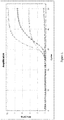
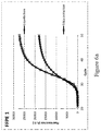
- Figure 1 visualizes amplification results on DNA liberated from an FFPE sample in the emulsified lysate containing the detergent Oleth®-8 (grey) and on DNA liberated from the FFPE sample in the commercial liquefaction composition (black).
- Cross-marked curves represent amplification on almost undiluted emulsified lysate samples (20 ⁇ L /5 ⁇ L, template/PCR mix), circle-marked curves represent amplification on 4-fold diluted emulsified lysate samples.
- the commercial liquefaction composition completely inhibits PCR, and therefore further 4-fold dilution was required to allow qPCR analysis.
- the present liquefaction composition containing the detergent Oleth®-8 is compatible with direct downstream PCR analysis, allowing more template DNA (higher copy number) to be used in the PCR and thereby improving the sensitivity of the PCR analysis.
- Example 4 Functionality of liquefaction compositions in a microfluidic system
- Liquefaction of 3 different FFPE samples was performed using 4 different buffers (with/without detergent and with/without proteinase K), using single consecutive 10 ⁇ m FFPE slices for each condition. Liquefaction and PCR processing of the sample was performed in a microfluidic system as described in EP1896180 , EP1904234 and EP2419705 . Samples were liquefied in 1 ml of the liquefaction composition and heated using the aforementioned conditions. The resulting liquefied mixture was used undiluted as input material for qPCR using the aforementioned amplification conditions. Amplifiable DNA was assessed via qPCR for the wildtype BRAF gene on the liquefied mixture tested.
- Table 2 summarizes the ⁇ Cq values obtained for the compositions containing the detergent relative to the reference compositions omitting the detergent.
- adding a detergent like Oleth®-8 to the liquefaction composition lowers the Cq value with an average of 3.8, indicating improved liberation of DNA compared to the reference composition without detergent.
- Adding proteinase K further improves the Cq value with an average of 1.5.
- ⁇ Cq (Avg ⁇ stdev) Composition reference Tris 3.8 ⁇ 1.4 Tris+ Oleth®-8 reference Tris +protK 5.3 ⁇ 4.1 Tris+protK+ Oleth®-8
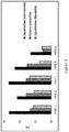
- Figure 3 indicates the effect of the DNA liberation procedure on qPCR performance of 5 different FFPE samples, containing variable amounts of the well-known PCR inhibitor, melanin.
- Samples 1-4 contain high amounts melanin (for instance >95 % for sample 4), while sample 5 contains no melanin.
- the present liquefaction composition results in superior PCR amplification of DNA compared to the commercial liquefaction composition, regardless of the presence of melanin.
- the present liquefaction composition generally results in better compatibility with downstream processes compared to column-based DNA extraction for high melanin containing samples. This is for instance evident from the Cq values depicted in Figure 3 for FFPE 4 following the different methods. A similar performance was observed when no melanin is present. Therefore other realtime PCR inhibitors that are not immediately apparent or known may be present in samples, for which the claimed compositions offer improved performance.
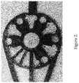
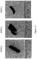
- Figure 4 is a visual representation of 3 high melanin-containing samples used in the experiment.
- Example 6 Emusifying capacity of the liquefaction buffer with lysis on the bench and lysis in the lysis chamber of the Idylla cartridge.
- one 10 ⁇ m slice, cut consecutively from the same FFPE block was submerged in 1 ml of liquefaction buffer.
- the slice was warmed to 60°C for 15 min in a 1.5 ml tube (Eppendorf) using a heat block (Eppendorf) while shaking (800rpm).
- the temperature was raised stepwise (room temperature, 45°C, 50°C, 54°C and 58°C) in 5 about minutes by a combination of peltier (heating) and piezo (high intensity focussed ultrasound or HIFU) functionalization.
- the temperature was raised to 60°C and maintained for 10 minutes under a HIFU power that never exceeded 2.25 W.
- HIFU treatment reproducibly results in more opaque liquefact and less paraffin deposit (arrow) on the walls of the tube after vortexing.
- Example 7 qRT-PCR curves obtained from liquefied material and silica-extracted RNA.
- RNA template material in the liquefaction condition was obtained by processing an FFPE section according to the liquefaction method described above. Briefly, the FFPE section was contacted with the liquefaction composition and heated to 60°C for 15 min, followed by 95°C for 10min in a 1.5 ml tube (Eppendorf) using a heat block (Eppendorf) while shaking (800rpm).
- RNA template material in the silica extraction condition was obtained by processing an FFPE section using the Qiagen QIAamp RNA FFPE Tissue Kit according to the manufacturer's instructions. Subsequently, 5 ⁇ L template obtained by each method was analyzed in a 25 ⁇ L qRT-PCR reaction using a RNA-specific assay for the housekeeping gene B2M.
- Figure 7 demonstrates that a similar threshold cycle (C t ) is obtained by using either the liquefaction or silica extraction method for liberating RNA from the FFPE sections. Accordingly, the RNA released in the emulsified lysate was suitable for direct microfluidic analysis
Applications Claiming Priority (3)
| Application Number | Priority Date | Filing Date | Title |
|---|---|---|---|
| EP13156609 | 2013-02-25 | ||
| PCT/EP2014/053154 WO2014128129A1 (fr) | 2013-02-25 | 2014-02-18 | Isolement d'acides nucléiques |
| EP14705753.3A EP2958997B1 (fr) | 2013-02-25 | 2014-02-18 | Isolation d'acides nucléiques |
Related Parent Applications (2)
| Application Number | Title | Priority Date | Filing Date |
|---|---|---|---|
| EP14705753.3A Division EP2958997B1 (fr) | 2013-02-25 | 2014-02-18 | Isolation d'acides nucléiques |
| EP14705753.3A Division-Into EP2958997B1 (fr) | 2013-02-25 | 2014-02-18 | Isolation d'acides nucléiques |
Publications (1)
| Publication Number | Publication Date |
|---|---|
| EP3674406A1 true EP3674406A1 (fr) | 2020-07-01 |
Family
ID=47749705
Family Applications (2)
| Application Number | Title | Priority Date | Filing Date |
|---|---|---|---|
| EP14705753.3A Active EP2958997B1 (fr) | 2013-02-25 | 2014-02-18 | Isolation d'acides nucléiques |
| EP19179423.9A Withdrawn EP3674406A1 (fr) | 2013-02-25 | 2014-02-18 | Isolation d'acides nucléiques |
Family Applications Before (1)
| Application Number | Title | Priority Date | Filing Date |
|---|---|---|---|
| EP14705753.3A Active EP2958997B1 (fr) | 2013-02-25 | 2014-02-18 | Isolation d'acides nucléiques |
Country Status (7)
| Country | Link |
|---|---|
| US (1) | US10196673B2 (fr) |
| EP (2) | EP2958997B1 (fr) |
| JP (1) | JP6567427B2 (fr) |
| CN (1) | CN105164259B (fr) |
| CA (1) | CA2901641C (fr) |
| ES (1) | ES2741956T3 (fr) |
| WO (1) | WO2014128129A1 (fr) |
Families Citing this family (14)
| Publication number | Priority date | Publication date | Assignee | Title |
|---|---|---|---|---|
| GB201106254D0 (en) | 2011-04-13 | 2011-05-25 | Frisen Jonas | Method and product |
| WO2014210223A1 (fr) | 2013-06-25 | 2014-12-31 | Prognosys Biosciences, Inc. | Essais biologiques à codage spatial faisant appel à un dispositif microfluidique |
| CN107532207B (zh) | 2015-04-10 | 2021-05-07 | 空间转录公司 | 生物样本的空间区别、多重核酸分析 |
| EP3782731A1 (fr) * | 2015-06-10 | 2021-02-24 | Biocartis N.V. | Détection améliorée d'adn méthylé |
| CN105861659A (zh) * | 2016-04-13 | 2016-08-17 | 武汉康录生物技术有限公司 | 一种用于荧光原位杂交ffpe组织切片的预处理液及预处理方法 |
| EP3586129B1 (fr) | 2017-03-28 | 2023-07-12 | Phase Scientific International, Ltd. | Procédé de diagnostic précis d'une maladie ciblant des biomarqueurs dans une biopsie liquide |
| CN110891664B (zh) | 2017-06-01 | 2022-05-17 | 相达生物科技美国有限公司 | 用于多孔材料中双水相分离的相分离行为改性剂 |
| WO2019046563A1 (fr) | 2017-09-01 | 2019-03-07 | Phase Diagnostics, Inc. | Procédé et dispositif d'utilisation de systèmes aqueux biphases (atps) pour améliorer le diagnostic des infections sexuellement transmissibles |
| WO2019046553A1 (fr) * | 2017-09-01 | 2019-03-07 | Phase Diagnostics, Inc. | Procédé et dispositif d'utilisation de systèmes aqueux à deux phases (atps) permettant l'amélioration du diagnostic de maladies dentaires et buccales |
| EP3740588A4 (fr) | 2018-01-19 | 2021-10-20 | Phase Scientific International, Ltd. | Procédé d'isolement et de purification d'acides nucléiques à l'aide d'un système à phases solide-liquide |
| AU2019210981A1 (en) | 2018-01-23 | 2020-09-03 | Biocartis Nv | Methods for the analysis of dissociation melt curve data |
| CN115135984A (zh) * | 2019-12-23 | 2022-09-30 | 10X基因组学有限公司 | 可逆固定试剂及其使用方法 |
| WO2021236929A1 (fr) | 2020-05-22 | 2021-11-25 | 10X Genomics, Inc. | Mesure spatio-temporelle simultanée de l'expression génique et de l'activité cellulaire |
| EP4237584A1 (fr) | 2020-10-29 | 2023-09-06 | Biocartis NV | Cartouche générique et procédé de détection multiplex d'acide nucléique |
Citations (7)
| Publication number | Priority date | Publication date | Assignee | Title |
|---|---|---|---|---|
| US6469159B1 (en) * | 1999-04-26 | 2002-10-22 | Robert T. Belly | Methods for extracting nucleic acids from tissue samples and paraffin-embedded tissues |
| EP1896180A2 (fr) | 2005-06-23 | 2008-03-12 | Koninklijke Philips Electronics N.V. | Cartouche, systeme et procede pour diagnostic medical automatique |
| EP1904234A1 (fr) | 2005-06-30 | 2008-04-02 | Koninklijke Philips Electronics N.V. | Cartouche pour diagnostic medical automatique |
| US20110076751A1 (en) * | 2008-05-30 | 2011-03-31 | Qiagen Gmbh | Lysis, binding and/or wash reagent for isolating and/or purifying nucleic acids |
| EP2419705A1 (fr) | 2009-04-14 | 2012-02-22 | Biocartis SA | Traitement d'un échantillon par une énergie acoustique focalisée |
| WO2012075133A1 (fr) | 2010-11-30 | 2012-06-07 | Life Technologies Corporation | Alkylèneglycols et polymères et copolymères de ceux-ci pour l'isolement direct d'acide nucléique à partir d'échantillons intégrés |
| WO2013020089A2 (fr) * | 2011-08-04 | 2013-02-07 | Sage Science, Inc. | Systèmes et procédés pour le traitement de fluides |
Family Cites Families (11)
| Publication number | Priority date | Publication date | Assignee | Title |
|---|---|---|---|---|
| US5538870A (en) * | 1994-09-21 | 1996-07-23 | Boehringer Mannheim Corporation | Method for preparing nucleic acids for analysis and kits useful therefor |
| JP4503712B2 (ja) * | 1996-09-09 | 2010-07-14 | 株式会社島津製作所 | 核酸合成法 |
| US20030134292A1 (en) * | 2001-10-30 | 2003-07-17 | Farchaus Joseph W. | Thermostable DNA polymerases and methods of making same |
| CN1922331A (zh) * | 2003-10-16 | 2007-02-28 | 第三次浪潮技术公司 | 体液的直接核酸检测 |
| CN1922317A (zh) * | 2003-12-24 | 2007-02-28 | 3M创新有限公司 | 使用微流体装置和浓缩步骤的核酸分离用方法和试剂盒 |
| EP2129780A2 (fr) * | 2007-03-14 | 2009-12-09 | Sierra Molecular Corporation | Compositions, systèmes et procédés de conservation et/ou de stabilisation d'une cellule et/ou d'une macromolécule |
| EP2454378B1 (fr) | 2009-07-17 | 2015-12-23 | Canon U.S. Life Sciences, Inc. | Méthodes et systèmes d'isolement d'adn sur un dispositif microfluidique |
| KR101851117B1 (ko) * | 2010-01-29 | 2018-04-23 | 마이크로닉스 인코포레이티드. | 샘플-투-앤서 마이크로유체 카트리지 |
| GB2483858A (en) * | 2010-09-21 | 2012-03-28 | Univ Hull | Amplifying nucleic acids using microfluidic device to perform PRC |
| CN102146112B (zh) * | 2011-01-25 | 2013-07-10 | 天根生化科技(北京)有限公司 | 从福尔马林固定石蜡包埋组织中提取脱氧核糖核酸的方法 |
| US9518901B2 (en) * | 2011-06-29 | 2016-12-13 | Kabushiki Kaisha Dnaform | Pretreatment method of biological sample, detection method of RNA, and pretreatment kit |
-
2014
- 2014-02-18 CA CA2901641A patent/CA2901641C/fr active Active
- 2014-02-18 EP EP14705753.3A patent/EP2958997B1/fr active Active
- 2014-02-18 JP JP2015558426A patent/JP6567427B2/ja active Active
- 2014-02-18 US US14/768,871 patent/US10196673B2/en active Active
- 2014-02-18 EP EP19179423.9A patent/EP3674406A1/fr not_active Withdrawn
- 2014-02-18 ES ES14705753T patent/ES2741956T3/es active Active
- 2014-02-18 WO PCT/EP2014/053154 patent/WO2014128129A1/fr active Application Filing
- 2014-02-18 CN CN201480023809.4A patent/CN105164259B/zh active Active
Patent Citations (7)
| Publication number | Priority date | Publication date | Assignee | Title |
|---|---|---|---|---|
| US6469159B1 (en) * | 1999-04-26 | 2002-10-22 | Robert T. Belly | Methods for extracting nucleic acids from tissue samples and paraffin-embedded tissues |
| EP1896180A2 (fr) | 2005-06-23 | 2008-03-12 | Koninklijke Philips Electronics N.V. | Cartouche, systeme et procede pour diagnostic medical automatique |
| EP1904234A1 (fr) | 2005-06-30 | 2008-04-02 | Koninklijke Philips Electronics N.V. | Cartouche pour diagnostic medical automatique |
| US20110076751A1 (en) * | 2008-05-30 | 2011-03-31 | Qiagen Gmbh | Lysis, binding and/or wash reagent for isolating and/or purifying nucleic acids |
| EP2419705A1 (fr) | 2009-04-14 | 2012-02-22 | Biocartis SA | Traitement d'un échantillon par une énergie acoustique focalisée |
| WO2012075133A1 (fr) | 2010-11-30 | 2012-06-07 | Life Technologies Corporation | Alkylèneglycols et polymères et copolymères de ceux-ci pour l'isolement direct d'acide nucléique à partir d'échantillons intégrés |
| WO2013020089A2 (fr) * | 2011-08-04 | 2013-02-07 | Sage Science, Inc. | Systèmes et procédés pour le traitement de fluides |
Non-Patent Citations (7)
| Title |
|---|
| ALAN ANDERSEN F: "Final Report on the Safety Assessment of Oleth-2, -3, -4, -5, -6, -7, -8, -9, -10, -11, -12, -15, -16, -20, -23, -25, -30, -40, -44, and -501", INTERNATIONAL JOURNAL OF TOXICOLOGY, TAYLOR AND FRANCIS, WASHINGTON, DC, US, vol. 18, no. supplement 2, 1 September 1999 (1999-09-01), pages 17 - 24, XP008134738, ISSN: 1091-5818, DOI: 10.1177/109158189901800205 * |
| CHAW Y.F.M. ET AL., BIOCHEMISTRY, vol. 19, 1980, pages 5525 - 5531 |
| GILBERT M.T.P. ET AL., PLOS ONE, vol. 2, no. 6, 2007, pages e537 |
| GOEZL ET AL., BIOCHEMICAL AND BIOPHYSICAL RESEARCH COMMUNICATION, vol. 130, no. 1, 1985, pages 118 - 126 |
| HAMFJORD ET AL., DIAGN MOL PATHOL, vol. 20, 2011, pages 158 - 165 |
| METZ B. ET AL., J. BIOL. CHEM, vol. 279, 2004, pages 6235 - 6243 |
| SLEBOS R J C ET AL: "A RAPID AND SIMPLE PROCEDURE FOR THE ROUTINE DETECTION OF RAS POINT MUTATIONS IN FORMALIN-FIXED, PARAFFIN-EMBEDDED TISSUES", DIAGNOSTIC MOLECULAR PATHOLOGY, NEW YORK, NY, US, vol. 1, no. 2, 1 June 1992 (1992-06-01), pages 136 - 141, XP009011205 * |
Also Published As
| Publication number | Publication date |
|---|---|
| JP2016507242A (ja) | 2016-03-10 |
| ES2741956T3 (es) | 2020-02-12 |
| WO2014128129A1 (fr) | 2014-08-28 |
| EP2958997B1 (fr) | 2019-07-17 |
| US20160002706A1 (en) | 2016-01-07 |
| JP6567427B2 (ja) | 2019-08-28 |
| CN105164259B (zh) | 2018-02-27 |
| US10196673B2 (en) | 2019-02-05 |
| CA2901641A1 (fr) | 2014-08-28 |
| CA2901641C (fr) | 2021-02-09 |
| CN105164259A (zh) | 2015-12-16 |
| EP2958997A1 (fr) | 2015-12-30 |
Similar Documents
| Publication | Publication Date | Title |
|---|---|---|
| US10196673B2 (en) | Isolation of nucleic acids | |
| EP2580348B1 (fr) | Procédé de détermination de cellules ou de tissu cibles pour l'extraction de biomolécules à partir d'échantillons biologiques non fixés au formol | |
| CN103827303B (zh) | 细胞外核酸的稳定化和分离 | |
| Erickson et al. | Quantitative RT-PCR gene expression analysis of laser microdissected tissue samples | |
| JP6204664B2 (ja) | Rnaおよびdnaの並行単離および/または並行精製のためのプロセス | |
| JP2011115173A (ja) | 細胞および組織からのrna迅速抽出 | |
| CN102146112B (zh) | 从福尔马林固定石蜡包埋组织中提取脱氧核糖核酸的方法 | |
| US20220315987A1 (en) | Fully automated nucleic acid extraction methods for tissue samples | |
| Khokhar et al. | Evaluation of Maxwell® 16 for automated DNA extraction from whole blood and formalin-fixed paraffin embedded (FFPE) tissue | |
| CN100429307C (zh) | 核酸检测方法及其系统 | |
| TW201446789A (zh) | 福馬林固定石蠟包埋樣本核酸分離方法 | |
| Frégeau et al. | Automated processing of forensic casework samples using robotic workstations equipped with nondisposable tips: contamination prevention | |
| Tüzmen et al. | Techniques for nucleic acid engineering: The foundation of gene manipulation | |
| Duval et al. | Optimized manual and automated recovery of amplifiable DNA from tissues preserved in buffered formalin and alcohol-based fixative | |
| Alqaydi et al. | Quantitative and qualitative study of STR DNA from ethanol and formalin fixed tissues | |
| Micke et al. | A fluid cover medium provides superior morphology and preserves RNA integrity in tissue sections for laser microdissection and pressure catapulting | |
| Jakubowska et al. | mRNA profiling for vaginal fluid and menstrual blood identification | |
| KR20110060156A (ko) | 핵산 추출용 조성물과 이를 이용한 핵산 추출방법 및 핵산의 증폭방법 | |
| Nechifor-Boila et al. | Evaluation of a DNA extraction and purification protocol using archived formalin-fixed paraffin-embedded tissues for BRAF mutations analysis in papillary thyroid Microcarcinomas | |
| CN111117998A (zh) | 微生物核酸萃取方法 | |
| Hewitson et al. | Laser capture microdissection of archival kidney tissue for qRT-PCR | |
| Malusecka et al. | Combining Laser-Assisted Microdisstection with/and Immunohistochemistry-RNA Quality of Clinical LCM-Derived Samples |
Legal Events
| Date | Code | Title | Description |
|---|---|---|---|
| PUAI | Public reference made under article 153(3) epc to a published international application that has entered the european phase |
Free format text: ORIGINAL CODE: 0009012 |
|
| STAA | Information on the status of an ep patent application or granted ep patent |
Free format text: STATUS: THE APPLICATION HAS BEEN PUBLISHED |
|
| AC | Divisional application: reference to earlier application |
Ref document number: 2958997 Country of ref document: EP Kind code of ref document: P |
|
| AK | Designated contracting states |
Kind code of ref document: A1 Designated state(s): AL AT BE BG CH CY CZ DE DK EE ES FI FR GB GR HR HU IE IS IT LI LT LU LV MC MK MT NL NO PL PT RO RS SE SI SK SM TR |
|
| STAA | Information on the status of an ep patent application or granted ep patent |
Free format text: STATUS: THE APPLICATION IS DEEMED TO BE WITHDRAWN |
|
| 18D | Application deemed to be withdrawn |
Effective date: 20210112 |