EP3673086B1 - Elektrochemischer nachweis von bakteriellen und / oder pilzinfektionen - Google Patents
Elektrochemischer nachweis von bakteriellen und / oder pilzinfektionen Download PDFInfo
- Publication number
- EP3673086B1 EP3673086B1 EP18765310.0A EP18765310A EP3673086B1 EP 3673086 B1 EP3673086 B1 EP 3673086B1 EP 18765310 A EP18765310 A EP 18765310A EP 3673086 B1 EP3673086 B1 EP 3673086B1
- Authority
- EP
- European Patent Office
- Prior art keywords
- cfu
- observed
- atcc
- gram
- sample
- Prior art date
- Legal status (The legal status is an assumption and is not a legal conclusion. Google has not performed a legal analysis and makes no representation as to the accuracy of the status listed.)
- Active
Links
Images


Classifications
-
- C—CHEMISTRY; METALLURGY
- C12—BIOCHEMISTRY; BEER; SPIRITS; WINE; VINEGAR; MICROBIOLOGY; ENZYMOLOGY; MUTATION OR GENETIC ENGINEERING
- C12Q—MEASURING OR TESTING PROCESSES INVOLVING ENZYMES, NUCLEIC ACIDS OR MICROORGANISMS; COMPOSITIONS OR TEST PAPERS THEREFOR; PROCESSES OF PREPARING SUCH COMPOSITIONS; CONDITION-RESPONSIVE CONTROL IN MICROBIOLOGICAL OR ENZYMOLOGICAL PROCESSES
- C12Q1/00—Measuring or testing processes involving enzymes, nucleic acids or microorganisms; Compositions therefor; Processes of preparing such compositions
- C12Q1/68—Measuring or testing processes involving enzymes, nucleic acids or microorganisms; Compositions therefor; Processes of preparing such compositions involving nucleic acids
- C12Q1/6876—Nucleic acid products used in the analysis of nucleic acids, e.g. primers or probes
- C12Q1/6888—Nucleic acid products used in the analysis of nucleic acids, e.g. primers or probes for detection or identification of organisms
- C12Q1/689—Nucleic acid products used in the analysis of nucleic acids, e.g. primers or probes for detection or identification of organisms for bacteria
-
- C—CHEMISTRY; METALLURGY
- C12—BIOCHEMISTRY; BEER; SPIRITS; WINE; VINEGAR; MICROBIOLOGY; ENZYMOLOGY; MUTATION OR GENETIC ENGINEERING
- C12Q—MEASURING OR TESTING PROCESSES INVOLVING ENZYMES, NUCLEIC ACIDS OR MICROORGANISMS; COMPOSITIONS OR TEST PAPERS THEREFOR; PROCESSES OF PREPARING SUCH COMPOSITIONS; CONDITION-RESPONSIVE CONTROL IN MICROBIOLOGICAL OR ENZYMOLOGICAL PROCESSES
- C12Q1/00—Measuring or testing processes involving enzymes, nucleic acids or microorganisms; Compositions therefor; Processes of preparing such compositions
- C12Q1/68—Measuring or testing processes involving enzymes, nucleic acids or microorganisms; Compositions therefor; Processes of preparing such compositions involving nucleic acids
- C12Q1/6844—Nucleic acid amplification reactions
- C12Q1/6848—Nucleic acid amplification reactions characterised by the means for preventing contamination or increasing the specificity or sensitivity of an amplification reaction
-
- C—CHEMISTRY; METALLURGY
- C12—BIOCHEMISTRY; BEER; SPIRITS; WINE; VINEGAR; MICROBIOLOGY; ENZYMOLOGY; MUTATION OR GENETIC ENGINEERING
- C12Q—MEASURING OR TESTING PROCESSES INVOLVING ENZYMES, NUCLEIC ACIDS OR MICROORGANISMS; COMPOSITIONS OR TEST PAPERS THEREFOR; PROCESSES OF PREPARING SUCH COMPOSITIONS; CONDITION-RESPONSIVE CONTROL IN MICROBIOLOGICAL OR ENZYMOLOGICAL PROCESSES
- C12Q1/00—Measuring or testing processes involving enzymes, nucleic acids or microorganisms; Compositions therefor; Processes of preparing such compositions
- C12Q1/68—Measuring or testing processes involving enzymes, nucleic acids or microorganisms; Compositions therefor; Processes of preparing such compositions involving nucleic acids
- C12Q1/6876—Nucleic acid products used in the analysis of nucleic acids, e.g. primers or probes
- C12Q1/6888—Nucleic acid products used in the analysis of nucleic acids, e.g. primers or probes for detection or identification of organisms
- C12Q1/6895—Nucleic acid products used in the analysis of nucleic acids, e.g. primers or probes for detection or identification of organisms for plants, fungi or algae
Definitions
- the invention relates to the field of molecular diagnostic methods, in particular, microfluidic devices for the detection of target analytes.
- Multiplex RT-PCR approaches have been described in which a number of commonly occurring human pathogens are potentially detected.
- One example of such a multiplex PCR method is described in Gosiewski et al (BMC Microbiology 2014, 14:144 ) and U.S. Publication No. 2015/0232916 , which discloses a nested PCR approach for detecting gram-positive and gram-negative bacteria, yeast, fungi and filamentous fungi from blood samples.
- a nested polymerase chain reaction involves two sets of primers, used in two successive runs of polymerase chain reaction, the second set intended to amplify a secondary target within the first run product.
- Nested PCR is applied in order to reduce nonspecific binding in products due to the amplification of unexpected primer binding sites, as it is unlikely that any of the unwanted PCR products contain binding sites for both the new primers in the second PCR run, ensuring the product from the second PCR has little contamination from unwanted products of primer dimers, hairpins, and alternative primer target sequences.
- the PCR method described in Gosiewski and U.S. Publication No. 2015/0232916 are relatively complex and require two cycling reactions, essentially doubling the time, effort and reagents required for the analysis.
- Electrochemical detection techniques have higher detection sensitivity than conventional luminescence techniques (e.g., fluorescence and phosphorescence) due to higher signal-to-noise ratios. Because of their sensitivity and ability to accurately measures low-concentrations of nucleic acids, electrochemical detection techniques are able to differentiate between pathogenic species representing a significant technological improvement over the prior art. But, because of their sensitivity, false positive detection rates are high. Indeed, where organisms are cultured, the growth media often contains non-viable organisms or DNA/nucleic acids, which would not affect culture, but could produce false positives in PCR. If a system is designed uniformly for increased sensitivity to detect low titers pathogens, frequent false positive results may occur from background organisms or DNA/nucleic acids. Alternatively, if system sensitivity is reduced to avoid background organism detection, low titer organisms may be missed, resulting in false negative detection.
- conventional luminescence techniques e.g., fluorescence and phosphorescence
- blood or other bodily fluids when blood or other bodily fluids are obtained from a subject they may be contaminated by skin cells, bacteria, fungi, viruses, phages, their respective nucleic acids (including RNA and DNA)and/or other undesirable molecules, or disinfectants.
- Antiseptics are crucial for the practice of medicine; however, currently used antiseptics have a significant failure rate which results in substantial additional medical costs. Antiseptics are commonly used prior to routine phlebotomy, in preparation for minor and major invasive procedures, and as part of routine infection control hand-washing practices. The failure of antiseptics often result in erroneous diagnostic tests.
- Pardo et al. (Diagnostic Microbiology and Infectious Disease, 84, 159-164, 2016 ), discloses the identification of Gram-positive bacteria using a multiplexed PCR-based diagnostic test; however, a method for testing for Gram-positive bacteria in combination with Gram-negative bacteria or fungi is not disclosed.
- the ability to detect infection is hampered by background contamination present in blood culture bottles, such as are used in gram staining, a common first step in any clinical pathogen diagnosis.
- the invention as defined in the appended claims, can not only differentiate between background contamination (from any blood culture matrix bottle) and clinically relevant infection but can also differentiate between gram-positive bacterial infection, gram-negative bacterial infection, fungal infection and can identify antibiotic resistance genes. Even more importantly, the invention can identify the contaminating pathogen and contaminating co-infection (if present) by its species. Because prior art methods failed to recognize background contamination as an issue or cannot discriminate by species the infecting pathogen and co-infecting pathogen, the invention allows for better antimicrobial stewardship and improved patient care outcomes.
- in vitro methods for the detection and/or identification of a human pathogen and/or genetic material thereof comprising subjecting a sample suspected of comprising a human pathogen and/or genetic material thereof to a single multiplex polymerase chain reaction (PCR), wherein said method (or system) comprises amplification of PCR products that enable discrimination between contaminating pathogen and/or genetic material present in the sample and infectious pathogen and/or genetic material present in the sample.
- PCR polymerase chain reaction
- the method allows for a single amplification step and single detection step for the detection of an actual pathogenic infection and not a putative contamination.
- the method does not require a purification step prior to amplification or detection.
- the method does not require determining whether amplification has occurred prior to detection.
- the method does not require dilution of the PCR sample.
- the method does not require additional testing or analysis to differentiate signaling due to contamination versus real pathogenic infections.
- the method can discriminate between gram-positive bacterial, gram-negative bacterial and fungal pathogens if present in said sample as well as identify antimicrobial resistance genes.
- An infection can be identified by its species, and a fungal co-infection can be identified by its genus whereas a bacterial co-infection of a different type than the infection (i.e. infection is GP and co infection is GN or vice versa) can be identified by its category (gram-positive or negative).
- the infection and co-infection are of the same type (i.e., both gram-positive or both gram-negative)
- the systems and methods can identify the species of the co-infection via a single PCR run.
- the infection and co-infection are of different types (i.e., the infection is gram-positive and the co-infection is gram-negative or fungal) the systems and methods can identify the species of the co-infection via a second single PCR run.
- the method can discriminate between background contamination and de-escalation targets.
- Background contamination from blood culture bottles is not detected by the methods (or system or devices) but organisms associated with possible contamination by blood draw such as Propionibacterium acnes, Staphylococcus epidermidis, Micrococcus, Lactobacillus or Corynebacterium (so called de-escalation targets) are identified.
- the method can further discriminate between (1) background contamination, (2) de-escalation targets and (3) clinically relevant gram-positive bacterial, gram-negative bacterial or fungal pathogens if present in the sample as well as identify antimicrobial resistance genes.
- nucleic and amino acid sequences listed in the accompanying sequence listing are shown using standard letter abbreviations for nucleotide bases, and three letter code for amino acids. Only one strand of each nucleic acid sequence is shown, but the complementary strand is understood as included by any reference to the displayed strand.
- Target nucleic acid or “analyte of interest”, or “target molecule” or “human pathogen nucleic acid” include genes, portions of genes, regulatory sequences of genes, mRNAs, rRNAs, tRNAs, siRNAs, cDNA and may be single stranded, double stranded or triple stranded.
- target nucleic acids are DNA from human pathogens, and are naturally occurring nucleic acids, as contrasted to the nucleic acids of capture probes and signal probes, which may include non-naturally occurring components.
- Some nucleic acid targets have polymorphisms, single nucleotide polymorphisms, deletions and alternate splice sequences, such as allelic variants.
- target nucleic acid may have a first target domain that binds the capture probe and a second target domain that binds a signal probe, and/or distinct primer binding sequences.
- Target nucleic acids are not generally provided with the cartridge as manufactured, but are contained in the liquid sample to be assayed; in contrast, "control analytes” or “control nucleic acids” are typically provided with the cartridge or are routinely present in a sample of a particular type and are assayed in order to ensure proper performance of the assay. Spiked samples may be used in certain quality control testing and for calibration, as is well known in the art.
- the target analyte is also referred to as "clinically relevant amplification” or “systemic infection” or “pathogen of interest” and is distinguished from, for example, contamination.
- sample is used in its broadest sense. In one sense, it is meant to include a specimen or culture obtained from any source, as well as biological and environmental samples. Biological samples may be obtained from animals (including humans) and encompass fluids, solids, tissues, and gases. Biological samples include blood products, such as plasma, serum and the like. In contrast with some commercial systems that require some off chip handling of the sample, generally including sample extraction (cell lysis, for example), and sample preparation prior to detection.
- a sample is loaded onto a BCID cartridge and the target analyte is extracted, amplified as necessary (for example, when the target analyte is a nucleic acid using polymerase chain reaction (PCR) techniques, although isothermal amplification methods can be utilized as well), and then detected using electrochemical detection, all on a microfluidic platform, generally referred to herein as a "multiplex cartridge” or a "fluid sample processing cartridge.”
- PCR polymerase chain reaction
- the BCID cartridge utilizes a sample preparation module as further described and shown in Figure 15 of U.S. Patent no. 9,598,722 .
- a sample preparation module as further described and shown in Figure 15 of U.S. Patent no. 9,598,722 .
- the sample is a blood sample that is treated as outlined herein.
- Environmental samples include environmental material such as surface matter, soil, water, crystals and industrial samples. Such examples are not, however, to be construed as limiting the sample types applicable to the present technology.
- nucleic acid or "oligonucleotide” or grammatical equivalents herein means at least two nucleotides covalently linked together.
- a nucleic acid of the present invention will generally contain phosphodiester bonds, although in some cases, for example in the creation of signal probes and sometimes capture probes, nucleic acid analogs are included that may have alternate backbones, comprising, for example, phosphoramides, phosphorothioates, phosphorodithioates, 0methylphophoroamidite linkages and peptide nucleic acid backbones and linkages, as well as those with positive backbones, non-ionic backbones nonribose backbones, including those containing one or more carbocyclic sugars are also included within the definition of nucleic acids. These modifications of the ribosephosphate backbone may be done to facilitate the addition of electron transfer moieties, or to increase the stability and half-life of such molecules in physiological environments.
- detection system refers to a method that enables visualization of PCR-amplified nucleic acid products.
- suitable detection systems include systems that depend on detection of color, radioactivity, fluorescence, chemiluminescence or electrochemical signals, with the latter finding particular use in the present invention.
- contamination or “contaminant” or “background contamination” or “contaminating pathogen and/or genetic material” or “unwanted contamination” as used herein refers to nucleic acids in the sample which are not a part of the nucleic acid population that is being targeted for amplification.
- nucleic acids found in the blood culture matrix for example, nucleic acids found in the blood culture matrix.
- the contaminant is from bacteria or fungus whose cell wall has been broken.
- the contaminant is from dead bacteria or fungi (as compared to live bacteria or fungi).
- the contaminant is from non-intact bacteria or fungi (as compared to intact bacteria or fungi).
- the contaminant is cell free bacterial or fungal DNA.
- the contaminant is at a lower concentration than the clinically relevant bacterial or fungal infection. In some embodiments, the contaminant is at a concentration of about 1000 to 100,000 copies per ml. In some embodiments, the contaminant is at a concentration of about 1000 to 100,000 copies per ml while the clinically relevant pathogen is at a concentration of 100,000 to 100,000,000 copies per ml. In some embodiments, the contaminant is at a concentration of about 10 to 1000-fold less than the concentration of the clinically relevant pathogen. In some embodiments, the contaminant is from the same bacteria as the clinically relevant bacteria. In some embodiments, the contaminant is from the same fungus as the clinically relevant fungus.
- the contaminant is from a different bacterium than the clinically relevant bacteria. In some embodiments, the contaminant is from a different fungus than the clinically relevant fungus. In some embodiments, the contaminant is from a fungus and the clinically relevant pathogen is bacteria. In some embodiments, the contaminant is from bacteria and the clinically relevant pathogen is a fungus.
- de-escalation targets means Propionibacterium acnes, Staphylococcus epidermidis, Micrococcus, Lactobacillus or Corynebacterium.
- An object is to distinguish between unwanted contamination (from blood culture bottles) and de-escalation targets (which may be present as a contamination from blood draw) but which may be a clinically relevant infection.
- An object is to distinguish between unwanted contamination, de-escalation targets, clinically relevant pathogens and determinants of antimicrobial resistance.
- infection means the invasion of a host organism's body by another organism or entity (pathogen), for example, a fungi or bacteria.
- pathogen for example, a fungi or bacteria.
- co-infection means "double infection,” “multiple infection,” or “serial infection” and is used to denote simultaneous infection with two or more infections/pathogens.
- determinants of antimicrobial resistance relates to a gene responsible for the development of resistance in the bacteria which actively counteracts the effect of an antibiotic. Particularly, genetic determinants of resistance to methicillin (mecA and mecC) and vancomycin (vanA and vanB) are envisaged. Genes associated with genetic determinants of resistance such as CTX-M, NDM, IMP, OXA, KPC, VIM are envisaged.
- the target sequence is generally amplified, and during amplification, a label is added.
- the compositions may additionally contain one or more labels at any position.
- label herein is meant an element (e.g. an isotope) or chemical compound that is attached to enable the detection of the compound.
- Preferred labels are radioactive isotopic labels, and colored or fluorescent dyes.
- the labels may be incorporated into the compound at any position.
- the compositions may also contain other moieties such as cross-linking agents to facilitate cross-linking of the target-probe complex. See for example, Lukhtanov et al., Nucl. Acids. Res. 24(4):683 (1996 ) and Tabone et al., Biochem. 33:375 (1994 ).
- the electrochemical detection system used herein uses a separate signal probe or label probe having an electron transfer moiety (ETM). That is, one portion of the label probe directly or indirectly binds to the target analyte, and one portion comprises a recruitment linker comprising covalently attached ETMs. In some systems, these may be the same.
- the ETM is responsive to an input waveform.
- the ETM is a metallocene.
- the metallocene is a ferrocene.
- the ferrocene is a ferrocene derivative.
- Preferred ferrocene derivatives can be N6 (FIG. 1D as shown in US2014/0323326 ), QW56 (FIG. 3A as shown in US2014/0323326 ), and QW80 (FIG. 3B as shown in US2014/0323326 ).
- electrochemical system or “electrochemical detection system” or “automated nucleic acid testing system” refers to a system that determines the presence and/or quantity of a redox analyte through measurements of electrical signal in a solution between a working electrode and a counter electrode, such as induced by a redox reaction or electrical potential from the release or absorption of ions.
- the redox reaction refers to the loss of electrons (oxidation) or gain of electrons (reduction) that a material undergoes during electrical stimulation such as applying a potential. Redox reactions take place at the working electrode, and which, for chemical detection, is typically constructed from an inert material such as platinum or carbon.
- the potential of the working electrode is measured against a reference electrode, which is typically a stable, well-behaved electrochemical half-cell such as silver/silver chloride.
- the electrochemical system can be used to support many different techniques for determining the presence and concentration of the target biomolecules including, but not limited to, various types of voltammetry, amperometry, potentiometry, coulometry, conductometry, and conductimetry such as AC voltammetry, differential pulse voltammetry, square wave voltammetry, electrochemical impedance spectroscopy, anodic stripping voltammetry, cyclic voltammetry, and fast scan cyclic voltammetry.
- the electrochemical system may further include one or more negative control electrode and a positive control electrode.
- a single electrochemical system may be used to detect and quantify more than one type of target analyte.
- the use of electrochemical systems is described in more detail in U.S. Patent Nos. 9,557,295, 8,501,921, 6,600,026, 6,740,518 and US2016/0129437 .
- pathogen refers to an organism (bacteria or fungi) that may affect the health status of the host, if that host is infected by that organism.
- a large number of human pathogens are outlined in the Tables, Examples and Lists herein. Included within the definition of human pathogen is the genetic material, usually DNA, that is contained within the pathogenic organism.
- amplicons that result from amplification reactions such as the PCR reactions described herein.
- analyzing the presence of a pathogen is used to describe a method to determine the presence or absence of a pathogen.
- the systems and methods disclosed herein do not require additional analysis to discriminate between background signaling due to contamination effects and real pathogenic infections and thus enable a decision on whether to apply a selective antibiotic therapy.
- thresholding or “threshold signal” or the like refers to a set signal level below which the reported call is “not detected,” above which the reported call is “detected.”
- PCR means "polymerase chain reaction.” PCR is a technique used in molecular biology to amplify a single copy or a few copies of a segment of DNA across several orders of magnitude, generating thousands to millions of copies of a particular DNA sequence. PCR reagents generally include pairs of primers, dNTPs and a DNA polyermase.
- single reaction or “single run” or “single multiplex PCR” or “single PCR” or “single nucleic acid amplification reaction” or “single amplification” or the like in this context refers to a standard PCR operating program.
- a single PCR run encompasses non-uniform PCR cycling (also referred to as heterogeneous PCR cycling, non-harmonized, uneven, unsymmetrical, mismatched PCR cycling and the like ) in a single cartridge, i.e., some samples being cycled 30 times while others are cycled 35 times but not two sequential PCR runs such as with nested PCR.
- heterogeneous single run PCR cycling were not utilized, there would be either a risk of false positives for the organisms that tend to have high contamination concentrations (such as Bacillus) or a risk of false negatives for organisms that tend to have slower growth in culture and therefore fewer copies of target sequence in the sample (such as E. Coli), or both if a compromise cycle were chosen.
- Using heterogeneous single run PCR cycles for different organisms improves the overall accuracy of the assay.
- the standard PCR operating program comprises a series of repeated temperature changes, called cycles, with each cycle consisting of 2 discrete temperature steps, referred to as denaturation and annealing/extension steps. The cycling is preceded by a single temperature step (hot start) at a high temperature (>90 °C) for enzyme activation.
- Nucleotide means a building block of DNA or RNA, consisting of one nitrogenous base, one phosphate molecule, and one sugar molecule (deoxyribose in DNA, ribose in RNA).
- Oligonucleotide means a short string of nucleotides. Oligonucleotides are often used as probes to find a matching sequence of DNA or RNA and can be labeled with a variety of labels, such as radioisotopes and fluorescent and chemiluminescent moieties and ferrocene labels.
- Primer means a short strand of oligonucleotides complementary to a specific target sequence of DNA, which is used to prime DNA synthesis.
- Some primer pools contain species-specific primers. Such species-specific primer pairs hybridize in the assay to a target nucleic acid sequence of only one of said target species (gram-positive bacterial, gram-negative bacterial or fungal).
- Some primer pools contain genus-specific primers. Each double stranded amplicon contains a blocking moiety (phosphorylation on one strand) so that exonuclease activity is blocked, thereby inhibiting digestion of the blocked strand and promoting digestion of the unblocked strand.
- Exonucleases are enzymes that work by cleaving nucleotides one at a time from the end (exo) of a polynucleotide chain. A hydrolyzing reaction that breaks phosphodiester bonds at either the 3' or the 5' end occurs.
- Uniplex means a PCR-based assay utilizing a single set of primers in each reaction that amplifies a single pathogen specific nucleic acid sequence
- Multiplex means a PCR-based assay utilizing multiple primer sets in a single reaction, where each primer can amplify a single pathogen specific nucleic acid sequence.
- End point PCR means one multiplexed PCR method for amplification and end point detection (i.e. after the log phase).
- Real-time PCR or “Q-PCT” refers to a homogenous PCR assay that permits continuous fluorescent monitoring of the kinetic progress of the amplification reaction.
- Methods of conducting real-time PCR are well known in the art and a number of systems are available commercially (see e.g. Higucho et al., "Kinetic PCR Analysis: Real-time Monitoring of DNA Amplification Reactions," Bio/Technology 11:1026-1030 (1993 ))
- capture probe refers to the nucleic acid sequence, specific to the individual pathogen that is immobilized on an inert matrix.
- the specificity of the capture probe should not be substantially affected by the presence of other capture probes, i.e., it still hybridizes to the target pathogens nucleic acid.
- a capture probe selected for one pathogen does not hybridize to a nucleic acid from another pathogen.
- Capture probes generally hybridize to a first target domain of an amplicon of a human pathogen as outlined herein.
- signal probe refers to the nucleic acid sequence, specific to the individual pathogen that is not immobilized on an inert matrix. Signal probes generally hybridize to a second target domain of an amplicon of a human pathogen as outlined herein, and they are generally labeled. Signaling probes in some embodiments are labeled with different labels that enable simultaneous use and differentiation between each of the labels. However, in the BCID-GP and GN panels disclosed the signaling probes are not labelled with different labels such that when the signaling probe binds, "pan-candida detection” is reported not the specific candida species detected (Candida albicans, Candida glabrata, Candida krusei, Candida parapsilosis).
- Hybridization refers to the process of joining two complementary strands of nucleic acid to form a double-stranded molecule; more specifically mentioned here is hybridization between the 'probe (capture or signal)' and the 'target' nucleic acid sequences.
- a “hybridization complex” comprises three nucleic acids: a target nucleic acid, a signal probe hybridized to a first target domain of the target nucleic acid and a capture probe hybridized to a second target domain of the target nucleic acid.
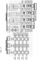
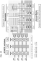
- the term “cartridge” or “consumable” is a Self-contained cartridge/consumable that includes the necessary components to perform a single BCID Panel test.
- a “cartridge” or “consumable” is a cartridge for performing assays in a closed sample preparation and reaction system as described in U.S. Patent no. 9,598,722 .
- the cartridges can comprise several components, including a biochip cartridge, a top plate, a liquid reagent module (LRM), and a housing that keeps the components together.
- the biochip cartage comprises a bottom substrate, a sample preparation zone, reagent zone, Sample Manipulation Zone, Amplification Zone, Detection Zones as further described in U.S. Patent Publication no.
- the substrate comprises one or more amplification pathways/zones.
- the top plate is spotted with reagents and primers. During the spotting process, phenol red is added to the reagents and primers so that spotting can be visualized.
- the LRM includes fluid filled blisters, as generally depicted in Figure 1 from U.S. Patent application publication no. 2014/0194305 .
- lysis buffer (which in some cases can be water for hypotonic lysis, or can be a commercially available lysis buffer, such as those containing chiatropic salts such as guanidinium salts, and or high/low pH, and/or surfactants such as sodium dodecyl sulfate (SDS), Polysorbate 20, Triton-X, etc. is contained within a blister that is activated to add lysis buffer to the sample.
- SDS sodium dodecyl sulfate
- Polysorbate 20 such as Tween ® 20
- the top plate may include a PDOT (or PEDOT) coating.
- PEDOT:PSS or poly(3,4-ethylenedioxythiophene) polystyrene sulfonate is a polymer mixture of two ionomers. One component in this mixture is made up of sodium polystyrene sulfonate which is a sulfonated polystyrene. Part of the sulfonyl groups are deprotonated and carry a negative charge.
- the other component poly(3,4-ethylenedioxythiophene) or PEDOT is a conjugated polymer and carries positive charges and is based on polythiophene. Together the charged macromolecules form a macromolecular salt.
- the top plate may be coated with Teflon ® , Cytop ® , or Fluoropel ® , preferably Cytop ® .
- Cytop ® is an amorphous fluoropolymer with high optical transparency and excellent chemical, thermal, electrical and surface properties.
- carrier sub-assembly means the bottom plate and top plate together.
- BCID-GP means Blood Culture Identification - Gram-Positive Panel.
- the BCID-GP panel includes all of the oligonucleotides and reagents for carrying out a nucleic acid amplification reaction for the targets listed in figure 14 as well as the capture and signal probes to form the hybridization complex necessary to detect the targets listed in figure 14 .
- phenol red is included in the reagents and primer mix pools as a visual tool to ensure the top plates are properly spotted.
- BCID-GN Blood Culture Identification - Gram-Negative Panel.
- the BCID-GN panel includes all of the oligonucleotides and reagents for carrying out a nucleic acid amplification reaction for the targets listed in figure 18 as well as the capture and signal probes to form the hybridization complex necessary to detect the targets listed in figure 18 .
- phenol red is included in the reagents and primer mix pools as a visual tool to ensure the top plates are properly spotted.
- BCID-FP Blood Culture Identification - Fungal Panel.
- the BCID-FP panel includes all of the oligonucleotides and reagents for carrying out a nucleic acid amplification reaction for the targets listed in figure 21 as well as the capture and signal probes to form the hybridization complex necessary to detect the targets listed in figure 21 .
- phenol red is included in the reagents and primer mix pools as a visual tool to ensure the top plates are properly spotted.
- the term "about” means encompassing plus or minus 10%. For example, about 90% refers to a range encompassing between 81% and 99% nucleotides. As used herein, the term “about” is synonymous with the term approximately.
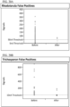
- amplifying or “amplification” in the context of nucleic acids refers to the production of multiple copies of a polynucleotide (generally referred to herein as "amplicons"), or a portion of the polynucleotide, typically starting from a small amount of the polynucleotide or a single polynucleotide molecule, where the amplification products or amplicons are generally detectable. Detection in the system ranges, for example, on the low end C. Kefyr is 200 CFU/mL without false positives due to contaminants. For fungal the upper detection limit for organisms is 1 ⁇ 10 5 .
- Amplification of polynucleotides encompasses a variety of chemical and enzymatic processes.
- the generation of multiple nucleic acid copies from one or a few copies of a target or template nucleic acid molecule during a polymerase chain reaction (PCR) or a ligase chain reaction (LCR) are forms of amplification.
- detect refers to an act of determining the existence or presence of one or more targets (e.g., microorganism nucleic acids, amplicons, etc.) in a sample.
- targets e.g., microorganism nucleic acids, amplicons, etc.
- target detection occurs when the amplicon forms a hybridization complex with the complimentary signal and capture probe.
- the amplicon comprises a length that is compatible with electrochemical detection which is typically less than 300 base pairs although many amplicons used herein are less than 150; indeed some amplicons used in the system are less than 100 base pairs.
- the amplicon is less than 300 base pairs, 200 base pairs, 150 base pairs, 100 base pairs, or 75 base pairs.
- the goal is to make a short amplicon because it is more efficient for exonuclease to make it single strand and also requires shorter amplification times.
- Bays as used herein are further described in U.S. Patent application no. 14/062860 , U.S. Patent Publication no. 2015/0323555 and U.S. Patent No. 9,598,722 .
- sLRM "simulated liquid reagent module"
- a blood culture sample that is manually prepared on the bench to mimic processing on an automated instrument.
- the present invention relates to an in vitro method as defined in the pending claims.
- the application of PCR using a multiplex PCR method enables a substantial reduction in electrochemical detection of contaminating pathogen and/or genetic material present in the sample while allowing infectious bacteria and/or fungi to be detected. Detection occurs when the infectious bacteria and/or fungi are amplified and the amplicon hybridizes with a signal and/or capture probe.
- the disclosed methods comprise providing a sample to the cartridge, providing PCR regents (including, but not limited to primers, dNTPs, DNA polymerase, exonucleases, etc.)for amplifying a locus from a different one of a plurality of target nucleic acid sequence to the sample, subjecting the sample to amplification conditions through a number of amplification cycles, detecting whether amplification has occurred, and identifying the target nucleic acid present in the sample wherein identifying comprises determining if the target nucleic acid is hybridized to signal and capture probes.
- PCR regents including, but not limited to primers, dNTPs, DNA polymerase, exonucleases, etc.
- non-uniform PCR cycling is used in a single cartridge, i.e., a single cartridge may cycle a sample and a first set of primers 30 times and cycle the sample and a second different set of primers 35 times (based on using different locations on the cartridge; reference is made to Figure 15 ).
- An infectious pathogen can be identified by its species.
- a co-infectious pathogen which is of the same type as the infection can be identified by its species.
- a co-infectious pathogen which is not the same type as the infection can be identified by its species.
- Co-infectious pathogens not being a member of a predetermined group are not identified because the steps performed with the reagents are adjusted to not detect pathogens not belonging to that group.
- 20-30 infectious pathogens can be identified on a single cartridge by its species or genus using a single PCR run while simultaneously being able to distinguish between systemic infection and punitive contamination.
- 30-40 or 40-50 infectious pathogens can be identified on a single cartridge by its species or genus using a single PCR run while simultaneously being able to distinguish between systemic infection and punitive contamination.
- At least 20, 20-60; 30-40 or 40-50 infectious pathogens can be identified on a single cartridge by its species or genus using a single PCR run while simultaneously being able to distinguish between systemic infection and punitive contamination and while simultaneously identifying fungal and bacteria co-infections by genus (fungal) or category (gram positive or gram negative).
- nucleic acids e.g. DNA
- purification, partial purification or isolation of nucleic acids is not needed to achieve sufficient sensitivity for detecting an infection while not detecting contaminants in the sample.
- nucleic acids need not be separated from proteins, sugars, and salts present in the original clinical sample. It is not necessary to partially or even completely isolate nucleic acid from the clinical sample after gram staining.
- the nucleic acid target (genome, gene or gene fragment (e.g., a restriction fragment) of the pathogen) may be in a purified, or in an isolated form.
- the sample may be treated with a compound which hydrolyzes nucleic acids aka a nuclease before amplification.
- the sample may be treated with DNase I, BENZONASE ® (nuclease), or S1 nuclease or combinations thereof before amplification, preferably before cell lysis.
- the design of amplification primers is performed on the basis of available sequence information with regard to the pre-selected target nucleic acid sequence regions of the specific pathogenic gram-positive bacteria to be amplified as well as with regard to the homologous sequences of those gram-positive and gram-negative bacteria, which shall not be amplified. More precisely, the set or sets of amplification primers are selected in such a way that there is a maximum sequence complementarity with respect to all target nucleic acid sequences of the selected predetermined pathogenic gram-positive bacteria species or genus, and, on the other hand, a minimum sequence complementarity with respect to nucleic acid sequences of all other non-selected gram-positive bacteria, gram-negative bacteria, i.e. those not belonging to the predetermined group or not being pathogenic, as well as fungi.
- the same method is applied to the BCID-GN cartridge and BCID-FN cartridge.
- the disclosure surprisingly shows that the analysis of fungi is possible in a single PCR reaction, without a nested PCR approach, in such a manner that a highly sensitive and very specific method is provided.
- This is surprising as generally, due to the slower growth of fungal infections, the fungal pathogens are present in lower amounts in the sample, and, thus, signal from contaminants can compete with the actual signal from the fungal pathogen.
- Previous attempts at PCR followed by detection have been bothered by high levels of false positives caused by contaminating pathogen and/or genetic material present in the sample and/or media bottle. See U.S. Application no. 2015/0232916 .
- the disclosure is, therefore, the first described single-run multiplex PCR method for discrimination between contaminating pathogen and/or genetic material present in the sample and infectious pathogen combined with discrimination between gram-positive pathogens, gram-negative bacterial pathogens, and fungal pathogens in said sample as well as antimicrobial resistance genes.
- the complexity of the present method is significantly reduced compared to alternative amplification schemes described previously, thereby increasing the user friendliness and reproducibility compared to those methods of the prior art.
- the PCR reaction comprises oligonucleotides that bind a DNA/ nucleic acid sequence of a bacterial pathogen.
- the oligonucleotides capable of binding a sequence of a bacterial pathogen enable discrimination between gram-positive and gram-negative bacteria.
- the oligonucleotides capable of binding a DNA/ nucleic acid sequence of a bacterial pathogen which, once amplified, attach to probes labeled so as to be distinguished from each other.
- the oligonucleotides designed for DNA amplification are able to amplify genetic material from a single pathogenic or potentially pathogenic bacteria (i.e. specific for sequence variation of a particular species or genus of a gram-positive bacteria) allowing detection of a specific species or genus of gram-positive bacterial infection and are run with oligonucleotides that detect the fungal genus or gram-negative genus and, as a result, is a broad-band, gram-negative bacterial and fungal detection method.
- the oligonucleotides designed for DNA amplification allow detection of a specific gram-negative bacterial infection (i.e.
- oligonucleotides that detect the fungal genus or the gram-positive bacteria genus or species but do not identify gram-positive or fungal infections by genus or species and, as a result, is a broad-band, gram-positive bacterial or fungal and detection method.
- the PCR reaction comprises oligonucleotides that bind a DNA sequence of a fungal pathogen.
- the oligonucleotides capable of binding a DNA sequence of a fungal pathogen which, once amplified, attach to probes labeled so as to be distinguished from each other.
- the signal probes comprise electrochemical labels, wherein multiple probes may be identified and differentiated from one another on the basis of distinct labels that emit electrical signals at different voltages from each other; see for example, U.S. Patent No. 7,935,481 and U.S. Patent Application no. 10/137,710 which disclose a plurality of probes each with at least one ETM with a unique redox potential. This is analogous to the "two color” or "four color” idea of competitive hybridization, and is also analogous to sequencing by hybridization. Probes and labels may be selected as required depending on the device used for analysis and the sample to be assessed as known by those skilled in the art. Preferred labels for signal probes include ferrocene and ferrocene derivatives. Ferrocene undergoes many reactions characteristic of aromatic compounds, enabling the preparation of substituted derivatives. Ferrocene derivatives (such as N6, QW56, and QW80) and are generally covalently attached to the signal probes.
- the method of the invention is carried out in a single multiplex, end point (PCR) reaction, otherwise known as a single PCR run (to be distinguished from nested PCR).
- PCR end point
- the invention is therefore characterized by the reduced number of PCR runs (single run) employed in the method compared to the prior art.
- the invention is therefore characterized by the reduced number of primers employed in the method compared to the prior art.
- the invention is therefore characterized by the reduced number of PCR runs (single run), PCR cycles (35 or 30) and primers employed in the method compared to the prior art.
- the invention is characterized in that there is a single PCR run of the sample in a single cartridge.
- the BCID-FP panel cycles 40 times because fungi is known to grow slower in culture and the assay is detuned by having primer mismatches or having dual zone detection or both. Additionally, a single run PCR with reduced cycling (less than 40 cycles) could result in false negatives because a single PCR run is insufficient to amplify and detect the organism. It was surprising and unexpected that the balance of sensitivity (detection of low titer infectious organisms) and non-detection of contaminates could be achieved in a single PCR run using end-point PCR not nested PCR.
- the electrochemical detection system employed needed to be made less sensitive, "detuned,” to eliminate or reduce detection of contaminants while remaining sensitive enough to detect clinically relevant infection. Detuning was achieved by reducing the PCR cycles in each single PCR run, increasing or decreasing the primer concentration, thresholding, primer mismatch or requiring one pathogen be detected in two detection zones and combinations thereof. While the molecular biology techniques used to detune were known, no one had applied them in the context of electrochemical detection in a single run PCR to detect pathogens but not background contamination.
- nested PCT systems tend to be focused on genus calls as opposed to species calls. As such, only a broadband antibiotic therapeutic approach is possible when a nested PCT system identifying only genus calls is used, which may, in fact, be poorly suited for the particular pathogen.
- the sample is obtained from a subject exhibiting one or more symptoms of systemic inflammatory response syndrome (SIRS), sepsis, severe sepsis and /or septic shock.
- SIRS systemic inflammatory response syndrome
- a significant benefit of this approach is the ability to subsequently prescribe an appropriate medicament during treatment.
- an appropriate antibiotic or an appropriate anti-fungal can be selected for treatment, thereby avoiding potentially useless antibiotic treatments and associated financial, health and environmental disadvantages.
- Patient care and antibiotic stewardship would be advanced by development and application of rapid diagnostics that provide accurate and timely information as to the nature of the infecting pathogen, including whether it is gram-positive bacterial, gram-negative bacterial, fungal and its resistance profile.
- the mixture of oligonucleotides and reagents for carrying out a single nucleic acid amplification reaction is further capable of identifying the species of the infection and genus of a co-infection.
- the mixture of oligonucleotides and reagents for carrying out a single nucleic acid amplification reaction is further capable of identifying the species or genus of the infection and spices or genus of a co-infection.
- the mixture of oligonucleotides and reagents for carrying out a single nucleic acid amplification reaction is further capable of identifying the species or genus of the infection and spices or type (gram-positive or gram negative) of co-infection.
- the BCID-GP Panel contains assays for the detection of genetic determinants of resistance to methicillin (mecA and mecC) and vancomycin (vanA and vanB) to aid in the identification of potentially antimicrobial resistant organisms in positive blood culture samples.
- the antimicrobial resistance gene detected may or may not be associated with the agent responsible for the disease.
- the BCID-GP Panel also contains targets designed to detect a broad range of organisms with a potentially misleading Gram stain result or organisms that may be missed by Gram staining altogether for example in the case of co-infections. These include a broad Pan Gram-Negative assay as well as a Pan Candida assay, both of which may provide data to facilitate the correct testing algorithm.
- the present disclosure relates to methods and systems for a) distinguishing between contamination and gram-positive bacterial infection, b) distinguishing between gram-positive bacterial species infection; c) distinguishing between some gram-positive bacterial species and some gram-positive genus infection(s); d) identifying but not differentiating gram-negative bacterial infection and fungal infection.
- the present disclosure further relates to methods and systems for identifying a pathogen that is likely a contamination from the blood draw.
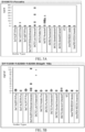
- the following bacterial organisms and resistance marker genes are identified using the BCID-GP Panel: Bacillus cereus group, Staphylococcus epidermidis, Bacillus subtilis group, Staphylococcus lugdunensis, Corynebacterium spp., Streptococcus, Enterococcus, Streptococcus agalactiae, Enterococcus faecalis, Streptococcus anginosus group, Enterococcus faecium, Streptococcus pneumonia, Lactobacillus, Streptococcus pyogenes, Listeria, Pan Gram-negative target (at least Enterobacteriaceae, Acinetobacter, Pseudomonas, Bacteroides, Stenotrophomonas), Listeria monocytogenes, Pan Candida target (Candida albicans, Candida glabrata, Candida krusei, Candida parapsilosis), Micrococcus,
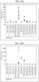
- Table 1 below shows that reported target call and the target species detected. Stated another way, some species are detected by the BCID-GP panel ("Targets detected” in Table 1 below) but not identified by the species in the call (report); instead, the call/report identifies the genus. Some species are detected by the BCID-GP panel ("Targets detected” in Table 1 below) and are identified by the species in the call (report). Some organisms can generate both the genus and species call.
- Table 1 Analytes Detected by the BCID-GP Panel Calls Reported Target Targets Detected 1 Streptococcus agalactiae Streptococcus agalactiae 2 Streptococcus anginosus group Streptococcus constellatus Streptococcus intermedius Streptococcus anginosus 3 Streptococcus pneumoniae Streptococcus pneumoniae 4 Streptococcus pyogenes Streptococcus pyogenes 5 Staphylococcus aureus Staphylococcus aureus 6 Staphylococcus epidermidis Staphylococcus epidermidis 7 Staphylococcus lugdunensis Staphylococcus lugdunensis 8 Enterococcus faecalis Enterococcus faecalis 9 Enterococcus faecium Enterococcus faecium 10 Bacillus subtil
- the Pan Gram-negative target in the BCID-GP panel can identify about 10 species of gram-negative bacteria. In a preferred embodiment the Pan Gram-negative target in the BCID-GP panel can identify at least 5, at least 10, at least 15, at least 20, at least 30, at least 40 at least 50 at least 60, at least 70, at least 80, at least 90, at least 100 or more species of gram-negative bacteria. In a preferred embodiment the Pan Gram-negative target in the BCID-GP panel can identify 30-100 species of gram-negative bacteria. In a preferred embodiment the Pan Gram-negative target in the BCID-GP panel can identify about 10%, about 20%, about 30%, about 40%, about 50%, about 60%, about 70%, about 80% or more species of gram-negative bacteria.
- the BCID-GP oligonucleotides capable of binding a sequence of a bacterial pathogen which enable discrimination between gram-positive species or genus were not designed to avoid or reduce detection of background contamination. It was a surprising and unexpected result that reducing cycling from 40 to 37, and in some cases from 40 to 35, and in some cases from 40 to 30 was sufficient to distinguish between background contamination, gram-positive bacteria species or genus infection, non-species gram-negative bacteria and non-species fungal infection.
- the BCID-GN Panel is a fully automated, qualitative, nucleic acid, multiplex in vitro diagnostic test for simultaneous detection and identification of multiple potentially pathogenic gram-negative bacterial organisms and select determinants of antimicrobial resistance in positive blood culture.
- the test also detects but does not differentiate gram-positive bacteria and several pathogenic Candida species.
- the test is able to detect 21 bacterial targets and 6 resistance genes, as well as multiple Candida species from a single cartridge (single PCR run) and most major gram-positive organisms, also as on a single cartridge (single PCR run).
- the following bacterial organisms are identified using the BCID-GN Panel: Acinetobacter baumannii, Klebsiella pneumoniae, Bacteroides fragilis, Morganella morganii, Citrobacter, Neisseria meningitides, Cronobacter sakazakii, Proteus, Enterobacter cloacae complex, Proteus mirabilis, Enterobacter (non-cloacae complex), Pseudomonas aeruginosa, Escherichia coli, Salmonella, Fusobacterium necrophorum, Serratia, Fusobacterium nucleatum, Serratia marcescens, Haemophilus influenza, Stenotrophomonas maltophilia, Klebsiella oxytoca.
- Table 2 Gram-Negative Analytes Detected by the BCID-GN Panel Reported Target Targets Detected Acinetobacter baumannii Acinetobacter baumannii Bacteroides fragilis Bacteroides fragilis Citrobacter Citrobacter brakii Citrobacter fruendii Citrobacter koseri Critrobacter youngae Citrobacter freundii / brakii Citrobacter freundii Citrobacter brakii Cronobacter sakazakii Cronobacter sakazakii Enterobacter (not cloacae complex) Enterobacter aerogenes Enterobacter amnigenus Enterobacter gergoviae(detect with amnigenus assay) Enterobacter cloacae complex Enterobacter asburiae Enterobacter cloacae Enterobacter hormaechei Escherichia coli Escherichia coli Fusobacterium (not necrophorum ) Fu
- the Pan Gram-positive target in the BCID-GN panel can identify about 15 species of gram- positive bacteria. In a preferred embodiment the Pan Gram-positive target in the BCID-GN panel can identify at least 10, at least 15, at least 20, at least 30, at least 40 at least 50 at least 60, at least 70, at least 80, at least 90, at least 100 or more species of gram- positive bacteria. In a preferred embodiment the Pan Gram- positive target in the BCID-GN panel can identify 30-100 species of gram- positive bacteria. In a preferred embodiment the Pan Gram- positive target in the BCID-GN panel can identify about 10%, about 20%, about 30%, about 40%, about 50%, about 60%, about 70%, about 80% or more species of gram-positive bacteria.
- the BCID-GN oligonucleotides capable of binding a sequence of a bacterial pathogen which enable discrimination between gram-negative species or genus were not designed to avoid or reduce detection of background contamination. It was a surprising and unexpected result that detuning the assay i.e., by merely reducing the PCR cycling, was sufficient to distinguish between background contamination, gram-negative bacteria species or genus infection, non-species gram-positive bacteria and non-species fungal infection.
- the BCID-GN Panel contains targets designed to detect a broad range of organisms with a potentially misleading Gram stain result or organisms that may be missed by Gram staining altogether for example in the case of co-infections. These include a Pan Gram-Positive assay as well as a Pan Candida assay, both of which may provide data to facilitate the correct testing algorithm.
- the present disclosure relates to methods and systems for a) distinguishing between background contamination and gram-negative bacterial infection; b) distinguishing between gram-negative bacterial species infection; c) distinguishing between some gram-negative bacterial species and some gram-negative genus infection(s); and d) detecting but not identifying gram-positive bacterial species or genus infection and fungal species infection.
- the present disclosure further relates to methods and systems for identifying a pathogen that is likely a contamination from the blood draw.
- Gram-negative bacteria are a common cause of bacteremia, being isolated from over 60% of positive blood cultures throughout the world. Antimicrobial resistance is common among gram-negative organisms, and multi-drug resistance is increasingly common in many species. When involved in bacteremia, the species belonging to this group have mortality rates ranging from 20% to over 90% in some populations.
- the Blood Culture Identification Fungal Pathogen Panel (BCID-FP Panel) is a fully automated, qualitative, nucleic acid, multiplex in vitro diagnostic test for simultaneous detection and identification of multiple potentially pathogenic fungal organisms in positive blood culture.
- the BCID-FP Panel is performed directly on blood culture samples identified as positive by a continuously monitoring blood culture system that demonstrates the presence of organisms as confirmed by Gram stain.
- the following fungal organisms are identified using the BCID-FP Panel: Candida auris, Candida albicans, Candida dubliniensis, Candida famata, Candida glabrata, Candida guilliermondii, Candida kefyr, Candida lusitaniae, Candida krusei, Candida parapsilosis, Candida tropicalis, Cryptococcus gattii, Cryptococcus neoformans, Fusarium, Malassezia furfur, Rhodotorula, and Trichosporon.
- the fungal species detected by the BCID-FP Panel are in Figure 21 .
- the BCID-FP panel detects but does not identify the following species in the call (report): solani set, dimerum, proliferatum, moniliforme, verticillioides, oxysporum, and sacchari.
- the BCID-FP panel detects but does not identify the following species in the call (report): mucilaginosa, and glutinis.
- the BCID-FP panel detects but does not identify the following species in the call (report): asteroid, coremiiforme and dermatis.
- the BCID-FP oligonucleotides capable of binding a sequence of a fungal pathogen which enable discrimination between fungal species or genus were not designed to avoid or reduce detection of background contamination. It was a surprising and unexpected result that merely creating primer mismatches and in some cases using dual zone detection was sufficient to distinguish between background contamination and fungal species or genus infection. It was non-obvious to intentionally decrease the sensitivity of the assay by intentionally introducing primer miss-matches.
- the present disclosure relates to methods and systems for a) distinguishing between background contamination and fungal infection.
- the present disclosure relates to methods and systems for a) distinguishing between background contamination, and detecting and identifying the species or genus of fungal infection.
- Invasive fungal infections are an increasingly common cause of sepsis in critically ill patients and are the source of significant morbidity and mortality.
- Candida species are by far the most prevalent, accounting for between 8-10% of all bloodstream infections in the US and 2-3% in Europe. Sepsis caused by invasive fungi is associated with mortality rates ranging from 15% to nearly 100% depending on the organism and underlying factors involved.
- the fungal primers, signal and capture probe sequences are below. Nucleotide sequences should have at least 80% sequence identity preferably more than 85%, preferably more than 90%, preferably more than 95% sequence identity, to the sequences provided herein. Table 3: Fungal Forward And Reverse Primers and SEQ ID NOs. Species Forward Sequence Reverse Sequence Rhodotorula 1 Rhodotorula 2 Trichosporon 1 Trichosporon 2 Trichosporon 3
- the method and system can identify the species of the co-infection based on the first single PCR run. If a co-infection is of a different type as the infection (i.e., the infection is gram-positive but a pan gram-negative and/or pan fungal co-infection is detected or the infection is gram-negative but a pan gram-positive and/or pan fungal co-infection is detected), the method and system can identify the genus of the fungal infection and the type of the co-infection (gram-positive or negative) based on a second single PCR run.
- the BCID-GP Panel includes two pan targets (Pan gram-negative and Pan Candida) designed to detect but not identify organisms that may be missed by Gram stain.
- the BCID-GN Panel includes two pan targets (Pan gram-positive and Pan Candida) designed to detect but not identify organisms that may be missed by Gram stain. If a pan target is identified in the BCID-GP Panel, then the BCID-GN and/or FN Panel can be run to identify the specific species or genus of the infection. Likewise, if a pan target is identified in the BCID-GN Panel, then the BCID-GP and/or FN Panel can be run to identify the specific species or genus of the infection.
- the method comprises the following steps: a) identify a first species or genus infection and a co-infection; b) identifying the species of the co-infection.
- the first infection is a gram-positive infection and the co-infection is a gram-negative infection or fungal infection.
- the first infection is a gram-negative infection and the co-infection is a gram-positive or fungal infection.
- the method comprises the following steps: a) providing a sample, b) bringing said sample into contact with a mixture of oligonucleotides and reagents for carrying out a nucleic acid amplification reaction, c) after DNA/ nucleic acid extraction carrying out a first single nucleic acid amplification reaction, d) obtaining a first result e) if four or more infections are present, obtaining a second result wherein obtaining a second result comprises f) providing the sample, g) bringing said sample into contact with a mixture of oligonucleotides the same as the oligonucleotides used to obtained the first result and reagents for carrying out a nucleic acid amplification reaction, h) after DNA / nucleic acid extraction carrying out a second single nucleic acid amplification reaction, and i) obtaining a second result.
- the method comprises the following steps: a) obtaining a first result b) analyzing the first result for a secondary infection c) if a secondary infection is present obtaining a second result wherein obtaining a second result comprises a) providing a sample, b) bringing said sample into contact with a mixture of oligonucleotides different from oligonucleotides used to obtained the first result and reagents for carrying out a nucleic acid amplification reaction, c) carrying out a single nucleic acid amplification reaction, and d) obtaining a second result.
- the method comprises the following steps: a) obtaining a gram-stain result (b) selecting a panel based on the gram stain result c) carry out a first single nucleic acid amplification reaction d) detecting the amplification products generated as a result of said first single nucleic acid amplification reaction d) if the amplification products do not match the gram stain result, select a second panel based on the results from the first single nucleic acid amplification reaction e) carry out a second single nucleic acid amplification reaction d) detect the amplification products generated as a result of said second single nucleic acid amplification reaction.
- the second single nucleic acid amplification reaction provides more specific species or genus identification than the first single nucleic acid amplification reaction for the co-infection.
- a method for identifying a fungal infection not identified by a gram stain comprising loading a sample suspected of having a bacterial infection based on a gram stain into a first cartridge, amplifying the sample using a single nucleic acid amplification reaction, identifying a pan- candida organism, and loading the sample into a second cartridge amplifying the sample using a second single nucleic acid amplification reaction and detecting the amplification products generated as a result of said second single nucleic acid amplification reaction.
- a method for screening a patient suspected of having a bacterial infection comprising a) performing a first test for the presence of a gram-positive bacterial infection; b) if the first test indicates a gram-negative bacterial infection is present performing a second test for the presence of a gram-negative bacterial infection wherein the first test and second test comprise amplifying the sample using a single nucleic acid amplification reaction.
- a method for screening a patient suspected of having a fungal infection comprising a) performing a first test for the presence of a bacterial infection; b) if the first test indicates a fungal infection is present performing a second test for the presence of a fungal infection wherein the first test and second test comprise amplifying the sample using a single nucleic acid amplification reaction.
- a method for identifying a plurality of organisms in a sample comprising providing a first portion of a sample from a patient to a first cartridge, bringing said first sample into contact with a mixture of oligonucleotides and reagents for carrying out a single nucleic acid amplification reaction, determining if a second pathogen is present in said first sample, providing a second portion of a sample from the patient to a second cartridge, b) bringing said sample into contact with a mixture of oligonucleotides and reagents for carrying out a second single nucleic acid amplification reaction, determining if a second pathogen is present in said second sample.
- the method of determining the existence of and identifying any one of up to a plurality of human pathogens in a sample comprising the steps of: a) after DNA/ nucleic acid extraction, performing a first detection process comprising carrying out a single nucleic acid amplification reaction, b) detecting and evaluating the amplification products generated as a result of said first single nucleic acid amplification reaction, c) after DNA/ nucleic acid extraction, performing a second detection process comprising carrying out a single nucleic acid amplification reaction, and d) detecting and evaluating the amplification products generated as a result of said second detection process, thereby identifying any one of up to a plurality of human pathogens in the sample.
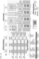
- the BCID-GP, GN and FP panels can be run in a cartridge comprising a bottom substrate.
- the bottom substrate (or printed circuit board) can contain 1, 2, 3 or more amplification pathways or pads (called the Amplification Zone). These can be used for individual PCR reactions (e.g. one droplet is moved up one path and down another, etc.) or for multiplexing (e.g. eight different droplets can be moved up and down four pathways).
- each PCR reaction can additionally be multiplexed. That is, for target specific amplification, the use of multiple primer sets in a single PCR reaction can be unwieldy, and thus the teaching herein allows multiple reactions to achieve higher levels of multiplexing. For example, for the evaluation of 21 different target sequences (for example, in screening for fungal infections), it may be desirable to run 3 different reactions of seven primer sets; e.g. a first PCR sample droplet (e.g. the bottom pathway) picks up the first set of 7 primer pairs (e.g.
- Primer Mix A a second droplet picks up the second set of 7 primer pairs
- Prime Mix C a third droplet picks up a third set
- the primers will be completely different in each set; in others, redundancy and/or internal controls are built into the system by adding the same primer sets to different tracks.
- the multiplexing flexibility represents one of the key advantageous and distinguishing features of the disclosure. The number of multiplexes can vary easily through software without the need to modify any physical components of the system.
- the amplification reactions for use in the present systems use sets of primers wherein one primer of each set has a blocked end that is impervious to standard exonucleases. That is, one strand of the double stranded amplicons that are generated in the PCR reaction is removed so that the resulting single stranded DNA amplicon can hybridize to the single stranded capture probe.
- one strand of the double stranded amplicon is digested, leaving only the detection strand.
- two droplets may be positioned in each PCR track and spaced in such a way that when one droplet is in the denaturation zone, the other is in one of combined annealing and extension zones, and vice versa.
- shuttling the droplets in tandem back and forth between the denaturation and annealing/extension zones one can amplify both of them in the same amount of time it would normally take to amplify a single droplet.
- Electrowetting, or digital microfluidics uses electrical fields to directly manipulate discrete droplets on the surface of a hydrophobically coated printed circuit board (PCB).
- PCB printed circuit board
- Sample and reagents are moved in a programmable fashion in the cartridge to complete all portions of the sample processing from nucleic acid extraction to detection.
- a sample is loaded into the cartridge and the cartridge is placed into the instrument.
- Nucleic acids are extracted and purified from the specimen via magnetic solid phase extraction (i.e. the use of magnetic beads to pre-concentrate analytes or targets, then move (elute) the beads containing the targets to a different location, where the targets are released for post-elution events.
- PCR is used to created double-stranded cDNA which is treated with exonuclease to create single-stranded DNA in preparation for electrochemical detection.
- the target amplicons are mixed with ferrocene-labeled signal probes that are complementary to the specific targets on the panel.
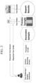
- Target sequences hybridize to the complementary signal probe and capture probes, which are bound to gold-plated electrodes, as shown in Figure 1 .
- the presence of each target is determined by voltammetry which generates specific electrical signals from the ferrocene-labeled signal probe.
- Figure 1 shows the hybridization complex.
- Target-specific capture probes are bound to the gold electrodes in the microarray on the cartridge.
- the amplified target DNA hybridizes to the capture probe and to a complementary ferrocene-labeled signal probe.
- the electrochemical analysis determines the presence or absence of targets using voltammetry.
- microfluidic systems in the electrochemical detection of target analytes is described in more detail in U.S. Patent Nos. 9,557,295, 8,501,921 , 6,600,026, 6,740,518 and US2016/0129437 .
- Step -1 Obtain sample after gram stain; Step 0. Load Sample; step 1. Combine Lysis Buffer with Sample (LRM), beads and Dispense Oil (Cartridge sub-assembly); step 2. Combine Binding Buffer with Sample (LRM) and Dispense Reconstitution Buffer (cartridge sub-assembly); step 3. Separate beads from sample bead mixture (LRM) and Rehydrate PCR reagent (cartridge sub-assembly); step 4.
- wash beads with Wash buffer (LRM) and Rehydrate PCR reagent (cartridge sub-assembly); step 5. Flush beads from LRM into cartridge; step 6. Final bead wash in cartridge sub-assembly and Quick Rinses (cartridge sub-assembly); step 7. Elute target analyte from beads; step 8. Combine PCR reagent with elute target (analyte); step 9. Dispense analyte drops mix into PCR staging area; step 10. Rehydrate PCR primers cocktail with each analyte drop; step 11. Transfer eluted analyte to thermal-cycling PCR area in the cartridge; step 12. Convert RNA into DNA with Reverse Transcriptase (optional step); step 13.
- step 14 Perform PCR cycling; step 14. Rehydrate exonuclease reagent; step 15. Combine PCR products with exonuclease reagent (ssDNA conversion); step 16. Exonuclease incubation and combine with Signal Probe cocktail (detection); step 17. Deliver PCR products and signal probe into Detection area; step 18. Incubate in eSensor area with capture probe bound to gold electrode; step 19. Scan and detect target analyte; step 20. Eject cartridge.
- microfluidic platform used herein is based on systems developed by Advanced Liquid Logic (ALL, currently a subsidiary of Illumina, Inc.), as more fully described in U.S. Patent app. no. 20140194305 .
- these technologies rely on the formation of microdroplets and the ability to independently transport, merge, mix and/or process the droplets, using electrical control of surface tension (i.e., electrowetting).
- liquid samples are contained within a microfluidic device between two parallel plates. One plate contains etched drive electrodes on its surface while the other plate contains either etched electrodes or a single, continuous plane electrode that is grounded or set to a reference potential (“biplanar electrowetting").
- Hydrophobic insulation covers the electrodes and an electric field is generated between electrodes on opposing plates. This electric field creates a surface-tension gradient that causes a droplet overlapping the energized electrode to move towards that electrode.
- the active electrowetting electrodes may be adjacent and on the same plane as the neighboring ground reference electrode, which is referred to as "coplanar electrowetting"). Through proper arrangement and control of the electrodes, a droplet can be transported by successively transferring it between adjacent electrodes.
- the patterned electrodes can be arranged in a two dimensional array so as to allow transport of a droplet to any location covered by that array.
- the space surrounding the droplets may be filled with a gas such as air or an immiscible fluid such as oil, with immiscible oils being preferred embodiments.
- the immiscible fluid may be a synthetic silicone oil. This silicone oil is present throughout the system, i.e., during amplification and detection.
- Signal probes are used in the electrochemical detection of target analytes on the surface of a monolayer.
- QW56 or QW80 are ferrocene labeled signal probes that can be prepared using routine DNA synthesis techniques essentially as described in commonly owned WO/2009/061941A2 and U.S. Pat. No 7,820,391 ).
- Figure 3A depicts QW 56 and Figure 3B depicts QW80.
- N6 (a ferrocene labeled signal probe) is another label that can be used; its synthesis is described in commonly owned U.S. Pat. No. 7,393,645 .
- Capture probes are used in the electrochemical detection of target analytes on the surface of a monolayer. Specifically, capture binding ligands (called capture probes when the target analyte is a nucleic acid) anchor target analytes to the electrode surface and form an assay complex.
- the assay complex further comprises an electron transfer moiety (ETM), that is directly or indirectly attached to the target analyte. That is, the presence of the ETM near the electrode surface is dependent on the presence of the target analyte. Electron transfer between the ETM and the electrode is initiated using a variety of techniques as known by those of skill in the art, and the output signals received and optionally processed as further known by those of skill in the art. Thus, by detecting electron transfer, the presence or absence of the target analyte is determined.
- ETM electron transfer moiety
- detection of an ETM is based on electron transfer through the stacked ⁇ -orbitals of double stranded nucleic acid.
- This basic mechanism is described in U.S. Pat. Nos. 5,591,578, 5,770,369, 5,705,348 , and WO98/20162 and is termed "mechanism-1" herein.
- mechanism-1 This basic mechanism is described in U.S. Pat. Nos. 5,591,578, 5,770,369, 5,705,348 , and WO98/20162 and is termed "mechanism-1" herein.
- mechanism-1 previously work has shown that electron transfer can proceed rapidly through the stacked ⁇ -orbitals of double stranded nucleic acid, and significantly more slowly through single-stranded nucleic acid. Accordingly, this can serve as the basis of an assay.
- ETMs either covalently to one of the strands or non-covalently to the hybridization complex through the use of hybridization indicators, described below
- ETMs either covalently to one of the strands or non-covalently to the hybridization complex through the use of hybridization indicators, described below
- the detection electrode preferably comprises a self-assembled monolayer (SAM) that serves to shield the electrode from redox-active species in the sample.
- SAM self-assembled monolayer
- the presence of ETMs on the surface of a SAM, that has been formulated to comprise slight "defects" (sometimes referred to herein as "microconduits", “nanoconduits” or “electroconduits”) can be directly detected. This basic idea is termed “mechanism-2" herein.
- the electroconduits allow particular ETMs access to the surface.
- the configuration of the electroconduit depends in part on the ETM chosen.
- the use of relatively hydrophobic ETMs allows the use of hydrophobic electroconduit forming species, which effectively exclude hydrophilic or charged ETMs.
- the use of more hydrophilic or charged species in the SAM may serve to exclude hydrophobic ETMs.
- an assay complex is formed that contains an ETM, which is then detected using the detection electrode and the signal processing techniques outlined herein.
- monitoring of the output signal at higher harmonic frequencies can be used to achieve higher signal to noise ratios, to increase the detection limits of target analytes.
- the ferrocene response reacts non-linearly, producing a harmonic response in the signal above that in the background; this harmonic signal from AC voltammetry is most likely the result of a harmonic distortion due to the nonlinear response of the electrochemical cell; see Yap, J. of Electroanalytical Chem. 454:33 (1998 ).
- any techniques that increase this non-linearity are desirable.
- techniques are used to increase the higher harmonic signals; thus, frequency and phase-sensitive lock-in detection is performed at both the fundamental frequency of the applied waveform and also at multiples of the fundamental frequency (i.e. the higher harmonics). Since the background capacitance responds relatively linearly to AC signals (a sine wave input AC voltage results in a relatively nondistorted sine wave output), very little upper harmonic current is produced in the background. This gives a dramatic increase in the signal to noise ratio. Thus, detection at the higher harmonic frequencies, particularly the third, fourth and fifth harmonics (although the harmonics from second to tenth or greater can also be used) is shown to result in dramatic suppression of the background currents associated with non-Faradaic processes (like double layer charging) that can overwhelm the signal from the target molecules.
- one method of increasing the non-linear harmonic response is to increase or vary the amplitude of the AC perturbation, although this may also be used in monitoring the fundamental frequency as well.
- the amplitude may be increased at high frequencies to increase the rate of electron transfer through the system, resulting in greater sensitivity.
- this may be used, for example, to induce responses in slower systems such as those that do not possess optimal spacing configurations
- Electrode initialization is another signal processing method to achieve higher signal to noise ratios, and to increase the detection limits of target analytes.
- the observed signal is a combination of signal from the target analyte (sample signal) and signal from the background, or noise.
- Electrode initialization provides variations in initiation signals (e.g. varying the "input") that can be used to increase the signal, decrease the noise, or make the signal more obvious or detectable in a background of noise.
- the input signal is AC/DC offset.
- the AC frequency ranges from 90-1000 Hz.
- the AC voltage ranges from -150 to 880 mV rms.
- electrode initialization is performed for 0.5-5 seconds and then stopped as described in U.S. Pat. App. No. 14/218,615 .
- the automated nucleic acid testing system aka electrochemical detection system described above includes a) an instrument bank comprising a plurality of biochip cartridge bays for insertion and analysis of a biochip cartridge, wherein each bay comprises: i) a top bay comprising actuators for a liquid reagent module (LRM); and ii) a bottom bay comprising electrical connections for an electrowetting electrode grid and detection electrodes; and b) a base station comprising: i) a central processing unit; and ii) a user interface comprising a touch screen display having a plurality of bay icons, each icon uniquely corresponding to one of said plurality of bays.
- LRM liquid reagent module
- a base station comprising: i) a central processing unit; and ii) a user interface comprising a touch screen display having a plurality of bay icons, each icon uniquely corresponding to one of said plurality of bays.
- Identification, detection or reporting results occurs when amplified target DNA hybridizes to its complementary signal probe and capture probes. Identification, detection or reporting results occurs when amplified target DNA hybridizes to its complementary signal probe and capture probes wherein the capture probe is bound to gold-plated electrodes. Identification, detection or reporting results occurs when a hybridization complex forms between the target DNA and signal and capture probes.
- the sample-to-answer system is designed to reduce avoidable medical errors. Preventable medical errors are now the third leading cause of death in the United States at more than 250,000 per year. See Martin A Makary, Michael Daniel. Medical error-the third leading cause of death in the US. BMJ, 2016 . Automating information transfer has been shown to be effective in reducing many common errors, including patient identity checking and result transcription. The sample-to-answer system is uniquely designed with patient safety features to help address this challenge in the lab.
- the sample-to-answer system includes a bi-directional LIS (also referred to as a communication system, an LIS communication system, bi-directional LIS communication system and the like) to automate and accelerate order entry and results reporting. See Figure 2 for a schematic of the bi-directional LIS reporting.
- a bi-directional LIS also referred to as a communication system, an LIS communication system, bi-directional LIS communication system and the like
- the physician test order allows the physician to specify a sample stability time (the time the sample is stable before results could be affected).
- the physician test order will further allow the physician to specify the time that a physician test order should remain on the physician test order list before processing.
- the physician test order is sent to the hospital's laboratory information system (LIS), a computer software that processes, stores and manages data from all stages of medical processes and tests.
- LIS laboratory information system
- the physician test order is accepted by the Hospital LIS and a pending test order (PTO) is created once the patient sample is received and accessioned by the lab into the hospital LIS.
- PTO pending test order
- the hospital's LIS sends the PTO to an LIS interchange which converts the PTO request from an HL7 or ASTM format to a CSV format and the PTO is now referred to as a test order or an interchange order or formatted test order and the like.
- HL7 and ASTM are a set of standards used in the transfer of information between clinical instruments and Laboratory Information Systems.
- the sample-to-answer system is able to communicate with any hospital LIS because it is driven by multiple standard messaging protocols such as HL7 and ASTM.
- the hospital's LIS system is updated the LIS interchange can be remotely updated (an update on the clinical instrument is not required).
- the sample-to-answer system further supports a "flat file format" i.e. non-standard file support for laboratories without automated interfaces (HL7 or ASTM).
- HL7 or ASTM automated interfaces
- tests can be imported and/or exported manually in a text format, CSV, TXT or XML formats.
- Automatic results can be released in XML format to a shared network location.
- test order is auto published with information associated with the PTO/sample such as patient identification, accession number, test ordered (BCID-GP, BCID-GN, BCID-FP etc.), patient type (e.g. pediatric, intensive care, maternity), patient location (e.g. pediatrics, ER, maternity), and/or time stamps such as sample collection, sample ordering, time received at central receiving, central receiving sort, transport to lab and/or accession of sample. These time stamps provide real-time monitoring by the instrument software of pending test order turn-around time.
- test ordered BCID-GP, BCID-GN, BCID-FP etc.
- patient type e.g. pediatric, intensive care, maternity
- patient location e.g. pediatrics, ER, maternity
- time stamps provide real-time monitoring by the instrument software of pending test order turn-around time.
- the automated nucleic acid testing system with communication capabilities is referred to as a "Clinical instrument" of sample-to-answer system.
- the LIS interchange receives the test order, it sends it to the sample-to-answer system's (clinical instrument's) CPU in the base station.
- the sample-to-answer system supports both serial and ethernet /RJ45 input/output connections to one or more hospital LIS.
- the Sample Stability feature or Sample Stability time allows the user to specify the stability time on a per assay basis.
- the software tracks PTO orders and sends an alert notification when an order has violated the threshold for sample stability.
- the sample-to-answer system includes "cleanup rules" to automatically delete outstanding pending test orders e.g. delete a PTO if it is in the Pending Test Order queue for a predetermined time (called max PTO time), preferably for more than one week, preferably for more than two weeks.
- bi-directional LIS capabilities improve PTO to detection report turnaround time, reduce labor costs, and eliminate potential transcription errors.
- the communication from the LIS interchange to the detection device's CPU can be referred to as the clinical instrument test order.
- a result is generated.
- a result is generated if the amplified target DNA/ nucleic acid hybridizes to its complementary signal probe and capture probes.
- the CPU in the base station then sends (either automatically or manually) a detection report (also referred to as a result report or test results) to the LIS interchange which converts the detection report into a physician test result report and sends the physician test result report to the hospital's LIS which then sends the physician result report to the physician or directly to the physician.
- the detection report/physician test result sent to the hospital's LIS or to the physician can include detected targets, non-detected targets, invalid results and/or control data.
- the sample-to-answer system can either auto release all information or hold all information for manual release. Alternatively, the sample-to-answer system can auto release some detection reports and hold some detection reports for manual release. For example, detected and non-detected targets can be auto-released while invalids can be manually released (i.e., released only after a lab supervisor approves for release). If the detection report shows 3 (triple infection) or fewer targets were identified/detected the detection report will automatically release to the hospital's LIS/physician. If the detection report shows greater than 3 ( i.e.
- a multiple infection error alert also called an alert notification
- the detection report includes the assay ordered. If a cartridge is inserted that does not match the assay ordered (e.g. a gram-negative assay is ordered but a respiratory assay is inserted) a "mismatch alert" is sent to the operator and/or physician and/or the additional target is noted in the detection report.
- Anomalous results that are not auto-released can require a manager signature before manual release. Such reporting minimizes the risk of reporting errors.
- the detection report can include time stamps such as sample collection time, sample ordering time, transport to central receiving time, central receiving sort time, transport to lab time, accession of sample time, time to process, and time to detection.
- Figure 3 includes an "order-to-report" timeline. The clock time stamps are commonly documented in hospital and laboratory information systems.
- sample results are reported in about 60-90 minutes after the start of running the sample, this is referred to as time to result (See figure 2 ) or sample to result.
- sample results are reported in about 60 minutes after the start of running the sample.
- sample results are reported in under 90 minutes after the start of running the sample.
- sample results are reported upon test completion.
- a detection report is sent immediately after the pathogen is identified by the detection system.
- the sample-to-answer system allows the operator to include comments in the detection report called detection report comments, e.g., to specify if the assay ordered matched the target detected, if the assay ordered does not match the target detected, if an additional target was detected in addition to the target for the assay ordered, if a second assay is recommended, if a resistance gene was identified, suggest a course of treatment such as antibiotic.
- Control reports or Control summary reports are generated based on the assay, test frequency and lot of cartridges from the supplier.
- Control reports provide information about the number of samples run, and when control runs are needed. When a control run is processed, the report shows the expected and actual result, if the control passed or failed. Control runs are typically run every 30 days or every lot change. The sample-to-answer system alerts to the operator 48 and/or 24 hours before a control run is needed.
- the system usage report provides analytics around system usage data and performance based on a specified date range. For example, the system usage report will show if higher or lower than average samples were run, if higher or lower than expected samples were run, if a bay has not been utilized, etc.
- System Usage Reports can be printed from the Clinical Instrument or remotely by the clinical instrument's provider.
- a service notification report is a report sent to the clinical instrument's provider to request remote access to the clinical instrument to trouble shoot errors such as when a device has exceeded downtime for a month, exceeded invalid runs, mean time to failure is too high, no LIS connectivity etc.
- the sample-to-answer system includes a number of automatic alerts.
- a Remote Practitioner Alert is an alert sent to practitioners to notify them that test results are available.
- a Non-Operator Alert is an alert sent to non-operators such as lab -managers, directors of labs etc. regarding test results.
- a Reportable Organism Alert is an alert sent based on a user-defined reportable organisms. For example, if a patient is diagnosed with an infectious disease, then an alert can be sent to the Department of Health.
- a Turnaround Time Violation Alert is an alert sent to the physician, operator or lab manager when the predetermined turnaround time is violated.
- a Sample Stability Time Violation Alert is an alert sent to the physician, operator or lab manager that the sample stability time was violated.
- a Duplicate Accession ID Alert is an alert notifying the operator that a sample with the same accession number was already run. Since each sample should have its own accession number, the operator should review for a possible error.
- a Multiple Infection Error Alert is an alert to notify the operator that there are 4 or more co-infections detected and the sample should be re-run.
- a Mismatch Alert is an alert sent to the operator or physician that a target is detected which does not match the assay ordered (e.g. a gram-negative assay is ordered but a fungal infection is identified).
- the mismatch can be the only target detected or can be in addition to a target expected to be detected by the assay ordered.
- the sample can be automatically re-run on the assay ordered or on another assay which matches the mismatch. For example, if the assay ordered was a BCID-GP assay but a fungal target was identified, the BCID-GP assay can be re-run and/or a BCID-FP assay is run.
- the detection system includes a user interface comprising a touch screen display having a plurality of bay icons, each icon uniquely corresponding to one of said plurality of bays.
- the user interface further includes hour, minute and second countdown timer on the bay icon to show the time left until a result will be reported.
- the user interface will display the bay status (whether the bay is empty, the presence or absence of a cartridge, whether the cartridge assay is underway, assay complete, and a process error) even while the user is logged out.
- the user interface audible clicks by default on a virtual keyboard.
- the user interface allows batch printing of reports.
- sample-to-answer system provides safeguards to ensure labs not only run controls when required but can easily track and report compliance. Indeed, the base station itself retains onboard QC test records to help ensure the lab runs controls when required. As discussed above, control reports are sent if an external control is due in 48 hours and/or 24 hours.
- the sample-to-answer system can prevent new runs if the detection system has not been qualified. This means that if a new lot is provided and a control should be run on the clinical instrument before running a patient sample, the instrument will prevent a patient sample test until the control is run.
- the Sample-to-answer system further supports the release of QC results to the hospital LIS either automatically or manually.
- patient data is automatically removed in all exported run data (troubleshooting logs and raw data calculations such as nA signal from targets, non-detected targets, controls etc.) for HIPPA compliance.
- sample-to-answer system tracks and reports required preventative maintenance. Such systems maximize lab efficiency by reducing administrative overhead.
- the sample-to-answer system provides the following compliance and data management tools: Integrated data analytics to easily monitor lab performance, on-demand epidemiology reports for export and simplified analysis in Excel (including disease prevalence in a geographic area); and fully configurable, auto-release of test results (detected targets as well as non-detected targets). All of these unique capabilities of the sample-to-answer system allow Lab Directors to reduce their time spent on routine administrative tasks and focus their limited resources on high-value activities that impact patient care and the bottom line.
- Epidemiology reports can be run from each base station individually or collectively from all of the base stations run in the laboratory via the LIS.
- the cartridge can be loaded into any bay at any time, this is referred to as "random access” or “random and continuous bay access” or “unassigned” or un-delegated” or unallocated” or unspecified” and the like.
- the cartridge need not be loaded into a specified bay. In this way, loading errors are avoided.
- the bay's CPU reads a second machine-readable information tag and confirms it matches the first machine-readable information tag.
- Enzymes such as BENZONASE ® degrade nucleic acid and have no proteolytic activity. As with all endonucleases, BENZONASE ® hydrolyzes internal phosphodiester bonds between specific nucleotides. Upon complete digestion, all free nucleic acids present in solution are reduced to oligonucleotides 2 to 4 bases in length. It is well known that nucleic acids may adhere to cell derived particles such as viruses. BENZONASE ® has been used to purify samples with viral targets. See e.g. U.S. Patent no. 9,428,768 .
- nucleases are useful in removing nucleic acid from non-viable organisms (bacteria or fungi) found in growth media without interfering with detection of a clinically relevant bacterial or fungal target in the sample.
- Example 1 Gram-Positive Panel, Blood culture contamination
- Desired blood culture bottles were collected.
- the rubber sealer of each blood culture bottle was cleaned with ethanol before puncturing it with a needle.
- 75uL from each bottle was aspirated.
- sLRM was performed (Bead beater sample- to lyse cell, add 300uL lysis buffer, 500uL binding buffer-wait 2 min, and wash with 150uL wash buffer). 100% of the washed magnetic beads were loaded onto the cartridge.
- H1 and H3 (annealing heaters) were run at 61.5°C.
- FIG. 5a shows Enterococcus faecalis
- Figure 5b shows Staphylococcus
- Figure 5c shows Enterococcus (genus)
- Figure 5d shows Pan-Candida were detected when negative blood culture matrices (no blood or bacterial targets) were tested on a BCID-GP cartridge.
- Figures 7a and 7b show 2 types of negative blood culture bottles (Bactec plus aerobic and Peds plus/f), using 75uL direct input tested on open bay runs. False positive signal from blood culture bottles is eliminated using a 30-cycle PCR. Only the positive internal controls (S. pombe and DT9) were detected.
- Figures 7c and 7d show that detection is possible (although weak) at 1xLOD (1x10⁵ CFU/mL) Enterococcus faecalis run on sLRMs using a 30-cycle PCR.
- Example 3 Gram-positive, Streptococcus spp. and P. acnes contamination
- Negative blood culture matrixes (10mL of blood in a blood culture bottle) were spiked with P. acnes. The P. acnes was then bead beaten. 100 uL of bead beaten sample and 1 S. pombe lyo pellet was then added to 300 uL of lysis buffer and incubated for 1 minute. Following that 500 uL of binding buffer was added and incubated for 2 minutes. The magnetic beads were then collected and washed once with 150 uL of wash, they were then resuspended in 150 uL of wash and transferred to open bays.
- Figure 9 shows that when 30 PCR cycles are used, the sLRM assay is still capable of detecting P. acnes at 1xLOD (1x10⁶ CFU/mL). ( Figure 9a and 9b ).
- NTC sLRMs were run with 100% bead loading to evaluate contamination levels. The results demonstrated that no Streptococcus spp or P. acnes signals were detected while the S. pombe and IC1 control signals were detected. Figure 12 .
- Example 4 Gram-positive, Contamination mitigation
- Example 5 Gram-positive, Detuning to eliminate Blood culture contamination
- Applicants reduced all cycling to 35 or 30 cycles. Even with the reduction in cycles, false positives were still detected. For example, Corynebacterium was reduced from 40 to 35 and then to 30 cycles but false positives persisted. Applicants then dropped the primer concentration by 50% to 250nM and the false positives were eliminated. Enterococccus false positives were eliminated when PCR cycles were dropped from 40 to 35 cycles and primer concentration was reduced by 50% to 250nM. S anginosus false positives were eliminated when PCR cycles were dropped from 40 to 35 cycles and primer concertation was reduced by 75% to 250 nM. The primer concentration for the other targets ranges from 125 to 1000 nM.
- the goal was to amplify PCR products in a single PCR run under conditions appropriate for the substantial reduction or elimination of electrochemical detection of contaminating pathogen and/or genetic material present in the sample.
- Applicants sought to reduce or eliminate the concentration of contaminating nucleic acids in the sample. Nucleases were employed to remove contaminating nucleic acids. Prior to Applicant's experiments, it was not known whether BENZONASE ® would be useful in removing nucleic acid from non-viable organisms (bacteria or fungi) found in growth media without interfering with detection of a clinically relevant bacterial or fungal target in the sample.
- Candida albicans (90028D-5) and Candida parapsilosis (22019D-5) DNA was spiked at 5000 copies/sample reaction into clinical samples with GP targets. Three samples were run with and without benzonase treatment. Table 9.1 Clinical Sample CTP10-104-5009 spiked with 5000c/reaction C. albicans Control Benzonaze Treatment Target Detection Mean nA Detection Mean nA Staphylococcus (expected target) 100% (3/3) 594.8 100% (3/3) 612.1 Staphylococcus epidermidis (expected target) 100% (3/3) 701.7 100% (3/3) 663.7 Pan Candida (C.
- Nuclease eliminates bottle contamination from gram-positive and gram-negative bacteria
- a 3x LOD organism mix comprising Escherichia coli (a gram-negative bacteria) at 3 ⁇ 10 5 CFU/mL, Lactobacillus paracasei (a gram-positive bacteria) at 3 ⁇ 10 6 CFU/mL, Streptococcus pyogenes (a gram-positive bacteria) at 3 ⁇ 10 5 CFU/mL, Listeria innocua (a gram-positive bacteria) at 3 ⁇ 10 6 CFU/mL, Candida glabrata (a fungus) at 3 ⁇ 10 5 CFU/mL and Bacillus cereus (a gram-positive bacteria) at 3 ⁇ 10 6 CFU/mL blood/BC matrix with 0, 5' or 2hrs BENZONASE ® treatment was tested and run on BCID-GP cartridge with 35, 30 and 40 PCR cycles. All LOD targets were detected with 5' and 2hr benzonase treatment with both 30 and 35 PCR cycling parameters. Not all contamination is eliminated with 40-cycle PCR.
- a second organism mix was tested on the BCID-GN panel which contains the following targets at 1xLOD: Escherichia coli/CTX-M (a gram-negative bacteria and antibiotic resistance marker), Candida glabrata (a fungus), Serratia marcescens (a gram-negative bacteria), Staphylococcus aureus (a gram-positive bacteria), Haemophilus influenza (a gram-negative bacteria), and Enterobacter cloacae/VIM (a gram-negative bacteria and antibiotic resistance marker).
- the mix was left untreated or treated with BENZONASE ® + 1mM MgCl2 for 5 min. at room temperature prior to running on a cartridge. The detuned assay conditions were used. All targets were detected in 19 of 19 tests with the BENZONASE ® -treated sample which demonstrates that assay sensitivity for gram-positive bacteria, gram negative bacteria, fungus, and antibiotic resistance markers is not compromised by the treatment.
- BENZONASE ® is well suited for reducing nucleic acid and/or non-viable bacterial and fungal organisms without adversely impacting the detection of clinically relevant bacteria (gram-positive/gram-negative) or fungal or antibiotic resistance gene targets.
- Example 7 Gram-Positive Panel, Limit of Detection (Analytical Sensitivity)
- Each of the 8 PCR drops contains an internal control S. pombe is the control target in PCR drops 1-4 (30-cycle PCR)
- Synthetic Control 1 (SC1) is the control target in PCR drops 5-8 (35-cycle PCR).
- PCR cycling conditions are as follows: Table 10: BCID-GP PCR cycling Denature 95.5°C Anneal/Extend 65.0°C Cycle No. Hot Start 30 sec. Step 1 3 sec. 30 sec. 1-30 or 1-35
- the limit of detection (LoD), or analytical sensitivity, was identified and verified for each assay on the BCID-GP Panel using quantified reference strains. To facilitate testing, five organism mixes were made at varying concentrations and serial dilutions were prepared in simulated blood culture sample matrix which is defined as the matrix from a negative blood culture bottle mixed with whole blood and EDTA in the same ratio as the manufacturer recommends for blood culture. At least 20 replicates per target were tested for each condition. The limit of detection was defined as the lowest concentration of each target that is detected in >95% of tested replicates. The confirmed LoD for each BCID-GP Panel organism is shown in Table 11.
- Table 11 LoD Results Summary Target Organism Strain LoD Concentration Bacillus cereus Group Bacillus cereus ATCC 21769 1 ⁇ 10 5 CFU/mL Bacillus subtilis Group Bacillus subtilis ATCC 55614 1 ⁇ 10 5 CFU/mL Corynebacterium Corynebacterium striatum ATCC 43735 1 ⁇ 10 6 CFU/mL Enterococcus Enterococcus casseliflavus ATCC 25788 1 ⁇ 10 5 CFU/mL Enterococcus faecalis Enterococcus faecalis ( vanB+ ) ATCC 51575 1 ⁇ 10 6 CFU/mL Enterococcus faecium Enterococcus faecium ( vanA +) ATCC BAA-2317 1 ⁇ 10 6 CFU/mL Lactobacillus Lactobacillus paracasei ATCC 25598 1 ⁇ 10 5 CFU/mL Listeria Listeria monocytogenes AT
- Example 8 Gram-positive, Analytical Reactivity (Inclusivity and Exclusivity)
- a panel of 158 strains/isolates representing the genetic, temporal and geographic diversity of each target on the BCID-GP Panel was evaluated to demonstrate analytical reactivity. Each strain was tested in triplicate at 1 x 10⁸ CFU/mL while each fungus was tested at 1 x 10⁶ CFU/mL in simulated sample matrix.
- Cross-reactivity of on-panel analytes was evaluated using data generated from the Analytical Reactivity study.
- Cross-reactivity of off-panel organisms was evaluated by testing a 30 member panel, containing clinically-relevant bacteria and fungi.
- Bacterial targets were tested at a concentration of ⁇ 1x10⁹ CFU/mL while fungi were tested at a concentration of ⁇ 1x10⁷ CFU/mL.
- ⁇ 1x10⁹ CFU/mL could not be achieved in culture for bacteria, a two-fold dilution of the stock material was used as reflected in Table 12.
- Table 12 summarizes the results of the on-panel organism strains tested. Each on-panel strain was tested in triplicate.
- Table 13 summarizes the results of the off-panel fungal and bacterial strains tested. No cross-reactivity was observed for any of the off nor on-panel organisms with any of the assays.
- Table 12 Analytical Reactivity (Inclusivity, Cross-Reactivity, and Exclusivity) Results Organism Strain Percent Detected Percent Positivity Cross-Reactivity Result Bacillus cereus ATCC 10876 100% 100% Not Observed Bacillus thuringiensis ATCC 10792 100% 100% Not Observed ATCC 35646 100% 100% Not Observed Bacillus amyloliquefaciens ATCC 23350 100% 100% Not Observed ATCC 23845 100% 100% Not Observed Bacillus atrophaeus ATCC 6455 100% 100% Not Observed ATCC 6537 100% 100% Not Observed Bacillus licheniformis ATCC 21039 100% 100% Not Observed ATCC 21667 100% 100% Not Observed Bacillus subtilis ATCC 15561 100% 100% Not Observ
- Detection of more than one clinically relevant on-panel organism in a sample was evaluated with the BCID-GP Panel using eight selected organisms which were grouped into mixes of two or three organisms per mix in a blood culture matrix. Test case scenarios paired mixes with one mix at approximately ten times the analytically determined limit of detection (10x LoD) and a second at high titer (1 ⁇ 10 8 CFU/mL for bacterial targets and 1 ⁇ 10 7 CFU/mL for Candida albicans ) and vice versa. The organism mixes and combined mixes are summarized in Table 14 and Table 15. All targets were detected in the combinations specified in Table 15 with the exception of mecA in combined mix 2. mecA (carried by S .
- Example 10 Gram-Positive Panel, Interfering Substances
- Table 16 Potentially Interfering Substances: Gram-Positive Organism List Mix Target(s) Organism Strain Concentra tion 1 Enterococcus faecium / vanA Enterococcus faecium ( vanA +) ATCC BAA-2317 1 ⁇ 10 7 CFU/mL Klebsiella pneumoniae Klebsiella pneumoniae ATCC 51503 1 ⁇ 10 7 CFU/mL Candida albicans Candida albicans ATCC 24433 1 ⁇ 10 7 CFU/mL Staphylococcus aureus / mecA Staphylococcus aureus ( mecA +) NRS 70 1 ⁇ 10 6 CFU/mL 2 Enterococcus faecalis / vanB Enterococcus faecalis ( vanB+ ) ATCC 51575 1 ⁇ 10 7 CFU/mL Streptococcus pneumoniae Streptococcus pneumoniae ATCC BAA-475 1 ⁇ 10 6
- Figure 17 shows representative data for the BCID-GN assay with reduced PCR cycling (cycling as indicated in Figure 18 ) showing no false positives were detected. Specifically, negative (no blood or bacterial targets) BacT/ALERT bottles were tested ( ⁇ 30 replicates) and the graph in Figure 17 shows only control signals; no contamination.
- Example 12 Gram-Negative Panel, Limit of Detection (Analytical Sensitivity)
- Each of the 8 PCR drops contains an internal control S. pombe is the control target in PCR drops 1-4 (35-cycle PCR)
- Synthetic Control 1 (SC1) is the control target in PCR drops 5-8 (30-cycle PCR).
- PCR cycling conditions are as follows: Table 22: BCID-GN PCR Cycling Denature Anneal/Extend Cycle No. Hot Start 30 sec. Step 1 3 sec. 27 sec. 1-10 Step 2 3 sec. 42 sec. 11-30 or 11-35
- the limit of detection (LoD), or analytical sensitivity, was identified and verified for each assay on the BCID-GN Panel using quantified reference strains. Serial dilutions were prepared in simulated blood culture sample matrix which is defined as the matrix from a negative blood culture bottle mixed with whole blood and EDTA in the same ratio as the manufacturer recommends for blood culture. One or more organisms per target were tested, with at least 20 replicates per organism tested. The limit of detection was defined as the lowest concentration of each target that is detected >95% of the time. The confirmed LoD for each BCID-GN Panel organism is shown in Table 23.
- Table 23 LoD Results Summary Target Organism Strain LoD Concentration Acinetobacter baumannii Acinetobacter baumannii (OXA-23+) NCTC 13421 1 ⁇ 10 6 CFU/mL Bacteroides fragilis Bacteroides fragilis ATCC 43860 1 ⁇ 10 4 CFU/mL Citrobacter koseri Citrobacter koseri ATCC 27156 1 ⁇ 10 6 CFU/mL Cronobacter sakazakii Cronobacter sakazakii ATCC 29004 1 ⁇ 10 6 CFU/mL Enterobacter non- cloacae complex Enterobacter aerogenes (OXA-48+) CDC #0074 1 ⁇ 10 5 CFU/mL Enterobacter amnigenus ATCC 33072 1 ⁇ 10 6 CFU/mL Enterobacter cloacae complex Enterobacter asburiae ATCC 35957 1 ⁇ 10 6 CFU/mL Enterobacter cloacae (VIM+)
- Example 13 Gram-Negative Panel, Analytical reactivity (Inclusivity and Exclusivity)
- a panel of 178 strains/isolates representing the genetic, temporal and geographic diversity of each target on the BCID-GN Panel was evaluated to demonstrate analytical reactivity.
- Each bacterial strain was tested in triplicate at 1 ⁇ 10 8 CFU/mL while each fungus was tested at 1 ⁇ 10 6 CFU/mL in simulated sample matrix.
- Cross-reactivity of on-panel analytes was evaluated using data generated from the Analytical Reactivity study.
- Cross-reactivity of off-panel organisms was evaluated by testing a 44 member panel including three antibiotic resistance markers.
- Bacterial targets were tested at a concentration of ⁇ 1 ⁇ 10 9 CFU/mL while fungi were tested at a concentration of ⁇ 1 ⁇ 10 7 CFU/mL. If the desired final concentration could not be achieved a 2 fold-dilution of the stock organism was used.
- Table 24 summarizes the results of the on-panel organism strains tested. Each on-panel strain was tested in triplicate.
- Table 25 summarizes the results of the off-panel fungal and bacterial strains tested.
- Shigella cross-reacts with Escherichia coli due to complete sequence homology as was expected based on the bioinformatic analysis.
- Escherichia hermanii may cross-react with at Enterobacter non- cloacae complex at > 1x 10 5 CFU/mL and with Serratia at > 1x 10 6 CFU/mL.
- Acinetobacter anitratus may cross-react with Acinetobacter baumannii at > 1x 10 4 CFU/mL.
- enterocolitica ATCC 9610 1 x 10 9 CFU/mL Not observed Yersinia kristensenii ATCC 33639 1 x 10 9 CFU/mL Not observed Bacteroides ovatus ATCC BAA-1296 1 x 10 9 CFU/mL Not observed Haemophilus haemolyticus ATCC 33390 1 x 10 9 CFU/mL Not observed Ralstonia insidiosa ATCC 49129 1 x 10 9 CFU/mL Not observed Vibrio alginolyticus ATCC 17749 1 x 10 9 CFU/mL Not observed Vibrio furnissii NCTC 11218 1 x 10 9 CFU/mL Not observed Prevotella intermedia ATCC 15032 1 x 10 9 CFU/mL Not observed Prevotella corporis ATCC 33547 1 x 10 9 CFU/mL Not observed Pantoea agglomerans ATCC 14537 1 x 10 9 CFU/mL Not observed Escherich
- Table 28 Potentially Interfering Substances: Gram-Negative Organism List Target(s) Organism Strain Concentration Enterobacter cloacae / VIM Enterobacter cloacae (VIM+ ) CDC #0154 1 x 10 7 CFU/mL Escherichia coli / CTX-M Escherichia coli (CTX-M+) NCTC 13441 1 x 10 7 CFU/mL Klebsiella pneumoniae Klebsiella pneumoniae ATCC 9436 1 x 10 7 CFU/mL Serratia / Serratia marcescens Serratia marcescens ATCC 14041 1 x 10 7 CFU/mL Pan Candida Candida glabrata ATCC 15126 1 x 10 6 CFU/mL Pan Gram-Positive Staphylococcus aureus ATCC BAA-2313 1 x 10 6 CFU/mL Table 29: Potentially Interfering Substances: Substance List Endogenous Substances Testing Concentration
- Table 32 shows the percentage of false positives obtained before mismatching and thresholding and the percentage of false positives after mismatching and thresholding.
- Figure 20 shows the signals obtained before and after detuning, i.e., before and after mismatching and thresholding for two targets Rhodotorula ( Figure 20a ) and Trichosporon ( Figure 20b ).
- BCID-FP Percentage false positives before detuning and after detuning Assay (threshold) False positives before mismatch, threshold False positives after mismatch, thresholding Rhodotorula ( ⁇ 50 nA) 30/782 (3.8%) 2/1254 (0.16%) Trichosporon ( ⁇ 10 nA) 11/2042 (0.54%) 1/1371 (0.07%)
- Example 17 Fungal Panel, Two Zone, One Target
- Table 33 shows the percentage of false positive before dual zone detection and after dual zone detection. Table 33, BCID-FP Dual Zone detection Target % False Positives Before Dual Zone % False Positives After Dual Zone C. parapsilosis 0.4% (4/926) 0.1% (2/1826) C. tropicalis 0.6% (8/1280) 0.1% (2/1877)
- Table 34 shows which targets were falsely detected before detuning (primer mismatch or dual zone detection and thresholding) and after as well as the thresholding for each target.
- Table 34 BCID-FP, Detection of false positives before detuning and after detuning Target Name Before Detuning After Detuning Threshold (nA)
- Candida auris Not Detected Not Detected 5 Candida albicans Not Detected Not Detected 5
- Candida dubliniensis Not Detected Not Detected 25 Candida famata Not Detected Not Detected 100
- Candida guilliermondii Not Detected Not Detected
- 25 kefyr Not Detected Not Detected
- Candida krusei Not Detected Not Detected
- Detected Not Detected 25 Cryptococcus gattii
- Example 18 Fungal Panel, Limit of Detection (Analytical Sensitivity)
- PCR cycling conditions are as follows: Table 35: BCID-FP PCR cycling Denature 96.3°C Anneal/Extend 62.5°C Cycle No. Hot Start 30 Step 1 3 sec 27 sec 1-15 Step 2 3 sec 40 sec 16-40
- the limit of detection (LoD) or analytical sensitivity was identified and verified for each fungal target on the BCID-FP Panel using quantified reference strains.
- Serial dilutions were prepared in simulated blood culture sample matrix (sample matrix), which is defined as the matrix from a negative blood culture bottle mixed with whole blood and EDTA in the same ratio as the manufacturer recommends for blood culture.
- Organisms were tested with at least 20 replicates split between two cartridge lots.
- the limit of detection was defined as the lowest concentration of each target that is detected ⁇ 95% of the time.
- the confirmed LoD for each BCID-FP Panel organism is shown in Table 36.
- Candida albicans Candida albicans ATCC 14053 1.0 x 10 4 CFU/mL Candida dubliniensis Candida dubliniensis ATCC MYA-577 3.0 x 10 4 CFU/mL Candida famata Candida famata CBS 767 3.0 x 10 3 CFU/mL Candida glabrata Candida glabrata ATCC 2001 1.0 x 10 4 CFU/mL Candida guilliermondii Candida guilliermondii ATCC 22017 1.0 x 10 4 CFU/mL Candida kefyr Candida kefyr ATCC 4135 2.0 x 10 2 CFU/mL Candida lusitaniae Candida lusitaniae ATCC 34449 1.0 x 10 4 CFU/mL Candid krusei Candid krusei ATCC 22985 2.0 x 10 4 CFU/mL Candida parapsilosis Candida parapsilosis ATCC 28475 3.0 x
- Example 19 Fungal Panel, Analytical Reactivity (Inclusivity, Cross-Reactivity and Exclusivity)
- Cross-reactivity of on-panel analytes was evaluated using data generated from the Analytical Reactivity study.
- Cross-reactivity of off-panel organisms was evaluated by testing a 36 member panel, containing clinically-relevant bacteria and fungi. Bacterial targets were tested at a concentration of ⁇ 1x10⁹ CFU/mL while fungi were tested at a concentration of >1x10⁷ CFU/mL whenever possible.
- Table 38 summarizes the results of the on-panel fungal strains tested. Each on-panel strain was tested in triplicate.
- Table 39 summarizes the results of the off-panel fungal and bacterial strains tested. No cross-reactivity was observed for any of the off- or on-panel organisms.
- Detection of more than one clinically relevant fungal organism in a sample was evaluated with the BCID-FP Panel using sample matrix spiked with Candida albicans paired with Candida glabrata or Candida parapsilosis.
- Candida albicans was tested at 1 x 10 7 CFU/mL in conjunction with the other two Candida species at 10x LoD, and both Candida glabrata and Candida parapsilosis were tested at 1x10 7 CFU/mL in combination with Candida albicans at 10x LoD. Additionally, Candida albicans was tested at 10x LoD in combination with nine off-panel bacteria each at ⁇ 1 x 10 9 CFU/mL.
- Example 22 Nuclease eliminates gram-positive, gram-negative and fungal false positives
- Clinical samples will be tested for additional gram-positive, gram-negative and fungal targets that had previously given false positives. Specifically, samples will be treated with BENZONASE ® + 1mM MgCl2 for 5 min. at 37C or room temperature. Samples will be run on a detuned assay. BENZONASE ® will eliminate all false positives. See Table 44.
- nuclease was added to eliminate or reduce signal from contaminating bacteria or fingus. But in those cases, the assay still needed to be "de-tuned” i.e. PCR cycling reduced to about 30 or about 35 cycles, thresholding, dual zone detection, optimization of primer concentration or combinations thereof. It is contemplated that the addition of a nuclease at optimized conditions (temperature, concentration, incubation time) can be added to a sample and detection of a contaminating bacteria or fungus is eliminated without “detuning" the assay. Determination of optimized nuclease conditions is within the skill of the artisan.
- Samples will be treated with BENZONASE ® and all false positives signals will be eliminated and all true targets will be detected without "detuning" the assay i.e., without reducing PCR cycling to about 30 or about 35 cycles, without thresholding, without employing dual zone detection, without optimization of primer concentration or combinations thereof. See Table 44.
- enterocolitica ATCC 9610 1 x 10 9 CFU/mL Not observed Yersinia kristensenii ATCC 33639 1 x 10 9 CFU/mL Not observed Bacteroides ovatus ATCC BAA-1296 1 x 10 9 CFU/mL Not observed Haemophilus haemolyticus ATCC 33390 1 x 10 9 CFU/mL Not observed Ralstonia insidiosa ATCC 49129 1 x 10 9 CFU/mL Not observed Vibrio alginolyticus ATCC 17749 1 x 10 9 CFU/mL Not observed Vibrio furnissii NCTC 11218 1 x 10 9 CFU/mL Not observed Prevotella intermedia ATCC 15032 1 x 10 9 CFU/mL Not observed Prevotella corporis ATCC 33547 1 x 10 9 CFU/mL Not observed Pantoea agglomerans ATCC 14537 1 x 10 9 CFU/mL Not observed Escherich
- Table 28 Potentially Interfering Substances: Gram-Negative Organism List Target(s) Organism Strain Concentration Enterobacter cloacae / VIM Enterobacter cloacae (VIM+) CDC #0154 1 x 10 7 CFU/mL Escherichia coli / CTX-M Escherichia coli (CTX-M+) NCTC 13441 1 x 10 7 CFU/mL Klebsiella pneumoniae Klebsiella pneumoniae ATCC 9436 1 x 10 7 CFU/mL Serratia / Serratia marcescens Serratia marcescens ATCC 14041 1 x 10 7 CFU/mL Pan Candida Candida glabrata ATCC 15126 1 x 10 6 CFU/mL Pan Gram-Positive Staphylococcus aureus ATCC BAA-2313 1 x 10 6 CFU/mL Table 29: Potentially Interfering Substances: Substance List Endagenous Substances Testing Concentration Bil
- Table 32 shows the percentage of false positives obtained before mismatching and thresholding and the percentage of false positives after mismatching and thresholding.
- Figure 20 shows the signals obtained before and after detuning, i.e., before and after mismatching and thresholding for two targets Rhodotorula ( Figure 20a ) and Trichosporon ( Figure 20b ).
- BCID-FP Percentage false positives before detuning and after detuning Assay (threshold) False positives before mismatch, threshold False positives after mismatch, thresholding Rhodotorula ( ⁇ 50 nA) 30/782 (3.8%) 2/1254 (0.16%) Trichosporon ( ⁇ 10 nA) 11/2042 (0.54%) 1/1371 (0.07%)
- Example 17 Fungal Panel, Two Zone, One Target
- Table 33 shows the percentage of false positive before dual zone detection and after dual zone detection. Table 33, BCID-FP Dual Zone detection Target % False Positives Before Dual Zone % False Positives After Dual Zone C. parapsilosis 0.4% (4/926) 0.1% (2/1826) C. tropicalis 0.6% (8/1280) 0.1% (2/1877)
- Table 34 shows which targets were falsely detected before detuning (primer mismatch or dual zone detection and thresholding) and after as well as the thresholding for each target.
- Table 34 BCID-FP, Detection of false positives before detuning and after detuning Target Name Before Detuning After Detuning Threshold (nA )
- Candida dubliniensis Not Detected Not Detected 25 Candida famata Not Detected Not Detected 100
- 25 guilliermondii Not Detected Not Detected
- Candida kefyr Not Detected Not Detected
- Candida parapsilosis 2 Detected Not Detected 25
- Candida tropicalis 2 Detected Not Detected 25
- Example 18 Fungal Panel, Limit of Detection (Analytical Sensitivity)
- the BCID-FP Multiplex primer pool and PCR cycles are shown in Figure 21 .
- S. pombe is the control target in PCR drops 1-4 (40-cycle PCR).
- PCR cycling conditions are as follows: Table 35: BCID-FP PCR cycling Denature 96.3°C Anneal/Extend 62.5°C Cycle No. Hot Start 30 Step 1 3 sec 27 sec 1-15 Step 2 3 sec 40 sec 16-40
- the limit of detection (LoD) or analytical sensitivity was identified and verified for each fungal target on the BCID-FP Panel using quantified reference strains.
- Serial dilutions were prepared in simulated blood culture sample matrix (sample matrix), which is defined as the matrix from a negative blood culture bottle mixed with whole blood and EDTA in the same ratio as the manufacturer recommends for blood culture.
- Organisms were tested with at least 20 replicates split between two cartridge lots.
- the limit of detection was defined as the lowest concentration of each target that is detected ⁇ 95% of the time.
- the confirmed LoD for each BCID-FP Panel organism is shown in Table 36.
- Candida albicans Candida albicans ATCC 14053 1.0 x 10 4 CFU/mL Candida dubliniensis Candida dubliniensis ATCC MYA-577 3.0 x 10 4 CFU/mL Candida famata Candida famata CBS 767 3.0 x 10 3 CFU/mL Candida glabrata Candida glabrata ATCC 2001 1.0 x 10 4 CFU/mL Target Organism LoD Concentration Candida guilliermondii Candida guilliermondii ATCC 22017 1.0 x 10 4 CFU/mL Candida kefyr Candida kefyr ATCC 4135 2.0 x 10 2 CFU/mL Candida lusitaniae Candida lusitaniae ATCC 34449 1.0 x 10 4 CFU/mL Candid krusei Candid krusei ATCC 22985 2.0 x 10 4 CFU/mL Candida parapsilosis ATCC
- Example 19 Fungal Panel, Analytical Reactivity (Inclusivity, Cross-Reactivity and Exclusivity)
- Cross-reactivity of on-panel analytes was evaluated using data generated from the Analytical Reactivity study.
- Cross-reactivity of off-panel organisms was evaluated by testing a 36 member panel, containing clinically-relevant bacteria and fungi. Bacterial targets were tested at a concentration of ⁇ 1x10⁹ CFU/mL while fungi were tested at a concentration of ⁇ 1x10⁷ CFU/mL whenever possible.
- Table 38 summarizes the results of the on-panel fungal strains tested. Each on-panel strain was tested in triplicate.
- Table 39 summarizes the results of the off-panel fungal and bacterial strains tested. No cross-reactivity was observed for any of the off- or on-panel organisms.
- Detection of more than one clinically relevant fungal organism in a sample was evaluated with the BCID-FP Panel using sample matrix spiked with Candida albicans paired with Candida glabrata or Candida parapsilosis.
- Candida albicans was tested at 1 x 10 7 CFU/mL in conjunction with the other two Candida species at 10x LoD, and both Candida glabrata and Candida parapsilosis were tested at 1x10 7 CFU/mL in combination with Candida albicans at 10x LoD. Additionally, Candida albicans was tested at 10x LoD in combination with nine off-panel bacteria each at ⁇ 1 x 10 9 CFU/mL.
- Example 22 Nuclease eliminates gram-positive, gram-negative and fungal false positives
- Clinical samples will be tested for additional gram-positive, gram-negative and fungal targets that had previously given false positives. Specifically, samples will be treated with BENZONASE ® + 1mM MgCl2 for 5 min. at 37C or room temperature. Samples will be run on a detuned assay. BENZONASE ® will eliminate all false positives. See Table 44.
- nuclease was added to eliminate or reduce signal from contaminating bacteria or fingus. But in those cases, the assay still needed to be "de-tuned” i.e. PCR cycling reduced to about 30 or about 35 cycles, thresholding, dual zone detection, optimization of primer concentration or combinations thereof. It is contemplated that the addition of a nuclease at optimized conditions (temperature, concentration, incubation time) can be added to a sample and detection of a contaminating bacteria or fungus is eliminated without “detuning" the assay. Determination of optimized nuclease conditions is within the skill of the artisan.
- Samples will be treated with BENZONASE ® and all false positives signals will be eliminated and all true targets will be detected without "detuning" the assay i.e., without reducing PCR cycling to about 30 or about 35 cycles, without thresholding, without employing dual zone detection, without optimization of primer concentration or combinations thereof. See Table 44.
Landscapes
- Chemical & Material Sciences (AREA)
- Life Sciences & Earth Sciences (AREA)
- Organic Chemistry (AREA)
- Proteomics, Peptides & Aminoacids (AREA)
- Analytical Chemistry (AREA)
- Engineering & Computer Science (AREA)
- Zoology (AREA)
- Wood Science & Technology (AREA)
- Health & Medical Sciences (AREA)
- Biotechnology (AREA)
- Molecular Biology (AREA)
- Bioinformatics & Cheminformatics (AREA)
- Immunology (AREA)
- Biophysics (AREA)
- Physics & Mathematics (AREA)
- Genetics & Genomics (AREA)
- Biochemistry (AREA)
- Microbiology (AREA)
- General Engineering & Computer Science (AREA)
- General Health & Medical Sciences (AREA)
- Chemical Kinetics & Catalysis (AREA)
- Botany (AREA)
- Mycology (AREA)
- Measuring Or Testing Involving Enzymes Or Micro-Organisms (AREA)
Claims (14)
- In-vitro-Verfahren zum Identifizieren eines ersten Mikroorganismus, der ein Individuum infiziert, anhand seiner Spezies und Identifizieren eines zweiten koinfizierenden Mikroorganismus eines anderen Typs anhand seiner Gattung, umfassend:(a) Laden einer Probe, die aus einer Gram-Färbungskultur erhalten wurde, in eine erste Patrone, wobei die Gram-Färbungskultur ergeben hat, dass die Probe einen ersten Mikroorganismus umfasst, der ein Individuum infiziert, und wobei (i) wenn der erste Mikroorganismus durch die Gram-Färbung als ein Gram-positives Bakterium identifiziert wurde, die erste Patrone einen ersten Multiplex-Pool, umfassend Reagenzien zum Amplifizieren von PAN-Zielen für Gram-negative Bakterien und Reagenzien zum Amplifizieren von Spezieszielen für Gram-positive Bakterien, und einen zweiten Multiplex-Pool, umfassend Reagenzien zum Amplifizieren von PAN-Zielen für Pilze und Reagenzien zum Amplifizieren von Spezieszielen für Gram-positive Bakterien, umfasst, wobei der zweite koinfizierende Mikroorganismus ein Gram-negatives Bakterium oder ein Pilz ist; oder (ii) wenn der erste Mikroorganismus durch die Gram-Färbung als ein Gram-negatives Bakterium identifiziert wurde, die Patrone einen ersten Multiplex-Pool, umfassend Reagenzien zum Amplifizieren von PAN-Zielen für Gram-positive Bakterien und Reagenzien zum Amplifizieren von Spezieszielen für Gram-negative Bakterien, und einen zweiten Multiplex-Pool, umfassend Reagenzien zum Amplifizieren von PAN-Zielen für Pilze und Reagenzien zum Amplifizieren von Spezieszielen für Gram-negative Bakterien, umfasst, wobei der zweite koinfizierende Mikroorganismus ein Gram-positives Bakterium oder ein Pilz ist;(b) Unterziehen der Probe einer ersten einzelnen Multiplex-Polymerasekettenreaktion (PCR) in der ersten Patrone zum Produzieren mindestens eines ersten Amplicons, das dem ersten Mikroorganismus entspricht, und eines zweiten Amplicons, das dem zweiten koinfizierenden Mikroorganismus entspricht, wobei die erste einzelne Multiplex-PCR 30 bis 35 Zyklen umfasst;(c) elektrochemisches Nachweisen des ersten Amplicons und des zweiten Amplicons, die in Schritt (b) produziert wurden, wodurch ein erstes Signal, das auf den ersten Mikroorganismus hinweist, und ein zweites Signal, das auf den zweiten koinfizierenden Mikroorganismus hinweist, erzeugt werden; und(d) Identifizieren der Spezies des ersten Mikroorganismus basierend auf dem ersten Signal und Identifizieren der Gattung des zweiten koinfizierenden Mikroorganismus basierend auf dem zweiten Signal.
- Verfahren nach Anspruch 1, wobei der erste Mikroorganismus ein Gram-positives Bakterium ist und der zweite koinfizierende Mikroorganismus ein Gram-negatives Bakterium oder ein Pilz ist.
- Verfahren nach Anspruch 1, wobei der erste Mikroorganismus ein Gram-negatives Bakterium ist und der zweite koinfizierende Mikroorganismus ein Gram-positives Bakterium oder ein Pilz ist.
- Verfahren nach Anspruch 2, ferner umfassend:Laden der Probe in eine zweite Patrone, wobei die zweite Patrone Reagenzien zum Amplifizieren von Spezieszielen für Gram-negative Bakterien oder Reagenzien zum Amplifizieren von Spezieszielen für Pilze umfasst; undUnterziehen der Probe einer zweiten einzelnen Multiplex-PCR zum Produzieren eines dritten Amplicons, das dem zweiten koinfizierenden Mikroorganismus entspricht; Nachweisen des dritten Amplicons und Erzeugen eines dritten Signals, das auf den zweiten koinfizierenden Mikroorganismus hinweist, wobei das dritte Signal die Spezies des zweiten koinfizierenden Mikroorganismus identifiziert.
- Verfahren nach Anspruch 3, ferner umfassend:Laden der Probe in eine zweite Patrone, wobei die zweite Patrone Reagenzien zum Amplifizieren von Spezieszielen für Gram-positive Bakterien oder Reagenzien zum Amplifizieren von Spezieszielen für Pilze umfasst;Unterziehen der Probe einer zweiten einzelnen Multiplex-PCR zum Produzieren eines dritten Amplicons, das dem zweiten koinfizierenden Mikroorganismus entspricht; undNachweisen des dritten Amplicons und Erzeugen eines dritten Signals, das auf den zweiten koinfizierenden Mikroorganismus hinweist, wobei das dritte Signal die Spezies des zweiten koinfizierenden Mikroorganismus identifiziert.
- Verfahren nach einem vorstehenden Anspruch, wobei die Reagenzien zum Amplifizieren der Ziele Primer einschließen und die Konzentration der Primer optimiert ist, um falsch positive Ergebnisse zu vermeiden.
- Verfahren nach einem vorstehenden Anspruch, wobei das erste Signal und das zweite Signal über einer Schwelle nachgewiesen werden.
- Verfahren nach einem vorstehenden Anspruch, wobei die Probe mit einer Nuklease behandelt wird, bevor die Probe einer PCR unterzogen wird.
- Verfahren nach einem vorstehenden Anspruch, wobei der erste Multiplex-Pool oder der zweite Multiplex-Pool ferner Reagenzien zum Amplifizieren von Antibiotika-Resistenzgenen umfasst.
- Verfahren nach einem der Ansprüche 1, 2, 4 und 6 bis 9, wobei der erste Mikroorganismus ein Propionibacterium acnes, ein Staphylococcus epidermidis, ein Micrococcus, ein Lactobacillus oder ein Corynebacterium ist.
- Verfahren nach einem vorstehenden Anspruch, wobei vor dem Erzeugen des ersten Signals und des zweiten Signals das erste Amplicon und das zweite Amplicon mit einer Vielzahl von Signalsonden und einer Vielzahl von Fängersonden in Kontakt gebracht werden, wobei eine erste Signalsonde und eine erste Fängersonde spezifisch für das erste Amplicon sind, wodurch ein erster Hybridisierungskomplex gebildet wird, wobei eine zweite Signalsonde und eine zweite Fängersonde spezifisch für das zweite Amplicon sind, wodurch ein zweiter Hybridisierungskomplex gebildet wird, und wobei der erste Hybridisierungskomplex und der zweite Hybridisierungskomplex mittels elektrochemischem Nachweis nachgewiesen werden.
- Verfahren nach einem vorstehenden Anspruch, wobei die Identität des ersten Mikroorganismus und des zweiten koinfizierenden Mikroorganismus einem Krankenhauslaborinformationssystem gemeldet werden.
- Verfahren nach einem vorstehenden Anspruch, wobei zwischen dem Laden der Probe in die erste Patrone und dem Identifizieren der Spezies des ersten Mikroorganismus insgesamt etwa 60 - 90 Minuten vergehen.
- Verfahren nach einem vorstehenden Anspruch, wobei die Probe von einem Individuum mit einem oder mehreren Symptom(en) von systemischem inflammatorischem Response-Syndrom (SIRS), Sepsis, schwerer Sepsis oder septischem Schock erhalten wird.
Priority Applications (1)
| Application Number | Priority Date | Filing Date | Title |
|---|---|---|---|
| EP25161603.3A EP4549590A3 (de) | 2017-08-24 | 2018-08-23 | Elektrochemischer nachweis von bakteriellen und/oder pilzinfektionen |
Applications Claiming Priority (5)
| Application Number | Priority Date | Filing Date | Title |
|---|---|---|---|
| US201762549852P | 2017-08-24 | 2017-08-24 | |
| US15/686,001 US20190062809A1 (en) | 2017-08-24 | 2017-08-24 | Electrochemical detection of bacterial and/or fungal infections |
| US15/828,074 US10106847B1 (en) | 2017-08-24 | 2017-11-30 | Electrochemical detection of bacterial and/or fungal infections |
| US16/054,960 US10273535B2 (en) | 2017-08-24 | 2018-08-03 | Electrochemical detection of bacterial and/or fungal infections |
| PCT/US2018/047781 WO2019040769A1 (en) | 2017-08-24 | 2018-08-23 | ELECTROCHEMICAL DETECTION OF BACTERIAL AND / OR FUNGAL INFECTIONS |
Related Child Applications (2)
| Application Number | Title | Priority Date | Filing Date |
|---|---|---|---|
| EP25161603.3A Division EP4549590A3 (de) | 2017-08-24 | 2018-08-23 | Elektrochemischer nachweis von bakteriellen und/oder pilzinfektionen |
| EP25161603.3A Division-Into EP4549590A3 (de) | 2017-08-24 | 2018-08-23 | Elektrochemischer nachweis von bakteriellen und/oder pilzinfektionen |
Publications (2)
| Publication Number | Publication Date |
|---|---|
| EP3673086A1 EP3673086A1 (de) | 2020-07-01 |
| EP3673086B1 true EP3673086B1 (de) | 2025-04-09 |
Family
ID=65439904
Family Applications (2)
| Application Number | Title | Priority Date | Filing Date |
|---|---|---|---|
| EP18765310.0A Active EP3673086B1 (de) | 2017-08-24 | 2018-08-23 | Elektrochemischer nachweis von bakteriellen und / oder pilzinfektionen |
| EP25161603.3A Pending EP4549590A3 (de) | 2017-08-24 | 2018-08-23 | Elektrochemischer nachweis von bakteriellen und/oder pilzinfektionen |
Family Applications After (1)
| Application Number | Title | Priority Date | Filing Date |
|---|---|---|---|
| EP25161603.3A Pending EP4549590A3 (de) | 2017-08-24 | 2018-08-23 | Elektrochemischer nachweis von bakteriellen und/oder pilzinfektionen |
Country Status (3)
| Country | Link |
|---|---|
| EP (2) | EP3673086B1 (de) |
| ES (1) | ES3030837T3 (de) |
| WO (1) | WO2019040769A1 (de) |
Families Citing this family (4)
| Publication number | Priority date | Publication date | Assignee | Title |
|---|---|---|---|---|
| US20190062809A1 (en) | 2017-08-24 | 2019-02-28 | Clinical Micro Sensors, Inc. (dba GenMark Diagnostics, Inc.) | Electrochemical detection of bacterial and/or fungal infections |
| IT202000005929A1 (it) * | 2020-03-19 | 2021-09-19 | Alifax Srl | Metodo per l’identificazione di microrganismi e/o delle loro resistenze a farmaci, cartuccia e relativo pannello di cartucce per eseguire il metodo |
| CN111705118B (zh) * | 2020-06-23 | 2023-01-10 | 宁夏医科大学总医院 | 一种基于靶基因高通量测序的血流感染检测试剂盒 |
| CN112322793A (zh) * | 2020-11-30 | 2021-02-05 | 中国科学院化学研究所 | 一种基于ccp-fret的rna病毒快速检测的方法 |
Family Cites Families (63)
| Publication number | Priority date | Publication date | Assignee | Title |
|---|---|---|---|---|
| GB8708201D0 (en) | 1987-04-06 | 1987-05-13 | Cogent Ltd | Gas sensor |
| US5591578A (en) | 1993-12-10 | 1997-01-07 | California Institute Of Technology | Nucleic acid mediated electron transfer |
| US5824473A (en) | 1993-12-10 | 1998-10-20 | California Institute Of Technology | Nucleic acid mediated electron transfer |
| GB9411515D0 (en) | 1994-06-09 | 1994-08-03 | Aromascan Plc | Detecting bacteria |
| GB9412632D0 (en) | 1994-06-23 | 1994-08-10 | Aromascan Plc | Semiconducting organic polymers |
| GB9412633D0 (en) | 1994-06-23 | 1994-08-10 | Aromascan Plc | Semiconducting organic polymers |
| GB9425207D0 (en) | 1994-12-14 | 1995-02-15 | Aromascan Plc | Semi-conducting organic polymers |
| GB9503760D0 (en) | 1995-02-24 | 1995-04-12 | Aromascan Plc | Neural networks |
| US5770365A (en) | 1995-08-25 | 1998-06-23 | Tm Technologies, Inc. | Nucleic acid capture moieties |
| GB9523406D0 (en) | 1995-11-16 | 1996-01-17 | Aromascan Plc | Sensor transduction |
| US6096273A (en) | 1996-11-05 | 2000-08-01 | Clinical Micro Sensors | Electrodes linked via conductive oligomers to nucleic acids |
| US7393645B2 (en) | 1996-11-05 | 2008-07-01 | Clinical Micro Sensors, Inc. | Compositions for the electronic detection of analytes utilizing monolayers |
| US7014992B1 (en) | 1996-11-05 | 2006-03-21 | Clinical Micro Sensors, Inc. | Conductive oligomers attached to electrodes and nucleoside analogs |
| US7045285B1 (en) | 1996-11-05 | 2006-05-16 | Clinical Micro Sensors, Inc. | Electronic transfer moieties attached to peptide nucleic acids |
| US7381525B1 (en) | 1997-03-07 | 2008-06-03 | Clinical Micro Sensors, Inc. | AC/DC voltage apparatus for detection of nucleic acids |
| US7160678B1 (en) | 1996-11-05 | 2007-01-09 | Clinical Micro Sensors, Inc. | Compositions for the electronic detection of analytes utilizing monolayers |
| GB9700012D0 (en) | 1997-01-02 | 1997-02-19 | Aromascan Plc | Improvements in the detection of bacteria |
| WO1998039409A1 (en) | 1997-03-06 | 1998-09-11 | Osmetech Plc | Microorganism analysis means |
| ATE498838T1 (de) | 1997-06-12 | 2011-03-15 | Clinical Micro Sensors Inc | Elektronische verfahren und vorrichtung zum nachweis von analyten |
| US6013459A (en) | 1997-06-12 | 2000-01-11 | Clinical Micro Sensors, Inc. | Detection of analytes using reorganization energy |
| GB9714166D0 (en) | 1997-07-05 | 1997-09-10 | Aromascan Plc | Apparatuses and methods for gas sampling |
| US6686150B1 (en) | 1998-01-27 | 2004-02-03 | Clinical Micro Sensors, Inc. | Amplification of nucleic acids with electronic detection |
| AU764926B2 (en) | 1998-01-27 | 2003-09-04 | Clinical Micro Sensors, Inc. | Amplification of nucleic acids with electronic detection |
| US6063573A (en) | 1998-01-27 | 2000-05-16 | Clinical Micro Sensors, Inc. | Cycling probe technology using electron transfer detection |
| GB9805867D0 (en) | 1998-03-20 | 1998-05-13 | Aromascan Plc | Sensor manufacture |
| DE69939464D1 (de) | 1998-05-06 | 2008-10-16 | Clinical Micro Sensors Inc | Elekronische verfahren zur erkennung von analyten mit hilfe von einfachschichten |
| US6290839B1 (en) | 1998-06-23 | 2001-09-18 | Clinical Micro Sensors, Inc. | Systems for electrophoretic transport and detection of analytes |
| US6761816B1 (en) | 1998-06-23 | 2004-07-13 | Clinical Micro Systems, Inc. | Printed circuit boards with monolayers and capture ligands |
| US7087148B1 (en) | 1998-06-23 | 2006-08-08 | Clinical Micro Sensors, Inc. | Binding acceleration techniques for the detection of analytes |
| GB9818176D0 (en) | 1998-08-21 | 1998-10-14 | Aromascan Plc | Method for detecting microorganisms |
| US6740518B1 (en) | 1998-09-17 | 2004-05-25 | Clinical Micro Sensors, Inc. | Signal detection techniques for the detection of analytes |
| AU1241000A (en) | 1998-10-27 | 2000-05-15 | Clinical Micro Sensors, Inc. | Detection of target analytes using particles and electrodes |
| US6833267B1 (en) | 1998-12-30 | 2004-12-21 | Clinical Micro Sensors, Inc. | Tissue collection devices containing biosensors |
| US6432723B1 (en) | 1999-01-22 | 2002-08-13 | Clinical Micro Sensors, Inc. | Biosensors utilizing ligand induced conformation changes |
| US7312087B2 (en) | 2000-01-11 | 2007-12-25 | Clinical Micro Sensors, Inc. | Devices and methods for biochip multiplexing |
| US20020177135A1 (en) | 1999-07-27 | 2002-11-28 | Doung Hau H. | Devices and methods for biochip multiplexing |
| US6942771B1 (en) | 1999-04-21 | 2005-09-13 | Clinical Micro Sensors, Inc. | Microfluidic systems in the electrochemical detection of target analytes |
| ES2319380T3 (es) | 1999-07-26 | 2009-05-07 | Clinical Micro Sensors, Inc. | Determinacion de secuencias de acido nucleico usando la deteccion electronica. |
| US6875619B2 (en) | 1999-11-12 | 2005-04-05 | Motorola, Inc. | Microfluidic devices comprising biochannels |
| US6596483B1 (en) | 1999-11-12 | 2003-07-22 | Motorola, Inc. | System and method for detecting molecules using an active pixel sensor |
| US6642046B1 (en) | 1999-12-09 | 2003-11-04 | Motorola, Inc. | Method and apparatus for performing biological reactions on a substrate surface |
| US6361958B1 (en) | 1999-11-12 | 2002-03-26 | Motorola, Inc. | Biochannel assay for hybridization with biomaterial |
| US6518024B2 (en) | 1999-12-13 | 2003-02-11 | Motorola, Inc. | Electrochemical detection of single base extension |
| US6824669B1 (en) | 2000-02-17 | 2004-11-30 | Motorola, Inc. | Protein and peptide sensors using electrical detection methods |
| US6753143B2 (en) | 2000-05-01 | 2004-06-22 | Clinical Micro Sensors, Inc. | Target analyte detection using asymmetrical self-assembled monolayers |
| US6602400B1 (en) | 2000-06-15 | 2003-08-05 | Motorola, Inc. | Method for enhanced bio-conjugation events |
| GB0120062D0 (en) | 2001-08-17 | 2001-10-10 | Osmetech Plc | Detection of bacterial vaginosis |
| KR20070104339A (ko) | 2004-11-03 | 2007-10-25 | 인트로겐 테라페티스, 인코퍼레이티드 | 아데노바이러스 벡터의 생산과 정제를 위한 신규한 방법 |
| US20090286691A1 (en) | 2006-01-20 | 2009-11-19 | Genein Co., Ltd. | Oligonucleotide for Detection of Bacteria Associated with Sepsis and Microarrays and Method for Detection of the Bacteria Using the Oligonucleotide |
| JP4590573B2 (ja) | 2006-02-21 | 2010-12-01 | 国立大学法人富山大学 | 感染症起因菌の迅速同定方法 |
| KR100868760B1 (ko) | 2006-09-28 | 2008-11-17 | 삼성전자주식회사 | 그람 음성 및 양성 박테리아를 구별하기 위한 프라이머세트, 프로브 세트, 방법 및 키트 |
| WO2008101196A1 (en) | 2007-02-15 | 2008-08-21 | Osmetech Molecular Diagnostics | Fluidics devices |
| US20110151453A1 (en) | 2007-07-11 | 2011-06-23 | Bergeron Michel G | Nucleic acid sequences and combination thereof for sensitive amplification and detection of bacterial and fungal sepsis pathogens |
| EP2220102B1 (de) | 2007-11-06 | 2014-04-16 | Clinical Micro Sensors, Inc. | Basenlose nukleotidanaloga und anwendungen davon |
| WO2012092238A1 (en) * | 2010-12-31 | 2012-07-05 | Zeus Scientific, Inc. | Improved methods for determining cell viability using molecular nucleic acid-based techniques |
| US10053726B2 (en) | 2012-09-10 | 2018-08-21 | Biofire Diagnostics, Llc | Multiple amplification cycle detection |
| US20140322706A1 (en) | 2012-10-24 | 2014-10-30 | Jon Faiz Kayyem | Integrated multipelx target analysis |
| CA2889415C (en) | 2012-10-24 | 2020-06-02 | Genmark Diagnostics, Inc. | Integrated multiplex target analysis |
| PL228161B1 (pl) * | 2013-05-21 | 2018-02-28 | Univ Jagiellonski | Sposób jednoczesnej detekcji bakterii i grzybów w preparacie biologicznym metoda PCR oraz zestaw do detekcji bakterii i grzybów w preparacie biologicznym metoda PCR |
| EP3172337B1 (de) | 2014-07-23 | 2020-04-01 | CONGEN Biotechnologie GmbH | Verfahren zum nachweis von sepsis |
| JP6218718B2 (ja) | 2014-10-22 | 2017-10-25 | 三菱電機株式会社 | 半導体評価装置及びその評価方法 |
| US9500663B2 (en) | 2014-11-11 | 2016-11-22 | Genmark Diagnostics, Inc. | Redundant identification for sample tracking on a diagnostic device |
| US9598722B2 (en) | 2014-11-11 | 2017-03-21 | Genmark Diagnostics, Inc. | Cartridge for performing assays in a closed sample preparation and reaction system |
-
2018
- 2018-08-23 EP EP18765310.0A patent/EP3673086B1/de active Active
- 2018-08-23 EP EP25161603.3A patent/EP4549590A3/de active Pending
- 2018-08-23 ES ES18765310T patent/ES3030837T3/es active Active
- 2018-08-23 WO PCT/US2018/047781 patent/WO2019040769A1/en not_active Ceased
Non-Patent Citations (2)
| Title |
|---|
| L. LINDHOLM ET AL: "Direct Identification of Gram-Positive Cocci from Routine Blood Cultures by Using AccuProbe Tests", JOURNAL OF CLINICAL MICROBIOLOGY, vol. 42, no. 12, 1 December 2004 (2004-12-01), US, pages 5609 - 5613, XP055514099, ISSN: 0095-1137, DOI: 10.1128/JCM.42.12.5609-5613.2004 * |
| Y. ZHAO ET AL: "Rapid Real-Time Nucleic Acid Sequence-Based Amplification-Molecular Beacon Platform To Detect Fungal and Bacterial Bloodstream Infections", JOURNAL OF CLINICAL MICROBIOLOGY, vol. 47, no. 7, 29 April 2009 (2009-04-29), pages 2067 - 2078, XP055138438, ISSN: 0095-1137, DOI: 10.1128/JCM.02230-08 * |
Also Published As
| Publication number | Publication date |
|---|---|
| EP3673086A1 (de) | 2020-07-01 |
| EP4549590A2 (de) | 2025-05-07 |
| EP4549590A3 (de) | 2025-08-06 |
| ES3030837T3 (en) | 2025-07-02 |
| WO2019040769A1 (en) | 2019-02-28 |
Similar Documents
| Publication | Publication Date | Title |
|---|---|---|
| US11021759B2 (en) | Electrochemical detection of bacterial and/or fungal infections | |
| CN106661606B (zh) | 用于检测和表征微生物的方法 | |
| EP2964785B1 (de) | Verfahren zur charakterisierung von mikroorganismen | |
| Hill-Cawthorne et al. | Recombinations in staphylococcal cassette chromosome mec elements compromise the molecular detection of methicillin resistance in Staphylococcus aureus | |
| EP3673086B1 (de) | Elektrochemischer nachweis von bakteriellen und / oder pilzinfektionen | |
| US20150344973A1 (en) | Method and System for Detection of an Organism | |
| JP5498954B2 (ja) | 抗生剤耐性微生物の検出 | |
| EP3835430B1 (de) | Zusammensetzungen und verfahren zum nachweis und zur analyse von mycobacterium tuberculosis | |
| CN105612254A (zh) | 用于扩增细菌dna的pcr中使用的引物组、用于检出及/或鉴定细菌菌种的试剂盒及检出及/或鉴定细菌菌种的方法 | |
| Liotti et al. | Development of a multiplex PCR platform for the rapid detection of bacteria, antibiotic resistance, and Candida in human blood samples | |
| Joseph et al. | Bacterial population genomics and infectious disease diagnostics | |
| WO2018156664A1 (en) | Methods and systems for microbial genetic test | |
| CN113444820A (zh) | 检测六种主要碳青霉烯酶基因引物和探针、试剂盒及方法 | |
| Motoshima et al. | Identification of bacteria directly from positive blood culture samples by DNA pyrosequencing of the 16S rRNA gene | |
| Pasko et al. | Staph ID/R: a rapid method for determining Staphylococcus species identity and detecting the mecA gene directly from positive blood culture | |
| US20140378337A1 (en) | Compositions and methods for detecting and identifying bacteria | |
| Hirvonen et al. | GenomEra MRSA/SA, a fully automated homogeneous PCR assay for rapid detection of Staphylococcus aureus and the marker of methicillin resistance in various sample matrixes | |
| Sakhno et al. | Microbial identification methods in pharmaceutical analysis: Comparison and evaluation | |
| HK40120293A (en) | Electrochemical detection of bacterial and/or fungal infections | |
| Diggle et al. | Automation of fluorescence-based PCR for confirmation of meningococcal disease | |
| HK40026569A (en) | Electrochemical detection of bacterial and/or fungal infections | |
| HK40026569B (en) | Electrochemical detection of bacterial and/or fungal infections | |
| US11732311B2 (en) | Tm mapping method | |
| Sajib et al. | Rapid and modular workflows for same-day sequencing-based detection of bloodstream infections and antimicrobial resistance determinants | |
| EP1925678A1 (de) | Polynukleotidmatrix-basiertes Verfahren zur Identifizierung von Mikroorganismen |
Legal Events
| Date | Code | Title | Description |
|---|---|---|---|
| STAA | Information on the status of an ep patent application or granted ep patent |
Free format text: STATUS: UNKNOWN |
|
| STAA | Information on the status of an ep patent application or granted ep patent |
Free format text: STATUS: THE INTERNATIONAL PUBLICATION HAS BEEN MADE |
|
| PUAI | Public reference made under article 153(3) epc to a published international application that has entered the european phase |
Free format text: ORIGINAL CODE: 0009012 |
|
| STAA | Information on the status of an ep patent application or granted ep patent |
Free format text: STATUS: REQUEST FOR EXAMINATION WAS MADE |
|
| 17P | Request for examination filed |
Effective date: 20200319 |
|
| AK | Designated contracting states |
Kind code of ref document: A1 Designated state(s): AL AT BE BG CH CY CZ DE DK EE ES FI FR GB GR HR HU IE IS IT LI LT LU LV MC MK MT NL NO PL PT RO RS SE SI SK SM TR |
|
| AX | Request for extension of the european patent |
Extension state: BA ME |
|
| DAV | Request for validation of the european patent (deleted) | ||
| DAX | Request for extension of the european patent (deleted) | ||
| REG | Reference to a national code |
Ref country code: HK Ref legal event code: DE Ref document number: 40026569 Country of ref document: HK |
|
| STAA | Information on the status of an ep patent application or granted ep patent |
Free format text: STATUS: EXAMINATION IS IN PROGRESS |
|
| 17Q | First examination report despatched |
Effective date: 20210616 |
|
| RAP1 | Party data changed (applicant data changed or rights of an application transferred) |
Owner name: F. HOFFMANN-LA ROCHE AG Owner name: ROCHE DIAGNOSTICS GMBH |
|
| GRAP | Despatch of communication of intention to grant a patent |
Free format text: ORIGINAL CODE: EPIDOSNIGR1 |
|
| STAA | Information on the status of an ep patent application or granted ep patent |
Free format text: STATUS: GRANT OF PATENT IS INTENDED |
|
| INTG | Intention to grant announced |
Effective date: 20241104 |
|
| GRAS | Grant fee paid |
Free format text: ORIGINAL CODE: EPIDOSNIGR3 |
|
| GRAA | (expected) grant |
Free format text: ORIGINAL CODE: 0009210 |
|
| STAA | Information on the status of an ep patent application or granted ep patent |
Free format text: STATUS: THE PATENT HAS BEEN GRANTED |
|
| AK | Designated contracting states |
Kind code of ref document: B1 Designated state(s): AL AT BE BG CH CY CZ DE DK EE ES FI FR GB GR HR HU IE IS IT LI LT LU LV MC MK MT NL NO PL PT RO RS SE SI SK SM TR |
|
| REG | Reference to a national code |
Ref country code: GB Ref legal event code: FG4D |
|
| REG | Reference to a national code |
Ref country code: CH Ref legal event code: EP |
|
| REG | Reference to a national code |
Ref country code: DE Ref legal event code: R096 Ref document number: 602018080963 Country of ref document: DE |
|
| REG | Reference to a national code |
Ref country code: IE Ref legal event code: FG4D |
|
| REG | Reference to a national code |
Ref country code: ES Ref legal event code: FG2A Ref document number: 3030837 Country of ref document: ES Kind code of ref document: T3 Effective date: 20250702 |
|
| REG | Reference to a national code |
Ref country code: NL Ref legal event code: MP Effective date: 20250409 |
|
| PG25 | Lapsed in a contracting state [announced via postgrant information from national office to epo] |
Ref country code: NL Free format text: LAPSE BECAUSE OF FAILURE TO SUBMIT A TRANSLATION OF THE DESCRIPTION OR TO PAY THE FEE WITHIN THE PRESCRIBED TIME-LIMIT Effective date: 20250409 |
|
| REG | Reference to a national code |
Ref country code: AT Ref legal event code: MK05 Ref document number: 1783574 Country of ref document: AT Kind code of ref document: T Effective date: 20250409 |
|
| PG25 | Lapsed in a contracting state [announced via postgrant information from national office to epo] |
Ref country code: PT Free format text: LAPSE BECAUSE OF FAILURE TO SUBMIT A TRANSLATION OF THE DESCRIPTION OR TO PAY THE FEE WITHIN THE PRESCRIBED TIME-LIMIT Effective date: 20250811 Ref country code: FI Free format text: LAPSE BECAUSE OF FAILURE TO SUBMIT A TRANSLATION OF THE DESCRIPTION OR TO PAY THE FEE WITHIN THE PRESCRIBED TIME-LIMIT Effective date: 20250409 |
|
| PGFP | Annual fee paid to national office [announced via postgrant information from national office to epo] |
Ref country code: ES Payment date: 20250901 Year of fee payment: 8 |
|
| PGFP | Annual fee paid to national office [announced via postgrant information from national office to epo] |
Ref country code: DE Payment date: 20250724 Year of fee payment: 8 |
|
| REG | Reference to a national code |
Ref country code: LT Ref legal event code: MG9D |
|
| PG25 | Lapsed in a contracting state [announced via postgrant information from national office to epo] |
Ref country code: NO Free format text: LAPSE BECAUSE OF FAILURE TO SUBMIT A TRANSLATION OF THE DESCRIPTION OR TO PAY THE FEE WITHIN THE PRESCRIBED TIME-LIMIT Effective date: 20250709 Ref country code: GR Free format text: LAPSE BECAUSE OF FAILURE TO SUBMIT A TRANSLATION OF THE DESCRIPTION OR TO PAY THE FEE WITHIN THE PRESCRIBED TIME-LIMIT Effective date: 20250710 |
|
| PG25 | Lapsed in a contracting state [announced via postgrant information from national office to epo] |
Ref country code: PL Free format text: LAPSE BECAUSE OF FAILURE TO SUBMIT A TRANSLATION OF THE DESCRIPTION OR TO PAY THE FEE WITHIN THE PRESCRIBED TIME-LIMIT Effective date: 20250409 |
|
| PGFP | Annual fee paid to national office [announced via postgrant information from national office to epo] |
Ref country code: IT Payment date: 20250723 Year of fee payment: 8 |
|
| PG25 | Lapsed in a contracting state [announced via postgrant information from national office to epo] |
Ref country code: BG Free format text: LAPSE BECAUSE OF FAILURE TO SUBMIT A TRANSLATION OF THE DESCRIPTION OR TO PAY THE FEE WITHIN THE PRESCRIBED TIME-LIMIT Effective date: 20250409 |
|
| PGFP | Annual fee paid to national office [announced via postgrant information from national office to epo] |
Ref country code: GB Payment date: 20250724 Year of fee payment: 8 |
|
| PG25 | Lapsed in a contracting state [announced via postgrant information from national office to epo] |
Ref country code: HR Free format text: LAPSE BECAUSE OF FAILURE TO SUBMIT A TRANSLATION OF THE DESCRIPTION OR TO PAY THE FEE WITHIN THE PRESCRIBED TIME-LIMIT Effective date: 20250409 |
|
| PG25 | Lapsed in a contracting state [announced via postgrant information from national office to epo] |
Ref country code: AT Free format text: LAPSE BECAUSE OF FAILURE TO SUBMIT A TRANSLATION OF THE DESCRIPTION OR TO PAY THE FEE WITHIN THE PRESCRIBED TIME-LIMIT Effective date: 20250409 |
|
| PGFP | Annual fee paid to national office [announced via postgrant information from national office to epo] |
Ref country code: FR Payment date: 20250725 Year of fee payment: 8 |
|
| PGFP | Annual fee paid to national office [announced via postgrant information from national office to epo] |
Ref country code: CH Payment date: 20250901 Year of fee payment: 8 |
|
| PG25 | Lapsed in a contracting state [announced via postgrant information from national office to epo] |
Ref country code: RS Free format text: LAPSE BECAUSE OF FAILURE TO SUBMIT A TRANSLATION OF THE DESCRIPTION OR TO PAY THE FEE WITHIN THE PRESCRIBED TIME-LIMIT Effective date: 20250709 |
|
| PG25 | Lapsed in a contracting state [announced via postgrant information from national office to epo] |
Ref country code: IS Free format text: LAPSE BECAUSE OF FAILURE TO SUBMIT A TRANSLATION OF THE DESCRIPTION OR TO PAY THE FEE WITHIN THE PRESCRIBED TIME-LIMIT Effective date: 20250809 |
|
| PG25 | Lapsed in a contracting state [announced via postgrant information from national office to epo] |
Ref country code: LV Free format text: LAPSE BECAUSE OF FAILURE TO SUBMIT A TRANSLATION OF THE DESCRIPTION OR TO PAY THE FEE WITHIN THE PRESCRIBED TIME-LIMIT Effective date: 20250409 |