EP3672483B1 - System zum nachweis von krebs in menschlichem gewebe - Google Patents
System zum nachweis von krebs in menschlichem gewebe Download PDFInfo
- Publication number
- EP3672483B1 EP3672483B1 EP19723621.9A EP19723621A EP3672483B1 EP 3672483 B1 EP3672483 B1 EP 3672483B1 EP 19723621 A EP19723621 A EP 19723621A EP 3672483 B1 EP3672483 B1 EP 3672483B1
- Authority
- EP
- European Patent Office
- Prior art keywords
- transmitter
- ghz
- antenna
- receiver
- microprocessor
- Prior art date
- Legal status (The legal status is an assumption and is not a legal conclusion. Google has not performed a legal analysis and makes no representation as to the accuracy of the status listed.)
- Active
Links
Images


Classifications
-
- A—HUMAN NECESSITIES
- A61—MEDICAL OR VETERINARY SCIENCE; HYGIENE
- A61B—DIAGNOSIS; SURGERY; IDENTIFICATION
- A61B5/00—Measuring for diagnostic purposes; Identification of persons
- A61B5/05—Detecting, measuring or recording for diagnosis by means of electric currents or magnetic fields; Measuring using microwaves or radio waves
- A61B5/0507—Detecting, measuring or recording for diagnosis by means of electric currents or magnetic fields; Measuring using microwaves or radio waves using microwaves or terahertz waves
-
- A—HUMAN NECESSITIES
- A61—MEDICAL OR VETERINARY SCIENCE; HYGIENE
- A61B—DIAGNOSIS; SURGERY; IDENTIFICATION
- A61B5/00—Measuring for diagnostic purposes; Identification of persons
- A61B5/0002—Remote monitoring of patients using telemetry, e.g. transmission of vital signals via a communication network
- A61B5/0004—Remote monitoring of patients using telemetry, e.g. transmission of vital signals via a communication network characterised by the type of physiological signal transmitted
-
- A—HUMAN NECESSITIES
- A61—MEDICAL OR VETERINARY SCIENCE; HYGIENE
- A61B—DIAGNOSIS; SURGERY; IDENTIFICATION
- A61B5/00—Measuring for diagnostic purposes; Identification of persons
- A61B5/0002—Remote monitoring of patients using telemetry, e.g. transmission of vital signals via a communication network
- A61B5/0015—Remote monitoring of patients using telemetry, e.g. transmission of vital signals via a communication network characterised by features of the telemetry system
-
- A—HUMAN NECESSITIES
- A61—MEDICAL OR VETERINARY SCIENCE; HYGIENE
- A61B—DIAGNOSIS; SURGERY; IDENTIFICATION
- A61B5/00—Measuring for diagnostic purposes; Identification of persons
- A61B5/0002—Remote monitoring of patients using telemetry, e.g. transmission of vital signals via a communication network
- A61B5/0026—Remote monitoring of patients using telemetry, e.g. transmission of vital signals via a communication network characterised by the transmission medium
- A61B5/0028—Body tissue as transmission medium, i.e. transmission systems where the medium is the human body
-
- A—HUMAN NECESSITIES
- A61—MEDICAL OR VETERINARY SCIENCE; HYGIENE
- A61B—DIAGNOSIS; SURGERY; IDENTIFICATION
- A61B5/00—Measuring for diagnostic purposes; Identification of persons
- A61B5/05—Detecting, measuring or recording for diagnosis by means of electric currents or magnetic fields; Measuring using microwaves or radio waves
- A61B5/053—Measuring electrical impedance or conductance of a portion of the body
-
- A—HUMAN NECESSITIES
- A61—MEDICAL OR VETERINARY SCIENCE; HYGIENE
- A61B—DIAGNOSIS; SURGERY; IDENTIFICATION
- A61B5/00—Measuring for diagnostic purposes; Identification of persons
- A61B5/42—Detecting, measuring or recording for evaluating the gastrointestinal, the endocrine or the exocrine systems
- A61B5/4222—Evaluating particular parts, e.g. particular organs
- A61B5/4227—Evaluating particular parts, e.g. particular organs endocrine glands, i.e. thyroid, adrenals, hypothalamic, pituitary
-
- A—HUMAN NECESSITIES
- A61—MEDICAL OR VETERINARY SCIENCE; HYGIENE
- A61B—DIAGNOSIS; SURGERY; IDENTIFICATION
- A61B5/00—Measuring for diagnostic purposes; Identification of persons
- A61B5/42—Detecting, measuring or recording for evaluating the gastrointestinal, the endocrine or the exocrine systems
- A61B5/4222—Evaluating particular parts, e.g. particular organs
- A61B5/4255—Intestines, colon or appendix
-
- A—HUMAN NECESSITIES
- A61—MEDICAL OR VETERINARY SCIENCE; HYGIENE
- A61B—DIAGNOSIS; SURGERY; IDENTIFICATION
- A61B5/00—Measuring for diagnostic purposes; Identification of persons
- A61B5/43—Detecting, measuring or recording for evaluating the reproductive systems
- A61B5/4306—Detecting, measuring or recording for evaluating the reproductive systems for evaluating the female reproductive systems, e.g. gynaecological evaluations
- A61B5/4312—Breast evaluation or disorder diagnosis
-
- A—HUMAN NECESSITIES
- A61—MEDICAL OR VETERINARY SCIENCE; HYGIENE
- A61B—DIAGNOSIS; SURGERY; IDENTIFICATION
- A61B5/00—Measuring for diagnostic purposes; Identification of persons
- A61B5/43—Detecting, measuring or recording for evaluating the reproductive systems
- A61B5/4375—Detecting, measuring or recording for evaluating the reproductive systems for evaluating the male reproductive system
- A61B5/4381—Prostate evaluation or disorder diagnosis
-
- A—HUMAN NECESSITIES
- A61—MEDICAL OR VETERINARY SCIENCE; HYGIENE
- A61B—DIAGNOSIS; SURGERY; IDENTIFICATION
- A61B5/00—Measuring for diagnostic purposes; Identification of persons
- A61B5/72—Signal processing specially adapted for physiological signals or for diagnostic purposes
- A61B5/7235—Details of waveform analysis
- A61B5/7246—Details of waveform analysis using correlation, e.g. template matching or determination of similarity
-
- A—HUMAN NECESSITIES
- A61—MEDICAL OR VETERINARY SCIENCE; HYGIENE
- A61B—DIAGNOSIS; SURGERY; IDENTIFICATION
- A61B5/00—Measuring for diagnostic purposes; Identification of persons
- A61B5/74—Details of notification to user or communication with user or patient; User input means
- A61B5/742—Details of notification to user or communication with user or patient; User input means using visual displays
-
- A—HUMAN NECESSITIES
- A61—MEDICAL OR VETERINARY SCIENCE; HYGIENE
- A61B—DIAGNOSIS; SURGERY; IDENTIFICATION
- A61B2560/00—Constructional details of operational features of apparatus; Accessories for medical measuring apparatus
- A61B2560/02—Operational features
- A61B2560/0204—Operational features of power management
-
- A—HUMAN NECESSITIES
- A61—MEDICAL OR VETERINARY SCIENCE; HYGIENE
- A61B—DIAGNOSIS; SURGERY; IDENTIFICATION
- A61B2562/00—Details of sensors; Constructional details of sensor housings or probes; Accessories for sensors
- A61B2562/02—Details of sensors specially adapted for in-vivo measurements
- A61B2562/0228—Microwave sensors
Definitions
- the present invention is directed to a system for recognition of biological alteration in tissues, in particular it relates to a system making use of electromagnetic waves with an operating frequency comprised between 2.3 and 2.5 GHz.
- WO 01/07909 discloses an apparatus comprising a coherent transceiver probe which generates radiofrequencies at three different wavelengths (436, 872 and 1308 MHz) and a spectrum analyzer. The probe is manually swept along the body of the patient and the decrease or disappearance of one of the three peaks is considered as an indicator of a type of alteration. Because of this, the three different wavelengths are considered essential.
- the apparatus commercially known as Trimprob, was used by many doctors, but considered scarcely reliable since the results are strongly dependent on the operator.
- EP 2 364 647 (Centro Studi e Ricerche Sant' Angela srl) discloses an apparatus for the detection of anomalies in biological tissues which tries to overcome the limits of the prior art.
- the apparatus addresses the problem of the lack of reliability by keeping the distance between transmitter and antenna constant.
- the apparatus of '647 tried also to overcome the dependency on the operator by moving the antenna automatically.
- the preferred frequency range is between 400 and 1000 MHz.
- EP 2 465 428 (Medielma srl) is also addressed to the identical problem and tries to solve it by increasing the number of receiving antennas so that one of the receiving antennas is always connected to the transmitter and, consequently, the apparatus is capable of detecting the anomalies.
- the apparatus operates at a frequency lower than 700 MHz.
- the patent considers very important to irradiate the tissue in "near-filed conditions". According to the patent, such condition can only be reached if the wavelength is lower than 700 MHz.
- Claim 1 of the granted patent is directed to a tissue anomaly detection system (1000), comprising: a probe antenna device (100) structured to radiate a narrowband incident radio-frequency signal at a frequency below 700 MHz to irradiate a tissue in near-field conditions so as to produce a resulting radio-frequency signal; a variable frequency oscillator (E-LS) that operates as an electromagnetic wave source connected to first and second modulating modules (MOD1-MOD2) to provide electromagnetic signals; a controller module (4) configured to control the a variable frequency oscillator (E-LS) that operates as electromagnetic wave source; a receiving device (200) structured to receive the resulting signal and provide corresponding received data; a processing module (300) structured to process the received data to provide an information indicating detection of an anomaly in said tissue; wherein: the probe antenna device has a fixed radiation frequency range and is structured so as that the resulting signal is a scattered signal resulting from a combination of the incident signal and an induced radio-frequency signal produced by the radiated tissue; the probe
- EP 3 257 439 discloses an evolution of the apparatus of '428, wherein the antenna radiating element is rotated of a predetermined angle both in clockwise and counter clockwise direction by means of a servo mechanism. Also in this case, the attempt is to reduce human activity and define a standard method for performing the analysis.
- US 2004/0058343 discloses a method for detection of normal or abnormal constituents in cells, tissues or organisms, including proteins and nucleic acids, which method comprises obtaining a spectrographic waveform pattern of absorption or perturbation of electromagnetic radiation caused by a nucleic acid, protein, cell, tissue or organism suspected of having an abnormal condition and comparing the pattern with the spectrographic waveform pattern of the absorbance or perturbance of the same radiation by the normal or standard object.
- US 2017/007149 discloses an imaging system for detection of anomalies in the human body by the use of microwaves in the range from 10 to 26 GHz.
- the system is intended as a substitute of X-ray imaging and does not suggest the use of a microwaves in the frequency range from 2 to 3 GHz.
- the present invention represents a very important step in this direction and provides an analytical tool with a reliability which is comparable to the more expensive imaging examinations.
- the present invention is directed to a system for recognition of biological alteration in human tissues, more specifically for the detection of a cancer in some organs of the human body, without necessarily go for an imaging examination and it results in a diagnosis which is accurate, not only in terms of the correct assessment whether a cancer is present, but also about the location of the cancer.
- the present invention is directed to a system for the detection of cancer, according to independent claim 1.
- the use of a frequency within the range 2.3-2.5 GHz allows a very reliable diagnosis and localization of the cancer.
- the invention is directed to a system for the detection of cancer comprising: a transmitter device comprising at least one transmitting antenna, a transmitter, and a power supply; a receiving device comprising more than one receiving antenna, a receiver, a pre-processing module, and a power supply; and a data processing device comprising a microprocessor, a display and a power supply; wherein the transmitter device and the receiving device are configured to operate at a frequency comprised between 2.3 GHz and 2.5 GHz.
- the block diagram of Fig.1 shows a transmitter device 100 comprising an antenna 101, a transmitter 102, and a power supply 103; a receiver device 200, comprising four receiving antennas 201, a receiver 202, a pre-processing module 204, and a power supply 203; and a data processing device 300 comprising a microprocessor 301, a display 302 and a power supply 303.
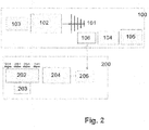
- Fig.2 represents another preferred embodiment of the invention wherein the data processing device 300 is integrated in the transmitter device 100 together with a Bluetooth transmitter/receiver 106.
- the transmitter 100 comprises an antenna 101, a transmitter 102, a power supply 103, a Bluetooth transmitter/receiver 106, a microprocessor 104 and a display 105.
- the receiving device 200 also comprises a Bluetooth transmitter 206 and sends the data to microprocessor 104 through Bluetooth transmitter/receiver 106.
- the operator can see directly on transmitter 100 the result of the analysis.
- Figure 3 is a picture of a prototype of the system according to the block diagram of figure 2 .
- the transmitter device comprises a screen which receives data from the receiver device through a Bluetooth connection. This prototype is very light and can be easily transported in a bag.
- the receiver is of a size of about 0.25 m x 0.20 m and contains four antennas positioned in a square configuration.
- Figure 4 shows the display of the transmitter of Figure 3 . Form the display the operator can follow the response of the analysis.
- the transmitter device comprises a single antenna, which is optionally a resonant cavity dipole antenna, or a directional antenna to be used in case of small organs, and a quartz oscillator to fix the specific emitted radio-frequency.
- Preferred types of directional antennas are Yagi antennas.
- the directional antenna used in the transmitter of the present inventions irradiates with an angle comprised between 10° and 30°.
- the transmitter is preferably structured to radiate a narrowband incident microwave frequency signal to irradiate a patient tissue.
- narrowband signal means a signal with a bandwidth Bw which is small enough to use the assumption that a response as a result of the interaction with the body can be considered constant within the bandwidth Bw, provided that 1/Bw is below the relaxation times of the irradiated patient biological tissues (typical Bw>1KHz).
- the frequency radiated by the transmitter device is comprised in the range from 2.3 GHz to 2.5 GHz, most preferably around about 2.4 GHZ, which means from 2.35 GHz to 2.45 GHz.
- the receiver device comprises more than one receiving antenna, a pre-processing module and a power supply.
- the power supply preferably comprises rechargeable batteries.
- the receiving antenna(s) are tuned on the frequency of the transmitting antenna, i.e. from 2.3 GHz to 2.5 GHz, preferably around about 2.4 GHZ, which means from 2.35 GHz to 2.45 GHz.
- the receiving antennas can be either directional or omni directional; preferably, the receiving antennas are capable of covering 180°.
- the pre-processing module preferably comprises a filter used to clean the signal excluding any other frequency different from the emitted frequency in order to eliminate electromagnetic noise of the environment.
- Signals from the system of receiving antennas are digitalized through the receiver and sent to the pre-processing module. From the pre-processing unit, the signal is sent to the processing unit which is preferably either in the transmitting device, or a separate device e.g. a laptop computer or an equivalent console.
- the data processing unit through a processing algorithm provides parameters representing measures of the backscattered wave-field in term of different characteristics such as attenuation, polarization, resonances and interferences.
- the data processing unit is configured to run algorithms which determine the presence of nulls or minima (i.e. values below a threshold) in the received signal and associated with the presence of tissue anomalies, more specifically cancers. Examples of these algorithms are disclosed in EP 2 465 428 .
- the data processing unit can also comprise a display and a power supply, which preferably comprises rechargeable batteries.
- the receiving device comprises a Bluetooth transmitter and communicates the data to transmitting device which comprises a data processing unit and visualizes the results of the diagnosis on a screen.
- the width of the receiver is preferably less than 0.30 m and the thickness less than 0.030 m. This reduced size and thickness of the receiver allows it to be placed on the medical couch under the patient in the position corresponding to the organ to be examined.
- the device according to the invention can be easily transported to a patient bed or in a different location without any problem. In fact, the entire device can be placed in a bag, which allows easy transportation.
- the overall weight of the device according to the invention is very limited, e.g. lower than 10 kg, preferably lower than 5 kg, more preferably lower than 2 kg.
- the device according to the invention can be used in any room and on any available medical couch and does not require a dedicated medical couch and room.
- the device of the present invention is preferably provided with rechargeable batteries which allow operation of the device without the need of external power supply.
- Another advantage of the device of the present invention is the capability of detecting the position of the cancer.
- the device according to the invention is capable of determining whether the cancer is located on the left or on the right lobe. To obtain this information, to date it was necessary to perform an NMR analysis. Analogously, for colorectal cancer, the device according to the invention allows a diagnosis of the intestinal tract where the cancer is present.
- the method of use of the device according to the invention is simple.
- the receiver is placed under the patient in correspondence to the organ to be checked.
- the transmitter is turned on and placed in contact with the body as close as possible to the organ to be examined.
- the screen of the system of Fig. 3 and 4 will provide the operator with a screenshot requesting selection of the organ to be examined.
- a possible procedure for prostate and colon examination is herewith presented as a way of example. However, it is evident that a different presentation of information is possible.
- other organs can be introduced in the menu of the system. If the organ is the prostate, the operator selects it and the screen will request examination of the left lobe of prostate. The operator will position the transmitter device in correspondence of the left lobe and will push a button on the screen to save the data. The screen will then request examination of the right lobe, and the operator will position the transmitter device on the right lobe and push the button again.
- the screen will request analysis of 6 different positions, which correspond to the relevant points to be examined: 1) right iliac fossa (caecum and ascending colon), 2) right hip (ascending colon and hepatic flexure), 3) mesogastrium (transverse colon, 4) left hip (splenic flexure and descending colon), 5) left iliac fossa (descending and sigmoid colon), 6) hypogastrium (sigmoid and colorectal colon).
- the operator will position the transmitter on each position in sequence and push the button to acquire the data.
- the system comprises a software adapted to the examination of at least one of the following organs: prostate, colon, breast and thyroid.
- the display of the system guides the examination and the operator by pushing a button saves the data measured by the system and indicating whether the examination is positive or negative.
- the system provides an outcome of the examination (positive or negative) and possibly the area where the cancer is located (left or right lobe for prostate, the area of the colon for colon, etc.).
- the processor is adapted to store data of a number of examinations corresponding to at least one working day.
- the processor can store at least 100 results of examinations.
- the operator can perform several examinations and at the end of the day connect the system to a processor such as a computer and download the data.
- the system of the invention is preferably adapted to transfer the stored data to an external processor. Transfer is preferably performed by the Bluetooth connection (106, 206).
- 155 urologic patients were evaluated by multiparametric NMR, and by the system of figure 3 and 4 according to the invention (2.4 GHz). The results are confronted with the results of a prostate transrectal eco guided biopsy, which is considered the gold standard in prostate cancer diagnosis.
- Table 1 show that the device according to the invention did not produce any false negative and a number of false positive comparable to the number obtained by NMR. It is important to note that false negatives are the most dangerous cases, since when a false negative occurs, a person which is diagnosed sane, is in fact affected by a cancer.
- Tables 3 and 4 report a statistical evaluation of the data of table 1 and 2 respectively by calculating sensitivity, specificity, negative predictive value (NPV), positive predictive value (PPV), and accuracy, wherein:
- Sensitivity TP / TP + FN ⁇ 100
- NPB TN / TN + FN ⁇ 100
- the device of the present invention is highly reliable and allows a screening of patients to detect the presence of a prostate cancer in an easy and economical manner.
- the device is also suitable for recognition of alteration of other organs, for example for the screening of colorectal cancer, breast cancer, thyroid cancer.
- a total of 107 consecutive patients fulfilling the inclusion criteria were enrolled over a period of 5 months.
- the most frequent indication for colonoscopy was constipation, diarrhea, abdominal pain or fecal blood.
- the system according to the invention detected and characterized all 32 adenocarcinomas and polyps.
- the method identified cancers and polyps with 96,97% sensitivity, 78,38% specificity, and 84,11% diagnostic accuracy, compared to colonoscopy.
- the positive predictive value was 66,67% and the negative predictive value 98,31%.
- Table 5 Sensitivity Specificity PPV NPV Accuracy 2.4 GHz 96.97 78.38 66.67 98.31 84.11
- Colonoscopy is a very invasive examination which requires a long preparation, while examination by the system according to the invention is easy to perform and does not require any preparation.
Landscapes
- Health & Medical Sciences (AREA)
- Life Sciences & Earth Sciences (AREA)
- Engineering & Computer Science (AREA)
- Veterinary Medicine (AREA)
- Animal Behavior & Ethology (AREA)
- Biophysics (AREA)
- Pathology (AREA)
- Physics & Mathematics (AREA)
- Biomedical Technology (AREA)
- Heart & Thoracic Surgery (AREA)
- Medical Informatics (AREA)
- Molecular Biology (AREA)
- Surgery (AREA)
- Public Health (AREA)
- General Health & Medical Sciences (AREA)
- Physiology (AREA)
- Computer Networks & Wireless Communication (AREA)
- Gynecology & Obstetrics (AREA)
- Reproductive Health (AREA)
- Endocrinology (AREA)
- Gastroenterology & Hepatology (AREA)
- Nuclear Medicine, Radiotherapy & Molecular Imaging (AREA)
- Radiology & Medical Imaging (AREA)
- Signal Processing (AREA)
- Computer Vision & Pattern Recognition (AREA)
- Artificial Intelligence (AREA)
- Psychiatry (AREA)
- Urology & Nephrology (AREA)
- Measuring And Recording Apparatus For Diagnosis (AREA)
- Measurement And Recording Of Electrical Phenomena And Electrical Characteristics Of The Living Body (AREA)
- Measurement Of The Respiration, Hearing Ability, Form, And Blood Characteristics Of Living Organisms (AREA)
- Magnetic Resonance Imaging Apparatus (AREA)
- Investigating Or Analysing Biological Materials (AREA)
Claims (11)
- System, das für die Erkennung von Krebs geeignet ist, wobei das System aufweist:a. eine Sendevorrichtung (100), die zum Ausstrahlen eines schmalbandigen, einfallenden Hochfrequenzsignals und zur Bestrahlung eines Patientengewebes eingerichtet ist, wobei die Sendevorrichtung (100) mindestens eine Sendeantenne (101), einen Sender (102) und eine Stromversorgung (103) umfasst;b. eine Empfangsvorrichtung (200) mit mehr als einer Empfangsantenne (201), einem Empfänger (202), einem Vorverarbeitungsmodul (204) und einer Stromversorgung (203); undc. eine Datenverarbeitungseinheit, die eingerichtet ist, Algorithmen auszuführen, die das Vorhandensein von Werten unterhalb eines Schwellwerts in dem empfangenen Signal bestimmen, die mit dem Vorhandensein eines Krebses in dem zu testenden Gewebe verbunden sind, mit einem Mikroprozessor (301; 104) und einer Anzeige (302; 105),
dadurch gekennzeichnet, dass die Sendevorrichtung (100) und die Empfangsvorrichtung (200) so ausgebildet sind, dass sie bei einer Frequenz zwischen 2,3 GHz und 2,5 GHz arbeiten. - System nach Anspruch 1, wobei die einen Mikroprozessor (104) und ein Display (105) aufweisende Datenverarbeitungseinheit zusammen mit einem Bluetooth-Sender/Empfänger (106) in die Sendevorrichtung (100) integriert ist, und wobei die Empfangsvorrichtung (200) ebenfalls einen Bluetooth-Sender/Empfänger (206) aufweist.
- System nach einem der Ansprüche 1-2, wobei die Betriebsfrequenz zwischen 2,35 GHz und 2,45 GHz liegt.
- System nach Anspruch 1-3, wobei die Sendevorrichtung eine einzelne Antenne und einen Quarzoszillator zur Fixierung der spezifischen ausgesendeten Funkfrequenz umfasst.
- System nach einem der Ansprüche 1-4, wobei die Sendeantenne (101) eine Richtantenne ist, die mit einem Winkel zwischen 10° und 30° abstrahlt.
- System nach Anspruch 5, wobei die Richtantenne eine Yagi-Antenne ist.
- System nach einem der Ansprüche 1-6, wobei die Stromversorgung (103, 203, 303) eine wiederaufladbare Batterie umfasst.
- System nach Anspruch 1-7, wobei der Prozessor eine Software umfasst, die zur Untersuchung von mindestens einem der folgenden Organe eingerichtet ist: Prostata, Kolon, Brust und Schilddrüse.
- System nach Anspruch 1-8, wobei der Mikroprozessor dafür eingerichtet ist, die Ergebnisse einer Anzahl von Untersuchungen zu speichern.
- System nach Anspruch 9, wobei der Mikroprozessor dafür eingerichtet ist, mindestens 100 Untersuchungsergebnisse zu speichern.
- System nach Anspruch 9-10, wobei das System dazu eingerichtet ist, die gespeicherten Daten an einen externen Prozessor zu übertragen.
Priority Applications (5)
| Application Number | Priority Date | Filing Date | Title |
|---|---|---|---|
| SM20210455T SMT202100455T1 (it) | 2018-10-29 | 2019-04-30 | Sistema per il riconoscimento di tumori nei tessuti umani |
| PL19723621T PL3672483T3 (pl) | 2018-10-29 | 2019-04-30 | System do wykrywania nowotworów w tkankach ludzkich |
| RS20210932A RS62205B1 (sr) | 2018-10-29 | 2019-04-30 | Sistem za detekciju kancera u humanim tkivima |
| HRP20211185TT HRP20211185T1 (hr) | 2018-10-29 | 2019-04-30 | Sustav za otkrivanje karcinoma u ljudskim tkivima |
| SI201930090T SI3672483T1 (sl) | 2018-10-29 | 2019-04-30 | Sistem za odkrivanje raka v človeških tkivih |
Applications Claiming Priority (2)
| Application Number | Priority Date | Filing Date | Title |
|---|---|---|---|
| EP18203207.8A EP3646785A1 (de) | 2018-10-29 | 2018-10-29 | Vorrichtung zur erkennung biologischer veränderungen in menschlichen geweben |
| PCT/EP2019/061088 WO2020088805A1 (en) | 2018-10-29 | 2019-04-30 | System for recognition of biological alteration in human tissues |
Publications (2)
| Publication Number | Publication Date |
|---|---|
| EP3672483A1 EP3672483A1 (de) | 2020-07-01 |
| EP3672483B1 true EP3672483B1 (de) | 2021-05-26 |
Family
ID=64316271
Family Applications (2)
| Application Number | Title | Priority Date | Filing Date |
|---|---|---|---|
| EP18203207.8A Withdrawn EP3646785A1 (de) | 2018-10-29 | 2018-10-29 | Vorrichtung zur erkennung biologischer veränderungen in menschlichen geweben |
| EP19723621.9A Active EP3672483B1 (de) | 2018-10-29 | 2019-04-30 | System zum nachweis von krebs in menschlichem gewebe |
Family Applications Before (1)
| Application Number | Title | Priority Date | Filing Date |
|---|---|---|---|
| EP18203207.8A Withdrawn EP3646785A1 (de) | 2018-10-29 | 2018-10-29 | Vorrichtung zur erkennung biologischer veränderungen in menschlichen geweben |
Country Status (21)
| Country | Link |
|---|---|
| US (1) | US20210378539A1 (de) |
| EP (2) | EP3646785A1 (de) |
| JP (2) | JP2022509376A (de) |
| KR (1) | KR102860258B1 (de) |
| CN (1) | CN112955072B (de) |
| AU (1) | AU2019373263B2 (de) |
| BR (1) | BR112021007941A2 (de) |
| CA (1) | CA3117073A1 (de) |
| CY (1) | CY1124398T1 (de) |
| DK (1) | DK3672483T3 (de) |
| ES (1) | ES2887929T3 (de) |
| HR (1) | HRP20211185T1 (de) |
| HU (1) | HUE055381T2 (de) |
| LT (1) | LT3672483T (de) |
| PL (1) | PL3672483T3 (de) |
| PT (1) | PT3672483T (de) |
| RS (1) | RS62205B1 (de) |
| SI (1) | SI3672483T1 (de) |
| SM (1) | SMT202100455T1 (de) |
| WO (1) | WO2020088805A1 (de) |
| ZA (1) | ZA202102676B (de) |
Families Citing this family (1)
| Publication number | Priority date | Publication date | Assignee | Title |
|---|---|---|---|---|
| DE202022101928U1 (de) | 2022-04-09 | 2022-07-01 | Manas Ranjan Chowdhury | Auf maschinellem Lernen basierendes System zur Erkennung von Brustkrebs mit einem Nahfeld-Mikrowellen-Antennensensor |
Family Cites Families (24)
| Publication number | Priority date | Publication date | Assignee | Title |
|---|---|---|---|---|
| US2017047A (en) * | 1932-03-11 | 1935-10-15 | Rca Corp | Directional antenna arrangement |
| FI58719C (fi) * | 1979-06-01 | 1981-04-10 | Instrumentarium Oy | Diagnostiseringsanordning foer broestkancer |
| US5829437A (en) * | 1994-07-01 | 1998-11-03 | Interstitial, Inc. | Microwave method and system to detect and locate cancers in heterogenous tissues |
| US5983124A (en) * | 1996-04-03 | 1999-11-09 | Microwave Medical Systems, Inc. | Microwave detection of tumors, particularly breast tumors |
| US10973397B2 (en) * | 1999-03-01 | 2021-04-13 | West View Research, Llc | Computerized information collection and processing apparatus |
| IT1310277B1 (it) | 1999-07-27 | 2002-02-11 | Clarbruno Vedruccio | Analizzatore elettromagnetico di anisotropia in sistemi chimiciorganizzati. |
| AU2003209357A1 (en) * | 2002-01-24 | 2003-09-02 | Breslin, John | Method of using electromagnetic absorption or perturbation spectra to diagnose and detect abnormalities in cells, tissues and organisms |
| US7811234B2 (en) * | 2002-08-01 | 2010-10-12 | California Institute Of Technology | Remote-sensing method and device |
| US7266407B2 (en) * | 2003-11-17 | 2007-09-04 | University Of Florida Research Foundation, Inc. | Multi-frequency microwave-induced thermoacoustic imaging of biological tissue |
| US8421642B1 (en) * | 2006-08-24 | 2013-04-16 | Navisense | System and method for sensorized user interface |
| JP2010540148A (ja) * | 2007-10-02 | 2010-12-24 | コーニンクレッカ フィリップス エレクトロニクス エヌ ヴィ | 電気的及び機械的心血管活動の検出 |
| EP2364647A1 (de) | 2010-03-09 | 2011-09-14 | Centro Studi e Ricerche Sant' Angela Srl | Automatische elektromagnetische Vorrichtung zur Erkennung und Diagnose von Anomalien in biologischen Geweben |
| PT2465428T (pt) | 2010-12-15 | 2017-07-17 | Medielma S R L | Sistema e método de deteção eletromagnética para a localização de tumores/calcificações em tecidos |
| US20130120081A1 (en) | 2011-11-14 | 2013-05-16 | Qualcomm Mems Technologies, Inc. | Combined resonators and passive circuit components for filter passband flattening |
| JP2014198067A (ja) * | 2013-03-29 | 2014-10-23 | 国立大学法人静岡大学 | 診断装置 |
| US9504404B1 (en) * | 2013-04-10 | 2016-11-29 | The University Of North Carolina At Charlotte | Antipodal vivaldi antenna array for biomedical imaging |
| US20140378810A1 (en) * | 2013-04-18 | 2014-12-25 | Digimarc Corporation | Physiologic data acquisition and analysis |
| KR102208687B1 (ko) * | 2014-02-26 | 2021-01-28 | 한국전자통신연구원 | 측정된 마이크로파 신호 분할에 의한 유방암 검출 장치 및 방법 |
| TWI563971B (zh) * | 2015-07-09 | 2017-01-01 | 緯創資通股份有限公司 | 微波成像裝置與方法 |
| JP6543146B2 (ja) * | 2015-09-14 | 2019-07-10 | 株式会社田定工作所 | マイクロ波を用いた呼吸器・循環器モニター装置 |
| KR101784394B1 (ko) * | 2016-03-31 | 2017-10-12 | 충남대학교산학협력단 | 유방암 자가 검사장치 및 그 방법 |
| EP3257439B1 (de) | 2016-06-13 | 2020-11-18 | Medielma S.r.l. | Gewebeanomaliedetektionsvorrichtung mit einer sondensendervorrichtung |
| WO2018127434A1 (en) * | 2017-01-09 | 2018-07-12 | Medfield Diagnostics Ab | Method and system for ensuring antenna contact and system function in applications of detecting internal dielectric properties in a body |
| US20180289291A1 (en) * | 2017-04-07 | 2018-10-11 | Bunnie Richie | Health diagnostic fingerprint scanner |
-
2018
- 2018-10-29 EP EP18203207.8A patent/EP3646785A1/de not_active Withdrawn
-
2019
- 2019-04-30 RS RS20210932A patent/RS62205B1/sr unknown
- 2019-04-30 BR BR112021007941-6A patent/BR112021007941A2/pt unknown
- 2019-04-30 EP EP19723621.9A patent/EP3672483B1/de active Active
- 2019-04-30 PT PT197236219T patent/PT3672483T/pt unknown
- 2019-04-30 US US17/287,518 patent/US20210378539A1/en active Pending
- 2019-04-30 PL PL19723621T patent/PL3672483T3/pl unknown
- 2019-04-30 SI SI201930090T patent/SI3672483T1/sl unknown
- 2019-04-30 WO PCT/EP2019/061088 patent/WO2020088805A1/en not_active Ceased
- 2019-04-30 JP JP2021547882A patent/JP2022509376A/ja active Pending
- 2019-04-30 SM SM20210455T patent/SMT202100455T1/it unknown
- 2019-04-30 CA CA3117073A patent/CA3117073A1/en active Pending
- 2019-04-30 CN CN201980071258.1A patent/CN112955072B/zh active Active
- 2019-04-30 LT LTEP19723621.9T patent/LT3672483T/lt unknown
- 2019-04-30 AU AU2019373263A patent/AU2019373263B2/en active Active
- 2019-04-30 HU HUE19723621A patent/HUE055381T2/hu unknown
- 2019-04-30 DK DK19723621.9T patent/DK3672483T3/da active
- 2019-04-30 KR KR1020217016492A patent/KR102860258B1/ko active Active
- 2019-04-30 HR HRP20211185TT patent/HRP20211185T1/hr unknown
- 2019-04-30 ES ES19723621T patent/ES2887929T3/es active Active
-
2021
- 2021-04-21 ZA ZA2021/02676A patent/ZA202102676B/en unknown
- 2021-07-08 CY CY20211100618T patent/CY1124398T1/el unknown
-
2023
- 2023-09-29 JP JP2023169721A patent/JP7635331B2/ja active Active
Also Published As
| Publication number | Publication date |
|---|---|
| HUE055381T2 (hu) | 2021-11-29 |
| SMT202100455T1 (it) | 2021-09-14 |
| CN112955072A (zh) | 2021-06-11 |
| SI3672483T1 (sl) | 2021-11-30 |
| JP2022509376A (ja) | 2022-01-20 |
| RS62205B1 (sr) | 2021-08-31 |
| AU2019373263A1 (en) | 2021-05-20 |
| HRP20211185T1 (hr) | 2021-10-15 |
| EP3646785A1 (de) | 2020-05-06 |
| JP2024001093A (ja) | 2024-01-09 |
| DK3672483T3 (da) | 2021-07-12 |
| CA3117073A1 (en) | 2020-05-07 |
| EP3672483A1 (de) | 2020-07-01 |
| CN112955072B (zh) | 2024-08-30 |
| KR102860258B1 (ko) | 2025-09-15 |
| PL3672483T3 (pl) | 2021-12-13 |
| BR112021007941A2 (pt) | 2021-07-27 |
| WO2020088805A1 (en) | 2020-05-07 |
| ZA202102676B (en) | 2022-07-27 |
| AU2019373263B2 (en) | 2024-11-21 |
| LT3672483T (lt) | 2021-09-27 |
| JP7635331B2 (ja) | 2025-02-25 |
| US20210378539A1 (en) | 2021-12-09 |
| KR20210086688A (ko) | 2021-07-08 |
| PT3672483T (pt) | 2021-07-22 |
| ES2887929T3 (es) | 2021-12-29 |
| CY1124398T1 (el) | 2022-07-22 |
Similar Documents
| Publication | Publication Date | Title |
|---|---|---|
| Mobashsher et al. | Design and experimental evaluation of a non-invasive microwave head imaging system for intracranial haemorrhage detection | |
| US8050740B2 (en) | Microwave-based examination using hypothesis testing | |
| KR101307514B1 (ko) | 마이크로파 영상 복원 장치 | |
| KR101308409B1 (ko) | 물체형상 3차원 측정 장치 | |
| JP7212827B2 (ja) | 診断装置、トモグラフィ処理の実行方法、診断プログラム | |
| WO2018127434A1 (en) | Method and system for ensuring antenna contact and system function in applications of detecting internal dielectric properties in a body | |
| EP3672483B1 (de) | System zum nachweis von krebs in menschlichem gewebe | |
| KR20190004511A (ko) | 테라헤르츠 전자기파를 이용한 영상 처리장치 | |
| US20150282715A1 (en) | Passive, noninvasive tomography | |
| US12575752B2 (en) | Microwave breast cancer screening system | |
| KR101052484B1 (ko) | 유방암 진단 장치 및 방법 | |
| HK40053231A (en) | System for recognition of biological alteration in human tissues | |
| HK40053231B (zh) | 用於识别人体组织内生物学变化的系统 | |
| Ueda et al. | Clinical Validations on Effective Skin Clutter Rejection for Microwave Breast Cancer Diagnosis | |
| Tashtarian et al. | Resolution Enhancement of Sub-Wavelength Imaging Using Magnetic-Dominated Fields | |
| US12471835B1 (en) | Methods and systems for detection and differentiation of tumors | |
| Rana | Development and evaluation of a sensor and antenna array for a portable microwave-based breast cancer detection system | |
| KR101380788B1 (ko) | 펄스파를 이용한 폐암 진단 장치 및 폐암 진단 방법 | |
| AlQasem et al. | Hemorrhagic Brain Strokes Detection Using Recurrent Neural Networks-Based Microwave Imaging Technique | |
| KR20180079742A (ko) | 전자파를 이용한 최소 침습 암 진단 시스템 및 방법 | |
| Kuwahara et al. | Clinical evaluation of microwave breast imaging using scattering tomography | |
| KR100780402B1 (ko) | 암 탐지 장치 | |
| Scapaticci et al. | Optimized antenna array layout in a microwave imaging system for brain stroke monitoring | |
| KR101556921B1 (ko) | 전자파를 이용한 고속 폐암 진단 장치 및 그 방법 | |
| KR101077556B1 (ko) | 유방암 진단 장치 및 방법 |
Legal Events
| Date | Code | Title | Description |
|---|---|---|---|
| STAA | Information on the status of an ep patent application or granted ep patent |
Free format text: STATUS: UNKNOWN |
|
| STAA | Information on the status of an ep patent application or granted ep patent |
Free format text: STATUS: THE INTERNATIONAL PUBLICATION HAS BEEN MADE |
|
| PUAI | Public reference made under article 153(3) epc to a published international application that has entered the european phase |
Free format text: ORIGINAL CODE: 0009012 |
|
| STAA | Information on the status of an ep patent application or granted ep patent |
Free format text: STATUS: REQUEST FOR EXAMINATION WAS MADE |
|
| 17P | Request for examination filed |
Effective date: 20191003 |
|
| AK | Designated contracting states |
Kind code of ref document: A1 Designated state(s): AL AT BE BG CH CY CZ DE DK EE ES FI FR GB GR HR HU IE IS IT LI LT LU LV MC MK MT NL NO PL PT RO RS SE SI SK SM TR |
|
| AX | Request for extension of the european patent |
Extension state: BA ME |
|
| RAP1 | Party data changed (applicant data changed or rights of an application transferred) |
Owner name: PAEGASUS MEDICAL SA |
|
| STAA | Information on the status of an ep patent application or granted ep patent |
Free format text: STATUS: EXAMINATION IS IN PROGRESS |
|
| 17Q | First examination report despatched |
Effective date: 20201215 |
|
| RIN1 | Information on inventor provided before grant (corrected) |
Inventor name: CANEPA, STEFANO NICOLO |
|
| REG | Reference to a national code |
Ref country code: DE Ref legal event code: R079 Ref document number: 602019004923 Country of ref document: DE Free format text: PREVIOUS MAIN CLASS: A61B0005050000 Ipc: A61B0005050700 |
|
| GRAP | Despatch of communication of intention to grant a patent |
Free format text: ORIGINAL CODE: EPIDOSNIGR1 |
|
| STAA | Information on the status of an ep patent application or granted ep patent |
Free format text: STATUS: GRANT OF PATENT IS INTENDED |
|
| RIC1 | Information provided on ipc code assigned before grant |
Ipc: A61B 5/00 20060101ALI20210129BHEP Ipc: A61B 5/0507 20210101AFI20210129BHEP Ipc: A61B 5/053 20210101ALI20210129BHEP |
|
| DAV | Request for validation of the european patent (deleted) | ||
| DAX | Request for extension of the european patent (deleted) | ||
| INTG | Intention to grant announced |
Effective date: 20210305 |
|
| GRAS | Grant fee paid |
Free format text: ORIGINAL CODE: EPIDOSNIGR3 |
|
| GRAA | (expected) grant |
Free format text: ORIGINAL CODE: 0009210 |
|
| STAA | Information on the status of an ep patent application or granted ep patent |
Free format text: STATUS: THE PATENT HAS BEEN GRANTED |
|
| AK | Designated contracting states |
Kind code of ref document: B1 Designated state(s): AL AT BE BG CH CY CZ DE DK EE ES FI FR GB GR HR HU IE IS IT LI LT LU LV MC MK MT NL NO PL PT RO RS SE SI SK SM TR |
|
| REG | Reference to a national code |
Ref country code: GB Ref legal event code: FG4D |
|
| REG | Reference to a national code |
Ref country code: CH Ref legal event code: EP |
|
| REG | Reference to a national code |
Ref country code: AT Ref legal event code: REF Ref document number: 1395386 Country of ref document: AT Kind code of ref document: T Effective date: 20210615 |
|
| REG | Reference to a national code |
Ref country code: DE Ref legal event code: R096 Ref document number: 602019004923 Country of ref document: DE |
|
| REG | Reference to a national code |
Ref country code: IE Ref legal event code: FG4D |
|
| REG | Reference to a national code |
Ref country code: FI Ref legal event code: FGE |
|
| REG | Reference to a national code |
Ref country code: DK Ref legal event code: T3 Effective date: 20210708 |
|
| REG | Reference to a national code |
Ref country code: RO Ref legal event code: EPE |
|
| REG | Reference to a national code |
Ref country code: PT Ref legal event code: SC4A Ref document number: 3672483 Country of ref document: PT Date of ref document: 20210722 Kind code of ref document: T Free format text: AVAILABILITY OF NATIONAL TRANSLATION Effective date: 20210716 |
|
| REG | Reference to a national code |
Ref country code: NO Ref legal event code: T2 Effective date: 20210526 |
|
| REG | Reference to a national code |
Ref country code: NL Ref legal event code: FP |
|
| REG | Reference to a national code |
Ref country code: SE Ref legal event code: TRGR |
|
| REG | Reference to a national code |
Ref country code: EE Ref legal event code: FG4A Ref document number: E021068 Country of ref document: EE Effective date: 20210728 |
|
| REG | Reference to a national code |
Ref country code: GR Ref legal event code: EP Ref document number: 20210402133 Country of ref document: GR Effective date: 20210915 |
|
| REG | Reference to a national code |
Ref country code: SK Ref legal event code: T3 Ref document number: E 37941 Country of ref document: SK |
|
| REG | Reference to a national code |
Ref country code: HR Ref legal event code: T1PR Ref document number: P20211185 Country of ref document: HR |
|
| REG | Reference to a national code |
Ref country code: HU Ref legal event code: AG4A Ref document number: E055381 Country of ref document: HU |
|
| PG25 | Lapsed in a contracting state [announced via postgrant information from national office to epo] |
Ref country code: IS Free format text: LAPSE BECAUSE OF FAILURE TO SUBMIT A TRANSLATION OF THE DESCRIPTION OR TO PAY THE FEE WITHIN THE PRESCRIBED TIME-LIMIT Effective date: 20210926 |
|
| REG | Reference to a national code |
Ref country code: ES Ref legal event code: FG2A Ref document number: 2887929 Country of ref document: ES Kind code of ref document: T3 Effective date: 20211229 |
|
| REG | Reference to a national code |
Ref country code: DE Ref legal event code: R097 Ref document number: 602019004923 Country of ref document: DE |
|
| PLBE | No opposition filed within time limit |
Free format text: ORIGINAL CODE: 0009261 |
|
| STAA | Information on the status of an ep patent application or granted ep patent |
Free format text: STATUS: NO OPPOSITION FILED WITHIN TIME LIMIT |
|
| 26N | No opposition filed |
Effective date: 20220301 |
|
| REG | Reference to a national code |
Ref country code: HR Ref legal event code: ODRP Ref document number: P20211185 Country of ref document: HR Payment date: 20220428 Year of fee payment: 4 |
|
| PG25 | Lapsed in a contracting state [announced via postgrant information from national office to epo] |
Ref country code: IS Free format text: LAPSE BECAUSE OF FAILURE TO SUBMIT A TRANSLATION OF THE DESCRIPTION OR TO PAY THE FEE WITHIN THE PRESCRIBED TIME-LIMIT Effective date: 20210926 |
|
| PG25 | Lapsed in a contracting state [announced via postgrant information from national office to epo] |
Ref country code: PT Free format text: LAPSE BECAUSE OF NON-PAYMENT OF DUE FEES Effective date: 20230201 |
|
| REG | Reference to a national code |
Ref country code: HR Ref legal event code: ODRP Ref document number: P20211185 Country of ref document: HR Payment date: 20230421 Year of fee payment: 5 |
|
| P01 | Opt-out of the competence of the unified patent court (upc) registered |
Effective date: 20230615 |
|
| PG25 | Lapsed in a contracting state [announced via postgrant information from national office to epo] |
Ref country code: PT Free format text: LAPSE BECAUSE OF NON-PAYMENT OF DUE FEES Effective date: 20230201 |
|
| PGRI | Patent reinstated in contracting state [announced from national office to epo] |
Ref country code: PT Effective date: 20230424 |
|
| REG | Reference to a national code |
Ref country code: HR Ref legal event code: ODRP Ref document number: P20211185 Country of ref document: HR Payment date: 20240415 Year of fee payment: 6 |
|
| REG | Reference to a national code |
Ref country code: DE Ref legal event code: R082 Ref document number: 602019004923 Country of ref document: DE Representative=s name: KBN IP PATENTANWAELTE PARTNERSCHAFT MBB, DE |
|
| REG | Reference to a national code |
Ref country code: HR Ref legal event code: ODRP Ref document number: P20211185 Country of ref document: HR Payment date: 20250424 Year of fee payment: 7 |
|
| PGFP | Annual fee paid to national office [announced via postgrant information from national office to epo] |
Ref country code: NL Payment date: 20250422 Year of fee payment: 7 |
|
| PGFP | Annual fee paid to national office [announced via postgrant information from national office to epo] |
Ref country code: LU Payment date: 20250422 Year of fee payment: 7 |
|
| PGFP | Annual fee paid to national office [announced via postgrant information from national office to epo] |
Ref country code: SM Payment date: 20250417 Year of fee payment: 7 |
|
| PGFP | Annual fee paid to national office [announced via postgrant information from national office to epo] |
Ref country code: MC Payment date: 20250418 Year of fee payment: 7 |
|
| PGFP | Annual fee paid to national office [announced via postgrant information from national office to epo] |
Ref country code: FI Payment date: 20250420 Year of fee payment: 7 |
|
| PGFP | Annual fee paid to national office [announced via postgrant information from national office to epo] |
Ref country code: PL Payment date: 20250408 Year of fee payment: 7 Ref country code: DE Payment date: 20250424 Year of fee payment: 7 |
|
| PGFP | Annual fee paid to national office [announced via postgrant information from national office to epo] |
Ref country code: GB Payment date: 20250423 Year of fee payment: 7 Ref country code: ES Payment date: 20250504 Year of fee payment: 7 Ref country code: DK Payment date: 20250416 Year of fee payment: 7 |
|
| PGFP | Annual fee paid to national office [announced via postgrant information from national office to epo] |
Ref country code: LT Payment date: 20250416 Year of fee payment: 7 |
|
| PGFP | Annual fee paid to national office [announced via postgrant information from national office to epo] |
Ref country code: HU Payment date: 20250423 Year of fee payment: 7 Ref country code: IS Payment date: 20250422 Year of fee payment: 7 Ref country code: RS Payment date: 20250423 Year of fee payment: 7 Ref country code: NO Payment date: 20250422 Year of fee payment: 7 |
|
| PGFP | Annual fee paid to national office [announced via postgrant information from national office to epo] |
Ref country code: AL Payment date: 20250424 Year of fee payment: 7 Ref country code: BE Payment date: 20250420 Year of fee payment: 7 Ref country code: IT Payment date: 20250424 Year of fee payment: 7 |
|
| PGFP | Annual fee paid to national office [announced via postgrant information from national office to epo] |
Ref country code: HR Payment date: 20250424 Year of fee payment: 7 |
|
| PGFP | Annual fee paid to national office [announced via postgrant information from national office to epo] |
Ref country code: PT Payment date: 20250409 Year of fee payment: 7 Ref country code: LV Payment date: 20250420 Year of fee payment: 7 |
|
| PGFP | Annual fee paid to national office [announced via postgrant information from national office to epo] |
Ref country code: FR Payment date: 20250424 Year of fee payment: 7 Ref country code: EE Payment date: 20250616 Year of fee payment: 7 |
|
| PGFP | Annual fee paid to national office [announced via postgrant information from national office to epo] |
Ref country code: MT Payment date: 20250416 Year of fee payment: 7 Ref country code: GR Payment date: 20250422 Year of fee payment: 7 Ref country code: BG Payment date: 20250422 Year of fee payment: 7 |
|
| PGFP | Annual fee paid to national office [announced via postgrant information from national office to epo] |
Ref country code: RO Payment date: 20250422 Year of fee payment: 7 Ref country code: AT Payment date: 20250417 Year of fee payment: 7 |
|
| PGFP | Annual fee paid to national office [announced via postgrant information from national office to epo] |
Ref country code: TR Payment date: 20250417 Year of fee payment: 7 Ref country code: SK Payment date: 20250417 Year of fee payment: 7 |
|
| PGFP | Annual fee paid to national office [announced via postgrant information from national office to epo] |
Ref country code: CZ Payment date: 20250416 Year of fee payment: 7 |
|
| PGFP | Annual fee paid to national office [announced via postgrant information from national office to epo] |
Ref country code: IE Payment date: 20250417 Year of fee payment: 7 |
|
| PGFP | Annual fee paid to national office [announced via postgrant information from national office to epo] |
Ref country code: SI Payment date: 20250416 Year of fee payment: 7 Ref country code: SE Payment date: 20250506 Year of fee payment: 7 |
|
| PGFP | Annual fee paid to national office [announced via postgrant information from national office to epo] |
Ref country code: CH Payment date: 20250817 Year of fee payment: 7 |
|
| PGFP | Annual fee paid to national office [announced via postgrant information from national office to epo] |
Ref country code: MK Payment date: 20250424 Year of fee payment: 7 |
|
| PGFP | Annual fee paid to national office [announced via postgrant information from national office to epo] |
Ref country code: CY Payment date: 20250414 Year of fee payment: 7 |