EP3671208A1 - Ige gesteuerte strukturbestimmung von zielmolekülen unter verwendung von cryo-em - Google Patents
Ige gesteuerte strukturbestimmung von zielmolekülen unter verwendung von cryo-em Download PDFInfo
- Publication number
- EP3671208A1 EP3671208A1 EP18215047.4A EP18215047A EP3671208A1 EP 3671208 A1 EP3671208 A1 EP 3671208A1 EP 18215047 A EP18215047 A EP 18215047A EP 3671208 A1 EP3671208 A1 EP 3671208A1
- Authority
- EP
- European Patent Office
- Prior art keywords
- ige
- target molecule
- complex
- domain
- fragment
- Prior art date
- Legal status (The legal status is an assumption and is not a legal conclusion. Google has not performed a legal analysis and makes no representation as to the accuracy of the status listed.)
- Withdrawn
Links
- 239000012634 fragment Substances 0.000 claims abstract description 51
- 238000000034 method Methods 0.000 claims abstract description 49
- 238000001493 electron microscopy Methods 0.000 claims abstract description 42
- 102000004169 proteins and genes Human genes 0.000 claims description 26
- 108090000623 proteins and genes Proteins 0.000 claims description 26
- 239000011230 binding agent Substances 0.000 claims description 15
- 230000001939 inductive effect Effects 0.000 claims description 11
- 230000001965 increasing effect Effects 0.000 claims description 9
- 230000000536 complexating effect Effects 0.000 claims description 4
- 230000001404 mediated effect Effects 0.000 claims description 2
- 239000003795 chemical substances by application Substances 0.000 abstract description 6
- 229950009923 ligelizumab Drugs 0.000 description 38
- 239000000427 antigen Substances 0.000 description 35
- 108091007433 antigens Proteins 0.000 description 30
- 102000036639 antigens Human genes 0.000 description 30
- 238000002439 negative-stain electron microscopy Methods 0.000 description 21
- 238000012935 Averaging Methods 0.000 description 13
- 108010047041 Complementarity Determining Regions Proteins 0.000 description 10
- 108010003723 Single-Domain Antibodies Proteins 0.000 description 10
- 230000008901 benefit Effects 0.000 description 7
- 239000000463 material Substances 0.000 description 7
- 239000002245 particle Substances 0.000 description 7
- 239000000523 sample Substances 0.000 description 7
- 239000000411 inducer Substances 0.000 description 6
- 230000003993 interaction Effects 0.000 description 6
- OKTJSMMVPCPJKN-UHFFFAOYSA-N Carbon Chemical compound [C] OKTJSMMVPCPJKN-UHFFFAOYSA-N 0.000 description 5
- 208000026935 allergic disease Diseases 0.000 description 5
- 238000004458 analytical method Methods 0.000 description 5
- 239000013078 crystal Substances 0.000 description 5
- 108091023037 Aptamer Proteins 0.000 description 4
- 108010054477 Immunoglobulin Fab Fragments Proteins 0.000 description 4
- 102000001706 Immunoglobulin Fab Fragments Human genes 0.000 description 4
- FAPWRFPIFSIZLT-UHFFFAOYSA-M Sodium chloride Chemical compound [Na+].[Cl-] FAPWRFPIFSIZLT-UHFFFAOYSA-M 0.000 description 4
- 239000000243 solution Substances 0.000 description 4
- RYGMFSIKBFXOCR-UHFFFAOYSA-N Copper Chemical compound [Cu] RYGMFSIKBFXOCR-UHFFFAOYSA-N 0.000 description 3
- 241000238631 Hexapoda Species 0.000 description 3
- 108060003951 Immunoglobulin Proteins 0.000 description 3
- 108090001090 Lectins Proteins 0.000 description 3
- 102000004856 Lectins Human genes 0.000 description 3
- 239000012082 adaptor molecule Substances 0.000 description 3
- 230000009286 beneficial effect Effects 0.000 description 3
- 230000015572 biosynthetic process Effects 0.000 description 3
- 229910052799 carbon Inorganic materials 0.000 description 3
- 210000004027 cell Anatomy 0.000 description 3
- 150000001875 compounds Chemical class 0.000 description 3
- 229910052802 copper Inorganic materials 0.000 description 3
- 239000010949 copper Substances 0.000 description 3
- 230000008014 freezing Effects 0.000 description 3
- 238000007710 freezing Methods 0.000 description 3
- 229910001385 heavy metal Inorganic materials 0.000 description 3
- 238000003384 imaging method Methods 0.000 description 3
- 102000018358 immunoglobulin Human genes 0.000 description 3
- 239000002523 lectin Substances 0.000 description 3
- 238000001000 micrograph Methods 0.000 description 3
- 238000002360 preparation method Methods 0.000 description 3
- 238000001464 small-angle X-ray scattering data Methods 0.000 description 3
- 238000013519 translation Methods 0.000 description 3
- JKMHFZQWWAIEOD-UHFFFAOYSA-N 2-[4-(2-hydroxyethyl)piperazin-1-yl]ethanesulfonic acid Chemical compound OCC[NH+]1CCN(CCS([O-])(=O)=O)CC1 JKMHFZQWWAIEOD-UHFFFAOYSA-N 0.000 description 2
- 108010078791 Carrier Proteins Proteins 0.000 description 2
- 108010061608 Dermatophagoides pteronyssinus antigen p 2 Proteins 0.000 description 2
- OTMSDBZUPAUEDD-UHFFFAOYSA-N Ethane Chemical compound CC OTMSDBZUPAUEDD-UHFFFAOYSA-N 0.000 description 2
- 239000007995 HEPES buffer Substances 0.000 description 2
- 206010020751 Hypersensitivity Diseases 0.000 description 2
- 102000018071 Immunoglobulin Fc Fragments Human genes 0.000 description 2
- 108010091135 Immunoglobulin Fc Fragments Proteins 0.000 description 2
- BAVYZALUXZFZLV-UHFFFAOYSA-N Methylamine Chemical compound NC BAVYZALUXZFZLV-UHFFFAOYSA-N 0.000 description 2
- 241001529936 Murinae Species 0.000 description 2
- 108010001267 Protein Subunits Proteins 0.000 description 2
- 102000002067 Protein Subunits Human genes 0.000 description 2
- 239000013566 allergen Substances 0.000 description 2
- 201000009961 allergic asthma Diseases 0.000 description 2
- 230000007815 allergy Effects 0.000 description 2
- 229910003481 amorphous carbon Inorganic materials 0.000 description 2
- 230000009830 antibody antigen interaction Effects 0.000 description 2
- 208000006673 asthma Diseases 0.000 description 2
- 208000024376 chronic urticaria Diseases 0.000 description 2
- 238000013480 data collection Methods 0.000 description 2
- 238000002050 diffraction method Methods 0.000 description 2
- 238000010494 dissociation reaction Methods 0.000 description 2
- 230000005593 dissociations Effects 0.000 description 2
- 239000003814 drug Substances 0.000 description 2
- 239000000428 dust Substances 0.000 description 2
- 230000000694 effects Effects 0.000 description 2
- 238000000635 electron micrograph Methods 0.000 description 2
- 238000002474 experimental method Methods 0.000 description 2
- 150000004676 glycans Chemical class 0.000 description 2
- 229910021389 graphene Inorganic materials 0.000 description 2
- 210000004408 hybridoma Anatomy 0.000 description 2
- 230000036039 immunity Effects 0.000 description 2
- 238000011835 investigation Methods 0.000 description 2
- 239000007788 liquid Substances 0.000 description 2
- 238000005259 measurement Methods 0.000 description 2
- 108091033319 polynucleotide Proteins 0.000 description 2
- 239000002157 polynucleotide Substances 0.000 description 2
- 102000040430 polynucleotide Human genes 0.000 description 2
- 238000012545 processing Methods 0.000 description 2
- 150000003839 salts Chemical class 0.000 description 2
- 238000001542 size-exclusion chromatography Methods 0.000 description 2
- 238000000235 small-angle X-ray scattering Methods 0.000 description 2
- 239000011780 sodium chloride Substances 0.000 description 2
- 238000010186 staining Methods 0.000 description 2
- 238000004627 transmission electron microscopy Methods 0.000 description 2
- PBYZMCDFOULPGH-UHFFFAOYSA-N tungstate Chemical compound [O-][W]([O-])(=O)=O PBYZMCDFOULPGH-UHFFFAOYSA-N 0.000 description 2
- SFIHWLKHBCDNCE-UHFFFAOYSA-N uranyl formate Chemical compound OC=O.OC=O.O=[U]=O SFIHWLKHBCDNCE-UHFFFAOYSA-N 0.000 description 2
- 241000894006 Bacteria Species 0.000 description 1
- SHZGCJCMOBCMKK-UHFFFAOYSA-N D-mannomethylose Natural products CC1OC(O)C(O)C(O)C1O SHZGCJCMOBCMKK-UHFFFAOYSA-N 0.000 description 1
- 206010012438 Dermatitis atopic Diseases 0.000 description 1
- 108090000790 Enzymes Proteins 0.000 description 1
- 102000004190 Enzymes Human genes 0.000 description 1
- 241000242711 Fasciola hepatica Species 0.000 description 1
- PNNNRSAQSRJVSB-SLPGGIOYSA-N Fucose Natural products C[C@H](O)[C@@H](O)[C@H](O)[C@H](O)C=O PNNNRSAQSRJVSB-SLPGGIOYSA-N 0.000 description 1
- 108090000045 G-Protein-Coupled Receptors Proteins 0.000 description 1
- 102000003688 G-Protein-Coupled Receptors Human genes 0.000 description 1
- 102000009438 IgE Receptors Human genes 0.000 description 1
- 108010073816 IgE Receptors Proteins 0.000 description 1
- 208000001718 Immediate Hypersensitivity Diseases 0.000 description 1
- 108010021625 Immunoglobulin Fragments Proteins 0.000 description 1
- 102000008394 Immunoglobulin Fragments Human genes 0.000 description 1
- 102000004310 Ion Channels Human genes 0.000 description 1
- 108090000862 Ion Channels Proteins 0.000 description 1
- SHZGCJCMOBCMKK-DHVFOXMCSA-N L-fucopyranose Chemical compound C[C@@H]1OC(O)[C@@H](O)[C@H](O)[C@@H]1O SHZGCJCMOBCMKK-DHVFOXMCSA-N 0.000 description 1
- 241000124008 Mammalia Species 0.000 description 1
- 241001465754 Metazoa Species 0.000 description 1
- ZOKXTWBITQBERF-UHFFFAOYSA-N Molybdenum Chemical compound [Mo] ZOKXTWBITQBERF-UHFFFAOYSA-N 0.000 description 1
- 101710159910 Movement protein Proteins 0.000 description 1
- 108010085220 Multiprotein Complexes Proteins 0.000 description 1
- 102000007474 Multiprotein Complexes Human genes 0.000 description 1
- 108091034117 Oligonucleotide Proteins 0.000 description 1
- 241000223960 Plasmodium falciparum Species 0.000 description 1
- 206010039085 Rhinitis allergic Diseases 0.000 description 1
- 241000242680 Schistosoma mansoni Species 0.000 description 1
- RTAQQCXQSZGOHL-UHFFFAOYSA-N Titanium Chemical compound [Ti] RTAQQCXQSZGOHL-UHFFFAOYSA-N 0.000 description 1
- 241000243777 Trichinella spiralis Species 0.000 description 1
- 206010045240 Type I hypersensitivity Diseases 0.000 description 1
- 229910052770 Uranium Inorganic materials 0.000 description 1
- COQLPRJCUIATTQ-UHFFFAOYSA-N Uranyl acetate Chemical compound O.O.O=[U]=O.CC(O)=O.CC(O)=O COQLPRJCUIATTQ-UHFFFAOYSA-N 0.000 description 1
- 206010052568 Urticaria chronic Diseases 0.000 description 1
- 238000004279 X-ray Guinier Methods 0.000 description 1
- JLCPHMBAVCMARE-UHFFFAOYSA-N [3-[[3-[[3-[[3-[[3-[[3-[[3-[[3-[[3-[[3-[[3-[[5-(2-amino-6-oxo-1H-purin-9-yl)-3-[[3-[[3-[[3-[[3-[[3-[[5-(2-amino-6-oxo-1H-purin-9-yl)-3-[[5-(2-amino-6-oxo-1H-purin-9-yl)-3-hydroxyoxolan-2-yl]methoxy-hydroxyphosphoryl]oxyoxolan-2-yl]methoxy-hydroxyphosphoryl]oxy-5-(5-methyl-2,4-dioxopyrimidin-1-yl)oxolan-2-yl]methoxy-hydroxyphosphoryl]oxy-5-(6-aminopurin-9-yl)oxolan-2-yl]methoxy-hydroxyphosphoryl]oxy-5-(6-aminopurin-9-yl)oxolan-2-yl]methoxy-hydroxyphosphoryl]oxy-5-(6-aminopurin-9-yl)oxolan-2-yl]methoxy-hydroxyphosphoryl]oxy-5-(6-aminopurin-9-yl)oxolan-2-yl]methoxy-hydroxyphosphoryl]oxyoxolan-2-yl]methoxy-hydroxyphosphoryl]oxy-5-(5-methyl-2,4-dioxopyrimidin-1-yl)oxolan-2-yl]methoxy-hydroxyphosphoryl]oxy-5-(4-amino-2-oxopyrimidin-1-yl)oxolan-2-yl]methoxy-hydroxyphosphoryl]oxy-5-(5-methyl-2,4-dioxopyrimidin-1-yl)oxolan-2-yl]methoxy-hydroxyphosphoryl]oxy-5-(5-methyl-2,4-dioxopyrimidin-1-yl)oxolan-2-yl]methoxy-hydroxyphosphoryl]oxy-5-(6-aminopurin-9-yl)oxolan-2-yl]methoxy-hydroxyphosphoryl]oxy-5-(6-aminopurin-9-yl)oxolan-2-yl]methoxy-hydroxyphosphoryl]oxy-5-(4-amino-2-oxopyrimidin-1-yl)oxolan-2-yl]methoxy-hydroxyphosphoryl]oxy-5-(4-amino-2-oxopyrimidin-1-yl)oxolan-2-yl]methoxy-hydroxyphosphoryl]oxy-5-(4-amino-2-oxopyrimidin-1-yl)oxolan-2-yl]methoxy-hydroxyphosphoryl]oxy-5-(6-aminopurin-9-yl)oxolan-2-yl]methoxy-hydroxyphosphoryl]oxy-5-(4-amino-2-oxopyrimidin-1-yl)oxolan-2-yl]methyl [5-(6-aminopurin-9-yl)-2-(hydroxymethyl)oxolan-3-yl] hydrogen phosphate Polymers Cc1cn(C2CC(OP(O)(=O)OCC3OC(CC3OP(O)(=O)OCC3OC(CC3O)n3cnc4c3nc(N)[nH]c4=O)n3cnc4c3nc(N)[nH]c4=O)C(COP(O)(=O)OC3CC(OC3COP(O)(=O)OC3CC(OC3COP(O)(=O)OC3CC(OC3COP(O)(=O)OC3CC(OC3COP(O)(=O)OC3CC(OC3COP(O)(=O)OC3CC(OC3COP(O)(=O)OC3CC(OC3COP(O)(=O)OC3CC(OC3COP(O)(=O)OC3CC(OC3COP(O)(=O)OC3CC(OC3COP(O)(=O)OC3CC(OC3COP(O)(=O)OC3CC(OC3COP(O)(=O)OC3CC(OC3COP(O)(=O)OC3CC(OC3COP(O)(=O)OC3CC(OC3COP(O)(=O)OC3CC(OC3COP(O)(=O)OC3CC(OC3CO)n3cnc4c(N)ncnc34)n3ccc(N)nc3=O)n3cnc4c(N)ncnc34)n3ccc(N)nc3=O)n3ccc(N)nc3=O)n3ccc(N)nc3=O)n3cnc4c(N)ncnc34)n3cnc4c(N)ncnc34)n3cc(C)c(=O)[nH]c3=O)n3cc(C)c(=O)[nH]c3=O)n3ccc(N)nc3=O)n3cc(C)c(=O)[nH]c3=O)n3cnc4c3nc(N)[nH]c4=O)n3cnc4c(N)ncnc34)n3cnc4c(N)ncnc34)n3cnc4c(N)ncnc34)n3cnc4c(N)ncnc34)O2)c(=O)[nH]c1=O JLCPHMBAVCMARE-UHFFFAOYSA-N 0.000 description 1
- 238000001042 affinity chromatography Methods 0.000 description 1
- 201000010105 allergic rhinitis Diseases 0.000 description 1
- 150000001413 amino acids Chemical class 0.000 description 1
- 238000013459 approach Methods 0.000 description 1
- 239000008346 aqueous phase Substances 0.000 description 1
- 208000010216 atopic IgE responsiveness Diseases 0.000 description 1
- 201000008937 atopic dermatitis Diseases 0.000 description 1
- 210000003651 basophil Anatomy 0.000 description 1
- 239000003153 chemical reaction reagent Substances 0.000 description 1
- 206010072757 chronic spontaneous urticaria Diseases 0.000 description 1
- 230000009918 complex formation Effects 0.000 description 1
- 238000010276 construction Methods 0.000 description 1
- 239000000356 contaminant Substances 0.000 description 1
- 238000002425 crystallisation Methods 0.000 description 1
- 230000008025 crystallization Effects 0.000 description 1
- 238000007405 data analysis Methods 0.000 description 1
- 230000001419 dependent effect Effects 0.000 description 1
- 238000000586 desensitisation Methods 0.000 description 1
- 238000011161 development Methods 0.000 description 1
- 230000018109 developmental process Effects 0.000 description 1
- 238000010790 dilution Methods 0.000 description 1
- 239000012895 dilution Substances 0.000 description 1
- 239000000539 dimer Substances 0.000 description 1
- 238000005315 distribution function Methods 0.000 description 1
- 229940079593 drug Drugs 0.000 description 1
- 230000009881 electrostatic interaction Effects 0.000 description 1
- 238000005516 engineering process Methods 0.000 description 1
- 210000003527 eukaryotic cell Anatomy 0.000 description 1
- PCHJSUWPFVWCPO-UHFFFAOYSA-N gold Chemical compound [Au] PCHJSUWPFVWCPO-UHFFFAOYSA-N 0.000 description 1
- 229910052737 gold Inorganic materials 0.000 description 1
- 239000010931 gold Substances 0.000 description 1
- 244000000013 helminth Species 0.000 description 1
- 210000005260 human cell Anatomy 0.000 description 1
- 229910052739 hydrogen Inorganic materials 0.000 description 1
- 239000001257 hydrogen Substances 0.000 description 1
- 230000002209 hydrophobic effect Effects 0.000 description 1
- 230000007124 immune defense Effects 0.000 description 1
- 238000009169 immunotherapy Methods 0.000 description 1
- 230000006872 improvement Effects 0.000 description 1
- 238000001727 in vivo Methods 0.000 description 1
- 239000002919 insect venom Substances 0.000 description 1
- 230000002452 interceptive effect Effects 0.000 description 1
- 239000011159 matrix material Substances 0.000 description 1
- 239000012528 membrane Substances 0.000 description 1
- 238000012986 modification Methods 0.000 description 1
- 230000004048 modification Effects 0.000 description 1
- 229910052750 molybdenum Inorganic materials 0.000 description 1
- 239000011733 molybdenum Substances 0.000 description 1
- 239000000178 monomer Substances 0.000 description 1
- 229930014626 natural product Natural products 0.000 description 1
- QGLKJKCYBOYXKC-UHFFFAOYSA-N nonaoxidotritungsten Chemical compound O=[W]1(=O)O[W](=O)(=O)O[W](=O)(=O)O1 QGLKJKCYBOYXKC-UHFFFAOYSA-N 0.000 description 1
- 239000002773 nucleotide Substances 0.000 description 1
- 125000003729 nucleotide group Chemical group 0.000 description 1
- 238000012856 packing Methods 0.000 description 1
- 244000045947 parasite Species 0.000 description 1
- 102000007863 pattern recognition receptors Human genes 0.000 description 1
- 108010089193 pattern recognition receptors Proteins 0.000 description 1
- 230000004962 physiological condition Effects 0.000 description 1
- 210000004180 plasmocyte Anatomy 0.000 description 1
- 244000000040 protozoan parasite Species 0.000 description 1
- 238000000746 purification Methods 0.000 description 1
- 230000005855 radiation Effects 0.000 description 1
- 108020003175 receptors Proteins 0.000 description 1
- 102000005962 receptors Human genes 0.000 description 1
- 230000004044 response Effects 0.000 description 1
- 238000012552 review Methods 0.000 description 1
- 239000012488 sample solution Substances 0.000 description 1
- 102000035087 single-pass transmembrane proteins Human genes 0.000 description 1
- 108091005496 single-pass transmembrane proteins Proteins 0.000 description 1
- 201000009890 sinusitis Diseases 0.000 description 1
- 238000002741 site-directed mutagenesis Methods 0.000 description 1
- 239000007787 solid Substances 0.000 description 1
- 239000000126 substance Substances 0.000 description 1
- 239000000758 substrate Substances 0.000 description 1
- 238000002198 surface plasmon resonance spectroscopy Methods 0.000 description 1
- 238000003325 tomography Methods 0.000 description 1
- 238000012546 transfer Methods 0.000 description 1
- 229940096911 trichinella spiralis Drugs 0.000 description 1
- 229910001930 tungsten oxide Inorganic materials 0.000 description 1
- 230000009959 type I hypersensitivity Effects 0.000 description 1
- DNYWZCXLKNTFFI-UHFFFAOYSA-N uranium Chemical compound [U][U][U][U][U][U][U][U][U][U][U][U][U][U][U][U][U][U][U][U][U][U][U][U][U][U][U][U][U][U][U][U][U][U][U][U][U][U][U][U][U][U][U][U][U][U][U][U][U][U][U][U][U][U][U][U][U][U][U][U][U][U][U][U][U][U][U][U][U][U][U][U][U][U][U][U][U][U][U][U][U][U][U][U][U][U][U][U][U][U][U][U][U][U][U][U][U][U][U][U][U][U][U][U][U][U][U][U][U][U][U][U][U][U] DNYWZCXLKNTFFI-UHFFFAOYSA-N 0.000 description 1
- 231100000611 venom Toxicity 0.000 description 1
- XLYOFNOQVPJJNP-UHFFFAOYSA-N water Substances O XLYOFNOQVPJJNP-UHFFFAOYSA-N 0.000 description 1
Images



Classifications
-
- G—PHYSICS
- G01—MEASURING; TESTING
- G01N—INVESTIGATING OR ANALYSING MATERIALS BY DETERMINING THEIR CHEMICAL OR PHYSICAL PROPERTIES
- G01N33/00—Investigating or analysing materials by specific methods not covered by groups G01N1/00 - G01N31/00
- G01N33/48—Biological material, e.g. blood, urine; Haemocytometers
- G01N33/50—Chemical analysis of biological material, e.g. blood, urine; Testing involving biospecific ligand binding methods; Immunological testing
- G01N33/53—Immunoassay; Biospecific binding assay; Materials therefor
Definitions
- the present invention relates to improved structure determination of molecules using EM, preferably cryo-EM, where IgE or fragments or derivatives thereof are used to increase rigidity and/or induce local two-fold symmetry and/or induce global symmetry.
- the present invention relates to structure determination of small proteins using cryo-EM.
- Proteins are essential to living organisms where they function e.g. as enzymes, transporters, structural components and transmitters of signals. Crystallography is the major tool for structure determination but requires crystallization of the protein. This is demanding in terms of time and cost and often impossible for flexible, non-globular and unstable proteins and large protein complexes. Recent developments have enabled structure determination of proteins by cryo electron microscopy (cryo-EM) comparable in resolution (1.5-5 ⁇ ) to those attainable by crystallography. However, determination by cryo-EM is currently not trivial. Preparation of cryo-EM grids with individually resolved protein molecules is not straight forward and access to microscopes is limited. A general approach facilitating and accelerating cryo-EM structure determination of proteins is therefore urgently required.
- cryo-EM cryo electron microscopy
- nsEM negative stain EM
- Cryo-EM works best with proteins having a molecular weight >200 kDa, since it relies on averaging thousands of low-contrast images of single molecules, and the signal-to-noise ratio in the data will increase with protein size and compactness. Symmetry within the investigated protein is also beneficial since internal averaging can then be applied.
- an improved method for structure determination using cryo-EM would be advantageous, and in particular a more efficient and/or reliable method for structure determination of small proteins would be advantageous.
- Antibody recognition of antigens is central to immunity.
- the antigen (Ag) is recognized by six complementarity determining regions (CDRs) on each Fab.
- IgG, IgA, IgM, IgD are flexible at the Fab-Fc junction (the hinge region, Fig 1A ). This flexibility facilitates bridging of two antigens and probably explains why there are only five available crystal structures and six EM structures of intact antibodies. The flexibility has also been confirmed by X-ray solution scattering for IgG, IgA and IgM.
- IgG-antigen complexes are therefore flexible, not compact and lack global two-fold symmetry.
- an nsEM study of IgE revealed molecules that look quite compact compared to EM micrographs of IgG, IgA, IgM, IgD ( Hui GK, et al. (2015) Biochem J 471(2): 167-185 ).
- the 170 kDa IgE contains an additional domain replacing the hinge compared to IgG ( Fig 1A-B ).
- the Fc flexes between acutely bent and extended conformations ( Drinkwater N, et al. Immunol Rev. 2015 Nov;268(1):222-35 ).
- an object of the present invention relates to the provision of a method for structural determination of target molecules using cryo-EM.
- the present invention relates to a method for determining the structure of a target molecule, the method comprising binding an IgE antibody or fragment derivative thereof to the target molecule, thereby producing a complex of the IgE antibody or fragment or derivative thereof and the target molecule; and determining the structure of the target molecule using preferably cryo-electron microscopy.
- one aspect of the invention relates to a method for determining the structure of a target molecule and/or the structure of an IgE antibody interacting with the target molecule and/or the structure of the binding site between an IgE antibody and the target molecule, the method comprising
- example 2 shows that surprisingly IgE antibodies have a very rigid structure.
- Example 3 shows that such a rigid structure is also present when the IgE antibody is bound to an antigen (exemplified by GFP).
- Such rigid structures are highly advantageous when performing structure determination using EM.
- Example 6 shows how structure determination using the method of the invention are performed for cryo-EM.
- said complex further comprises a binding agent for an ⁇ -domain of IgE, such as for the ⁇ 2 and/or the ⁇ 3 domain and/or the ⁇ 4 domain, preferably for the ⁇ 2, and/or the ⁇ 3 domain.
- a binding agent for an ⁇ -domain of IgE such as for the ⁇ 2 and/or the ⁇ 3 domain and/or the ⁇ 4 domain, preferably for the ⁇ 2, and/or the ⁇ 3 domain.
- Example 4 shows that when the IgE antibody is bound to a molecule interacting with CH ⁇ 3 (exemplified by the ligelizumab Fab), apparent global two-fold symmetry is obtained rather than the local two-fold symmetry observed for IgE and IgE-GFP.
- Example 5 shows global two-fold symmetry of a complex of IgE-GFP-ligelizumab.
- Another aspect of the present invention relates to the use of an IgE antibody or derivative or fragment thereof as a binding agent to a target molecule for which the structure is to be determined by electron microscopy.
- Yet another aspect of the present invention relates to the use of a binding agent for an ⁇ -domain of IgE, such as for the ⁇ 2 and/or the ⁇ 3 domain and/or the ⁇ 4 domain, preferably for the ⁇ 2, and/or the ⁇ 3 domain, for inducing global two-fold symmetry of a complex of the IgE and target molecule bound thereto.
- a binding agent for an ⁇ -domain of IgE such as for the ⁇ 2 and/or the ⁇ 3 domain and/or the ⁇ 4 domain, preferably for the ⁇ 2, and/or the ⁇ 3 domain
- Still another aspect of the present invention is to provide a cryo-EM grid loaded with a complex of IgE antibody or fragment or derivative thereof and a target molecule.
- Immunoglobulin E (IgE)
- Immunoglobulin E is a type of antibody (or immunoglobulin (Ig) "isotype") that has only been found in mammals. IgE is naturally synthesised by plasma cells. Monomers of IgE consist of two heavy chains ( ⁇ chain) and two light chains, with the ⁇ chain containing 4 Ig constant domains (C ⁇ 1-C ⁇ 4 ( figure 1B ). This is in contrast to IgG which only contains 3 constant domains (C ⁇ 1-C ⁇ 3) ( Figure 1A ).
- IgE's main natural function is supposed to be immunity to parasites such as helminths like Schistosoma mansoni, Trichinella spiralis, and Fasciola hepatica. IgE is also utilized during immune defense against certain protozoan parasites such as Plasmodium falciparum.
- IgE also has an essential role in type I hypersensitivity, which manifests in various allergic diseases, such as allergic asthma, most types of sinusitis, allergic rhinitis, and specific types of chronic urticaria and atopic dermatitis. IgE plays a pivotal role in responses to allergens, including but not limited to drugs, insect venoms, food, ect. and antigen preparations used in desensitization immunotherapy.
- EM Transmission electron microscopy
- Cryo-Electron Microscopy or cryo-EM is an electron microscopy (EM) technique applied on samples cooled to cryogenic temperatures and embedded in an environment of vitreous water.
- An aqueous sample solution is applied to a grid-mesh and quickly frozen to avoid ice formation.
- Negative stain EM is a method where an aqueous sample is applied to a grid-mesh and subsequently embedded in a heavy metal salt.
- the heavy metal salt is often derived from uranium, tungstate or molybdenum, and is used to increase the contrast of the specimen of interest.
- K D is the equilibrium dissociation constant, a ratio of k off /k on , between the antibody and its antigen.
- K D and affinity are inversely related.
- the K D value relates to the concentration of antibody (the amount of antibody needed for a particular experiment) and so the lower the K D value (lower concentration) and thus the higher the affinity of the antibody.
- a molecule contains two copies of a protomer molecule, the equivalent parts are related by local symmetry defined with symmetry operators. If these symmetry operators (as described by rotation and translation matrices) for the different parts are different, the symmetry is local. Local symmetry averaging can be used to improve the resolution of a reconstruction (the 3D density) obtained from cryo-EM micrographs.
- symmetry averaging can be used to improve the resolution of a reconstruction (the 3D density) obtained from cryo-EM micrographs and is technically simpler and more powerful than local symmetry averaging.
- Ligelizumab is a humanized monoclonal antibody designed for anti-IgE treatment of allergic diseases such as allergy, allergic asthma and chronic spontaneous urticaria.
- an aspect of the invention relates to a method for determining the structure of a target molecule and/or the structure of an IgE antibody interacting with the target molecule and/or the structure of the binding site between an IgE antibody and the target molecule, the method comprising
- the method is for determining the structure of a target molecule.
- the structure of the interface between the antibody and the target molecule may also be determined.
- the method is for structure determination of an IgE antibody interacting with the target molecule.
- Such antibody/antigen interface structure determination may be relevant e.g. for therapeutics where improvement in antigen-antibody interactions may be based on the structure of the antigen-antibody interface.
- the method is for structure determination of the binding site between an IgE antibody and the target molecule.
- the target molecule is a protein.
- the protein has at length in the range 10-5000 amino acids. Certain types of proteins may be particular relevant targets for structure determination.
- the protein is selected from the group consisting of membrane spanning proteins, soluble proteins, and protein-nucleic acid complexes.
- the protein is selected from the group consisting of G-protein coupled receptors, single-pass transmembrane proteins, ion channels, active and passive transporters, receptors and pattern recognition receptors.
- the target molecule is a polynucleotide, such as RNA or DNA.
- the polynucleotide has a length in the range of 20-2000 nucleotides.
- the target molecule may also be a chemical or natural compound.
- the target molecule has a molecular weight in the range 1000-10000 Da.
- the target molecule for which the structure is to be determined has a molecular weight of less than 200 kDa, such as less than 150 kDa, such as less than 100 kDa, such as less than 50 kDa, such as less than 10 kDa or such as in the range 5-200 kDa, preferably in the range 5-50 kDa.
- the size limitation is avoided by including the weight of the IgE antibody or fragments or derivatives thereof.
- the complex formation between IgE and the target molecule does not interfere with particle averaging.
- said complex of the target molecule and IgE antibody or fragment or derivative thereof is at least 200 kDa, such as at least 250 kDa, such as at least 270 kDa, preferably such as at least 300 kDa.
- the structure of the target molecule is determined at a maximum resolution higher than 5 ⁇ such as higher than 4.5 ⁇ , such as higher than 4 ⁇ , such as higher than 3 ⁇ or such as higher than 2 ⁇ .
- high resolution is equivalent to a lower numerical value with the unit ⁇ .
- the fragment or derivative of the IgE may comprise different structural elements to maintain the desired function.
- said fragment or derivative of IgE comprises at least:
- the IgE comprises sufficient structural elements to maintain the desired function.
- said fragment or derivative of IgE is capable of forming a complex with the target molecule.
- said fragment or derivative of IgE increases complex size, and/or increases rigidity of the complex and/or induces local two-fold symmetry of the complex and/or induces global two-fold symmetry of the complex.
- the antigen binding part might derive from other antibody isotypes such as IgG, IgA, IgM, IgD and might derive from animal-derived antibodies of the IgE isotype or other antibody isotypes.
- the said antigen binding part comprises at least one complementarity determining region (CDR), or 2, 3, 4, 5, 6 CDRs (preferably 6 CDRs).
- the antigen binding part may comprise at least one variable region or, or both variable regions, or the entire light chain or the entire Fab.
- said fragment or derivative comprises a non-IgE binding domain having affinity for the target molecule.
- the IgE antibody or fragment or derivative thereof is selected from the group consisting of mammalian IgE, such as human IgE, mouse IgE, rat IgE, dog IgE, preferably human IgE.
- mammalian IgE such as human IgE, mouse IgE, rat IgE, dog IgE, preferably human IgE.
- high rigidity is observed for two different recombinant IgE molecules, the HMM5-IgE and the H10-IgE, with distinct origin and antigen specificities expressed in either mammalian or insect cells.
- high rigidity is shown for a complex of IgE and GFP. Overall this suggests that rigidity is a general feature of IgE molecules.
- the IgE antibody or fragment or derivative thereof is a monoclonal antibody.
- the step of structure determination of the method may use different EM technologies.
- said electron microscopy is selected from cryo-electron microscopy and negative stain electron microscopy.
- cryo-EM is used in example 6.
- the step of determining the structure of the target molecule using cryo-electron microscopy comprises one or more of the following steps, preferably all of the steps of
- the step of determining the structure of the target molecule using negative stain electron microscopy comprises one or more of the following steps, preferably all of the steps of:
- the structure determination step may comprise only structure determination of the target molecule, however, often the structure of both the target molecule and the IgE (or at least part thereof) will be determined. Thus, in an embodiment, the structure of the target molecule and at least part of the IgE, or all of the IgE is determined.
- the binding between the antibody and the target molecule may take place by one or more interactions as well known for antibody-antigen interactions.
- the binding between the IgE antibody or derivative or fragment thereof and the target molecule is through noncovalent interactions, such as electrostatic interactions, hydrogen bonds, Van der Waals interactions, and/or hydrophobic interactions.
- the binding between the IgE antibody or derivative or fragment thereof and the target molecule involves the CDR regions of the antibody.
- a linker/adapter molecule could be positioned between the IgE and the target molecule to facilitate binding.
- the binding between the IgE antibody or derivative or fragment thereof and the target molecule is facilitated or directly mediated through a linker/adapter molecule, preferably a non-covalently bound linker/adapter molecule.
- An advantage of the linker/adapter is that a single IgE antibody with specificity for the adapter can be used. Then only the adapter has to be partly modified to bind to different target molecules to be structurally determined.
- the linker/adapter molecule is in contact with the IgE through the CDR regions of the IgE.
- the GFP molecule in example 3 would act as a linker/adapter if the complex comprised another target molecule bound to the GFP.
- the IgE recognizes constant regions of the adapter molecule whereas variable regions of the adapter molecule contacts the target molecules in a rigid manner.
- the adapter molecule is selected from the group consisting of antibody fragments, single domain antibodies, Nanobodies, aptamers based on oligonucleotides or other frameworks for molecular evolution, preferably Nanobodies.
- Example 7 elucidates how adapter molecules in the form of Nanobodies can be used in a method according to the invention.
- the strength of interaction between the antibody and target molecule may also vary.
- the binding affinity for the IgE antibody or fragment or derivative thereof to the target molecule is at the most 10 ⁇ M, such as at the most 5 ⁇ M, such as at the most 1 ⁇ M, under physiological conditions.
- said step of complexing is for increasing complex size, and/or for increasing rigidity of the complex and/or for inducing local two-fold symmetry and/or inducing global two-fold symmetry of the complex.
- said complex further comprises a binding agent for an ⁇ -domain of IgE, preferably for the ⁇ 2 and or the ⁇ 3 domain.
- a binding agent for an ⁇ -domain of IgE preferably for the ⁇ 2 and or the ⁇ 3 domain.
- the binding agent for an ⁇ -domain of IgE is an antibody or fragment thereof, such as a Fab fragment, a F(ab')2, a Fv, a Fd, a dAb, a scFv fragment or a single-domain antibody (sdAb) such as a Nanobody, an aptamer, darpins, affibodies, chemical compounds or a lectin.
- the binding agent for an ⁇ -domain of IgE is selected from the group consisting of ligelizumab (ligelizumab fab), an anti-epsilon-Fab, and the 8D6 antibody (8D6 Fab).
- ligelizumab (fab) (and similar molecules as listed above) may be considered a global two-fold symmetry inducer of the complex.
- Example 5 shows global symmetry of a GFP-IgE-ligelizumab Fab complex.
- the method comprises an additional step of binding a global two-fold inducing agent of the IgE to the Fc part of IgE.
- said additional step may take place before, during or after binding of the target molecule to the IgE.
- the molar ratio of the global two-fold inducing agent to IgE antibody or fragment or derivative thereof is in the range 10:1 to 1:10, such as 5:1 to 1:1 preferably 2:1.
- 2:1 will be the case where one IgE antibody comprises two epitopes for two global two-fold symmetry inducers (such as ligelizumab fab) (see example 4).
- the target molecules to have their structure determined are bound to the IgE antibody or fragment or derivative thereof at a ratio of target molecule to antibody or fragment or derivative thereof is in the range 10:1 and 0.1:1, such as 5:1 to 1:1 preferably 2:1. 2:1 will be the case where one IgE antibody binds two target molecules (such as GFP) (see example 3).
- an aspect of the invention relates to the use of an IgE antibody or derivative or fragment thereof as a binding agent to a target molecule for which the structure is to be determined by electron microscopy.
- said use is for increasing complex size, and/or for increasing rigidity of the complex and/or for inducing local two-fold symmetry of the complex or for inducing global two-fold symmetry of the complex.
- the IgE antibody or fragment or derivative thereof is selected from the group consisting of mammalian IgE, such as human IgE, mouse IgE, rat IgE, dog IgE, preferably human IgE.
- a binding agent for an ⁇ -domain of IgE may induce global two-fold symmetry of the complex.
- Global two-fold symmetry of the complex is advantageous for structure determination when using EM.
- an aspect of the invention relates to the use of a binding agent for ⁇ -domains of IgE, such as for the ⁇ 2 and/or the ⁇ 3 domain and/or ⁇ 4 domain, preferably for the ⁇ 2 and/or the ⁇ 3 domain for inducing global two-fold symmetry of a complex of the IgE and target molecule bound thereto.
- the binding agent (or global two-fold symmetry inducer), is selected from the group consisting of an antibody or fragments thereof, such as a Fab fragment, a F(ab')2, a Fv, a Fd, a dAb, a scFv fragment or a single-domain antibody (sdAb) such as a Nanobody, an aptamer, darpins, affibodies, chemical compounds and lectins.
- an antibody or fragments thereof such as a Fab fragment, a F(ab')2, a Fv, a Fd, a dAb, a scFv fragment or a single-domain antibody (sdAb) such as a Nanobody, an aptamer, darpins, affibodies, chemical compounds and lectins.
- the binding agent for an ⁇ -domain of IgE is selected from the group consisting of ligelizumab (fab), an anti-epsilon-Fab, and the 8D6 antibody (fab).
- an aspect of the invention relates to a cryo-EM grid loaded with a complex of IgE antibody or fragment or derivative thereof and a target molecule.
- the complex further comprises an agent (global two-fold symmetry inducer) bound to an ⁇ -domain of IgE, preferably to the ⁇ 2 and/or the ⁇ 3 domain.
- the agent (global two-fold symmetry inducer) is selected from the group consisting of an antibody or fragments thereof, such as a Fab fragment, a F(ab')2, a Fv, a Fd, a dAb, a scFv fragment or a single-domain antibody (sdAb) such as a Nanobody, an aptamer, darpins, affibodies, chemical compounds and lectins.
- the agent bound to the ⁇ -domain of IgE is selected from the group consisting of ligelizumab (fab), an anti-epsilon-Fab, and the 8D6 antibody (fab).
- cryo-EM grid is loaded in a (cryo-) electron microscope.
- the HMM5 IgE antibody was selected locally and produced as IgE in insect cells and is specific for a well-known glycan epitope present on a variety of allergens, a modification of the core glycan by an 1,3-linked fucose.
- the H10 IgE ( Christensen et al. J Allergy Clin Immunol. 2008 Aug;122(2):298-304 ) is cloned from a murine hybridoma line and reacts with the house dust mite allergen Der p 2. It exhibits high affinity for an epitope involving the N-terminus of the protein.
- the commercially available GFP-specific IgE (AbD18705_hIgE) is generated from a synthetic human repertoire.
- H10-IgE locally produced
- GFP-IgE purchased from Bio-rad
- the proteins were purified by affinity chromatography.
- the IgE complexes were reconstituted with the non-IgE component(s) in excess and purified by size exclusion chromatography.
- the data were extrapolated to infinite dilution to remove any interparticle interference effects using ALMERGE.
- the pair distribution function was calculated by indirect Fourier Transform using GNOM.
- Carbon evaporated copper grids (G400-C3) were glow-discharged for 45 seconds at 25 mA using an easiGlow (PELCO).
- the grid was washed twice in 3 ⁇ L of 20 mM HEPES pH 7.5, 50 mM NaCl followed by a staining step using a 3 ⁇ L drop of 2 % (w/v) uranyl formate allowing it to stain the grid for 45 seconds.
- the grids were imaged on a 120 kV Tecnai G2 spirit. Automated data collection was performed at a nominal magnification of 67.000x and a defocus ranging from -0.5 to -1.5 ⁇ m using leginon. Particles were picked using DoG picker in the Appion framework. 2D and 3D classification, initial model generation and 3D refinement was performed in RELION. Stochastic gradient descent (SGD) was used for initial model generation. For IgE in complex with ligelizumab, 3D classification and refinement were performed using C2 symmetry.
- SGD Stochastic gradient descent
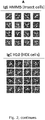
- IgE is rigid at the resolution of negative stain EM and adopts one major conformation.
- IgE rigidity for two different recombinant IgE molecules ( Fig 2A ), the HMM5-IgE and the H10-IgE, with distinct origin and antigen specificities expressed in either mammalian or insect cells is important, as it suggests that rigidity is a general feature of IgE.
- IgE's have a very rigid structure, which may be beneficial for EM structure determinations of target molecules. Isolated IgE possesses local two-fold symmetry that is beneficial during reconstruction of the 3D density.
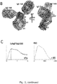
- Figure 3C illustrates how local two-fold symmetry may be used to improve the 3D density.
- the IgE-antigen complex is divided into 2x3 so called masks which are related by local two-fold symmetry each defined by a single rotation and a single translation operator. As the symmetry is only local, these three sets of operators are distinct from each other.
- This IgE-GFP complex data further show that the IgE rigidity is also a feature of IgE-antigen complexes and that structures of a target molecule bound to the IgE CDR regions can be determined be electron microscopy.
- Ligelizumab is a second-generation anti-IgE antibody, which prohibits the formation of the IgE - Fc ⁇ RI complex and thereby is intended to inhibit allergic responses in vivo.
- the aim was to determine the structural consequence of ligelizumab binding to IgE Fc.
- the formed IgE-ligelizumab Fab complex was formed by SEC and analyzed by nsEM analysis. See also example 1.
- the 3D reconstruction is slightly distorted due to preferred orientations, clear densities were observed for both the ligelizumab Fab and the IgE Fc and Fab moieties ( Fig. 4A ).
- the ligelizumab Fabs interact mainly with CH ⁇ 3 but may also interact with CH ⁇ 2.
- the two Fab's of IgE are pointing away from the Fc and does not seem to be contacted by the ligelizumab Fab.
- the extended conformation of IgE Fc observed in a complex between IgE and an anti- ⁇ -chain antibody fits well with the negative stain EM envelope ( Fig. 4A ).
- the Fab of IgE is pointing away from the Fc and does not seem to be obstructed by the ligelizumab Fab.
- the advantage of including ligelizumab in the complex is that an apparent global two-fold symmetry is obtained, rather than the local two-fold symmetry observed for IgE and IgE-GFP.
- An advantage of global two-fold symmetry for EM and in for particular cryo-EM is that by continuously applying the two-fold symmetry during the iterative 3D reconstruction and averaging, the reliability of the underlying alignment of particles is increased. This also allows for better removal of improper particles corresponding to e.g. contaminants, partially denatured or aggregated protein. Hence, two-fold averaging of two identical subunits increases the resolution of the final cryo-EM 3D reconstruction
- Example 5 Structure determination of antigen using IgE and a global symmetry inducer
- GFP-IgE-Ligelizumab Fab complex was formed by SEC and analysed by nsEM analysis. See also example 1.
- the 3D reconstruction of GFP-IgE-ligelizumab Fab complex is very similar to the reconstruction obtained for the IgE-ligelizumab Fab complex except for the presence of GFP.
- the GFP-IgE-ligelizumab Fab complex also displays global two-fold symmetry (data not shown).
- the use of two-fold symmetry leads to improved resolution of the GFP, as compared to performing the reconstruction without two-fold symmetry imposed.
- the density corresponding to the (IgE Fab - GFP) moiety is very similar in size to the one observed for the (IgE Fab - GFP) moiety in the IgE-GFP complex itself, where only local twofold symmetry is present.
- Ligelizumab Fab is also able to induce global two-fold symmetry of IgE bound to an antigen. This allows for more accurate alignment during 3D classification and leads to improved reconstruction of the antigen. This shows that it is feasible to determine IgE-Antigen structures using ligelizumab Fab to induce global two-fold symmetry.
- the H10 IgE is cloned from a murine hybridoma line and reacts with the house dust mite allergen Der p2. It exhibits high affinity for an epitope at the N-terminus of the protein.
- Recombinant Der p2 is prepared in bacteria.
- the IgE-Der p2 complex is reconstituted with and purified by size exclusion chromatography.
- cryo-EM solution containing the complex is applied to glow-discharged holey carbon grids, which are flash frozen in liquid ethane using a Vitrobot Mark III. After identification of suitable grids data collection is performed on a 300 kV Titan Krios equipped with a direct electron detector. Data processing is performed on a high performance computing cluster. Local symmetry averaging is carried out after each iteration of 3D refinement. Partial crystal structures are docked into the cryo-EM density map followed by model building and real space refinement against the map.
- Variants of Der p2 predicted by the structure not to be recognized by the H10 IgE are constructed by site directed mutagenesis and the interaction of H10 IgE with wildtype and variant Der p2 is evaluated by surface plasmon resonance and in basophil degranulation experiments.
- the successful structure determination of IgE in complex with a 14 kDa target molecule demonstrates the principle that IgE-complexes facilitate structure determination by cryo-EM of small target molecules.
- Nanobodies small single domain antibodies called Nanobodies are used. As compared to conventional antibodies they are faster, cheaper and easier to produce and their antigen affinity is fully comparable to that of conventional antibodies. Like conventional antibodies they have a conserved framework and variable complementarity determining regions that recognize the antigen. Hence, first an IgE binding the nanobody constant region without interfering with antigen binding is developed, but this can then be used for all possible nanobodies. Second, for each target molecule a nanobody is developed binding the target with high affinity. Third, a complex is formed between the anti-nanobody IgE, the target molecule specific nanobody and the target molecule. The complex is isolated SEC and is analysed by negative stain EM or cryo-EM.
- nanobody-bound target molecule is determined in complex with the anti-nanobody IgE.
- the local symmetry is used to improve the 3D reconstruction of the (target molecule) complex.
- an adaptor molecule allows for determination of an unlimited number of target molecule structures using only a single IgE specific towards the conserved framework of the adaptor molecules.
- the increased size and local twofold symmetry allows for more reliable 3D reconstruction.
- the presented data shows that the IgE-(ligelizumab) 2 complex is a highly rigid module that allows structures of Ag 2 -IgE-FabX 2 complexes to be determined by cryo-EM taking advantage of the size of the resulting complex (270 kDa+2 ⁇ antigen Mw), the rigidity of IgE, and global two-fold symmetry.
- this concept transforms a standard reagent (antibody) into a vehicle for pipeline structure determination.
Landscapes
- Health & Medical Sciences (AREA)
- Life Sciences & Earth Sciences (AREA)
- Immunology (AREA)
- Engineering & Computer Science (AREA)
- Molecular Biology (AREA)
- Biomedical Technology (AREA)
- Chemical & Material Sciences (AREA)
- Hematology (AREA)
- Urology & Nephrology (AREA)
- Biotechnology (AREA)
- Microbiology (AREA)
- Cell Biology (AREA)
- Food Science & Technology (AREA)
- Medicinal Chemistry (AREA)
- Physics & Mathematics (AREA)
- Analytical Chemistry (AREA)
- Biochemistry (AREA)
- General Health & Medical Sciences (AREA)
- General Physics & Mathematics (AREA)
- Pathology (AREA)
- Peptides Or Proteins (AREA)
Priority Applications (1)
| Application Number | Priority Date | Filing Date | Title |
|---|---|---|---|
| EP18215047.4A EP3671208A1 (de) | 2018-12-21 | 2018-12-21 | Ige gesteuerte strukturbestimmung von zielmolekülen unter verwendung von cryo-em |
Applications Claiming Priority (1)
| Application Number | Priority Date | Filing Date | Title |
|---|---|---|---|
| EP18215047.4A EP3671208A1 (de) | 2018-12-21 | 2018-12-21 | Ige gesteuerte strukturbestimmung von zielmolekülen unter verwendung von cryo-em |
Publications (1)
| Publication Number | Publication Date |
|---|---|
| EP3671208A1 true EP3671208A1 (de) | 2020-06-24 |
Family
ID=65019251
Family Applications (1)
| Application Number | Title | Priority Date | Filing Date |
|---|---|---|---|
| EP18215047.4A Withdrawn EP3671208A1 (de) | 2018-12-21 | 2018-12-21 | Ige gesteuerte strukturbestimmung von zielmolekülen unter verwendung von cryo-em |
Country Status (1)
| Country | Link |
|---|---|
| EP (1) | EP3671208A1 (de) |
Cited By (1)
| Publication number | Priority date | Publication date | Assignee | Title |
|---|---|---|---|---|
| WO2025047982A1 (ja) * | 2023-08-31 | 2025-03-06 | 国立研究開発法人理化学研究所 | 原子構造推定装置、原子構造推定方法、および原子構造推定プログラム |
-
2018
- 2018-12-21 EP EP18215047.4A patent/EP3671208A1/de not_active Withdrawn
Non-Patent Citations (10)
| Title |
|---|
| "Antibody humanization methods - a review and update", BIOTECHNOL GENET ENG REV., vol. 29, 2013, pages 175 - 86 |
| CHRISTENSEN ET AL., J ALLERGY CLIN IMMUNOL., vol. 122, no. 2, August 2008 (2008-08-01), pages 298 - 304 |
| DRINKWATER N ET AL., IMMUNOL REV., vol. 268, no. 1, November 2015 (2015-11-01), pages 222 - 35 |
| HUI GK ET AL., BIOCHEM J, vol. 471, no. 2, 2015, pages 167 - 185 |
| SAPHIRE EO ET AL., J MOL BIOL, vol. 319, no. 1, 2002, pages 9 - 18 |
| SELB REGINA ET AL: "Critical and direct involvement of the CD23 stalk region in IgE binding", JOURNAL OF ALLERGY AND CLINICAL IMMUNOLOGY, ELSEVIER, AMSTERDAM, NL, vol. 139, no. 1, 7 May 2016 (2016-05-07), pages 281, XP029877396, ISSN: 0091-6749, DOI: 10.1016/J.JACI.2016.04.015 * |
| SUTTON BJ; DAVIES AM, IMMUNOL REV, vol. 268, no. 1, 2015, pages 222 - 235 |
| XING ZHANG ET AL: "3D Structural Fluctuation of IgG1 Antibody Revealed by Individual Particle Electron Tomography", SCIENTIFIC REPORTS, vol. 5, no. 1, 1 September 2015 (2015-09-01), XP055553684, DOI: 10.1038/srep09803 * |
| YU GUIMEI ET AL: "Antibody-based affinity cryo-EM grid", METHODS, ACADEMIC PRESS, NL, vol. 100, 21 January 2016 (2016-01-21), pages 16 - 24, XP029520583, ISSN: 1046-2023, DOI: 10.1016/J.YMETH.2016.01.010 * |
| ZHANG X ET AL., SCI REP, vol. 5, 2015, pages 9803 |
Cited By (1)
| Publication number | Priority date | Publication date | Assignee | Title |
|---|---|---|---|---|
| WO2025047982A1 (ja) * | 2023-08-31 | 2025-03-06 | 国立研究開発法人理化学研究所 | 原子構造推定装置、原子構造推定方法、および原子構造推定プログラム |
Similar Documents
| Publication | Publication Date | Title |
|---|---|---|
| AU2014209857B2 (en) | Method and kit for detecting 1,25-dihydroxyvitamin D and related antibodies | |
| Kintzer et al. | Structural basis for activation of voltage sensor domains in an ion channel TPC1 | |
| KR20160031550A (ko) | 펩티드 단편의 조제 방법 및 그것에 사용되는 펩티드 단편 조제용 킷, 및 분석 방법 | |
| WO2017106797A1 (en) | Grid coatings for capture of proteins and other compounds | |
| EP3342783B1 (de) | Verfahren zur steuerung der affinität eines antikörpers gegen ein antigen, antikörper mit veränderter affinität für ein antigen und verfahren zur herstellung davon | |
| EP3533459A1 (de) | Anti-pla2-gib-antikörper und die verwendungen davon | |
| Le Brun et al. | The structural orientation of antibody layers bound to engineered biosensor surfaces | |
| EP2949675B1 (de) | Humanisierte anti-hmgb1-antikörper oder antigenbindendes fragment davon | |
| AU2018294758B2 (en) | Method for measuring serum amyloid A of various animals and reagent for measurement thereof | |
| CN112204049A (zh) | 抗甲状腺激素(t4)重组抗体或抗原结合片段 | |
| EP3671208A1 (de) | Ige gesteuerte strukturbestimmung von zielmolekülen unter verwendung von cryo-em | |
| JP2019536860A (ja) | 調整可能なアミン比重を有するコーティング | |
| Hendrickson et al. | Production of monoclonal antibodies against fullerene C 60 and development of a fullerene enzyme immunoassay | |
| US20130052669A1 (en) | Compositions and methods for reliably detecting and/or measuring the amount of a modified target protein in a sample | |
| US20220380456A1 (en) | Nanodisc-specific antigen-binding chimeric proteins | |
| Yuan et al. | A sensitive and group-specific monoclonal antibody-based indirect competitive ELISA for the determination of salbutamol in swine meat and liver samples | |
| EP3437657A1 (de) | Antibody, der durch den ersatz von cystein an den aminosäurepositionen 80 und 171 der kabat-nummerierung geändert ist | |
| JP2021508471A (ja) | 抗vegf抗体のvegf受容体遮断選択性を改善する方法 | |
| EP4019540A1 (de) | Verfahren zur verbesserung der affinität eines antikörpers für ein antigen und verwendung davon | |
| Esser et al. | Crystal structure of the antigen-binding fragment of a monoclonal antibody specific for the multidrug-resistance-linked ABC transporter human P-glycoprotein | |
| CN112639471A (zh) | 在模拟体内条件下的治疗性蛋白质选择 | |
| KR20170120697A (ko) | 반응장을 제한한 프로테아제 분해 반응에 의한 항체로부터 펩티드 단편을 얻는 방법 | |
| CN118388658A (zh) | 一种靶向nmda受体人源抗体的筛选方法 | |
| US20210347910A1 (en) | Method for improving thermal stability of antibody and method for producing modified antibody | |
| JP2022181707A (ja) | 非変性チャネルロドプシン検出キット |
Legal Events
| Date | Code | Title | Description |
|---|---|---|---|
| PUAI | Public reference made under article 153(3) epc to a published international application that has entered the european phase |
Free format text: ORIGINAL CODE: 0009012 |
|
| STAA | Information on the status of an ep patent application or granted ep patent |
Free format text: STATUS: THE APPLICATION HAS BEEN PUBLISHED |
|
| AK | Designated contracting states |
Kind code of ref document: A1 Designated state(s): AL AT BE BG CH CY CZ DE DK EE ES FI FR GB GR HR HU IE IS IT LI LT LU LV MC MK MT NL NO PL PT RO RS SE SI SK SM TR |
|
| AX | Request for extension of the european patent |
Extension state: BA ME |
|
| STAA | Information on the status of an ep patent application or granted ep patent |
Free format text: STATUS: THE APPLICATION IS DEEMED TO BE WITHDRAWN |
|
| 18D | Application deemed to be withdrawn |
Effective date: 20210112 |