EP3669372B1 - Détermination d'ultrasons d'un bronchogramme aérien dynamique et dispositifs associés et systèmes - Google Patents
Détermination d'ultrasons d'un bronchogramme aérien dynamique et dispositifs associés et systèmes Download PDFInfo
- Publication number
- EP3669372B1 EP3669372B1 EP18759894.1A EP18759894A EP3669372B1 EP 3669372 B1 EP3669372 B1 EP 3669372B1 EP 18759894 A EP18759894 A EP 18759894A EP 3669372 B1 EP3669372 B1 EP 3669372B1
- Authority
- EP
- European Patent Office
- Prior art keywords
- image data
- ultrasound imaging
- dynamic
- image
- data frames
- Prior art date
- Legal status (The legal status is an assumption and is not a legal conclusion. Google has not performed a legal analysis and makes no representation as to the accuracy of the status listed.)
- Active
Links
- 238000002604 ultrasonography Methods 0.000 title description 26
- 238000012545 processing Methods 0.000 claims description 58
- 210000004072 lung Anatomy 0.000 claims description 53
- 238000012285 ultrasound imaging Methods 0.000 claims description 53
- 238000004891 communication Methods 0.000 claims description 20
- 239000000523 sample Substances 0.000 claims description 18
- 230000000241 respiratory effect Effects 0.000 claims description 13
- 230000002123 temporal effect Effects 0.000 claims description 12
- 238000000034 method Methods 0.000 description 33
- 238000001514 detection method Methods 0.000 description 28
- 206010035664 Pneumonia Diseases 0.000 description 25
- 206010025080 Lung consolidation Diseases 0.000 description 14
- 238000010586 diagram Methods 0.000 description 14
- 230000003068 static effect Effects 0.000 description 9
- 238000003745 diagnosis Methods 0.000 description 8
- 210000000038 chest Anatomy 0.000 description 7
- 230000003434 inspiratory effect Effects 0.000 description 6
- 230000033001 locomotion Effects 0.000 description 6
- 108010034154 ABS 212 Proteins 0.000 description 5
- 230000007246 mechanism Effects 0.000 description 5
- 125000004122 cyclic group Chemical group 0.000 description 4
- 230000000737 periodic effect Effects 0.000 description 4
- 238000004445 quantitative analysis Methods 0.000 description 4
- 241001465754 Metazoa Species 0.000 description 3
- 208000007123 Pulmonary Atelectasis Diseases 0.000 description 3
- 238000013459 approach Methods 0.000 description 3
- 238000003384 imaging method Methods 0.000 description 3
- 238000012216 screening Methods 0.000 description 3
- 238000012360 testing method Methods 0.000 description 3
- 206010003598 Atelectasis Diseases 0.000 description 2
- 208000019693 Lung disease Diseases 0.000 description 2
- 238000004458 analytical method Methods 0.000 description 2
- 230000008901 benefit Effects 0.000 description 2
- 238000013170 computed tomography imaging Methods 0.000 description 2
- 208000024891 symptom Diseases 0.000 description 2
- 206010008479 Chest Pain Diseases 0.000 description 1
- 206010011224 Cough Diseases 0.000 description 1
- 208000000059 Dyspnea Diseases 0.000 description 1
- 206010013975 Dyspnoeas Diseases 0.000 description 1
- 241000845082 Panama Species 0.000 description 1
- 208000037656 Respiratory Sounds Diseases 0.000 description 1
- 230000004075 alteration Effects 0.000 description 1
- 238000004873 anchoring Methods 0.000 description 1
- 230000005540 biological transmission Effects 0.000 description 1
- 230000008859 change Effects 0.000 description 1
- 238000006243 chemical reaction Methods 0.000 description 1
- 230000008878 coupling Effects 0.000 description 1
- 238000010168 coupling process Methods 0.000 description 1
- 238000005859 coupling reaction Methods 0.000 description 1
- 201000010099 disease Diseases 0.000 description 1
- 208000037265 diseases, disorders, signs and symptoms Diseases 0.000 description 1
- 230000002708 enhancing effect Effects 0.000 description 1
- 238000011156 evaluation Methods 0.000 description 1
- 210000000416 exudates and transudate Anatomy 0.000 description 1
- 238000001914 filtration Methods 0.000 description 1
- 239000012530 fluid Substances 0.000 description 1
- 230000006870 function Effects 0.000 description 1
- 238000009499 grossing Methods 0.000 description 1
- 208000021760 high fever Diseases 0.000 description 1
- 238000010191 image analysis Methods 0.000 description 1
- 208000015181 infectious disease Diseases 0.000 description 1
- 230000002757 inflammatory effect Effects 0.000 description 1
- 238000012986 modification Methods 0.000 description 1
- 230000004048 modification Effects 0.000 description 1
- 208000024356 pleural disease Diseases 0.000 description 1
- 230000001737 promoting effect Effects 0.000 description 1
- 238000002601 radiography Methods 0.000 description 1
- 230000029058 respiratory gaseous exchange Effects 0.000 description 1
- 230000004044 response Effects 0.000 description 1
- 230000035945 sensitivity Effects 0.000 description 1
- 208000013220 shortness of breath Diseases 0.000 description 1
- 238000001228 spectrum Methods 0.000 description 1
- 210000000115 thoracic cavity Anatomy 0.000 description 1
- 210000000779 thoracic wall Anatomy 0.000 description 1
- 238000003325 tomography Methods 0.000 description 1
- 230000000007 visual effect Effects 0.000 description 1
Images


Classifications
-
- G—PHYSICS
- G16—INFORMATION AND COMMUNICATION TECHNOLOGY [ICT] SPECIALLY ADAPTED FOR SPECIFIC APPLICATION FIELDS
- G16H—HEALTHCARE INFORMATICS, i.e. INFORMATION AND COMMUNICATION TECHNOLOGY [ICT] SPECIALLY ADAPTED FOR THE HANDLING OR PROCESSING OF MEDICAL OR HEALTHCARE DATA
- G16H30/00—ICT specially adapted for the handling or processing of medical images
- G16H30/40—ICT specially adapted for the handling or processing of medical images for processing medical images, e.g. editing
-
- A—HUMAN NECESSITIES
- A61—MEDICAL OR VETERINARY SCIENCE; HYGIENE
- A61B—DIAGNOSIS; SURGERY; IDENTIFICATION
- A61B8/00—Diagnosis using ultrasonic, sonic or infrasonic waves
- A61B8/08—Detecting organic movements or changes, e.g. tumours, cysts, swellings
-
- A—HUMAN NECESSITIES
- A61—MEDICAL OR VETERINARY SCIENCE; HYGIENE
- A61B—DIAGNOSIS; SURGERY; IDENTIFICATION
- A61B8/00—Diagnosis using ultrasonic, sonic or infrasonic waves
- A61B8/52—Devices using data or image processing specially adapted for diagnosis using ultrasonic, sonic or infrasonic waves
- A61B8/5215—Devices using data or image processing specially adapted for diagnosis using ultrasonic, sonic or infrasonic waves involving processing of medical diagnostic data
- A61B8/5223—Devices using data or image processing specially adapted for diagnosis using ultrasonic, sonic or infrasonic waves involving processing of medical diagnostic data for extracting a diagnostic or physiological parameter from medical diagnostic data
-
- G—PHYSICS
- G06—COMPUTING; CALCULATING OR COUNTING
- G06T—IMAGE DATA PROCESSING OR GENERATION, IN GENERAL
- G06T7/00—Image analysis
- G06T7/0002—Inspection of images, e.g. flaw detection
- G06T7/0012—Biomedical image inspection
- G06T7/0014—Biomedical image inspection using an image reference approach
- G06T7/0016—Biomedical image inspection using an image reference approach involving temporal comparison
-
- G—PHYSICS
- G06—COMPUTING; CALCULATING OR COUNTING
- G06T—IMAGE DATA PROCESSING OR GENERATION, IN GENERAL
- G06T7/00—Image analysis
- G06T7/20—Analysis of motion
- G06T7/215—Motion-based segmentation
-
- G—PHYSICS
- G06—COMPUTING; CALCULATING OR COUNTING
- G06T—IMAGE DATA PROCESSING OR GENERATION, IN GENERAL
- G06T7/00—Image analysis
- G06T7/20—Analysis of motion
- G06T7/223—Analysis of motion using block-matching
-
- G—PHYSICS
- G16—INFORMATION AND COMMUNICATION TECHNOLOGY [ICT] SPECIALLY ADAPTED FOR SPECIFIC APPLICATION FIELDS
- G16H—HEALTHCARE INFORMATICS, i.e. INFORMATION AND COMMUNICATION TECHNOLOGY [ICT] SPECIALLY ADAPTED FOR THE HANDLING OR PROCESSING OF MEDICAL OR HEALTHCARE DATA
- G16H40/00—ICT specially adapted for the management or administration of healthcare resources or facilities; ICT specially adapted for the management or operation of medical equipment or devices
- G16H40/60—ICT specially adapted for the management or administration of healthcare resources or facilities; ICT specially adapted for the management or operation of medical equipment or devices for the operation of medical equipment or devices
- G16H40/63—ICT specially adapted for the management or administration of healthcare resources or facilities; ICT specially adapted for the management or operation of medical equipment or devices for the operation of medical equipment or devices for local operation
-
- G—PHYSICS
- G16—INFORMATION AND COMMUNICATION TECHNOLOGY [ICT] SPECIALLY ADAPTED FOR SPECIFIC APPLICATION FIELDS
- G16H—HEALTHCARE INFORMATICS, i.e. INFORMATION AND COMMUNICATION TECHNOLOGY [ICT] SPECIALLY ADAPTED FOR THE HANDLING OR PROCESSING OF MEDICAL OR HEALTHCARE DATA
- G16H50/00—ICT specially adapted for medical diagnosis, medical simulation or medical data mining; ICT specially adapted for detecting, monitoring or modelling epidemics or pandemics
- G16H50/30—ICT specially adapted for medical diagnosis, medical simulation or medical data mining; ICT specially adapted for detecting, monitoring or modelling epidemics or pandemics for calculating health indices; for individual health risk assessment
-
- A—HUMAN NECESSITIES
- A61—MEDICAL OR VETERINARY SCIENCE; HYGIENE
- A61B—DIAGNOSIS; SURGERY; IDENTIFICATION
- A61B8/00—Diagnosis using ultrasonic, sonic or infrasonic waves
- A61B8/08—Detecting organic movements or changes, e.g. tumours, cysts, swellings
- A61B8/0858—Detecting organic movements or changes, e.g. tumours, cysts, swellings involving measuring tissue layers, e.g. skin, interfaces
-
- A—HUMAN NECESSITIES
- A61—MEDICAL OR VETERINARY SCIENCE; HYGIENE
- A61B—DIAGNOSIS; SURGERY; IDENTIFICATION
- A61B8/00—Diagnosis using ultrasonic, sonic or infrasonic waves
- A61B8/13—Tomography
-
- A—HUMAN NECESSITIES
- A61—MEDICAL OR VETERINARY SCIENCE; HYGIENE
- A61B—DIAGNOSIS; SURGERY; IDENTIFICATION
- A61B8/00—Diagnosis using ultrasonic, sonic or infrasonic waves
- A61B8/46—Ultrasonic, sonic or infrasonic diagnostic devices with special arrangements for interfacing with the operator or the patient
- A61B8/461—Displaying means of special interest
-
- A—HUMAN NECESSITIES
- A61—MEDICAL OR VETERINARY SCIENCE; HYGIENE
- A61B—DIAGNOSIS; SURGERY; IDENTIFICATION
- A61B8/00—Diagnosis using ultrasonic, sonic or infrasonic waves
- A61B8/46—Ultrasonic, sonic or infrasonic diagnostic devices with special arrangements for interfacing with the operator or the patient
- A61B8/467—Ultrasonic, sonic or infrasonic diagnostic devices with special arrangements for interfacing with the operator or the patient characterised by special input means
- A61B8/469—Ultrasonic, sonic or infrasonic diagnostic devices with special arrangements for interfacing with the operator or the patient characterised by special input means for selection of a region of interest
-
- A—HUMAN NECESSITIES
- A61—MEDICAL OR VETERINARY SCIENCE; HYGIENE
- A61B—DIAGNOSIS; SURGERY; IDENTIFICATION
- A61B8/00—Diagnosis using ultrasonic, sonic or infrasonic waves
- A61B8/52—Devices using data or image processing specially adapted for diagnosis using ultrasonic, sonic or infrasonic waves
- A61B8/5215—Devices using data or image processing specially adapted for diagnosis using ultrasonic, sonic or infrasonic waves involving processing of medical diagnostic data
- A61B8/5238—Devices using data or image processing specially adapted for diagnosis using ultrasonic, sonic or infrasonic waves involving processing of medical diagnostic data for combining image data of patient, e.g. merging several images from different acquisition modes into one image
- A61B8/5246—Devices using data or image processing specially adapted for diagnosis using ultrasonic, sonic or infrasonic waves involving processing of medical diagnostic data for combining image data of patient, e.g. merging several images from different acquisition modes into one image combining images from the same or different imaging techniques, e.g. color Doppler and B-mode
-
- G—PHYSICS
- G01—MEASURING; TESTING
- G01S—RADIO DIRECTION-FINDING; RADIO NAVIGATION; DETERMINING DISTANCE OR VELOCITY BY USE OF RADIO WAVES; LOCATING OR PRESENCE-DETECTING BY USE OF THE REFLECTION OR RERADIATION OF RADIO WAVES; ANALOGOUS ARRANGEMENTS USING OTHER WAVES
- G01S7/00—Details of systems according to groups G01S13/00, G01S15/00, G01S17/00
- G01S7/52—Details of systems according to groups G01S13/00, G01S15/00, G01S17/00 of systems according to group G01S15/00
- G01S7/52017—Details of systems according to groups G01S13/00, G01S15/00, G01S17/00 of systems according to group G01S15/00 particularly adapted to short-range imaging
- G01S7/52079—Constructional features
- G01S7/5208—Constructional features with integration of processing functions inside probe or scanhead
-
- G—PHYSICS
- G06—COMPUTING; CALCULATING OR COUNTING
- G06T—IMAGE DATA PROCESSING OR GENERATION, IN GENERAL
- G06T2207/00—Indexing scheme for image analysis or image enhancement
- G06T2207/10—Image acquisition modality
- G06T2207/10016—Video; Image sequence
-
- G—PHYSICS
- G06—COMPUTING; CALCULATING OR COUNTING
- G06T—IMAGE DATA PROCESSING OR GENERATION, IN GENERAL
- G06T2207/00—Indexing scheme for image analysis or image enhancement
- G06T2207/10—Image acquisition modality
- G06T2207/10132—Ultrasound image
-
- G—PHYSICS
- G06—COMPUTING; CALCULATING OR COUNTING
- G06T—IMAGE DATA PROCESSING OR GENERATION, IN GENERAL
- G06T2207/00—Indexing scheme for image analysis or image enhancement
- G06T2207/20—Special algorithmic details
- G06T2207/20212—Image combination
-
- G—PHYSICS
- G06—COMPUTING; CALCULATING OR COUNTING
- G06T—IMAGE DATA PROCESSING OR GENERATION, IN GENERAL
- G06T2207/00—Indexing scheme for image analysis or image enhancement
- G06T2207/20—Special algorithmic details
- G06T2207/20212—Image combination
- G06T2207/20224—Image subtraction
-
- G—PHYSICS
- G06—COMPUTING; CALCULATING OR COUNTING
- G06T—IMAGE DATA PROCESSING OR GENERATION, IN GENERAL
- G06T2207/00—Indexing scheme for image analysis or image enhancement
- G06T2207/30—Subject of image; Context of image processing
- G06T2207/30004—Biomedical image processing
- G06T2207/30061—Lung
Definitions
- the present disclosure relates generally to ultrasound imaging and, in particular, to providing automated systems and methods for identifying the presence of a dynamic air bronchogram (AB) indicative of pneumonia based on lung ultrasound imaging.
- AB dynamic air bronchogram
- PN Pneumonia
- CXR chest radiography
- bedside CXR may provide limited image quality.
- CXR is a time-consuming procedure for emergency situations and the interpretation of bedside CXR may be challenging, requiring extensive radiologic experience to avoid misinterpretation of the wide spectrum of pleural and pulmonary diseases.
- CT imaging can be expensive and may have a higher radiation exposure than CXR.
- CCUs critical care units
- ICUs intensive care units
- Ultrasound imaging especially point-of-care ultrasound (POC-US) at bedside
- POC-US point-of-care ultrasound
- Recent studies have shown that lung ultrasound imaging can be useful and effective in diagnosis of PN with a relatively high accuracy.
- ultrasound images may not show useful or adequate information for a normal aerated lung, whereas a lung consolidation may appear as bright spots or bright structures under ultrasound imaging.
- B-lines in ultrasound images may be indicative of PN at an early stage.
- B-lines in ultrasound images may be indicative of PN at an early stage.
- ultrasound imaging-based PN diagnosis may require a well-trained or experienced physician or clinician to analyze and interpret acquired lung image.
- Embodiments of the present disclosure provide mechanisms for diagnosing PN by identifying and indicating the presence of a dynamic air bronchogram (AB) based on lung ultrasound images in an automated manner.
- dynamic ABs correspond to bright spots or pixels that change or move over time due to respiratory cycles.
- dynamic AB is identified based on a variation of a number of bright spots or pixels across a number of image frames over time.
- dynamic AB is identified based on a temporal intensity variation of bright spots or pixels across a number of image frames over time.
- the appearance of bronchial trees corresponding to dynamic ABs are enhanced in lung ultrasound images by accumulating differences across image data frames over time.
- an ultrasound imaging system includes an interface coupled to an ultrasound imaging component and configured to receive a plurality of image data frames representative of a subject's body including at least a portion of a lung; a processing component in communication with the interface and configured to determine a metric for each image data frame of the plurality of image data frames based on a threshold comparison; and determine a dynamic air bronchogram (AB) condition of the subject's body based on a variation across the metrics of the plurality of image data frames.
- AB dynamic air bronchogram
- each image data frame includes a plurality of pixel values representing pixel intensities of an image of the subject's body.
- the plurality of image data frames represent images of the subject's body across a time period including at least one respiratory cycle.
- the processing component is configured to determine the metric for each image data frame of the plurality of image data frames by determining a number of the plurality of pixel values in each image data frame that satisfies a threshold.
- the processing component is configured to determine the dynamic AB condition by determining a ratio between a maximum of the metrics and a minimum of the metrics; and determining the dynamic AB condition based on the ratio.
- the processing component is further configured to identify a region-of-interest (ROI) from the plurality of image data frames corresponding to the at least a portion of the lung; and determine the metrics based on the ROI.
- ROI region-of-interest
- the ultrasound imaging system further includes a display component configured to display a result of the dynamic AB condition.
- the ultrasound imaging system further includes an ultrasound imaging probe including the ultrasound imaging component; the processing component; and a display component configured to display a result of the dynamic AB condition.
- an ultrasound imaging system includes an interface coupled to an ultrasound imaging component and configured to receive a plurality of image data frames representative of a subject's body including at least a portion of a lung; a processing component in communication with the interface and configured to determine differential data frames based on differences across consecutive image data frames of the plurality of image data frames; determine an accumulated data frame based on a sum of the differential data frames; and determine a dynamic air bronchogram (AB) condition of the subject's body based on the accumulated data frame.
- AB dynamic air bronchogram
- the plurality of image data frames represent images of the subject's body across a time period including at least one respiratory cycle.
- each image data frame includes a plurality of pixel values representing pixel intensities of an image of the subject's body.
- each differential data frame includes a plurality of difference values
- the processing component is further configured to determine each differential data frame by determining each difference value of the plurality of difference values by determining an absolute difference between a pixel value of a first data frame of the plurality of image data frames and a pixel value of a second data frame of the plurality of image data frames, wherein the first data frame is adjacent to the second data frame, and wherein the pixel value of the first data frame and the pixel value of the second data frame represents a same sub-portion of the at least a portion of the lung.
- each differential data frame includes a first plurality of pixel values, wherein the accumulated data frame includes a plurality of sum values, and wherein the processing component is further configured to determine the dynamic AB condition by determining each sum value of the plurality of sum values by accumulating a second plurality of pixel values across the differential data frames, wherein the second plurality of pixel values across the differential data frames represent a same portion of the subject's body; and determining the dynamic AB condition based on the plurality of sum values.
- the ultrasound imaging system of claim 9 further comprising a display component configured to display the accumulated data frame.
- an ultrasound imaging system includes an interface coupled to an ultrasound imaging component and configured to receive a plurality of image data frames representative of a subject's body including at least a portion of a lung; a processing component in communication with the interface and configured to identify a subset of data from the plurality of image data frames based on a threshold comparison; and determine a dynamic air bronchogram (AB) condition of the subject's body based on a temporal variation across the subset of data.
- AB dynamic air bronchogram
- each image data frame of the plurality of image data frames includes a plurality of pixel values representing pixel intensities of an image of the subject's body.
- the plurality of image data frames represent images of the subject's body across a time period including at least one respiratory cycle.
- the processing component is configured to identify the subset of data by selecting one or more pixel values from each image data frame of the plurality of image data frames corresponding to a same sub-portion of the at least a portion of the lung and satisfying a threshold; determine a first value for each image data frame of the plurality of image data frames based on the one or more pixel values of a corresponding image data frame; and determine the dynamic AB condition based on a ratio between a maximum of the first values and a minimum of the first values.
- the processing component is configured to apply a filter across the first values prior to the determining the dynamic AB condition.
- the ultrasound imaging system further includes a display component configured to display a result of the dynamic AB condition.
- a method of ultrasound imaging diagnostic includes receiving, from an ultrasound imaging probe, a plurality of image data frames associated with a subject's body including at least a portion of a lung; determining a first value for each image data frame of the plurality of image data frames based on a threshold comparison; and determine a dynamic air bronchogram (AB) condition of the subject's body based on a variation across the first values of the plurality of image data frames.
- AB dynamic air bronchogram
- each image data frame includes a plurality of pixel values representing pixel intensities of an image of the subject's body.
- the plurality of image data frames represents images of the subject's body across a time period including at least one respiratory cycle.
- the determining the first value includes determining a number of the plurality of pixel values in each image data frame that satisfies a threshold, and wherein the determining the dynamic AB condition includes determining a ratio between a maximum of the first values and a minimum of the first values; and determining the dynamic AB condition based on the ratio.
- the method further includes identifying a region of interest (ROI) from the plurality of image data frames corresponding to the at least a portion of the lung; and determining the first values based on the ROI. In some embodiments, the method further includes displaying, at a display component, a result of the dynamic AB condition.
- ROI region of interest
- FIG. 1 is a schematic diagram of an ultrasound imaging system 100, according to aspects of the present disclosure.
- the system 100 is used for scanning an area or volume of a patient's body.
- the system 100 includes an ultrasound imaging probe 110 in communication with a host 130 over a communication interface or link 120.
- the probe 110 includes a transducer array 112, a beamformer 114, a processing component 116, a display 117, and a communication interface 118.
- the host 130 includes a display 132, a communication interface 136, and a communication interface 136.
- the transducer array 112 emits ultrasound signals towards an anatomical object 105 and receives echo signals reflected from the object 105 back to the transducer array 112.
- the transducer array 112 may include acoustic elements arranged in a one-dimensional (ID) array or in a two-dimensional (2D) array.
- the beamformer 114 is coupled to the transducer array 112.
- the beamformer 114 controls the transducer array 112, for example, for transmission of the ultrasound signals and reception of the ultrasound echo signals.
- the beamformer 114 provides image signals to the processing component 116 based on the response or the received ultrasound echo signals.
- the beamformer 114 may include multiple stages of beamforming. The beamforming can reduce the number of signal lines for coupling to the processing component 116.
- the transducer array 112 in combination with the beamformer 114 may be referred to as an ultrasound imaging component.
- the processing component 116 is coupled to the beamformer 114.
- the processing component 116 generates image data from the image signals.
- the processing component 116 may be implemented as a combination of software components and hardware components.
- the processing component 116 may be implemented on a field programmable gate array (FPGA) and may include programmable state machines to control the processing and conversion of the image signals to the image data.
- FPGA field programmable gate array
- the processing component 116 may perform filtering and/or quadrature demodulation to condition the image signals.
- the processing component 116 may perform analytic detection on the filtered signals.
- the display 117 is coupled to the processing component 116.
- the display 132 may be a screen or any suitable display integral with the housing of the probe 110.
- the display 117 may be configured to display the results of the analytic detection.
- the communication interface 118 is coupled to the processing component 116.
- the communication interface 118 transmits the image signals to the host 130 via the communication link 120.
- the communication interface 136 may receive the image signals.
- the host 130 may be any suitable computing and display device, such as a workstation, a personal computer (PC), a laptop, a tablet, or a mobile phone.
- the communication link 120 may be any suitable communication link.
- the communication link 120 may be a wired link, such as a universal serial bus (USB) link or an Ethernet link.
- the communication link 120 nay be a wireless link, such as an ultra-wideband (UWB) link, an Institute of Electrical and Electronics Engineers (IEEE) 802.11 WiFi link, or a Bluetooth link.
- UWB ultra-wideband
- IEEE Institute of Electrical and Electronics Engineers
- the processing component 134 is coupled to the communication interface 136.
- the processing component 134 may be implemented as a combination of software components and hardware components.
- the processing component 134 may include a central processing unit (CPU), a digital signal processor (DSP), an application-specific integrated circuit (ASIC), a controller, a FPGA device, another hardware device, a firmware device, or any combination thereof configured to perform the operations described herein.
- the processing component 134 may also be implemented as a combination of computing devices, e.g., a combination of a DSP and a microprocessor, a plurality of microprocessors, one or more microprocessors in conjunction with a DSP core, or any other such configuration.
- the processing component 134 can be configured to perform image processing and image analysis for various diagnostic modalities.
- the display 132 is coupled to the processing component 134.
- the display 132 may be a monitor or any suitable display.
- the display 132 is configured to display images and/or diagnostic results processed by the processing component 134
- the system 100 can be configured for dynamic AB detection.
- the object 105 may correspond to a portion of a patient's body including at least a portion of the patient's lung.
- the probe 110 transmits the image signals (e.g., the echo signals) received from the transducer array 112 to the host 130.
- the processing component 134 can detect dynamic ABs from ultrasound images and indicate a positive dynamic AB detection and a location of the detected dynamic AB or a negative dynamic AB detection on the display 132.
- the processing component 134 can identify dynamic ABs based on a variation of a number of bright spots or pixels across a number of image frames over time.
- the processing component 134 can identify dynamic ABs based on a temporal intensity variation of bright spots or pixels across a number of image frames over time.
- the processing component 134 can enhance the appearance or visibility of bronchial trees in ultrasound images. Mechanisms for detecting dynamic ABs and enhancing bronchial trees are described in greater detail herein.
- the processing component 116 on the probe 110 can be configured to perform dynamic AB detection eliminating the need of a host. In such an embodiment, dynamic AB detection results can be displayed on the integral display 117.
- One of the ultrasound imaging sign for identifying PN is the detection of a positive dynamic AB within a lung consolidation.
- An AB is a tubular outline of an airway visible under lung ultrasound imaging due to the filling of surrounding alveoli by fluid or inflammatory exudates.
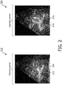
- FIG. 2 illustrates the dynamics of ABs from an expiratory period to an inspiratory period, according to aspects of the present disclosure.
- the image 210 is acquired during an expiratory period of a patient and the image 220 is acquired during an inspiratory period of the patient.
- ABs 212 appear as tubular bright structures.
- the ABs 214 appear as tubular bright structures.
- the appearance of ABs e.g., bright structures
- the images 210 and 220 show movements of punctiform ABs 216 (e.g., bright spots) to punctiform ABs 218.
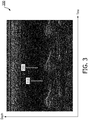
- FIG. 3 illustrates the dynamics of ABs in an area of a lung consolidation over time, according to aspects of the present disclosure.
- the x-axis represents time in some constant units and the y-axis represents depths in some constant units.
- the image 300 illustrates a real-time ultrasound scan line as a function of time.
- the transducer array 112 is used to generate one beam directed along a scan line towards an area within a lung consolidation and repeat at some time intervals.
- the image 300 shows the echo signals received from transducer array 112 corresponding to the emitted beam.
- the bright cyclic patterned lines show the movements or shift of ABs, such as the ABs 212, 214, 216, and 218, in a periodic manner.
- the cyclic or periodic pattern corresponds to the expiratory period 310 and inspiratory period 320 as marked in the image 300.
- dynamic ABs can be detected based on intensity variations of pixels across image frames over one or more respiratory cycles.
- real-time visual identification of dynamic ABs may be difficult or unreliable due to the fast movements or motions of the dynamic ABs while the lung in the background of the ultrasound images may still be moving during respiration, where a respiratory cycle may span a duration of about 3 seconds to about 6 seconds.
- the clinician that performs the lung ultrasound imaging e.g., at a POC-US
- FIG. 4 is a schematic diagram illustrating an automatic dynamic AB detection scheme 400, according to aspects of the present disclosure.
- the scheme 400 can be employed by the system 100 for dynamic AB detection.
- the scheme 400 can be implemented by the processing component 134 on the host 130 or the processing component 116 on the probe 110.
- the implementation of the scheme 400 can be divided between the host 130 and the probe 110.
- the scheme 400 begins with receiving a number of image frames 410.
- the image frames 410 may be generated by using the probe 110, where ultrasound signal energy is emitted from the transducer array 112 towards a patient's body (e.g., the object 105) and echo signals are received by the transducer array 112 forming the image frames 410.
- the probe 110 can be positioned on the patient's body to obtain an anterior chest view (e.g., from above the chest area) or a lateral chest view (e.g., from the side of the chest area) including at least some portions of the patient's lungs.
- Each image frame 410 may include a plurality of pixel values (e.g., amplitudes) representing pixel intensities of an image of the patient's body.
- the image frames 410 may be taken over a time period including at least one respiratory cycle (e.g., an expiratory period and an inspiratory period).
- the image frames 410 are shown as Frame(i) to Frame (i+N) representing images of the patient's body from a time instant (i) to a time instant (i+N), where i and N are positive integers.
- the image frames 410 can include other portions of the patient's body in addition to the patient's lung.
- the scheme 400 can apply a region-of-interest (ROI) identification component 420 to the image frames 410 to identify portions of the image frames corresponding to the patient's lung for subsequent processing.
- ROI region-of-interest
- One approach to identifying an area of a lung is based on a pleural line identification.
- a pleural line may appear as a bright line in an ultrasound image (shown in FIG.5 ) and the region below the pleural line may correspond to the lung.
- the ROI identification component 420 can identify an ROI in an area of the lung with a potential lung consolidation for the subsequent processing.
- the ROI identification component 420 may perform bulk background motion detection using block-matching on a frame-by-frame basis, for example, based correlations across the image frames 410.
- the ROI identification component 420 may use the initial frame (e.g., Frame (i)) as an anchoring frame to align or register subsequent image frames 410 (e.g., Frame (i+1) to Frame (i+N)) to the initial image frame 410 based on the background motion information for subsequent operations described below.
- the alignment or registration allows the operations to be performed on the same portion of the lung for each image frame 410.
- a threshold component 430 can be applied to each image frame 410.
- the threshold component 430 determines the number of pixels in the ROI of a corresponding image frame 410 that are above a predetermined threshold.
- the number of pixels above the threshold may be represented by a count value 432.
- the pixels that are above the threshold may correspond to bright spots as shown in the ABs 212, 214, 216, and 218.
- the predetermined threshold may be configured to any suitable value depending on the dynamic range of the pixel values and the amplitude and/or pixel distribution in the ROI.
- the dynamic range of the pixel intensity values may be configured to be between about 0 and about 255.
- the threshold may be configured to a value between about 40 to about 80.
- a maximum component 440 and a minimum component 450 can be applied to the count values 432.
- the maximum component 440 determines a maximum value 442 of the count values across the image frames 410.
- the minimum component 450 determines a minimum value 452 of the count values 432 across the image frames 410.
- the maximum value 442 and the minimum value 452 can be normalized such that the maximum value 442 has a value of one.
- an AB index component 460 can be applied to determine a dynamic AB diagnostic result.
- dynamic ABs are shown as bright structures (e.g., the ABs 212 and 214) or bright spots (e.g., the ABs 216 and 218) varying over time.
- the AB index component 460 identifies the dynamics of the ABs by computing a ratio between the maximum value 442 and the minimum value 452. The ratio may be referred to as an AB index.
- the AB index component 460 may compare the ratio to a predetermined threshold. When there is a large variation between the maximum value 442 and the minimum value 452, a dynamic AB condition may be positive.
- a dynamic AB condition may be negative and a static AB condition may be present.
- the presence of a dynamic AB condition may indicate a high likelihood of PN, while the presence of a static AB condition may indicate a high likelihood of atelectasis (e.g., lung collapse without infection) or other lung diseases.
- the dynamic AB diagnostic result can be displayed on the display 132 and/or the display 117. In addition, the result may indicate the location of the dynamic AB condition in the lung.
- FIG. 5 is an ultrasound image 500 illustrating dynamic ABs, according to aspects of the present disclosure.
- the image 500 may be acquired using the system 100.
- the image 500 may represent an image of a patient's body (e.g., the object 105) including an area of a lung.
- the image 500 illustrates the presence of ABs 512 and lung consolidation 506 in an area of the patient's lung.
- a pleural interface 502 or pleural boundary appears as a bright line across the image 500.
- the area below the pleural interface 502 corresponds to the patient's lung.
- the area above the pleural interface 510 corresponds to the patient's chest wall 504.
- FIG. 6 is a graph 600 illustrating variations of a number of bright pixels across a number of image frames over time in the presences of dynamic ABs, according to aspects of the present disclosure.
- the x-axis represents frame number.
- the y-axis represents normalized number of pixels in an ROI within image frames that are above a predetermined intensity threshold.
- the graph 600 is generated using the scheme 400.
- the plot 610 shows the variations of the number of pixels above the threshold (e.g., the count values 432 in a normalized form) across a number of image frames (e.g., the image frames 410) over time in the region of the dynamic ABs 512 shown in the image 500. As can be seen, the plot 610 varies between about 0.84 to about 1.
- the large difference between the minimum (e.g., 0.84) and the maximum (e.g., 1) and the cyclic patterns observed in the plot 610 may indicate the presence of a dynamic AB condition.
- the cyclic or periodic patterns may correspond to respiratory cycles of the patient.
- FIG. 7 is an ultrasound image 700 illustrating static ABs, according to aspects of the present disclosure.
- the image 700 may be acquired using the system 100.
- the image 700 may represent an image of a patient's body (e.g., the object 105) including an area of a lung.
- the image 700 illustrates the presence of static ABs 712 in an area of the patient's lung.
- FIG. 8 is a graph 800 illustrating variations of a number of bright pixels across a number of image frames over time in the absence of dynamic ABs, according to aspects of the present disclosure.
- the x-axis represents frame number.
- the y-axis represents normalized number of pixels in an ROI within image frames that are above a predetermined intensity threshold.
- the graph 800 is generated using the scheme 400.
- the plot 810 shows the variations of the number of pixels above the threshold (e.g., the count values 432 in a normalized form) across a number of image frames (e.g., the image frames 410) over time in the region of the static ABs 712 of the image 700.
- the plot 810 is relatively static varying between about 0.987 to about 1.
- the small difference between the minimum (e.g., 0.987) and the maximum (e.g., 1) and the relatively static pattern observed in the plot 810 may indicate the presence of a static AB condition (e.g., an atelectasis condition).
- FIG. 9 is a schematic diagram illustrating a dynamic AB detection scheme 900, according to aspects of the present disclosure.
- the scheme 900 can be employed by the system 100 for dynamic AB detection.
- the scheme 900 can be implemented by the processing component 134 on the host 130 or the processing component 116 on the probe 110.
- the implementation of the scheme 900 can be divided between the host 130 and the probe 110.
- the scheme 900 begins with receiving a number of image frames 910 similar to the image frames 410.
- each image frame 910 includes pixel intensity values representing an image of a patient's body including at least a portion of the patient's lung.
- the scheme 900 can apply an ROI identification component 920 to the image frames 910.
- the ROI identification component 920 may be substantially similar to the ROI identification component 420.
- the ROI identification component 920 may identify a subset of data or pixels from the image frames 910 for dynamic AB condition determination.
- the identification may include selecting one or more pixel values from each image frame 910 corresponding to the same portion of the patient's lung around a lung consolidation.
- the ROI identification component 920 may output image data subsets 930 including pixels within the ROI.
- the subsets 930 are shown as Frame (i, k) to Frame (i+N, k) representing a subset k within Frame (i) to a subset k within Frame (i+1), respectively.
- each subset 930 includes one pixel value
- the pixel value is represented by an intensity value 932.
- a spatial filter may be applied to each subset 930 to produce an average intensity value 932.
- a maximum component 940 and a minimum component 950 can be applied to the intensity values 932.
- the maximum component 940 determines a maximum value 942 of the intensity values 932.
- the minimum component 950 determines a minimum value 952 of the intensity values 932.
- the maximum value 942 and the minimum value 952 can be normalized such that the maximum value 942 has a value of 1.
- a temporal filter e.g., a smoothing filter
- a temporal intensity variation determination component 960 can be applied to determine a dynamic AB diagnostic result.
- the temporal intensity variation determination component 960 determines a temporal intensity variation across the subsets 930 over time.
- the temporal intensity variation determination component 960 can determine a ratio between the maximum value 942 and the minimum value 952.
- the temporal intensity variation determination component 960 can compare the ratio to a predetermined threshold and determine whether a dynamic AB condition is present based on the threshold comparison. Similar to the scheme 400, a large variation between the maximum value 942 and the minimum value 952 is indicative of a positive dynamic AB condition and a small variation between the maximum value 942 and the minimum value 952 is indicative of a negative dynamic AB condition.
- FIG. 10 is a schematic diagram illustrating a bronchial tree enhancement scheme 1000, according to aspects of the present disclosure.
- the scheme 1000 can be employed by the system 100 to enhance bronchial trees within a lung consolidation in lung ultrasound images for dynamic AB diagnosis.
- the scheme 1000 can be implemented by the processing component 134 on the host 130.
- the scheme 1000 begins with receiving a number of image frames 1010 similar to the image frames 410 and 910.
- each image frame 1010 includes pixel intensity values representing an image of a patient's body including at least a portion of the patient's lung.
- the scheme 1000 applies a difference component 1020 to each pair of adjacent or consecutive image frames 1010 (e.g., Frame (i) and Frame (i+1)).
- the difference component 1020 computes a difference between the adjacent image frames 1010 to produce a differential image frame 1022.
- the difference component 1020 subtracts the pixel values in the Frame (i) by the pixel values in the Frame (i+1) on a pixel-by-pixel basis.
- the pixel values of the Frame (i) and the pixel values of the Frame (i+1) correspond to the same sub-portion of the patient's lung.
- An absolute component 1030 can be applied to the pixel values in the differential image frames 1022 to produce differential image frames 1032 with absolute difference pixel values.
- a sum component 1040 can be applied to accumulate the differential image frames 1032 to produce an accumulated image frame 1042.
- the sum component 1040 sums the pixel values of the differential image frames 1032 on a pixel-by-pixel basis.
- the image frames 1010 include a bronchial tree
- the appearance or visibility of the bronchial tree may be enhanced in the accumulated image frame 1042 (shown in FIG. 11D below).
- the detection of the bronchial tree indicates a positive dynamic AB condition.
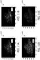
- FIGS. 11A-11D illustrates various image frames corresponding to various stages in the scheme 1000 described above with respect to FIG. 10 .
- the scheme 1000 is applied to eighty sequential ultrasound image frames, represented by Frame(1), Frame(2), ..., Frame (80).
- FIG. 11A is an image frame 1110 illustrating a bronchial tree 1112 at a time instant, according to aspects of the present disclosure.
- the image frame 1110 may be similar to the image frames 410, 910, and 1010.
- the image frame 1110 may represent a first image frame (e.g., Frame (1)) of the eighty sequential image frames.
- the bright Y-shaped branching structure in the middle portion of the image frame 1110 corresponds to the bronchial tree 1112 within a lung consolidation of a patient.
- the bright curved horizontal structure at the bottom of the image frame 1110 corresponds to the patient's spine.
- FIG. 11B is an image frame 1120 illustrating a bronchial tree 1112 at another time instant, according to aspects of the present disclosure.
- the image frame 1120 may represent the last image frame (e.g., Frame (80)) of the eight sequential image frames.
- FIG. 11C is a differential image frame 1130 illustrating a difference between a pair of consecutive image frames including a bronchial tree 1112, according to aspects of the present disclosure.
- the differential image frame 1130 may represent a differential image frame 1032 at the output of the absolute component 1030 in the scheme 1000.
- the differential image frame 1130 may be computed by subtracting Frame (80) from Frame (79) pixel by pixel.
- FIG. 11D is an accumulated image frame 1140 illustrating an enhanced bronchial tree 1112, according to aspects of the present disclosure.
- the image frame 1140 may represent an accumulated image frame 1042 at the output of the sum component 1040 in the scheme 1000. Comparing the accumulated image frame 1140 to the originally acquired image frames 1110 and 1120, the appearance or visibility of the bronchial tree 1112 is enhanced in the accumulated image frame 1140.
- FIG. 12 is a graph 1200 illustrating variations of a number of bright pixels across a number of image frames over time in the presence of dynamic ABs, according to aspects of the present disclosure.
- the x-axis represents frame number.
- the y-axis represents normalized number of pixels in an ROI within image frames that are above a predetermined intensity threshold.
- the graph 1200 is generated by applying the scheme 400 to the eight sequential image frames used for illustrating the scheme 1000 in FIG. 11 .
- the plot 1210 shows variations of the normalized number of pixels above a predetermined threshold across a number of image frames (e.g., the image frames 1010) over time in the region of the bronchial tree 1112 shown in the image frames 1110 and 1120.
- the plot 1210 varies between about 0.53 to about 1.
- the large difference between the minimum (e.g., 0.53) and the maximum (e.g., 1) and the periodic waveform observed in the plot 1210 may indicate the presence of a dynamic AB condition.
- FIG. 13 is a flow diagram of a dynamic AB detection method 1300, according to aspects of the present disclosure. Steps of the method 1300 can be executed by a computing device (e.g., a processor, processing circuit, and/or other suitable component) of an ultrasound imaging probe, such as the probe 110, or a host such as the host 130.
- the method 1300 may employ similar mechanisms as in the scheme 400 as described with respect to FIG. 4 .
- the method 1300 includes a number of enumerated steps, but embodiments of the method 1300 may include additional steps before, after, and in between the enumerated steps. In some embodiments, one or more of the enumerated steps may be omitted or performed in a different order.
- the method 1300 includes receiving a plurality of image data frames (e.g., the image frames 410, 910, 1010, 1110, and 1120) representative of a subject's body (e.g., the object 105) including at least a portion of a lung.
- the subject's body may be a human body or an animal body.
- the method 1300 includes determining a metric (e.g., the count values 432) for each image data frame of the plurality of image data frames, for example, using the threshold component 430.
- a metric e.g., the count values 432
- the method 1300 includes determining a dynamic AB condition of the subject's body based on a variation across the metrics of the plurality of image data frames.
- the maximum component 440 can be applied to the metrics to compute a maximum value (e.g., the maximum value 442) of the metrics and the minimum component 450 can be applied to the metrics to compute a minimum value (e.g., the minimum value 452) of the metrics.
- the AB index component 460 can be applied to compute a ratio between the maximum value and the minimum value and compare the ratio to a predetermine threshold. When the ratio satisfy the predetermine threshold, a positive dynamic AB condition may be present. When the ratio fails to satisfy the predetermined threshold, a dynamic AB condition may be absent.
- FIG. 14 is a flow diagram of a dynamic AB detection method 1400, according to aspects of the present disclosure. Steps of the method 1400 can be executed by a computing device (e.g., a processor, processing circuit, and/or other suitable component) of an ultrasound imaging probe, such as the probe 110, or a host such as the host 130.
- the method 1400 may employ similar mechanisms as in the scheme 900 as described with respect to FIG. 9 .
- the method 1400 includes a number of enumerated steps, but embodiments of the method 1400 may include additional steps before, after, and in between the enumerated steps. In some embodiments, one or more of the enumerated steps may be omitted or performed in a different order.
- the method 1400 includes receiving a plurality of image data frames (e.g., the image frames 410, 910, 1010, 1110, and 1120) representative of a subject's body (e.g., the object 105) including at least a portion of a lung.
- the subject's body may be a human body or an animal body.
- the method 1400 includes identifying a subset of data (e.g., the subsets 930) from the plurality of image data frames based on a threshold comparison, for example, using the ROI identification component 920.
- the method 1400 includes determining a dynamic AB condition of the subject's body based on a temporal variation across the subset of data.
- the subset of data may include a portion (e.g., one or more pixel values) of each image data frame and a spatial filter can be applied to a corresponding portion of each image frame to determine a first value (e.g., the intensity values 932) for each image data frame.
- the maximum component 940 can be applied to the first values to compute a maximum value (e.g., the maximum value 942) of the first values and the minimum component 950 can be applied to the first values to compute a minimum value (e.g., the minimum value 952) of the first values.
- the temporal intensity variation determination component 960 can be applied to compute a ratio between the maximum value and the minimum value and compare the ratio to a predetermine threshold.
- a positive dynamic AB condition may be present.
- a dynamic AB condition may be absent.
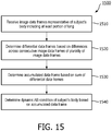
- FIG. 15 is a flow diagram of a bronchial tree enhancement method 1500, according to aspects of the present disclosure. Steps of the method 1500 can be executed by a computing device (e.g., a processor, processing circuit, and/or other suitable component) of a host such as the host 130.
- the method 1500 may employ similar mechanisms as in the scheme 1000 as described with respect to FIG. 10 .
- the method 1500 includes a number of enumerated steps, but embodiments of the method 1500 may include additional steps before, after, and in between the enumerated steps. In some embodiments, one or more of the enumerated steps may be omitted or performed in a different order.
- the method 1500 includes receiving a plurality of image data frames (e.g., the image frames 410, 910, 1010, 1110, and 1120) representative of a subject's body (e.g., the object 105) including at least a portion of a lung.
- the subject's body may be a human body or an animal body.
- the method 1500 includes determining differential data frames (e.g., the differential image frames 1032) based on differences across consecutive image data frames of the plurality of image data frames.
- the method 1500 includes determining an accumulated data frame (e.g., the accumulated image frame 1042) based on a sum of the differential data frames.
- the method 1500 includes determining a dynamic AB condition of the subject's body based on the accumulated data frame.
- the accumulated data frame shows an enhanced appearance or visibility of the bronchial tree in a location within a lung consolidation.
- the determination may be on the observation of the bronchial tree.
- the automatic detection of dynamic ABs without the need for a well-trained clinician to interpret ultrasound lung images allow point-of-care ultrasound (POC-US) imaging to be used for PN screening and diagnosis.
- the automatic dynamic AB detection can produce a diagnostic result in a short duration of time.
- PN examination time can be shortened when compared to a full chest PN examination.
- the disclosed embodiments provide a standardized testing protocol.
- the use of the standardized test protocol can produce more consistent diagnostic results than the subjective evaluations and analysis under different physicians and clinicians.
- the standardized test protocol can be carried out easily for screening and suitable for use in time critical situations (e.g., during an emergency).
- the enhanced display of bronchial tree can assist a physician to identify a PN location quickly and easily.
- the disclosed embodiments are suitable for use with pediatric patients and/or pregnant women where radiation exposure is a concern.
Landscapes
- Engineering & Computer Science (AREA)
- Health & Medical Sciences (AREA)
- Life Sciences & Earth Sciences (AREA)
- Medical Informatics (AREA)
- General Health & Medical Sciences (AREA)
- Physics & Mathematics (AREA)
- Public Health (AREA)
- Nuclear Medicine, Radiotherapy & Molecular Imaging (AREA)
- Radiology & Medical Imaging (AREA)
- Biomedical Technology (AREA)
- Computer Vision & Pattern Recognition (AREA)
- Pathology (AREA)
- Theoretical Computer Science (AREA)
- General Physics & Mathematics (AREA)
- Biophysics (AREA)
- Heart & Thoracic Surgery (AREA)
- Molecular Biology (AREA)
- Surgery (AREA)
- Animal Behavior & Ethology (AREA)
- Veterinary Medicine (AREA)
- Epidemiology (AREA)
- Primary Health Care (AREA)
- Quality & Reliability (AREA)
- Physiology (AREA)
- Multimedia (AREA)
- Data Mining & Analysis (AREA)
- Databases & Information Systems (AREA)
- Business, Economics & Management (AREA)
- General Business, Economics & Management (AREA)
- Ultra Sonic Daignosis Equipment (AREA)
Claims (14)
- Système d'imagerie par ultrasons, comprenant :une interface couplée à un composant d'imagerie par ultrasons et configurée pour recevoir une pluralité de trames de données d'image représentant un corps d'un sujet comprenant au moins une partie d'un poumon, chaque trame de données d'image de la pluralité de trames de données d'image comprenant une pluralité de valeurs de pixel représentant des intensités de pixel d'une image du corps du sujet ;un composant de traitement en communication avec l'interface et configuré :pour déterminer une métrique de valeur de comptage pour chaque trame de données d'image de la pluralité de trames de données d'image par comptage d'un nombre de la pluralité de valeurs de pixel dans chaque trame de données d'image, laquelle satisfait un seuil ; etpour déterminer une condition de bronchogramme aérien (AB) dynamique du corps du sujet en fonction d'une variation à travers les métriques de valeur de comptage de la pluralité de trames de données d'image.
- Système d'imagerie par ultrasons selon la revendication 1, dans lequel la pluralité de trames de données d'image représente des images du corps du sujet sur une période de temps comprenant au moins un cycle respiratoire.
- Système d'imagerie par ultrasons selon l'une quelconque des revendications 1 à 2, dans lequel le composant de traitement est configuré pour déterminer la condition d'AB dynamique :par détermination d'un rapport entre un maximum des métriques et un minimum des métriques ; etpour déterminer la condition d'AB dynamique en fonction du rapport.
- Système d'imagerie par ultrasons selon l'une quelconque des revendications 1 à 3, dans lequel le composant de traitement est en outre configuré :pour identifier une zone d'intérêt (ROI) à partir de la pluralité de trames de données d'image correspondant à l'au moins une partie du poumon ; etpour déterminer les métriques en fonction de la ROI.
- Système d'imagerie par ultrasons selon l'une quelconque des revendications 1 à 4, comprenant en outre un composant d'affichage configuré pour afficher un résultat de la condition d'AB dynamique.
- Système d'imagerie par ultrasons selon l'une quelconque des revendications 1 à 5, comprenant en outre une sonde d'imagerie par ultrasons comprenant :le composant d'imagerie par ultrasons ;le composant de traitement ; etun composant d'affichage configuré pour afficher un résultat de la condition d'AB dynamique.
- Système d'imagerie par ultrasons, comprenant :une interface couplée à un composant d'imagerie par ultrasons et configurée pour recevoir une pluralité de trames de données d'image représentant un corps d'un sujet comprenant au moins une partie d'un poumon, chaque trame de données d'image de la pluralité de trames de données d'image comprenant une pluralité de valeurs de pixel représentant des intensités de pixel d'une image du corps du sujet ;un composant de traitement en communication avec l'interface et configuré :pour déterminer des trames de données différentielles en fonction des différences entre des trames de données d'image consécutives de la pluralité de trames de données d'image par soustraction d'une valeur de pixel d'une première trame de données d'image de la pluralité de trames de données d'image d'une valeur de pixel d'une deuxième trame de données d'image de la pluralité de trames de données d'image, la deuxième trame de données d'image étant adjacente à la première trame de données d'image ;pour déterminer une trame de données accumulées en fonction d'une somme des trames de données différentielles ; etpour déterminer un bronchogramme aérien (AB) dynamique du corps du sujet en fonction de la trame de données accumulées.
- Système d'imagerie par ultrasons selon la revendication 7, dans lequel la pluralité de trames de données d'image représente des images du corps du sujet sur une période de temps comprenant au moins un cycle respiratoire.
- Système d'imagerie par ultrasons selon l'une quelconque des revendications 7 à 8, dans lequel chaque trame de données différentielles comprend une pluralité de valeurs de différence, dans lequel le composant de traitement est en outre configuré pour déterminer une première trame de données différentielle des trames de données différentielles :par détermination d'une valeur différentielle pour la première trame de données différentielles par détermination d'une valeur absolue d'un résultat de la soustraction,dans lequel la valeur de pixel de la première trame de données d'image et la valeur de pixel de la deuxième trame de données d'image représentent une même sous-partie de l'au moins une partie du poumon.
- Système d'imagerie par ultrasons selon l'une quelconque des revendications 7 à 9, dans lequel chaque trame de données différentielles comprend une deuxième pluralité de valeurs de pixel, dans lequel la trame de données accumulées comprend une pluralité de valeurs de somme, et dans lequel le composant de traitement est en outre configuré pour déterminer la condition d'AB dynamique :par détermination de chaque valeur de somme de la pluralité de valeurs de somme par accumulation d'une troisième pluralité de valeurs de pixel à travers les trames de données différentielles, dans lequel la troisième pluralité de valeurs de pixel à travers les trames de données différentielles représente une même partie du corps du sujet ; etpar la détermination de la condition d'AB dynamique en fonction de la pluralité de valeurs de somme.
- Système d'imagerie par ultrasons selon l'une quelconque des revendications 7 à 10, comprenant en outre un composant d'affichage configuré pour afficher la trame de données accumulées.
- Système d'imagerie par ultrasons, comprenant :une interface couplée à un composant d'imagerie par ultrasons et configurée pour recevoir une pluralité de trames de données d'image représentant un corps d'un sujet comprenant au moins une partie d'un poumon sur une période de temps comprenant au moins un cycle respiratoire,chaque trame de données d'image de la pluralité de trames de données d'imagecomprenant une pluralité de valeurs de pixel représentant les intensités de pixel d'une image du corps du sujet ;un composant de traitement en communication avec l'interface et configuré :pour identifier un sous-ensemble de données à partir de la pluralité de trames de données d'image par sélection d'au moins une valeur de pixel à partir de chaque trame de données d'image de la pluralité de trames de données d'image correspondant à une même sous-partie d'au moins une partie du poumon ; etpour déterminer une métrique d'intensité pour chaque trame de données d'image à partir du sous-ensemble de données ; etpour déterminer un bronchogramme aérien (AB) dynamique du corps du sujet en fonction d'une variation temporelle dans le sous-ensemble de données par comparaison d'un maximum des métriques d'intensité sur la période de temps avec un minimum des métriques d'intensité sur la période.
- Système d'imagerie par ultrasons selon la revendication 12, dans lequel le composant de traitement est configuré :
pour déterminer la condition d'AB dynamique en fonction d'un rapport entre le maximum des métriques d'intensité et le minimum des métriques d'intensité. - Système d'imagerie par ultrasons selon la revendication 13, dans lequel le composant de traitement est configuré pour appliquer un filtre à travers les métriques d'intensité avant la détermination de la condition d'AB dynamique.
Applications Claiming Priority (3)
| Application Number | Priority Date | Filing Date | Title |
|---|---|---|---|
| CN2017097624 | 2017-08-16 | ||
| US201862628361P | 2018-02-09 | 2018-02-09 | |
| PCT/EP2018/072256 WO2019034743A1 (fr) | 2017-08-16 | 2018-08-16 | Détermination d'ultrasons d'un bronchogramme aérien dynamique et dispositifs associés, systèmes et procédés associés |
Publications (2)
| Publication Number | Publication Date |
|---|---|
| EP3669372A1 EP3669372A1 (fr) | 2020-06-24 |
| EP3669372B1 true EP3669372B1 (fr) | 2021-05-12 |
Family
ID=63405184
Family Applications (1)
| Application Number | Title | Priority Date | Filing Date |
|---|---|---|---|
| EP18759894.1A Active EP3669372B1 (fr) | 2017-08-16 | 2018-08-16 | Détermination d'ultrasons d'un bronchogramme aérien dynamique et dispositifs associés et systèmes |
Country Status (5)
| Country | Link |
|---|---|
| US (1) | US11896433B2 (fr) |
| EP (1) | EP3669372B1 (fr) |
| JP (1) | JP6890716B2 (fr) |
| CN (1) | CN111837196A (fr) |
| WO (1) | WO2019034743A1 (fr) |
Families Citing this family (4)
| Publication number | Priority date | Publication date | Assignee | Title |
|---|---|---|---|---|
| AU2020332834A1 (en) * | 2019-08-22 | 2022-02-17 | Boston Scientific Scimed, Inc. | Systems and methods for processing electronic medical images to determine enhanced electronic medical images |
| IT202000009667A1 (it) * | 2020-05-05 | 2021-11-05 | Imedicals S R L | Dispositivo e metodo per la diagnosi di una polmonite di tipo covid-19 mediante analisi di immagini ecografiche |
| US20230165567A1 (en) * | 2020-05-08 | 2023-06-01 | The Regents Of The University Of Michigan | Ultrasound signal correlation phase estimation of lung motion |
| US12070357B2 (en) * | 2021-12-09 | 2024-08-27 | GE Precision Healthcare LLC | System and method for automatic association and display of video loop subject matter for enhanced identification |
Family Cites Families (10)
| Publication number | Priority date | Publication date | Assignee | Title |
|---|---|---|---|---|
| US7128712B2 (en) * | 2004-06-21 | 2006-10-31 | General Electric Company | Adaptive ultrasound imaging system |
| US9044194B2 (en) * | 2010-08-27 | 2015-06-02 | Konica Minolta, Inc. | Thoracic diagnosis assistance system and computer readable storage medium |
| US8891881B2 (en) * | 2012-01-25 | 2014-11-18 | General Electric Company | System and method for identifying an optimal image frame for ultrasound imaging |
| US8914097B2 (en) * | 2012-01-30 | 2014-12-16 | The Johns Hopkins University | Automated pneumothorax detection |
| WO2013181300A1 (fr) | 2012-05-29 | 2013-12-05 | The Board Of Trustees Of The Leland Stanford Jr. University | Appareil, systèmes et procédés de surveillance d'eau extravasculaire pulmonaire |
| US9339210B2 (en) * | 2013-05-08 | 2016-05-17 | Koninklijke Philips N.V. | Device for obtaining a vital sign of a subject |
| HRPK20130491B3 (hr) | 2013-06-04 | 2016-03-25 | Sveučilište U Rijeci Medicinski Fakultet | Postupak za određivanje i brojenje b-linija kod ultrazvučnog dijagnosticiranja bolesti pluća |
| KR20150002932A (ko) | 2013-06-26 | 2015-01-08 | 삼성전자주식회사 | 초음파 영상 디스플레이 방법 및 장치 |
| CN107072637B (zh) | 2014-09-25 | 2020-07-28 | 皇家飞利浦有限公司 | 用于自动气胸检测的设备和方法 |
| EP3349664B1 (fr) * | 2015-09-17 | 2020-03-11 | Koninklijke Philips N.V. | Distinction de glissement de poumon et de mouvement externe |
-
2018
- 2018-08-16 US US16/638,827 patent/US11896433B2/en active Active
- 2018-08-16 CN CN201880067278.7A patent/CN111837196A/zh active Pending
- 2018-08-16 EP EP18759894.1A patent/EP3669372B1/fr active Active
- 2018-08-16 JP JP2020507991A patent/JP6890716B2/ja active Active
- 2018-08-16 WO PCT/EP2018/072256 patent/WO2019034743A1/fr unknown
Non-Patent Citations (1)
| Title |
|---|
| None * |
Also Published As
| Publication number | Publication date |
|---|---|
| US20210128116A1 (en) | 2021-05-06 |
| JP2020531073A (ja) | 2020-11-05 |
| WO2019034743A1 (fr) | 2019-02-21 |
| US11896433B2 (en) | 2024-02-13 |
| JP6890716B2 (ja) | 2021-06-18 |
| EP3669372A1 (fr) | 2020-06-24 |
| CN111837196A (zh) | 2020-10-27 |
Similar Documents
| Publication | Publication Date | Title |
|---|---|---|
| EP3669372B1 (fr) | Détermination d'ultrasons d'un bronchogramme aérien dynamique et dispositifs associés et systèmes | |
| US10970926B2 (en) | System and method for lung-volume-gated x-ray imaging | |
| CN105701331B (zh) | 计算机辅助诊断设备和计算机辅助诊断方法 | |
| US8777854B2 (en) | Method and system for ultrasound based automated detection, quantification and tracking of pathologies | |
| JP5068519B2 (ja) | 悪性腫瘍を自動的に特徴評価するルーチンを含む機械読み取り可能な媒体及び装置 | |
| US20210233243A1 (en) | Diagnostic support program | |
| US8659603B2 (en) | System and method for center point trajectory mapping | |
| US10098602B2 (en) | Apparatus and method for processing a medical image of a body lumen | |
| CN105120738B (zh) | 狭窄治疗规划 | |
| US20210353244A1 (en) | System and method for determining radiation parameters | |
| JP6253085B2 (ja) | X線動画像解析装置、x線動画像解析プログラム及びx線動画像撮像装置 | |
| JP2020508771A (ja) | 肺および血管の健康状態を検査し評価する方法 | |
| US10891732B2 (en) | Dynamic image processing system | |
| JP2016002251A (ja) | 画像処理装置、画像処理方法、およびプログラム | |
| JP2019180899A (ja) | 医用画像処理装置 | |
| CN114098796A (zh) | 用于检测医学图像中的胸膜不规则性的方法和系统 | |
| US20220398720A1 (en) | Diagnostic support program | |
| US20230169653A1 (en) | Medical image processing apparatus, medical image processing method, and storage medium | |
| CN103908293B (zh) | 用于测量医学图像的医学成像系统和方法 | |
| WO2023032954A1 (fr) | Procédé de traitement d'informations, programme et dispositif de diagnostic d'image | |
| KR20150054738A (ko) | 진단 영상 장치 및 그 동작 방법 | |
| WO2024104857A1 (fr) | Détection automatique de point de mesure pour une mesure d'anatomie dans des images anatomiques |
Legal Events
| Date | Code | Title | Description |
|---|---|---|---|
| STAA | Information on the status of an ep patent application or granted ep patent |
Free format text: STATUS: UNKNOWN |
|
| STAA | Information on the status of an ep patent application or granted ep patent |
Free format text: STATUS: THE INTERNATIONAL PUBLICATION HAS BEEN MADE |
|
| PUAI | Public reference made under article 153(3) epc to a published international application that has entered the european phase |
Free format text: ORIGINAL CODE: 0009012 |
|
| STAA | Information on the status of an ep patent application or granted ep patent |
Free format text: STATUS: REQUEST FOR EXAMINATION WAS MADE |
|
| 17P | Request for examination filed |
Effective date: 20200316 |
|
| AK | Designated contracting states |
Kind code of ref document: A1 Designated state(s): AL AT BE BG CH CY CZ DE DK EE ES FI FR GB GR HR HU IE IS IT LI LT LU LV MC MK MT NL NO PL PT RO RS SE SI SK SM TR |
|
| AX | Request for extension of the european patent |
Extension state: BA ME |
|
| DAV | Request for validation of the european patent (deleted) | ||
| DAX | Request for extension of the european patent (deleted) | ||
| GRAP | Despatch of communication of intention to grant a patent |
Free format text: ORIGINAL CODE: EPIDOSNIGR1 |
|
| STAA | Information on the status of an ep patent application or granted ep patent |
Free format text: STATUS: GRANT OF PATENT IS INTENDED |
|
| INTG | Intention to grant announced |
Effective date: 20201211 |
|
| GRAS | Grant fee paid |
Free format text: ORIGINAL CODE: EPIDOSNIGR3 |
|
| GRAA | (expected) grant |
Free format text: ORIGINAL CODE: 0009210 |
|
| STAA | Information on the status of an ep patent application or granted ep patent |
Free format text: STATUS: THE PATENT HAS BEEN GRANTED |
|
| AK | Designated contracting states |
Kind code of ref document: B1 Designated state(s): AL AT BE BG CH CY CZ DE DK EE ES FI FR GB GR HR HU IE IS IT LI LT LU LV MC MK MT NL NO PL PT RO RS SE SI SK SM TR |
|
| REG | Reference to a national code |
Ref country code: GB Ref legal event code: FG4D |
|
| REG | Reference to a national code |
Ref country code: CH Ref legal event code: EP |
|
| REG | Reference to a national code |
Ref country code: DE Ref legal event code: R096 Ref document number: 602018017083 Country of ref document: DE |
|
| REG | Reference to a national code |
Ref country code: IE Ref legal event code: FG4D |
|
| REG | Reference to a national code |
Ref country code: AT Ref legal event code: REF Ref document number: 1392719 Country of ref document: AT Kind code of ref document: T Effective date: 20210615 |
|
| REG | Reference to a national code |
Ref country code: DE Ref legal event code: R084 Ref document number: 602018017083 Country of ref document: DE |
|
| REG | Reference to a national code |
Ref country code: GB Ref legal event code: 746 Effective date: 20210817 |
|
| REG | Reference to a national code |
Ref country code: LT Ref legal event code: MG9D |
|
| REG | Reference to a national code |
Ref country code: AT Ref legal event code: MK05 Ref document number: 1392719 Country of ref document: AT Kind code of ref document: T Effective date: 20210512 |
|
| REG | Reference to a national code |
Ref country code: NL Ref legal event code: MP Effective date: 20210512 |
|
| PG25 | Lapsed in a contracting state [announced via postgrant information from national office to epo] |
Ref country code: LT Free format text: LAPSE BECAUSE OF FAILURE TO SUBMIT A TRANSLATION OF THE DESCRIPTION OR TO PAY THE FEE WITHIN THE PRESCRIBED TIME-LIMIT Effective date: 20210512 Ref country code: HR Free format text: LAPSE BECAUSE OF FAILURE TO SUBMIT A TRANSLATION OF THE DESCRIPTION OR TO PAY THE FEE WITHIN THE PRESCRIBED TIME-LIMIT Effective date: 20210512 Ref country code: FI Free format text: LAPSE BECAUSE OF FAILURE TO SUBMIT A TRANSLATION OF THE DESCRIPTION OR TO PAY THE FEE WITHIN THE PRESCRIBED TIME-LIMIT Effective date: 20210512 Ref country code: BG Free format text: LAPSE BECAUSE OF FAILURE TO SUBMIT A TRANSLATION OF THE DESCRIPTION OR TO PAY THE FEE WITHIN THE PRESCRIBED TIME-LIMIT Effective date: 20210812 Ref country code: AT Free format text: LAPSE BECAUSE OF FAILURE TO SUBMIT A TRANSLATION OF THE DESCRIPTION OR TO PAY THE FEE WITHIN THE PRESCRIBED TIME-LIMIT Effective date: 20210512 |
|
| PG25 | Lapsed in a contracting state [announced via postgrant information from national office to epo] |
Ref country code: LV Free format text: LAPSE BECAUSE OF FAILURE TO SUBMIT A TRANSLATION OF THE DESCRIPTION OR TO PAY THE FEE WITHIN THE PRESCRIBED TIME-LIMIT Effective date: 20210512 Ref country code: NO Free format text: LAPSE BECAUSE OF FAILURE TO SUBMIT A TRANSLATION OF THE DESCRIPTION OR TO PAY THE FEE WITHIN THE PRESCRIBED TIME-LIMIT Effective date: 20210812 Ref country code: PL Free format text: LAPSE BECAUSE OF FAILURE TO SUBMIT A TRANSLATION OF THE DESCRIPTION OR TO PAY THE FEE WITHIN THE PRESCRIBED TIME-LIMIT Effective date: 20210512 Ref country code: PT Free format text: LAPSE BECAUSE OF FAILURE TO SUBMIT A TRANSLATION OF THE DESCRIPTION OR TO PAY THE FEE WITHIN THE PRESCRIBED TIME-LIMIT Effective date: 20210913 Ref country code: RS Free format text: LAPSE BECAUSE OF FAILURE TO SUBMIT A TRANSLATION OF THE DESCRIPTION OR TO PAY THE FEE WITHIN THE PRESCRIBED TIME-LIMIT Effective date: 20210512 Ref country code: SE Free format text: LAPSE BECAUSE OF FAILURE TO SUBMIT A TRANSLATION OF THE DESCRIPTION OR TO PAY THE FEE WITHIN THE PRESCRIBED TIME-LIMIT Effective date: 20210512 Ref country code: IS Free format text: LAPSE BECAUSE OF FAILURE TO SUBMIT A TRANSLATION OF THE DESCRIPTION OR TO PAY THE FEE WITHIN THE PRESCRIBED TIME-LIMIT Effective date: 20210912 Ref country code: GR Free format text: LAPSE BECAUSE OF FAILURE TO SUBMIT A TRANSLATION OF THE DESCRIPTION OR TO PAY THE FEE WITHIN THE PRESCRIBED TIME-LIMIT Effective date: 20210813 |
|
| PG25 | Lapsed in a contracting state [announced via postgrant information from national office to epo] |
Ref country code: NL Free format text: LAPSE BECAUSE OF FAILURE TO SUBMIT A TRANSLATION OF THE DESCRIPTION OR TO PAY THE FEE WITHIN THE PRESCRIBED TIME-LIMIT Effective date: 20210512 |
|
| PG25 | Lapsed in a contracting state [announced via postgrant information from national office to epo] |
Ref country code: ES Free format text: LAPSE BECAUSE OF FAILURE TO SUBMIT A TRANSLATION OF THE DESCRIPTION OR TO PAY THE FEE WITHIN THE PRESCRIBED TIME-LIMIT Effective date: 20210512 Ref country code: EE Free format text: LAPSE BECAUSE OF FAILURE TO SUBMIT A TRANSLATION OF THE DESCRIPTION OR TO PAY THE FEE WITHIN THE PRESCRIBED TIME-LIMIT Effective date: 20210512 Ref country code: SK Free format text: LAPSE BECAUSE OF FAILURE TO SUBMIT A TRANSLATION OF THE DESCRIPTION OR TO PAY THE FEE WITHIN THE PRESCRIBED TIME-LIMIT Effective date: 20210512 Ref country code: CZ Free format text: LAPSE BECAUSE OF FAILURE TO SUBMIT A TRANSLATION OF THE DESCRIPTION OR TO PAY THE FEE WITHIN THE PRESCRIBED TIME-LIMIT Effective date: 20210512 Ref country code: DK Free format text: LAPSE BECAUSE OF FAILURE TO SUBMIT A TRANSLATION OF THE DESCRIPTION OR TO PAY THE FEE WITHIN THE PRESCRIBED TIME-LIMIT Effective date: 20210512 Ref country code: RO Free format text: LAPSE BECAUSE OF FAILURE TO SUBMIT A TRANSLATION OF THE DESCRIPTION OR TO PAY THE FEE WITHIN THE PRESCRIBED TIME-LIMIT Effective date: 20210512 Ref country code: SM Free format text: LAPSE BECAUSE OF FAILURE TO SUBMIT A TRANSLATION OF THE DESCRIPTION OR TO PAY THE FEE WITHIN THE PRESCRIBED TIME-LIMIT Effective date: 20210512 |
|
| REG | Reference to a national code |
Ref country code: DE Ref legal event code: R097 Ref document number: 602018017083 Country of ref document: DE |
|
| PLBE | No opposition filed within time limit |
Free format text: ORIGINAL CODE: 0009261 |
|
| STAA | Information on the status of an ep patent application or granted ep patent |
Free format text: STATUS: NO OPPOSITION FILED WITHIN TIME LIMIT |
|
| REG | Reference to a national code |
Ref country code: CH Ref legal event code: PL |
|
| PG25 | Lapsed in a contracting state [announced via postgrant information from national office to epo] |
Ref country code: MC Free format text: LAPSE BECAUSE OF FAILURE TO SUBMIT A TRANSLATION OF THE DESCRIPTION OR TO PAY THE FEE WITHIN THE PRESCRIBED TIME-LIMIT Effective date: 20210512 |
|
| 26N | No opposition filed |
Effective date: 20220215 |
|
| REG | Reference to a national code |
Ref country code: BE Ref legal event code: MM Effective date: 20210831 |
|
| PG25 | Lapsed in a contracting state [announced via postgrant information from national office to epo] |
Ref country code: LI Free format text: LAPSE BECAUSE OF NON-PAYMENT OF DUE FEES Effective date: 20210831 Ref country code: CH Free format text: LAPSE BECAUSE OF NON-PAYMENT OF DUE FEES Effective date: 20210831 |
|
| PG25 | Lapsed in a contracting state [announced via postgrant information from national office to epo] |
Ref country code: IS Free format text: LAPSE BECAUSE OF FAILURE TO SUBMIT A TRANSLATION OF THE DESCRIPTION OR TO PAY THE FEE WITHIN THE PRESCRIBED TIME-LIMIT Effective date: 20210912 Ref country code: LU Free format text: LAPSE BECAUSE OF NON-PAYMENT OF DUE FEES Effective date: 20210816 Ref country code: AL Free format text: LAPSE BECAUSE OF FAILURE TO SUBMIT A TRANSLATION OF THE DESCRIPTION OR TO PAY THE FEE WITHIN THE PRESCRIBED TIME-LIMIT Effective date: 20210512 |
|
| PG25 | Lapsed in a contracting state [announced via postgrant information from national office to epo] |
Ref country code: IT Free format text: LAPSE BECAUSE OF FAILURE TO SUBMIT A TRANSLATION OF THE DESCRIPTION OR TO PAY THE FEE WITHIN THE PRESCRIBED TIME-LIMIT Effective date: 20210512 Ref country code: IE Free format text: LAPSE BECAUSE OF NON-PAYMENT OF DUE FEES Effective date: 20210816 Ref country code: BE Free format text: LAPSE BECAUSE OF NON-PAYMENT OF DUE FEES Effective date: 20210831 |
|
| PG25 | Lapsed in a contracting state [announced via postgrant information from national office to epo] |
Ref country code: CY Free format text: LAPSE BECAUSE OF FAILURE TO SUBMIT A TRANSLATION OF THE DESCRIPTION OR TO PAY THE FEE WITHIN THE PRESCRIBED TIME-LIMIT Effective date: 20210512 |
|
| PG25 | Lapsed in a contracting state [announced via postgrant information from national office to epo] |
Ref country code: HU Free format text: LAPSE BECAUSE OF FAILURE TO SUBMIT A TRANSLATION OF THE DESCRIPTION OR TO PAY THE FEE WITHIN THE PRESCRIBED TIME-LIMIT; INVALID AB INITIO Effective date: 20180816 |
|
| PGFP | Annual fee paid to national office [announced via postgrant information from national office to epo] |
Ref country code: GB Payment date: 20230822 Year of fee payment: 6 |
|
| PGFP | Annual fee paid to national office [announced via postgrant information from national office to epo] |
Ref country code: FR Payment date: 20230824 Year of fee payment: 6 Ref country code: DE Payment date: 20230828 Year of fee payment: 6 |
|
| PG25 | Lapsed in a contracting state [announced via postgrant information from national office to epo] |
Ref country code: MK Free format text: LAPSE BECAUSE OF FAILURE TO SUBMIT A TRANSLATION OF THE DESCRIPTION OR TO PAY THE FEE WITHIN THE PRESCRIBED TIME-LIMIT Effective date: 20210512 |