EP3665481B1 - Carbohydrate sensors - Google Patents
Carbohydrate sensors Download PDFInfo
- Publication number
- EP3665481B1 EP3665481B1 EP18843350.2A EP18843350A EP3665481B1 EP 3665481 B1 EP3665481 B1 EP 3665481B1 EP 18843350 A EP18843350 A EP 18843350A EP 3665481 B1 EP3665481 B1 EP 3665481B1
- Authority
- EP
- European Patent Office
- Prior art keywords
- seq
- lactose
- domain
- gfp
- acceptor
- Prior art date
- Legal status (The legal status is an assumption and is not a legal conclusion. Google has not performed a legal analysis and makes no representation as to the accuracy of the status listed.)
- Active
Links
Images


Classifications
-
- G—PHYSICS
- G01—MEASURING; TESTING
- G01N—INVESTIGATING OR ANALYSING MATERIALS BY DETERMINING THEIR CHEMICAL OR PHYSICAL PROPERTIES
- G01N33/00—Investigating or analysing materials by specific methods not covered by groups G01N1/00 - G01N31/00
- G01N33/02—Food
- G01N33/04—Dairy products
-
- G—PHYSICS
- G01—MEASURING; TESTING
- G01N—INVESTIGATING OR ANALYSING MATERIALS BY DETERMINING THEIR CHEMICAL OR PHYSICAL PROPERTIES
- G01N33/00—Investigating or analysing materials by specific methods not covered by groups G01N1/00 - G01N31/00
- G01N33/48—Biological material, e.g. blood, urine; Haemocytometers
- G01N33/50—Chemical analysis of biological material, e.g. blood, urine; Testing involving biospecific ligand binding methods; Immunological testing
- G01N33/66—Chemical analysis of biological material, e.g. blood, urine; Testing involving biospecific ligand binding methods; Immunological testing involving blood sugars, e.g. galactose
-
- G—PHYSICS
- G01—MEASURING; TESTING
- G01N—INVESTIGATING OR ANALYSING MATERIALS BY DETERMINING THEIR CHEMICAL OR PHYSICAL PROPERTIES
- G01N33/00—Investigating or analysing materials by specific methods not covered by groups G01N1/00 - G01N31/00
- G01N33/48—Biological material, e.g. blood, urine; Haemocytometers
- G01N33/50—Chemical analysis of biological material, e.g. blood, urine; Testing involving biospecific ligand binding methods; Immunological testing
- G01N33/53—Immunoassay; Biospecific binding assay; Materials therefor
- G01N33/536—Immunoassay; Biospecific binding assay; Materials therefor with immune complex formed in liquid phase
- G01N33/542—Immunoassay; Biospecific binding assay; Materials therefor with immune complex formed in liquid phase with steric inhibition or signal modification, e.g. fluorescent quenching
-
- C—CHEMISTRY; METALLURGY
- C07—ORGANIC CHEMISTRY
- C07K—PEPTIDES
- C07K14/00—Peptides having more than 20 amino acids; Gastrins; Somatostatins; Melanotropins; Derivatives thereof
- C07K14/195—Peptides having more than 20 amino acids; Gastrins; Somatostatins; Melanotropins; Derivatives thereof from bacteria
-
- C—CHEMISTRY; METALLURGY
- C12—BIOCHEMISTRY; BEER; SPIRITS; WINE; VINEGAR; MICROBIOLOGY; ENZYMOLOGY; MUTATION OR GENETIC ENGINEERING
- C12N—MICROORGANISMS OR ENZYMES; COMPOSITIONS THEREOF; PROPAGATING, PRESERVING, OR MAINTAINING MICROORGANISMS; MUTATION OR GENETIC ENGINEERING; CULTURE MEDIA
- C12N15/00—Mutation or genetic engineering; DNA or RNA concerning genetic engineering, vectors, e.g. plasmids, or their isolation, preparation or purification; Use of hosts therefor
- C12N15/09—Recombinant DNA-technology
- C12N15/11—DNA or RNA fragments; Modified forms thereof; Non-coding nucleic acids having a biological activity
- C12N15/62—DNA sequences coding for fusion proteins
-
- C—CHEMISTRY; METALLURGY
- C12—BIOCHEMISTRY; BEER; SPIRITS; WINE; VINEGAR; MICROBIOLOGY; ENZYMOLOGY; MUTATION OR GENETIC ENGINEERING
- C12N—MICROORGANISMS OR ENZYMES; COMPOSITIONS THEREOF; PROPAGATING, PRESERVING, OR MAINTAINING MICROORGANISMS; MUTATION OR GENETIC ENGINEERING; CULTURE MEDIA
- C12N15/00—Mutation or genetic engineering; DNA or RNA concerning genetic engineering, vectors, e.g. plasmids, or their isolation, preparation or purification; Use of hosts therefor
- C12N15/09—Recombinant DNA-technology
- C12N15/63—Introduction of foreign genetic material using vectors; Vectors; Use of hosts therefor; Regulation of expression
- C12N15/70—Vectors or expression systems specially adapted for E. coli
-
- C—CHEMISTRY; METALLURGY
- C12—BIOCHEMISTRY; BEER; SPIRITS; WINE; VINEGAR; MICROBIOLOGY; ENZYMOLOGY; MUTATION OR GENETIC ENGINEERING
- C12N—MICROORGANISMS OR ENZYMES; COMPOSITIONS THEREOF; PROPAGATING, PRESERVING, OR MAINTAINING MICROORGANISMS; MUTATION OR GENETIC ENGINEERING; CULTURE MEDIA
- C12N9/00—Enzymes; Proenzymes; Compositions thereof; Processes for preparing, activating, inhibiting, separating or purifying enzymes
- C12N9/0004—Oxidoreductases (1.)
- C12N9/0069—Oxidoreductases (1.) acting on single donors with incorporation of molecular oxygen, i.e. oxygenases (1.13)
-
- C—CHEMISTRY; METALLURGY
- C12—BIOCHEMISTRY; BEER; SPIRITS; WINE; VINEGAR; MICROBIOLOGY; ENZYMOLOGY; MUTATION OR GENETIC ENGINEERING
- C12Y—ENZYMES
- C12Y113/00—Oxidoreductases acting on single donors with incorporation of molecular oxygen (oxygenases) (1.13)
- C12Y113/12—Oxidoreductases acting on single donors with incorporation of molecular oxygen (oxygenases) (1.13) with incorporation of one atom of oxygen (internal monooxygenases or internal mixed function oxidases)(1.13.12)
- C12Y113/12007—Photinus-luciferin 4-monooxygenase (ATP-hydrolysing) (1.13.12.7), i.e. firefly-luciferase
-
- G—PHYSICS
- G01—MEASURING; TESTING
- G01N—INVESTIGATING OR ANALYSING MATERIALS BY DETERMINING THEIR CHEMICAL OR PHYSICAL PROPERTIES
- G01N21/00—Investigating or analysing materials by the use of optical means, i.e. using sub-millimetre waves, infrared, visible or ultraviolet light
- G01N21/62—Systems in which the material investigated is excited whereby it emits light or causes a change in wavelength of the incident light
- G01N21/63—Systems in which the material investigated is excited whereby it emits light or causes a change in wavelength of the incident light optically excited
- G01N21/64—Fluorescence; Phosphorescence
- G01N21/6486—Measuring fluorescence of biological material, e.g. DNA, RNA, cells
-
- G—PHYSICS
- G01—MEASURING; TESTING
- G01N—INVESTIGATING OR ANALYSING MATERIALS BY DETERMINING THEIR CHEMICAL OR PHYSICAL PROPERTIES
- G01N21/00—Investigating or analysing materials by the use of optical means, i.e. using sub-millimetre waves, infrared, visible or ultraviolet light
- G01N21/75—Systems in which material is subjected to a chemical reaction, the progress or the result of the reaction being investigated
- G01N21/76—Chemiluminescence; Bioluminescence
- G01N21/763—Bioluminescence
-
- G—PHYSICS
- G01—MEASURING; TESTING
- G01N—INVESTIGATING OR ANALYSING MATERIALS BY DETERMINING THEIR CHEMICAL OR PHYSICAL PROPERTIES
- G01N33/00—Investigating or analysing materials by specific methods not covered by groups G01N1/00 - G01N31/00
- G01N33/48—Biological material, e.g. blood, urine; Haemocytometers
- G01N33/50—Chemical analysis of biological material, e.g. blood, urine; Testing involving biospecific ligand binding methods; Immunological testing
- G01N33/53—Immunoassay; Biospecific binding assay; Materials therefor
- G01N33/543—Immunoassay; Biospecific binding assay; Materials therefor with an insoluble carrier for immobilising immunochemicals
- G01N33/54306—Solid-phase reaction mechanisms
-
- G—PHYSICS
- G01—MEASURING; TESTING
- G01N—INVESTIGATING OR ANALYSING MATERIALS BY DETERMINING THEIR CHEMICAL OR PHYSICAL PROPERTIES
- G01N33/00—Investigating or analysing materials by specific methods not covered by groups G01N1/00 - G01N31/00
- G01N33/48—Biological material, e.g. blood, urine; Haemocytometers
- G01N33/50—Chemical analysis of biological material, e.g. blood, urine; Testing involving biospecific ligand binding methods; Immunological testing
- G01N33/68—Chemical analysis of biological material, e.g. blood, urine; Testing involving biospecific ligand binding methods; Immunological testing involving proteins, peptides or amino acids
- G01N33/6872—Intracellular protein regulatory factors and their receptors, e.g. including ion channels
-
- G—PHYSICS
- G01—MEASURING; TESTING
- G01N—INVESTIGATING OR ANALYSING MATERIALS BY DETERMINING THEIR CHEMICAL OR PHYSICAL PROPERTIES
- G01N33/00—Investigating or analysing materials by specific methods not covered by groups G01N1/00 - G01N31/00
- G01N33/48—Biological material, e.g. blood, urine; Haemocytometers
- G01N33/50—Chemical analysis of biological material, e.g. blood, urine; Testing involving biospecific ligand binding methods; Immunological testing
- G01N33/68—Chemical analysis of biological material, e.g. blood, urine; Testing involving biospecific ligand binding methods; Immunological testing involving proteins, peptides or amino acids
- G01N33/6875—Nucleoproteins
-
- C—CHEMISTRY; METALLURGY
- C07—ORGANIC CHEMISTRY
- C07K—PEPTIDES
- C07K2319/00—Fusion polypeptide
- C07K2319/60—Fusion polypeptide containing spectroscopic/fluorescent detection, e.g. green fluorescent protein [GFP]
-
- C—CHEMISTRY; METALLURGY
- C07—ORGANIC CHEMISTRY
- C07K—PEPTIDES
- C07K2319/00—Fusion polypeptide
- C07K2319/80—Fusion polypeptide containing a DNA binding domain, e.g. Lacl or Tet-repressor
-
- G—PHYSICS
- G01—MEASURING; TESTING
- G01N—INVESTIGATING OR ANALYSING MATERIALS BY DETERMINING THEIR CHEMICAL OR PHYSICAL PROPERTIES
- G01N2333/00—Assays involving biological materials from specific organisms or of a specific nature
- G01N2333/90—Enzymes; Proenzymes
- G01N2333/902—Oxidoreductases (1.)
- G01N2333/90241—Oxidoreductases (1.) acting on single donors with incorporation of molecular oxygen, i.e. oxygenases (1.13)
-
- G—PHYSICS
- G01—MEASURING; TESTING
- G01N—INVESTIGATING OR ANALYSING MATERIALS BY DETERMINING THEIR CHEMICAL OR PHYSICAL PROPERTIES
- G01N2400/00—Assays, e.g. immunoassays or enzyme assays, involving carbohydrates
Definitions
- the present invention relates to sensors and methods for detecting lactose or lactulose in a sample.
- Assays for detecting carbohydrates are widely used in the food and medical industries. Of particular interest to the food industry are assays for detecting carbohydrates, such as lactose, in dairy products.
- lactose e.g. 3.9-4.8% (w/v)
- w/v lactose
- this method is inaccurate for measuring lower levels of lactose, such as below 1% (w/v) lactose, and requires expensive equipment.
- the present inventors have provided sensors and methods for detecting carbohydrates, namely lactose or lactulose.
- the sensors and methods can be used to measure the lactose content of a dairy product. In some embodiments, the sensors and methods can be used to classify dairy products based on their lactose content.
- a polypeptide suitable for use in a method of the invention may be defined by the extent of identity (% identity) of its amino acid sequence to a reference amino acid sequence, or by having a greater % identity to one reference amino acid sequence than to another.
- the query sequence is at least 100 amino acids in length and the GAP analysis aligns the two sequences over a region of at least 100 amino acids. Even more preferably, the query sequence is at least 250 amino acids in length and the GAP analysis aligns the two sequences over a region of at least 250 amino acids. Even more preferably, the query sequence is at least 450 amino acids in length and the GAP analysis aligns the two sequences over a region of at least 450 amino acids. Even more preferably, the GAP analysis aligns two sequences over their entire length.
- the term about refers to +/- 10%, more preferably +/- 5%, even more preferably +/- 1%, of the designated value.
- % concentration is weight/volume (%w/v).
- sensor and “sensor molecule” are used interchangeably.
- the sensor of the claimed invention in its general sense is defined in claim 1. It comprises inter alia:
- the senor is a continuous stretch of amino acids (in other words, the sensor is a single polypeptide).
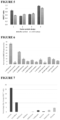
- the carbohydrate binding domain, chemiluminescent donor protein domain and acceptor domain are a single stretch of amino acids such as, but not limited to, a chemiluminescent donor protein domain covalently attached to the N-terminus of the carbohydrate binding domain and an acceptor protein domain covalently attached to the C-terminus of the carbohydrate binding domain, or an acceptor protein domain covalently attached to the N-terminus of the carbohydrate binding domain and a chemiluminescent donor protein domain covalently attached to the C-terminus of the carbohydrate binding domain. Examples are provided in Figure 1 .
- the sensor molecule of the claimed invention has an amino acid sequence which is at least 80% identical to that provided in SEQ ID NO:1. Other cases are disclosed but not claimed per se.
- the polypeptide has a sequence selected from the group consisting of SEQ ID NO: 15, SEQ ID NO: 16, SEQ ID NO: 17, SEQ ID NO: 18, SEQ ID NO: 25, SEQ ID NO: 26, SEQ ID NO: 27 and SEQ ID NO: 28 or a fragment or variant thereof.
- the polypeptide can have a sequence at least 30%, 35%, 40%, 45%, 50%, 55%, 60%, 65%, 70%, 75%, 80%, 85%, 90%, 95%, 96%, 97%, 98%, 99% or 100% identical to the sequence shown in any one of SEQ ID NO: 15, SEQ ID NO: 16, SEQ ID NO: 17, SEQ ID NO: 18, SEQ ID NO: 25, SEQ ID NO: 26, SEQ ID NO: 27 and SEQ ID NO: 28 or a sequence at least 30%, 35%, 40%, 45%, 50%, 55%, 60%, 65%, 70%, 75%, 80%, 85%, 90%, 95%, 96%, 97%, 98%, 99% or 100% identical to a portion thereof.
- a "portion" of a polypeptide retains the relevant activity of the polypeptide, for example, the portion of the polypeptide retains the ability to bind the carbohydrate.
- the polypeptide has a sequence selected from the group consisting of SEQ ID NO: 15, SEQ ID NO: 16, SEQ ID NO: 17 and SEQ ID NO: 18 or a fragment or variant thereof.
- the polypeptide can have a sequence at least 80%, 85%, 90%, 95%, 96%, 97%, 98%, 99% or 100% identical to the sequence shown in any one of SEQ ID NO: 15, SEQ ID NO: 16, SEQ ID NO: 17 and SEQ ID NO: 18, or in some cases a sequence at least 30%, 35%, 40%, 45%, 50%, 55%, 60%, 65%, 70%, 75%, 80%, 85%, 90%, 95%, 96%, 97%, 98%, 99% or 100% identical to a portion thereof.
- the polypeptide has a sequence selected from the group consisting of SEQ ID NO: 25, SEQ ID NO: 26, SEQ ID NO: 27 and SEQ ID NO: 28 or a fragment or variant thereof.
- the polypeptide can have a sequence at least 30%, 35%, 40%, 45%, 50%, 55%, 60%, 65%, 70%, 75%, 80%, 85%, 90%, 95%, 96%, 97%, 98%, 99% or 100% identical to the sequence shown in any one of SEQ ID NO: 25, SEQ ID NO: 26, SEQ ID NO: 27 and SEQ ID NO: 28, or a sequence at least 30%, 35%, 40%, 45%, 50%, 55%, 60%, 65%, 70%, 75%, 80%, 85%, 90%, 95%, 96%, 97%, 98%, 99% or 100% identical to a portion thereof.
- nucleic acid which comprises a polynucleotide sequence encoding a sensor as defined herein.
- the nucleic acid is an isolated nucleic acid.
- the nucleic acid molecule comprises a sequence encoding a polypeptide sequence selected from the group consisting of SEQ ID NO: 15, SEQ ID NO: 16, SEQ ID NO: 17, SEQ ID NO: 18, SEQ ID NO: 25, SEQ ID NO: 26, SEQ ID NO: 27 and SEQ ID NO: 28.
- the nucleic acid molecule comprises a sequence encoding a polypeptide sequence selected from the group consisting of SEQ ID NO: 15, SEQ ID NO: 16, SEQ ID NO: 17 and SEQ ID NO: 18. In some cases, the nucleic acid molecule comprises a sequence encoding a polypeptide sequence selected from the group consisting of SEQ ID NO: 25, SEQ ID NO: 26, SEQ ID NO: 27 and SEQ ID NO: 28. In some cases, the nucleic acid molecule comprises a sequence encoding the polypeptide sequence of SEQ ID NO: 15 or SEQ ID NO: 25. In some embodiments, the nucleic acid molecule comprises a sequence encoding the polypeptide sequence of SEQ ID NO: 15.
- the nucleic acid molecule comprises a sequence encoding the polypeptide sequence of SEQ ID NO: 25. In some cases, the nucleic acid molecule comprises a sequence encoding the polypeptide having a sequence at least 30%, 35%, 40%, 45%, 50%, 55%, 60%, 65%, 70%, 75%, 80%, 85%, 90%, 95%, 96%, 97%, 98%, 99% or 100% identical to the sequence shown in any one of SEQ ID NO: 15, SEQ ID NO: 16, SEQ ID NO: 17, SEQ ID NO: 18, SEQ ID NO: 25, SEQ ID NO: 26, SEQ ID NO: 27 and SEQ ID NO: 28 or a sequence at least 30%, 35%, 40%, 45%, 50%, 55%, 60%, 65%, 70%, 75%, 80%, 85%, 90%, 95%, 96%, 97%, 98%, 99% or 100% identical to a portion thereof.
- the nucleic acid molecule comprises a sequence encoding the polypeptide having a sequence at least 80%, 85%, 90%, 95%, 96%, 97%, 98%, 99% or 100% identical to the sequence shown in any one of SEQ ID NO: 15, SEQ ID NO: 16, SEQ ID NO: 17 and SEQ ID NO: 18, or in some cases a sequence at least 30%, 35%, 40%, 45%, 50%, 55%, 60%, 65%, 70%, 75%, 80%, 85%, 90%, 95%, 96%, 97%, 98%, 99% or 100% identical to a portion thereof.
- the nucleic acid molecule comprises a sequence encoding the polypeptide having a sequence at least 30%, 35%, 40%, 45%, 50%, 55%, 60%, 65%, 70%, 75%, 80%, 85%, 90%, 95%, 96%, 97%, 98%, 99% or 100% identical to the sequence shown in any one of SEQ ID NO: 25, SEQ ID NO: 26, SEQ ID NO: 27 and SEQ ID NO: 28, or a sequence at least 30%, 35%, 40%, 45%, 50%, 55%, 60%, 65%, 70%, 75%, 80%, 85%, 90%, 95%, 96%, 97%, 98%, 99% or 100% identical to a portion thereof.
- the nucleic acid molecule comprises a sequence selected from the group consisting of SEQ ID NO: 19, SEQ ID NO: 20, SEQ ID NO: 21, SEQ ID NO: 22, SEQ ID NO: 29, SEQ ID NO: 30, SEQ ID NO: 31 and SEQ ID NO: 32, or a sequence at least 30%, 35%, 40%, 45%, 50%, 55%, 60%, 65%, 70%, 75%, 80%, 85%, 90%, 95%, 96%, 97%, 98%, 99% or 100% identical to a portion thereof of any one of SEQ ID NO: 19, SEQ ID NO: 20, SEQ ID NO: 21, SEQ ID NO: 22, SEQ ID NO: 29, SEQ ID NO: 30, SEQ ID NO: 31 and SEQ ID NO: 32.
- the nucleic acid molecule comprises a sequence selected from the group consisting of SEQ ID NO: 19, SEQ ID NO: 20, SEQ ID NO: 21 and SEQ ID NO: 22, or a sequence at least 30%, 35%, 40%, 45%, 50%, 55%, 60%, 65%, 70%, 75%, 80%, 85%, 90%, 95%, 96%, 97%, 98%, 99% or 100% identical to a portion thereof of any one of SEQ ID NO: 19, SEQ ID NO: 20, SEQ ID NO: 21 and SEQ ID NO: 22.
- the nucleic acid molecule comprises a sequence selected from the group consisting of SEQ ID NO: 19, SEQ ID NO: 20, SEQ ID NO: 21 and SEQ ID NO: 22, or a sequence at least 30%, 35%, 40%, 45%, 50%, 55%, 60%, 65%, 70%, 75%, 80%, 85%, 90%, 95%, 96%, 97%, 98%, 99% or 100% identical to a portion thereof of any one of SEQ ID NO: 29, SEQ ID NO: 30, SEQ ID NO: 31 and SEQ ID NO: 32.
- the present disclosure provides a sensor molecule for detecting lactose or lactulose comprising a bacterial BgaR transcription factor or variant thereof, covalently joined to a resonance energy transfer donor domain and a resonance energy transfer acceptor domain, wherein the spatial location and/or dipole orientation of the donor domain relative to the acceptor domain is altered when lactose binds to the transcription factor. Binding of lactose or lactulose to the sensor molecules of this aspect produces a change in resonance energy transfer, for example a change in BRET or a change in FRET.
- the present disclosure provides a sensor molecule for detecting lactose.
- present disclosure provides a sensor molecule for detecting lactulose.
- the senor is a continuous stretch of amino acids (in other words, the sensor is a single polypeptide).
- the bacterial BgaR transcription factor or variant thereof, resonance energy transfer donor domain and resonance energy transfer acceptor domain are a single stretch of amino acids such as, but not limited to, a donor protein domain covalently attached to the N-terminus of the bacterial BgaR transcription factor and an acceptor protein domain covalently attached to the C-terminus of the bacterial BgaR transcription factor, or an acceptor protein domain covalently attached to the N-terminus of the bacterial BgaR transcription factor and a donor protein domain covalently attached to the C-terminus of the bacterial BgaR transcription factor.
- the polypeptide has the sequence provided in SEQ ID NO: 23 or a fragment or variant thereof.
- the polypeptide can have a sequence at least 80%, 85%, 90%, 95%, 96%, 97%, 98%, 99% or 100% identical to the sequence shown in SEQ ID NO: 23, or in some cases a sequence at least 30%, 35%, 40%, 45%, 50%, 55%, 60%, 65%, 70%, 75%, 80%, 85%, 90%, 95%, 96%, 97%, 98%, 99% or 100% identical to a portion thereof.
- nucleic acid molecule which comprises a polynucleotide sequence encoding a sensor as defined herein.
- the nucleic acid molecule is an isolated nucleic acid molecule.
- the nucleic acid molecule comprises a sequence encoding the polypeptide sequence provided in SEQ ID NO: 23 or a fragment or variant thereof.
- the nucleic acid molecule comprises a sequence encoding the polypeptide having a sequence at least 80%, 85%, 90%, 95%, 96%, 97%, 98%, 99% or 100% identical to the sequence provided in SEQ ID NO: 23, or in some cases a sequence at least 30%, 35%, 40%, 45%, 50%, 55%, 60%, 65%, 70%, 75%, 80%, 85%, 90%, 95%, 96%, 97%, 98%, 99% or 100% identical to a portion thereof.
- the nucleic acid molecule comprises the sequence provided in SEQ ID NO: 24, or a sequence at least 30%, 35%, 40%, 45%, 50%, 55%, 60%, 65%, 70%, 75%, 80%, 85%, 90%, 95%, 96%, 97%, 98%, 99% or 100% identical to a portion thereof.
- substantially identical is used herein to refer to a first amino acid that contains a sufficient or minimum number of amino acid residues that are i) identical to, or ii) conservative substitutions of aligned amino acid residues in a second amino acid sequence such that the first and second amino acid sequences can have a common structural domain and/or common functional activity.
- amino acid sequences that contain a common structural domain having at least about 85%, 90%, 91%, 92%, 93%, 94%, 95%, 96%, 97%, 98% or 99% identity to the sequence specified are termed substantially identical.
- nucleotide sequence in the context of nucleotide sequence, the term "substantially identical" is used herein to refer to a first nucleic acid sequence that contains a sufficient or minimum number of nucleotides that are identical to aligned nucleotides in a second nucleic acid sequence such that the first and second nucleotide sequences encode a polypeptide having common functional activity, or encode a common structural polypeptide domain or a common functional polypeptide activity.
- nucleotide sequences having at least about 85%, 90%, 91%, 92%, 93%, 94%, 95%, 96%, 97%, 98% or 99% identity to the sequence specified are termed substantially identical.
- the sequences are aligned for optimal comparison purposes (e.g., gaps can be introduced in one or both of a first and a second amino acid or nucleic acid sequence for optimal alignment and non-homologous sequences can be disregarded for comparison purposes).
- the length of a reference sequence aligned for comparison purposes is at least 30%, preferably at least 40%, more preferably at least 50%, 60%, and even more preferably at least 70%, 80%, 90%, 100% of the length of the reference sequence.
- amino acid residues or nucleotides at corresponding amino acid positions or nucleotide positions are then compared.
- a position in the first sequence is occupied by the same amino acid residue or nucleotide as the corresponding position in the second sequence, then the molecules are identical at that position (as used herein amino acid or nucleic acid “identity” is equivalent to amino acid or nucleic acid “homology”).
- the percent identity between the two sequences is a function of the number of identical positions shared by the sequences, taking into account the number of gaps, and the length of each gap, which need to be introduced for optimal alignment of the two sequences.
- the comparison of sequences and determination of percent identity between two sequences can be accomplished using a mathematical algorithm.
- the percent identity between two amino acid sequences is determined using the Needleman and Wunsch (1970) algorithm which has been incorporated into the GAP program in the GCG software package, using either a Blossum 62 matrix or a PAM250 matrix, and a gap weight of 16, 14, 12, 10, 8, 6, or 4 and a length weight of 1, 2, 3, 4, 5, or 6.
- the percent identity between two nucleotide sequences is determined using the GAP program in the GCG software package, using a NWSgapdna.CMP matrix and a gap weight of 40, 50, 60, 70, or 80 and a length weight of 1, 2, 3, 4, 5, or 6.
- the percent identity between two amino acid or nucleotide sequences can be determined using the algorithm of Meyers and Miller (1989) which has been incorporated into the ALIGN program (version 2.0), using, for example, a PAM120 weight residue table, a gap length penalty of 12 and a gap penalty of 4.
- nucleic acid and protein sequences described herein can be used as a "query sequence" to perform a search against public databases to, for example, identify other family members or related sequences.
- Such searches can be performed using, for example, the NBLAST and XBLAST programs (version 2.0) of Altschul, et al., (1990), as well as BLASTp.
- Gapped BLAST can be utilized as described in Altschul et al., (1997).
- the default parameters of the respective programs e.g., BLASTp, XBLAST and NBLAST
- BLASTp the default parameters of the respective programs
- XBLAST the default parameters of the respective programs
- Nucleic acid molecules corresponding to natural allelic variants, homologs, orthologs, or other related sequences (e.g., paralogs) of the sequences described herein can be isolated based on their homology to the nucleic acids encoding the amino acid sequences disclosed herein, for example by performing standard or stringent hybridization reactions using all or a portion of the known sequences as probes. Such methods for nucleic acid hybridization and cloning are well known in the art.
- homologs of the peptides as provided herein typically have structural similarity with such peptides.
- a homolog of a polypeptide includes one or more conservative amino acid substitutions, which may be selected from the same or different members of the class to which the amino acid belongs.
- sequences may also have deletions, insertions or substitutions of amino acid residues which produce a silent change and result in a functionally equivalent substance.
- Deliberate amino acid substitutions may be made on the basis of similarity in polarity, charge, solubility, hydrophobicity, hydrophilicity, and/or the amphipathic nature of the residues as long as the secondary binding activity of the substance is retained.
- negatively charged amino acids include aspartic acid and glutamic acid; positively charged amino acids include lysine and arginine; and amino acids with uncharged polar head groups having similar hydrophilicity values include leucine, isoleucine, valine, glycine, alanine, asparagine, glutamine, serine, threonine, phenylalanine, and tyrosine.
- substitution and replacement are both used herein to mean the interchange of an existing amino acid residue with an alternative residue
- like-for-like substitution such as basic for basic, acidic for acidic, polar for polar, etc.
- Non-conservative substitution may also occur e.g., from one class of residue to another or alternatively involving the inclusion of unnatural amino acids such as ornithine (hereinafter referred to as Z), diaminobutyric acid ornithine (hereinafter referred to as B), norleucine ornithine (hereinafter referred to as O), pyridylalanine, thienylalanine, naphthylalanine and phenylglycine.
- Z ornithine
- B diaminobutyric acid ornithine
- O norleucine ornithine
- Conservative substitutions that may be made are, for example, within the groups of basic amino acids (arginine, lysine and histidine), acidic amino acids (glutamic acid and aspartic acid), aliphatic amino acids (alanine, valine, leucine, isoleucine), polar amino acids (glutamine, asparagine, serine, threonine), aromatic amino acids (phenylalanine, tryptophan and tyrosine), hydroxyl amino acids (serine, threonine), large amino acids (phenylalanine and tryptophan) and small amino acids (glycine, alanine).
- the nucleic acid molecule may contain other sequences such as primer sites, transcription factor binding sites, vector insertion sites and sequences which resist nucleolytic degradation (e.g. polyadenosine tails).
- the nucleic acid molecule may be DNA or RNA and may include synthetic nucleotides, provided that the polynucleotide is still capable of being translated in order to synthesize a protein of the invention.
- the nucleic acid forms part of a vector such as a plasmid.
- the plasmid comprises other elements such as a prokaryotic origin of replication (for example, the E. coli OR1 origin of replication) an autonomous replication sequence, a centromere sequence; a promoter sequence capable of expressing the nucleic acid in the host cell which is operably linker to the nucleic acid, a terminator sequence located downstream of the nucleic acid sequence, an antibiotic resistance gene and/or a secretion signal sequence.
- a vector comprising an autonomous replication sequence is also a yeast artificial chromosome.
- the vector is a virus, such as a bacteriophage and comprises, in addition to the nucleic acid sequence of the invention, nucleic acid sequences for replication of the bacteriophage, such as structural proteins, promoters, transcription activators and the like.
- the nucleic acid or vector of the invention may be used to transfect or transform host cells in order to synthesize the sensor molecule of the present disclosure.
- Suitable host cells include prokaryotic cells such as E. coli and eukaryotic cells such as yeast cells, or mammalian or plant cell lines. Host cells are transfected or transformed using techniques known in the art such as electroporation; calcium phosphate base methods; a biolistic technique or by use of a viral vector.
- the nucleic acid or vector of the invention is transcribed as necessary and translated.
- the synthesized protein is extracted from the host cell, either by virtue of its being secreted from the cell due to, for example, the presence of secretion signal in the vector, or by lysis of the host cell and purification of the protein therefrom.
- a process for producing a sensor molecule as defined herein the process comprising cultivating a host cell or a vector as defined herein under conditions which allow expression of the polynucleotide encoding the polypeptide, and recovering the expressed polypeptide.
- the senor is provided as a cell-free composition.
- the term "cell free composition” refers to an isolated composition which contains few, if any, intact cells and which comprises the sensor. Examples of cell free compositions include cell (such as yeast cell) extracts and compositions containing an isolated and/or recombinant sensor molecules (such as proteins). Methods for preparing cell-free compositions from cells are well-known in the art.
- the sensor molecules optionally comprise at least one linker.
- the sensor may comprise a linker which connects the carbohydrate binding domain (or helix-turn-helix transcription factor comprising the carbohydrate binding domain) to the chemiluminescent donor domain and/or acceptor domain.
- the sensor molecule may comprise a linker at the N- and/or C-terminus of the sensor molecule.
- the sensor molecule comprises at least one peptide linker.
- a linker can be located at the N- and/or C-terminus of the carbohydrate binding domain (or helix-turn-helix transcription factor comprising the carbohydrate binding domain).
- the linker is a peptide or polypeptide.
- the linker comprises one or more glycine, serine and/or threonine residues.
- the linker comprises an amino acid sequence selected from GSSGGS (SEQ ID NO: 2), GGSGGS (SEQ ID NO: 3), GGTGGG (SEQ ID NO: 4), GGGGGT (SEQ ID NO: 5) LQGGTGGG (SEQ ID NO: 6), FEGGTGGG (SEQ ID NO: 7) and GGSGGSL (SEQ ID NO: 8).
- the linker is 25 amino acids or less, 20 amino acids or less, 15 amino acids or less, 10 amino acids or less, 8 amino acids or less, 6 amino acids or less, 4 amino acids or less, or 3 amino acids or less. In some embodiments, the linker is between 1 and 10 amino acids in length, between 2 and 9 amino acids in length or between 4 and 8 amino acids in length.
- the linker sequence can be located at the N-terminus of the carbohydrate binding domain, the C-terminus of the carbohydrate binding domain or both. When a linker is located at both the N- and C-terminus, the linker sequence can be the same or different.
- the linker may serve one or more of the following purposes: (i) help ensure that the carbohydrate binding site is in a preferred conformation for binding; (ii) improve the accessibility of the carbohydrate binding site; (iii) increase the magnitude of the change in spatial location and/or dipole orientation of the chemiluminescent donor domain relative to the acceptor domain (for example, the linker sequence can function to increase the BRET ratio); and/or (iv) optimise the spatial location and/or dipole orientation of the chemiluminescent donor domain relative to the acceptor domain.
- the sensor further comprises protease cleavage sites and/or purification tags.
- the linker comprises an amino acid or series of amino acids than can be used for purification and/or for attachment of the chemiluminescent donor domain and/or acceptor domain.
- the linker can comprise a histidine tag for purification or self-assembly with the chemiluminescent donor domain and/or acceptor domain.
- the linker can comprise a reactive group (e.g. cysteine or lysine) for addition of the chemiluminescent donor domain and/or acceptor domain.
- the sensor comprises a protease cleavage site. The protease cleavage site may be used to remove purification tags.
- polypeptides described herein can be produced in a variety of ways, including production and recovery of natural polypeptides, production and recovery of recombinant polypeptides, and chemical synthesis of the polypeptides.
- an isolated polypeptide is produced by culturing a cell capable of expressing the polypeptide under conditions effective to produce the polypeptide, and recovering the polypeptide.
- Effective culture conditions can be determined by the person skilled in the art include, but are not limited to, effective media, bioreactor, temperature, pH and oxygen conditions that permit polypeptide production.
- An effective medium refers to any medium in which a cell is cultured to produce a polypeptide.
- Such medium typically comprises an aqueous medium having assimilable carbon, nitrogen and phosphate sources, and appropriate salts, minerals, metals and other nutrients, such as vitamins.
- Cells can be cultured in conventional fermentation bioreactors, shake flasks, test tubes, microtiter dishes, and petri plates. Culturing can be carried out at a temperature, pH and oxygen content appropriate for a recombinant cell. Such culturing conditions are within the expertise of one of ordinary skill in the art.
- polypeptides described herein may be extracted and purified from recombinant cells, such as plant, bacteria or yeast cells, producing said polypeptide by methods known to the person skilled in the art.
- the method involves extracting total soluble proteins by homogenizing cells/tissues/plants and isolating the hexa-histidine polypeptide using a Ni-NTA or Talon. Additional purification may be achieved with conventional gel or affinity chromatography.
- the sensors of the present disclosure are capable of binding to carbohydrates.
- carbohydrate as used herein is defined broadly and refers to monosaccharides, oligosaccharides and polysaccharides as well as substances derived from monosaccharides, for example by reduction of the carbonyl group (forming alditols), by oxidation of one or more terminal groups to carboxylic acids, or by replacement of one or more hydroxy group(s) by a hydrogen atom, an amino group, thiol group or similar groups (forming a derivative). It also includes derivatives of these compounds (see IUPAC. Compendium of Chemical Terminology, 2nd ed. (the "Gold Book”) compiled by A. D. McNaught and A. Wilkinson.
- carbohydrates can contain asymmetric centers and therefore have stereoisomers.
- the carbohydrates useful in the sensors, methods, kits and compositions of this disclosure may be in either the D-stereoisomeric and/or the L-forms (enantiomers) form. Both the open chain and closed ring structure fall within the definition of carbohydrate.
- the claimed invention though relates to lactose or lactulose. Other cases are disclosed but are not claimed per se.
- the carbohydrate is lactose. In some embodiements, the carbohydrate is lactulose. In preferred embodiments, the carbohydrate is lactose.
- a "carbohydrate binding domain” is a polypeptide capable of binding to a carbohydrate.
- Carbohydrate binding domains comprise at least one binding site that binds to a carbohydrate.
- binding to a carbohydrate refers to non-covalent binding of a carbohydrate to a carbohydrate binding domain. Such binding may involve non-covalent interactions such as salt bridges, hydrogen bonds, van der Waal forces, stacking forces, complex formation or combinations thereof between the carbohydrate and the carbohydrate binding domain binding domain. It may also include interactions with water molecules in the binding site.
- Suitable carbohydrate binding domains may be present on a polypeptide chain that consists solely of the binding domain amino acid sequence or may be present in the context of a larger polypeptide molecule (i.e., one which comprises amino acids other than those of the binding domain). Accordingly, the carbohydrate binding domain may be a full-length protein (for example, a full length helix-turn-helix transcription factor) or a fragment (for example, a fragment of a helix-turn-helix transcription factor comprising a carbohydrate binding domain) or variant thereof.
- the carbohydrate binding domain can comprise either natural or non-natural amino acid sequences.
- the minimum length of the carbohydrate binding domain which maintains binding to the carbohydrate and undergoes a conformational change which is sufficient and suitable for carbohydrate detection as described herein can be determined by the person skilled in the art.
- the carbohydrate binding domain is a naturally occurring polypeptide or a variant of a naturally occurring polypeptide.
- the carbohydrate binding domain is an amino acid that is altered (i.e., by insertion, deletion or substitution of at least one amino acid or nucleotide, as the case may be) such that the carbohydrate binding domain sequence is no longer as found in nature.
- the position of the variation is within the residues which form the carbohydrate binding domain.
- the variant may comprise either natural or non-natural amino acid sequences.
- the variant carbohydrate binding domain comprises an amino acid sequence which at least 30% identical to a naturally occurring carbohydrate binding domain of a helix-turn-helix transcription factor.
- the variant carbohydrate binding domain comprises an amino acid sequence which is at least 30% identical, at least 35% identical, at least 40% identical, at least 45% identical, at least 50% identical, at least 55% identical, at least 60% identical, at least 65% identical, at least 70% identical, at least 75% identical, at least 80% identical, at least 85% identical, at least 90% identical, at least 95% identical, at least 98% identical, at least 99% identical, or at least 99.5% identical to a naturally occurring carbohydrate binding domain of a helix-turn-helix transcription factor.
- the carbohydrate binding domain is a sugar binding domain.
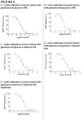
- the carbohydrate binding domain binds to a carbohydrate with a high affinity. In some cases, the carbohydrate binding domain binds to a carbohydrate with half-maximal binding occurring at a carbohydrate concentration of 1 nM or below, 10 nM or below, 50 nM or below, 100 nM or below, 500 nM or below, 1 ⁇ M or below, 10 ⁇ M or below, 50 ⁇ M or below, 100 ⁇ M or below, 500 ⁇ M or below, 1 mM or below or 10 mM or below.
- the EC 50 is between approximately 0.1 ⁇ M and 150 ⁇ M, between approximately 1 ⁇ M and 100 ⁇ M or between approximately 5 ⁇ M and 50 ⁇ M. In some embodiments, the EC 50 is between approximately 10 ⁇ M and 25 ⁇ M. Alternatively, in some embodiments the EC 50 is between approximately 0.1 mM and 150 mM, between approximately 1 mM and 100 mM, between approximately 2 mM and 50 mM or between approximately 2 mM and 5 mM.
- a suitable carbohydrate binding domains Upon binding of a carbohydrate to the carbohydrate binding domain, a suitable carbohydrate binding domains undergo a conformational change which is sufficient and suitable for carbohydrate detection as described herein.
- Carbohydrate binding domains useful in the sensors of the present disclosure are derived from transcription factors which contain a helix-turn-helix (HTH) domain (also referred to as a helix-turn-helix motif). That is, the sensors comprise a carbohydrate binding domain of a helix-turn-helix transcription factor, or a variant of the carbohydrate domain. In some cases, the sensor molecule comprises the carbohydrate binding domain of a helix-turn-helix transcription factor and one or more additional amino acids present in the helix-turn-helix transcription factor. For example, the sensor may also comprise one or more functional domains (for example, the DNA binding domain) also present in the helix-turn-helix transcription factor.
- HTH helix-turn-helix
- the sensors comprise a carbohydrate binding domain of a helix-turn-helix transcription factor, or a variant of the carbohydrate domain.
- the sensor molecule comprises the carbohydrate binding domain of a helix-turn-helix
- the polypeptide lacks the helix-turn-helix domain of the transcription factor. In an alternate case, the polypeptide has the helix-turn-helix domain of the transcription factor. Any variant, portion or fragment useful in the sensors described herein retains the ability to bind to a carbohydrate.
- the carbohydrate binding domain comprises a protein fold disclosed to bind to a carbohydrate. Examples of protein folds which bind to a carbohydrate include the Nudix hydrolase fold, a carbohydrate-binding module, or the AraC carbohydrate recognition domain.
- the HTH transcription factor is a bacterial HTH transcription factor.
- the HTH transcription factor may originate from gram-negative bacteria or gram-positive bacteria. Examples of such HTH transcription factors are shown in Table 1.
- Naturally occurring species variants of the HTH transcription factors listed in Table 1 can also be used, in addition to variants or fragments thereof as discussed herein.
- Homologues (such as orthologues originating from related species of bacteria) of the HTH transcription factors listed in Table 1 can also be used in the sensor molecules described herein.
- the term "HTH transcription factor” includes variants, portion, fragments or derivatives of any naturally occurring HTH transcription factor as long as the variant, portion, fragment or derivative retains the ability to bind a carbohydrate.
- Exemplary helix-turn-helix transcription factors Transcription Factor Sugar or sugar derivative
- Example Accession number (from UniProt) YvoA/NgaR N-acetylglucosamine (GlcNAc)/ glucosamine-6-phosphate 034817, Q795E9, TrmB maltose, trehalose, maltotriose, longer maltodextrins, sucrose, and glucose Q7LYW4, Q9HGZ9, Q9HPW0 AraR arabinose A2QJX5, Q5BGE2, P96711 AraC arabinose P0A9E0, P96711 TreR trehalose-6-phosphate P36673, P39796, P36674 MurQ N-acetylmuramic acid (MurNAc)-6-phosphate P76535, Q45582, Q8ZN25 LacI allolactose P03023, BgaR lactose Q8XMB9, BAB8047
- BgaR is a transcription factor from Clostridium perfringins strain 13 (CPE0770; UniProt Accession Number: Q8XMB9) and is a putative member of the AraR subfamily (Hartman et al., 2011). BgaR binds to lactose and forms part of a lactose-inducible regulatory system
- the claimed invention relates to BgaR (as defined in claim 1) as the core sensing component for binding lactose or lactulose. Other cases are disclosed but not claimed per se .
- the senor comprises the helix-turn-helix transcription factor BgaR or variant thereof that has an amino acid sequence which is at least 80% identical, at least 85% identical, at least 90% identical, at least 95% identical, at least 98% identical, at least 99% identical, at least 99.5% identical to that provided in SEQ ID NO: 1.
- the carbohydrate binding domain has an amino acid sequence which is at least 30%, 35%, 40%, 45%, 50%, 55%, 60%, 65%, 70%, 75%, 80%, 85%, 90%, 95%, 96%, 97%, 98%, 99% or 100% identical to a portion thereof (e.g., a portion comprising amino acids 1-179, amino acids 1-171, 1-157, amino acids 1-150, amino acids 12-179, amino acids 12-171, amino acids 12-150, amino acids 16-179, amino acids 16-171, amino acids 16 - 151 or amino acids 16-129 of SEQ ID NO: 1).
- the carbohydrate binding domain comprises an amino acid sequence provided as SEQ ID NO: 9 or is a fragment or variant thereof that retains carbohydrate binding activity.
- the carbohydrate binding domain has an amino acid sequence which is at least 30%, 35%, 40%, 45%, 50%, 55%, 60%, 65%, 70%, 75%, 80%, 85%, 90%, 95%, 96%, 97%, 98%, 99% or 100% identical to SEQ ID NO: 9.
- the minimum carbohydrate binding domain of BgaR (or another HTH transcription factor) can be determined using techniques known to the person skilled in the art.
- the protein sequence described herein can be used as a "query sequence" to perform a search against the conserved domain database to, for example, identify the putative carbohydrate binding domain and/or HTH domain (Marchler-Bauer et al., 2017; Marchler-Bauer et al., 2015; Marchler-Bauer et al., 2011; Marchler-Bauer and Bryant, 2004).
- the default parameters can be used.
- the carbohydrate binding domain of BgaR comprises amino acids 1-179, amino acids 1-171, amino acids 1-157, amino acids 1-150, amino amino acids 12-179, amino acids 12-171, amino acids 12-157, amino acids 12-150, amino acids 16-179, amino acids 16-171, amino acids 16-157, acids 16 - 151 or amino acids 16-129 of SEQ ID NO: 1.
- the carbohydrate binding domain of BgaR comprises amino acids 1-150, amino acids 1-171, amino acids 1-179, amino acids 12 - 171 or amino acids 12-150 of SEQ ID NO: 1.
- the carbohydrate binding domain has an amino acid sequence which is at least 30%, 35%, 40%, 45%, 50%, 55%, 60%, 65%, 70%, 75%, 80%, 85%, 90%, 95%, 96%, 97%, 98%, 99% or 100% identical to amino acids 1-157, amino acids 16 - 151 or amino acids 16-129 of SEQ ID NO: 1.
- the carbohydrate binding domain has an amino acid sequence which is at least 30%, 35%, 40%, 45%, 50%, 55%, 60%, 65%, 70%, 75%, 80%, 85%, 90%, 95%, 96%, 97%, 98%, 99% or 100% identical to amino acids 1-150, amino acids 1-171, amino acids 12 - 150 or amino acids 12-171 of SEQ ID NO: 1.
- the carbohydrate binding domain comprises an amino acid sequence provided as SEQ ID NO: 9.
- the carbohydrate binding domain has an amino acid sequence which is at least 30% identical, at least 35% identical, at least 40% identical, at least 45% identical, at least 50% identical, at least 55% identical, at least 60% identical, at least 65% identical, at least 70% identical, at least 75% identical, at least 80% identical, at least 85% identical, at least 90% identical, at least 95% identical, at least 98% identical, at least 99% identical, at least 99.5% identical, or 100% identical to that provided in SEQ ID NO: 9.
- the carbohydrate binding domain has an amino acid sequence which is at least 30%, 35%, 40%, 45%, 50%, 55%, 60%, 65%, 70%, 75%, 80%, 85%, 90%, 95%, 96%, 97%, 98%, 99% or 100% identical to a portion of SEQ ID NO: 9 or at least 30%, 35%, 40%, 45%, 50%, 55%, 60%, 65%, 70%, 75%, 80%, 85%, 90%, 95%, 96%, 97%, 98%, 99% or 100% identical to a portion of SEQ ID NO: 9.
- the transcription factor comprises a carbohydrate binding domain that comprises an amino acid sequence which is at least 30% identical, at least 35% identical, at least 40% identical, at least 45% identical, at least 50% identical, at least 55% identical, at least 60% identical, at least 65% identical, at least 70% identical, at least 75% identical, at least 80% identical, at least 85% identical, at least 90% identical, at least 95% identical, at least 98% identical, at least 99% identical, at least 99.5% identical to the carbohydrate binding domain of BgaR (for example, the amino acids in SEQ ID NO: 9 or to amino acids 1-179, amino acids 1-171, amino acids 1-150, amino acids 12-179, amino acids 12-171 or amino acids 12-150).
- Suitable transcription factors include, but are not limited to, putative lactose operon transcription activator from Clostridium perfringens (UniProt Accession No: A0A133MUX6), AraC family transcriptional regulator ( Clostridium perfringens D str. JGS1721 ) (UniProt Accession No: B1V7N0), AraC family transcriptional regulator ( Clostridium perfringens ) (UniProt Accession No: A0A127EGD8), arabinose operon regulatory protein (uncultured Clostridium sp.
- AraC family transcriptional regulator Staphylococcus pseudintermedius ) (UniProt Accession No: A0A166PPM9), AraC family transcriptional regulator ( Staphylococcus hyicus ) (UniProt Accession No: A0A2T4R7G1), AraC family transcriptional regulator ( Staphylococcus delphini ) (UniProt Accession No: A0A2A4HCU9), AraC family transcriptional regulator ( Staphylococcus agnetis ) (UniProt Accession No: A0A2T4MS83), Lactose operon transcription activator ( Staphylococcus xylosus ) (UniProt Accession No: 033813), Lactose operon transcription activator ( Staphylococcus saprophyticus ) (UniProt Accession No: A0A1D4LKB2), Putative lactose operon transcription activator ( Staphylococcus
- the senor comprises a transcription factor comprising an amino acid sequence selected from the group consisting of the amino acid sequence provided in SEQ ID NO: 37, SEQ ID NO: 38, SEQ ID NO: 39, SEQ ID NO: 40, SEQ ID NO: 41, SEQ ID NO: 42, SEQ ID NO: 43, SEQ ID NO: 44, SEQ ID NO: 45, SEQ ID NO: 46, SEQ ID NO: 48, SEQ ID NO: 49, SEQ ID NO: 50, SEQ ID NO: 51, SEQ ID NO: 52, SEQ ID NO: 53, SEQ ID NO: 54 and SEQ ID NO: 55, or is a fragment or variant thereof.
- a transcription factor comprising an amino acid sequence selected from the group consisting of the amino acid sequence provided in SEQ ID NO: 37, SEQ ID NO: 38, SEQ ID NO: 39, SEQ ID NO: 40, SEQ ID NO: 41, SEQ ID NO: 42, SEQ ID NO: 43, SEQ ID NO: 44, SEQ ID NO: 45, SEQ
- the sensor comprises a transcription factor having an amino acid sequence which is at least 30% identical, at least 35% identical, at least 40% identical, at least 45% identical, at least 50% identical, at least 55% identical, at least 60% identical, at least 65% identical, at least 70% identical, at least 75% identical, at least 80% identical, at least 85% identical, at least 90% identical, at least 95% identical, at least 98% identical, at least 99% identical, at least 99.5% identical to that provided in SEQ ID NO: 37, SEQ ID NO: 38, SEQ ID NO: 39, SEQ ID NO: 40, SEQ ID NO: 41, SEQ ID NO: 42, SEQ ID NO: 43, SEQ ID NO: 44, SEQ ID NO: 45, SEQ ID NO: 46, SEQ ID NO: 47, SEQ ID NO: 48, SEQ ID NO: 49, SEQ ID NO: 50, SEQ ID NO: 51, SEQ ID NO: 52, SEQ ID NO: 53, SEQ ID NO: 54 or SEQ ID NO: 55, or is a transcription factor
- the carbohydrate binding domain comprises an amino acid sequence selected from the group consisting of the amino acid sequence provided in SEQ ID NO: 56, SEQ ID NO: 57, SEQ ID NO: 58, SEQ ID NO: 59, SEQ ID NO: 60, SEQ ID NO: 61, SEQ ID NO: 62, SEQ ID NO: 63, SEQ ID NO: 64, SEQ ID NO: 65, SEQ ID NO: 66, SEQ ID NO: 67, SEQ ID NO: 68, SEQ ID NO: 69, SEQ ID NO: 70, SEQ ID NO: 71, SEQ ID NO: 72, SEQ ID NO: 73 or SEQ ID NO: 74, or is a fragment or variant thereof.
- the carbohydrate binding domain has an amino acid sequence which is at least 30% identical, at least 35% identical, at least 40% identical, at least 45% identical, at least 50% identical, at least 55% identical, at least 60% identical, at least 65% identical, at least 70% identical, at least 75% identical, at least 80% identical, at least 85% identical, at least 90% identical, at least 95% identical, at least 98% identical, at least 99% identical, at least 99.5% identical to the amino acid sequence provided in SEQ ID NO: 56, SEQ ID NO: 57, SEQ ID NO: 58, SEQ ID NO: 59, SEQ ID NO: 60, SEQ ID NO: 61, SEQ ID NO: 62, SEQ ID NO: 63, SEQ ID NO: 64, SEQ ID NO: 65, SEQ ID NO: 66, SEQ ID NO: 67, SEQ ID NO: 68, SEQ ID NO: 69, SEQ ID NO: 70, SEQ ID NO: 71, SEQ ID NO: 72, SEQ ID NO:
- the nucleic acid molecule comprises a sequence encoding the polypeptide having a sequence at least 30%, 35%, 40%, 45%, 50%, 55%, 60%, 65%, 70%, 75%, 80%, 85%, 90%, 95%, 96%, 97%, 98%, 99% or 100% identical to the sequence shown in any one of SEQ ID NO: 15, SEQ ID NO: 16, SEQ ID NO: 17, SEQ ID NO: 18, SEQ ID NO: 25, SEQ ID NO: 26, SEQ ID NO: 27 or SEQ ID NO: 28, or a sequence at least 30%, 35%, 40%, 45%, 50%, 55%, 60%, 65%, 70%, 75%, 80%, 85%, 90%, 95%, 96%, 97%, 98%, 99% or 100% identical to a portion thereof (e.g., a portion comprising amino acids 11 - 349, amino acids 18 - 349, amino acids 28 - 349, amino acids 38 - 349, or amino acids 39 - 349 of any one of SEQ ID NO
- the nucleic acid molecule comprises a sequence encoding the polypeptide having a sequence at least 80%, 85%, 90%, 95%, 96%, 97%, 98%, 99% or 100% identical to the sequence shown in any one of SEQ ID NO: 15, SEQ ID NO: 16, SEQ ID NO: 17 or SEQ ID NO: 18, or in some cases a sequence at least 30%, 35%, 40%, 45%, 50%, 55%, 60%, 65%, 70%, 75%, 80%, 85%, 90%, 95%, 96%, 97%, 98%, 99% or 100% identical to a portion thereof (e.g., a portion comprising amino acids 11 -349, amino acids 18 - 349, amino acids 28 - 349, amino acids 38 - 349, or amino acids 39 - 349 of any one of SEQ ID NO: 15, SEQ ID NO: 16, SEQ ID NO: 17 or SEQ ID NO: 18).
- the nucleic acid molecule comprises a sequence encoding the polypeptide having a sequence at least 50%, 55%, 60%, 65%, 70%, 75%, 80%, 85%, 90%, 95%, 96%, 97%, 98%, 99% or 100% identical to the sequence shown in any one of SEQ ID NO: 25, SEQ ID NO: 26, SEQ ID NO: 27 or SEQ ID NO: 28, or a sequence at least 30%, 35%, 40%, 45%, 50%, 55%, 60%, 65%, 70%, 75%, 80%, 85%, 90%, 95%, 96%, 97%, 98%, 99% or 100% identical to a portion thereof (e.g., a portion comprising amino acids 11 -349, amino acids 18 - 349, amino acids 28 - 349, amino acids 38 - 349, or amino acids 39 - 349 of any one of SEQ ID NO: 25, SEQ ID NO: 26, SEQ ID NO: 27 or SEQ ID NO: 28).
- the nucleic acid molecule comprises a sequence selected from the group consisting of SEQ ID NO: 19, SEQ ID NO: 20, SEQ ID NO: 21 and SEQ ID NO: 22 or a fragment or variant thereof.
- the nucleic acid molecule comprises a sequence encoding the polypeptide having a sequence at least 50%, 55%, 60%, 65%, 70%, 75%, 80%, 85%, 90%, 95%, 96%, 97%, 98%, 99% or 100% identical to the sequence shown in any one of SEQ ID NO: 19, SEQ ID NO: 20, SEQ ID NO: 21 and SEQ ID NO: 22, or a sequence at least 50%, 55%, 60%, 65%, 70%, 75%, 80%, 85%, 90%, 95%, 96%, 97%, 98%, 99% or 100% identical to a portion thereof of any one of SEQ ID NO: 19, SEQ ID NO: 20, SEQ ID NO: 21 and SEQ ID NO: 22.
- the nucleic acid molecule comprises a sequence selected from the group consisting of SEQ ID NO: 19, SEQ ID NO: 20, SEQ ID NO: 21, SEQ ID NO: 22, SEQ ID NO: 29, SEQ ID NO: 30, SEQ ID NO: 31 and SEQ ID NO: 32 or a fragment or variant thereof.
- the nucleic acid molecule comprises a sequence encoding the polypeptide having a sequence at least 50%, 55%, 60%, 65%, 70%, 75%, 80%, 85%, 90%, 95%, 96%, 97%, 98%, 99% or 100% identical to the sequence shown in any one of SEQ ID NO: 19, SEQ ID NO: 20, SEQ ID NO: 21 and SEQ ID NO: 22, or a sequence at least 50%, 55%, 60%, 65%, 70%, 75%, 80%, 85%, 90%, 95%, 96%, 97%, 98%, 99% or 100% identical to a portion thereof of any one of SEQ ID NO: 19, SEQ ID NO: 20, SEQ ID NO: 21, SEQ ID NO: 22, SEQ ID NO: 29, SEQ ID NO: 30, SEQ ID NO: 31 and SEQ ID NO: 32.
- the nucleic acid molecule comprises a sequence selected from the group consisting of SEQ ID NO: 29, SEQ ID NO: 30, SEQ ID NO: 31 and SEQ ID NO: 32 or a fragment or variant thereof.
- the nucleic acid molecule comprises a sequence encoding the polypeptide having a sequence at least 50%, 55%, 60%, 65%, 70%, 75%, 80%, 85%, 90%, 95%, 96%, 97%, 98%, 99% or 100% identical to the sequence shown in any one of SEQ ID NO: 19, SEQ ID NO: 20, SEQ ID NO: 21 and SEQ ID NO: 22, or a sequence at least 50%, 55%, 60%, 65%, 70%, 75%, 80%, 85%, 90%, 95%, 96%, 97%, 98%, 99% or 100% identical to a portion thereof of any one of SEQ ID NO: 29, SEQ ID NO: 30, SEQ ID NO: 31 and SEQ ID NO: 32.
- substantially identical is used herein to refer to a first amino acid that contains a sufficient or minimum number of amino acid residues that are i) identical to, or ii) conservative substitutions of aligned amino acid residues in a second amino acid sequence such that the first and second amino acid sequences can have a common structural domain and/or common functional activity.
- amino acid sequences that contain a common structural domain having at least about 85%, 90%, 91%, 92%, 93%, 94%, 95%, 96%, 97%, 98% or 99% or 100% identity to the sequence specified are termed substantially identical.
- Binding of a carbohydrate, such as lactose, to the sensors of the present disclosure can result in a change in Resonance Energy Transfer (RET), including, but not limited to, bioluminescent resonance energy transfer (“BRET”) and fluorescence resonance energy transfer (“FRET”).
- RET Resonance Energy Transfer
- BRET bioluminescent resonance energy transfer
- FRET fluorescence resonance energy transfer
- BRET is a proximity assay based on the non-radiative transfer of energy between a bioluminescent protein donor and an acceptor molecule.
- Bioluminescent resonance energy transfer and “BRET” are used interchangeably.
- FRET is a proximity assay based on the non-radiative transfer of energy between two chromophores, for example, two fluorophores.
- FRET fluorescence resonance energy transfer
- the sensor molecule comprises a donor domain and an acceptor domain covalently attached to the transcription factor or fragment or variant thereof.
- the donor domain is a chemiluminescent donor domain.
- the donor domain is a fluorophore.
- the acceptor domain is a fluorescent acceptor domain, such as a fluorophore.
- the donor domain is covalently attached to the N-terminus of the transcription factor or fragment or variant thereof and the acceptor domain is covalently attached to the C-terminus of the transcription factor or fragment or variant thereof.
- the donor domain is covalently attached to the C-terminus of the transcription factor or fragment or variant thereof and the acceptor domain is covalently attached to the N-terminus of the transcription factor or fragment or variant thereof.
- the sensor molecules of the present disclosure comprise a donor domain.
- the donor domain is capable of serving as a donor domain in a resonance energy transfer pair (for example, in a BRET pair or a FRET pair) and, depending on context, is also referred to herein as a "resonance energy transfer donor domain".
- the term "donor” means a molecule that emits light, for example a molecule which, when irradiated with light of a certain wavelength, emits light or a molecule which causes the emission of light as the result of a chemical reaction.
- Suitable donor domains include chemiluminescent domains and fluorescent domains.
- the donor domain capable of serving as a donor domain in a BRET pair can be a chemiluminescent donor domain.
- Chemiluminescence is the emission of energy with limited emission of heat (luminescence), as the result of a chemical reaction.
- the term "chemiluminescence” is used herein to encompass bioluminescence, which relies upon the activity of an enzyme.
- Non-enzymatic chemiluminescence is the result of chemical reactions between an organic dye and an oxidizing agent in the presence of a catalyst.
- Chemiluminescence emission occurs as the energy from the excited states of organic dyes, which are chemically induced, decays to ground state. The duration and the intensity of the chemiluminescence emission are mostly dependent on the extent of the chemical reagents present in the reaction solution.
- the chemiluminescent donor domain is a bioluminescent protein.
- bioluminescent protein refers to any protein capable of acting on a suitable substrate to generate luminescence.
- a bioluminescent protein is an enzyme which converts a substrate into an activated product which then releases energy as it relaxes.
- the activated product (generated by the activity of the bioluminescent protein on the substrate) is the source of the bioluminescent protein-generated luminescence that is transferred to the acceptor molecule.
- bioluminescent proteins there are a number of different bioluminescent proteins that can be employed in this invention (see, for example, Table 2).
- Light-emitting systems have been known and isolated from many luminescent organisms including bacteria, protozoa, coelenterates, molluscs, fish, millipedes, flies, fungi, worms, crustaceans, and beetles, particularly click beetles of genus Pyrophorus and the fireflies of the genera Photinus, Photuris, and Luciola. Additional organisms displaying bioluminescence are listed in WO 00/024878 , WO 99/049019 and Viviani (2002). Table 2. Exemplary bioluminescent proteins.
- Luciferin Insect Orphelia fultoni North American glow worm
- Insect Clluc Pyrophorus plagiophthalamus click beetle
- Luciferin Jellyfish Aequorin Aequorea 44.9 460-470 Coelenterazine Sea pansy
- RLuc Renilla reniformis 36
- Coelenterazine Sea pansy (modified)
- RLuc8 Renilla reniformis (modified) 36 487 (peak) Coelenterazine /Deep Blue C
- RLuc2 Renilla reniformis Modified
- M185V/Q235A 36
- Coelenterazine Sea pansy (modified)
- RLuc8.6 -535 Renilla reniformis (modified)
- bioluminescent protein can be used in the sensors of the present disclosure.
- One very well-known example is the class of proteins known as luciferases which catalyse an energy-yielding chemical reaction in which a specific biochemical substance, a luciferin (a naturally occurring fluorophore), is oxidized by an enzyme having a luciferase activity (Hastings, 1996).
- luciferases which catalyse an energy-yielding chemical reaction in which a specific biochemical substance, a luciferin (a naturally occurring fluorophore), is oxidized by an enzyme having a luciferase activity (Hastings, 1996).
- prokaryotic and eukaryotic including species of bacteria, algae, fungi, insects, fish and other marine forms can emit light energy in this manner and each has specific luciferase activities and luciferins which are chemically distinct from those of other organisms.
- Luciferin/luciferase systems are very diverse in form, chemistry and function. Bioluminescent proteins with luciferase activity are thus available from a variety of sources or by a variety of means. Examples of bioluminescent proteins with luciferase activity may be found in US 5,229,285 , 5,219,737 , 5,843,746 , 5,196,524 , and 5,670,356 . Two of the most widely used luciferases are: (i) Renilla luciferase (from R .
- reniformis a 35 kDa protein, which uses coelenterazine as a substrate and emits light at 480 nm (Lorenz et al., 1991); and (ii) Firefly luciferase (from Photinus pyralis ), a 61 kDa protein, which uses luciferin as a substrate and emits light at 560 nm (de Wet et al., 1987).
- Gaussia luciferase (from Gaussia princeps ) has been used in biochemical assays (Verhaegen et al., 2002). Gaussia luciferase is a 20 kDa protein that oxidises coelenterazine in a rapid reaction resulting in a bright light emission at 470 nm.
- Luciferases useful for the present invention have also been characterized from Anachnocampa sp ( WO 2007/019634 ). These enzymes are about 59 kDa in size and are ATP-dependent luciferases that catalyse luminescence reactions with emission spectra within the blue portion of the spectrum.
- Biologically active variants or fragments of naturally occurring bioluminescent protein can readily be produced by those skilled in the art.
- Three examples of such variants useful for the invention are RLuc2 (Loening et al., 2006), RLuc8 (Loening et al., 2006) and RLuc8.6-535 (Loening et al., 2007) which are each variants of Renilla luciferase.
- the sequence of the BRET chemiluminescent donor is chosen to have greater thermal stability than sensor molecules incorporating native Renilla luciferase sensors.
- RLuc2 or RLuc8 are convenient examples of suitable choices, which consequently exhibit ⁇ 5x or ⁇ 10x higher luminance than sensors incorporating the native Renilla luciferase sequence. Such enhanced luminance has significant benefits as it permits more economical use of reagents for any given time resolution.
- non-luciferase, bioluminescent proteins that can be employed in this invention are any enzymes which can act on suitable substrates to generate a luminescent signal.
- enzymes include ⁇ -galactosidase, lactamase, horseradish peroxidase, alkaline phosphatase, ⁇ -glucuronidase and ⁇ -glucosidase.
- Synthetic luminescent substrates for these enzymes are well known in the art and are commercially available from companies, such as Tropix Inc. (Bedford, MA, USA).
- the bioluminescent protein is a luciferase, a ⁇ -galactosidase, a lactamase, a horseradish peroxidase, an alkaline phosphatase, a ⁇ -glucuronidase or a ⁇ -glucosidase.
- the bioluminescent protein is luciferase.
- Suitable luciferase include, but are not limited to a Renilla luciferase, a Firefly luciferase (e.g.
- PpyRE8, PpyRE10) a Coelenterate luciferase, a North American glow worm luciferase, a click beetle luciferase, a railroad worm luciferase, a bacterial luciferase, a Gaussia luciferase, Aequorin, an Arachnocampa luciferase, an Oplophorus gracilirostris luciferase or a biologically active variant or fragment of any one, or chimera of two or more, thereof.
- the preferred luciferase is RLuc8 or a variant thereof.
- a "biologically active fragment” is a portion of a polypeptide as described herein which maintains a defined activity of the full-length polypeptide.
- a "biologically active variant” is a molecule which differs from a naturally occurring and/or defined molecule by one or more amino acids but maintains a defined activity, such as defined above for biologically active fragments.
- Biologically active variants are typically least 50%, more preferably at least 80%, more preferably at least 90%, more preferably at least 95%, more preferably at least 97%, and even more preferably at least 99% identical to the naturally occurring and/or defined molecule.
- a bioluminescent protein with a small molecular weight is used to prevent an inhibition of the interaction due to steric hindrance.
- the bioluminescent protein preferably consists of a single polypeptide chain. Also the bioluminescent proteins preferably do not form oligomers or aggregates.
- the bioluminescent proteins Renilla luciferase, Gaussia luciferase and Firefly luciferase meet all or most of these criteria.
- the chemiluminescent donor domain is capable of modifying a substrate.
- substrate refers to any molecule that can be used in conjunction with a chemiluminescent donor to generate or absorb luminescence. The choice of the substrate can impact on the wavelength and the intensity of the light generated by the chemiluminescent donor.
- the bioluminescent protein has a substrate selected from luciferin, calcium, coelenterazine, furimazine or a derivative, analogue or stabilised derivative of coelenterazine, luciferin or furimazine.
- Coelenterazine is a widely known substrate which occurs in cnidarians, copepods, chaetognaths, ctenophores, decapod shrimps, mysid shrimps, radiolarians and some fish taxa (Greer and Szalay, 2002).
- coelenterazine analogues/derivatives are available that result in light emission between 418 and 547 nm (Inouye et al., 1997, Loening et al., 2007).
- a coelenterazine analogue/derivative 400A, DeepBlueC has been described emitting light at 400 nm with Renilla luciferase ( WO 01/46691 ).
- Other examples of coelenterazine analogues/derivatives are EnduRen, Prolume purple, Prolume purple II, Prolume purple III, ViviRen and Furimazine.
- Other examples of coelenterazine analogues/derivatives include, but are not limited to, compounds disclosed in PCT/US2013057660 and US20140302539 .
- luciferin is defined broadly and refers to a class of light-emitting biological pigments found in organisms capable of bioluminescence as well as synthetic analogues or functionally equivalent chemicals, which are oxidised in the presence of the enzyme luciferase to produce oxyluciferin and energy in the form of light.
- D-luciferin, or 2-(6-hydroxybenzothiazol-2-yl)-2-thiazoline-4-carboxylic acid was first isolated from the firefly Photinus pyralis.
- luciferin also includes derivatives or analogues of luciferin.
- luciferin In addition to entirely synthetic luciferin, such as cyclic alkylaminoluciferin (CycLuc1), there are at least five general types of biologically evolved luciferin, which are each chemically different and catalysed by chemically and structurally different luciferases that employ a wide range of different cofactors.
- First is firefly luciferin, the substrate of firefly luciferase, which requires ATP for catalysis (EC 1.13.12.7).
- Bacterial luciferase is FMNH-dependent.
- dinoflagellate luciferin a tetrapyrrolic chlorophyll derivative found in dinoflagellates (marine plankton), the organisms responsible for night-time ocean phosphorescence.
- Dinoflagellate luciferase catalyses the oxidation of dinoflagellate luciferin and consists of three identical and catalytically active domains.
- imidazolopyrazine vargulin which is found in certain ostracods and deep-sea fish, for example, Porichthys.
- Last is coelenterazine (an imidazolpyrazine), the light-emitter of the protein Aequorin, found in radiolarians, ctenophores, cnidarians, squid, copepods, chaetognaths, fish and shrimp.
- the bioluminescent protein requires a co-factor.
- co-factors include, but are not necessarily limited to, ATP, magnesium, oxygen, FMNH 2 , calcium, or a combination of any two or more thereof.
- the resonance energy transfer donor domain is a fluorescent donor domain.
- the fluorescent donor domain can be a fluorescent protein or a non-protein.
- the fluorescent donor domain is a non-protein.
- fluorophores that are suitable for use as the donor domain include, but are not limited to, Alexa Fluor dye (e.g. AF680, AF750), Bodipy dye, Cy dye, fluorescein, dansyl, umbelliferone, fluorescent microsphere, luminescent microsphere, fluorescent nanocrystal, Marina Blue, Cascade Blue, Cascade Yellow, Pacific Blue, Oregon Green, Tetramethylrhodamine, Rhodamine, Texas Red, rare earth element chelates, or any combination or derivatives thereof.
- the donor domain is a fluorescent protein.
- Non-limiting examples include proteins such as green fluorescent protein (GFP), blue fluorescent variant of GFP (BFP), cyan fluorescent variant of GFP (CFP), yellow fluorescent variant of GFP (YFP), enhanced GFP (EGFP), enhanced CFP (ECFP), enhanced YFP (EYFP), GFPS65T, Emerald, Venus, mOrange, Topaz, GFPuv, destabilised EGFP (dEGFP), destabilised ECFP (dECFP), destabilised EYFP (dEYFP), HcRed, t-HcRed, DsRed, DsRed2, t-dimer2, t-dimer2(12), mRFP1, pocilloporin, Renilla GFP, Monster GFP, paGFP, TdTomato, mCherry, Kaede protein, TagRFP, TurBoFB or a Phycobiliprotein, or a biologically active variant or fragment of any one thereof.
- the sensor molecules of the present disclosure also comprise an acceptor domain.
- the acceptor domain is capable of serving as an acceptor domain in a resonance energy transfer pair (for example, in a BRET pair or a FRET pair) and, depending on context, is also referred to herein as a "resonance energy transfer acceptor domain".
- an "acceptor domain” is any molecule that is capable of accepting energy emitted as a result of the activity of the donor domain.
- the acceptor domain (also referred to herein as “acceptor molecule”) is a fluorescent acceptor domain.
- fluorescent acceptor domain also referred herein to as “fluorescent acceptor molecule” refers to any compound which can accept energy emitted as a result of the activity of a donor domain, and re-emit it as light energy.
- acceptor domains There are a number of different acceptor domains that can be employed in this invention. Suitable acceptor domains may be a protein or non-proteinaceous.
- the fluorescent acceptor domain is a fluorescent protein.
- a fluorescent protein is a group of fluorophores that includes the green fluorescent protein from the jellyfish Aequorea victoria and numerous other variants (GFPs) arising from the application of molecular biology, for example mutagenesis and chimeric protein technologies (Tsien, 1998). GFPs are classified based on the distinctive component of their chromophores, each class having distinct excitation and emission wavelengths: class 1, wild-type mixture of neutral phenol and anionic phenolate: class 2, phenolate anion: class 3, neutral phenol: class 4, phenolate anion with stacked s-electron system: class 5, indole: class 6, imidazole: and class 7, phenyl.
- a naturally occurring acceptor molecule which has been mutated (variants) can also be useful for the present invention.
- One example of an engineered system which is suitable for BRET is a Renilla luciferase and enhanced yellow mutant of GFP (EYFP) pairing which do not directly interact to a significant degree with one another alone in the absence of a mediating protein(s) (in this case, the G protein coupled receptor) (Xu et al., 1999).
- Examples include, but are not limited to, green fluorescent protein (GFP), blue fluorescent variant of GFP (BFP), cyan fluorescent variant of GFP (CFP), yellow fluorescent variant of GFP (YFP), enhanced GFP (EGFP), enhanced GFP (EGFP), enhanced CFP (ECFP), enhanced YFP (EYFP), GFPS65T, Emerald, Venus, mOrange, Topaz, GFPuv, destabilised EGFP (dEGFP), destabilised ECFP (dECFP), destabilised EYFP (dEYFP), HcRed, t-HcRed, DsRed, DsRed2, t-dimer2, t-dimer2(12), mRFP1, pocilloporin, Renilla GFP, Monster GFP, paGFP, Kaede protein, TdTomato, mCherry, TagRFP, TurBoFB or a Phycobiliprotein, or a biologically active variant or fragment of any one thereof.
- the preferred GFP
- the fluorescent acceptor domain is a non-protein.
- acceptor molecules that are not proteins include, but are not limited to, Alexa Fluor dye (e.g. AF680, AF750), Bodipy dye, Cy dye, fluorescein, dansyl, umbelliferone, fluorescent microsphere, luminescent microsphere, fluorescent nanocrystal, Marina Blue, Cascade Blue, Cascade Yellow, Pacific Blue, Oregon Green, Tetramethylrhodamine, Rhodamine, Texas Red, rare earth element chelates, or any combination or derivatives thereof.
- donor-acceptor combinations can be used in the sensors of the present invention.
- the donor-acceptor combination should be capable of serving as a BRET pair or a FRET pair.
- a worker skilled in the art would be able to select a donor and acceptor pair which permits efficient energy transfer.
- the separation and relative orientation of the donor domain and the acceptor domain, in the presence and/or absence of the carbohydrate is within ⁇ 50% of the Förster distance.
- the term "the separation and relative orientation of the donor domain and the acceptor domain, in the presence and/or the absence of carbohydrate, is within ⁇ 50% of the Förster distance" refers to the steady state RET measurements which can be carried out within a range of ⁇ 50% of R 0 . This phrase encompasses an efficiency of luminescence energy transfer from the donor domain to the acceptor domain in the range of 10-90%.
- the Förster distance of the chemiluminescent donor domain and the acceptor domain is at least 4 nm, is at least 5.6 nm, or is at least 6 nm. In some embodiments, the Förster distance is less than 12 nm, less than 11 nm, less than 10 nm or less than 9 nm. In some embodiments, the Förster distance of the donor domain and the acceptor domain is between about 4 nm and about 11 nm, is between about 5.6 nm and about 11 nm or is between about 7 nm and about 11 nm.
- the carbohydrate binding domain may be, for example, a full length HTH transcription factor, a fragment thereof that retains carbohydrate binding activity, a carbohydrate binding domain of a HTH transcription factor, or a variant thereof.
- a criterion which should be considered in determining suitable pairings is the relative emission/fluorescence spectrum of the acceptor molecule compared to that of the donor.
- the emission spectrum of the donor should overlap with the absorbance spectrum of the acceptor molecule such that the light energy from the donor emission is at a wavelength that is able to excite the acceptor molecule and thereby promote acceptor molecule fluorescence when the two molecules are in a proper proximity and orientation with respect to one another.
- Renilla luciferase/EGFP pairing is not as good as a Renilla luciferase/EYEF pairing based on observable emission spectral peaks (Xu et al., 1999; Wang et al., (1997) in Bioluminescence and Chemiluminescence: Molecular Reporting with Photons, eds. Hastings et al., (Wiley, New York), pp. 419-422 ).
- protein fusions for example are prepared containing the selected donor and acceptor domains and are tested, in the presence of an appropriate substrate if required.
- the donor and acceptor domains do not spuriously associate with each other.
- this can be accomplished by separate co-expression of a bioluminescent protein and acceptor molecule in the same cells and then monitoring the luminescence spectrum in order to determine if BRET occurs. This may be achieved, for example, using the method of Xu et al., (1999).
- the selected bioluminescent protein and acceptor molecule form a suitable BRET pair if little or no BRET is observed. Similar experiments can be performed for FRET pairs.
- the sensor molecules of the present disclosure comprise a chemiluminescent donor domain and fluorescent acceptor domain.
- the donor emission can be manipulated by modifications to the substrate.
- the substrate is coelenterazine.
- the rationale behind altering the donor emission is to improve the resolution between donor emission and acceptor emissions.
- the original BRET system uses the Renilla luciferase as donor, EYFP (or Topaz) as the acceptor and coelenterazine h derivative as the substrate. These components when combined in a BRET assay, generate light in the 475-480 nm range for the bioluminescent protein and the 525-530 nm range for the acceptor molecule, giving a spectral resolution of 45-55 nm.
- Renilla luciferase generates a broad emission peak overlapping substantially the GFP emission, which in turn contributes to decrease the signal to noise of the system.
- One BRET system for use in the present invention has coel400a as the Renilla luciferase substrate and provides broad spectral resolution between donor and acceptor emission wavelengths ( ⁇ 105nm). Renilla luciferase with coel400a generates light between 390-400 nm and a GFP derivative (GFP 2 ) was prepared which absorbs light in this range and re-emits light at 505-508 nm.
- GFP 2 GFP derivative
- this BRET system provides an excellent biological tool to monitor binding of a carbohydrate to the sensors of the present application.
- smaller Stokes shift BRET systems would also allow sensitive measurement of carbohydrates.
- coelenterazine derivatives are known in the art, including coel400a, that generate light at various wavelengths (distinct from that generated by the wild type coelenterazine) as a result of Renilla luciferase activity.
- coel400a that generate light at various wavelengths (distinct from that generated by the wild type coelenterazine) as a result of Renilla luciferase activity.
- a worker skilled in the art would appreciate that because the light emission peak of the donor has changed, it is necessary to select an acceptor molecule which will absorb light at this wavelength and thereby permit efficient energy transfer. This can be done, for example by altering a GFP class 4 such that it becomes a class 3 or 1 GFP.
- Spectral overlapping between light emission of the donor and the light absorption peak of the acceptor is one condition among others for an efficient energy transfer.
- Class 3 and 1 GFPs are known to absorb light at 400 nm and re-emit between 505-511 nm.
- RLuc8 RLuc2 Native 470 nm Fluorescein 495/519 nm
- the preferred bioluminescent protein and acceptor domain pair is RLuc8 and GFP 2 .
- the sensor molecules of the present disclosure comprise a fluorescent donor domain and a fluorescent acceptor domain.
- any appropriately selected fluorophore can be used as the donor and/or acceptor, provided that the emission spectrum of the donor overlaps sufficiently with the excitation spectrum of the acceptor.
- a criterion which should be considered in determining suitable pairings is the excitation spectrum of the acceptor molecule compared to that of the donor. As the person skilled in the art would appreciate there should be minimum direct excitation of the acceptor domain at the excitation maximum of the donor domain.
- Fluorescent donor Wavelength of acceptor (Em) Fluorescence acceptor molecule Wavelength of acceptor (Exc) FITC 520 nm TRITC 550 nm Cy3 566 nm Cy5 649 nm EGFP 508 nm Cy3 554 nm CFP 477 nm YFP 514 nm EGFP 508 nm YFP 514 nm GFP 2 YFP 514 nm ECFP 475 nm EYFP 513 nm mTurquioise2 474 nm mCitrine 516 nm mClover3 518 nm mRuby3 558 nm eqFP650 650 nm iRFP 690 nm mAmetrine 526
- the preferred donor domain and acceptor domain pair is CFP and YFP.
- Binding of a carbohydrate to the carbohydrate binding domain of the sensors of the present disclosure alters the spatial location and/or dipole orientation of the donor domain relative to the acceptor domain.
- the alteration in spatial location and/or dipole orientation results in a change in BRET.
- the alteration in spatial location and/or dipole orientation results in a change in FRET.
- spatial location refers to the three dimensional positioning of the donor relative to the acceptor molecule which changes as a result of the analyte binding or releasing from the sensor molecule.
- dipole orientation refers to the direction in three-dimensional space of the dipole moment associated either with the donor and/or the acceptor molecule relative their orientation in three-dimensional space.
- the dipole moment is a consequence of a variation in electrical charge over a molecule.
- the energy transfer occurring between the bioluminescent protein and acceptor molecule is presented as calculated ratios from the emissions measured using optical filters (one for the acceptor molecule emission and the other for the bioluminescent protein emission) that select specific wavelengths (see equation 1).
- E a / E d BRET ratio where E a is defined as the acceptor molecule emission intensity (emission light is selected using a specific filter adapted for the emission of the acceptor) and E d is defined as the bioluminescent protein emission intensity (emission light is selected using a specific filter adapted for the emission of the bioluminescent protein).
- optical filters may be any type of filter that permits wavelength discrimination suitable for BRET.
- optical filters used in accordance with the present invention can be interference filters, long pass filters, short pass filters, etc.
- Intensities (usually in counts per second (CPS) or relative luminescence units (RLU)) of the wavelengths passing through filters can be quantified using either a photo-multiplier tube (PMT), photodiode, including a cascade photodiode, photodiode array or a sensitive camera such as a charge coupled device (CCD) camera.
- the quantified signals are subsequently used to calculate BRET ratios and represent energy transfer efficiency.
- the BRET ratio increases with increasing intensity of the acceptor emission.
- a ratio of the acceptor emission intensity over the donor emission intensity is determined (see equation 1), which is a number expressed in arbitrary units that reflects energy transfer efficiency. The ratio increases with an increase of energy transfer efficiency (see Xu et al., 1999).
- Energy transfer efficiencies can also be represented using the inverse ratio of donor emission intensity over acceptor emission intensity (see equation 2). In this case, ratios decrease with increasing energy transfer efficiency.
- the emission intensities Prior to performing this calculation the emission intensities are corrected for the presence of background light and auto-luminescence of the substrate. This correction is generally made by subtracting the emission intensity, measured at the appropriate wavelength, from a control sample containing the substrate but no bioluminescent protein, acceptor molecule or polypeptide of the invention.
- E d / E a BRET ratio where E a and E d are as defined above.
- the light intensity of the bioluminescent protein and acceptor molecule emission can also be quantified using a monochromator-based instrument such as a spectrofluorometer, a charged coupled device (CCD) camera or a diode array detector.
- a monochromator-based instrument such as a spectrofluorometer, a charged coupled device (CCD) camera or a diode array detector.
- CCD charged coupled device
- the emission scan is performed such that both bioluminescent protein and acceptor molecule emission peaks are detected upon addition of the substrate.
- the areas under the peaks represent the relative light intensities and are used to calculate the ratios, as outlined above.
- Any instrument capable of measuring lights for the bioluminescent protein and acceptor molecule from the same sample can be used to monitor the BRET system of the present invention.
- the acceptor molecule emission alone is suitable for effective detection and/or quantification of BRET.
- the energy transfer efficiency is represented using only the acceptor emission intensity. It would be readily apparent to one skilled in the art that in order to measure energy transfer, one can use the acceptor emission intensity without making any ratio calculation. This is due to the fact that ideally the acceptor molecule will emit light only if it absorbs the light transferred from the bioluminescent protein. In this case only one light filter is necessary.
- the bioluminescent protein emission alone is suitable for effective detection and/or quantification of BRET.
- the energy transfer efficiency is calculated using only the bioluminescent protein emission intensity. It would be readily apparent to one skilled in the art that in order to measure energy transfer, one can use the donor emission intensity without making any ratio calculation. This is due to the fact that as the acceptor molecule absorbs the light transferred from the bioluminescent protein there is a corresponding decrease in detectable emission from the bioluminescent protein. In this case only one light filter is necessary.
- the energy transfer efficiency is represented using a ratiometric measurement which only requires one optical filter for the measurement.
- light intensity for the donor or the acceptor is determined using the appropriate optical filter and another measurement of the samples is made without the use of any filter (intensity of the open spectrum). In this latter measurement, total light output (for all wavelengths) is quantified. Ratio calculations are then made using either equation 3 or 4. For the equation 3, only the optical filter for the acceptor is required. For the equation 4, only the optical filter for the donor is required.
- E a and E d are as defined above and E o is defined as the emission intensity for all wavelengths combined (open spectrum).
- equations 1 through 4 can be derived from equations 1 through 4.
- one such derivative involves correcting for background light present at the emission wavelength for bioluminescent protein and/or acceptor molecule.
- the BRETCount instrument is a modified TopCount, wherein the TopCount is a microtiterplate scintillation and luminescence counter sold by Packard Instrument (Meriden, CT). Unlike classical counters which utilise two photomultiplier tubes (PMTs) in coincidence to eliminate background noise, TopCount employs single- PMT technology and time-resolved pulse counting for noise reduction to allow counting in standard opaque microtiter plates. The use of opaque microtiterplates can reduce optical crosstalk to negligible level. TopCount comes in various formats, including 1, 2, 6 and 12 detectors (PMTs), which allow simultaneous reading of 1, 2, 6 or 12 samples, respectively.
- PMTs photomultiplier tubes
- BRET Beside the BRETCount, other commercially available instruments are capable of performing BRET: the Victor 2 (Wallac, Finland (Perkin Elmer Life Sciences)) and the Fusion (Packard Instrument, Meriden).
- BRET can be performed using readers that can detect at least the acceptor molecule emission and preferably two wavelengths (for the acceptor molecule and the bioluminescent protein) or more.
- BRET is a ratiometric technique which can eliminate data variability caused by fluctuations in light output due to variations in assay volume, assay conditions and signal decay across different wells in a plate. RET-based reactions are homogeneous, generally occurring in solution without solid-phase attachment. This allows for detection of analytes in different forms such as liquid, gas and even particulates without separation.
- a sensor molecule as defined herein is a sensor molecule that can used to detect and/or measure lactose concentration. Accordingly, the present disclosure provides a sensor molecule for detecting lactose comprising a bacterial transcription factor which is capable of binding lactose or variant thereof, covalently joined to a resonance energy transfer donor domain and a resonance energy transfer acceptor domain, wherein the spatial location and/or dipole orientation of the donor domain relative to the acceptor domain is altered when lactose binds to the transcription factor.
- the present disclosure provides a sensor molecule for detecting lactose comprising a bacterial BgaR transcription factor or variant thereof, covalently joined to a resonance energy transfer donor domain and a resonance energy transfer acceptor domain, wherein the spatial location and/or dipole orientation of the donor domain relative to the acceptor domain is altered when lactose binds to the transcription factor. Binding of lactose to the sensor produces a change in resonance energy transfer (RET) such that a change in RET indicates lactose is present. Depending on the chosen donor domain and acceptor domain the change in RET can be a change in BRET or a change in FRET.
- RET resonance energy transfer
- the BgaR transcription factor or variant thereof has an amino acid sequence which is at least 80%, 85%, 90%, 95%, 98%, 99% or 100% identical to that provided in SEQ ID NO: 1. In some embodiments, the BgaR transcription factor is 100% identical to that provided in SEQ ID NO: 1. In some cases, the BgaR transcription factor or variant thereof has an amino acid sequence which is at least 50%, 60%, 70%, 80%, 85%, 90%, 95%, 98%, 99% or 100% identical to that provided in SEQ ID NO: 9. In some cases, the BgaR transcription factor or variant thereof is 100% identical to that provided in SEQ ID NO: 9.
- the resonance energy transfer donor domain is a bioluminescent protein.
- suitable bioluminescent proteins include luciferase, a ⁇ -galactosidase, a lactamase, a horseradish peroxidase, an alkaline phosphatase, a ⁇ -glucuronidase or a ⁇ -glucosidase.
- the bioluminescent protein is a luciferase.
- the luciferase can be selected from the group consisting of Renilla luciferase, a Firefly luciferase, a Coelenterate luciferase, a North American glow worm luciferase, a click beetle luciferase, a railroad worm luciferase, a bacterial luciferase, a Gaussia luciferase, Aequorin, an Arachnocampa luciferase, and an Oplophorus gracilirostris luciferase or a biologically active variant or fragment of any one, or chimera of two or more, thereof.
- the resonance energy transfer donor domain is a Renilla luciferase. In some embodiments, the resonance energy transfer donor domain is RLuc8. In some embodiments, the resonance energy transfer donor domain is capable of modifying a substrate.
- substrates include luciferin, calcium, coelenterazine, furimazine or a derivative, analogue or stabilised derivative of coelenterazine, luciferin or furimazine. In these embodiments, binding of lactose to the sensor molecule results in a change in BRET.
- the resonance energy transfer donor domain is a fluorescent protein.
- suitable fluorescent proteins include green fluorescent protein (GFP), blue fluorescent variant of GFP (BFP), cyan fluorescent variant of GFP (CFP), yellow fluorescent variant of GFP (YFP), enhanced GFP (EGFP), enhanced CFP (ECFP), enhanced YFP (EYFP), GFPS65T, Emerald, Venus, mOrange, Topaz, GFPuv, destabilised EGFP (dEGFP), destabilised ECFP (dECFP), destabilised EYFP (dEYFP), HcRed, t-HcRed, DsRed, DsRed2, t-dimer2, tdimer2(12), mRFP1, pocilloporin, Renilla GFP, Monster GFP, paGFP, Kaede protein, tdTomato, mCherry, TagRFP, TurBoFB and a Phycobiliprotein, and a biologically active variant or fragment of any
- the resonance energy transfer acceptor domain is a fluorescent acceptor domain.
- the fluorescent acceptor domain is a fluorescent protein.
- suitable fluorescent proteins include green fluorescent protein (GFP), blue fluorescent variant of GFP (BFP), cyan fluorescent variant of GFP (CFP), yellow fluorescent variant of GFP (YFP), enhanced GFP (EGFP), enhanced CFP (ECFP), enhanced YFP (EYFP), GFPS65T, Emerald, Venus, mOrange, Topaz, GFPuv, destabilised EGFP (dEGFP), destabilised ECFP (dECFP), destabilised EYFP (dEYFP), HcRed, t-HcRed, DsRed, DsRed2, t-dimer2, tdimer2(12), mRFP1, pocilloporin, Renilla GFP, Monster GFP, paGFP, Kaede protein, tdTomato, mCherry, TagRFP, TurBoFB and
- the donor domain is CFP or a variant thereof and the acceptor domain is YFP or a variant thereof.
- the sensor further comprises a linker between YFP and BgaR and/or between CFP and BgaR.
- the sensor further comprises protease cleavage sites and/or purification tags.
- the sensor comprises an amino acid sequence which is at least 80% identical, at least 85% identical, at least 90% identical, at least 95% identical, at least 98% identical, at least 99% identical, at least 99.5% identical to the amino acid sequence provided in SEQ ID NO: 23.
- the sensor is 100% identical to that provided in SEQ ID NO: 23. In these embodiments, binding of lactose to the sensor molecule results in a change in FRET.
- the donor domain is Renilla luciferase or a variant thereof and the acceptor domain is GFP or a variant thereof.
- the donor domain can be RLuc8 and the acceptor domain can be GFP 2 .
- the sensor molecule is a single polypeptide comprising RLuc8-BgaR-GFP 2 .
- the sensor molecule is a single polypeptide comprising GFP 2 -BgaR-RLuc8.
- the sensor further comprises a linker between GFP 2 and BgaR and/or between RLuc8 and BgaR.
- the sensor further comprises protease cleavage sites and/or purification tags.
- the senor comprises an amino acid sequence which is at least 30% identical, at least 35% identical, at least 40% identical, at least 45% identical, at least 50% identical, at least 55% identical, at least 60% identical, at least 65% identical, at least 70% identical, at least 75% identical, at least 80% identical, at least 85% identical, at least 90% identical, at least 95% identical, at least 98% identical, at least 99% identical, at least 99.5% identical to an amino acid sequence selected from the group consisting of SEQ ID NO: 15, SEQ ID NO: 16, SEQ ID NO: 17, SEQ ID NO: 18, SEQ ID NO: 25, SEQ ID NO: 26, SEQ ID NO: 27, SEQ ID NO: 28.
- the senor comprises an amino acid sequence which is at least 30% identical, at least 35% identical, at least 40% identical, at least 45% identical, at least 50% identical, at least 55% identical, at least 60% identical, at least 65% identical, at least 70% identical, at least 75% identical, at least 80% identical, at least 85% identical, at least 90% identical, at least 95% identical, at least 98% identical, at least 99% identical, at least 99.5% identical to an amino acid sequence selected from the group consisting of SEQ ID NO: 33, SEQ ID NO: 34, SEQ ID NO: 35 and SEQ ID NO: 36.
- the senor comprises an amino acid sequence which is at least 80% identical, at least 85% identical, at least 90% identical, at least 95% identical, at least 98% identical, at least 99% identical, at least 99.5% identical to an amino acid sequence selected from the group consisting of SEQ ID NO: 15, SEQ ID NO: 16, SEQ ID NO: 17 and SEQ ID NO: 18.
- the senor comprises an amino acid sequence which is at least 30% identical, at least 35% identical, at least 40% identical, at least 45% identical, at least 50% identical, at least 55% identical, at least 60% identical, at least 65% identical, at least 70% identical, at least 75% identical, at least 80% identical, at least 85% identical, at least 90% identical, at least 95% identical, at least 98% identical, at least 99% identical, at least 99.5% identical to an amino acid sequence selected from the group consisting of SEQ ID NO: 25, SEQ ID NO: 26, SEQ ID NO: 27 and SEQ ID NO: 28.
- the senor has an amino acid sequence which is 100% identical to an amino acid sequence selected from the group consisting of SEQ ID NO: 15, SEQ ID NO: 16, SEQ ID NO: 17, SEQ ID NO: 18, SEQ ID NO: 25, SEQ ID NO: 26, SEQ ID NO: 27 and SEQ ID NO: 28. In some embodiments, the sensor has an amino acid sequence which is 100% identical to an amino acid sequence selected from the group consisting of SEQ ID NO: 15, SEQ ID NO: 16, SEQ ID NO: 17 and SEQ ID NO: 18. In some cases, the sensor has an amino acid sequence which is 100% identical to an amino acid sequence selected from the group consisting of SEQ ID NO: 25, SEQ ID NO: 26, SEQ ID NO: 27 and SEQ ID NO: 28.
- the senor has an amino acid sequence which is 100% identical to an amino acid sequence selected from the group consisting of SEQ ID NO: 33, SEQ ID NO: 34, SEQ ID NO: 35 and SEQ ID NO: 36.
- the sensor comprises an amino acid sequence selected from the group consisting of SEQ ID NO: 15, SEQ ID NO: 16, SEQ ID NO: 17, SEQ ID NO: 18, SEQ ID NO: 25, SEQ ID NO: 26, SEQ ID NO: 27 and SEQ ID NO: 28 or is a fragment or variant thereof.
- the sensor comprises an amino acid sequence selected from the group consisting of SEQ ID NO: 15, SEQ ID NO: 16, SEQ ID NO: 17 and SEQ ID NO: 18 or is a fragment or variant thereof.
- the senor comprises an amino acid sequence selected from the group consisting of SEQ ID NO: 25, SEQ ID NO: 26, SEQ ID NO: 27 and SEQ ID NO: 28 or is a fragment or variant thereof.
- binding of lactose to the sensor molecule results in a change in BRET.
- a sensor molecule that can be used to detect and/or measure lactose concentration is of particular interest for use in determining residual lactose in lactose-free products.
- High levels of lactose (5%) are found in milk and milk products (cream, butter, ice cream, cheese, powdered milk) (Fernandes Silveira et al., 2015).
- lactose-free and lactose-reduced products There is a growing market for lactose-free and lactose-reduced products, but there is no cheap, fast, sensitive method to measure residual amounts of lactose in dairy following treatment to remove or reduce the amounts of lactose.
- lactose-reduced dairy products must contain no more than 0.3% lactose.
- Lactose-free products are defined as having undetectable levels of the disaccharide, a definition which is subject to interpretation. However, levels of lactose below 0.01% are required for the European and Chinese markets.
- lactose-reduced and lactose-free milk samples are sent away from the processing plant to specialised laboratories for analysis. This incurs additional costs, due to logistics, need for specialised laboratory equipment and expertise, and the need for additional holding of the goods being assessed. In addition to the costs associated with current analysis methods and storage, not all lactose-free milk is tested, leading to an uneven treatment of milk and potential unreliability of products for the consumer.
- the methods and sensors would provide dairy processors with a fast, sensitive, selective, inline method for the measurement of low levels of lactose in milk at the processing plant.
- Measurement of residual lactose in milk represents a challenge on at least two levels: i) the amount of lactose in milk following enzymatic treatment to degrade the lactose is approximately 0.01% w/v; ii) selectivity due to the presence of high concentrations of lactose-derived monosaccharides that might interfere with the measurement of lactose.
- the methods and sensors described in some embodiments will be able to detect lactose at a concentration of approximately 0.0001% w/v or more, approximately 0.0003% w/v or more, approximately 0.0005% w/v or more, approximately 0.0007% w/v or more, approximately 0.001% w/v or more, approximately 0.003% w/v or more, approximately 0.005% w/v or more, approximately 0.007% w/v or more, approximately 0.01% w/v or more, approximately 0.03% w/v or more, approximately 0.05% w/v or more, approximately 0.07% w/v or more, or approximately 0.1% w/v or more.
- the methods and sensors described in some embodiments will be able to detect lactose in the presence of other carbohydrates, for example lactose-derived monosaccharides and/or lactulose.
- the methods and sensors described can detect lactose in the presence of at least 0.1 mM, at least 1 mM, at least 10 mM, at least 20 mM, at least 50 mM, at least 100 mM, at least 130 mM, at least 200 mM, at least 260 mM carbohydrate, at least 300 mM, or at least 350 mM total carbohydrate.
- the total carbohydrate concentrations exclude lactose (for example, if the sample comprises lactose, galactose and glucose, the concentration refers to the concentration of glucose and galactose).
- the methods and sensors described can detect lactose in the presence of at least 0.1 mM, at least 1 mM, at least 10 mM, at least 20 mM, at least 50 mM, at least 100 mM, at least 130 mM, at least 200 mM, at least 260 mM carbohydrate, at least 300 mM, or at least 350 mM glucose and galactose.
- a further example of a sensor molecule as defined herein is a sensor molecule that can used to detect and/or measure lactulose concentration. Accordingly, in some embodiments the present disclosure provides a sensor molecule as defined herein for detecting lactulose. In some embodiments, the methods and sensors described herein will be able to detect lactulose at a concentration of approximately 0.05 mM or more, approximately 0.1 mM or more, approximately 0.5 mM or more, approximately 1 mM or more, approximately 1.5 mM or more, approximately 1.8 mM or more or approximately 2 mM or more. In some embodiments, the methods and sensors described in some embodiments will be able to detect lactulose at a concentration of approximately 0.1 mM or more.
- compositions for use in detecting lactose or lactulose.
- a composition comprising a sensor in accordance with the present invention and an acceptable carrier.
- the term "acceptable carrier” includes any and all solids or solvents (such as phosphate buffered saline buffers, water, saline) dispersion media, coatings, and the like, compatible with the methods and uses of the present invention.
- the acceptable carriers must be 'acceptable' in the sense of being compatible with the other ingredients of the composition, not damaging the carbohydrates being tested for and not inhibiting binding of the carbohydrate to the carbohydrate binding domain.
- suitable acceptable carriers are known in the art and are selected based on the end use application.
- the sensors of the present application can be used to detect the presence or absence of a carbohydrate in a sample, and if present may also be used to determine the amount of the carbohydrate present in the sample. Therefore, in some embodiments there is provided a method of detecting lactose or lactulose in a sample, the method comprising i) contacting a sample with the sensor molecule of the present invention; and ii) determining if the spatial location and/or dipole orientation of the chemiluminescent donor domain relative to the acceptor domain has been altered in the presence of the sample, wherein an alteration of the spatial location and/or dipole orientation of the chemiluminescent donor domain relative to the acceptor domain indicates the carbohydrate is present in the sample. In some embodiments, determining if the spatial location and/or dipole orientation of the chemiluminescent donor domain relative to the acceptor domain has been altered in the presence of the sample comprises measuring the BRET ratio before and after addition of the sample.
- the method further comprises determining the concentration of the carbohydrate in the sample.
- the sensors can be used to detect and quantify carbohydrates in a sample.
- the "sample” can be any substance or composition that has the potential to contain a carbohydrate.
- the sample is air, liquid, biological material or soil.
- the sample is selected from the group consisting of a dairy product or an extract thereof, soil or an extract thereof, biological materials or an extract thereof and the like.
- the sample may be obtained directly from the environment or source, or may be extracted and/or at least partially purified by a suitable procedure before a method of the invention is performed.
- the sample comprises a biological material.
- biological materials is defined broadly and includes any material derived in whole or in part from an organism. Biological materials include, but are not limited to, bodily fluids, cells, soft tissues (such as connective and non-connective tissue) and hard tissues (such as bone and cartilage).
- the bodily fluids are blood, serum, sputum, mucus, pus, peritoneal fluid, urine or other bodily fluids.
- such materials may have been harvested from a living organism and then subjected to further processing and/or chemical treatment.
- the sensor is not used to detect a carbohydrate within a living cell. In some embodiments, the sensor is used ex vivo.
- the sample comprises a dairy product.
- dairy product includes milk and products derived partially or in full from milk.
- the milk may be obtained from any mammal, for example cow, sheep, goat, horse, camel, buffalo, human and the like.
- Dairy products include, but are not limited to, raw milk, low fat milk, skim milk, pasteurized milk, extended shelf life milk, UHT milk, lactose-modified UHT milk, fortified UHT milk, flavoured UHT milk, and combinations of these products as well as UHT infant formula, cheese, yoghurt, whey, buttermilk, cream, milk powder, powdered infant formula, ice-cream and butter and the like.
- the sample is milk or diluted milk.
- the dairy product may also be an extract, such as a partially purified portion, of dairy product comprising, or suspected of comprising, the carbohydrate of interest.
- the sensors of the present invention can be used to detect lactose in a dairy product. Accordingly, there is provided a method of detecting lactose in a dairy product, the method comprising i) contacting a sample with the sensor molecule of the present invention; and ii) determining if the spatial location and/or dipole orientation of the chemiluminescent donor domain relative to the acceptor domain has been altered in the presence of the sample, wherein an alteration of the spatial location and/or dipole orientation of the chemiluminescent donor domain relative to the acceptor domain indicates that lactose is present in the sample.
- the sensors of the present invention can also be used to monitor the concentrations of carbohydrate in a sample.
- the sensors of the present invention can be used to detect lactulose in a dairy product. Accordingly, there is provided a method of detecting lactulose in a dairy product, the method comprising i) contacting a sample with the sensor molecule of the present invention; and ii) determining if the spatial location and/or dipole orientation of the chemiluminescent donor domain relative to the acceptor domain has been altered in the presence of the sample, wherein an alteration of the spatial location and/or dipole orientation of the chemiluminescent donor domain relative to the acceptor domain indicates that lactulose is present in the sample. In some embodiments, the method further comprises determining the concentration of lactulose in the sample.
- Lactulose has been proposed by the International Dairy Federation and the European Union as an indicator of milk damage caused by heat-treatment and as a criterion to distinguish between pasteurized milk ([lactulose] ⁇ 10 ⁇ M), ultra-high temperature (UHT) -treated milk ([lactulose] ⁇ 1.8 mM) and in-container sterilized milk ([lactulose]>1.8 mM) (Marconi et al., 2004; Montilla et al., 1996).
- the sensors of the present invention can be used to monitor the concentrations of lactulose in a sample.
- the sensors of the present invention can be used to provide an indication of milk damage caused by heat-treatment.
- the sensors of the present invention can be used to distinguish between various forms of milk, such as pasteurized milk, ultra-high temperature (UHT) -treated milk and in-container sterilized milk.
- a sensor molecule for detecting lactose comprising a bacterial BgaR transcription factor or variant thereof, covalently joined to a resonance energy transfer donor domain and a resonance energy transfer acceptor domain, wherein the spatial location and/or dipole orientation of the donor domain relative to the acceptor domain is altered when lactose binds to the transcription factor.
- the use further comprises determining the concentration of lactose in the sample.
- a sensor molecule for detecting lactulose comprising a bacterial BgaR transcription factor or variant thereof, covalently joined to a resonance energy transfer donor domain and a resonance energy transfer acceptor domain, wherein the spatial location and/or dipole orientation of the donor domain relative to the acceptor domain is altered when lactulose binds to the transcription factor.
- the use further comprises determining the concentration of lactulose in the sample.
- the sensors of the present invention can also be multiplexed.
- two or more different sensor molecules are provided which detect different carbohydrates.
- a sensor molecule of the present invention that detects lactose can be multiplexed with sensors that detect other carbohydrates such as lactulose, galactose and/or glucose.
- each different sensor molecule may include a different donor and/or acceptor molecule such that they emit at different wavelengths to enable the detection and quantification of different target compounds.
- each different sensor molecule may comprise the same donor and/or acceptor molecule.
- a single fluidic detection chamber is used.
- a multi-channel detection device may be used.
- the sample is an aqueous liquid.
- the sample includes but is not limited to, milk, fruit juices, other beverages and bodily fluids including blood serum.
- the methods of the present invention can be performed on any system suitable for measuring a detectable change.
- the methods of the present invention can be performed in a batch (for example batch format using a plate reader) or flow format.
- the methods of the present invention can be performed in a microplate format using a microplate reader equipped with the appropriate filters.
- the methods of the present invention can also be performed on a microfluidic device, such as described in WO2013/155553 .
- the present invention provides a kit comprising the sensor as described herein.
- the kit further comprises a standard (such as a lactose and/or a lactulose standard).
- DNA constructs encoding BgaR (codon optimized for expression in E. coli ) were synthesised by GenScript (USA). If required, linker sequences were added to the DNA construct by PCR using BgaR synthesised by GenScript as the template and the appropriate primers (Table 5). The primers included the linker sequence (if required) and restriction sites for PstI or BstBI.
- the amplified PCR product was digested with PstI and BstBI and cloned into pRSET vector (BioLabs, Australia), previously cloned with GFP 2 and RLuc8 (for the LacB sensors) or CFP and YFP (for the LacF sensors), using the PstI/BstBI restriction sites, such that the expressed fusion protein had an N- terminal histidine tag. Table 5.
- Oligonucleotides used in the preparation of the lactose sensors Orientation Sequence P1 Forward AAAAAA CTGCAG ATGCAGATTCTGTG (SEQ ID NO:10) P2 Reverse ACACAC TTCGAA AATGCTCGGTTTAT (SEQ ID NO:11) P3 Forward AAAAAA CTGCAG GGTGGTACCGGAGGCGGCATGCAGATTCTGTGGAAAAA(SEQ ID NO:12) P4 Reverse AAAAAA TTCGAA GCCGCCTCCGGTACCACCAATGCTCGGTTTATTAACTT(SEQ ID NO:13) P5 Reverse AAAAAA CTGCAG AATGCTCGGTTTAT (SEQ ID NO:14)
- LB 10 g tryptone, 5 g yeast extract, 5 g NaCl per L of water (pH 7.4)
- 2% (v/v) glucose and 100 ⁇ g/mL ampicillin was inoculated with a single colony and incubated at 37°C, 200 rpm until it reached an Abs 600nm of 0.8.
- 200 mL LB containing 100 ⁇ g/mL ampicillin was inoculated with a single colony and the culture was incubated at 28°C for 48 h with shaking at 200 rpm and the cells were harvested.
- Cells were harvested by centrifugation at 5000 x g (4°C) for 10 minutes, washed with phosphate buffered saline (PBS; 137 mM NaCl, 2.7 mM KCl, 10 mM Na 2 HPO 4 , 1.8 mM KH 2 PO 4 , pH 7.4) and resuspended in sodium phosphate buffer (50 mM Na 2 HPO 4 , 300 mM NaCl, pH 7.0).
- PBS phosphate buffered saline
- the cells suspension was passed through a homogenizer (Microfluidics M-110P (Newton, Massachusetts, USA)) at a pressure of ⁇ 20,000psi and the soluble protein fraction was isolated by centrifugation at 15,000 x g (4°C) for 15 minutes. Proteins were purified using cobalt affinity chromatography (TALON ® Superflow Metal Affinity Resin (Takara Clontech, Australia)) according to the manufacturer's instructions.
- TALON ® Superflow Metal Affinity Resin Takara Clontech, Australia
- the sample was dialysed against Tris buffer (50 mM Tris, 100 mM NaCl, 0.1 mM EDTA, pH 8.0) using a dialysis unit (GE Healthcare, Vivaspin 6, 10 kDa MWCO). Aliquots of 500 ⁇ L of the purified protein were snap-frozen in liquid nitrogen and stored at -80°C. Protein concentrations were determined by absorbance at 280 nm.
- the purified LacB1 sensor is a polypeptide having the sequence SEQ ID NO: 15.
- the LacB1 sensor contains GFP 2 -BgaR-RLuc8.
- the purified LacF1 sensor is a polypeptide having the sequence SEQ ID NO: 23.
- the LacF1 sensor contains CFP-BgaR-YFP.
- a schematic of the His tagged LacB1 and LacF1 sensors is shown in Figure 2 .
- BRET assays were carried out in 96-well plates with a final volume of 100 ⁇ L.
- the purified sensor and lactose were diluted to the desired concentration using PBS (58 mM Na 2 H 2 PO 4 , 17 mM NaH 2 PO 4 , 68 mM NaCl, pH 7.4).
- the LacB1 sensor was incubated for 5 minutes at 30°C with 1 mM lactose or water.
- 5 ⁇ L of coelenterazine 400a in EtOH was added (to a final coelenterazine 400a concentration of 17 ⁇ M) and the spectral scans were recorded immediately.
- FRET measurements were carried out in a similar manner to the BRET 2 assays, with the following modifications.
- Spectral scans were recorded with a SpectraMax M3 plate-reading spectrofluorimeter (Molecular Devices) in luminescence mode (20 nm increments) in white 96-well plates (Opti-plate TM -96, PerkinElmer).
- BRET 2 ratio was calculated as the ratio of acceptor emission intensity at 500 nm to donor emission intensity at 420 nm.
- FRET ratio was calculated as the ratio of acceptor emission intensity at 520 nm to donor emission intensity at 480 nm.
- RET ratios were measured for both the LacB1 sensor and the LacF1 sensor in the presence of water or 1 mM lactose ( Figure 2 ).
- the presence of 1 mM lactose resulted in a decrease in the RET ratio.
- the change was from 1.09 ⁇ 0.01 to 0.800 ⁇ 0.003.
- the change was from 1.216 ⁇ 0.004 to 1.089 ⁇ 0.004. Therefore 1 mM lactose caused a 27% drop in the RET ratio for the LacB1 sensor compared with a 10% decrease for LacF 1 sensor.
- BRET assays were carried out in 96-well plates with a final volume of 100 ⁇ L.
- the purified sensor and lactose were diluted to the desired concentration using PBS (58 mM Na 2 H 2 PO 4 , 17 mM NaH 2 PO 4 , 68 mM NaCl, pH 7.4).
- Purified sensor (1 ⁇ M) was incubated for 30 minutes at 30°C with varying amounts of lactose (0.000036%-0.36% w/v) or other carbohydrate.
- Spectral scans were recorded immediately after the addition of the substrate.
- Spectral scans were recorded with a Spectramax M2 plate-reading spectrofluorimeter (Molecular Devices).
- BRET 2 ratios were calculated as the ratio of the maximum acceptor emission intensity (500 nm) to maximum donor emission intensity (420 nm).
- the BRET 2 ratio for the LacB1 sensor in the presence of increasing amounts of lactose is shown in Figure 3 .
- lactose sensors were constructed in which the linker sequence -GGTGGG- was included before and after BgaR (LacB2; SEQ ID NO: 16), before BgaR (LacB3; SEQ ID NO: 17) and after BgaR (LacB4; SEQ ID NO: 18).
- the LacB2 sensor contains RLuc8-GGTGGG-BgaR-GGTGGG-GFP 2 .
- the LacB3 sensor contains RLuc8-GGTGGG-BgaR-GFP 2 .
- the LacB4 sensor contains RLuc8-BgaR-GGTGGG-GFP 2 .
- FIG. 4 A schematic representation of the lactose sensors is shown in Figure 4 .
- the linker location is indicated by a darkened section compared to the same area of LacB1. Binding of these sensors to 1 mM lactose was assessed using the BRET assay described in Example 3.
- the BRET 2 ratio for the LacB2, LacB3 and LacB4 sensors in the presence and absence of 1 mM lactose is shown in Figure 5 .
- the BRET 2 ratio for the LacB1 sensor in the presence and absence of 1 mM Lactose is included as a comparison. While all sensors exhibited a change in the BRET 2 ratio in the presence of 1 mM Lactose, LacB 1 (no linkers) gave the most substantial change in BRET 2 ratio in the presence of 1 mM lactose.
- lactose-free and lactose reduced milk uses ⁇ galactosidase (also referred to as lactase) to break the disaccharide lactose into its two component monosaccharides, galactose and glucose.
- Galactose and glucose remain in the final milk product in high concentrations. Due to their structural similarities with lactose, galactose and glucose have the potential to competitively bind to a lactose biosensor, interfering with the measurement of trace levels of lactose.
- the heat treatment of milk to yield long-life product results in partial isomerization of the disaccharide lactose to lactulose.
- levels of lactulose reach 0.32 to 2.16 mM (0.011 to 0.074% (w/v)) in UHT milk (Morales et al., 2000; Marconi et al., 2004).
- Lactulose is not hydrolysed to its component monosaccharides by ⁇ -galactosidase treatment.
- the presence of lactulose in heat treated milk has the potential to interfere with measurement of low levels of lactose in dairy products.
- LacB1 sensor was incubated separately with 0.1 mM or 1 mM lactose, 1 mM lactulose, melibiose, maltose, cellobiose, trehalose or sucrose or 1 mM or 10 mM galactose or glucose.
- PBS phosphate buffer saline
- a 'corrected' calibration curve was also constructed for the LacB1 and LacF1 sensors in the presence of glucose and galactose using lactose-free full fat milk which had been dialysed to remove low molecular weight components, particularly lactose, galactose and glucose, as the diluent.
- full cream lactose-free milk was dialysed according to the following protocol. 10 mL of lactose-free full-fat milk was dialysed twice against 1000 mL PBS at 4°C for 24h to remove any residual lactose present in lactose-free milk. 1 mL aliquots of the dialysed milk were frozen on dry ice and stored at -80°C. The dialysed milk was used as the ⁇ milk matrix' at a 1 in 10 dilution in PBS.
- Figure 8A and 8B shows the corrected calibration curves in PBS.
- Figure 8C, 8D and 8E shows the corrected calibration curves in 10% dialysed milk in PBS.
- the corrected calibration curves are linear over at least 2 log units to allow lactose quantification. Samples with higher lactose content can be analysed using the same method by diluting the sampling 10:90 with buffer (see Figure 8 ).
- lactose caused the largest change in BRET 2 ratio, followed by lactulose. Since LacB1 exhibited the largest responses to lactose and lactulose, the affinity of the biosensor for each respective sugar was investigated further.
- Spectral scans were recorded with a SpectraMax M3 plate-reading spectrofluorimeter (Molecular Devices) in luminescence mode (20 nm increments) in white 96-well plates (Opti-plate TM -96, PerkinElmer). 1 ⁇ M of purified protein was used for the BRET assay, in a final volume of 100 ⁇ L, where the protein and analyte were diluted in phosphate-buffered saline (PBS; 10 mM phosphate, 137 mM NaCl, 2.7 mM KCl, pH 7.3) or 10% (v/v) dialysed lactose-free, full cream milk in PBS.
- PBS phosphate-buffered saline
- the purified protein was incubated for 5 minutes at 30°C with lactose or lactulose. At the end of the incubation time, 5 ⁇ L of coelenterazine 400a in EtOH was added (to a final coelenterazine 400a concentration of 17 ⁇ M) and the spectral scans were recorded immediately.
- BRET 2 ratio was calculated as the ratio of acceptor emission intensity at 500 nm to donor emission intensity at 420 nm.
- the changes in BRET 2 ratio for the LacB 1 sensor in the presence of lactose or lactulose is shown in Figure 9 .
- the response of LacB 1 to lactose and lactulose (in PBS) was concentration dependent.
- the response of LacB 1 to lactose was quasi-linear over almost 3 log units with an EC 50 of 12 ⁇ 1 ⁇ M and a limit of detection of 1 ⁇ M.
- the affinity of LacB 1 for lactulose was approximately 150 fold weaker, with an EC 50 of 2.4 ⁇ 0.2 mM.
- the limit of detection for lactulose was 0.1 mM i.e. 100 fold higher than for lactose.
- the lactulose response was quasi-linear over almost 2 log units.
- LacB1 for lactulose (0.1 mM) is 10-fold higher than the lactulose levels found in pasteurized milk (10 ⁇ M), which means it could be used to determine any relevant level of lactose in lactase treated pasteurized milk.
- the response of the LacB1 sensor to lactose in 10% (v/v) dialysed milk is concentration dependent with an EC 50 of 21 ⁇ 2 ⁇ M, linearity over almost 3 log units and a limit of detection of 1 ⁇ M.
- the sensitivity of LacB1 to lactose in 10% (v/v) dialysed milk and saturating concentrations of galactose and glucose is statistically different from that observed in PBS only (11-14 ⁇ M & 18-23 ⁇ M).
- the affinity of LacB 1 for lactose was not decreased dramatically by the presence of either 10% (v/v) dialysed milk or high concentrations of glucose and galactose. Without wishing to be bound by theory, it is thought that this is due to the high selectivity of the sensor for lactose and/or due to the efficiency of the BRET 2 transduction mechanism in complex media.
- LacB1 binding with lactose and lactulose highlights the intrinsic power of using binding proteins as analyte recognition elements for biosensing, particularly when coupled with the BRET 2 transduction mechanism for detecting the change.
- the lactose binding transcriptional regulator, BgaR, used to construct LacB1 yielded sensitivity in the low micromolar range, with the ability to discriminate between structurally related disaccharides, as demonstrated by the 200-fold difference in EC 50 observed between lactose and the second most potent sugar tested, lactulose.
- Example 7 LacB1 binding to lactose in a simulated milk system
- Full cream milk was dialysed against water to eliminate small molecules. Briefly, 20 mL of full cream lactose-free milk was dialysed twice against 1 L of water at 4°C for 90 minutes in a D-tube TM Dialyzer (Merck, 3.5 kDa MWCO). 1 mL aliquots of the dialysed milk were frozen on dry ice and stored at -80°C.
- the ten-fold dilution factor was chosen to accurately simulate assay conditions when measuring lactose in samples at or below the 300 ⁇ M 'lactose-free' threshold, i.e. following lactase treatment.
- the BRET assays was performed as described in Example 6.
- the changes in BRET 2 ratio for the LacB1 sensor in a simulated milk system is shown in Figure 9 .
- the LOD for lactose was 0.2 ⁇ M (0.00003% w/v).
- the EC 50 for lactose changed marginally under these conditions, from 12 to 21 ⁇ M, but the difference was not statistically different.
- the EC 50 for lactose is approximately 15 fold lower than the most stringent objective regulatory standard (0.01% w/v) for "lactose free" dairy products.
- LacB1 was diluted to 1200 ⁇ M in assay buffer (0.45% gelatine in phosphate buffer saline: 0.45% (w/v) gelatine from fresh water fish skin (Sigma Aldrich), 58 mM Na 2 H 2 PO 4 , 17 mM NaH 2 PO 4 , 68 mM NaCl, pH 7.4).
- 35 ⁇ L of analyte (30 ⁇ M or 3 mM lactose in assay buffer or assay buffer alone), LacB1 (1200 ⁇ M) and coelenterazine 400a (30 ⁇ M in 15% EtOH/assay buffer) were placed one in each of the three inlets of the CYBERTONGUE ® microfluidic chip and the assay was performed at a flow rate of 1200 ⁇ L/h for 100 sec.
- BRET ratios were recorded using the CYBERTONGUE ® device with a flow rate of 1200 ⁇ L/h and the donor and acceptor luminescence intensities averaged between 80 and 100 sec. BRET 2 ratios were calculated by the CYBERTONGUE ® device software program as the ratio of the maximum acceptor emission intensity (green filter) to maximum donor emission intensity (blue filter).
- FIG. 10 An example CYBERTONGUE ® device trace for assay buffer with 3 mM lactose is shown in Figure 10 .
- the CYBERTONGUE ® assay can be used to detect lactose at both 30 ⁇ M and 3 mM.
- the addition of 30 ⁇ M and 3 mM lactose resulted in approximately 22% and 41% changes in the BRET 2 ratios, respectively.
- the present inventors compared the lactose estimates obtained with using the sensor described herein, calibrated against known amounts of lactose in PBS or 10% of a dialysed milk matrix with two methods currently in commercial use, a coupled-enzyme lactose assay kit (BioVision) and HPLC with refractive index detector performed in a NATA accredited analytical laboratory.
- the concentration of lactose in whole milk was estimated using a commercial kit (The BioVision lactose colorimetric/fluorometric assay kit (San Francisco, USA, #K624-100)), by HPLC with refractive index (RI) detection and using the LacB1 sensor described herein.
- the sample used in these assays was whole pasteurized cow's milk purchased from a supermarket.
- the nutritional panel on the carton of milk stated a representative value for lactose of 137 mM.
- the BioVision lactose colorimetric/fluorometric assay kit was used in accordance with the manufacturer's instructions to estimate lactose and galactose concentration. Briefly, a standard curve was prepared using 0, 2, 4, 6, 8 or 10 ⁇ L of a lactose standard (1 mM in the provided lactose assay buffer). The required volume was pipetted into inidivual wells of a clear 96-well plate (UV-star microplate, Greiner). Whole pasteurized cow's milk purchased was diluted approximately 10 4 fold in water and 10 ⁇ L was used for the assay.
- HPLC estimation of lactose concentration was performed by a commercial laboratory. Briefly, 200 mL of whole milk was frozen and stored at -80°C and shipped on dry ice to a commercial NATA-accredited testing laboratory (DTS/Asure Quality, Melbourne, Australia). Analysis was performed according to the laboratory's standard commercial protocol, using HPLC and RI detection (Chaves-Servin et al., 2004; Southgate, 1969). Results were reported as g of sugar per 100 mL of milk, using 1.033 g/mL as the density of full cream whole milk. No error values were reported.
- LacB1 sensor calibrated against known amounts of lactose in PBS or 10% of a dialysed milk matrix, as described herein.
- the EC 50 of LacB1 for lactose is 12 ⁇ M, i.e. approximately 10 4 - fold lower than the lactose concentration found in unmodified cow's milk. Consequently, whole milk samples were diluted 3200 fold in water prior to lactose estimation.
- lactose concentration of pasteurized whole cow's determined using the LacB1 sensor, the BioVision kit and HPLC is presented in Table 6.
- the inventors estimated the lactose concentration of the whole milk sample to be 129 ⁇ 1 mM.
- LacB1 sensor As described herein the inventors estimated that the lactose concentration in the whole milk sample was 157 ⁇ 6 mM. Table 6. Comparison of lactose concentration in pasteurized whole cow's determined using the LacB1 sensor and two independent methods. [Lactose] (mM) [Lactose] (% w/v) LacB1 sensor 157 + 6 5.4 ⁇ 0.2 Coupled enzyme assay (BioVision) 129 ⁇ 1 4.4 ⁇ 0.03 HPLC with RI detection* 134* 4.6
- a further use of a lactose biosensor is to measure lactose in different grades of lactase treated milk, characterized as “reduced lactose” or “lactose-free".
- lactose-treated milk is challenging due to the low level of the analyte, the complexity of the milk medium and the presence of high levels of glucose and galactose that can interfere with the measurement of lactose itself.
- Food Standards Australia and New Zealand (FSANZ) specifies lactose-reduced dairy as containing no more than 0.3% (8.8 mM) lactose whereas lactose-free products should contain "no detectable lactose", a subjective, method-dependent definition.
- European authorities specify an objective threshold for lactose-free foods at 0.01% (w/v) (0.3 mM).
- Milk is a complex matrix comprising proteins and lipids each at concentrations of approximately 3% (w/v) (Kailasapathy, 2009).
- analytical laboratories routinely precipitate fats and proteins from milk samples before analyzing the sugar content by HPLC or colorimetric coupled-enzyme assays.
- work-up of samples prior to analysis increases the risk of error due to yield variation and modification of sample volumes.
- concentration of a carbohydrate for example lactose
- lactose concentration was estimated using a commercial kit (The BioVision lactose colorimetric/fluorometric assay kit (San Francisco, USA, #K624-100)), by HPLC with refractive index (RI) detection and using the LacB1 sensor as described for Example 9.
- the sample used in these assays was commercially obtained full cream, "lactose-free" cow's milk. A ten-fold dilution of the "lactose-free" cow's milk was used for the LacB1 assay.
- the concentration of galactose in the sample was also determined using the commercial kit (The BioVision lactose colorimetric/fluorometric assay kit (San Francisco, USA, #K624-100)) and by HPLC with refractive index (RI) detection using standard protocols.
- the LacB1 sensor was used to estimate lactose concentration in a ten-fold dilution of commercially obtained full cream, "lactose-free" cow's milk.
- the BRET 2 ratio was decreased by 16%, equivalent to a concentration of 2.7 ⁇ 0.1 ⁇ M, corresponding to 27 ⁇ 1 ⁇ M lactose in the original milk sample (Table 7).
- the LacB1 sensor appears to be suitable for directly determining the concentration of residual lactose in lactose free commercial dairy products.
Landscapes
- Health & Medical Sciences (AREA)
- Life Sciences & Earth Sciences (AREA)
- Engineering & Computer Science (AREA)
- Chemical & Material Sciences (AREA)
- Immunology (AREA)
- Molecular Biology (AREA)
- Biomedical Technology (AREA)
- General Health & Medical Sciences (AREA)
- Biochemistry (AREA)
- Hematology (AREA)
- Physics & Mathematics (AREA)
- Urology & Nephrology (AREA)
- Genetics & Genomics (AREA)
- Biotechnology (AREA)
- Medicinal Chemistry (AREA)
- Analytical Chemistry (AREA)
- Pathology (AREA)
- General Physics & Mathematics (AREA)
- Food Science & Technology (AREA)
- Organic Chemistry (AREA)
- Microbiology (AREA)
- Cell Biology (AREA)
- Zoology (AREA)
- Bioinformatics & Cheminformatics (AREA)
- Wood Science & Technology (AREA)
- General Engineering & Computer Science (AREA)
- Biophysics (AREA)
- Proteomics, Peptides & Aminoacids (AREA)
- Chemical Kinetics & Catalysis (AREA)
- Plant Pathology (AREA)
- Diabetes (AREA)
- Nuclear Medicine, Radiotherapy & Molecular Imaging (AREA)
- Plasma & Fusion (AREA)
- Gastroenterology & Hepatology (AREA)
- Measuring Or Testing Involving Enzymes Or Micro-Organisms (AREA)
- Peptides Or Proteins (AREA)
- Micro-Organisms Or Cultivation Processes Thereof (AREA)
- Investigating, Analyzing Materials By Fluorescence Or Luminescence (AREA)
- Investigating Or Analysing Materials By The Use Of Chemical Reactions (AREA)
- Enzymes And Modification Thereof (AREA)
Applications Claiming Priority (2)
| Application Number | Priority Date | Filing Date | Title |
|---|---|---|---|
| AU2017903148A AU2017903148A0 (en) | 2017-08-08 | Carbohydrate sensors | |
| PCT/AU2018/050824 WO2019028504A1 (en) | 2017-08-08 | 2018-08-07 | CARBON HYDRATES SENSORS |
Publications (4)
| Publication Number | Publication Date |
|---|---|
| EP3665481A1 EP3665481A1 (en) | 2020-06-17 |
| EP3665481A4 EP3665481A4 (en) | 2021-08-11 |
| EP3665481B1 true EP3665481B1 (en) | 2023-10-11 |
| EP3665481C0 EP3665481C0 (en) | 2023-10-11 |
Family
ID=65273001
Family Applications (1)
| Application Number | Title | Priority Date | Filing Date |
|---|---|---|---|
| EP18843350.2A Active EP3665481B1 (en) | 2017-08-08 | 2018-08-07 | Carbohydrate sensors |
Country Status (13)
| Country | Link |
|---|---|
| US (1) | US11662349B2 (cg-RX-API-DMAC7.html) |
| EP (1) | EP3665481B1 (cg-RX-API-DMAC7.html) |
| JP (1) | JP7246372B2 (cg-RX-API-DMAC7.html) |
| KR (1) | KR102660777B1 (cg-RX-API-DMAC7.html) |
| CN (1) | CN111164426B (cg-RX-API-DMAC7.html) |
| AR (1) | AR112932A1 (cg-RX-API-DMAC7.html) |
| AU (1) | AU2018315053B2 (cg-RX-API-DMAC7.html) |
| CA (1) | CA3071994A1 (cg-RX-API-DMAC7.html) |
| IL (1) | IL272468A (cg-RX-API-DMAC7.html) |
| PL (1) | PL3665481T3 (cg-RX-API-DMAC7.html) |
| SG (1) | SG11202001121WA (cg-RX-API-DMAC7.html) |
| WO (1) | WO2019028504A1 (cg-RX-API-DMAC7.html) |
| ZA (1) | ZA202001115B (cg-RX-API-DMAC7.html) |
Families Citing this family (6)
| Publication number | Priority date | Publication date | Assignee | Title |
|---|---|---|---|---|
| AU2013204332B2 (en) | 2012-04-16 | 2015-07-16 | Commonwealth Scientific And Industrial Research Organisation | Methods and systems for detecting an analyte or classifying a sample |
| US11952612B2 (en) | 2016-11-14 | 2024-04-09 | Commonwealth Scientific And Industrial Research Organisation | Protease sensor molecules |
| SG11202001121WA (en) | 2017-08-08 | 2020-03-30 | Commw Scient Ind Res Org | Carbohydrate sensors |
| CN119955899A (zh) | 2018-10-03 | 2025-05-09 | 普洛麦格公司 | 用于稳定腔肠素及其类似物和衍生物的组合物、方法和试剂盒 |
| EP4605544A1 (en) * | 2022-10-21 | 2025-08-27 | PPB Technology Pty Ltd | Method of detecting an analyte in a sample |
| CN116083465B (zh) * | 2023-01-03 | 2024-01-05 | 态创生物科技(广州)有限公司 | 乳糖负传感质粒及负反馈乳糖生物传感器 |
Family Cites Families (63)
| Publication number | Priority date | Publication date | Assignee | Title |
|---|---|---|---|---|
| GB2191110B (en) | 1986-06-06 | 1989-12-06 | Plessey Co Plc | Chromatographic separation device |
| US4908112A (en) | 1988-06-16 | 1990-03-13 | E. I. Du Pont De Nemours & Co. | Silicon semiconductor wafer for analyzing micronic biological samples |
| US5196524A (en) | 1989-01-06 | 1993-03-23 | Eli Lilly And Company | Fusion reporter gene for bacterial luciferase |
| US5126022A (en) | 1990-02-28 | 1992-06-30 | Soane Tecnologies, Inc. | Method and device for moving molecules by the application of a plurality of electrical fields |
| US5750015A (en) | 1990-02-28 | 1998-05-12 | Soane Biosciences | Method and device for moving molecules by the application of a plurality of electrical fields |
| US5219737A (en) | 1990-03-27 | 1993-06-15 | Kikkoman Corporation | Mutant luciferase of a firefly, mutant luciferase genes, recombinant dnas containing the genes and a method of producing mutant luciferase |
| US5229285A (en) | 1991-06-27 | 1993-07-20 | Kikkoman Corporation | Thermostable luciferase of firefly, thermostable luciferase gene of firefly, novel recombinant dna, and process for the preparation of thermostable luciferase of firefly |
| US5580523A (en) | 1994-04-01 | 1996-12-03 | Bard; Allen J. | Integrated chemical synthesizers |
| JP3466765B2 (ja) | 1994-07-27 | 2003-11-17 | キッコーマン株式会社 | ビオチン化ホタルルシフェラーゼ、ビオチン化ホタルルシフェラーゼ遺伝子、新規な組み換え体dna、ビオチン化ホタルルシフェラーゼの製造法及び生物発光分析法 |
| US6001229A (en) | 1994-08-01 | 1999-12-14 | Lockheed Martin Energy Systems, Inc. | Apparatus and method for performing microfluidic manipulations for chemical analysis |
| US5571410A (en) | 1994-10-19 | 1996-11-05 | Hewlett Packard Company | Fully integrated miniaturized planar liquid sample handling and analysis device |
| US5670356A (en) | 1994-12-12 | 1997-09-23 | Promega Corporation | Modified luciferase |
| US6228604B1 (en) | 1996-04-10 | 2001-05-08 | Loma Linda University | Secreted renilla luciferase |
| US5885470A (en) | 1997-04-14 | 1999-03-23 | Caliper Technologies Corporation | Controlled fluid transport in microfabricated polymeric substrates |
| EP1064360B1 (en) | 1998-03-27 | 2008-03-05 | Prolume, Ltd. | Luciferases, gfp fluorescent proteins, their nucleic acids and the use thereof in diagnostics |
| ATE435654T1 (de) * | 1998-07-17 | 2009-07-15 | Univ Maryland | Manipulierte proteine zum analyten nachweis |
| GB9823468D0 (en) | 1998-10-28 | 1998-12-23 | Secr Defence | Novel enzyme |
| EP1194693B1 (en) | 1999-06-28 | 2006-10-25 | California Institute Of Technology | Microfabricated elastomeric valve and pump systems |
| US20040214227A1 (en) | 1999-12-22 | 2004-10-28 | Erik Joly | Bioluminescence resonance energy transfer (bret) fusion molecule and method of use |
| ATE320000T1 (de) | 1999-12-22 | 2006-03-15 | Perkinelmer Biosignal Inc | Ein biolumineszenz resonanz energie transfer (bret) system mit breitem spektralen resolution zwischen donator- und akzeptor-emission wellenlänge und ihre anwendung |
| WO2002010433A2 (en) | 2000-05-03 | 2002-02-07 | Expressive Constructs, Inc. | Method and device for detecting bacterial contamination |
| US20030049799A1 (en) * | 2000-10-23 | 2003-03-13 | Schwartz John Jacob | Engineered stimulus-responsive switches |
| US6949377B2 (en) | 2001-03-05 | 2005-09-27 | Ho Winston Z | Chemiluminescence-based microfluidic biochip |
| WO2003015923A1 (en) | 2001-08-20 | 2003-02-27 | Biomicro Systems, Inc. | Fluid mixing in low aspect ratio chambers |
| US7338760B2 (en) | 2001-10-26 | 2008-03-04 | Ntu Ventures Private Limited | Sample preparation integrated chip |
| AU2003222253A1 (en) | 2002-03-05 | 2003-09-22 | Monogram Biosciences, Inc. | Multiplex analysis using membrane-bound sensitizers |
| EP1413584B1 (en) | 2002-10-21 | 2005-03-02 | AXXAM S.r.l. | Photoprotein with improved bioluminescence |
| US7968287B2 (en) * | 2004-10-08 | 2011-06-28 | Medical Research Council Harvard University | In vitro evolution in microfluidic systems |
| US7262859B2 (en) | 2004-10-13 | 2007-08-28 | U.S. Genomics, Inc. | Systems and methods for measurement optimization |
| EP1815246A4 (en) | 2004-10-14 | 2009-12-16 | Carnegie Inst Of Washington | DEVELOPING SENSORS SENSITIVE TO FLUORESCENCE ENERGY RESONANCE TRANSFER (FRET) AND METHODS OF USING THE SAME |
| WO2006105616A1 (en) | 2005-04-08 | 2006-10-12 | Commonwealth Scientific And Industrial Research Organisation | Method for microfluidic mixing and mixing device |
| US8227572B2 (en) | 2005-08-19 | 2012-07-24 | Commonwealth Scientific And Industrial Research Organisation | Arachnocampa luciferases |
| WO2007033385A2 (en) | 2005-09-13 | 2007-03-22 | Fluidigm Corporation | Microfluidic assay devices and methods |
| US20080311047A1 (en) | 2005-11-16 | 2008-12-18 | Thijs Kaper | Multimetric Biosensors and Methods of Using Same |
| WO2007092909A2 (en) | 2006-02-07 | 2007-08-16 | Expressive Constructs, Inc. | Molecular interaction sensors |
| DE102007002580A1 (de) | 2007-01-11 | 2008-07-17 | C-Lecta Gmbh | Proteolytische Abspaltung von Protein-Tags in der rekombinanten Protein-Herstellung |
| US9034402B2 (en) | 2007-04-16 | 2015-05-19 | Solae, Llc | Protein hydrolysate compositions having improved sensory characteristics and physical properties |
| EP2149050A2 (en) | 2007-04-27 | 2010-02-03 | The Regents Of The University Of California | Device and methods for detection of airborne agents |
| US9714941B2 (en) | 2007-07-31 | 2017-07-25 | Massachusetts Institute Of Technology | Bio-sensing nanodevice |
| EP2205357B1 (en) | 2007-10-02 | 2020-07-15 | Abbott Rapid Diagnostics International Unlimited Company | Assay device and method |
| JP5737673B2 (ja) | 2008-11-10 | 2015-06-17 | 国立大学法人名古屋大学 | アレルゲンのエピトープ又はその候補の検出方法及びその利用 |
| US8268977B2 (en) | 2008-11-20 | 2012-09-18 | The Board Of Trustees Of The Leland Stanford Junior University | Strongly quenching oligomeric excimer/quencher pairs for detection schemes |
| KR101834574B1 (ko) | 2009-01-29 | 2018-03-05 | 커먼웰쓰 사이언티픽 앤드 인더스트리얼 리서치 오가니제이션 | G 단백질 커플링 수용체 활성화 측정 |
| EP2221606A3 (en) | 2009-02-11 | 2012-06-06 | Samsung Electronics Co., Ltd. | Integrated bio-chip and method of fabricating the integrated bio-chip |
| US9841426B2 (en) | 2009-03-11 | 2017-12-12 | Marker Gene Technologies, Inc | Intracellular organelle peptide targeted enzyme substrates |
| WO2010138794A2 (en) * | 2009-05-29 | 2010-12-02 | The University Of North Carolina At Chapel Hill | Regulation of sodium channels by plunc proteins |
| JP2011038922A (ja) | 2009-08-12 | 2011-02-24 | Sony Corp | 光検出用チップおよび該光検出用チップを用いた光検出装置 |
| US8524457B2 (en) | 2009-09-22 | 2013-09-03 | William Patterson | Method for the selection of specific affinity binders by homogeneous noncompetitive assay |
| WO2011091037A2 (en) | 2010-01-19 | 2011-07-28 | President And Fellows Of Harvard College | Rapid pathogen diagnostic device and method |
| WO2012034023A2 (en) * | 2010-09-10 | 2012-03-15 | University Of Delaware | Recombinant clostridia that fix co2 and co and uses thereof |
| US8828678B2 (en) | 2010-11-16 | 2014-09-09 | Enzo Life Sciences, Inc. | Self-immolative probes for enzyme activity detection |
| WO2012149047A1 (en) | 2011-04-29 | 2012-11-01 | Sequenom, Inc. | Multimer glycosylated nucleic acid binding protein conjugates and uses thereof |
| AU2013204332B2 (en) | 2012-04-16 | 2015-07-16 | Commonwealth Scientific And Industrial Research Organisation | Methods and systems for detecting an analyte or classifying a sample |
| RU2642794C2 (ru) | 2012-07-02 | 2018-01-26 | Дед Си Уоркс Лтд., | Погружные плоские устройства для проведения прямого осмоса |
| US9193990B2 (en) | 2013-03-15 | 2015-11-24 | Richard B. Thompson | Bioluminescent metal ion assay |
| JP6040846B2 (ja) | 2013-04-08 | 2016-12-07 | Jnc株式会社 | セレンテラジン類縁体 |
| WO2014207515A1 (en) | 2013-06-25 | 2014-12-31 | Tubitak (Turkiye Bilimsel Ve Teknolojik Arastirma Kurumu) | Fluorescence method for detecting nuclease activity |
| EP3022316B1 (en) | 2013-07-18 | 2017-11-29 | Ecole Polytechnique Fédérale de Lausanne (EPFL) | Means and methods for bioluminescence resonance energy transfer (bret) analysis in a biological sample |
| ES2899191T3 (es) | 2014-04-03 | 2022-03-10 | AllerGenis LLC | Péptidos y procedimientos de detección de alergia alimentaria |
| EP3259365B1 (en) | 2015-02-16 | 2020-04-01 | Max-Planck-Gesellschaft zur Förderung der Wissenschaften e.V. | Sensors, methods and kits for detecting nicotinamide adenine dinucleotides |
| WO2017087912A2 (en) * | 2015-11-20 | 2017-05-26 | Duke University | Ratiometric biosensors and non-geometrically modulated fret |
| SG11202001121WA (en) | 2017-08-08 | 2020-03-30 | Commw Scient Ind Res Org | Carbohydrate sensors |
| JP2020531022A (ja) | 2017-08-24 | 2020-11-05 | コモンウェルス サイエンティフィック アンド インダストリアル リサーチ オーガナイゼーション | ヒドロラーゼを検出するためのbretセンサー分子 |
-
2018
- 2018-08-07 SG SG11202001121WA patent/SG11202001121WA/en unknown
- 2018-08-07 AR ARP180102253A patent/AR112932A1/es not_active Application Discontinuation
- 2018-08-07 PL PL18843350.2T patent/PL3665481T3/pl unknown
- 2018-08-07 JP JP2020506917A patent/JP7246372B2/ja active Active
- 2018-08-07 US US16/636,613 patent/US11662349B2/en active Active
- 2018-08-07 AU AU2018315053A patent/AU2018315053B2/en active Active
- 2018-08-07 CN CN201880063917.2A patent/CN111164426B/zh active Active
- 2018-08-07 KR KR1020207006768A patent/KR102660777B1/ko active Active
- 2018-08-07 WO PCT/AU2018/050824 patent/WO2019028504A1/en not_active Ceased
- 2018-08-07 EP EP18843350.2A patent/EP3665481B1/en active Active
- 2018-08-07 CA CA3071994A patent/CA3071994A1/en active Pending
-
2020
- 2020-02-04 IL IL272468A patent/IL272468A/en unknown
- 2020-02-21 ZA ZA2020/01115A patent/ZA202001115B/en unknown
Also Published As
| Publication number | Publication date |
|---|---|
| CA3071994A1 (en) | 2019-02-14 |
| US20200319195A1 (en) | 2020-10-08 |
| CN111164426A (zh) | 2020-05-15 |
| AU2018315053A1 (en) | 2020-02-27 |
| SG11202001121WA (en) | 2020-03-30 |
| JP2020537489A (ja) | 2020-12-24 |
| JP7246372B2 (ja) | 2023-03-27 |
| EP3665481C0 (en) | 2023-10-11 |
| KR102660777B1 (ko) | 2024-04-24 |
| AU2018315053B2 (en) | 2022-04-28 |
| EP3665481A1 (en) | 2020-06-17 |
| ZA202001115B (en) | 2021-08-25 |
| IL272468A (en) | 2020-03-31 |
| WO2019028504A1 (en) | 2019-02-14 |
| EP3665481A4 (en) | 2021-08-11 |
| CN111164426B (zh) | 2023-12-08 |
| PL3665481T3 (pl) | 2024-02-05 |
| US11662349B2 (en) | 2023-05-30 |
| AR112932A1 (es) | 2020-01-08 |
| KR20200052284A (ko) | 2020-05-14 |
Similar Documents
| Publication | Publication Date | Title |
|---|---|---|
| EP3665481B1 (en) | Carbohydrate sensors | |
| EP3538665B1 (en) | Protease sensor molecules | |
| AU2018321580B2 (en) | BRET sensor molecules for detecting hydrolases | |
| US20080213796A1 (en) | GFP-SNAP25 Fluorescence Release Assay for Botulinum Toxin Protease Activity | |
| Dacres et al. | Comparison of enhanced bioluminescence energy transfer donors for protease biosensors | |
| CN116287106B (zh) | 增强萤火虫萤光素酶atp生物发光法检测性能的方法及试剂 | |
| Frey et al. | Evidence for a carbohydrate-binding module (CBM) of Tannerella forsythia NanH sialidase, key to interactions at the host–pathogen interface | |
| US20240077423A1 (en) | A transcriptional regulator specifically responding to d-2-hydroxyglutarate and application thereof | |
| Dacres et al. | Bioluminescence resonance energy transfer biosensor for measuring activity of a protease secreted by Pseudomonas fluorescens growing in milk | |
| US8871465B2 (en) | Method for identifying bacteria from the Bacillus cereus group | |
| Bourdès et al. | Mining the Sinorhizobium meliloti transportome to develop FRET biosensors for sugars, dicarboxylates and cyclic polyols | |
| Surre et al. | Enhanced detection of carbapenemase-producing Enterobacteriaceae by an optimized phenol red assay | |
| US9322048B2 (en) | Kit for identifying bacteria from the Bacillus cereus group | |
| US12422369B2 (en) | Immunosensor | |
| US12130282B2 (en) | Universal fluorescence assay of high affinity ligand transport | |
| Li et al. | In vivo associations of Escherichia coli NarJ with a peptide of the first 50 residues of nitrate reductase catalytic subunit NarG | |
| East et al. | Biosensors for ligand detection | |
| Beyer et al. | Expression, purification, and characterization of aspartic endopeptidases: Plasmodium plasmepsins and “short” recombinant human pseudocathepsin | |
| Behjousiar et al. | FIBS-enabled noninvasive metabolic profiling |
Legal Events
| Date | Code | Title | Description |
|---|---|---|---|
| STAA | Information on the status of an ep patent application or granted ep patent |
Free format text: STATUS: THE INTERNATIONAL PUBLICATION HAS BEEN MADE |
|
| PUAI | Public reference made under article 153(3) epc to a published international application that has entered the european phase |
Free format text: ORIGINAL CODE: 0009012 |
|
| STAA | Information on the status of an ep patent application or granted ep patent |
Free format text: STATUS: REQUEST FOR EXAMINATION WAS MADE |
|
| 17P | Request for examination filed |
Effective date: 20200220 |
|
| AK | Designated contracting states |
Kind code of ref document: A1 Designated state(s): AL AT BE BG CH CY CZ DE DK EE ES FI FR GB GR HR HU IE IS IT LI LT LU LV MC MK MT NL NO PL PT RO RS SE SI SK SM TR |
|
| AX | Request for extension of the european patent |
Extension state: BA ME |
|
| DAV | Request for validation of the european patent (deleted) | ||
| DAX | Request for extension of the european patent (deleted) | ||
| RIC1 | Information provided on ipc code assigned before grant |
Ipc: G01N 33/52 20060101AFI20210408BHEP Ipc: C12N 15/62 20060101ALI20210408BHEP Ipc: G01N 21/64 20060101ALI20210408BHEP Ipc: G01N 21/76 20060101ALI20210408BHEP Ipc: G01N 21/78 20060101ALI20210408BHEP Ipc: G01N 33/04 20060101ALI20210408BHEP Ipc: G01N 33/542 20060101ALI20210408BHEP Ipc: G01N 33/68 20060101ALI20210408BHEP Ipc: G01N 33/66 20060101ALI20210408BHEP |
|
| A4 | Supplementary search report drawn up and despatched |
Effective date: 20210708 |
|
| RIC1 | Information provided on ipc code assigned before grant |
Ipc: G01N 33/52 20060101AFI20210702BHEP Ipc: C12N 15/62 20060101ALI20210702BHEP Ipc: G01N 21/64 20060101ALI20210702BHEP Ipc: G01N 21/76 20060101ALI20210702BHEP Ipc: G01N 21/78 20060101ALI20210702BHEP Ipc: G01N 33/04 20060101ALI20210702BHEP Ipc: G01N 33/542 20060101ALI20210702BHEP Ipc: G01N 33/68 20060101ALI20210702BHEP Ipc: G01N 33/66 20060101ALI20210702BHEP |
|
| STAA | Information on the status of an ep patent application or granted ep patent |
Free format text: STATUS: EXAMINATION IS IN PROGRESS |
|
| 17Q | First examination report despatched |
Effective date: 20220316 |
|
| GRAP | Despatch of communication of intention to grant a patent |
Free format text: ORIGINAL CODE: EPIDOSNIGR1 |
|
| STAA | Information on the status of an ep patent application or granted ep patent |
Free format text: STATUS: GRANT OF PATENT IS INTENDED |
|
| INTG | Intention to grant announced |
Effective date: 20230412 |
|
| P01 | Opt-out of the competence of the unified patent court (upc) registered |
Effective date: 20230525 |
|
| GRAS | Grant fee paid |
Free format text: ORIGINAL CODE: EPIDOSNIGR3 |
|
| GRAA | (expected) grant |
Free format text: ORIGINAL CODE: 0009210 |
|
| STAA | Information on the status of an ep patent application or granted ep patent |
Free format text: STATUS: THE PATENT HAS BEEN GRANTED |
|
| RAP1 | Party data changed (applicant data changed or rights of an application transferred) |
Owner name: PPB TECHNOLOGY PTY LTD |
|
| AK | Designated contracting states |
Kind code of ref document: B1 Designated state(s): AL AT BE BG CH CY CZ DE DK EE ES FI FR GB GR HR HU IE IS IT LI LT LU LV MC MK MT NL NO PL PT RO RS SE SI SK SM TR |
|
| REG | Reference to a national code |
Ref country code: GB Ref legal event code: FG4D |
|
| REG | Reference to a national code |
Ref country code: CH Ref legal event code: EP |
|
| REG | Reference to a national code |
Ref country code: DE Ref legal event code: R096 Ref document number: 602018059310 Country of ref document: DE |
|
| REG | Reference to a national code |
Ref country code: IE Ref legal event code: FG4D |
|
| P04 | Withdrawal of opt-out of the competence of the unified patent court (upc) registered |
Effective date: 20231106 |
|
| U01 | Request for unitary effect filed |
Effective date: 20231106 |
|
| U07 | Unitary effect registered |
Designated state(s): AT BE BG DE DK EE FI FR IT LT LU LV MT NL PT SE SI Effective date: 20231109 |
|
| PG25 | Lapsed in a contracting state [announced via postgrant information from national office to epo] |
Ref country code: GR Free format text: LAPSE BECAUSE OF FAILURE TO SUBMIT A TRANSLATION OF THE DESCRIPTION OR TO PAY THE FEE WITHIN THE PRESCRIBED TIME-LIMIT Effective date: 20240112 |
|
| PG25 | Lapsed in a contracting state [announced via postgrant information from national office to epo] |
Ref country code: IS Free format text: LAPSE BECAUSE OF FAILURE TO SUBMIT A TRANSLATION OF THE DESCRIPTION OR TO PAY THE FEE WITHIN THE PRESCRIBED TIME-LIMIT Effective date: 20240211 |
|
| PG25 | Lapsed in a contracting state [announced via postgrant information from national office to epo] |
Ref country code: ES Free format text: LAPSE BECAUSE OF FAILURE TO SUBMIT A TRANSLATION OF THE DESCRIPTION OR TO PAY THE FEE WITHIN THE PRESCRIBED TIME-LIMIT Effective date: 20231011 |
|
| PG25 | Lapsed in a contracting state [announced via postgrant information from national office to epo] |
Ref country code: IS Free format text: LAPSE BECAUSE OF FAILURE TO SUBMIT A TRANSLATION OF THE DESCRIPTION OR TO PAY THE FEE WITHIN THE PRESCRIBED TIME-LIMIT Effective date: 20240211 Ref country code: GR Free format text: LAPSE BECAUSE OF FAILURE TO SUBMIT A TRANSLATION OF THE DESCRIPTION OR TO PAY THE FEE WITHIN THE PRESCRIBED TIME-LIMIT Effective date: 20240112 Ref country code: ES Free format text: LAPSE BECAUSE OF FAILURE TO SUBMIT A TRANSLATION OF THE DESCRIPTION OR TO PAY THE FEE WITHIN THE PRESCRIBED TIME-LIMIT Effective date: 20231011 |
|
| PG25 | Lapsed in a contracting state [announced via postgrant information from national office to epo] |
Ref country code: RS Free format text: LAPSE BECAUSE OF FAILURE TO SUBMIT A TRANSLATION OF THE DESCRIPTION OR TO PAY THE FEE WITHIN THE PRESCRIBED TIME-LIMIT Effective date: 20231011 Ref country code: NO Free format text: LAPSE BECAUSE OF FAILURE TO SUBMIT A TRANSLATION OF THE DESCRIPTION OR TO PAY THE FEE WITHIN THE PRESCRIBED TIME-LIMIT Effective date: 20240111 Ref country code: HR Free format text: LAPSE BECAUSE OF FAILURE TO SUBMIT A TRANSLATION OF THE DESCRIPTION OR TO PAY THE FEE WITHIN THE PRESCRIBED TIME-LIMIT Effective date: 20231011 |
|
| REG | Reference to a national code |
Ref country code: DE Ref legal event code: R097 Ref document number: 602018059310 Country of ref document: DE |
|
| PG25 | Lapsed in a contracting state [announced via postgrant information from national office to epo] |
Ref country code: CZ Free format text: LAPSE BECAUSE OF FAILURE TO SUBMIT A TRANSLATION OF THE DESCRIPTION OR TO PAY THE FEE WITHIN THE PRESCRIBED TIME-LIMIT Effective date: 20231011 |
|
| PG25 | Lapsed in a contracting state [announced via postgrant information from national office to epo] |
Ref country code: SK Free format text: LAPSE BECAUSE OF FAILURE TO SUBMIT A TRANSLATION OF THE DESCRIPTION OR TO PAY THE FEE WITHIN THE PRESCRIBED TIME-LIMIT Effective date: 20231011 |
|
| PG25 | Lapsed in a contracting state [announced via postgrant information from national office to epo] |
Ref country code: SM Free format text: LAPSE BECAUSE OF FAILURE TO SUBMIT A TRANSLATION OF THE DESCRIPTION OR TO PAY THE FEE WITHIN THE PRESCRIBED TIME-LIMIT Effective date: 20231011 Ref country code: SK Free format text: LAPSE BECAUSE OF FAILURE TO SUBMIT A TRANSLATION OF THE DESCRIPTION OR TO PAY THE FEE WITHIN THE PRESCRIBED TIME-LIMIT Effective date: 20231011 Ref country code: RO Free format text: LAPSE BECAUSE OF FAILURE TO SUBMIT A TRANSLATION OF THE DESCRIPTION OR TO PAY THE FEE WITHIN THE PRESCRIBED TIME-LIMIT Effective date: 20231011 Ref country code: CZ Free format text: LAPSE BECAUSE OF FAILURE TO SUBMIT A TRANSLATION OF THE DESCRIPTION OR TO PAY THE FEE WITHIN THE PRESCRIBED TIME-LIMIT Effective date: 20231011 |
|
| PLBE | No opposition filed within time limit |
Free format text: ORIGINAL CODE: 0009261 |
|
| STAA | Information on the status of an ep patent application or granted ep patent |
Free format text: STATUS: NO OPPOSITION FILED WITHIN TIME LIMIT |
|
| U20 | Renewal fee for the european patent with unitary effect paid |
Year of fee payment: 7 Effective date: 20240802 |
|
| 26N | No opposition filed |
Effective date: 20240712 |
|
| P05 | Withdrawal of opt-out of the competence of the unified patent court (upc) changed |
Free format text: CASE NUMBER: APP_585551/2023 Effective date: 20231109 |
|
| REG | Reference to a national code |
Ref country code: CH Ref legal event code: PL |
|
| PG25 | Lapsed in a contracting state [announced via postgrant information from national office to epo] |
Ref country code: MC Free format text: LAPSE BECAUSE OF FAILURE TO SUBMIT A TRANSLATION OF THE DESCRIPTION OR TO PAY THE FEE WITHIN THE PRESCRIBED TIME-LIMIT Effective date: 20231011 Ref country code: CH Free format text: LAPSE BECAUSE OF NON-PAYMENT OF DUE FEES Effective date: 20240831 |
|
| U20 | Renewal fee for the european patent with unitary effect paid |
Year of fee payment: 8 Effective date: 20250707 |
|
| PGFP | Annual fee paid to national office [announced via postgrant information from national office to epo] |
Ref country code: PL Payment date: 20250730 Year of fee payment: 8 |
|
| PGFP | Annual fee paid to national office [announced via postgrant information from national office to epo] |
Ref country code: GB Payment date: 20250707 Year of fee payment: 8 |
|
| PGFP | Annual fee paid to national office [announced via postgrant information from national office to epo] |
Ref country code: IE Payment date: 20250707 Year of fee payment: 8 |