EP3662837B1 - Verfahren zur bereitstellung von bilddaten eines hohlen organs - Google Patents
Verfahren zur bereitstellung von bilddaten eines hohlen organs Download PDFInfo
- Publication number
- EP3662837B1 EP3662837B1 EP19166066.1A EP19166066A EP3662837B1 EP 3662837 B1 EP3662837 B1 EP 3662837B1 EP 19166066 A EP19166066 A EP 19166066A EP 3662837 B1 EP3662837 B1 EP 3662837B1
- Authority
- EP
- European Patent Office
- Prior art keywords
- contrast agent
- representation
- hollow organ
- lumen
- image data
- Prior art date
- Legal status (The legal status is an assumption and is not a legal conclusion. Google has not performed a legal analysis and makes no representation as to the accuracy of the status listed.)
- Active
Links
Images


Classifications
-
- A—HUMAN NECESSITIES
- A61—MEDICAL OR VETERINARY SCIENCE; HYGIENE
- A61B—DIAGNOSIS; SURGERY; IDENTIFICATION
- A61B6/00—Apparatus or devices for radiation diagnosis; Apparatus or devices for radiation diagnosis combined with radiation therapy equipment
- A61B6/48—Diagnostic techniques
- A61B6/481—Diagnostic techniques involving the use of contrast agents
-
- A—HUMAN NECESSITIES
- A61—MEDICAL OR VETERINARY SCIENCE; HYGIENE
- A61B—DIAGNOSIS; SURGERY; IDENTIFICATION
- A61B6/00—Apparatus or devices for radiation diagnosis; Apparatus or devices for radiation diagnosis combined with radiation therapy equipment
- A61B6/02—Arrangements for diagnosis sequentially in different planes; Stereoscopic radiation diagnosis
- A61B6/03—Computed tomography [CT]
- A61B6/032—Transmission computed tomography [CT]
-
- A—HUMAN NECESSITIES
- A61—MEDICAL OR VETERINARY SCIENCE; HYGIENE
- A61B—DIAGNOSIS; SURGERY; IDENTIFICATION
- A61B6/00—Apparatus or devices for radiation diagnosis; Apparatus or devices for radiation diagnosis combined with radiation therapy equipment
- A61B6/42—Arrangements for detecting radiation specially adapted for radiation diagnosis
- A61B6/4208—Arrangements for detecting radiation specially adapted for radiation diagnosis characterised by using a particular type of detector
- A61B6/4241—Arrangements for detecting radiation specially adapted for radiation diagnosis characterised by using a particular type of detector using energy resolving detectors, e.g. photon counting
-
- A—HUMAN NECESSITIES
- A61—MEDICAL OR VETERINARY SCIENCE; HYGIENE
- A61B—DIAGNOSIS; SURGERY; IDENTIFICATION
- A61B6/00—Apparatus or devices for radiation diagnosis; Apparatus or devices for radiation diagnosis combined with radiation therapy equipment
- A61B6/46—Arrangements for interfacing with the operator or the patient
- A61B6/467—Arrangements for interfacing with the operator or the patient characterised by special input means
- A61B6/469—Arrangements for interfacing with the operator or the patient characterised by special input means for selecting a region of interest [ROI]
-
- A—HUMAN NECESSITIES
- A61—MEDICAL OR VETERINARY SCIENCE; HYGIENE
- A61B—DIAGNOSIS; SURGERY; IDENTIFICATION
- A61B6/00—Apparatus or devices for radiation diagnosis; Apparatus or devices for radiation diagnosis combined with radiation therapy equipment
- A61B6/48—Diagnostic techniques
- A61B6/482—Diagnostic techniques involving multiple energy imaging
-
- A—HUMAN NECESSITIES
- A61—MEDICAL OR VETERINARY SCIENCE; HYGIENE
- A61B—DIAGNOSIS; SURGERY; IDENTIFICATION
- A61B6/00—Apparatus or devices for radiation diagnosis; Apparatus or devices for radiation diagnosis combined with radiation therapy equipment
- A61B6/50—Apparatus or devices for radiation diagnosis; Apparatus or devices for radiation diagnosis combined with radiation therapy equipment specially adapted for specific body parts; specially adapted for specific clinical applications
-
- A—HUMAN NECESSITIES
- A61—MEDICAL OR VETERINARY SCIENCE; HYGIENE
- A61B—DIAGNOSIS; SURGERY; IDENTIFICATION
- A61B6/00—Apparatus or devices for radiation diagnosis; Apparatus or devices for radiation diagnosis combined with radiation therapy equipment
- A61B6/52—Devices using data or image processing specially adapted for radiation diagnosis
- A61B6/5205—Devices using data or image processing specially adapted for radiation diagnosis involving processing of raw data to produce diagnostic data
-
- A—HUMAN NECESSITIES
- A61—MEDICAL OR VETERINARY SCIENCE; HYGIENE
- A61M—DEVICES FOR INTRODUCING MEDIA INTO, OR ONTO, THE BODY; DEVICES FOR TRANSDUCING BODY MEDIA OR FOR TAKING MEDIA FROM THE BODY; DEVICES FOR PRODUCING OR ENDING SLEEP OR STUPOR
- A61M5/00—Devices for bringing media into the body in a subcutaneous, intra-vascular or intramuscular way; Accessories therefor, e.g. filling or cleaning devices, arm-rests
- A61M5/007—Devices for bringing media into the body in a subcutaneous, intra-vascular or intramuscular way; Accessories therefor, e.g. filling or cleaning devices, arm-rests for contrast media
Definitions
- the invention relates to a method for providing image data of a hollow organ.
- CT computed tomography
- CM contrast media
- iodine maps showing the presence of contrast media are helpful, giving clear guidance and supporting the reader for making diagnosis. Also, structural changes can be accessed and taken into consideration for diagnosis, e.g. high iodine uptake in a dedicated phase of liver perfusion can be indicative for certain types of malignancies like hepatocellular carcinoma.
- CM contrast media
- intra-venous administered agent for example in the case of the digestive system, where in addition to intra-venous administered agent also oral contrast agent is administered and anatomical structures are very variable, reading of CT images is even more challenging and takes lots of time and effort.
- the oral agent visualizes the lumen of small intestine and colon and stretches also the wall to reveal better the local anatomy.
- the intravenous agent visualizes the respective walls.
- enhancement indicates vitality, abnormal behavior indicates pathologies, e.g. cancer, and absence indicates ischemia and finally necrosis.
- agents are used for x-ray-based examinations to visualize the lumen of the digestive system. Depending on the clinical questions, they are applied orally or injected directly to the area of interest via a catheter or drainage. This includes also rectal fillings in case of colon or rectal pathologies. In CT, also water and air (for virtual colonoscopy) are applied. However, often these oral agents contain dense materials, for example iodine or barium, to visualize the lumen and also to visualize better stenosis and abnormalities in the passage of this agent when taken orally.
- contrast agents for the lumen which contain high attenuating materials like iodine are also supporting the detection of small perforations which are hard to visualize when applying water due to a lack of differentiation between edema/ascites and oral given water which leaks into the peritoneum as consequence of a e.g. bowel perforation.
- VRTs volume rendering techniques
- Visualization of the wall is simply based on very high attenuation difference between soft tissue and air.
- the attenuation may be measured in Hounsfield units (HU).
- Very dense stool and content can in rare cases also achieve very high HU values which can be similar to the attenuation resulting from intravenous CM application.
- CM enhanced lumen can mask the stretched wall and the enhancement under certain circumstances.
- iodine maps is not helpful in such a case because there is no differentiation between the wall enhancement and the density of the intraluminal iodine.
- CM enhancement of parenchymal organs will overlay conventional projection approaches and result in non-diagnostic images.
- Masking of pathologies by oral and intravenous CM application can be compensated by performing multiple scans to gain also dynamic information which can be used for further diagnosis. This often results in a time consuming and tiring reading process.
- WO 2017/223343 A1 discloses a method of making a colloidal nanoparticle contrast agent for enhanced spectral CT image of a subject.
- the underlying technical problem of the invention is to facilitate an improved assessment of different parts of a hollow organ. This problem is solved by the subject matter of the independent claims.
- the dependent claims are related to further aspects of the invention.
- the invention relates to a method for providing image data of a hollow organ as claimed in claim 1.
- the spectrally resolved computed tomography data may be generated, for example, by scanning the examination area using a spectral CT technique.
- the spectrally resolved computed tomography data are acquired while the first contrast agent and the second contrast agent both are present in the examination area comprising the hollow anatomical structure.
- the first contrast agent may be applied significantly earlier than the second contrast agent.
- the contrast agent filling of the lumen may be a liquid contrast agent filling and/or may consist substantially of the first contrast agent.
- the spectrally resolved computed tomography data may be, for example, photon counting computed tomography data, dual source computed tomography data or split filter computed tomography data.
- spectrally resolved computed tomography data may be generated that allow extraction of at least two energy levels, preferable three energy levels.
- the material separation algorithm may be, for example, a k-edge imaging algorithm.
- the material separation algorithm may be configured to separate both contrast agents from each other and quantify each of the contrast agents.
- the material separation algorithm may be configured to calculate a two or three material decomposition based on the spectrally resolved computed tomography data.
- a region of interest in the image data may be determined, for example, based on a user input in a graphical user interface that displays the image data.
- the region of interest in the image data may be classified, in particular automatically classified, in respect of the presence of the first contrast agent and the presence of the second contrast agent.
- the region of interest may be classified, for example, as either comprising both the first contrast agent and the second contrast agent or missing at least one of the first contrast agent and the second contrast agent. Only when both agents are present in a region of interest (ROI), that ROI is close to the wall (according to the presence of the first contrast agent) and perfusion is happening (according to the presence of the second contrast agent). If first contrast agent is not in the ROI, the assessed ROI is not close to the wall of the lumen at all. This allows removal of intrinsically distracting, non-relevant structures.
- ROI region of interest
- the representation of the border of the contrast agent filling of the lumen can be generated, for example, by applying a segmentation algorithm onto the first image data.
- a position of the wall of the hollow anatomical structure can be determined, for example, based on the representation of the border of the contrast agent filling of the lumen. For example, pixels of the second image data that are located adjacent to and/or in very close surroundings of the border of the contrast agent filling of the lumen and show presence of the second contrast agent, in particular, exceeding a predefined threshold value for the presence of the second contrast agent, may be assigned to the representation of the wall of the hollow organ.
- the presence of the second contrast agent at the position of the wall of the hollow anatomical structure is indicative, in particular, of a perfusion of the wall, and therefore of a vitality of the wall. Missing the second contrast agent at the position of a given region of the wall may indicate potential presence of necrosis and/or ischemia in that region of the wall.
- a representation of a plurality of anatomical structures comprising the second contrast agent is generated based on the second image data.
- a region of the representation of the plurality of anatomical structures may be determined, the region being adjacent to the representation of the border of the contrast agent filling of the lumen.
- the representation of the wall of the hollow organ may be generated based on the region of the representation of the plurality of anatomical structures that is adjacent to the representation of the border of the contrast agent filling of the lumen. For example, at least one part of the representation of the plurality of anatomical structures can be selected that is adjacent to the representation of the border of the contrast agent filling of the lumen, thereby obtaining the representation of the wall of the hollow organ.
- a region of the representation of the plurality of anatomical structures can be considered adjacent to the representation of the border of the contrast agent filling of the lumen, if for each pixel of that region the distance between that pixel and the representation of the border of the contrast agent filing of the lumen is below a predefined threshold value for the distance.
- the image data may comprise at least one of the representation of the border of the contrast agent filling of the lumen, the representation of the wall of the hollow organ, and the representation of the at least one portion of the lumen that is external to the contrast agent filling of the lumen and adjacent to the wall of the hollow structure.
- the image data can be provided, for example, by transmitting a signal that carries the image data and/or by writing the image data into a computer-readable medium and/or by displaying the image data on a display.
- Each of the representations mentioned herein may be visualized, for example, using two-dimensional and/or three-dimensional methods, in particular in form of a layer and/or a surface.
- a 2D and/or 3D image of the wall of the hollow organ may be generated, showing only the areas of double contrast, i. e. presence of both agents in close surrounding, being an indicator for vital tissue of the wall of the hollow organ. Holes and missing structures in such an image indicate necrosis and/or ischemia. This allows a faster and more comfortable reading of the image data.
- the hollow organ may be, for example, a bowel.
- the hollow organ may be, for example, a colon or a small intestine.
- the at least one portion of the lumen may comprise, in particular may consist of, a solid material, for example, dense stool, and/or a gas, for example, air.
- the first contrast agent is based on a first material with a first k-edge and the second contrast agent is based on a second material with a second k-edge.
- the second contrast agent has a different absorption spectrum than the first contrast agent, for example, if the distance between the first k-edge and the second k-edge is non-zero, in particular, at least 1 Kiloelectronvolt, for example, at least 10 Kiloelectronvolts, and/or if the absorption spectrum of the second agent has a different shape than the absorption spectrum of the first agent.
- the distance between the first k-edge and the second k-edge may be at least 10 Kiloelectronvolts, for example at least 15 Kiloelectronvolts, in particular, at least 20 Kiloelectronvolts.
- the first contrast agent is based on tungsten, holmium or gadolinium and/or the second contrast agent is based on iodine or barium.
- the second contrast agent is based on tungsten, holmium or gadolinium and/or the first contrast agent is based on iodine or barium.
- the first contrast agent may be gadolinium-based, and the second contrast agent may be iodine-based.
- the first contrast agent is based on iron or manganese and/or the second contrast agent is based on iodine or barium.
- the expression "based on” can in particular be understood as meaning “using, inter alia”.
- wording according to which a first feature is calculated (or generated, determined etc.) based on a second feature does not preclude the possibility of the first feature being calculated (or generated, determined etc.) based on a third feature.
- Fig. 1 shows a hollow organ O with a first contrast agent C1 and a second contrast agent C2 applied to the hollow organ O.
- the first contrast agent C1 is applied to the lumen L of the hollow organ O.
- the first contrast agent C1 accumulates in the lumen L of the hollow organ O and forms a contrast agent filling of the lumen L.
- the second contrast agent C2 is applied to a blood vessel system V using an injector N.
- the blood vessel system V supplies the wall W of the hollow organ O and a parenchymal organ Y.
- the second contrast agent C2 accumulates in the blood vessel system V, the wall W of the hollow organ O and the parenchymal organ Y.
- the wall W of the hollow organ O is encompassing the lumen L of the hollow organ O.
- the parenchymal organ Y is separated from the hollow organ O, for example by an interlayer of fat.
- the hollow organ O shown in Fig. 1 is a bowel.
- the lumen L of the hollow organ O comprises two portions A and S, each being external to the contrast agent filling of the lumen L and adjacent to the wall W of the hollow organ O.
- Portion A consists of air.
- Portion S consists of stool.
- the parenchymal organ Y shown in Fig. 1 is spleen.
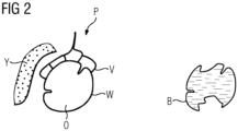
- Fig. 2 shows a plurality P of anatomical structures comprising the second contrast agent C2 and a border B of a contrast agent filling of the lumen L.
- the plurality P of anatomical structures comprises the blood vessel system V, the wall W of the hollow organ O and the parenchymal organ Y.
- Significant amounts of the second contrast agent C2 are present in each of these anatomical structures.
- the plurality P of anatomical structures is enhanced by the second contrast agent C2.
- the border B of the contrast agent filling of the lumen L follows the wall W of the hollow organ O with exception of those parts, where the border B of the contrast agent filling of the lumen L is separated from the wall W of the hollow organ O by portion A or portion S.
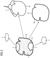
- Fig. 3 shows representations of different parts of the hollow organ O, in particular the representation of the wall W of the hollow organ O, the representation of the border B of the contrast agent filling of the lumen L, a representation of the portion A and a representation of the portion S. Furthermore, a representation of the border BL of the lumen L is shown. The representation of the border BL of the lumen L can be generated based on the representation of the border B of the contrast agent filling of the lumen L and the representation of the wall W of the hollow organ O.
- a course of the border BL of the lumen L can be estimated for those parts, where the border B of the contrast agent filling of the lumen L is separated from the wall W of the hollow organ O by portion A or portion S.
- Fig. 4 shows a diagram illustrating a method for providing image data of a hollow organ O, comprising
Landscapes
- Health & Medical Sciences (AREA)
- Life Sciences & Earth Sciences (AREA)
- Engineering & Computer Science (AREA)
- Medical Informatics (AREA)
- Public Health (AREA)
- Veterinary Medicine (AREA)
- Animal Behavior & Ethology (AREA)
- General Health & Medical Sciences (AREA)
- Biomedical Technology (AREA)
- Heart & Thoracic Surgery (AREA)
- Biophysics (AREA)
- Pathology (AREA)
- Surgery (AREA)
- Physics & Mathematics (AREA)
- Molecular Biology (AREA)
- High Energy & Nuclear Physics (AREA)
- Radiology & Medical Imaging (AREA)
- Nuclear Medicine, Radiotherapy & Molecular Imaging (AREA)
- Optics & Photonics (AREA)
- Dentistry (AREA)
- Oral & Maxillofacial Surgery (AREA)
- Hematology (AREA)
- Anesthesiology (AREA)
- Vascular Medicine (AREA)
- Computer Vision & Pattern Recognition (AREA)
- Human Computer Interaction (AREA)
- Pulmonology (AREA)
- Theoretical Computer Science (AREA)
- Apparatus For Radiation Diagnosis (AREA)
- Epidemiology (AREA)
Claims (9)
- Verfahren zur Bereitstellung von Bilddaten eines hohlen Organs (O), umfassend- Generieren (GD) von spektral aufgelösten Computertomographiedaten eines Untersuchungsbereichs, umfassend ein hohles Organ (O), wobei das hohle Organ (O) ein Organ des Verdauungssystems ist, umfassend ein Lumen, das mit einem ersten Kontrastmittel (C1) gefüllt ist, wobei das erste Kontrastmittel (C1) oral angewendet wurde, wobei nach der Anwendung des ersten Kontrastmittels (C1) in einem Blutgefäßsystem (V), einer Wand (W) des hohlen Organs (O) und einem parenchymalen Organ (Y) ein zweites Kontrastmittel (C2) akkumuliert wird, wobei das Blutgefäßsystem (V) die Wand (W) des hohlen Organs (O) und das parenchymale Organ (Y) versorgt, wobei das zweite Kontrastmittel (C2) ein anderes Absorptionsspektrum als das erste Kontrastmittel (C1) umfasst,- Berechnen (CI) von ersten Bilddaten, die eine Anwesenheit des ersten Kontrastmittels (C1) angeben, und von zweiten Bilddaten, die eine Anwesenheit des zweiten Kontrastmittels (C2) angeben, durch Anwenden eines Materialtrennalgorithmus auf die spektral aufgelösten Computertomographiedaten,- Bereitstellen (PI) der Bilddaten des hohlen Organs (O), umfassend die ersten Bilddaten und die zweiten Bilddaten,- Generieren einer Darstellung einer Grenze (B) der Kontrastmittelfüllung des Lumens (L) basierend auf den ersten Bilddaten,- Generieren einer Darstellung der Wand (W) des hohlen Organs (O) basierend auf der Darstellung der Grenze (B) der Kontrastmittelfüllung des Lumens (L) und den zweiten Bilddaten,- wobei das Lumen (L) des hohlen Organs (O) mindestens einen Anteil (A, S) außerhalb der Kontrastmittelfüllung des Lumens (L) und benachbart zu der Wand (W) des hohlen Organs (O) umfasst,- wobei eine Darstellung des mindestens einen Anteils (A, S) des Lumens (L) basierend auf der Darstellung der Grenze (B) der Kontrastmittelfüllung des Lumens (L) und der Darstellung der Wand (W) des hohlen Organs (O) generiert wird.
- Verfahren nach Anspruch 1,- wobei die spektral aufgelösten Computertomographiedaten photonenzählende Computertomographiedaten sind.
- Verfahren nach Anspruch 1 oder 2,- wobei der Materialtrennalgorithmus ein k-Kanten-Bildgebungsalgorithmus ist.
- Verfahren nach einem der Ansprüche 1 bis 3,- wobei eine Interessensregion in den Bilddaten bestimmt wird,- wobei die Interessensregion in den Bilddaten in Bezug auf die Anwesenheit des ersten Kontrastmittels (C1) und die Anwesenheit des zweiten Kontrastmittels (C2) klassifiziert wird.
- Verfahren nach einem der Ansprüche 1 bis 4,- wobei eine Darstellung einer Vielzahl (P) von anatomischen Strukturen, umfassend das zweite Kontrastmittel (C2), basierend auf den zweiten Bilddaten generiert wird,- wobei eine Region der Darstellung der Vielzahl (P) von anatomischen Strukturen bestimmt wird, die sich benachbart zu der Darstellung der Grenze (B) der Kontrastmittelfüllung des Lumens (L) befindet,- wobei die Darstellung der Wand (W) des Hohlorgans (O) basierend auf der Region der Darstellung der Vielzahl (P) von anatomischen Strukturen generiert wird, die sich benachbart zu der Darstellung der Grenze (B) der Kontrastmittelfüllung des Lumens (L) befindet.
- Verfahren nach einem der Ansprüche 1 bis 5,- wobei der mindestens eine Anteil des Lumens (L) ein solides Material und/oder ein Gas umfasst.
- Verfahren nach einem der Ansprüche 1 bis 6,- wobei das hohle Organ (O) ein Colon oder ein Dünndarm ist.
- Verfahren nach einem der Ansprüche 1 bis 7,- wobei das erste Kontrastmittel (C1) auf einem ersten Material mit einer ersten k-Kante basiert,- wobei das zweite Kontrastmittel (C2) auf einem zweiten Material mit einer zweiten k-Kante basiert, und- wobei ein Abstand zwischen der ersten k-Kante und der zweiten k-Kante mindestens 10 Kiloelektronenvolt beträgt.
- Verfahren nach einem der Ansprüche 1 bis 8,- wobei das erste Kontrastmittel (C1) auf Wolfram, Holmium oder Gadolinium basiert, und/oder- wobei das zweite Kontrastmittel (C2) auf Iod oder Barium basiert.
Priority Applications (2)
| Application Number | Priority Date | Filing Date | Title |
|---|---|---|---|
| EP19166066.1A EP3662837B1 (de) | 2019-03-29 | 2019-03-29 | Verfahren zur bereitstellung von bilddaten eines hohlen organs |
| US16/819,347 US11857145B2 (en) | 2019-03-29 | 2020-03-16 | Method for providing image data of a hollow organ |
Applications Claiming Priority (1)
| Application Number | Priority Date | Filing Date | Title |
|---|---|---|---|
| EP19166066.1A EP3662837B1 (de) | 2019-03-29 | 2019-03-29 | Verfahren zur bereitstellung von bilddaten eines hohlen organs |
Publications (3)
| Publication Number | Publication Date |
|---|---|
| EP3662837A1 EP3662837A1 (de) | 2020-06-10 |
| EP3662837B1 true EP3662837B1 (de) | 2025-04-30 |
| EP3662837C0 EP3662837C0 (de) | 2025-04-30 |
Family
ID=66001130
Family Applications (1)
| Application Number | Title | Priority Date | Filing Date |
|---|---|---|---|
| EP19166066.1A Active EP3662837B1 (de) | 2019-03-29 | 2019-03-29 | Verfahren zur bereitstellung von bilddaten eines hohlen organs |
Country Status (2)
| Country | Link |
|---|---|
| US (1) | US11857145B2 (de) |
| EP (1) | EP3662837B1 (de) |
Families Citing this family (1)
| Publication number | Priority date | Publication date | Assignee | Title |
|---|---|---|---|---|
| DE102021207957A1 (de) | 2021-07-23 | 2023-01-26 | Siemens Healthcare Gmbh | Computerimplementiertes Verfahren zur Auswertung eines angiographischen Computertomographiedatensatzes, Auswertungseinrichtung, Computerprogramm und elektronisch lesbarer Datenträger |
Family Cites Families (34)
| Publication number | Priority date | Publication date | Assignee | Title |
|---|---|---|---|---|
| CH632199A5 (de) | 1978-09-04 | 1982-09-30 | Schweizerische Lokomotiv | Schienenfahrzeug. |
| US4662379A (en) | 1984-12-20 | 1987-05-05 | Stanford University | Coronary artery imaging system using gated tomosynthesis |
| US5611342A (en) * | 1994-02-15 | 1997-03-18 | Molecular Biosystems, Inc. | Method of computer tomography imaging the gastrointestinal tract and surrounding upper abdominal tissues and organs using an orally administered low density contrast medium |
| DE4405505A1 (de) | 1994-02-21 | 1995-08-31 | Siemens Ag | Computertomograph |
| DE4410970C1 (de) | 1994-03-29 | 1995-07-20 | Talbot Waggonfab | Wankstütze für Schienenfahrzeuge mit einer Querneigeeinrichtung |
| DE10122875C1 (de) | 2001-05-11 | 2003-02-13 | Siemens Ag | Kombiniertes 3D-Angio-Volumenrekonstruktionsverfahren |
| US7054406B2 (en) | 2002-09-05 | 2006-05-30 | Kabushiki Kaisha Toshiba | X-ray CT apparatus and method of measuring CT values |
| WO2005009206A2 (en) | 2003-06-25 | 2005-02-03 | Besson Guy M | Dynamic multi-spectral imaging system |
| US7582283B2 (en) * | 2004-02-13 | 2009-09-01 | Wisconsin Alumni Research Foundation | Contrast agents to improve gastrointestinal tract opacification during abdominal and pelvic CT scans |
| WO2005096694A2 (en) | 2004-04-08 | 2005-10-20 | Yeda Research And Development Co., Ltd. | Three time point lung cancer detection, diagnosis and assessment of prognosis |
| US7218702B2 (en) | 2004-05-10 | 2007-05-15 | Wisconsin Alumni Research Foundation | X-ray system for use in image guided procedures |
| US7352885B2 (en) | 2004-09-30 | 2008-04-01 | General Electric Company | Method and system for multi-energy tomosynthesis |
| US7209536B2 (en) * | 2004-11-19 | 2007-04-24 | General Electric Company | CT colonography system |
| US8131336B2 (en) * | 2006-06-01 | 2012-03-06 | University Of Washington | Automated in vivo plaque composition evaluation |
| JP5091644B2 (ja) | 2006-12-04 | 2012-12-05 | 株式会社東芝 | X線コンピュータ断層撮像装置及び医用画像処理装置 |
| JP5010375B2 (ja) | 2007-07-18 | 2012-08-29 | 株式会社東芝 | 医用画像診断装置 |
| JP2009028065A (ja) | 2007-07-24 | 2009-02-12 | Toshiba Corp | X線ct装置 |
| JP5794752B2 (ja) | 2007-07-24 | 2015-10-14 | 株式会社東芝 | X線コンピュータ断層撮影装置及び画像処理装置 |
| DE102007037996A1 (de) | 2007-08-10 | 2009-02-19 | Siemens Ag | Verfahren zur dreidimensionalen Darstellung einer bewegten Struktur durch ein tomographisches Verfahren |
| DE102007050438B4 (de) * | 2007-10-22 | 2015-12-10 | Siemens Aktiengesellschaft | Verfahren und CT-System zur simultanen Darstellung der Durchblutung von Muskelgewebe und Gefäßen |
| JP5234905B2 (ja) | 2007-11-20 | 2013-07-10 | 東芝メディカルシステムズ株式会社 | X線ct装置および心筋パーフュージョン像生成システム |
| RU2376181C2 (ru) | 2008-02-04 | 2009-12-20 | Открытое акционерное общество "Крюковский вагоностроительный завод" (ОАО "КВСЗ") | Тележка пассажирского вагона |
| FR2932298B1 (fr) | 2008-06-06 | 2010-07-30 | Gen Electric | Procede de traitement d'une image radiologique d'un organe |
| US7920674B2 (en) | 2008-10-10 | 2011-04-05 | Samsung Electronics Co., Ltd. | Apparatus and method for image processing |
| WO2010113045A2 (de) | 2009-03-30 | 2010-10-07 | Bombardier Transportation Gmbh | Fahrzeug mit wankkompensation |
| DE102009014866A1 (de) | 2009-03-30 | 2010-10-28 | Bombardier Transportation Gmbh | Fahrzeug mit Wankkompensation |
| DE102009033452B4 (de) * | 2009-07-16 | 2011-06-30 | Siemens Aktiengesellschaft, 80333 | Verfahren zur Bereitstellung eines segmentierten Volumendatensatzes für eine virtuelle Koloskopie sowie zugehörige Gegenstände |
| DE102010041920A1 (de) | 2010-10-04 | 2012-04-05 | Siemens Aktiengesellschaft | Verfahren zur Darstellung einer Konzentration eines Kontrastmittels in einem vorbestimmten Volumenabschnitt mittels Tomosynthese und entsprechendes Tomosynthesegerät |
| JP2013128661A (ja) | 2011-12-21 | 2013-07-04 | Canon Inc | ステレオx線撮影装置、ステレオx線撮影方法 |
| DE102012215997B4 (de) | 2012-09-10 | 2022-10-06 | Siemens Healthcare Gmbh | Kontrastverstärkte Aufnahme von Objekten |
| WO2014077396A1 (ja) * | 2012-11-16 | 2014-05-22 | 株式会社東芝 | 超音波診断装置及び画像処理方法 |
| DE102014213464A1 (de) | 2014-07-10 | 2016-01-14 | Siemens Aktiengesellschaft | Verfahren zur kombinierten Dual-Energy-Mammographie- und Tomosynthesebildgebung und Tomosynthesegerät |
| DE102014214772B4 (de) | 2014-07-28 | 2025-02-20 | Siemens Healthineers Ag | Verfahren zur Erstellung eines Gefäßbildes und Verfahren zur Fluoroskopie-Darstellung von Gefäßen sowie entsprechend ausgestaltetes bildgebendes System |
| US20200179539A1 (en) * | 2016-06-22 | 2020-06-11 | Board Of Regents, The University Of Texas System | Contrast Agents and Methods of Making the Same for Spectral CT That Exhibit Cloaking and Auto-Segmentation |
-
2019
- 2019-03-29 EP EP19166066.1A patent/EP3662837B1/de active Active
-
2020
- 2020-03-16 US US16/819,347 patent/US11857145B2/en active Active
Also Published As
| Publication number | Publication date |
|---|---|
| US20200305816A1 (en) | 2020-10-01 |
| EP3662837C0 (de) | 2025-04-30 |
| EP3662837A1 (de) | 2020-06-10 |
| US11857145B2 (en) | 2024-01-02 |
Similar Documents
| Publication | Publication Date | Title |
|---|---|---|
| Fishman et al. | Volume rendering versus maximum intensity projection in CT angiography: what works best, when, and why | |
| Frellesen et al. | Dual-energy CT of the pancreas: improved carcinoma-to-pancreas contrast with a noise-optimized monoenergetic reconstruction algorithm | |
| Higgins et al. | Virtual bronchoscopy for three--dimensional pulmonary image assessment: state of the art and future needs. | |
| JP2006051365A (ja) | 肺潅流または肺密度を3d視覚化するためのシステムおよび方法、並びにその統計的分析方法 | |
| Frellesen et al. | Noise-optimized advanced image-based virtual monoenergetic imaging for improved visualization of lung cancer: comparison with traditional virtual monoenergetic imaging | |
| US10140715B2 (en) | Method and system for computing digital tomosynthesis images | |
| US20070197898A1 (en) | Method for examination of vessels in a patient on the basis of image data recorded by means of a scanner within an examination area | |
| Godoy et al. | Basic principles and postprocessing techniques of dual-energy CT: illustrated by selected congenital abnormalities of the thorax | |
| JP2008006274A (ja) | 医用画像処理装置、及び医用画像処理方法 | |
| JP2004057411A (ja) | 医療用可視画像の生成方法 | |
| Rowe et al. | Image processing from 2D to 3D | |
| EP3662837B1 (de) | Verfahren zur bereitstellung von bilddaten eines hohlen organs | |
| US8548566B2 (en) | Rendering method and apparatus | |
| Takebayashi et al. | Computerized tomography nephroscopic images of renal pelvic carcinoma | |
| US8908938B2 (en) | Method and device for providing a segmented volume data record for a virtual colonoscopy, and computer program product | |
| Kassavin et al. | Computed tomography colonography: 2025 update | |
| Shepard | Thoracic Imaging The Requisites E-Book | |
| Tamm et al. | Advanced 3-D imaging for the evaluation of pancreatic cancer with multidetector CT | |
| JP6755468B2 (ja) | 医用画像処理装置 | |
| JP2010131315A (ja) | 医用画像処理装置及び医用画像処理プログラム | |
| JP2007275318A (ja) | 画像表示装置、画像表示方法およびそのプログラム | |
| Alfidi et al. | Computed body tomography | |
| CA2663261A1 (en) | Visualization of volumetric medical imaging data | |
| Jo et al. | Imaging of the mediastinum: Mimics of malignancy | |
| US20110285695A1 (en) | Pictorial Representation in Virtual Endoscopy |
Legal Events
| Date | Code | Title | Description |
|---|---|---|---|
| PUAI | Public reference made under article 153(3) epc to a published international application that has entered the european phase |
Free format text: ORIGINAL CODE: 0009012 |
|
| STAA | Information on the status of an ep patent application or granted ep patent |
Free format text: STATUS: THE APPLICATION HAS BEEN PUBLISHED |
|
| AK | Designated contracting states |
Kind code of ref document: A1 Designated state(s): AL AT BE BG CH CY CZ DE DK EE ES FI FR GB GR HR HU IE IS IT LI LT LU LV MC MK MT NL NO PL PT RO RS SE SI SK SM TR |
|
| AX | Request for extension of the european patent |
Extension state: BA ME |
|
| STAA | Information on the status of an ep patent application or granted ep patent |
Free format text: STATUS: REQUEST FOR EXAMINATION WAS MADE |
|
| 17P | Request for examination filed |
Effective date: 20201208 |
|
| RBV | Designated contracting states (corrected) |
Designated state(s): AL AT BE BG CH CY CZ DE DK EE ES FI FR GB GR HR HU IE IS IT LI LT LU LV MC MK MT NL NO PL PT RO RS SE SI SK SM TR |
|
| STAA | Information on the status of an ep patent application or granted ep patent |
Free format text: STATUS: EXAMINATION IS IN PROGRESS |
|
| 17Q | First examination report despatched |
Effective date: 20230519 |
|
| RAP1 | Party data changed (applicant data changed or rights of an application transferred) |
Owner name: SIEMENS HEALTHINEERS AG |
|
| GRAP | Despatch of communication of intention to grant a patent |
Free format text: ORIGINAL CODE: EPIDOSNIGR1 |
|
| STAA | Information on the status of an ep patent application or granted ep patent |
Free format text: STATUS: GRANT OF PATENT IS INTENDED |
|
| RIC1 | Information provided on ipc code assigned before grant |
Ipc: A61B 6/50 20240101ALI20241112BHEP Ipc: A61B 6/00 20060101AFI20241112BHEP |
|
| INTG | Intention to grant announced |
Effective date: 20241127 |
|
| GRAS | Grant fee paid |
Free format text: ORIGINAL CODE: EPIDOSNIGR3 |
|
| GRAA | (expected) grant |
Free format text: ORIGINAL CODE: 0009210 |
|
| STAA | Information on the status of an ep patent application or granted ep patent |
Free format text: STATUS: THE PATENT HAS BEEN GRANTED |
|
| AK | Designated contracting states |
Kind code of ref document: B1 Designated state(s): AL AT BE BG CH CY CZ DE DK EE ES FI FR GB GR HR HU IE IS IT LI LT LU LV MC MK MT NL NO PL PT RO RS SE SI SK SM TR |
|
| REG | Reference to a national code |
Ref country code: CH Ref legal event code: EP Ref country code: GB Ref legal event code: FG4D |
|
| REG | Reference to a national code |
Ref country code: IE Ref legal event code: FG4D |
|
| REG | Reference to a national code |
Ref country code: DE Ref legal event code: R096 Ref document number: 602019069242 Country of ref document: DE |
|
| U01 | Request for unitary effect filed |
Effective date: 20250430 |
|
| U07 | Unitary effect registered |
Designated state(s): AT BE BG DE DK EE FI FR IT LT LU LV MT NL PT RO SE SI Effective date: 20250508 |
|
| PG25 | Lapsed in a contracting state [announced via postgrant information from national office to epo] |
Ref country code: ES Free format text: LAPSE BECAUSE OF FAILURE TO SUBMIT A TRANSLATION OF THE DESCRIPTION OR TO PAY THE FEE WITHIN THE PRESCRIBED TIME-LIMIT Effective date: 20250430 |
|
| PG25 | Lapsed in a contracting state [announced via postgrant information from national office to epo] |
Ref country code: NO Free format text: LAPSE BECAUSE OF FAILURE TO SUBMIT A TRANSLATION OF THE DESCRIPTION OR TO PAY THE FEE WITHIN THE PRESCRIBED TIME-LIMIT Effective date: 20250730 Ref country code: GR Free format text: LAPSE BECAUSE OF FAILURE TO SUBMIT A TRANSLATION OF THE DESCRIPTION OR TO PAY THE FEE WITHIN THE PRESCRIBED TIME-LIMIT Effective date: 20250731 |
|
| PG25 | Lapsed in a contracting state [announced via postgrant information from national office to epo] |
Ref country code: PL Free format text: LAPSE BECAUSE OF FAILURE TO SUBMIT A TRANSLATION OF THE DESCRIPTION OR TO PAY THE FEE WITHIN THE PRESCRIBED TIME-LIMIT Effective date: 20250430 |
|
| PG25 | Lapsed in a contracting state [announced via postgrant information from national office to epo] |
Ref country code: HR Free format text: LAPSE BECAUSE OF FAILURE TO SUBMIT A TRANSLATION OF THE DESCRIPTION OR TO PAY THE FEE WITHIN THE PRESCRIBED TIME-LIMIT Effective date: 20250430 |
|
| PG25 | Lapsed in a contracting state [announced via postgrant information from national office to epo] |
Ref country code: RS Free format text: LAPSE BECAUSE OF FAILURE TO SUBMIT A TRANSLATION OF THE DESCRIPTION OR TO PAY THE FEE WITHIN THE PRESCRIBED TIME-LIMIT Effective date: 20250731 |
|
| PG25 | Lapsed in a contracting state [announced via postgrant information from national office to epo] |
Ref country code: IS Free format text: LAPSE BECAUSE OF FAILURE TO SUBMIT A TRANSLATION OF THE DESCRIPTION OR TO PAY THE FEE WITHIN THE PRESCRIBED TIME-LIMIT Effective date: 20250830 |
|
| PG25 | Lapsed in a contracting state [announced via postgrant information from national office to epo] |
Ref country code: SM Free format text: LAPSE BECAUSE OF FAILURE TO SUBMIT A TRANSLATION OF THE DESCRIPTION OR TO PAY THE FEE WITHIN THE PRESCRIBED TIME-LIMIT Effective date: 20250430 |
|
| PG25 | Lapsed in a contracting state [announced via postgrant information from national office to epo] |
Ref country code: CZ Free format text: LAPSE BECAUSE OF FAILURE TO SUBMIT A TRANSLATION OF THE DESCRIPTION OR TO PAY THE FEE WITHIN THE PRESCRIBED TIME-LIMIT Effective date: 20250430 |
|
| PG25 | Lapsed in a contracting state [announced via postgrant information from national office to epo] |
Ref country code: SK Free format text: LAPSE BECAUSE OF FAILURE TO SUBMIT A TRANSLATION OF THE DESCRIPTION OR TO PAY THE FEE WITHIN THE PRESCRIBED TIME-LIMIT Effective date: 20250430 |
|
| PLBE | No opposition filed within time limit |
Free format text: ORIGINAL CODE: 0009261 |
|
| STAA | Information on the status of an ep patent application or granted ep patent |
Free format text: STATUS: NO OPPOSITION FILED WITHIN TIME LIMIT |
|
| REG | Reference to a national code |
Ref country code: CH Ref legal event code: L10 Free format text: ST27 STATUS EVENT CODE: U-0-0-L10-L00 (AS PROVIDED BY THE NATIONAL OFFICE) Effective date: 20260311 |