EP3597761B1 - Prostata-krebszelllinien, gensignaturen und verwendungen davon - Google Patents
Prostata-krebszelllinien, gensignaturen und verwendungen davon Download PDFInfo
- Publication number
- EP3597761B1 EP3597761B1 EP19173075.3A EP19173075A EP3597761B1 EP 3597761 B1 EP3597761 B1 EP 3597761B1 EP 19173075 A EP19173075 A EP 19173075A EP 3597761 B1 EP3597761 B1 EP 3597761B1
- Authority
- EP
- European Patent Office
- Prior art keywords
- prostate
- prostate cancer
- oncogene
- cancer
- tumor
- Prior art date
- Legal status (The legal status is an assumption and is not a legal conclusion. Google has not performed a legal analysis and makes no representation as to the accuracy of the status listed.)
- Active
Links
Images


Classifications
-
- A—HUMAN NECESSITIES
- A01—AGRICULTURE; FORESTRY; ANIMAL HUSBANDRY; HUNTING; TRAPPING; FISHING
- A01K—ANIMAL HUSBANDRY; AVICULTURE; APICULTURE; PISCICULTURE; FISHING; REARING OR BREEDING ANIMALS, NOT OTHERWISE PROVIDED FOR; NEW BREEDS OF ANIMALS
- A01K67/00—Rearing or breeding animals, not otherwise provided for; New or modified breeds of animals
- A01K67/027—New or modified breeds of vertebrates
- A01K67/0275—Genetically modified vertebrates, e.g. transgenic
-
- A—HUMAN NECESSITIES
- A61—MEDICAL OR VETERINARY SCIENCE; HYGIENE
- A61K—PREPARATIONS FOR MEDICAL, DENTAL OR TOILETRY PURPOSES
- A61K31/00—Medicinal preparations containing organic active ingredients
- A61K31/33—Heterocyclic compounds
- A61K31/395—Heterocyclic compounds having nitrogen as a ring hetero atom, e.g. guanethidine or rifamycins
- A61K31/435—Heterocyclic compounds having nitrogen as a ring hetero atom, e.g. guanethidine or rifamycins having six-membered rings with one nitrogen as the only ring hetero atom
- A61K31/46—8-Azabicyclo [3.2.1] octane; Derivatives thereof, e.g. atropine, cocaine
-
- A—HUMAN NECESSITIES
- A61—MEDICAL OR VETERINARY SCIENCE; HYGIENE
- A61K—PREPARATIONS FOR MEDICAL, DENTAL OR TOILETRY PURPOSES
- A61K31/00—Medicinal preparations containing organic active ingredients
- A61K31/33—Heterocyclic compounds
- A61K31/395—Heterocyclic compounds having nitrogen as a ring hetero atom, e.g. guanethidine or rifamycins
- A61K31/495—Heterocyclic compounds having nitrogen as a ring hetero atom, e.g. guanethidine or rifamycins having six-membered rings with two or more nitrogen atoms as the only ring heteroatoms, e.g. piperazine or tetrazines
- A61K31/505—Pyrimidines; Hydrogenated pyrimidines, e.g. trimethoprim
- A61K31/506—Pyrimidines; Hydrogenated pyrimidines, e.g. trimethoprim not condensed and containing further heterocyclic rings
-
- A—HUMAN NECESSITIES
- A61—MEDICAL OR VETERINARY SCIENCE; HYGIENE
- A61P—SPECIFIC THERAPEUTIC ACTIVITY OF CHEMICAL COMPOUNDS OR MEDICINAL PREPARATIONS
- A61P35/00—Antineoplastic agents
-
- A—HUMAN NECESSITIES
- A61—MEDICAL OR VETERINARY SCIENCE; HYGIENE
- A61P—SPECIFIC THERAPEUTIC ACTIVITY OF CHEMICAL COMPOUNDS OR MEDICINAL PREPARATIONS
- A61P35/00—Antineoplastic agents
- A61P35/04—Antineoplastic agents specific for metastasis
-
- A—HUMAN NECESSITIES
- A61—MEDICAL OR VETERINARY SCIENCE; HYGIENE
- A61P—SPECIFIC THERAPEUTIC ACTIVITY OF CHEMICAL COMPOUNDS OR MEDICINAL PREPARATIONS
- A61P43/00—Drugs for specific purposes, not provided for in groups A61P1/00-A61P41/00
-
- C—CHEMISTRY; METALLURGY
- C12—BIOCHEMISTRY; BEER; SPIRITS; WINE; VINEGAR; MICROBIOLOGY; ENZYMOLOGY; MUTATION OR GENETIC ENGINEERING
- C12N—MICROORGANISMS OR ENZYMES; COMPOSITIONS THEREOF; PROPAGATING, PRESERVING, OR MAINTAINING MICROORGANISMS; MUTATION OR GENETIC ENGINEERING; CULTURE MEDIA
- C12N5/00—Undifferentiated human, animal or plant cells, e.g. cell lines; Tissues; Cultivation or maintenance thereof; Culture media therefor
- C12N5/06—Animal cells or tissues; Human cells or tissues
- C12N5/0602—Vertebrate cells
- C12N5/0693—Tumour cells; Cancer cells
-
- C—CHEMISTRY; METALLURGY
- C12—BIOCHEMISTRY; BEER; SPIRITS; WINE; VINEGAR; MICROBIOLOGY; ENZYMOLOGY; MUTATION OR GENETIC ENGINEERING
- C12Q—MEASURING OR TESTING PROCESSES INVOLVING ENZYMES, NUCLEIC ACIDS OR MICROORGANISMS; COMPOSITIONS OR TEST PAPERS THEREFOR; PROCESSES OF PREPARING SUCH COMPOSITIONS; CONDITION-RESPONSIVE CONTROL IN MICROBIOLOGICAL OR ENZYMOLOGICAL PROCESSES
- C12Q1/00—Measuring or testing processes involving enzymes, nucleic acids or microorganisms; Compositions therefor; Processes of preparing such compositions
- C12Q1/68—Measuring or testing processes involving enzymes, nucleic acids or microorganisms; Compositions therefor; Processes of preparing such compositions involving nucleic acids
- C12Q1/6876—Nucleic acid products used in the analysis of nucleic acids, e.g. primers or probes
- C12Q1/6883—Nucleic acid products used in the analysis of nucleic acids, e.g. primers or probes for diseases caused by alterations of genetic material
- C12Q1/6886—Nucleic acid products used in the analysis of nucleic acids, e.g. primers or probes for diseases caused by alterations of genetic material for cancer
-
- G—PHYSICS
- G01—MEASURING; TESTING
- G01N—INVESTIGATING OR ANALYSING MATERIALS BY DETERMINING THEIR CHEMICAL OR PHYSICAL PROPERTIES
- G01N33/00—Investigating or analysing materials by specific methods not covered by groups G01N1/00 - G01N31/00
- G01N33/48—Biological material, e.g. blood, urine; Haemocytometers
- G01N33/50—Chemical analysis of biological material, e.g. blood, urine; Testing involving biospecific ligand binding methods; Immunological testing
- G01N33/53—Immunoassay; Biospecific binding assay; Materials therefor
- G01N33/574—Immunoassay; Biospecific binding assay; Materials therefor for cancer
Definitions
- the present invention relates to a CCR5 antagonist for use in inhibiting metastasis of cancer.
- Methods and compositions for diagnosing and treating cancer, including prostate cancer are disclosed herein.
- methods and compositions useful for prostate cancer diagnostics, research, treatment stratification, and treatment are also disclosed herein.
- cells and transgenic, non-human mammals that can be used in these methods.
- Cancer is a significant health problem throughout the world. Although advances have been made in detection and therapy of cancer, no vaccine or other universally successful method for prevention and/or treatment is currently available. Current therapies, which are generally based on a combination of chemotherapy or surgery and radiation, continue to prove inadequate in many patients.
- Prostate cancer for example, is a significant health problem for men in the United States and throughout the world. Although advances have been made in the detection and treatment of the disease, prostate cancer remains an important cause of cancer-related deaths in men, affecting more than 221,000 men in the United States each year. For men in North America, the life-time odds of getting prostate cancer are now 19.6%, with a 4.6% risk of death. Prostate cancer was the cause of approximately 250,000 deaths worldwide in 2009.
- PSA level below 4.0 ng/mL is considered as normal.
- prostate cancer was diagnosed in 15.2 percent of men with a PSA level at or below 4.0 ng/mL. Fifteen percent of those men, or approximately 2.3 percent overall, had high-grade cancers. According to Smith DS et al. 25 to 35 percent of men who had a PSA level between 4.1 and 9.9 ng/mL and who underwent a prostate biopsy were found to have prostate cancer, while 65 to 75 percent of the remaining men did not have prostate cancer. Thus, there is no specific normal or abnormal PSA level.
- a CCR5 antagonist for use in inhibiting metastasis of cancer susceptible to treatment in an immunocompetent subject at risk for metastasis of cancer, in accordance with the claims appended hereto. Any examples which do not fall under the scope of the appended claims are provided for background reference purposes.
- a mammalian prostate cancer cell line comprising at least one or more of a set of primary mammalian epithelial cells which have been infected with a retroviral vector carrying an oncogene.
- the oncogene is selected from the group consisting of c-Myc, Ha-Ras, NeuT, c-Src and combinations thereof and in which said oncogene or combination of genes is expressed.
- the mammalian prostate cancer cell line can include any suitable mammalian cell, including primary murine epithelial cells.
- the primary mammalian epithelial cells may be derived from any immune competent mammal, including immune competent rodents, including rats and mice.
- the present disclosure provides an animal model of cancer comprising an immune competent mammal implanted with a cancer cell line transformed with one or more of a set of oncogenes selected from the group consisting of c-Myc, Ha-Ras, NeuT, c-Src and combinations thereof
- an immunocompetent transgenic mouse created using the mammalian prostate cancer cell line of the present disclosure develops a prostate tumor capable of producing a detectable molecular genetic signature based on an expression level of one or more of a set of oncogenes selected from the group consisting of c-Myc, Ha-Ras, NeuT, c-Src and combinations thereof
- the present disclosure provides a method for the in vitro production of immortalized primary mammalian epithelial cells, the method comprising infecting primary mammalian epithelial cells with a retroviral vector carrying an oncogene selected from the group consisting of c-Myc, Ha-Ras, NeuT, c-Src and combinations thereof to provide infected cells, wherein said primary mammalian epithelial cells are capable of being infected by said retroviral vector and under conditions whereby the c-Myc, Ha-Ras, NeuT, c-Src and combinations thereof are expressed in said infected cells.
- the present disclosure provides a method for diagnosing a prostate cancer, the method comprising: (a) providing a biological test sample from a subject afflicted with a prostate cancer or suspected of having prostate cancer or at risk for developing prostate cancer; (b) determining a level of at least one biological marker or a molecular genetic signature based on a gene expression pattern or activity of one or more of a set of genes in the test sample, wherein the one or more set of genes are selected from the group consisting of c-Myc, Ha-Ras, NeuT, c-Src and combinations thereof; (c) comparing the level of said at least one biological marker or said molecular genetic signature in said test sample to the level of the biological marker or the level of the molecular genetic signature in a control sample, wherein an elevated level of the biological marker or the molecular genetic signature in said test sample relative to the level of the biological marker or the molecular genetic signature in said control sample is a diagnostic indicator of the presence of prostate cancer in said subject.
- the present disclosure provides a method of classifying a cancer tumor, including a prostate tumor, the method comprising: (a) providing a cancer tumor or a prostate tumor sample; (b) detecting a molecular genetic signature derived from gene expression pattern or activity of one or more of a set of genes in the sample, wherein the genes are selected from the group consisting of c-Myc, Ha-Ras, NeuT, c-Src and combinations thereof; and (c) classifying the prostate tumor as belonging to a tumor subclass based on the results of the detecting step (b).
- the present disclosure provides a method of stratifying a subject having a cancer tumor, including a prostate tumor, for a clinical trial, the method comprising: (a) providing a sample derived from a subject having the cancer tumor or the prostate tumor; (b) detecting a molecular genetic signature derived from gene expression pattern or activity of one or more of a set of genes in the sample, wherein the genes are selected from the group consisting of c-Myc, Ha-Ras, NeuT, c-Src, ErbB2 and combinations thereof; and (c) stratifying the subject for a clinical trial based on the results of the detecting step.
- the present disclosure provides a method of selecting a treatment for a subject having a prostate tumor, the method comprising: (a) providing a sample derived from a subject having a prostate tumor; (b) detecting a molecular genetic signature derived from a gene expression pattern or activity of one or more of a set of genes in the sample, wherein the genes are selected from the group consisting of c-Myc, Ha-Ras, NeuT, c-Src and combinations thereof; and (c) selecting a treatment based on the results of the detecting step.
- the present disclosure provides a non-naturally occurring cell produced by transforming a cell with one or more exogenous oncogenes, allowing the cell to divide at least once, wherein the cell is a mammalian cell transformed by a vector containing the one or more exogenous oncogenes, wherein the one or more exogenous oncogenes are selected from the group consisting of c-Myc, Ha-Ras, NeuT, c-Src and combinations thereof.
- CCR5 chemokine receptor type
- CCL5 chemokine receptor type
- immune competent or "immunocompetent” animals.
- one teaching of the present disclosure provides cancer cell lines that can be implanted in immune competent, or immunocompetent, animals, including humans and non-human animals, including mammals.
- exemplary non-human mammals include, for example, rodents such as rats, guinea pigs, and mice, and faint animals such as pigs, sheep, goats, horses, and cattle.
- any type of cell in the body may be a source of cancer
- any suitable type of cancer cell line may be used in the present disclosure.
- a suitable cancer type includes carcinoma cancer of the epithelial cells), sarcoma (cancer of the bone, muscle or other connective tissues), lymphoma (cancer of the lymphatic system), leukemia (cancer of blood cells or blood precursor cells) and melanoma (cancer of the pigment-providing cells).
- a prostate cancer cell line comprising at least one or more of a set of primary mammalian epithelial cells which have been infected with a retroviral vector carrying an oncogene selected from the group consisting of c-Myc, Ha-Ras, NeuT, c-Src and combinations thereof, and wherein said oncogene or combinations of genes are expressed.
- a mouse prostate cancer cell line wherein murine prostate cells are transduced with an oncogene selected from the group consisting of c-Myc, Ha-Ras, NeuT, c-Src and combinations thereof.
- Suitable mouse prostate cell line can be obtained by infecting the primary murine epithelial cells with a retroviral vector carrying the oncogene under conditions that allow the oncogene to be expressed in the primary murine epithelial cells.
- Transgenic immunocompetent mouse models of human cancers have the potential to be more reflective of human cancers than xenograft models because, inter alia, transgenic mice form tumors in situ, (i.e., in an environment more similar to the human tumor and in the setting of a normal immune system). Therefore, in some examples of the present disclosure, immunocompetent non-human mammals are engineered to express one or more of the oncogenes described herein, including c-Myc, Ha-Ras, NeuT, c-Src or combinations thereof, and to develop cancer
- transgenic mouse models of human cancer in research.
- small-animal X-ray computed tomography could be used to monitor progression of tumor relatively cheaply and also as highly quantitative three-dimensional method for visualizing blood vessels and angiogenesis preclinically.
- Using such a method it is possible to achieve rapid and accurate assessment of vascularity during preclinical therapeutic trials in living mice.
- Tumor assessment with microCT enables rapid qualitative visual renderings of data as well as quantitative analysis of tumor blood volume, vessel density, vessel caliber, degree of branching, and tortuosity using segmentation analysis.
- immunocompetent mice examples include (random bred CD1, Charles River Laboratories, St. Constant, PQ), C57BI/6J (B6), C57BI/6x129/J Fl (F1, Jackson Laboratories, Bar Harbor, ME), FVB/N, C57BV6, BALB/c and ND4.
- FVB/N mice are used to engineer the transgenic mouse models in accordance with the present disclosure.
- FVB/N mice are suitable for most transgenic experiments and genetic analyses contemplated and/or described herein.
- the inbred FVB/N strain is characterized by vigorous reproductive performance and consistently large litters and fertilized FVB/N eggs contain large and prominent pronuclei, which facilitate microinjection of DNA.
- An immunocompetent transgenic mouse created using the mammalian prostate cancer cell line of the present disclosure develops a prostate tumor capable of producing a detectable molecular genetic signature based on an expression level of one or more of a set of oncogenes selected from the group consisting of c-Myc, Ha-Ras, NeuT, c-Src and combinations thereof.
- Metastasis is the leading cause of death in cancer patients.
- Current chemotherapeutic anti-cancer treatments use cytotoxic, hormonal or immunomodulator drugs aimed at decreasing the number of cancer cells in the patient's body.
- cytotoxic, hormonal or immunomodulator drugs aimed at decreasing the number of cancer cells in the patient's body.
- a growing body of evidence suggests that most metastatic cells are resistant to anti-cancer drugs and therefore currently available drugs are not effectively stopping the dissemination of cancer cells to other tissues or organs.
- there is no effective method for treating most metastatic tumors despite the numerous and diverse therapeutic innovations in the cancer therapeutic field.
- a “vector” or “construct” refers to a macromolecule or complex of molecules comprising a polynucleotide to be delivered to a host cell, either in vitro or in vivo.
- the polynucleotide to be delivered may comprise a sequence of interest for gene therapy.
- Vectors include, for example, transposons and other site-specific mobile elements, viral vectors, e.g., adenovirus, adenoassociated virus (AAV), poxvirus, papillomavirus, lentivirus, herpesvirus, foamivirus and retrovirus vectors, and including pseudotyped viruses, liposomes and other lipid-containing complexes, and other macromolecular complexes capable of mediating delivery of a polynucleotide to a host cell, e.g., DNA coated gold particles, polymer-DNA complexes, liposome-DNA complexes, liposome-polymer-DNA complexes, virus-polymer-DNA complexes, e.g., adenovirus-polylysine-DNA complexes, and antibody-DNA complexes.
- viral vectors e.g., adenovirus, adenoassociated virus (AAV), poxvirus, papillomavirus, lentivirus, herpe
- Vectors can also comprise other components or functionalities that further modulate gene delivery and/or gene expression, or that otherwise provide beneficial properties to the cells to which the vectors will be introduced.
- Such other components include, for example, components that influence binding or targeting to cells (including components that mediate cell-type or tissue-specific binding); components that influence uptake of the vector nucleic acid by the cell; components that influence localization of the polynucleotide within the cell after uptake (such as agents mediating nuclear localization); and components that influence expression of the polynucleotide.
- Such components also might include markers, such as detectable and/or selectable markers that can be used to detect or select for cells that have taken up and are expressing the nucleic acid delivered by the vector.
- Such components can be provided as a natural feature of the vector (such as the use of certain viral vectors which have components or functionalities mediating binding and uptake), or vectors can be modified to provide such functionalities.
- vectors can be provided as a natural feature of the vector (such as the use of certain viral vectors which have components or functionalities mediating binding and uptake), or vectors can be modified to provide such functionalities.
- a large variety of such vectors are known in the art and are generally available.
- the vector When a vector is maintained in a host cell, the vector can either be stably replicated by the cells during mitosis as an autonomous structure, incorporated within the genome of the host cell, or maintained in the host cell's nucleus or cytoplasm.
- Human Prostate cancer has embedded within the genetic makeup and signatures that reflect oncogenic signaling.
- the cell lines of the present disclosure advantageously reflect one or more of these oncogenic signaling pathways, thus facilitating, for example, the testing of oncogene specific compounds or nucleic acids based therapies.
- prostate cancer cell lines as provided herein are useful for the testing of and/or screening for oncogene specific compounds or nucleic acids based therapies.
- oncogene specific inhibitors examples include inibitors for c-Myc, Ha-Ras, c-Src, and ErbB2 oncogenes.
- suitable anti-cancer agents targeting the ErbBs which are in clinical use or development, including those that fall in the categories of chimeric or humanised monoclonal antibodies against the ErbB family and Small Molecule ErbB Tyrosine Kinase Inhibitors.
- the chimeric or humanised monoclonal include antibodies that prevent ligand-binding and ligand-dependent receptor activation (e.g., Cetuximab that targets the ligand-binding subdomain III of ErbB 1), antibodies that interfere with ligand-independent receptor activation (e.g., Trastuzumab that targets subdomain IV of ErbB2), and antibodies that prevent receptor heterodimerisation (e.g. the anti-ErbB2 antibody Pertuzumab that targets an area around the dimerisation loop in subdomain II of ErbB2).
- ligand-binding and ligand-dependent receptor activation e.g., Cetuximab that targets the ligand-binding subdomain III of ErbB 1
- antibodies that interfere with ligand-independent receptor activation e.g., Trastuzumab that targets subdomain IV of ErbB2
- antibodies that prevent receptor heterodimerisation e.g. the anti-ErbB2 antibody Pertu
- Exemplary Small molecule ErbB tyrosine kinase inhibitors include two ErbB 1-specific tyrosine kinase inhibitors Gefitinib/Iressa and Erlotinib, which have been approved for the treatment of non-small cell lung cancer, and the dual ErbB 1/ErbB2 inhibitor Lapatinib, which is marketed as TYKERB ® and is indicated in combination with capecitabine for the treatment of patients with advanced or metastatic breast cancer; and in combination with letrozole for the treatment of postmenopausal women with hormone receptor positive metastatic breast cancer that overexpresses the HER2.
- Examples of c-Src protein tyrosine kinase inhibitors that are useful in the present disclosure include, but are not limited to, the compounds which are generically and specifically disclosed in WO 1996/10028 , WO 1997/28161 , WO1997/32879 and WO1997/49706 , including those belonging to the structure classes of pyrrolopyrimidines, especially pyrrolo[2,3-d]pyrimidines, purines, pyrazo-pyrimidines, especially pyrazo[3,4-d]pyrimidines, pyrazopyrimidines, especially pyrazo[3,4-d]pyrimidines and pyridopyrimidines, especially pyrido[2,3-d]pyrimidines.
- Exemplary compounds include compounds of formulae I-IV below.
- the above compounds can be prepared and administered as described in the cited documents.
- the compound of formula I can be prepared and formulated as described in WO 1996/10028 .
- the compound of formula II and its preparation is disclosed in Example 111c3 of WO 1997/16452 .
- the compound of formula IV can be prepared in analogy thereof. Both latter compounds can be formulated as described in WO 1997/16452 .
- the compound of formula III is discussed by R. Gamse et al. in J. Bone Miner. Res. 14 (Suppi. 1), 1999, S487 .
- Additional useful compounds are described by T. Schindler, F. Sicheri et al. in Molecular Cell, 1999 (3), 639, 647 ; J. M. Hamby et al., J. Med. Chem. 40, 1997, 2296- 2303 ; R. L. Panek et al., J. Pharmacol. Exp. Ther. 283, 1997, 1433-1444 ; and S. R. Klutchko et al., J. Med. Chem. 41, 1998, 3276-3292 .
- src inhibitors include SKI606, also known as bosutinib (by Wyeth) and the compound dasatinib, also know as Spyrcel (by Bristol-Myers Squibb) which is disclosed in WO 2000/62778 and U.S. Pat. No. 6,596,746 .
- Small-molecule c-Myc inhibitors include 10074-G5, also known as Biphenyl-2-yl-(7-nitrobenzo[1,2,5]oxadiazol-4-ylamine; Quarfloxin (also known as CX-3543); and 10074-G5, also known as Biphenyl-2-yl-(7-nitrobenzo[1,2,5]oxadiazol-4-ylamine.
- CX-3543 reportedly suppresses c-Myc activity by binding to the c-Myc quadruplex, four-stranded DNA secondary structures that regulate transcription of specific oncogenes including c-Myc.
- c-Myc inhibitors include the compounds disclosed in United States Patent Application Publication No. 2007/0203188 (published August 30, 2007 ).
- the present disclosure provides a method for the in vitro production of immortalized primary mammalian epithelial cells, the method comprising infecting primary mammalian epithelial cells with a retroviral vector carrying an oncogene selected from the group consisting of c-Myc, Ha-Ras, NeuT, c-Src and combinations thereof to provide infected cells, wherein said primary mammalian epithelial cells are capable of being infected by said retroviral vector and under conditions whereby the c-Myc, Ha-Ras, NeuT, c-Src and combinations thereof are expressed in said infected cells.
- Retroviral and Adenoviral Expression Systems are suitable gene delivery systems.
- Retroviral-mediated gene transfer is widely used to express proteins in a variety of cell lines, including hematopoietic cells for a variety of reasons, including analysis of their effects on blood cell proliferation, differentiation, and biological function.
- the Murine Stem Cell Virus (“MSCV”) Retroviral Expression System contains vectors that are optimized for introducing and expressing target genes in pluripotent cell lines, including murine or human hematopoietic, embryonic stem (ES), and embryonal carcinoma (EC) cells.
- Adenoviral Expression Systems include the ViraPowerTM Adenoviral Expression System (by Life Technologies), which is useful, for example, for creation of a replication-incompetent adenovirus that can be used to deliver and transiently express target gene(s) of interest in either dividing or non-dividing mammalian cells.
- the present disclosure provides a method for diagnosing a prostate cancer, the method comprising: (a) providing a biological test sample from a subject afflicted with a prostate cancer or suspected of having prostate cancer or at risk for developing prostate cancer; (b) determining a level of at least one biological marker or a molecular genetic signature based on a gene expression pattern in the test sample that is associated with the diagnosis of the prostate cancer;(c) comparing the level of said at least one biological marker or said molecular genetic signature in said test sample to the level of the biological marker or the level of the molecular genetic signature in a control sample, wherein an elevated level of the biological marker or the molecular genetic signature in said test sample relative to the level of the biological marker or the molecular genetic signature in said control sample is a diagnostic indicator of the presence of prostate cancer in said subject.
- biological specimens include nucleic acid derived from the tumor under obtained from the patient.
- Nucleic acid may be derived from the tumor either by biopsy, or from cells derived from the tumor in urine or proteins made as a consequence of the gene signature secreted into the patients blood.
- the present disclosure provides a method of classifying a prostate tumor, the method comprising: (a) providing a prostate tumor sample; (b) detecting a molecular genetic signature derived from gene expression pattern or activity of one or more of a set of genes in the sample, wherein the genes are selected from the group consisting of c-Myc, a-Ras, NeuT, c-Src and combinations thereof; and (c) classifying the prostate tumor as belonging to a tumor subclass based on the results of the detecting step.
- the present disclosure provides a method of stratifying a subject having a cancer tumor, including a prostate tumor, for a clinical trial, the method comprising: (a) providing a sample derived from a subject having the cancer tumor or the prostate tumor; (b) detecting a molecular genetic signature derived from gene expression pattern or activity of one or more of a set of genes in the sample, wherein the genes are selected from the group consisting of c-Myc, Ha-Ras, NeuT, c-Src, ErbB2 and combinations thereof; and (c) stratifying the subject for a clinical trial based on the results of the detecting step.
- subjects are stratified into subcategories that are based on the presence of a molecular genetic signature and/ or the functional pathways linked to the molecular genetic signature.
- the molecular genetic signature is based on a gene expression pattern or activity of one or more of a set of genes in a test sample derived from a subject having a cancer tumor, in particular prostate cancer tumor, wherein the one or more set of genes are selected from the group consisting of c-Myc, Ha-Ras, NeuT, c-Src and combinations thereof
- the present disclosure provides a method of selecting a treatment for a subject having a prostate tumor, the method comprising: (a) providing a sample derived from a subject having a prostate tumor; (b) detecting a molecular genetic signature derived from gene expression pattern or activity of one or more of a set of genes in the sample, wherein the genes are selected from the group consisting of c-Myc, Ha-Ras, NeuT, c-Src and combinations thereof; and (c) selecting a treatment based on the results of the detecting step (b). (ie oncogene based therapies would be given based on the onco signature in the patients tumor)
- the present disclosure provides a non-naturally occurring cell produced by transforming a cell with one or more exogenous oncogenes, allowing the cell to divide at least once, wherein the cell is a human cell transformed by a vector containing the one or more exogenous oncogenes, wherein the one or more exogenous oncogenes are selected from the group consisting of c-Myc, Ha-Ras, NeuT, c-Src and combinations thereof.
- the signature could be applied to genetic material derived from the prostate cancer - ie in biofluids related to the prostate including prostate, urine, blood or other biospecimens.
- the signature may also be applied to other cancer types in particular as shown for breast cancer.
- the c-Myc signature the signature is seen in both prostate tumors and in the breast tumors derived from transgenic expression of c-Myc in the mammary gland of transgenic mice (Figure 7C).
- the present disclosure includes identifying a cancer by obtaining a biological sample from a subject afflicted with prostate cancer or suspected of having prostate cancer or at risk for developing prostate cancer and determining whether genetic material from the biological sample has a genetic signature as described herein.
- the biological sample is derived from a prostate biopsy.
- the biological sample is derived from biofluids related to the prostate, including prostate, urine, blood or other biospecimens.
- the method of the present disclosure includes massaging a prostate prior to obtaining a urine sample.for genetic signature identification.
- the method includes other fluids as described above.
- Oligonucleotide sequences can be introduced into cells as is known in the art. Transfection, electroporation, fusion, liposomes, colloidal polymeric particles and viral and nonviral vectors as well as other means known in the art may be used to deliver the oligonucleotide sequences to the cell. The method of delivery selected will depend at least on the cells to be treated and the location of the cells and will be known to those skilled in the art. Localization can be achieved by liposomes, having specific markers on the surface for directing the liposome, by having injection directly into the tissue containing the target cells, by having depot associated in spatial proximity with the target cells, specific receptor mediated uptake, viral vectors, or the like. Oncogenes can be introduced into cells by transduction or transfection. Transduction can conducted using either retroviral or other viral delivery systems
- mice were in the FVB strain.
- Mouse prostate epithelial cell culture were isolated from prostate glands of 12 week old male mice and maintained as previously described [42] and analyzed after 25 passages with at least three lines of each genotype.
- Cells were seeded in 24-well-plates at a concentration of 1x10⁴ cells / well, each sample in triplicate x 7 days. Growing transformed cells in DMEM medium with 10% FBS, while control PEC cells were cultured in prostate epithelial primary culture medium. Harvest cells every 24 hours, suspended cells in 100 ul PBS, added an equal volume of 0.4% Trypan blue, after 5 minutes counted cells by CountessTM Autocounted Cell Counter (C10227, Invitrogen Carlsbad, CA).
- 1x10⁶ cells in 100 ⁇ l volume were injected subcutaneously into 7-8 weeks FVB male mice.
- Cell suspension we mixed with a 20% by volume BD Matrigel (BD Biosciences, Bedford, MA), resulting in a final cell concentration of 10-7 cells/ml.
- Tumor growth was measured by vernier calipers twice a week. Tumor samples were harvested after 30 days (except NeuT-induced tumors which were harvested after 16 days).
- the PEC (prostate epithelial cells) lines transformed with Ha-Ras, v-Src, and NeuT oncogenes were generated and transfected with a lentiviral vector containing the luc2 gene to generate stable bioluminescent cancer cell lines.
- the Luc2-tomato red expression vector was previously described ( Liu, H., et al., Proc Natl Acad Sci U.S.A. 2010, 107, 18115-18120 ).
- the isogenic PEC lines were maintained in Dulbecco's modified Eagle's medium (DMEM) supplemented with 10% fetal bovine serum (FBS) and 1% penicillin-Streptomycin and cultured in 5% CO2 at 37 °C.
- DMEM Dulbecco's modified Eagle's medium
- FBS fetal bovine serum
- penicillin-Streptomycin penicillin-Streptomycin
- Cells were grown to confluence on 12-well plates in DMEM medium containing 10% FBS. The monolayers were wounded with a P10 micropipette tip. The cells were washed immediately after scoring with PBS and serum-free DMEM was added (16). The cell migration was monitored for 20h, and pictures were taken at 9h, 12h, 15h and 20h time points using an Axiovert 200 Zeiss microscope system. Images were analyzed using ImageJ software.
- Mouse cytokine arrays spotted on nitrocellulose membrane were obtained from Raybiotech.
- Conditioned medium was prepared by culturing cells in serum-free DMEM for 48hours. Membranes were then processed according to the manufacturer's instructions for assessment of secreted cytokines in conditioned medium (see Katiyar, S. et al., Mol Cell Biol 2007, 27, 1356-1369 ).
- mice Male nu/nu mice, 12weeks old, were anesthetized by exposure to 3% isoflurane.
- Anesthetized mice received 1x10⁶ cells suspended in 50 ⁇ L of Dulbecco's Phosphate Buffer Saline lacking of calcium and magnesium (DPBS) and 50 ⁇ L of BD Matrigel Membrane Basement (BD Biosciences, Bedford, MA) by subcutaneous injection below one dorsal flank. The injection has been performed using 27G'/2 needle. Tumor progression has been followed by bioluminescence measurement once a week until tumor excision using as described previously (18). Three mice at a time were monitored dorsally. Regions of interest (ROI) from displayed images were drawn around the tumor sites or the metastatic lesion and quantified using the Living Image 3.0 software. (Caliper Life Sciences). Tumor samples were harvested after 3 weeks. All experiments involving mice were carried out under the approval of Thomas Jefferson University's IACUC.
- ROI Regions of interest
- mice Male FVB mice, 8 weeks old, were anesthetized by exposure to 3% isoflurane. 2x10⁵ cancer cells suspended in 1004 of DPBS were injected into the left ventricle of the heart of the mouse. Injections were performed using a 30G1/2 needle and a 1mL syringe. To confirm the presence of cells in the systemic circulation, animals were imaged using IVIS LUMINA XR system (Caliper Life Sciences, Hopkinton MA). A successful intracardiac injection was indicated by systemic bioluminescence distributed through the animal body. Mice not properly injected were removed from the study.
- mice received the substrate of luciferase, DLuciferin (Gold Biotechnology), at 15mg/mL in PBS by intraperitoneal injection of 10 ⁇ L of Luciferin stock solution per gram of body weight (manufacturer's recommendation) and were anesthetized by exposure to 3% isoflurane.
- D-luciferin injection animals were placed inside the camera box of the IVIS Lumina XR (Caliper Life Sciences, Hopkinton MA) and received continuous exposure to 2.5% isoflurane. Imaging times ranges from 5 seconds (for later time points) to 5 minutes (for earlier time points), depending on the bioluminescence of metastatic lesion. Only one mouse was imaged ventrally.
- Results were analyzed using Living Image 3.0 software.
- D-Luciferin was diluted in PBS to final concentration of 300 ⁇ g/mL and used to soak freshly isolated organs for 2 to 3 minutes before imaging. Animal experiments were approved by the Thomas Jefferson University's IACUC.
- the three-dimensional invasion assay was performed as previously reported. Briefly, 100 ml of 1.67 mg/ml Rat Tail collagen type I (BD Biosciences) was pipetted into the top chamber of a 24-well 8 mm pore transwell (Corning, Lowell, MA). The transwell was incubated at 37°C overnight to allow the collagen to solidify. 30,000 cells were then seeded on the bottom of the transwell membrane and allowed to attach. Serum-free growth medium was placed into the bottom chamber, while lOug/mlosteopontin (R&D System), or 15ng/m1 CCLS (R&D System), or 10% FBS was used as a chemo attractant in the medium of the upper chamber.
- Rat Tail collagen type I (BD Biosciences) was pipetted into the top chamber of a 24-well 8 mm pore transwell (Corning, Lowell, MA). The transwell was incubated at 37°C overnight to allow the collagen to solidify. 30,000 cells were then seeded on the bottom
- the cells were then chemo attracted across the filter through the collagen above for three days.
- Cells were fixed in 4% formaldehyde, permeabilized with 0.2% Triton-X in PBS and then stained with 40 mg/ml propidium iodide (PI) for 2 h. Fluorescence was analyzed by confocal zsections (one section every 20 mm) at 10x magnification from the bottom of the filter using a Zeiss LSM 510 Meta inverted confocal microscope at the Kimmel Cancer Center Bioimaging Facilit.
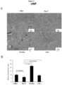
- Tumor samples and soft tissues were fixed in 4% para-formaldehyde (PFA, Fisher) and processed for paraffin bedding, sectioning, H&E andimtnunohistochemistry (IHC). Bones were fixed in 4% PFA at 4°C for 72h, decalcified in 0.5M EDTA (pH 8) for 7 days at 4°C, and embedded in paraffin (19). vWF staining on tumor sections was performed by the Pathology Core Facility of KCC.
- PFA para-formaldehyde
- CK14 (Covance) PRB-155P and CK8 (clonelE8, Covance) MMS-162P staining was performed after deparaffinization and rehydration without performing the antigen retrieval treatment on bones and brains samples to distinguish basal from luminal prostate epithelial cells.
- TRAP thyroid-resistant acid phosphatase staining was performed after deparaffinization and rehydratation as directed by the manufacturer (Sigma-Aldrich) to identify active osteoclasts at the surface between metastatic lesion and compact bone (5) (20).
- Tetrachrome method was performed on bones to identify woven bone in the osteoblastic lesion areas (5,21). (RV202, Santa Cruz Biotechnology) staining was performed only on brain samples to discriminate sarcoma from carcinoma.
- RNA quality assessment is based on the RNA Integrity Number (RIN).
- Microarray data was preprocessed using background correction, quantile normalization, and summarization were performed on the Mouse Gene 1.0 ST gene expression microarrays using the Robust Multichip Analysis (RMA) workflow in Affymetrix Expression Console version 1.1 [Affymetrix, Inc., Santa Clara, CA].
- RMA Robust Multichip Analysis
- Differentially expressed genes were identified for each of the four over-expressing cell lines, by performing pairwise comparisons against the Pec control cell line. These comparisons were performed using significance analysis of microarrays (SAM) with a false discovery rate cutoff of 1% and two-fold change cutoff.
- SAM significance analysis of microarrays
- Western blot assays were performed in PEC cells as indicated. Cells were pelleted and lysed in buffer (50 mM HEPES, pH 7.2, 150 mMNaCl, 1 mM EDTA, 1 mM EGTA, 1 mM DTT, 0.1% Tween 20) supplemented with protease inhibitor cocktail (Roche Diagnostics, Mannheim, Germany). Antibodies used for Western blot are: AR (H-280, Santa Cruz Biotechnology).
- Microarray analysis methods Preprocessing and Differential Expression Analysis.
- Microarray data was preprocessed using background correction, quantile normalization, and summarization were performed on the Mouse Gene 1.0 ST gene expression microarrays using the Robust Multichip Analysis (RMA) [45] workflow in Affymetrix Expression Console version 1.1 [Affymetrix, Inc., Santa Clara, CA].
- RMA Robust Multichip Analysis
- Differentially expressed genes were identified for each of the four over-expressing cell lines, c-Myc, NeuT, Ha-Ras, and v-Src, by performing pairwise comparisons against the PEC control cell line. These comparisons were performed using significance analysis of microarrays (SAM) with a false discovery rate cutoff of 1% and two-fold change cutoff.
- SAM significance analysis of microarrays
- ROC receiver operating characteristic
- the first dataset comprises 185 samples log2 normalized mRNA expression data was downloaded from the MSKCC prostate cancer database website (http://cbio.mskcc.org/cancergenomics/prostate/dataJ) [20]. Gene profiles in this MSKCC dataset were median-centered and Pearson's correlation was computed between each MSKCC sample and the log2 fold change profile for each oncogenespecific signature.
- the second prostate cancer dataset including distant metastasis samples, was previously obtained from the MSKCC Gerald Laboratory and samples were processed as described in [33]. Briefly, microarray samples were processed using the Robust Multichip Average (RMA) procedure [45] with custom CDFs dated July 30, 2009 (version 12) [47].
- RMA Robust Multichip Average
- the expression patterns of the four oncogene over-expressing cell lines were compared against expression data for clinical traits including disease stage, grade, and recurrence, previously published by Lapointe et al. [3].
- the dataset was downloaded from the publications website. This dataset contains 5153 probesets and 112 prostate cancer samples with annotations including tumor grade, tumor stage, and disease recurrence. Probeset annotations were updated using the Array Infounation Library Universal Navigator GPL3044 annotation file dated 7/9/2009 [48]. Probetsets lacking gene symbol annotations were eliminated as well as probesets with at least 25% missing values, resulting in a total of 4232 features for 3327 unique genes. Differential expression analysis was performed for advanced stage vs. early stage, high grade vs.
- Oncogene transformed prostate cell lines convey contact-independent growth.
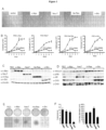
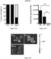
- Fig. 1A Primary prostate epithelial cell cultures were established from the ventral prostates of FVB mice ( FIG. 1A ). Cells were transduced with retroviral expression vectors encoding a single distinct oncogene (c-Myc, Ha-Ras (V-12), v-Src and NeuT, an activating mutant of ErbB2). The cellular morphology of the prostate epithelial cells was altered over the four-week period ( Fig.1B ). Individual colonies of oncogene-transduced cells were selected and characterized. Cellular growth assays were conducted by cell counting ( Fig. 1C ). A substantial growth advantage was observed in each oncogene transduced cell line compared with primary prostate epithelial cells.
- the oncogene transduced PEC lines were examined for growth in soft agar. Colony size and number were characterized for each oncogene ( Fig. 2B ) [27]. Non-transformed PECs failed to grow in soft agar as previously described [27]. Oncogene transduction increased the size and number of colonies ( Fig. 2C, D ).
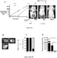
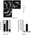
- Fig. 3A Tumor formation studies were conducted in FVB mice. A subcutaneous injection of lx10 6 cells resulted in tumor growth. Serial measurements were conducted by vernier calipers. Each of the prostate tumor lines grew subcutaneously in immune competent mice. Growth was sustained for c-Myc, Ha-Ras and v-Src transformed PECs ( Fig. 3A ). The extirpated tumors were hemorrhagic ( Fig. 3B ) with histological features of poorly differentiated prostate adenocarcinoma ( Fig. 3C , Supplement 1).
- VWF Von Willebrand factor
- mRNA was prepared from the oncogene transformed PEC cell lines.
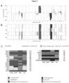
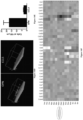
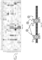
- Microarray analysis identified a total of 2635 out of 22115 genes that were significantly altered in expression (at least two-fold change) in oncogene overexpressing cell lines when compared with non-transformed prostate epithelial cell control samples ( Fig. 5 ).
- the heatmap of genes identified as significantly different in their expression (at least 2-fold change) is shown in Figure 5 .
- the rows of the heatmap represent unique genes and are displayed by their pattern of up- and down-regulation for all four of the oncogene induced cell lines
- a sizable number of up- and down-regulated genes were shared amongst all four cell lines (Group 1; 251 genes).
- Group 1 contains genes that share differential expression patterns across all four cell lines
- group 15 contains genes whose differential expression is specific to the Src cell lines.
- Genes with up and down-regulation specific to Ha-Ras were the most prevalent (Group 14; 584 genes), followed by c-Myc-specific genes (Group 8; 332 genes), NeuT-specific genes (Group 12; 277 genes), and v-Src-specific genes (Group 15; 215 genes).
- the murine prostate oncogene expression signature in high grade and advanced stage human prostate cancer is the murine prostate oncogene expression signature in high grade and advanced stage human prostate cancer.
- the prostate "oncogene expression signature” was defined as genes that were significantly altered in expression level and that were uniquely altered in expression by a specific oncogene compared with primary prostate epithelial cells ( Fig. 5 ).
- the oncogene expression signature thereby identified was compared to gene signatures obtained from other published databases to identify similarities to other well-studied disease phenotypes and cell lines. Comparisons were performed against gene signatures representative of differential expression in advanced state vs. early stage prostate cancer, high grade vs low grade prostate cancer, recurrent vs. nonrecurrent prostate cancer [3].
- the gene signature heatmaps representing advanced stage/early stage, high grade/low grade, and recurrent/nonrecurrent prostate cancer phenotypes [3] are shown on the left and the heatmaps on the right represent genes that are differentially expressed in the prostate oncogene expression signature.
- the heatmaps in Figures 6-8 are labeled with the percentage of genes within the "oncogene expression signature" that are differentially expressed.
- P values for the statistical significance of the similarity between the genes expressed in the prostate cancer cell lines and the gene signature of the disease phenotype are shown. P values are based on the hypergeometric distribution and represent the probability of these genes being differentially expressed in the disease phenotype if they were selected at random. Low p values indicate a degree of similarity between an oncogene cell line and a disease phenotype that is unlikely to occur merely by chance.
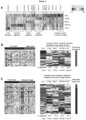
- c-Myc specific gene expression signature in prostate cancer epithelial cells resembles the c-Myc signature in fibroblasts and mammary tumors.
- Fig. 7A depicts the genes shared between c-Myc transduced fibroblasts ( Fig. 7A ), c-Myc-induced mammary tumors (Fig. 7C) (left-hand heatmaps) and the c-Myc induced prostate oncogene expression signature (right-hand heatmaps).
- c-Myc cell lines demonstrated the largest proportion of similarity with both the Myc transduced fibroblasts (92%) ( Fig. 7A ) and the c-Myc-induced mammary tumors (85%) (Fig 7C).
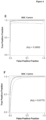
- ROC curves have been used previously to evaluate the diagnostic ability of PSA [29, 30] as well as its ability to identify metastatic disease [31].
- the c-Myc signature ROC curve exhibited better sensitivity/specificity characteristics than PSA, as evident in the area under the ROC curves.
- the area under the ROC curve represents the potential of a variable to discriminate between two conditions [32], indicating that the c-Myc signature performs as a better discriminator of metastatic disease than serum PSA levels.
- the c-Myc signature correlates with metastatic prostate cancer.
- Prostate oncogene induced gene expression and recurrence free survival in human prostate cancer Prostate oncogene induced gene expression and recurrence free survival in human prostate cancer.
- Figure 8A provides a heatmap showing the expression profiles of genes in the human prostate cancer samples from the Glinsky's data set, which was upregulated in the oncogene-transformed prostate cancer cell lines. Genes that were highly expressed in each of the four prostate oncogene cell lines had a significant association with poor outcome (p ⁇ 0.005) ( Fig. 8B,E ).
- FIG. 1 Establishment of Oncogene Transformed Prostate Cancer Cell Lines.
- A FVB mice, (II) prostates were used to establish Primary prostate epithelial cells (PEC) as shown by phase contrast microscopy (ventral prostates of male FVB mice at 12 weeks of age).
- B Schematic representation of the methods deployed and phase contrast microscopy of oncogene induced cell lines derived from PEC transduced by distinct oncogenes (c-Myc, NeuT, Ha-Ras, v-Src). Photo of individual colonies derived from oncogene-transduced PEC that were selected and characterized.
- C Growth curves of PEC lines detei 'lined by cell counting. Data are mean ⁇ SEM of N>3 separate experiments.
- A Western Blot analysis of 3 separate clones of each oncogene induced PEC shows antibodies were used for the detection of c-Myc, NeuT, Ha-Ras and v-Src as shown. GDI is used as a protein loading control.
- B Soft agar assays of oncogene transduced PEC. Non-transformed PEC failed to grow in soft agar.
- C The size (C) and number (D) of colonies from oncogene transduced PEC lines are shown as mean + SEM of N>5 separate experiments.
- FIG. 3 Prostate epithelial cell lines grow in immune competent mice.
- A PEC tumor diameter, determined by vernier caliper measurement, is shown as days after inoculation in FVB mice. The diameter mean ⁇ SEM for N>5 separate experiments. The NeuT tumors grew for 2 weeks then decreased in size.
- B Photograph of representative tumor derived from oncogene-induced lines. NeuT induced tumors were harvested at 15 days after cell injection.
- C Hemotoxylin and eosin and
- D VWF staining of PEC demonstrates poorly differentiated prostate adenocarcinoma with local vascularity.
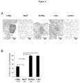
- Figure 4 Oncogene transformed prostate epithelial cell tumors metastasize to lung.
- Figure 5 Hierarchical clustering of microarray gene expression.
- Figure 6 Genes associated with high grade and advanced stage human prostate cancer.
- Heatmaps of the left-hand side represent the prostate cancer high grade and advanced stage signatures, while heatmaps on the right represent genes that are differentially expressed in at least one of the four prostate cancer cell lines. The percentage of these genes that are differentially expressed within each individual prostate cancer cell line is shown in the respective columns along with the p value for the degree of similarity with the high grade and advanced stage phenotypes.
- Figure 7 c-Myc and Ha-Ras specific oncogene signature in prostate tumors is conserved in fibroblasts.
- Heatmaps of (A) and (B) show the genes that are differentially expressed in the oncogene-induced prostate cancer cell lines and in c-Myc and Ha-Ras transduced fibroblasts (3T3 cell line) [46]. Heatmaps of the left-hand side represent the transduced fibroblasts gene signatures, while heatmaps on the right represent genes that are differentially expressed in at least one of the four prostate cancer cell lines. Heatmap of (C) shows the intersection of genes that are differentially expressed in the c-Myc prostate cancer cell line and in the respective c-Myc-induced mouse mammary tumor samples [46].
- Heatmaps of the left-hand side represent the tumor sample gene signatures, while heatmaps on the right represent genes that are differentially expressed in the prostate cancer cell lines.
- the p values shown under each prostate cell line heatmap represent the significance of the overlap between the prostate and mammary cancer datasets.
- (D) ROC curve for the utility of PSA (orange) and the c-Myc signature (green) to identify metastatic disease.
- the x-axis represents the false positive rate (1-specificity) and the y-axis represents the true positive rate (sensitivity).
- the dashed line represents no discriminative ability.
- a heatmap depicts the consistency between each of the four prostate cancer cell line signatures and samples in MSKCC prostate cancer datasets. Red indicates positive Pearson correlation, while blue indicates negative Pearson correlation.
- Figure 8 Gene expression correlates of oncogene transformed prostate cancer cell lines with recurrence-free survival.
- A Expression profile of human prostate cancer samples [11] that were upregulated in the oncogene transferred prostate cancer cell lines. Bars along the bottom of the heatmap indicate whether a sample has high (upper 25th percentile) or low expression (lower 75th percentile) based on genes upregulated in each of the four oncogene-transformed cell lines. Kaplan Meier curves are shown for high and low expression populations for (B) c-Myc upregulated genes, (C) NeuT upregulated genes, (D) Ha-Ras upregulated genes, (E) v- Src upregulated genes.
- IHC Immunohistochemical staining of PEC tumors for VWF as a marker of neoangiogenesis. Representative example of IHC for VWF for each cell line.
- B The relative concentration of blood vessels are shown for the four oncogene induced mouse prostate tumors. Quantitation of VWF staining from oncogene-induced prostate tumors. Data are mean ⁇ SEM for N>3 separate tumors. In the NeuT group, the blood vessel concentration is lower and Ha-Ras vascularity is greater (p ⁇ 0.01).
- Medium for primary culture comprised F-12 500m1, 10% FBS, Insulin 5ug/ml, EGF lOng/ml, Hydrocortisone lug/ml, transferrin 5ng/ml, bovine pituitary extraction 30ug/ml, 1X pen-strep, and 1 X Gentamicin.
- F-12 500m1 10% FBS, Insulin 5ug/ml, EGF lOng/ml, Hydrocortisone lug/ml, transferrin 5ng/ml, bovine pituitary extraction 30ug/ml, 1X pen-strep, and 1 X Gentamicin.
- PBS Proliferin 5ng/ml
- bovine pituitary extraction 30ug/ml
- 1X pen-strep 1 X Gentamicin.
- pBABE-IRES-target gene was transfected into 293T cells by calcium phosphate precipitation.
- DNA and CaC12 were mixed in HBS buffer, and the mixture made up to afinal volume of lml, which was allowed to stand for 20mins at RT. This mixture was then mixed into 293T cells and the cells put into incubator for 5 hours then the incubation medium was removedand replace with fresh medium. After 48 hours, the supernatant of 293T cells was collected, mixed with equal volume of fresh medium, and the resulting mixture was filtered by 0.45um filter. Polybrene (final concentration 8ug/m1) was then added into the mixture, which was then added into prostate epithelial cells which were in passage one. After another 48 hours of infection, the medium was removed and replace with fresh medium (DMEM,10% FBS).
- a culture medium comprised DMEM, 10% FBS, and lx pen-strep.was added puromysin, and the final concentration was made up to 1-2ug/ml.
- the cells were repeatedly treated with puromysin for at least 1 month, until the negative cells were dead, and the positive clones with oncogene expression were left. When the clones were big enough, picked the clones by were picked by cloning cylinders (Specialty Media, cat# TR-1004), and the cells were appropriately marked. The cells were then grown for at least 25 passages. Characterized in assays of growth in tissue culture soft agar, in vivo implantation, metastasis, microarray and histopathology.
- Figure 1 illustrates oncogene transduced PEC lines foam colonies in soft agar.
- Figure 1A illustrates phase contrast microscopy of oncogene induced cell lines were transduced by distinct oncogenes (c-Myc, NeuT, Ha-Ras, v-Src). Photo of individual colonies derived from oncogene-transduced PEC that were selected and characterized.
- Figure 1B illustrates growth curves of PEC lines determined by cell counting. Data are mean ⁇ SEM of N>3 separate experiments.
- Figures 1C and 1D describe Western Blot analysis of 3 separate clones of each oncogene induced PEC with antibodies as shown for detection of c-Myc, NeuT, Ha-Ras and v-Src and Figures 1D describes markers of basal (CK5) vs luminal (CK8) prostate cancer.
- GDI is used as a protein loading control.
- Figure lE describes soft agar assays of oncogene transduced PEC. Non-transformed PEC failed to grow in soft agar.
- the size ( Figure 1E ) and number ( Figure 1F ) of colonies from oncogene transduced PEC lines are shown as mean ⁇ SEM of N>5 separate experiments.
- Figure 2 depicts copy number aberrations in the four oncogene cell lines assessed by array CGH.
- Figure 2A illustrates the percentage of the four cell lines sharing copy gain or loss regions is shown as a function of genomic position.
- Figure 2B illustrates regions of copy gain (red) or loss (blue) for each of the four cell lines are shown as a function of genomic position.
- oncogenes are identified with mRNA overexpression (red), DNA amplification (yellow), or both (purple) among the four oncogene cell lines, with corresponding amplification in the MKSCC prostate cancer database (listed on righthand side).
- tumor suppressor genes are identified with mRNA underexpression (blue), DNA copy loss (pink), or both (orange) among the four oncogene cell lines, with corresponding copy loss in the MKSCC prostate cancer database (listed on right-hand side).
- Figure 3 illustrates prostate epithelial cell lines grow in immune competent mice.
- Figure 3A PEC tumor diameter determined by vernier caliper measurement is shown as days after inoculation in FVB 30 mice. The diameter mean ⁇ SEM for N>5 separate experiments.
- Figure 3B shows photograph of representative tumor derived from oncogene-induced lines. NeuT induced tumors were harvested at 15 days after cell injection.
- Figure 3C shows Hematoxylin and eosin staining at low and high magnification (see also supplemental data 1-4).
- Figure 4 shows oncogene transformed prostate epithelial cell tumors metastasize to lung.
- Figure 4A shows Hematoxylin and eosin stain of murine lung post tumor implantation demonstrating representative example of lung metastasis.
- Figure 4B shows Frequency of lung metastases were detected in mice for c-Myc, NeuT and v-Src PEC groups 5 weeks after subcutaneous injection. The rates were 100% frequency in Ha-Ras and v-Src groups.
- Figure 5 shows hierarchical clustering of microarray gene expression.
- Heatmaps of the left-hand side represent the prostate cancer high grade and advanced stage signatures, while heatmaps on the right represent genes that are differentially expressed in the four prostate cancer cell lines. The percentage of these genes that are differentially expressed within each individual prostate cancer cell line is shown in the respective columns along with the p value for the degree of similarity with the high grade and advanced stage phenotypes.
- Figure 6 shows c-Myc- and Ha-Ras-specific oncogene signatures in prostate tumors are conserved in other tissues.
- Heatmaps show genes that are differentially expressed in the oncogene-induced prostate cancer cell lines and in Figure 6A Ha-Ras and Figure 6B c-Myc fibroblasts (3T3 cell line).
- Figure 6C A heatmap shows the intersection of genes that are differentially expressed in the c- Myc prostate cancer cell line and mouse mammary tumor samples. The p values shown under each prostate cell line heatmap represent the significance of the overlap between the prostate and fibroblast/mammary tumor signatures.
- Figure 6D shows a classifier based on canonical analysis of c-Myc signature distinguishes human tumor (red) from normal tissue (light blue), along the x-axis, in the Lapointe 2004 dataset.
- ROC curves for the classifier performance are shown for E) the Lapointe 2004 dataset and F) the Taylor 2010 MSKCC dataset, with AUC values of 0.990 and 0.977, respectively.
- Figure 7 shows gene expression correlates of oncogene transformed prostate cancer cell lines with recurrence-free survival.
- Figure 7A shows expression profile of human prostate cancer samples (13) that were upregulated in the oncogene transferred prostate cancer cell lines. Bars along the bottom of the heatmap indicate whether a sample has high (upper 25th percentile) or low expression (lower 75th percentile) based on genes upregulated in each of the four oncogenetransformed cell lines. Kaplan Meier curves are shown for high and low expression populations for ( Figure 7B ) c-Myc upregulated genes.
- Figure 8 illustrates gene expression correlates of oncogene transformed prostate cancer cell lines with recurrence-free survival.
- Figure 9 illustrates histological features of poorly differentiated prostate adenocarcinoma.
- Figures 10A-10D demonstrate that Src enhances 3D matrigel invasion of isogenic prostate cancer cell lines.
- Figure 10D shows mean distances of invasion ⁇ SEM from 3 independent experiments for PEC lines (PEC-NeuT, PEC-Ras, and PEC-Src). As shown in Figures 10C and 10D , invasion into matrigel was more efficient for the c-Src transduced line. Statistical analysis of the results shown was conducted using the Student's t test.
- Figures 11A and 11B demonstrate isogenic prostate cancer cell line tumors are vascular.
- subcutaneous tumor growth in NCR nude mice was quantitated over 3 weeks following subcutaneous innoculation of 1 x10⁵ cells for each of the 3 lines (PEC-NeuT, PEC-Ras, and PEC-Src) using normalized photon flux to quantitate tumor volume. Mean sizes + SEM from 3 independent experiments for PEC lines are shown.
- the lines grew as subcutaneous tumors in NCR nude mice; the relative size as determined by photon flux suggested more rapid growth amongst the Ras-derived lines.
- Statistical analysis of the data was conducted using the Student's t test.
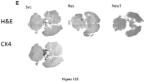
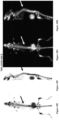
- FIGS 12A-12E demonstrate prostate cancer lines develop metastasis. Tomato-Red. Cells were injected into the left cardiac ventricle of 16 mice for each cell line. Bioluminescence images were acquired and quantified 14 days after xenografting. Representative in vivo images of mice are shown in Figure 12A . As shown, upon introduction of tumors into the arterial circulation via the left ventricle of the heart (I.C. Injection), tumors developed rapidly in multiple organs, including liver, brain, bladder, adrenal gland and kidney, within two weeks of injection.
- I.C. Injection the left ventricle of the heart
- Figure 12B shows representative images of brain metastasis in mice following the intracardiac injection of the isogenic prostate cancer lines.
- Figure 12E shows H&E staining of brain metastasis formed after 2 weeks of PEC-Src and PEC-NeuT intracardiac injection. Also, CK14 staining corroborated the presence of prostate epithelial cells within the brain (arrow).
- Figures 13A-13C demonstrate liver metastasis of prostate tumor cell lines. Isogenic PEC lines expressing the Luc2-Tomato-Red fusion protein were injected into the ventricle of FVB mice and the in vivo bioluminescent signal quantified.
- Figure 12A illustrates the percentage of mice with liver tumors. Hepatic metastasis in the mice injected with the Ras and Src PEC lines developed liver metastasis (-100% of the animals). Although the NeuT tumors grew subcutaneously, none developed liver metastasis.
- Figure 12B illustrates the tumor size determined by photonflux and Figure 13C illustrates representative mice images showing liver metastasis.
- Kidney metastasis were identified in the mice injected with the Ras and Src derived lines.
- Figure 13D illustrates the percentage of mice with kidney tumors.
- Figure 13E illustrates size of kidney tumors by photon flux and
- Figure 13F illustrates representative images of kidney metastasis.
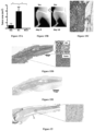
- Figures 14A-14C demonstrate isogenic prostate cancer cell lines develop osteolytic bone metastases.
- Figure 14A illustrates representative in vivo images of FVB mice that underwent intracardiac injection of PEC lines expressing Luc2-Tomato-Red fusion protein and the in vivo bioluminescent signal was quantified.
- FIG 14A the bony metastasis of the mice were osteolytic in nature.
- Figure 14C illustrates the size of-tumor mass on photon flux. Histological analysis of the osteolytic bone lesions of the prostate tumors evidenced, adenocarcinoma resembling the primary tumor.
- Figures 15A-15F demonstrate Src enhances osteolytic prostate cancer bone metastases.
- FVB mice 2 weeks after PEC-Src intracardiac injection developed osteolytic bone lesions.
- Figure 15A shows tumor area in bones was significantly increased in the PEC-Src group compared with PEC-Ras and PEC-NeuT. The area of the tumor was six-fold greater in c-Src compared to Ras driven tumors.
- Figure 15B illustrates representative X-Rays before (t0) and 14d (t14) after intracardiac injection of cells. Low density areas colocalized with the metastatic tumors (arrowhead) indicating osteolytic lesions. The bone lesions were found primarily at the epiphyseal junction as osteolytic lesions at two weeks.
- FIG. 15C Hisological analysis confirmed adenocarcinoma at the site of bony metastasis.
- Tartrate resistant acid phosphatase (“TRAP”) staining shown in Figure 15C , corroborated the presence of osteoblast (arrows) in the bone-tumor interface.
- Figure 15D shows Haematoxylin Eosin (“H&E”) staining of bone metastasis formed after.
- Figures 15E and 15F show cytokeratin (CK) 14 staining and CK8 staining respectively, both corroborating the presence of epithelial cells within bone.
- H&E Haematoxylin Eosin
- FIGS 16A-16G demonstrate osteolytic prostate cancer cell lines express function CCL5 and osteopontin (“OPN") receptors.
- Figures 16A-16D show Fluorescence-Activated Cell Sorting ("FACS") analysis of CCR5 expression on PEC lines. As shown, FACS analysis using a CCR5 specific antibody confirmed the presence of the CCR5 receptor in PEC lines.
- FACS Fluorescence-Activated Cell Sorting
- Figures 16E and 16F show results of Matrigel invasion assays of the PEC-Src line conducted using OPN as CD44 ligand and CCL5 as CCR5 ligand and quantified as mean + SEM ( Figure 16 G) .
- Figures 16D and 16E the addition of OPN or CCR5 enhanced PEC invasiveness into matrigel.
- the in vivo tumor gene expression in the oncogene transformed PEC was compared with the expression in vivo in the dorsolateral ventral prostate of mice with the same strain.
- Figure 16F shows chemokine receptor and ligand gene expression of prostate tumor cell lines in tissue culture.
- Figure 16G shows chemokine receptor and ligand gene expression of prostate tumor cell lines after subcutaneous implantation.
- the gene expression showed a notable up regulation in vivo of the cytokine and chemokines.
- upregulation of CCR5 and CCR2 was observed in the c-Myc, NeuT and Src PEC lines.
- Upregulation of the receptor ligands CCL2, CCL7, and CCL8 was observed (2- to 3-fold).
- CCL7, CCL8 and CCL5 are ligands for CCR5, CCL8 and CCL5 for CCR1 ( Figure 15H).
- microarray based gene expression profiling showed activation of a CCR5 signaling module when the PEC lines were grown in vivo vs tissue culture.
- CCL2 binds CCR2 and CCR4. Given that CCR5 and its ligands CCL5, CCL7 and CCL8 were induced in the PEC in vivo, the effect of the CCR5 antagonist on prostate tumor growth was examined.
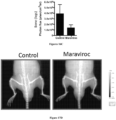
- Figures 17A-17D demonstrate that CCR5 antagonists block spinal osteolytic prostate cancer metastasis.
- PEC lines transduced with vectors expressing the Luc2-Tomato-Red fusion protein were injected into the ventricle of FVB mice and the in vivo bioluminescent signal was quantified after 2 weeks. Mice were treated with oral maraviroc (8 mg/kg) or control.
- Figure 17A illustrates representative examples of mice from each group.
- Figure 17B illustrates photon flux as a volumetric analysis of total tumor mass and
- the CCRS antagonist Maraviroc (8 mg/kg oral) reduced total body metastatic burden by >50% and reduced bony metastasis by >50% (see Figures 17C and 17D ).
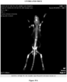
- Flurine-18, Sodium fluoride (“F-18-NaF”) imaging correlated with X-ray analysis demonstrated the presence of spine metastasis ( Figures 18A-1811).
- Daily oral treatment with Maraviroc reduced spine metastasis by >90% ( Figures 19A-19B ).
Landscapes
- Health & Medical Sciences (AREA)
- Life Sciences & Earth Sciences (AREA)
- Chemical & Material Sciences (AREA)
- Engineering & Computer Science (AREA)
- General Health & Medical Sciences (AREA)
- Organic Chemistry (AREA)
- Biomedical Technology (AREA)
- Biotechnology (AREA)
- Zoology (AREA)
- Wood Science & Technology (AREA)
- Genetics & Genomics (AREA)
- Animal Behavior & Ethology (AREA)
- Veterinary Medicine (AREA)
- Medicinal Chemistry (AREA)
- Bioinformatics & Cheminformatics (AREA)
- Immunology (AREA)
- Oncology (AREA)
- Public Health (AREA)
- Pharmacology & Pharmacy (AREA)
- Microbiology (AREA)
- Biochemistry (AREA)
- Proteomics, Peptides & Aminoacids (AREA)
- Analytical Chemistry (AREA)
- Pathology (AREA)
- Molecular Biology (AREA)
- General Engineering & Computer Science (AREA)
- Cell Biology (AREA)
- Chemical Kinetics & Catalysis (AREA)
- Nuclear Medicine, Radiotherapy & Molecular Imaging (AREA)
- General Chemical & Material Sciences (AREA)
- Physics & Mathematics (AREA)
- Environmental Sciences (AREA)
- Hematology (AREA)
- Epidemiology (AREA)
- Hospice & Palliative Care (AREA)
- Urology & Nephrology (AREA)
- Biophysics (AREA)
- Animal Husbandry (AREA)
- Food Science & Technology (AREA)
- Emergency Medicine (AREA)
Claims (5)
- CCR5-Antagonist zur Verwendung beim Inhibieren einer Metastasierung von Krebs, der auf eine Behandlung in einem immunkompetenten Subjekt mit einem Risiko des Entwickelns einer Metastasierung von Krebs anspricht.
- CCR5-Antagonist zur Verwendung nach Anspruch 1, wobei der CCR5-Antagonist eine Tumormetastasierung in einem oder mehreren Organen, ausgewählt aus der Gruppe bestehend aus Leber, Gehirn, Blase, Lunge, Nebenniere, Niere, Knochen und Kombinationen davon, inhibiert.
- CCR5-Antagonist zur Verwendung nach einem der Ansprüche 1 bis 2 beim Verhindern von Krebs oder beim Inhibieren einer Metastasierung von Krebs, wobei der Krebs Prostatakrebs ist.
- CCR5-Antagonist zur Verwendung nach einem der Ansprüche 1 bis 3, wobei die Verwendung die tägliche orale Verabreichung von Maraviroc umfasst.
- CCR5-Antagonist zur Verwendung nach einem der Ansprüche 1 bis 4, wobei das immunkompetente Subjekt ein Mensch ist.
Applications Claiming Priority (3)
| Application Number | Priority Date | Filing Date | Title |
|---|---|---|---|
| US201161450767P | 2011-03-09 | 2011-03-09 | |
| PCT/US2012/028546 WO2012122499A2 (en) | 2011-03-09 | 2012-03-09 | Prostate cancer cell lines, gene signatures and uses thereof |
| EP12755544.9A EP2683643B1 (de) | 2011-03-09 | 2012-03-09 | Prostata-krebszelllinien, gensignaturen und verwendungen davon |
Related Parent Applications (1)
| Application Number | Title | Priority Date | Filing Date |
|---|---|---|---|
| EP12755544.9A Division EP2683643B1 (de) | 2011-03-09 | 2012-03-09 | Prostata-krebszelllinien, gensignaturen und verwendungen davon |
Publications (2)
| Publication Number | Publication Date |
|---|---|
| EP3597761A1 EP3597761A1 (de) | 2020-01-22 |
| EP3597761B1 true EP3597761B1 (de) | 2025-05-07 |
Family
ID=46798838
Family Applications (2)
| Application Number | Title | Priority Date | Filing Date |
|---|---|---|---|
| EP19173075.3A Active EP3597761B1 (de) | 2011-03-09 | 2012-03-09 | Prostata-krebszelllinien, gensignaturen und verwendungen davon |
| EP12755544.9A Active EP2683643B1 (de) | 2011-03-09 | 2012-03-09 | Prostata-krebszelllinien, gensignaturen und verwendungen davon |
Family Applications After (1)
| Application Number | Title | Priority Date | Filing Date |
|---|---|---|---|
| EP12755544.9A Active EP2683643B1 (de) | 2011-03-09 | 2012-03-09 | Prostata-krebszelllinien, gensignaturen und verwendungen davon |
Country Status (11)
| Country | Link |
|---|---|
| US (2) | US10952415B2 (de) |
| EP (2) | EP3597761B1 (de) |
| JP (2) | JP2014518610A (de) |
| CN (1) | CN104169425A (de) |
| AU (1) | AU2012225232B2 (de) |
| BR (1) | BR112013023050A8 (de) |
| CA (1) | CA2829219C (de) |
| DK (2) | DK3597761T3 (de) |
| ES (2) | ES3034910T3 (de) |
| MX (1) | MX2013010330A (de) |
| WO (1) | WO2012122499A2 (de) |
Families Citing this family (17)
| Publication number | Priority date | Publication date | Assignee | Title |
|---|---|---|---|---|
| EP3597761B1 (de) | 2011-03-09 | 2025-05-07 | Richard G. Pestell | Prostata-krebszelllinien, gensignaturen und verwendungen davon |
| EP2861302A4 (de) * | 2012-05-14 | 2016-08-24 | Prostagene Llc | Verwendung von ccr5-modulatoren zur behandlung von krebs |
| CN103881975B (zh) * | 2014-04-04 | 2015-10-21 | 武汉大学 | 人前列腺癌细胞及其原代分离培养和传代培养方法与用途 |
| FR3022142B1 (fr) * | 2014-06-16 | 2019-07-12 | Universite Paul Sabatier - Toulouse Iii | Inhibition de la chimiokine ccl7 ou de son recepteur ccr3 pour le traitement et le diagnostic du cancer de la prostate |
| US9994912B2 (en) | 2014-07-03 | 2018-06-12 | Abbott Molecular Inc. | Materials and methods for assessing progression of prostate cancer |
| WO2016145294A1 (en) * | 2015-03-12 | 2016-09-15 | The University Of Chicago | Methods for determining prognosis for breast cancer patients |
| JP2018532992A (ja) * | 2015-09-09 | 2018-11-08 | ソマロジック, インコーポレイテッドSomaLogic, Inc. | 個別化薬物治療計画の開発方法、及びプロテオミックプロファイルに基づく標的薬物開発 |
| CA3023487A1 (en) | 2016-05-10 | 2017-11-16 | National University Corporation Tokyo Medical And Dental University | Expression inhibitor of inflammation promoting factor, screening method for active ingredient thereof, expression cassette useful for said method, diagnostic agent and diagnosis method |
| CN111315882B (zh) | 2017-11-09 | 2024-09-24 | 国立大学法人东京医科齿科大学 | 癌症促进因子表达抑制剂、其有效成分的筛选方法、对该方法有用的表达盒、诊断药和诊断方法 |
| US10721069B2 (en) | 2018-08-18 | 2020-07-21 | Eygs Llp | Methods and systems for enhancing privacy and efficiency on distributed ledger-based networks |
| US11316691B2 (en) | 2019-04-15 | 2022-04-26 | Eygs Llp | Methods and systems for enhancing network privacy of multiple party documents on distributed ledger-based networks |
| US11206138B2 (en) * | 2019-05-02 | 2021-12-21 | Ernst & Young U.S. Llp | Biosignature-based tokenization of assets in a blockchain |
| US11232439B2 (en) | 2019-08-09 | 2022-01-25 | Eygs Llp | Methods and systems for preventing transaction tracing on distributed ledger-based networks |
| EP4062585B1 (de) | 2019-11-20 | 2025-10-29 | Eygs LLP | Systeme, vorrichtungen und verfahren zum identifizieren und sicheren speichern von unterscheidungsmerkmalen in einem verteilten hauptbuch innerhalb eines auf einem verteilten hauptbuch basierenden netzwerks basierend auf austauschbaren und nicht austauschbaren token |
| EP4136600A1 (de) | 2020-04-15 | 2023-02-22 | Eygs LLP | Intelligente behauptungstoken zur authentifizierung und steuerung von netzwerkkommunikationen unter verwendung eines verteilten kontos |
| US11629196B2 (en) | 2020-04-27 | 2023-04-18 | Incelldx, Inc. | Method of treating SARS-CoV-2-associated hypercytokinemia by administering a human monoclonal antibody (PRO-140) that inhibits CCR5/CCL5 binding interactions |
| US11402391B2 (en) | 2020-12-21 | 2022-08-02 | Incelldx, Inc. | Methods of treating a long-hauler subject for chronic COVID-19 by administering a CCR5 or CCL5 antagonist |
Family Cites Families (35)
| Publication number | Priority date | Publication date | Assignee | Title |
|---|---|---|---|---|
| FI941572L (fi) | 1991-10-07 | 1994-05-27 | Oncologix Inc | Anti-erbB-2-monoklonaalisten vasta-aineiden yhdistelmä ja käyttömenetelmä |
| EP1514934B1 (de) | 1992-02-06 | 2008-12-31 | Novartis Vaccines and Diagnostics, Inc. | Marker für Krebs und biosynthetisches Bindeprotein dafür |
| IL112248A0 (en) | 1994-01-25 | 1995-03-30 | Warner Lambert Co | Tricyclic heteroaromatic compounds and pharmaceutical compositions containing them |
| US5869485A (en) | 1994-09-29 | 1999-02-09 | Novartis Finance Corp. | Pyrrolo 2,3-d!pyrimidines and their use |
| US20040151719A1 (en) | 1995-06-06 | 2004-08-05 | Human Genome Sciences, Inc. | Human G-protein chemokine receptor (CCR5) HDGNR10 |
| GB9520822D0 (en) | 1995-10-11 | 1995-12-13 | Wellcome Found | Therapeutically active compounds |
| AU7296896A (en) | 1995-11-01 | 1997-05-22 | Novartis Ag | Purine derivatives and processes for their preparation |
| CH690773A5 (de) | 1996-02-01 | 2001-01-15 | Novartis Ag | Pyrrolo(2,3-d)pyrimide und ihre Verwendung. |
| WO1997032879A1 (de) | 1996-03-06 | 1997-09-12 | Novartis Ag | 7-ALKYL-PYRROLO[2,3-d]PYRIMIDINE |
| AU3176297A (en) | 1996-06-25 | 1998-01-14 | Novartis Ag | Substituted 7-amino-pyrrolo{3,2-d}pyrimidines and the use thereof |
| AR007857A1 (es) | 1996-07-13 | 1999-11-24 | Glaxo Group Ltd | Compuestos heterociclicos fusionados como inhibidores de proteina tirosina quinasa, sus metodos de preparacion, intermediarios uso en medicina ycomposiciones farmaceuticas que los contienen. |
| CA2260058A1 (en) | 1996-07-13 | 1998-01-22 | Kathryn Jane Smith | Bicyclic heteroaromatic compounds as protein tyrosine kinase inhibitors |
| GB9800575D0 (en) | 1998-01-12 | 1998-03-11 | Glaxo Group Ltd | Heterocyclic compounds |
| RS49779B (sr) | 1998-01-12 | 2008-06-05 | Glaxo Group Limited, | Biciklična heteroaromatična jedinjenja kao inhibitori protein tirozin kinaze |
| PE20001420A1 (es) | 1998-12-23 | 2000-12-18 | Pfizer | Moduladores de ccr5 |
| EP2308833A3 (de) | 1999-04-15 | 2011-09-28 | Bristol-Myers Squibb Company | Zyklische Proteintyrosinkinaseinhibitoren |
| US6689765B2 (en) | 1999-05-04 | 2004-02-10 | Schering Corporation | Piperazine derivatives useful as CCR5 antagonists |
| US7825121B2 (en) | 1999-05-04 | 2010-11-02 | Schering Corporation | Piperazine derivatives useful as CCR5 antagonists |
| JP2003523744A (ja) | 2000-02-09 | 2003-08-12 | ヒューマン ジノーム サイエンシーズ, インコーポレイテッド | Ccr5に対する抗体 |
| US6667314B2 (en) | 2000-05-26 | 2003-12-23 | Pfizer, Inc. | Tropane derivatives useful in therapy |
| DE10034109C1 (de) | 2000-07-13 | 2001-09-06 | Juergen W Mueller | Verfahren zur Verknüpfung geographischer und kommerzieller Daten sowie deren Bereitstellung |
| CN1564826A (zh) | 2001-02-09 | 2005-01-12 | 人类基因组科学公司 | 人类g蛋白趋化因子受体(ccr5)hdgnr10 |
| US20020182624A1 (en) * | 2001-02-28 | 2002-12-05 | Eos Biotechnology, Inc. | Chemokine receptors and disease |
| GB0108876D0 (en) | 2001-04-09 | 2001-05-30 | Novartis Ag | Organic Compounds |
| EP1570860A4 (de) | 2002-12-13 | 2009-06-03 | Ono Pharmaceutical Co | Antagonisten- und agonistenbindung an die stark bindende stelle des chemokinrezeptors |
| JPWO2004098638A1 (ja) | 2003-05-06 | 2006-07-13 | 小野薬品工業株式会社 | エフェクター細胞機能阻害剤 |
| CA2594602A1 (en) | 2004-12-23 | 2006-06-29 | Pfizer Limited | 8-aza-bicyclo (3.2.1) octane derivatives with an activity on chemokine ccr5 receptors |
| US20090118175A1 (en) | 2005-05-06 | 2009-05-07 | Macina Roberto A | Compositions and Methods for Detection, Prognosis and Treatment of Breast Cancer |
| US7872027B2 (en) | 2006-02-17 | 2011-01-18 | University Of Pittsburgh - Of The Commonwealth System Of Higher Education | Low molecular weight Myc-max inhibitors |
| KR20090008217A (ko) | 2006-03-10 | 2009-01-21 | 오노 야꾸힝 고교 가부시키가이샤 | 질소 함유 복소환 유도체 및 이들을 유효 성분으로 하는 약제 |
| TWI442932B (zh) | 2008-02-11 | 2014-07-01 | Ferring Int Ct Sa | 以GnRH拮抗劑治療攝護腺癌的方法 |
| US7897356B2 (en) | 2008-11-12 | 2011-03-01 | Caris Life Sciences | Methods and systems of using exosomes for determining phenotypes |
| EP3597761B1 (de) | 2011-03-09 | 2025-05-07 | Richard G. Pestell | Prostata-krebszelllinien, gensignaturen und verwendungen davon |
| US9665874B2 (en) | 2012-03-13 | 2017-05-30 | American Express Travel Related Services Company, Inc. | Systems and methods for tailoring marketing |
| US10884952B2 (en) | 2016-09-30 | 2021-01-05 | Intel Corporation | Enforcing memory operand types using protection keys |
-
2012
- 2012-03-09 EP EP19173075.3A patent/EP3597761B1/de active Active
- 2012-03-09 BR BR112013023050A patent/BR112013023050A8/pt not_active Application Discontinuation
- 2012-03-09 JP JP2013557917A patent/JP2014518610A/ja active Pending
- 2012-03-09 ES ES19173075T patent/ES3034910T3/es active Active
- 2012-03-09 CA CA2829219A patent/CA2829219C/en active Active
- 2012-03-09 WO PCT/US2012/028546 patent/WO2012122499A2/en not_active Ceased
- 2012-03-09 MX MX2013010330A patent/MX2013010330A/es unknown
- 2012-03-09 DK DK19173075.3T patent/DK3597761T3/da active
- 2012-03-09 DK DK12755544.9T patent/DK2683643T3/da active
- 2012-03-09 AU AU2012225232A patent/AU2012225232B2/en active Active
- 2012-03-09 EP EP12755544.9A patent/EP2683643B1/de active Active
- 2012-03-09 US US14/003,384 patent/US10952415B2/en active Active
- 2012-03-09 ES ES12755544T patent/ES2740399T3/es active Active
- 2012-03-09 CN CN201280020337.8A patent/CN104169425A/zh active Pending
-
2016
- 2016-11-10 JP JP2016219554A patent/JP2017078070A/ja active Pending
-
2021
- 2021-02-04 US US17/167,578 patent/US20240224960A1/en active Pending
Non-Patent Citations (1)
| Title |
|---|
| ZHANG X ET AL: "Anibamine, a natural product CCR5 antagonist, as a novel lead for the development of anti-prostate cancer agents", BIOORGANIC & MEDICINAL CHEMISTRY LETTERS, ELSEVIER, AMSTERDAM, NL, vol. 20, no. 15, 1 August 2010 (2010-08-01), pages 4627 - 4630, XP027137556, ISSN: 0960-894X, [retrieved on 20100608] * |
Also Published As
| Publication number | Publication date |
|---|---|
| WO2012122499A2 (en) | 2012-09-13 |
| JP2014518610A (ja) | 2014-08-07 |
| CN104169425A (zh) | 2014-11-26 |
| CA2829219C (en) | 2021-03-16 |
| US20240224960A1 (en) | 2024-07-11 |
| AU2012225232B2 (en) | 2016-05-12 |
| EP2683643A2 (de) | 2014-01-15 |
| US10952415B2 (en) | 2021-03-23 |
| BR112013023050A8 (pt) | 2018-09-25 |
| DK2683643T3 (da) | 2019-08-12 |
| BR112013023050A2 (pt) | 2016-12-06 |
| ES3034910T3 (en) | 2025-08-26 |
| EP3597761A1 (de) | 2020-01-22 |
| MX2013010330A (es) | 2014-11-26 |
| AU2012225232A1 (en) | 2013-05-09 |
| US20140109245A1 (en) | 2014-04-17 |
| JP2017078070A (ja) | 2017-04-27 |
| DK3597761T3 (da) | 2025-08-18 |
| WO2012122499A3 (en) | 2014-04-17 |
| CA2829219A1 (en) | 2012-09-13 |
| ES2740399T3 (es) | 2020-02-05 |
| EP2683643B1 (de) | 2019-05-08 |
| EP2683643A4 (de) | 2015-04-29 |
Similar Documents
| Publication | Publication Date | Title |
|---|---|---|
| EP3597761B1 (de) | Prostata-krebszelllinien, gensignaturen und verwendungen davon | |
| Charytonowicz et al. | PPARγ agonists enhance ET-743–induced adipogenic differentiation in a transgenic mouse model of myxoid round cell liposarcoma | |
| Uramoto et al. | Expression of selected gene for acquired drug resistance to EGFR-TKI in lung adenocarcinoma | |
| La Manna et al. | Dual-mTOR inhibitor rapalink-1 reduces prostate cancer patient-derived xenograft growth and alters tumor heterogeneity | |
| Wei et al. | Exosomes derived from MDR cells induce cetuximab resistance in CRC via PI3K/AKT signaling‑mediated Sox2 and PD‑L1 expression | |
| Zhou et al. | Novel ETV1 mutation in small cell lung cancer transformation resistant to EGFR tyrosine kinase inhibitors | |
| Zhu et al. | Individualized therapy based on the combination of mini-PDX and NGS for a patient with metastatic AFP-producing and HER-2 amplified gastric cancer | |
| Fan et al. | A refractory case of CDKN2A/B loss metastatic intrahepatic cholangiocarcinoma achieving a partial response after first-line treatment with palbociclib | |
| Wang et al. | Synergistic effect of lenvatinib and chemotherapy in hepatocellular carcinoma using preclinical models | |
| Ruzzi et al. | ADK-VR2, a cell line derived from a treatment-naïve patient with SDC4-ROS1 fusion-positive primarily crizotinib-resistant NSCLC: a novel preclinical model for new drug development of ROS1-rearranged NSCLC | |
| Wang et al. | Bioengineered Tumor‐Derived Extracellular Vehicles Suppressed Colorectal Cancer Liver Metastasis and Bevacizumab Resistance | |
| Jaurand et al. | Biomolecular pathways and malignant pleural mesothelioma | |
| US20220307089A1 (en) | Prognostic markers of metastatic cancer | |
| HK1194097B (en) | Prostate cancer cell lines, gene signatures and uses thereof | |
| HK1194097A (en) | Prostate cancer cell lines, gene signatures and uses thereof | |
| Chen et al. | In Vivo Murine Model of Leukemia Cell‐Induced Spinal Bone Destruction | |
| Ito et al. | Src-homology 2 domain-containing phosphatase 2 in resected EGFR mutation-positive lung adenocarcinoma | |
| Milech et al. | MEIS proteins as partners of the TLX1/HOX11 oncoprotein | |
| Lam et al. | Oncogenic mutation profiling in new lung cancer and mesothelioma cell lines | |
| US12427148B2 (en) | Cancer treatment targeted to tumor adaptive responses to protein synthesis stress | |
| Smith | HOX Gene Dysregulation and Therapeutic Targeting in Neuroendocrine Prostate Cancer | |
| Kim et al. | Detection of RET (rearranged during transfection) variants and their downstream signal molecules in RET rearranged lung adenocarcinoma patients | |
| Chen et al. | Establishment and characterization of a sigmoid colon cancer organoid with spinal metastasis | |
| Xu et al. | A translational study on the survival and molecular mechanism of PD-L1 expression in EGFR-mutant NSCLC treated with osimertinib | |
| Tabrizian | Signaling Pathways: The Driving Force Behind Lineage Plasticity in Treatment Resistance Prostate Cancer |
Legal Events
| Date | Code | Title | Description |
|---|---|---|---|
| PUAI | Public reference made under article 153(3) epc to a published international application that has entered the european phase |
Free format text: ORIGINAL CODE: 0009012 |
|
| STAA | Information on the status of an ep patent application or granted ep patent |
Free format text: STATUS: THE APPLICATION HAS BEEN PUBLISHED |
|
| AC | Divisional application: reference to earlier application |
Ref document number: 2683643 Country of ref document: EP Kind code of ref document: P |
|
| AK | Designated contracting states |
Kind code of ref document: A1 Designated state(s): AL AT BE BG CH CY CZ DE DK EE ES FI FR GB GR HR HU IE IS IT LI LT LU LV MC MK MT NL NO PL PT RO RS SE SI SK SM TR |
|
| STAA | Information on the status of an ep patent application or granted ep patent |
Free format text: STATUS: REQUEST FOR EXAMINATION WAS MADE |
|
| 17P | Request for examination filed |
Effective date: 20200715 |
|
| RBV | Designated contracting states (corrected) |
Designated state(s): AL AT BE BG CH CY CZ DE DK EE ES FI FR GB GR HR HU IE IS IT LI LT LU LV MC MK MT NL NO PL PT RO RS SE SI SK SM TR |
|
| STAA | Information on the status of an ep patent application or granted ep patent |
Free format text: STATUS: EXAMINATION IS IN PROGRESS |
|
| 17Q | First examination report despatched |
Effective date: 20220203 |
|
| P01 | Opt-out of the competence of the unified patent court (upc) registered |
Effective date: 20230517 |
|
| GRAP | Despatch of communication of intention to grant a patent |
Free format text: ORIGINAL CODE: EPIDOSNIGR1 |
|
| STAA | Information on the status of an ep patent application or granted ep patent |
Free format text: STATUS: GRANT OF PATENT IS INTENDED |
|
| GRAS | Grant fee paid |
Free format text: ORIGINAL CODE: EPIDOSNIGR3 |
|
| INTG | Intention to grant announced |
Effective date: 20250212 |
|
| GRAA | (expected) grant |
Free format text: ORIGINAL CODE: 0009210 |
|
| STAA | Information on the status of an ep patent application or granted ep patent |
Free format text: STATUS: THE PATENT HAS BEEN GRANTED |
|
| AC | Divisional application: reference to earlier application |
Ref document number: 2683643 Country of ref document: EP Kind code of ref document: P |
|
| AK | Designated contracting states |
Kind code of ref document: B1 Designated state(s): AL AT BE BG CH CY CZ DE DK EE ES FI FR GB GR HR HU IE IS IT LI LT LU LV MC MK MT NL NO PL PT RO RS SE SI SK SM TR |
|
| REG | Reference to a national code |
Ref country code: GB Ref legal event code: FG4D |
|
| REG | Reference to a national code |
Ref country code: CH Ref legal event code: EP |
|
| REG | Reference to a national code |
Ref country code: DE Ref legal event code: R096 Ref document number: 602012081509 Country of ref document: DE |
|
| REG | Reference to a national code |
Ref country code: IE Ref legal event code: FG4D |
|
| REG | Reference to a national code |
Ref country code: NL Ref legal event code: FP |
|
| REG | Reference to a national code |
Ref country code: DK Ref legal event code: T3 Effective date: 20250806 |
|
| REG | Reference to a national code |
Ref country code: ES Ref legal event code: FG2A Ref document number: 3034910 Country of ref document: ES Kind code of ref document: T3 Effective date: 20250826 |
|
| PG25 | Lapsed in a contracting state [announced via postgrant information from national office to epo] |
Ref country code: PT Free format text: LAPSE BECAUSE OF FAILURE TO SUBMIT A TRANSLATION OF THE DESCRIPTION OR TO PAY THE FEE WITHIN THE PRESCRIBED TIME-LIMIT Effective date: 20250908 Ref country code: FI Free format text: LAPSE BECAUSE OF FAILURE TO SUBMIT A TRANSLATION OF THE DESCRIPTION OR TO PAY THE FEE WITHIN THE PRESCRIBED TIME-LIMIT Effective date: 20250507 |
|
| REG | Reference to a national code |
Ref country code: LT Ref legal event code: MG9D |
|
| PG25 | Lapsed in a contracting state [announced via postgrant information from national office to epo] |
Ref country code: GR Free format text: LAPSE BECAUSE OF FAILURE TO SUBMIT A TRANSLATION OF THE DESCRIPTION OR TO PAY THE FEE WITHIN THE PRESCRIBED TIME-LIMIT Effective date: 20250808 Ref country code: NO Free format text: LAPSE BECAUSE OF FAILURE TO SUBMIT A TRANSLATION OF THE DESCRIPTION OR TO PAY THE FEE WITHIN THE PRESCRIBED TIME-LIMIT Effective date: 20250807 |
|
| PG25 | Lapsed in a contracting state [announced via postgrant information from national office to epo] |
Ref country code: PL Free format text: LAPSE BECAUSE OF FAILURE TO SUBMIT A TRANSLATION OF THE DESCRIPTION OR TO PAY THE FEE WITHIN THE PRESCRIBED TIME-LIMIT Effective date: 20250507 |
|
| PG25 | Lapsed in a contracting state [announced via postgrant information from national office to epo] |
Ref country code: BG Free format text: LAPSE BECAUSE OF FAILURE TO SUBMIT A TRANSLATION OF THE DESCRIPTION OR TO PAY THE FEE WITHIN THE PRESCRIBED TIME-LIMIT Effective date: 20250507 |
|
| PG25 | Lapsed in a contracting state [announced via postgrant information from national office to epo] |
Ref country code: HR Free format text: LAPSE BECAUSE OF FAILURE TO SUBMIT A TRANSLATION OF THE DESCRIPTION OR TO PAY THE FEE WITHIN THE PRESCRIBED TIME-LIMIT Effective date: 20250507 |
|
| PG25 | Lapsed in a contracting state [announced via postgrant information from national office to epo] |
Ref country code: RS Free format text: LAPSE BECAUSE OF FAILURE TO SUBMIT A TRANSLATION OF THE DESCRIPTION OR TO PAY THE FEE WITHIN THE PRESCRIBED TIME-LIMIT Effective date: 20250807 |
|
| PG25 | Lapsed in a contracting state [announced via postgrant information from national office to epo] |
Ref country code: IS Free format text: LAPSE BECAUSE OF FAILURE TO SUBMIT A TRANSLATION OF THE DESCRIPTION OR TO PAY THE FEE WITHIN THE PRESCRIBED TIME-LIMIT Effective date: 20250907 |
|
| PG25 | Lapsed in a contracting state [announced via postgrant information from national office to epo] |
Ref country code: LV Free format text: LAPSE BECAUSE OF FAILURE TO SUBMIT A TRANSLATION OF THE DESCRIPTION OR TO PAY THE FEE WITHIN THE PRESCRIBED TIME-LIMIT Effective date: 20250507 |