EP3572094A1 - Zusammensetzung zur photodynamischen therapie unter verwendung eines fluoreszenzprotein exprimierenden gens und eines photosensibilisators sowie photodynamisches therapieverfahren damit - Google Patents
Zusammensetzung zur photodynamischen therapie unter verwendung eines fluoreszenzprotein exprimierenden gens und eines photosensibilisators sowie photodynamisches therapieverfahren damit Download PDFInfo
- Publication number
- EP3572094A1 EP3572094A1 EP18739276.6A EP18739276A EP3572094A1 EP 3572094 A1 EP3572094 A1 EP 3572094A1 EP 18739276 A EP18739276 A EP 18739276A EP 3572094 A1 EP3572094 A1 EP 3572094A1
- Authority
- EP
- European Patent Office
- Prior art keywords
- cancer
- fluorescent protein
- cells
- cell
- composition
- Prior art date
- Legal status (The legal status is an assumption and is not a legal conclusion. Google has not performed a legal analysis and makes no representation as to the accuracy of the status listed.)
- Withdrawn
Links
Images


Classifications
-
- A—HUMAN NECESSITIES
- A61—MEDICAL OR VETERINARY SCIENCE; HYGIENE
- A61K—PREPARATIONS FOR MEDICAL, DENTAL OR TOILETRY PURPOSES
- A61K48/00—Medicinal preparations containing genetic material which is inserted into cells of the living body to treat genetic diseases; Gene therapy
- A61K48/005—Medicinal preparations containing genetic material which is inserted into cells of the living body to treat genetic diseases; Gene therapy characterised by an aspect of the 'active' part of the composition delivered, i.e. the nucleic acid delivered
- A61K48/0058—Nucleic acids adapted for tissue specific expression, e.g. having tissue specific promoters as part of a contruct
-
- A—HUMAN NECESSITIES
- A61—MEDICAL OR VETERINARY SCIENCE; HYGIENE
- A61K—PREPARATIONS FOR MEDICAL, DENTAL OR TOILETRY PURPOSES
- A61K41/00—Medicinal preparations obtained by treating materials with wave energy or particle radiation ; Therapies using these preparations
- A61K41/0057—Photodynamic therapy with a photosensitizer, i.e. agent able to produce reactive oxygen species upon exposure to light or radiation, e.g. UV or visible light; photocleavage of nucleic acids with an agent
-
- A—HUMAN NECESSITIES
- A61—MEDICAL OR VETERINARY SCIENCE; HYGIENE
- A61K—PREPARATIONS FOR MEDICAL, DENTAL OR TOILETRY PURPOSES
- A61K41/00—Medicinal preparations obtained by treating materials with wave energy or particle radiation ; Therapies using these preparations
- A61K41/0023—Aggression treatment or altering
-
- A—HUMAN NECESSITIES
- A61—MEDICAL OR VETERINARY SCIENCE; HYGIENE
- A61K—PREPARATIONS FOR MEDICAL, DENTAL OR TOILETRY PURPOSES
- A61K41/00—Medicinal preparations obtained by treating materials with wave energy or particle radiation ; Therapies using these preparations
- A61K41/0057—Photodynamic therapy with a photosensitizer, i.e. agent able to produce reactive oxygen species upon exposure to light or radiation, e.g. UV or visible light; photocleavage of nucleic acids with an agent
- A61K41/0061—5-aminolevulinic acid-based PDT: 5-ALA-PDT involving porphyrins or precursors of protoporphyrins generated in vivo from 5-ALA
-
- A—HUMAN NECESSITIES
- A61—MEDICAL OR VETERINARY SCIENCE; HYGIENE
- A61K—PREPARATIONS FOR MEDICAL, DENTAL OR TOILETRY PURPOSES
- A61K41/00—Medicinal preparations obtained by treating materials with wave energy or particle radiation ; Therapies using these preparations
- A61K41/0057—Photodynamic therapy with a photosensitizer, i.e. agent able to produce reactive oxygen species upon exposure to light or radiation, e.g. UV or visible light; photocleavage of nucleic acids with an agent
- A61K41/0071—PDT with porphyrins having exactly 20 ring atoms, i.e. based on the non-expanded tetrapyrrolic ring system, e.g. bacteriochlorin, chlorin-e6, or phthalocyanines
-
- A—HUMAN NECESSITIES
- A61—MEDICAL OR VETERINARY SCIENCE; HYGIENE
- A61M—DEVICES FOR INTRODUCING MEDIA INTO, OR ONTO, THE BODY; DEVICES FOR TRANSDUCING BODY MEDIA OR FOR TAKING MEDIA FROM THE BODY; DEVICES FOR PRODUCING OR ENDING SLEEP OR STUPOR
- A61M1/00—Suction or pumping devices for medical purposes; Devices for carrying-off, for treatment of, or for carrying-over, body-liquids; Drainage systems
- A61M1/36—Other treatment of blood in a by-pass of the natural circulatory system, e.g. temperature adaptation, irradiation ; Extra-corporeal blood circuits
- A61M1/3681—Other treatment of blood in a by-pass of the natural circulatory system, e.g. temperature adaptation, irradiation ; Extra-corporeal blood circuits by irradiation
- A61M1/3683—Other treatment of blood in a by-pass of the natural circulatory system, e.g. temperature adaptation, irradiation ; Extra-corporeal blood circuits by irradiation using photoactive agents
-
- A—HUMAN NECESSITIES
- A61—MEDICAL OR VETERINARY SCIENCE; HYGIENE
- A61N—ELECTROTHERAPY; MAGNETOTHERAPY; RADIATION THERAPY; ULTRASOUND THERAPY
- A61N5/00—Radiation therapy
- A61N5/06—Radiation therapy using light
- A61N5/0613—Apparatus adapted for a specific treatment
- A61N5/062—Photodynamic therapy, i.e. excitation of an agent
-
- A—HUMAN NECESSITIES
- A61—MEDICAL OR VETERINARY SCIENCE; HYGIENE
- A61P—SPECIFIC THERAPEUTIC ACTIVITY OF CHEMICAL COMPOUNDS OR MEDICINAL PREPARATIONS
- A61P35/00—Antineoplastic agents
-
- A—HUMAN NECESSITIES
- A61—MEDICAL OR VETERINARY SCIENCE; HYGIENE
- A61N—ELECTROTHERAPY; MAGNETOTHERAPY; RADIATION THERAPY; ULTRASOUND THERAPY
- A61N5/00—Radiation therapy
- A61N5/06—Radiation therapy using light
- A61N2005/063—Radiation therapy using light comprising light transmitting means, e.g. optical fibres
-
- A—HUMAN NECESSITIES
- A61—MEDICAL OR VETERINARY SCIENCE; HYGIENE
- A61N—ELECTROTHERAPY; MAGNETOTHERAPY; RADIATION THERAPY; ULTRASOUND THERAPY
- A61N5/00—Radiation therapy
- A61N5/06—Radiation therapy using light
- A61N2005/0658—Radiation therapy using light characterised by the wavelength of light used
- A61N2005/0662—Visible light
- A61N2005/0663—Coloured light
-
- A—HUMAN NECESSITIES
- A61—MEDICAL OR VETERINARY SCIENCE; HYGIENE
- A61N—ELECTROTHERAPY; MAGNETOTHERAPY; RADIATION THERAPY; ULTRASOUND THERAPY
- A61N5/00—Radiation therapy
- A61N5/06—Radiation therapy using light
- A61N5/067—Radiation therapy using light using laser light
Definitions
- the present invention relates to a composition for photodynamic therapy using a gene expressing a fluorescent protein and a photosensitizer and to a photodynamic therapy method using the same. More specifically, the present invention relates to a composition for photodynamic therapy capable of selectively killing only cancer cells without affecting normal cells by treating the cancer cells, into which genes expressing fluorescent proteins have been introduced, with a photosensitizer and then irradiating light source to the cancer cells, and to a photodynamic therapy method using the same.
- a photosensitizer a substance that reacts sensitively to light is administered to the body. Then, when light is irradiated to the body from the outside, a singlet oxygen or a free radical is generated due to a chemical reaction caused by abundant oxygens in the body and the external light applied to the body. Then, this singlet oxygen or free radical induces cell death of various lesion sites or cancer cells.
- the photodynamic therapy is one of the most promising cancer treatment methods.
- the photosensitizer has a slow metabolism in the human body, and, thus, side effects occur due to the photo toxicity. Further, the concentration of the photosensitizer in the tumor is low to exhibit low effective treatment effect. Further, the photosensitizer accumulates in the body for a long period of time other than the treatment duration, thereby leading to side effects. Therefore, there is a need to develop a new photosensitizer for increasing the tumor targeting to reduce the side effects and effectively treating the cancer cells with laser therewith. Furthermore, the tumor cell killing effects by the photodynamic therapy are related to the penetration depth of light within the cancer mass. The effect of light in the tissue decreases exponentially based on the distance.
- Tissue weakening is affected by optimal absorbing and scattering by endogenous molecules and the drug chromophore itself.
- the maximum transmittance of skin tissue occurs in the 700 to 800 nm region.
- a development of a photosensitizer that exhibits the maximum absorbance within this region is required.
- Effective penetration depth of the light at 630 nm was between 1 and 3 mm, whereas effective penetration depth at 700 to 850 nm was at least 6 mm. Therefore, an ideal photosensitizer should exhibit strong absorbance in the near infrared region.
- Photodynamic therapy has been used to treat malignant areas such as skin and bladder, including head and neck, as well as pre-malignant lesions such as Barrett's esophagus and cervical dysplasia.
- FRET fluorescence resonance energy transfer
- BRET bioluminescence resonance energy transfer
- BRET uses external light and donor chromophore instead of bioluminescent source.
- Bioluminescence is based on enzyme-substrate reactions to enable the activation of photosensitizers regardless of disease sites.
- the light emitted by luciferin reacts with intracellular firefly luciferase to activate the photosensitizer.
- light destroys up to 90% of tumor cells.
- Hsu et al proposed the possibility of using BRET-based quantum dots as a light source substitute for photodynamic therapy.
- the present inventors have attempted to develop a photodynamic therapy method that increases the efficacy and specificity to cancer cells.
- cancer cells into various fluorescent protein-expressing genes have been introduced are treated with the photosensitizer and then are irradiated with light source, thereby to selectively kill the cancer cells without affecting normal cells.
- the present invention has been accomplished by developing a composition for photodynamic therapy containing the gene expressing the fluorescent protein and the photosensitizer as active ingredients, and a photodynamic therapy method using the same.
- a purpose of the present invention is to provide a composition for photodynamic therapy, a photodynamic therapy method and a kit for photodynamic therapy, the composition containing a photosensitizer and at least one selected from the group consisting of a virus vector carrying a gene expressing a fluorescent protein, an antibody coupled to the fluorescent protein, a fluorescent dye, and a fluorescent substance.
- the present invention provides a composition for photodynamic therapy, the composition containing a photosensitizer and at least one selected from the group consisting of a virus vector carrying a gene expressing a fluorescent protein, an antibody coupled to the fluorescent protein, a fluorescent dye, and a fluorescent substance.
- the present invention provides a photodynamic therapy method for a subject, the method comprising: introducing, into the subject, at least one selected from the group consisting of a virus vector carrying a gene expressing a fluorescent protein, an antibody coupled to the fluorescent protein, a fluorescent dye, and a fluorescent substance; administering a photosensitizer to the subject; and irradiating light to the subject.
- the present invention provides a kit for photodynamic therapy, the kit including a composition for photodynamic therapy, the composition containing a photosensitizer and at least one selected from the group consisting of a virus vector carrying a gene expressing a fluorescent protein, an antibody coupled to the fluorescent protein, a fluorescent dye, and a fluorescent substance; and a light source.
- the present invention provides use of a composition for photodynamic therapy, the composition containing a photosensitizer and at least one selected from the group consisting of a virus vector carrying a gene expressing a fluorescent protein, an antibody coupled to the fluorescent protein, a fluorescent dye, and a fluorescent substance.
- the present inventors provide a novel method of removing cancer stem cells, particularly, Lgr5+ cells, using cFRET-based PDT using rose bengal (RB).
- RB rose bengal
- the present inventors have found that the photodynamic therapy is capable of selectively killing only cancer cells without affecting normal cells by treating the cancer cells, into which genes expressing various fluorescent proteins have been introduced, with a photosensitizer and then irradiating light source to the cancer cells.
- the genes expressing fluorescent proteins and photosensitizer may be used as effective components of the photodynamic therapy composition.
- the present invention provides a composition for photodynamic therapy, the composition containing a photosensitizer and at least one selected from the group consisting of a virus vector carrying a gene expressing a fluorescent protein, an antibody coupled to the fluorescent protein, a fluorescent dye, and a fluorescent substance.
- the fluorescent protein may be a green fluorescent protein, a red fluorescent protein, or a yellow fluorescent protein.
- the peak of the light-emission spectrum of the green fluorescent protein is 508 nm and the peak of the excitation spectrum thereof is 489 nm. However, depending on the production company, the peak range of the light-emission spectrum thereof may vary slightly and may be included in the scope of the present invention.
- the peak of the light-emission spectrum of the red fluorescent protein is 570 to 600 nm, and more specifically, the peak of the light-emission spectrum thereof is 584 nm and the peak of the excitation spectrum thereof is 555 nm.
- the peak range of the light-emission spectrum thereof may vary slightly and may be included in the scope of the present invention.
- the peak of the light-emission spectrum of the yellow fluorescent protein is 520 to 550 nm, and more specifically, the peak of the light-emission spectrum thereof is 539 nm and the peak of the excitation spectrum thereof is 529 nm.
- the peak range of the light-emission spectrum thereof may vary slightly and may be included in the scope of the present invention.
- the fluorescent dye or fluorescent substance may be acridine orange, 4',6'-diamidine-2'-phenylindole (DAPI), fluorescein isothiocyanate, tetramethylrhodamine (TRICT), rhodamine-B isothiocyanate (RITC), phycoerythrin (PE) or cyanin, for example, Cy3 or Cy5.
- DAPI 4',6'-diamidine-2'-phenylindole
- fluorescein isothiocyanate fluorescein isothiocyanate
- TRICT tetramethylrhodamine
- rhodamine-B isothiocyanate phycoerythrin
- PE phycoerythrin
- cyanin for example, Cy3 or Cy5.
- the present invention is not limited thereto.
- the photosensitizer may be rose bengal, tin ethyl etiopurpurin, hematoporphyrin or 5-ALA (5-Aminolevulinic acid hydrochloride).
- the rose bengal has a maximum absorbance spectrum of 549 nm and a shoulder absorbance spectrum of 510 nm. Because the light-emission wavelength of green fluorescent protein may overlap with the shoulder absorbing wavelength of rose Bengal, FRET (fluorescence resonance energy transfer) may occur.
- the tin ethyl etiopurpurin is a photosensitizer, which may be referred to as Sn (IV) etiopurpurin, SnET2, PhotoPoint SnET2, tin etiopurpurin or Rostaporfin.
- Sn (IV) etiopurpurin SnET2, PhotoPoint SnET2, tin etiopurpurin or Rostaporfin.
- the tin ethyl etiopurpurin has an absorbance spectrum of 640 nm to 660 nm.
- the FRET may occur because the light-emission wavelength of the red fluorescent protein is likely to overlap with the absorbing wavelength of the tin ethyl etiopurpurin.
- the hematoporphyrin has an absorbance spectrum of 620 nm to 640 nm. FRET may occur because the light-emission wavelength of the red fluorescent protein or yellow fluorescent protein may overlap with the absorbing wavelength of hematoporphyrin.
- the 5-ALA has an absorbance spectrum of 350 to 640 nm, more specifically 610 to 640 nm. FRET may occur because the light-emission wavelength of red fluorescent protein or yellow fluorescent protein may overlap with the absorbing wavelength of hematoporphyrin.
- Photodynamic therapy is realized using cellular fluorescence resonance energy transfer during the photodynamic therapy using the green fluorescent protein-rose bengal, red fluorescent protein-tin ethyl etiopurpurin, red fluorescent protein-hematoporphyrin, red fluorescent protein-5-ALA, yellow fluorescent protein-rose bengal, yellow fluorescent protein-hematoporphyrin or yellow fluorescent protein-5-ALA combinations.
- the composition may target and selectively remove a cell having a gene expressing a green fluorescent protein, a red fluorescent protein, or a yellow fluorescent protein introduced thereto, or a cell labeled with an antibody coupled to the fluorescent protein, the fluorescent dye and the fluorescent substance. More specifically, the composition can target and selectively remove cancer cells, circular tumor cells, immune cells, or adipocytes into which genes expressing the green fluorescent protein, red fluorescent protein or yellow fluorescent protein have been introduced. Alternatively, the composition can target and selectively remove cancer cells, circular tumor cells, immune cells, or adipocytes labeled with a composition comprising an antibody binding to a fluorescent protein, a fluorescent dye and a fluorescent substance. More specifically, Lgr5-positive cancer stem cells, breast cancer cells or lung cancer cells may be selectively targeted and removed by the present composition. However, the present invention is not limited thereto.
- the composition is selectively accumulated in cancer tissue to allow singlet oxygen or free radical to be generated by laser irradiation.
- photosensitizer which is a substance that reacts sensitively to light
- a singlet oxygen or free radical is generated via a chemical reaction due to abundant oxygen and external light in the body.
- the present invention is based on the principle that such singlet oxygen or free radicals induce apoptosis or cell deaths and destruction of various lesion sites or cancer cells.
- the cancer may be selected from the group consisting of colon polyp, colon cancer, rectal cancer, anal cancer, small intestine cancer, breast cancer, lung cancer, gastric cancer, liver cancer, blood cancer, chronic or acute leukemia, bone marrow cancer, lymphocytic lymphoma, bone cancer, pancreatic cancer, skin cancer, head and neck cancer, skin melanoma, ocular melanoma, uterine sarcoma, ovarian cancer, fallopian tube cancer, endometrial cancer, cervical cancer, endocrine cancer, thyroid cancer, parathyroid cancer, kidney cancer, soft tissue tumor, urinary tract cancer, prostate cancer, bronchial cancer, Barrett's esophagus, cervical dysplasia, renal cancer, and ureter cancer. More specifically, the cancer may be breast cancer or lung cancer, but is not limited thereto.
- the composition for the photodynamic therapy may treat the cancer via selective killing of cancer cells by treating cells having a gene expressing a green fluorescent protein introduced thereto using rose bengal and by irradiating blue light of 470 to 490 nm thereto. Further, when irradiating the blue light of wavelength smaller than 470 nm, the green fluorescent proteins may fail to photo-react in cells. When irradiating the blue light of wavelength greater than 490 nm, this affects normal cells around the cancer cells into which the green fluorescent protein has been introduced, which can lead to cell death.
- cells having a gene expressing a red fluorescent protein introduced thereto are subjected to treatment using tin ethyl etiopurpurin, hematoporphyrin, or 5-ALA and then to irradiation of the yellow light of 565 to 590 nm for 2 seconds or more.
- the cancer diseases may be treated via selective killing of cancer cells.
- the red fluorescent proteins may fail to photo-react in cells.
- this affects normal cells around the cancer cells into which the red fluorescent protein has been introduced, which can lead to cell death.
- light irradiation is performed in less than 2 seconds, the red fluorescent proteins may fail to photo-react within cells.
- a cell having a gene expressing a yellow fluorescent protein introduced thereto is subjected to treatment using rose bengal as the photosensitizer, and then to irradiation of blue light of 470 to 490 nm thereto, such that the cell having the gene expressing the yellow fluorescent protein introduced thereto is selectively killed.
- a cell having a gene expressing a yellow fluorescent protein introduced thereto is subjected to treatment using hematoporphyrin, or 5-ALA as the photosensitizer, and then to irradiation of yellow light of 565 to 590 nm thereto, such that the cell having the gene expressing the yellow fluorescent protein introduced thereto is selectively killed.
- the cancer diseases may be treated via selective killing of cancer cells.
- the composition may be photo-activated in vivo or ex vivo. More specifically, the composition may be photo-activated in vitro.
- the present inventors applied rose bengal as a photosensitizer to Lgr5 positive cancer stem cells or lung cancer cells having the green fluorescent protein and irradiated blue light thereto to selectively kill only the cancer cells without affecting the normal cells.
- the present inventors applied tin ethyl etiopurpurin, hematoporphyrin or 5-ALA as a photosensitizer to breast cancer cells or lung cancer cells having the red fluorescent protein and irradiated yellow light thereto to selectively kill only the cancer cells without affecting the normal cells.
- the present inventors treated lung cancer cells having the yellow fluorescent protein introduced thereto using rose bengal as a photosensitizer and irradiated blue light thereto to selectively kill the cancer cells without affecting normal cells; or treated lung cancer cells having the yellow fluorescent protein introduced thereto using hematoporphyrin or 5-ALA, and then irradiated yellow light thereto to selectively kill the cancer cells without affecting normal cells.
- the present inventors confirmed that the composition for photodynamic therapy may be capable of selectively killing only cancer cells without affecting normal cells by treating the cancer cells, into which genes expressing various fluorescent proteins have been introduced, with a photosensitizer and then irradiating light source to the cancer cells. Therefore, the genes expressing various fluorescent proteins and photosensitizer may be usefully employed as active ingredients for the composition for photodynamic therapy.
- the present invention provides a photodynamic therapy method for a subject, the method comprising:
- the fluorescent protein may be a green fluorescent protein, a red fluorescent protein or a yellow fluorescent protein.
- the photosensitizer may be rose bengal, tin ethyl etiopurpurin, hematoporphyrin or 5-ALA.
- the fluorescent dye or fluorescent substance may be acridine orange, 4',6'-diamidine-2'-phenylindole (DAPI), fluorescein isothiocyanate, tetramethylrhodamine (TRICT), rhodamine-B isothiocyanate (RITC), phycoerythrin (PE) or cyanin, for example, Cy3 or Cy5.
- DAPI 4',6'-diamidine-2'-phenylindole
- fluorescein isothiocyanate fluorescein isothiocyanate
- TRICT tetramethylrhodamine
- rhodamine-B isothiocyanate phycoerythrin
- PE phycoerythrin
- cyanin for example, Cy3 or Cy5.
- the present invention is not limited thereto.
- the light may be blue light of 470 to 490 nm or yellow light of 565 to 590 nm.
- the composition for the photodynamic therapy may treat the cancer via selective killing of cancer cells by treating cells having a gene expressing a green fluorescent protein introduced thereto using rose bengal and by irradiating blue light of 470 to 490 nm thereto.
- the composition for the photodynamic therapy may treat the cancer via selective killing of cancer cells by treating a cell having a gene expressing a red fluorescent protein introduced thereto is subjected to treatment using tin ethyl etiopurpurin, hematoporphyrin, or 5-ALA, and then to irradiation of yellow light of 565 to 590 nm thereto.
- the composition for the photodynamic therapy may treat the cancer via selective killing of cancer cells by treating a cell having a gene expressing a yellow fluorescent protein introduced thereto is subjected to treatment using rose bengal, and then to irradiation of blue light of 470 to 490 nm thereto.
- the composition for the photodynamic therapy may treat the cancer via selective killing of cancer cells by treating a cell having a gene expressing a yellow fluorescent protein introduced thereto is subjected to treatment using hematoporphyrin, or 5-ALA, and then to irradiation of yellow light of 565 to 590 nm thereto.
- the subject may be a human or a subject other than a human with a cancer, e.g. a cancer characterized by cancer stem cells.
- the term "subject other than human” refers to animals such as pigs, cows, horses, sheep, goats, and dogs, except for humans, whose symptoms can be improved by administration thereto of the composition for photodynamic therapy according to the present invention. Specifically, the term refers to an animal other than a human having a cancer.
- the photodynamic therapy method according to the present invention may allow the cancer diseases to be effectively treated.
- the treatment method in accordance with the present invention can kill cancer cells in the blood of the subject using a hemodialysis method.
- the blood of a subject to which the composition of photodynamic therapy according to the present invention is administered is discharged out and then light is applied thereto to more effectively kill the cancer cells.
- hemodialysis refers to a therapeutic method in which a subject's blood passes through a dialysis machine to filter water and waste materials with a special filter, and then the blood is injected back into the patient's body.
- the composition of photodynamic therapy according to the present invention is injected to the target and then the target may be irradiated with light source by inputting a endoscopic type device capable of irradiating light source into the target to kill cancer cells more effectively. It will be apparent, however, to one of ordinary skill in the art from the foregoing descriptions that the present invention is not limited to the above defined target or site but may be applied to any cancerous site to which the same method may be applied.
- the composition for photodynamic therapy may be capable of selectively killing only cancer cells without affecting normal cells by treating the cancer cells, into which genes expressing various fluorescent proteins have been introduced, with a photosensitizer and then irradiating light source to the cancer cells. Therefore, the genes expressing various fluorescent proteins and photosensitizer may be usefully employed in the photodynamic therapy method.
- the present invention provides a kit for use in photodynamic therapy.
- the kit includes:
- the descriptions of the fluorescent protein, fluorescent dye, fluorescent substance, photosensitizer and light source may be the same as those in the composition for the photodynamic therapy and thus will be omitted. Hereinafter, only a specific configuration of the kit will be described.
- the kit may be a kit for treating cancer.
- the cancer may be selected from the group consisting of colon polyp, colon cancer, rectal cancer, anal cancer, small intestine cancer, breast cancer, lung cancer, gastric cancer, liver cancer, blood cancer, chronic or acute leukemia, bone marrow cancer, lymphocytic lymphoma, bone cancer, pancreatic cancer, skin cancer, head and neck cancer, skin melanoma, ocular melanoma, uterine sarcoma, ovarian cancer, fallopian tube cancer, endometrial cancer, cervical cancer, endocrine cancer, thyroid cancer, parathyroid cancer, kidney cancer, soft tissue tumor, urinary tract cancer, prostate cancer, bronchial cancer, Barrett's esophagus, cervical dysplasia, renal cancer, and ureter cancer. More specifically, the cancer may be breast cancer or lung cancer, but is not limited thereto.
- the present inventors confirmed that the composition for photodynamic therapy may be capable of selectively killing only cancer cells without affecting normal cells by treating the cancer cells, into which genes expressing various fluorescent proteins have been introduced, with a photosensitizer and then irradiating light source to the cancer cells. Therefore, the genes expressing various fluorescent proteins and photosensitizer may be usefully employed as active ingredients constituting the kit for photodynamic therapy.
- the present invention provides a use of a composition for photodynamic therapy, the composition containing a photosensitizer and at least one selected from the group consisting of a virus vector carrying a gene expressing a fluorescent protein, an antibody coupled to the fluorescent protein, a fluorescent dye, and a fluorescent substance.
- the descriptions of the fluorescent protein, and photosensitizer may be the same as those in the composition for the photodynamic therapy and thus will be omitted.
- the present inventors confirmed that the composition for photodynamic therapy may be capable of selectively killing only cancer cells without affecting normal cells by treating the cancer cells, into which genes expressing various fluorescent proteins have been introduced, with a photosensitizer and then irradiating light source to the cancer cells. Therefore, the genes expressing various fluorescent proteins and photosensitizer may be usefully employed for photodynamic therapy.
- B16-F10 (Mus musculus skin melanoma), NCI-H460 (human non-small cell lung cancer cells), MDA-MB-231 (metastatic human breast cancer cell line), 4T1 (Mus musculus mammary breast cancer) and A549 (adenocarcinomic human alveolar basal epithelial cell) cell lines were purchased from Korean Cell Line Bank.
- B16F10 cells were placed in Dulbecco's Modified Eagle Medium (Hyclone); the other cells were cultured in RPMI (Roswell Park Memorial Institute)-1640 medium supplemented with 10% FBS in a humidified atmosphere containing 5% CO 2 at 37°C.
- Transient transfections were performed using a lipofectamine 2000 reagent (Invitrogen, Carlsbad, Calif., USA).
- the cells were cultured in RPMI medium (Gibco) containing 10% FBS and antibiotics at 37°C and 5% CO 2 .
- the cell growth rate was observed with an inverted microscope.
- After 24 hours of the transformation cells were digested with 0.25% trypsin.
- the cultures were transferred to plates for further culturing with RPMI medium containing 0.6 mg/ml G418 and 10% FBS and then cells were cultured therein for 10 days.
- the cells When observing the amount of resistant cell clones, the cells were digested with 0.25% trypsin. The cells were then transferred to a new culture flask using an aseptic pipette for further incubation. These are GFP- clone and GFP+ clones. GFP- and GFP+ cells were selected based on similar growth rates and tumor growth.
- red fluorescent protein RFP
- breast cancer cells with RFP introduced thereto were prepared.
- pCMV RFP C-HA vector (Thermo, 82025) plasmid was added to a 4T1 cell line as mouse breast cancer cell line in the same method as described in the 1-1, which in turn was cultured. Then, RFP- and RFP+ 4T1 cells were selected.
- Non-small cell lung cancer cells with GFP, RFP or YFP introduced thereto were prepared to confirm the photodynamic therapy activity using light source and photosensitizer in GFP+, RFP+ or YFP+ cells.
- pcDNA3-GFP Addgene Plasmid #74165, pcDNA3.2 YFP Addgene Plasmid #84910, or pcmCherry 3.1(-) Addgene Plasmid #62803 were added to A549 cell lines as human non-small cell lung cancer cell lines which in turn were cultured. Then, GFP+ A549, RFP+ A549 and YFP+ A549 cells were selected.
- the photo inducing death of GFP- and GFP+ cells after blue light irradiation was measured by MTT analysis in the presence or absence of rose bengal (RB).
- the specific experimental procedure is as follows.
- RB was diluted with DMEM containing 10% FBS to produce RB at concentrations of 6.25, 12.5, 25, 50, and 100 ⁇ M.
- GFP- and GFP+ cells were pre-incubated in 96-well, black, and clear bottom plates at a density of 2 ⁇ 10 4 cells per well. Then cells were incubated with RB for 4 hours. The addition of a fresh culture medium without photosensitizer to the wells was performed. This was used as an untreated control. Cells were washed 3 times with PBS buffer.
- the toxicity of pure RB on GFP+ and GFP- cells was measured in the dark room.
- the fresh culture medium was added to the wells, and cells were cultured again for 24 hours.
- the medium was then measured by MTT assay.
- 150 ⁇ L of solubilizing solution and stop solution were added thereto, followed by incubation at 37°C for 4 hours.
- the absorbance of the reaction solution was measured at 570 nm.
- Cell viability was calculated by the following equation: OD treated / OD control ⁇ 100 ⁇ % .
- Th cells were seeded on a 24-well plate at a density of 5 ⁇ 10 4 per well. After the photosensitizer treatment thereto, blue light was irradiated thereto as described above. Each culture medium was prepared.
- LDH Cytotoxicity Assay kit (Cayman Chemical Company, Ann Arbor, MI, USA) was used according to the producer protocol. 100 ⁇ L of the supernatant of the cultured cells was transferred from the well to a corresponding well of the new plate, and then 100 ⁇ L of the reaction solution was added to each well. The plates were incubated for 30 minutes at room temperature while executing gentle shaking thereof with an orbital shaker. The absorbance was measured at 490 nm using a plate reader.
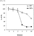
- the yellow light was irradiated over time, and then photo inducing death of RFP+ and RFP- cells was measured by MTT analysis under the presence of tin ethyl etiopurpurin.
- the specific experimental procedure is as follows.
- tin ethyl etiopurpurin (Miravant Medical Technology Inc, USA) was diluted with RPMI-1640 medium containing 10% FBS to produce 1 ⁇ M concentration of tin ethyl etiopurpurin.
- the RFP- or RFP+ 4T1 cells selected from the above 1-3 were pre-incubated in a 24-well plate at a density of 5 ⁇ 10 4 cells per well. The pre-cultured cells were then incubated with tin ethyl etiopurpurin for 4 h. After 4 hours, the cells were washed three times with PBS buffer.
- LDH analysis was then performed using the LDH Cytotoxicity Assay kit (Cayman Chemical Company, USA) according to the manufacturer's procedure. 100 ⁇ l of the supernatant of the cultured cells was transferred from the well to a corresponding well of the new plate, and then 100 ⁇ l of the reaction solution was added to each well. The plates were incubated for 30 minutes at room temperature while executing gentle shaking thereof with an orbital shaker. The absorbance was measured at 490 nm using a plate reader.
- MTT analysis was performed.
- fresh culture medium was added to the well containing the irradiated cells, and then the cells were cultured again for 24 hours.
- MTT assay was then performed using the MTT Assay kit according to the manufacturer's procedure. 150 ⁇ L of the solubilizing solution and stop solution were added thereto followed by incubation at 37°C for 4 hours. The absorbance of the reaction solution was measured at 570 nm. Cell viability was calculated by the following equation: OD treated / OD control ⁇ 100 ⁇ % .
- the cytotoxicity of RFP+ or GFP+ cells was measured by MTT analysis in the presence of Ce6 (Chlorine (e6)), rose bengal or tin ethyl etiopurpurin as photosensitizer after red light, blue light or yellow light irradiation.
- Ce6 Chlorine (e6)
- rose bengal rose bengal
- tin ethyl etiopurpurin as photosensitizer after red light, blue light or yellow light irradiation.
- the specific experimental procedure is as follows.
- Ce6 santacruz, sc-263067
- rose bengal Sigma Aldrich, 330000-5G
- tin ethyl etiopurpurin as a photosensitizer was diluted with RPMI-1640 medium containing 10% FBS.
- rose bengal or tin ethyl etiopurpurin was prepared.
- GFP+ 4T1 cells selected in the 1-4 and RFP+ 4T1 cells selected in the 1-3 were pre-cultured at a density of 2 ⁇ 10 4 cells/well in 96-well, black, and clear bottom plates.
- the pre-cultured cells were then incubated with Ce6, rose bengal or tin ethyl etiopurpurin for 4 h.
- 4T1 cells without RFP or GFP introduced thereto were used as controls.
- the cells were washed three times with PBS buffer.
- RB 363 mg/mL, 53 nM/mL
- blue light 2 min
- the mouse body weight and tumor volume were measured twice a week.
- the mice were euthanized. Tumors were removed therefrom and fixed to 10% neutral buffered formalin. The next day, the image was captured.
- Similar experiments were performed on GFP+ and GFP-H460 cells. About 2 ⁇ 10 6 /100 ⁇ L of cells were injected subcutaneously into both sides thereof.
- Another group of 5 mice as wild-type and GFP-Lgr5 mice was set to a group that was not chemically treated.
- RB was intravenously injected (50 nM/mL, 0.75 ml/kg) into the wild type and GFP-Lgr5 mice of each experimental group twice a week for 7 weeks. And, in 4 hours after the RB injection, blue light (2 min) irradiation was performed via an anal penetration.
- FLIVOTM probes were diluted with PBS containing 1% DMSO at doses according to animal weight.
- the fluorescent red probe FLIVOTM (Immunochemistry Technologies LLC, Abcys SA, Paris, France) was intravenously injected into wild type and GFP-Lgr5 mice. The mouse was euthanized after 1 hour and the colon tissue was separated therefrom. Fluorescent signals from FLIVOTM were imaged using a confocal microscope.
- Customized cylindrical diffusion fibers were used to uniformly irradiate the colon cancer cell with laser (SOMTA, Ltd.). Light diffusing through the optical fiber core was radially irradiated (360°) to the diffusion part, where the light was scattered in a multitudinous manner.
- the cylindrical diffusing fibers were bendable.
- the diameter and length of the diffuser were 600 ⁇ m and 2 cm, respectively.
- 473 nm blue light was irradiated through the optical waveguide to the colon while irradiated uniformly in the radial direction.
- the radiation power of the diffusion part in the unit area was adjusted to between 22 and 25 mW. Fiber transmission losses were negligible because short-length fibers (5 m) were used.
- cells were harvested using trypsin, suspended in 20 ml of the culture medium, and placed in a 50 ml tube. Using a pump, the cell solution passed through the tubing at a flow rate of 1 ml/min and was irradiated with light of 473 nm at an intensity of 80 mW. This process was repeated 2, 4, 8, and 10 times. The resulting cells were seeded in a 1-ml 12-well plate. The next day, double staining was performed to compare the death amounts of non-GFP and GFP cells with each other.
- cells were harvested using trypsin, suspended in 20 ml of the culture medium, and placed in a 50 ml tube. Using a pump, the cell solution passed through the tubing at a flow rate of 1 ml/min and was irradiated with light of 473 nm at an intensity of 80 mW. This process was repeated 2, 4, 8, and 10 times. The resulting cells were seeded in a 1-ml 12-well plate. The next day, double staining was performed to compare the death amounts of non-GFP and GFP cells with each other. As a result, it was confirmed that only GFP cells were specifically killed.
- PDT-treated cells were seeded onto a 12-well plate (1 mL per well) while controlling the PDT-treated cells to have a confluence of 50% or larger.
- PBS was diluted to 20 ⁇ g/ml and 2 ⁇ g/ml (2X) respectively using Hoechst 33342 (Invitrogen, H3570) and PI stock solution (1000X, Sigma, P4170). 1 ml of this solution was added to the sample (at the same volume as the culture medium) and incubated at 37°C for 20 minutes.
- the sample was taken out and scraped with a cell lifter, collected in a 1.5 ml tube, centrifuged at 1200 rpm for 3 minutes, suspended in PBS and transferred to 48 (200 ⁇ l) or 96 (100 ⁇ l) well plates. Cells were observed using an optical microscope and photographed (Merge photograph). The number of dead cells (PI-stained cells)/total number of cells ⁇ 100 per cell image was calculated. Then, the cell death percentage was measured by calculating the average value between the calculated values of three cell images.
- GFP cells GFP-H460
- RB rose bengal
- the cells were harvested using trypsin, suspended in FBS pre-culture medium at 2 ⁇ 10 6 cells/300 ⁇ l, and placed in a 1.5 ml tube. Grouping was performed as shown in the following table. [Table 1] Number of mice No cancer cell GFP - cancer cells No blue light 2 3 Blue light 2 3 Six out of ten 8 weeks aged SD rats were injected in IV (tail) manner with the cell solution.
- the bronchial sections of the 5 SD rats were incised.
- the carotid artery and venous vein were discovered and cannulated using a 279 ⁇ m ID ⁇ 609 ⁇ m OD ⁇ 152 ⁇ m wall sized polyethylene tubing (BD, 427401) wet with heparin.
- BD, 427401 polyethylene tubing
- Irradiation of 473 nm light with an intensity of 80 mW to the cannulated tubing site was performed for 5 minutes.
- the irradiated rats were weighed and incubated overnight in a constant-temperature constant-humid chamber.
- GFP-cancer cells were counted using a fluorescent microscope (Nikon, Diaphot 300), and the numbers of GFP-cancer cells according to the presence or absence of light irradiation were compared with each other.
- (2) 0.5 ⁇ 10 7 of non-GFP cells and 0.5 ⁇ 10 7 of GFP cells were mixed with each other and seeded (total 1 ⁇ 10 7 cells).
- 100 ⁇ M of rose bengal (RB) was applied thereto (in the subsequent procedure, light exposure was avoided as much as possible).
- RB rose bengal
- cells were harvested using trypsin, suspended in FBS pre-culture medium at 1 ⁇ 10 6 cells/300 ⁇ l, and placed in a 1.5-ml tube.
- Six out of ten 8 weeks aged SD rats were injected in IV (tail) manner with the cell solution.
- the bronchial sections of the 5 SD rats were incised.
- the carotid artery and venous vein were discovered and cannulated using a 279 ⁇ m ID ⁇ 609 ⁇ m OD ⁇ 152 ⁇ m wall sized polyethylene tubing (BD, 427401) wet with heparin.
- BD, 427401 polyethylene tubing
- Irradiation of 473 nm light with an intensity of 80 mW to the cannulated tubing site was performed for 5 minutes.
- the irradiated rats were weighed and incubated overnight in a constant-temperature constant-humid chamber.
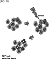
- rose bengal and tin ethyl etiopurpurin at 1 ⁇ M concentration were produced in the same method as described in the 2-4. Then, the tin ethyl etiopurpurin was applied to RFP+ 4T1 cells selected from the 1-3. The rose bengal was applied to GFP+ 4T1 cells selected from 1-4. Then, the cells were cultured for 4 hours and washed three times with PBS buffer. Next, as shown in the schematic diagram of FIG. 16 , an artificial skin tissue (AST, thickness 1 mm) (Geistlich Mucograft, Geistlich Pharma AG) was placed between the light source and the cell. Thus, the actual skin condition was established.
- AST thickness 1 mm
- 1 ⁇ 10 6 cells of GFP- and GFP+ NCI-H460 cell lines as produced by the method described in the 1-1 were cultured in a 75 cm 2 cell culture dish containing RPMI-1640 medium supplemented with 10% FBS. After 12 hours, the culture medium of the cell line was replaced with RPMI-1640 medium without FBS. Then, RBs of various concentrations (0 uM, 5 uM, 10 uM, 50 uM) were added thereto. The culturing was done for 4 hours to accumulate RB in the cells. Then, the cells were obtained by centrifugation. Pellets of each cell group were suspended in 1 ml PBS.
- the light-emission spectrum of each cell group was measured using a spectrofluorometer (JASCO Inc.). Measurements in each group were repeated 5 times.
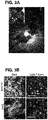
- the maximum absorbance and the shoulder absorbance spectrum of RB are 549 nm and 510 nm, respectively.
- the maximum absorbance of eGFP was 489 nm.
- the peak of the light-emission spectrum of eGFP is 508 nm.
- the shoulder absorbance range of RB covers the light-emission wavelength of eGFP irradiated with blue laser light (Laser-star co. Ltd, LSB473-80-dot) ( FIG. 1A ). Therefore, the FRET phenomenon is possible between RB and eGFP.
- the FRET technique using the living cell fluorescent light according to the present invention was named cFRET.
- RB was used to treat eGFP+ and eGFP- 4T1 cells.
- the blue laser light (473 nm) was irradiated thereto, the cells selectively died ( FIG. 1B ).
- the RB is more easily activated by light at 510 nm which is released from GFP in GFP+ cells and thus the activated RB may help to generate reactive oxygen species (ROS) from neighboring oxygen-rich environments ( FIG. 1C ).
- ROS reactive oxygen species
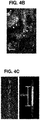
- eGFP- and eGFP+ 4T1 cells Two types of cancer cells (eGFP- and eGFP+ 4T1 cells) were prepared and mixed with each other at a ratio of 1 : 1 randomly. After the exposure of the mixture to laser light (473 nm) for 6 hours, selective cytotoxicity was induced in eGFP+ cells ( FIG. 2B ).
- cytotoxicity in eGFP+ cells was significantly higher than that in eGFP- cells after laser irradiation ( FIG. 2B ).
- the percentages of dead cells varied according to concentration of the photosensitizer and light exposure time.
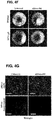
- a mixture of the eGFP+ and eGFP-sublines of H460 and B16F10 cell lines were grafted to the side of the mouse. Tumors were grown for 10 to 15 days and then blue laser light (473 nm) was repeatedly irradiated to the tumors within each mouse skin flap ( FIG. 3A ). Selective eGFP+ cell death was observed after the second round of irradiation. The GFP signal was found to be significantly reduced in the irradiated region compared to the initial tumor region.
- GFP+ and GFP-B16F10 cells were injected to the mouse individually. Tumor growth was monitored. When the mouse was irradiated with 473 nm laser light, tumor growth was slightly inhibited in the GFP+ cell without the photosensitizer treatment. When both of the photosensitizer treatment and 473 nm laser light irradiation were applied thereto, tumor growth was markedly reduced. However, this inhibition did not appear to be statistically significant by Student's t-test.
- FIG. 3D and 3E When RB-treated GFP+ cells were irradiated with laser light of 473 nm, the remarkable tumor inhibiting effect was obtained ( FIG. 3D and 3E ). In tumors of GFP+ cell-transplanted mice, significant tissue damage and cell death were further observed ( FIG. 3F ). Expression levels of cell death markers such as Bax and p53 were measured at the site of cell death. Significant cell damage was observed after cFRET PDT. Immunohistochemical studies using TNF-alpha indicate that cell death is mostly due to necrosis.
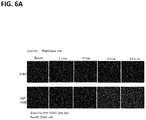
- Lgr5+ colon stem cells migrate to luminal surface after the AOM, DSS; in the experiment using Lgr5-EGFP-IRES-creERT2 knock-in mouse, migration of eGFP+-Lgr5+ colon stem cells was observed. Observation of the cleaned tissue with BABB solution showed that many eGFP+-Lgr5+ cells were located on the luminal surface of the crypt and were immersed in the polyp ( FIG. 4A and FIG. 4B ).
- Optical fibers capable of emitting radially light were produced to uniformly irradiate light to the colon epithelium of mice ( FIG. 4C ). Cylindrical diffusing fibers (diameter, 600 ⁇ m) were carefully inserted into the mouse colon via the anus; 473 nm blue laser light was used for laser emission ( FIG. 4D ).
- the peak of the excitation spectrum of RFP is 555 nm and the peak of the light-emission spectrum thereof is 584 nm.
- tin ethyl etiopurpurin has an absorbance spectrum of 640 to 660 nm.
- the RFP light-emission yield in the light absorbing region of 640 to 660 nm of tin ethyl etiopurpurin is about 50%. Therefore, in order to check that RFP and tin ethyl etiopurpurin can be used for photodynamic therapy using FRET as shown in the schematic diagram of FIG.
- RFP+ 4T1 cells were treated with tin ethyl etiopurpurin and then the wavelength of light that can allow photo-reaction of the RFP was irradiated thereto over time.
- the cell death percentage was measured using the LDH (lactate dehydrogenase) analytical method and MTT analytical method.
- RFP+ cell death As a result, for the RFP+ cell, RFP has photo reaction in the yellow laser light irradiation, and, thus, the activated tin ethyl etiopurpurin induced cell death. Further, RFP+ cell death increased considerably ( FIG. 11 and FIG. 12 ) in 2 seconds after the light irradiation, compare to the RFP-cell.
- RFP+ 4T1 cells were treated with tin ethyl etiopurpurin based on varying concentrations, and then the wavelength of light that can allow photoreaction of the RFP was irradiated thereto.
- the cell death percentage was measured using the MTT analytical method.
- cytotoxicity of RFP+ or GFP+ cells was measured by MTT assay in the presence of Ce6 (Chlorine (e6)), rose bengal or tin ethyl etiopurpurin as photosensitizer.
- Fluorescent intensities of GFP- and GFP+ cells after blue light irradiation were measured in the presence or absence of RB.
- the emission intensity at 508 nm as the emission peak of GFP decreases as the concentration of RB increases.
- the reduced energy was found to increase at 580 to 590 nm as the emission peak of RB. Therefore, it was confirmed through the results that the cells could be selectively killed by GFP and RB according to the FRET principle ( Fig. 15 ).
- the wavelength at which the RFP can photo-react is in a longer wavelength region than the wavelength at which GFP can photo-react.
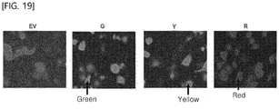
- GFP+ A549 cells expressing GFP In order to check whether other photosensitizer other than RB and tin ethyl etiopurpurin can be applied to photodynamic therapy using FRET and to determine whether fluorescent proteins other than GFP and RFP could be applied thereto, GFP+ A549 cells expressing GFP, RFP+ A549 cells expressing RFP and YFP+ A549 cells expressing YFP were prepared as shown in FIG. 19 . The cells were treated with RB, hematoporphyrin or 5-ALA and were irradiated with blue laser light (488 nm) or yellow laser light (570 nm). MTT assay was used to measure cell death levels.
- the cell death effect of the YFP+ cell is higher than that of GFP+ cell ( FIG. 20 ). This suggests that the emission peak of YFP is at about 540 nm which is closer to the absorbing wavelength peak of RB.
- the present invention relates to a composition for photodynamic therapy using a gene expressing a fluorescent protein, and a photosensitizer, and to a photodynamic therapy method using the same.
- the genes that express the fluorescent protein and the photosensitizers in accordance with the present invention may be used to treat cancer diseases.
Landscapes
- Health & Medical Sciences (AREA)
- Life Sciences & Earth Sciences (AREA)
- Chemical & Material Sciences (AREA)
- Animal Behavior & Ethology (AREA)
- Veterinary Medicine (AREA)
- Public Health (AREA)
- General Health & Medical Sciences (AREA)
- Pharmacology & Pharmacy (AREA)
- Medicinal Chemistry (AREA)
- Engineering & Computer Science (AREA)
- Epidemiology (AREA)
- Biomedical Technology (AREA)
- Molecular Biology (AREA)
- Biochemistry (AREA)
- Nuclear Medicine, Radiotherapy & Molecular Imaging (AREA)
- Radiology & Medical Imaging (AREA)
- Pathology (AREA)
- Heart & Thoracic Surgery (AREA)
- Vascular Medicine (AREA)
- General Chemical & Material Sciences (AREA)
- Organic Chemistry (AREA)
- Chemical Kinetics & Catalysis (AREA)
- Biophysics (AREA)
- Anesthesiology (AREA)
- Genetics & Genomics (AREA)
- Hematology (AREA)
- Cardiology (AREA)
- Biotechnology (AREA)
- Optics & Photonics (AREA)
- Physics & Mathematics (AREA)
- Medicines That Contain Protein Lipid Enzymes And Other Medicines (AREA)
- Pharmaceuticals Containing Other Organic And Inorganic Compounds (AREA)
- Micro-Organisms Or Cultivation Processes Thereof (AREA)
- Medicinal Preparation (AREA)
- Peptides Or Proteins (AREA)
- Medicines Containing Antibodies Or Antigens For Use As Internal Diagnostic Agents (AREA)
- Acyclic And Carbocyclic Compounds In Medicinal Compositions (AREA)
- Investigating Or Analysing Biological Materials (AREA)
- Immobilizing And Processing Of Enzymes And Microorganisms (AREA)
Applications Claiming Priority (2)
| Application Number | Priority Date | Filing Date | Title |
|---|---|---|---|
| KR1020170007301A KR101911725B1 (ko) | 2017-01-16 | 2017-01-16 | 적색형광단백질을 발현하는 유전자 및 틴 에틸 에티오푸르푸린을 이용한 광역학 치료용 조성물 및 이를 이용한 광역학 치료방법 |
| PCT/KR2018/000759 WO2018131989A1 (ko) | 2017-01-16 | 2018-01-16 | 형광단백질을 발현하는 유전자 및 광감작제를 이용한 광역학 치료용 조성물 및 이를 이용한 광역학 치료방법 |
Publications (2)
| Publication Number | Publication Date |
|---|---|
| EP3572094A1 true EP3572094A1 (de) | 2019-11-27 |
| EP3572094A4 EP3572094A4 (de) | 2020-10-21 |
Family
ID=62839928
Family Applications (1)
| Application Number | Title | Priority Date | Filing Date |
|---|---|---|---|
| EP18739276.6A Withdrawn EP3572094A4 (de) | 2017-01-16 | 2018-01-16 | Zusammensetzung zur photodynamischen therapie unter verwendung eines fluoreszenzprotein exprimierenden gens und eines photosensibilisators sowie photodynamisches therapieverfahren damit |
Country Status (6)
| Country | Link |
|---|---|
| US (1) | US20190365927A1 (de) |
| EP (1) | EP3572094A4 (de) |
| JP (1) | JP6940185B2 (de) |
| KR (1) | KR101911725B1 (de) |
| CN (1) | CN110198741B (de) |
| WO (1) | WO2018131989A1 (de) |
Families Citing this family (3)
| Publication number | Priority date | Publication date | Assignee | Title |
|---|---|---|---|---|
| WO2018181276A1 (ja) * | 2017-03-28 | 2018-10-04 | 学校法人慶應義塾 | ヒト組織幹細胞及びその使用 |
| CN111855541A (zh) * | 2019-04-26 | 2020-10-30 | 朱德新 | 循环肿瘤细胞检测试剂、试剂盒和检测方法 |
| WO2021192113A1 (ja) * | 2020-03-25 | 2021-09-30 | 大塚メディカルデバイス株式会社 | がんの処置方法およびそのためのシステム |
Family Cites Families (12)
| Publication number | Priority date | Publication date | Assignee | Title |
|---|---|---|---|---|
| EP1346059B1 (de) * | 2000-11-29 | 2012-05-23 | PCI Biotech AS | Photochemische internalisierung zur zuführung von molekülen in das zytosol |
| GB0520436D0 (en) * | 2005-10-07 | 2005-11-16 | Photobiotics Ltd | Biological materials and uses thereof |
| KR101035269B1 (ko) | 2007-04-23 | 2011-05-26 | 한국과학기술연구원 | 고분자 유도체-광감작제 복합체를 이용한 새로운 광역학치료제 |
| KR20100065294A (ko) * | 2007-08-03 | 2010-06-16 | 국립대학법인 홋가이도 다이가쿠 | 군청색 형광 단백질 |
| US8361775B2 (en) * | 2008-05-14 | 2013-01-29 | Novadaq Technologies Inc. | Imaging methods and compositions comprising fluorescent dyes associated with viral components for nerve imaging |
| WO2010151074A2 (ko) | 2009-06-26 | 2010-12-29 | 주식회사 진코스 | 양자점-클로린 유도체의 접합체를 함유하는 광감작제 및 이를 포함하는 광역학 치료에 사용하기 위한 암 치료 및 진단용 조성물 |
| WO2011090333A2 (ko) | 2010-01-20 | 2011-07-28 | 주식회사 진코스 | 클로린 유도체와 불포화 지방산의 접합체, 이를 함유하는 광감작제, 및 이를 포함하는 광역학 치료에 사용하기 위한 암 치료용 조성물 |
| AU2012305327B2 (en) * | 2011-09-05 | 2016-07-14 | Hiroshi Maeda | Polymer-type fluorescent molecule probe |
| KR101419124B1 (ko) | 2011-09-16 | 2014-07-11 | 가톨릭대학교 산학협력단 | 폴리에틸렌옥사이드-폴리프로필렌옥사이드 블록공중합체와 광감작제가 공유 결합된 광역학 치료용 복합체 |
| KR101323848B1 (ko) * | 2012-05-18 | 2013-11-01 | 광주과학기술원 | 소수성 형광입자 및 광감작제-운반 폴리머좀 복합체 |
| WO2014055960A1 (en) * | 2012-10-05 | 2014-04-10 | Genelux Corporation | Energy absorbing-based diagnostic and therapeutic methods employing nucleic acid molecules encoding chromophore-producing enzymes |
| KR101668561B1 (ko) * | 2015-07-13 | 2016-10-21 | 원광대학교산학협력단 | 녹색형광단백질을 발현하는 유전자 및 로즈 벵갈을 이용한 암 질환의 광역학 치료용 조성물 및 이를 이용한 광역학 치료방법 |
-
2017
- 2017-01-16 KR KR1020170007301A patent/KR101911725B1/ko active Active
-
2018
- 2018-01-16 JP JP2019559250A patent/JP6940185B2/ja not_active Expired - Fee Related
- 2018-01-16 CN CN201880007163.9A patent/CN110198741B/zh not_active Expired - Fee Related
- 2018-01-16 EP EP18739276.6A patent/EP3572094A4/de not_active Withdrawn
- 2018-01-16 US US16/478,175 patent/US20190365927A1/en not_active Abandoned
- 2018-01-16 WO PCT/KR2018/000759 patent/WO2018131989A1/ko not_active Ceased
Also Published As
| Publication number | Publication date |
|---|---|
| JP2020513212A (ja) | 2020-05-07 |
| WO2018131989A1 (ko) | 2018-07-19 |
| JP6940185B2 (ja) | 2021-09-22 |
| KR20180084335A (ko) | 2018-07-25 |
| EP3572094A4 (de) | 2020-10-21 |
| CN110198741A (zh) | 2019-09-03 |
| CN110198741B (zh) | 2022-02-08 |
| KR101911725B1 (ko) | 2018-12-28 |
| US20190365927A1 (en) | 2019-12-05 |
Similar Documents
| Publication | Publication Date | Title |
|---|---|---|
| Wang et al. | Anti-metastatic and pro-apoptotic effects elicited by combination photodynamic therapy with sonodynamic therapy on breast cancer both in vitro and in vivo | |
| Ao et al. | An upconversion nanoparticle enables near infrared-optogenetic manipulation of the caenorhabditis elegans motor circuit | |
| Mitsunaga et al. | Immediate in vivo target-specific cancer cell death after near infrared photoimmunotherapy | |
| Chen et al. | TLD1433 photosensitizer inhibits conjunctival melanoma cells in zebrafish ectopic and orthotopic tumour models | |
| Hodgkinson et al. | Cervical cancer cells (HeLa) response to photodynamic therapy using a zinc phthalocyanine photosensitizer | |
| Nagaya et al. | Endoscopic near infrared photoimmunotherapy using a fiber optic diffuser for peritoneal dissemination of gastric cancer | |
| Ribera et al. | Treatment of hepatic fibrosis in mice based on targeted plasmonic hyperthermia | |
| Bhanja et al. | Photodynamic therapy for glioblastoma: illuminating the path toward clinical applicability | |
| Zhu et al. | An NIR triphenylamine grafted BODIPY derivative with high photothermal conversion efficiency and singlet oxygen generation for imaging guided phototherapy | |
| TW201811394A (zh) | 用於修飾介導生物活性或與生物活性有關之靶結構之醫藥組合物及套組 | |
| CN113456613A (zh) | 一种近红外光激活型巨噬细胞-纳米前药靶向递药系统的构建及其应用 | |
| KR20090094331A (ko) | 광선 역학적 치료(pdt)를 이용한 이상 전기 전도 차단 장치 | |
| KR101668561B1 (ko) | 녹색형광단백질을 발현하는 유전자 및 로즈 벵갈을 이용한 암 질환의 광역학 치료용 조성물 및 이를 이용한 광역학 치료방법 | |
| EP3572094A1 (de) | Zusammensetzung zur photodynamischen therapie unter verwendung eines fluoreszenzprotein exprimierenden gens und eines photosensibilisators sowie photodynamisches therapieverfahren damit | |
| Yan et al. | Photodynamic treatment of tumor with bacteria expressing killerred | |
| Czarnecka-Czapczyńska et al. | Photodynamic therapy of breast cancer in animal models and their potential use in clinical trials-role of the photosensitizers: A review | |
| Chan et al. | Near-infrared-activated fluorescence resonance energy transfer-based nanocomposite to sense mmp2-overexpressing oral cancer cells | |
| Itoh et al. | A new drug-free cancer therapy using ultraviolet pulsed irradiation. PDT (photodynamic therapy) to PPT (pulsed photon therapy) | |
| CN101518528B (zh) | 一组碳花青染料类的近红外荧光化合物的用途 | |
| Zipf et al. | Direct stimulation of gastric smooth muscle cells via Gq proteins with light | |
| Park et al. | Bifunctional tumor-targeted bioprobe for phototheranosis | |
| KR102182630B1 (ko) | 친환경 스마트 광감작제 및 이를 포함하는 광줄기세포 치료제 | |
| Zhou et al. | Enhanced ferroptosis in triple-negative cancer cells achieved by a type-I cell membrane-targeted tetrabenzophenazine photosensitizer | |
| Li et al. | Oral delivery of aptamer-decorated SICTERS Raman probes for colonoscopy-guided resection and photothermal immunization of microtumors | |
| Sun et al. | Benzochloroporphyrin derivative photosensitizer-mediated photodynamic therapy for Ewing sarcoma |
Legal Events
| Date | Code | Title | Description |
|---|---|---|---|
| STAA | Information on the status of an ep patent application or granted ep patent |
Free format text: STATUS: THE INTERNATIONAL PUBLICATION HAS BEEN MADE |
|
| PUAI | Public reference made under article 153(3) epc to a published international application that has entered the european phase |
Free format text: ORIGINAL CODE: 0009012 |
|
| STAA | Information on the status of an ep patent application or granted ep patent |
Free format text: STATUS: REQUEST FOR EXAMINATION WAS MADE |
|
| 17P | Request for examination filed |
Effective date: 20190730 |
|
| AK | Designated contracting states |
Kind code of ref document: A1 Designated state(s): AL AT BE BG CH CY CZ DE DK EE ES FI FR GB GR HR HU IE IS IT LI LT LU LV MC MK MT NL NO PL PT RO RS SE SI SK SM TR |
|
| AX | Request for extension of the european patent |
Extension state: BA ME |
|
| DAV | Request for validation of the european patent (deleted) | ||
| DAX | Request for extension of the european patent (deleted) | ||
| A4 | Supplementary search report drawn up and despatched |
Effective date: 20200918 |
|
| RIC1 | Information provided on ipc code assigned before grant |
Ipc: A61K 41/00 20200101AFI20200914BHEP Ipc: A61P 35/00 20060101ALI20200914BHEP |
|
| STAA | Information on the status of an ep patent application or granted ep patent |
Free format text: STATUS: EXAMINATION IS IN PROGRESS |
|
| 17Q | First examination report despatched |
Effective date: 20230808 |
|
| STAA | Information on the status of an ep patent application or granted ep patent |
Free format text: STATUS: THE APPLICATION IS DEEMED TO BE WITHDRAWN |
|
| 18D | Application deemed to be withdrawn |
Effective date: 20231219 |