EP3545104B1 - Codage à barres en tandem de molécules cibles pour leur quantification absolue à une résolution d'entité unique - Google Patents
Codage à barres en tandem de molécules cibles pour leur quantification absolue à une résolution d'entité unique Download PDFInfo
- Publication number
- EP3545104B1 EP3545104B1 EP17837944.2A EP17837944A EP3545104B1 EP 3545104 B1 EP3545104 B1 EP 3545104B1 EP 17837944 A EP17837944 A EP 17837944A EP 3545104 B1 EP3545104 B1 EP 3545104B1
- Authority
- EP
- European Patent Office
- Prior art keywords
- uei
- droplets
- dna
- droplet
- molecular
- Prior art date
- Legal status (The legal status is an assumption and is not a legal conclusion. Google has not performed a legal analysis and makes no representation as to the accuracy of the status listed.)
- Active
Links
Images


Classifications
-
- C—CHEMISTRY; METALLURGY
- C12—BIOCHEMISTRY; BEER; SPIRITS; WINE; VINEGAR; MICROBIOLOGY; ENZYMOLOGY; MUTATION OR GENETIC ENGINEERING
- C12Q—MEASURING OR TESTING PROCESSES INVOLVING ENZYMES, NUCLEIC ACIDS OR MICROORGANISMS; COMPOSITIONS OR TEST PAPERS THEREFOR; PROCESSES OF PREPARING SUCH COMPOSITIONS; CONDITION-RESPONSIVE CONTROL IN MICROBIOLOGICAL OR ENZYMOLOGICAL PROCESSES
- C12Q1/00—Measuring or testing processes involving enzymes, nucleic acids or microorganisms; Compositions therefor; Processes of preparing such compositions
- C12Q1/68—Measuring or testing processes involving enzymes, nucleic acids or microorganisms; Compositions therefor; Processes of preparing such compositions involving nucleic acids
- C12Q1/6806—Preparing nucleic acids for analysis, e.g. for polymerase chain reaction [PCR] assay
-
- B—PERFORMING OPERATIONS; TRANSPORTING
- B01—PHYSICAL OR CHEMICAL PROCESSES OR APPARATUS IN GENERAL
- B01L—CHEMICAL OR PHYSICAL LABORATORY APPARATUS FOR GENERAL USE
- B01L3/00—Containers or dishes for laboratory use, e.g. laboratory glassware; Droppers
- B01L3/50—Containers for the purpose of retaining a material to be analysed, e.g. test tubes
- B01L3/502—Containers for the purpose of retaining a material to be analysed, e.g. test tubes with fluid transport, e.g. in multi-compartment structures
- B01L3/5027—Containers for the purpose of retaining a material to be analysed, e.g. test tubes with fluid transport, e.g. in multi-compartment structures by integrated microfluidic structures, i.e. dimensions of channels and chambers are such that surface tension forces are important, e.g. lab-on-a-chip
- B01L3/50273—Containers for the purpose of retaining a material to be analysed, e.g. test tubes with fluid transport, e.g. in multi-compartment structures by integrated microfluidic structures, i.e. dimensions of channels and chambers are such that surface tension forces are important, e.g. lab-on-a-chip characterised by the means or forces applied to move the fluids
-
- C—CHEMISTRY; METALLURGY
- C12—BIOCHEMISTRY; BEER; SPIRITS; WINE; VINEGAR; MICROBIOLOGY; ENZYMOLOGY; MUTATION OR GENETIC ENGINEERING
- C12Q—MEASURING OR TESTING PROCESSES INVOLVING ENZYMES, NUCLEIC ACIDS OR MICROORGANISMS; COMPOSITIONS OR TEST PAPERS THEREFOR; PROCESSES OF PREPARING SUCH COMPOSITIONS; CONDITION-RESPONSIVE CONTROL IN MICROBIOLOGICAL OR ENZYMOLOGICAL PROCESSES
- C12Q2563/00—Nucleic acid detection characterized by the use of physical, structural and functional properties
- C12Q2563/159—Microreactors, e.g. emulsion PCR or sequencing, droplet PCR, microcapsules, i.e. non-liquid containers with a range of different permeability's for different reaction components
Definitions
- the present invention relates to methods and systems for labelling nucleic acids and other biological molecules from entities, e.g. cells, within emulsion droplets in high throughput regimes while preserving the integrity of the single-entity information.
- Single cell analytics are gaining popularity due to the insight that taking into account the heterogeneity of a population of cell may be of capital interest to understand the function and behaviour of diverse biological systems.
- RNA levels are considered a useful marker of phenotypic heterogeneity and, as a consequence, considerable efforts were done to analyse RNA content in single cells.
- Probe-dependent methods including fluorescence in situ hybridization (FISH) or reporter fusions to fluorescent proteins, was replaced with the probe-independent RNA-seq technique in which cellular RNA molecules are converted into cDNA and subsequently sequenced in parallel using next-generation sequencing technology.
- FISH fluorescence in situ hybridization
- RNA-seq Single-cell RNA-seq requires the isolation of individual cells, the conversion of cellular RNA into cDNA and the massively parallel sequencing of cDNA libraries.
- microfluidic droplets also provide a compartment in which cells can be isolated.
- droplets of one phase are generated in another, immiscible phase by exploiting capillary instabilities in a microfluidic two-phase flow.
- a surfactant to either or both of the phases stabilizes the droplets against coalescence and allows them to function as discrete microreactors.
- RNA-seq analysis may require the profiling of several thousands if not millions of representative individual cells
- barcoding strategies have been developed to reduce sequencing costs and increase throughput.
- unique cellular identifiers it has made possible to pool up a multitude of cells for simultaneous sequencing since each read could subsequently be assigned to its original cell through the unique cellular barcode (Islam et al., 2012).
- the main technical challenge when combining barcoding strategy and compartmentalization of cells into droplets is to ensure that each droplet carries a different barcode and thus that the integrity of the single cell information is preserved.
- Each cell may be coencapsulated with a distinctly barcoded particle, such as bead (Macosko et al. 2015) or hydrogel microsphere (Klein et al., 2015), in a nano-liter scale droplet.
- a distinctly barcoded particle such as bead (Macosko et al. 2015) or hydrogel microsphere (Klein et al., 2015)
- Each of these particles contains more than 10 8 individual primers that share the same "cell barcode”.
- the number of droplets created greatly exceeds the number of particles or cells injected, so that a droplet will generally contain zero or one cell and zero or one particle (Macosko et al. 2015; Klein et al., 2015).
- a barcode-library emulsion may be produced using a microfluidic device consisting of 96 drop-makers creating millions of drops containing a high concentration of a single one of the 96 barcodes (Rotem et al. 2015). Each cell-bearing drop is then paired and fused with one barcode- drop. However, to ensure that each cell-bearing drop is fused with at most one barcode drop, only half of the cell-bearing drops actually fuse with a barcode drop. Furthermore, cases where two cell-bearing drops fuse with a single barcode drop or where two barcode drops fuse with a single cell-bearing drop, introduce errors in the resultant labelling and are a potential source of noise (Rotem et al. 2015).
- RNA levels have been recognized as useful marker for phenotypic heterogeneity
- current methods provide limited information since levels of protein or other biological molecules cannot be assessed with the same system.
- quantification of the protein expression at the single cell level which is critical for complete characterization of the phenotypic states, is generally based on fluorescence imaging methods.
- the present invention provides a new method of labelling any target molecules from a plurality of entities in high throughput regimes while preserving the integrity of the single-entity information.
- the present invention relates to a method of labelling a plurality of molecular targets from a plurality of entities while preserving the integrity of the single-entity information, said method comprising providing a first set of emulsion droplets comprising droplets containing labelled molecular targets, wherein each of these droplets contains a plurality of molecular targets originating from no more than one entity and wherein, in each of these droplets, each molecular target is labelled with a molecular identification DNA sequence comprising (i) a unique molecular identification (UMI) barcode which is different for each molecular target and (ii) an overhang or an overhang producing restriction site; providing a second set of emulsion droplets comprising droplets containing entity identification sequences, wherein each of these droplets contains at least one entity identification sequence which is a DNA sequence, preferably a double stranded DNA sequence, comprising a unique entity identification (UEI) barcode which is different for each droplet of the second set
- UMI unique molecular
- the method may further comprise encapsulating a plurality of entities within emulsion droplets, each droplet containing no more than one entity, and optionally lysing said entities within the droplets to release molecular targets; labelling said molecular targets with probes, each probe comprising a capture moiety capable of specific binding or ligation to a molecular target or to an adaptor linked to said molecular target, and a DNA moiety comprising (i) a region proximal to the capture moiety and comprising the unique molecular identification (UMI) sequence and (ii) a region distal from the capture moiety and comprising an overhang or an overhang producing restriction site, thereby obtaining the first set of emulsion droplets.
- UMI unique molecular identification
- the method may further comprise
- UEI calibrator barcodes and UEI barcodes and labelled molecular targets are assembled through restriction enzyme digestion and ligation of compatible overhangs of (i) UEI calibrators and entity identification sequences and of (ii) entity identification sequences and labelled molecular targets.
- UEI calibrator barcodes, UEI barcodes and labelled molecular targets may be assembled through restriction enzyme digestion and ligation of compatible overhangs of (i) UEI calibrators and labelled molecular targets and (ii) UEI calibrators and entity identification sequences, or of i) UEI calibrators and entity identification sequences and (ii) entity identification sequences and labelled molecular targets.
- entity identification sequences and UEI-calibrators may be assembled through their compatible overhangs before amplification and, after fusion of droplets of the first and second sets, the amplified fragment comprising UEI calibrator and UEI barcodes is ligated to labelled molecular targets through compatible overhangs of i) UEI calibrators and labelled molecular targets or (ii) entity identification sequences and labelled molecular targets.
- At least some of molecular targets are nucleic acids and at least some probes comprise a capture moiety which is a single stranded DNA region which drives the specific recognition of a nucleic acid molecular target through conventional Watson-Crick base-pairing interactions and a DNA moiety comprising a 3' single stranded region comprising the unique molecular identification (UMI) sequence and a 5'double-stranded region comprising the overhang or overhang producing restriction site.
- UMI unique molecular identification
- said nucleic acid molecular targets are labelled using said probes as priming sites for a DNA polymerase synthetizing complementary strands of molecular targets.
- RNA molecules are RNA molecules and the DNA polymerase is a reverse transcriptase.
- At least some probes comprise a capture moiety which is
- the binding moiety or the first domain of the chimeric protein is selected from the group consisting of an antibody, a ligand of a ligand/anti-ligand couple, a peptide aptamer, a nucleic acid aptamer, a protein tag, or a chemical probe (e.g.
- suicide substrate reacting specifically with a molecular target or a class of molecular targets, preferably is an antibody
- the first domain of the protein bridge is an immunoglobulin-binding bacterial protein, preferably is domains A to E of protein A
- the second domain of the protein bridge or the chimeric protein is selected from the group consisting of SNAP-tag, CLIP-tag or Halo-Tag, preferably is a SNAP-tag.
- At least some probes comprise a capture moiety comprising an antibody moiety specific to a molecular target and a protein bridge, said protein bridge comprising a first domain that binds to a Fc region of the antibody moiety and a second domain that binds to the DNA moiety, preferably a SNAP-tag.
- the present invention also relates to a method of quantifying one or several molecular targets from a plurality of entities with single-entity resolution, said method comprising labelling said molecular targets according to the method of the invention; capturing said labelled molecular targets, amplifying sequences comprising UMI and UEI barcodes, and optionally UEI-calibrator barcodes sequencing amplified sequences.
- the sequencing of UMI and UEI barcodes, and optionally UEI-calibrator barcodes, allows to unambiguously assign each molecular target to a droplet/entity and thus to quantify the content of molecular targets in each droplet/entity.
- the entity is a cell, or a particle or an oil-in-water emulsion droplet exposing molecular targets on its outer surface. More preferably, the entity is a cell.
- the method may further comprise
- the present invention further relates to the use of a kit to label a plurality of molecular targets from a plurality of entities according to the method of the invention or to quantify one or several molecular targets from a plurality of entities with single-entity resolution according to the method of the invention, wherein the kit comprises a microfluidic device comprising
- the invention relates to the use of a kit to label a plurality of molecular targets from a plurality of entities according to the method of the invention or to quantify one or several molecular targets from a plurality of entities with single-entity resolution according to the method of the invention, wherein the kit comprises
- the inventors conceived a new method of labelling any target molecules from a plurality of entities in high throughput regimes, i.e. allowing the analysis of several thousands of entities per run, while preserving the integrity of the single-entity information.
- This method is based on a tandem molecular barcoding in which all molecular targets (nucleic acids, proteins,...) are labelled (i) with a first unique barcode (unique molecular identification barcode or UMI barcode) which is different for each molecular target from an entity, and (ii) with a tag sequence coding the entity from which the molecular target originates, i.e. unique entity identification barcode or UEI barcode which is different for each entity but identical for all molecular targets originating from the same entity.
- UMI barcode unique molecular identification barcode
- UEI barcode unique entity identification barcode
- the present invention relates to a method of labelling a plurality of molecular targets from a plurality of entities, said method comprising providing a first set of emulsion droplets comprising droplets containing labelled molecular targets, wherein each of these droplets contains a plurality of molecular targets originating from no more than one entity and wherein, in each of these droplets, each molecular target is labelled with a molecular identification DNA sequence comprising (i) a unique molecular identification (UMI) barcode which is different for each molecular target and (ii) an overhang or an overhang producing restriction site; providing a second set of emulsion droplets comprising droplets containing entity identification sequences, wherein each of these droplets contains at least one entity identification sequence which is a DNA sequence, preferably a double-stranded DNA sequence, comprising a unique entity identification (UEI) barcode which is different for each droplet of the second set, and an overhang producing restriction site; fusing droplets of
- the method of the invention may be used to label molecular targets from any type of entities.
- entity refers to any entity comprising or exposing on its surface, molecular targets as defined below.
- this term refers to a cell, or refers to a particle or an emulsion droplet, preferably an oil-in-water emulsion droplet, exposing molecular targets on its outer surface.
- the term "cell” refers to a prokaryotic cell or a eukaryotic cell such as animal, plant, fungal or algae cell.
- the population of cells to be processed may be homogenous, i.e. comprising only one cellular type, or may be heterogeneous, i.e. comprising several cellular types.
- the population of cells is obtained from a tissue sample, preferably an animal tissue sample, more preferably from a pathological sample such as a tumor sample.
- the population of cells is a population of bacterial, fungal or algae cells, preferably of bacterial or fungal cells. This population may comprise bacteria, fungi or algae of the same species or bacteria, fungi or algae of different species.
- particle and “bead” are used herein interchangeably and refer to any solid support, preferably a spherical solid support, of 50 nm to 10 ⁇ m in size which is suitable to expose one or several molecular targets on its outer surface.
- these terms may refer to polymer beads (e.g. polyacrylamide, agarose, polystyrene), latex beads, magnetic beads or hydrogel beads.
- Methods for covalent or non-covalent binding of molecular targets such as nucleic acids or proteins, to beads are well known by the skilled person and various techniques are commercially available. In particular, this binding may be carried out through reactive groups on the surface of the particle.
- nucleic acids may be attached to the surface by carbodiimide-mediated end-attachment of 5'-phosphate and 5'-NH2 modified nucleic acids to respectively amino and carboxyl beads.
- Proteins may also be covalently or non-covalently attached to beads via any suitable method such as using sulphate, amidine, carboxyl, carboxyl/sulphate or chloromethyl modified beads.
- entity may also refer to an emulsion droplet, preferably an oil-in-water emulsion droplet, exposing molecular targets on its outer surface.
- Molecular targets may be covalently or non-covalently attached to the droplet through reactive groups exposed on the surface of the droplets such as nitrilotriacetate which can specifically interact with his-tagged proteins, or through any other functional moiety which is able to covalently or non-covalently interact with a molecular target of interest.
- the skilled person may use any known method to produce such emulsion droplets exposing molecular targets on its outer surface, in particular methods described in international patent application WO 2017/174610 .
- the method of the invention allows labelling molecular targets from a high number of entities in a single run.
- the term "plurality of entities” refers to at least 1,000 entities, preferably at least 5,000 entities, more preferably at least 10,000 entities, and even more preferably at least 50,000 entities.
- target molecule refers to any kind of molecules, and in particular any kind of molecules which may be possibly present in a cell.
- the molecular target can be a biomolecule, i.e. a molecule that is naturally present in living organisms, or a chemical compound that is not naturally found in living organism such as pharmaceutical drugs, toxicants, heavy metals, pollutants, etc...
- the molecular target is a biomolecule.
- biomolecules include, but are not limited to, nucleic acids, e.g.
- DNA or RNA molecules proteins such as antibodies, enzymes or growth factors, lipids such as fatty acids, glycolipids, sterols or glycerolipids, vitamins, hormones, neurotransmitters, and carbohydrates, e.g., mono-, oligo- and polysaccharides.
- polypeptide polypeptide
- peptide protein
- protein protein
- the terms “polypeptide”, “peptide” and “protein” are used interchangeably to refer to a polymer of amino acid residues, and are not limited to a minimum length.
- the protein may comprise any post-translational modification such as phosphorylation, acetylation, amidation, methylation, glycosylation or lipidation.
- nucleic acid or “polynucleotide” refers to a polymeric form of nucleotides of any length, either ribonucleotides or deoxyribonucleotides.
- One of the main advantage of the method of the invention is the possibility to label, in a single run, different types of molecular targets such as proteins and nucleic acids.
- the term "plurality of molecular targets” may refer to different copies of the same molecule, e.g. different copies of the same mRNA or of the same protein, or may refer to different copies of different molecules, e.g. different copies of a mRNA and different copies of a protein.
- molecular targets are different copies of the same molecule.
- the molecule is biomolecule, more preferably a nucleic acid or a protein, even more preferably a RNA molecule or a protein.
- molecular targets are different copies of different molecules.
- said molecules are biomolecules, more preferably are nucleic acids and/or proteins, even more preferably RNA molecules and/or proteins.
- molecular targets are different copies of at least two different nucleic acids, preferably RNA.
- molecular targets are different copies of at least two different proteins.
- molecular targets are different copies of one or several nucleic acid, preferably RNA, and different copies of one or several proteins.
- the method of the invention is implemented using one or several microfluidic systems, i.e. at least one step of the method is implemented using a microfluidic system.
- the method is implemented using several microfluidic systems, for example a microfluidic system to generate the first set of emulsion droplets, a microfluidic system to generate the second set of emulsion droplets and a microfluidic system to fuse the two sets, to incorporate UEI barcodes and optionally to conduct some subsequent steps.
- the method is implemented using a microfluidic system wherein the first set of emulsion droplets and/or the second set of emulsion droplets are generated and wherein droplets of the two sets are fused.
- emulsion droplet As used herein, the terms “emulsion droplet”, “droplet” and “microfluidic droplet” are used interchangeably and may refer to a water-in-oil emulsion droplet (also named w/o droplet) , i.e. an isolated portion of an aqueous phase that is completely surrounded by an oil phase, an oil-in-water emulsion droplet (also named o/w droplet), i.e. an isolated portion of an oil phase that is completely surrounded by an aqueous phase, a water-in-oil-in-water emulsion droplet (also named w/o/w droplet) consisting of an aqueous droplet inside an oil droplet, i.e.
- w/o droplet water-in-oil emulsion droplet
- an aqueous core and an oil shell, surrounded by an aqueous carrier fluid or an oil-in-water-in-oil emulsion droplet (also named o/w/o droplet) consisting of an oil droplet inside an aqueous droplet, i.e. an oil core and an aqueous shell, surrounded by an oil carrier fluid.
- this term refers to a w/o emulsion droplet.
- a droplet may be spherical or of other shapes depending on the external environment.
- the droplet has a volume of less than 100 nL, preferably of less than 10 nL, and more preferably of less than 1 nL.
- a droplet may have a volume ranging from 2 pL to 1 nL, preferably from 2 to 500 pL, more preferably from 2 to 100 pL.
- the droplets have a homogenous distribution of diameters, i.e., the droplets may have a distribution of diameters such that no more than about 10%, about 5%, about 3%, about 1%, about 0.03%, or about 0.01% of the droplets have an average diameter greater than about 10%, about 5%, about 3%, about 1%, about 0.03%, or about 0.01% of the average diameter of the droplets.
- the emulsion is a monodispersed emulsion, i.e. an emulsion comprising droplets of the same volume. Techniques for producing such a homogenous distribution of diameters are well-known by the skilled person (see for example WO 2004/091763 ).
- the aqueous phase is typically water or an aqueous buffer solution, such as but not limited to Tris-HCl buffer, Tris-acetate buffer, phosphate buffer saline (PBS) or acetate buffer.
- the aqueous phase is an aqueous buffer solution.
- the aqueous phase may comprise bovine serum albumin or additive such as Pluronic.
- the aqueous phase is chosen in order to be compatible with enzymatic reactions performed during the process of the invention, such as enzymatic digestion, amplification, ligation, etc...
- An example of such aqueous phase includes, but is not limited to, CutSmart restriction enzyme buffer (New England Biolabs).
- the oil phase used to generate the emulsion droplets may be selected from the group consisting of fluorinated oil such as FC40 oil (3M®), FC43 (3M®), FC77 oil (3M®), FC72 (3M®), FC84 (3M®), FC70 (3M®), Novec-7500 (3M®), Novec-7100 (3M®), perfluorohexane, perfluorooctane, perfluorodecane, Galden-HT135 oil (Solvay Solexis), Galden-HT170 oil (Solvay Solexis), Galden-HT110 oil (Solvay Solexis), Galden-HT90 oil (Solvay Solexis), Galden-HT70 oil (Solvay Solexis), Galden PFPE liquids, Galden® SV Fluids or H-Galden® ZV Fluids; and hydrocarbon oils such as Mineral oils, Light mineral oil, Adepsine oil, Albolene, Cable oil, Baby Oil, Drakeol, Electrical Insulating Oil
- the oil phase is fluorinated oil such as Novec-7500, FC40 oil, Galden-HT135 oil or FC77 oil, more preferably is Novec-7500.
- fluorinated oil such as Novec-7500, FC40 oil, Galden-HT135 oil or FC77 oil, more preferably is Novec-7500.
- suitable phase oil may easily select suitable phase oil to implement the methods of the invention.
- the emulsion droplets comprise one or several surfactants.
- Said surfactant(s) can aid in controlling or optimizing droplet size, flow and uniformity and stabilizing aqueous emulsions.
- Suitable surfactants for preparing the emulsion droplets used in the present invention are typically non-ionic and contain at least one hydrophilic head and one or several lipophilic tails, preferably one (diblock surfactant) or two (triblock surfactant) lipophilic tails.
- Said hydrophilic head(s) and the tail(s) may be directly linked, or linked via a spacer moiety.
- surfactants include, but are not limited to, sorbitan-based carboxylic acid esters such as sorbitan monolaurate (Span 20), sorbitan monopalmitate (Span 40), sorbitan monostearate (Span 60) and sorbitan monooleate (Span 80); block copolymers of polyethylene glycol and polypropylene glycol such as the triblock copolymer EA-surfactant (RainDance Technologies), DMP (dimorpholino phosphate)-surfactant (Baret, Kleinschmidt, et al., 2009) and Jeffamine-surfactant; polymeric silicon-based surfactants such as Abil EM 90; triton X-100; and fluorinated surfactants such as PFPE-PEG and perfluorinated polyethers (e.g., Krytox-PEG, DuPont Krytox 157 FSL, FSM, and/or ESH).
- preferred surfactants are examples of PFPE-P
- the emulsion droplets comprise one or several functionalized surfactants at their interface.
- a “functionalized surfactant” refers to a surfactant which bears at least one functional moiety either on one of its hydrophilic head(s) or lipophilic tail(s), preferably on a hydrophilic head.
- a “functional moiety” is virtually any chemical or biological entity which provides the surfactant with a function of interest.
- the functional moiety can enable to create a covalent or non covalent interaction between the surfactant and a molecular target of interest. Thanks to the use of such functionalized surfactants, molecular targets may be exposed on the surface of emulsion droplets.
- the interface of these droplets may comprise only functionalized surfactant(s) or a mix of functionalized and non-functionalized surfactants.
- the ratio between functionalized and non-functionalized surfactants may vary and can be easily adapted by the skilled person.
- functionalized surfactant may represent from 1 to 100% (w/w) of total surfactants, preferably from 2 to 80% (w/w), and more preferably from 5 to 50% (w/w).
- the total amount of surfactant in the carrier oil is preferably chosen in order to ensure stability of the emulsion and prevent spontaneous coalescence of droplets.
- the carrier oil comprises from 0.5 to 10% (w/w), preferably from 1 to 8% (w/w), and more preferably from 2 to 5% (w/w) of surfactant.
- the emulsion can be prepared by any method known by the skilled artisan.
- the emulsion can be prepared on a microfluidic system.
- the first set of emulsion droplets comprises droplets containing labelled molecular targets, wherein each of these droplets contains a plurality of molecular targets originating from no more than one entity and wherein, in each of these droplets, molecular targets are labelled with a molecular identification DNA sequence.
- the first set of emulsion droplets may be a w/o or o/w/o emulsion depending on the nature of the entities.
- the entities are cells or particles and emulsion droplets of the first set are w/o emulsion droplets.
- the entities are o/w emulsion droplets exposing molecular targets on their outer surfaces and emulsion droplets of the first set are o/w/o emulsion droplets.
- the first set of emulsion droplets comprises at least 10,000 droplets, preferably at least 100,000 droplets, preferably at least 500,000 droplets, and even more preferably at least 1,000,000 droplets.
- the first set of emulsion droplets is obtained by
- entities are particles or o/w emulsion droplets exposing molecular targets on their outer surfaces, and the first set of emulsion droplets is obtained by
- labelling of molecular targets may be performed in bulk, and then entities attached to labelled molecular targets may be encapsulated.
- the method may comprise
- entities are cells and the first set of emulsion droplets is obtained by
- cells may be first dissociated and optionally filtered or centrifuged to remove clumps of two or more cells before encapsulation.
- Cells may typically be suspended in an aqueous buffer such as PBS buffer.
- the entity number density (entities per unit volume) has to be adjusted to minimize incidences of two or more entities becoming captured in the same droplet.
- entities may be encapsulated at a density of less than 1 entity per droplet, preferably at a density of less than 0.2 entity per droplet, in order to prevent co-encapsulation of two or more entities.
- the entity number density and the average occupancy is adjusted in order to ensure that most, preferably at least 98%, or all of the droplets have only zero or one entity present in them.
- entity-bearing droplets may be produced at high frequency, e.g. ranging from 0.5 kHz to 15 kHz, preferably from 1 kHz to 10 kHz, more preferably from 1 kHz to 5 kHz.
- cells after encapsulation, cells may be lysed within the droplets in order to release molecular targets.
- Cell lysis may be performed using any method known by the skilled person such as using physical, chemical or biological means.
- cells may be lysed using radiation (e.g. UV, X or ⁇ -rays) or laser (see e.g. Rau et al., 2004).
- the lysis may also be induced by osmotic shock or by addition of a detergent or enzyme (see, e.g. Kintses et al., 2012; Novak et al., 2011; Brown & Audet, 2008).
- the lysis may also be induced by heat shock.
- the lysis is induced by a lysis agent.
- the lysis agent comprises one or several components altering the osmotic balance, one or several detergents and/or one or several enzymes. More preferably, the lysis agent is Triton X-100, BugBuster® reagent (Merck Millipore), Nonidet P40TM (MP BioMedical), M-PERTM (Thermo Scientific) or B-PERTM (Thermo Scientific).
- the lysis agent is directly added to the aqueous phase of the droplets before encapsulation.
- an aqueous stream containing the cells may be combined with a stream of aqueous solution containing the lysis agent just before generation of droplets (see, e.g. Fig. 1A ).
- the emulsion may be then generated, collected and incubated to allow cell lysis.
- the lysis agent is introduced inside the droplet after droplet generation by any known technique such as pico-injection or droplet fusion.
- the emulsion may be then collected and incubated to allow cell lysis.
- the emulsion is incubated from 5 minutes to 1 hour and at a temperature ranging from 4°C to 25°C to allow cell lysis.
- the lysis may be induced by a heat treatment.
- the emulsion may be incubated from 5 minutes to 1 hour and at a temperature up to 95°C to allow cell lysis.
- the skilled person can easily adapted the incubation temperature during the lysis to the used method.
- the w/o interface of the first set droplets comprises functionalized surfactant(s)
- some or all molecular targets released by cell lysis may be bound by said surfactant(s) and concentrated onto the inner w/o interface of droplets.
- these w/o droplets can be convert into o/w droplets using droplet inversion as presented in international patent application WO 2017/174610 .
- molecular targets can be released from said particles or from the surface of said o/w emulsion droplets by the action of a cleaving agent (e.g. restriction enzyme).
- a cleaving agent e.g. restriction enzyme
- one or several additional reagents may be added to the aqueous phase before collection and incubation of the first set emulsion.
- additional reagents may include, but are not limited to DNases, RNases, proteases, protease inhibitors and/or nuclease inhibitors.
- molecular targets are RNA and one or several additional reagents, preferably comprising one or several DNases and/or one or several proteases, are added to the aqueous phase.
- molecular targets are proteins and/or RNA and additional reagents, preferably comprising one or several DNases, are added to the aqueous phase.
- Additional reagent(s) and lysis agent may be added simultaneously to the aqueous phase, i.e. directly added to the aqueous phase of the droplets just before encapsulation or after droplet generation by any known technique such as pico-injection or droplet fusion. Alternatively, additional reagent(s) and lysis agent may be added sequentially.
- the lysis agent may be added to the aqueous phase before encapsulation by co-flowing a flow of an aqueous solution containing the entities and a flow of a solution containing the lysis agent, and additional reagent(s) may be added after encapsulation, and vice-versa, or the lysis agent and additional reagent(s) may be added sequentially after encapsulation, e.g. separate pico-injection or droplet fusion.
- additional reagent(s) and lysis agent are added simultaneously to the aqueous phase, i.e. directly added to the aqueous phase of the droplets just before encapsulation (see, e.g. Fig. 1A ).
- molecular targets are labelled with a molecular identification DNA sequence comprising (i) a unique molecular identification (UMI) barcode which is different for each molecular target and (ii) an overhang or an overhang producing restriction site.
- UMI unique molecular identification
- the "UMI sequence” or “UMI barcode” is a randomized nucleotide sequence assigning a unique barcode to each molecular target and thus allows further performing the digital detection/counting of molecular targets initially present into or onto the entity, their absolute quantification and correcting for amplification biases. Indeed, in a droplet, each probe carries a unique identification number (the UMI) and therefore counting the number of different UMI gives the absolute number of labelled molecular targets.
- the UMI sequence is a randomized nucleotide sequence having a length of at least 5 nucleotides, preferably a length from 5 to 15 nucleotides, more preferably a length from 5 to 10 nucleotides.
- the randomized sequence can be a stretch of contiguous randomized nucleotides or a stretch of semi-randomized nucleotides (i.e. contiguous randomized nucleotides spaced by constant nucleotides).
- Typical examples of a stretch of semi-randomized nucleotides are stretches where several randomized dinucleotides are spaced by constant dinucleotides, or stretches where several randomized trinucleotides are spaced by constant trinucleotides.
- the UMI sequence is a stretch of semi-randomized nucleotides, in particular a stretch where several randomized dinucleotides are spaced by constant dinucleotides.
- An overhang is required to add the UEI barcode and optionally the UEI-calibrator barcode, as detailed below, after fusion with the droplets of the second set.
- This overhang may be a 3'overhang or a 5'overhang, preferably is a 3'overhang.
- the molecular identification DNA sequence may comprise an overhang (3' or 5' overhang) compatible with a cohesive end generated by a restriction enzyme, or may comprise an overhang producing restriction site.
- the molecular identification DNA sequence comprises an overhang, preferably a 3' overhang, compatible with a cohesive end generated by a restriction enzyme.
- an overhang preferably a 3' overhang, compatible with a cohesive end generated by a restriction enzyme.
- This overhang is compatible with the cleavage product of the restriction enzyme which is used to digest double stranded DNA sequences bearing UEI barcodes or UEI calibrators and comprised in the droplets of the second set as detailed below. Using digestion and ligation, this overhang allows UEI, and optionally UEI-calibrator, addition to each UMI sequence.
- the molecular identification DNA sequence comprises an overhang producing restriction site, i.e. a restriction site generating a 3' or 5'overhang, preferably 3'overhang, which is compatible with the cleavage product of the restriction enzyme which is used to digest double stranded DNA sequences bearing UEI barcodes or UEI calibrators and comprised in the droplets of the second set.
- the overhang producing restriction site on the molecular identification DNA sequence is recognized by the same enzyme than the overhang producing restriction site on double stranded DNA sequences bearing UEI calibrators (see below).
- overhangs on labelled molecular targets may be generated while digesting DNA sequences bearing UEI barcodes and DNA sequences bearing UEI calibrators after fusion of droplets of the first and second sets.
- molecular targets may be released from said particles or from the surface of said o/w emulsion droplets by the action of a restriction enzyme, thereby generating a 3' or 5'overhang.
- the molecular identification DNA sequence may further comprise a type identifier sequence which is a short predefined sequence, preferably having a length from 4 to 8 nucleotides, coding for the nature (e.g. nucleic acid or protein) and/or the identity (e.g. GFP mRNA) of the molecular target.
- the type identifier sequence is proximal to the UMI sequence, more preferably directly adjacent to the UMI sequence.
- labelling of molecular targets with UMI barcodes is performed using probes, each probe comprising a capture moiety capable of specific binding to a molecular target or specific ligation to a molecular target or an adaptor linked to said molecular target and a DNA moiety comprising the molecular identification DNA sequence, i.e. (i) a region proximal to the capture moiety and comprising the unique molecular identification (UMI) sequence, and optionally a type identifier sequence, and (ii) a region distal from the capture moiety and comprising an overhang, preferably an overhang compatible with a cohesive end generated by a restriction enzyme, or an overhang producing restriction site.
- the region distal from the capture moiety comprises a 3'overhang or a 3'overhang producing restriction site.
- the 5'end is a phosphorylated 5' end.
- the restriction site or overhang is separated from the UMI sequence by at least 10 nucleotides, preferably at least 20 nucleotides, and more preferably 20 to 40 nucleotides.
- this separating region has a melting temperature of at least 50°C, more preferably at least 55°C, is GC rich in order to form stable duplexes, does not exhibit any sequence identity with a nucleic acid found in the organism from which the cell is originated and does not contain one of the restriction sites later used for labelling. In preferred embodiments, this region is identical for all probes.
- the DNA moiety comprises, from the capture moiety to the restriction site or overhang, a type identifier sequence of 4 to 8 nucleotides, preferably of 4 nucleotides, a UMI sequence of 5 to 15 nucleotides, preferably of 8 nucleotides, and a region of 20 to 40 nucleotides comprising the restriction site or overhang.
- the nature of the capture moiety and the structure of the DNA moiety may differ according to type and/or the chemical structure of molecular targets. Since molecular targets of different types and/or chemical structures may be labelled simultaneously, probes contained in the same droplet may have different structures.
- At least some of molecular targets are nucleic acids and at least some probes specific of said nucleic acids are DNA probes comprising a capture moiety which is a single stranded DNA region which drives the specific recognition of a nucleic acid molecular target, or the specific recognition of a nucleic acid adaptor linked to said molecular target, through conventional Watson-Crick base-pairing interactions, and a DNA moiety comprising (i) a 3' single stranded region proximal to the capture moiety and comprising the unique molecular identification (UMI) sequence, and optionally a type identifier sequence, and (ii) a 5'double-stranded region distal from the capture moiety and comprising an overhang, preferably an overhang compatible with a cohesive end generated by a restriction enzyme, or an overhang producing restriction site.
- UMI unique molecular identification
- the DNA moiety comprises (i) a 3' single stranded region proximal to the capture moiety and comprising the unique molecular identification (UMI) sequence, and optionally a type identifier sequence, and (ii) a 5'double-stranded region distal from the capture moiety and comprising a 3'overhang, preferably compatible with a cohesive end generated by a restriction enzyme.
- UMI unique molecular identification
- type identifier sequence optionally a type identifier sequence
- a 5'double-stranded region distal from the capture moiety and comprising a 3'overhang preferably compatible with a cohesive end generated by a restriction enzyme.
- DNA probes may be produced by any method known by the skilled person such as chemical synthesis.
- the length of the capture moiety has to be sufficient to allow the specific recognition of the target molecule through hybridization.
- the capture moiety is a single stranded DNA region of at least 8 nucleotide long, preferably of 8 to 25 nucleotide long, more preferably of 10 to 15 nucleotide long.
- the melting temperature (Tm) of the perfect hybrid formed upon association of the capture moiety with the molecular target is preferably adjusted (e.g. by modulating the length of the sequence specific to the target nucleic acid) in order to be ranged between 30°C and 70°C, preferably between 30°C and 60°C, more preferably between 40°C and 60°C, and even more preferably between 40°C and 50°C.
- the difference between melting temperature (Tm) of all probes specific to nucleic acid molecular targets is lower than 3°C, more preferably lower than 2°C and even more preferably lower than 1°C.
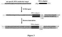
- the capture moiety may be specific to a particular DNA or RNA or may be complementary to a sequence region common to all RNAs, e.g. the capture moiety may be a poly-T tract which is complementary to the poly-A tails of eukaryotic mRNAs or complementary to a nucleic acid adaptor, preferably a DNA adaptor, added to all RNA, for instance through the action of an RNA ligase.
- the capture moiety drives the specific recognition of a nucleic acid adaptor linked to the molecular targets of interest, e.g. all RNAs.
- a nucleic acid adaptor linked to the molecular targets of interest, e.g. all RNAs.
- Such adaptor may be, for example, pre-adenylated oligonucleotides (5'-App oligos) which act as substrates for T4 ligases and thus can be ligated to any RNA molecule.
- Typically such adaptor may be a single stranded DNA region of at least 8 nucleotide long, preferably of 8 to 25 nucleotide, more preferably of 10 to 15 nucleotide long. An exemplary illustration of such embodiment is presented in Figure 3 .
- At least some of molecular targets are nucleic acids and at least some probes specific of said nucleic acids are DNA probes comprising a capture moiety which is a single stranded DNA region which is able to ligate to a nucleic acid molecular target, and a DNA moiety comprising (i) a 5' single stranded region proximal to the capture moiety and comprising the unique molecular identification (UMI) sequence, and optionally a type identifier sequence, and (ii) a 3'double-stranded region distal from the capture moiety and comprising an overhang, preferably an overhang compatible with a cohesive end generated by a restriction enzyme, or an overhang producing restriction site.
- UMI unique molecular identification
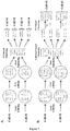
- the capture moiety is a 5' single stranded DNA region comprising 5',5'-adenyl pyrophosphoryl moiety (App) onto its 5'-end.
- App 5',5'-adenyl pyrophosphoryl moiety
- Such moiety may act as substrate for T4 ligases and thus can be ligated to the 3' end any RNA molecule.
- the 5'App extremity of the capture moiety directly interacts with the RNA molecular target and ligates to said target in the presence of T4 ligase. Exemplary illustrations of such embodiment are presented in Figures 4A and 4B .
- DNA probes may be produced by any method known by the skilled person such as chemical synthesis.
- the capture moiety is specific to a particular DNA or RNA molecule, more preferably is specific to a particular RNA molecule.
- nucleic acid molecular targets may be labelled using probes as described above, and in particular the capture moiety of said probes, as priming sites for a DNA polymerase synthetizing complementary strands of molecular targets.
- DNA and/or RNA molecular targets are converted into barcoded complementary DNA (cDNA) upon reverse transcription or other DNA polymerization reaction.
- some molecular targets are RNA molecules and the DNA polymerase is a reverse transcriptase.
- the DNA polymerase is a reverse transcriptase.
- reverse transcription can occur and first strand of complementary DNA (cDNA) can be synthesized, said cDNA comprising the UMI sequence of the DNA moiety of the probe.
- DNA polymerization reaction or reverse transcription requires appropriate conditions, for example the presence of an appropriate buffer and DNA polymerase enzyme, temperatures appropriate for annealing of the probes to targeted RNAs or DNAs and the activity of the enzyme and optionally presence of DTT. These conditions mainly depend on the polymerase and may be adapted according to the supplier guidance.
- the method further comprises, after lysis step when necessary, contacting molecular targets with a labelling mixture comprising said DNA probes as described above, at least one DNA polymerase, a dNTP mix (dATP, dCTP, dGTP, dTTP) and optionally an appropriate buffer, and converting RNA molecular targets into cDNA comprising a molecular identification DNA sequence (UMI) using polymerization reaction.
- a labelling mixture comprising said DNA probes as described above, at least one DNA polymerase, a dNTP mix (dATP, dCTP, dGTP, dTTP) and optionally an appropriate buffer, and converting RNA molecular targets into cDNA comprising a molecular identification DNA sequence (UMI) using polymerization reaction.
- UMI molecular identification DNA sequence
- probes may be chimeric molecules made of synthetic DNA oligonucleotides, i.e. the DNA moiety, covalently associated to a second molecule, i.e. the capture moiety, targeting specifically and with high affinity the target molecule and making possible to specifically label any molecule with a signal amplifiable and readable.
- probes may be specific of any type of molecular targets, preferably are specific of protein molecular targets.
- the capture moiety of these probes may be any suitable capture moiety of these probes.
- the affinity of a molecule X for its partner Y can generally be represented by the dissociation constant (Kd).
- Kd representing the affinity between the capture moiety and the molecular target of interest is from 1.10 -7 M or lower, preferably from 1.10 -8 M or lower, and even more preferably from 1.10 -9 M or lower.
- At least some probes comprise a capture moiety which is a binding moiety that specifically binds to a molecular target and is directly bound to the DNA moiety.
- the binding moiety is covalently bound to the DNA moiety.
- binding moieties include, but are not limited to, antibodies, ligands of ligand/anti-ligand couples, peptide and nucleic acid aptamers, protein tags, or chemical probes (e.g. suicide substrate) reacting specifically with a molecular target or a class of molecular targets.
- ligand/anti-ligand couples include, but are not limited to, antibody/antigen or ligand/receptor.
- the molecular target is an antibody and the binding moiety is an antigen recognized by said antibody, or vice-versa.
- the molecular target is a receptor and the binding moiety is a ligand recognized by said receptor, or vice-versa.
- protein tags are well-known by the skilled person (see for example Young et al. Biotechnol. J. 2012, 7, 620-634 ) and may be used in the present invention.
- protein tags include, but are not limited to, biotin (for binding to streptavidin or avidin derivatives), glutathione (for binding to proteins or other substances linked to glutathione-S-transferase), lectins (for binding to sugar moieties), c-myc tag, hemaglutinin antigen (HA) tag, thioredoxin tag, FLAG tag, polyArg tag, polyHis tag, Strep-tag, OmpA signal sequence tag, calmodulin-binding peptide, chitin-binding domain, cellulose-binding domain, S-tag, and Softag3, and the like.
- biotin for binding to streptavidin or avidin derivatives
- glutathione for binding to proteins or other substances linked to glutathione-S-transferase
- a multitude of chemical probes are well-known by the skilled person (see for example Niphakis and Cravatt, Ann. Rev. of Biochem. 2014, 83, 341-77 and Willems et al. Bioconjugate Chem. 2014, 25, 1181-91 ) and may be used in the present invention.
- Examples of chemical probes include, but are not limited to, electrophile or photoreactive Activity-Based Probes (ABP), suicide substrate-based ABP and inhibitors-based ABP.
- probes are aptamer-based probes, i.e. probes comprising a nucleic acid or peptide aptamer as capture moiety.
- aptamers interact with their targets by recognizing a specific three-dimensional structure. Aptamers can specifically recognize a wide range of targets, such as proteins, nucleic acids, ions or small molecules such as drugs and toxins.
- Peptides aptamers consist of a short variable peptide loop attached at both ends to a protein scaffold such as the bacterial protein thioredoxin-A. Typically, the variable loop length is composed of ten to twenty amino acids.
- Peptide aptamer specific of a target of interest may be selected using any method known by the skilled person such as the yeast two-hybrid system or Phage Display.
- Peptides aptamers may be produced by any method known by the skilled person such as chemical synthesis or production in a recombinant bacterium followed by purification.
- nucleic acid aptamers are a class of small nucleic acid ligands that are composed of RNA or single-stranded DNA oligonucleotides and have high specificity and affinity for their targets.
- Systematic Evolution of Ligands by EXponential enrichment (SELEX) technology to develop nucleic acid aptamers specific of a target of interest is well known by the skilled person and may be used to obtain aptamers specific of a particular molecular target.
- Nucleic acid aptamers may be produced by any method known by the skilled person such as chemical synthesis or in vitro transcription for RNA aptamers.
- nucleic acid aptamers used as capture moiety are selected from the group consisting of DNA aptamers, RNA aptamers, XNA aptamers (nucleic acid aptamer comprising xeno nucleotides) and aptmers (which are composed entirely of an unnatural L-ribonucleic acid backbone).
- XNA aptamers nucleic acid aptamer comprising xeno nucleotides
- spiegelmers which are composed entirely of an unnatural L-ribonucleic acid backbone.
- probes are antibody-based probes, i.e. probes comprising an antibody as capture moiety.
- antibody herein is used in the broadest sense and specifically covers monoclonal antibodies, polyclonal antibodies, antibody fragments, and derivatives thereof, so long as they specifically bind to the molecular target of interest.
- the antibody may be a full length monoclonal or polyclonal antibody, preferably a full length monoclonal antibody.
- this term refers to an antibody with heavy chains that contain an Fc region.
- Fc Fc fragment
- Fc region used herein is meant the polypeptide comprising the constant region of an antibody excluding the first constant region immunoglobulin domain.
- Fc refers to the last two constant region immunoglobulin domains of IgA, IgD, and IgG, and the last three constant region immunoglobulin domains of IgE and IgM, and the flexible hinge N-terminal to these domains.
- the antibody is a full length monoclonal or polyclonal IgG antibody, preferably a full length monoclonal IgG antibody.
- a large number of specific and high affinity monoclonal antibodies are currently available on the market.
- antibody fragment refers to a protein comprising a portion of a full length antibody, generally the antigen binding or variable domain thereof.
- antibody fragments include Fab, Fab', F(ab) 2 , F(ab') 2 , F(ab) 3 , Fv (typically the VL and VH domains of a single arm of an antibody), single-chain Fv (ScFv), dsFv, Fd (typically the VH and CH1 domains) and dAb (typically a VH domain) fragments, nanobodies, minibodies, diabodies, triabodies, tetrabodies, kappa bodies, linear antibodies, and other antibody fragments that retain antigen-binding function (e.g.
- Antibody fragments can be made by various techniques, including but not limited to proteolytic digestion of intact antibody as well as recombinant host cells (e.g. E. coli or phage). These techniques are well-known by the skilled person and are extensively described in the literature.
- the antibody fragment is selected from the group consisting of Fab', F(ab) 2 , F(ab') 2 , F(ab) 3 , Fv, single-chain Fv (ScFv) fragments and nanobodies.
- antibody derivative refers to an antibody provided herein, e.g. a full-length antibody or a fragment of an antibody, wherein one or more of the amino acids are chemically modified, e.g. by alkylation, PEGylation, acylation, ester or amide formation or the like.
- this term may refer to an antibody provided herein that is further modified to contain additional nonproteinaceous moieties that are known in the art and readily available.
- the capture moiety is selected from the group consisting of monoclonal and polyclonal antibodies, Fab', F(ab) 2 , F(ab') 2 , F(ab) 3 , Fv, single-chain Fv (ScFv) fragments and nanobodies, and derivatives thereof.
- the capture moiety is selected from the group consisting of a monoclonal antibody, a ScFv fragment or a nanobody.
- At least some probes comprise a capture moiety which is a chimeric protein comprising a first domain that specifically binds to a single molecular target and a second domain that binds to a single DNA moiety.
- a capture moiety which is a chimeric protein comprising a first domain that specifically binds to a single molecular target and a second domain that binds to a single DNA moiety.
- the second domain is covalently bound to the DNA moiety.
- the first domain of the chimeric protein specifically binds to a single molecular target.
- first domains include, but are not limited to, antibodies, ligands of ligand/anti-ligand couples, peptide and nucleic acid aptamers, protein tags, and chemical probes, as described above.
- the first domain of the chimeric protein is selected from the group consisting of antibodies and peptide aptamers, more preferably is a monoclonal antibody.
- the first domain of the chimeric protein is an antibody, preferably selected from the group consisting of monoclonal and polyclonal antibodies, Fab', F(ab) 2 , F(ab') 2 , F(ab) 3 , Fv, single-chain Fv (ScFv) fragments and nanobodies, and derivatives thereof. More preferably, the first domain of the chimeric protein is selected from the group consisting of a monoclonal antibody, a ScFv fragment or a nanobody, and even more preferably from the group consisting of a ScFv fragment or a nanobody.
- the second domain that covalently binds to the DNA moiety may be any domain allowing covalently grafting of a single nucleic acid.
- Examples of such domains include, but are not limited to, SNAP-tag® (New England Biolabs), CLIP- tag® (New England Biolabs), Halo- tag® (Promega).
- the second domain is a SNAP-tag®.
- the SNAP-tag is a 20 kDa mutant of the DNA repair protein O 6 -alkylguanine-DNA alkyltransferase that reacts specifically and rapidly with benzylguanine (BG) derivatives leading to irreversible covalent association of the SNAP-tag with the DNA moiety attached to BG.
- BG benzylguanine
- the chimeric protein used as capture moiety and comprising the first and second domains may be produced as fusion protein using any well-known recombinant engineering technology, before to be covalently associated to the DNA moiety of the probe.
- At least some probes comprise a capture moiety comprising (i) a binding moiety that specifically binds to a molecular target and (ii) a protein bridge, said protein bridge comprising a first domain that binds to the binding moiety and a second domain that binds to the DNA moiety.
- the second domain is covalently bound to the DNA moiety.
- the first domain may be covalently or non-covalently bound to the binding moiety, preferably non-covalently bound.
- the non-covalent interaction is preferably turned into covalent interaction by cross-linking the first domain and the binding moiety.
- the second domain of the protein bridge may be as described above for the chimeric protein, i.e. any domain allowing covalently grafting of a single nucleic acid.
- the second domain of the protein bridge is a SNAP-tag®.
- the first domain of the protein bridge may be any domain allowing covalent or non-covalent interaction with the binding moiety, preferably non covalent interaction.
- Examples of such domain includes, but are not limited to, immunoglobulin-binding bacterial proteins such as protein A, protein A/G, protein G and protein L.
- the first domain of the protein bridge is an immunoglobulin-binding bacterial protein and the binding moiety is an antibody, preferably an antibody containing a Fc region, more preferably a full length monoclonal or polyclonal IgG antibody, preferably a full length monoclonal IgG antibody.
- the immunoglobulin-binding bacterial protein is preferably selected from protein A, protein A/G, protein G and protein L, one or several IgG-binding domains thereof, and functional derivatives thereof.
- Protein A is a cell surface protein found in Staphylococcus aureus. It has the property of binding the Fc region of a mammalian antibody, in particular of IgG class antibodies.
- the amino-terminal region of this protein contains five highly homologous IgG-binding domains (termed E, D, A, B and C), and the carboxy terminal region anchors the protein to the cell wall and membrane. All five IgG-binding domains of protein A bind to IgG via the Fc region and in principle, each of these domains is sufficient for binding to the Fc-portion of an IgG.
- the first domain of the protein bridge is selected from the group consisting of domains A, B, C D and E of protein A, combinations thereof and functional derivatives thereof retaining IgG binding functionality of wild-type protein A.
- the first domain of the protein bridge comprises domains A to E of protein A.
- the protein bridge may be produced as fusion protein using any well-known recombinant engineering technology, before to be covalently associated to the DNA moiety of the probe.
- the first domain is located at the N-terminal part of the protein bridge and the second domain is located at the C-terminal part of the protein bridge.
- the protein bridge may further comprise at the C-terminal extremity an affinity tag (e.g. a polyhistidine-tag) to facilitate its purification.
- an affinity tag e.g. a polyhistidine-tag
- the protein bridge comprises an immunoglobulin-binding bacterial protein, preferably domains A to E of protein A, as first domain, a SNAP-tag® as second domain and a monoclonal or polyclonal IgG antibody as binding moiety, preferably a monoclonal IgG antibody.
- an immunoglobulin-binding bacterial protein preferably domains A to E of protein A, as first domain, a SNAP-tag® as second domain and a monoclonal or polyclonal IgG antibody as binding moiety, preferably a monoclonal IgG antibody.
- the binding moiety may be covalently or non-covalently bound to the protein bridge.
- the protein bridge can be cross-linked with the binding moiety to ensure long-term physical link.
- the DNA moiety consists of a double stranded DNA molecule comprising an overhang, preferably compatible with a cohesive end generated by a restriction enzyme, or an overhang producing restriction site as described above for DNA probes.
- the overhang comprised in the DNA moiety or generated by the restriction site is a 3'overhang.
- the DNA moiety comprises a unique molecular identification (UMI) sequence, and optionally a type identifier sequence.
- UMI unique molecular identification
- the DNA moiety further comprises a sequencing primer annealing sequence which is proximal to the capture moiety. After labelling of molecular targets, this sequence allows direct amplification using sequencing primers.
- the method further comprises, after lysis step when necessary, contacting molecular targets with a labelling mixture comprising at least one chimeric probe.
- the labelling mixture comprising probes, and optionally reagents to perform DNA polymerization reaction, may be added to the aqueous phase of the droplets before encapsulation of cells (i.e. by direct inclusion in the mixture or via a co-flow) or after droplet generation by any known technique such as pico-injection or droplet fusion.
- the labelling mixture is added after incubation allowing cell lysis and/or elimination/degradation of some non-targeted molecules.
- the labelling mixture is encapsulated into w/o droplets and these droplets are fused with droplets comprising molecular targets.
- the content of each droplet comprising the labelling mixture should be substantially identical.
- the second set of emulsion droplets comprises droplets containing entity identification sequences, wherein each of these droplets contains at least one entity identification sequence.
- the entity identification sequence is a double stranded DNA sequence of 40 to 100 nucleotide long, preferably of 50 to 70 nucleotide long, comprising a unique entity identification (UEI) barcode which is different for each droplet of the second set, and an overhang producing restriction site, preferably a 3 'overhang producing restriction site.
- UEI unique entity identification
- the "UEI sequence” or "UEI barcode” is a randomized nucleotide sequence assigning the same barcode to each molecular target originating from the same entity.
- the UEI sequence is a randomized nucleotide sequence having a length of at least 8 nucleotides, preferably a length from 8 to 20 nucleotides, more preferably a length from 8 to 15 nucleotides.
- the randomized sequence can be a stretch of contiguous randomized nucleotides or a stretch of semi-randomized nucleotides (i.e. contiguous randomized nucleotides spaced by constant nucleotides).
- Typical examples of a stretch of semi-randomized nucleotides are stretches where several randomized dinucleotides are spaced by constant dinucleotides, or stretches where several randomized trinucleotides are spaced by constant trinucleotides.
- the UEI sequence is a stretch of semi-randomized nucleotides, in particular a stretch where several randomized dinucleotides are spaced by constant dinucleotides.
- the restriction site comprised in the entity identification sequence may generate, upon digestion with the corresponding restriction enzyme, an overhang compatible with the overhangs of labelled molecular targets, or an overhang compatible with the overhangs of UEI calibrators as described below, and thus allows addition of UEI sequence to the molecular identification DNA sequence.
- this restriction site is a non-palindromic cleavage site.
- the entity identification sequence may further comprise a sequencing primer annealing sequence adjacent to the UEI barcode and at the opposite end of the restriction site.
- the entity identification sequence comprises a constant region allowing for amplification of the UEI-bearing DNA, adjacent to the restriction site and at the opposite end of the UEI barcode and the sequencing primer annealing sequence when present.
- this constant region has a length of 15 to 35 nucleotides, preferably of 20 to 30 nucleotides.
- this constant region has a melting temperature comprised between 50°C and 70°C.
- all entity identification sequences have to same sequence except for the UEI barcode sequence, i.e. they exhibit the same sequencing primer annealing sequence, the same restriction site and the same constant region.
- the UEI sequence has to be different for each droplet, it also has to be present in large number of identical copies in each droplet.
- the method further comprises encapsulating a plurality of entity identification sequences within emulsion droplets, each droplet containing no more than one entity identification sequence, with an amplification reaction mixture, and amplifying the entity identification sequences within droplets, thereby obtaining the second set of emulsion droplets.
- entity identification sequences may be encapsulated in single or double stranded form, preferably in double stranded form.
- the amplification reaction mixture comprises all reagents required to perform DNA amplification into the droplets, i.e. typically a DNA polymerase, primers, buffers, dNTPs, salts (e.g. MgCl 2 ), etc... Primers are designed in order to allow the complete amplification of the entity identification sequence.
- amplification relies on alternating cycles of heating and cooling (i.e., thermal cycling) to achieve successive rounds of replication (e.g., PCR).
- Methods of amplifying genetic elements compartmentalized in emulsion droplets are well-know and widely practiced by the skilled person (see for example, Chang et al. Lab Chip.
- the amplification may be performed by any known technique such as polymerase chain reaction (PCR), nucleic acid sequence-based amplification (NASBA), loop-mediated isothermal amplification (LAMP), helicase-dependent amplification (HDA), rolling circle amplification (RCA), multiple displacement amplification (MDA) and recombinase polymerase amplification (RPA).
- PCR polymerase chain reaction
- NASBA nucleic acid sequence-based amplification
- LAMP loop-mediated isothermal amplification
- HDA helicase-dependent amplification
- RCA rolling circle amplification
- MDA multiple displacement amplification
- RPA recombinase polymerase amplification
- entity identification sequences are amplified into the droplets by PCR amplification.
- the DNA solution comprising entity identification sequences is strongly diluted, e.g. in the amplification reaction mixture, such that, according to Poisson statistics driving molecule distribution, droplet occupancy, i.e. the percentage of droplets comprising a entity identification sequence, is limited to less than 20%, preferably to less than 10%.
- the method further comprises, before encapsulating entity identification sequences and amplification reaction mixture within emulsion droplets, diluting entity identification sequences, preferably into the amplification reaction mixture, in order to obtain a droplet occupancy of less than 20%, preferably less than 10%.
- the method further comprises encapsulating a plurality of entity identification sequences within emulsion droplets in the presence of UEI-calibrators and an amplification reaction mixture, wherein at least some droplets comprise one or several entity identification sequences and one or several UEI-calibrators; and amplifying entity identification sequences and/or UEI-calibrators within droplets, preferably entity identification sequences and UEI-calibrators using a multiplex reaction; thereby obtaining the second set of emulsion droplets.
- Entity identification sequences and UEI-calibrators may be encapsulated in single or double stranded form, preferably in double stranded form.
- a higher amount of primers specific of the entity identification sequences is used relative to those specific for the UEI-calibrators.
- dilutions of entity identification sequence solution and UEI-calibrator solution are adjusted in order to co-encapsulate 2 to 10 entity identification sequences per droplet, preferably 4 to 6 entity identification sequences per droplet, and 2 to 10 UEI-calibrators per droplet, preferably 4 to 6 UEI-calibrators per droplet.
- UEI-calibrators are DNA sequences, preferably double stranded DNA sequences, comprising a unique calibrator barcode which is different for each UEI-calibrator and for each droplet, and one or two overhang producing restriction sites, preferably generating overhangs compatible with overhangs of digested entity identification sequences and/or labelled molecular targets.
- these overhang producing restriction sites are non-palindromic cleavage sites.
- these restriction sites generate 3' overhangs.
- these restriction sites are different from the restriction site generating compatible overhangs on entity identification sequences and/or the restriction site generating compatible overhangs on labelled molecular targets.
- the "UEI-calibrator sequence” or “UEI-calibrator barcode” is a randomized nucleotide sequence having a length of at least 15 nucleotides, preferably a length from 15 to 40 nucleotides, more preferably a length from 15 to 20 nucleotides.
- the randomized sequence can be a stretch of contiguous randomized nucleotides or a stretch of semi-randomized nucleotides (i.e. contiguous randomized nucleotides spaced by constant nucleotides).
- Typical examples of a stretch of semi-randomized nucleotides are stretches where several randomized dinucleotides are spaced by constant dinucleotides, or stretches where several randomized trinucleotides are spaced by constant trinucleotides.
- the UEI-calibrator barcode is a stretch of semi-randomized nucleotides, in particular a stretch where several randomized dinucleotides are spaced by constant dinucleotides.
- UEI-calibrators comprise an overhang producing restriction site which generates, upon digestion with the corresponding restriction enzyme, an overhang compatible with overhangs of digested entity identification sequences comprising UEI barcodes.
- both types of molecules i.e. UEI-calibrator sequences and entity identification sequences, carry restriction sites generating compatible extremities, the subsequent digestion/ligation step used for UEI addition to labelled molecular targets, leads to the formation of different chimeras between UEI and UEI-calibrator barcodes.
- UEI-calibrators may further comprise a sequencing primer annealing sequence adjacent to the UEI-calibrator barcode and at the opposite end of the restriction site generating overhangs compatible with digested entity identification sequences.
- UEI-calibrators may further comprise a binding tag allowing specific capture of the molecule at the extremity proximal to the sequencing primer annealing sequence.
- the binding tag is selected from biotin or digoxigenin, more preferably is biotin.
- UEI-calibrators comprise a constant region adjacent to the restriction site and at the opposite end of the UEI-calibrator barcode and the sequencing primer annealing sequence when present.
- This constant region allows amplifying UEI-calibrators, preferably using PCR amplification.
- this region may comprise a primer binding site.
- this constant region has a length of 10 to 35 nucleotides, preferably of 15 to 25 nucleotides.
- this constant region is orthogonal to that of entity identification sequences in order to prevent unwilling hybridization.
- UEI-calibrators may further comprise an additional region comprised between the UEI-calibrator barcode and the sequencing primer annealing sequence when present.
- This region acts as a spacer between the sequencing primer annealing sequence and the UEI-calibrator barcode and may be used to adjust the length of the amplified sequences comprising UEI and UEI-calibrator barcodes to the length of the amplified sequences comprising UMI and UEI barcodes to limit potent amplification biases.
- An exemplary illustration of such UEI-calibrator is presented in Figure 9 .
- UEI-calibrators comprise two overhang producing restriction sites, a first restriction site generating an overhang compatible with overhangs of digested entity identification sequences comprising UEI barcodes and a second restriction site generating an overhang compatible with overhangs of labelled molecular targets.
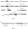
- the subsequent digestion/ligation step used for UEI addition to labelled molecular targets leads to the formation of tripartite molecules comprising a labelled molecular target, an UEI sequence and a UEI-calibrator barcode (see e.g. Figure 8 ).
- UEI-calibrators may comprise constant regions adjacent to each of the two overhang producing restriction sites. These constant regions allow amplifying UEI-calibrators, preferably using PCR amplification. In particular, these regions may comprise primer binding sites. Preferably, these constant regions have a length of 10 to 35 nucleotides, preferably of 15 to 25 nucleotides. Preferably these constant regions are orthogonal to that of entity identification sequences in order to prevent unwilling hybridization. An exemplary illustration of such UEI-calibrator is presented in Figure 9 .
- all UEI-calibrators have to same sequence except for the UEI-calibrator barcode sequence, i.e. they exhibit, the same restriction site, the same constant region(s) and optionally the same sequencing primer annealing sequence.
- multiplex amplification of entity identification sequences and UEI-calibrators within droplets may be performed using any method known by the skilled person, preferably multiplex PCR.
- the amplification reaction mixture comprises all reagents required to perform DNA amplification into the droplets, i.e. typically a DNA polymerase, primers, buffers, dNTPs, salts (e.g. MgCl 2 ), etc... and primers are designed in order to allow the complete amplification of entity identification sequences and UEI calibrators.
- the amplification reaction mixture comprises a higher amount of primers specific of the entity identification sequences relative to those specific for the UEI-calibrators. More preferably, the amplification reaction mixture comprises at least 5 times, preferably at least 10 times, and more preferably at least 100 times, higher amount of primers specific of the cell identification sequences relative to those specific for the UEI-calibrators.
- the amplification reaction mixture may comprise similar amounts of primers specific of the entity identification sequences and primers specific for the UEI-calibrators.
- UEI sequence and UEI-calibrators may be assembled through their compatible overhangs before amplification.
- amplification reaction therefore directly amplify a fragment comprising UEI sequence and UEI-calibrators. After fusion with the first set of droplets, said fragment is ligated to labelled molecular targets.
- the first set of emulsion droplets comprises molecular targets labelled with molecular identification sequence comprising UMI barcodes
- the second set of emulsion droplets comprises entity identification sequences comprising UEI barcodes, and optionally UEI calibrators.
- the method of the invention comprises fusing droplets of the first set with droplets of the second set wherein a droplet of the first set is fused with no more than one droplet of the second set.
- any technique known by the skilled person may be used to fuse a first droplet and a second droplet together to create a combined droplet.
- opposite electric charges may be given to the first and second droplets (i.e., positive and negative charges, not necessarily of the same magnitude), which may increase the electrical interaction of the two droplets such that fusion or coalescence of the droplets can occur due to their opposite electric charges.
- an electric field may be applied to the droplets, the droplets may be passed through a capacitor, a chemical reaction may cause the droplets to become charged, etc.
- any technique known by the skilled person may be used to ensure that a droplet of the first set is fused with no more than one droplet of the second set.
- droplets may be paired through a pairing channel before to reach the coalescence point.
- the use of such channel is well-known by the skilled person in order to control fusion of microfluidic droplets.
- droplets of the first and second sets should have different sizes.
- the pairing channel is a long channel having a width larger than the smallest droplets and narrower than the largest droplets. As a consequence, the small droplet catches the large one and pairs of droplets are formed at the exit of the channel. This configuration ensures that droplets properly pairwise prior to reaching the coalescence point.
- An exemplary embodiment of such pairing channel is illustrated in Figure 10 .
- droplets of the first set and/or droplets of the second sets are generated on separate microfluidic system(s) and re-injected into the device.
- droplets are spaced with oil streams and synchronized before to enter the pairing channel.
- At least 60%, more preferably at least 80% of droplets, and even more preferably at least 95% of the first set are fused with a droplet of the second set.
- UEI barcodes are incorporated into labelled molecular targets through restriction enzyme digestion and ligation.
- UEI barcodes are incorporated into labelled molecular targets through restriction enzyme digestion and ligation of the compatible overhangs of labelled molecular targets and digested entity identification sequences comprising UEI barcodes.
- droplets of the second set comprise UEI calibrators and said UEI calibrators comprise a overhang producing restriction site
- UEI calibrator barcodes and UEI barcodes on one hand and UEI barcodes and labelled molecular targets on the other hand, may be assembled through restriction enzyme digestion and ligation of compatible overhangs of (i) UEI calibrators and entity identification sequences and of (ii) entity identification sequences and labelled molecular targets.
- entity identification sequences are appended to both the labelled molecular targets and the UEI-calibrators through a digestion/ligation coupled reaction. An exemplary illustration of such embodiment is presented in Figure 7B .
- UEI calibrator barcodes, UEI barcodes and labelled molecular targets are assembled through restriction enzyme digestion and ligation of compatible overhangs of i) UEI calibrators and entity identification sequences and (ii) entity identification sequences and labelled molecular targets.
- UEI calibrator barcodes, UEI barcodes and labelled molecular targets are assembled to through restriction enzyme digestion and ligation of compatible overhangs of (i) UEI calibrators and labelled molecular targets on one hand and (ii) UEI calibrators and entity identification sequences on the other hand.
- the entity identification sequence, the UEI-calibrator and the labelled molecular target together form a tripartite molecule.
- An exemplary illustration of such embodiment is presented in Figure 8 .
- restriction sites comprised on entity identification sequences and UEI calibrators are identical and only one restriction enzyme is used to digest entity identification sequences and UEI calibrators.
- this restriction site and restriction sites comprised on entity identification sequences and UEI calibrators may be identical or different, preferably different.
- restriction sites comprised on entity identification sequences and UEI calibrators are different and two different restriction enzymes are used to digest entity identification sequences and UEI calibrators.
- the use of different restriction sites and of labels with compatible extremities ensures that a productive ligation event leads to the destruction of the restriction site. Therefore, the resulting chimeric molecule will not be a substrate of the restriction enzymes present in the mixture and the equilibrium is pulled toward the formation of the wished ligation products.
- this restriction site may be identical or different of the restriction site comprised on entity identification sequences or on UEI calibrators.
- this restriction site is identical to the restriction site comprised on UEI calibrators.
- these restriction sites are non-palindromic in order to ensure directionality of the association of cell identification sequences, UEI calibrators and labelled molecular targets.
- the fragment comprising UEI sequence and UEI-calibrators can be ligated to labelled molecular targets through compatible overhangs of i) UEI calibrators and labelled molecular targets or (ii) entity identification sequences and labelled molecular targets.
- DNA ligase and optionally buffer may be added to the droplets after droplet fusion, e.g. by pico-injection or droplet fusion, or concomitantly to droplet fusion, e.g. by injection at a T-junction (see e.g. figure 10 ). Injection may be performed at the coalescence point where the presence of electrodes triggers both droplet fusion and enzyme delivery.
- the emulsion may be then collected and incubated to allow digestion and ligation and thus association of UEI barcodes and labelled molecular targets, and UEI-calibrator barcodes when present.
- a sequencing primer annealing sequence may be added to labelled molecular target after incorporating UEI barcodes, and optionally UEI-calibrator barcodes.
- the method may further comprise, after incorporating UEI barcodes, and optionally UEI-calibrator barcodes, performing primer extension reaction using primers comprising from their 3'end to their 5'end, a region that hybridizes to complementary strands of molecular targets, i.e. to cDNA, and a sequencing primer annealing sequence.
- the primers may further comprise a binding tag allowing the specific capture of primer extension reaction products.
- the binding tag is biotin or digoxigenin, more preferably is biotin.
- Primer extension reaction may be performed as described above and reaction mixture may be brought into the droplet using any known method such as pico-injection or droplet fusion. Alternatively, the primer extension reaction can be performed in bulk upon droplet breaking and content recovery.
- droplets may be broken and their content may be recovered, i.e. droplet lysate, e.g. to be further analysed/sequenced.
- the present invention also relates to a method of quantifying one or several molecular targets from a plurality of entities with single-entity resolution, said method comprising labelling said molecular targets according to the method of the invention as described above, capturing said labelled molecular targets comprising UMI, UEI, and optionally UEI calibrator barcodes, amplifying sequences comprising UMI and UEI barcodes, and optionally UEI-calibrator barcodes sequencing said amplified sequences.
- the step of capturing labelled molecular target comprising UMI, UEI, and optionally UEI calibrator barcodes, allows removing from the droplet lysate untargeted molecules, unreacted entity identification sequences and/or UEI calibrators when present.
- This step may be performed using any capture molecule which is able to specifically bind molecular targets such as an antibody or nucleic acid specific of a molecular target, attached to a support.
- the capture molecule may be directly or indirectly attached to the support.
- the capture molecule may comprise a binding tag, e.g. biotin, interacting with a partner, e.g. streptavidin, linked to the support.
- the capture molecule may be for example a biotinylated monoclonal antibody specific of a targeted protein, or synthetic DNA oligonucleotide specific of a cDNA produced from a targeted RNA.
- a binding tag can be added during the synthesis of the second cDNA strand by primer extension mentioned above.
- the support may be chosen in order to allow washing of unreacted molecules.
- the support is beads, more preferably streptavidin conjugated beads.
- the support is magnetic beads, in particular streptavidin conjugated magnetic beads.
- nucleic acids comprising (i) the molecular identification DNA sequence comprising the UMI barcode and optionally the type identifier and (ii) the entity identification sequence comprising the UEI barcode are then sequenced using any method known by the skilled person, preferably using a next generation sequencing method.
- the nucleic acids to be sequenced comprise i) the molecular identification DNA sequence comprising the UMI barcode and optionally the type identifier, (ii) the entity identification sequence comprising the UEI barcode and (iii) the UEI-calibrator barcode.
- the nucleic acids to be sequenced comprise (i) the molecular identification DNA sequence comprising the UMI barcode and optionally the type identifier and the entity identification sequence comprising the UEI barcode, and (ii) the entity identification sequence comprising the UEI barcode and the UEI-calibrator barcode.
- said sequences may be added before the sequencing step, preferably by DNA amplification using primers comprising said sequences or by ligation of oligonucleotides comprising said sequences.
- the molecular target is RNA
- a part of this RNA is also sequenced.
- One of the main advantage of the present invention is the possibility of pooling all captured labelled molecular targets prior to the sequencing step, whatever the type of molecular targets (e.g. RNA or protein). Sequences may be then analyzed using any method known by the skilled person such as bioinformatics to cluster said sequences and determining the absolute quantification of molecular targets.
- UEI and UEI-calibrator barcodes are used to cluster the set of UEI labeling the molecules originating from the same entity.
- UEI barcodes are used to cluster molecules according to their entity of origin.
- molecules are clustered according to their type and identity, optionally using the type Identifiers, and, finally, redundant sequences are eliminated using the UMI barcodes giving access to the absolute quantification of each molecule with single-entity resolution.
- a further disclosure relates to a microfluidic device suitable for implementing at least one step of the methods of the invention.
- microfluidic device As used herein, the term "microfluidic device”, “microfluidic chip” or “microfluidic system” refers to a device, apparatus or system including at least one microfluidic channel.
- the microfluidic system may be or comprise silicon-based chips and may be fabricated using a variety of techniques, including, but not limited to, hot embossing, molding of elastomers, injection molding, LIGA, soft lithography, silicon fabrication and related thin film processing techniques.
- Suitable materials for fabricating a microfluidic device include, but are not limited to, cyclic olefin copolymer (COC), polycarbonate, poly(dimethylsiloxane) (PDMS), poly(methyl methacrylate) (PMMA), and glass.
- COC cyclic olefin copolymer
- PDMS poly(dimethylsiloxane)
- PMMA poly(methyl methacrylate)
- microfluidic devices are prepared by standard soft lithography techniques in PDMS and subsequent bonding to glass microscope slides.
- a passivating agent may be necessary. Suitable passivating agents are known in the art and include, but are not limited to silanes, fluorosilanes, parylene, n -dodecyl- ⁇ -D-maltoside (DDM), poloxamers such as Pluronics.
- the term “channel” refers to a feature on or in an article (e.g., a substrate) that at least partially directs the flow of a fluid.
- the term “microfluidic channel” refers to a channel having a cross-sectional dimension of less than 1 mm, typically less than 500 ⁇ m, 200 ⁇ m, 150 ⁇ m, 100 ⁇ m or 50 ⁇ m, and a ratio of length to largest cross-sectional dimension of at least 2:1, more typically at least 3: 1, 5:1, 10: 1 or more. It should be noted that the terms “microfluidic channel”, microchannel” and “channel” are used interchangeably in this description.
- the channel can have any cross-sectional shape (circular, oval, triangular, irregular, square or rectangular, or the like). Preferably, the channel has a square or rectangular cross-sectional shape.
- the channel can be, partially or entirely, covered or uncovered.
- cross-sectional dimension of a channel is measured perpendicular to the direction of fluid flow.
- microfluidic device of the invention comprises
- upstream refers to components or modules in the direction opposite to the flow of fluids from a given reference point in a microfluidic system.
- downstream refers to components or modules in the direction of the flow of fluids from a given reference point in a microfluidic system.
- the microfluidic device of the invention comprises two emulsion re-injection modules a droplet-pairing module, and a module coupling droplet fusion to injection.
- FIG. 10 An exemplary embodiment of such microfluidic device is presented in Figure 10 .
- the microfluidic device of the invention comprises an emulsion re-injection module and an on-chip droplet generation module, a droplet-pairing module, and a module coupling droplet fusion to injection.
- the microfluidic device of the invention comprises two on-chip droplet generation modules, a droplet-pairing module, and a module coupling droplet fusion to injection.
- an emulsion re-injection module may be easily designed by the skilled person based on any known techniques.
- an emulsion re-injection module comprises a ⁇ -shaped structure where injected droplets are spaced by carrier oil supplying by at least one, preferably two side channels connected with the re-injection channel.
- the module for generating droplets may be easily designed by the skilled person based on any known techniques.
- emulsion droplets may be produced in the droplet generation module by any technique known by the skilled person such as drop-breakoff in co-flowing streams, cross-flowing streams in a T-shaped junction (see for example WO 2002/068104 ), and hydrodynamic flow-focussing (reviewed by Christopher and Anna, 2007, J. Phys. D: Appl. Phys. 40, R319-R336 ).
- the droplet-pairing module is a channel with dimensions allowing the contact between droplets of the two sets.
- the width of the channel is about the diameter of the larger droplets and the depth of the channel is lower than the diameter of the larger droplets.
- the depth of the channel is about the diameter of the larger droplets and the width of the channel is lower than the diameter of the larger droplets.
- the length of the pairing channel has to be sufficient to obtain a contact between droplets of the first and second sets.
- the time of contact is greater than 1 ms, preferably greater than 4 ms.
- the contact time ⁇ refers to the time in which paired droplets stay in physical contact before reaching the end of the pairing channel.
- the length of the pairing channel is ranging from 100 ⁇ m to 10 mm, preferably from 500 ⁇ m to 2 mm, and more preferably is about 1.5 mm.
- the module coupling droplet fusion to injection is preferably a module wherein droplets, after pairing, are exposed to an electric field destabilizing their interface thanks to the proximity of electrodes, and are, in the same time, contacted with a stream injected in the channel. Destabilization of the interface then leads not only to droplet coalescence but also to infusion of the injected stream into droplets.
- the microfluidic device may further comprise a collection module wherein fused droplets are recovered.
- the microfluidic device of the invention may comprise an inlet downstream to the module coupling droplet fusion to injection and upstream to the collection module.
- This inlet may be used to inject additional surfactant in order to increase the stability of fused droplets and to prevent any coalescence during the storage.
- the present invention also relates to a kit comprising a microfluidic device according to the invention and as described above.
- the kit may further comprise one or several microfluidic chips comprising an on-chip droplet generation module.
- kit of the invention may further comprise
- the present invention also to a kit comprising
- the present invention further relates to the use of a kit of the invention to label a plurality of molecular targets from a plurality of entities according to the method of the invention, or to quantify one or several molecular targets from a plurality of entities according to the method of the invention. All embodiments described above for the methods, the microfluidic device and the kit of the invention are also encompassed in this aspect.
- indefinite article “a” or “an” does not exclude the possibility that more than one of the element is present, unless the context clearly requires that there be one and only one of the elements.
- the indefinite article “a” or “an” thus usually means “at least one”.
- the term "about” refers to a range of values ⁇ 10% of the specified value.
- “about 20” includes ⁇ 10 % of 20, or from 18 to 22.
- the term “about” refers to a range of values ⁇ 5 % of the specified value.
- microfluidic chips were fabricated using the same procedure and they were manipulated using on the same workstation.
- Microfluidic devices were obtained using a classic replica molding process as described previously in (Mazutis et al., 2009). Briefly, devices were designed on Autocad (Autodesk 2014), negative photomasks were printed (Selba S.A.) and used to prepare molds by standard photolithography methods. SU8-2010 and SU8-2025 photoresist (MicroChem Corp.) were used to pattern 10 to 40 ⁇ m deep channels onto silicon wafers (Siltronix). Microfluidic devices were then fabricated in polydimethylsiloxane (PDMS, Silgard 184, Dow-Corning) using conventional soft lithography methods (Xia and Whitesides, 1998).
- PDMS polydimethylsiloxane
- Silgard 184 Silgard 184
- channels were passivated with a solution of 1% (v/v) 1H, 1H, 2H, 2H-perfluorodecyltrichlorosilane (97%, ABCR GmbH and Co,) in HFE7500 (3M) and subsequently flushed with compressed air. Key dimensions and depth of microfluidic devices are given on concerned figures and in their captions.
- Aqueous phases were loaded in I.D. 0.75 mm PTFE tubings (Thermo Scientifc) and oils were loaded in 2 mL Micrew Tubes (Thermo Scientific). Liquids were injected into microfluidic devices at constant and highly controlled flow-rates using a 7-bar MFCSTM pressure-driven flow controller (Fluigent) equipped with Flowells (7 ⁇ L/min flow-meters) allowing for operation in flow-rate controlled mode.
- MFCSTM pressure-driven flow controller Fluid
- the optical setup was based on an inverted microscope (Nikon Eclipse Ti-S) mounted on a vibration-dampening platform (Thorlabs B75150AE).
- the beams of a 488 nm laser (CrystaLaser DL488-050-O) and a 561 nm laser (Cobolt DPL 561-NM-100MW) were combined using a dichroic mirror (Semrock 2F495-DI03-2536).
- Random sequences corresponding to the UCI or the Calibrator are represented by N arrays and are underlined whereas the UMI is represented by an italicized N arrays.
- the sequence digested by AlwNI (CAGNNNCTG) and DraIII (CACNNNGTG) are shown in bold. * These oligonucleotides are 5' phosphorylated to allow their ligation with another DNA by T4 DNA ligase. ** [BG]: benzyl-guanine; ispl8: 18 carbon-long internal spacer.
- Example 1 Cells preparation, individualization and lysis
- E. coli bacteria T7-Xpress Lys/I q , GFP null strain New England Biolabs transformed either with a plasmid carrying GFP gene under the control of T7 RNA polymerase promoter or a plasmid bearing an unrelated construct were used as model bacteria and will be later summarized as strains GFP+ and GFP- respectively. Note that both plasmids confer ampicillin resistance to the bacteria allowing for their co-culture.
- a cell pre-culture was first obtained by inoculating a 2YT media supplemented with 0.1 mg/mL ampicillin and 2 % glucose, and incubating it over-night at 37°C and under agitation. 500 ⁇ L of this starter (OD 600 of 3.77) were then used to inoculate 20 mL of the same medium, and the bacteria were allowed to grow until the culture reached an OD 600 of 0.6. Next, the cells were fluorescently stained to allow for monitoring cells during the microfluidic steps. To do so, 2 mL of this culture were stained using 5 ⁇ M SYTO 9 (molecular probes by Life technologies, ref 34854) for 2 hours in the dark, following supplier recommendations. Cells were washed once with PBS buffer 1x before being pelleted for further experiments.
- SYTO 9 molecular probes by Life technologies, ref 34854
- Pellets of labelled cells were diluted to reach an OD 600 of 3.75 (giving a droplet occupancy of ⁇ 20%) in a CutSmart® buffer 1x (50 mM Potassium acetate, 20 mM Tris acetate pH7.9 at 25°C, 10 mM Magnesium acetate and 0.1 mg/mL BSA, New England Biolabs) supplemented with 37.5 ⁇ g/mL dextran Texas red 70 000 MW (used as droplet tracker), and 0.05 % Pluronic F68. The mixture was then loaded into a 0.5 mL PCR tube containing a magnetic stirring bar (5 mm length and 2 mm diameter) closed by a plug of PDMS.
- a CutSmart® buffer 1x 50 mM Potassium acetate, 20 mM Tris acetate pH7.9 at 25°C, 10 mM Magnesium acetate and 0.1 mg/mL BSA, New England Biolabs
- Droplets contained into a 0.2 mL PCR tube closed by a plug of PDMS were reinjected into a droplet analyzer ( Figure 12 , inlet 1) where they were spaced by an oil stream ( Figure 12 , inlet 2) and their fluorescence content was analyzed by the optical set-up introduced above.
- the Texas Red contained into each droplet allows identifying a droplet as an orange peaks ( Figure 13 ).
- the staining of cell nucleic acids by Syto9 enables both detecting bacteria and appreciating their lysed/integer status.
- UIs Unique Identifiers
- a UI is made of a pair of random sequences (the UEI ("unique entity identifier") and the UEI Calibrator) surrounded by constant sequence regions encompassing restriction sites generating compatible extremities later used to recombine both sequences together ( Figure 15 ).
- UEI-Calibrator and UEI sequences should be first amplified in a duplex PCR prior to being recombined together through a specific restriction/ligation reaction. Then, the pool of UI contained into the same droplet constitute the signature of the droplet that can later be used to reassign a given encoded molecule to the droplet it originates from.
- the mixture was then loaded into a length of PTFE tubing (I.D. 0.75 mm tubing; Thermo Scientifc) and connected to the Fluigent infusion device at one side of the tubing while the other the other side was connected to a 40 ⁇ m deep droplet generator ( Figure 17 , inlet 1).
- the tube was then placed in a thermocycler and the emulsion was subjected to an initial denaturation step of 30 sec at 95°C followed by 25 cycles of 5 sec at 95°C and 30 sec at 65°C.
- the emulsion was broken using 50 ⁇ L of 1H, 1H, 2H, 2H, perfluoro-1-octanol (Sigma-Aldrich) and 5 ⁇ L were loaded on an 8% native PAGE.
- Forming the UI requires recombining together the UEI-Calibrator and the UEI through a restriction/ligation coupled reaction. To do so, we choose two non-palindromic restriction sites producing compatible 3' overhangs. AlwNI (5'-CAGNNN/CTG-3') and DraIII (5'-CACNNN/GTG-3') are two enzymes digesting sequences (bolded in Table 1 ) fulfilling these criteria. Digestion at these sites generates two compatible sequences that can recombine together and form new sequences (i.e. 5'-CAGNNNGTG-3' and 5'-CACNNNCTG-3') that can no longer be digested by the enzymes, pulling therefore the equilibrium toward the formation of recombined molecules.
- the duplex PCR established in section b was repeated using template molecules 7 and 8 and amplification primers 3, 5 and 6. However, primer 2 was exchanged for primer 9 that allowed introducing Illu-1 sequence, an adaptor sequence later used for next generation sequencing analysis.
- primer 9 that allowed introducing Illu-1 sequence, an adaptor sequence later used for next generation sequencing analysis.
- UI formation we performed a set of experiments using two relative concentrations of UEI-Calibrators and UEI in the droplets. This ratio was changed by varying the concentration of the corresponding primers in the PCR mixture. However, the diversity of the barcodes was preserved by initiating each experiment with the same average number of template per PCR droplet.
- Each template/primers mixture was then introduced into 200 ⁇ L of reaction mixture containing 0.2 mM of each dNTP, 0.1 % Pluronic F68, 0.67 mg/mL dextran Texas Red (Invitrogen), 6 nM of a Taq polymerase produced in the laboratory and 1x CutSmart® buffer (50 mM Potassium acetate, 20 mM Tris acetate pH7.9 at 25°C, 10 mM Magnesium acetate and 0.1 mg/mL BSA). The mixture was dispersed into 100 pL droplets as above and the emulsion was collected for 20 min in a 0.2 mL tube closed by a plug of PDMS and thermocycled as before.
- 1x CutSmart® buffer 50 mM Potassium acetate, 20 mM Tris acetate pH7.9 at 25°C, 10 mM Magnesium acetate and 0.1 mg/mL BSA.
- the emulsion Upon thermocycling, the emulsion was reinjected into a droplet picoinjector ( Figure 18 , inlet 1) at 500 nL/min and the droplets were spaced by a stream of Novec 7500 oil supplemented with 2 % Krytox-Jeffamine 1000 diblock copolymer fluorosurfactant infused at 1600 nL/min through a second inlet ( Figure 18 , inlet 2).
- CutSmartTMbuffer 1x 50 mM Potassium acetate, 20 mM Tris acetate pH7.9 at 25°C, 10 mM Magnesium acetate and 0.1 mg/mL BSA
- 1 mM rATP 7 mM DTT
- 3 U/ ⁇ L DraIII HF New England Biolabs
- 3 U/ ⁇ L AlwN1 New England Biolabs
- 30 U/ ⁇ L T4 DNA ligase (New England Biolabs)
- 20 ⁇ M coumarin acetic used as injection tracker) and 0.1% pluronic F68 was prepared.
- the mixture was loaded into a length of PTFE tubing (I.D.
- the Ct value was determined for each condition ( Figure 19 , Top and middle panels) and the quality of the amplified material was verified by loading an aliquot of each qPCR reaction onto a native polyacrylamide gel ( Figure 19 , bottom left panel).
- ⁇ 1000 droplets from the emulsion containing the 1/1 UEI-Calibrator/UEI ratio were transferred in a new tube where they were broken with 50 ⁇ L of 1H, 1H, 2H, 2H, perfluoro-1-octanol (Sigma-Aldrich) and the released DNA was recovered in 1x CutSmartTM buffer.
- the DNA was then indexed with primers N503 and N702 using Nextera index kit (ref FC-121-1011, Illumina) following supplier specifications. To limit the risk of unwanted mutations during the indexing step, the reaction was performed using Phusion DNA polymerase (ThermoScientific). The band containing indexed DNA was then purified on a 1% agarose gel electrophoresis and the DNA recovered with the kit Wizard SV Gel and PCR clean up system (Promega) ( Figure 19 , right panel). Last, the resulting library was loaded onto an Illumina V2-300 cycles flow-cell and the DNA was sequenced on a MiSeq device (Illumina).
- Nextera index kit Ref FC-121-1011, Illumina
- sequences were analyzed using a Python-based bioinformatics algorithm ( Figure 20 ).
- the algorithm works in 3 main steps. First, raw sequences obtained from the sequencer are quality-filtered and those with a Q score below 30 are removed from the pool. Moreover, sequences presenting mutations in the non-randomized region or showing an inappropriate length are also removed from the pool. Second, UEI-Calibrator and UEI sequences are extracted and pairs coming from the same droplet are clustered together. Briefly, all the UEIs associated with a given UEI-Calibrator are clustered together. Then, all the UEI-calibrator sharing the UEIs contained into the same cluster are also clustered together.
- clusters of UIs are obtained and form a signature of the droplet.
- the signatures can be used to reassign each molecule from a pool to the droplet it originates from.
- the UI signature readily forms only in the presence of restriction/ligation enzymes. Indeed, the qPCR analysis revealed that whatever the UEI-Calibrator/UEI ratio used, more than 10 additional amplification cycles (delta Ct > 10) are required the reach the threshold in the absence of recombination enzymes ( Figure 19 , Top panel, compare columns - and + enzymes), indicating that there is at least a thousand times less recombined material in the absence of enzymes. Moreover, electrophoresis gel analysis ( Figure 19 , left panel ) showed that specific band of the expected size is obtained only in the presence of the enzymes (lanes 2, 4 and 7).
- UI formation is highly controlled as it occurs only in the presence of specific enzymes and does not form spontaneously form during the PCR reaction, which limits the risk of forming UI in non-specific way. Not only this recombination was found to work in tubes ( Figure 19 , left panel, lanes 1 to 4) but it also works in droplets ( Figure 19 , left panel, lanes 5 to 7) starting from PCR amplification products also prepared in droplets. As in tubes, the presence of UI in droplets requires the presence of the specific restriction/ligation enzymes (compare lanes 6 and 7).
- Template UEI-N15-DraIII (7) and Template AlwNI-N15-Calibrator-DraIII ( 8 )) diluted into 0.2 mg/mL yeast total RNA (Ambion) were introduced into 200 ⁇ L of a reaction mixture containing 0.2 ⁇ M of each forward (molecules 2 and 6 ) and reverse (molecules 3 and 5 ) primers, 0.2 mM of each dNTP, 0.1 % Pluronic F68, 0.67 mg/mL dextran Texas Red (Invitrogen), 6 nM of a Taq polymerase produced in the laboratory and 1x CutSmart® buffer (50 mM Potassium acetate, 20 mM Tris acetate pH7.9 at 25°C, 10 mM Magnesium acetate and 0.1 mg/mL BSA.
- the starting amount of each template introduced in the reaction mixture was respectively raised to 14 atto moles and 56 atto moles.
- 100 pL droplets were generated at a frequency of 300 droplets per second using the same chip as before ( Figure 17 ) and the emulsion was collected and thermocycled as in section c.
- an enzyme mixture was picoinjected into each droplet and the emulsion was incubated to allow the recombination to occur.
- ⁇ 1000 droplets were transferred into a new tube where they were broken by adding 20 ⁇ L of 1H, 1H, 2H, 2H, perfluoro-1-octanol (Sigma-Aldrich) and the released DNA was recovered in 200 ⁇ L of PCR mixture containing Q5 DNA polymerase (New England Biolabs) and its buffer at the recommended concentration, 0.2 mM of each dNTP and a Nextera primer pair (N703 and N502 to index the lambda 1.3 condition; N705 and N504 to index the lambda 4 condition; N706 and N517 to index the lambda 12 condition; Illumina) at the recommended concentration.
- UIs are obtained through a digestion/ligation process, they can also be appended to another DNA molecule provided it possesses a site compatible with one of the restriction products.
- This DNA could be either a reverse transcription product or a DNA attached to another molecule such as a protein.
- UI components were first generated as in Example 2 by a duplex PCR co-amplification. To do so, 500 atto moles of template DNAs 7 and 11 were first PCR amplified in a volume of 200 ⁇ L as described in Example 2 section a using 0.2 ⁇ M of primers 2, 3, 5 and 12. Target DNA was prepared by annealing together 1.25 pmol of the molecule 13 and 1.25 pmol of the molecule 14 in 20 ⁇ L of 1x CutSmart® buffer, 0.25 mM dNTP, 10 mM DTT, 0.1 ⁇ M FAM, 0.15 mg/mL dextran Texas Red.
- the enzyme mixture was prepared by supplementing a 1x CutSmart® buffer with 6 mM rATP, 60 mM DTT, 8 U/ ⁇ L DraIII HF, 8 U/ ⁇ L AlwN1, 80 U/ ⁇ L T4 DNA ligase, 55 ⁇ M coumarin acetic and 0.3% Pluronic F68. 10 ⁇ L of duplex PCR were then mixed with 4 ⁇ L of target DNA solution and 3 ⁇ L of Enzyme mixture and the resulting mixture was incubated overnight at 37°C. As a control, the same reaction was performed but restriction enzymes and the ligase were omitted from the enzyme mixture.
- UIs can be used to encode a DNA displaying an overhang compatible with one of the restriction site present in the UI.
- UEI-Calibrators and UEIs were co-amplified in droplets using a duplex PCR prior to adding the DNA to label and the enzyme mixture to each droplet.
- An extended version of the molecule 7 encompassing the sequencing adapter Illu1 was first prepared by primer extension. Briefly, 10 pmols of the template 7 were mixed with 50 pmols of primer 9 in a 50 ⁇ L reaction mixture containing 0.2 mM of each dNTP, 1U of Phusion DNA polymerase (ThermoScientific) and the corresponding buffer at the recommended working concentration. The mixture was then incubated for 5 minutes at 98°C and 10 minutes at 72°C. Extension product was then purified on an agarose gel and the DNA recovered with the Wizard SV Gel and PCR clean up system (Promega). The extended molecule is now called Template UEI-N15-DraIII-Illu1 and corresponds to molecule 15.
- the mixture was then loaded into a length of PTFE tubing (I.D. 0.75 mm tubing; Thermo Scientifc) and connected to the Fluigent infusion device at one side of the tubing while the other the other side was connected to a 40 ⁇ m deep droplet generator ( Figure 17 , inlet 1).
- Target DNA was prepared by mixing and annealing together 33 femto moles of oligonucleotide 13 (corresponding to 1000 DNA molecule per 20 pL) and 66 femto moles of oligonucleotide 14 in 400 ⁇ L of solution containing a 1x CutSmart® buffer, 0.25 mM dNTP, 10 mM DTT, 0.1 ⁇ M FAM (droplet tracker fluorescent dye), 0.15 mg/mL dextran Texas red, 0.005 % Pluronic F68.
- a 1x CutSmart® buffer 0.25 mM dNTP, 10 mM DTT, 0.1 ⁇ M FAM (droplet tracker fluorescent dye), 0.15 mg/mL dextran Texas red, 0.005 % Pluronic F68.
- Droplets were collected ( Figure 17 , outlet 3) into a 0.2 mL PCR tube closed by a plug of PDMS for via a length of tubing.
- the enzyme mixture was prepared by mixing together 9 mM rATP, 30 mM DTT, 3 U/ ⁇ L DraIII HF (New England Biolabs), 3 U/ ⁇ L AlwN1 (New England Biolabs), 15 U/ ⁇ L T4 DNA ligase (New England Biolabs), 20 ⁇ M coumarin acetic (injection tracker) and 0.1% pluronic F68 in a 1x CutSmart® buffer.
- the enzyme mixture was loaded into a length of PTFE tubing (I.D.
- UEI-Calibrator/UEI-containing 100 pL droplets were reinjected ( Figure 27 , inlet 1) at a frequency of ⁇ 100 droplets per second by infusing the emulsion at 400 nL/min while spacing them with a stream of oil (Novec 7500 supplemented with 2 % Krytox-Jeffamine 1000 diblock copolymer fluorosurfactant) infused into the chip ( Figure 27 , inlet 2) at a flow-rate of 1300 nL/min.
- Target DNA-containing 20 pL droplets were reinjected ( Figure 27 , inlet 3) at a frequency of ⁇ 100 droplets per second by infusing the emulsion at 100 nL/min while spacing them with a stream of oil (Novec 7500 supplemented with 2 % Krytox-Jeffamine 1000 diblock copolymer fluorosurfactant) infused into the chip ( Figure 27 , inlet 4) at a flow-rate of 1200 nL/min. Pairs of droplets were allowed to form while the droplets were circulating into a short delay line.
- Pair-wised droplets were then fused when passing in front of an electrode pair to which a squared AC field (800 V, 30 Hz) was applied using a function generator connected to a high voltage amplifier (TREK Model 623B).
- a function generator connected to a high voltage amplifier (TREK Model 623B).
- 25 pL of enzyme mixture was delivered to each droplet by injecting the enzyme mixture into the chip ( Figure 27 , inlet 5) and infusing it at 100 nL/min.
- Fused and picoinjected droplets were collected ( Figure 27 , outlet 6) for 20 minutes in a 0.5 mL tube under mineral oil.
- the emulsion was then incubated in a thermocycler and subjected to 15 repeats of the program: incubate for 15 min at 37°C and 45 min at 16°C.
- DNA copy number distribution was very tight, indicating a high homogeneity of the barcoding process (i.e. formation and addition of UIs) among the different droplets.
- Each sequence-UMI-UI was found at least 10 times in the sequence pool, indicating that the library was analyzed with a 10-times coverage. This indicates that the tight distribution of the droplets content does results from the saturation of the methods and is rather representative of the actual content of the emulsion.
- the results presented in this example show that our methodological and analytical algorithm allows for quantifying the DNA content of a droplet in reproducible way by attaching an UI to it.
- Example 4 Converting RNA into cDNA encodable by an UI
- RNA can be converted, in the droplets, into a cDNA that could be used as a substrate of our labelling technology.
- RT composite reverse transcription
- RNA III from Staphylococcus aureus (Benito et al , 2000).
- the RNA was prepared by in vitro transcription using T7 RNA polymerase using the same procedure described in (Ryckelynck et al , 2015). Upon transcription, RNA was purified on a size exclusion column Nap5 (GE-Healthcare) and quantified using a Nanodrop device.
- RNA and RT reagents were first emulsified separately and mixed together through droplet fusion (Mazutis et al , 2009). Moreover, to evaluate the sensitivity of the reverse transcription process, we prepared emulsions containing 10, 100 or 1000 RNA molecules per droplet. Then the reverse transcription was added to each droplet and the RT allowed to proceed prior to analyzing the produced cDNA.
- RNA-III 0.6, 6 or 60 femto moles of RNA-III (allowing for having respectively, 10, 100 or 1000 RNA molecule per 2 pL droplet) were introduced into a 100 ⁇ L of a solution containing CutSmart® buffer, 1.5 mg/mL Dextran Texas Red (Invitrogen), 0.25% Pluronic F68. The mixture was then loaded into a length of PTFE tubing (I.D. 0.75 mm tubing; Thermo Scientifc) and connected to the Fluigent infusion device at one side of the tubing while the other the other side was connected to a 10 ⁇ m deep droplet generator ( Figure 32 , inlet 1).
- An oil phase made of Novec 7500 supplemented with 3 % Krytox-Jeffamine 1000 diblock copolymer fluorosurfactant was also infused into the chip ( Figure 32 , inlet 2) and used to produce 2 pL droplets at a rate of 9000 droplets per second by infusing oil and aqueous phases at 1450 nL/min and 700 nL/min respectively.
- Droplets were collected ( Figure 32 , outlet 3) via a length of tubing into a 0.2 mL PCR tube closed by a plug of PDMS.
- 200 ⁇ L of reverse transcription mixture were prepared by supplementing a CutSmart® buffer prepared at the recommended concentration with 1.25 pmols of RT-UMI-RNAIII primer (molecule 18 ), 2.5 pmol of AntiSBACA primer (molecule 14 ), 0.25 mM of each dNTP, 10 mM DTT, 0.1 ⁇ M FAM (droplet tracker) and 125 U of Reverse Transcriptase Maxima RNase H minus (Thermo Scientific).
- the mixture was loaded into a length of PTFE tubing (I.D.
- the 2 pL droplets containing the RNA were also reinjected into the same chip ( Figure 33 , inlet 3) at a frequency of ⁇ 1200 droplets per second by infusing them at 140 nL/min and spacing them with a stream of surfactant-free Novec 7500 fluorinated oil (3M) infused into the chip ( Figure 33 , inlet 4) at 1400 nL/min. Pairs of droplet were formed and fused by a squared AC field (450 V, 30 Hz) that was applied to built-in electrodes and obtained from a function generator connected to a high voltage amplifier (TREK Model 623B). Fused droplets were then collected ( Figure 33 , outlet 5) under mineral oil.
- the same reaction was performed in bulk by mixing 1 volume of RNA dilution with 9 volumes of reverse transcription mixture and by incubating both the bulk mixture and the emulsions for 1 hour at 55°C. Furthermore, a second control reaction in which the reverse transcriptase was omitted was also performed. Upon incubation, emulsions were broken using of 1H, 1H, 2H, 2H, perfluoro-1-octanol (Sigma-Aldrich) and the aqueous phases were recovered.
- cDNA obtained from the different conditions was then analyzed by qPCR using the SsoFast Evagreen Supermix kit (Bio-Rad) supplemented with the primers 20 and 21. Moreover, the amplification products were analyzed on an 8% native polyacrylamide gel.
- emulsions of 2 pL droplets containing each 10, 100 or 1000 molecules of RNA-III were produced and fused to 18 pL droplets containing an RT mixture.
- a control the same experiment was performed in a bulk format.
- qPCR analysis revealed that RT occurred with the same efficiency both in bulk and emulsified format ( Figure 34 ). Indeed, in both formats Ct values obtained with bulk and emulsified reactions were very close and significantly higher than that of a control reaction where the reverse transcriptase was omitted.
- the analysis on gel confirmed that the qPCR product obtained in both conditions had the expected size, whereas only a low size PCR side product was obtained with the negative control.
- gfp mRNA was prepared by in vitro transcription using T7 RNA polymerase using the same procedure described in (Ryckelynck et al , 2015). Upon transcription, RNA was purified on a size exclusion column Nap5 (GE-Healthcare) and quantified using a Nanodrop device. 16.67 femto moles of purified gfp mRNA were reverse transcribed in 20 ⁇ L CutSmart® buffer prepared at the recommended concentration and supplemented with 1.25 pmol of RT primer (molecule 23 ), 1.25 pmol of the complementary oligonucleotide (molecule 24 ), 1 mM dNTP, 10 mM DTT, 10 U of AMV reverse transcriptase, 0.1 ⁇ M FAM.
- the target RNA (gfp mRNA) was properly reversed transcribed and the 4 types of barcodes (UEI-Calibrator/UEI) were appended to the resulting cDNA.
- barcodes deprived of randomized regions or affording N2x5 semi-randomized regions gave homogeneous PCR products (lanes 1 and 2 on Figure 35 )
- more smeary bands were observed with the N4443 and N15 regions.
- secondary amplification products of smaller size tend to significantly accumulate with N4443 and N15. Therefore, semi-randomized barcodes such as the N2x5 represent an attractive alternative to the more conventional N15 barcode design.
- a template DNA (molecule 31 ) possessing a unique AlwNI as well as a UMI of 8 randomized positions was PCR amplified using a primer modified with a benzyl-guanine (BG) group (molecule 32 ) allowing for grafting the DNA on the SNAP module of NaBAb and a primer modified with an Alexa-488 fluorescent group (molecule 33 ) to fluorescently label the DNA.
- 10 pmols of template 31 were mixed with 1 nmol of each primer ( 32 and 33 ) in 1 mL of PCR solution containing 0.2 mM of each dNTP, 20 U of Q5 DNA polymerase (New England Biolabs) and the corresponding buffer at the recommended concentration.
- Amplification products were the purified using the kit Wizard SV Gel and PCR clean up system (Promega) and quantified with a Nanodrop device. 200 pmols of BG/Alexa488 dually labelled DNA were then mixed with 180 pmol of purified NaBAb in 1 mL of CutSmart® buffer (New England Biolabs) diluted at the recommended concentration (1x) and supplemented with 1 mM DTT.
- the NaBAb fusion protein can be used to specifically label IgG with a unique DNA molecule and that, even though it is covalently attached to the NaBAb protein, this DNA can further serve as UI acceptor using our restriction/ligation strategy.
- the recombination occurs in native conditions preserving the association of non-covalent complexes (e.g. antibody/antigen)
- the combined use of NaBAb-DNA/IgG complex and of UI encoding can be used to label and encoded IgG-recognized targets (e.g. protein) and quantify them by Next Generation sequencing (e.g. MiSeq).
- UI can be further attached to a target DNA molecule displaying a 3' overhang compatible with a restriction site present on the UI both in bulk (Example 3.a) and in droplets (Example 3.b).
- UI-based signature allowed to unambiguously assign each DNA sequence to the droplet it originated with a great droplet-to-droplet reproducibility.
- RNA can be efficiently reverse transcribed into cDNA in droplet (Example 4.a) using reverse transcription primers either competent for being later coupled with an UI (Example 4.b) or conjugated to an UI prior to being used for reverse transcribing the target RNA (Example 5).
- UI UI
- Example 5 Similar yield of RT and UI coupling were obtained.
- Our encoding strategy is not limited to nucleic acids since we showed that our UI grafting strategy can also be applied to IgG labeled with a single DNA molecule possessing a restriction site making it compatible with our restriction/ligation approach (Example 6). This last scenario makes possible to encode non-nucleic acids targets such as proteins in a way that preserve non-covalent biological complexes (e.g. antibody/antigen complexes).
- Example 1 cells can be isolated and lysed into small 4 pL droplets (Example 1). Not only the material can be released from the cells, but the size of the droplets makes the directly compatible with the use of the droplet fusion device ( Figure 33 ). Therefore, cell extract can be directly supplemented with the reagents allowing for reverse transcribing target RNAs and/or antibodies coupled to a single DNA molecule. Upon reverse transcription/antigen capture, a different set of UEI-Calibrators/UEIs can then be delivered to each droplet together with the restriction/ligation enzymes using the Barcoding chip ( Figure 27 ) validated in Example 3.
Claims (16)
- Procédé de marquage d'une pluralité de cibles moléculaires provenant d'une pluralité d'entités tout en préservant l'intégrité de l'information au niveau de l'entité unique, ledit procédé comprenant
la fourniture d'un premier ensemble de gouttelettes d'émulsion comprenant des gouttelettes contenant des cibles moléculaires marquées, dans lequel chacune de ces gouttelettes contient une pluralité de cibles moléculaires provenant d'au plus une entité et dans lequel, dans chacune de ces gouttelettes, chaque cible moléculaire est marquée avec une séquence ADN d'identification moléculaire comprenant (i) un code- barres d'identification moléculaire unique (UMI) qui est différent pour chaque cible moléculaire et (ii) un surplomb ou un site de restriction produisant un surplomb;
la fourniture d'un deuxième ensemble de gouttelettes d'émulsion comprenant des gouttelettes contenant des séquences d'identification d'entité, dans lequel chacune de ces gouttelettes contient au moins une séquence d'identification d'entité qui est une séquence d'ADN comprenant un code-barres d'identification d'entité unique (UEI) qui est différent pour chaque gouttelette du deuxième ensemble, et un site de restriction produisant un surplomb ;
la fusion de gouttelettes du premier ensemble avec des gouttelettes du second ensemble, dans laquelle une gouttelette du premier ensemble est fusionnée avec au plus une gouttelette du second ensemble ; et
la ligature des codes-barres UEI à des cibles moléculaires marquées après digestion par enzyme de restriction,
et
éventuellement la cassure de l'émulsion,
où l'entité est une cellule, une particule ou une gouttelette d'émulsion. - Procédé selon la revendication 1, dans lequel le procédé comprend en outre
l'encapsulation d'une pluralité d'entités dans des gouttelettes d'émulsion, chaque gouttelette ne contenant pas plus d'une entité, et éventuellement la lyse desdites entités dans les gouttelettes pour libérer des cibles moléculaires ;
le marquage desdites cibles moléculaires avec des sondes, chaque sonde comprenant
un élément de capture capable de se lier ou de se ligaturer spécifiquement à une cible moléculaire ou à un adaptateur lié à ladite cible moléculaire, et
un élément ADN comprenant (i) une région proximale à l'élément de capture et comprenant la séquence d'identification moléculaire unique (UMI) et (ii) une région distale de l'élément de capture et comprenant un surplomb ou un site de restriction produisant un surplomb,
obtenant ainsi le premier ensemble de gouttelettes d'émulsion. - Procédé selon la revendication 1 ou 2, dans lequel le procédé comprend en outre l'encapsulation d'une pluralité de séquences d'identification d'entités dans des gouttelettes d'émulsion, chaque gouttelette ne contenant pas plus d'une séquence d'identification d'entité, avec un mélange réactionnel d'amplification, et
l'amplification des séquences d'identification d'entités dans les gouttelettes obtenant ainsi le deuxième ensemble de gouttelettes d'émulsion. - Procédé selon la revendication 1 ou 2, dans lequel le procédé comprend en outre
l'encapsulation d'une pluralité de séquences d'identification d'entités dans des gouttelettes d'émulsion en présence de calibrateurs-UEI, dans lequel au moins certaines gouttelettes comprennent une ou plusieurs séquences d'identification d'entités et un ou plusieurs calibrateurs-UEI, et dans lequel lesdits calibrateurs-UEI sont des séquences d'ADN comprenant un code-barres de calibrateur unique qui est différent pour chaque calibrateur-UEI et pour chaque gouttelette, et un ou deux sites de restriction produisant un surplomb ; et
l'amplification des séquences d'identification d'entités et/ou des calibrateurs-UEI dans les gouttelettes ;
obtenant ainsi le deuxième ensemble de gouttelettes d'émulsion. - Procédé selon la revendication 4, dans lequel, après fusion des gouttelettes des premier et deuxième ensembles, (i) les codes-barres des calibrateurs-UEI et les codes-barres UEI et (ii) les codes-barres UEI et les cibles moléculaires marquées sont assemblés par digestion par enzyme de restriction et ligature des surplombs compatibles (i) des calibrateurs-UEI et des séquences d'identification d'entités et (ii) des séquences d'identification d'entités et des cibles moléculaires marquées.
- Procédé selon la revendication 4, dans lequel, après fusion des gouttelettes des premier et deuxième ensembles, les codes-barres des calibrateurs-UEI, les codes-barres UEI et les cibles moléculaires marquées sont assemblés par digestion par enzyme de restriction et ligature des surplombs compatibles (i) des calibrateurs-UEI et des cibles moléculaires marquées et (ii) des calibrateurs-UEI et des séquences d'identification d'entités, ou (i) des calibrateurs-UEI et des séquences d'identification d'entités et (ii) des séquences d'identification d'entités et des cibles moléculaires marquées.
- Procédé selon la revendication 4, dans lequel les séquences d'identification d'entités et les calibrateurs-UEI sont assemblés par leurs surplombs compatibles avant amplification et, après fusion des gouttelettes du premier et du second ensembles, le fragment amplifié comprenant le calibrateur-UEI et les codes-barres UEI est ligaturé à des cibles moléculaires marquées par les surplombs compatibles i) des calibrateurs-UEI et des cibles moléculaires marquées ou ii) des séquences d'identification d'entités et des cibles moléculaires marquées.
- Procédé selon l'une quelconque des revendications 1 à 7, dans lequel au moins certaines des cibles moléculaires sont des acides nucléiques et au moins certaines sondes comprennent
un élément de capture qui est une région d'ADN simple brin qui entraîne la reconnaissance spécifique d'une cible moléculaire d'acide nucléique par les interactions classiques d'appariement de bases de Watson-Crick et
un élément ADN comprenant une région simple brin 3' comprenant la séquence moléculaire unique d'identification (UMI) et une région double brin 5' comprenant le surplomb ou le site de restriction produisant le surplomb. - Procédé selon la revendication 8, dans lequel lesdites cibles moléculaires d'acide nucléique sont marquées en utilisant lesdites sondes comme sites d'amorçage pour une ADN polymérase synthétisant des brins complémentaires de cibles moléculaires.
- Procédé selon la revendication 8 ou 9, dans lequel au moins certaines des cibles moléculaires sont des molécules d'ARN et l'ADN polymérase est une transcriptase inverse.
- Procédé selon l'une quelconque des revendications 2 à 10, dans lequel au moins certaines sondes comprennent un élément de capture qui est(i) un élément de liaison qui se lie spécifiquement à une cible moléculaire et est directement lié à l'élément ADN,(ii) une protéine chimérique comprenant un premier domaine qui se lie spécifiquement à une cible moléculaire et un second domaine qui se lie à l'élément ADN, ou(iii) un élément de liaison qui se lie spécifiquement à une cible moléculaire et un pont protéique, ledit pont protéique comprenant un premier domaine qui se lie à l'élément de liaison et un second domaine qui se lie à l'élément ADN.
- Procédé selon la revendication 11, dans lequel (i) l'élément de liaison ou le premier domaine de la protéine chimérique est choisi dans le groupe constitué par un anticorps, un ligand d'un couple ligand/anti-ligand, un aptamère peptidique, un aptamère d'acide nucléique, une étiquette protéique ou une sonde chimique (par exemple un substrat suicide, des sondes ABP basées sur l'activité) réagissant spécifiquement avec une cible moléculaire ou une classe de cibles moléculaires, de préférence est un anticorps, (ii) le premier domaine du pont protéique est une protéine bactérienne liant l'immunoglobuline, de préférence est les domaines A à E de la protéine A, et/ou (iii) le second domaine du pont protéique ou de la protéine chimérique est choisi dans le groupe constitué par une étiquette SNAP, une étiquette CLIP ou une étiquette Halo, de préférence est une étiquette SNAP.
- Procédé selon l'une quelconque des revendications 2 à 12, dans lequel au moins certaines sondes comprennent un élément de capture comprenant un élément d'anticorps spécifique d'une cible moléculaire et un pont protéique, ledit pont protéique comprenant un premier domaine qui se lie à une région Fc de l'élément d'anticorps et un second domaine qui se lie à l'élément ADN, de préférence une étiquette SNAP.
- Procédé de quantification d'une ou plusieurs cibles moléculaires provenant d'une pluralité d'entités avec une résolution au niveau de l'entité unique, ledit procédé comprenant
le marquage desdites cibles moléculaires selon le procédé de l'une quelconque des revendications 1 à 13;
la capture desdites cibles moléculaires marquées,
l'amplification des séquences comprenant des codes-barres UMI et UEI, et éventuellement des codes-barres de calibrateur-UEI
le séquençage des séquences amplifiées. - Procédé selon l'une quelconque des revendications 1 à 14, dans lequel l'entité est une particule ou une gouttelette d'émulsion exposant des cibles moléculaires sur sa surface externe, et le procédé comprend en outre- le marquage desdites cibles moléculaires avec des sondes, chaque sonde comprenant
un élément de capture capable de se lier ou de se ligaturer spécifiquement à une cible moléculaire ou à un adaptateur lié à ladite cible moléculaire, et
un élément d'ADN comprenant (i) une région proximale de l'élément de capture et comprenant la séquence d'identification moléculaire unique (UMI) et (ii) une région distale de l'élément de capture et comprenant un surplomb ou un site de restriction produisant un surplomb, et- l'encapsulation des entités attachées à des cibles moléculaires marquées dans des gouttelettes d'émulsion, chaque gouttelette ne contenant pas plus d'une entité,obtenant ainsi le premier ensemble de gouttelettes d'émulsion. - Utilisation d'un kit pour marquer une pluralité de cibles moléculaires provenant d'une pluralité d'entités selon le procédé de l'une quelconque des revendications 1 à 13 et 15, ou pour quantifier une ou plusieurs cibles moléculaires provenant d'une pluralité d'entités avec une résolution au niveau de l'entité unique selon le procédé de la revendication 14 ou 15, dans laquelle le kit comprend- un appareil microfluidique et/ou,- une ou plusieurs sondes telles que définies dans la revendication 2 ; et/ou- une ou plusieurs séquences d'identification d'entités telles que définies dans la revendication 1 ; et/ou- un ou plusieurs calibrateurs-UEI tels que définis dans la revendication 4 ; et/ou- une ou plusieurs amorces adaptées pour amplifier les séquences d'identification d'entités et/ou les calibrateurs-UEI ; et/ou- une phase aqueuse et/ou une phase huileuse ; et éventuellement- une brochure fournissant des lignes directrices pour l'utilisation d'un tel kit.
Applications Claiming Priority (2)
| Application Number | Priority Date | Filing Date | Title |
|---|---|---|---|
| EP16306543 | 2016-11-23 | ||
| PCT/EP2017/080264 WO2018096058A1 (fr) | 2016-11-23 | 2017-11-23 | Code-barres en tandem de molécules cibles pour leur quantification absolue à une résolution d'entité unique |
Publications (2)
| Publication Number | Publication Date |
|---|---|
| EP3545104A1 EP3545104A1 (fr) | 2019-10-02 |
| EP3545104B1 true EP3545104B1 (fr) | 2021-02-24 |
Family
ID=57570025
Family Applications (1)
| Application Number | Title | Priority Date | Filing Date |
|---|---|---|---|
| EP17837944.2A Active EP3545104B1 (fr) | 2016-11-23 | 2017-11-23 | Codage à barres en tandem de molécules cibles pour leur quantification absolue à une résolution d'entité unique |
Country Status (4)
| Country | Link |
|---|---|
| US (1) | US20190316180A1 (fr) |
| EP (1) | EP3545104B1 (fr) |
| CN (1) | CN110114473A (fr) |
| WO (1) | WO2018096058A1 (fr) |
Families Citing this family (2)
| Publication number | Priority date | Publication date | Assignee | Title |
|---|---|---|---|---|
| GB201414177D0 (en) * | 2014-08-11 | 2014-09-24 | Sphere Fluidics Ltd | Droplet Sorting |
| US20210214791A1 (en) * | 2018-05-22 | 2021-07-15 | Universite De Strasbourg | Absolute quantification of target molecules at single-entity resolution using tandem barcoding |
Family Cites Families (8)
| Publication number | Priority date | Publication date | Assignee | Title |
|---|---|---|---|---|
| EP1721658B2 (fr) | 2001-02-23 | 2020-08-05 | Japan Science and Technology Agency | Procédé et appareil pour la production de micro-capsules |
| JP2006523142A (ja) | 2003-04-10 | 2006-10-12 | プレジデント・アンド・フェロウズ・オブ・ハーバード・カレッジ | 流体種の形成および制御 |
| PL2814959T3 (pl) * | 2012-02-17 | 2018-07-31 | Fred Hutchinson Cancer Research Center | Kompozycje i sposoby do dokładnej identyfikacji mutacji |
| US20150141257A1 (en) * | 2013-08-02 | 2015-05-21 | Roche Nimblegen, Inc. | Sequence capture method using specialized capture probes (heatseq) |
| JP6853667B2 (ja) * | 2014-04-21 | 2021-03-31 | プレジデント アンド フェローズ オブ ハーバード カレッジ | 核酸をバーコーディングするためのシステムおよび方法 |
| EP3194593B1 (fr) * | 2014-09-15 | 2019-02-06 | AbVitro LLC | Séquençage à haut débit de banque de nucléotides |
| US11111519B2 (en) * | 2015-02-04 | 2021-09-07 | The Regents Of The University Of California | Sequencing of nucleic acids via barcoding in discrete entities |
| CN109073647B (zh) | 2016-04-05 | 2022-07-12 | 斯特拉斯堡大学 | 用于捕获分子靶标的液滴内表面工程 |
-
2017
- 2017-11-23 CN CN201780080795.3A patent/CN110114473A/zh active Pending
- 2017-11-23 US US16/463,492 patent/US20190316180A1/en not_active Abandoned
- 2017-11-23 EP EP17837944.2A patent/EP3545104B1/fr active Active
- 2017-11-23 WO PCT/EP2017/080264 patent/WO2018096058A1/fr unknown
Non-Patent Citations (1)
| Title |
|---|
| None * |
Also Published As
| Publication number | Publication date |
|---|---|
| CN110114473A (zh) | 2019-08-09 |
| WO2018096058A1 (fr) | 2018-05-31 |
| US20190316180A1 (en) | 2019-10-17 |
| EP3545104A1 (fr) | 2019-10-02 |
Similar Documents
| Publication | Publication Date | Title |
|---|---|---|
| US11768198B2 (en) | Compositions and methods for molecular labeling | |
| US20230417765A1 (en) | Digital protein quantification | |
| CN103890245B (zh) | 核酸编码反应 | |
| JP2023159234A (ja) | 単分散エマルションを生成する方法 | |
| US20140303005A1 (en) | Rare cell analysis after negative selection | |
| JP2018508198A (ja) | 別個の実体におけるバーコード付加による核酸のシーケンシング | |
| US11427853B2 (en) | Single cell analysis | |
| EP4324962A2 (fr) | Procédés et compositions pour déconvolutionner des codes à barres de partition | |
| EP3545104B1 (fr) | Codage à barres en tandem de molécules cibles pour leur quantification absolue à une résolution d'entité unique | |
| US20210214791A1 (en) | Absolute quantification of target molecules at single-entity resolution using tandem barcoding | |
| JP6041154B2 (ja) | 並列反応方法およびスクリーニング方法 | |
| JP6015894B2 (ja) | タンパク質アレイの製造方法及びタンパク質アレイ | |
| US20240102005A1 (en) | Methods and systems for engineering antibodies, and antigen-binding fragments thereof, to have altered characteristics | |
| US11965877B2 (en) | Compositions and methods for molecular labeling | |
| US11965891B2 (en) | Digital protein quantification |
Legal Events
| Date | Code | Title | Description |
|---|---|---|---|
| STAA | Information on the status of an ep patent application or granted ep patent |
Free format text: STATUS: UNKNOWN |
|
| STAA | Information on the status of an ep patent application or granted ep patent |
Free format text: STATUS: THE INTERNATIONAL PUBLICATION HAS BEEN MADE |
|
| PUAI | Public reference made under article 153(3) epc to a published international application that has entered the european phase |
Free format text: ORIGINAL CODE: 0009012 |
|
| STAA | Information on the status of an ep patent application or granted ep patent |
Free format text: STATUS: REQUEST FOR EXAMINATION WAS MADE |
|
| 17P | Request for examination filed |
Effective date: 20190523 |
|
| AK | Designated contracting states |
Kind code of ref document: A1 Designated state(s): AL AT BE BG CH CY CZ DE DK EE ES FI FR GB GR HR HU IE IS IT LI LT LU LV MC MK MT NL NO PL PT RO RS SE SI SK SM TR |
|
| AX | Request for extension of the european patent |
Extension state: BA ME |
|
| DAV | Request for validation of the european patent (deleted) | ||
| DAX | Request for extension of the european patent (deleted) | ||
| GRAP | Despatch of communication of intention to grant a patent |
Free format text: ORIGINAL CODE: EPIDOSNIGR1 |
|
| STAA | Information on the status of an ep patent application or granted ep patent |
Free format text: STATUS: GRANT OF PATENT IS INTENDED |
|
| INTG | Intention to grant announced |
Effective date: 20200922 |
|
| GRAS | Grant fee paid |
Free format text: ORIGINAL CODE: EPIDOSNIGR3 |
|
| GRAA | (expected) grant |
Free format text: ORIGINAL CODE: 0009210 |
|
| STAA | Information on the status of an ep patent application or granted ep patent |
Free format text: STATUS: THE PATENT HAS BEEN GRANTED |
|
| AK | Designated contracting states |
Kind code of ref document: B1 Designated state(s): AL AT BE BG CH CY CZ DE DK EE ES FI FR GB GR HR HU IE IS IT LI LT LU LV MC MK MT NL NO PL PT RO RS SE SI SK SM TR |
|
| REG | Reference to a national code |
Ref country code: CH Ref legal event code: EP |
|
| REG | Reference to a national code |
Ref country code: AT Ref legal event code: REF Ref document number: 1364500 Country of ref document: AT Kind code of ref document: T Effective date: 20210315 |
|
| REG | Reference to a national code |
Ref country code: IE Ref legal event code: FG4D |
|
| REG | Reference to a national code |
Ref country code: DE Ref legal event code: R096 Ref document number: 602017033520 Country of ref document: DE |
|
| REG | Reference to a national code |
Ref country code: LT Ref legal event code: MG9D |
|
| REG | Reference to a national code |
Ref country code: NL Ref legal event code: MP Effective date: 20210224 |
|
| PG25 | Lapsed in a contracting state [announced via postgrant information from national office to epo] |
Ref country code: LT Free format text: LAPSE BECAUSE OF FAILURE TO SUBMIT A TRANSLATION OF THE DESCRIPTION OR TO PAY THE FEE WITHIN THE PRESCRIBED TIME-LIMIT Effective date: 20210224 Ref country code: BG Free format text: LAPSE BECAUSE OF FAILURE TO SUBMIT A TRANSLATION OF THE DESCRIPTION OR TO PAY THE FEE WITHIN THE PRESCRIBED TIME-LIMIT Effective date: 20210524 Ref country code: PT Free format text: LAPSE BECAUSE OF FAILURE TO SUBMIT A TRANSLATION OF THE DESCRIPTION OR TO PAY THE FEE WITHIN THE PRESCRIBED TIME-LIMIT Effective date: 20210624 Ref country code: NO Free format text: LAPSE BECAUSE OF FAILURE TO SUBMIT A TRANSLATION OF THE DESCRIPTION OR TO PAY THE FEE WITHIN THE PRESCRIBED TIME-LIMIT Effective date: 20210524 Ref country code: HR Free format text: LAPSE BECAUSE OF FAILURE TO SUBMIT A TRANSLATION OF THE DESCRIPTION OR TO PAY THE FEE WITHIN THE PRESCRIBED TIME-LIMIT Effective date: 20210224 Ref country code: GR Free format text: LAPSE BECAUSE OF FAILURE TO SUBMIT A TRANSLATION OF THE DESCRIPTION OR TO PAY THE FEE WITHIN THE PRESCRIBED TIME-LIMIT Effective date: 20210525 Ref country code: FI Free format text: LAPSE BECAUSE OF FAILURE TO SUBMIT A TRANSLATION OF THE DESCRIPTION OR TO PAY THE FEE WITHIN THE PRESCRIBED TIME-LIMIT Effective date: 20210224 |
|
| REG | Reference to a national code |
Ref country code: AT Ref legal event code: MK05 Ref document number: 1364500 Country of ref document: AT Kind code of ref document: T Effective date: 20210224 |
|
| PG25 | Lapsed in a contracting state [announced via postgrant information from national office to epo] |
Ref country code: SE Free format text: LAPSE BECAUSE OF FAILURE TO SUBMIT A TRANSLATION OF THE DESCRIPTION OR TO PAY THE FEE WITHIN THE PRESCRIBED TIME-LIMIT Effective date: 20210224 Ref country code: LV Free format text: LAPSE BECAUSE OF FAILURE TO SUBMIT A TRANSLATION OF THE DESCRIPTION OR TO PAY THE FEE WITHIN THE PRESCRIBED TIME-LIMIT Effective date: 20210224 Ref country code: PL Free format text: LAPSE BECAUSE OF FAILURE TO SUBMIT A TRANSLATION OF THE DESCRIPTION OR TO PAY THE FEE WITHIN THE PRESCRIBED TIME-LIMIT Effective date: 20210224 Ref country code: NL Free format text: LAPSE BECAUSE OF FAILURE TO SUBMIT A TRANSLATION OF THE DESCRIPTION OR TO PAY THE FEE WITHIN THE PRESCRIBED TIME-LIMIT Effective date: 20210224 Ref country code: RS Free format text: LAPSE BECAUSE OF FAILURE TO SUBMIT A TRANSLATION OF THE DESCRIPTION OR TO PAY THE FEE WITHIN THE PRESCRIBED TIME-LIMIT Effective date: 20210224 |
|
| PG25 | Lapsed in a contracting state [announced via postgrant information from national office to epo] |
Ref country code: IS Free format text: LAPSE BECAUSE OF FAILURE TO SUBMIT A TRANSLATION OF THE DESCRIPTION OR TO PAY THE FEE WITHIN THE PRESCRIBED TIME-LIMIT Effective date: 20210624 |
|
| PG25 | Lapsed in a contracting state [announced via postgrant information from national office to epo] |
Ref country code: EE Free format text: LAPSE BECAUSE OF FAILURE TO SUBMIT A TRANSLATION OF THE DESCRIPTION OR TO PAY THE FEE WITHIN THE PRESCRIBED TIME-LIMIT Effective date: 20210224 Ref country code: CZ Free format text: LAPSE BECAUSE OF FAILURE TO SUBMIT A TRANSLATION OF THE DESCRIPTION OR TO PAY THE FEE WITHIN THE PRESCRIBED TIME-LIMIT Effective date: 20210224 Ref country code: AT Free format text: LAPSE BECAUSE OF FAILURE TO SUBMIT A TRANSLATION OF THE DESCRIPTION OR TO PAY THE FEE WITHIN THE PRESCRIBED TIME-LIMIT Effective date: 20210224 Ref country code: SM Free format text: LAPSE BECAUSE OF FAILURE TO SUBMIT A TRANSLATION OF THE DESCRIPTION OR TO PAY THE FEE WITHIN THE PRESCRIBED TIME-LIMIT Effective date: 20210224 |
|
| REG | Reference to a national code |
Ref country code: DE Ref legal event code: R097 Ref document number: 602017033520 Country of ref document: DE |
|
| PG25 | Lapsed in a contracting state [announced via postgrant information from national office to epo] |
Ref country code: DK Free format text: LAPSE BECAUSE OF FAILURE TO SUBMIT A TRANSLATION OF THE DESCRIPTION OR TO PAY THE FEE WITHIN THE PRESCRIBED TIME-LIMIT Effective date: 20210224 Ref country code: SK Free format text: LAPSE BECAUSE OF FAILURE TO SUBMIT A TRANSLATION OF THE DESCRIPTION OR TO PAY THE FEE WITHIN THE PRESCRIBED TIME-LIMIT Effective date: 20210224 Ref country code: RO Free format text: LAPSE BECAUSE OF FAILURE TO SUBMIT A TRANSLATION OF THE DESCRIPTION OR TO PAY THE FEE WITHIN THE PRESCRIBED TIME-LIMIT Effective date: 20210224 |
|
| PLBE | No opposition filed within time limit |
Free format text: ORIGINAL CODE: 0009261 |
|
| STAA | Information on the status of an ep patent application or granted ep patent |
Free format text: STATUS: NO OPPOSITION FILED WITHIN TIME LIMIT |
|
| PG25 | Lapsed in a contracting state [announced via postgrant information from national office to epo] |
Ref country code: ES Free format text: LAPSE BECAUSE OF FAILURE TO SUBMIT A TRANSLATION OF THE DESCRIPTION OR TO PAY THE FEE WITHIN THE PRESCRIBED TIME-LIMIT Effective date: 20210224 Ref country code: AL Free format text: LAPSE BECAUSE OF FAILURE TO SUBMIT A TRANSLATION OF THE DESCRIPTION OR TO PAY THE FEE WITHIN THE PRESCRIBED TIME-LIMIT Effective date: 20210224 |
|
| 26N | No opposition filed |
Effective date: 20211125 |
|
| PG25 | Lapsed in a contracting state [announced via postgrant information from national office to epo] |
Ref country code: SI Free format text: LAPSE BECAUSE OF FAILURE TO SUBMIT A TRANSLATION OF THE DESCRIPTION OR TO PAY THE FEE WITHIN THE PRESCRIBED TIME-LIMIT Effective date: 20210224 |
|
| PG25 | Lapsed in a contracting state [announced via postgrant information from national office to epo] |
Ref country code: IT Free format text: LAPSE BECAUSE OF FAILURE TO SUBMIT A TRANSLATION OF THE DESCRIPTION OR TO PAY THE FEE WITHIN THE PRESCRIBED TIME-LIMIT Effective date: 20210224 |
|
| PG25 | Lapsed in a contracting state [announced via postgrant information from national office to epo] |
Ref country code: IS Free format text: LAPSE BECAUSE OF FAILURE TO SUBMIT A TRANSLATION OF THE DESCRIPTION OR TO PAY THE FEE WITHIN THE PRESCRIBED TIME-LIMIT Effective date: 20210624 |
|
| REG | Reference to a national code |
Ref country code: DE Ref legal event code: R119 Ref document number: 602017033520 Country of ref document: DE |
|
| PG25 | Lapsed in a contracting state [announced via postgrant information from national office to epo] |
Ref country code: MC Free format text: LAPSE BECAUSE OF FAILURE TO SUBMIT A TRANSLATION OF THE DESCRIPTION OR TO PAY THE FEE WITHIN THE PRESCRIBED TIME-LIMIT Effective date: 20210224 |
|
| REG | Reference to a national code |
Ref country code: CH Ref legal event code: PL |
|
| GBPC | Gb: european patent ceased through non-payment of renewal fee |
Effective date: 20211123 |
|
| PG25 | Lapsed in a contracting state [announced via postgrant information from national office to epo] |
Ref country code: LU Free format text: LAPSE BECAUSE OF NON-PAYMENT OF DUE FEES Effective date: 20211123 Ref country code: BE Free format text: LAPSE BECAUSE OF NON-PAYMENT OF DUE FEES Effective date: 20211130 |
|
| REG | Reference to a national code |
Ref country code: BE Ref legal event code: MM Effective date: 20211130 |
|
| PG25 | Lapsed in a contracting state [announced via postgrant information from national office to epo] |
Ref country code: IE Free format text: LAPSE BECAUSE OF NON-PAYMENT OF DUE FEES Effective date: 20211123 Ref country code: GB Free format text: LAPSE BECAUSE OF NON-PAYMENT OF DUE FEES Effective date: 20211123 Ref country code: DE Free format text: LAPSE BECAUSE OF NON-PAYMENT OF DUE FEES Effective date: 20220601 |
|
| PG25 | Lapsed in a contracting state [announced via postgrant information from national office to epo] |
Ref country code: FR Free format text: LAPSE BECAUSE OF NON-PAYMENT OF DUE FEES Effective date: 20211130 |
|
| PG25 | Lapsed in a contracting state [announced via postgrant information from national office to epo] |
Ref country code: CY Free format text: LAPSE BECAUSE OF FAILURE TO SUBMIT A TRANSLATION OF THE DESCRIPTION OR TO PAY THE FEE WITHIN THE PRESCRIBED TIME-LIMIT Effective date: 20210224 |
|
| PG25 | Lapsed in a contracting state [announced via postgrant information from national office to epo] |
Ref country code: LI Free format text: LAPSE BECAUSE OF NON-PAYMENT OF DUE FEES Effective date: 20220630 Ref country code: HU Free format text: LAPSE BECAUSE OF FAILURE TO SUBMIT A TRANSLATION OF THE DESCRIPTION OR TO PAY THE FEE WITHIN THE PRESCRIBED TIME-LIMIT; INVALID AB INITIO Effective date: 20171123 Ref country code: CH Free format text: LAPSE BECAUSE OF NON-PAYMENT OF DUE FEES Effective date: 20220630 |