EP3515559B1 - Compositions and methods for identification, assessment, prevention, and treatment of aml using usp10 biomarkers and modulators - Google Patents
Compositions and methods for identification, assessment, prevention, and treatment of aml using usp10 biomarkers and modulators Download PDFInfo
- Publication number
- EP3515559B1 EP3515559B1 EP17853809.6A EP17853809A EP3515559B1 EP 3515559 B1 EP3515559 B1 EP 3515559B1 EP 17853809 A EP17853809 A EP 17853809A EP 3515559 B1 EP3515559 B1 EP 3515559B1
- Authority
- EP
- European Patent Office
- Prior art keywords
- usp10
- biomarker
- aml
- flt3
- activity
- Prior art date
- Legal status (The legal status is an assumption and is not a legal conclusion. Google has not performed a legal analysis and makes no representation as to the accuracy of the status listed.)
- Active
Links
Images


Classifications
-
- C—CHEMISTRY; METALLURGY
- C07—ORGANIC CHEMISTRY
- C07K—PEPTIDES
- C07K16/00—Immunoglobulins [IGs], e.g. monoclonal or polyclonal antibodies
- C07K16/18—Immunoglobulins [IGs], e.g. monoclonal or polyclonal antibodies against material from animals or humans
- C07K16/28—Immunoglobulins [IGs], e.g. monoclonal or polyclonal antibodies against material from animals or humans against receptors, cell surface antigens or cell surface determinants
- C07K16/30—Immunoglobulins [IGs], e.g. monoclonal or polyclonal antibodies against material from animals or humans against receptors, cell surface antigens or cell surface determinants from tumour cells
- C07K16/3061—Blood cells
-
- A—HUMAN NECESSITIES
- A01—AGRICULTURE; FORESTRY; ANIMAL HUSBANDRY; HUNTING; TRAPPING; FISHING
- A01K—ANIMAL HUSBANDRY; AVICULTURE; APICULTURE; PISCICULTURE; FISHING; REARING OR BREEDING ANIMALS, NOT OTHERWISE PROVIDED FOR; NEW BREEDS OF ANIMALS
- A01K67/00—Rearing or breeding animals, not otherwise provided for; New or modified breeds of animals
- A01K67/027—New or modified breeds of vertebrates
- A01K67/0271—Chimeric vertebrates, e.g. comprising exogenous cells
-
- A—HUMAN NECESSITIES
- A61—MEDICAL OR VETERINARY SCIENCE; HYGIENE
- A61P—SPECIFIC THERAPEUTIC ACTIVITY OF CHEMICAL COMPOUNDS OR MEDICINAL PREPARATIONS
- A61P31/00—Antiinfectives, i.e. antibiotics, antiseptics, chemotherapeutics
-
- A—HUMAN NECESSITIES
- A61—MEDICAL OR VETERINARY SCIENCE; HYGIENE
- A61P—SPECIFIC THERAPEUTIC ACTIVITY OF CHEMICAL COMPOUNDS OR MEDICINAL PREPARATIONS
- A61P35/00—Antineoplastic agents
- A61P35/02—Antineoplastic agents specific for leukemia
-
- C—CHEMISTRY; METALLURGY
- C12—BIOCHEMISTRY; BEER; SPIRITS; WINE; VINEGAR; MICROBIOLOGY; ENZYMOLOGY; MUTATION OR GENETIC ENGINEERING
- C12Q—MEASURING OR TESTING PROCESSES INVOLVING ENZYMES, NUCLEIC ACIDS OR MICROORGANISMS; COMPOSITIONS OR TEST PAPERS THEREFOR; PROCESSES OF PREPARING SUCH COMPOSITIONS; CONDITION-RESPONSIVE CONTROL IN MICROBIOLOGICAL OR ENZYMOLOGICAL PROCESSES
- C12Q1/00—Measuring or testing processes involving enzymes, nucleic acids or microorganisms; Compositions therefor; Processes of preparing such compositions
- C12Q1/02—Measuring or testing processes involving enzymes, nucleic acids or microorganisms; Compositions therefor; Processes of preparing such compositions involving viable microorganisms
- C12Q1/025—Measuring or testing processes involving enzymes, nucleic acids or microorganisms; Compositions therefor; Processes of preparing such compositions involving viable microorganisms for testing or evaluating the effect of chemical or biological compounds, e.g. drugs, cosmetics
-
- C—CHEMISTRY; METALLURGY
- C12—BIOCHEMISTRY; BEER; SPIRITS; WINE; VINEGAR; MICROBIOLOGY; ENZYMOLOGY; MUTATION OR GENETIC ENGINEERING
- C12Q—MEASURING OR TESTING PROCESSES INVOLVING ENZYMES, NUCLEIC ACIDS OR MICROORGANISMS; COMPOSITIONS OR TEST PAPERS THEREFOR; PROCESSES OF PREPARING SUCH COMPOSITIONS; CONDITION-RESPONSIVE CONTROL IN MICROBIOLOGICAL OR ENZYMOLOGICAL PROCESSES
- C12Q1/00—Measuring or testing processes involving enzymes, nucleic acids or microorganisms; Compositions therefor; Processes of preparing such compositions
- C12Q1/68—Measuring or testing processes involving enzymes, nucleic acids or microorganisms; Compositions therefor; Processes of preparing such compositions involving nucleic acids
- C12Q1/6806—Preparing nucleic acids for analysis, e.g. for polymerase chain reaction [PCR] assay
-
- C—CHEMISTRY; METALLURGY
- C12—BIOCHEMISTRY; BEER; SPIRITS; WINE; VINEGAR; MICROBIOLOGY; ENZYMOLOGY; MUTATION OR GENETIC ENGINEERING
- C12Q—MEASURING OR TESTING PROCESSES INVOLVING ENZYMES, NUCLEIC ACIDS OR MICROORGANISMS; COMPOSITIONS OR TEST PAPERS THEREFOR; PROCESSES OF PREPARING SUCH COMPOSITIONS; CONDITION-RESPONSIVE CONTROL IN MICROBIOLOGICAL OR ENZYMOLOGICAL PROCESSES
- C12Q1/00—Measuring or testing processes involving enzymes, nucleic acids or microorganisms; Compositions therefor; Processes of preparing such compositions
- C12Q1/68—Measuring or testing processes involving enzymes, nucleic acids or microorganisms; Compositions therefor; Processes of preparing such compositions involving nucleic acids
- C12Q1/6813—Hybridisation assays
- C12Q1/6841—In situ hybridisation
-
- C—CHEMISTRY; METALLURGY
- C12—BIOCHEMISTRY; BEER; SPIRITS; WINE; VINEGAR; MICROBIOLOGY; ENZYMOLOGY; MUTATION OR GENETIC ENGINEERING
- C12Q—MEASURING OR TESTING PROCESSES INVOLVING ENZYMES, NUCLEIC ACIDS OR MICROORGANISMS; COMPOSITIONS OR TEST PAPERS THEREFOR; PROCESSES OF PREPARING SUCH COMPOSITIONS; CONDITION-RESPONSIVE CONTROL IN MICROBIOLOGICAL OR ENZYMOLOGICAL PROCESSES
- C12Q1/00—Measuring or testing processes involving enzymes, nucleic acids or microorganisms; Compositions therefor; Processes of preparing such compositions
- C12Q1/68—Measuring or testing processes involving enzymes, nucleic acids or microorganisms; Compositions therefor; Processes of preparing such compositions involving nucleic acids
- C12Q1/6844—Nucleic acid amplification reactions
- C12Q1/6851—Quantitative amplification
-
- C—CHEMISTRY; METALLURGY
- C12—BIOCHEMISTRY; BEER; SPIRITS; WINE; VINEGAR; MICROBIOLOGY; ENZYMOLOGY; MUTATION OR GENETIC ENGINEERING
- C12Q—MEASURING OR TESTING PROCESSES INVOLVING ENZYMES, NUCLEIC ACIDS OR MICROORGANISMS; COMPOSITIONS OR TEST PAPERS THEREFOR; PROCESSES OF PREPARING SUCH COMPOSITIONS; CONDITION-RESPONSIVE CONTROL IN MICROBIOLOGICAL OR ENZYMOLOGICAL PROCESSES
- C12Q1/00—Measuring or testing processes involving enzymes, nucleic acids or microorganisms; Compositions therefor; Processes of preparing such compositions
- C12Q1/68—Measuring or testing processes involving enzymes, nucleic acids or microorganisms; Compositions therefor; Processes of preparing such compositions involving nucleic acids
- C12Q1/6844—Nucleic acid amplification reactions
- C12Q1/686—Polymerase chain reaction [PCR]
-
- C—CHEMISTRY; METALLURGY
- C12—BIOCHEMISTRY; BEER; SPIRITS; WINE; VINEGAR; MICROBIOLOGY; ENZYMOLOGY; MUTATION OR GENETIC ENGINEERING
- C12Q—MEASURING OR TESTING PROCESSES INVOLVING ENZYMES, NUCLEIC ACIDS OR MICROORGANISMS; COMPOSITIONS OR TEST PAPERS THEREFOR; PROCESSES OF PREPARING SUCH COMPOSITIONS; CONDITION-RESPONSIVE CONTROL IN MICROBIOLOGICAL OR ENZYMOLOGICAL PROCESSES
- C12Q1/00—Measuring or testing processes involving enzymes, nucleic acids or microorganisms; Compositions therefor; Processes of preparing such compositions
- C12Q1/68—Measuring or testing processes involving enzymes, nucleic acids or microorganisms; Compositions therefor; Processes of preparing such compositions involving nucleic acids
- C12Q1/6876—Nucleic acid products used in the analysis of nucleic acids, e.g. primers or probes
- C12Q1/6883—Nucleic acid products used in the analysis of nucleic acids, e.g. primers or probes for diseases caused by alterations of genetic material
- C12Q1/6886—Nucleic acid products used in the analysis of nucleic acids, e.g. primers or probes for diseases caused by alterations of genetic material for cancer
-
- G—PHYSICS
- G01—MEASURING; TESTING
- G01N—INVESTIGATING OR ANALYSING MATERIALS BY DETERMINING THEIR CHEMICAL OR PHYSICAL PROPERTIES
- G01N33/00—Investigating or analysing materials by specific methods not covered by groups G01N1/00 - G01N31/00
- G01N33/48—Biological material, e.g. blood, urine; Haemocytometers
- G01N33/50—Chemical analysis of biological material, e.g. blood, urine; Testing involving biospecific ligand binding methods; Immunological testing
- G01N33/53—Immunoassay; Biospecific binding assay; Materials therefor
- G01N33/574—Immunoassay; Biospecific binding assay; Materials therefor for cancer
- G01N33/57407—Specifically defined cancers
- G01N33/57426—Specifically defined cancers leukemia
-
- A—HUMAN NECESSITIES
- A01—AGRICULTURE; FORESTRY; ANIMAL HUSBANDRY; HUNTING; TRAPPING; FISHING
- A01K—ANIMAL HUSBANDRY; AVICULTURE; APICULTURE; PISCICULTURE; FISHING; REARING OR BREEDING ANIMALS, NOT OTHERWISE PROVIDED FOR; NEW BREEDS OF ANIMALS
- A01K2267/00—Animals characterised by purpose
- A01K2267/03—Animal model, e.g. for test or diseases
- A01K2267/0331—Animal model for proliferative diseases
-
- A—HUMAN NECESSITIES
- A61—MEDICAL OR VETERINARY SCIENCE; HYGIENE
- A61K—PREPARATIONS FOR MEDICAL, DENTAL OR TOILETRY PURPOSES
- A61K45/00—Medicinal preparations containing active ingredients not provided for in groups A61K31/00 - A61K41/00
- A61K45/06—Mixtures of active ingredients without chemical characterisation, e.g. antiphlogistics and cardiaca
-
- C—CHEMISTRY; METALLURGY
- C12—BIOCHEMISTRY; BEER; SPIRITS; WINE; VINEGAR; MICROBIOLOGY; ENZYMOLOGY; MUTATION OR GENETIC ENGINEERING
- C12Q—MEASURING OR TESTING PROCESSES INVOLVING ENZYMES, NUCLEIC ACIDS OR MICROORGANISMS; COMPOSITIONS OR TEST PAPERS THEREFOR; PROCESSES OF PREPARING SUCH COMPOSITIONS; CONDITION-RESPONSIVE CONTROL IN MICROBIOLOGICAL OR ENZYMOLOGICAL PROCESSES
- C12Q2600/00—Oligonucleotides characterized by their use
- C12Q2600/106—Pharmacogenomics, i.e. genetic variability in individual responses to drugs and drug metabolism
-
- C—CHEMISTRY; METALLURGY
- C12—BIOCHEMISTRY; BEER; SPIRITS; WINE; VINEGAR; MICROBIOLOGY; ENZYMOLOGY; MUTATION OR GENETIC ENGINEERING
- C12Q—MEASURING OR TESTING PROCESSES INVOLVING ENZYMES, NUCLEIC ACIDS OR MICROORGANISMS; COMPOSITIONS OR TEST PAPERS THEREFOR; PROCESSES OF PREPARING SUCH COMPOSITIONS; CONDITION-RESPONSIVE CONTROL IN MICROBIOLOGICAL OR ENZYMOLOGICAL PROCESSES
- C12Q2600/00—Oligonucleotides characterized by their use
- C12Q2600/158—Expression markers
-
- G—PHYSICS
- G01—MEASURING; TESTING
- G01N—INVESTIGATING OR ANALYSING MATERIALS BY DETERMINING THEIR CHEMICAL OR PHYSICAL PROPERTIES
- G01N2800/00—Detection or diagnosis of diseases
- G01N2800/52—Predicting or monitoring the response to treatment, e.g. for selection of therapy based on assay results in personalised medicine; Prognosis
Definitions
- AML Acute myeloid leukemia
- FMS-like tyrosine kinase 3 FLT3
- FLT3 In normal cells, in response to binding of FLT3 ligand to the FLT3 extracellular domain, FLT3 homodimerizes, autophosphorylates and activates downstream effectors involved in apoptosis, proliferation and differentiation of hematopoietic cells. Consistent with the main function of FLT3 being regulation of hematopoiesis, FLT3 knockout mice are viable but have hematological abnormalities.
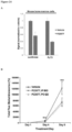
- a number of FLT3 kinase domain inhibitors including SU11248, SU5416, CEP-701 and PKC412 (midostaurin), have been shown to induce partial, and usually brief, remissions in clinical trials of relapsed AML patients when administered as single agents ( Weisberg et al. (2009) Drug Resist. Updates 12:81-89 ).
- midostaurin was shown to increase survival when combined with standard chemotherapy.
- This trial (RATIFY (CALGB 10603)) enrolled 717 AML patients with FLT3 mutations, randomized between midostaurin and placebo. Overall survival was increased in the midostaurin arm compared to the placebo arm (74.7 months vs.
- the present invention is based, at least in part, on the discovery that treatment approaches focusing on FLT3 degradation as opposed to or in addition to kinase inhibition are useful for treating cancers driven by FLT3.
- FLT3 the most commonly mutated gene in AML, is associated with a poor prognosis.
- FLT3 kinase inhibitors display significant clinical activity against acute myeloblastic leukemia (AML) with activating FLT3 mutations.
- AML acute myeloblastic leukemia
- drug resistance often develops rapidly.
- drug treatment leads to a compensatory increase in FLT3 protein, which may contribute to clinical drug resistance.
- the invention is directed in one aspect to an agent that inhibits the expression level and/or activity of at least one USP10 biomarker for use in treating a subject afflicted with FLT3 mutation-positive acute myeloblastic leukemia (AML), optionally wherein the at least one USP10 biomarker is selected from the group of USP10 biomarkers listed in Table 1, preferably
- the invention is directed in another aspect to an agent that inhibits the expression level and/or activity of at least one USP10 biomarker for use in inhibiting hyperproliferative growth of FLT3 mutation-positive AML cells, optionally wherein the at least one USP10 biomarker is selected from the group of USP10 biomarkers listed in Table 1, preferably
- the invention is directed in another aspect to an in vitro or ex vivo method of determining whether a subject afflicted with FLT3 mutation-positive AML would benefit from USP10 inhibitor therapy, the method comprising:
- the invention is directed in another aspect to a method of determining whether a subject afflicted with AML would not benefit from USP10 inhibitor therapy that inhibits the level of FLT3 protein, the method comprising detecting the presence of a FLT3 mutation in a biological sample from the subject, wherein the absence of a FLT3 mutation in the subject sample indicates that the subject afflicted with the AML would not benefit from USP10 inhibitor therapy that inhibits the level of FLT3 protein, preferably
- the invention is directed in another aspect to an in vitro or ex vivo method of assessing the efficacy of an agent for use in treating FLT3 mutation-positive AML in a subject, comprising:
- the invention is directed in another aspect to a method of monitoring the progression of FLT3 mutation-positive AML in a subject, comprising:
- the invention is directed in another aspect to an in vitro or ex vivo cell-based method for identifying an agent that inhibits hyperproliferative growth of AML cells, wherein the AML cells comprise a FLT3 mutation, the method comprising:
- the invention is directed in another aspect to a cell-free method for identifying an agent that inhibits hyperproliferative growth of AML cells, wherein the AML cells comprise a FLT3 mutation, the method comprising:
- a method of treating a subject afflicted with acute myeloblastic leukemia comprising administering to the subject an agent that inhibits the copy number, amount, and/or activity of at least one USP10 biomarker, thereby treating the subject afflicted with the AML, optionally wherein the at least one USP10 biomarker is selected from the group of USP10 biomarkers listed in Table 1.
- the agent is administered in a pharmaceutically acceptable formulation.
- the agent directly binds the at least one biomarker, optionally wherein the at least one USP10 biomarker is selected from the group of USP10 biomarkers listed in Table 1.
- the at least one USP10 biomarker is human USP10 or an ortholog thereof.
- the method further comprises administering at least one additional anti-cancer agents, optionally wherein the at least one additional anti-cancer agent inhibits the copy number, amount, and/or activity of at least one biomarker listed in Table 2.
- a method of inhibiting hyperproliferative growth of AML cells comprising contacting the AML cells with an agent that inhibits the copy number, amount, and/or activity of at least one USP10 biomarker, thereby inhibiting hyperproliferative growth of the AML cells, optionally wherein the at least one USP10 biomarker is selected from the group of USP10 biomarkers listed in Table 1.
- the step of contacting occurs in vivo, ex vivo, or in vitro, optionally wherein the AML cells die.
- the agent is administered in a pharmaceutically acceptable formulation.
- the agent directly binds the at least one biomarker, optionally wherein the USP10 biomarker is selected from the group of USP10 biomarkers listed in Table 1.
- the at least one USP10 biomarker is human USP10 or an ortholog thereof.
- the method further comprises administering at least one additional anti-cancer agents, optionally wherein the at least one additional anti-cancer agent inhibits the copy number, amount, and/or activity of at least one biomarker listed in Table 2.
- a method of determining whether a subject afflicted with AML or at risk for developing AML would benefit from USP10 inhibitor therapy comprising: a) obtaining a biological sample from the subject; b) determining the copy number, amount, and/or activity of at least one USP10 biomarker, optionally wherein the at least one USP10 biomarker is selected from the group consisting of USP10 biomarkers listed in Table 1, in a subject sample; c) determining the copy number, amount, and/or activity of the at least one USP10 biomarker in a control; and d) comparing the copy number, amount, and/or activity of the at least one USP10 biomarker detected in steps b) and c), wherein the presence of, or a significant increase in the copy number, amount, and/or activity of, the at least one USP10 biomarker in the subject sample relative to the control copy number, amount, and/or activity of the at least one USP10 biomarker indicates that the
- the method further comprises recommending, prescribing, or administering USP10 inhibitor therapy if the AML is determined to benefit from USP10 inhibitor therapy.
- the method further comprises recommending, prescribing, or administering anti-AML therapy other than USP10 inhibitor therapy if the AML is determined to not benefit from USP10 inhibitor therapy.
- the anti-AML therapy is selected from the group consisting of targeted therapy, chemotherapy, radiation therapy, and/or hormonal therapy.
- the control sample is determined from a cancerous or non-cancerous sample from either the patient or a member of the same species to which the patient belongs.
- the control sample comprises cells.
- the method further comprises determining responsiveness to USP10 inhibitor therapy measured by at least one criteria selected from the group consisting of clinical benefit rate, survival until mortality, pathological complete response, semi-quantitative measures of pathologic response, clinical complete remission, clinical partial remission, clinical stable disease, recurrence-free survival, metastasis free survival, disease free survival, circulating tumor cell decrease, circulating marker response, and RECIST criteria.
- a method of assessing the efficacy of an agent for treating AML in a subject comprising: a) detecting in a first subject sample and maintained in the presence of the agent the copy number, amount or activity of at least one USP10 biomarker, optionally wherein the USP10 biomarker is selected from the group of USP10 biomarkers listed in Table 1; b) detecting the copy number, amount, and/or activity of the at least one USP10 biomarker in a second subject sample and maintained in the absence of the test compound; and c) comparing the copy number, amount, and/or activity of the at least one USP10 biomarker from steps a) and b), wherein a significantly increased copy number, amount, and/or activity of the at least one USP10 biomarker in the first subject sample relative to the second subject sample, indicates that the agent treats the AML in the subject.
- a method of monitoring the progression of AML in a subject comprising: a) detecting in a subject sample at a first point in time the copy number, amount, and/or activity of at least one USP10 biomarker, optionally wherein the USP10 biomarker is selected from the group of USP10 biomarkers listed in Table 1; b) repeating step a) during at least one subsequent point in time after administration of a therapeutic agent; and c) comparing the copy number, amount, and/or activity detected in steps a) and b), wherein a significantly increased copy number, amount, and/or activity of the at least one USP10 biomarker in the first subject sample relative to at least one subsequent subject sample, indicates that the agent treats the AML in the subject.
- the subject between the first point in time and the subsequent point in time, has undergone treatment, completed treatment, and/or is in remission for the AML. In another embodiment, the subject has undergone USP10 inhibitor therapy between the first point in time and the subsequent point in time.
- the first and/or at least one subsequent sample is selected from the group consisting of ex vivo and in vivo samples. In yet another embodiment, the first and/or at least one subsequent sample is obtained from an animal model of AML. In another embodiment, the first and/or at least one subsequent sample is a portion of a single sample or pooled samples obtained from the subject.
- a cell-based method for identifying an agent that modulates hyperproliferative growth of AML cells and/or AML cell death comprising: a) contacting a cell expressing at least one USP10 biomarker, optionally wherein the USP10 biomarker is selected from the group of USP10 biomarkers listed in Table 1, with a test agent; and b) determining the effect of the test agent on the copy number, level of expression, or level of activity of the at least one USP10 biomarker to thereby identify an agent that that modulates hyperproliferative growth of AML cells and/or AML cell death.
- said cells are isolated from an animal model of AML.
- said cells are from a subject afflicted with AML. In yet another embodiment, said cells are unresponsive to USP10 inhibitor therapy.
- the step of contacting occurs in vivo, ex vivo, or in vitro, optionally wherein the agent inhibits hyperproliferative growth of AML cells and/or promotes AML cell death.
- the method further comprises determining the ability of the test agent to bind to the at least one USP10 biomarker before or after determining the effect of the test agent on the copy number, level of expression, or level of activity of the at least one USP10 biomarker, optionally wherein the agent inhibits hyperproliferative growth of AML cells and/or promotes AML cell death.
- the sample comprises cells, cell lines, histological slides, paraffin embedded tissue, fresh frozen tissue, fresh tissue, biopsies, blood, plasma, serum, buccal scrape, saliva, cerebrospinal fluid, urine, stool, mucus, or bone marrow, obtained from the subject.
- a cell-free method for identifying an agent that inhibits hyperproliferative growth of AML cells and/or promotes AML cell death comprising: a) determining the effect of a test agent on the amount or activity of at least one USP10 biomarker, optionally wherein the USP10 biomarker is selected from the group of USP10 biomarkers listed in Table 1, contacted with a test agent; b) determining the amount or activity of the at least one USP10 biomarker maintained in the absence of the test agent; and c) comparing the amount and/or activity of the at least one USP10 biomarker from steps a) and b), wherein a significantly decreased amount, and/or activity of the at least one USP10 biomarker in step a) relative to step b), identifies the test agent as an agent that inhibits hyperproliferative growth of AML cells and/or promotes AML cell death.
- the method further comprises determining the ability of the test agent to bind to the at least one USP10 biomarker before or after determining the effect of the test agent on the amount or activity of the at least one USP10 biomarker. In another embodiment, the method further comprises contacting an AML cell expressing the at least one USP10 biomarker with the test agent to confirm the ability of the test agent to inhibit hyperproliferative growth of AML cells and/or promote AML cell death.
- the copy number is assessed by microarray, quantitative PCR (qPCR), high-throughput sequencing, comparative genomic hybridization (CGH), or fluorescent in situ hybridization (FISH).
- the amount of the at least one USP10 biomarker is assessed by detecting the presence in the samples of a polynucleotide molecule encoding the biomarker or a portion of said polynucleotide molecule.
- the polynucleotide molecule is an mRNA, cDNA, or functional variants or fragments thereof.
- the step of detecting further comprises amplifying the polynucleotide molecule.
- the amount of the at least one biomarker is assessed by annealing a nucleic acid probe with the sample of the polynucleotide encoding the one or more biomarkers or a portion of said polynucleotide molecule under stringent hybridization conditions.
- the amount of the at least one biomarker is assessed by detecting the presence a polypeptide of the at least one USP10 biomarker.
- the presence of said polypeptide is detected using a reagent which specifically binds with said polypeptide.
- the reagent is selected from the group consisting of an antibody, an antibody derivative, and an antibody fragment.
- the activity of the at least one USP10 biomarker is assessed by determining the magnitude of modulation of the activity or expression level of at least one downstream target of the at least one USP10 biomarker.
- the at least one downstream target of the at least one USP10 biomarker is a human FLT3 or an ortholog thereof.
- the human FLT3 or an ortholog thereof is at least one human FLT3 selected from the group consisting of biomarkers listed in Table 2.
- the USP10 inhibitor therapy or test agent is an inhibitor selected from the group consisting of a small molecule, antisense nucleic acid, interfering RNA, shRNA, siRNA, miRNA, piwiRNA, aptamer, ribozyme, genome editing, dominant-negative protein binding partner, and combinations thereof.
- the USP10 inhibitor therapy or test agent is a small molecule.
- the small molecule is selected from the group consisting of small molecules listed in Figures 1-22 and Table 8.
- the USP10 inhibitor therapy or test agent is identified in a high-throughput screen.
- the USP10 inhibitor therapy or test agent also inhibits the activity or expression level of USP7.
- the USP10 inhibitor therapy or test agent does not inhibit the activity or expression level of p53.
- the at least one USP10 biomarker is 2, 3, 4, 5, 6, 7, 8, 9, 10, or more USP10 biomarkers.
- the USP10 inhibitor therapy or test agent modulates the activity or expression level of at least one downstream target of USP10.
- the activity or expression level of the at least one downstream target of USP10 is decreased.
- the at least one downstream target of USP10 is a human FLT3 or an ortholog thereof.
- the human FLT3 or an ortholog thereof is at least one human FLT3 selected from the group consisting of biomarkers listed in Table 2.
- the AML is adult AML or pediatric AML.
- the subject is a mammal, such as an animal model of AML or a human.
- numeric value refers to a compound name as follows:
- FLT3 kinase inhibitors display significant clinical activity against acute myeloblastic leukemia (AML) with activating FLT3 mutations.
- AML acute myeloblastic leukemia
- drug resistance often develops rapidly.
- drug treatment leads to a compensatory increase in FLT3 protein, which may contribute to clinical drug resistance.
- DAB deubiquitylating
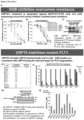
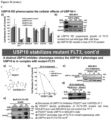
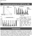
- USP10 is a FLT3 regulator (e.g. , a stabilizer of FLT3 activating mutants that drive AML) and that focusing on FLT3 degradation by modulating USP10, as opposed to focusing on FLT3 kinase inhibition, can treat AML.
- an element means one element or more than one element.
- administering is intended to include routes of administration which allow an agent to perform its intended function.
- routes of administration for treatment of a body which can be used include injection (subcutaneous, intravenous, parenterally, intraperitoneally, intrathecal, etc.), oral, inhalation, and transdermal routes.
- the injection can be bolus injections or can be continuous infusion.
- the agent can be coated with or disposed in a selected material to protect it from natural conditions which may detrimentally affect its ability to perform its intended function.
- the agent may be administered alone, or in conjunction with a pharmaceutically acceptable carrier.
- the agent also may be administered as a prodrug, which is converted to its active form in vivo.
- altered amount refers to increased or decreased copy number (e.g., germline and/or somatic) of a biomarker nucleic acid, e.g., increased or decreased expression level in a cancer sample, as compared to the expression level or copy number of the biomarker nucleic acid in a control sample.
- altered amount of a biomarker also includes an increased or decreased protein level of a biomarker protein in a sample, e.g., a cancer sample, as compared to the corresponding protein level in a normal, control sample.
- an altered amount of a biomarker protein may be determined by detecting posttranslational modification such as methylation status of the marker, which may affect the expression or activity of the biomarker protein.
- the amount of a biomarker in a subject is "significantly" higher or lower than the normal and/or amount of the biomarker, if the amount of the biomarker is greater or less, respectively, than the normal or control level by an amount greater than the standard error of the assay employed to assess amount, and preferably at least 20%, 30%, 40%, 50%, 60%, 70%, 80%, 90%, 100%, 150%, 200%, 300%, 350%, 400%, 500%, 600%, 700%, 800%, 900%, 1000% or than that amount.
- the amount of the biomarker in the subject can be considered "significantly" higher or lower than the normal and/or control amount if the amount is at least about two, and preferably at least about 5%, 10%, 15%, 20%, 25%, 30%, 35%, 40%, 45%, 50%, 55%, 60%, 65%, 70%, 75%, 80%, 85%, 90%, 95%, 100%, 105%, 110%, 115%, 120%, 125%, 130%, 135%, 140%, 145%, 150%, 155%, 160%, 165%, 170%, 175%, 180%, 185%, 190%, 195%, two times, three times, four times, five times, or more, or any range in between, such as 5%-100%, higher or lower, respectively, than the normal and/or control amount of the biomarker.
- Such significant modulation values can be applied to any metric described herein, such as altered level of expression, altered activity, changes in cancer cell hyperproliferative growth, changes in cancer cell death, changes in biomarker inhibition, changes in test agent
- altered level of expression of a biomarker refers to an expression level or copy number of the biomarker in a test sample, e.g., a sample derived from a patient suffering from cancer, that is greater or less than the standard error of the assay employed to assess expression or copy number, and is preferably at least twice, and more preferably three, four, five or ten or more times the expression level or copy number of the biomarker in a control sample (e.g., sample from a healthy subjects not having the associated disease) and preferably, the average expression level or copy number of the biomarker in several control samples.
- a test sample e.g., a sample derived from a patient suffering from cancer
- a control sample e.g., sample from a healthy subjects not having the associated disease
- the altered level of expression is greater or less than the standard error of the assay employed to assess expression or copy number, and is preferably at least twice, and more preferably three, four, five or ten or more times the expression level or copy number of the biomarker in a control sample (e.g., sample from a healthy subject not having the associated disease) and preferably, the average expression level or copy number of the biomarker in several control samples.
- a control sample e.g., sample from a healthy subject not having the associated disease
- altered activity of a biomarker refers to an activity of the biomarker which is increased or decreased in a disease state, e.g., in a cancer sample, as compared to the activity of the biomarker in a normal, control sample.
- Altered activity of the biomarker may be the result of, for example, altered expression of the biomarker, altered protein level of the biomarker, altered structure of the biomarker, or, e.g., an altered interaction with other proteins involved in the same or different pathway as the biomarker or altered interaction with transcriptional activators or inhibitors.
- altered structure of a biomarker refers to the presence of mutations or allelic variants within a biomarker nucleic acid or protein, e.g., mutations which affect expression or activity of the biomarker nucleic acid or protein, as compared to the normal or wild-type gene or protein.
- mutations include, but are not limited to substitutions, deletions, or addition mutations. Mutations may be present in the coding or non-coding region of the biomarker nucleic acid.
- antibody broadly encompass naturally-occurring forms of antibodies (e.g. IgG, IgA, IgM, IgE) and recombinant antibodies, such as single-chain antibodies, chimeric and humanized antibodies and multi-specific antibodies, as well as fragments and derivatives of all of the foregoing, which fragments and derivatives have at least an antigenic binding site.
- Antibody derivatives may comprise a protein or chemical moiety conjugated to an antibody.
- intrabodies are well-known antigen-binding molecules having the characteristic of antibodies, but that are capable of being expressed within cells in order to bind and/or inhibit intracellular targets of interest ( Chen et al. (1994) Human Gene Ther. 5:595-601 ).
- Methods are well-known in the art for adapting antibodies to target (e.g., inhibit) intracellular moieties, such as the use of single-chain antibodies (scFvs), modification of immunoglobulin VL domains for hyperstability, modification of antibodies to resist the reducing intracellular environment, generating fusion proteins that increase intracellular stability and/or modulate intracellular localization, and the like.
- Intracellular antibodies can also be introduced and expressed in one or more cells, tissues or organs of a multicellular organism, for example for prophylactic and/or therapeutic purposes (e.g. , as a gene therapy) (see, at least PCT Publs. WO 08/020079 , WO 94/02610 , WO 95/22618 , and WO 03/014960 ; U.S. Pat. No. 7,004,940 ; Cattaneo and Biocca (1997) Intracellular Antibodies: Development and Applications (Landes and Springer-Verlag publs .); Kontermann (2004) Methods 34:163-170 ; Cohen et al.
- antibody as used herein also includes an "antigen-binding portion" of an antibody (or simply “antibody portion”).
- antigen-binding portion refers to one or more fragments of an antibody that retain the ability to specifically bind to an antigen (e.g., a biomarker polypeptide or fragment thereof). It has been shown that the antigen-binding function of an antibody can be performed by fragments of a full-length antibody.
- binding fragments encompassed within the term "antigen-binding portion" of an antibody include (i) a Fab fragment, a monovalent fragment consisting of the VL, VH, CL and CH1 domains; (ii) a F(ab') 2 fragment, a bivalent fragment comprising two Fab fragments linked by a disulfide bridge at the hinge region; (iii) a Fd fragment consisting of the VH and CH1 domains; (iv) a Fv fragment consisting of the VL and VH domains of a single arm of an antibody, (v) a dAb fragment ( Ward et al., (1989) Nature 341:544-546 ), which consists of a VH domain; and (vi) an isolated complementarity determining region (CDR).
- a Fab fragment a monovalent fragment consisting of the VL, VH, CL and CH1 domains
- a F(ab') 2 fragment a bivalent fragment comprising two Fab fragments linked by
- the two domains of the Fv fragment, VL and VH are coded for by separate genes, they can be joined, using recombinant methods, by a synthetic linker that enables them to be made as a single protein chain in which the VL and VH regions pair to form monovalent polypeptides (known as single chain Fv (scFv); see e.g., Bird et al. (1988) Science 242:423-426 ; and Huston et al. (1988) Proc. Natl. Acad. Sci. USA 85:5879-5883 ; and Osbourn et al. 1998, Nature Biotechnology 16: 778 ).
- scFv single chain Fv
- Such single chain antibodies are also intended to be encompassed within the term "antigen-binding portion" of an antibody.
- Any VH and VL sequences of specific scFv can be linked to human immunoglobulin constant region cDNA or genomic sequences, in order to generate expression vectors encoding complete IgG polypeptides or other isotypes.
- VH and VL can also be used in the generation of Fab, Fv or other fragments of immunoglobulins using either protein chemistry or recombinant DNA technology.
- Other forms of single chain antibodies, such as diabodies are also encompassed.
- Diabodies are bivalent, bispecific antibodies in which VH and VL domains are expressed on a single polypeptide chain, but using a linker that is too short to allow for pairing between the two domains on the same chain, thereby forcing the domains to pair with complementary domains of another chain and creating two antigen binding sites (see e.g., Holliger et al. (1993) Proc. Natl. Acad. Sci. U.S.A. 90:6444-6448 ; Poljak et al. (1994) Structure 2:1121-1123 ).
- an antibody or antigen-binding portion thereof may be part of larger immunoadhesion polypeptides, formed by covalent or noncovalent association of the antibody or antibody portion with one or more other proteins or peptides.
- immunoadhesion polypeptides include use of the streptavidin core region to make a tetrameric scFv polypeptide ( Kipriyanov et al. (1995) Human Antibodies and Hybridomas 6:93-101 ) and use of a cysteine residue, biomarker peptide and a C-terminal polyhistidine tag to make bivalent and biotinylated scFv polypeptides ( Kipriyanov et al. (1994) Mol. Immunol.
- Antibody portions such as Fab and F(ab') 2 fragments, can be prepared from whole antibodies using conventional techniques, such as papain or pepsin digestion, respectively, of whole antibodies.
- antibodies, antibody portions and immunoadhesion polypeptides can be obtained using standard recombinant DNA techniques, as described herein.
- Antibodies may be polyclonal or monoclonal; xenogeneic, allogeneic, or syngeneic; or modified forms thereof (e.g. humanized, chimeric, etc.). Antibodies may also be fully human. Preferably, antibodies of the invention bind specifically or substantially specifically to a biomarker polypeptide or fragment thereof.
- monoclonal antibodies and “monoclonal antibody composition”, as used herein, refer to a population of antibody polypeptides that contain only one species of an antigen binding site capable of immunoreacting with a particular epitope of an antigen
- polyclonal antibodies and “polyclonal antibody composition” refer to a population of antibody polypeptides that contain multiple species of antigen binding sites capable of interacting with a particular antigen.
- a monoclonal antibody composition typically displays a single binding affinity for a particular antigen with which it immunoreacts.
- Antibodies may also be "humanized,” which is intended to include antibodies made by a non-human cell having variable and constant regions which have been altered to more closely resemble antibodies that would be made by a human cell. For example, by altering the non-human antibody amino acid sequence to incorporate amino acids found in human germline immunoglobulin sequences.
- the humanized antibodies of the invention may include amino acid residues not encoded by human germline immunoglobulin sequences ( e.g., mutations introduced by random or site-specific mutagenesis in vitro or by somatic mutation in vivo ), for example in the CDRs.
- the term "humanized antibody”, as used herein, also includes antibodies in which CDR sequences derived from the germline of another mammalian species, such as a mouse, have been grafted onto human framework sequences.
- the term "assigned score” refers to the numerical value designated for each of the biomarkers after being measured in a patient sample.
- the assigned score correlates to the absence, presence or inferred amount of the biomarker in the sample.
- the assigned score can be generated manually ( e.g., by visual inspection) or with the aid of instrumentation for image acquisition and analysis.
- the assigned score is determined by a qualitative assessment, for example, detection of a fluorescent readout on a graded scale, or quantitative assessment.
- an "aggregate score” which refers to the combination of assigned scores from a plurality of measured biomarkers, is determined.
- the aggregate score is a summation of assigned scores.
- combination of assigned scores involves performing mathematical operations on the assigned scores before combining them into an aggregate score.
- the aggregate score is also referred to herein as the predictive score.”
- Biomarker refers to a measurable entity of the present invention that has been determined to be predictive of anti-AML therapy (e.g., USP10 inhibitor therapy) effects.
- Biomarkers can include, without limitation, nucleic acids (e.g., genomic nucleic acids and/or transcribed nucleic acids) and proteins, particularly those involved shown in Table 1.
- nucleic acids e.g., genomic nucleic acids and/or transcribed nucleic acids
- proteins particularly those involved shown in Table 1.
- Many biomarkers listed in Table 1 are also useful as therapeutic targets. In one embodiment, such targets are USP10 members shown in Table 1 and/or Flt3 members shown in Table 2.
- blocking antibody or an antibody “antagonist” is one which inhibits or reduces at least one biological activity of the antigen(s) it binds.
- the blocking antibodies or antagonist antibodies or fragments thereof described herein substantially or completely inhibit a given biological activity of the antigen(s).
- body fluid refers to fluids that are excreted or secreted from the body as well as fluid that are normally not (e.g. amniotic fluid, aqueous humor, bile, blood and blood plasma, cerebrospinal fluid, cerumen and earwax, cowper's fluid or pre-ejaculatory fluid, chyle, chyme, stool, female ejaculate, interstitial fluid, intracellular fluid, lymph, menses, breast milk, mucus, pleural fluid, pus, saliva, sebum, semen, serum, sweat, synovial fluid, tears, urine, vaginal lubrication, vitreous humor, and vomit).
- fluid e.g. amniotic fluid, aqueous humor, bile, blood and blood plasma, cerebrospinal fluid, cerumen and earwax, cowper's fluid or pre-ejaculatory fluid, chyle, chyme, stool, female ejaculate, interstitial fluid, intracellular fluid,
- cancer or “tumor” or “hyperproliferative” refer to the presence of cells possessing characteristics typical of cancer-causing cells, such as uncontrolled proliferation, immortality, metastatic potential, rapid growth and proliferation rate, and certain characteristic morphological features. In some embodiments, such cells exhibit such characteristics in part or in full due to the expression and activity of oncogenes, such as FLT3 having mutations that activate FLT3 kinase activity. Cancer cells are often in the form of a tumor, but such cells may exist alone within an animal, or may be a non-tumorigenic cancer cell, such as a leukemia cell. As used herein, the term "cancer” includes premalignant as well as malignant cancers.
- Cancers include, but are not limited to, B cell cancer, e.g., multiple myeloma, Waldenström's macroglobulinemia, the heavy chain diseases, such as, for example, alpha chain disease, gamma chain disease, and mu chain disease, benign monoclonal gammopathy, and immunocytic amyloidosis, melanomas, breast cancer, lung cancer, bronchus cancer, colorectal cancer, prostate cancer, pancreatic cancer, stomach cancer, ovarian cancer, urinary bladder cancer, brain or central nervous system cancer, peripheral nervous system cancer, esophageal cancer, cervical cancer, uterine or endometrial cancer, cancer of the oral cavity or pharynx, liver cancer, kidney cancer, testicular cancer, biliary tract cancer, small bowel or appendix cancer, salivary gland cancer, thyroid gland cancer, adrenal gland cancer, osteosarcoma, chondrosarcoma, cancer of hematologic tissues, and the like.
- the heavy chain diseases such as, for
- cancers are epithlelial in nature and include but are not limited to, bladder cancer, breast cancer, cervical cancer, colon cancer, gynecologic cancers, renal cancer, laryngeal cancer, lung cancer, oral cancer, head and neck cancer, ovarian cancer, pancreatic cancer, prostate cancer, or skin cancer.
- the cancer is breast cancer, prostate cancer, lung cancer, or colon cancer.
- the epithelial cancer is non-small-cell lung cancer, nonpapillary renal cell carcinoma, cervical carcinoma, ovarian carcinoma ( e.g., serous ovarian carcinoma), or breast carcinoma.
- the epithelial cancers may be characterized in various other ways including, but not limited to, serous, endometrioid, mucinous, clear cell, Brenner, or undifferentiated.
- the cancer is acute myeloblastic leukemia (AML).
- AML can be adult AML, pediatric AML, or both.
- Acute myeloid leukemia (AML) also known as acute myelogenous leukemia, acute myeloblastic leukemia, acute granulocytic leukemia or acute nonlymphocytic leukemia, is a fast-growing form of cancer of the blood and bone marrow characterized by fatigue, shortness of breath, easy bruising and bleeding, and increased risk of infection.
- AML is the most common type of acute leukemia. It occurs when the bone marrow begins to make blasts, cells that have not yet completely matured. These blasts normally develop into white blood cells.
- AML AML involves higher percentages of dedifferentiated and undifferentiated cells, including more blasts (myeloblasts, monoblasts, and megakaryoblasts) than other leukemias. AML subtypes are classified based on the cell type from which the leukemia develops.
- the eight common AML subtypes include myeloblastic (M0) on special analysis, myeloblastic (M1) without maturation, myeloblastic (M2) with maturation, promyeloctic (M3), myelomonocytic (M4), monocytic (M5), erythroleukemia (M6), and megakaryocytic.
- myeloblastic M1
- myeloblastic M2
- M3 myeloblastic
- M4 myelomonocytic
- M5 monocytic
- erythroleukemia M6
- megakaryocytic Generally, the standard of care of treating AML is initial treatment with chemotherapy aimed at inducing a remission, although additional chemotherapy or a hematopoietic stem cell transplant may follow.
- AML AML-associated leukemia
- Some generalized symptoms include fever, fatigue, weight loss or loss of appetite, shortness of breath, anemia, easy bruising or bleeding, petechiae (flat, pin-head sized spots under the skin caused by bleeding), bone and joint pain, and persistent or frequent infections.
- Enlargement of the spleen may occur in AML, but it is typically mild and asymptomatic.
- Lymph node swelling is rare in AML, in contrast to acute lymphoblastic leukemia.
- the skin is involved about 10% of the time in the form of leukemia cutis. Rarely, Sweet's syndrome, a paraneoplastic inflammation of the skin, can occur with AML.
- AML AML-associated leukemia .
- a solid leukemic mass or tumor outside of the bone marrow called a chloroma.
- the first clue to a diagnosis of AML is typically an abnormal result on a complete blood count. While an excess of abnormal white blood cells (leukocytosis) is a common finding, and leukemic blasts are sometimes seen, AML can also present with isolated decreases in platelets, red blood cells, or even with a low white blood cell count (leukopenia).
- a definitive diagnosis of AML can be made by examination of the peripheral blood smear when there are circulating leukemic blasts, a definitive diagnosis usually requires an adequate bone marrow aspiration and biopsy.
- Marrow or blood is examined under light microscopy, as well as flow cytometry, to diagnose the presence of leukemia, to differentiate AML from other types of leukemia (e.g. acute lymphoblastic leukemia - ALL), and to classify the subtype of disease.
- a sample of marrow or blood is typically also tested for chromosomal abnormalities by routine cytogenetics or fluorescent in situ hybridization.
- the two most commonly used classification schemata for AML are the older French-American-British (FAB) system and the newer World Health Organization (WHO) system.
- WHO World Health Organization
- the diagnosis of AML is established by demonstrating involvement of more than 20% of the blood and/or bone marrow by leukemic myeloblasts, except in the three best prognosis forms of AML with recurrent genetic abnormalities (t(8;21), inv(16), and t(15;17)) in which the presence of the genetic abnormality is diagnostic irrespective of blast percent.
- the French-American-British (FAB) classification involves a blast percentage of at least 30% in bone marrow (BM) or peripheral blood (PB) for the diagnosis of AML.
- AML must be carefully differentiated from "preleukemic” conditions such as myelodysplastic or myeloproliferative syndromes, which are treated differently. Fluorescent in situ hybridization performed on blood or bone marrow is often used for diagnosis since it can identify the chromosomal translocation [t(15;17)(q22;q12);] (PML/RARA fusion protein oncogene) that characterizes APL, which is different from AML.
- coding region refers to regions of a nucleotide sequence comprising codons which are translated into amino acid residues
- non-coding region refers to regions of a nucleotide sequence that are not translated into amino acids (e.g., 5' and 3' untranslated regions).
- complementary refers to the broad concept of sequence complementarity between regions of two nucleic acid strands or between two regions of the same nucleic acid strand. It is known that an adenine residue of a first nucleic acid region is capable of forming specific hydrogen bonds ("base pairing") with a residue of a second nucleic acid region which is antiparallel to the first region if the residue is thymine or uracil. Similarly, it is known that a cytosine residue of a first nucleic acid strand is capable of base pairing with a residue of a second nucleic acid strand which is antiparallel to the first strand if the residue is guanine.
- a first region of a nucleic acid is complementary to a second region of the same or a different nucleic acid if, when the two regions are arranged in an antiparallel fashion, at least one nucleotide residue of the first region is capable of base pairing with a residue of the second region.
- the first region comprises a first portion and the second region comprises a second portion, whereby, when the first and second portions are arranged in an antiparallel fashion, at least about 50%, and preferably at least about 75%, at least about 90%, or at least about 95% of the nucleotide residues of the first portion are capable of base pairing with nucleotide residues in the second portion. More preferably, all nucleotide residues of the first portion are capable of base pairing with nucleotide residues in the second portion.
- control refers to any reference standard suitable to provide a comparison to the expression products in the test sample.
- the control comprises obtaining a "control sample” from which expression product levels are detected and compared to the expression product levels from the test sample.
- a control sample may comprise any suitable sample, including but not limited to a sample from a control cancer patient (can be stored sample or previous sample measurement) with a known outcome; normal tissue or cells isolated from a subject, such as a normal patient or the cancer patient, cultured primary cells/tissues isolated from a subject such as a normal subject or the cancer patient, adjacent normal cells/tissues obtained from the same organ or body location of the cancer patient, a tissue or cell sample isolated from a normal subject, or a primary cells/tissues obtained from a depository.
- control may comprise a reference standard expression product level from any suitable source, including but not limited to housekeeping genes, an expression product level range from normal tissue (or other previously analyzed control sample), a previously determined expression product level range within a test sample from a group of patients, or a set of patients with a certain outcome (for example, survival for one, two, three, four years, etc.) or receiving a certain treatment (for example, standard of care cancer therapy).
- a certain outcome for example, survival for one, two, three, four years, etc.
- a certain treatment for example, standard of care cancer therapy
- control samples and reference standard expression product levels can be used in combination as controls in the methods of the present invention.
- control may comprise normal or non-cancerous cell/tissue sample.
- control may comprise an expression level for a set of patients, such as a set of cancer patients, or for a set of cancer patients receiving a certain treatment, or for a set of patients with one outcome versus another outcome.
- the specific expression product level of each patient can be assigned to a percentile level of expression, or expressed as either higher or lower than the mean or average of the reference standard expression level.
- control may comprise normal cells, cells from patients treated with combination chemotherapy, and cells from patients having benign cancer.
- control may also comprise a measured value for example, average level of expression of a particular gene in a population compared to the level of expression of a housekeeping gene in the same population.
- control comprises a ratio transformation of expression product levels, including but not limited to determining a ratio of expression product levels of two genes in the test sample and comparing it to any suitable ratio of the same two genes in a reference standard; determining expression product levels of the two or more genes in the test sample and determining a difference in expression product levels in any suitable control; and determining expression product levels of the two or more genes in the test sample, normalizing their expression to expression of housekeeping genes in the test sample, and comparing to any suitable control.
- control comprises a control sample which is of the same lineage and/or type as the test sample.
- control may comprise expression product levels grouped as percentiles within or based on a set of patient samples, such as all patients with cancer.
- a control expression product level is established wherein higher or lower levels of expression product relative to, for instance, a particular percentile, are used as the basis for predicting outcome.
- a control expression product level is established using expression product levels from cancer control patients with a known outcome, and the expression product levels from the test sample are compared to the control expression product level as the basis for predicting outcome.
- the methods of the invention are not limited to use of a specific cut-point in comparing the level of expression product in the test sample to the control.
- the "copy number" of a biomarker nucleic acid refers to the number of DNA sequences in a cell (e.g., germline and/or somatic) encoding a particular gene product. Generally, for a given gene, a mammal has two copies of each gene. The copy number can be increased, however, by gene amplification or duplication, or reduced by deletion. For example, germline copy number changes include changes at one or more genomic loci, wherein said one or more genomic loci are not accounted for by the number of copies in the normal complement of germline copies in a control (e.g., the normal copy number in germline DNA for the same species as that from which the specific germline DNA and corresponding copy number were determined).
- Somatic copy number changes include changes at one or more genomic loci, wherein said one or more genomic loci are not accounted for by the number of copies in germline DNA of a control (e.g., copy number in germline DNA for the same subject as that from which the somatic DNA and corresponding copy number were determined).
- the "normal" copy number (e.g., germline and/or somatic) of a biomarker nucleic acid or "normal” level of expression of a biomarker nucleic acid, or protein is the activity/level of expression or copy number in a biological sample, e.g., a sample containing tissue, whole blood, serum, plasma, buccal scrape, saliva, cerebrospinal fluid, urine, stool, and bone marrow, from a subject, e.g., a human, not afflicted with cancer, or from a corresponding non-cancerous tissue in the same subject who has cancer.
- a biological sample e.g., a sample containing tissue, whole blood, serum, plasma, buccal scrape, saliva, cerebrospinal fluid, urine, stool, and bone marrow
- determining a suitable treatment regimen for the subject is taken to mean the determination of a treatment regimen (i.e., a single therapy or a combination of different therapies that are used for the prevention and/or treatment of the cancer in the subject) for a subject that is started, modified and/or ended based or essentially based or at least partially based on the results of the analysis according to the present invention.
- a treatment regimen i.e., a single therapy or a combination of different therapies that are used for the prevention and/or treatment of the cancer in the subject
- determining whether to provide targeted therapy against a cancer to provide anti-cancer therapy e.g., USP10 inhibitor therapy
- Another example is starting an adjuvant therapy after surgery whose purpose is to decrease the risk of recurrence, another would be to modify the dosage of a particular chemotherapy.
- the determination can, in addition to the results of the analysis according to the present invention, be based on personal characteristics of the subject to be treated. In most cases, the actual determination of the suitable treatment regimen for the subject will be
- expression signature refers to a group of two or more coordinately expressed biomarkers.
- the genes, proteins, and the like making up this signature may be expressed in a specific cell lineage, stage of differentiation, or during a particular biological response.
- the biomarkers can reflect biological aspects of the tumors in which they are expressed, such as the cell of origin of the cancer, the nature of the non-malignant cells in the biopsy, and the oncogenic mechanisms responsible for the cancer.
- Expression data and gene expression levels can be stored on computer readable media, e.g., the computer readable medium used in conjunction with a microarray or chip reading device. Such expression data can be manipulated to generate expression signatures.
- a molecule is "fixed” or "affixed” to a substrate if it is covalently or non-covalently associated with the substrate such that the substrate can be rinsed with a fluid (e.g. standard saline citrate, pH 7.4) without a substantial fraction of the molecule dissociating from the substrate.
- a fluid e.g. standard saline citrate, pH 7.4
- FLT3 refers to Fms-related tyrosine kinase 3, as a cytokine receptor which belongs to the receptor tyrosine kinase class III, and is alternatively known as "Fms-Related Tyrosine Kinase 3," “stem cell tyrosine kinase 1,” “Fms-Like Tyrosine Kinase 3,” “FL cytokine receptor,” “CD135,” “CD135 Antigen,” “EC 2.7.10.1,” “EC 2.7.10,” “FLK-2,” “STK1,” "growth factor receptor tyrosine kinase Type III,” “fetal liver kinase 2,” and “receptor-type tyrosine-protein kinase FLT3.” Somatic mutations that lead to constitutive activation of FLT3 are frequent in AML patients.
- Nucleic acid and amino acid sequence for FLT3 nucleic acids and protein are known in the art and are publicly available in the GenBank database maintained by the U.S. National Center for Biotechnology Information.
- human FLT3 nucleic acid sequences are well-known and include, for example, NM_004119.2 (variant 1, representing the shorter transcript and encoding the protein) and NR_130706.1 (variant 2, which contains an alternate internal exon compared to variant 1.
- Variant 2 is non-coding because the use of the 5'-most expected translational start codon as used in variant 1 renders the transcript a candidate for nonsense-mediated mRNA decay (NMD).
- Additional Flt3 human sequences include, without limitation, XM_017020486.1, XM_017020489.1, XM_017020487.1, XM_017020488.1, XM_011535015.2, XM_011535017.2, and XM_011535018.2.
- Human FLT3 amino acid sequences are well-known and include, for example, NP_004110.2 (variant 1, as above), XP_016875975.1, XP_016875978.1, XP_016875976.1, XP_016875977.1, XP_011533317.1, XP_011533319.1, and XP_011533320.1.
- Nucleic acid and amino acid sequence for FLT3 orthologs in other species are also well-known and include, for example, chimpanzee ( Pan troglodytes ) FLT3 (XM_509601.5 and XP_509601.2), rhesus monkey ( Macaca mulatta ) FLT3 (XM_015120801.1 and XP_014976287.1, XM_015120802.1 and XP_014976288.1, XM_001117913.2 and XP_001117913.1, XM_015120803.1 and XP_014976289.1), dog ( Canis lupus familiaris ) FLT3 (NM_001020811.1 and NP_001018647.1, XM_005635382.2 and XP_005635439.1, XM_014107333.1 and XP_013962808.1, XM_014107331.1 and XP_013962806.1,
- FLT3 inhibitors are well-known in the art and include, without limitation, sunitinib, sorafenib, midostaurin (PKC412), lestaurtinib (CEP-701), tandautinib (MLN518), quizartinib (AC220), and KW-2449 ( Wiernik et al. (2010) Clin. Adv. Hematol. Oncol. 8:429-437 ).
- anti-FLT3 detection agents are well-known in the art and include, without limitation, antibodies OAAF00442 (Aviva Systems Biology), 8F2 (Cell Signaling Technology), ab66035 (Abcam), PE A2F10 (eBioscience) ( Ju et al. (2011) Hybridoma 30:61-67 ; Piloto et al. (2005) Cancer Res. 65:1514-1522 )).
- homologous refers to nucleotide sequence similarity between two regions of the same nucleic acid strand or between regions of two different nucleic acid strands. When a nucleotide residue position in both regions is occupied by the same nucleotide residue, then the regions are homologous at that position. A first region is homologous to a second region if at least one nucleotide residue position of each region is occupied by the same residue. Homology between two regions is expressed in terms of the proportion of nucleotide residue positions of the two regions that are occupied by the same nucleotide residue.
- a region having the nucleotide sequence 5'-ATTGCC-3' and a region having the nucleotide sequence 5'-TATGGC-3' share 50% homology.
- the first region comprises a first portion and the second region comprises a second portion, whereby, at least about 50%, and preferably at least about 75%, at least about 90%, or at least about 95% of the nucleotide residue positions of each of the portions are occupied by the same nucleotide residue. More preferably, all nucleotide residue positions of each of the portions are occupied by the same nucleotide residue.
- Immune cell refers to cells that play a role in the immune response.
- Immune cells are of hematopoietic origin, and include lymphocytes, such as B cells and T cells; natural killer cells; myeloid cells, such as monocytes, macrophages, eosinophils, mast cells, basophils, and granulocytes.
- Immune checkpoint refers to a group of molecules on the cell surface of CD4+ and/or CD8+ T cells that fine-tune immune responses by down-modulating or inhibiting an anti-tumor immune response.
- Immune checkpoint proteins are well-known in the art and include, without limitation, CTLA-4, PD-1, VISTA, B7-H2, B7-H3, PD-L1, B7-H4, B7-H6, 2B4, ICOS, HVEM, PD-L2, CD160, gp49B, PIR-B, KIR family receptors, TIM-1, TIM-3, TIM-4, LAG-3, BTLA, SIRPalpha (CD47), CD48, 2B4 (CD244), B7.1, B7.2, ILT-2, ILT-4, TIGIT, and A2aR (see, for example, WO 2012/177624 ).
- the term further encompasses biologically active protein fragment, as well as nucleic acids encoding full-length immune checkpoint proteins and
- PD-1 refers to a member of the immunoglobulin gene superfamily that functions as a coinhibitory receptor having PD-L1 and PD-L2 as known ligands.
- PD-1 was previously identified using a subtraction cloning based approach to select for genes upregulated during TCR-induced activated T cell death.
- PD-1 is a member of the CD28/CTLA-4 family of molecules based on its ability to bind to PD-L1. Like CTLA-4, PD-1 is rapidly induced on the surface of T-cells in response to anti-CD3 ( Agata et al. 25 (1996) Int. Immunol.
- PD-1 is also induced on the surface of B-cells (in response to anti-IgM). PD-1 is also expressed on a subset of thymocytes and myeloid cells (Agata et al. (1996) supra; Nishimura et al. (1996) Int. Immunol. 8:773 ).
- Anti-immune checkpoint therapy refers to the use of agents that inhibit immune checkpoint nucleic acids and/or proteins. Immune checkpoints share the common function of providing inhibitory signals that suppress immune response and inhibition of one or more immune checkpoints can block or otherwise neutralize inhibitory signaling to thereby upregulate an immune response in order to more efficaciously treat cancer.
- agents useful for inhibiting immune checkpoints include antibodies, small molecules, peptides, peptidomimetics, natural ligands, and derivatives of natural ligands, that can either bind and/or inactivate or inhibit immune checkpoint proteins, or fragments thereof; as well as RNA interference, antisense, nucleic acid aptamers, etc.
- agents for upregulating an immune response include antibodies against one or more immune checkpoint proteins block the interaction between the proteins and its natural receptor(s); a non-activating form of one or more immune checkpoint proteins (e.g., a dominant negative polypeptide); small molecules or peptides that block the interaction between one or more immune checkpoint proteins and its natural receptor(s); fusion proteins (e.g. the extracellular portion of an immune checkpoint inhibition protein fused to the Fc portion of an antibody or immunoglobulin) that bind to its natural receptor(s); nucleic acid molecules that block immune checkpoint nucleic acid transcription or translation; and the like.
- a non-activating form of one or more immune checkpoint proteins e.g., a dominant negative polypeptide
- small molecules or peptides that block the interaction between one or more immune checkpoint proteins and its natural receptor(s)
- fusion proteins e.g. the extracellular portion of an immune checkpoint inhibition protein fused to the Fc portion of an antibody or immunoglobulin
- agents can directly block the interaction between the one or more immune checkpoints and its natural receptor(s) (e.g., antibodies) to prevent inhibitory signaling and upregulate an immune response.
- agents can indirectly block the interaction between one or more immune checkpoint proteins and its natural receptor(s) to prevent inhibitory signaling and upregulate an immune response.
- a soluble version of an immune checkpoint protein ligand such as a stabilized extracellular domain can binding to its receptor to indirectly reduce the effective concentration of the receptor to bind to an appropriate ligand.
- anti-PD-1 antibodies, anti-PD-L1 antibodies, and/or anti-PD-L2 antibodies are used to inhibit immune checkpoints.
- immune checkpoint inhibitors are known and publicly available including, for example, Keytruda ® (pembrolizumab; anti-PD-1 antibody), Opdivo ® (nivolumab; anti-PD-1 antibody), Tecentriq ® (atezolizumab; anti-PD-L1 antibody), durvalumab (anti-PD-L1 antibody), and the like.
- immune response includes T cell mediated and/or B cell mediated immune responses.
- exemplary immune responses include T cell responses, e.g., cytokine production and cellular cytotoxicity.
- immune response includes immune responses that are indirectly affected by T cell activation, e.g., antibody production (humoral responses) and activation of cytokine responsive cells, e.g., macrophages.
- immunotherapeutic agent can include any molecule, peptide, antibody or other agent which can stimulate a host immune system to generate an immune response to a tumor or cancer in the subject.
- Various immunotherapeutic agents are useful in the compositions and methods described herein.
- cancer is “inhibited” if at least one symptom of the cancer is alleviated, terminated, slowed, or prevented. As used herein, cancer is also “inhibited” if recurrence or metastasis of the cancer is reduced, slowed, delayed, or prevented. Similarly, a biological function, such as the function of a protein, is inhibited if it is decreased as compared to a reference state, such as a control like a wild-type state.
- USP10 activity of a USP10 protein that is contacted with a USP10 inhibitor is inhibited or deficient if the stability of FLT3 kinase is decreased due to contact with the USP10 inhibitor, in comparison to the USP10 protein not contacted with the USP10 inhibitor.
- kinase activity of a mutant FLT3 kinase is inhibited or deficient if the kinase activity is decreased due to the mutation and/or contact with the inhibitor, in comparison to the wild-type FLT3 kinase and/or the mutant FLT3 kinase not contacted with the inhibitor.
- Such inhibition or deficiency can be induced, such as by application of agent at a particular time and/or place, or can be constitutive, such as by a heritable mutation.
- Such inhibition or deficiency can also be partial or complete (e.g., essentially no measurable activity in comparison to a reference state, such as a control like a wild-type state). Essentially complete inhibition or deficiency is referred to as blocked.
- interaction when referring to an interaction between two molecules, refers to the physical contact (e.g., binding) of the molecules with one another. Generally, such an interaction results in an activity (which produces a biological effect) of one or both of said molecules.
- isolated protein refers to a protein that is substantially free of other proteins, cellular material, separation medium, and culture medium when isolated from cells or produced by recombinant DNA techniques, or chemical precursors or other chemicals when chemically synthesized.
- isolated or purified protein or biologically active portion thereof is substantially free of cellular material or other contaminating proteins from the cell or tissue source from which the antibody, polypeptide, peptide or fusion protein is derived, or substantially free from chemical precursors or other chemicals when chemically synthesized.
- substantially free of cellular material includes preparations of a biomarker polypeptide or fragment thereof, in which the protein is separated from cellular components of the cells from which it is isolated or recombinantly produced.
- the language "substantially free of cellular material” includes preparations of a biomarker protein or fragment thereof, having less than about 30% (by dry weight) of non-biomarker protein (also referred to herein as a "contaminating protein”), more preferably less than about 20% of non-biomarker protein, still more preferably less than about 10% of non-biomarker protein, and most preferably less than about 5% non-biomarker protein.
- non-biomarker protein also referred to herein as a "contaminating protein”
- polypeptide, peptide or fusion protein or fragment thereof e.g., a biologically active fragment thereof
- it is also preferably substantially free of culture medium, i.e., culture medium represents less than about 20%, more preferably less than about 10%, and most preferably less than about 5% of the volume of the protein preparation.
- kits is any manufacture (e.g. a package or container) comprising at least one reagent, e.g. a probe or small molecule, for specifically detecting and/or affecting the expression of a marker of the invention.
- the kit may be promoted, distributed, or sold as a unit for performing the methods of the present invention.
- the kit may comprise one or more reagents necessary to express a composition useful in the methods of the present invention.
- the kit may further comprise a reference standard, e.g., a nucleic acid encoding a protein that does not affect or regulate signaling pathways controlling cell growth, division, migration, survival or apoptosis.
- control proteins including, but not limited to, common molecular tags (e.g., green fluorescent protein and beta-galactosidase), proteins not classified in any of pathway encompassing cell growth, division, migration, survival or apoptosis by GeneOntology reference, or ubiquitous housekeeping proteins.
- Reagents in the kit may be provided in individual containers or as mixtures of two or more reagents in a single container.
- instructional materials which describe the use of the compositions within the kit can be included.
- neoadjuvant therapy refers to a treatment given before the primary treatment.
- neoadjuvant therapy can include chemotherapy, radiation therapy, and hormone therapy.
- the "normal" level of expression of a biomarker is the level of expression of the biomarker in cells of a subject, e.g., a human patient, not afflicted with a cancer.
- An “over-expression” or “significantly higher level of expression” of a biomarker refers to an expression level in a test sample that is greater than the standard error of the assay employed to assess expression, and is preferably at least 10%, and more preferably 1.2, 1.3, 1.4, 1.5, 1.6, 1.7, 1.8, 1.9, 2.0, 2.1, 2.1, 2.2, 2.3, 2.4, 2.5, 2.6, 2.7, 2.8, 2.9, 3, 3.5, 4, 4.5, 5, 5.5, 6, 6.5, 7, 7.5, 8, 8.5, 9, 9.5, 10, 10.5, 11, 12, 13, 14, 15, 16, 17, 18, 19, 20 times or more higher than the expression activity or level of the biomarker in a control sample (e.g., sample from a healthy subject not having the biomarker associated disease) and preferably, the
- a "significantly lower level of expression" of a biomarker refers to an expression level in a test sample that is at least 10%, and more preferably 1.2, 1.3, 1.4, 1.5, 1.6, 1.7, 1.8, 1.9, 2.0, 2.1, 2.1, 2.2, 2.3, 2.4, 2.5, 2.6, 2.7, 2.8, 2.9, 3, 3.5, 4, 4.5, 5, 5.5, 6, 6.5, 7, 7.5, 8, 8.5, 9, 9.5, 10, 10.5, 11, 12, 13, 14, 15, 16, 17, 18, 19, 20 times or more lower than the expression level of the biomarker in a control sample (e.g., sample from a healthy subject not having the biomarker associated disease) and preferably, the average expression level of the biomarker in several control samples.
- a control sample e.g., sample from a healthy subject not having the biomarker associated disease
- an "over-expression” or “significantly higher level of expression” of a biomarker refers to an expression level in a test sample that is greater than the standard error of the assay employed to assess expression, and is preferably at least 10%, and more preferably 1.2, 1.3, 1.4, 1.5, 1.6, 1.7, 1.8, 1.9, 2.0, 2.1, 2.1, 2.2, 2.3, 2.4, 2.5, 2.6, 2.7, 2.8, 2.9, 3, 3.5, 4, 4.5, 5, 5.5, 6, 6.5, 7, 7.5, 8, 8.5, 9, 9.5, 10, 10.5, 11, 12, 13, 14, 15, 16, 17, 18, 19, 20 times or more higher than the expression activity or level of the biomarker in a control sample (e.g., sample from a healthy subject not having the biomarker associated disease) and preferably, the average expression level of the biomarker in several control samples.
- a control sample e.g., sample from a healthy subject not having the biomarker associated disease
- a "significantly lower level of expression" of a biomarker refers to an expression level in a test sample that is at least 10%, and more preferably 1.2, 1.3, 1.4, 1.5, 1.6, 1.7, 1.8, 1.9, 2.0, 2.1, 2.1, 2.2, 2.3, 2.4, 2.5, 2.6, 2.7, 2.8, 2.9, 3, 3.5, 4, 4.5, 5, 5.5, 6, 6.5, 7, 7.5, 8, 8.5, 9, 9.5, 10, 10.5, 11, 12, 13, 14, 15, 16, 17, 18, 19, 20 times or more lower than the expression level of the biomarker in a control sample (e.g., sample from a healthy subject not having the biomarker associated disease) and preferably, the average expression level of the biomarker in several control samples.
- a control sample e.g., sample from a healthy subject not having the biomarker associated disease
- signaling levels can also be applied to any other measured parameter described herein, such as for expression, inhibition, cytotoxicity, cell growth, and the like.
- P53 refers to the well-known tumor suppressor, p53 (see, for example, Meek (2015) Biochem J. 469:325-346 ; Ballinger et al. (2015) Curr. Opin. Oncol. 27:332-337 ; Amelio and Melino (2015) Trends Biochem. Sci. 40:425-434 ; Saha et al. (2014) Prog. Biophys. Mol. Biol. 117:250-263 ; Tchelebit et al. (2014) Subcell. Biochem. 85:133-159 ; Yeudall (2014) Subcell. Biochem. 85:105-117 ; Santoro et al. (2014) Subcell. Biochem. 85:91-103 ; Girardini et al.
- the p53 gene which is located at 17p13.1, encodes at least 15 protein isoforms.
- the protein structure of the p53 protein is well-known and is characterized by certain domains.
- wild-type functional human p53 comprises:
- Mutations that make p53 deficient in cancer usually occur in the DBD. Most of these mutations destroy the ability of the protein to bind to its target DNA sequences, and thus prevents transcriptional activation of these genes. As such, mutations in the DBD are recessive loss-of-function mutations. Molecules of p53 with mutations in the OD dimerize with wild-type p53, and prevent them from activating transcription. Therefore, OD mutations have a dominant negative effect on the function of p53.
- Mutations in p53 nucleic acids that either do not encode functional p53 protein or p53 protein having reduced function are well-known in the art, as described above, and can be generated by any number of well-known types of mutation including, for example, a missense mutation (base change that alters the encoded amino acid), a nonsense mutation (base change that alters the encoded amino acid to a premature stop codon), a frameshift mutation (base addition or loss in a manner that is not a multiple of 3), an insertion mutation (any base addition, large or small in number, that alters the function of the encoded protein), a deletion mutation (any base deletion, large or small in number, that alters the function of the encoded protein), or a rearrangement mutation (any alteration, large or small, that alters the function of the encoded protein while retaining the starting amount of bases).
- a missense mutation base change that alters the encoded amino acid
- a nonsense mutation base change that alters the encoded amino acid to a premature stop codon
- mutations can be combined, such as when rearrangements are accompanied by additions and/or deletions, or multiple missense mutations are combined.
- the mutation is a genetic null (any mutation that completes ablates the function of the encoded protein) that arises in the germline, somatically, or both. This description of mutation types applies to any marker described herein.
- Assays for determining p53 activity, or reduction thereof, are well-known and commercially available (see, for example, Qiagen Cignal ® p53 reporter kit, Active Motif ® TransAM ® p53 reporter kit; Cayman Chemical p53 transcription factor assay kit item number 600020, Genecopoeia TM TF-Detect TM human p53 activity assay kit; Hiraki et al. (2015) Cell Chem. Biol. 22:1206-1216 ; Flaman et al. (1995) Proc. Natl. Acad. Sci. USA 92:3963-3967 (1995 ); and Kovvali et al. (2001) Nucl. Acids Res. 29:e28 ).
- Nucleic acid and amino acid sequences for p53 nucleic acids and protein are known in the art and are publicly available in the GenBank database maintained by the U.S. National Center for Biotechnology Information.
- human p53 nucleic acid and amino acid sequences are well-known and include, for example, NM_000546.5 (variant 1) and NP_000537.3 (isoform a); NM_001126112.2 (variant 2) and NP_001119584.1 (isoform a); NM_001126114.2 (variant 3) and NP_001119586.1 (isoform b); NM_001126113.2 (variant 4) and NP_001119585.1 (isoform c); NM_001126115.1 (variant 5) and NP_001119587.1 (isoform d); NM_001126116.1 (variant 6) and NP_001119588.1 (isoform e); NM_001126117.1 (variant 7) and NP_001119589.1 (is
- Nucleic acid and amino acid sequences of p53 orthologs in other species are also well-known and include, for example, mouse p53 (NM_001127233.1, NP_001120705.1, NM_011640.3, and NP_035770.2), chimpanzee p53 (XM_001172077.4 and XP_001172077.2), monkey p53 (NM_001047151.2 and NP_001040616.1), dog p53 (NM_001003210.1 and NP_001003210.1), cow p53 (NM_174201.2 and NP_776626.1), frog p53 (NM_001001903.1 and NP_001001903.1), and zebrafish p53 (NM_001271820.1, NP_001258749.1, NM_131327.3, and NP_571402.1).
- mouse p53 NM_001127233.1, NP_001120705.1, NM_011640.3, and NP_035
- p53 can further be used to refer to any combination of features described herein regarding p53.
- any combination of class, sequence composition, percentage identify, sequence length, domain structure, functional activity, etc. can be used to describe p53 as used according to the present invention.
- pre-determined biomarker amount and/or activity measurement(s) may be a biomarker amount and/or activity measurement(s) used to, by way of example only, evaluate a subject that may be selected for a particular treatment, evaluate a response to a treatment such as one or more USP10 inhibitors alone or in combination with one or more FLT3 inhibitors, and/or evaluate the disease state.
- a pre-determined biomarker amount and/or activity measurement(s) may be determined in populations of patients with or without cancer.
- the pre-determined biomarker amount and/or activity measurement(s) can be a single number, equally applicable to every patient, or the pre-determined biomarker amount and/or activity measurement(s) can vary according to specific subpopulations of patients.
- Age, weight, height, and other factors of a subject may affect the pre-determined biomarker amount and/or activity measurement(s) of the individual.
- the pre-determined biomarker amount and/or activity can be determined for each subject individually.
- the amounts determined and/or compared in a method described herein are based on absolute measurements.
- the amounts determined and/or compared in a method described herein are based on relative measurements, such as ratios ( e.g., serum biomarker normalized to the expression of housekeeping or otherwise generally constant biomarker).
- the pre-determined biomarker amount and/or activity measurement(s) can be any suitable standard.
- the pre-determined biomarker amount and/or activity measurement(s) can be obtained from the same or a different human for whom a patient selection is being assessed.
- the pre-determined biomarker amount and/or activity measurement(s) can be obtained from a previous assessment of the same patient. In such a manner, the progress of the selection of the patient can be monitored over time.
- the control can be obtained from an assessment of another human or multiple humans, e.g., selected groups of humans, if the subject is a human.
- the extent of the selection of the human for whom selection is being assessed can be compared to suitable other humans, e.g., other humans who are in a similar situation to the human of interest, such as those suffering from similar or the same condition(s) and/or of the same ethnic group.
- predictive includes the use of a biomarker nucleic acid and/or protein status, e.g., over- or under- activity, emergence, expression, growth, remission, recurrence or resistance of tumors before, during or after therapy, for determining the likelihood of response of a cancer to anti-cancer therapy, such as USP10 inhibitor therapy (e.g., USP10 inhibitors either alone or in combination with FLT3 inhibitors).
- USP10 inhibitor therapy e.g., USP10 inhibitors either alone or in combination with FLT3 inhibitors.
- Such predictive use of the biomarker may be confirmed by, e.g., (1) increased or decreased copy number ( e.g., by FISH, FISH plus SKY, single-molecule sequencing, e.g., as described in the art at least at J.
- Biotechnol., 86:289-301 , or qPCR overexpression or underexpression of a biomarker nucleic acid (e.g., by ISH, Northern Blot, or qPCR), increased or decreased biomarker protein (e.g., by IHC) and/or biomarker target, or increased or decreased activity, e.g., in more than about 5%, 6%, 7%, 8%, 9%, 10%, 11%, 12%, 13%, 14%, 15%, 20%, 25%, 30%, 40%, 50%, 60%, 70%, 80%, 90%, 95%, 100%, or more of assayed human cancers types or cancer samples; (2) its absolute or relatively modulated presence or absence in a biological sample, e.g., a sample containing tissue, whole blood, serum, plasma, buccal scrape, saliva, cerebrospinal fluid, urine, stool, or bone marrow, from a subject, e.g.
- a biological sample e.g., a sample containing tissue,
- a human, afflicted with cancer (3) its absolute or relatively modulated presence or absence in clinical subset of patients with cancer (e.g., those responding to a particular anti-cancer therapy (e.g., USP10 inhibitors either alone or in combination with FLT3 inhibitors) or those developing resistance thereto).
- a particular anti-cancer therapy e.g., USP10 inhibitors either alone or in combination with FLT3 inhibitors
- prevent refers to reducing the probability of developing a disease, disorder, or condition in a subject, who does not have, but is at risk of or susceptible to developing a disease, disorder, or condition.
- probe refers to any molecule which is capable of selectively binding to a specifically intended target molecule, for example, a nucleotide transcript or protein encoded by or corresponding to a biomarker nucleic acid. Probes can be either synthesized by one skilled in the art, or derived from appropriate biological preparations. For purposes of detection of the target molecule, probes may be specifically designed to be labeled, as described herein. Examples of molecules that can be utilized as probes include, but are not limited to, RNA, DNA, proteins, antibodies, and organic molecules.
- prognosis includes a prediction of the probable course and outcome of cancer or the likelihood of recovery from the disease.
- use of statistical algorithms provides a prognosis of cancer in an individual.
- the prognosis can be surgery, development of a clinical subtype of cancer (e.g., solid tumors, such as lung cancer, melanoma, and renal cell carcinoma), development of one or more clinical factors, development of intestinal cancer, or recovery from the disease.
- a clinical subtype of cancer e.g., solid tumors, such as lung cancer, melanoma, and renal cell carcinoma
- response to anti-cancer therapy relates to any response of the hyperproliferative disorder (e.g., cancer) to an anti-cancer therapy (e.g., USP10 inhibitors either alone or in combination with FLT3 inhibitors), preferably to a change in cancer cell numbers, tumor mass, and/or volume after initiation of neoadjuvant or adjuvant chemotherapy.
- Hyperproliferative disorder response may be assessed, for example for efficacy or in a neoadjuvant or adjuvant situation, where the size of a tumor after systemic intervention can be compared to the initial size and dimensions as measured by CT, PET, mammogram, ultrasound or palpation. Responses may also be assessed by caliper measurement or pathological examination of the tumor after biopsy or surgical resection. Response may be recorded in a quantitative fashion like percentage change in tumor volume or in a qualitative fashion like "pathological complete response" (pCR), "clinical complete remission” (cCR), “clinical partial remission” (cPR), “clinical stable disease” (cSD), “clinical progressive disease” (cPD) or other qualitative criteria.
- pCR pathological complete response
- cCR clinical complete remission
- cPR clinical partial remission
- cSD clinical stable disease
- cPD clinical progressive disease
- Assessment of hyperproliferative disorder response may be done early after the onset of neoadjuvant or adjuvant therapy, e.g., after a few hours, days, weeks or preferably after a few months.
- a typical endpoint for response assessment is upon termination of neoadjuvant chemotherapy or upon surgical removal of residual tumor cells and/or the tumor bed. This is typically three months after initiation of neoadjuvant therapy.
- clinical efficacy of the therapeutic treatments described herein may be determined by measuring the clinical benefit rate (CBR).
- the clinical benefit rate is measured by determining the sum of the percentage of patients who are in complete remission (CR), the number of patients who are in partial remission (PR) and the number of patients having stable disease (SD) at a time point at least 6 months out from the end of therapy.
- the CBR for a particular cancer therapeutic regimen is at least 25%, 30%, 35%, 40%, 45%, 50%, 55%, 60%, 65%, 70%, 75%, 80%, 85%, or more.
- Additional criteria for evaluating the response to cancer therapies are related to "survival,” which includes all of the following: survival until mortality, also known as overall survival (wherein said mortality may be either irrespective of cause or tumor related); "recurrence-free survival” (wherein the term recurrence shall include both localized and distant recurrence); metastasis free survival; disease free survival (wherein the term disease shall include cancer and diseases associated therewith).
- the length of said survival may be calculated by reference to a defined start point (e.g., time of diagnosis or start of treatment) and end point (e.g., death, recurrence or metastasis).
- criteria for efficacy of treatment can be expanded to include response to chemotherapy, probability of survival, probability of metastasis within a given time period, and probability of tumor recurrence.
- a particular cancer therapeutic regimen can be administered to a population of subjects and the outcome can be correlated to biomarker measurements that were determined prior to administration of any cancer therapy.
- the outcome measurement may be pathologic response to therapy given in the neoadjuvant setting.
- outcome measures such as overall survival and disease-free survival can be monitored over a period of time for subjects following cancer therapy for whom biomarker measurement values are known.
- the doses administered are standard doses known in the art for cancer therapeutic agents. The period of time for which subjects are monitored can vary.
- Biomarker measurement threshold values that correlate to outcome of a cancer therapy can be determined using well-known methods in the art, such as those described in the Examples section.
- resistance refers to an acquired or natural resistance of a cancer sample or a mammal to a cancer therapy (i.e., being nonresponsive to or having reduced or limited response to the therapeutic treatment), such as having a reduced response to a therapeutic treatment by 5% or more, for example, 10%, 15%, 20%, 25%, 30%, 35%, 40%, 45%, 50%, 55%, 60%, 65%, 70%, 75%, 80%, 85%, 90%, 95%, 100%, or more, to 2-fold, 3-fold, 4-fold, 5-fold, 10-fold, 15-fold, 20-fold or more.
- the reduction in response can be measured by comparing with the same cancer sample or mammal before the resistance is acquired, or by comparing with a different cancer sample or a mammal who is known to have no resistance to the therapeutic treatment.
- a typical acquired resistance to chemotherapy is called "multidrug resistance.”
- the multidrug resistance can be mediated by P-glycoprotein or can be mediated by other mechanisms, or it can occur when a mammal is infected with a multi-drug-resistant microorganism or a combination of microorganisms.
- the term "reverses resistance” means that the use of a second agent in combination with a primary cancer therapy (e.g., chemotherapeutic or radiation therapy) is able to produce a significant decrease in tumor volume at a level of statistical significance (e.g., p ⁇ 0.05) when compared to tumor volume of untreated tumor in the circumstance where the primary cancer therapy (e.g., chemotherapeutic or radiation therapy) alone is unable to produce a statistically significant decrease in tumor volume compared to tumor volume of untreated tumor. This generally applies to tumor volume measurements made at a time when the untreated tumor is growing log rhythmically.
- a primary cancer therapy e.g., chemotherapeutic or radiation therapy
- response refers to an anti-cancer response, e.g. in the sense of reduction of tumor size or inhibiting tumor growth.
- the terms can also refer to an improved prognosis, for example, as reflected by an increased time to recurrence, which is the period to first recurrence censoring for second primary cancer as a first event or death without evidence of recurrence, or an increased overall survival, which is the period from treatment to death from any cause.
- To respond or to have a response means there is a beneficial endpoint attained when exposed to a stimulus. Alternatively, a negative or detrimental symptom is minimized, mitigated or attenuated on exposure to a stimulus. It will be appreciated that evaluating the likelihood that a tumor or subject will exhibit a favorable response is equivalent to evaluating the likelihood that the tumor or subject will not exhibit favorable response ( i.e. , will exhibit a lack of response or be non-responsive).
- RNA interfering agent is defined as any agent which interferes with or inhibits expression of a target biomarker gene by RNA interference (RNAi).
- RNA interfering agents include, but are not limited to, nucleic acid molecules including RNA molecules which are homologous to the target biomarker gene of the invention, or a fragment thereof, short interfering RNA (siRNA), and small molecules which interfere with or inhibit expression of a target biomarker nucleic acid by RNA interference (RNAi).
- RNA interference is an evolutionally conserved process whereby the expression or introduction of RNA of a sequence that is identical or highly similar to a target biomarker nucleic acid results in the sequence specific degradation or specific post-transcriptional gene silencing (PTGS) of messenger RNA (mRNA) transcribed from that targeted gene (see Coburn, G. and Cullen, B. (2002) J. of Virology 76(18):9225 ), thereby inhibiting expression of the target biomarker nucleic acid.
- mRNA messenger RNA
- the RNA is double stranded RNA (dsRNA). This process has been described in plants, invertebrates, and mammalian cells.
- RNAi is initiated by the dsRNA-specific endonuclease Dicer, which promotes processive cleavage of long dsRNA into double-stranded fragments termed siRNAs.
- siRNAs are incorporated into a protein complex that recognizes and cleaves target mRNAs.
- RNAi can also be initiated by introducing nucleic acid molecules, e.g., synthetic siRNAs, shRNAs, or other RNA interfering agents, to inhibit or silence the expression of target biomarker nucleic acids.
- inhibiting target biomarker nucleic acid expression includes any decrease in expression or protein activity or level of the target biomarker nucleic acid or protein encoded by the target biomarker nucleic acid.
- the decrease may be of at least 30%, 40%, 50%, 60%, 70%, 80%, 90%, 95% or 99% or more as compared to the expression of a target biomarker nucleic acid or the activity or level of the protein encoded by a target biomarker nucleic acid which has not been targeted by an RNA interfering agent.
- genome editing can be used to modulate the copy number or genetic sequence of a biomarker of interest, such as constitutive or induced knockout or mutation of a USP10 biomarker of interest.
- the CRISPR-Cas system can be used for precise editing of genomic nucleic acids (e.g., for creating non-functional or null mutations).
- the CRISPR guide RNA and/or the Cas enzyme may be expressed.
- a vector containing only the guide RNA can be administered to an animal or cells transgenic for the Cas9 enzyme. Similar strategies may be used (e.g., designer zinc finger, transcription activator-like effectors (TALEs) or homing meganucleases).
- TALEs transcription activator-like effectors
- sample used for detecting or determining the presence or level of at least one biomarker is typically whole blood, plasma, serum, saliva, urine, stool ( e.g., feces), tears, and any other bodily fluid (e.g., as described above under the definition of "body fluids"), or a tissue sample (e.g., biopsy) such as a small intestine, colon sample, or surgical resection tissue.
- the method of the present invention further comprises obtaining the sample from the individual prior to detecting or determining the presence or level of at least one marker in the sample.
- selective inhibition refers to the agent's ability to selectively reduce the target signaling activity as compared to off-target signaling activity, via direct or interact interaction with the target.
- an agent that selectively inhibits USP10 over another deubiquitylating (DUB) enzyme, such as USP7 has an activity against USP10 that is at least 5%, 10%, 15%, 20%, 25%, 30%, 35%, 40%, 45%, 50%, 55%, 60%, 65%, 70%, 75%, 80%, 85%, 90%, 95%, 100%, 110%, 120%, 130%, 140%, 150%, 160%, 170%, 180%, 190%, or 2x (times) more than the compound's activity against the comparison protein (e.g ., at least about 3x, 4x, 5x, 6x, 7x, 8x, 9x, 10x, 15x, 20x, 25x, 30x, 35x, 40x, 45
- USP7/HAUSP peers virus-associated USP
- USP7 also contains an N-terminal TRAF-like MATH domain ( Zapata et al. (2001) J. Biol. Chem. 276:24242-24252 ) and a C-terminal domain that contains at least five ubiquitin-like domains ( Faesen et al. (2011) Mol. Cell 44:147-159 ).
- USP7 is primarily a nuclear protein and localizes to a subset of PML bodies ( Everett et al. (1999) J. Virol. 73:417-426 ; Muratani et al. (2002) Nat. Cell Biol. 4:106-110 ).
- USP7 has been shown to regulate the steady-state level of several poly-ubiquitinated substrates.
- USP7 alters the level of the p53 and p16 INK4a tumor suppressors through Mdm2 stabilization and Bmi1/Mel18 stabilization, respectively ( Cummins et al. (2004) Nature 428 ; Li et al. (2004) Mol. Cell 13:8790-896 ; Maertens et al. (2010) EMBO J. 29:2553-2565 ).
- USP7 binding to p53 was recently shown to be regulated by TSPYL5, a protein potentially involved in breast oncogenesis through its competition with p53 for binding to the same region of USP7 ( Epping et al. (2011) Nat. Cell Biol. 13:102-108 ). Additional proteins involved in genomic integrity and regulation, such as the DNMT1 DNA methylase and the claspin adaptor, are also stabilized by USP7 ( Du et al. (2010) Sci. Signal. 3:ra80 ; Faustrup et al. (2009) J. Cell Biol. 184:13-19 ). USP7 has also been shown to regulate the cellular compartmentalization of several mono-ubiquitinated substrates by deubiquitination.
- cancer means to alter cancer cells or tumor cells in a way that allows for more effective treatment of the associated cancer with a cancer therapy (e.g., USP10 inhibitors either alone or in combination with FLT3 inhibitors; chemotherapeutic; and/or radiation therapy).
- a cancer therapy e.g., USP10 inhibitors either alone or in combination with FLT3 inhibitors; chemotherapeutic; and/or radiation therapy.
- normal cells are not affected to an extent that causes the normal cells to be unduly injured by the anti-cancer therapy (e.g., USP10 inhibitors either alone or in combination with FLT3 inhibitors).
- An increased sensitivity or a reduced sensitivity to a therapeutic treatment is measured according to a known method in the art for the particular treatment and methods described herein below, including, but not limited to, cell proliferative assays ( Tanigawa N, Kern D H, Kikasa Y, Morton D L, Cancer Res 1982; 42: 2159-2164 ), cell death assays ( Weisenthal L M, Shoemaker R H, Marsden J A, Dill P L, Baker J A, Moran E M, Cancer Res 1984; 94: 161-173 ; Weisenthal L M, Lippman M E, Cancer Treat Rep 1985; 69: 615-632 ; Weisenthal L M, In: Kaspers G J L, Pieters R, Twentyman P R, Weisenthal L M, Veerman A J P, eds.
- the sensitivity or resistance may also be measured in animal by measuring the tumor size reduction over a period of time, for example, 6 month for human and 4-6 weeks for mouse.
- a composition or a method sensitizes response to a therapeutic treatment if the increase in treatment sensitivity or the reduction in resistance is 5% or more, for example, 10%, 15%, 20%, 25%, 30%, 35%, 40%, 45%, 50%, 55%, 60%, 65%, 70%, 75%, 80%, 85%, 90%, 95%, 100%, or more, to 2-fold, 3-fold, 4-fold, 5-fold, 10-fold, 15-fold, 20-fold or more, compared to treatment sensitivity or resistance in the absence of such composition or method.
- the determination of sensitivity or resistance to a therapeutic treatment is routine in the art and within the skill of an ordinarily skilled clinician. It is to be understood that any method described herein for enhancing the efficacy of a cancer therapy can be equally applied to methods for sensitizing hyperproliferative or otherwise cancerous cells (e.g., resistant cells) to the cancer therapy.
- the term “synergistic effect” refers to the combined effect of two or more therapeutic agents, such as two or more USP10 inhibitors, a USP10 inhibitor and a FLT3 inhibitor, USP10 inhibitors either alone or in combination with FLT3 inhibitors, and the like, can be greater than the sum of the separate effects of the anticancer agents alone.
- siRNA Short interfering RNA
- small interfering RNA is defined as an agent which functions to inhibit expression of a target biomarker nucleic acid, e.g., by RNAi.
- An siRNA may be chemically synthesized, may be produced by in vitro transcription, or may be produced within a host cell.
- siRNA is a double stranded RNA (dsRNA) molecule of about 15 to about 40 nucleotides in length, preferably about 15 to about 28 nucleotides, more preferably about 19 to about 25 nucleotides in length, and more preferably about 19, 20, 21, or 22 nucleotides in length, and may contain a 3' and/or 5' overhang on each strand having a length of about 0, 1, 2, 3, 4, or 5 nucleotides.
- the length of the overhang is independent between the two strands, i.e., the length of the overhang on one strand is not dependent on the length of the overhang on the second strand.
- the siRNA is capable of promoting RNA interference through degradation or specific post-transcriptional gene silencing (PTGS) of the target messenger RNA (mRNA).
- PTGS post-transcriptional gene silencing
- an siRNA is a small hairpin (also called stem loop) RNA (shRNA).
- shRNAs are composed of a short (e.g., 19-25 nucleotide) antisense strand, followed by a 5-9 nucleotide loop, and the analogous sense strand.
- the sense strand may precede the nucleotide loop structure and the antisense strand may follow.
- shRNAs may be contained in plasmids, retroviruses, and lentiviruses and expressed from, for example, the pol III U6 promoter, or another promoter (see, e.g., Stewart, et al. (2003) RNA Apr;9(4):493-501 n).
- RNA interfering agents e.g., siRNA molecules
- RNA interfering agents may be administered to a patient having or at risk for having cancer, to inhibit expression of a biomarker gene which is overexpressed in cancer and thereby treat, prevent, or inhibit cancer in the subject.
- subject refers to any healthy animal, mammal or human, or any animal, mammal or human afflicted with a cancer, e.g., lung, ovarian, pancreatic, liver, breast, prostate, and colon carcinomas, as well as melanoma and multiple myeloma.
- a cancer e.g., lung, ovarian, pancreatic, liver, breast, prostate, and colon carcinomas, as well as melanoma and multiple myeloma.
- subject is interchangeable with "patient.”
- survival includes all of the following: survival until mortality, also known as overall survival (wherein said mortality may be either irrespective of cause or tumor related); "recurrence-free survival” (wherein the term recurrence shall include both localized and distant recurrence); metastasis free survival; disease free survival (wherein the term disease shall include cancer and diseases associated therewith).
- the length of said survival may be calculated by reference to a defined start point (e.g. time of diagnosis or start of treatment) and end point (e.g. death, recurrence or metastasis).
- criteria for efficacy of treatment can be expanded to include response to chemotherapy, probability of survival, probability of metastasis within a given time period, and probability of tumor recurrence.
- therapeutic effect refers to a local or systemic effect in animals, particularly mammals, and more particularly humans, caused by a pharmacologically active substance.
- the term thus means any substance intended for use in the diagnosis, cure, mitigation, treatment or prevention of disease or in the enhancement of desirable physical or mental development and conditions in an animal or human.
- therapeutically-effective amount means that amount of such a substance that produces some desired local or systemic effect at a reasonable benefit/risk ratio applicable to any treatment.
- a therapeutically effective amount of a compound will depend on its therapeutic index, solubility, and the like.
- certain compounds discovered by the methods of the present invention may be administered in a sufficient amount to produce a reasonable benefit/risk ratio applicable to such treatment.
- terapéuticaally-effective amount and “effective amount” as used herein means that amount of a compound, material, or composition comprising a compound of the present invention which is effective for producing some desired therapeutic effect in at least a sub-population of cells in an animal at a reasonable benefit/risk ratio applicable to any medical treatment.
- Toxicity and therapeutic efficacy of subject compounds may be determined by standard pharmaceutical procedures in cell cultures or experimental animals, e.g., for determining the LD 50 and the ED 50 . Compositions that exhibit large therapeutic indices are preferred.
- the LD 50 (lethal dosage) can be measured and can be, for example, at least 10%, 20%, 30%, 40%, 50%, 60%, 70%, 80%, 90%, 100%, 200%, 300%, 400%, 500%, 600%, 700%, 800%, 900%, 1000% or more reduced for the agent relative to no administration of the agent.
- the ED 50 i.e., the concentration which achieves a half-maximal inhibition of symptoms
- the ED 50 i.e., the concentration which achieves a half-maximal inhibition of symptoms
- the ED 50 i.e., the concentration which achieves a half-maximal inhibition of symptoms
- the ED 50 i.e., the concentration which achieves a half-maximal inhibition of symptoms
- the ED 50 i.e., the concentration which achieves a half-maximal inhibition of symptoms
- the IC 50 i.e., the concentration which achieves half-maximal cytotoxic or cytostatic effect on cancer cells
- the IC 50 can be measured and can be, for example, at least 10%, 20%, 30%, 40%, 50%, 60%, 70%, 80%, 90%, 100%, 200%, 300%, 400%, 500%, 600%, 700%, 800%, 900%, 1000% or more increased for the agent relative to no administration of the agent.
- cancer cell growth in an assay can be inhibited by at least about 10%, 15%, 20%, 25%, 30%, 35%, 40%, 45%, 50%, 55%, 60%, 65%, 70%, 75%, 80%, 85%, 90%, 95%, or even 100%.
- Cancer cell death can be promoted by at least about 10%, 15%, 20%, 25%, 30%, 35%, 40%, 45%, 50%, 55%, 60%, 65%, 70%, 75%, 80%, 85%, 90%, 95%, or even 100%. In another embodiment, at least about a 10%, 15%, 20%, 25%, 30%, 35%, 40%, 45%, 50%, 55%, 60%, 65%, 70%, 75%, 80%, 85%, 90%, 95%, or even 100% decrease in cancer cell numbers and/or a solid malignancy can be achieved.
- a “transcribed polynucleotide” or “nucleotide transcript” is a polynucleotide (e.g. an mRNA, hnRNA, a cDNA, or an analog of such RNA or cDNA) which is complementary to or homologous with all or a portion of a mature mRNA made by transcription of a biomarker nucleic acid and normal post-transcriptional processing (e.g. splicing), if any, of the RNA transcript, and reverse transcription of the RNA transcript.
- a polynucleotide e.g. an mRNA, hnRNA, a cDNA, or an analog of such RNA or cDNA
- USP10 belongs to the largest family of DUB enzymes referred to as the ubiquitin specific protease (USP) family. This family is comprised of 56 cysteine protease members that are most well known for their ability to remove post-translational ubiquitin tags that mark substrates for proteosomal degradation thereby resulting in stabilization of the substrate.
- the reported substrates of USP10 include Beclin 1 ( Liu et al. (2011) Cell 147:223-234 ), CFTR ( Bomberger et al. (2009) J. Biol. Chem. 284:18778-18789 ), and p53 ( Yuan et al. (2010) Cell 140:384-396 ).
- USP10 is ubiquitously expressed and, as is true for many DUBs, may have diverse functions depending of the cellular context.
- USP10 functions as a co-factor of the DNA-bound androgen receptor complex, and is inhibited by Ras-GAP SH3 domain binding protein (G3BP) in the Ras-GTPase pathway ( Faus et al. (2005) Mol. Cell Endocrinol. 245:138-146 ; Soncini et al. (2001) Oncogene 20:3869-3879 ).
- G3BP Ras-GAP SH3 domain binding protein
- Nucleic acid and amino acid sequence for USP10 nucleic acids and protein are well-known in the art and are publicly available in the GenBank database maintained by the U.S. National Center for Biotechnology Information.
- human USP10 nucleic acid sequences are well-known and include, for example, NM_001272075.1 (variant 1, representing the longest transcript and encoding the longest isoform 1) and NM_005153.2 (variant 2, which lacks an alternate exon in the 5' end compared to variant 1).
- Isoform 2 encoded by variant 2 has a shorter and distinct N-terminus compared to isoform 1).
- NR_073577 transcription variant 3
- NR_073578.1 (transcript variant 4), lacks four alternate internal exons, compared to variant 1.
- This variant is represented as non-coding due to the presence of two upstream ORFs that are predicted to interfere with translation of the longest ORF; translation of either of the upstream ORFs renders the transcript a candidate for nonsense-mediated mRNA decay (NMD)), and predicted sequences XM_017023869.1, XM_017023864.1, XM_011523441.1, XM_011523440.1, XM_017023868.1, XM_011523443.1, XM_017023863.1, XM_006721332.1, XM_017023867.1, XM_017023865.1, and XM_017023866.1.
- Human FLT3 amino acid sequences are well-known and include, for example, NP_001259004.1 (isoform 1), NP_005144.2 (isoform 2) and predicted sequences XP_016879358.1, XP_016879353.1, XP_011521743.1, XP_011521742.1, XP_016879357.1, XP_011521745.1, XP_016879352.1, XP_006721395.1, XP_016879356.1, XP_016879354.1 and XP_016879355.1.
- Nucleic acid and amino acid sequence for USP10 orthologs in other species are also well-known and include, for example, chimpanzee (Pan troglodytes) USP10 (XM_016930295.1 and XP_016785784.1, XM_009431337.2 and XP_009429612.2); rhesus monkey (Macaca mulatta) USP10 (1XM_015126723.1 and XP_014982209.1, XM_015126724.1 and XP_014982210.1); dog (Canis lupus familiaris) USP10 (XM_005620883.1 and XP_005620940.1); cattle (Bos taurus) USP10 (BC142223.1 and AA142224.1); mouse (Mus musculus) USP10 (NM_001310630.1 and NP_001297559.1, NM_009462.2 and NP_033488.1, XM_00653084
- Inhibitors of USP10 are also well-known in the art and include, spautin-1 (specific and potent autophagy inhibitor-1), a derivative of MBCQ that binds to USP10 and inhibits deubiquitinase activity.
- Various anti-USP10 antibodies are commercially available recognizing the N-terminus, C-terminus, or internal region of USP10.
- USP10 variants and mutations are also well-known and include, for example, a T-to-A substitution at position 42 of SEQ ID NO: 4 which, when combined with an S-to-A substitution at position 337 of SEQ ID NO: 4, abolishes its phosphorylation by Ataxia telangiectasia mutated (ATM) (Yuan et al., 2010); a T-to-E substitution at position 42 of SEQ ID NO: 4 which, when combined with an S-to-D substitution at position 337 of SEQ ID NO: 4, results in a Phospho-mimetic mutant that translocates to the nucleus in absence of genotoxic stress (Yuan et al., 2010); a C-to-A substitution at position 424 of SEQ ID NO: 4 which abolishes its de-ubiquitinating activity (Soncini et al., 2001).
- ATM Ataxia telangiectasia mutated
- nucleotide triplet An important and well known feature of the genetic code is its redundancy, whereby, for most of the amino acids used to make proteins, more than one coding nucleotide triplet may be employed (illustrated above). Therefore, a number of different nucleotide sequences may code for a given amino acid sequence. Such nucleotide sequences are considered functionally equivalent since they result in the production of the same amino acid sequence in all organisms (although certain organisms may translate some sequences more efficiently than they do others). Moreover, occasionally, a methylated variant of a purine or pyrimidine may be found in a given nucleotide sequence. Such methylations do not affect the coding relationship between the trinucleotide codon and the corresponding amino acid.
- nucleotide sequence of a DNA or RNA encoding a biomarker nucleic acid can be used to derive the polypeptide amino acid sequence, using the genetic code to translate the DNA or RNA into an amino acid sequence.
- polypeptide amino acid sequence corresponding nucleotide sequences that can encode the polypeptide can be deduced from the genetic code (which, because of its redundancy, will produce multiple nucleic acid sequences for any given amino acid sequence).
- description and/or disclosure herein of a nucleotide sequence which encodes a polypeptide should be considered to also include description and/or disclosure of the amino acid sequence encoded by the nucleotide sequence.
- description and/or disclosure of a polypeptide amino acid sequence herein should be considered to also include description and/or disclosure of all possible nucleotide sequences that can encode the amino acid sequence.
- nucleic acid and amino acid sequence information for the loci and biomarkers of the present invention and related biomarkers are well known in the art and readily available on publicly available databases, such as the National Center for Biotechnology Information (NCBI).
- NCBI National Center for Biotechnology Information
- exemplary nucleic acid and amino acid sequences derived from publicly available sequence databases are provided below.
- biomarkers described above are presented below in Tables 1 and 2. It is to be noted that the terms described above can further be used to refer to any combination of features described herein regarding the biomarkers. For example, any combination of sequence composition, percentage identify, sequence length, domain structure, functional activity, etc. can be used to describe a biomarker of the present invention.
- RNA nucleic acid molecules e.g., thymines replaced with uredines
- DNA or RNA nucleic acid sequences comprising a nucleic acid sequence having at least 95%, 96%, 97%, 98%, 99%, 99.5%, or more identity across their full length with the nucleic acid sequence of any SEQ ID NO listed in Table 1, or a portion thereof, and such nucleic acid molecules having a function of the full-length nucleic acid as described further herein.
- polypeptide molecules comprising an amino acid sequence having at least 95%, 96%, 97%, 98%, 99%, 99.5%, or more identity across their full length with an amino acid sequence of any SEQ ID NO listed in Table 1, or a portion thereof, and such polypeptides having a function of the full-length polypeptide as described further herein.
- polypeptide molecules comprising an amino acid sequence having at least 80%, 81%, 82%, 83%, 84%, 85%, 86%, 87%, 88%, 89%, 90%, 91%, 92%, 93%, 94%, 95%, 96%, 97%, 98%, 99%, 99.5%, or more identity across their full length with an amino acid sequence of any SEQ ID NO listed in Table 1, or a portion thereof.
- polypeptides can have a function of the full-length polypeptide as described further herein.
- RNA nucleic acid molecules e.g., thymines replaced with uredines
- nucleic acid molecules encoding orthologs of the encoded proteins
- DNA or RNA nucleic acid sequences comprising a nucleic acid sequence having at least 80%, 81%, 82%, 83%, 84%, 85%, 86%, 87%, 88%, 89%, 90%, 91%, 92%, 93%, 94%, 95%, 96%, 97%, 98%, 99%, 99.5%, or more identity across their full length with the nucleic acid sequence of any SEQ ID NO listed in Table 2, or a portion thereof.
- nucleic acid molecules can have a function of the full-length nucleic acid as described further herein.
- Table 2 includes orthologs of the proteins, as well as polypeptide molecules comprising an amino acid sequence having at least 80%, 81%, 82%, 83%, 84%, 85%, 86%, 87%, 88%, 89%, 90%, 91%, 92%, 93%, 94%, 95%, 96%, 97%, 98%, 99%, 99.5%, or more identity across their full length with an amino acid sequence of any SEQ ID NO listed in Table 2, or a portion thereof.
- polypeptides can have a function of the full-length polypeptide as described further herein.
- FLT3 mutants described below and/or in the Examples, particularly activating mutations that enhance FLT3 kinase activity to drive cancer, which are well-known.
- Such mutants include, for example, a Y-to-F substitution at position 589 of SEQ ID NO: 8, which reduces phosphorylation in response to ligand binding and abolishes activation of STATSA, but has no effect on the phosphorylation of the constitutively activated mutant kinase variants ( Kiyoi et al. (1998) Leukemia 12:1333-1337 ; Rocnik et al.
- FLT3 The potential amino acid modifications of FLT3 are also well known and include, for example, disulfide bonds at positions 35/65, 103/114, 199/206, 232/241, 272/330, 368/407, and 381/392 of SEQ ID NO: 8, glycosylations at positions of 43, 100, 151, 306, 323, 351, 354, 473, 502, and 541 of SEQ ID NO: 8and phosphorylation at positions 572, 574, 589, 591, 599, 726, 759, 768, 793, 842, 955, 969, and 993 of SEQ ID NO: 8 ( Verstraete et al.
- Post-translational modifications for FLT3 include, for example, N-glycosylation with complex N-glycans with sialic acid (Schmidt-Arras et al., 2005; Arora et al., 2011; Verstraete et al., 2011), autophosphorylation on several tyrosine residues in response to FLT3LG binding (which also increases phosphorylation of mutant kinases that are constitutively activated), dephosphorylation by PTPRJ/DEP-1, PTPN1, PTPN6/SHP-1, and to a lesser degree by PTPN12 (dephosphorylation is important for FLT3 export from the endoplasmic reticulum and location at the cell membrane), and rapid ubiquitination by UBE2L6 and the E3 ubiquitin-protein ligase SIAH1 after autophosphorylation, leading to its proteasomal degradation ( Buchwald et al. (2010) Leukemia 24:1412-1421 ; Arora et al.
- Natural variants of human FLT3 amino acid sequence include, for example, a D-to-G variant at position 7, a V-to-A variant at position 158, a V-to-M variant at position 194, a T-toM variant at position 227, a D-to-N variant at position 324, a D-to-V variant at position 358, a I-to-L variant at position 417, a V-to-I variant at position 557, a D-to-E, D-to-H, D-to-N, D-to-V, or D-to-Y variant at position 835, and a I-to-M variant at position 836 of SEQ ID NO: 8.
- the variants at position 835 was found in acute lymphoblastic leukemia patients and in acute myelogenous leukemia patients, where somatic mutations lead to constitutively activated FLT3 ( Yamamoto et al. (2001) Blood 97:2434-2439 ; Taketani et al., (2004) Blood 103:1085-1088 ; Abu-Duhier et al. (2001) Br. J. Haematol 113:983-988 ).
- the variant at position 836 was also found in acute lymphoblastic leukemia patients (Taketani et al., 2004).
- FLT3 mutations can be divided into 2 categories: (1) internal tandem duplications (FLT3/ITD mutations) in or near the juxtamembrane domain of the receptor and (2) point mutations resulting in single amino acid substitutions occurring within the activation loop of the tyrosine kinase domain (FLT3/TKD mutations, e.g., on position 835 and/or 836 of SEQ ID NO: 8).
- FLT3/ITD mutations internal tandem duplications in or near the juxtamembrane domain of the receptor
- FLT3/TKD mutations point mutations resulting in single amino acid substitutions occurring within the activation loop of the tyrosine kinase domain
- ITD In-frame internal tandem duplication
- ITD mutations of FLT3 have been shown to be an independent poor prognostic factor in several studies ( Thomasger et al., (2002) Blood 100:59-66 ; Thiede et al., (2002) Blood 99:4326-4335 ) and mutation status can delineate a "poor risk” group from a previously homogeneous "intermediate risk” group. Biallelic mutations are noted in approximately 10% and are associated with an even poorer outcome.
- point mutation of codon 835 of FLT3 has been reported in 7-8% of cases of de novo AML (Yamamoto et al., 2001). This mutation results in up-regulation of the function of the kinase domain, the prognostic significance of which is controversial. Such mutations have also been found in ALL associated with cytogenetic hyperdiploidy or abnormalities of MLL ( Stubbs et al., (2008) Leukemia 22:66-77 ) as well as in myeloid sarcoma ( Ansari-Lari et al., (2004) Br. J. Haematol. 126:785-791 ).
- the subject for whom cancer treatment is administered or who is predicted likelihood of efficacy of an anti-cancer therapy is determined, is a mammal (e.g., mouse, rat, primate, non-human mammal, domestic animal such as dog, cat, cow, horse), and is preferably a human.
- an anti-cancer therapy e.g., at least one USP10 inhibitor, either alone or in combination with at least one FLT3 inhibitor
- the subject has not undergone treatment, such as chemotherapy, radiation therapy, targeted therapy, and/or anti-cancer therapy (e.g., at least one USP10 inhibitor, either alone or in combination with at least one FLT3 inhibitor).
- treatment such as chemotherapy, radiation therapy, targeted therapy, and/or anti-cancer therapy (e.g., at least one USP10 inhibitor, either alone or in combination with at least one FLT3 inhibitor).
- the subject has had surgery to remove cancerous or precancerous tissue, such as by blood compartment purification.
- the cancerous tissue has not been removed, e.g., the cancerous tissue may be located in an inoperable region of the body, such as in a tissue that is essential for life, or in a region where a surgical procedure would cause considerable risk of harm to the patient.
- the methods of the invention can be used to determine the responsiveness to anti-cancer therapy (e.g., at least one USP10 inhibitor, either alone or in combination with at least one FLT3 inhibitor) of many different cancers in subjects such as those described above.
- the cancers are hematologic cancers, such as leukemia.
- the cancers are solid tumors, such as lung cancer, melanoma, and/or renal cell carcinoma.
- the cancer is an epithelial cancer such as, but not limited to, brain cancer (e.g., glioblastomas), bladder cancer, breast cancer, cervical cancer, colon cancer, gynecologic cancers, renal cancer, laryngeal cancer, lung cancer, oral cancer, head and neck cancer, ovarian cancer, pancreatic cancer, prostate cancer, or skin cancer.
- brain cancer e.g., glioblastomas
- bladder cancer e.g., breast cancer, cervical cancer, colon cancer, gynecologic cancers, renal cancer, laryngeal cancer, lung cancer, oral cancer, head and neck cancer, ovarian cancer, pancreatic cancer, prostate cancer, or skin cancer.
- breast cancer e.g., glioblastomas
- cervical cancer e.g., glioblastomas
- colon cancer e.gynecologic cancers
- renal cancer e.gynecologic cancers
- laryngeal cancer e.gynecologic
- biomarker presence, absence, amount, and/or activity measurement(s) in a sample from a subject is compared to a predetermined control (standard) sample.
- the sample from the subject is typically from a diseased tissue, such as cancer cells or tissues.
- the control sample can be from the same subject or from a different subject.
- the control sample is typically a normal, non-diseased sample.
- the control sample can be from a diseased tissue.
- the control sample can be a combination of samples from several different subjects.
- the biomarker amount and/or activity measurement(s) from a subject is compared to a pre-determined level.
- This pre-determined level is typically obtained from normal samples, such as the normal copy number, amount, or activity of a biomarker in the cell or tissue type of a member of the same species as from which the test sample was obtained or a non-diseased cell or tissue from the subject from which the test samples was obtained.
- a "pre-determined" biomarker amount and/or activity measurement(s) may be a biomarker amount and/or activity measurement(s) used to, by way of example only, evaluate a subject that may be selected for treatment, evaluate a response to an anti-cancer therapy (e.g., at least one USP10 inhibitor, either alone or in combination with at least one FLT3 inhibitor), and/or evaluate a response to a combination anti-cancer therapy (e.g., at least one USP10 inhibitor, either alone or in combination with at least one FLT3 inhibitor, plus anti-immunoinhibitory therapy).
- a pre-determined biomarker amount and/or activity measurement(s) may be determined in populations of patients with or without cancer.
- the pre-determined biomarker amount and/or activity measurement(s) can be a single number, equally applicable to every patient, or the pre-determined biomarker amount and/or activity measurement(s) can vary according to specific subpopulations of patients. Age, weight, height, and other factors of a subject may affect the pre-determined biomarker amount and/or activity measurement(s) of the individual. Furthermore, the pre-determined biomarker amount and/or activity can be determined for each subject individually. In one embodiment, the amounts determined and/or compared in a method described herein are based on absolute measurements. In another embodiment, the amounts determined and/or compared in a method described herein are based on relative measurements, such as ratios (e.g., biomarker expression normalized to the expression of a housekeeping gene, or gene expression at various time points).
- ratios e.g., biomarker expression normalized to the expression of a housekeeping gene, or gene expression at various time points.
- the pre-determined biomarker amount and/or activity measurement(s) can be any suitable standard.
- the pre-determined biomarker amount and/or activity measurement(s) can be obtained from the same or a different human for whom a patient selection is being assessed.
- the pre-determined biomarker amount and/or activity measurement(s) can be obtained from a previous assessment of the same patient. In such a manner, the progress of the selection of the patient can be monitored over time.
- the control can be obtained from an assessment of another human or multiple humans, e.g., selected groups of humans, if the subject is a human.
- the extent of the selection of the human for whom selection is being assessed can be compared to suitable other humans, e.g., other humans who are in a similar situation to the human of interest, such as those suffering from similar or the same condition(s) and/or of the same ethnic group.
- the change of biomarker amount and/or activity measurement(s) from the pre-determined level is about 0.5 fold, about 1.0 fold, about 1.5 fold, about 2.0 fold, about 2.5 fold, about 3.0 fold, about 3.5 fold, about 4.0 fold, about 4.5 fold, or about 5.0 fold or greater.
- the fold change is less than about 1, less than about 5, less than about 10, less than about 20, less than about 30, less than about 40, or less than about 50.
- the fold change in biomarker amount and/or activity measurement(s) compared to a predetermined level is more than about 1, more than about 5, more than about 10, more than about 20, more than about 30, more than about 40, or more than about 50.
- Body fluids refer to fluids that are excreted or secreted from the body as well as fluids that are normally not (e.g., amniotic fluid, aqueous humor, bile, blood and blood plasma, cerebrospinal fluid, cerumen and earwax, cowper's fluid or pre-ej aculatory fluid, chyle, chyme, stool, female ejaculate, interstitial fluid, intracellular fluid, lymph, menses, breast milk, mucus, pleural fluid, pus, saliva, sebum, semen, serum, sweat, synovial fluid, tears, urine, vaginal lubrication, vitreous humor, vomit).
- the subject and/or control sample is selected from the group consisting of cells, cell lines, histological slides, paraffin embedded tissues, biopsies, whole blood, nipple aspirate, serum, plasma, buccal scrape, saliva, cerebrospinal fluid, urine, stool, and bone marrow.
- the sample is serum, plasma, or urine. In another embodiment, the sample is serum.
- the samples can be collected from individuals repeatedly over a longitudinal period of time (e.g., once or more on the order of days, weeks, months, annually, biannually, etc.). Obtaining numerous samples from an individual over a period of time can be used to verify results from earlier detections and/or to identify an alteration in biological pattern as a result of, for example, disease progression, drug treatment, etc. For example, subject samples can be taken and monitored every month, every two months, or combinations of one, two, or three month intervals according to the invention.
- biomarker amount and/or activity measurements of the subject obtained over time can be conveniently compared with each other, as well as with those of normal controls during the monitoring period, thereby providing the subject's own values, as an internal, or personal, control for long-term monitoring.
- Sample preparation and separation can involve any of the procedures, depending on the type of sample collected and/or analysis of biomarker measurement(s).
- Such procedures include, by way of example only, concentration, dilution, adjustment of pH, removal of high abundance polypeptides (e.g., albumin, gamma globulin, and transferrin, etc.), addition of preservatives and calibrants, addition of protease inhibitors, addition of denaturants, desalting of samples, concentration of sample proteins, extraction and purification of lipids.
- the sample preparation can also isolate molecules that are bound in non-covalent complexes to other protein (e.g., carrier proteins).
- carrier proteins e.g., albumin
- This process may isolate those molecules bound to a specific carrier protein (e.g., albumin), or use a more general process, such as the release of bound molecules from all carrier proteins via protein denaturation, for example using an acid, followed by removal of the carrier proteins.
- Removal of undesired proteins (e.g., high abundance, uninformative, or undetectable proteins) from a sample can be achieved using high affinity reagents, high molecular weight filters, ultracentrifugation and/or electrodialysis.
- High affinity reagents include antibodies or other reagents (e.g., aptamers) that selectively bind to high abundance proteins.
- Sample preparation could also include ion exchange chromatography, metal ion affinity chromatography, gel filtration, hydrophobic chromatography, chromatofocusing, adsorption chromatography, isoelectric focusing and related techniques.
- Molecular weight filters include membranes that separate molecules on the basis of size and molecular weight. Such filters may further employ reverse osmosis, nanofiltration, ultrafiltration and microfiltration.
- Ultracentrifugation is a method for removing undesired polypeptides from a sample. Ultracentrifugation is the centrifugation of a sample at about 15,000-60,000 rpm while monitoring with an optical system the sedimentation (or lack thereof) of particles. Electrodialysis is a procedure which uses an electromembrane or semipermable membrane in a process in which ions are transported through semi-permeable membranes from one solution to another under the influence of a potential gradient.
- the membranes used in electrodialysis may have the ability to selectively transport ions having positive or negative charge, reject ions of the opposite charge, or to allow species to migrate through a semipermable membrane based on size and charge, it renders electrodialysis useful for concentration, removal, or separation of electrolytes.
- Separation and purification in the present invention may include any procedure known in the art, such as capillary electrophoresis (e.g., in capillary or on-chip) or chromatography (e.g., in capillary, column or on a chip).
- Electrophoresis is a method which can be used to separate ionic molecules under the influence of an electric field. Electrophoresis can be conducted in a gel, capillary, or in a microchannel on a chip. Examples of gels used for electrophoresis include starch, acrylamide, polyethylene oxides, agarose, or combinations thereof.
- a gel can be modified by its cross-linking, addition of detergents, or denaturants, immobilization of enzymes or antibodies (affinity electrophoresis) or substrates (zymography) and incorporation of a pH gradient.
- capillaries used for electrophoresis include capillaries that interface with an electrospray.
- CE Capillary electrophoresis
- CZE capillary zone electrophoresis
- CIEF capillary isoelectric focusing
- cITP capillary isotachophoresis
- CEC capillary electrochromatography
- Capillary isotachophoresis is a technique in which the analytes move through the capillary at a constant speed but are nevertheless separated by their respective mobilities.
- Capillary zone electrophoresis also known as free-solution CE (FSCE)
- FSCE free-solution CE
- CIEF Capillary isoelectric focusing
- CEC is a hybrid technique between traditional high performance liquid chromatography (HPLC) and CE.
- Chromatography can be based on the differential adsorption and elution of certain analytes or partitioning of analytes between mobile and stationary phases.
- Different examples of chromatography include, but not limited to, liquid chromatography (LC), gas chromatography (GC), high performance liquid chromatography (HPLC), etc.
- nucleic acid molecule is intended to include DNA molecules (e.g., cDNA or genomic DNA) and RNA molecules (e.g., mRNA) and analogs of the DNA or RNA generated using nucleotide analogs.
- the nucleic acid molecule can be single-stranded or double-stranded, but preferably is double-stranded DNA.
- an “isolated” nucleic acid molecule is one which is separated from other nucleic acid molecules which are present in the natural source of the nucleic acid molecule.
- an “isolated” nucleic acid molecule is free of sequences (preferably protein-encoding sequences) which naturally flank the nucleic acid (i.e ., sequences located at the 5' and 3' ends of the nucleic acid) in the genomic DNA of the organism from which the nucleic acid is derived.
- the isolated nucleic acid molecule can contain less than about 5 kB, 4 kB, 3 kB, 2 kB, 1 kB, 0.5 kB or 0.1 kB of nucleotide sequences which naturally flank the nucleic acid molecule in genomic DNA of the cell from which the nucleic acid is derived.
- an "isolated" nucleic acid molecule such as a cDNA molecule, can be substantially free of other cellular material or culture medium when produced by recombinant techniques, or substantially free of chemical precursors or other chemicals when chemically synthesized.
- a biomarker nucleic acid molecule of the present invention can be isolated using standard molecular biology techniques and the sequence information in the database records described herein. Using all or a portion of such nucleic acid sequences, nucleic acid molecules of the invention can be isolated using standard hybridization and cloning techniques (e.g., as described in Sambrook et al., ed., Molecular Cloning: A Laboratory Manual, 2nd ed., Cold Spring Harbor Laboratory Press, Cold Spring Harbor, NY, 1989 ).
- a nucleic acid molecule of the invention can be amplified using cDNA, mRNA, or genomic DNA as a template and appropriate oligonucleotide primers according to standard PCR amplification techniques.
- the nucleic acid molecules so amplified can be cloned into an appropriate vector and characterized by DNA sequence analysis.
- oligonucleotides corresponding to all or a portion of a nucleic acid molecule of the invention can be prepared by standard synthetic techniques, e.g., using an automated DNA synthesizer.
- nucleic acid molecule of the invention can comprise only a portion of a nucleic acid sequence, wherein the full length nucleic acid sequence comprises a marker of the invention or which encodes a polypeptide corresponding to a marker of the invention.
- nucleic acid molecules can be used, for example, as a probe or primer.
- the probe/primer typically is used as one or more substantially purified oligonucleotides.
- the oligonucleotide typically comprises a region of nucleotide sequence that hybridizes under stringent conditions to at least about 7, preferably about 15, more preferably about 25, 50, 75, 100, 125, 150, 175, 200, 250, 300, 350, or 400 or more consecutive nucleotides of a biomarker nucleic acid sequence.
- Probes based on the sequence of a biomarker nucleic acid molecule can be used to detect transcripts or genomic sequences corresponding to one or more markers of the invention.
- the probe comprises a label group attached thereto, e.g., a radioisotope, a fluorescent compound, an enzyme, or an enzyme co-factor.
- a biomarker nucleic acid molecules that differ, due to degeneracy of the genetic code, from the nucleotide sequence of nucleic acid molecules encoding a protein which corresponds to the biomarker, and thus encode the same protein, are also contemplated.
- DNA sequence polymorphisms that lead to changes in the amino acid sequence can exist within a population (e.g., the human population). Such genetic polymorphisms can exist among individuals within a population due to natural allelic variation. An allele is one of a group of genes which occur alternatively at a given genetic locus.
- DNA polymorphisms that affect RNA expression levels can also exist that may affect the overall expression level of that gene (e.g., by affecting regulation or degradation).
- allele refers to alternative forms of a gene or portions thereof. Alleles occupy the same locus or position on homologous chromosomes. When a subject has two identical alleles of a gene, the subject is said to be homozygous for the gene or allele. When a subject has two different alleles of a gene, the subject is said to be heterozygous for the gene or allele.
- biomarker alleles can differ from each other in a single nucleotide, or several nucleotides, and can include substitutions, deletions, and insertions of nucleotides.
- An allele of a gene can also be a form of a gene containing one or more mutations.
- allelic variant of a polymorphic region of gene refers to an alternative form of a gene having one of several possible nucleotide sequences found in that region of the gene in the population.
- allelic variant is meant to encompass functional allelic variants, non-functional allelic variants, SNPs, mutations and polymorphisms.
- single nucleotide polymorphism refers to a polymorphic site occupied by a single nucleotide, which is the site of variation between allelic sequences.
- the site is usually preceded by and followed by highly conserved sequences of the allele (e.g., sequences that vary in less than 1/100 or 1/1000 members of a population).
- a SNP usually arises due to substitution of one nucleotide for another at the polymorphic site.
- SNPs can also arise from a deletion of a nucleotide or an insertion of a nucleotide relative to a reference allele.
- the polymorphic site is occupied by a base other than the reference base.
- the altered allele can contain a "C” (cytidine), “G” (guanine), or "A” (adenine) at the polymorphic site.
- SNP's may occur in protein-coding nucleic acid sequences, in which case they may give rise to a defective or otherwise variant protein, or genetic disease. Such a SNP may alter the coding sequence of the gene and therefore specify another amino acid (a "missense” SNP) or a SNP may introduce a stop codon (a "nonsense” SNP).
- SNP When a SNP does not alter the amino acid sequence of a protein, the SNP is called "silent.” SNP's may also occur in noncoding regions of the nucleotide sequence. This may result in defective protein expression, e.g., as a result of alternative spicing, or it may have no effect on the function of the protein.
- the terms "gene” and “recombinant gene” refer to nucleic acid molecules comprising an open reading frame encoding a polypeptide corresponding to a marker of the invention.
- Such natural allelic variations can typically result in 1-5% variance in the nucleotide sequence of a given gene.
- Alternative alleles can be identified by sequencing the gene of interest in a number of different individuals. This can be readily carried out by using hybridization probes to identify the same genetic locus in a variety of individuals. Any and all such nucleotide variations and resulting amino acid polymorphisms or variations that are the result of natural allelic variation and that do not alter the functional activity are intended to be within the scope of the invention.
- a biomarker nucleic acid molecule is at least 7, 15, 20, 25, 30, 40, 60, 80, 100, 150, 200, 250, 300, 350, 400, 450, 550, 650, 700, 800, 900, 1000, 1100, 1200, 1300, 1400, 1500, 1600, 1700, 1800, 1900, 2000, 2200, 2400, 2600, 2800, 3000, 3500, 4000, 4500, or more nucleotides in length and hybridizes under stringent conditions to a nucleic acid molecule corresponding to a marker of the invention or to a nucleic acid molecule encoding a protein corresponding to a marker of the invention.
- hybridizes under stringent conditions is intended to describe conditions for hybridization and washing under which nucleotide sequences at least 60% (65%, 70%, 75%, 80%, preferably 85%) identical to each other typically remain hybridized to each other.
- stringent conditions are known to those skilled in the art and can be found in sections 6.3.1-6.3.6 of Current Protocols in Molecular Biology, John Wiley & Sons, N.Y. (1989 ).
- a preferred, non-limiting example of stringent hybridization conditions are hybridization in 6X sodium chloride/sodium citrate (SSC) at about 45°C, followed by one or more washes in 0.2X SSC, 0.1% SDS at 50-65°C.
- allelic variants of a nucleic acid molecule of the invention can exist in the population, the skilled artisan will further appreciate that sequence changes can be introduced by mutation thereby leading to changes in the amino acid sequence of the encoded protein, without altering the biological activity of the protein encoded thereby.
- sequence changes can be introduced by mutation thereby leading to changes in the amino acid sequence of the encoded protein, without altering the biological activity of the protein encoded thereby.
- a "non-essential" amino acid residue is a residue that can be altered from the wild-type sequence without altering the biological activity, whereas an "essential" amino acid residue is required for biological activity.
- amino acid residues that are not conserved or only semi-conserved among homologs of various species may be non-essential for activity and thus would be likely targets for alteration.
- amino acid residues that are conserved among the homologs of various species e.g., murine and human
- amino acid residues that are conserved among the homologs of various species may be essential for activity and thus would not be likely targets for alteration.
- nucleic acid molecules encoding a polypeptide of the invention that contain changes in amino acid residues that are not essential for activity.
- polypeptides differ in amino acid sequence from the naturally-occurring proteins which correspond to the markers of the invention, yet retain biological activity.
- a biomarker protein has an amino acid sequence that is at least about 40% identical, 50%, 60%, 70%, 75%, 80%, 83%, 85%, 87.5%, 90%, 91%, 92%, 93%, 94%, 95%, 96%, 97%, 98%, 99% or identical to the amino acid sequence of a biomarker protein described herein.
- An isolated nucleic acid molecule encoding a variant protein can be created by introducing one or more nucleotide substitutions, additions or deletions into the nucleotide sequence of nucleic acids of the invention, such that one or more amino acid residue substitutions, additions, or deletions are introduced into the encoded protein. Mutations can be introduced by standard techniques, such as site-directed mutagenesis and PCR-mediated mutagenesis. Preferably, conservative amino acid substitutions are made at one or more predicted non-essential amino acid residues.
- a "conservative amino acid substitution” is one in which the amino acid residue is replaced with an amino acid residue having a similar side chain. Families of amino acid residues having similar side chains have been defined in the art.
- amino acids with basic side chains e.g., lysine, arginine, histidine
- acidic side chains e.g., aspartic acid, glutamic acid
- uncharged polar side chains e.g., glycine, asparagine, glutamine, serine, threonine, tyrosine, cysteine
- non-polar side chains e.g., alanine, valine, leucine, isoleucine, proline, phenylalanine, methionine, tryptophan
- beta-branched side chains e.g., threonine, valine, isoleucine
- aromatic side chains e.g., tyrosine, phenylalanine, tryptophan, histidine
- mutations can be introduced randomly along all or part of the coding sequence, such as by saturation mutagenesis, and the resultant mutants can be screened for biological activity to identify mutants that retain activity.
- the encoded protein can be expressed recombinantly and the activity of the protein can be determined.
- the present invention further contemplates the use of anti-biomarker antisense nucleic acid molecules, i.e., molecules which are complementary to a sense nucleic acid of the invention, e.g., complementary to the coding strand of a double-stranded cDNA molecule corresponding to a marker of the invention or complementary to an mRNA sequence corresponding to a marker of the invention.
- an antisense nucleic acid molecule of the invention can hydrogen bond to (i.e. anneal with) a sense nucleic acid of the invention.
- the antisense nucleic acid can be complementary to an entire coding strand, or to only a portion thereof, e.g., all or part of the protein coding region (or open reading frame).
- An antisense nucleic acid molecule can also be antisense to all or part of a non-coding region of the coding strand of a nucleotide sequence encoding a polypeptide of the invention.
- the non-coding regions (“5' and 3' untranslated regions") are the 5' and 3' sequences which flank the coding region and are not translated into amino acids.
- An antisense oligonucleotide can be, for example, about 5, 10, 15, 20, 25, 30, 35, 40, 45, or 50 or more nucleotides in length.
- An antisense nucleic acid can be constructed using chemical synthesis and enzymatic ligation reactions using procedures known in the art.
- an antisense nucleic acid e.g., an antisense oligonucleotide
- an antisense nucleic acid e.g., an antisense oligonucleotide
- modified nucleotides which can be used to generate the antisense nucleic acid include 5-fluorouracil, 5-bromouracil, 5-chlorouracil, 5-iodouracil, hypoxanthine, xanthine, 4-acetylcytosine, 5-(carboxyhydroxylmethyl) uracil, 5-carboxymethylaminomethyl-2-thiouridine, 5-carboxymethylaminomethyluracil, dihydrouracil, beta-D-galactosylqueosine, inosine, N6-isopentenyladenine, 1-methylguanine, 1-methylinosine, 2,2-dimethylguanine, 2-methyladenine, 2-methylguanine, 3-methylcytosine, 5-methylcytosine, N6-adenine, 7-methylguanine, 5-methylaminomethyluracil, 5-methoxyaminomethyl-2-thiouracil, beta-D-mannosylqueosine, 5'-methoxycar
- the antisense nucleic acid can be produced biologically using an expression vector into which a nucleic acid has been sub-cloned in an antisense orientation (i.e., RNA transcribed from the inserted nucleic acid will be of an antisense orientation to a target nucleic acid of interest, described further in the following subsection).
- the antisense nucleic acid molecules of the invention are typically administered to a subject or generated in situ such that they hybridize with or bind to cellular mRNA and/or genomic DNA encoding a polypeptide corresponding to a selected marker of the invention to thereby inhibit expression of the marker, e.g ., by inhibiting transcription and/or translation.
- the hybridization can be by conventional nucleotide complementarity to form a stable duplex, or, for example, in the case of an antisense nucleic acid molecule which binds to DNA duplexes, through specific interactions in the major groove of the double helix.
- antisense nucleic acid molecules of the invention examples include direct injection at a tissue site or infusion of the antisense nucleic acid into a blood- or bone marrow-associated body fluid.
- antisense nucleic acid molecules can be modified to target selected cells and then administered systemically.
- antisense molecules can be modified such that they specifically bind to receptors or antigens expressed on a selected cell surface, e.g ., by linking the antisense nucleic acid molecules to peptides or antibodies which bind to cell surface receptors or antigens.
- the antisense nucleic acid molecules can also be delivered to cells using the vectors described herein. To achieve sufficient intracellular concentrations of the antisense molecules, vector constructs in which the antisense nucleic acid molecule is placed under the control of a strong pol II or pol III promoter are preferred.
- An antisense nucleic acid molecule of the invention can be an ⁇ -anomeric nucleic acid molecule.
- An ⁇ -anomeric nucleic acid molecule forms specific double-stranded hybrids with complementary RNA in which, contrary to the usual ⁇ -units, the strands run parallel to each other ( Gaultier et al., 1987, Nucleic Acids Res. 15:6625-6641 ).
- the antisense nucleic acid molecule can also comprise a 2'-o-methylribonucleotide ( Inoue et al., 1987, Nucleic Acids Res. 15:6131-6148 ) or a chimeric RNA-DNA analogue ( Inoue et al., 1987, FEBS Lett. 215:327-330 ).
- Ribozymes are catalytic RNA molecules with ribonuclease activity which are capable of cleaving a single-stranded nucleic acid, such as an mRNA, to which they have a complementary region.
- ribozymes e.g ., hammerhead ribozymes as described in Haselhoff and Gerlach, 1988, Nature 334:585-591
- a ribozyme having specificity for a nucleic acid molecule encoding a polypeptide corresponding to a marker of the invention can be designed based upon the nucleotide sequence of a cDNA corresponding to the marker.
- a derivative of a Tetrahymena L-19 IVS RNA can be constructed in which the nucleotide sequence of the active site is complementary to the nucleotide sequence to be cleaved (see Cech et al. U.S. Patent No. 4,987,071 ; and Cech et al. U.S. Patent No. 5,116,742 ).
- an mRNA encoding a polypeptide of the invention can be used to select a catalytic RNA having a specific ribonuclease activity from a pool of RNA molecules (see, e.g., Bartel and Szostak, 1993, Science 261:1411-1418 ).
- the present invention also encompasses nucleic acid molecules which form triple helical structures.
- expression of a biomarker protein can be inhibited by targeting nucleotide sequences complementary to the regulatory region of the gene encoding the polypeptide (e.g., the promoter and/or enhancer) to form triple helical structures that prevent transcription of the gene in target cells.
- nucleotide sequences complementary to the regulatory region of the gene encoding the polypeptide e.g., the promoter and/or enhancer
- the polypeptide e.g., the promoter and/or enhancer
- the nucleic acid molecules of the present invention can be modified at the base moiety, sugar moiety or phosphate backbone to improve, e.g., the stability, hybridization, or solubility of the molecule.
- the deoxyribose phosphate backbone of the nucleic acid molecules can be modified to generate peptide nucleic acid molecules (see Hyrup et al., 1996, Bioorganic & Medicinal Chemistry 4(1): 5-23 ).
- peptide nucleic acids refer to nucleic acid mimics, e.g., DNA mimics, in which the deoxyribose phosphate backbone is replaced by a pseudopeptide backbone and only the four natural nucleobases are retained.
- the neutral backbone of PNAs has been shown to allow for specific hybridization to DNA and RNA under conditions of low ionic strength.
- the synthesis of PNA oligomers can be performed using standard solid phase peptide synthesis protocols as described in Hyrup et al. (1996), supra; Perry-O'Keefe et al. (1996) Proc. Natl. Acad. Sci. USA 93:14670-675 .
- PNAs can be used in therapeutic and diagnostic applications.
- PNAs can be used as antisense or antigene agents for sequence-specific modulation of gene expression by, e.g., inducing transcription or translation arrest or inhibiting replication.
- PNAs can also be used, e.g., in the analysis of single base pair mutations in a gene by, e.g., PNA directed PCR clamping; as artificial restriction enzymes when used in combination with other enzymes, e.g., S1 nucleases (Hyrup (1996), supra; or as probes or primers for DNA sequence and hybridization (Hyrup, 1996, supra; Perry-O'Keefe et al., 1996, Proc. Natl. Acad. Sci. USA 93:14670-675 ).
- PNAs can be modified, e.g., to enhance their stability or cellular uptake, by attaching lipophilic or other helper groups to PNA, by the formation of PNA-DNA chimeras, or by the use of liposomes or other techniques of drug delivery known in the art.
- PNA-DNA chimeras can be generated which can combine the advantageous properties of PNA and DNA.
- Such chimeras allow DNA recognition enzymes, e.g., RNASE H and DNA polymerases, to interact with the DNA portion while the PNA portion would provide high binding affinity and specificity.
- PNA-DNA chimeras can be linked using linkers of appropriate lengths selected in terms of base stacking, number of bonds between the nucleobases, and orientation (Hyrup, 1996, supra).
- the synthesis of PNA-DNA chimeras can be performed as described in Hyrup (1996), supra, and Finn et al. (1996) Nucleic Acids Res. 24(17):3357-63 .
- a DNA chain can be synthesized on a solid support using standard phosphoramidite coupling chemistry and modified nucleoside analogs.
- the oligonucleotide can include other appended groups such as peptides (e.g., for targeting host cell receptors in vivo), or agents facilitating transport across the cell membrane (see, e.g., Letsinger et al., 1989, Proc. Natl. Acad. Sci. USA 86:6553-6556 ; Lemaitre et al., 1987, Proc. Natl. Acad. Sci. USA 84:648-652 ; PCT Publication No. WO 88/09810 ) or the blood-brain barrier (see, e.g., PCT Publication No. WO 89/10134 ).
- other appended groups such as peptides (e.g., for targeting host cell receptors in vivo), or agents facilitating transport across the cell membrane (see, e.g., Letsinger et al., 1989, Proc. Natl. Acad. Sci. USA 86:6553-6556 ; Lemaitre
- oligonucleotides can be modified with hybridization-triggered cleavage agents (see, e.g., Krol et al., 1988, Bio/Techniques 6:958-976 ) or intercalating agents (see, e.g., Zon, 1988, Pharm. Res. 5:539-549 ).
- the oligonucleotide can be conjugated to another molecule, e.g., a peptide, hybridization triggered cross-linking agent, transport agent, hybridization-triggered cleavage agent, etc.
- the native polypeptide corresponding to a marker can be isolated from cells or tissue sources by an appropriate purification scheme using standard protein purification techniques.
- polypeptides corresponding to a marker of the invention are produced by recombinant DNA techniques.
- a polypeptide corresponding to a marker of the invention can be synthesized chemically using standard peptide synthesis techniques.
- an “isolated” or “purified” protein or biologically active portion thereof is substantially free of cellular material or other contaminating proteins from the cell or tissue source from which the protein is derived, or substantially free of chemical precursors or other chemicals when chemically synthesized.
- the language “substantially free of cellular material” includes preparations of protein in which the protein is separated from cellular components of the cells from which it is isolated or recombinantly produced.
- protein that is substantially free of cellular material includes preparations of protein having less than about 30%, 20%, 10%, or 5% (by dry weight) of heterologous protein (also referred to herein as a "contaminating protein").
- the protein or biologically active portion thereof is recombinantly produced, it is also preferably substantially free of culture medium, i.e., culture medium represents less than about 20%, 10%, or 5% of the volume of the protein preparation.
- culture medium represents less than about 20%, 10%, or 5% of the volume of the protein preparation.
- the protein is produced by chemical synthesis, it is preferably substantially free of chemical precursors or other chemicals, i.e., it is separated from chemical precursors or other chemicals which are involved in the synthesis of the protein. Accordingly such preparations of the protein have less than about 30%, 20%, 10%, 5% (by dry weight) of chemical precursors or compounds other than the polypeptide of interest.
- Biomarker polypeptides include polypeptides comprising amino acid sequences sufficiently identical to or derived from a biomarker protein amino acid sequence described herein, but which includes fewer amino acids than the full length protein, and exhibit at least one activity of the corresponding full-length protein.
- biologically active portions comprise a domain or motif with at least one activity of the corresponding protein.
- a biologically active portion of a protein of the invention can be a polypeptide which is, for example, 10, 25, 50, 100 or more amino acids in length.
- other biologically active portions, in which other regions of the protein are deleted can be prepared by recombinant techniques and evaluated for one or more of the functional activities of the native form of a polypeptide of the invention.
- Preferred polypeptides have an amino acid sequence of a biomarker protein encoded by a nucleic acid molecule described herein.
- Other useful proteins are substantially identical (e.g., at least about 40%, preferably 50%, 60%, 70%, 75%, 80%, 83%, 85%, 88%, 90%, 91%, 92%, 93%, 94%, 95%, 96%, 97%, 98%, or 99%) to one of these sequences and retain the functional activity of the protein of the corresponding naturally-occurring protein yet differ in amino acid sequence due to natural allelic variation or mutagenesis.
- the sequences are aligned for optimal comparison purposes (e.g., gaps can be introduced in the sequence of a first amino acid or nucleic acid sequence for optimal alignment with a second amino or nucleic acid sequence).
- the amino acid residues or nucleotides at corresponding amino acid positions or nucleotide positions are then compared. When a position in the first sequence is occupied by the same amino acid residue or nucleotide as the corresponding position in the second sequence, then the molecules are identical at that position.
- the determination of percent identity between two sequences can be accomplished using a mathematical algorithm.
- a preferred, non-limiting example of a mathematical algorithm utilized for the comparison of two sequences is the algorithm of Karlin and Altschul (1990) Proc. Natl. Acad. Sci. USA 87:2264-2268 , modified as in Karlin and Altschul (1993) Proc. Natl. Acad. Sci. USA 90:5873-5877 .
- Such an algorithm is incorporated into the NBLAST and XBLAST programs of Altschul, et al. (1990) J. Mol. Biol. 215:403-410 .
- Gapped BLAST can be utilized as described in Altschul et al. (1997) Nucleic Acids Res. 25:3389-3402 .
- PSI-Blast can be used to perform an iterated search which detects distant relationships between molecules.
- a PAM120 weight residue table When utilizing the ALIGN program for comparing amino acid sequences, a PAM120 weight residue table, a gap length penalty of 12, and a gap penalty of 4 can be used. Yet another useful algorithm for identifying regions of local sequence similarity and alignment is the FASTA algorithm as described in Pearson and Lipman (1988) Proc. Natl. Acad. Sci. USA 85:2444-2448 . When using the FASTA algorithm for comparing nucleotide or amino acid sequences, a PAM120 weight residue table can, for example, be used with a k-tuple value of 2.
- the percent identity between two sequences can be determined using techniques similar to those described above, with or without allowing gaps. In calculating percent identity, only exact matches are counted.
- the invention also provides chimeric or fusion proteins corresponding to a biomarker protein.
- a "chimeric protein” or “fusion protein” comprises all or part (preferably a biologically active part) of a polypeptide corresponding to a marker of the invention operably linked to a heterologous polypeptide (i.e., a polypeptide other than the polypeptide corresponding to the marker).
- a heterologous polypeptide i.e., a polypeptide other than the polypeptide corresponding to the marker.
- the term "operably linked” is intended to indicate that the polypeptide of the invention and the heterologous polypeptide are fused in-frame to each other.
- the heterologous polypeptide can be fused to the amino-terminus or the carboxyl-terminus of the polypeptide of the invention.
- One useful fusion protein is a GST fusion protein in which a polypeptide corresponding to a marker of the invention is fused to the carboxyl terminus of GST sequences. Such fusion proteins can facilitate the purification of a recombinant polypeptide of the invention.
- the fusion protein contains a heterologous signal sequence, immunoglobulin fusion protein, toxin, or other useful protein sequence.
- Chimeric and fusion proteins of the invention can be produced by standard recombinant DNA techniques.
- the fusion gene can be synthesized by conventional techniques including automated DNA synthesizers. Alternatively, PCR amplification of gene fragments can be carried out using anchor primers which give rise to complementary overhangs between two consecutive gene fragments which can subsequently be annealed and re-amplified to generate a chimeric gene sequence (see, e.g., Ausubel et al., supra).
- fusion moiety e.g., a GST polypeptide
- a nucleic acid encoding a polypeptide of the invention can be cloned into such an expression vector such that the fusion moiety is linked in-frame to the polypeptide of the invention.
- a signal sequence can be used to facilitate secretion and isolation of the secreted protein or other proteins of interest.
- Signal sequences are typically characterized by a core of hydrophobic amino acids which are generally cleaved from the mature protein during secretion in one or more cleavage events.
- Such signal peptides contain processing sites that allow cleavage of the signal sequence from the mature proteins as they pass through the secretory pathway.
- the invention pertains to the described polypeptides having a signal sequence, as well as to polypeptides from which the signal sequence has been proteolytically cleaved (i.e., the cleavage products).
- a nucleic acid sequence encoding a signal sequence can be operably linked in an expression vector to a protein of interest, such as a protein which is ordinarily not secreted or is otherwise difficult to isolate.
- the signal sequence directs secretion of the protein, such as from a eukaryotic host into which the expression vector is transformed, and the signal sequence is subsequently or concurrently cleaved.
- the protein can then be readily purified from the extracellular medium by art recognized methods.
- the signal sequence can be linked to the protein of interest using a sequence which facilitates purification, such as with a GST domain.
- the present invention also pertains to variants of the biomarker polypeptides described herein.
- Such variants have an altered amino acid sequence which can function as either agonists (mimetics) or as antagonists.
- Variants can be generated by mutagenesis, e.g., discrete point mutation or truncation.
- An agonist can retain substantially the same, or a subset, of the biological activities of the naturally occurring form of the protein.
- An antagonist of a protein can inhibit one or more of the activities of the naturally occurring form of the protein by, for example, competitively binding to a downstream or upstream member of a cellular signaling cascade which includes the protein of interest.
- specific biological effects can be elicited by treatment with a variant of limited function. Treatment of a subject with a variant having a subset of the biological activities of the naturally occurring form of the protein can have fewer side effects in a subject relative to treatment with the naturally occurring form of the protein.
- Variants of a biomarker protein which function as either agonists (mimetics) or as antagonists can be identified by screening combinatorial libraries of mutants, e.g., truncation mutants, of the protein of the invention for agonist or antagonist activity.
- a variegated library of variants is generated by combinatorial mutagenesis at the nucleic acid level and is encoded by a variegated gene library.
- a variegated library of variants can be produced by, for example, enzymatically ligating a mixture of synthetic oligonucleotides into gene sequences such that a degenerate set of potential protein sequences is expressible as individual polypeptides, or alternatively, as a set of larger fusion proteins (e.g., for phage display).
- a degenerate set of potential protein sequences is expressible as individual polypeptides, or alternatively, as a set of larger fusion proteins (e.g., for phage display).
- libraries of fragments of the coding sequence of a polypeptide corresponding to a marker of the invention can be used to generate a variegated population of polypeptides for screening and subsequent selection of variants.
- a library of coding sequence fragments can be generated by treating a double stranded PCR fragment of the coding sequence of interest with a nuclease under conditions wherein nicking occurs only about once per molecule, denaturing the double stranded DNA, renaturing the DNA to form double stranded DNA which can include sense/antisense pairs from different nicked products, removing single stranded portions from reformed duplexes by treatment with S1 nuclease, and ligating the resulting fragment library into an expression vector.
- an expression library can be derived which encodes amino terminal and internal fragments of various sizes of the protein of interest.
- biomarker nucleic acid and/or biomarker polypeptide molecules described herein can be facilitated by using standard recombinant techniques.
- such techniques use vectors, preferably expression vectors, containing a nucleic acid encoding a biomarker polypeptide or a portion of such a polypeptide.
- vector refers to a nucleic acid molecule capable of transporting another nucleic acid to which it has been linked.
- plasmid refers to a circular double stranded DNA loop into which additional DNA segments can be ligated.
- viral vector Another type of vector, wherein additional DNA segments can be ligated into the viral genome.
- vectors are capable of autonomous replication in a host cell into which they are introduced (e.g., bacterial vectors having a bacterial origin of replication and episomal mammalian vectors).
- Other vectors e.g., non-episomal mammalian vectors
- certain vectors namely expression vectors, are capable of directing the expression of genes to which they are operably linked.
- expression vectors of utility in recombinant DNA techniques are often in the form of plasmids (vectors).
- the present invention is intended to include such other forms of expression vectors, such as viral vectors (e.g., replication defective retroviruses, adenoviruses and adeno-associated viruses), which serve equivalent functions.
- the recombinant expression vectors of the invention comprise a nucleic acid of the invention in a form suitable for expression of the nucleic acid in a host cell.
- the recombinant expression vectors include one or more regulatory sequences, selected on the basis of the host cells to be used for expression, which is operably linked to the nucleic acid sequence to be expressed.
- "operably linked" is intended to mean that the nucleotide sequence of interest is linked to the regulatory sequence(s) in a manner which allows for expression of the nucleotide sequence (e.g., in an in vitro transcription/translation system or in a host cell when the vector is introduced into the host cell).
- regulatory sequence is intended to include promoters, enhancers and other expression control elements (e.g., polyadenylation signals). Such regulatory sequences are described, for example, in Goeddel, Methods in Enzymology: Gene Expression Technology vol. 185, Academic Press, San Diego, CA (1991 ). Regulatory sequences include those which direct constitutive expression of a nucleotide sequence in many types of host cell and those which direct expression of the nucleotide sequence only in certain host cells (e.g., tissue-specific regulatory sequences). It will be appreciated by those skilled in the art that the design of the expression vector can depend on such factors as the choice of the host cell to be transformed, the level of expression of protein desired, and the like.
- the expression vectors of the invention can be introduced into host cells to thereby produce proteins or peptides, including fusion proteins or peptides, encoded by nucleic acids as described herein.
- the recombinant expression vectors for use in the invention can be designed for expression of a polypeptide corresponding to a marker of the invention in prokaryotic (e.g., E. coli) or eukaryotic cells (e.g., insect cells ⁇ using baculovirus expression vectors ⁇ , yeast cells or mammalian cells). Suitable host cells are discussed further in Goeddel, supra.
- the recombinant expression vector can be transcribed and translated in vitro, for example using T7 promoter regulatory sequences and T7 polymerase.
- Fusion vectors add a number of amino acids to a protein encoded therein, usually to the amino terminus of the recombinant protein.
- Such fusion vectors typically serve three purposes: 1) to increase expression of recombinant protein; 2) to increase the solubility of the recombinant protein; and 3) to aid in the purification of the recombinant protein by acting as a ligand in affinity purification.
- a proteolytic cleavage site is introduced at the junction of the fusion moiety and the recombinant protein to enable separation of the recombinant protein from the fusion moiety subsequent to purification of the fusion protein.
- enzymes, and their cognate recognition sequences include Factor Xa, thrombin and enterokinase.
- Typical fusion expression vectors include pGEX ( Pharmacia Biotech Inc; Smith and Johnson, 1988, Gene 67:31-40 ), pMAL (New England Biolabs, Beverly, MA) and pRIT5 (Pharmacia, Piscataway, NJ) which fuse glutathione S-transferase (GST), maltose E binding protein, or protein A, respectively, to the target recombinant protein.
- GST glutathione S-transferase
- maltose E binding protein or protein A, respectively
- Suitable inducible non-fusion E. coli expression vectors include pTrc ( Amann et al., 1988, Gene 69:301-315 ) and pET 11d ( Studier et al., p. 60-89, In Gene Expression Technology: Methods in Enzymology vol. 185, Academic Press, San Diego, CA, 1991 ).
- Target biomarker nucleic acid expression from the pTrc vector relies on host RNA polymerase transcription from a hybrid trp-lac fusion promoter.
- Target biomarker nucleic acid expression from the pET 11d vector relies on transcription from a T7 gn10-lac fusion promoter mediated by a co-expressed viral RNA polymerase (T7 gn1). This viral polymerase is supplied by host strains BL21 (DE3) or HMS174(DE3) from a resident prophage harboring a T7 gn1 gene under the transcriptional control of the lacUV 5 promoter.
- One strategy to maximize recombinant protein expression in E. coli is to express the protein in a host bacterium with an impaired capacity to proteolytically cleave the recombinant protein ( Gottesman, p. 119-128, In Gene Expression Technology: Methods in Enzymology vol. 185, Academic Press, San Diego, CA, 1990 .
- Another strategy is to alter the nucleic acid sequence of the nucleic acid to be inserted into an expression vector so that the individual codons for each amino acid are those preferentially utilized in E. coli ( Wada et al., 1992, Nucleic Acids Res. 20:2111-2118 ).
- Such alteration of nucleic acid sequences of the invention can be carried out by standard DNA synthesis techniques.
- the expression vector is a yeast expression vector.
- yeast expression vectors for expression in yeast S. cerevisiae include pYepSec1 ( Baldari et al., 1987, EMBO J. 6:229-234 ), pMFa ( Kurjan and Herskowitz, 1982, Cell 30:933-943 ), pJRY88 ( Schultz et al., 1987, Gene 54:113-123 ), pYES2 (Invitrogen Corporation, San Diego, CA), and pPicZ (Invitrogen Corp, San Diego, CA).
- the expression vector is a baculovirus expression vector.
- Baculovirus vectors available for expression of proteins in cultured insect cells include the pAc series ( Smith et al., 1983, Mol. Cell Biol. 3:2156-2165 ) and the pVL series ( Lucklow and Summers, 1989, Virology 170:31-39 ).
- a nucleic acid of the present invention is expressed in mammalian cells using a mammalian expression vector.
- mammalian expression vectors include pCDM8 ( Seed, 1987, Nature 329:840 ) and pMT2PC ( Kaufman et al., 1987, EMBO J. 6:187-195 ).
- the expression vector's control functions are often provided by viral regulatory elements.
- commonly used promoters are derived from polyoma, Adenovirus 2, cytomegalovirus and Simian Virus 40.
- suitable expression systems for both prokaryotic and eukaryotic cells see chapters 16 and 17 of Sambrook et al., supra.
- the recombinant mammalian expression vector is capable of directing expression of the nucleic acid preferentially in a particular cell type (e.g., tissue-specific regulatory elements are used to express the nucleic acid).
- tissue-specific regulatory elements are known in the art.
- suitable tissue-specific promoters include the albumin promoter (liver-specific; Pinkert et al., 1987, Genes Dev. 1:268-277 ), lymphoid-specific promoters ( Calame and Eaton, 1988, Adv. Immunol. 43:235-275 ), in particular promoters of T cell receptors ( Winoto and Baltimore, 1989, EMBO J.
- promoters are also encompassed, for example the murine hox promoters ( Kessel and Gruss, 1990, Science 249:374-379 ) and the ⁇ -fetoprotein promoter ( Camper and Tilghman, 1989, Genes Dev. 3:537-546 ).
- the present invention further provides a recombinant expression vector comprising a DNA molecule cloned into the expression vector in an antisense orientation. That is, the DNA molecule is operably linked to a regulatory sequence in a manner which allows for expression (by transcription of the DNA molecule) of an RNA molecule which is antisense to the mRNA encoding a polypeptide of the invention. Regulatory sequences operably linked to a nucleic acid cloned in the antisense orientation can be chosen which direct the continuous expression of the antisense RNA molecule in a variety of cell types, for instance viral promoters and/or enhancers, or regulatory sequences can be chosen which direct constitutive, tissue-specific or cell type specific expression of antisense RNA.
- the antisense expression vector can be in the form of a recombinant plasmid, phagemid, or attenuated virus in which antisense nucleic acids are produced under the control of a high efficiency regulatory region, the activity of which can be determined by the cell type into which the vector is introduced.
- host cell and "recombinant host cell” are used interchangeably herein. It is understood that such terms refer not only to the particular subject cell but to the progeny or potential progeny of such a cell. Because certain modifications may occur in succeeding generations due to either mutation or environmental influences, such progeny may not, in fact, be identical to the parent cell, but are still included within the scope of the term as used herein.
- a host cell can be any prokaryotic (e.g., E. coli) or eukaryotic cell (e.g., insect cells, yeast or mammalian cells).
- prokaryotic e.g., E. coli
- eukaryotic cell e.g., insect cells, yeast or mammalian cells.
- Vector DNA can be introduced into prokaryotic or eukaryotic cells via conventional transformation or transfection techniques.
- transformation and “transfection” are intended to refer to a variety of art-recognized techniques for introducing foreign nucleic acid into a host cell, including calcium phosphate or calcium chloride coprecipitation, DEAE-dextran-mediated transfection, lipofection, or electroporation. Suitable methods for transforming or transfecting host cells can be found in Sambrook, et al. (supra), and other laboratory manuals.
- a gene that encodes a selectable marker (e.g., for resistance to antibiotics) is generally introduced into the host cells along with the gene of interest.
- selectable markers include those which confer resistance to drugs, such as G418, hygromycin and methotrexate.
- Cells stably transfected with the introduced nucleic acid can be identified by drug selection (e.g., cells that have incorporated the selectable marker gene will survive, while the other cells die).
- Biomarker nucleic acids and/or biomarker polypeptides can be analyzed according to the methods described herein and techniques known to the skilled artisan to identify such genetic or expression alterations useful for the present invention including, but not limited to, 1) an alteration in the level of a biomarker transcript or polypeptide, 2) a deletion or addition of one or more nucleotides from a biomarker gene, 4) a substitution of one or more nucleotides of a biomarker gene, 5) aberrant modification of a biomarker gene, such as an expression regulatory region, and the like.
- a biological sample is tested for the presence of copy number changes in genomic loci containing the genomic marker.
- the dereased copy number of at least one biomarker listed in Table 1 is predictive of better outcome of USP10 inhibitor therapy (e.g., at least one USP10 inhibitor, either alone or in combination with at least one FLT3 inhibitor).
- a copy number of at least 3, 4, 5, 6, 7, 8, 9, or 10 of at least one biomarker listed in Table 1 is predictive of likely responsive to USP10 inhibitor therapy (e.g., at least one USP10 inhibitor, either alone or in combination with at least one FLT3 inhibitor).
- Hybridization-based assays include, but are not limited to, traditional "direct probe” methods, such as Southern blots, in situ hybridization (e.g., FISH and FISH plus SKY) methods, and "comparative probe” methods, such as comparative genomic hybridization (CGH), e.g., cDNA-based or oligonucleotide-based CGH.
- CGH comparative genomic hybridization
- the methods can be used in a wide variety of formats including, but not limited to, substrate (e.g. membrane or glass) bound methods or array-based approaches.
- evaluating the biomarker gene copy number in a sample involves a Southern Blot.
- a Southern Blot the genomic DNA (typically fragmented and separated on an electrophoretic gel) is hybridized to a probe specific for the target region. Comparison of the intensity of the hybridization signal from the probe for the target region with control probe signal from analysis of normal genomic DNA (e.g., a non-amplified portion of the same or related cell, tissue, organ, etc.) provides an estimate of the relative copy number of the target nucleic acid.
- a Northern blot may be utilized for evaluating the copy number of encoding nucleic acid in a sample.
- mRNA is hybridized to a probe specific for the target region.
- Comparison of the intensity of the hybridization signal from the probe for the target region with control probe signal from analysis of normal RNA provides an estimate of the relative copy number of the target nucleic acid.
- RNA e.g., a non-amplified portion of the same or related cell, tissue, organ, etc.
- other methods well known in the art to detect RNA can be used, such that higher or lower expression relative to an appropriate control (e.g., a non-amplified portion of the same or related cell tissue, organ, etc.) provides an estimate of the relative copy number of the target nucleic acid.
- in situ hybridization comprises the following steps: (1) fixation of tissue or biological structure to be analyzed; (2) prehybridization treatment of the biological structure to increase accessibility of target DNA, and to reduce nonspecific binding; (3) hybridization of the mixture of nucleic acids to the nucleic acid in the biological structure or tissue; (4) post-hybridization washes to remove nucleic acid fragments not bound in the hybridization and (5) detection of the hybridized nucleic acid fragments.
- the reagent used in each of these steps and the conditions for use vary depending on the particular application.
- a nucleic acid In a typical in situ hybridization assay, cells are fixed to a solid support, typically a glass slide. If a nucleic acid is to be probed, the cells are typically denatured with heat or alkali. The cells are then contacted with a hybridization solution at a moderate temperature to permit annealing of labeled probes specific to the nucleic acid sequence encoding the protein. The targets (e.g., cells) are then typically washed at a predetermined stringency or at an increasing stringency until an appropriate signal to noise ratio is obtained. The probes are typically labeled, e.g., with radioisotopes or fluorescent reporters. In one embodiment, probes are sufficiently long so as to specifically hybridize with the target nucleic acid(s) under stringent conditions. Probes generally range in length from about 200 bases to about 1000 bases. In some applications it is necessary to block the hybridization capacity of repetitive sequences. Thus, in some embodiments, tRNA, human genomic DNA, or Cot-I DNA is used to block
- genomic DNA is isolated from normal reference cells, as well as from test cells (e.g., tumor cells) and amplified, if necessary.
- the two nucleic acids are differentially labeled and then hybridized in situ to metaphase chromosomes of a reference cell.
- the repetitive sequences in both the reference and test DNAs are either removed or their hybridization capacity is reduced by some means, for example by prehybridization with appropriate blocking nucleic acids and/or including such blocking nucleic acid sequences for said repetitive sequences during said hybridization.
- the bound, labeled DNA sequences are then rendered in a visualizable form, if necessary.
- Chromosomal regions in the test cells which are at increased or decreased copy number can be identified by detecting regions where the ratio of signal from the two DNAs is altered. For example, those regions that have decreased in copy number in the test cells will show relatively lower signal from the test DNA than the reference compared to other regions of the genome. Regions that have been increased in copy number in the test cells will show relatively higher signal from the test DNA. Where there are chromosomal deletions or multiplications, differences in the ratio of the signals from the two labels will be detected and the ratio will provide a measure of the copy number.
- array CGH array CGH
- the immobilized chromosome element is replaced with a collection of solid support bound target nucleic acids on an array, allowing for a large or complete percentage of the genome to be represented in the collection of solid support bound targets.
- Target nucleic acids may comprise cDNAs, genomic DNAs, oligonucleotides (e.g., to detect single nucleotide polymorphisms) and the like.
- Array-based CGH may also be performed with single-color labeling (as opposed to labeling the control and the possible tumor sample with two different dyes and mixing them prior to hybridization, which will yield a ratio due to competitive hybridization of probes on the arrays).
- amplification-based assays can be used to measure copy number.
- the nucleic acid sequences act as a template in an amplification reaction (e.g., Polymerase Chain Reaction (PCR)).
- PCR Polymerase Chain Reaction
- the amount of amplification product will be proportional to the amount of template in the original sample.
- Comparison to appropriate controls, e.g. healthy tissue, provides a measure of the copy number.
- Fluorogenic quantitative PCR may also be used in the methods of the invention. In fluorogenic quantitative PCR, quantitation is based on amount of fluorescence signals, e.g., TaqMan and SYBR green.
- ligase chain reaction (LCR) (see Wu and Wallace (1989) Genomics 4: 560 , Landegren, et al. (1988) Science 241:1077 , and Barringer et al. (1990) Gene 89: 117 ), transcription amplification ( Kwoh, et al. (1989) Proc. Natl. Acad. Sci. USA 86: 1173 ), self-sustained sequence replication ( Guatelli, et al. (1990) Proc. Nat. Acad. Sci. USA 87: 1874 ), dot PCR, and linker adapter PCR, etc.
- LCR ligase chain reaction
- Loss of heterozygosity (LOH) and major copy proportion (MCP) mapping may also be used to identify regions of amplification or deletion.
- Biomarker expression may be assessed by any of a wide variety of well-known methods for detecting expression of a transcribed molecule or protein.
- Non-limiting examples of such methods include immunological methods for detection of secreted, cell-surface, cytoplasmic, or nuclear proteins, protein purification methods, protein function or activity assays, nucleic acid hybridization methods, nucleic acid reverse transcription methods, and nucleic acid amplification methods.
- activity of a particular gene is characterized by a measure of gene transcript (e.g. mRNA), by a measure of the quantity of translated protein, or by a measure of gene product activity.
- Biomarker expression can be monitored in a variety of ways, including by detecting mRNA levels, protein levels, or protein activity, any of which can be measured using standard techniques. Detection can involve quantification of the level of gene expression (e.g., genomic DNA, cDNA, mRNA, protein, or enzyme activity), or, alternatively, can be a qualitative assessment of the level of gene expression, in particular in comparison with a control level. The type of level being detected will be clear from the context.
- detecting or determining expression levels of a biomarker and functionally similar homologs thereof, including a fragment or genetic alteration thereof (e.g., in regulatory or promoter regions thereof) comprises detecting or determining RNA levels for the marker of interest.
- one or more cells from the subject to be tested are obtained and RNA is isolated from the cells.
- a sample of breast tissue cells is obtained from the subject.
- RNA is obtained from a single cell.
- a cell can be isolated from a tissue sample by laser capture microdissection (LCM).
- LCM laser capture microdissection
- a cell can be isolated from a tissue section, including a stained tissue section, thereby assuring that the desired cell is isolated (see, e.g., Bonner et al. (1997) Science 278: 1481 ; Emmert-Buck et al. (1996) Science 274:998 ; Fend et al. (1999) Am. J. Path. 154: 61 and Murakami et al. (2000) Kidney Int. 58:1346 ).
- Murakami et al., supra describe isolation of a cell from a previously immunostained tissue section.
- RNA can be extracted.
- Methods for establishing cultures of non-transformed cells, i.e., primary cell cultures, are known in the art.
- RNA in the tissue and cells may quickly become degraded. Accordingly, in a preferred embodiment, the tissue or cells obtained from a subject is snap frozen as soon as possible.
- RNA can be extracted from the tissue sample by a variety of methods, e.g., the guanidium thiocyanate lysis followed by CsCl centrifugation ( Chirgwin et al., 1979, Biochemistry 18:5294-5299 ).
- RNA from single cells can be obtained as described in methods for preparing cDNA libraries from single cells, such as those described in Dulac, C. (1998) Curr. Top. Dev. Biol. 36, 245 and Jena et al. (1996) J. Immunol. Methods 190:199 . Care to avoid RNA degradation must be taken, e.g., by inclusion of RNAsin.
- RNA sample can then be enriched in particular species.
- poly(A)+ RNA is isolated from the RNA sample.
- such purification takes advantage of the poly-A tails on mRNA.
- poly-T oligonucleotides may be immobilized within on a solid support to serve as affinity ligands for mRNA. Kits for this purpose are commercially available, e.g., the MessageMaker kit (Life Technologies, Grand Island, NY).
- the RNA population is enriched in marker sequences. Enrichment can be undertaken, e.g., by primer-specific cDNA synthesis, or multiple rounds of linear amplification based on cDNA synthesis and template-directed in vitro transcription (see, e.g., Wang et al. (1989) PNAS 86, 9717 ; Dulac et al., supra, and Jena et al., supra).
- RNA enriched or not in particular species or sequences
- an "amplification process" is designed to strengthen, increase, or augment a molecule within the RNA.
- an amplification process such as RT-PCR can be utilized to amplify the mRNA, such that a signal is detectable or detection is enhanced.
- Such an amplification process is beneficial particularly when the biological, tissue, or tumor sample is of a small size or volume.
- RNAscribe mRNA into cDNA followed by polymerase chain reaction RT-PCR
- RT-AGLCR reverse transcribe mRNA into cDNA followed by symmetric gap ligase chain reaction
- amplification methods which can be utilized herein include but are not limited to the so-called "NASBA” or “3SR” technique described in PNAS USA 87: 1874-1878 (1990 ) and also described in Nature 350 (No. 6313): 91-92 (1991 ); Q-beta amplification as described in published European Patent Application (EPA) No. 4544610 ; strand displacement amplification (as described in G. T. Walker et al., Clin. Chem. 42: 9-13 (1996 ) and European Patent Application No.
- Northern analysis involves running a preparation of RNA on a denaturing agarose gel, and transferring it to a suitable support, such as activated cellulose, nitrocellulose or glass or nylon membranes. Radiolabeled cDNA or RNA is then hybridized to the preparation, washed and analyzed by autoradiography.
- In situ hybridization visualization may also be employed, wherein a radioactively labeled antisense RNA probe is hybridized with a thin section of a biopsy sample, washed, cleaved with RNase and exposed to a sensitive emulsion for autoradiography.
- the samples may be stained with hematoxylin to demonstrate the histological composition of the sample, and dark field imaging with a suitable light filter shows the developed emulsion.
- Nonradioactive labels such as digoxigenin may also be used.
- mRNA expression can be detected on a DNA array, chip or a microarray.
- Labeled nucleic acids of a test sample obtained from a subject may be hybridized to a solid surface comprising biomarker DNA. Positive hybridization signal is obtained with the sample containing biomarker transcripts.
- Methods of preparing DNA arrays and their use are well known in the art (see, e.g., U.S. Pat. Nos: 6,618,6796 ; 6,379,897 ; 6,664,377 ; 6,451,536 ; 548,257 ; U.S. 20030157485 and Schena et al. (1995) Science 20, 467-470 ; Gerhold et al. (1999) Trends In Biochem. Sci. 24, 168-173 ; and Lennon et al. (2000) Drug Discovery Today 5, 59-65 .
- Serial Analysis of Gene Expression can also be performed (See for example U.S. Patent Application 20030215858 ).
- mRNA is extracted from the biological sample to be tested, reverse transcribed, and fluorescently-labeled cDNA probes are generated.
- the microarrays capable of hybridizing to marker cDNA are then probed with the labeled cDNA probes, the slides scanned and fluorescence intensity measured. This intensity correlates with the hybridization intensity and expression levels.
- probes that can be used in the methods described herein include cDNA, riboprobes, synthetic oligonucleotides and genomic probes.
- the type of probe used will generally be dictated by the particular situation, such as riboprobes for in situ hybridization, and cDNA for Northern blotting, for example.
- the probe is directed to nucleotide regions unique to the RNA.
- the probes may be as short as is required to differentially recognize marker mRNA transcripts, and may be as short as, for example, 15 bases; however, probes of at least 17, 18, 19 or 20 or more bases can be used.
- the primers and probes hybridize specifically under stringent conditions to a DNA fragment having the nucleotide sequence corresponding to the marker.
- stringent conditions means hybridization will occur only if there is at least 95% identity in nucleotide sequences. In another embodiment, hybridization under "stringent conditions" occurs when there is at least 97% identity between the sequences.
- the form of labeling of the probes may be any that is appropriate, such as the use of radioisotopes, for example, 32 P and 35 S. Labeling with radioisotopes may be achieved, whether the probe is synthesized chemically or biologically, by the use of suitably labeled bases.
- the biological sample contains polypeptide molecules from the test subject.
- the biological sample can contain mRNA molecules from the test subject or genomic DNA molecules from the test subject.
- the methods further involve obtaining a control biological sample from a control subject, contacting the control sample with a compound or agent capable of detecting marker polypeptide, mRNA, genomic DNA, or fragments thereof, such that the presence of the marker polypeptide, mRNA, genomic DNA, or fragments thereof, is detected in the biological sample, and comparing the presence of the marker polypeptide, mRNA, genomic DNA, or fragments thereof, in the control sample with the presence of the marker polypeptide, mRNA, genomic DNA, or fragments thereof in the test sample.
- the activity or level of a biomarker protein can be detected and/or quantified by detecting or quantifying the expressed polypeptide.
- the polypeptide can be detected and quantified by any of a number of means well known to those of skill in the art. Aberrant levels of polypeptide expression of the polypeptides encoded by a biomarker nucleic acid and functionally similar homologs thereof, including a fragment or genetic alteration thereof (e.g., in regulatory or promoter regions thereof) are associated with the likelihood of response of a cancer to an anti-cancer therapy (e.g., USP10 inhibitor therapy). Any method known in the art for detecting polypeptides can be used.
- Such methods include, but are not limited to, immunodiffusion, immunoelectrophoresis, radioimmunoassay (RIA), enzyme-linked immunosorbent assays (ELISAs), immunofluorescent assays, Western blotting, binder-ligand assays, immunohistochemical techniques, agglutination, complement assays, high performance liquid chromatography (HPLC), thin layer chromatography (TLC), hyperdiffusion chromatography, and the like (e.g., Basic and Clinical Immunology, Sites and Terr, eds., Appleton and Lange, Norwalk, Conn. pp 217-262, 1991 ).
- binder-ligand immunoassay methods including reacting antibodies with an epitope or epitopes and competitively displacing a labeled polypeptide or derivative thereof.
- ELISA and RIA procedures may be conducted such that a desired biomarker protein standard is labeled (with a radioisotope such as 125 I or 35 S, or an assayable enzyme, such as horseradish peroxidase or alkaline phosphatase), and, together with the unlabelled sample, brought into contact with the corresponding antibody, whereon a second antibody is used to bind the first, and radioactivity or the immobilized enzyme assayed (competitive assay).
- a radioisotope such as 125 I or 35 S, or an assayable enzyme, such as horseradish peroxidase or alkaline phosphatase
- biomarker protein in the sample is allowed to react with the corresponding immobilized antibody, radioisotope- or enzyme-labeled anti-biomarker proteinantibody is allowed to react with the system, and radioactivity or the enzyme assayed (ELISA-sandwich assay).
- radioactivity or the enzyme assayed ELISA-sandwich assay.
- Other conventional methods may also be employed as suitable.
- a “one-step” assay involves contacting antigen with immobilized antibody and, without washing, contacting the mixture with labeled antibody.
- a “two-step” assay involves washing before contacting, the mixture with labeled antibody.
- Other conventional methods may also be employed as suitable.
- a method for measuring biomarker protein levels comprises the steps of: contacting a biological specimen with an antibody or variant (e.g., fragment) thereof which selectively binds the biomarker protein, and detecting whether said antibody or variant thereof is bound to said sample and thereby measuring the levels of the biomarker protein.
- an antibody or variant e.g., fragment
- Enzymatic and radiolabeling of biomarker protein and/or the antibodies may be effected by conventional means.
- Such means will generally include covalent linking of the enzyme to the antigen or the antibody in question, such as by glutaraldehyde, specifically so as not to adversely affect the activity of the enzyme, by which is meant that the enzyme must still be capable of interacting with its substrate, although it is not necessary for all of the enzyme to be active, provided that enough remains active to permit the assay to be effected.
- some techniques for binding enzyme are non-specific (such as using formaldehyde), and will only yield a proportion of active enzyme.
- Enzymes employable for labeling are not particularly limited, but may be selected from the members of the oxidase group, for example. These catalyze production of hydrogen peroxide by reaction with their substrates, and glucose oxidase is often used for its good stability, ease of availability and cheapness, as well as the ready availability of its substrate (glucose). Activity of the oxidase may be assayed by measuring the concentration of hydrogen peroxide formed after reaction of the enzyme-labeled antibody with the substrate under controlled conditions well-known in the art.
- biomarker protein may be detected according to a practitioner's preference based upon the present disclosure.
- One such technique is Western blotting ( Towbin et at., Proc. Nat. Acad. Sci. 76:4350 (1979 )), wherein a suitably treated sample is run on an SDS-PAGE gel before being transferred to a solid support, such as a nitrocellulose filter.
- Anti-biomarker protein antibodies (unlabeled) are then brought into contact with the support and assayed by a secondary immunological reagent, such as labeled protein A or anti-immunoglobulin (suitable labels including 125 I, horseradish peroxidase and alkaline phosphatase). Chromatographic detection may also be used.
- Immunohistochemistry may be used to detect expression of biomarker protein, e.g., in a biopsy sample.
- a suitable antibody is brought into contact with, for example, a thin layer of cells, washed, and then contacted with a second, labeled antibody.
- Labeling may be by fluorescent markers, enzymes, such as peroxidase, avidin, or radiolabelling. The assay is scored visually, using microscopy.
- Anti-biomarker protein antibodies may also be used for imaging purposes, for example, to detect the presence of biomarker protein in cells and tissues of a subject.
- Suitable labels include radioisotopes, iodine ( 125 I, 121 I), carbon ( 14 C), sulphur ( 35 S), tritium ( 3 H), indium ( 112 In), and technetium ( 99 mTc), fluorescent labels, such as fluorescein and rhodamine, and biotin.
- antibodies are not detectable, as such, from outside the body, and so must be labeled, or otherwise modified, to permit detection.
- Markers for this purpose may be any that do not substantially interfere with the antibody binding, but which allow external detection.
- Suitable markers may include those that may be detected by X-radiography, NMR or MRI.
- suitable markers include any radioisotope that emits detectable radiation but that is not overtly harmful to the subject, such as barium or cesium, for example.
- Suitable markers for NMR and MRI generally include those with a detectable characteristic spin, such as deuterium, which may be incorporated into the antibody by suitable labeling of nutrients for the relevant hybridoma, for example.
- the size of the subject, and the imaging system used, will determine the quantity of imaging moiety needed to produce diagnostic images.
- the quantity of radioactivity injected will normally range from about 5 to 20 millicuries of technetium-99.
- the labeled antibody or antibody fragment will then preferentially accumulate at the location of cells which contain biomarker protein. The labeled antibody or antibody fragment can then be detected using known techniques.
- Antibodies that may be used to detect biomarker protein include any antibody, whether natural or synthetic, full length or a fragment thereof, monoclonal or polyclonal, that binds sufficiently strongly and specifically to the biomarker protein to be detected.
- An antibody may have a K d of at most about 10 -6 M, 10 -7 M, 10 -8 M, 10 -9 M, 10 -10 M, 10 -11 M, or 10 -12 M.
- the phrase "specifically binds" refers to binding of, for example, an antibody to an epitope or antigen or antigenic determinant in such a manner that binding can be displaced or competed with a second preparation of identical or similar epitope, antigen or antigenic determinant.
- An antibody may bind preferentially to the biomarker protein relative to other proteins, such as related proteins.
- Antibodies are commercially available or may be prepared according to methods known in the art.
- Antibodies and derivatives thereof that may be used encompass polyclonal or monoclonal antibodies, chimeric, human, humanized, primatized (CDR-grafted), veneered or single-chain antibodies as well as functional fragments, i.e., biomarker protein binding fragments, of antibodies.
- antibody fragments capable of binding to a biomarker protein or portions thereof including, but not limited to, Fv, Fab, Fab' and F(ab') 2 fragments can be used.
- Such fragments can be produced by enzymatic cleavage or by recombinant techniques. For example, papain or pepsin cleavage can generate Fab or F(ab') 2 fragments, respectively.
- Fab or F(ab') 2 fragments can also be used to generate Fab or F(ab') 2 fragments.
- Antibodies can also be produced in a variety of truncated forms using antibody genes in which one or more stop codons have been introduced upstream of the natural stop site.
- a chimeric gene encoding a F(ab') 2 heavy chain portion can be designed to include DNA sequences encoding the CH, domain and hinge region of the heavy chain.
- agents that specifically bind to a biomarker protein other than antibodies are used, such as peptides.
- Peptides that specifically bind to a biomarker protein can be identified by any means known in the art. For example, specific peptide binders of a biomarker protein can be screened for using peptide phage display libraries.
- biomarker nucleic acid and/or biomarker polypeptide molecule can be used to identify the presence of a structural alteration in a biomarker nucleic acid and/or biomarker polypeptide molecule in order to, for example, identify sequences or agents that affect translation of iron-sulfur cluster biosynthesis-related genes.
- detection of the alteration involves the use of a probe/primer in a polymerase chain reaction (PCR) (see, e.g., U.S. Pat. Nos. 4,683,195 and 4,683,202 ), such as anchor PCR or RACE PCR, or, alternatively, in a ligation chain reaction (LCR) (see, e.g., Landegran et al. (1988) Science 241:1077-1080 ; and Nakazawa et al. (1994) Proc. Natl. Acad. Sci.
- PCR polymerase chain reaction
- LCR ligation chain reaction
- This method can include the steps of collecting a sample of cells from a subject, isolating nucleic acid (e.g., genomic, mRNA or both) from the cells of the sample, contacting the nucleic acid sample with one or more primers which specifically hybridize to a biomarker gene under conditions such that hybridization and amplification of the biomarker gene (if present) occurs, and detecting the presence or absence of an amplification product, or detecting the size of the amplification product and comparing the length to a control sample. It is anticipated that PCR and/or LCR may be desirable to use as a preliminary amplification step in conjunction with any of the techniques used for detecting mutations described herein.
- nucleic acid e.g., genomic, mRNA or both
- Alternative amplification methods include: self sustained sequence replication ( Guatelli, J. C. et al. (1990) Proc. Natl. Acad. Sci. USA 87:1874-1878 ), transcriptional amplification system ( Kwoh, D. Y. et al. (1989) Proc. Natl. Acad. Sci. USA 86:1173-1177 ), Q-Beta Replicase ( Lizardi, P. M. et al. (1988) Bio-Technology 6:1197 ), or any other nucleic acid amplification method, followed by the detection of the amplified molecules using techniques well known to those of skill in the art. These detection schemes are especially useful for the detection of nucleic acid molecules if such molecules are present in very low numbers.
- mutations in a biomarker nucleic acid from a sample cell can be identified by alterations in restriction enzyme cleavage patterns.
- sample and control DNA is isolated, amplified (optionally), digested with one or more restriction endonucleases, and fragment length sizes are determined by gel electrophoresis and compared. Differences in fragment length sizes between sample and control DNA indicates mutations in the sample DNA.
- sequence specific ribozymes see, for example, U.S. Pat. No. 5,498,531 ) can be used to score for the presence of specific mutations by development or loss of a ribozyme cleavage site.
- biomarker nucleic acid can be identified by hybridizing a sample and control nucleic acids, e.g., DNA or RNA, to high density arrays containing hundreds or thousands of oligonucleotide probes ( Cronin, M. T. et al. (1996) Hum. Mutat. 7:244-255 ; Kozal, M. J. et al. (1996) Nat. Med. 2:753-759 ).
- biomarker genetic mutations can be identified in two dimensional arrays containing light-generated DNA probes as described in Cronin et al. (1996) supra.
- a first hybridization array of probes can be used to scan through long stretches of DNA in a sample and control to identify base changes between the sequences by making linear arrays of sequential, overlapping probes. This step allows the identification of point mutations. This step is followed by a second hybridization array that allows the characterization of specific mutations by using smaller, specialized probe arrays complementary to all variants or mutations detected. Each mutation array is composed of parallel probe sets, one complementary to the wild-type gene and the other complementary to the mutant gene.
- biomarker genetic mutations can be identified in a variety of contexts, including, for example, germline and somatic mutations.
- any of a variety of sequencing reactions known in the art can be used to directly sequence a biomarker gene and detect mutations by comparing the sequence of the sample biomarker with the corresponding wild-type (control) sequence.
- Examples of sequencing reactions include those based on techniques developed by Maxam and Gilbert (1977) Proc. Natl. Acad. Sci. USA 74:560 or Sanger (1977) Proc. Natl. Acad Sci. USA 74:5463 .
- any of a variety of automated sequencing procedures can be utilized when performing the diagnostic assays ( Naeve (1995) Biotechniques 19:448-53 ), including sequencing by mass spectrometry (see, e.g., PCT International Publication No. WO 94/16101 ; Cohen et al. (1996) Adv. Chromatogr. 36:127-162 ; and Griffin et al. (1993) Appl. Biochem. Biotechnol. 38:147-159 ).
- RNA/RNA or RNA/DNA heteroduplexes Other methods for detecting mutations in a biomarker gene include methods in which protection from cleavage agents is used to detect mismatched bases in RNA/RNA or RNA/DNA heteroduplexes ( Myers et al. (1985) Science 230:1242 ).
- the art technique of "mismatch cleavage" starts by providing heteroduplexes formed by hybridizing (labeled) RNA or DNA containing the wild-type biomarker sequence with potentially mutant RNA or DNA obtained from a tissue sample.
- the double-stranded duplexes are treated with an agent which cleaves single-stranded regions of the duplex such as which will exist due to base pair mismatches between the control and sample strands.
- RNA/DNA duplexes can be treated with RNase and DNA/DNA hybrids treated with SI nuclease to enzymatically digest the mismatched regions.
- either DNA/DNA or RNA/DNA duplexes can be treated with hydroxylamine or osmium tetroxide and with piperidine in order to digest mismatched regions. After digestion of the mismatched regions, the resulting material is then separated by size on denaturing polyacrylamide gels to determine the site of mutation. See, for example, Cotton et al. (1988) Proc. Natl. Acad. Sci. USA 85:4397 and Saleeba et al. (1992) Methods Enzymol. 217:286-295 .
- the control DNA or RNA can be labeled for detection.
- the mismatch cleavage reaction employs one or more proteins that recognize mismatched base pairs in double-stranded DNA (so called "DNA mismatch repair" enzymes) in defined systems for detecting and mapping point mutations in biomarker cDNAs obtained from samples of cells.
- DNA mismatch repair enzymes
- the mutY enzyme of E. coli cleaves A at G/A mismatches and the thymidine DNA glycosylase from HeLa cells cleaves T at G/T mismatches ( Hsu et al. (1994) Carcinogenesis 15:1657-1662 ).
- a probe based on a biomarker sequence e.g., a wild-type biomarker treated with a DNA mismatch repair enzyme, and the cleavage products, if any, can be detected from electrophoresis protocols or the like (e.g., U.S. Pat. No. 5,459,039 .)
- alterations in electrophoretic mobility can be used to identify mutations in biomarker genes.
- SSCP single strand conformation polymorphism
- Single-stranded DNA fragments of sample and control biomarker nucleic acids will be denatured and allowed to renature.
- the secondary structure of single-stranded nucleic acids varies according to sequence, the resulting alteration in electrophoretic mobility enables the detection of even a single base change.
- the DNA fragments may be labeled or detected with labeled probes.
- the sensitivity of the assay may be enhanced by using RNA (rather than DNA), in which the secondary structure is more sensitive to a change in sequence.
- the subject method utilizes heteroduplex analysis to separate double stranded heteroduplex molecules on the basis of changes in electrophoretic mobility ( Keen et al. (1991) Trends Genet. 7:5 ) .
- the movement of mutant or wild-type fragments in polyacrylamide gels containing a gradient of denaturant is assayed using denaturing gradient gel electrophoresis (DGGE) ( Myers et al. (1985) Nature 313:495 ).
- DGGE denaturing gradient gel electrophoresis
- DNA will be modified to ensure that it does not completely denature, for example by adding a GC clamp of approximately 40 bp of high-melting GC-rich DNA by PCR.
- a temperature gradient is used in place of a denaturing gradient to identify differences in the mobility of control and sample DNA ( Rosenbaum and Reissner (1987) Biophys. Chem. 265:12753 ).
- oligonucleotide primers may be prepared in which the known mutation is placed centrally and then hybridized to target DNA under conditions which permit hybridization only if a perfect match is found ( Saiki et al. (1986) Nature 324:163 ; Saiki et al. (1989) Proc. Natl. Acad. Sci. USA 86:6230 ).
- Such allele specific oligonucleotides are hybridized to PCR amplified target DNA or a number of different mutations when the oligonucleotides are attached to the hybridizing membrane and hybridized with labeled target DNA.
- Oligonucleotides used as primers for specific amplification may carry the mutation of interest in the center of the molecule (so that amplification depends on differential hybridization) ( Gibbs et al. (1989) Nucleic Acids Res. 17:2437-2448 ) or at the extreme 3' end of one primer where, under appropriate conditions, mismatch can prevent, or reduce polymerase extension ( Prossner (1993) Tibtech 11:238 ).
- amplification may also be performed using Taq ligase for amplification ( Barany (1991) Proc. Natl. Acad. Sci USA 88:189 ). In such cases, ligation will occur only if there is a perfect match at the 3' end of the 5' sequence making it possible to detect the presence of a known mutation at a specific site by looking for the presence or absence of amplification.
- anti-cancer therapy e.g., at least one USP10 inhibitor, either alone or in combination with at least one FLT3 inhibitor
- the efficacy of anti-cancer therapy is predicted according to biomarker presence, absence, amount and/or activity associated with a cancer (e.g., cancer) in a subject according to the methods described herein.
- such anti-cancer therapy e.g., at least one USP10 inhibitor, either alone or in combination with at least one FLT3 inhibitor
- therapies e.g., at least one USP10 inhibitor, either alone or in combination with at least one FLT3 inhibitor , and anti-immunoinhibitory therapies
- can be administered to a desired subject or once a subject is indicated as being a likely responder to anti-cancer therapy e.g., at least one USP10 inhibitor, either alone or in combination with at least one FLT3 inhibitor.
- such anti-cancer therapy e.g., at least one USP10 inhibitor, either alone or in combination with at least one FLT3 inhibitor
- an alternative treatment regimen such as targeted and/or untargeted anti-cancer therapies can be administered.
- Combination therapies are also contemplated and can comprise, for example, one or more chemotherapeutic agents and radiation, one or more chemotherapeutic agents and immunotherapy, or one or more chemotherapeutic agents, radiation and chemotherapy, each combination of which can be with or without anti-cancer therapy (e.g., at least one USP10 inhibitor, either alone or in combination with at least one FLT3 inhibitor).
- anti-cancer therapy e.g., at least one USP10 inhibitor, either alone or in combination with at least one FLT3 inhibitor.
- targeted therapy refers to administration of agents that selectively interact with a chosen biomolecule to thereby treat cancer.
- targeted therepy regarding the inhibition of immune checkpoint inhibitor is useful in combination with the methods of the present invention.
- immune checkpoint inhibitor means a group of molecules on the cell surface of CD4+ and/or CD8+ T cells that fine-tune immune responses by down-modulating or inhibiting an anti-tumor immune response.
- Immune checkpoint proteins are well known in the art and include, without limitation, CTLA-4, PD-1, VISTA, B7-H2, B7-H3, PD-L1, B7-H4, B7-H6, 2B4, ICOS, HVEM, PD-L2, CD160, gp49B, PIR-B, KIR family receptors, TIM-1, TIM-3, TIM-4, LAG-3, BTLA, SIRPalpha (CD47), CD48, 2B4 (CD244), B7.1, B7.2, ILT-2, ILT-4, TIGIT, and A2aR (see, for example, WO 2012/177624 ). Inhibition of one or more immune checkpoint inhibitors can block or otherwise neutralize inhibitory signaling to thereby upregulate an immune response in order to more efficaciously treat cancer.
- Immunotherapy is one form of targeted therapy that may comprise, for example, the use of cancer vaccines and/or sensitized antigen presenting cells.
- an oncolytic virus is a virus that is able to infect and lyse cancer cells, while leaving normal cells unharmed, making them potentially useful in cancer therapy. Replication of oncolytic viruses both facilitates tumor cell destruction and also produces dose amplification at the tumor site. They may also act as vectors for anticancer genes, allowing them to be specifically delivered to the tumor site.
- the immunotherapy can involve passive immunity for short-term protection of a host, achieved by the administration of pre-formed antibody directed against a cancer antigen or disease antigen (e.g., administration of a monoclonal antibody, optionally linked to a chemotherapeutic agent or toxin, to a tumor antigen).
- a cancer antigen or disease antigen e.g., administration of a monoclonal antibody, optionally linked to a chemotherapeutic agent or toxin, to a tumor antigen.
- anti-VEGF and mTOR inhibitors are known to be effective in treating renal cell carcinoma.
- Immunotherapy can also focus on using the cytotoxic lymphocyte-recognized epitopes of cancer cell lines.
- antisense polynucleotides can be used to selectively modulate biomolecules that are linked to the initiation, progression, and/or pathology of a tumor or cancer.
- untargeted therapy referes to administration of agents that do not selectively interact with a chosen biomolecule yet treat cancer.
- Representative examples of untargeted therapies include, without limitation, chemotherapy, gene therapy, and radiation therapy.
- Chemotherapy includes the administration of a chemotherapeutic agent.
- a chemotherapeutic agent may be, but is not limited to, those selected from among the following groups of compounds: platinum compounds, cytotoxic antibiotics, antimetabolities, anti-mitotic agents, alkylating agents, arsenic compounds, DNA topoisomerase inhibitors, taxanes, nucleoside analogues, plant alkaloids, and toxins; and synthetic derivatives thereof.
- Exemplary compounds include, but are not limited to, alkylating agents: cisplatin, treosulfan, and trofosfamide; plant alkaloids: vinblastine, paclitaxel, docetaxol; DNA topoisomerase inhibitors: teniposide, crisnatol, and mitomycin; anti-folates: methotrexate, mycophenolic acid, and hydroxyurea; pyrimidine analogs: 5-fluorouracil, doxifluridine, and cytosine arabinoside; purine analogs: mercaptopurine and thioguanine; DNA antimetabolites: 2'-deoxy-5-fluorouridine, aphidicolin glycinate, and pyrazoloimidazole; and antimitotic agents: halichondrin, colchicine, and rhizoxin.
- alkylating agents cisplatin, treosulfan, and trofosfamide
- compositions comprising one or more chemotherapeutic agents (e.g., FLAG, CHOP) may also be used.
- FLAG comprises fludarabine, cytosine arabinoside (Ara-C) and G-CSF.
- CHOP comprises cyclophosphamide, vincristine, doxorubicin, and prednisone.
- PARP e.g., PARP-1 and/or PARP-2
- inhibitors are used and such inhibitors are well known in the art ( e .
- PARP catalyzes the conversion of .beta.-nicotinamide adenine dinucleotide (NAD+) into nicotinamide and poly-ADP-ribose (PAR). Both poly (ADP-ribose) and PARP have been linked to regulation of transcription, cell proliferation, genomic stability, and carcinogenesis ( Bouchard V. J. et.al. Experimental Hematology, Volume 31, Number 6, June 2003, pp. 446-454 (9); Herceg Z.; Wang Z.-Q. Mutation Research/Fundamental and Molecular Mechanisms of Mutagenesis, Volume 477, Number 1, 2 Jun. 2001, pp. 97-110 (14)).
- Poly(ADP-ribose) polymerase 1 is a key molecule in the repair of DNA single-strand breaks (SSBs) ( de Murcia J. et al. 1997. Proc Natl Acad Sci USA 94:7303-7307 ; Schreiber V, Dantzer F, Ame J C, de Murcia G (2006) Nat Rev Mol Cell Biol 7:517-528 ; Wang Z Q, et al. (1997) Genes Dev 11:2347-2358 ).
- Knockout of SSB repair by inhibition of PARP1 function induces DNA double-strand breaks (DSBs) that can trigger synthetic lethality in cancer cells with defective homology-directed DSB repair ( Bryant H E, et al. (2005) Nature 434:913-917 ; Farmer H, et al. (2005) Nature 434:917-921 ).
- DSBs DNA double-strand breaks
- radiation therapy is used.
- the radiation used in radiation therapy can be ionizing radiation.
- Radiation therapy can also be gamma rays, X-rays, or proton beams.
- Examples of radiation therapy include, but are not limited to, external-beam radiation therapy, interstitial implantation of radioisotopes (I-125, palladium, iridium), radioisotopes such as strontium-89, thoracic radiation therapy, intraperitoneal P-32 radiation therapy, and/or total abdominal and pelvic radiation therapy.
- radioisotopes I-125, palladium, iridium
- radioisotopes such as strontium-89
- thoracic radiation therapy such as strontium-89
- thoracic radiation therapy such as strontium-89
- thoracic radiation therapy such as strontium-89
- thoracic radiation therapy such as strontium-89
- thoracic radiation therapy such as strontium-89
- the radiation therapy can be administered as external beam radiation or teletherapy wherein the radiation is directed from a remote source.
- the radiation treatment can also be administered as internal therapy or brachytherapy wherein a radioactive source is placed inside the body close to cancer cells or a tumor mass.
- photodynamic therapy comprising the administration of photosensitizers, such as hematoporphyrin and its derivatives, Vertoporfin (BPD-MA), phthalocyanine, photosensitizer Pc4, demethoxy-hypocrellin A; and 2BA-2-DMHA.
- hormone therapy is used.
- Hormonal therapeutic treatments can comprise, for example, hormonal agonists, hormonal antagonists (e.g., flutamide, bicalutamide, tamoxifen, raloxifene, leuprolide acetate (LUPRON), LH-RH antagonists), inhibitors of hormone biosynthesis and processing, and steroids (e.g., dexamethasone, retinoids, deltoids, betamethasone, cortisol, cortisone, prednisone, dehydrotestosterone, glucocorticoids, mineralocorticoids, estrogen, testosterone, progestins), vitamin A derivatives (e.g., all-trans retinoic acid (ATRA)); vitamin D3 analogs; antigestagens (e.g., mifepristone, onapristone), or antiandrogens (e.g., cyproterone acetate).
- hormonal antagonists e.g., flutamide, bicalu
- hyperthermia a procedure in which body tissue is exposed to high temperatures (up to 106°F.) is used. Heat may help shrink tumors by damaging cells or depriving them of substances they need to live.
- Hyperthermia therapy can be local, regional, and whole-body hyperthermia, using external and internal heating devices. Hyperthermia is almost always used with other forms of therapy (e.g., radiation therapy, chemotherapy, and biological therapy) to try to increase their effectiveness.
- Local hyperthermia refers to heat that is applied to a very small area, such as a tumor. The area may be heated externally with high-frequency waves aimed at a tumor from a device outside the body.
- sterile probes may be used, including thin, heated wires or hollow tubes filled with warm water; implanted microwave antennae; and radiofrequency electrodes.
- regional hyperthermia an organ or a limb is heated. Magnets and devices that produce high energy are placed over the region to be heated.
- perfusion some of the patient's blood is removed, heated, and then pumped (perfused) into the region that is to be heated internally.
- Whole-body heating is used to treat metastatic cancer that has spread throughout the body. It can be accomplished using warm-water blankets, hot wax, inductive coils (like those in electric blankets), or thermal chambers (similar to large incubators). Hyperthermia does not cause any marked increase in radiation side effects or complications. Heat applied directly to the skin, however, can cause discomfort or even significant local pain in about half the patients treated. It can also cause blisters, which generally heal rapidly.
- photodynamic therapy also called PDT, photoradiation therapy, phototherapy, or photochemotherapy
- PDT photoradiation therapy
- phototherapy phototherapy
- photochemotherapy is used for the treatment of some types of cancer. It is based on the discovery that certain chemicals known as photosensitizing agents can kill one-celled organisms when the organisms are exposed to a particular type of light.
- PDT destroys cancer cells through the use of a fixed-frequency laser light in combination with a photosensitizing agent.
- the photosensitizing agent is injected into the bloodstream and absorbed by cells all over the body. The agent remains in cancer cells for a longer time than it does in normal cells.
- the photosensitizing agent absorbs the light and produces an active form of oxygen that destroys the treated cancer cells.
- the laser light used in PDT can be directed through a fiber-optic (a very thin glass strand).
- the fiber-optic is placed close to the cancer to deliver the proper amount of light.
- the fiber-optic can be directed through a bronchoscope into the lungs for the treatment of lung cancer or through an endoscope into the esophagus for the treatment of esophageal cancer.
- PDT is mainly used to treat tumors on or just under the skin or on the lining of internal organs.
- Photodynamic therapy makes the skin and eyes sensitive to light for 6 weeks or more after treatment. Patients are advised to avoid direct sunlight and bright indoor light for at least 6 weeks. If patients must go outdoors, they need to wear protective clothing, including sunglasses.
- Other temporary side effects of PDT are related to the treatment of specific areas and can include coughing, trouble swallowing, abdominal pain, and painful breathing or shortness of breath. In December 1995, the U.S.
- FDA Food and Drug Administration
- porfimer sodium or Photofrin ®
- Photofrin ® a photosensitizing agent
- the FDA approved porfimer sodium for the treatment of early nonsmall cell lung cancer in patients for whom the usual treatments for lung cancer are not appropriate.
- the National Cancer Institute and other institutions are supporting clinical trials (research studies) to evaluate the use of photodynamic therapy for several types of cancer, including cancers of the bladder, brain, larynx, and oral cavity.
- laser therapy is used to harness high-intensity light to destroy cancer cells.
- This technique is often used to relieve symptoms of cancer such as bleeding or obstruction, especially when the cancer cannot be cured by other treatments. It may also be used to treat cancer by shrinking or destroying tumors.
- the term "laser” stands for light amplification by stimulated emission of radiation. Ordinary light, such as that from a light bulb, has many wavelengths and spreads in all directions. Laser light, on the other hand, has a specific wavelength and is focused in a narrow beam. This type of high-intensity light contains a lot of energy. Lasers are very powerful and may be used to cut through steel or to shape diamonds.
- the CO 2 laser is also able to cut the skin. The laser is used in this way to remove skin cancers.
- Lasers sterilizes the surgery site, thus reducing the risk of infection. Less operating time may be needed because the precision of the laser allows for a smaller incision. Healing time is often shortened; since laser heat seals blood vessels, there is less bleeding, swelling, or scarring. Laser surgery may be less complicated. For example, with fiber optics, laser light can be directed to parts of the body without making a large incision. More procedures may be done on an outpatient basis. Lasers can be used in two ways to treat cancer: by shrinking or destroying a tumor with heat, or by activating a chemical--known as a photosensitizing agent--that destroys cancer cells.
- CO 2 and Nd:YAG lasers are used to shrink or destroy tumors. They may be used with endoscopes, tubes that allow physicians to see into certain areas of the body, such as the bladder. The light from some lasers can be transmitted through a flexible endoscope fitted with fiber optics. This allows physicians to see and work in parts of the body that could not otherwise be reached except by surgery and therefore allows very precise aiming of the laser beam. Lasers also may be used with low-power microscopes, giving the doctor a clear view of the site being treated.
- Lasers Used with other instruments, laser systems can produce a cutting area as small as 200 microns in diameter--less than the width of a very fine thread.
- Lasers are used to treat many types of cancer.
- Laser surgery is a standard treatment for certain stages of glottis (vocal cord), cervical, skin, lung, vaginal, vulvar, and penile cancers.
- laser surgery is also used to help relieve symptoms caused by cancer (palliative care).
- lasers may be used to shrink or destroy a tumor that is blocking a patient's trachea (windpipe), making it easier to breathe. It is also sometimes used for palliation in colorectal and anal cancer.
- LITT Laser-induced interstitial thermotherapy
- hyperthermia a cancer treatment
- heat may help shrink tumors by damaging cells or depriving them of substances they need to live.
- lasers are directed to interstitial areas (areas between organs) in the body. The laser light then raises the temperature of the tumor, which damages or destroys cancer cells.
- the duration and/or dose of treatment with anti-cancer therapy may vary according to the particular USP10 inhibitor agent or combination thereof.
- An appropriate treatment time for a particular cancer therapeutic agent will be appreciated by the skilled artisan.
- the invention contemplates the continued assessment of optimal treatment schedules for each cancer therapeutic agent, where the phenotype of the cancer of the subject as determined by the methods of the invention is a factor in determining optimal treatment doses and schedules.
- any means for the introduction of a polynucleotide into mammals, human or non-human, or cells thereof may be adapted to the practice of this invention for the delivery of the various constructs of the invention into the intended recipient.
- the DNA constructs are delivered to cells by transfection, i.e., by delivery of "naked" DNA or in a complex with a colloidal dispersion system.
- a colloidal system includes macromolecule complexes, nanocapsules, microspheres, beads, and lipid-based systems including oil-in-water emulsions, micelles, mixed micelles, and liposomes.
- the preferred colloidal system of this invention is a lipid-complexed or liposome-formulated DNA.
- a plasmid containing a transgene bearing the desired DNA constructs may first be experimentally optimized for expression (e . g ., inclusion of an intron in the 5' untranslated region and elimination of unnecessary sequences ( Felgner, et al., Ann NY Acad Sci 126-139, 1995 ).
- Formulation of DNA, e.g. with various lipid or liposome materials may then be effected using known methods and materials and delivered to the recipient mammal.
- the targeting of liposomes can be classified based on anatomical and mechanistic factors.
- Anatomical classification is based on the level of selectivity, for example, organ-specific, cell-specific, and organelle-specific.
- Mechanistic targeting can be distinguished based upon whether it is passive or active. Passive targeting utilizes the natural tendency of liposomes to distribute to cells of the reticulo-endothelial system (RES) in organs, which contain sinusoidal capillaries.
- RES reticulo-endothelial system
- Active targeting involves alteration of the liposome by coupling the liposome to a specific ligand such as a monoclonal antibody, sugar, glycolipid, or protein, or by changing the composition or size of the liposome in order to achieve targeting to organs and cell types other than the naturally occurring sites of localization.
- a specific ligand such as a monoclonal antibody, sugar, glycolipid, or protein
- the surface of the targeted delivery system may be modified in a variety of ways.
- lipid groups can be incorporated into the lipid bilayer of the liposome in order to maintain the targeting ligand in stable association with the liposomal bilayer.
- Various linking groups can be used for joining the lipid chains to the targeting ligand. Naked DNA or DNA associated with a delivery vehicle, e.g., liposomes, can be administered to several sites in a subject (see below).
- Nucleic acids can be delivered in any desired vector. These include viral or non-viral vectors, including adenovirus vectors, adeno-associated virus vectors, retrovirus vectors, lentivirus vectors, and plasmid vectors. Exemplary types of viruses include HSV (herpes simplex virus), AAV (adeno associated virus), HIV (human immunodeficiency virus), BIV (bovine immunodeficiency virus), and MLV (murine leukemia virus). Nucleic acids can be administered in any desired format that provides sufficiently efficient delivery levels, including in virus particles, in liposomes, in nanoparticles, and complexed to polymers.
- viral or non-viral vectors including adenovirus vectors, adeno-associated virus vectors, retrovirus vectors, lentivirus vectors, and plasmid vectors. Exemplary types of viruses include HSV (herpes simplex virus), AAV (adeno associated virus), HIV (human immunodeficiency virus), BIV (bovine
- the nucleic acids encoding a protein or nucleic acid of interest may be in a plasmid or viral vector, or other vector as is known in the art. Such vectors are well known and any can be selected for a particular application.
- the gene delivery vehicle comprises a promoter and a demethylase coding sequence.
- Preferred promoters are tissue-specific promoters and promoters which are activated by cellular proliferation, such as the thymidine kinase and thymidylate synthase promoters.
- promoters which are activatable by infection with a virus such as the ⁇ - and ⁇ -interferon promoters, and promoters which are activatable by a hormone, such as estrogen.
- promoters which can be used include the Moloney virus LTR, the CMV promoter, and the mouse albumin promoter.
- a promoter may be constitutive or inducible.
- naked polynucleotide molecules are used as gene delivery vehicles, as described in WO 90/11092 and U.S. Patent 5,580,859 .
- Such gene delivery vehicles can be either growth factor DNA or RNA and, in certain embodiments, are linked to killed adenovirus. Curiel et al., Hum. Gene. Ther. 3:147-154, 1992 .
- Other vehicles which can optionally be used include DNA-ligand ( Wu et al., J. Biol. Chem. 264:16985-16987, 1989 ), lipid-DNA combinations ( Felgner et al., Proc. Natl. Acad. Sci.
- a gene delivery vehicle can optionally comprise viral sequences such as a viral origin of replication or packaging signal. These viral sequences can be selected from viruses such as astrovirus, coronavirus, orthomyxovirus, papovavirus, paramyxovirus, parvovirus, picornavirus, poxvirus, retrovirus, togavirus or adenovirus.
- the growth factor gene delivery vehicle is a recombinant retroviral vector. Recombinant retroviruses and various uses thereof have been described in numerous references including, for example, Mann et al., Cell 33:153, 1983 , Cane and Mulligan, Proc. Nat'l. Acad. Sci.
- Herpes virus e.g., Herpes Simplex Virus ( U.S. Patent No. 5,631,236 by Woo et al., issued May 20, 1997 and WO 00/08191 by Neurovex ), vaccinia virus ( Ridgeway (1988) Ridgeway, "Mammalian expression vectors," In: Rodriguez R L, Denhardt D T, ed.
- Vectors A survey of molecular cloning vectors and their uses.
- RNA viruses include an alphavirus, a poxivirus, an arena virus, a vaccinia virus, a polio virus, and the like.
- target DNA in the genome can be manipulated using well-known methods in the art.
- the target DNA in the genome can be manipulated by deletion, insertion, and/or mutation are retroviral insertion, artificial chromosome techniques, gene insertion, random insertion with tissue specific promoters, gene targeting, transposable elements and/or any other method for introducing foreign DNA or producing modified DNA/modified nuclear DNA.
- Other modification techniques include deleting DNA sequences from a genome and/or altering nuclear DNA sequences. Nuclear DNA sequences, for example, may be altered by site-directed mutagenesis.
- biomarker polypeptides, and fragments thereof can be administered to subjects.
- fusion proteins can be constructed and administered which have enhanced biological properties.
- biomarker polypeptides, and fragment thereof can be modified according to well-known pharmacological methods in the art (e.g., pegylation, glycosylation, oligomerization, etc. ) in order to further enhance desirable biological activities, such as increased bioavailability and decreased proteolytic degradation.
- Clinical efficacy can be measured by any method known in the art.
- the response to an anti-cancer therapy e.g., at least one USP10 inhibitor, either alone or in combination with at least one FLT3 inhibitor
- the cancer e.g., a tumor
- the therapy preferably to a change in tumor mass and/or volume after initiation of neoadjuvant or adjuvant chemotherapy.
- Tumor response may be assessed in a neoadjuvant or adjuvant situation where the size of a tumor after systemic intervention can be compared to the initial size and dimensions as measured by CT, PET, mammogram, ultrasound or palpation and the cellularity of a tumor can be estimated histologically and compared to the cellularity of a tumor biopsy taken before initiation of treatment.
- Response may also be assessed by caliper measurement or pathological examination of the tumor after biopsy or surgical resection.
- Response may be recorded in a quantitative fashion like percentage change in tumor volume or cellularity or using a semi-quantitative scoring system such as residual cancer burden ( Symmans et al., J. Clin. Oncol.
- cCR pathological complete response
- cPR clinical partial remission
- cSD clinical stable disease
- cPD clinical progressive disease
- Assessment of tumor response may be performed early after the onset of neoadjuvant or adjuvant therapy, e.g., after a few hours, days, weeks or preferably after a few months.
- a typical endpoint for response assessment is upon termination of neoadjuvant chemotherapy or upon surgical removal of residual tumor cells and/or the tumor bed.
- clinical efficacy of the therapeutic treatments described herein may be determined by measuring the clinical benefit rate (CBR).
- CBR clinical benefit rate
- the clinical benefit rate is measured by determining the sum of the percentage of patients who are in complete remission (CR), the number of patients who are in partial remission (PR) and the number of patients having stable disease (SD) at a time point at least 6 months out from the end of therapy.
- the CBR for a particular USP10 inhibitor therapeutic regimen is at least 25%, 30%, 35%, 40%, 45%, 50%, 55%, 60%, 65%, 70%, 75%, 80%, 85%, or more.
- Additional criteria for evaluating the response to anti-cancer therapy are related to "survival,” which includes all of the following: survival until mortality, also known as overall survival (wherein said mortality may be either irrespective of cause or tumor related); "recurrence-free survival" (wherein the term recurrence shall include both localized and distant recurrence); metastasis free survival; disease free survival (wherein the term disease shall include cancer and diseases associated therewith).
- the length of said survival may be calculated by reference to a defined start point (e.g., time of diagnosis or start of treatment) and end point (e.g., death, recurrence or metastasis).
- criteria for efficacy of treatment can be expanded to include response to chemotherapy, probability of survival, probability of metastasis within a given time period, and probability of tumor recurrence.
- a particular USP10 inhibitor therapeutic regimen can be administered to a population of subjects and the outcome can be correlated to biomarker measurements that were determined prior to administration of any anti-cancer therapy (e.g., at least one USP10 inhibitor, either alone or in combination with at least one FLT3 inhibitor).
- the outcome measurement may be pathologic response to therapy given in the neoadjuvant setting.
- outcome measures such as overall survival and disease-free survival can be monitored over a period of time for subjects following anti-cancer therapy (e.g., at least one USP10 inhibitor, either alone or in combination with at least one FLT3 inhibitor) for whom biomarker measurement values are known.
- the same doses of USP10 inhibitor agents are administered to each subject.
- the doses administered are standard doses known in the art for USP10 inhibitor agents.
- the period of time for which subjects are monitored can vary. For example, subjects may be monitored for at least 2, 4, 6, 8, 10, 12, 14, 16, 18, 20, 25, 30, 35, 40, 45, 50, 55, or 60 months.
- Biomarker measurement threshold values that correlate to outcome of an anti-cancer therapy e.g., at least one USP10 inhibitor, either alone or in combination with at least one FLT3 inhibitor
- Biomarker measurement threshold values that correlate to outcome of an anti-cancer therapy can be determined using methods such as those described in the Examples section.
- compositions described herein can be used in a variety of diagnostic, prognostic, and therapeutic applications regarding biomarkers described herein, such as those listed in Tables 1 and 2.
- any method described herein such as a diagnostic method, prognostic method, therapeutic method, or combination thereof, all steps of the method can be performed by a single actor or, alternatively, by more than one actor.
- diagnosis can be performed directly by the actor providing therapeutic treatment.
- a person providing a therapeutic agent can request that a diagnostic assay be performed.
- the diagnostician and/or the therapeutic interventionist can interpret the diagnostic assay results to determine a therapeutic strategy.
- such alternative processes can apply to other assays, such as prognostic assays.
- the assays provide a method for identifying whether a cancer is likely to respond to anti-cancer therapy (e.g., at least one USP10 inhibitor, either alone or in combination with at least one FLT3 inhibitor) and/or whether an agent can inhibit the growth of or kill a cancer cell that is unlikely to respond to anti-cancer therapy (e.g., at least one USP10 inhibitor, either alone or in combination with at least one FLT3 inhibitor).
- anti-cancer therapy e.g., at least one USP10 inhibitor, either alone or in combination with at least one FLT3 inhibitor
- the invention relates to assays for screening test agents which bind to, or modulate the biological activity of, at least one biomarker listed in Table 1 and/or Table 2.
- a method for identifying such an agent entails determining the ability of the agent to modulate, e.g. inhibit, the at least one biomarker listed in Table 1 and/or Table 2.
- an assay is a cell-free or cell-based assay, comprising contacting at least one biomarker listed in Table 1 and/or Table 2, with a test agent, and determining the ability of the test agent to modulate (e.g. inhibit) the enzymatic activity of the biomarker, such as by measuring direct binding of substrates or by measuring indirect parameters as described below.
- an assay is a cell-free or cell-based assay, comprising contacting at least one biomarker listed in Table 1 and/or Table 2, with a test agent, and determining the ability of the test agent to modulate the ability of the biomarker to regulate USP10 and/or FLT3, such as by measuring direct binding of substrates or by measuring indirect parameters as described below.
- biomarker protein in a direct binding assay, can be coupled with a radioisotope or enzymatic label such that binding can be determined by detecting the labeled protein or molecule in a complex.
- the targets can be labeled with 125 I, 35 S, 14 C, or 3 H, either directly or indirectly, and the radioisotope detected by direct counting of radioemmission or by scintillation counting.
- the targets can be enzymatically labeled with, for example, horseradish peroxidase, alkaline phosphatase, or luciferase, and the enzymatic label detected by determination of conversion of an appropriate substrate to product.
- Determining the interaction between biomarker and substrate can also be accomplished using standard binding or enzymatic analysis assays.
- Binding of a test agent to a target can be accomplished in any vessel suitable for containing the reactants.
- vessels include microtiter plates, test tubes, and micro-centrifuge tubes.
- Immobilized forms of the antibodies of the present invention can also include antibodies bound to a solid phase like a porous, microporous (with an average pore diameter less than about one micron) or macroporous (with an average pore diameter of more than about 10 microns) material, such as a membrane, cellulose, nitrocellulose, or glass fibers; a bead, such as that made of agarose or polyacrylamide or latex; or a surface of a dish, plate, or well, such as one made of polystyrene.
- a solid phase like a porous, microporous (with an average pore diameter less than about one micron) or macroporous (with an average pore diameter of more than about 10 microns) material, such as a membrane, cellulose, nitrocellulose, or glass fibers
- determining the ability of the agent to modulate the interaction between the biomarker and its natural binding partner can be accomplished by determining the ability of the test agent to modulate the activity of a polypeptide or other product that functions downstream or upstream of its position within the USP10.
- the present invention further pertains to novel agents identified by the above-described screening assays. Accordingly, it is within the scope of this invention to further use an agent identified as described herein in an appropriate animal model.
- an agent identified as described herein can be used in an animal model to determine the efficacy, toxicity, or side effects of treatment with such an agent.
- an antibody identified as described herein can be used in an animal model to determine the mechanism of action of such an agent.
- the present invention also pertains to the field of predictive medicine in which diagnostic assays, prognostic assays, and monitoring clinical trials are used for prognostic (predictive) purposes to thereby treat an individual prophylactically.
- diagnostic assays for determining the presence, absence, amount, and/or activity level of a biomarker described herein, such as those listed in Table 1, in the context of a biological sample (e.g., blood, serum, cells, or tissue) to thereby determine whether an individual afflicted with a cancer is likely to respond to anti-cancer therapy (e.g., at least one USP10 inhibitor, either alone or in combination with at least one FLT3 inhibitor), whether in an original or recurrent cancer.
- anti-cancer therapy e.g., at least one USP10 inhibitor, either alone or in combination with at least one FLT3 inhibitor
- Such assays can be used for prognostic or predictive purpose to thereby prophylactically treat an individual prior to the onset or after recurrence of a disorder characterized by or associated with biomarker polypeptide, nucleic acid expression or activity.
- biomarker polypeptide nucleic acid expression or activity.
- any method can use one or more (e.g., combinations) of biomarkers described herein, such as those listed in Table 1 and/or Table 2.
- Another aspect of the present invention pertains to monitoring the influence of agents (e.g., drugs, compounds, and small nucleic acid-based molecules) on the expression or activity of a biomarker listed in Table 1 and/or Table 2.
- agents e.g., drugs, compounds, and small nucleic acid-based molecules
- the methods of the present invention implement a computer program and computer system.
- a computer program can be used to perform the algorithms described herein.
- a computer system can also store and manipulate data generated by the methods of the present invention which comprises a plurality of biomarker signal changes/profiles which can be used by a computer system in implementing the methods of this invention.
- a computer system receives biomarker expression data; (ii) stores the data; and (iii) compares the data in any number of ways described herein (e.g., analysis relative to appropriate controls) to determine the state of informative biomarkers from cancerous or precancerous tissue.
- a computer system (i) compares the determined expression biomarker level to a threshold value; and (ii) outputs an indication of whether said biomarker level is significantly modulated (e.g., above or below) the threshold value, or a phenotype based on said indication.
- such computer systems are also considered part of the present invention.
- Numerous types of computer systems can be used to implement the analytic methods of this invention according to knowledge possessed by a skilled artisan in the bioinformatics and/or computer arts.
- Several software components can be loaded into memory during operation of such a computer system.
- the software components can comprise both software components that are standard in the art and components that are special to the present invention (e.g., dCHIP software described in Lin et al. (2004) Bioinformatics 20, 1233-1240 ; radial basis machine learning algorithms (RBM) known in the art).
- dCHIP software described in Lin et al. (2004) Bioinformatics 20, 1233-1240
- RBM radial basis machine learning algorithms
- the methods of the invention can also be programmed or modeled in mathematical software packages that allow symbolic entry of equations and high-level specification of processing, including specific algorithms to be used, thereby freeing a user of the need to procedurally program individual equations and algorithms.
- Such packages include, e.g., Matlab from Mathworks (Natick, Mass.), Mathematica from Wolfram Research (Champaign, III.) or S-Plus from MathSoft (Seattle, Wash.).
- the computer comprises a database for storage of biomarker data.
- biomarker data can be accessed and used to perform comparisons of interest at a later point in time.
- biomarker expression profiles of a sample derived from the non-cancerous tissue of a subject and/or profiles generated from population-based distributions of informative loci of interest in relevant populations of the same species can be stored and later compared to that of a sample derived from the cancerous tissue of the subject or tissue suspected of being cancerous of the subject.
- the present invention provides, in part, methods, systems, and code for accurately classifying whether a biological sample is associated with a cancer that is likely to respond to anti-cancer therapy (e.g., at least one USP10 inhibitor, either alone or in combination with at least one FLT3 inhibitor).
- the present invention is useful for classifying a sample (e.g., from a subject) as associated with or at risk for responding to or not responding to anti-cancer therapy (e.g., at least one USP10 inhibitor, either alone or in combination with at least one FLT3 inhibitor) using a statistical algorithm and/or empirical data (e.g., the amount or activity of at least one biomarker listed in Table 1 and/or Table 2).
- An exemplary method for detecting the amount or activity of a biomarker listed in Table 1 and/or Table 2, and thus useful for classifying whether a sample is likely or unlikely to respond to anti-cancer therapy involves obtaining a biological sample from a test subject and contacting the biological sample with an agent, such as a protein-binding agent like an antibody or antigen-binding fragment thereof, or a nucleic acid-binding agent like an oligonucleotide, capable of detecting the amount or activity of the biomarker in the biological sample.
- an agent such as a protein-binding agent like an antibody or antigen-binding fragment thereof, or a nucleic acid-binding agent like an oligonucleotide, capable of detecting the amount or activity of the biomarker in the biological sample.
- the statistical algorithm is a single learning statistical classifier system.
- a single learning statistical classifier system can be used to classify a sample as a based upon a prediction or probability value and the presence or level of the biomarker.
- a single learning statistical classifier system typically classifies the sample as, for example, a likely anti-cancer therapy (e.g., at least one USP10 inhibitor, either alone or in combination with at least one FLT3 inhibitor) responder or progressor sample with a sensitivity, specificity, positive predictive value, negative predictive value, and/or overall accuracy of at least about 75%, 76%, 77%, 78%, 79%, 80%, 81%, 82%, 83%, 84%, 85%, 86%, 87%, 88%, 89%, 90%, 91%, 92%, 93%, 94%, 95%, 96%, 97%, 98%, or 99%.
- a likely anti-cancer therapy e.g., at least one USP10 inhibitor, either alone or in combination with at least one FLT3 inhibitor
- responder or progressor sample with a sensitivity, specificity, positive predictive value, negative predictive value, and/or overall accuracy of at least about 75%, 76%, 77%, 78%, 79%, 80%, 81%, 8
- learning statistical classifier systems include a machine learning algorithmic technique capable of adapting to complex data sets (e.g., panel of markers of interest) and making decisions based upon such data sets.
- a single learning statistical classifier system such as a classification tree (e.g., random forest) is used.
- a combination of 2, 3, 4, 5, 6, 7, 8, 9, 10, or more learning statistical classifier systems are used, preferably in tandem.
- Examples of learning statistical classifier systems include, but are not limited to, those using inductive learning (e.g., decision/classification trees such as random forests, classification and regression trees (C&RT), boosted trees, etc.), Probably Approximately Correct (PAC) learning, connectionist learning (e.g., neural networks (NN), artificial neural networks (ANN), neuro fuzzy networks (NFN), network structures, perceptrons such as multi-layer perceptrons, multi-layer feed-forward networks, applications of neural networks, Bayesian learning in belief networks, etc.), reinforcement learning (e.g., passive learning in a known environment such as naive learning, adaptive dynamic learning, and temporal difference learning, passive learning in an unknown environment, active learning in an unknown environment, learning action-value functions, applications of reinforcement learning, etc.), and genetic algorithms and evolutionary programming.
- inductive learning e.g., decision/classification trees such as random forests, classification and regression trees (C&RT), boosted trees, etc.
- PAC Probably Approximately Correct
- connectionist learning e.g., neural networks
- the method of the present invention further comprises sending the sample classification results to a clinician, e.g., an oncologist.
- a clinician e.g., an oncologist.
- diagnosis of a subject is followed by administering to the individual a therapeutically effective amount of a defined treatment based upon the diagnosis.
- the methods further involve obtaining a control biological sample (e . g ., biological sample from a subject who does not have a cancer or whose cancer is susceptible to anti-cancer therapy (e.g., at least one USP10 inhibitor, either alone or in combination with at least one FLT3 inhibitor), a biological sample from the subject during remission, or a biological sample from the subject during treatment for developing a cancer progressing despite anti-cancer therapy (e.g., at least one USP10 inhibitor, either alone or in combination with at least one FLT3 inhibitor).
- a control biological sample e. g ., biological sample from a subject who does not have a cancer or whose cancer is susceptible to anti-cancer therapy (e.g., at least one USP10 inhibitor, either alone or in combination with at least one FLT3 inhibitor)
- at least one USP10 inhibitor either alone or in combination with at least one FLT3 inhibitor
- the diagnostic methods described herein can furthermore be utilized to identify subjects having or at risk of developing a cancer that is likely or unlikely to be responsive to anti-cancer therapy (e.g., at least one USP10 inhibitor, either alone or in combination with at least one FLT3 inhibitor).
- anti-cancer therapy e.g., at least one USP10 inhibitor, either alone or in combination with at least one FLT3 inhibitor.
- the assays described herein, such as the preceding diagnostic assays or the following assays can be utilized to identify a subject having or at risk of developing a disorder associated with a misregulation of the amount or activity of at least one biomarker described in, for example, Table 1 and/or Table 2, such as in cancer.
- the prognostic assays can be utilized to identify a subject having or at risk for developing a disorder associated with a misregulation of the at least one biomarker described in Table 1 and/or Table 2, such as in cancer.
- the prognostic assays described herein can be used to determine whether a subject can be administered an agent (e.g., an agonist, antagonist, peptidomimetic, polypeptide, peptide, nucleic acid, small molecule, or other drug candidate) to treat a disease or disorder associated with the aberrant biomarker expression or activity.
- an agent e.g., an agonist, antagonist, peptidomimetic, polypeptide, peptide, nucleic acid, small molecule, or other drug candidate
- Another aspect of the invention pertains to methods of modulating the expression or activity of one or more biomarkers described herein (e.g., those listed in Table 1, Table 2, and the Examples, or fragments thereof,) for therapeutic purposes.
- the biomarkers of the present invention have been demonstrated to correlate with cancers. Accordingly, the activity and/or expression of the biomarker, as well as the interaction between one or more biomarkers or a fragment thereof and its natural binding partner(s) or a fragment(s) thereof, can be modulated in order to treat cancers.
- Modulatory methods of the invention involve contacting a cell with one or more biomarkers of the invention, including one or more biomarkers of the invention, including one or more biomarkers listed in Table 1, Table 2, and the Examples, or a fragment thereof or agent that modulates one or more of the activities of biomarker activity associated with the cell.
- An agent that modulates biomarker activity can be an agent as described herein, such as a nucleic acid or a polypeptide, a naturally-occurring binding partner of the biomarker, an antibody against the biomarker, a combination of antibodies against the biomarker and antibodies against other immune related targets, one or more biomarkers agonist or antagonist, a peptidomimetic of one or more biomarkers agonist or antagonist, one or more biomarkers peptidomimetic, other small molecule, or small RNA directed against or a mimic of one or more biomarkers nucleic acid gene expression product.
- an agent as described herein such as a nucleic acid or a polypeptide, a naturally-occurring binding partner of the biomarker, an antibody against the biomarker, a combination of antibodies against the biomarker and antibodies against other immune related targets, one or more biomarkers agonist or antagonist, a peptidomimetic of one or more biomarkers agonist or antagonist, one or more biomarkers peptidomim
- An agent that modulates the expression of one or more biomarkers of the present invention including one or more biomarkers of the invention, including one or more biomarkers listed in Table 1, Table 2, and the Examples, or a fragment thereof is, e.g., an antisense nucleic acid molecule, RNAi molecule, shRNA, mature miRNA, pre-miRNA, pri-miRNA, miRNA*, anti-miRNA, or a miRNA binding site, or a variant thereof, or other small RNA molecule, triplex oligonucleotide, ribozyme, or recombinant vector for expression of one or more biomarkers polypeptide.
- an antisense nucleic acid molecule e.g., an antisense nucleic acid molecule, RNAi molecule, shRNA, mature miRNA, pre-miRNA, pri-miRNA, miRNA*, anti-miRNA, or a miRNA binding site, or a variant thereof, or other small RNA molecule, triplex
- an oligonucleotide complementary to the area around one or more biomarkers polypeptide translation initiation site can be synthesized.
- One or more antisense oligonucleotides can be added to cell media, typically at 200 ⁇ g/ml, or administered to a patient to prevent the synthesis of one or more biomarkers polypeptide.
- the antisense oligonucleotide is taken up by cells and hybridizes to one or more biomarkers mRNA to prevent translation.
- an oligonucleotide which binds double-stranded DNA to form a triplex construct to prevent DNA unwinding and transcription can be used. As a result of either, synthesis of biomarker polypeptide is blocked.
- biomarker expression is modulated, preferably, such modulation occurs by a means other than by knocking out the biomarker gene.
- Agents which modulate expression by virtue of the fact that they control the amount of biomarker in a cell, also modulate the total amount of biomarker activity in a cell.
- the agent stimulates one or more activities of one or more biomarkers of the invention, including one or more biomarkers listed in Table 1 and the Examples or a fragment thereof.
- stimulatory agents include active biomarker polypeptide or a fragment thereof and a nucleic acid molecule encoding the biomarker or a fragment thereof that has been introduced into the cell (e.g., cDNA, mRNA, shRNAs, siRNAs, small RNAs, mature miRNA, pre-miRNA, pri-miRNA, miRNA*, anti-miRNA, or a miRNA binding site, or a variant thereof, or other functionally equivalent molecule known to a skilled artisan).
- the agent inhibits one or more biomarker activities.
- the agent inhibits or enhances the interaction of the biomarker with its natural binding partner(s).
- inhibitory agents include antisense nucleic acid molecules, anti-biomarker antibodies, biomarker inhibitors, and compounds identified in the screening assays described herein.
- modulatory methods can be performed in vitro (e.g., by contacting the cell with the agent) or, alternatively, by contacting an agent with cells in vivo ( e.g., by administering the agent to a subject).
- the present invention provides methods of treating an individual afflicted with a condition or disorder that would benefit from up- or down-modulation of one or more biomarkers of the present invention listed in Table 1 or 2 and the Examples or a fragment thereof, e.g., a disorder characterized by unwanted, insufficient, or aberrant expression or activity of the biomarker or fragments thereof.
- the method involves administering an agent (e.g., an agent identified by a screening assay described herein), or combination of agents that modulates (e.g., upregulates or downregulates) biomarker expression or activity.
- an agent e.g., an agent identified by a screening assay described herein
- the method involves administering one or more biomarkers polypeptide or nucleic acid molecule as therapy to compensate for reduced, aberrant, or unwanted biomarker expression or activity.
- Stimulation of biomarker activity is desirable in situations in which the biomarker is abnormally downregulated and/or in which increased biomarker activity is likely to have a beneficial effect.
- inhibition of biomarker activity is desirable in situations in which biomarker is abnormally upregulated and/or in which decreased biomarker activity is likely to have a beneficial effect.
- these modulatory agents can also be administered in combination therapy with, e.g., chemotherapeutic agents, hormones, antiangiogens, radiolabelled, compounds, or with surgery, cryotherapy, and/or radiotherapy.
- chemotherapeutic agents e.g., hormones, antiangiogens, radiolabelled, compounds, or with surgery, cryotherapy, and/or radiotherapy.
- the preceding treatment methods can be administered in conjunction with other forms of conventional therapy (e.g., standard-of-care treatments for cancer well known to the skilled artisan), either consecutively with, pre- or post-conventional therapy.
- these modulatory agents can be administered with a therapeutically effective dose of chemotherapeutic agent.
- these modulatory agents are administered in conjunction with chemotherapy to enhance the activity and efficacy of the chemotherapeutic agent.
- the Physicians' Desk Reference discloses dosages of chemotherapeutic agents that have been used in the treatment of various cancers.
- the dosing regimen and dosages of these aforementioned chemotherapeutic drugs that are therapeutically effective will depend on the particular melanoma, being treated, the extent of the disease and other factors familiar to the physician of skill in the art and can be determined by the physician.
- the present invention provides pharmaceutically acceptable compositions which comprise a therapeutically-effective amount of an agent that modulates (e.g., decreases) biomarker expression and/or activity, formulated together with one or more pharmaceutically acceptable carriers (additives) and/or diluents.
- compositions of the present invention may be specially formulated for administration in solid or liquid form, including those adapted for the following: (1) oral administration, for example, drenches (aqueous or non-aqueous solutions or suspensions), tablets, boluses, powders, granules, pastes; (2) parenteral administration, for example, by subcutaneous, intramuscular or intravenous injection as, for example, a sterile solution or suspension; (3) topical application, for example, as a cream, ointment or spray applied to the skin; (4) intravaginally or intrarectally, for example, as a pessary, cream or foam; or (5) aerosol, for example, as an aqueous aerosol, liposomal preparation or solid particles containing the compound.
- oral administration for example, drenches (aqueous or non-aqueous solutions or suspensions), tablets, boluses, powders, granules, pastes
- parenteral administration for example, by subcutaneous, intramuscular or intravenous injection
- therapeutically-effective amount means that amount of an agent that modulates (e.g., inhibits) biomarker expression and/or activity which is effective for producing some desired therapeutic effect, e.g., cancer treatment, at a reasonable benefit/risk ratio.
- phrases "pharmaceutically acceptable” is employed herein to refer to those agents, materials, compositions, and/or dosage forms which are, within the scope of sound medical judgment, suitable for use in contact with the tissues of human beings and animals without excessive toxicity, irritation, allergic response, or other problem or complication, commensurate with a reasonable benefit/risk ratio.
- pharmaceutically-acceptable carrier means a pharmaceutically-acceptable material, composition or vehicle, such as a liquid or solid filler, diluent, excipient, solvent or encapsulating material, involved in carrying or transporting the subject chemical from one organ, or portion of the body, to another organ, or portion of the body.
- a pharmaceutically-acceptable material such as a liquid or solid filler, diluent, excipient, solvent or encapsulating material, involved in carrying or transporting the subject chemical from one organ, or portion of the body, to another organ, or portion of the body.
- Each carrier must be “acceptable” in the sense of being compatible with the other ingredients of the formulation and not injurious to the subject.
- materials which can serve as pharmaceutically-acceptable carriers include: (1) sugars, such as lactose, glucose and sucrose; (2) starches, such as corn starch and potato starch; (3) cellulose, and its derivatives, such as sodium carboxymethyl cellulose, ethyl cellulose and cellulose acetate; (4) powdered tragacanth; (5) malt; (6) gelatin; (7) talc; (8) excipients, such as cocoa butter and suppository waxes; (9) oils, such as peanut oil, cottonseed oil, safflower oil, sesame oil, olive oil, corn oil and soybean oil; (10) glycols, such as propylene glycol; (11) polyols, such as glycerin, sorbitol, mannitol and polyethylene glycol; (12) esters, such as ethyl oleate and ethyl laurate; (13) agar; (14) buffering agents, such as magnesium hydroxide and aluminum hydrox
- pharmaceutically-acceptable salts refers to the relatively non-toxic, inorganic and organic acid addition salts of the agents that modulates (e.g., inhibits) biomarker expression and/or activity. These salts can be prepared in situ during the final isolation and purification of the respiration uncoupling agents, or by separately reacting a purified respiration uncoupling agent in its free base form with a suitable organic or inorganic acid, and isolating the salt thus formed.
- Representative salts include the hydrobromide, hydrochloride, sulfate, bisulfate, phosphate, nitrate, acetate, valerate, oleate, palmitate, stearate, laurate, benzoate, lactate, phosphate, tosylate, citrate, maleate, fumarate, succinate, tartrate, napthylate, mesylate, glucoheptonate, lactobionate, and laurylsulphonate salts and the like (See, for example, Berge et al. (1977) "Pharmaceutical Salts", J. Pharm. Sci. 66:1-19 ).
- the agents useful in the methods of the present invention may contain one or more acidic functional groups and, thus, are capable of forming pharmaceutically-acceptable salts with pharmaceutically-acceptable bases.
- pharmaceutically-acceptable salts in these instances refers to the relatively non-toxic, inorganic and organic base addition salts of agents that modulates (e.g., inhibits) biomarker expression.
- These salts can likewise be prepared in situ during the final isolation and purification of the respiration uncoupling agents, or by separately reacting the purified respiration uncoupling agent in its free acid form with a suitable base, such as the hydroxide, carbonate or bicarbonate of a pharmaceutically-acceptable metal cation, with ammonia, or with a pharmaceutically-acceptable organic primary, secondary or tertiary amine.
- a suitable base such as the hydroxide, carbonate or bicarbonate of a pharmaceutically-acceptable metal cation, with ammonia, or with a pharmaceutically-acceptable organic primary, secondary or tertiary amine.
- Representative alkali or alkaline earth salts include the lithium, sodium, potassium, calcium, magnesium, and aluminum salts and the like.
- Organic amines useful for the formation of base addition salts include ethylamine, diethylamine, ethylenediamine, ethanolamine, diethanolamine, piperazine and the like (see, for example, Berge et al., supra ) .
- wetting agents such as sodium lauryl sulfate and magnesium stearate, as well as coloring agents, release agents, coating agents, sweetening, flavoring and perfuming agents, preservatives and antioxidants can also be present in the compositions.
- antioxidants examples include: (1) water soluble antioxidants, such as ascorbic acid, cysteine hydrochloride, sodium bisulfate, sodium metabisulfite, sodium sulfite and the like; (2) oil-soluble antioxidants, such as ascorbyl palmitate, butylated hydroxyanisole (BHA), butylated hydroxytoluene (BHT), lecithin, propyl gallate, alpha-tocopherol, and the like; and (3) metal chelating agents, such as citric acid, ethylenediamine tetraacetic acid (EDTA), sorbitol, tartaric acid, phosphoric acid, and the like.
- water soluble antioxidants such as ascorbic acid, cysteine hydrochloride, sodium bisulfate, sodium metabisulfite, sodium sulfite and the like
- oil-soluble antioxidants such as ascorbyl palmitate, butylated hydroxyanisole (BHA), butylated hydroxytoluene (BHT), le
- Formulations useful in the methods of the present invention include those suitable for oral, nasal, topical (including buccal and sublingual), rectal, vaginal, aerosol and/or parenteral administration.
- the formulations may conveniently be presented in unit dosage form and may be prepared by any methods well known in the art of pharmacy.
- the amount of active ingredient which can be combined with a carrier material to produce a single dosage form will vary depending upon the host being treated, the particular mode of administration.
- the amount of active ingredient, which can be combined with a carrier material to produce a single dosage form will generally be that amount of the compound which produces a therapeutic effect. Generally, out of one hundred per cent, this amount will range from about 1 per cent to about ninety-nine percent of active ingredient, preferably from about 5 per cent to about 70 per cent, most preferably from about 10 per cent to about 30 per cent.
- Methods of preparing these formulations or compositions include the step of bringing into association an agent that modulates (e.g., inhibits) biomarker expression and/or activity, with the carrier and, optionally, one or more accessory ingredients.
- the formulations are prepared by uniformly and intimately bringing into association a respiration uncoupling agent with liquid carriers, or finely divided solid carriers, or both, and then, if necessary, shaping the product.
- Formulations suitable for oral administration may be in the form of capsules, cachets, pills, tablets, lozenges (using a flavored basis, usually sucrose and acacia or tragacanth), powders, granules, or as a solution or a suspension in an aqueous or non-aqueous liquid, or as an oil-in-water or water-in-oil liquid emulsion, or as an elixir or syrup, or as pastilles (using an inert base, such as gelatin and glycerin, or sucrose and acacia) and/or as mouth washes and the like, each containing a predetermined amount of a respiration uncoupling agent as an active ingredient.
- a compound may also be administered as a bolus, electuary or paste.
- the active ingredient is mixed with one or more pharmaceutically-acceptable carriers, such as sodium citrate or dicalcium phosphate, and/or any of the following: (1) fillers or extenders, such as starches, lactose, sucrose, glucose, mannitol, and/or silicic acid; (2) binders, such as, for example, carboxymethylcellulose, alginates, gelatin, polyvinyl pyrrolidone, sucrose and/or acacia; (3) humectants, such as glycerol; (4) disintegrating agents, such as agar-agar, calcium carbonate, potato or tapioca starch, alginic acid, certain silicates, and sodium carbonate; (5) solution retarding agents, such as paraffin; (6) absorption accelerators, such as quaternary ammonium compounds; (7) wetting agents, such as, for example, acet
- compositions may also comprise buffering agents.
- Solid compositions of a similar type may also be employed as fillers in soft and hard-filled gelatin capsules using such excipients as lactose or milk sugars, as well as high molecular weight polyethylene glycols and the like.
- Tablets, and other solid dosage forms may optionally be scored or prepared with coatings and shells, such as enteric coatings and other coatings well known in the pharmaceutical-formulating art. They may also be formulated so as to provide slow or controlled release of the active ingredient therein using, for example, hydroxypropylmethyl cellulose in varying proportions to provide the desired release profile, other polymer matrices, liposomes and/or microspheres. They may be sterilized by, for example, filtration through a bacteria-retaining filter, or by incorporating sterilizing agents in the form of sterile solid compositions, which can be dissolved in sterile water, or some other sterile injectable medium immediately before use.
- compositions may also optionally contain opacifying agents and may be of a composition that they release the active ingredient(s) only, or preferentially, in a certain portion of the gastrointestinal tract, optionally, in a delayed manner.
- opacifying agents include polymeric substances and waxes.
- the active ingredient can also be in micro-encapsulated form, if appropriate, with one or more of the above-described excipients.
- Liquid dosage forms for oral administration include pharmaceutically acceptable emulsions, microemulsions, solutions, suspensions, syrups and elixirs.
- the liquid dosage forms may contain inert diluents commonly used in the art, such as, for example, water or other solvents, solubilizing agents and emulsifiers, such as ethyl alcohol, isopropyl alcohol, ethyl carbonate, ethyl acetate, benzyl alcohol, benzyl benzoate, propylene glycol, 1,3-butylene glycol, oils (in particular, cottonseed, groundnut, corn, germ, olive, castor and sesame oils), glycerol, tetrahydrofuryl alcohol, polyethylene glycols and fatty acid esters of sorbitan, and mixtures thereof.
- inert diluents commonly used in the art, such as, for example, water or other solvents, solubilizing agents and emulsifiers
- the oral compositions can also include adjuvants such as wetting agents, emulsifying and suspending agents, sweetening, flavoring, coloring, perfuming and preservative agents.
- adjuvants such as wetting agents, emulsifying and suspending agents, sweetening, flavoring, coloring, perfuming and preservative agents.
- Suspensions in addition to the active agent may contain suspending agents as, for example, ethoxylated isostearyl alcohols, polyoxyethylene sorbitol and sorbitan esters, microcrystalline cellulose, aluminum metahydroxide, bentonite, agar-agar and tragacanth, and mixtures thereof.
- suspending agents as, for example, ethoxylated isostearyl alcohols, polyoxyethylene sorbitol and sorbitan esters, microcrystalline cellulose, aluminum metahydroxide, bentonite, agar-agar and tragacanth, and mixtures thereof.
- Formulations for rectal or vaginal administration may be presented as a suppository, which may be prepared by mixing one or more respiration uncoupling agents with one or more suitable nonirritating excipients or carriers comprising, for example, cocoa butter, polyethylene glycol, a suppository wax or a salicylate, and which is solid at room temperature, but liquid at body temperature and, therefore, will melt in the rectum or vaginal cavity and release the active agent.
- suitable nonirritating excipients or carriers comprising, for example, cocoa butter, polyethylene glycol, a suppository wax or a salicylate, and which is solid at room temperature, but liquid at body temperature and, therefore, will melt in the rectum or vaginal cavity and release the active agent.
- Formulations which are suitable for vaginal administration also include pessaries, tampons, creams, gels, pastes, foams or spray formulations containing such carriers as are known in the art to be appropriate.
- Dosage forms for the topical or transdermal administration of an agent that modulates (e.g., inhibits) biomarker expression and/or activity include powders, sprays, ointments, pastes, creams, lotions, gels, solutions, patches and inhalants.
- the active component may be mixed under sterile conditions with a pharmaceutically-acceptable carrier, and with any preservatives, buffers, or propellants which may be required.
- the ointments, pastes, creams and gels may contain, in addition to a respiration uncoupling agent, excipients, such as animal and vegetable fats, oils, waxes, paraffins, starch, tragacanth, cellulose derivatives, polyethylene glycols, silicones, bentonites, silicic acid, talc and zinc oxide, or mixtures thereof.
- excipients such as animal and vegetable fats, oils, waxes, paraffins, starch, tragacanth, cellulose derivatives, polyethylene glycols, silicones, bentonites, silicic acid, talc and zinc oxide, or mixtures thereof.
- the agent that modulates (e.g., inhibits) biomarker expression and/or activity can be alternatively administered by aerosol. This is accomplished by preparing an aqueous aerosol, liposomal preparation or solid particles containing the compound. A nonaqueous (e.g., fluorocarbon propellant) suspension could be used. Sonic nebulizers are preferred because they minimize exposing the agent to shear, which can result in degradation of the compound.
- an aqueous aerosol is made by formulating an aqueous solution or suspension of the agent together with conventional pharmaceutically acceptable carriers and stabilizers.
- the carriers and stabilizers vary with the requirements of the particular compound, but typically include nonionic surfactants (Tweens, Pluronics, or polyethylene glycol), innocuous proteins like serum albumin, sorbitan esters, oleic acid, lecithin, amino acids such as glycine, buffers, salts, sugars or sugar alcohols.
- Aerosols generally are prepared from isotonic solutions.
- Transdermal patches have the added advantage of providing controlled delivery of a respiration uncoupling agent to the body.
- dosage forms can be made by dissolving or dispersing the agent in the proper medium.
- Absorption enhancers can also be used to increase the flux of the peptidomimetic across the skin. The rate of such flux can be controlled by either providing a rate controlling membrane or dispersing the peptidomimetic in a polymer matrix or gel.
- Ophthalmic formulations are also contemplated as being within the scope of this invention.
- compositions of this invention suitable for parenteral administration comprise one or more respiration uncoupling agents in combination with one or more pharmaceutically-acceptable sterile isotonic aqueous or nonaqueous solutions, dispersions, suspensions or emulsions, or sterile powders which may be reconstituted into sterile injectable solutions or dispersions just prior to use, which may contain antioxidants, buffers, bacteriostats, solutes which render the formulation isotonic with the blood of the intended recipient or suspending or thickening agents.
- aqueous and nonaqueous carriers examples include water, ethanol, polyols (such as glycerol, propylene glycol, polyethylene glycol, and the like), and suitable mixtures thereof, vegetable oils, such as olive oil, and injectable organic esters, such as ethyl oleate.
- polyols such as glycerol, propylene glycol, polyethylene glycol, and the like
- vegetable oils such as olive oil
- injectable organic esters such as ethyl oleate.
- Proper fluidity can be maintained, for example, by the use of coating materials, such as lecithin, by the maintenance of the required particle size in the case of dispersions, and by the use of surfactants.
- compositions may also contain adjuvants such as preservatives, wetting agents, emulsifying agents and dispersing agents. Prevention of the action of microorganisms may be ensured by the inclusion of various antibacterial and antifungal agents, for example, paraben, chlorobutanol, phenol sorbic acid, and the like. It may also be desirable to include isotonic agents, such as sugars, sodium chloride, and the like into the compositions. In addition, prolonged absorption of the injectable pharmaceutical form may be brought about by the inclusion of agents which delay absorption such as aluminum monostearate and gelatin.
- adjuvants such as preservatives, wetting agents, emulsifying agents and dispersing agents.
- Prevention of the action of microorganisms may be ensured by the inclusion of various antibacterial and antifungal agents, for example, paraben, chlorobutanol, phenol sorbic acid, and the like. It may also be desirable to include isotonic agents, such as sugars, sodium chloride
- the absorption of the drug in order to prolong the effect of a drug, it is desirable to slow the absorption of the drug from subcutaneous or intramuscular injection. This may be accomplished by the use of a liquid suspension of crystalline or amorphous material having poor water solubility. The rate of absorption of the drug then depends upon its rate of dissolution, which, in turn, may depend upon crystal size and crystalline form. Alternatively, delayed absorption of a parenterally-administered drug form is accomplished by dissolving or suspending the drug in an oil vehicle.
- Injectable depot forms are made by forming microencapsule matrices of an agent that modulates (e.g., inhibits) biomarker expression and/or activity, in biodegradable polymers such as polylactide-polyglycolide. Depending on the ratio of drug to polymer, and the nature of the particular polymer employed, the rate of drug release can be controlled. Examples of other biodegradable polymers include poly(orthoesters) and poly(anhydrides). Depot injectable formulations are also prepared by entrapping the drug in liposomes or microemulsions, which are compatible with body tissue.
- respiration uncoupling agents of the present invention are administered as pharmaceuticals, to humans and animals, they can be given per se or as a pharmaceutical composition containing, for example, 0.1 to 99.5% (more preferably, 0.5 to 90%) of active ingredient in combination with a pharmaceutically acceptable carrier.
- Actual dosage levels of the active ingredients in the pharmaceutical compositions of this invention may be determined by the methods of the present invention so as to obtain an amount of the active ingredient, which is effective to achieve the desired therapeutic response for a particular subject, composition, and mode of administration, without being toxic to the subject.
- the nucleic acid molecules of the invention can be inserted into vectors and used as gene therapy vectors.
- Gene therapy vectors can be delivered to a subject by, for example, intravenous injection, local administration (see U.S. Pat. No. 5,328,470 ) or by stereotactic injection (see e.g., Chen et al. (1994) Proc. Natl. Acad. Sci. USA 91:3054 3057 ).
- the pharmaceutical preparation of the gene therapy vector can include the gene therapy vector in an acceptable diluent, or can comprise a slow release matrix in which the gene delivery vehicle is imbedded.
- the pharmaceutical preparation can include one or more cells which produce the gene delivery system.
- kits for detecting and/or modulating biomarkers described herein may also include instructional materials disclosing or describing the use of the kit or an antibody of the disclosed invention in a method of the disclosed invention as provided herein.
- a kit may also include additional components to facilitate the particular application for which the kit is designed.
- a kit may additionally contain means of detecting the label (e.g., enzyme substrates for enzymatic labels, filter sets to detect fluorescent labels, appropriate secondary labels such as a sheep anti-mouse-HRP, etc. ) and reagents necessary for controls (e.g., control biological samples or standards).
- a kit may additionally include buffers and other reagents recognized for use in a method of the disclosed invention. Non-limiting examples include agents to reduce non-specific binding, such as a carrier protein or a detergent.
- Example 1 Materials and Methods for Examples 2-8
- FLT3-ITD- or FLT3-D835Y-containing MSCV retroviruses were transfected into the IL-3-dependent murine hematopoietic cell line Ba/F3 as previously described (Kelly et al., 2002).
- Nomo-1, P31-FUJ, and NB4 were obtained from Dr. Gary Gilliland.
- MV4,11 cells were obtained from Dr. Anthony Letai. Hel, K562, THP, U937, TF-1 and K052 cells were purchased from the American Type Culture Collection (ATCC) (Manassas, VA, USA).
- the human AML-derived, FLT3-ITD-expressing line, MOLM14 (Matsuo et al., 1997), was provided to us by Dr.
- All cell lines used in this study were cultured with 5% CO 2 at 37°C, at a concentration of 2 ⁇ 10 5 to 5 ⁇ 10 5 in RPMI (Mediatech, Inc., Herndon, VA) with 10% fetal bovine serum (FBS) and supplemented with 2% L-glutamine and 1% penicillin/streptomycin. Exceptions include TF-1 and OCI-AML5 cells, which were cultured in RPMI media with 10% FBS and supplemented with 2% L-glutamine and 1% pen/strep and human GM-CSF (2 ng/mL).
- PBMCs were generously provided by Dr. Steven Treon and Dr. Guang Yang.
- DUB inhibitors HBX19818, P22077 and 1247825-37-1, were purchased from Medchem Express and dissolved in DMSO to obtain a 10 mM stock solution.
- HBX19818 analogs were purchased from ChemDiv and dissolved in DMSO to obtain a 10 mM stock solution.
- Serial dilutions were then made, to obtain final dilutions for cellular assays with a final concentration of DMSO not exceeding 0.1%.
- MOLM14 cells were treated for three hours with P22077 and Ba/F3-FLT3-ITD cells were treated for 7 hours with HBX-19818.
- Cells were harvested, washed with PBS, and lysed in 1% NP-40, 10% glycerol, 2% sodium orthovanadate, and HALT protease inhibitor cocktail (ThermoFisher). Lysate was diluted to 50 ug in 30 uL lysis buffer with 1% DTT and incubated on ice for 15 minutes. 0.25 ug HA-Ub-VS was added, and the sample was gently rocked at room temperature for 30 minutes, then denatured with LDS sample buffer.
- Ba/F3 cells were treated with the indicated compounds for 23 hours, then harvested and washed with PBS.
- mRNA was extracted using the RNEasy ® Mini Kit (Qiagen) and converted to cDNA using SuperScript ® III reverse transcriptase (ThermoFisher) and a SimpliAmp TM thermal cycler (ThermoFisher).
- Real-time PCR was carried out in a 96-well plate using TaqMan ® probes and an Applied Biosystems ® 7500 FAST Real-Time PCR system (ThermoFisher). Relative gene expression was calculated by comparison to a GAPDH reference probe.
- the membrane was treated with a horseradish peroxidase-conjugated goat anti-rabbit IgG, incubated with Peirce ECL Western Blotting Substrate (ThermoFisher), and imaged in a dark room.
- Cell pellets were sonicated in buffer A (50 mM HEPES (pH 7.5), 300 mM NaCl, 10% glycerol, 10 mM Imidazole, and 3 mM BME) and the resulting lysate was centrifuged at 30,000 xg for 30 min.
- Ni-NTA beads Qiagen were mixed with lysate supernatant for 30 min and washed with buffer A.
- the eluent was concentrated and passed through a Superdex ® 200 10/300GL column (GE Healthcare) in a buffer containing 20 mM HEPES (pH 7.5), 150 mM NaCl, and 1 mM DTT. Fractions were pooled, concentrated to 20 mg/ml, and frozen at -80°C.
- the initial rate of the reaction was measured by collecting fluorescence data at one minute intervals over 30-minute period using a CLARIOstar ® fluorescence plate reader at excitation and emission wavelength of 345 and 445 nm, respectively.
- the calculated initial rate values were plotted against inhibitor concentrations to determine IC 50 values.
- FLT3/Flk-2 (C-20) (sc-479) and Ub (P4D1) (mouse, sc-8017) were purchased from Santa Cruz Biotechnology, Inc. (Dallas, TX) and used at 1:1000 dilution for immunoblotting.
- Anti-pTyr (mouse, clone 4G10) was purchased from Upstate Biotechnology (Lake Placid, NY) and was used at 1:1000 dilution.
- Anti-HAUSP/USP7 antibody (rabbit, ab4080) and anti-ubiquitin antibody (rabbit, ab7780) were purchased from Abcam (Cambridge, MA) and used at 1:1000 dilution.
- Protein lysate preparation, immunoblotting, and immunoprecipitation were carried out as previously described in Weisberg et al. (2002) Cancer Cell 1:433-443 .
- HEK 293T cells were cultured in DMEM containing 10% FBS, at 37°C and in a 5% CO 2 incubator, and transfected using polyethylenimine (PEI) (Polysciences) according to the manufacturer's instructions.
- PI polyethylenimine
- Ba/F3-FLT3-ITD and MOLM14 cells were maintained in RPMI 1640 medium containing 10% FBS, at 37°C and in a 5% CO 2 incubator.
- For the endogenous ubiquitination assay Ba/F3-FLT3-ITD or MOLM14 cells were treated with HBX19818 or P22077, or DMSO control, for 4 or 24h at 0, 5, 10, or 20 ⁇ M. Cells were then collected and lysed. Immunoprecipitation was carried out using an anti-FLT3 antibody. Immunoblots were analyzed using anti-ubiquitin or anti-FLT3 antibodies.
- cell viability was first determined using the Trypan Blue exclusion assay to quantify cells for cell seeding, and the CellTiter-Glo Luminescent Cell Viability assay (Promega, Madison, WI) was then implemented for proliferation studies. Single agents were added simultaneously at fixed ratios to cells. Cell viability was expressed as the function of growth affected (FA) drug-treated versus control cells; data were analyzed by Calcusyn software (Biosoft, Ferguson, MO and Cambridge, UK), which was utilized for synergy measurement and based on isobologram generation and the method of Chou-Talalay (1984) (REF). This method utilizes the median effect principle to quantify the effects of drug combinations to determine whether they give greater effects together than expected from a simple summation of their individual effects.
- FA growth affected
- REF Chou-Talalay (1984)
- concentrations for each drug were used, alone and together, based on each drug's IC50 value (which is commonly used to select appropriate ratios for drug combinations). Specifically, concentrations of DUB inhibitor and kinase inhibitor were tested alone and combined as follows: 0.25X IC50, 0.5X IC50, IC50, 2X IC50, and 4X IC50.
- Statistical analysis is automatically part of the computations.
- pLKO.1 puro lentiviral shRNA vector particles against USP10 and USP7 were purchased from Sigma-Aldrich (St. Louis, MO). Cells were incubated with the viral particles in the presence of 8 ⁇ g/ml Polybrene ® for 24 hours, and the cells were selected with 1-2 ⁇ g/ml puromycin for 72 hours. Following selection, cells were used for the studies described.
- Viral particles were produced co-transfecting pLKO.1 containing shRNA or scramble (purchased from Sigma-Aldrich) together with psPAX2 (addgene#12260) and pMD2.G (addgene#12259), concentrated using LENTI-X concentrator (Clontech). MOLM14 cells were then infected in presence of 5 ug/ml polybrene and selection was started 48h post infection using 1 ug/ml puromycin.
- anti-cytochrome C clone 6H2.B4 conjugated to Alexa Fluor ® 647 was diluted 1:50 in 10x staining buffer (10% BSA, 2% Tween-20 and 0.02% sodium azide in PBS) and 10 ⁇ l of this antibody-containing buffer was added to each well for a final dilution of 1:400.
- Cells were stained overnight at 4°C in dark and data were acquired on a BD LSRFortessa TM analyzer (BD Biosciences). Priming change ( ⁇ ) was calculated by comparing cytochrome C abundance in treated cells to that of DMSO-treated control cells.
- vehicle % DMSO, +90% D5W IP QD
- P22077, 15 mg/kg IP QD dissolved in 10% DMSO, + 90% D5
- mice were sacrificed on day 21 of treatment. Bone marrow was flushed from mouse femurs, and spleens and livers were dissected and preserved first in formalin, followed 24 hours later by preservation in 70% ethanol. P22077 at 15 mg/kg was generally well-tolerated for the 21-day treatment period with little change in weight (approximately 2-3g on average loss for both vehicle-treated and P22077-treated; none of the mice were below 15% weight loss). All AML primagraft samples used in the studies described herein were obtained through the Public Repository of Xenografts (proxe.org).
- Ba/F3-FLT3-ITD cells were transduced with a VSVG-pseudotyped retrovirus comprised of the firefly luciferase coding region (from pGL3-basic; Promega, Madison, WI) cloned into PMSCV puro (Clontech, Mountain View, CA). Cells were neomycin selected to produce the Ba/F3-FLT3-ITD (luc+) cell line. Bioluminescence imaging was carried out as previously described in Weisberg et al. (2005) Cancer Cell 7:129-141 .
- vehicle 10% DMSO in 90% [20%] HPBCD, IP BID
- P22077 50 mg/kg, 10% DMSO in 90% [20%] HPBCD, IP BID
- Bone marrow cell suspensions were then analyzed for FLT3 levels by flow cytometry using a CD135-PE conjugated antibody (Cat. # IM2234U, Beckman Coulter, Marseille, France).
- Flow cytometry was carried out as previously described, according to standard protocols (Weisberg et al., 2011). Briefly, a FACS Fortessa TM flow cytometry machine equipped with FACSDiva TM analytical software was used for analyzing the percentage of FLT3-positive cells.
- Flow cytometry was carried out as previously described in Weisberg et al. (2011) PLoS One 6:e25351, according to standard protocols. Briefly, a BD FACSCanto TM flow cytometry machine equipped with BD FACSDiva TM analytical software was used for analyzing the percentage of FLT3-positive cells.
- the trypan blue exclusion assay has been previously described in Weisberg et al. (2002) Cancer Cell 1:433-443 and was used for quantification of cells prior to seeding for CellTiter-Glo ® assays.
- the CellTiter-Glo ® assay (Promega, Madison, WI) was used for proliferation studies and carried out according to manufacturer instructions. Cell viability is reported as percentage of control (untreated) cells, and error bars represent the standard error of the mean for each data point.
- Mononuclear cells were isolated from samples from AML patients identified as harboring mutant FLT3. Cells were tested in liquid culture (DMEM supplemented with 20% FBS) in the presence of different concentrations of single and combined agents. All blood and bone marrow samples from AML patients were obtained under approval of the Dana Farber Cancer Institute Institutional Review Board.
- reaction was sealed and incubated for 30 min at room temperature and stopped by adding TFA to a final concentration of 2% (v/v).
- 1.050 ⁇ l of each reaction was copied in a fresh plate and spiked with 0.15 ⁇ l of 16 ⁇ M 15 N-ubiquitin as internal standard and mixed 1:1 with 2.5 DHAP matrix freshly prepared (7.6 mg of 2,5 DHAP in 375 ml ethanol and 125 ml of an aqueous 12 mg/ml diammonium hydrogen citrate).
- Reaction and matrix were mixed and 200 nl of mixture was spotted in duplicate onto MTP AnchorChip 1,536 TF (600 mm anchor, Bruker Daltonics).
- Reflector mode was used with optimized voltages for reflector-1 (26.45 kV) and reflector-2 (13.40 kV), ion sources (lonSource-1: 25.0 kV, lonSource-2: 22.87 kV) and pulsed ion extraction (320 ns).
- An amount of 3,500 shots were summed up in "random walk” and with "large” smartbeam laser focus.
- Spectra were automatically calibrated on the 15 N-Ub m/z and processed using smoothing (Savitzky-Golay algorithm) and baseline subtraction ('TopHat') for reproducible peak annotation on non-resolved isotope distributions: one cycle, 0.2 m/z for the width. For area calculation, the complete isotopic distribution was taken into account.
- An in-house made script was used to report - 15 N and mono-ubiquitin areas; plotting of graphs, calculation of standard deviation and coefficient of variation (%) were processed in Microsoft Excel.
- FLAG-HA-USP10 was a gift from Wade Harper lab [Addgene (#22543)] (Sowa et al., 2009). This construct was used to create the corresponding USP10 catalytic dead construct (USP10 C424S) using site directed mutagenesis according to the manufacturer's instruction. Viral particles were produced co-transfecting USP10 WT, C424S or control vector together with GAG/POL and VSV-G containing vectors in 293T cells, and concentrated using LENTI-X concentrator (Clontech). MOLM14 cells were then infected in presence of 5 ug/ml polybrene and selection was started 48h post infection using 1 ug/ml puromycin. Expression of exogenous USP10 was confirmed by HA blot.
- Example 2 A screen for DUB inhibitors that selectively inhibit growth of mutant FLT3-dependent AML and induce mutant FLT3 degradation identifies HBX19818
- HBX19818 effects of HBX19818 on FLT3 mutant-expressing cells were confirmed not to be unique to the Ba/F3 system.
- HBX19818 also suppressed the growth of the FLT3-ITD positive AML cell lines, MOLM13-luc+, MOLM14, and MV4,11 in a dose-dependent manner with concentrations in the same range ( Figure 1I and Table 3).
- the selectivity of HBX19818 toward mutant FLT3 was supported by the substantially higher sensitivity of the three mutant FLT3-expessing human AML lines to HBX19818 as compared to a panel of wild-type (wt) FLT3 or null FLT3-expressing human leukemia lines following 24 hours of treatment ( Figure 1I and Table 3).
- IC 50 s of 4.4, 9.6, and 8.1 ⁇ M were observed for MOLM13-luc+, MOLM14, and MV4,11, respectively, compared to IC 50 s of 12.8, 20.7, 25.7, 16.6, and 18.5 for TF-1, U937, HEL, K052, and K562 cells, respectively (Table 3). Differences in drug responsiveness were sustained up to 72 hours of treatment. HBX19818 treatment led to increased priming of mutant FLT3-expressing cells for apoptosis ( Figure 1J ).
- MOLM13-luc+, MOLM14 and MV4,11 express FLT3-ITD, whereas other cells are FLT3 wt or FLT3 null 24 hr assay MOLM1 3-luc+ MOLM14 MV4,11 TF-1 U937 HEL K052 K562 HBX19818 (IC50, ⁇ M) 4.4+/- 1.3 9.6+/- 0.9 8.1+/-0.14 12.8+/- 0.5 20.7+/- 0.8 25.7+/-0.6 16.6+/-0 18.5+/-1.7 C673-0105 (IC50, ⁇ M) 7.9+/- 2.7 10.1+/- 0.6 7.5+/- 0.5 12.5+/- 2.5 16.2+/- 0.8 34.4+/-1.6 23.5+/-2.1 23.7+/-3.7 C598-0563 (IC50, ⁇ M) 8.8+/- 1.9 9.9+/- 1.5 9.7+/- 1.6 10.0+/- 0 13.7+/- 0.14 19.6+
- Ubiquitin tags can encode either proteasomal or lysosomal degradation.
- FLT3 has been reported to undergo degradation by both pathways. It was determined that HBX19818-, as well as P22077- (another USP10-targeting inhibitor and described in further detail below), induced FLT3-ITD degradation is partially rescued by inhibition of the lysosome ( Figure 6A ), and qPCR analysis confirmed the reduction in FLT3 levels occurred at the protein level only ( Figure 6B ).
- Example 3 USP10 inhibitory activity of HBX19818 drives degradation of FLT3-ITD
- HBX19818 as well as P222077 in Example 4, were reported to be two irreversible inhibitors of ubiquitin specific protease 7 (USP7), a deubiquitylating enzyme best known for its role in stabilization of MDM2 ( Reverdy et al. (2012) Chem. Biol. 19:467-477 ; Chauhan et al. (2012) Cancer Cell 22:345-358 ).
- USP7 ubiquitin specific protease 7
- profiling of the compound in vitro against a panel of 33 recombinant DUB enzymes at a concentration of 10 ⁇ M using diubiquitin as substrate, identified USP10 as the most potently inhibited DUB (USP10 IC 50 14 ⁇ M) ( Ritorto et al. (2014) Nat. Commun.
- HBX19818 shortened the half-life of FLT3-ITD from 3-4 hr to around 2 hr, and was observed to shorten the half-life of FLT3-ITD to a greater extent than wt FLT3 ( Figure 7G-7I .
- Data are suggestive that this differential responsiveness to HBX19818 between wt FLT3 and FLT3-ITD may be due to modest differences in the inherent overall stability/half-life of these proteins.
- HBX19818 strongly induced FLT3 degradation in these two cell lines at 20 ⁇ M ( Figures 7C-7D ).
- HBX19818 analogs (chemical structures shown in Figure 12A , their anti-proliferation IC 50 s shown in Table 9) were acquire and their USP10 inhibitory activity was tested in a biochemical assay, impact on FLT3 protein levels, and anti-proliferative effects against Ba/F3-FLT3-ITD cells. Good correlation among these parameters was observed, supporting USP10 as the relevant target of HBX19818 ( Figures 12B-12E and 13A-13E ).
- C598-0563 which inhibits USP10 comparably to HBX19818 ( Figure 13B ) was observed to suppress cell growth and induce loss of FLT3 at similar concentrations ( Figures 13A and 13C ), whereas C673-0105, which inhibits USP10 comparably to HBX19818 ( figure 13B ) but no longer inhibits USP7 (IC 50 >> 100 ⁇ M, Table 9), had similar function as C598-0563 ( Figures 12C and 13A ).
- the more potent USP10 inhibitor, C598-0466 ( Figure 13B ) has a lower anti-proliferation EC 50 and induces FLT3 degradation at lower concentrations ( Figures 13A-13B and 13D ).
- the more potent HBX19818 analog, C598-0466 maintained specificity for FLT3 mutant MOLM13, MOLM14, and MV4,11 cell lines relative to the FLT3 null cell line, TF-1, and other leukemia lines not driven by FLT3 ( Figure 13F and Table 9), and led to a loss in cell surface FLT3 expression ( Figure 2 ).
- Example 4 A distinct USP10 inhibitor chemotype mimics the HBX19818 phenotype
- biochemical assays have revealed 1247825-37-1 to be characteristically more multi-targeted in nature than P22077, and thus this compound may be subject to off-target effects ( Figure 15A ).
- Treatment of FLT3-ITD-positive Ba/F3, MOLM13-luc+ and MOLM14 cells with P22077 and 1247825-37-1 resulted in FLT3 and Beclin-1 degradation, which was similar to HBX19818, and reduced cell survival following 22-24 hours of treatment ( Figures 14B-14G and 15B-15D ).
- Beclin-1 like FLT3, was also strongly decreased in 10 ⁇ M P22077-treated MOLM14 cells ( Figure 14F ) and partially degraded in Ba/F3-FLT3-ITD cells ( Figure 12F ). It should be noted that although only validated as USP10 and USP7 inhibitors both HBX19818 and P22077 exhibit at least some degree of inhibitory activity against additional DUBs, and potentially non-DUB targets, which likely contributes to anti-proliferative effects observed at concentrations below where USP10 is well inhibited by compound.
- Both HBX19818 and P22077 suppressed the growth of the FLT3-ITD-positive AML cell lines, MOLM13-luc+, MOLM14, and MV4,11, in a dose-dependent manner with selectivity toward mutant FLT3-expressing cells versus wt or null FLT3-expressing cells ( Figure 1I , Figure 14C , Table 3). It should be noted, however, that several human hematopoietic cell lines not driven by oncogenic FLT3 displayed relative sensitivity to P22077, which can likely be attributed to the multi-targeted nature of this agent.
- 1247825-37-1 was found as a second generation USP7 inhibitor, derived from P22077, which also inhibits USP10 ( Figure 13B and Figure 15A ) and phenocopies HBX19818 and P22077 in terms of selective inhibition of proliferation of mutant FLT3-expressing cells over null FLT3-expressing TF-1 cells, concomitant targeted induction of FLT3 degradation with no degradation of downstream effectors of FLT3 signaling, and USP10 target engagement ( Figure 14B, 14D , 14J , 15C , and 15E ).
- Example 5 USP10 lead inhibitors do not degrade p53
- USP10 has been reported as a regulator of tumor suppressor p53 localization and stability. Because a drug that degrades wt p53 could be undesirable, it was sought to elucidate whether pharmacological USP10 inhibition impacted p53 levels in AML cell lines expressing the transcription factor. Treatment of MOLM13 and MOLM14 cells with HBX19818 or P22077 did not result in a decrease of p53 levels and, in fact, if anything, a modest increase in p53 levels was observed ( Figures 7C-7D and 14G ). However, consistent with previous reports, hairpin KD of USP10 results in a reduction in p53 levels in both of the FLT3-ITD positive AML cell lines ( Figures 7C-7D ).
- HBX19818 and P22077 were both originally reported as USP7 inhibitors.
- USP7 stabilizes MDM2 leading to increased ubiquitylation and degradation of p53.
- pharmacological USP7 inhibition decreased MDM2 levels and increased p53 levels.
- the USP7 inhibitory activity of the inhibitors may counteract any potential effects on p53 degradation by USP10. Further development of selective USP10 inhibitors should help clarify the potentially opposing effects of inhibition of USP7 and USP10 deubiquitylating activity on p53 levels.
- the small molecule and genetic KD results described above provide strong validation of USP10 as a novel target for FLT3-ITD mutant AML.
- HBX19818 and P22077 treatments were equipotent against Ba/F3-FLT3-ITD cells versus Ba/F3-FLT3-ITD cells expressing the TKD point mutations, however less potent toward Ba/F3 cells engineered to over-express wt FLT3 ( Figures 17D-17E ).
- HBX19818 and P22077 induced degradation of FLT3 in the FLT3 kinase inhibitor-resistant cells at concentrations that were ineffective in promoting FLT3 degradation in Ba/F3-wt FLT3 cells ( Figures 17F-17H , Figure 2B , Figure 1H , and Figures 18A-18C ).
- TKD point mutants were confirmed to express constitutively activated FLT3 ( Figures 18D-18E ).
- HBX19818 and P22077 showed similar potency toward parental MOLM13 cells and MOLM13 cells rendered resistant to midostaurin following prolonged culture in the presence of the drug (Weisberg et al., 2011) ( Figures 17I-17K ).
- the midostaurin-resistant MOLM13 cells were characterized as highly over-expressing FLT3 protein, believed to contribute to their resistance (Weisberg et al., 2011).
- Example 7 HBX19818 synergizes with FLT3 kinase inhibitors
- Combination index (CI) analysis indicated synergistic anti-proliferative effects (valued less than 1 indicate synergy) at 25%, 50%, 75% and 90% growth inhibition for MOLM13-luc+ cells and Ba/F3-FLT3-ITD cells and at 50%, 75%, and 90% growth inhibition for MOLM14 cells ( Figures 18G , 19C , and 19F ) upon treatment with either kinase inhibitor and concomitant HBX19818 treatment.
- Example 8 Pharmacological inhibition of USP10 inhibits growth of FLT3 mutant PDXs and primary tumor samples and leads to anti-leukemic activity in vivo
- HBX19818 selected HBX19818 analogs, P22077, 1247825-37-1, and Compound 2 were evaluated for ability to block growth of primary tumor cells isolated from two FLT3-ITD positive AML patients and two FLT3 mutant primagrafts. All USP10 inhibitors tested caused a dose-dependent reduction in survival against both patient samples and primagrafts ( Figures 20A-20C and 21 ).
- HBX19818 was less potent toward two donor peripheral blood mononucleated cell (PBMC) samples from healthy donors while P22077 was less potent toward one of two PBMCs samples tested against ( Figure 20C , Tables 4-7, Figure 21D ).
- the selective USP7 inhibitor, Compound 2 had little to no effect on survival of these samples ( Figures 20A-20C , 21A, and 21C ).
- HBX19818, P22077, 1247825-37-1, and several of the HBX19818 analogs were evaluate for activity against PBMCs in order to assess the therapeutic window of USP10 inhibitors for FLT3-ITD positive tumor cells over peripheral blood mononucleated cells (PBMCs) from healthy donors.
- Mutant FLT3-expressing AML primagraft cells were more sensitive than two donor PBMC samples to all of the inhibitors following 24-72 hours of treatment ( Figures 20C and 21D and Tables 4-7). Enough cells were obtained from one primagraft to enable analysis of FLT3 levels via immunoblotting. There was no trace of FLT3 following 21 hours of treatment with either HBX19818 or P22077 at a concentration of 20 ⁇ M, indicating a strong reduction of FLT3 levels ( Figure 22A ).
- Source tumor karyotype 47,X,-Y,del(6)(q15q21),+8,+14[15]/47,idem,t(1;9)(q23;q34)[4]//46,XX[1]
- FISH positive nuc ish(DXZ1x1)[161/200]//(DXZ1x2)[39/200]
- Immunophenotype positive CD45(dim), HLA-DR, CD13, CD33, CD117 (subset), CD15 (subset), CD19 (aberrant) and CD7 (aberrant dim)
- Immunophenotype negative CD34, CD10, CD20, CD79a, cytoplasmic CD22, and other monocytic, B and T lymphoid markers Presenting WBC: 260000
- FLT3 FLT3-ITD c.2503C>A p.D835Y - in 43.0% of 1767 reads
- NMP1 c.859_860insTCTG p.W288fs*>9 - in 38.1% of 412 reads
- Table 7 Patient information for FLT3-ITD-positive AML primagraft "AML8" Pathologic diagnosis: AML M5a WHO classification:
- Ba/F3-FLT3-ITD-luc+ cells like Ba/F3-FLT3-ITD cells, were tested as a control for sensitivity to midostaurin and P22077. These controls were observed to respond similarly in terms of growth inhibition to both compounds ( Figures 23C-23D ). Fifteen mg/kg P22077 and 50 mg/kg P22077, administered once daily IP, visibly suppressed the growth of Ba/F3-FLT3-ITD-luc+ cells in mice compared to vehicle controls following 9 days of treatment ( Figures 20D and 25 ). Importantly, there was no significant difference in weight observed between vehicle- and drug-treated mice treated for up to 11 days ( Figures 23F-23G ). There was also generally no evidence of vital organ toxicity in the mouse studies.
- the five-year survival rate for AML patients is only 20% ( De Kouchkovsky and Abdul-Hay, (2016) Blood Cancer J, 6, e441 ).
- the prognosis is especially poor for AML patients with FLT3-ITD mutations as these are associated with aggressive and lethal disease ( Martelli et al., (2013) Blood Rev, 27, 13-22 ).
- Treatment with FLT3 kinase inhibitors unfortunately provides responses of only short duration due to emergence of drug resistance ( Weisberg et al., (2010), Mol Cancer Ther, 9, 2468-77 ).
- the DUB USP10 is shown herein as a critical effector enzyme of tumor growth and survival in FLT3-ITD mutant-positive AML.
- Two chemical classes of USP10 inhibitor that promote FLT3 degradation and confer an anti-proliferative effect in vitro and in vivo Most studies aimed at identification of the DUB responsible for stabilization of a substrate of interest start with a genetic-based screen, typically knockdown or over-expression of individual DUBs, measuring protein levels.
- a novel approach, frequently utilized in the kinase field but not yet reported in the DUB field, is shown herein by a screen of small molecule DUB inhibitors for ability to selectively suppress growth of mutant FLT3-expressing cells over wt FLT3-expressing cells.
- mutant FLT3 may offer a significant clinical advantage over FLT3 kinase inhibitors that inhibit both wt and mutant enzyme by sparing wt FLT3 in normal hematopoietic cells.
- Phosphorylation of the FLT3 receptor has been shown to be necessary for FLT3 ubiquitination and degradation by the E3 ubiquitin ligase CBL ( Lavagna-Se'venier et al., (1998) Leukemia, 12, 301-10 ; Sargin et al., (2007) Blood 110, 1004-12 ).
Landscapes
- Health & Medical Sciences (AREA)
- Chemical & Material Sciences (AREA)
- Life Sciences & Earth Sciences (AREA)
- Organic Chemistry (AREA)
- Engineering & Computer Science (AREA)
- Immunology (AREA)
- Proteomics, Peptides & Aminoacids (AREA)
- General Health & Medical Sciences (AREA)
- Molecular Biology (AREA)
- Zoology (AREA)
- Wood Science & Technology (AREA)
- Analytical Chemistry (AREA)
- Hematology (AREA)
- Biochemistry (AREA)
- Microbiology (AREA)
- Physics & Mathematics (AREA)
- Biotechnology (AREA)
- Genetics & Genomics (AREA)
- Medicinal Chemistry (AREA)
- Bioinformatics & Cheminformatics (AREA)
- Biophysics (AREA)
- Chemical Kinetics & Catalysis (AREA)
- Oncology (AREA)
- Urology & Nephrology (AREA)
- Pathology (AREA)
- Biomedical Technology (AREA)
- General Engineering & Computer Science (AREA)
- Cell Biology (AREA)
- Animal Behavior & Ethology (AREA)
- Nuclear Medicine, Radiotherapy & Molecular Imaging (AREA)
- Public Health (AREA)
- Veterinary Medicine (AREA)
- General Chemical & Material Sciences (AREA)
- Pharmacology & Pharmacy (AREA)
- Hospice & Palliative Care (AREA)
- Food Science & Technology (AREA)
- General Physics & Mathematics (AREA)
- Environmental Sciences (AREA)
- Communicable Diseases (AREA)
- Animal Husbandry (AREA)
Applications Claiming Priority (2)
| Application Number | Priority Date | Filing Date | Title |
|---|---|---|---|
| US201662397100P | 2016-09-20 | 2016-09-20 | |
| PCT/US2017/052506 WO2018057618A1 (en) | 2016-09-20 | 2017-09-20 | Compositions and methods for identification, assessment, prevention, and treatment of aml using usp10 biomarkers and modulators |
Publications (3)
| Publication Number | Publication Date |
|---|---|
| EP3515559A1 EP3515559A1 (en) | 2019-07-31 |
| EP3515559A4 EP3515559A4 (en) | 2020-07-15 |
| EP3515559B1 true EP3515559B1 (en) | 2025-05-21 |
Family
ID=61690641
Family Applications (1)
| Application Number | Title | Priority Date | Filing Date |
|---|---|---|---|
| EP17853809.6A Active EP3515559B1 (en) | 2016-09-20 | 2017-09-20 | Compositions and methods for identification, assessment, prevention, and treatment of aml using usp10 biomarkers and modulators |
Country Status (7)
| Country | Link |
|---|---|
| US (1) | US11136409B2 (enExample) |
| EP (1) | EP3515559B1 (enExample) |
| JP (2) | JP7466308B2 (enExample) |
| AU (1) | AU2017332721B2 (enExample) |
| CA (1) | CA3034643A1 (enExample) |
| ES (1) | ES3033182T3 (enExample) |
| WO (1) | WO2018057618A1 (enExample) |
Families Citing this family (10)
| Publication number | Priority date | Publication date | Assignee | Title |
|---|---|---|---|---|
| GB201617758D0 (en) * | 2016-10-20 | 2016-12-07 | Almac Discovery Limited | Pharmaceutical compounds |
| WO2020165315A1 (en) * | 2019-02-14 | 2020-08-20 | INSERM (Institut National de la Santé et de la Recherche Médicale) | Use of usp7 inhibitors for the treatment of acute myeloid leukemia (aml) |
| US20220281886A1 (en) * | 2019-05-22 | 2022-09-08 | Merck Sharp Dohme Corp. | Natriuretic peptide receptor a agonists useful for the treatment of cardiometabolic diseases, kidney disease and diabetes |
| CN117427085A (zh) * | 2020-02-21 | 2024-01-23 | 中国科学院上海药物研究所 | 金诺芬等老药及其组合物在抗单正链rna病毒中的应用 |
| CN115427076B (zh) * | 2020-02-28 | 2025-01-03 | 免疫学有限公司 | 用于治疗冠状病毒感染的人去泛素化酶抑制剂 |
| CN113838206B (zh) * | 2021-09-28 | 2023-05-16 | 南京工业大学 | 一种基于bim的假山瀑布三维设计方法 |
| CN113912595B (zh) * | 2021-10-12 | 2024-03-15 | 中国药科大学 | 一类含有噻唑或噻二唑结构的化合物及其应用 |
| CN114807233B (zh) * | 2022-05-05 | 2023-04-18 | 江苏省人民医院(南京医科大学第一附属医院) | 一种巨噬细胞特异性usp13过表达的重组腺相关病毒及其应用 |
| WO2024097876A1 (en) * | 2022-11-02 | 2024-05-10 | Board Of Regents, The University Of Texas System | Methods relating to optimizing therapy selection for acute myeloid leukemia |
| CN120131695A (zh) * | 2025-03-06 | 2025-06-13 | 浙江大学 | 金诺芬在制备治疗usp6基因重排及异常激活疾病药物中的应用 |
Family Cites Families (74)
| Publication number | Priority date | Publication date | Assignee | Title |
|---|---|---|---|---|
| US548257A (en) | 1895-10-22 | Hay rake and loader | ||
| US4405712A (en) | 1981-07-01 | 1983-09-20 | The United States Of America As Represented By The Department Of Health And Human Services | LTR-Vectors |
| GB8308235D0 (en) | 1983-03-25 | 1983-05-05 | Celltech Ltd | Polypeptides |
| US4816567A (en) | 1983-04-08 | 1989-03-28 | Genentech, Inc. | Recombinant immunoglobin preparations |
| GB8422238D0 (en) | 1984-09-03 | 1984-10-10 | Neuberger M S | Chimeric proteins |
| US4683202A (en) | 1985-03-28 | 1987-07-28 | Cetus Corporation | Process for amplifying nucleic acid sequences |
| US4683195A (en) | 1986-01-30 | 1987-07-28 | Cetus Corporation | Process for amplifying, detecting, and/or-cloning nucleic acid sequences |
| US4777127A (en) | 1985-09-30 | 1988-10-11 | Labsystems Oy | Human retrovirus-related products and methods of diagnosing and treating conditions associated with said retrovirus |
| US5225539A (en) | 1986-03-27 | 1993-07-06 | Medical Research Council | Recombinant altered antibodies and methods of making altered antibodies |
| GB8607679D0 (en) | 1986-03-27 | 1986-04-30 | Winter G P | Recombinant dna product |
| JP2874751B2 (ja) | 1986-04-09 | 1999-03-24 | ジェンザイム・コーポレーション | 希望する蛋白質をミルク中へ分泌する遺伝子移植動物 |
| US4861719A (en) | 1986-04-25 | 1989-08-29 | Fred Hutchinson Cancer Research Center | DNA constructs for retrovirus packaging cell lines |
| US4902505A (en) | 1986-07-30 | 1990-02-20 | Alkermes | Chimeric peptides for neuropeptide delivery through the blood-brain barrier |
| US5322770A (en) | 1989-12-22 | 1994-06-21 | Hoffman-Laroche Inc. | Reverse transcription with thermostable DNA polymerases - high temperature reverse transcription |
| US4946778A (en) | 1987-09-21 | 1990-08-07 | Genex Corporation | Single polypeptide chain binding molecules |
| US5116742A (en) | 1986-12-03 | 1992-05-26 | University Patents, Inc. | RNA ribozyme restriction endoribonucleases and methods |
| US4987071A (en) | 1986-12-03 | 1991-01-22 | University Patents, Inc. | RNA ribozyme polymerases, dephosphorylases, restriction endoribonucleases and methods |
| GB8702816D0 (en) | 1987-02-07 | 1987-03-11 | Al Sumidaie A M K | Obtaining retrovirus-containing fraction |
| US5219740A (en) | 1987-02-13 | 1993-06-15 | Fred Hutchinson Cancer Research Center | Retroviral gene transfer into diploid fibroblasts for gene therapy |
| US4980289A (en) | 1987-04-27 | 1990-12-25 | Wisconsin Alumni Research Foundation | Promoter deficient retroviral vector |
| US4904582A (en) | 1987-06-11 | 1990-02-27 | Synthetic Genetics | Novel amphiphilic nucleic acid conjugates |
| US4873316A (en) | 1987-06-23 | 1989-10-10 | Biogen, Inc. | Isolation of exogenous recombinant proteins from the milk of transgenic mammals |
| EP0633318A1 (en) | 1987-09-11 | 1995-01-11 | Whitehead Institute For Biomedical Research | Transduced fibroblasts and uses therefor |
| WO1989005349A1 (en) | 1987-12-09 | 1989-06-15 | The Australian National University | Method of combating viral infections |
| AP129A (en) | 1988-06-03 | 1991-04-17 | Smithkline Biologicals S A | Expression of retrovirus gag protein eukaryotic cells |
| EP0432216A1 (en) | 1988-09-01 | 1991-06-19 | Whitehead Institute For Biomedical Research | Recombinant retroviruses with amphotropic and ecotropic host ranges |
| IL162181A (en) | 1988-12-28 | 2006-04-10 | Pdl Biopharma Inc | A method of producing humanized immunoglubulin, and polynucleotides encoding the same |
| EP0454781B1 (en) | 1989-01-23 | 1998-12-16 | Chiron Corporation | Recombinant cells for therapies of infection and hyperproliferative disorders and preparation thereof |
| US5703055A (en) | 1989-03-21 | 1997-12-30 | Wisconsin Alumni Research Foundation | Generation of antibodies through lipid mediated DNA delivery |
| DE69034168T3 (de) | 1989-03-21 | 2013-04-11 | Vical, Inc. | Expression von exogenen Polynukleotidsequenzen in Wirbeltieren |
| US5328470A (en) | 1989-03-31 | 1994-07-12 | The Regents Of The University Of Michigan | Treatment of diseases by site-specific instillation of cells or site-specific transformation of cells and kits therefor |
| US5459039A (en) | 1989-05-12 | 1995-10-17 | Duke University | Methods for mapping genetic mutations |
| EP1645635A3 (en) | 1989-08-18 | 2010-07-07 | Oxford Biomedica (UK) Limited | Replication defective recombinant retroviruses expressing a palliative |
| GB8919607D0 (en) | 1989-08-30 | 1989-10-11 | Wellcome Found | Novel entities for cancer therapy |
| AU647741B2 (en) | 1989-12-01 | 1994-03-31 | Regents Of The University Of California, The | Methods and compositions for chromosome-specific staining |
| DE69132905T2 (de) | 1990-12-06 | 2002-08-01 | Affymetrix, Inc. (N.D.Ges.D.Staates Delaware) | Detektion von Nukleinsäuresequenzen |
| DE69233482T2 (de) | 1991-05-17 | 2006-01-12 | Merck & Co., Inc. | Verfahren zur Verminderung der Immunogenität der variablen Antikörperdomänen |
| WO1993010218A1 (en) | 1991-11-14 | 1993-05-27 | The United States Government As Represented By The Secretary Of The Department Of Health And Human Services | Vectors including foreign genes and negative selective markers |
| GB9125623D0 (en) | 1991-12-02 | 1992-01-29 | Dynal As | Cell modification |
| JP3645903B2 (ja) | 1992-03-04 | 2005-05-11 | ザ リージェンツ オブ ザ ユニバーシティ オブ カリフォルニア | 比較ゲノムハイブリダイゼーション(cgh) |
| US5976790A (en) | 1992-03-04 | 1999-11-02 | The Regents Of The University Of California | Comparative Genomic Hybridization (CGH) |
| CA2135073C (en) | 1992-05-06 | 2002-11-19 | Daniel L. Kacian | Nucleic acid sequence amplification method, composition and kit |
| JPH07507689A (ja) | 1992-06-08 | 1995-08-31 | ザ リージェンツ オブ ザ ユニバーシティ オブ カリフォルニア | 特定組織のターゲティング方法及び組成物 |
| JPH09507741A (ja) | 1992-06-10 | 1997-08-12 | アメリカ合衆国 | ヒト血清による不活性化に耐性のあるベクター粒子 |
| EP0651805B1 (en) | 1992-07-17 | 2006-12-13 | Dana Farber Cancer Institute | Method of intracellular binding of target molecules |
| GB2269175A (en) | 1992-07-31 | 1994-02-02 | Imperial College | Retroviral vectors |
| DE69433811T2 (de) | 1993-01-07 | 2005-06-23 | Sequenom, Inc., San Diego | Dns - sequenzierung durch massenspektronomie |
| US5679647A (en) | 1993-08-26 | 1997-10-21 | The Regents Of The University Of California | Methods and devices for immunizing a host against tumor-associated antigens through administration of naked polynucleotides which encode tumor-associated antigenic peptides |
| US5631236A (en) | 1993-08-26 | 1997-05-20 | Baylor College Of Medicine | Gene therapy for solid tumors, using a DNA sequence encoding HSV-Tk or VZV-Tk |
| US5498531A (en) | 1993-09-10 | 1996-03-12 | President And Fellows Of Harvard College | Intron-mediated recombinant techniques and reagents |
| DE4344726C2 (de) | 1993-12-27 | 1997-09-25 | Deutsches Krebsforsch | Verfahren zum Nachweis von nicht balanciertem genetischen Material einer Spezies oder zum Nachweis der Genexpression in Zellen einer Spezies |
| US5587384A (en) | 1994-02-04 | 1996-12-24 | The Johns Hopkins University | Inhibitors of poly(ADP-ribose) synthetase and use thereof to treat NMDA neurotoxicity |
| EP0745134A1 (en) | 1994-02-22 | 1996-12-04 | Danafarber Cancer Institute | Nucleic acid delivery system, method of synthesis and uses thereof |
| US5648211A (en) | 1994-04-18 | 1997-07-15 | Becton, Dickinson And Company | Strand displacement amplification using thermophilic enzymes |
| US6379897B1 (en) | 2000-11-09 | 2002-04-30 | Nanogen, Inc. | Methods for gene expression monitoring on electronic microarrays |
| US5830645A (en) | 1994-12-09 | 1998-11-03 | The Regents Of The University Of California | Comparative fluorescence hybridization to nucleic acid arrays |
| US6465611B1 (en) | 1997-02-25 | 2002-10-15 | Corixa Corporation | Compounds for immunotherapy of prostate cancer and methods for their use |
| GB9816781D0 (en) | 1998-07-31 | 1998-09-30 | Univ London | Herpes virus vectors for dendritic cells |
| CA2255430C (en) | 1998-12-10 | 2003-08-26 | P. Wedge Co. Ltd. | A swivel device for a windcone tower assembly |
| AU2001286171B2 (en) | 2001-05-25 | 2008-01-10 | Serono Genetics Institute S.A. | Human CDNAs and proteins and uses thereof |
| JP2004538324A (ja) | 2001-08-03 | 2004-12-24 | メディカル リサーチ カウンシル | 細胞内抗体 |
| US20030215858A1 (en) | 2002-04-08 | 2003-11-20 | Baylor College Of Medicine | Enhanced gene expression system |
| US7004940B2 (en) | 2002-10-10 | 2006-02-28 | Ethicon, Inc. | Devices for performing thermal ablation having movable ultrasound transducers |
| WO2005118865A2 (en) | 2004-05-28 | 2005-12-15 | St. Jude Children's Research Hospital | Diagnosis and treatment of drug resistant leukemia |
| US8299081B2 (en) * | 2005-05-13 | 2012-10-30 | Novartis Ag | Methods for treating drug resistant cancer |
| EP2057191A1 (en) | 2006-08-18 | 2009-05-13 | Ablynx N.V. | Amino acid sequences directed against il-6r and polypeptides comprising the same for the treatment of deseases and disorders associated with il-6-mediated signalling |
| PT4160212T (pt) | 2008-01-15 | 2024-06-25 | Univ Leland Stanford Junior | Marcadores de células estaminais de leucemia mielóide aguda |
| CA2783351C (en) | 2009-12-10 | 2021-09-07 | Regents Of The University Of Minnesota | Tal effector-mediated dna modification |
| EP2723381A4 (en) | 2011-06-21 | 2015-03-18 | Univ Johns Hopkins | FOCUSED RADIATION FOR THE REINFORCEMENT OF IMMUNE TREATMENTS AGAINST NEOPLASMS |
| US9181535B2 (en) | 2012-09-24 | 2015-11-10 | The Chinese University Of Hong Kong | Transcription activator-like effector nucleases (TALENs) |
| US8697359B1 (en) | 2012-12-12 | 2014-04-15 | The Broad Institute, Inc. | CRISPR-Cas systems and methods for altering expression of gene products |
| BR112015016282A2 (pt) * | 2013-01-07 | 2017-07-11 | Arog Pharmaceuticals Inc | crenolanibe para tratamento de distúrbios proliferativos de flt3 mutado |
| US10463658B2 (en) | 2013-10-25 | 2019-11-05 | Videra Pharmaceuticals, Llc | Method of inhibiting FLT3 kinase |
| WO2015069935A1 (en) | 2013-11-06 | 2015-05-14 | Memorial Sloan-Kettering Cancer Center | Compositions and methods for the treatment of acute myeloid leukemias and myelodysplastic syndromes |
-
2017
- 2017-09-20 US US16/331,712 patent/US11136409B2/en active Active
- 2017-09-20 ES ES17853809T patent/ES3033182T3/es active Active
- 2017-09-20 WO PCT/US2017/052506 patent/WO2018057618A1/en not_active Ceased
- 2017-09-20 AU AU2017332721A patent/AU2017332721B2/en active Active
- 2017-09-20 CA CA3034643A patent/CA3034643A1/en active Pending
- 2017-09-20 JP JP2019537032A patent/JP7466308B2/ja active Active
- 2017-09-20 EP EP17853809.6A patent/EP3515559B1/en active Active
-
2022
- 2022-06-10 JP JP2022094297A patent/JP2022116353A/ja active Pending
Also Published As
| Publication number | Publication date |
|---|---|
| JP2019534321A (ja) | 2019-11-28 |
| JP7466308B2 (ja) | 2024-04-12 |
| US20190202929A1 (en) | 2019-07-04 |
| AU2017332721A1 (en) | 2019-03-21 |
| WO2018057618A1 (en) | 2018-03-29 |
| AU2017332721B2 (en) | 2023-11-09 |
| US11136409B2 (en) | 2021-10-05 |
| EP3515559A4 (en) | 2020-07-15 |
| ES3033182T3 (en) | 2025-07-31 |
| EP3515559A1 (en) | 2019-07-31 |
| JP2022116353A (ja) | 2022-08-09 |
| CA3034643A1 (en) | 2018-03-29 |
Similar Documents
| Publication | Publication Date | Title |
|---|---|---|
| EP3515559B1 (en) | Compositions and methods for identification, assessment, prevention, and treatment of aml using usp10 biomarkers and modulators | |
| US20240092904A1 (en) | Methods for sensitizing cancer cells to t cell-mediated killing by modulating molecular pathways | |
| US20200206344A1 (en) | Methods for modulating the interaction between ews-fli1 and baf complexes | |
| US20170130271A1 (en) | Tumor suppressor and oncogene biomarkers predictive of anti-immune checkpoint inhibitor response | |
| US20160299146A1 (en) | Kynurenine Pathway Biomarkers Predictive of Anti-Immune Checkpoint Inhibitor Response | |
| US20170115291A1 (en) | Activating JAK Kinase Biomarkers Predictive of Anti-Immune Checkpoint Inhibitor Response | |
| US20210267991A1 (en) | Methods for treating pten deficient epithelial cancers using a combination of anti-pi3kbeta and anti-immune checkpoint agents | |
| US11674950B2 (en) | Methods determining and treating cellular resistance to ADP-rtbosylating toxin | |
| EP3204516B1 (en) | Angiopoietin-2 biomarkers predictive of anti-immune checkpoint response | |
| US20210102948A1 (en) | Anti-galectin antibody biomarkers predictive of anti-immune checkpoint and anti-angiogenesis responses | |
| CA2977532A1 (en) | Pd-l2 biomarkers predictive of pd-1 pathway inhibitor responses in esophagogastric cancers | |
| US11366100B2 (en) | P13K-MTORC1-S6K1 signaling pathway biomarkers predictive of anti-cancer responses | |
| US10947596B2 (en) | Compositions and methods for identification, assessment, prevention, and treatment of cancer using NFS1 biomarkers and modulators | |
| US11852631B2 (en) | Biomarkers predictive of anti-immune checkpoint response | |
| US11318139B2 (en) | Methods for treating brain metastases using combinations of anti-P13K and anti-mTOR agents | |
| US10378060B2 (en) | ZNF365/ZFP365 biomarker predictive of anti-cancer response |
Legal Events
| Date | Code | Title | Description |
|---|---|---|---|
| STAA | Information on the status of an ep patent application or granted ep patent |
Free format text: STATUS: THE INTERNATIONAL PUBLICATION HAS BEEN MADE |
|
| PUAI | Public reference made under article 153(3) epc to a published international application that has entered the european phase |
Free format text: ORIGINAL CODE: 0009012 |
|
| STAA | Information on the status of an ep patent application or granted ep patent |
Free format text: STATUS: REQUEST FOR EXAMINATION WAS MADE |
|
| 17P | Request for examination filed |
Effective date: 20190415 |
|
| AK | Designated contracting states |
Kind code of ref document: A1 Designated state(s): AL AT BE BG CH CY CZ DE DK EE ES FI FR GB GR HR HU IE IS IT LI LT LU LV MC MK MT NL NO PL PT RO RS SE SI SK SM TR |
|
| AX | Request for extension of the european patent |
Extension state: BA ME |
|
| DAV | Request for validation of the european patent (deleted) | ||
| A4 | Supplementary search report drawn up and despatched |
Effective date: 20200612 |
|
| RIC1 | Information provided on ipc code assigned before grant |
Ipc: A61P 35/02 20060101AFI20200606BHEP Ipc: C12Q 1/6886 20180101ALI20200606BHEP Ipc: C12N 5/09 20100101ALI20200606BHEP Ipc: C07K 16/30 20060101ALI20200606BHEP |
|
| STAA | Information on the status of an ep patent application or granted ep patent |
Free format text: STATUS: EXAMINATION IS IN PROGRESS |
|
| 17Q | First examination report despatched |
Effective date: 20210507 |
|
| P01 | Opt-out of the competence of the unified patent court (upc) registered |
Effective date: 20230530 |
|
| GRAP | Despatch of communication of intention to grant a patent |
Free format text: ORIGINAL CODE: EPIDOSNIGR1 |
|
| STAA | Information on the status of an ep patent application or granted ep patent |
Free format text: STATUS: GRANT OF PATENT IS INTENDED |
|
| INTG | Intention to grant announced |
Effective date: 20240710 |
|
| RIN1 | Information on inventor provided before grant (corrected) |
Inventor name: GRIFFIN, JAMES DOUGLAS Inventor name: WEISBERG, ELLEN Inventor name: BUHRLAGE, SARA |
|
| GRAJ | Information related to disapproval of communication of intention to grant by the applicant or resumption of examination proceedings by the epo deleted |
Free format text: ORIGINAL CODE: EPIDOSDIGR1 |
|
| STAA | Information on the status of an ep patent application or granted ep patent |
Free format text: STATUS: EXAMINATION IS IN PROGRESS |
|
| GRAP | Despatch of communication of intention to grant a patent |
Free format text: ORIGINAL CODE: EPIDOSNIGR1 |
|
| STAA | Information on the status of an ep patent application or granted ep patent |
Free format text: STATUS: GRANT OF PATENT IS INTENDED |
|
| INTC | Intention to grant announced (deleted) | ||
| GRAJ | Information related to disapproval of communication of intention to grant by the applicant or resumption of examination proceedings by the epo deleted |
Free format text: ORIGINAL CODE: EPIDOSDIGR1 |
|
| STAA | Information on the status of an ep patent application or granted ep patent |
Free format text: STATUS: EXAMINATION IS IN PROGRESS |
|
| INTG | Intention to grant announced |
Effective date: 20241107 |
|
| GRAP | Despatch of communication of intention to grant a patent |
Free format text: ORIGINAL CODE: EPIDOSNIGR1 |
|
| STAA | Information on the status of an ep patent application or granted ep patent |
Free format text: STATUS: GRANT OF PATENT IS INTENDED |
|
| INTC | Intention to grant announced (deleted) | ||
| INTG | Intention to grant announced |
Effective date: 20241217 |
|
| GRAS | Grant fee paid |
Free format text: ORIGINAL CODE: EPIDOSNIGR3 |
|
| GRAA | (expected) grant |
Free format text: ORIGINAL CODE: 0009210 |
|
| STAA | Information on the status of an ep patent application or granted ep patent |
Free format text: STATUS: THE PATENT HAS BEEN GRANTED |
|
| AK | Designated contracting states |
Kind code of ref document: B1 Designated state(s): AL AT BE BG CH CY CZ DE DK EE ES FI FR GB GR HR HU IE IS IT LI LT LU LV MC MK MT NL NO PL PT RO RS SE SI SK SM TR |
|
| REG | Reference to a national code |
Ref country code: GB Ref legal event code: FG4D |
|
| REG | Reference to a national code |
Ref country code: CH Ref legal event code: EP |
|
| REG | Reference to a national code |
Ref country code: DE Ref legal event code: R096 Ref document number: 602017089598 Country of ref document: DE |
|
| REG | Reference to a national code |
Ref country code: IE Ref legal event code: FG4D |
|
| REG | Reference to a national code |
Ref country code: ES Ref legal event code: FG2A Ref document number: 3033182 Country of ref document: ES Kind code of ref document: T3 Effective date: 20250731 |
|
| RAP2 | Party data changed (patent owner data changed or rights of a patent transferred) |
Owner name: DANA-FARBER CANCER INSTITUTE, INC. |
|
| REG | Reference to a national code |
Ref country code: NL Ref legal event code: MP Effective date: 20250521 |
|
| REG | Reference to a national code |
Ref country code: CH Ref legal event code: U11 Free format text: ST27 STATUS EVENT CODE: U-0-0-U10-U11 (AS PROVIDED BY THE NATIONAL OFFICE) Effective date: 20251001 |
|
| PG25 | Lapsed in a contracting state [announced via postgrant information from national office to epo] |
Ref country code: PT Free format text: LAPSE BECAUSE OF FAILURE TO SUBMIT A TRANSLATION OF THE DESCRIPTION OR TO PAY THE FEE WITHIN THE PRESCRIBED TIME-LIMIT Effective date: 20250922 Ref country code: FI Free format text: LAPSE BECAUSE OF FAILURE TO SUBMIT A TRANSLATION OF THE DESCRIPTION OR TO PAY THE FEE WITHIN THE PRESCRIBED TIME-LIMIT Effective date: 20250521 |
|
| PGFP | Annual fee paid to national office [announced via postgrant information from national office to epo] |
Ref country code: DE Payment date: 20250929 Year of fee payment: 9 |
|
| REG | Reference to a national code |
Ref country code: LT Ref legal event code: MG9D |
|
| PG25 | Lapsed in a contracting state [announced via postgrant information from national office to epo] |
Ref country code: GR Free format text: LAPSE BECAUSE OF FAILURE TO SUBMIT A TRANSLATION OF THE DESCRIPTION OR TO PAY THE FEE WITHIN THE PRESCRIBED TIME-LIMIT Effective date: 20250822 Ref country code: NO Free format text: LAPSE BECAUSE OF FAILURE TO SUBMIT A TRANSLATION OF THE DESCRIPTION OR TO PAY THE FEE WITHIN THE PRESCRIBED TIME-LIMIT Effective date: 20250821 |
|
| PG25 | Lapsed in a contracting state [announced via postgrant information from national office to epo] |
Ref country code: NL Free format text: LAPSE BECAUSE OF FAILURE TO SUBMIT A TRANSLATION OF THE DESCRIPTION OR TO PAY THE FEE WITHIN THE PRESCRIBED TIME-LIMIT Effective date: 20250521 Ref country code: PL Free format text: LAPSE BECAUSE OF FAILURE TO SUBMIT A TRANSLATION OF THE DESCRIPTION OR TO PAY THE FEE WITHIN THE PRESCRIBED TIME-LIMIT Effective date: 20250521 |
|
| PGFP | Annual fee paid to national office [announced via postgrant information from national office to epo] |
Ref country code: IT Payment date: 20250919 Year of fee payment: 9 |
|
| PG25 | Lapsed in a contracting state [announced via postgrant information from national office to epo] |
Ref country code: BG Free format text: LAPSE BECAUSE OF FAILURE TO SUBMIT A TRANSLATION OF THE DESCRIPTION OR TO PAY THE FEE WITHIN THE PRESCRIBED TIME-LIMIT Effective date: 20250521 |
|
| PGFP | Annual fee paid to national office [announced via postgrant information from national office to epo] |
Ref country code: GB Payment date: 20250929 Year of fee payment: 9 |
|
| PG25 | Lapsed in a contracting state [announced via postgrant information from national office to epo] |
Ref country code: HR Free format text: LAPSE BECAUSE OF FAILURE TO SUBMIT A TRANSLATION OF THE DESCRIPTION OR TO PAY THE FEE WITHIN THE PRESCRIBED TIME-LIMIT Effective date: 20250521 |
|
| PGFP | Annual fee paid to national office [announced via postgrant information from national office to epo] |
Ref country code: FR Payment date: 20250925 Year of fee payment: 9 |
|
| PG25 | Lapsed in a contracting state [announced via postgrant information from national office to epo] |
Ref country code: RS Free format text: LAPSE BECAUSE OF FAILURE TO SUBMIT A TRANSLATION OF THE DESCRIPTION OR TO PAY THE FEE WITHIN THE PRESCRIBED TIME-LIMIT Effective date: 20250821 |
|
| PG25 | Lapsed in a contracting state [announced via postgrant information from national office to epo] |
Ref country code: IS Free format text: LAPSE BECAUSE OF FAILURE TO SUBMIT A TRANSLATION OF THE DESCRIPTION OR TO PAY THE FEE WITHIN THE PRESCRIBED TIME-LIMIT Effective date: 20250921 |
|
| PG25 | Lapsed in a contracting state [announced via postgrant information from national office to epo] |
Ref country code: LV Free format text: LAPSE BECAUSE OF FAILURE TO SUBMIT A TRANSLATION OF THE DESCRIPTION OR TO PAY THE FEE WITHIN THE PRESCRIBED TIME-LIMIT Effective date: 20250521 |