EP3506894B1 - Macrophages/microglia in neuro-inflammation associated with neurodegenerative diseases - Google Patents
Macrophages/microglia in neuro-inflammation associated with neurodegenerative diseases Download PDFInfo
- Publication number
- EP3506894B1 EP3506894B1 EP17847576.0A EP17847576A EP3506894B1 EP 3506894 B1 EP3506894 B1 EP 3506894B1 EP 17847576 A EP17847576 A EP 17847576A EP 3506894 B1 EP3506894 B1 EP 3506894B1
- Authority
- EP
- European Patent Office
- Prior art keywords
- day
- compound
- cromolyn
- als
- cells
- Prior art date
- Legal status (The legal status is an assumption and is not a legal conclusion. Google has not performed a legal analysis and makes no representation as to the accuracy of the status listed.)
- Active
Links
- 230000004770 neurodegeneration Effects 0.000 title claims description 8
- 210000000274 microglia Anatomy 0.000 title description 40
- 210000002540 macrophage Anatomy 0.000 title description 18
- 208000036110 Neuroinflammatory disease Diseases 0.000 title description 9
- 230000003959 neuroinflammation Effects 0.000 title description 9
- 208000015122 neurodegenerative disease Diseases 0.000 title description 4
- 150000001875 compounds Chemical class 0.000 claims description 77
- 206010002026 amyotrophic lateral sclerosis Diseases 0.000 claims description 27
- 238000011282 treatment Methods 0.000 claims description 26
- 239000003814 drug Substances 0.000 claims description 24
- 230000004054 inflammatory process Effects 0.000 claims description 22
- 206010061218 Inflammation Diseases 0.000 claims description 21
- 210000002569 neuron Anatomy 0.000 claims description 17
- 229940079593 drug Drugs 0.000 claims description 16
- 230000003110 anti-inflammatory effect Effects 0.000 claims description 12
- 208000018737 Parkinson disease Diseases 0.000 claims description 11
- 239000008194 pharmaceutical composition Substances 0.000 claims description 10
- 230000008685 targeting Effects 0.000 claims description 8
- 239000000843 powder Substances 0.000 claims description 7
- 208000032382 Ischaemic stroke Diseases 0.000 claims description 6
- 239000000725 suspension Substances 0.000 claims description 5
- 208000023105 Huntington disease Diseases 0.000 claims description 4
- 208000024777 Prion disease Diseases 0.000 claims description 4
- 230000002518 glial effect Effects 0.000 claims description 4
- 239000003607 modifier Substances 0.000 claims description 4
- 239000003826 tablet Substances 0.000 claims description 4
- FTALBRSUTCGOEG-UHFFFAOYSA-N Riluzole Chemical compound C1=C(OC(F)(F)F)C=C2SC(N)=NC2=C1 FTALBRSUTCGOEG-UHFFFAOYSA-N 0.000 claims description 3
- 108020004459 Small interfering RNA Proteins 0.000 claims description 3
- 108010021188 Superoxide Dismutase-1 Proteins 0.000 claims description 3
- 239000003937 drug carrier Substances 0.000 claims description 3
- 108091070501 miRNA Proteins 0.000 claims description 3
- 239000002679 microRNA Substances 0.000 claims description 3
- 229960004181 riluzole Drugs 0.000 claims description 3
- 239000008247 solid mixture Substances 0.000 claims description 3
- 208000034799 Tauopathies Diseases 0.000 claims description 2
- 239000000443 aerosol Substances 0.000 claims description 2
- 206010002022 amyloidosis Diseases 0.000 claims description 2
- 229940090047 auto-injector Drugs 0.000 claims description 2
- 239000002775 capsule Substances 0.000 claims description 2
- 239000008187 granular material Substances 0.000 claims description 2
- 239000007788 liquid Substances 0.000 claims description 2
- 239000003182 parenteral nutrition solution Substances 0.000 claims description 2
- 239000006187 pill Substances 0.000 claims description 2
- 239000007921 spray Substances 0.000 claims description 2
- 239000000829 suppository Substances 0.000 claims description 2
- 102100026882 Alpha-synuclein Human genes 0.000 claims 1
- 102100038836 Superoxide dismutase [Cu-Zn] Human genes 0.000 claims 1
- 108090000185 alpha-Synuclein Proteins 0.000 claims 1
- 239000003708 ampul Substances 0.000 claims 1
- 230000002744 anti-aggregatory effect Effects 0.000 claims 1
- 239000006196 drop Substances 0.000 claims 1
- IAZDPXIOMUYVGZ-UHFFFAOYSA-N Dimethylsulphoxide Chemical compound CS(C)=O IAZDPXIOMUYVGZ-UHFFFAOYSA-N 0.000 description 123
- IMZMKUWMOSJXDT-UHFFFAOYSA-N cromoglycic acid Chemical compound O1C(C(O)=O)=CC(=O)C2=C1C=CC=C2OCC(O)COC1=CC=CC2=C1C(=O)C=C(C(O)=O)O2 IMZMKUWMOSJXDT-UHFFFAOYSA-N 0.000 description 99
- 210000004027 cell Anatomy 0.000 description 96
- 229960000265 cromoglicic acid Drugs 0.000 description 88
- 230000002025 microglial effect Effects 0.000 description 63
- YMWUJEATGCHHMB-UHFFFAOYSA-N Dichloromethane Chemical compound ClCCl YMWUJEATGCHHMB-UHFFFAOYSA-N 0.000 description 48
- 108010064539 amyloid beta-protein (1-42) Proteins 0.000 description 45
- 208000024827 Alzheimer disease Diseases 0.000 description 37
- 239000000203 mixture Substances 0.000 description 27
- OKKJLVBELUTLKV-UHFFFAOYSA-N Methanol Chemical compound OC OKKJLVBELUTLKV-UHFFFAOYSA-N 0.000 description 24
- 230000004913 activation Effects 0.000 description 22
- 102000004127 Cytokines Human genes 0.000 description 20
- 108090000695 Cytokines Proteins 0.000 description 20
- -1 2-Hydroxy-1.3-propanediyl Chemical group 0.000 description 18
- 239000013543 active substance Substances 0.000 description 18
- 241000699670 Mus sp. Species 0.000 description 17
- 210000004556 brain Anatomy 0.000 description 14
- 230000000694 effects Effects 0.000 description 14
- 238000000034 method Methods 0.000 description 13
- 230000000770 proinflammatory effect Effects 0.000 description 13
- LFQSCWFLJHTTHZ-UHFFFAOYSA-N Ethanol Chemical compound CCO LFQSCWFLJHTTHZ-UHFFFAOYSA-N 0.000 description 12
- 230000001404 mediated effect Effects 0.000 description 12
- 230000006724 microglial activation Effects 0.000 description 12
- 230000007170 pathology Effects 0.000 description 11
- 239000003981 vehicle Substances 0.000 description 11
- VYPSYNLAJGMNEJ-UHFFFAOYSA-N Silicium dioxide Chemical compound O=[Si]=O VYPSYNLAJGMNEJ-UHFFFAOYSA-N 0.000 description 10
- 210000001616 monocyte Anatomy 0.000 description 10
- WEVYAHXRMPXWCK-UHFFFAOYSA-N Acetonitrile Chemical compound CC#N WEVYAHXRMPXWCK-UHFFFAOYSA-N 0.000 description 9
- 102000013455 Amyloid beta-Peptides Human genes 0.000 description 9
- 108010090849 Amyloid beta-Peptides Proteins 0.000 description 9
- 230000014509 gene expression Effects 0.000 description 9
- 230000004044 response Effects 0.000 description 9
- 239000000243 solution Substances 0.000 description 9
- 239000002904 solvent Substances 0.000 description 9
- RVMGXWBCQGAWBR-UHFFFAOYSA-N 4-oxo-1-benzopyran-2-carboxylic acid Chemical compound C1=CC=C2OC(C(=O)O)=CC(=O)C2=C1 RVMGXWBCQGAWBR-UHFFFAOYSA-N 0.000 description 8
- 102000003814 Interleukin-10 Human genes 0.000 description 8
- 108090000174 Interleukin-10 Proteins 0.000 description 8
- 102000004388 Interleukin-4 Human genes 0.000 description 8
- 108090000978 Interleukin-4 Proteins 0.000 description 8
- HEDRZPFGACZZDS-MICDWDOJSA-N Trichloro(2H)methane Chemical compound [2H]C(Cl)(Cl)Cl HEDRZPFGACZZDS-MICDWDOJSA-N 0.000 description 8
- 238000003556 assay Methods 0.000 description 8
- 230000005764 inhibitory process Effects 0.000 description 8
- 229940124597 therapeutic agent Drugs 0.000 description 8
- 238000005160 1H NMR spectroscopy Methods 0.000 description 7
- 208000037259 Amyloid Plaque Diseases 0.000 description 7
- 238000002965 ELISA Methods 0.000 description 7
- HEFNNWSXXWATRW-UHFFFAOYSA-N Ibuprofen Chemical compound CC(C)CC1=CC=C(C(C)C(O)=O)C=C1 HEFNNWSXXWATRW-UHFFFAOYSA-N 0.000 description 7
- 230000000052 comparative effect Effects 0.000 description 7
- 230000001419 dependent effect Effects 0.000 description 7
- 229960001680 ibuprofen Drugs 0.000 description 7
- 229940076144 interleukin-10 Drugs 0.000 description 7
- 125000002496 methyl group Chemical group [H]C([H])([H])* 0.000 description 7
- 230000000242 pagocytic effect Effects 0.000 description 7
- 101150053137 AIF1 gene Proteins 0.000 description 6
- IAZDPXIOMUYVGZ-WFGJKAKNSA-N Dimethyl sulfoxide Chemical compound [2H]C([2H])([2H])S(=O)C([2H])([2H])[2H] IAZDPXIOMUYVGZ-WFGJKAKNSA-N 0.000 description 6
- 101000934338 Homo sapiens Myeloid cell surface antigen CD33 Proteins 0.000 description 6
- UIIMBOGNXHQVGW-UHFFFAOYSA-M Sodium bicarbonate Chemical compound [Na+].OC([O-])=O UIIMBOGNXHQVGW-UHFFFAOYSA-M 0.000 description 6
- 125000003118 aryl group Chemical group 0.000 description 6
- 239000002552 dosage form Substances 0.000 description 6
- 231100000673 dose–response relationship Toxicity 0.000 description 6
- 238000002474 experimental method Methods 0.000 description 6
- 238000011534 incubation Methods 0.000 description 6
- 239000007787 solid Substances 0.000 description 6
- XLYOFNOQVPJJNP-UHFFFAOYSA-N water Substances O XLYOFNOQVPJJNP-UHFFFAOYSA-N 0.000 description 6
- RTZKZFJDLAIYFH-UHFFFAOYSA-N Diethyl ether Chemical compound CCOCC RTZKZFJDLAIYFH-UHFFFAOYSA-N 0.000 description 5
- 238000008157 ELISA kit Methods 0.000 description 5
- 102000003855 L-lactate dehydrogenase Human genes 0.000 description 5
- 108700023483 L-lactate dehydrogenases Proteins 0.000 description 5
- 241001465754 Metazoa Species 0.000 description 5
- 241000699660 Mus musculus Species 0.000 description 5
- 108060008682 Tumor Necrosis Factor Proteins 0.000 description 5
- 102000000852 Tumor Necrosis Factor-alpha Human genes 0.000 description 5
- 230000013629 beta-amyloid clearance Effects 0.000 description 5
- 238000006243 chemical reaction Methods 0.000 description 5
- 210000001320 hippocampus Anatomy 0.000 description 5
- 238000000338 in vitro Methods 0.000 description 5
- 230000007246 mechanism Effects 0.000 description 5
- 108090000765 processed proteins & peptides Proteins 0.000 description 5
- 231100000331 toxic Toxicity 0.000 description 5
- 230000002588 toxic effect Effects 0.000 description 5
- 231100000419 toxicity Toxicity 0.000 description 5
- 230000001988 toxicity Effects 0.000 description 5
- 238000011830 transgenic mouse model Methods 0.000 description 5
- 206010061818 Disease progression Diseases 0.000 description 4
- QXNVGIXVLWOKEQ-UHFFFAOYSA-N Disodium Chemical compound [Na][Na] QXNVGIXVLWOKEQ-UHFFFAOYSA-N 0.000 description 4
- 238000012286 ELISA Assay Methods 0.000 description 4
- CSNNHWWHGAXBCP-UHFFFAOYSA-L Magnesium sulfate Chemical compound [Mg+2].[O-][S+2]([O-])([O-])[O-] CSNNHWWHGAXBCP-UHFFFAOYSA-L 0.000 description 4
- 102100025243 Myeloid cell surface antigen CD33 Human genes 0.000 description 4
- 230000032683 aging Effects 0.000 description 4
- 230000006933 amyloid-beta aggregation Effects 0.000 description 4
- 238000004458 analytical method Methods 0.000 description 4
- 230000037396 body weight Effects 0.000 description 4
- 239000013592 cell lysate Substances 0.000 description 4
- 239000013058 crude material Substances 0.000 description 4
- 230000005750 disease progression Effects 0.000 description 4
- CBOIHMRHGLHBPB-UHFFFAOYSA-N hydroxymethyl Chemical compound O[CH2] CBOIHMRHGLHBPB-UHFFFAOYSA-N 0.000 description 4
- 230000002757 inflammatory effect Effects 0.000 description 4
- HQKMJHAJHXVSDF-UHFFFAOYSA-L magnesium stearate Chemical compound [Mg+2].CCCCCCCCCCCCCCCCCC([O-])=O.CCCCCCCCCCCCCCCCCC([O-])=O HQKMJHAJHXVSDF-UHFFFAOYSA-L 0.000 description 4
- 230000008569 process Effects 0.000 description 4
- 108090000623 proteins and genes Proteins 0.000 description 4
- 230000002829 reductive effect Effects 0.000 description 4
- 239000000741 silica gel Substances 0.000 description 4
- 229910002027 silica gel Inorganic materials 0.000 description 4
- 239000011550 stock solution Substances 0.000 description 4
- WFDIJRYMOXRFFG-UHFFFAOYSA-N Acetic anhydride Chemical compound CC(=O)OC(C)=O WFDIJRYMOXRFFG-UHFFFAOYSA-N 0.000 description 3
- UHOVQNZJYSORNB-UHFFFAOYSA-N Benzene Chemical compound C1=CC=CC=C1 UHOVQNZJYSORNB-UHFFFAOYSA-N 0.000 description 3
- 241000282412 Homo Species 0.000 description 3
- 206010057249 Phagocytosis Diseases 0.000 description 3
- 102000029797 Prion Human genes 0.000 description 3
- 108091000054 Prion Proteins 0.000 description 3
- PMZURENOXWZQFD-UHFFFAOYSA-L Sodium Sulfate Chemical compound [Na+].[Na+].[O-]S([O-])(=O)=O PMZURENOXWZQFD-UHFFFAOYSA-L 0.000 description 3
- HEMHJVSKTPXQMS-UHFFFAOYSA-M Sodium hydroxide Chemical compound [OH-].[Na+] HEMHJVSKTPXQMS-UHFFFAOYSA-M 0.000 description 3
- 230000002776 aggregation Effects 0.000 description 3
- 238000004220 aggregation Methods 0.000 description 3
- VREFGVBLTWBCJP-UHFFFAOYSA-N alprazolam Chemical compound C12=CC(Cl)=CC=C2N2C(C)=NN=C2CN=C1C1=CC=CC=C1 VREFGVBLTWBCJP-UHFFFAOYSA-N 0.000 description 3
- 229940121363 anti-inflammatory agent Drugs 0.000 description 3
- 239000002260 anti-inflammatory agent Substances 0.000 description 3
- 239000012131 assay buffer Substances 0.000 description 3
- 238000004113 cell culture Methods 0.000 description 3
- 210000000170 cell membrane Anatomy 0.000 description 3
- 230000001413 cellular effect Effects 0.000 description 3
- 230000008021 deposition Effects 0.000 description 3
- UAOMVDZJSHZZME-UHFFFAOYSA-N diisopropylamine Chemical compound CC(C)NC(C)C UAOMVDZJSHZZME-UHFFFAOYSA-N 0.000 description 3
- 239000002158 endotoxin Substances 0.000 description 3
- 238000001704 evaporation Methods 0.000 description 3
- 238000009472 formulation Methods 0.000 description 3
- 230000028993 immune response Effects 0.000 description 3
- 230000003834 intracellular effect Effects 0.000 description 3
- 229920006008 lipopolysaccharide Polymers 0.000 description 3
- 238000004519 manufacturing process Methods 0.000 description 3
- 230000037361 pathway Effects 0.000 description 3
- 230000008782 phagocytosis Effects 0.000 description 3
- 230000010287 polarization Effects 0.000 description 3
- 239000000047 product Substances 0.000 description 3
- 102000004169 proteins and genes Human genes 0.000 description 3
- 238000011002 quantification Methods 0.000 description 3
- 230000009467 reduction Effects 0.000 description 3
- 239000000377 silicon dioxide Substances 0.000 description 3
- 229910000030 sodium bicarbonate Inorganic materials 0.000 description 3
- 238000002560 therapeutic procedure Methods 0.000 description 3
- 102000019034 Chemokines Human genes 0.000 description 2
- 108010012236 Chemokines Proteins 0.000 description 2
- 102000010834 Extracellular Matrix Proteins Human genes 0.000 description 2
- 108010037362 Extracellular Matrix Proteins Proteins 0.000 description 2
- 108010074328 Interferon-gamma Proteins 0.000 description 2
- 102000008070 Interferon-gamma Human genes 0.000 description 2
- 108010002352 Interleukin-1 Proteins 0.000 description 2
- 102000000589 Interleukin-1 Human genes 0.000 description 2
- 102000015696 Interleukins Human genes 0.000 description 2
- 108010063738 Interleukins Proteins 0.000 description 2
- 231100000416 LDH assay Toxicity 0.000 description 2
- TWRXJAOTZQYOKJ-UHFFFAOYSA-L Magnesium chloride Chemical compound [Mg+2].[Cl-].[Cl-] TWRXJAOTZQYOKJ-UHFFFAOYSA-L 0.000 description 2
- 238000005481 NMR spectroscopy Methods 0.000 description 2
- 206010029350 Neurotoxicity Diseases 0.000 description 2
- JUJWROOIHBZHMG-UHFFFAOYSA-N Pyridine Chemical compound C1=CC=NC=C1 JUJWROOIHBZHMG-UHFFFAOYSA-N 0.000 description 2
- FAPWRFPIFSIZLT-UHFFFAOYSA-M Sodium chloride Chemical compound [Na+].[Cl-] FAPWRFPIFSIZLT-UHFFFAOYSA-M 0.000 description 2
- 102000008221 Superoxide Dismutase-1 Human genes 0.000 description 2
- 102100024333 Toll-like receptor 2 Human genes 0.000 description 2
- 206010044221 Toxic encephalopathy Diseases 0.000 description 2
- 238000010162 Tukey test Methods 0.000 description 2
- 230000002378 acidificating effect Effects 0.000 description 2
- 210000001642 activated microglia Anatomy 0.000 description 2
- 239000004480 active ingredient Substances 0.000 description 2
- 230000009286 beneficial effect Effects 0.000 description 2
- 230000008901 benefit Effects 0.000 description 2
- 230000033228 biological regulation Effects 0.000 description 2
- 230000015572 biosynthetic process Effects 0.000 description 2
- 239000012876 carrier material Substances 0.000 description 2
- 230000015556 catabolic process Effects 0.000 description 2
- 210000003169 central nervous system Anatomy 0.000 description 2
- 230000008859 change Effects 0.000 description 2
- GGRHYQCXXYLUTL-UHFFFAOYSA-N chloromethyl 2,2-dimethylpropanoate Chemical compound CC(C)(C)C(=O)OCCl GGRHYQCXXYLUTL-UHFFFAOYSA-N 0.000 description 2
- 238000004587 chromatography analysis Methods 0.000 description 2
- 238000004624 confocal microscopy Methods 0.000 description 2
- 230000003247 decreasing effect Effects 0.000 description 2
- 238000006731 degradation reaction Methods 0.000 description 2
- 201000010099 disease Diseases 0.000 description 2
- 208000037265 diseases, disorders, signs and symptoms Diseases 0.000 description 2
- 150000002148 esters Chemical class 0.000 description 2
- 230000008020 evaporation Effects 0.000 description 2
- 230000003492 excitotoxic effect Effects 0.000 description 2
- 210000002744 extracellular matrix Anatomy 0.000 description 2
- 239000000284 extract Substances 0.000 description 2
- 230000006870 function Effects 0.000 description 2
- 102000056982 human CD33 Human genes 0.000 description 2
- 238000001727 in vivo Methods 0.000 description 2
- 230000003993 interaction Effects 0.000 description 2
- 229960003130 interferon gamma Drugs 0.000 description 2
- 229940028885 interleukin-4 Drugs 0.000 description 2
- 238000002843 lactate dehydrogenase assay Methods 0.000 description 2
- 230000003902 lesion Effects 0.000 description 2
- 230000004807 localization Effects 0.000 description 2
- 239000006166 lysate Substances 0.000 description 2
- 235000019359 magnesium stearate Nutrition 0.000 description 2
- 229910052943 magnesium sulfate Inorganic materials 0.000 description 2
- 210000004980 monocyte derived macrophage Anatomy 0.000 description 2
- 230000000877 morphologic effect Effects 0.000 description 2
- 210000002161 motor neuron Anatomy 0.000 description 2
- 238000010172 mouse model Methods 0.000 description 2
- CAMWVBRDIKKGII-UHFFFAOYSA-M n,n-dimethyl-4-(1-methylpyridin-1-ium-4-yl)aniline;iodide Chemical compound [I-].C1=CC(N(C)C)=CC=C1C1=CC=[N+](C)C=C1 CAMWVBRDIKKGII-UHFFFAOYSA-M 0.000 description 2
- 230000001537 neural effect Effects 0.000 description 2
- 210000002682 neurofibrillary tangle Anatomy 0.000 description 2
- 230000004766 neurogenesis Effects 0.000 description 2
- 230000003962 neuroinflammatory response Effects 0.000 description 2
- 230000004112 neuroprotection Effects 0.000 description 2
- 231100000189 neurotoxic Toxicity 0.000 description 2
- 230000002887 neurotoxic effect Effects 0.000 description 2
- 231100000228 neurotoxicity Toxicity 0.000 description 2
- 230000007135 neurotoxicity Effects 0.000 description 2
- 229940021182 non-steroidal anti-inflammatory drug Drugs 0.000 description 2
- 238000001543 one-way ANOVA Methods 0.000 description 2
- 230000002018 overexpression Effects 0.000 description 2
- 239000000546 pharmaceutical excipient Substances 0.000 description 2
- 229950010765 pivalate Drugs 0.000 description 2
- BWHMMNNQKKPAPP-UHFFFAOYSA-L potassium carbonate Chemical compound [K+].[K+].[O-]C([O-])=O BWHMMNNQKKPAPP-UHFFFAOYSA-L 0.000 description 2
- 102000004196 processed proteins & peptides Human genes 0.000 description 2
- 230000002062 proliferating effect Effects 0.000 description 2
- 239000001294 propane Substances 0.000 description 2
- 239000011541 reaction mixture Substances 0.000 description 2
- 102000005962 receptors Human genes 0.000 description 2
- 108020003175 receptors Proteins 0.000 description 2
- 238000010992 reflux Methods 0.000 description 2
- 230000001105 regulatory effect Effects 0.000 description 2
- 230000028327 secretion Effects 0.000 description 2
- 230000011664 signaling Effects 0.000 description 2
- 239000011734 sodium Substances 0.000 description 2
- 238000010186 staining Methods 0.000 description 2
- 230000000638 stimulation Effects 0.000 description 2
- 230000002459 sustained effect Effects 0.000 description 2
- 238000003786 synthesis reaction Methods 0.000 description 2
- 238000012360 testing method Methods 0.000 description 2
- 230000001225 therapeutic effect Effects 0.000 description 2
- 125000000391 vinyl group Chemical group [H]C([*])=C([H])[H] 0.000 description 2
- 229920002554 vinyl polymer Polymers 0.000 description 2
- BLXLFBOGYMXMHO-UHFFFAOYSA-N 1-[2-[3-(2-acetyl-3-hydroxyphenoxy)-2-fluoropropoxy]-6-hydroxyphenyl]ethanone Chemical compound CC(=O)C1=C(O)C=CC=C1OCC(F)COC1=CC=CC(O)=C1C(C)=O BLXLFBOGYMXMHO-UHFFFAOYSA-N 0.000 description 1
- YPTJKHVBDCRKNF-UHFFFAOYSA-N 2',6'-Dihydroxyacetophenone Chemical compound CC(=O)C1=C(O)C=CC=C1O YPTJKHVBDCRKNF-UHFFFAOYSA-N 0.000 description 1
- YEROQAQOWDWNLZ-UHFFFAOYSA-N 2-(2-hydroxyethyl)-5-[2-hydroxy-3-[2-(2-hydroxyethyl)-4-oxochromen-5-yl]oxypropoxy]chromen-4-one Chemical compound OC(COC1=C2C(C=C(OC2=CC=C1)CCO)=O)COC1=C2C(C=C(OC2=CC=C1)CCO)=O YEROQAQOWDWNLZ-UHFFFAOYSA-N 0.000 description 1
- JRMAQQQTXDJDNC-UHFFFAOYSA-M 2-ethoxy-2-oxoacetate Chemical compound CCOC(=O)C([O-])=O JRMAQQQTXDJDNC-UHFFFAOYSA-M 0.000 description 1
- SNSKDOLKXIZLDY-UHFFFAOYSA-N 5-[3-(2-carboxy-4-oxochromen-5-yl)oxy-2-fluoropropoxy]-4-oxochromene-2-carboxylic acid Chemical compound O1C(C(O)=O)=CC(=O)C2=C1C=CC=C2OCC(F)COC1=C2C(=O)C=C(C(=O)O)OC2=CC=C1 SNSKDOLKXIZLDY-UHFFFAOYSA-N 0.000 description 1
- 238000010175 APPswe/PSEN1dE9 Methods 0.000 description 1
- 102100037839 Acidic mammalian chitinase Human genes 0.000 description 1
- 101710178876 Acidic mammalian chitinase Proteins 0.000 description 1
- GUBGYTABKSRVRQ-XLOQQCSPSA-N Alpha-Lactose Chemical compound O[C@@H]1[C@@H](O)[C@@H](O)[C@@H](CO)O[C@H]1O[C@@H]1[C@@H](CO)O[C@H](O)[C@H](O)[C@H]1O GUBGYTABKSRVRQ-XLOQQCSPSA-N 0.000 description 1
- WSVLPVUVIUVCRA-KPKNDVKVSA-N Alpha-lactose monohydrate Chemical compound O.O[C@@H]1[C@@H](O)[C@@H](O)[C@@H](CO)O[C@H]1O[C@@H]1[C@@H](CO)O[C@H](O)[C@H](O)[C@H]1O WSVLPVUVIUVCRA-KPKNDVKVSA-N 0.000 description 1
- 102000002659 Amyloid Precursor Protein Secretases Human genes 0.000 description 1
- 108010043324 Amyloid Precursor Protein Secretases Proteins 0.000 description 1
- 102000009091 Amyloidogenic Proteins Human genes 0.000 description 1
- 108010048112 Amyloidogenic Proteins Proteins 0.000 description 1
- 238000009020 BCA Protein Assay Kit Methods 0.000 description 1
- UXVMQQNJUSDDNG-UHFFFAOYSA-L Calcium chloride Chemical compound [Cl-].[Cl-].[Ca+2] UXVMQQNJUSDDNG-UHFFFAOYSA-L 0.000 description 1
- 102000000844 Cell Surface Receptors Human genes 0.000 description 1
- 108010001857 Cell Surface Receptors Proteins 0.000 description 1
- 206010057248 Cell death Diseases 0.000 description 1
- 229920002261 Corn starch Polymers 0.000 description 1
- 102000003903 Cyclin-dependent kinases Human genes 0.000 description 1
- 108090000266 Cyclin-dependent kinases Proteins 0.000 description 1
- FBPFZTCFMRRESA-FSIIMWSLSA-N D-Glucitol Natural products OC[C@H](O)[C@H](O)[C@@H](O)[C@H](O)CO FBPFZTCFMRRESA-FSIIMWSLSA-N 0.000 description 1
- FBPFZTCFMRRESA-JGWLITMVSA-N D-glucitol Chemical compound OC[C@H](O)[C@@H](O)[C@H](O)[C@H](O)CO FBPFZTCFMRRESA-JGWLITMVSA-N 0.000 description 1
- 102000009058 Death Domain Receptors Human genes 0.000 description 1
- 108010049207 Death Domain Receptors Proteins 0.000 description 1
- 235000019739 Dicalciumphosphate Nutrition 0.000 description 1
- 239000006144 Dulbecco’s modified Eagle's medium Substances 0.000 description 1
- 102000003974 Fibroblast growth factor 2 Human genes 0.000 description 1
- 108090000379 Fibroblast growth factor 2 Proteins 0.000 description 1
- WQZGKKKJIJFFOK-GASJEMHNSA-N Glucose Natural products OC[C@H]1OC(O)[C@H](O)[C@@H](O)[C@@H]1O WQZGKKKJIJFFOK-GASJEMHNSA-N 0.000 description 1
- PEDCQBHIVMGVHV-UHFFFAOYSA-N Glycerine Chemical compound OCC(O)CO PEDCQBHIVMGVHV-UHFFFAOYSA-N 0.000 description 1
- 108060003393 Granulin Proteins 0.000 description 1
- HTTJABKRGRZYRN-UHFFFAOYSA-N Heparin Chemical compound OC1C(NC(=O)C)C(O)OC(COS(O)(=O)=O)C1OC1C(OS(O)(=O)=O)C(O)C(OC2C(C(OS(O)(=O)=O)C(OC3C(C(O)C(O)C(O3)C(O)=O)OS(O)(=O)=O)C(CO)O2)NS(O)(=O)=O)C(C(O)=O)O1 HTTJABKRGRZYRN-UHFFFAOYSA-N 0.000 description 1
- 101000599951 Homo sapiens Insulin-like growth factor I Proteins 0.000 description 1
- 101000950695 Homo sapiens Mitogen-activated protein kinase 8 Proteins 0.000 description 1
- 101000946889 Homo sapiens Monocyte differentiation antigen CD14 Proteins 0.000 description 1
- 101000831567 Homo sapiens Toll-like receptor 2 Proteins 0.000 description 1
- 101000611183 Homo sapiens Tumor necrosis factor Proteins 0.000 description 1
- 102100037852 Insulin-like growth factor I Human genes 0.000 description 1
- 108090001005 Interleukin-6 Proteins 0.000 description 1
- 102000004889 Interleukin-6 Human genes 0.000 description 1
- GUBGYTABKSRVRQ-QKKXKWKRSA-N Lactose Natural products OC[C@H]1O[C@@H](O[C@H]2[C@H](O)[C@@H](O)C(O)O[C@@H]2CO)[C@H](O)[C@@H](O)[C@H]1O GUBGYTABKSRVRQ-QKKXKWKRSA-N 0.000 description 1
- 241000124008 Mammalia Species 0.000 description 1
- 208000026139 Memory disease Diseases 0.000 description 1
- 102100037808 Mitogen-activated protein kinase 8 Human genes 0.000 description 1
- 102100035877 Monocyte differentiation antigen CD14 Human genes 0.000 description 1
- 241000699666 Mus <mouse, genus> Species 0.000 description 1
- NSGDYZCDUPSTQT-UHFFFAOYSA-N N-[5-bromo-1-[(4-fluorophenyl)methyl]-4-methyl-2-oxopyridin-3-yl]cycloheptanecarboxamide Chemical compound Cc1c(Br)cn(Cc2ccc(F)cc2)c(=O)c1NC(=O)C1CCCCCC1 NSGDYZCDUPSTQT-UHFFFAOYSA-N 0.000 description 1
- 208000012902 Nervous system disease Diseases 0.000 description 1
- 208000025966 Neurological disease Diseases 0.000 description 1
- 229910019142 PO4 Inorganic materials 0.000 description 1
- 108091005804 Peptidases Proteins 0.000 description 1
- 102000004160 Phosphoric Monoester Hydrolases Human genes 0.000 description 1
- 108090000608 Phosphoric Monoester Hydrolases Proteins 0.000 description 1
- 239000004365 Protease Substances 0.000 description 1
- 102000000874 Pyrin Domain-Containing 3 Protein NLR Family Human genes 0.000 description 1
- 108010001946 Pyrin Domain-Containing 3 Protein NLR Family Proteins 0.000 description 1
- 239000012083 RIPA buffer Substances 0.000 description 1
- 102100037486 Reverse transcriptase/ribonuclease H Human genes 0.000 description 1
- 235000021355 Stearic acid Nutrition 0.000 description 1
- CZMRCDWAGMRECN-UGDNZRGBSA-N Sucrose Chemical compound O[C@H]1[C@H](O)[C@@H](CO)O[C@@]1(CO)O[C@@H]1[C@H](O)[C@@H](O)[C@H](O)[C@@H](CO)O1 CZMRCDWAGMRECN-UGDNZRGBSA-N 0.000 description 1
- 229930006000 Sucrose Natural products 0.000 description 1
- 102000004874 Synaptophysin Human genes 0.000 description 1
- 108090001076 Synaptophysin Proteins 0.000 description 1
- 210000001744 T-lymphocyte Anatomy 0.000 description 1
- 108010060804 Toll-Like Receptor 4 Proteins 0.000 description 1
- 102000008235 Toll-Like Receptor 9 Human genes 0.000 description 1
- 108010060818 Toll-Like Receptor 9 Proteins 0.000 description 1
- 102000002689 Toll-like receptor Human genes 0.000 description 1
- 108020000411 Toll-like receptor Proteins 0.000 description 1
- 108010060888 Toll-like receptor 2 Proteins 0.000 description 1
- 102100039360 Toll-like receptor 4 Human genes 0.000 description 1
- 102000004887 Transforming Growth Factor beta Human genes 0.000 description 1
- 108090001012 Transforming Growth Factor beta Proteins 0.000 description 1
- XSTXAVWGXDQKEL-UHFFFAOYSA-N Trichloroethylene Chemical compound ClC=C(Cl)Cl XSTXAVWGXDQKEL-UHFFFAOYSA-N 0.000 description 1
- 108060008683 Tumor Necrosis Factor Receptor Proteins 0.000 description 1
- 102100040247 Tumor necrosis factor Human genes 0.000 description 1
- 102100033732 Tumor necrosis factor receptor superfamily member 1A Human genes 0.000 description 1
- 101710187743 Tumor necrosis factor receptor superfamily member 1A Proteins 0.000 description 1
- 238000002679 ablation Methods 0.000 description 1
- 238000009825 accumulation Methods 0.000 description 1
- 230000009471 action Effects 0.000 description 1
- 239000000556 agonist Substances 0.000 description 1
- 208000026935 allergic disease Diseases 0.000 description 1
- 230000003941 amyloidogenesis Effects 0.000 description 1
- 230000003942 amyloidogenic effect Effects 0.000 description 1
- 238000010171 animal model Methods 0.000 description 1
- 229940124599 anti-inflammatory drug Drugs 0.000 description 1
- 210000000612 antigen-presenting cell Anatomy 0.000 description 1
- 238000013459 approach Methods 0.000 description 1
- 239000008346 aqueous phase Substances 0.000 description 1
- 208000006673 asthma Diseases 0.000 description 1
- 230000007341 astrogliosis Effects 0.000 description 1
- 230000006399 behavior Effects 0.000 description 1
- WQZGKKKJIJFFOK-VFUOTHLCSA-N beta-D-glucose Chemical compound OC[C@H]1O[C@@H](O)[C@H](O)[C@@H](O)[C@@H]1O WQZGKKKJIJFFOK-VFUOTHLCSA-N 0.000 description 1
- 230000004193 beta-amyloid degradation Effects 0.000 description 1
- 210000004369 blood Anatomy 0.000 description 1
- 239000008280 blood Substances 0.000 description 1
- 210000000133 brain stem Anatomy 0.000 description 1
- 239000001110 calcium chloride Substances 0.000 description 1
- 229910001628 calcium chloride Inorganic materials 0.000 description 1
- 239000001506 calcium phosphate Substances 0.000 description 1
- 125000003178 carboxy group Chemical group [H]OC(*)=O 0.000 description 1
- 230000003197 catalytic effect Effects 0.000 description 1
- 230000012292 cell migration Effects 0.000 description 1
- 210000001175 cerebrospinal fluid Anatomy 0.000 description 1
- 238000012512 characterization method Methods 0.000 description 1
- 239000003795 chemical substances by application Substances 0.000 description 1
- 230000001684 chronic effect Effects 0.000 description 1
- 230000006720 chronic neuroinflammation Effects 0.000 description 1
- 230000008045 co-localization Effects 0.000 description 1
- 238000004440 column chromatography Methods 0.000 description 1
- 238000011284 combination treatment Methods 0.000 description 1
- 239000008120 corn starch Substances 0.000 description 1
- 210000003618 cortical neuron Anatomy 0.000 description 1
- 230000009089 cytolysis Effects 0.000 description 1
- 231100000433 cytotoxic Toxicity 0.000 description 1
- 230000001472 cytotoxic effect Effects 0.000 description 1
- 231100000135 cytotoxicity Toxicity 0.000 description 1
- 230000003013 cytotoxicity Effects 0.000 description 1
- 230000009849 deactivation Effects 0.000 description 1
- 230000007423 decrease Effects 0.000 description 1
- 230000006735 deficit Effects 0.000 description 1
- 230000000593 degrading effect Effects 0.000 description 1
- 238000012217 deletion Methods 0.000 description 1
- 230000037430 deletion Effects 0.000 description 1
- NEFBYIFKOOEVPA-UHFFFAOYSA-K dicalcium phosphate Chemical compound [Ca+2].[Ca+2].[O-]P([O-])([O-])=O NEFBYIFKOOEVPA-UHFFFAOYSA-K 0.000 description 1
- 229940038472 dicalcium phosphate Drugs 0.000 description 1
- 229910000390 dicalcium phosphate Inorganic materials 0.000 description 1
- CSJLBAMHHLJAAS-UHFFFAOYSA-N diethylaminosulfur trifluoride Substances CCN(CC)S(F)(F)F CSJLBAMHHLJAAS-UHFFFAOYSA-N 0.000 description 1
- 229940043279 diisopropylamine Drugs 0.000 description 1
- 238000009826 distribution Methods 0.000 description 1
- 230000003828 downregulation Effects 0.000 description 1
- 239000003596 drug target Substances 0.000 description 1
- 230000008519 endogenous mechanism Effects 0.000 description 1
- KRBBSLDXYDBFEC-UHFFFAOYSA-N ethyl 5-[3-(2-ethoxycarbonyl-4-oxochromen-5-yl)oxy-2-hydroxypropoxy]-4-oxochromene-2-carboxylate Chemical compound O1C(C(=O)OCC)=CC(=O)C2=C1C=CC=C2OCC(O)COC1=C2C(=O)C=C(C(=O)OCC)OC2=CC=C1 KRBBSLDXYDBFEC-UHFFFAOYSA-N 0.000 description 1
- 230000005713 exacerbation Effects 0.000 description 1
- 230000005284 excitation Effects 0.000 description 1
- 231100000318 excitotoxic Toxicity 0.000 description 1
- 231100000063 excitotoxicity Toxicity 0.000 description 1
- 230000001747 exhibiting effect Effects 0.000 description 1
- 238000013401 experimental design Methods 0.000 description 1
- 239000000706 filtrate Substances 0.000 description 1
- 239000012634 fragment Substances 0.000 description 1
- 238000001476 gene delivery Methods 0.000 description 1
- 230000004914 glial activation Effects 0.000 description 1
- 239000008103 glucose Substances 0.000 description 1
- 235000011187 glycerol Nutrition 0.000 description 1
- 239000003102 growth factor Substances 0.000 description 1
- 230000036541 health Effects 0.000 description 1
- 229960002897 heparin Drugs 0.000 description 1
- 229920000669 heparin Polymers 0.000 description 1
- 231100000171 higher toxicity Toxicity 0.000 description 1
- 230000000971 hippocampal effect Effects 0.000 description 1
- 210000003630 histaminocyte Anatomy 0.000 description 1
- 239000008240 homogeneous mixture Substances 0.000 description 1
- 238000010191 image analysis Methods 0.000 description 1
- 230000001900 immune effect Effects 0.000 description 1
- 238000010166 immunofluorescence Methods 0.000 description 1
- 238000012744 immunostaining Methods 0.000 description 1
- 230000001771 impaired effect Effects 0.000 description 1
- 230000000415 inactivating effect Effects 0.000 description 1
- 230000006698 induction Effects 0.000 description 1
- 208000015181 infectious disease Diseases 0.000 description 1
- 230000037456 inflammatory mechanism Effects 0.000 description 1
- 230000028709 inflammatory response Effects 0.000 description 1
- 238000001802 infusion Methods 0.000 description 1
- 239000004615 ingredient Substances 0.000 description 1
- 239000003112 inhibitor Substances 0.000 description 1
- 230000002401 inhibitory effect Effects 0.000 description 1
- 239000007924 injection Substances 0.000 description 1
- 238000002347 injection Methods 0.000 description 1
- 230000015788 innate immune response Effects 0.000 description 1
- 230000037041 intracellular level Effects 0.000 description 1
- 238000007917 intracranial administration Methods 0.000 description 1
- 238000007912 intraperitoneal administration Methods 0.000 description 1
- 239000007928 intraperitoneal injection Substances 0.000 description 1
- 239000008101 lactose Substances 0.000 description 1
- 229960001375 lactose Drugs 0.000 description 1
- 229960001021 lactose monohydrate Drugs 0.000 description 1
- 230000013016 learning Effects 0.000 description 1
- 210000004072 lung Anatomy 0.000 description 1
- 229910001629 magnesium chloride Inorganic materials 0.000 description 1
- 239000003550 marker Substances 0.000 description 1
- 230000007388 microgliosis Effects 0.000 description 1
- 210000002864 mononuclear phagocyte Anatomy 0.000 description 1
- 210000003205 muscle Anatomy 0.000 description 1
- 230000031990 negative regulation of inflammatory response Effects 0.000 description 1
- 230000000626 neurodegenerative effect Effects 0.000 description 1
- 210000004498 neuroglial cell Anatomy 0.000 description 1
- 230000006762 neuroinflammatory activation Effects 0.000 description 1
- 230000004693 neuron damage Effects 0.000 description 1
- 230000000324 neuroprotective effect Effects 0.000 description 1
- 230000000508 neurotrophic effect Effects 0.000 description 1
- QIQXTHQIDYTFRH-UHFFFAOYSA-N octadecanoic acid Chemical compound CCCCCCCCCCCCCCCCCC(O)=O QIQXTHQIDYTFRH-UHFFFAOYSA-N 0.000 description 1
- OQCDKBAXFALNLD-UHFFFAOYSA-N octadecanoic acid Natural products CCCCCCCC(C)CCCCCCCCC(O)=O OQCDKBAXFALNLD-UHFFFAOYSA-N 0.000 description 1
- 238000006384 oligomerization reaction Methods 0.000 description 1
- 102000002574 p38 Mitogen-Activated Protein Kinases Human genes 0.000 description 1
- 108010068338 p38 Mitogen-Activated Protein Kinases Proteins 0.000 description 1
- 244000052769 pathogen Species 0.000 description 1
- 230000008506 pathogenesis Effects 0.000 description 1
- 230000001575 pathological effect Effects 0.000 description 1
- 230000008807 pathological lesion Effects 0.000 description 1
- 239000008177 pharmaceutical agent Substances 0.000 description 1
- 239000008024 pharmaceutical diluent Substances 0.000 description 1
- 230000003285 pharmacodynamic effect Effects 0.000 description 1
- 230000026731 phosphorylation Effects 0.000 description 1
- 238000006366 phosphorylation reaction Methods 0.000 description 1
- 230000007505 plaque formation Effects 0.000 description 1
- 229910000027 potassium carbonate Inorganic materials 0.000 description 1
- 230000007112 pro inflammatory response Effects 0.000 description 1
- 230000000750 progressive effect Effects 0.000 description 1
- 230000001681 protective effect Effects 0.000 description 1
- 230000001012 protector Effects 0.000 description 1
- UMJSCPRVCHMLSP-UHFFFAOYSA-N pyridine Natural products COC1=CC=CN=C1 UMJSCPRVCHMLSP-UHFFFAOYSA-N 0.000 description 1
- 239000003642 reactive oxygen metabolite Substances 0.000 description 1
- 230000007115 recruitment Effects 0.000 description 1
- 230000008929 regeneration Effects 0.000 description 1
- 238000011069 regeneration method Methods 0.000 description 1
- 238000007634 remodeling Methods 0.000 description 1
- 230000008521 reorganization Effects 0.000 description 1
- 238000011160 research Methods 0.000 description 1
- 230000002441 reversible effect Effects 0.000 description 1
- 238000012552 review Methods 0.000 description 1
- 230000019491 signal transduction Effects 0.000 description 1
- 235000017557 sodium bicarbonate Nutrition 0.000 description 1
- 239000012279 sodium borohydride Substances 0.000 description 1
- 229910000033 sodium borohydride Inorganic materials 0.000 description 1
- 239000011780 sodium chloride Substances 0.000 description 1
- QDRKDTQENPPHOJ-UHFFFAOYSA-N sodium ethoxide Chemical compound [Na+].CC[O-] QDRKDTQENPPHOJ-UHFFFAOYSA-N 0.000 description 1
- 159000000000 sodium salts Chemical class 0.000 description 1
- 229910052938 sodium sulfate Inorganic materials 0.000 description 1
- 235000011152 sodium sulphate Nutrition 0.000 description 1
- 239000007909 solid dosage form Substances 0.000 description 1
- 239000000600 sorbitol Substances 0.000 description 1
- 235000010356 sorbitol Nutrition 0.000 description 1
- 238000001228 spectrum Methods 0.000 description 1
- 210000000278 spinal cord Anatomy 0.000 description 1
- 208000020431 spinal cord injury Diseases 0.000 description 1
- 210000005250 spinal neuron Anatomy 0.000 description 1
- 239000008117 stearic acid Substances 0.000 description 1
- 238000003756 stirring Methods 0.000 description 1
- 239000005720 sucrose Substances 0.000 description 1
- 239000006228 supernatant Substances 0.000 description 1
- 230000009897 systematic effect Effects 0.000 description 1
- 230000009885 systemic effect Effects 0.000 description 1
- 239000000454 talc Substances 0.000 description 1
- 229910052623 talc Inorganic materials 0.000 description 1
- 235000012222 talc Nutrition 0.000 description 1
- JADVWWSKYZXRGX-UHFFFAOYSA-M thioflavine T Chemical compound [Cl-].C1=CC(N(C)C)=CC=C1C1=[N+](C)C2=CC=C(C)C=C2S1 JADVWWSKYZXRGX-UHFFFAOYSA-M 0.000 description 1
- 230000036962 time dependent Effects 0.000 description 1
- 210000001519 tissue Anatomy 0.000 description 1
- 231100000167 toxic agent Toxicity 0.000 description 1
- 239000003440 toxic substance Substances 0.000 description 1
- 238000012301 transgenic model Methods 0.000 description 1
- ILJSQTXMGCGYMG-UHFFFAOYSA-N triacetic acid Chemical compound CC(=O)CC(=O)CC(O)=O ILJSQTXMGCGYMG-UHFFFAOYSA-N 0.000 description 1
- 230000001960 triggered effect Effects 0.000 description 1
- 102000003298 tumor necrosis factor receptor Human genes 0.000 description 1
- 230000031836 visual learning Effects 0.000 description 1
- 239000003039 volatile agent Substances 0.000 description 1
- 230000002747 voluntary effect Effects 0.000 description 1
- 230000029663 wound healing Effects 0.000 description 1
Images


Classifications
-
- A—HUMAN NECESSITIES
- A61—MEDICAL OR VETERINARY SCIENCE; HYGIENE
- A61K—PREPARATIONS FOR MEDICAL, DENTAL OR TOILETRY PURPOSES
- A61K31/00—Medicinal preparations containing organic active ingredients
- A61K31/33—Heterocyclic compounds
- A61K31/335—Heterocyclic compounds having oxygen as the only ring hetero atom, e.g. fungichromin
- A61K31/35—Heterocyclic compounds having oxygen as the only ring hetero atom, e.g. fungichromin having six-membered rings with one oxygen as the only ring hetero atom
- A61K31/352—Heterocyclic compounds having oxygen as the only ring hetero atom, e.g. fungichromin having six-membered rings with one oxygen as the only ring hetero atom condensed with carbocyclic rings, e.g. methantheline
-
- A—HUMAN NECESSITIES
- A61—MEDICAL OR VETERINARY SCIENCE; HYGIENE
- A61K—PREPARATIONS FOR MEDICAL, DENTAL OR TOILETRY PURPOSES
- A61K31/00—Medicinal preparations containing organic active ingredients
- A61K31/33—Heterocyclic compounds
- A61K31/395—Heterocyclic compounds having nitrogen as a ring hetero atom, e.g. guanethidine or rifamycins
- A61K31/41—Heterocyclic compounds having nitrogen as a ring hetero atom, e.g. guanethidine or rifamycins having five-membered rings with two or more ring hetero atoms, at least one of which being nitrogen, e.g. tetrazole
- A61K31/425—Thiazoles
- A61K31/428—Thiazoles condensed with carbocyclic rings
-
- A—HUMAN NECESSITIES
- A61—MEDICAL OR VETERINARY SCIENCE; HYGIENE
- A61K—PREPARATIONS FOR MEDICAL, DENTAL OR TOILETRY PURPOSES
- A61K31/00—Medicinal preparations containing organic active ingredients
- A61K31/70—Carbohydrates; Sugars; Derivatives thereof
- A61K31/7088—Compounds having three or more nucleosides or nucleotides
- A61K31/7105—Natural ribonucleic acids, i.e. containing only riboses attached to adenine, guanine, cytosine or uracil and having 3'-5' phosphodiester links
-
- A—HUMAN NECESSITIES
- A61—MEDICAL OR VETERINARY SCIENCE; HYGIENE
- A61K—PREPARATIONS FOR MEDICAL, DENTAL OR TOILETRY PURPOSES
- A61K45/00—Medicinal preparations containing active ingredients not provided for in groups A61K31/00 - A61K41/00
- A61K45/06—Mixtures of active ingredients without chemical characterisation, e.g. antiphlogistics and cardiaca
-
- A—HUMAN NECESSITIES
- A61—MEDICAL OR VETERINARY SCIENCE; HYGIENE
- A61K—PREPARATIONS FOR MEDICAL, DENTAL OR TOILETRY PURPOSES
- A61K9/00—Medicinal preparations characterised by special physical form
- A61K9/0012—Galenical forms characterised by the site of application
- A61K9/0019—Injectable compositions; Intramuscular, intravenous, arterial, subcutaneous administration; Compositions to be administered through the skin in an invasive manner
-
- A—HUMAN NECESSITIES
- A61—MEDICAL OR VETERINARY SCIENCE; HYGIENE
- A61K—PREPARATIONS FOR MEDICAL, DENTAL OR TOILETRY PURPOSES
- A61K9/00—Medicinal preparations characterised by special physical form
- A61K9/0012—Galenical forms characterised by the site of application
- A61K9/007—Pulmonary tract; Aromatherapy
- A61K9/0073—Sprays or powders for inhalation; Aerolised or nebulised preparations generated by other means than thermal energy
-
- A—HUMAN NECESSITIES
- A61—MEDICAL OR VETERINARY SCIENCE; HYGIENE
- A61P—SPECIFIC THERAPEUTIC ACTIVITY OF CHEMICAL COMPOUNDS OR MEDICINAL PREPARATIONS
- A61P25/00—Drugs for disorders of the nervous system
-
- A—HUMAN NECESSITIES
- A61—MEDICAL OR VETERINARY SCIENCE; HYGIENE
- A61P—SPECIFIC THERAPEUTIC ACTIVITY OF CHEMICAL COMPOUNDS OR MEDICINAL PREPARATIONS
- A61P25/00—Drugs for disorders of the nervous system
- A61P25/14—Drugs for disorders of the nervous system for treating abnormal movements, e.g. chorea, dyskinesia
-
- A—HUMAN NECESSITIES
- A61—MEDICAL OR VETERINARY SCIENCE; HYGIENE
- A61P—SPECIFIC THERAPEUTIC ACTIVITY OF CHEMICAL COMPOUNDS OR MEDICINAL PREPARATIONS
- A61P25/00—Drugs for disorders of the nervous system
- A61P25/28—Drugs for disorders of the nervous system for treating neurodegenerative disorders of the central nervous system, e.g. nootropic agents, cognition enhancers, drugs for treating Alzheimer's disease or other forms of dementia
-
- A—HUMAN NECESSITIES
- A61—MEDICAL OR VETERINARY SCIENCE; HYGIENE
- A61P—SPECIFIC THERAPEUTIC ACTIVITY OF CHEMICAL COMPOUNDS OR MEDICINAL PREPARATIONS
- A61P9/00—Drugs for disorders of the cardiovascular system
- A61P9/10—Drugs for disorders of the cardiovascular system for treating ischaemic or atherosclerotic diseases, e.g. antianginal drugs, coronary vasodilators, drugs for myocardial infarction, retinopathy, cerebrovascula insufficiency, renal arteriosclerosis
-
- C—CHEMISTRY; METALLURGY
- C07—ORGANIC CHEMISTRY
- C07D—HETEROCYCLIC COMPOUNDS
- C07D311/00—Heterocyclic compounds containing six-membered rings having one oxygen atom as the only hetero atom, condensed with other rings
- C07D311/02—Heterocyclic compounds containing six-membered rings having one oxygen atom as the only hetero atom, condensed with other rings ortho- or peri-condensed with carbocyclic rings or ring systems
- C07D311/04—Benzo[b]pyrans, not hydrogenated in the carbocyclic ring
- C07D311/22—Benzo[b]pyrans, not hydrogenated in the carbocyclic ring with oxygen or sulfur atoms directly attached in position 4
- C07D311/24—Benzo[b]pyrans, not hydrogenated in the carbocyclic ring with oxygen or sulfur atoms directly attached in position 4 with carbon atoms having three bonds to hetero atoms with at the most one bond to halogen, e.g. ester or nitrile radicals, directly attached in position 2
Definitions
- a ⁇ amyloid- ⁇
- M1 classical activation
- M2a alternative activation
- M2b type II alternative activation
- M2c acquired deactivation
- microglia While resident microglial cells surrounding A ⁇ plaques are not as efficacious in degrading A ⁇ as newly infiltrated macrophages or monocytes (See, Thériault, et al., 2015; Varnum, et al., "The classification of microglial activation phenotypes on neurodegeneration and regeneration in Alzheimer's disease brain,” Arch. Immunol. Ther. Exp. (Warsz), 2012, 60(4):251-66 ), it has been shown that microglia are indeed capable of internalizing fibrillar and soluble A ⁇ , but are unable to process these peptides. (See Chung, et al., "Uptake, degradation, and release of fibrillar and soluble forms of Alzheimer's amyloid beta-peptide by microglial cells," J. Biol. Chem., 1999, 274:32301-8 ).
- microglia undergo a switch from an M2- to an M1-skewed activation phenotype during aging.
- M2- a switch from an M2- to an M1-skewed activation phenotype during aging.
- Varnum, et al., 2012 Gratchev, et al., "Mphi1 and Mphi2 can be re-polarized by Th2 or Th1 cytokines, respectively, and respond to exogenous danger signals”
- Immunobiology, 2006, 211(6-8):473-486 Colton, et al., "Expression profiles for macrophage alternative activation genes in AD and in mouse models of AD," J. Neuroinflammation, 2006, 3:27 ).
- microglia are activated by extracellularly deposited A ⁇ peptide ( Lotz, et al., "Amyloid beta peptide 1-40 enhances the action of Toll-like receptor-2 and -4 agonists but antagonizes Toll-like receptor-9-induced inflammation in primary mouse microglial cell cultures," J. Neurochem., 2005, 94:289-298 ; Reed-Geaghan, et al., "CD14 and toll-like receptors 2 and 4 are required for fibrillar A ⁇ -stimulated microglial activation," J. Neurosci., 2009, 29: 11982- 11992 ).
- M1 activated microglia can produce reactive oxygen species and result in increased production of pro-inflammatory cytokines such as TNF ⁇ and interleukin (IL)-1 ⁇ .
- the M1-type response of microglial cells has been shown to lower amyloid load but exacerbate neurofibrillary tangle pathology.
- Shaftel et al. Shaftel, et al., "Sustained hippocampal IL- 1 ⁇ overexpression mediates chronic neuroinflammation and ameliorates Alzheimer plaque pathology," J. Clin. Invest., 2007, 117(6):1595-604 ) have shown that IL-1 ⁇ expression may underlie a beneficial neuroinflammatory response in AD, and that IL-1 ⁇ overexpression in the hippocampus of APP/PS 1 transgenic mice results in decreased amyloid burden.
- the authors suggest that IL-1 ⁇ -mediated activation of microglia is the mechanism for the reductions in amyloid deposition.
- Macrophage M2 activation is associated with mediators that are known to contribute to the anti-inflammatory actions and reorganization of extracellular matrix ( Zhu, et al., "Acidic mammalian chitinase in asthmatic Th2 inflammation and IL-13 pathway activation", Science, 2004, 304(5677):1678-82 ; Walker, et al., 2015; Wilcock, et al., 2012).
- Microglia with M2a phenotypes have increased phagocytosis and produce growth factors such as insulin-like growth factor-1 and anti-inflammatory cytokines such as IL-10.
- M2a Stimulation of macrophages by IL-4 and/or IL-13 results in an M2a state, sometimes called a wound-healing macrophage ( Edwards, et al., "Biochemical and functional characterization of three activated macrophage populations," J. Leukoc Biol., 2006, 80(6):1298-307 ) and it is generally characterized by low production of pro-inflammatory cytokines (IL-1, TNF and IL-6).
- IL-1, TNF and IL-6 pro-inflammatory cytokines
- the M2a responses are primarily observed in allergic responses, extracellular matrix deposition, and remodeling.
- M2b macrophages are unique in that they express high levels of pro-inflammatory cytokines, characteristic of M1 activation, but also express high levels of the anti-inflammatory cytokine IL-10. (See, Moser DM., "The many faces of macrophage activation,” J. Leukoc Biol., 2003, 73(2):209-12 ).
- M2c macrophage state is stimulated by IL-10 and is sometimes referred to as a regulatory macrophage.
- M2c macrophages have anti-inflammatory activity that plays a role in the phagocytosis of cellular debris without the classical pro-inflammatory response (See, Moser DM., 2003). These cells express TGF ⁇ and high IL-10 as well as matrix proteins. (See, Mantovani, et al., "The chemokine system in diverse forms of macrophage activation and polarization," Trends Immunol., 2004, 25:677-686 ; Wilcock, et al., 2012). Plunkett et al.
- microglia activated by extracellularly deposited A ⁇ protect neurons by triggering anti-inflammatory/neurotrophic M2 activation and by clearing A ⁇ via phagocytosis. This is a potential avenue for new therapeutic targets.
- Yamamoto, et al. "Interferon- gamma and tumor necrosis factor-alpha regulate amyloid-beta plaque deposition and beta- secretase expression in Swedish mutant APP transgenic mice," Am. J.
- Pathol. 2007, 170:680-692 ; Yamamoto, et al., "Cytokine-mediated inhibition of fibrillar amyloid-beta peptide degradation by human mononuclear phagocytes," J. Immunol., 2008, 181:3877-3886 ).
- Mantovani et al. (Mantovani, et al., 2004) studied the effect of IL-4 as an important modulator of M2a microglial activation. It has been shown that gene delivery of IL-4 into APP+PS1 mice partially suppressed glial accumulation in the hippocampus, directly enhanced neurogenesis, restored impaired spatial learning, and also reduced A ⁇ deposition (Kiyota, et al., 2010).
- Yamamoto et al. (Yamamoto, et al., 2007, 2008) examined macrophage-mediated A ⁇ degradation using pro- and anti-inflammatory cytokines in primary cultured human monocyte-derived macrophages (MDM) and microglia. These studies showed that anti-inflammatory and regulatory cytokines lead to an increase in M2a or M2c activation and enhanced A ⁇ clearance. Kiyota et al. (Kiyota et al., 2011) have shown sustained expression of IL-4 reduced astro/microgliosis, amyloid- ⁇ peptide (A ⁇ ) oligomerization and deposition, and enhanced neurogenesis.
- MDM monocyte-derived macrophages
- the invention encompasses methods of treating a neuron inflammation condition comprising administering a therapeutically effective amount to a patient in need thereof of a compound having the following formula: (F-ET-Cromolyn) wherein the neuron inflammation condition is selection from amyotrophic lateral sclerosis (ALS), Huntington's Disease, Parkinson's Disease (PD), ischemic stroke, and a condition associated with prion disease.
- ALS amyotrophic lateral sclerosis
- PD Parkinson's Disease
- ischemic stroke ischemic stroke
- a condition associated with prion disease a condition associated with prion disease.
- the method uses the following compounds: Cromolyn Disodium; F-Cromolyn Diacid; ET-Cromolyn; Triol-Cromolyn; F-Triol-Cromolyn; Ac-Triol-Cromolyn; POM-Cromolyn; or
- the compounds may be administered intraperitoneally (IP) and/or intravenously (IV).
- IP intraperitoneally
- IV intravenously
- the compounds may be administered at a dose between about 1 mg and about 1000 mg per day.
- the method of administration may be transdermally or by inhalation.
- the method is a method of treating ALS further comprising co-administering CD4+; siRNA; miRNA that ameliorate ALS; glial morphology modifier; SOD1 control; Riluzole; or another M1; M2 conversion active drug that controls neuroinflammation.
- the invention encompasses anti-inflammatory compounds to reduce the toxic effect of pro-inflammatory cytokines by converting microglia from a pro-inflammatory M1 state to an M2 state in which the toxic effects are reduced and their phagocytic activity toward amyloidosis, tauopathies and other cytotoxic events is enhanced.
- the invention also encompasses the use of the compounds to affect therapy early in the disease process.
- the compounds described herein are the only effective, non-cytokine (e.g. IL-10) compounds exhibiting M1-to-M2 activity.
- the invention encompasses a compound having the following formula: for use in the treatment of a neuron inflammation condition in a patient in need thereof, wherein the neuron inflammation condition is selected from amyotrophic lateral sclerosis (ALS), Huntington's Disease, Parkinson's disease (PD), ischemic stroke, and a condition associated with prion disease.
- Certain comparative examples also include 5-[3-(2-carboxy-4-oxochromen-5-yl)oxy-2-hydroxypropoxy]-4-oxochromene-2-carboxylic acid derivatives and isomeric forms.
- the compounds may be used to treat ALS including, but not limited to, slowing down or halting the progression of the disease.
- the compounds may be administered in combination with other anti-inflammatory agents to control the spread of the progressive and fatal effect of ALS.
- the invention encompasses a combination treatment for ALS of M1, M2 conversion active drugs that control neuroinflammation, such as the drugs in the above formulas, with other immune targeting therapies such as CD4+, siRNA, miRNA that ameliorates ALS, glial morphology modifiers, SOD 1 controls, or Riluzole, the only approved drug for ALS.
- M1, M2 conversion active drugs that control neuroinflammation such as the drugs in the above formulas
- other immune targeting therapies such as CD4+, siRNA, miRNA that ameliorates ALS, glial morphology modifiers, SOD 1 controls, or Riluzole, the only approved drug for ALS.
- the compounds will slow down or halt neuron damage for neurons located in the brain stem and/or the spinal cord, neurons, or motor neurons that affect voluntary body muscles.
- the compounds may be administered using known methods for the administration of drugs, for example, IP, IV, transdermally, by inhalation.
- the invention relates to methods of treating or slowing down the aggressive progression of a neurological disease, such as AD, Ischemic Stroke, ALS, or Prion, and the compound is administered by infusion or intraperitoneal administration.
- the invention also provides pharmaceutical compositions comprising one or more compounds described herein in association with a pharmaceutically acceptable carrier.
- these compositions are in unit dosage forms such as tablets, pills, capsules, powders, granules, sterile parenteral solutions or suspensions, metered aerosol or liquid sprays, drops, ampoules, auto-injector devices or suppositories; for oral, parenteral, intranasal, sublingual or rectal administration, or for administration by inhalation or insufflation.
- the compounds may be incorporated into transdermal patches designed to deliver the appropriate amount of the drug in a continuous fashion.
- the principal active ingredient is mixed with a pharmaceutically acceptable carrier, e.g. conventional tableting ingredients such as corn starch, lactose, sucrose, sorbitol, talc, stearic acid, magnesium stearate, dicalcium phosphate or gums, and other pharmaceutical diluents, e.g. water, to form a solid preformulation composition containing a homogeneous mixture.
- a pharmaceutically acceptable carrier e.g. conventional tableting ingredients such as corn starch, lactose, sucrose, sorbitol, talc, stearic acid, magnesium stearate, dicalcium phosphate or gums, and other pharmaceutical diluents, e.g. water
- a pharmaceutically acceptable carrier e.g. conventional tableting ingredients such as corn starch, lactose, sucrose, sorbitol, talc, stearic acid, magnesium stearate, dicalcium phosphate or gums, and other pharmaceutical dilu
- a dry powder composition is micronized for inhalation to the lungs. See for example, U.S. Patent Application publication 2016/0263257 , and in particular regarding the dry powder cromolyn formulations described therein.
- the dry powder composition further comprises at least one excipient.
- the at least one excipient comprises Lactose monohydrate and/or Magnesium stearate.
- the compounds may be administered in doses that treat the particular indication.
- the dose is specifically tailored to lead to blood, brain, and CSF concentrations that allow the drugs to act as M1-to-M2 modifiers.
- Such doses may include from about 1 mg to about 1000 mg per day.
- the dosage of the active agents will generally be dependent upon a number of factors, including the pharmacodynamic characteristics of the compound, mode and route of administration of the compound, the health of the patient being treated, the extent of treatment desired, the nature and kind of concurrent therapy, if any, and the frequency of treatment and the nature of the effect desired.
- dosage ranges of the compound often range from about 0.001 to about 250 mg/kg body weight per day.
- a dosage may range from about 0.1 to about 25 mg/kg body weight.
- some variability in this general dosage range may be required depending on the age and weight of the subject being treated, the intended route of administration, the particular agent being administered, and the like.
- the determination of dosage ranges and optimal dosages for a particular mammal is also well within the ability of one of ordinary skill in the art having the benefit of the instant disclosure.
- Dosages for compounds may be as low as 5 ng/d.
- Dosage ranges for active agents may be from 5 ng/d to 100mg/day. In certain embodiments, dosage ranges for active agents may be from about 5 ng/day to about 10 ng/day, about 15 ng/day, about 20 ng/day, about 25 ng/day, about 30 ng/day, about 35 ng/day, about 40 ng/day, about 45 ng/day, about 50 ng/day, about 60 ng/day, about 70 ng/day, about 80 ng/day, about 90 ng/day, about 100 ng/day, about 200 ng/day, about 300 ng/day, about 400 ng/day, about 500 ng/day, about 600 ng/day, about 700 ng/day, about 800 ng/day, or about 900 ng/day.
- dosage ranges for compounds may be from about 1 ⁇ g/day to about 2 ⁇ g/day, about 3 ⁇ g/day, about 4 ⁇ g/day, about 5 ⁇ g/day, about 10 ⁇ g/day, about 15 ⁇ g/day, about 20 ⁇ g/day, about 30 ⁇ g/day, about 40 ⁇ g/day, about 50 ⁇ g/day, about 60 ⁇ g/day, about 70 ⁇ g/day, about 80 ⁇ g/day, about 90 ⁇ g/day, about 100 ⁇ g/day, about 200 ⁇ g/day, about 300 ⁇ g/day, about 400 ⁇ g/day about 500 ⁇ g/day, about 600 ⁇ g/day, about 700 ⁇ g/day, about 800 ⁇ g/day, or about 900 ⁇ g/day.
- dosage ranges for active agents may be from about 1mg/day to about 2 mg/day, about 3 mg/day, about 4 mg/day, about 5 mg/day, about 10 mg/day, about 15 mg/day, about 20 mg/day, about 30 mg/day, about 40 mg/day, about 50 mg/day, about 60 mg/day, about 70 mg/day, about 80 mg/day, about 90 mg/day, about 100 mg/day, about 200 mg/day, about 300 mg/day, about 400 mg/day, about 500 mg/day, about 600 mg/day, about 700 mg/day, about 800 mg/day, or about 900 mg/day.
- the compounds are administered in pM or nM concentrations. In certain embodiments, the compounds are administered in about 1 pM, about 2 pM, about 3 pM, about 4 pM, about 5 pM, about 6 pM, about 7 pM, about 8 pM, about 9 pM, about 10 pM, about 20 pM, about 30 pM, about 40 pM, about 50 pM, about 60 pM, about 70 pM, about 80 pM, about 90 pM, about 100 pM, about 200 pM, about 300 pM, about 400 pM, about 500 pM, about 600 pM, about 700 pM, about 800 pM, about 900 pM, about 1 nM, about 2 nM, about 3 nM, about 4 nM, about 5 nM, about 6 nM, about 7 nM, about 8 nM, about 9 nM, about 10 nM, about 20 pM,
- the dosage form is a solid dosage form, and the size of the compound in the dosage form is important.
- the compound is less than about 3 ⁇ m, less than about 2 ⁇ m, or less than about 1 ⁇ m in diameter.
- the active agent is about 0.1 ⁇ m to about 3.0 ⁇ m in diameter. In certain embodiments, the active agent is from about 0.5 ⁇ m to about 1.5 ⁇ m in diameter.
- the active agent is about 0.2 ⁇ m, about 0.3 ⁇ m, about 0.4 ⁇ m, about 0.5 ⁇ m, about 0.6 ⁇ m, about 0.7 ⁇ m, about 0.8 ⁇ m, about 0.9 ⁇ m, about 1.0 ⁇ m, about 1.1 ⁇ m, about 1.2 ⁇ m, about 1.3 ⁇ m, about 1.4 ⁇ m, or about 1.5 ⁇ m in diameter.
- a formulation intended for oral administration to humans may contain from about 0.1 mg to about 5 g of the active agent (or compound) compounded with an appropriate and convenient carrier material varying from about 5% to about 95% of the total composition.
- Unit dosages will generally contain between about 0.5 mg to about 1500 mg of the active agent.
- the dosage may be about: 1 mg, 2 mg, 3 mg, 4 mg, 5 mg, 6 mg, 7 mg, 8 mg, 9 mg, 10 mg, 11 mg, 12 mg, 13 mg, 14 mg, 15 mg, 16 mg, 17 mg, 18 mg, 19 mg, 20 mg, 21 mg, 22 mg, 23 mg, 24 mg 25 mg, 26 mg, 27 mg, 28 mg, 29 mg, 30 mg, 31 mg, 32 mg, 33 mg, 34 mg 35 mg, 36 mg, 37 mg, 38 mg, 39 mg, 40 mg, 41 mg, 42 mg, 43 mg, 44 mg, 45 mg, 46 mg, 47 mg, 48 mg, 49 mg, 50 mg, 55 mg, 60 mg, 65, mg, 70 mg, 75 mg, 80 mg, 85 mg, 90 mg, 95 mg, 100 mg, 200 mg, 300 mg, 400 mg, 500 mg, 600 mg, 800 mg, or 100 mg, etc., up to about 1500 mg of the compound.
- the invention relates to combination of two active agents.
- the ratio of the first active agent to the second active agent is about: 200:1, 190:1, 180:1, 170:1, 160:1, 150:1, 140:1, 130:1, 120:1, 110:1, 100:1, 90:1, 80:1, 70:1, 60:1, 50:1, 40:1, 30:1, 20:1, 15:1, 10:1, 9:1, 8:1, 7:1, 6:1, or 5:1. It further may be preferable to have a more equal distribution of pharmaceutical agents.
- the ratio of the first active agent to the second active agent is about: 4:1, 3:1, 2:1, 1:1, 1:2, 1:3, or 1:4. It may also be advantageous for the pharmaceutical combination to have a relatively large amount of the second component compared to the first component. In certain instances, the ratio of the second active agent to the first active agent is about 200:1, 190:1, 180:1, 170:1, 160:1, 150:1, 140:1, 130:1, 120:1, 110:1, 100:1, 90:1, 80:1, 70:1, 60:1, 50:1, 40:1, 30:1, 20:1, 15:1, 10:1, 9:1, 8:1, 7:1, 6:1, or 5:1.
- a composition comprising any of the above identified combinations of the first therapeutic agent and second therapeutic agent may be administered in divided doses about 1, 2, 3, 4, 5, 6, or more times per day or in a form that will provide a rate of release effective to attain the desired results.
- the dosage form may contain both the first and second active agents.
- the dosage form may be administered one time per day if it contains both the first and second active agents.
- a formulation intended for oral administration to humans may contain from about 0.1 mg to about 5 g of the first therapeutic agent and about 0.1 to about 5 g of the second therapeutic agent, both of which are compounded with an appropriate and convenient about of carrier material varying from about 5% to about 95% of the total composition.
- Unit dosages will generally contain between about 0.5 mg to about 1500 mg of the first therapeutic agent and 0.5 mg to 1500 mg of the second therapeutic agent.
- the dosage may be about: 25 mg, 50 mg, 100 mg, 200 mg, 300 mg, 400 mg, 500 mg, 600 mg, 800 mg, or 100 mg, etc., up to about 1500 mg of the first therapeutic agent.
- the dosage may be about: 25 mg, 50 mg, 100 mg, 200 mg, 300 mg, 400 mg, 500 mg, 600 mg, 800 mg, or 100 mg, etc., up to about 1500 mg of the second therapeutic agent.
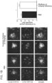
- Fig. 1B illustrates representative images of localization of amyloid deposits (6E10) and microglia (Iba1) in mice treated with Cromolyn Sodium (3.15mg/kg) or PBS daily for seven days.
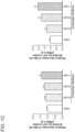
- Fig. 1C illustrates the effect of Cromolyn Sodium on microglial A ⁇ uptake in vitro. Microglial cells were cultured and incubated with 50 nM of synthetic A ⁇ 40 or A ⁇ 42 and 0, 10 nM, 10 ⁇ M or 1 mM of Cromolyn Sodium for 16 hours. After the incubation, the concentrations of A ⁇ x-40 ( Fig.
- FIG. 2 illustrates representative plaques of all the plaques and the microglial cells surrounding those deposits in Tg-2576 mice of the study.
- An image analysis looking at the percentage of Iba-1 positive processes colocalizing with the amyloid staining versus the total amount of Iba-1 signal surrounding the plaque demonstrated that there was more Iba-1/Amyloid colocalization when the mice were treated with Cromolyn Sodium as opposed to any other groups. This result correlates with our results in Example 1 and our in vitro data.
- BV2 microglial cell cultures were treated with cromolyn and/or ibuprofen (10 ⁇ M, 100 ⁇ M, 1 mM) for 16 hours. Afterwards, cells were incubated with soluble A ⁇ 42 and the compounds for 3 hours. After incubation, cells were collected for ELISA analysis.
- Figure 3 graphically illustrates the results of BV2 microglial cells treated with cromolyn, and with cromolyn and ibuprofen exhibit increased A ⁇ 42 uptake levels relative to BV2 microglia treated with the vehicle.
- Acetic anhydride (0.5 g, 4.6 mmol) was slowly added to a mixture of 5,5'-[(2-hydroxy-1,3-propanediyl)bis(oxy)]bis[4-oxo-4 H -1-benzopyran-2-ethanol (0.5 g, 1.14 mmol) in pyridine (20 mL) cooled to 0-5°C. The mixture was stirred for 3 hr at 0-5°C and then allowed to warm to room temperature. TLC indicted the reaction was complete. Methylene chloride was added and the mixture was washed with 10% HCl until the aqueous phase was acidic. The methylene chloride layer was dried over anhydrous sodium sulfate and solvent was evaporated.
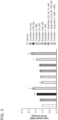
- Example 4 A ⁇ aggregation inhibition assay.
- Synthetic Aa ⁇ 42 in final 5 uM was incubated with 10, 100, 1,000 nM of test compounds for 1 hour.
- the aggregation was initiated with heparin at 0.5 mg/ml in final concentration.
- the assay buffer consisted of 125 mM NaCl, 2.5 mM KCl, 1 mM MgCl 2 , 1.25 mM Na 2 H 2 PO 4 , 2 mM CaCl 2 , 25 mM Glucose, and NaHCO 3 to adjust pH to 7.4.
- the assay buffer was used as a control.
- the aggregation was measured by intensity of Thioflavin T binding, which was detected by fluorescent excitation/emission at 450 nm/480 nm (Spectra Max M3 plate reader, Molecular Devices) in a kinetic mode. Aggregation was recorded as the kinetics was calculated as Vmax by the plate reader's software.
- the assay was performed in triplicate and expressed as standard mean ⁇ SD. Blue dotted line indicate the assay buffer control.
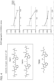
- Figure 4 illustrates the results of the assay.
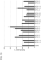
- Figure 5 graphically illustrates the results of a one-way of the differences in the A ⁇ levels and the ratios of A ⁇ (42:40).
- BV2-CD33 WT The effect of compounds in BV2 cells stably expressing full-length human CD33 was assessed to explore whether they reverse CD33-mediated inhibition of A ⁇ uptake ( Griciuc et al., 2013 Neuron 78, 631-643 ).
- the compound numbers, molecular weight and concentration of the stock solutions are summarized in Table 1.
- the invention relates to cromolyn derivative C4.
- Cromolyn derivatives C1-C3 and C5-C8 are not part of the invention.
- Cromolyn derivatives, C3 and C4 displayed lower solubility in DMSO in comparison to C1, C2, C5, C6, C7 and C8. Therefore, a 25 mM stock solutions for all the compounds except for C3 and C4 were prepared.
- the stock solutions for C3 and C4 were prepared at 5 mM and 7.5 mM, respectively.
- C1 is the parent compound - cromolyn disodium.
- Table 1 Summary of compounds tested in microglial cells Compound Number Compound Name Stock Solution (mM) C1 Cromolyn Disodium 25 C2 F-Cromolyn Diacid 25 C3 ET-Cromolyn 5 C4 F-ET-Cromolyn 7.5 C5 Triol-Cromolyn 25 C6 F-Triol-Cromolyn 25 C7 Ac-Triol-Cromolyn 25 C8 POM-Cromolyn 25
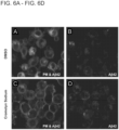
- naive BV2 cells were treated with DMSO (control) or cromolyn at 500 ⁇ M for 16 hours. Afterwards, cells were washed with PBS and treated with DMSO or cromolyn in the presence of the fluorescently-tagged A ⁇ 42 peptide (400 nM, red) for 2 hours. At the end of the treatment, the cells were washed and labeled them with a plasma membrane dye (green). Using confocal microscopy and the fluorescence signal in the red channel, the levels of intracellular A ⁇ 42 peptide were quantified. All the quantifications were performed by a blind observer with the ImageJ software. Remarkably, cromolyn sodium led to increased uptake of A ⁇ 42 in naive BV2 microglial cells ( Fig. 6A-Fig. 6D ).
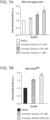
- cromolyn sodium modulates A ⁇ 42 uptake in na ⁇ ve BV2 microglial cells was determined by using the ELISA assay. Additionally, whether cromolyn sodium leads to increased A ⁇ 42 uptake levels in BV2 cells stably expressing full-length human CD33 (BV2-CD33 WT ) was determined. To this purpose, both naive BV2 and BV2-CD33 WT cell lines were treated with DMSO (control) or cromolyn at different concentrations for 16 hours. Then, the cells were washed with PBS and treated with DMSO or cromolyn and soluble untagged A ⁇ 42 peptide (400 nM) for 2 hours. The collected cell lysates were analyzed for A ⁇ 42 uptake levels using the A ⁇ 42-specific ELISA kit from Wako. The ELISA results were normalized to the protein concentration levels that were previously quantified using the BCA assay.
- naive BV2 or BV2-CD33 WT cells were plated in proliferating media.
- cells were treated with DMSO (control) or the compounds at different concentrations in proliferating media for 3 hours.
- C1, C2, C5, C6, C7 and C8 were tested at 10, 50, 100 and 150 ⁇ M, while C3 and C4 were assessed at 5, 25, 50 and 75 ⁇ M due to solubility limit in DMSO.
- cells were washed with PBS and treated with DMSO or compounds in the presence of the untagged A ⁇ 42 peptide (400 nM) in DMEM media for 2 hours.
- naive BV2 microglial cells were incubated with DMSO (vehicle) or cromolyn derivatives at different concentrations for 3 hours. The cells were then washed and incubated with DMSO or compounds and soluble untagged A ⁇ 42 for additional 2 hours. Afterwards, the cell media was collected and measured LDH released by the damaged cells to identify the compounds that induce cytolysis. The LDH assay showed that the cromolyn derivative C8 is the only compound showing toxicity when tested at 100 and 150 ⁇ M ( Fig. 8 ). Therefore, 100 and 150 ⁇ M concentrations for C8 were excluded from the A ⁇ 42 uptake assays.
- Example 8 Modulation of A ⁇ 42 uptake in microglial cells by cromolyn derivatives
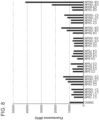
- naive BV2 microglial cells were treated with DMSO (control) or cromolyn derivative compounds at different concentrations for 3 hours. Afterwards, the cells were washed and treated with DMSO or compounds in the presence of untagged A ⁇ 42 peptide for 2 hours. At the end of the treatment, the cell lysates were collected. The analysis for intracellular A ⁇ 42 levels is performed using an A ⁇ 42-specific ELISA kit. The parent compound C1 (cromolyn sodium) led to a modest increase of A ⁇ 42 uptake at 100 and 150 ⁇ M in BV2 cells.
- the C1 aliquot received with the other cromolyn derivatives displayed lower solubility in DMSO than the C1 aliquot that was sent to us the first time (without the cromolyn derivatives).
- the compound C6 led to a robust inhibition of A ⁇ 42 uptake in BV2 microglial cells.
- the cromolyn derivative C4 led to an increased uptake of A ⁇ 42 peptide at 75 ⁇ M in naive BV2 microglial cells ( Fig. 9 ).
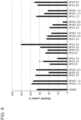
- BV2-CD33 WT cells were treated with DMSO (control) or cromolyn derivatives at different concentrations ranging between 5 and 150 ⁇ M.
- the cromolyn derivatives C1 and C3-8 were tested.
- the compound C2 was tested with other cromolyn derivatives in the second set of experiments.
- Treatment with the compound C4 at 75 ⁇ M resulted in a two-fold increase in A ⁇ 42 uptake in comparison to DMSO treatment and displayed a dose-dependent effect at 50 ⁇ M ( Fig. 10 ).
- the IC 50 for C4 was 54.7 ⁇ M in BV2-CD33 WT cells.
- the compound C6 exhibits a dose-dependent effect in mediating inhibition of A ⁇ 42 uptake in BV2-CD33 WT cells when compared to DMSO treatment.
Description
- This application claims the benefit of priority to
U.S. Provisional Patent Application serial number 62/382,192, filed August 31, 2016 - The invention is defined in the appended claims and encompasses methods of treating a neuron inflammation condition comprising administered a therapeutically effective amount to a patient in need thereof of at least one compound having the following formula:

- Strategies to modulate monocyte and microglial activity have been studied, especially those that can protect against microglia-mediated neurotoxicity. (See, Zhao et al., "Protective effects of an anti-inflammatory cytokine, interleukin-4, on motoneuron toxicity induced by activated microglia," J. Neurochem. 2006, 99:1176-1187; Heneka et al., "NLRP3 is activated in Alzheimer's disease and contributes to pathology in APP/PS1 mice," Nature, 2013, 493(7434):674-8; Theeriault, et al., "The dynamics of monocytes and microglia in Alzheimer's disease," Alzheimers Res Ther., 2015, 7:41; Nau et al., "Strategies to increase the activity of microglia as efficient protectors of the brain against infections," Front Cell Neurosci., 2014, 8: 138.) Overall, it is clear that more focused studies are needed to better establish how each inflammatory state can modulate the pathology of neurodegenerative diseases such as Alzheimer's Disease (AD) and Amyotrophic Lateral Sclerosis (ALS). Early activation of monocytes and microglia has potential to decelerate neurodegenerative progression by modulating immune responses to increase the intrinsic phagocytic capacity of monocytes and microglia without triggering secretion of pro-inflammatory cytokines that could worsen neurodegeneration.
- The role of the neuro-inflammatory response in the presence of amyloid plaques and neurofibrillary tangles in the brain and its associated neuronal loss in the pathology of AD is well established and extensively studied. See, Walker et al., "Immune phenotypes of microglia in human neurodegenerative disease: challenges to detecting microglial polarization in human brains," Alzheimers Res Ther., 2015, 7:56; Theerialut et al., 2015; Wilcock, DM, "A Changing Perspective on the Role of Neuroinflammation in Alzheimer's Disease," International Journal of Alzheimer's Disease, 2012, Article ID 495243; McGeer et al., "Targeting microglia for the treatment of Alzheimer's disease," Expert Opin Ther Targets, 2015, 19(4):497-506). Numerous studies show that microglial-mediated inflammation contributes to the progression of AD and that microglial cells are found in close association with amyloid-β (Aβ) deposits. (See, Mandrekar, et al., "Microglia and Inflammation in Alzheimer's Disease," CNS Neurol Disord Drug Targets, 2010, 9(2): 156-167).
- It is known that the changes in properties of microglia-the brain-resident macrophages-depend on their response to different stimuli in their microenvironment (e.g. cytokines), resulting in a range of phenotypes. Based on the changes in expression of cytokines, receptors, and other markers, monocyte and macrophage states have been defined as: classical activation (M1), alternative activation (M2a), type II alternative activation (M2b), and acquired deactivation (M2c). (See, Walker et al., 2015; Martinez et al., "Alternative activation of macrophages: an immunologic functional perspective," Annu Rev Immunol. 2009, 27:451-83; Mantovani et al., "The chemokine system in diverse forms of macrophage activation and polarization," Trends Immunol., 2004, 25:677-686; Sternberg, EM., "Neural regulation of innate immunity: a coordinated nonspecific host response to pathogens," Nat Rev Immunol., 2006, 6(4):318-28). Recently, a number of studies have attempted to elucidate the role of these phenotypes in the AD brain and determine the mechanisms through which these cells contribute to AD-related neuro-inflammation. (See, Mandrekar et al. 2012; McGeer et al., 2015; and Wilcock, 2012).
- Interaction of microglia with fibrillar Aβ leads to their phenotypic activation, and has recently been suggested to play a role in neuroprotection. (See Zhao et al., 2006; Figueiredo et al., "Neuron-microglia crosstalk up-regulates neuronal FGF-2 expression which mediates neuroprotection against excitotoxicity via JNK1/2," J. Neurochem., 2008 Oct.,107(1):73-85). It has been shown in numerous studies, in both mice and humans, that glial cells respond to the presence of AD pathological lesions (plaques and tangles) by changing their morphological characteristics, expressing numerous cell surface receptors, and surrounding the lesions. (See, Perlmutter et al., "Morphologic association between microglia and senile plaque amyloid in Alzheimer's disease," Neurosci Lett., 1990, 119:1, 32-36; Combs, et al., "Identification of microglial signal transduction pathways mediating a neurotoxic response to amyloidogenic fragments of β-amyloid and prion proteins," J. Neurosci., 1999,19:3, 928-939). On the other hand, macrophage and microglial activation in response to cellular debris in the AD brain, and the subsequent release of pro-inflammatory cytokines, leads to accelerated neurodegeneration. This, in turn, creates more cellular debris and accelerates disease progression. (See, Rubio-Perez et al., "A Review: Inflammatory Process in Alzheimer's Disease, Role of Cytokines," Scientific World Journal, 2012, 756357; McGeer, et al., "The importance of inflammatory mechanisms in Alzheimer disease," Exp. Gerontol. 1998, 33:5, 371-378; Akiyama, et al., "Inflammation and Alzheimer's disease," Neurobiol Aging, 2000, 21(3), 383-421; Liu, et al., "TLR2 is a primary receptor for Alzheimer's amyloid β peptide to trigger neuroinflammatory activation," J. Immunol. 2012, 188(3):1098-107).
- Several studies have focused on microglial activation and its role in the clearance of AD lesions leading to the reduction of amyloid deposits in the brain. (See, DiCarlo, et al., "Intrahippocampal LPS injections reduce Aβ load in APP+PS1 transgenic mice," Neurobiol of Aging, 2001, 22:6, 1007-1012; Herber, et al., "Time-dependent reduction in Aβ levels after intracranial LPS administration in APP transgenic mice," Exp. Neurol., 2004, 190(1):245-53; Liu, et al., 2012). While resident microglial cells surrounding Aβ plaques are not as efficacious in degrading Aβ as newly infiltrated macrophages or monocytes (See, Thériault, et al., 2015; Varnum, et al., "The classification of microglial activation phenotypes on neurodegeneration and regeneration in Alzheimer's disease brain," Arch. Immunol. Ther. Exp. (Warsz), 2012, 60(4):251-66), it has been shown that microglia are indeed capable of internalizing fibrillar and soluble Aβ, but are unable to process these peptides. (See Chung, et al., "Uptake, degradation, and release of fibrillar and soluble forms of Alzheimer's amyloid beta-peptide by microglial cells," J. Biol. Chem., 1999, 274:32301-8).
- Further, it has been postulated that microglia undergo a switch from an M2- to an M1-skewed activation phenotype during aging. (See, Heneka et al., 2013; Varnum, et al., 2012; Gratchev, et al., "Mphi1 and Mphi2 can be re-polarized by Th2 or Th1 cytokines, respectively, and respond to exogenous danger signals," Immunobiology, 2006, 211(6-8):473-486; Colton, et al., "Expression profiles for macrophage alternative activation genes in AD and in mouse models of AD," J. Neuroinflammation, 2006, 3:27). However, how the immune response in the brain is driven in AD is still a matter of debate, especially whether neuroinflammation can be triggered by age-related systemic inflammation. (See, Thériault, et al., 2015). It has been shown that stimulation of microglia could enhance their intrinsic phagocytic capacity to degrade Aβ more efficaciously; a number of strategies to modulate microglial response have been proposed. (See, Mandrekar, 2010; Kiyota, et al., "CNS expression of anti- inflammatory cytokine interleukin-4 attenuates Alzheimer's disease-like pathogenesis in APP +PS1 bigenic mice," FASEB J. 2010, 24:3093-3102; He, et al., "Deletion of tumor necrosis factor death receptor inhibits amyloid beta generation and prevents learning and memory deficits in Alzheimer's mice," J. Cell Biol., 2007, 178:829-841; Varnum, et al., 2012).
- It has been shown that microglia are activated by extracellularly deposited Aβ peptide (Lotz, et al., "Amyloid beta peptide 1-40 enhances the action of Toll-like receptor-2 and -4 agonists but antagonizes Toll-like receptor-9-induced inflammation in primary mouse microglial cell cultures," J. Neurochem., 2005, 94:289-298; Reed-Geaghan, et al., "CD14 and toll-). This is similar to microglial activation in response to the presence of interferon-γ (IFNγ), tumor necrosis factor alpha (TNFα) from T cells, or antigen-presenting cells. M1 activated microglia can produce reactive oxygen species and result in increased production of pro-inflammatory cytokines such as TNFα and interleukin (IL)-1β.
- The M1-type response of microglial cells has been shown to lower amyloid load but exacerbate neurofibrillary tangle pathology. Shaftel et al. (Shaftel, et al., "Sustained hippocampal IL- 1β overexpression mediates chronic neuroinflammation and ameliorates Alzheimer plaque pathology," J. Clin. Invest., 2007, 117(6):1595-604) have shown that IL-1β expression may underlie a beneficial neuroinflammatory response in AD, and that IL-1β overexpression in the hippocampus of APP/
PS 1 transgenic mice results in decreased amyloid burden. The authors suggest that IL-1β-mediated activation of microglia is the mechanism for the reductions in amyloid deposition. Further, Montgomery et al. (Montgomery, et al., "Ablation of TNF-RI/RII expression in Alzheimer's disease mice leads to an unexpected enhancement of pathology: implications for chronic pan-TNF-α suppressive therapeutic strategies in the brain," Am. J. Pathol., 2011, 179(4):2053-70) have shown that intact TNF-receptor signaling is critical for microglial-mediated uptake of extracellular amyloid-peptide. While M1 inflammatory phenotypes appear to improve the amyloid pathology in numerous studies, induction of M1 phenotypes in tau transgenic mice or cell culture results in the exacerbation of tau pathology. (See, Kitazawa, et al., "Lipopolysaccharide-induced inflammation exacerbates tau pathology by a cyclin-dependent kinase 5- mediated pathway in a transgenic model of Alzheimer's disease," J. Neurosci., 2005, 28;25(39):8843-53.; Li, et al., "Interleukin-1 mediates pathological effects of microglia on tau phosphorylation and on synaptophysin synthesis in cortical neurons through a p38-MAPK pathway," J. Neurosci., 2003, 1;23(5):1605-1 1). - Macrophage M2 activation is associated with mediators that are known to contribute to the anti-inflammatory actions and reorganization of extracellular matrix (Zhu, et al., "Acidic mammalian chitinase in asthmatic Th2 inflammation and IL-13 pathway activation", Science, 2004, 304(5677):1678-82; Walker, et al., 2015; Wilcock, et al., 2012). Microglia with M2a phenotypes have increased phagocytosis and produce growth factors such as insulin-like growth factor-1 and anti-inflammatory cytokines such as IL-10. Stimulation of macrophages by IL-4 and/or IL-13 results in an M2a state, sometimes called a wound-healing macrophage (Edwards, et al., "Biochemical and functional characterization of three activated macrophage populations," J. Leukoc Biol., 2006, 80(6):1298-307) and it is generally characterized by low production of pro-inflammatory cytokines (IL-1, TNF and IL-6). The M2a responses are primarily observed in allergic responses, extracellular matrix deposition, and remodeling.
- M2b macrophages are unique in that they express high levels of pro-inflammatory cytokines, characteristic of M1 activation, but also express high levels of the anti-inflammatory cytokine IL-10. (See, Moser DM., "The many faces of macrophage activation," J. Leukoc Biol., 2003, 73(2):209-12).
- Finally, the M2c macrophage state is stimulated by IL-10 and is sometimes referred to as a regulatory macrophage. M2c macrophages have anti-inflammatory activity that plays a role in the phagocytosis of cellular debris without the classical pro-inflammatory response (See, Moser DM., 2003). These cells express TGFβ and high IL-10 as well as matrix proteins. (See, Mantovani, et al., "The chemokine system in diverse forms of macrophage activation and polarization," Trends Immunol., 2004, 25:677-686; Wilcock, et al., 2012). Plunkett et al. (Plunkett, et al., "Effects of interleukin-10 (IL-10) on pain behavior and gene expression following excitotoxic spinal cord injury in the rat," Exp. Neurol., 2001; 168:144-154) reported that IL-10 mediated anti-inflammatory responses including decreasing glial activation and production of pro-inflammatory cytokines.
- However, the mechanism of M2 microglial activation and role it plays in AD and plaque pathology is still not well understood. (See, Mandrekar, et al., 2010). Further, a number of studies suggested that there is a switch in microglial activation status in response to disease progression (Colton, et al., 2006; Jimenez, et al., "Inflammatory response in the hippocampus of PS1M146L/ APP751SL mouse model of Alzheimer's disease: age-dependent switch in the microglial phenotype from alternative to classic," J. Neurosci., 2008, 28:11650-11661). It has been reported in animal studies that microglial activation phenotypes switch from M2 to M1 during disease progression (Jimenez, et al., 2008; Nolan, et al., "Role of interleukin-4 in regulation of age-related inflammatory changes in the hippocampus," J. Biol. Chem., 2005; 280:9354-9362; Maher, et al., "Downregulation of IL-4-induced signalling in hippocampus contributes to deficits in LTP in the aged rat," Neurobiol. Aging, 2005, 26:717-728), suggesting an increased classical activation phenotype over the alternative phenotype with age. It is generally agreed that microglia activated by extracellularly deposited Aβ protect neurons by triggering anti-inflammatory/neurotrophic M2 activation and by clearing Aβ via phagocytosis. This is a potential avenue for new therapeutic targets. (See, He, et al., 2007; Yamamoto, et al., "Interferon- gamma and tumor necrosis factor-alpha regulate amyloid-beta plaque deposition and beta- secretase expression in Swedish mutant APP transgenic mice," Am. J. Pathol., 2007, 170:680-692; Yamamoto, et al., "Cytokine-mediated inhibition of fibrillar amyloid-beta peptide degradation by human mononuclear phagocytes," J. Immunol., 2008, 181:3877-3886).
- Mantovani et al. (Mantovani, et al., 2004) studied the effect of IL-4 as an important modulator of M2a microglial activation. It has been shown that gene delivery of IL-4 into APP+PS1 mice partially suppressed glial accumulation in the hippocampus, directly enhanced neurogenesis, restored impaired spatial learning, and also reduced Aβ deposition (Kiyota, et al., 2010).
- Yamamoto et al. (Yamamoto, et al., 2007, 2008) examined macrophage-mediated Aβ degradation using pro- and anti-inflammatory cytokines in primary cultured human monocyte-derived macrophages (MDM) and microglia. These studies showed that anti-inflammatory and regulatory cytokines lead to an increase in M2a or M2c activation and enhanced Aβ clearance. Kiyota et al. (Kiyota et al., 2011) have shown sustained expression of IL-4 reduced astro/microgliosis, amyloid-β peptide (Aβ) oligomerization and deposition, and enhanced neurogenesis.
- Several approaches have been proposed to modulate microglial activation as potential targets for AD treatment. (See, Thériault, et al., 2015; Cherry, et al., "Neuroinflammation and M2 microglia: the good, the bad, and the inflamed," J. Neuroinflammation, 2014, 11:98; Mandrekar, et al., 2010; Vernum, et al., 2012). It has been suggested that use of anti-inflammatory drugs, like non-steroidal anti-inflammatory drugs (NSAIDs), to halt the progression of AD could be suppressing both pro-inflammatory and anti-inflammatory activation by endogenous molecules, inactivating the beneficial effect of M2 microglial functions and endogenous mechanisms of plaque clearance. (See, Wilcock, et al., 2012, Cherry, et al., 2014; Theeriault, et al., 2015).
- Research has focused primarily on two areas: anti-inflammatory agents to temper toxic effect of pro-inflammatory cytokines; and converting microglia from this M1 state to an M2 state in which the toxic effects are reduced and their phagocytic activity toward Aβ is enhanced. It was suggested (McGreer, et al., 2012) that potential treatments should be administered early in the disease progression.
- Strategies that modulate monocyte and microglial activity have been studied, especially those that can protect against microglia-mediated neurotoxicity (Zhao, et al., 2006; Heneka, et al., 2013; Therlaut, et al., 2015; Nau, et al., 2014). Overall, it is clear that more focused studies need to be performed to better establish how each inflammatory state can modulate the pathologies of AD. It is generally accepted that early activation of monocytes and microglia has potential to decelerate AD progression by modulating immune responses to increase the intrinsic phagocytic capacity of monocytes and microglia without triggering secretion of pro inflammatory cytokines that could worsen AD.
- The invention encompasses methods of treating a neuron inflammation condition comprising administering a therapeutically effective amount to a patient in need thereof of a compound having the following formula:

- In other comparative examples, the method uses the following compounds:


- In one embodiment, the compounds may be administered intraperitoneally (IP) and/or intravenously (IV). The compounds may be administered at a dose between about 1 mg and about 1000 mg per day. The method of administration may be transdermally or by inhalation.
- In another embodiment, the method is a method of treating ALS further comprising co-administering CD4+; siRNA; miRNA that ameliorate ALS; glial morphology modifier; SOD1 control; Riluzole; or another M1; M2 conversion active drug that controls neuroinflammation.
-
-
Fig.1A illustrates the quantification of the plasmatic levels of Aβx-40 and Aβx-42 one week after treatment with PBS or escalating doses of Cromolyn Sodium (n=3-5 mice/group). -
Fig. 1B illustrates representative images of localization of amyloid deposits (6E10) and microglia (Iba1) in mice treated with Cromolyn Sodium (3.15mg/kg) or PBS daily for seven days. The bar figure illustrates the results from analyzing plaques for each animal. Scale bar=10 µm. -
Fig. 1C illustrates the effect of Cromolyn Sodium on microglial Aβ uptake in vitro, where after the incubation, the concentrations of Aβx-40 (Fig. 1C left) Aβx-42 (Fig. 1C , right) in media were measured using Aβ ELISA. -
Fig. 2 illustrates the plaques and the microglial cells surrounding those deposits in Tg-2576 mice of the study of Example 2. The figure shows representative pictures of amyloid deposits and Iba-1 positive microglia. -
Fig. 3 illustrates the results of BV2 microglial cells treated with cromolyn, and with cromolyn and ibuprofen exhibit increased Aβ42 uptake levels relative to BV2 microglia treated with the vehicle. -
Fig. 4 illustrates the results of an Aβ aggregation inhibition assay using various compounds described herein. -
Fig. 5 graphically illustrates that Cromolyn significantly affects the levels of brain TBS soluble Aβ and the ratios of Aβ (42:40). -
Fig. 6A shows naive BV2 microglial cells treated with DMSO (control) for 16 h. Afterwards, cells were incubated with fluorescently-labeled Aβ42 and DMSO or cromolyn sodium for 2 hours. After incubation, cells were labeled with a plasma membrane dye (PM) and imaged. -
Fig. 6B shows naive BV2 microglial cells treated with DMSO (control) for 16 h. Afterwards, cells were incubated with fluorescently-labeled Aβ42 and DMSO or cromolyn sodium for 2 hours. -
Fig. 6C showns naive BV2 microglial cells treated with cromolyn sodium (500 µM) for 16 hours. Afterwards, cells were incubated with fluorescently-labeled Aβ42 and DMSO or cromolyn sodium for 2 hours. After incubation, cells were labeled with a plasma membrane dye (PM) and imaged. -
Fig. 6D showns naive BV2 microglial cells treated with cromolyn sodium (500 µM) for 16 hours. Afterwards, cells were incubated with fluorescently-labeled Aβ42 and DMSO or cromolyn sodium for 2 hours. -
Fig. 7A graphically illustrates that cromolyn sodium promotes microglial Aβ42 uptake. BV2 microglial cells were treated with DMSO or different concentrations of cromolyn sodium for 16 hours. Then, cells were incubated with soluble untagged Aβ42 and DMSO or cromolyn sodium for 2 hours, and collected for ELISA analysis. Both naïve BV2 and BV2-CD33WT microglial cells treated with cromolyn sodium exhibited increased Aβ42 uptake levels in comparison to cells treated with the vehicle (DMSO). -
Fig 7B graphically illustrates that cromolyn sodium promotes microglial Aβ42 uptake. BV2 cells stably expressing CD33 (BV2-CD33WT) were treated with DMSO or different concentrations of cromolyn sodium for 16 hours. Then, cells were incubated with soluble untagged Aβ42 and DMSO or cromolyn sodium for 2 hours, and collected for ELISA analysis. Both naive BV2 and BV2-CD33WT microglial cells treated with cromolyn sodium exhibited increased Aβ42 uptake levels in comparison to cells treated with the vehicle (DMSO). -
Fig. 8 graphically illustrates that compound C8 displays toxicity when tested at 100 µM or higher concentration in LDH assay. Naive BV2 microglial cells were treated with DMSO or cromolyn derivatives for 3 hours at different concentrations. C1, C2, C5, C6, C7 and C8 were tested at 10, 50, 100 and 150 µM, while C3 and C4 were assessed at 5, 25, 50 and 75 µM due to solubility limit in DMSO. Afterwards, cells were incubated with soluble untagged Aβ42 peptide and DMSO or cromolyn derivatives for 2 hours. At the end of the treatment, cell media was collected and compound toxicity was assessed with the lactate dehydrogenase (LDH) assay. BV2 microglial cells treated with the cromolyn derivative C8 exhibited increased toxicity at 100 and 150 µM in comparison to cells treated with the vehicle (DMSO). -
Fig 9 graphically illustrates that compound C4 promotes Aβ42 uptake in naive BV2 microglial cells. BV2 cells were treated with DMSO (vehicle) or cromolyn derivatives at different concentrations ranging from 5 to 150 µM for 3 hours. Then, cells were incubated with soluble untagged Aβ42 and DMSO or cromolyn derivatives for additional 2 hours and collected for ELISA analysis. BV2 microglial cells treated with the cromolyn derivative C4 at 75 µM exhibited significantly increased Aβ42 uptake levels in comparison to cells treated with the vehicle. -
Fig. 10 graphically illustrates that compound C4 promotes Aβ42 uptake in microglial BV2-CD33WT cells. Microglial cells stably expressing CD33WT were treated with DMSO as control or cromolyn derivatives (C1, C3-8) at different concentrations for 3 hours. Afterwards, cells were incubated with DMSO or cromolyn derivatives in the presence of the Aβ42 peptide for additional 2 hours. Cell lysates were analyzed for intracellular levels of Aβ42 using an Aβ42-specific ELISA kit. Treatment with the cromolyn derivative C4 at 75 µM led to increased uptake of Aβ42 in BV2-CD33WT cells in comparison to DMSO treatment and displayed a dose-dependent effect at 50 µM. -
Fig. 11 graphically illustrates that compound C4 promotes Aβ42 uptake in BV2-CD33WT cells. BV2-CD33WT cells were treated with DMSO (vehicle) or cromolyn derivatives (C1, C2, and C4-7) at different concentrations for 3 hours. Afterwards, cells were treated with DMSO or cromolyn derivatives and soluble Aβ42 peptide for 2 hours. Cell lysates were analyzed using Aβ42-specific ELISA kit and intracellular Aβ42 levels were quantified. The cromolyn derivative C4 effectively induced Aβ42 uptake at 50 and 75 µM in BV2-CD33WT cells in comparison to cells treated with DMSO. - Ischemic stroke, Alzheimer's Disease (AD), Amyotrophic Lateral Sclerosis (ALS or Lou Gehrig's disease), Prion and other neurodegenerative disorders are associated with microglial activation and mast cell migration, as well as with monocytes and other cell types that produce a barrage of toxic cytokines and debris that enhance inflammation. In certain embodiments, the invention encompasses anti-inflammatory compounds to reduce the toxic effect of pro-inflammatory cytokines by converting microglia from a pro-inflammatory M1 state to an M2 state in which the toxic effects are reduced and their phagocytic activity toward amyloidosis, tauopathies and other cytotoxic events is enhanced. In certain embodiments, the invention also encompasses the use of the compounds to affect therapy early in the disease process.
- Many drugs used as anti-inflammatory agents showed no efficacy in the conversion of microglia from M1 to M2, nor do they enhance the modulation of microglia from M1 to M2. To the best of applicant's knowledge, the compounds described herein are the only effective, non-cytokine (e.g. IL-10) compounds exhibiting M1-to-M2 activity. Thus the invention encompasses a compound having the following formula:

- Compounds of comparative examples include those having the following formula and their analogs and isomers:

- X = OH, F, OCOCH3
- Y = CO2Na, CO2H, CO2Et, CH2OH, CH2OCOCH3, CO2CH2OCO(CH3)3
- Certain comparative examples also include 5-[3-(2-carboxy-4-oxochromen-5-yl)oxy-2-hydroxypropoxy]-4-oxochromene-2-carboxylic acid derivatives and isomeric forms.
- In certain embodiments, the compounds may be used to treat ALS including, but not limited to, slowing down or halting the progression of the disease. In certain embodiments, the compounds may be administered in combination with other anti-inflammatory agents to control the spread of the progressive and fatal effect of ALS.
- In certain embodiments, the invention encompasses a combination treatment for ALS of M1, M2 conversion active drugs that control neuroinflammation, such as the drugs in the above formulas, with other immune targeting therapies such as CD4+, siRNA, miRNA that ameliorates ALS, glial morphology modifiers,
SOD 1 controls, or Riluzole, the only approved drug for ALS. - In other embodiments, the compounds will slow down or halt neuron damage for neurons located in the brain stem and/or the spinal cord, neurons, or motor neurons that affect voluntary body muscles.
- In certain embodiments, the compounds may be administered using known methods for the administration of drugs, for example, IP, IV, transdermally, by inhalation. In certain embodiments, the invention relates to methods of treating or slowing down the aggressive progression of a neurological disease, such as AD, Ischemic Stroke, ALS, or Prion, and the compound is administered by infusion or intraperitoneal administration.
- In certain embodiments, the invention also provides pharmaceutical compositions comprising one or more compounds described herein in association with a pharmaceutically acceptable carrier. Preferably these compositions are in unit dosage forms such as tablets, pills, capsules, powders, granules, sterile parenteral solutions or suspensions, metered aerosol or liquid sprays, drops, ampoules, auto-injector devices or suppositories; for oral, parenteral, intranasal, sublingual or rectal administration, or for administration by inhalation or insufflation. It is also envisioned that the compounds may be incorporated into transdermal patches designed to deliver the appropriate amount of the drug in a continuous fashion.
- For preparing solid compositions such as powders and tablets, the principal active ingredient is mixed with a pharmaceutically acceptable carrier, e.g. conventional tableting ingredients such as corn starch, lactose, sucrose, sorbitol, talc, stearic acid, magnesium stearate, dicalcium phosphate or gums, and other pharmaceutical diluents, e.g. water, to form a solid preformulation composition containing a homogeneous mixture. When referring to these preformulation compositions as homogeneous, it is meant that the active ingredient is dispersed evenly throughout the composition so that the composition may be easily subdivided into equally effective unit dosage forms.
- In some embodiments, a dry powder composition is micronized for inhalation to the lungs. See for example,
U.S. Patent Application publication 2016/0263257 , and in particular regarding the dry powder cromolyn formulations described therein. In other embodiments, the dry powder composition further comprises at least one excipient. In certain embodiments, the at least one excipient comprises Lactose monohydrate and/or Magnesium stearate. - In certain embodiments, the compounds may be administered in doses that treat the particular indication. In particular, the dose is specifically tailored to lead to blood, brain, and CSF concentrations that allow the drugs to act as M1-to-M2 modifiers. Such doses may include from about 1 mg to about 1000 mg per day.
- The dosage of the active agents will generally be dependent upon a number of factors, including the pharmacodynamic characteristics of the compound, mode and route of administration of the compound, the health of the patient being treated, the extent of treatment desired, the nature and kind of concurrent therapy, if any, and the frequency of treatment and the nature of the effect desired. In general, dosage ranges of the compound often range from about 0.001 to about 250 mg/kg body weight per day. For a normal adult having a body weight of about 70 kg, a dosage may range from about 0.1 to about 25 mg/kg body weight. However, some variability in this general dosage range may be required depending on the age and weight of the subject being treated, the intended route of administration, the particular agent being administered, and the like. Importantly, the determination of dosage ranges and optimal dosages for a particular mammal is also well within the ability of one of ordinary skill in the art having the benefit of the instant disclosure.
- Dosages for compounds may be as low as 5 ng/d. In certain embodiments, about 10 ng/day, about 15 ng/day, about 20 ng/day, about 25 ng/day, about 30 ng/day, about 35 ng/day, about 40 ng/day, about 45 ng/day, about 50 ng/day, about 60 ng/day, about 70 ng/day, about 80 ng/day, about 90 ng/day, about 100 ng/day, about 200 ng/day, about 300 ng/day, about 400 ng/day, about 500 ng/day, about 600 ng/day, about 700 ng/day, about 800 ng/day, about 900 ng/day, about 1 µg/day, about 2 µg/day, about 3 µg/day, about 4 µg/day, about 5 µg/day, about 10 µg/day, about 15 µg/day, about 20 µg/day, about 30 µg/day, about 40 µg/day, about 50 µg/day, about 60 µg/day, about 70 µg/day, about 80 µg/day, about 90 µg/day, about 100 µg/day, about 200 µg/day, about 300 µg/day, about 400 µg/day about 500 µg/day, about 600 µg/day, about 700 µg/day, about 800 µg/day, about 900 µg/day, about 1 mg/day, about 2 mg/day, about 3 mg/day, about 4 mg/day, about 5 mg/day, about 10 mg/day, about 15 mg/day, about 20 mg/day, about 30 mg/day, about 40 mg/day or about 50 mg/day of the compound is administered.
- Dosage ranges for active agents may be from 5 ng/d to 100mg/day. In certain embodiments, dosage ranges for active agents may be from about 5 ng/day to about 10 ng/day, about 15 ng/day, about 20 ng/day, about 25 ng/day, about 30 ng/day, about 35 ng/day, about 40 ng/day, about 45 ng/day, about 50 ng/day, about 60 ng/day, about 70 ng/day, about 80 ng/day, about 90 ng/day, about 100 ng/day, about 200 ng/day, about 300 ng/day, about 400 ng/day, about 500 ng/day, about 600 ng/day, about 700 ng/day, about 800 ng/day, or about 900 ng/day. In certain embodiments, dosage ranges for compounds may be from about 1µg/day to about 2 µg/day, about 3 µg/day, about 4 µg/day, about 5 µg/day, about 10 µg/day, about 15 µg/day, about 20 µg/day, about 30 µg/day, about 40 µg/day, about 50 µg/day, about 60 µg/day, about 70 µg/day, about 80 µg/day, about 90 µg/day, about 100 µg/day, about 200 µg/day, about 300 µg/day, about 400 µg/day about 500 µg/day, about 600 µg/day, about 700 µg/day, about 800 µg/day, or about 900 µg/day. In certain embodiments, dosage ranges for active agents may be from about 1mg/day to about 2 mg/day, about 3 mg/day, about 4 mg/day, about 5 mg/day, about 10 mg/day, about 15 mg/day, about 20 mg/day, about 30 mg/day, about 40 mg/day, about 50 mg/day, about 60 mg/day, about 70 mg/day, about 80 mg/day, about 90 mg/day, about 100 mg/day, about 200 mg/day, about 300 mg/day, about 400 mg/day, about 500 mg/day, about 600 mg/day, about 700 mg/day, about 800 mg/day, or about 900 mg/day.
- In certain embodiments, the compounds are administered in pM or nM concentrations. In certain embodiments, the compounds are administered in about 1 pM, about 2 pM, about 3 pM, about 4 pM, about 5 pM, about 6 pM, about 7 pM, about 8 pM, about 9 pM, about 10 pM, about 20 pM, about 30 pM, about 40 pM, about 50 pM, about 60 pM, about 70 pM, about 80 pM, about 90 pM, about 100 pM, about 200 pM, about 300 pM, about 400 pM, about 500 pM, about 600 pM, about 700 pM, about 800 pM, about 900 pM, about 1 nM, about 2 nM, about 3 nM, about 4 nM, about 5 nM, about 6 nM, about 7 nM, about 8 nM, about 9 nM, about 10 nM, about 20 nM, about 30 nM, about 40 nM, about 50 nM, about 60 nM, about 70 nM, about 80 nM, about 90 nM, about 100 nM, about 300 nM, about 400 nM, about 500 nM, about 600 nM, about 700 nM, about 800 nM, or about 900 nM, concentrations.
- In certain embodiments, the dosage form is a solid dosage form, and the size of the compound in the dosage form is important. In certain embodiments, the compound is less than about 3 µm, less than about 2 µm, or less than about 1 µm in diameter. In certain embodiments, the active agent is about 0.1 µm to about 3.0 µm in diameter. In certain embodiments, the active agent is from about 0.5 µm to about 1.5 µm in diameter. In certain embodiments, the active agent is about 0.2 µm, about 0.3 µm, about 0.4 µm, about 0.5 µm, about 0.6 µm, about 0.7 µm, about 0.8 µm, about 0.9 µm, about 1.0 µm, about 1.1 µm, about 1.2 µm, about 1.3 µm, about 1.4 µm, or about 1.5 µm in diameter.
- For example, a formulation intended for oral administration to humans may contain from about 0.1 mg to about 5 g of the active agent (or compound) compounded with an appropriate and convenient carrier material varying from about 5% to about 95% of the total composition. Unit dosages will generally contain between about 0.5 mg to about 1500 mg of the active agent. The dosage may be about: 1 mg, 2 mg, 3 mg, 4 mg, 5 mg, 6 mg, 7 mg, 8 mg, 9 mg, 10 mg, 11 mg, 12 mg, 13 mg, 14 mg, 15 mg, 16 mg, 17 mg, 18 mg, 19 mg, 20 mg, 21 mg, 22 mg, 23 mg, 24 mg 25 mg, 26 mg, 27 mg, 28 mg, 29 mg, 30 mg, 31 mg, 32 mg, 33 mg, 34 mg 35 mg, 36 mg, 37 mg, 38 mg, 39 mg, 40 mg, 41 mg, 42 mg, 43 mg, 44 mg, 45 mg, 46 mg, 47 mg, 48 mg, 49 mg, 50 mg, 55 mg, 60 mg, 65, mg, 70 mg, 75 mg, 80 mg, 85 mg, 90 mg, 95 mg, 100 mg, 200 mg, 300 mg, 400 mg, 500 mg, 600 mg, 800 mg, or 100 mg, etc., up to about 1500 mg of the compound.
- In certain embodiments, the invention relates to combination of two active agents. In certain embodiments, it may be advantageous for the pharmaceutical combination to be comprised of a relatively large amount of the first component compared to a second component. In certain instances, the ratio of the first active agent to the second active agent is about: 200:1, 190:1, 180:1, 170:1, 160:1, 150:1, 140:1, 130:1, 120:1, 110:1, 100:1, 90:1, 80:1, 70:1, 60:1, 50:1, 40:1, 30:1, 20:1, 15:1, 10:1, 9:1, 8:1, 7:1, 6:1, or 5:1. It further may be preferable to have a more equal distribution of pharmaceutical agents. In certain instances, the ratio of the first active agent to the second active agent is about: 4:1, 3:1, 2:1, 1:1, 1:2, 1:3, or 1:4. It may also be advantageous for the pharmaceutical combination to have a relatively large amount of the second component compared to the first component. In certain instances, the ratio of the second active agent to the first active agent is about 200:1, 190:1, 180:1, 170:1, 160:1, 150:1, 140:1, 130:1, 120:1, 110:1, 100:1, 90:1, 80:1, 70:1, 60:1, 50:1, 40:1, 30:1, 20:1, 15:1, 10:1, 9:1, 8:1, 7:1, 6:1, or 5:1. A composition comprising any of the above identified combinations of the first therapeutic agent and second therapeutic agent may be administered in divided doses about 1, 2, 3, 4, 5, 6, or more times per day or in a form that will provide a rate of release effective to attain the desired results. The dosage form may contain both the first and second active agents. The dosage form may be administered one time per day if it contains both the first and second active agents.
- For example, a formulation intended for oral administration to humans may contain from about 0.1 mg to about 5 g of the first therapeutic agent and about 0.1 to about 5 g of the second therapeutic agent, both of which are compounded with an appropriate and convenient about of carrier material varying from about 5% to about 95% of the total composition. Unit dosages will generally contain between about 0.5 mg to about 1500 mg of the first therapeutic agent and 0.5 mg to 1500 mg of the second therapeutic agent. The dosage may be about: 25 mg, 50 mg, 100 mg, 200 mg, 300 mg, 400 mg, 500 mg, 600 mg, 800 mg, or 100 mg, etc., up to about 1500 mg of the first therapeutic agent. The dosage may be about: 25 mg, 50 mg, 100 mg, 200 mg, 300 mg, 400 mg, 500 mg, 600 mg, 800 mg, or 100 mg, etc., up to about 1500 mg of the second therapeutic agent.
- Our studies in PS1/PSS animal model showed that Cromolyn sodium impacted the interaction of microglial cells with amyloid deposits and eventually affected Aβ clearance by microglia. We first performed a double immunostaining between Aβ and the microglial marker Iba1 in brain sections of mice treated with PBS or the highest dose of Cromolyn sodium (3.15mg/kg). A systematic analysis of the overlap between both stainings revealed that animals that received Cromolyn Sodium showed a higher percentage of Iba1 immunoreactivity overlapping with amyloid (
Fig. 1B ), which may indicate a modest increased recruitment of microglia around plaques induced by the compound. - To go further in our understanding of these mechanisms, and considering that evaluating change in microglial function is challenging in vivo, we used an additional in vitro system of Aβ microglial uptake. Synthetic Aβ40 and Aβ42 peptides were applied to microglia in culture in the presence or absence of Cromolyn Sodium.
- After 16 hours of incubation, we observed a dose dependent decrease of Aβ40 and Aβ42 levels in presence of Cromolyn Sodium, indicating that the impact of Cromolyn Sodium on Aβ aggregation mechanisms may promote Aβ clearance by microglial uptake (
Fig. 1C ). The combination of those in vivo and in vitro results may suggest that, in addition to inhibiting Aβ fibrillization, Cromolyn Sodium affected microglial activation and Aβ clearance. - Cromolyn Sodium does not affect the levels of Aβ in the plasma but promotes microglial Aβ clearance.
Fig. 1A illustrates the quantification of the plasmatic levels of Aβx-40 and Aβx-42 one week after treatment with PBS or escalating doses of Cromolyn Sodium (n=3-5 mice/group).Fig. 1B illustrates representative images of localization of amyloid deposits (6E10) and microglia (Iba1) in mice treated with Cromolyn Sodium (3.15mg/kg) or PBS daily for seven days. The percentage of amyloid occupied by Iba1 positive processes was calculated for each deposit and showed an increased overlap between Aβ and Iba1 after treatment with Cromolyn Sodium (n=3 mice for PBS and n=5 mice for Cromolyn Sodium). Between 20 to 20 plaques were evaluated for each animal). Scale bar=10 µm.Fig. 1C illustrates the effect of Cromolyn Sodium on microglial Aβ uptake in vitro. Microglial cells were cultured and incubated with 50 nM of synthetic Aβ40 or Aβ42 and 0, 10 nM, 10 µM or 1 mM of Cromolyn Sodium for 16 hours. After the incubation, the concentrations of Aβx-40 (Fig. 1C left) Aβx-42 (Fig. 1C , right) in media were measured using Aβ ELISA and normalized with microglia cell number and according to the PBS control condition. (n=3 experiments; *,P<0.05, **, P<0.01) - In other animal studies of microglia activation and M1, M2 conversion showed that cromolyn is the only of many drugs tested that effected this change and exhibited phagocytic activity.
Figure 2 illustrates representative plaques of all the plaques and the microglial cells surrounding those deposits in Tg-2576 mice of the study. An image analysis looking at the percentage of Iba-1 positive processes colocalizing with the amyloid staining versus the total amount of Iba-1 signal surrounding the plaque demonstrated that there was more Iba-1/Amyloid colocalization when the mice were treated with Cromolyn Sodium as opposed to any other groups. This result correlates with our results in Example 1 and our in vitro data. - Cromolyn, but not ibuprofen promotes microglial Aβ42 uptake, their combination improved uptake over either ibuprofen or cromolyn alone. BV2 microglial cell cultures were treated with cromolyn and/or ibuprofen (10 µM, 100 µM, 1 mM) for 16 hours. Afterwards, cells were incubated with soluble Aβ42 and the compounds for 3 hours. After incubation, cells were collected for ELISA analysis. BV2 microglial cells treated with cromolyn (100 µM, 1 mM), and with cromolyn and ibuprofen (100 µM, 1 mM for each compound) exhibit increased Aβ42 uptake levels relative to BV2 microglia treated with the vehicle. Results were derived from three independent experiments; **p < 0.01, ***p < 0.001, one-way ANOVA, Tukey's test). Data are represented as mean ± SEM.
Figure 3 graphically illustrates the results of BV2 microglial cells treated with cromolyn, and with cromolyn and ibuprofen exhibit increased Aβ42 uptake levels relative to BV2 microglia treated with the vehicle. -

- A suspension of cromolyn sodium salt (1.0 g, 2 mmol) in EtOH (100 mL) and con. HCl (1 mL) was heated in a sealed reactor tube for 24 h at 100°C. The white solid was dissolved to give a clear colorless solution while hot. It was allowed to cool to room temperature and NaHCO3 (1.0 g) was added. After stirring for 30 min at 25°C, solvent was removed by roto-evaporation. Chromatography on silica gel of the crude material using 5:95 methanol/methylene chloride yielded the diethyl ester (0.8 g, 76 % yield); mp 154-156°C; 1H NMR (CDCl3, 300 MHz) δ 1.42 (t, 3H, J=7.1 Hz, CH3), 2.73 (br s, 1H, OH), 4.44 (q, 4H, J=7.1 Hz, 2OCH2CH3), 4.32-4.59 (m, 5H, CHOH, 2OCH2), 6.80 (s, 2H, 2 vinyl-H), 6.99 (d, 2H, J=8.24Hz, 2Aro-H), 7.12 (d, 2H, J=8.24Hz, 2Aro-H), 7.17 (d, 2H, J=8.24Hz, 2Aro-H), 7.71 (t, 2H, J=8.24 2Aro-H).
-

- A solution of 1,3-bis(4-methylbezenesulfonate propanetriol (2.7 g, 6.78 mmol) in methylene chloride (20 mL) at 0-5°C was treated with DAST (2.18 g, 13.6 mmol). The mixture was stirred at 0-5°C for 30 then allowed to warm to 25°C and stirred for 16 hr. The mixture was poured into a sat'd sodium bicarbonate solution (30 mL) and layers separated. The methylene chloride layer dried (sodium sulfate). After solvent removal, the crude material was chromatographed on silica gel (methylene chloride) to yield 0.82 g (30%) of a solid; mp 99-102°C; 1H NMR (CDCl3), δ 2.5 (s, 6H, CH3), 4.15 (dd, 4H, J=12.3, 4.6 Hz,CH2, 4.8 (dq, 1H, J=47, 4.6, CHF), 7.45 (d, 4H, J=8.1Hz, Aro-H), 7.75 (d, 4H, J=8.4Hz, Aro-H).
-

- A mixture of 3-bis(4-methylbezenesulfonate)-2-fluoropropanediol (1.0, 2.5 mmol), 2,6-dihydroxyacetophenone (0.76 g, 5.0 mmol) and potassium carbonate (0.69 g) in acetonitrile (40 mL) was heated under reflux for 16 hr. The mixture was filtered and the filtrate was evaporated. The crude material was chromatographed on silica gel (acetonitrile/methylene chloride 5:95) to yield 0.57g (40%) of product; mp 162-165°C; 1H NMR (d6-DMSO), δ 2.5 (s, 6H, 2CH3), 4.38 (m, 4H, 2CH2), 5.22 (br d 1H, J=49Hz, CHF), 6.45 (m, 4H, 4Aro-H), 7.28 (t, 2H, J=4.55Hz, 2Aro-H).
-

- A mixture of 1,3-bis(2-acety-3-hydroxyphenoxy)-2-fluoropropane (200 mg, 0.52 mmol) and ethyl oxalate (2 mL) was added to a solution of sodium ethoxide (87 mg Na) in ethanol (10 mL) and benzene (10 mL). The mixture was heated at reflux for 16 hr, cooled and diluted with ether (50 mL). The precipitated sodium salt was filtered, washed with ether and dried. It was then dissolved in water and acidified with 10% HCl to obtain a sticky solid. The solid was refluxed in ethanol (20 mL) with a catalytic amount of 36% HCL for 1 hr. The mixture was poured into 50 mL of water and extracted twice with methylene chloride (50 mL). The extracts were combined and dried. After solvent removal, the crude material was chromatographed on silica gel (acetonitrile/methylene chloride 10:90) to yield 0.12 g (45%) of product; mp 166-170°C; 1H NMR (CDCl3), δ 1.42 (t, 6H, J=7.14Hz, 2CH3), 4.58 (q, 4H, J=7.14Hz 2CH2), 4.65 (m, 4H, 2CH2), 5.35 (dq, 1H, J=46Hz, J=4.4HZ, CHF), 6.90 (s, 2H, vinyl-H), 6.95 (d, 2H, J=8.24Hz, 2Aro-H), 7.13(d, 2H, J=8.24Hz, 2Aro-H),7.17 (d, 2H, J=8.24Hz, 2Aro-H) 7.6 (t, 2H, J=8.24 2Aro-H).
-

- A suspension of 5,5'-[(2-fluoro-1,3-propanediyl)bis(oxy)]bis[4-oxo-4H-1-benzopyran-2-carboxylic acid diethyl ester (100 mg, 0.19 mmol) in methanol (20 mL) and 1 M sodium hydroxide (2 mL) was heated at 80°C for 1 hr. The solution was acidified with 10% HCl and volatiles were removed. A solution of methanol/ methylene chloride (50:50) was added to the solid and the mixture was filtered. Evaporation afforded 76 mg (85%) of product; 1H NMR (d6-DMSO), δ 4.65 (m, 4H, 2CH2), 5.32 (br d, 1H, J=46Hz, CHF), 6.80 (s, 2H, 2vinyl-H), 7.2 (d, 2H, J=8.24Hz, 2Aro-H), 7.71 (t, 2H, J=8.24 2Aro-H).
-

- To a suspension of 5,5'-(2-hydroxytrimethylenedioxy)bis(4-oxochromene-2-carboxylic acid) diethyl ester (1.0 g, 1.86 mmol) in methanol (60 ml) and methylene chloride (40 mL) NaBH4 (0.14 g, 3.72 mmol) was added in portions over a 1 h period. The mixture was stirred at 25°C until it was clear (approx. 5 h) at which time the solution was quenched by dropwise addition of 1M HCl until acidic. Solvent was evaporated and the residue was extracted with methylene chloride. The combined organic extracts were washed with water and dried over anhydrous sodium sulfate. After evaporation, the residue was purified by column chromatography (5:95 methanol/methylene chloride) to yield 0.5 g (50%) of the triol; 1H NMR (DMSO-d6, 300 MHz) δ 2.73 (s, 3H, OH), 4.25-4.36 (m, 9H, 2OCH2, CH-O), 6.13 (s, 2H, 2 vinyl H), 7.04 (d, 2H, J = 8.4 Hz, aromatic H), 7.07 (d, 2H, J = 8.4 Hz, aromatic H), 7.63 (t, 2H, J = 8.2 Hz, aromatic H).
-

- The above procedure for 5,5'-(2-hydroxytrimethylenedioxy)bis(4-oxochromene-2-ethanol) was used. 1H NMR (DMSO-d6, 300 MHz) δ 2.73 (s, 3H, OH), 4.25-4.36 (m, 8H, 2OCH2, CH-O), 5.35 (br d, 1H, J=46Hz, CHF), 6.13 (s, 2H, 2 vinyl H), 7.04 (d, 2H, J = 8.4 Hz, aromatic H), 7.07 (d, 2H, J = 8.4 Hz, aromatic H), 7.63 (t, 2H, J = 8.2 Hz, aromatic H).
-

- To a stirred solution of cromolyn diacid (1.0 g, 2.7 mm) in 20 mL of DMF was added diisopropylamine (0.7mL) and 1.0 g (6.5 mmol) chloromethylpivalate. The reaction mixture was stirred at 60°C for 4 hr, water was added and the mixture was extracted with separated, dried (MgSO4) and the solvent removed in vacuo. The solvent was removed and the residue was chromatographed on silica 4% methanol in methylene chloride to give 1.2 g (65%) of the pivalate compound; mp 135-140°C; H1 NMR (CDCl), δ 1.24 (s, 18 H, CH3), 4.36 (m, 2 H, OCH2), 4.49 (m, 1 H, CHOH), 4.51 (m, 2H, OCH2), ), 6.00 (s, 4H, CH-O-CO), 6.98 (m, 4H, 2vinyl-H, 2Aro-H), 7.13 (d, 2H, J=8.24Hz, 2Aro-H), 7.61 (t, 2H, J=8.24 2Aro-H).
-

- To a stirred solution of 5,5'-[(2-fluoro-1,3-propanediyl)bis(oxy)]bis[4-oxo-4H-1-benzopyran-2-carboxylic acid (1.0 g, 2.1 mmol) in 20 mL of DMF was added iisopropylamine (0.7 mL) and 1.0 g (6.5 mmol) chloromethylpivalate. The reaction mixture was stirred at 60°C for 4 hr, water was added and the mixture was extracted with separated, dried (MgSO4) and the solvent removed in vacuo. The solvent was removed and the residue was chromatographed on silica using 2% methanol in methylene chloride to give 1.0g (70%) of the pivalate compound; mp 130-133°C; δ 1.21 (s, 18 H, CH3), 4.36 (m, 4 m, 2OCH2), 4.49 (br d, 1H, J=46Hz, CHF), 6.00 (s, 4H, CH-O-CO), 6.98 (m, 4H, 2vinyl-H, 2Aro-H), 7.13 (d, 2H, J=8.24Hz, 2Aro-H), 7.61 (t, 2H, J=8.24 2Aro-H).
-

- Acetic anhydride (0.5 g, 4.6 mmol)) was slowly added to a mixture of 5,5'-[(2-hydroxy-1,3-propanediyl)bis(oxy)]bis[4-oxo-4H-1-benzopyran-2-ethanol (0.5 g, 1.14 mmol) in pyridine (20 mL) cooled to 0-5°C. The mixture was stirred for 3 hr at 0-5°C and then allowed to warm to room temperature. TLC indicted the reaction was complete. Methylene chloride was added and the mixture was washed with 10% HCl until the aqueous phase was acidic. The methylene chloride layer was dried over anhydrous sodium sulfate and solvent was evaporated. Chromatography on silica using 3% methanol in methylene chloride gave 0.45 g (72%) of the triacetate compound; mp 122-125°C; H1 NMR (CDCl3), δ 2.16 (s, 9 H, CH3), 4.58 (m, 2 H, CH2OH), 4.66 (m, 2H, CH2OH), 4.94 (s, 4 H, CH2OH), 5.66 (m, 1 H, CHOH), 6.15 (s, 2H, 2vinyl-H), 6.94 (d, 2H, 2Aro-H), 6.97 (d, 2H, J=8.24Hz, 2Aro-H), 7.52 (t, 2H, J=8.24, 2Aro-H).
- Experimental design. 3-month old Tg2576 mice were acclimatized for 2 months, and then randomly assigned to different treatment groups. They included the control group (n=10) with vehicle treatment, the cromolyn low dose group and cromolyn high dose group. The treatments were conducted through IP injection with PBS based on 0.1mL/30g body weight, 3 times per week for 3 additional months. All mice were sacrificed at 8-month old. Tissues were harvested and processed for postmortem analysis.
- Synthetic Aaβ42 in final 5 uM was incubated with 10, 100, 1,000 nM of test compounds for 1 hour. The aggregation was initiated with heparin at 0.5 mg/ml in final concentration. The assay buffer consisted of 125 mM NaCl, 2.5 mM KCl, 1 mM MgCl2, 1.25 mM Na2H2PO4, 2 mM CaCl2, 25 mM Glucose, and NaHCO3 to adjust pH to 7.4. The assay buffer was used as a control. The aggregation was measured by intensity of Thioflavin T binding, which was detected by fluorescent excitation/emission at 450 nm/480 nm (Spectra Max M3 plate reader, Molecular Devices) in a kinetic mode. Aggregation was recorded as the kinetics was calculated as Vmax by the plate reader's software. The assay was performed in triplicate and expressed as standard mean ± SD. Blue dotted line indicate the assay buffer control.
Figure 4 illustrates the results of the assay. - Cromolyn significantly affected the levels of brain TBS soluble Aβ and the ratios of Aβ (42:40). A-B. MSD (mess scale to measure
Aβ Aβ 42/40 and ah higher Aβ 38 that is not implicated in plaque formation.Figure 5 graphically illustrates the results of a one-way of the differences in the Aβ levels and the ratios of Aβ (42:40). - Confocal microscopy and ELISA assays were used to assess the effect of cromolyn and its derivative compounds on Aβ42 uptake in microglial cells. The BV2 microglial cell line, which was previously found to efficiently take up and degrade exogenously-added Aβ42, was used (Jiang, Q., et al. (2008) Neuron 58, 681-693; Mandrekar et al., 2009 J. Neurosci. 29, 4252-4262). The compounds were tested in naive BV2 microglial cells to investigate whether they modulate Aβ uptake. The effect of compounds in BV2 cells stably expressing full-length human CD33 (BV2-CD33WT) was assessed to explore whether they reverse CD33-mediated inhibition of Aβ uptake (Griciuc et al., 2013 Neuron 78, 631-643).
- The compound numbers, molecular weight and concentration of the stock solutions are summarized in Table 1. The invention relates to cromolyn derivative C4. Cromolyn derivatives C1-C3 and C5-C8 are not part of the invention. Cromolyn derivatives, C3 and C4, displayed lower solubility in DMSO in comparison to C1, C2, C5, C6, C7 and C8. Therefore, a 25 mM stock solutions for all the compounds except for C3 and C4 were prepared. The stock solutions for C3 and C4 were prepared at 5 mM and 7.5 mM, respectively. C1 is the parent compound - cromolyn disodium.
Table 1: Summary of compounds tested in microglial cells Compound Number Compound Name Stock Solution (mM) C1 Cromolyn Disodium 25 C2 F-Cromolyn Diacid 25 C3 ET-Cromolyn 5 C4 F-ET-Cromolyn 7.5 C5 Triol-Cromolyn 25 C6 F-Triol-Cromolyn 25 C7 Ac-Triol-Cromolyn 25 C8 POM-Cromolyn 25 - To investigate the effect of cromolyn sodium on Aβ42 uptake in microglial cells, naive BV2 cells were treated with DMSO (control) or cromolyn at 500 µM for 16 hours. Afterwards, cells were washed with PBS and treated with DMSO or cromolyn in the presence of the fluorescently-tagged Aβ42 peptide (400 nM, red) for 2 hours. At the end of the treatment, the cells were washed and labeled them with a plasma membrane dye (green). Using confocal microscopy and the fluorescence signal in the red channel, the levels of intracellular Aβ42 peptide were quantified. All the quantifications were performed by a blind observer with the ImageJ software. Remarkably, cromolyn sodium led to increased uptake of Aβ42 in naive BV2 microglial cells (
Fig. 6A-Fig. 6D ). - Furthermore, whether cromolyn sodium modulates Aβ42 uptake in naïve BV2 microglial cells was determined by using the ELISA assay. Additionally, whether cromolyn sodium leads to increased Aβ42 uptake levels in BV2 cells stably expressing full-length human CD33 (BV2-CD33WT) was determined. To this purpose, both naive BV2 and BV2-CD33WT cell lines were treated with DMSO (control) or cromolyn at different concentrations for 16 hours. Then, the cells were washed with PBS and treated with DMSO or cromolyn and soluble untagged Aβ42 peptide (400 nM) for 2 hours. The collected cell lysates were analyzed for Aβ42 uptake levels using the Aβ42-specific ELISA kit from Wako. The ELISA results were normalized to the protein concentration levels that were previously quantified using the BCA assay.
- Cromolyn sodium led to increased Aβ42 uptake at 100 µM and 1 mM in naive BV2 microglial cells (
Fig. 7A ) and thus, confirmed the immunofluorescence results by ELISA assay. Cromolyn sodium also led to increased levels of internalized Aβ42 at 10 µM and 500 µM in BV2-CD33WT cells (Fig. 7B , ELISA assay) and reversed CD33-mediated inhibition of Aβ42 uptake in microglial cells. In conclusion, treatment with cromolyn sodium showed a dose-dependent effect in modulating Aβ42 uptake levels in naive BV2 and BV2-CD33WT cell lines. - To investigate the effect of cromolyn derivatives on Aβ42 uptake in microglia, naive BV2 or BV2-CD33WT cells were plated in proliferating media. On the following day, cells were treated with DMSO (control) or the compounds at different concentrations in proliferating media for 3 hours. C1, C2, C5, C6, C7 and C8 were tested at 10, 50, 100 and 150 µM, while C3 and C4 were assessed at 5, 25, 50 and 75 µM due to solubility limit in DMSO. Afterwards, cells were washed with PBS and treated with DMSO or compounds in the presence of the untagged Aβ42 peptide (400 nM) in DMEM media for 2 hours. Compound toxicity was assessed in the media collected at the end of the treatment with CytoTox-ONE™ lactate dehydrogenase (LDH) assay. The remaining cells in the plate were washed with cold PBS and lysed with RIPA buffer supplemented with protease and phosphatase inhibitors. Protein concentrations in the lysate supernatants were determined using the Pierce™ BCA protein assay kit and 2-3 µg/well of total protein from each lysate was analyzed for Aβ42 uptake using the Aβ42 ELISA kit from Wako. Toxic compound concentrations were excluded from further studies.
- To investigate whether cromolyn derivatives induce cytotoxicity at higher doses, naive BV2 microglial cells were incubated with DMSO (vehicle) or cromolyn derivatives at different concentrations for 3 hours. The cells were then washed and incubated with DMSO or compounds and soluble untagged Aβ42 for additional 2 hours. Afterwards, the cell media was collected and measured LDH released by the damaged cells to identify the compounds that induce cytolysis. The LDH assay showed that the cromolyn derivative C8 is the only compound showing toxicity when tested at 100 and 150µM (
Fig. 8 ). Therefore, 100 and 150 µM concentrations for C8 were excluded from the Aβ42 uptake assays. - To test whether cromolyn derivatives modulate Aβ42 uptake, naive BV2 microglial cells were treated with DMSO (control) or cromolyn derivative compounds at different concentrations for 3 hours. Afterwards, the cells were washed and treated with DMSO or compounds in the presence of untagged Aβ42 peptide for 2 hours. At the end of the treatment, the cell lysates were collected. The analysis for intracellular Aβ42 levels is performed using an Aβ42-specific ELISA kit. The parent compound C1 (cromolyn sodium) led to a modest increase of Aβ42 uptake at 100 and 150 µM in BV2 cells. The C1 aliquot received with the other cromolyn derivatives displayed lower solubility in DMSO than the C1 aliquot that was sent to us the first time (without the cromolyn derivatives). Interestingly, the compound C6 led to a robust inhibition of Aβ42 uptake in BV2 microglial cells. Remarkably, the cromolyn derivative C4 led to an increased uptake of Aβ42 peptide at 75 µM in naive BV2 microglial cells (
Fig. 9 ). - Further, whether cromolyn derivatives impact Aβ42 uptake and clearance in BV2-CD33WT cells was determined by two independent sets of experiments. BV2-CD33WT cells were treated with DMSO (control) or cromolyn derivatives at different concentrations ranging between 5 and 150 µM.
- In the first set of experiments, the cromolyn derivatives C1 and C3-8 were tested. The compound C2 was tested with other cromolyn derivatives in the second set of experiments. Treatment with the compound C4 at 75 µM resulted in a two-fold increase in Aβ42 uptake in comparison to DMSO treatment and displayed a dose-dependent effect at 50 µM (
Fig. 10 ). Using the GraphPad Prism 7 Software, the IC50 for C4 was 54.7 µM in BV2-CD33WT cells. The compound C6 exhibits a dose-dependent effect in mediating inhibition of Aβ42 uptake in BV2-CD33WT cells when compared to DMSO treatment. - In the second set of experiments, the cromolyn derivatives C1, C2, and C4-7 in BV2-CD33WT cells was tested. These results confirmed prior results that the compound C4 was the most effective in increasing the Aβ42 uptake at 75 µM and displayed a dose-dependent effect at lower concentrations when compared to DMSO treatment (
Fig. 11 ). Thus, these results suggest that the compound C4 led to increased Aβ42 uptake levels in BV2-CD33WT cells and reversed the CD33-mediated inhibition of Aβ uptake and clearance (Figs. 10 and11 ). - These results suggest that the cromolyn derivative C4 induced microglial uptake and clearance of Aβ42 and enhanced skewing of microglial cells from the neurotoxic/pro-inflammatory towards neuroprotective/pro-phagocytic activation phenotype.
Specific comparative examples include:


Claims (12)
- A compound having the following formula:

- The compound for use of claim 1 wherein when the neuron inflammation condition is ALS, the compound is administered via IP and/or IV.
- The compound for use of claim 1, wherein the compound is administered transdermally or by inhalation.
- The compound for use of claim 1, wherein when the neuron inflammation condition is ALS, the use further comprises co-administering a second compound selected from CD4+; siRNA; miRNA that ameliorates ALS; glial morphology modifier; SOD1 control; Riluzole, an anti-aggregation drug, and a targeting drug for AD.
- The compound for use of claim 1, wherein when the neuron inflammation condition is ALS, the use further comprises co-administering a second compound selected from an antibody targeting drug that ameliorates ALS and an anti-inflammatory targeting drug that ameliorates ALS.
- The compound for use of any one of claims 1-3, wherein the use further comprises co-administering a second compound selected from a targeting drug that ameliorates neurodegeneration associated with amyloidosis or tauopathies.
- The compound for use of claim 1, wherein when the neuron inflammation condition is PD, the use further comprises co-administering a second compound selected from an alpha synuclein targeting drug that ameliorates PD and a Parkinson's targeting drug that ameliorates PD.
- A pharmaceutical composition for use in the treatment of a neuron inflammation condition in a patient in need thereof, wherein the neuron inflammation condition is selected from amyotrophic lateral sclerosis (ALS), Huntington's Disease, Parkinson's disease (PD), ischemic stroke, and a condition associated with prion disease; and wherein the pharmaceutical composition comprises a pharmaceutically acceptable carrier and

- The pharmaceutical composition for use of claim 8, wherein the pharmaceutical composition is in the form of a tablet, pill, capsule, powder, granule, sterile parenteral solution or suspension, metered aerosol or liquid spray, drop, ampoule, auto-injector device or suppository.
- The pharmaceutical composition for use of claim 8, wherein the pharmaceutical composition is a solid composition.
- The pharmaceutical composition for use of claim 10, wherein the solid composition is a powder or a tablet.
- The pharmaceutical composition for use of claim 8 or claim 9, wherein the pharmaceutical composition is administered orally, parenterally, intranasally, sublingually, rectally, by inhalation, or by insufflation.
Applications Claiming Priority (2)
| Application Number | Priority Date | Filing Date | Title |
|---|---|---|---|
| US201662382192P | 2016-08-31 | 2016-08-31 | |
| PCT/US2017/049702 WO2018045217A1 (en) | 2016-08-31 | 2017-08-31 | Macrophages/microglia in neuro-inflammation associated with neurodegenerative diseases |
Related Child Applications (1)
| Application Number | Title | Priority Date | Filing Date |
|---|---|---|---|
| EP23185892.9 Division-Into | 2023-07-17 |
Publications (4)
| Publication Number | Publication Date |
|---|---|
| EP3506894A1 EP3506894A1 (en) | 2019-07-10 |
| EP3506894A4 EP3506894A4 (en) | 2020-07-29 |
| EP3506894C0 EP3506894C0 (en) | 2023-08-23 |
| EP3506894B1 true EP3506894B1 (en) | 2023-08-23 |
Family
ID=61309436
Family Applications (1)
| Application Number | Title | Priority Date | Filing Date |
|---|---|---|---|
| EP17847576.0A Active EP3506894B1 (en) | 2016-08-31 | 2017-08-31 | Macrophages/microglia in neuro-inflammation associated with neurodegenerative diseases |
Country Status (8)
| Country | Link |
|---|---|
| US (3) | US20190240194A1 (en) |
| EP (1) | EP3506894B1 (en) |
| JP (2) | JP2019524865A (en) |
| KR (1) | KR20190044647A (en) |
| CN (2) | CN116889562A (en) |
| AU (1) | AU2017321782B2 (en) |
| CA (1) | CA3033079A1 (en) |
| WO (1) | WO2018045217A1 (en) |
Families Citing this family (15)
| Publication number | Priority date | Publication date | Assignee | Title |
|---|---|---|---|---|
| US9925282B2 (en) | 2009-01-29 | 2018-03-27 | The General Hospital Corporation | Cromolyn derivatives and related methods of imaging and treatment |
| EP2911664B1 (en) | 2012-10-25 | 2019-05-08 | The General Hospital Corporation | Combination therapies for the treatment of alzheimer's disease and related disorders |
| US10525005B2 (en) | 2013-05-23 | 2020-01-07 | The General Hospital Corporation | Cromolyn compositions and methods thereof |
| CA2928028A1 (en) | 2013-10-22 | 2015-04-30 | The General Hospital Corporation | Cromolyn derivatives and related methods of imaging and treatment |
| AU2016359674A1 (en) * | 2015-11-23 | 2018-06-14 | The General Hospital Corporation | Compositions and methods for treating ischemic stroke |
| WO2018045217A1 (en) | 2016-08-31 | 2018-03-08 | The General Hospital Corporation | Macrophages/microglia in neuro-inflammation associated with neurodegenerative diseases |
| MX2020000250A (en) * | 2017-07-08 | 2021-03-25 | Massachusetts Gen Hospital | Screening platform to identify therapeutic drugs or agents for treatment of alzheimer's disease. |
| MX2020000577A (en) | 2017-07-20 | 2020-09-10 | Aztherapies Inc | Powdered formulations of cromolyn sodium and ibuprofen. |
| JP2021521122A (en) * | 2018-04-09 | 2021-08-26 | ザ ジェネラル ホスピタル コーポレイション | Combination therapy for the treatment of amyotrophic lateral sclerosis and related disorders |
| KR20210071943A (en) * | 2018-07-02 | 2021-06-16 | 더 제너럴 하스피탈 코포레이션 | Powdered Formulation of Cromolin Sodium and α-Lactose |
| CN113038945A (en) * | 2018-09-05 | 2021-06-25 | 通用医疗公司 | Methods of treating cytokine release syndrome |
| US20220062222A1 (en) * | 2018-12-10 | 2022-03-03 | The General Hospital Corporation | Cromolyn esters and uses thereof |
| CN112375058A (en) * | 2020-11-11 | 2021-02-19 | 杭州卢普生物科技有限公司 | Preparation method of diethyl cromoglycate and sodium cromoglycate |
| US20240067635A1 (en) * | 2020-12-28 | 2024-02-29 | David R. Elmaleh | Cromolyn derivatives and uses thereof |
| WO2022178278A1 (en) * | 2021-02-19 | 2022-08-25 | The Trustees Of Columbia University In The City Of New York | Compositions and methods for modeling human microglia |
Family Cites Families (128)
| Publication number | Priority date | Publication date | Assignee | Title |
|---|---|---|---|---|
| GB1144905A (en) | 1965-03-25 | 1969-03-12 | Fisons Pharmaceuticals Ltd | Substituted bis-(2-carboxy-chromonyl-oxy) derivatives and preparation and pharmaceutical compositions thereof |
| US3686412A (en) | 1965-03-25 | 1972-08-22 | Colin Fitzmaurice | Compositions containing bis-chromonyl compounds for inhibiting antigen-antibody reactions |
| GB1242211A (en) | 1967-08-08 | 1971-08-11 | Fisons Pharmaceuticals Ltd | Pharmaceutical composition |
| US3957965A (en) | 1967-08-08 | 1976-05-18 | Fisons Limited | Sodium chromoglycate inhalation medicament |
| GB1257162A (en) | 1968-02-16 | 1971-12-15 | ||
| US4405598A (en) | 1976-01-30 | 1983-09-20 | Fisons, Limited | Composition for treating asthma |
| DE3104294A1 (en) | 1981-02-07 | 1982-08-19 | Hoechst Ag, 6000 Frankfurt | SUBSTITUTED DIAZASPIRODECANE, THEIR PRODUCTION, THEIR USE AS STABILIZERS FOR ORGANIC POLYMERS, AND THE STABILIZED POLYMERS |
| US4429545A (en) | 1981-08-03 | 1984-02-07 | Ocean & Atmospheric Science, Inc. | Solar heating system |
| BE897058A (en) | 1982-06-25 | 1983-12-16 | Sandoz Sa | USE OF A SPIRO-SUCCINIMIDE DERIVATIVE IN THE TREATMENT OF ALZHEIMER-TYPE DEMENTIA |
| US4996296A (en) | 1983-07-27 | 1991-02-26 | Yeda Research & Development Co., Ltd. | Cromolyn binding protein in highly purifed form, and methods for the isolation thereof |
| US4919915A (en) | 1987-03-03 | 1990-04-24 | Paul Averback | Method for detecting the ability to prevent red-to-green congophilic birefringence |
| US5231170A (en) | 1986-08-27 | 1993-07-27 | Paul Averback | Antibodies to dense microspheres |
| EP0705614B1 (en) | 1989-04-28 | 2002-09-25 | Riker Laboratories, Inc. | Dry powder inhalation device |
| US5376386A (en) | 1990-01-24 | 1994-12-27 | British Technology Group Limited | Aerosol carriers |
| DE4405387A1 (en) | 1994-02-19 | 1995-08-24 | Hoechst Ag | Process for the preparation of polyalkyl-1-oxa-diazaspirodecane compounds |
| US6168776B1 (en) | 1994-07-19 | 2001-01-02 | University Of Pittsburgh | Alkyl, alkenyl and alkynyl Chrysamine G derivatives for the antemortem diagnosis of Alzheimer's disease and in vivo imaging and prevention of amyloid deposition |
| TR199600329A2 (en) | 1995-05-05 | 1997-03-21 | Hoffmann La Roche | New sulfuric acid esters of sugar alcohols. |
| US6026809A (en) | 1996-01-25 | 2000-02-22 | Microdose Technologies, Inc. | Inhalation device |
| US6126919A (en) | 1997-02-07 | 2000-10-03 | 3M Innovative Properties Company | Biocompatible compounds for pharmaceutical drug delivery systems |
| US6565885B1 (en) | 1997-09-29 | 2003-05-20 | Inhale Therapeutic Systems, Inc. | Methods of spray drying pharmaceutical compositions |
| AU757153B2 (en) | 1997-09-29 | 2003-02-06 | Novartis Ag | Stabilized preparations for use in metered dose inhalers |
| US6309623B1 (en) | 1997-09-29 | 2001-10-30 | Inhale Therapeutic Systems, Inc. | Stabilized preparations for use in metered dose inhalers |
| US5904937A (en) | 1997-10-03 | 1999-05-18 | Fmc Corporation | Taste masked pharmaceutical compositions |
| TWI239847B (en) | 1997-12-02 | 2005-09-21 | Elan Pharm Inc | N-terminal fragment of Abeta peptide and an adjuvant for preventing and treating amyloidogenic disease |
| ES2253898T3 (en) | 1998-06-12 | 2006-06-01 | Microdose Technologies Inc. | DEVICE FOR THE ADMINISTRATION OF PHARMACOS AND MEDICINES. |
| US6197963B1 (en) | 1998-08-13 | 2001-03-06 | The Trustees Of The University Of Pennsylvania | Non-peptide peptidomimetics |
| EP1131059B1 (en) | 1998-11-13 | 2003-03-05 | Jago Research Ag | Dry powder for inhalation |
| EP1140105B1 (en) | 1998-12-24 | 2003-10-22 | Janssen Pharmaceutica N.V. | Controlled release galantamine composition |
| JP2001151673A (en) | 1999-09-06 | 2001-06-05 | Nikken Chem Co Ltd | Production of powdery preparation for inhalation |
| US20020107173A1 (en) | 1999-11-04 | 2002-08-08 | Lawrence Friedhoff | Method of treating amyloid beta precursor disorders |
| US20020009491A1 (en) | 2000-02-14 | 2002-01-24 | Rothbard Jonathan B. | Compositions and methods for enhancing drug delivery across biological membranes and tissues |
| ATE306933T1 (en) | 2000-02-21 | 2005-11-15 | Pharmexa As | METHOD FOR DOWN-REGULATION OF AMYLOID |
| JP2003530437A (en) | 2000-04-13 | 2003-10-14 | マヨ ファウンデーション フォー メディカル エデュケーション アンド リサーチ | Aβ42 lowering substance |
| US20080021085A1 (en) | 2000-04-13 | 2008-01-24 | Mayo Foundation For Medical Education And Research | Method of reducing abeta42 and treating diseases |
| PE20011227A1 (en) | 2000-04-17 | 2002-01-07 | Chiesi Farma Spa | PHARMACEUTICAL FORMULATIONS FOR DRY POWDER INHALERS IN THE FORM OF HARD AGGLOMERATES |
| AR035642A1 (en) | 2000-05-26 | 2004-06-23 | Pharmacia Corp | USE OF A CELECOXIB COMPOSITION FOR QUICK PAIN RELIEF |
| US20020016359A1 (en) | 2000-06-29 | 2002-02-07 | Hellberg Mark R. | Compositions and methods of treating neurodegenerative diseases |
| US8519005B2 (en) | 2000-07-27 | 2013-08-27 | Thomas N. Thomas | Compositions and methods to prevent toxicity of antiinflammatory agents and enhance their efficacy |
| FR2815030A1 (en) | 2000-10-05 | 2002-04-12 | Lipha | NITROSO DIPHENYLAMINE DERIVATIVES, PHARMACEUTICAL COMPOSITIONS CONTAINING THEM AND THEIR USE FOR THE PREPARATION OF MEDICAMENTS |
| US6511960B2 (en) | 2001-01-05 | 2003-01-28 | Alphamed Pharmaceuticals Corp | Cromolyn for eye and ear infections |
| EP1381604B1 (en) | 2001-04-23 | 2006-12-27 | The Trustees Of The University Of Pennsylvania | Amyloid plaque aggregation inhibitors and diagnostic imaging agents |
| AU2002352974A1 (en) | 2001-11-29 | 2003-06-10 | Emisphere Technologies, Inc. | Formulations for oral administration of cromolyn sodium |
| ATE453423T1 (en) | 2002-05-10 | 2010-01-15 | Oriel Therapeutics Inc | DRY POWDER INHALER HAVING A PIEZOELECTRIC POLYMER POWERED DELIVERY DEVICE AND ASSOCIATED BLISTER PACKAGE CONTAINING A PIEZOELECTRIC POLYMER MATERIAL |
| EP1601380A1 (en) | 2003-02-13 | 2005-12-07 | Licentia OY | Use of a mast cell activation or degranulation blocking agent in the manufacture of a medicament for the treatment of cerebral ischemia |
| US8163734B2 (en) * | 2003-02-13 | 2012-04-24 | Perttu J. Lindsberg | Use of a mast cell activation or degranulation blocking agent in the manufacture of a medicament for the treatment of a patient subjected to thrombolyses |
| US20040223918A1 (en) | 2003-05-07 | 2004-11-11 | Chrysalis Technologies Incorporated | Aerosolization of cromolyn sodium using a capillary aerosol generator |
| GB0321607D0 (en) | 2003-09-15 | 2003-10-15 | Vectura Ltd | Manufacture of pharmaceutical compositions |
| EP1686960A4 (en) | 2003-10-28 | 2007-03-07 | Glaxo Group Ltd | Inhalable pharmaceutical formulations employing lactose anhydrate and methods of administering the same |
| GB0326632D0 (en) | 2003-11-14 | 2003-12-17 | Jagotec Ag | Dry powder formulations |
| DE10355559A1 (en) | 2003-11-21 | 2005-06-23 | Orthogen Ag | Transskin |
| US8088935B2 (en) | 2003-12-23 | 2012-01-03 | Ironwood Pharmaceuticals, Inc. | Compounds and methods for the treatment of asthma |
| WO2005077394A1 (en) | 2004-02-11 | 2005-08-25 | Ramot At Tel-Aviv University Ltd | Compositions for treatment of cancer and inflammation with curcumin and at least one nsaid |
| US20050182044A1 (en) | 2004-02-17 | 2005-08-18 | Bruinsma Gosse B. | Combinatorial therapy with an acetylcholinesterase inhibitor and (3aR)-1,3a,8-trimethyl-1,2,3,3a,8,8a-hexahydropyrrolo[2,3,-b]indol-5-yl phenylcarbamate |
| WO2006001877A2 (en) | 2004-04-13 | 2006-01-05 | Myriad Genetics, Inc. | Combination treatment for neurodegenerative disorders comprising r-flurbiprofen |
| HUE042304T2 (en) | 2004-04-23 | 2019-06-28 | Cydex Pharmaceuticals Inc | DPI Formulation Containing Sulfoalkyl Ether Cyclodextrin |
| RU2428987C2 (en) | 2004-08-30 | 2011-09-20 | Сео Хонг Ю | Neuroprotective action of soluble ursodeoxycholic acid in models of focal ischemia |
| WO2006050165A2 (en) * | 2004-11-01 | 2006-05-11 | Seo Hong Yoo | Methods and compositions for reducing neurodegeneration in amyotrophic lateral sclerosis |
| GB0426146D0 (en) | 2004-11-29 | 2004-12-29 | Bioxell Spa | Therapeutic peptides and method |
| BRPI0612972A2 (en) | 2005-04-22 | 2010-12-14 | Genentech Inc | Method for treating alzheimer's disease, Method for treating dementia, article of manufacture and uses of a cd20 antibody |
| EP1893576A4 (en) | 2005-05-27 | 2010-03-17 | Univ Kingston | Treatment of protein folding disorders |
| WO2007070851A2 (en) | 2005-12-15 | 2007-06-21 | Acusphere, Inc. | Processes for making particle-based pharmaceutical formulations for pulmonary or nasal administration |
| US8263645B2 (en) | 2006-02-03 | 2012-09-11 | Pari Pharma Gmbh | Disodium cromoglycate compositions and methods for administering same |
| AR059356A1 (en) | 2006-02-14 | 2008-03-26 | Astrazeneca Ab | NEW RADIOLIGANDS |
| WO2007102059A1 (en) | 2006-03-06 | 2007-09-13 | Pfizer Japan Inc. | Sulfonyl benzimidazole derivatives |
| EA200801967A1 (en) | 2006-03-09 | 2009-04-28 | Ворэта Фармэсьютиклз Инк. | DOSED DOSAGE FORM ON THE BASIS OF CYCLOGEXANE POLYOL (VARIANTS), MAKING THE TREATMENT, MEANS OF ITS TREATMENT, A METHOD OF TREATING THE DISEASE OF THE ALTSHEIMER WITH ITS HELP AND CYCLOGEXANE POLYOUS AS A QUALITY, ITSELF COURSE |
| WO2009017467A1 (en) | 2007-07-27 | 2009-02-05 | Elan Pharma International Limited | Treatment of amyloidogenic diseases |
| EP2023919A4 (en) | 2006-05-08 | 2010-12-22 | Molecular Neuroimaging Llc | Compounds and amyloid probes thereof for therapeutic and imaging uses |
| US8445437B2 (en) | 2006-07-27 | 2013-05-21 | The Brigham And Women's Hospital, Inc. | Treatment and prevention of cardiovascular disease using mast cell stabilizers |
| US20100173960A1 (en) | 2006-09-21 | 2010-07-08 | Antonio Cruz | The Combination of a Cyclohexanehexol and a NSAID for the Treatment of Neurodegenerative Diseases |
| WO2008061373A1 (en) | 2006-11-24 | 2008-05-29 | Waratah Pharmaceuticals Inc. | Combination treatments for alzheimer's disease and similar diseases |
| CN101970000A (en) | 2007-04-18 | 2011-02-09 | 杨森阿尔茨海默氏症免疫治疗公司 | Prevention and treatment of cerebral amyloid angiopathy |
| DK2142513T3 (en) | 2007-04-18 | 2014-06-10 | Probiodrug Ag | Nitrovinyl diamine derivatives as glutaminyl cyclase inhibitors |
| US7858803B2 (en) | 2007-04-27 | 2010-12-28 | The General Hospital Corporation | Imaging tracers for early detection and treatment of amyloid plaques caused by Alzheimer's disease and related disorders |
| CA2693771A1 (en) | 2007-07-13 | 2009-01-22 | Abbott Biotechnology Ltd. | Methods and compositions for pulmonary administration of a tnf.alpha. inhibitor |
| GB0714134D0 (en) | 2007-07-19 | 2007-08-29 | Norton Healthcare Ltd | Dry-powder medicament |
| JO3076B1 (en) | 2007-10-17 | 2017-03-15 | Janssen Alzheimer Immunotherap | Immunotherapy regimes dependent on apoe status |
| CN102046638B (en) | 2008-03-21 | 2015-06-03 | 综合医院公司 | Compounds and compositions for the detection and treatment of alzheimer's disease and related disorders |
| WO2009133128A1 (en) | 2008-04-29 | 2009-11-05 | Pharnext | Combination compositions for treating alzheimer disease and related disorders with zonisamide and acamprosate |
| KR20110117146A (en) | 2009-01-22 | 2011-10-26 | 라퀄리아 파마 인코포레이티드 | N-substituted saturated heterocyclic sulfone compounds with cb2 receptor agonistic activity |
| US8381454B1 (en) | 2009-01-23 | 2013-02-26 | Markus R. Robinson | Segmented, elongated, expandable, 4-season, double-walled, low-cost, rigid extruded plastic panel structures |
| US9925282B2 (en) | 2009-01-29 | 2018-03-27 | The General Hospital Corporation | Cromolyn derivatives and related methods of imaging and treatment |
| JP5731406B2 (en) * | 2009-01-29 | 2015-06-10 | ザ ジェネラル ホスピタル コーポレイション | CROMORIN DERIVATIVE, THERAPEUTIC METHOD AND CONTRAST METHOD USING CROMORIN DERIVATIVE |
| PE20121094A1 (en) | 2009-03-05 | 2012-09-13 | Abbvie Inc | IL-17 BINDING PROTEINS |
| AR076105A1 (en) | 2009-03-11 | 2011-05-18 | Plexxikon Inc | PIRROL DERIVATIVES [2,3-B] PIRIDINE TO INHIBIT THE RAF QUINASA |
| US8377947B2 (en) | 2009-03-16 | 2013-02-19 | Chien-Hung Chen | Treating alzheimer's disease and osteoporosis and reducing aging |
| PL2248517T3 (en) | 2009-05-08 | 2014-08-29 | Pari Pharma Gmbh | Concentrated mast cell stabilizing pharmaceutical formulations |
| EP2461694A4 (en) * | 2009-08-06 | 2013-06-19 | Neuraltus Pharmaceuticals Inc | Treatment of macrophage-related disorders |
| PT2480541E (en) | 2009-09-24 | 2016-01-26 | Wista Lab Ltd | Crystalline methylthionium chloride (methylene blue) hydrates |
| EP2322163A1 (en) | 2009-11-03 | 2011-05-18 | Pharnext | New therapeutics approaches for treating alzheimer disease |
| US20110262442A1 (en) | 2009-11-06 | 2011-10-27 | Adenios, Inc. | Compositions for treating cns disorders |
| ES2739888T3 (en) | 2009-11-30 | 2020-02-04 | Adare Pharmaceuticals Inc | Compositions and pharmaceutical tablets with compressible coating and manufacturing methods |
| US20110256064A1 (en) | 2010-04-16 | 2011-10-20 | Ac Immune, S.A. | Novel Compounds for the Treatment of Diseases Associated with Amyloid or Amyloid-like Proteins |
| WO2011136754A1 (en) | 2010-04-26 | 2011-11-03 | Mahmut Bilgic | A medicament developed for the treatment of respiratory diseases |
| CN103347500A (en) * | 2010-08-12 | 2013-10-09 | 利发利希奥公司 | Compositions and methods for treatment of taupathy |
| PL2621494T3 (en) | 2010-09-30 | 2019-04-30 | Chiesi Farm Spa | Use of magnesium stearate in dry powder formulations for inhalation |
| US20120121656A1 (en) | 2010-11-15 | 2012-05-17 | Revalesio Corporation | Methods and compositions for protecting against neurotoxicity of a neurotoxic agent, and improving motor coordination associated with a neurodegenerative condition or disease |
| ES2594703T3 (en) | 2011-02-11 | 2016-12-22 | Wista Laboratories Ltd. | Phenothiazine diamonium salts and their uses |
| BR112014024287A2 (en) | 2012-03-27 | 2018-05-08 | Duke University | compositions and methods for the prevention and treatment of mast cell-induced vascular leakage |
| BR112014029442A2 (en) | 2012-05-25 | 2017-06-27 | Xlear Inc | xylitol based antimucosal compositions and related methods and compositions |
| EP3441467A3 (en) * | 2012-08-31 | 2019-04-24 | The General Hospital Corporation | Biotin complexes for treatment and diagnosis of alzheimer's disease |
| TWI632911B (en) | 2012-10-23 | 2018-08-21 | 帝人製藥股份有限公司 | Therapeutic and preventive agents for tumor lysis syndrome |
| EP2911664B1 (en) | 2012-10-25 | 2019-05-08 | The General Hospital Corporation | Combination therapies for the treatment of alzheimer's disease and related disorders |
| US10058530B2 (en) | 2012-10-25 | 2018-08-28 | The General Hospital Corporation | Combination therapies for the treatment of Alzheimer's disease and related disorders |
| MX2015016102A (en) | 2013-05-23 | 2016-08-08 | Aztherapies Inc | Methods for delivering cromolyn. |
| US10525005B2 (en) | 2013-05-23 | 2020-01-07 | The General Hospital Corporation | Cromolyn compositions and methods thereof |
| CA2928028A1 (en) * | 2013-10-22 | 2015-04-30 | The General Hospital Corporation | Cromolyn derivatives and related methods of imaging and treatment |
| PT3104853T (en) | 2014-02-10 | 2020-01-14 | Respivant Sciences Gmbh | Mast cell stabilizers treatment for systemic disorders |
| HUE049323T2 (en) * | 2014-02-10 | 2020-09-28 | Respivant Sciences Gmbh | Mast cell stabilizers for lung disease treatment |
| CA2967659A1 (en) | 2014-11-21 | 2016-05-26 | Biohaven Pharmaceutical Holding Company Ltd. | Sublingual administration of riluzole |
| US11723864B2 (en) | 2015-05-29 | 2023-08-15 | The Texas A&M University System | Antimicrobial and anti-inflammatory compositions |
| EP3331522A1 (en) | 2015-08-07 | 2018-06-13 | Patara Pharma LLC | Methods for the treatment of mast cell related disorders with mast cell stabilizers |
| US10265296B2 (en) | 2015-08-07 | 2019-04-23 | Respivant Sciences Gmbh | Methods for the treatment of systemic disorders treatable with mast cell stabilizers, including mast cell related disorders |
| US20180311236A1 (en) * | 2015-10-28 | 2018-11-01 | Ab Science | Use of masitinib and other mast cell inhibitors for treatment of parkinson's disease |
| CN114042061A (en) | 2015-11-19 | 2022-02-15 | 通用医院有限公司 | Use of a composition comprising cromolyn for the preparation of a medicament for the treatment of non-amnestic mild cognitive impairment |
| AU2016359674A1 (en) | 2015-11-23 | 2018-06-14 | The General Hospital Corporation | Compositions and methods for treating ischemic stroke |
| HUE057398T2 (en) * | 2016-03-25 | 2022-05-28 | Ab Science | Use of masitinib for treatment of an amyotrophic lateral sclerosis patient subpopulation |
| WO2018045217A1 (en) | 2016-08-31 | 2018-03-08 | The General Hospital Corporation | Macrophages/microglia in neuro-inflammation associated with neurodegenerative diseases |
| RU2760682C2 (en) | 2016-09-08 | 2021-11-29 | Имерго Терапьютикс, Инк. | Mast cell stabilizers for the treatment of hypercytokinemia and viral infection |
| MX2020000577A (en) | 2017-07-20 | 2020-09-10 | Aztherapies Inc | Powdered formulations of cromolyn sodium and ibuprofen. |
| CN108164409B (en) | 2018-01-24 | 2021-06-18 | 温州医科大学 | 2-benzylidene-1-indanone analogue and application thereof |
| JP2021521122A (en) | 2018-04-09 | 2021-08-26 | ザ ジェネラル ホスピタル コーポレイション | Combination therapy for the treatment of amyotrophic lateral sclerosis and related disorders |
| CN108403708B (en) | 2018-05-22 | 2019-06-14 | 滨州医学院 | Application of the fraxin in preparation prevention or treatment acute respiratory distress syndrome drug |
| KR20210071943A (en) | 2018-07-02 | 2021-06-16 | 더 제너럴 하스피탈 코포레이션 | Powdered Formulation of Cromolin Sodium and α-Lactose |
| CN113038945A (en) | 2018-09-05 | 2021-06-25 | 通用医疗公司 | Methods of treating cytokine release syndrome |
| US20220062222A1 (en) | 2018-12-10 | 2022-03-03 | The General Hospital Corporation | Cromolyn esters and uses thereof |
| JP2023520580A (en) | 2020-04-06 | 2023-05-17 | ザ ジェネラル ホスピタル コーポレイション | Methods of treating coronavirus-induced inflammatory conditions |
| JP2023529834A (en) | 2020-06-04 | 2023-07-12 | ザ ジェネラル ホスピタル コーポレイション | How to treat coronavirus infection |
| US20240067635A1 (en) | 2020-12-28 | 2024-02-29 | David R. Elmaleh | Cromolyn derivatives and uses thereof |
-
2017
- 2017-08-31 WO PCT/US2017/049702 patent/WO2018045217A1/en unknown
- 2017-08-31 US US16/328,956 patent/US20190240194A1/en not_active Abandoned
- 2017-08-31 AU AU2017321782A patent/AU2017321782B2/en active Active
- 2017-08-31 CN CN202310692160.6A patent/CN116889562A/en active Pending
- 2017-08-31 EP EP17847576.0A patent/EP3506894B1/en active Active
- 2017-08-31 CA CA3033079A patent/CA3033079A1/en active Pending
- 2017-08-31 CN CN201780051052.3A patent/CN109922800B/en active Active
- 2017-08-31 KR KR1020197008717A patent/KR20190044647A/en active IP Right Grant
- 2017-08-31 JP JP2019511620A patent/JP2019524865A/en active Pending
-
2021
- 2021-05-18 US US17/323,514 patent/US11679095B2/en active Active
-
2022
- 2022-08-17 JP JP2022130220A patent/JP2022171666A/en active Pending
-
2023
- 2023-06-16 US US18/210,992 patent/US20240082207A1/en active Pending
Non-Patent Citations (2)
| Title |
|---|
| CIRCULATION, vol. 116, no. 4, 24 July 2007 (2007-07-24), pages 411 - 418, ISSN: 0009-7322(print), DOI: 10.1161/CIRCULATIONAHA.106.655423 * |
| DATABASE BIOSIS [online] BIOSCIENCES INFORMATION SERVICE, PHILADELPHIA, PA, US; 24 July 2007 (2007-07-24), STRBIAN DANIEL ET AL: "Mast cell stabilization reduces hemorrhage formation and mortality after administration of thrombolytics in experimental ischemic stroke", Database accession no. PREV200700504140 * |
Also Published As
| Publication number | Publication date |
|---|---|
| CN116889562A (en) | 2023-10-17 |
| JP2022171666A (en) | 2022-11-11 |
| KR20190044647A (en) | 2019-04-30 |
| EP3506894C0 (en) | 2023-08-23 |
| WO2018045217A1 (en) | 2018-03-08 |
| CN109922800A (en) | 2019-06-21 |
| CA3033079A1 (en) | 2018-03-08 |
| AU2017321782A1 (en) | 2019-03-07 |
| US20220079914A1 (en) | 2022-03-17 |
| JP2019524865A (en) | 2019-09-05 |
| US20240082207A1 (en) | 2024-03-14 |
| US11679095B2 (en) | 2023-06-20 |
| US20190240194A1 (en) | 2019-08-08 |
| AU2017321782B2 (en) | 2022-03-10 |
| EP3506894A1 (en) | 2019-07-10 |
| EP3506894A4 (en) | 2020-07-29 |
| CN109922800B (en) | 2023-06-13 |
| WO2018045217A8 (en) | 2019-03-14 |
Similar Documents
| Publication | Publication Date | Title |
|---|---|---|
| EP3506894B1 (en) | Macrophages/microglia in neuro-inflammation associated with neurodegenerative diseases | |
| US11026963B2 (en) | Senolytic compositions and uses thereof | |
| US20220226270A1 (en) | Very-long-chain polyunsaturated fatty acids, elovanoid hydroxylated derivatives, and methods of use | |
| TWI526427B (en) | Phenylketone carboxylate compounds and pharmaceutical uses thereof | |
| JP4788999B2 (en) | Branched chain carboxylic acid compounds and uses thereof | |
| AU2020358970A1 (en) | Chinone-, hydrochinome- and naphthochinone-analogues of vatiquione for treatment of mitochondrial disorder diseases | |
| JP2008517901A (en) | Orally active cannabinoid analogues | |
| JP4354180B2 (en) | Mild cognitive impairment treatment | |
| JP7295145B2 (en) | Medicaments and uses thereof for treating neurodegenerative diseases | |
| CN117136050A (en) | Compositions and methods for treating neuronal disorders using cannabinoids | |
| US20230137021A1 (en) | Drug Conjugates of Sugar Derivatives and Uses Thereof as Senolytic Agents | |
| RU2799454C2 (en) | Therapeutic drug for the treatment of neurodegenerative diseases and its use | |
| KR20210123321A (en) | Inhibitors of leukotriene synthesis | |
| JP2021520412A (en) | Therapy for protein misfolding disease | |
| TW202406554A (en) | Methods of treating neuroinflammatory conditions | |
| JP2021514350A (en) | New compounds for the prevention and treatment of neurodegenerative diseases and their applications |
Legal Events
| Date | Code | Title | Description |
|---|---|---|---|
| STAA | Information on the status of an ep patent application or granted ep patent |
Free format text: STATUS: THE INTERNATIONAL PUBLICATION HAS BEEN MADE |
|
| PUAI | Public reference made under article 153(3) epc to a published international application that has entered the european phase |
Free format text: ORIGINAL CODE: 0009012 |
|
| STAA | Information on the status of an ep patent application or granted ep patent |
Free format text: STATUS: REQUEST FOR EXAMINATION WAS MADE |
|
| 17P | Request for examination filed |
Effective date: 20190305 |
|
| AK | Designated contracting states |
Kind code of ref document: A1 Designated state(s): AL AT BE BG CH CY CZ DE DK EE ES FI FR GB GR HR HU IE IS IT LI LT LU LV MC MK MT NL NO PL PT RO RS SE SI SK SM TR |
|
| AX | Request for extension of the european patent |
Extension state: BA ME |
|
| DAV | Request for validation of the european patent (deleted) | ||
| DAX | Request for extension of the european patent (deleted) | ||
| REG | Reference to a national code |
Ref country code: HK Ref legal event code: DE Ref document number: 40010348 Country of ref document: HK |
|
| A4 | Supplementary search report drawn up and despatched |
Effective date: 20200630 |
|
| RIC1 | Information provided on ipc code assigned before grant |
Ipc: A61K 31/35 20060101AFI20200623BHEP Ipc: C07D 311/22 20060101ALI20200623BHEP Ipc: C07D 311/04 20060101ALI20200623BHEP Ipc: C07D 311/02 20060101ALI20200623BHEP Ipc: A61K 31/335 20060101ALI20200623BHEP Ipc: A61P 25/00 20060101ALI20200623BHEP Ipc: A61K 31/352 20060101ALI20200623BHEP |
|
| GRAP | Despatch of communication of intention to grant a patent |
Free format text: ORIGINAL CODE: EPIDOSNIGR1 |
|
| STAA | Information on the status of an ep patent application or granted ep patent |
Free format text: STATUS: GRANT OF PATENT IS INTENDED |
|
| INTG | Intention to grant announced |
Effective date: 20230322 |
|
| P01 | Opt-out of the competence of the unified patent court (upc) registered |
Effective date: 20230529 |
|
| GRAS | Grant fee paid |
Free format text: ORIGINAL CODE: EPIDOSNIGR3 |
|
| GRAA | (expected) grant |
Free format text: ORIGINAL CODE: 0009210 |
|
| STAA | Information on the status of an ep patent application or granted ep patent |
Free format text: STATUS: THE PATENT HAS BEEN GRANTED |
|
| AK | Designated contracting states |
Kind code of ref document: B1 Designated state(s): AL AT BE BG CH CY CZ DE DK EE ES FI FR GB GR HR HU IE IS IT LI LT LU LV MC MK MT NL NO PL PT RO RS SE SI SK SM TR |
|
| REG | Reference to a national code |
Ref country code: GB Ref legal event code: FG4D |
|
| REG | Reference to a national code |
Ref country code: CH Ref legal event code: EP |
|
| REG | Reference to a national code |
Ref country code: IE Ref legal event code: FG4D |
|
| REG | Reference to a national code |
Ref country code: DE Ref legal event code: R096 Ref document number: 602017073223 Country of ref document: DE |
|
| P04 | Withdrawal of opt-out of the competence of the unified patent court (upc) registered |
Effective date: 20230925 |
|
| U01 | Request for unitary effect filed |
Effective date: 20230922 |
|
| U07 | Unitary effect registered |
Designated state(s): AT BE BG DE DK EE FI FR IT LT LU LV MT NL PT SE SI Effective date: 20230928 |
|
| U20 | Renewal fee paid [unitary effect] |
Year of fee payment: 7 Effective date: 20231127 |
|
| PG25 | Lapsed in a contracting state [announced via postgrant information from national office to epo] |
Ref country code: GR Free format text: LAPSE BECAUSE OF FAILURE TO SUBMIT A TRANSLATION OF THE DESCRIPTION OR TO PAY THE FEE WITHIN THE PRESCRIBED TIME-LIMIT Effective date: 20231124 |
|
| PGFP | Annual fee paid to national office [announced via postgrant information from national office to epo] |
Ref country code: GB Payment date: 20231127 Year of fee payment: 7 |
|
| PGFP | Annual fee paid to national office [announced via postgrant information from national office to epo] |
Ref country code: ES Payment date: 20231201 Year of fee payment: 7 |
|
| PG25 | Lapsed in a contracting state [announced via postgrant information from national office to epo] |
Ref country code: IS Free format text: LAPSE BECAUSE OF FAILURE TO SUBMIT A TRANSLATION OF THE DESCRIPTION OR TO PAY THE FEE WITHIN THE PRESCRIBED TIME-LIMIT Effective date: 20231223 |
|
| PG25 | Lapsed in a contracting state [announced via postgrant information from national office to epo] |
Ref country code: RS Free format text: LAPSE BECAUSE OF FAILURE TO SUBMIT A TRANSLATION OF THE DESCRIPTION OR TO PAY THE FEE WITHIN THE PRESCRIBED TIME-LIMIT Effective date: 20230823 Ref country code: NO Free format text: LAPSE BECAUSE OF FAILURE TO SUBMIT A TRANSLATION OF THE DESCRIPTION OR TO PAY THE FEE WITHIN THE PRESCRIBED TIME-LIMIT Effective date: 20231123 Ref country code: IS Free format text: LAPSE BECAUSE OF FAILURE TO SUBMIT A TRANSLATION OF THE DESCRIPTION OR TO PAY THE FEE WITHIN THE PRESCRIBED TIME-LIMIT Effective date: 20231223 Ref country code: HR Free format text: LAPSE BECAUSE OF FAILURE TO SUBMIT A TRANSLATION OF THE DESCRIPTION OR TO PAY THE FEE WITHIN THE PRESCRIBED TIME-LIMIT Effective date: 20230823 Ref country code: GR Free format text: LAPSE BECAUSE OF FAILURE TO SUBMIT A TRANSLATION OF THE DESCRIPTION OR TO PAY THE FEE WITHIN THE PRESCRIBED TIME-LIMIT Effective date: 20231124 |
|
| PGFP | Annual fee paid to national office [announced via postgrant information from national office to epo] |
Ref country code: CH Payment date: 20231130 Year of fee payment: 7 |
|
| PG25 | Lapsed in a contracting state [announced via postgrant information from national office to epo] |
Ref country code: PL Free format text: LAPSE BECAUSE OF FAILURE TO SUBMIT A TRANSLATION OF THE DESCRIPTION OR TO PAY THE FEE WITHIN THE PRESCRIBED TIME-LIMIT Effective date: 20230823 |