EP3504655B1 - Predictive apparatus for assisting a physician during ophthalmic surgery - Google Patents
Predictive apparatus for assisting a physician during ophthalmic surgery Download PDFInfo
- Publication number
- EP3504655B1 EP3504655B1 EP17771581.0A EP17771581A EP3504655B1 EP 3504655 B1 EP3504655 B1 EP 3504655B1 EP 17771581 A EP17771581 A EP 17771581A EP 3504655 B1 EP3504655 B1 EP 3504655B1
- Authority
- EP
- European Patent Office
- Prior art keywords
- quasi
- real time
- eye
- time images
- image
- Prior art date
- Legal status (The legal status is an assumption and is not a legal conclusion. Google has not performed a legal analysis and makes no representation as to the accuracy of the status listed.)
- Active
Links
Images


Classifications
-
- A—HUMAN NECESSITIES
- A61—MEDICAL OR VETERINARY SCIENCE; HYGIENE
- A61B—DIAGNOSIS; SURGERY; IDENTIFICATION
- A61B34/00—Computer-aided surgery; Manipulators or robots specially adapted for use in surgery
- A61B34/25—User interfaces for surgical systems
-
- G—PHYSICS
- G16—INFORMATION AND COMMUNICATION TECHNOLOGY [ICT] SPECIALLY ADAPTED FOR SPECIFIC APPLICATION FIELDS
- G16H—HEALTHCARE INFORMATICS, i.e. INFORMATION AND COMMUNICATION TECHNOLOGY [ICT] SPECIALLY ADAPTED FOR THE HANDLING OR PROCESSING OF MEDICAL OR HEALTHCARE DATA
- G16H30/00—ICT specially adapted for the handling or processing of medical images
- G16H30/40—ICT specially adapted for the handling or processing of medical images for processing medical images, e.g. editing
-
- A—HUMAN NECESSITIES
- A61—MEDICAL OR VETERINARY SCIENCE; HYGIENE
- A61B—DIAGNOSIS; SURGERY; IDENTIFICATION
- A61B3/00—Apparatus for testing the eyes; Instruments for examining the eyes
- A61B3/10—Objective types, i.e. instruments for examining the eyes independent of the patients' perceptions or reactions
- A61B3/102—Objective types, i.e. instruments for examining the eyes independent of the patients' perceptions or reactions for optical coherence tomography [OCT]
-
- A—HUMAN NECESSITIES
- A61—MEDICAL OR VETERINARY SCIENCE; HYGIENE
- A61B—DIAGNOSIS; SURGERY; IDENTIFICATION
- A61B8/00—Diagnosis using ultrasonic, sonic or infrasonic waves
- A61B8/10—Eye inspection
-
- A—HUMAN NECESSITIES
- A61—MEDICAL OR VETERINARY SCIENCE; HYGIENE
- A61F—FILTERS IMPLANTABLE INTO BLOOD VESSELS; PROSTHESES; DEVICES PROVIDING PATENCY TO, OR PREVENTING COLLAPSING OF, TUBULAR STRUCTURES OF THE BODY, e.g. STENTS; ORTHOPAEDIC, NURSING OR CONTRACEPTIVE DEVICES; FOMENTATION; TREATMENT OR PROTECTION OF EYES OR EARS; BANDAGES, DRESSINGS OR ABSORBENT PADS; FIRST-AID KITS
- A61F9/00—Methods or devices for treatment of the eyes; Devices for putting in contact-lenses; Devices to correct squinting; Apparatus to guide the blind; Protective devices for the eyes, carried on the body or in the hand
- A61F9/007—Methods or devices for eye surgery
- A61F9/00736—Instruments for removal of intra-ocular material or intra-ocular injection, e.g. cataract instruments
-
- G—PHYSICS
- G16—INFORMATION AND COMMUNICATION TECHNOLOGY [ICT] SPECIALLY ADAPTED FOR SPECIFIC APPLICATION FIELDS
- G16H—HEALTHCARE INFORMATICS, i.e. INFORMATION AND COMMUNICATION TECHNOLOGY [ICT] SPECIALLY ADAPTED FOR THE HANDLING OR PROCESSING OF MEDICAL OR HEALTHCARE DATA
- G16H20/00—ICT specially adapted for therapies or health-improving plans, e.g. for handling prescriptions, for steering therapy or for monitoring patient compliance
- G16H20/40—ICT specially adapted for therapies or health-improving plans, e.g. for handling prescriptions, for steering therapy or for monitoring patient compliance relating to mechanical, radiation or invasive therapies, e.g. surgery, laser therapy, dialysis or acupuncture
-
- G—PHYSICS
- G16—INFORMATION AND COMMUNICATION TECHNOLOGY [ICT] SPECIALLY ADAPTED FOR SPECIFIC APPLICATION FIELDS
- G16H—HEALTHCARE INFORMATICS, i.e. INFORMATION AND COMMUNICATION TECHNOLOGY [ICT] SPECIALLY ADAPTED FOR THE HANDLING OR PROCESSING OF MEDICAL OR HEALTHCARE DATA
- G16H50/00—ICT specially adapted for medical diagnosis, medical simulation or medical data mining; ICT specially adapted for detecting, monitoring or modelling epidemics or pandemics
- G16H50/50—ICT specially adapted for medical diagnosis, medical simulation or medical data mining; ICT specially adapted for detecting, monitoring or modelling epidemics or pandemics for simulation or modelling of medical disorders
-
- A—HUMAN NECESSITIES
- A61—MEDICAL OR VETERINARY SCIENCE; HYGIENE
- A61B—DIAGNOSIS; SURGERY; IDENTIFICATION
- A61B34/00—Computer-aided surgery; Manipulators or robots specially adapted for use in surgery
- A61B34/20—Surgical navigation systems; Devices for tracking or guiding surgical instruments, e.g. for frameless stereotaxis
- A61B2034/2046—Tracking techniques
- A61B2034/2065—Tracking using image or pattern recognition
-
- A—HUMAN NECESSITIES
- A61—MEDICAL OR VETERINARY SCIENCE; HYGIENE
- A61B—DIAGNOSIS; SURGERY; IDENTIFICATION
- A61B34/00—Computer-aided surgery; Manipulators or robots specially adapted for use in surgery
- A61B34/25—User interfaces for surgical systems
- A61B2034/252—User interfaces for surgical systems indicating steps of a surgical procedure
-
- A—HUMAN NECESSITIES
- A61—MEDICAL OR VETERINARY SCIENCE; HYGIENE
- A61F—FILTERS IMPLANTABLE INTO BLOOD VESSELS; PROSTHESES; DEVICES PROVIDING PATENCY TO, OR PREVENTING COLLAPSING OF, TUBULAR STRUCTURES OF THE BODY, e.g. STENTS; ORTHOPAEDIC, NURSING OR CONTRACEPTIVE DEVICES; FOMENTATION; TREATMENT OR PROTECTION OF EYES OR EARS; BANDAGES, DRESSINGS OR ABSORBENT PADS; FIRST-AID KITS
- A61F9/00—Methods or devices for treatment of the eyes; Devices for putting in contact-lenses; Devices to correct squinting; Apparatus to guide the blind; Protective devices for the eyes, carried on the body or in the hand
- A61F9/007—Methods or devices for eye surgery
- A61F9/008—Methods or devices for eye surgery using laser
- A61F2009/00844—Feedback systems
- A61F2009/00851—Optical coherence topography [OCT]
Definitions
- the human eye sees by transmitting and refracting light through a clear outer portion of the eye called the cornea, focusing the light via a lens, transmitting the focused light through the vitreal cavity and onto the retina.
- the quality of the focused image depends on many factors including but not limited to the size, shape and length of the eye, the quality of the vitreous humor, and the shape and transparency of the cornea and lens. Trauma, age, disease and/or another malady may cause an individual's vision to degrade.
- the treatment for such conditions includes ophthalmic surgery.
- ERM epiretinal membrane
- a physician may perform a fundus exam by dilating and examining the eye. The physician may also photograph or create a drawing of the eye during the exam. Surgery may then be scheduled. The physician may prepare a surgical plan based on the photograph and clinical notes from the exam. The surgical plan indicates where in the vitreal cavity the ERM was present during the exam and may note likely positions at which cuts can be made to the ERM for removal. The physician may start the surgery based in part on the surgical plan, and proceed based on the current status of the patient.
- the status of the eye may have changed significantly between the time of the last clinical exam and the surgery.
- the physician may need to make changes to the surgical plan on the fly.
- the situation presented to the physician may be very complex.
- the starting point for the ERM removal or other procedure and/or the next step in the procedure may be difficult to determine.
- WO 2016/064867 A2 discloses a method and a system to determine intraocular pressures of different regions of an eye. This document does not further specify the use of this information to assist a physician during surgery.
- US 2011/0106102 A1 which represents the closest prior art, discloses a method and a system to assist a surgeon during eipretineal membrane removal. This assistance is not determined based on intraocular pressure of the eye determined from quasi-real time images.
- the exemplary embodiments relate to mechanisms for assisting physicians during surgeries including ophthalmic surgery.
- the following description is presented to enable one of ordinary skill in the art to make and use the invention and is provided in the context of a patent application and its requirements.
- Various modifications to the exemplary embodiments and the generic principles and features described herein will be readily apparent.
- the exemplary embodiments are mainly described in terms of particular methods and systems provided in particular implementations. However, the methods and systems will operate effectively in other implementations. Phrases such as "exemplary embodiment”, “one embodiment” and “another embodiment” may refer to the same or different embodiments as well as to multiple embodiments.
- the embodiments will be described with respect to systems and/or devices having certain components.
- a quasi-real time image may include one or more quasi-real time images
- an expected result may include one or more expected results
- a recommended next procedure may include one or more procedures
- a next procedure may include one or more next procedures and so on.
- the system includes one or more processors and a memory.
- the one or more processors may be configured to execute instructions stored in the memory to cause and control the process set forth in the drawings and described below.
- a processor may include one or more microprocessors, field-programmable gate arrays (FPGAs), controllers, or any other suitable computing devices or resources, and memory may take the form of volatile or non-volatile memory including, without limitation, magnetic media, optical media, random access memory (RAM), read-only memory (ROM), removable media, or any other suitable memory component.
- Memory may store instructions for programs and algorithms that, when executed by a processor, implement the functionality described herein with respect to any such processor, memory, or component that includes processing functionality.
- aspects of the method and system may take the form of an entirely hardware embodiment, an entirely software embodiment (including firmware, resident software, microcode, etc.) or an embodiment combining software and hardware aspects.
- aspects of the method and system may take the form of a software component(s) executed on at least one processor and which may be embodied in one or more computer readable medium(s) having computer readable program code embodied thereon.
- a method and system assist a physician in performing an ophthalmic surgery.
- the method includes receiving a quasi-real time image of at least a first portion of the eye. This portion of the eye includes an operating field for the ophthalmic surgery.
- a recommended next region and a recommended next procedure are determined based on the quasi-real time image and a computational model of the eye.
- An expected next result for the recommended next procedure is calculated using the quasi-real time image and the computational model.
- the recommended next region, the recommended next procedure and the expected result are provided to the physician.
- FIG. 1 is a flow chart depicting an exemplary embodiment of a method 100 for assisting a physician during ophthalmic surgery using quasi-real time image(s). For simplicity, some steps may be omitted, interleaved, performed in another order and/or combined.
- the method 100 may include executing instructions on one or more processors. Further, the method 100 is described in the context of ophthalmic surgery. However, the method 100 may be extended to other types of surgery.
- At least one quasi-real time image of at least a portion of the eye is received, via step 102.
- Receipt of the image in step 102 may include receiving data for the image from a separate imaging system or capturing the image by a portion of the system carrying out the method 100.
- Step 102 need not include rendering the image for the physician. Instead, step 102 includes obtaining data for the eye.
- the quasi-real time image(s) are captured in situ. In other words, the quasi-real time image(s) are captured in the operating room. Further, quasi-real time images may include the entire eye or a portion of the eye. However, the operating field in which the physician desires to perform the next surgical procedure is shown in the quasi-real time image(s).
- the quasi-real time image(s) may include optical coherence tomograph(s) (OCTs), ultrasound image(s), high frequency ultrasound image(s), ultrasound biomicroscopy (UBM) image(s) and/or other image(s).
- OCTs optical coherence tomograph
- ultrasound image(s) high frequency ultrasound image(s)
- the term image may refer to a quantitative scan.
- the quasi-real time image may include the volume of the eye or simply a cross-section of the eye.
- video or other mechanism for showing the progression of time may be part of the quasi-real time image(s) received in step 102.
- the resolution of the imaging technique is sufficiently to allow the physician to view the relevant features of the eye within the operating field.
- the quasi-real time image is termed "quasi-real time" because the procedures used to capture the images are sufficiently fast to be performed during surgery.
- the image may be provided in not more than thirty minutes.
- capturing the image may be completed in not more than ten minutes.
- capturing the quasi-real time image may require not more than one minute.
- capturing an image may include any focusing and/or other processes performed.
- step 102 may include obtaining multiple quasi-real time images using optical coherence tomography (OCT) at different intraocular pressures (lOPs) for the patient's eye.
- OCT optical coherence tomography
- lOPs intraocular pressures
- an OCT image of the eye is acquired at each IOP.
- Different IOPs may result in different distortions for high stress regions than for low stress regions. Further, thinning or tearing of particular components of the eye may be better indicated at different IOPs.
- a single, concatenating image or model of the eye indicating the high and low stress regions may be formed as described below.
- a recommended next region and a recommended next procedure are determined based on the quasi-real time image(s) and a computational model of the eye, via step 104.
- the computational model of the eye may include data that are specific to the patient as well as data characteristic of portions of eye.
- the quasi-real time image(s) received in step 102 or a pre-operation image of the patient's eye may be used to determine sizes of various components of the eye and/or expected locations of features such as an ERM. Such data may be unique to the patient.
- the computational model may also include mechanical properties of the eye such as the tensile strength of certain tissue within the eye. Such data may be characteristic to the tissue across different patients.
- a finite element analysis (FEM) model of the eye may be generated and used as the computational model of the eye.
- FEM finite element analysis
- step 104 data for the quasi-real time image(s) received in step 102 are processed.
- the stresses in particular regions may be determined from the distortions seen in the quasi-real time image data at various IOPs.
- striations due to higher stress, fold marks, thinning, tears and/or other issues in various regions may be determined based on the data acquired and the computational model, which may indicate how an eye is expected to behave.
- Determination of the recommended next region and next procedure in step 104 may include identifying regions of high stress or other issues within the operating fields.
- step 104 may also include generating data for an arrow near tissue under higher stress and/or near thinned tissue.
- Step 104 may also include generating a visual model of the eye. For example, the one color (e.g. red) may be selected for high stress regions or regions near retinal tears and another color (e.g. blue) may be selected for lower stress regions. Thus, regions which are more problematic and/or are likely candidates for the next procedure are determined.
- the one color e.g. red
- another color e.g. blue
- step 104 may include explicitly determining a specific recommended procedure.
- the recommended procedure is known for the particular operation underway.
- the next procedure is typically cutting a section of the ERM.
- highlighting a region of high stress may inherently indicate the next procedure (a cut).
- An expected next result for the recommended next procedure is also calculated using the quasi-real time image and the computational model, via step 108.
- the next recommended procedure (a cut) at a particular, recommended region releases stress in that region.
- the procedure may also result in a release of the ERM in that location.
- step 108 includes using the computational model of the eye to determine the reaction of surrounding tissue to a release of stress in that region.
- the ERM may be expected to move in a particular direction. Step 108 models this response.
- the recommended next region, the recommended next procedure and the expected result are provided to the physician, via step 108. Portions of step 108 may be performed at different times.
- the recommended next region and recommended next procedure may be performed by rendering the quasi-real time image or model that is generated in step 104.
- an arrow may be placed near tissue under higher stress and/or near thinned tissue to indicate the recommended next region and/or procedure.
- the quasi-real time image may simply be rendered and shown to the physician to allow the physician to analyze the image.
- Step 108 may also include rendering the visual model of the eye generated in step 104.
- the image may render high stress regions or regions near retinal tears in one color (e.g. red) and lower stress regions in another color (e.g. blue).
- Step 108 may thus include rendering the model of the eye calculated in step 106.
- FIGS. 2A-2D depict exemplary embodiments of quasi-real time images and models of the eye 200 including recommendations and expected results of procedures.
- FIGS. 2A-2D are not to scale and for explanatory purposes only. Thus, a particular patient, condition or response is not intended to be shown in FIGS. 2A-2D .
- FIG. 2A depicts an image 200 of the eye.
- the cornea 202, lens 204, iris 206, pupil 208, vitreal cavity 210 and retina 220 are indicated for the purposes of explanation.
- Region 230 in the vitreal cavity 210 may be an ERM, a region of high stress or other issue.
- region 230 is an ERM 230.
- the image 200 may be or be part of a quasi-real time image taken just before or at some time during surgery. Alternatively, the image 200 may be a pre-operation image taken previously that happens to continue to represent the condition of the eye.
- FIG 2B depicts an image 200 of the eye with recommended regions 232 and 234 indicated by arrows.
- the recommended regions 232 and 234 may be high stress regions and/or regions where the ERM 230 is pulling on the retina 220.
- the image 200 may be purely a model or may be the image 200 shown in FIG. 2A with recommended regions 232 and 234 highlighted. Because the ERM 230 is to be removed, the recommended procedure (cutting the ERM 230) is inherently known.
- the image 200' may be rendered on a graphical display for the physician to view. In other embodiments, the recommendations may be provided in another manner.
- FIG. 2C depicts an image 200' or an expected result of the recommended procedure for recommended region 232 being carried out.
- the image 200' may be considered to be a model of the eye in the event that a cut is made at the region 232.
- the ERM 230' is modeled to shrink away from the region 232, change shape and rotate. Other changes in shape and/or position might be modeled for different stresses.
- the image 200' may be rendered on a graphical display for the physician to view in response to the physician selecting the region 232.
- the expected result may be provided in another manner.
- FIG. 2C depicts an image 200" or an expected result of the recommended procedure for the recommended region 234 being carried out.
- the image 200" may be considered to be a model of the eye in the event that a cut is made at the region 234.
- the ERM 230" is modeled to shrink away from the region 234, change shape and rotate. Other changes in shape and/or position might be modeled for different stresses.
- the image 200" may be rendered on a graphical display for the physician to view in response to the physician selecting the region 234.
- the expected result may be provided in another manner.
- a surgeon may be better able to perform surgery on the eye.
- the method 100 may be used to provide up-to-date information on the eye and indicate to the physician whether their surgical plan is still appropriate. If not, the surgeon may opt to proceed in a different manner. After one or more procedures (e.g. cuts) have been performed as part of the surgery, the method 100 may be repeated.
- the surgeon may determine whether the eye is responding as expected and may be able to adjust for deviations made to the surgical plan.
- the surgeon may also be able to have a general idea of how the eye is expected to respond prior to a particular procedure and be able to better select the appropriate option. The ability of the physician to carry out surgery is, therefore, improved.
- the method 100 may be particularly useful where the surgeon is presented with a situation that is very complex and/or has altered significantly since formation of the surgical plan.
- the method 100 may have particular utility for conditions, such as diabetic retinopathy or proliferative vitreoretinopathy, that progress relatively quickly and/or which present the surgeon with a complicated pathology. The ability of the physician to carry out surgery is, therefore, improved.
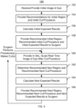
- FIG. 3 is a flow chart depicting an exemplary embodiment of a method 150 for assisting a physician during ophthalmic surgery using quasi-real time image(s). For simplicity, some steps may be omitted, interleaved, performed in another order and/or combined.
- the method 150 may include executing instructions on one or more processors. Further, the method 150 is described in the context of ophthalmic surgery. However, the method 150 may be extended to other types of surgery.
- At least one initial image of at least a portion of the eye is received, via step 152.
- Receipt of the image in step 152 may include receiving data for the image from a separate imaging system or capturing the image by a portion of the system carrying out the method 150.
- the image received in step 152 may, but need not be a quasi-real time image.
- the image(s) received in step 152 may include OCTs, ultrasound image(s), high frequency ultrasound image(s), UBM image(s) and/or another three-dimensional image(s)
- a recommended initial region and a recommended initial procedure are determined based on the initial image(s) and a computational model of the eye, via step 154.
- the computational model of the eye may be analogous the computational model discussed above for the method 100.
- data for the initial image(s) received in step 152 are processed.
- the stresses in particular regions may be determined from the distortions seen in the initial image data.
- striations due to higher stress, fold marks, thinning, tears and/or other issues in various regions may be determined based on the image data and the computational model.
- Step 154 may be performed in a manner analogous to step 104, described above.
- the initial image which may or may not be a quasi-real time image, is used.
- step 154 may include explicitly determining a specific recommended procedure.
- the recommended procedure is known for the particular operation underway.
- Step 156 An initial expected result for the initial procedure is calculated, via step 156.
- Step 156 may be analogous to step 106, described above.
- the initial image which may or may not be a quasi-real time image, is used.
- the initial recommended region, the initial recommended procedure and the initial expected result may be provided, to the physician, via step 158.
- Step 158 is analogous to step 108.
- image(s) of the eye and/or a model of the eye may be displayed for the physician. In some embodiments, therefore, this information is provided graphically to the physician.
- another mechanism for providing the initial recommended region, the initial recommended procedure and the initial expected result is used.
- the surgeon may then perform one or more procedures, such as making cut(s).
- the surgeon may opt to take the recommendation(s) provided in step 158 or perform another procedure.
- the surgeon may desire to make a cut at a different location.
- the surgeon may also perform multiple procedures.
- At least one in situ, quasi-real time image of at least a portion of the eye is received, via step 160.
- Receipt of the image in step 160 may include receiving data for the image from a separate imaging system or capturing the image by a portion of the system carrying out the method 150.
- Step 160 need not include rendering the image for the physician. Instead, step 160 includes obtaining data for the eye. Step 160 is thus analogous to step 102.
- a recommended next region and a recommended next procedure are determined based on the quasi-real time image(s) and a computational model of the eye, via step 162.
- Step 162 is analogous to step 104.
- step 164 includes using the computational model of the eye to determine the reaction of surrounding tissue to a release of stress in that region.
- the recommended next region, the recommended next procedure and the expected result are provided to the physician, via step 166. Portions of step 166 may be performed at different times. For example, the recommended next region and recommended next procedure may be performed by rendering the quasi-real time image or model that is generated in step 162. Providing the expected result to the physician may be performed in response to input received. For example, if a particular recommended region is selected, then the expected result of performing the recommended procedure at that region is provided in step 166. Step 166 may thus include rendering the model of the eye calculated in step 164.
- the surgeon may then be allowed to execute one or more other procedure(s). For example, one or more other cuts may be made.
- the physician can, but need not, follow the recommendations provided in the method 150.
- Step 160 may then be returned to and the eye rescanned.
- the recommendations for the next step and next region may be determined with the new scan and expected results of the new recommendations determined in step 164.
- These new recommendations and new expected results may be provided to the physician, via step 166.
- steps 160, 162, 164 and 166 may be iteratively repeated to assist the surgeon. These steps can, but need not, be repeated every time the surgeon performs a procedure. Alternatively, the steps 160, 162, 164 and 166 may be repeated at selected time(s) during the operation. Thus, the physician may opt to repeat these steps only when s/he deems it helpful or necessary.
- the method 150 may commence using the surgeon's previous information (a more dated initial image) and/or may use a quasi-real time image that is recently captured. Thus, the physician may determine whether their surgical plan is still appropriate. After one or more procedures have been performed as part of the surgery, the steps 160, 162, 164 and 166 may be carried out or repeated. Thus, the surgeon may determine whether the eye is responding as expected and may be able to adjust for their actions throughout surgery. The surgeon may also be able to have a general idea of how the eye is expected to respond prior to a particular procedure and be able to better select the appropriate option. Consequently, the ability of the physician to carry out ophthalmic surgery may be enhanced.
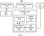
- FIG. 4 is a block diagram of an exemplary embodiment of an apparatus 300 for assisting a physician during ophthalmic surgery using quasi-real time image(s). For simplicity, only some components are shown. In addition, the components depicted in FIG. 4 may be packaged together in a single apparatus such as an OCT or other imaging system. Alternatively, certain components, such as portions of data collection and processing, may be implemented separately. Further, the components may be implemented in hardware and, in some cases, software. Also shown in FIG. 4 is the sample eye 302 to be interrogated.
- the apparatus 300 includes an imaging system 310, a controller/processor 320, a prediction unit 330 and a user interface (U/I) 340.
- the imaging system 310 may be separate from the remainder of the system 300. Consequently, the imaging system 310 is shown as connected by dashed lines. If part of the apparatus 300, the imaging system 310 may be is controlled by the processor 320.
- the operator may input instructions and receive output from the U/l 340. For example, the operator may set the regions of the eye 302 scanned by the imaging system 310, view results or otherwise provide instructions and receive output from the system 300.
- the controller/processor 320 is linked with or controls a system that sets the IOP for the eye 302 or other features. Thus, the controller processor 320 may be used to control quasi-real time image capture.
- the prediction unit 330 may be implemented at least in part in software.
- the prediction unit 330 processes data from the imaging system 310.
- image data 332 and computational model 334 of the eye are shown.
- Portions of the computational model 334 may be stored in memory and are indicated as such in FIG. 4 .
- values for the tensile strength or density of various portions of the eye 302 as well as parameters for the patient may be stored for the computational model 334.
- an FEA model or other model of the eye may be generated and used.
- the recommendation/expected result generator 336 processes the image data 332 and uses the computational model 334 to determine the recommended region(s), recommended procedure(s) and expected result(s).
- the optional renderer 338 may be graphically displayed to the physician on U/l 340.
- the optional renderer 338 may also be used to simply display the quasi-real time image data on the U/l 340.
- the apparatus 300 thus allows the eye 302 to be scanned and mapped during surgery, data for the eye to be processed and recommendations and expected responses of the eye 302 to be determined. Using the apparatus 300, therefore, the method 100 and/or 150 may be implemented. One or more of the benefits of the methods 100 and/or 150 may thus be achieved.
Landscapes
- Health & Medical Sciences (AREA)
- Engineering & Computer Science (AREA)
- Public Health (AREA)
- Life Sciences & Earth Sciences (AREA)
- Medical Informatics (AREA)
- General Health & Medical Sciences (AREA)
- Nuclear Medicine, Radiotherapy & Molecular Imaging (AREA)
- Surgery (AREA)
- Biomedical Technology (AREA)
- Primary Health Care (AREA)
- Epidemiology (AREA)
- Animal Behavior & Ethology (AREA)
- Heart & Thoracic Surgery (AREA)
- Ophthalmology & Optometry (AREA)
- Veterinary Medicine (AREA)
- Radiology & Medical Imaging (AREA)
- Molecular Biology (AREA)
- Pathology (AREA)
- Physics & Mathematics (AREA)
- Biophysics (AREA)
- Databases & Information Systems (AREA)
- Data Mining & Analysis (AREA)
- Vascular Medicine (AREA)
- Human Computer Interaction (AREA)
- Robotics (AREA)
- Urology & Nephrology (AREA)
- Eye Examination Apparatus (AREA)
- Image Processing (AREA)
Applications Claiming Priority (2)
| Application Number | Priority Date | Filing Date | Title |
|---|---|---|---|
| US15/245,328 US10842573B2 (en) | 2016-08-24 | 2016-08-24 | Predictive apparatus for assisting a physician during ophthalmic surgery |
| PCT/IB2017/055087 WO2018037357A1 (en) | 2016-08-24 | 2017-08-23 | Predictive apparatus for assisting a physician during ophthalmic surgery |
Publications (2)
| Publication Number | Publication Date |
|---|---|
| EP3504655A1 EP3504655A1 (en) | 2019-07-03 |
| EP3504655B1 true EP3504655B1 (en) | 2024-03-13 |
Family
ID=59923489
Family Applications (1)
| Application Number | Title | Priority Date | Filing Date |
|---|---|---|---|
| EP17771581.0A Active EP3504655B1 (en) | 2016-08-24 | 2017-08-23 | Predictive apparatus for assisting a physician during ophthalmic surgery |
Country Status (7)
| Country | Link |
|---|---|
| US (2) | US10842573B2 (enExample) |
| EP (1) | EP3504655B1 (enExample) |
| JP (1) | JP7100623B2 (enExample) |
| CN (1) | CN109643582A (enExample) |
| AU (1) | AU2017315286A1 (enExample) |
| CA (1) | CA3031180A1 (enExample) |
| WO (1) | WO2018037357A1 (enExample) |
Families Citing this family (16)
| Publication number | Priority date | Publication date | Assignee | Title |
|---|---|---|---|---|
| US10842573B2 (en) * | 2016-08-24 | 2020-11-24 | Alcon Inc. | Predictive apparatus for assisting a physician during ophthalmic surgery |
| US12350198B2 (en) | 2016-09-28 | 2025-07-08 | Lensar, Inc. | Systems for laser eye surgery |
| US12465436B2 (en) * | 2018-10-19 | 2025-11-11 | Lensar, Inc. | Cloud based system cataract treatment database and algorithm system |
| EP3962424A4 (en) * | 2019-05-03 | 2023-03-29 | Lensar, Inc. | CLOUD SYSTEM CATARACT TREATMENT DATABASE AND ALGORITHM SYSTEM |
| CN115335013A (zh) | 2020-01-03 | 2022-11-11 | 雷萨公司 | 紧凑型可重新配置的集成激光超声乳化系统和使用方法 |
| DE102020102012B4 (de) * | 2020-01-28 | 2022-12-01 | Carl Zeiss Meditec Ag | Anordnung mit einer OCT-Einrichtung für das Ermitteln einer 3D-Rekonstruktion eines Objektbereichsvolumens sowie Computerprogramm und computerimplementiertes Verfahren hierfür |
| CN111616800B (zh) * | 2020-06-09 | 2023-06-09 | 电子科技大学 | 眼科手术导航系统 |
| JP7516172B2 (ja) * | 2020-09-08 | 2024-07-16 | キヤノンメディカルシステムズ株式会社 | 超音波診断装置およびプログラム |
| DE102021100645B4 (de) * | 2021-01-14 | 2024-12-19 | Schwind Eye-Tech-Solutions Gmbh | Verfahren zum Prognostizieren einer zukünftigen Position eines Zielpunktes eines Auges zum Ausgleichen einer Latenz einer Bildauswertung; Steuereinrichtung sowie Behandlungsvorrichtung |
| DE102022133005A1 (de) | 2022-12-12 | 2024-06-13 | Carl Zeiss Meditec Ag | Augenchirurgie-Operationssystem, Computerprogramm und Verfahren für das Bereitstellen einer Bewertungsinformation betreffend das Führen eines Operationswerkzeugs |
| DE102023113284A1 (de) | 2023-05-22 | 2024-11-28 | Carl Zeiss Meditec Ag | Augenchirurgie-Visualisierungssystem sowie Betriebsverfahren und Computerprogramm |
| DE102023128995A1 (de) | 2023-10-23 | 2024-10-17 | Carl Zeiss Meditec Ag | Verfahren zum Erzeugen einer Hilfsinformation, Computerprogramm, augenmedizinische Analysevorrichtung sowie ophthalmochirurgisches System |
| DE102023128999A1 (de) | 2023-10-23 | 2025-04-24 | Carl Zeiss Meditec Ag | Verfahren zum Bestimmen eines Gewebeverhaltens eines Augengewebes mit mehreren Modellen, Computerprogramm, augenmedizinische Analysevorrichtung sowie ophthalmochirurgisches System |
| DE102023128997A1 (de) * | 2023-10-23 | 2025-04-24 | Carl Zeiss Meditec Ag | Verfahren zum Bestimmen eines Gewebeverhaltens eines Augengewebes mit neuronalem Netz, Computerprogramm, augenmedizinische Analysevorrichtung sowie ophthalmochirurgisches System |
| DE102023128994A1 (de) | 2023-10-23 | 2024-10-17 | Carl Zeiss Meditec Ag | Verfahren zum simulativen Bestimmen einer Eigenschaft eines realen Gewebes eines realen Auges, Computerprogramm, augenmedizinische Analysevorrichtung sowie ophthalmochirurgisches System |
| DE102024105571A1 (de) * | 2024-02-28 | 2025-08-28 | Carl Zeiss Meditec Ag | Vorrichtung zum Entfernen des Glaskörpers eines Auges, robotisches Chirurgiesystem und Verfahren zum Einstellen eines einstellbaren Parameterwertes für wenigstens einen Betriebsparameter eines Abschnitts eines chirurgisches Instruments zum Entfernen des Glaskörpers eines Auges |
Citations (2)
| Publication number | Priority date | Publication date | Assignee | Title |
|---|---|---|---|---|
| US20110106102A1 (en) * | 2009-10-30 | 2011-05-05 | The Johns Hopkins University | Surgical Instrument and Systems with Integrated Optical Sensor |
| WO2016064867A2 (en) * | 2014-10-20 | 2016-04-28 | The Regents Of The University Of California | Optical intraocular sensor and sensing method |
Family Cites Families (17)
| Publication number | Priority date | Publication date | Assignee | Title |
|---|---|---|---|---|
| US5891131A (en) | 1993-02-01 | 1999-04-06 | Arizona Board Of Regents | Method and apparatus for automated simulation and design of corneal refractive procedures |
| US6213998B1 (en) * | 1998-04-02 | 2001-04-10 | Vanderbilt University | Laser surgical cutting probe and system |
| US7192412B1 (en) * | 2002-09-14 | 2007-03-20 | Glaukos Corporation | Targeted stent placement and multi-stent therapy |
| CN1781466A (zh) * | 2004-12-03 | 2006-06-07 | 上海市杨浦区民办华都医院 | 眼科准分子激光手术安全质量控制系统及方法 |
| EP1733744A1 (en) * | 2005-06-17 | 2006-12-20 | Ludwig-Maximilians-Universität München | Method, dye and medicament for staining the internal limiting membrane and/or the capsule of an eye |
| EP2656780B1 (en) * | 2006-05-26 | 2016-04-27 | The Cleveland Clinic Foundation | System for measuring biomechanical properties in an eye |
| US20080082088A1 (en) * | 2006-09-05 | 2008-04-03 | Intralase Corp. | System and method for resecting corneal tissue |
| US20100049447A1 (en) | 2008-08-22 | 2010-02-25 | Gholam Peyman | Method of modeling the behavior of an eye subjected to an external force |
| US9411938B2 (en) | 2009-04-02 | 2016-08-09 | Sie Ag, Surgical Instrument Engineering | System for defining cuts in eye tissue |
| US8414124B2 (en) * | 2009-10-21 | 2013-04-09 | Sis Ag, Surgical Instrument Systems | Device and method for measuring a cornea |
| US8591031B2 (en) * | 2010-11-19 | 2013-11-26 | Ziemer Ophthalmic Systems Ag | Device and method for determining the visual field |
| CN101999910A (zh) * | 2010-12-09 | 2011-04-06 | 天津迈达医学科技有限公司 | 眼科超声测量设备中应用的自适应时间-增益补偿方法 |
| JP6415553B2 (ja) | 2013-07-29 | 2018-10-31 | バイオプティジェン, インコーポレイテッドBioptigen, Inc. | 外科手術用手技光干渉断層計及び関連するシステム及びその方法 |
| WO2016082017A1 (en) | 2014-11-27 | 2016-06-02 | Synaptive Medical (Barbados) Inc. | Method, system and apparatus for quantitative surgical image registration |
| DE102014018516B4 (de) * | 2014-12-12 | 2024-08-08 | Carl Zeiss Meditec Ag | System zur Augenuntersuchung mittels spannungsabhängiger Parameter |
| TWI568408B (zh) * | 2015-12-23 | 2017-02-01 | 財團法人工業技術研究院 | 一種眼壓檢測裝置及其檢測方法 |
| US10842573B2 (en) * | 2016-08-24 | 2020-11-24 | Alcon Inc. | Predictive apparatus for assisting a physician during ophthalmic surgery |
-
2016
- 2016-08-24 US US15/245,328 patent/US10842573B2/en active Active
-
2017
- 2017-08-23 JP JP2019510898A patent/JP7100623B2/ja active Active
- 2017-08-23 CN CN201780051784.2A patent/CN109643582A/zh active Pending
- 2017-08-23 AU AU2017315286A patent/AU2017315286A1/en not_active Abandoned
- 2017-08-23 CA CA3031180A patent/CA3031180A1/en not_active Abandoned
- 2017-08-23 WO PCT/IB2017/055087 patent/WO2018037357A1/en not_active Ceased
- 2017-08-23 EP EP17771581.0A patent/EP3504655B1/en active Active
-
2020
- 2020-10-05 US US17/063,383 patent/US20210113281A1/en not_active Abandoned
Patent Citations (2)
| Publication number | Priority date | Publication date | Assignee | Title |
|---|---|---|---|---|
| US20110106102A1 (en) * | 2009-10-30 | 2011-05-05 | The Johns Hopkins University | Surgical Instrument and Systems with Integrated Optical Sensor |
| WO2016064867A2 (en) * | 2014-10-20 | 2016-04-28 | The Regents Of The University Of California | Optical intraocular sensor and sensing method |
Also Published As
| Publication number | Publication date |
|---|---|
| US20180055581A1 (en) | 2018-03-01 |
| CA3031180A1 (en) | 2018-03-01 |
| US20210113281A1 (en) | 2021-04-22 |
| JP7100623B2 (ja) | 2022-07-13 |
| AU2017315286A1 (en) | 2019-02-07 |
| WO2018037357A1 (en) | 2018-03-01 |
| EP3504655A1 (en) | 2019-07-03 |
| JP2019526334A (ja) | 2019-09-19 |
| CN109643582A (zh) | 2019-04-16 |
| US10842573B2 (en) | 2020-11-24 |
Similar Documents
| Publication | Publication Date | Title |
|---|---|---|
| EP3504655B1 (en) | Predictive apparatus for assisting a physician during ophthalmic surgery | |
| JP7413147B2 (ja) | 画像処理装置、画像処理方法、及びプログラム | |
| JP7196908B2 (ja) | 眼科画像処理装置および眼科画像処理プログラム | |
| CN106999298B (zh) | 图像处理装置、图像处理方法以及手术显微镜 | |
| WO2022235596A1 (en) | Intraoperative image-guided tool for ophthalmic surgery | |
| JP7332463B2 (ja) | 制御装置、光干渉断層撮影装置、光干渉断層撮影装置の制御方法、及びプログラム | |
| JP2024152821A (ja) | スリットランプ顕微鏡 | |
| JP6901403B2 (ja) | Oct画像の修正 | |
| JP6289462B2 (ja) | 画像処理装置及び画像処理方法 | |
| JP2021164535A (ja) | 画像処理装置、画像処理方法、及びプログラム | |
| WO2019074077A1 (ja) | 眼科システム、画像信号出力方法、画像信号出力装置、プログラム、及び三次元眼底画像生成方法 | |
| WO2020179588A1 (ja) | 手術顕微鏡システム、画像処理方法、プログラム、及び画像処理装置 | |
| KR101478396B1 (ko) | 화상 처리 장치, 화상 처리 방법, 및 프로그램 기록 매체 | |
| JP2018147387A (ja) | 眼科診療情報処理システム及び眼科診療情報処理方法 | |
| JP2018033693A (ja) | 画像処理装置、画像処理方法、およびプログラム | |
| JP7563384B2 (ja) | 医療画像処理装置および医療画像処理プログラム | |
| JP7709047B2 (ja) | 医療情報処理プログラム、および医療情報処理装置 | |
| JP2022111159A (ja) | レーザ治療装置及び眼科情報処理装置 | |
| JP2019208852A (ja) | 眼科画像処理装置、および眼科画像処理プログラム | |
| JP7612990B2 (ja) | 眼科画像処理装置および眼科画像処理プログラム | |
| JP2023149647A (ja) | 眼科画像処理装置および眼科画像処理プログラム | |
| JP2018147386A (ja) | 眼科診療情報処理システム及び眼科診療情報処理方法 | |
| JP7468163B2 (ja) | 眼科画像処理プログラムおよび眼科画像処理装置 | |
| US20240013397A1 (en) | Ophthalmologic image processing system, ophthalmologic image processing device, and storage medium for storing ophthalmologic image processing program | |
| JP7415570B2 (ja) | 眼科撮影装置制御用プログラム、眼科撮影システム、および、眼科撮影装置 |
Legal Events
| Date | Code | Title | Description |
|---|---|---|---|
| STAA | Information on the status of an ep patent application or granted ep patent |
Free format text: STATUS: UNKNOWN |
|
| STAA | Information on the status of an ep patent application or granted ep patent |
Free format text: STATUS: THE INTERNATIONAL PUBLICATION HAS BEEN MADE |
|
| PUAI | Public reference made under article 153(3) epc to a published international application that has entered the european phase |
Free format text: ORIGINAL CODE: 0009012 |
|
| STAA | Information on the status of an ep patent application or granted ep patent |
Free format text: STATUS: REQUEST FOR EXAMINATION WAS MADE |
|
| 17P | Request for examination filed |
Effective date: 20190322 |
|
| AK | Designated contracting states |
Kind code of ref document: A1 Designated state(s): AL AT BE BG CH CY CZ DE DK EE ES FI FR GB GR HR HU IE IS IT LI LT LU LV MC MK MT NL NO PL PT RO RS SE SI SK SM TR |
|
| AX | Request for extension of the european patent |
Extension state: BA ME |
|
| RIN1 | Information on inventor provided before grant (corrected) |
Inventor name: PAPAC, MICHAEL Inventor name: SANCHEZ, JR., ROBERT |
|
| DAV | Request for validation of the european patent (deleted) | ||
| DAX | Request for extension of the european patent (deleted) | ||
| RAP1 | Party data changed (applicant data changed or rights of an application transferred) |
Owner name: ALCON INC. |
|
| STAA | Information on the status of an ep patent application or granted ep patent |
Free format text: STATUS: EXAMINATION IS IN PROGRESS |
|
| 17Q | First examination report despatched |
Effective date: 20210426 |
|
| P01 | Opt-out of the competence of the unified patent court (upc) registered |
Effective date: 20230507 |
|
| REG | Reference to a national code |
Ref country code: DE Ref legal event code: R079 Free format text: PREVIOUS MAIN CLASS: G06F0019000000 Ipc: G16H0050500000 Ref document number: 602017079993 Country of ref document: DE |
|
| GRAP | Despatch of communication of intention to grant a patent |
Free format text: ORIGINAL CODE: EPIDOSNIGR1 |
|
| STAA | Information on the status of an ep patent application or granted ep patent |
Free format text: STATUS: GRANT OF PATENT IS INTENDED |
|
| RIC1 | Information provided on ipc code assigned before grant |
Ipc: G16H 30/40 20180101ALI20230905BHEP Ipc: G16H 20/40 20180101ALI20230905BHEP Ipc: G16H 50/50 20180101AFI20230905BHEP |
|
| INTG | Intention to grant announced |
Effective date: 20231009 |
|
| RIN1 | Information on inventor provided before grant (corrected) |
Inventor name: SANCHEZ, JR., ROBERT Inventor name: PAPAC, MICHAEL |
|
| GRAS | Grant fee paid |
Free format text: ORIGINAL CODE: EPIDOSNIGR3 |
|
| GRAA | (expected) grant |
Free format text: ORIGINAL CODE: 0009210 |
|
| STAA | Information on the status of an ep patent application or granted ep patent |
Free format text: STATUS: THE PATENT HAS BEEN GRANTED |
|
| AK | Designated contracting states |
Kind code of ref document: B1 Designated state(s): AL AT BE BG CH CY CZ DE DK EE ES FI FR GB GR HR HU IE IS IT LI LT LU LV MC MK MT NL NO PL PT RO RS SE SI SK SM TR |
|
| REG | Reference to a national code |
Ref country code: GB Ref legal event code: FG4D |
|
| REG | Reference to a national code |
Ref country code: CH Ref legal event code: EP |
|
| REG | Reference to a national code |
Ref country code: DE Ref legal event code: R096 Ref document number: 602017079993 Country of ref document: DE |
|
| REG | Reference to a national code |
Ref country code: IE Ref legal event code: FG4D |
|
| PG25 | Lapsed in a contracting state [announced via postgrant information from national office to epo] |
Ref country code: LT Free format text: LAPSE BECAUSE OF FAILURE TO SUBMIT A TRANSLATION OF THE DESCRIPTION OR TO PAY THE FEE WITHIN THE PRESCRIBED TIME-LIMIT Effective date: 20240313 |
|
| REG | Reference to a national code |
Ref country code: LT Ref legal event code: MG9D |
|
| PG25 | Lapsed in a contracting state [announced via postgrant information from national office to epo] |
Ref country code: GR Free format text: LAPSE BECAUSE OF FAILURE TO SUBMIT A TRANSLATION OF THE DESCRIPTION OR TO PAY THE FEE WITHIN THE PRESCRIBED TIME-LIMIT Effective date: 20240614 |
|
| REG | Reference to a national code |
Ref country code: NL Ref legal event code: MP Effective date: 20240313 |
|
| PG25 | Lapsed in a contracting state [announced via postgrant information from national office to epo] |
Ref country code: RS Free format text: LAPSE BECAUSE OF FAILURE TO SUBMIT A TRANSLATION OF THE DESCRIPTION OR TO PAY THE FEE WITHIN THE PRESCRIBED TIME-LIMIT Effective date: 20240613 Ref country code: HR Free format text: LAPSE BECAUSE OF FAILURE TO SUBMIT A TRANSLATION OF THE DESCRIPTION OR TO PAY THE FEE WITHIN THE PRESCRIBED TIME-LIMIT Effective date: 20240313 |
|
| PG25 | Lapsed in a contracting state [announced via postgrant information from national office to epo] |
Ref country code: ES Free format text: LAPSE BECAUSE OF FAILURE TO SUBMIT A TRANSLATION OF THE DESCRIPTION OR TO PAY THE FEE WITHIN THE PRESCRIBED TIME-LIMIT Effective date: 20240313 |
|
| PG25 | Lapsed in a contracting state [announced via postgrant information from national office to epo] |
Ref country code: RS Free format text: LAPSE BECAUSE OF FAILURE TO SUBMIT A TRANSLATION OF THE DESCRIPTION OR TO PAY THE FEE WITHIN THE PRESCRIBED TIME-LIMIT Effective date: 20240613 Ref country code: NO Free format text: LAPSE BECAUSE OF FAILURE TO SUBMIT A TRANSLATION OF THE DESCRIPTION OR TO PAY THE FEE WITHIN THE PRESCRIBED TIME-LIMIT Effective date: 20240613 Ref country code: LT Free format text: LAPSE BECAUSE OF FAILURE TO SUBMIT A TRANSLATION OF THE DESCRIPTION OR TO PAY THE FEE WITHIN THE PRESCRIBED TIME-LIMIT Effective date: 20240313 Ref country code: HR Free format text: LAPSE BECAUSE OF FAILURE TO SUBMIT A TRANSLATION OF THE DESCRIPTION OR TO PAY THE FEE WITHIN THE PRESCRIBED TIME-LIMIT Effective date: 20240313 Ref country code: GR Free format text: LAPSE BECAUSE OF FAILURE TO SUBMIT A TRANSLATION OF THE DESCRIPTION OR TO PAY THE FEE WITHIN THE PRESCRIBED TIME-LIMIT Effective date: 20240614 Ref country code: FI Free format text: LAPSE BECAUSE OF FAILURE TO SUBMIT A TRANSLATION OF THE DESCRIPTION OR TO PAY THE FEE WITHIN THE PRESCRIBED TIME-LIMIT Effective date: 20240313 Ref country code: ES Free format text: LAPSE BECAUSE OF FAILURE TO SUBMIT A TRANSLATION OF THE DESCRIPTION OR TO PAY THE FEE WITHIN THE PRESCRIBED TIME-LIMIT Effective date: 20240313 Ref country code: BG Free format text: LAPSE BECAUSE OF FAILURE TO SUBMIT A TRANSLATION OF THE DESCRIPTION OR TO PAY THE FEE WITHIN THE PRESCRIBED TIME-LIMIT Effective date: 20240313 |
|
| REG | Reference to a national code |
Ref country code: AT Ref legal event code: MK05 Ref document number: 1666548 Country of ref document: AT Kind code of ref document: T Effective date: 20240313 |
|
| PG25 | Lapsed in a contracting state [announced via postgrant information from national office to epo] |
Ref country code: SE Free format text: LAPSE BECAUSE OF FAILURE TO SUBMIT A TRANSLATION OF THE DESCRIPTION OR TO PAY THE FEE WITHIN THE PRESCRIBED TIME-LIMIT Effective date: 20240313 Ref country code: LV Free format text: LAPSE BECAUSE OF FAILURE TO SUBMIT A TRANSLATION OF THE DESCRIPTION OR TO PAY THE FEE WITHIN THE PRESCRIBED TIME-LIMIT Effective date: 20240313 |
|
| PG25 | Lapsed in a contracting state [announced via postgrant information from national office to epo] |
Ref country code: NL Free format text: LAPSE BECAUSE OF FAILURE TO SUBMIT A TRANSLATION OF THE DESCRIPTION OR TO PAY THE FEE WITHIN THE PRESCRIBED TIME-LIMIT Effective date: 20240313 |
|
| PG25 | Lapsed in a contracting state [announced via postgrant information from national office to epo] |
Ref country code: NL Free format text: LAPSE BECAUSE OF FAILURE TO SUBMIT A TRANSLATION OF THE DESCRIPTION OR TO PAY THE FEE WITHIN THE PRESCRIBED TIME-LIMIT Effective date: 20240313 |
|
| PG25 | Lapsed in a contracting state [announced via postgrant information from national office to epo] |
Ref country code: IS Free format text: LAPSE BECAUSE OF FAILURE TO SUBMIT A TRANSLATION OF THE DESCRIPTION OR TO PAY THE FEE WITHIN THE PRESCRIBED TIME-LIMIT Effective date: 20240713 |
|
| PG25 | Lapsed in a contracting state [announced via postgrant information from national office to epo] |
Ref country code: PT Free format text: LAPSE BECAUSE OF FAILURE TO SUBMIT A TRANSLATION OF THE DESCRIPTION OR TO PAY THE FEE WITHIN THE PRESCRIBED TIME-LIMIT Effective date: 20240715 Ref country code: SM Free format text: LAPSE BECAUSE OF FAILURE TO SUBMIT A TRANSLATION OF THE DESCRIPTION OR TO PAY THE FEE WITHIN THE PRESCRIBED TIME-LIMIT Effective date: 20240313 |
|
| PG25 | Lapsed in a contracting state [announced via postgrant information from national office to epo] |
Ref country code: CZ Free format text: LAPSE BECAUSE OF FAILURE TO SUBMIT A TRANSLATION OF THE DESCRIPTION OR TO PAY THE FEE WITHIN THE PRESCRIBED TIME-LIMIT Effective date: 20240313 Ref country code: EE Free format text: LAPSE BECAUSE OF FAILURE TO SUBMIT A TRANSLATION OF THE DESCRIPTION OR TO PAY THE FEE WITHIN THE PRESCRIBED TIME-LIMIT Effective date: 20240313 |
|
| PG25 | Lapsed in a contracting state [announced via postgrant information from national office to epo] |
Ref country code: AT Free format text: LAPSE BECAUSE OF FAILURE TO SUBMIT A TRANSLATION OF THE DESCRIPTION OR TO PAY THE FEE WITHIN THE PRESCRIBED TIME-LIMIT Effective date: 20240313 |
|
| PG25 | Lapsed in a contracting state [announced via postgrant information from national office to epo] |
Ref country code: PL Free format text: LAPSE BECAUSE OF FAILURE TO SUBMIT A TRANSLATION OF THE DESCRIPTION OR TO PAY THE FEE WITHIN THE PRESCRIBED TIME-LIMIT Effective date: 20240313 |
|
| PG25 | Lapsed in a contracting state [announced via postgrant information from national office to epo] |
Ref country code: SK Free format text: LAPSE BECAUSE OF FAILURE TO SUBMIT A TRANSLATION OF THE DESCRIPTION OR TO PAY THE FEE WITHIN THE PRESCRIBED TIME-LIMIT Effective date: 20240313 |
|
| PG25 | Lapsed in a contracting state [announced via postgrant information from national office to epo] |
Ref country code: SM Free format text: LAPSE BECAUSE OF FAILURE TO SUBMIT A TRANSLATION OF THE DESCRIPTION OR TO PAY THE FEE WITHIN THE PRESCRIBED TIME-LIMIT Effective date: 20240313 Ref country code: SK Free format text: LAPSE BECAUSE OF FAILURE TO SUBMIT A TRANSLATION OF THE DESCRIPTION OR TO PAY THE FEE WITHIN THE PRESCRIBED TIME-LIMIT Effective date: 20240313 Ref country code: RO Free format text: LAPSE BECAUSE OF FAILURE TO SUBMIT A TRANSLATION OF THE DESCRIPTION OR TO PAY THE FEE WITHIN THE PRESCRIBED TIME-LIMIT Effective date: 20240313 Ref country code: PT Free format text: LAPSE BECAUSE OF FAILURE TO SUBMIT A TRANSLATION OF THE DESCRIPTION OR TO PAY THE FEE WITHIN THE PRESCRIBED TIME-LIMIT Effective date: 20240715 Ref country code: PL Free format text: LAPSE BECAUSE OF FAILURE TO SUBMIT A TRANSLATION OF THE DESCRIPTION OR TO PAY THE FEE WITHIN THE PRESCRIBED TIME-LIMIT Effective date: 20240313 Ref country code: IS Free format text: LAPSE BECAUSE OF FAILURE TO SUBMIT A TRANSLATION OF THE DESCRIPTION OR TO PAY THE FEE WITHIN THE PRESCRIBED TIME-LIMIT Effective date: 20240713 Ref country code: EE Free format text: LAPSE BECAUSE OF FAILURE TO SUBMIT A TRANSLATION OF THE DESCRIPTION OR TO PAY THE FEE WITHIN THE PRESCRIBED TIME-LIMIT Effective date: 20240313 Ref country code: CZ Free format text: LAPSE BECAUSE OF FAILURE TO SUBMIT A TRANSLATION OF THE DESCRIPTION OR TO PAY THE FEE WITHIN THE PRESCRIBED TIME-LIMIT Effective date: 20240313 Ref country code: AT Free format text: LAPSE BECAUSE OF FAILURE TO SUBMIT A TRANSLATION OF THE DESCRIPTION OR TO PAY THE FEE WITHIN THE PRESCRIBED TIME-LIMIT Effective date: 20240313 |
|
| PG25 | Lapsed in a contracting state [announced via postgrant information from national office to epo] |
Ref country code: IT Free format text: LAPSE BECAUSE OF FAILURE TO SUBMIT A TRANSLATION OF THE DESCRIPTION OR TO PAY THE FEE WITHIN THE PRESCRIBED TIME-LIMIT Effective date: 20240313 |
|
| REG | Reference to a national code |
Ref country code: DE Ref legal event code: R097 Ref document number: 602017079993 Country of ref document: DE |
|
| PG25 | Lapsed in a contracting state [announced via postgrant information from national office to epo] |
Ref country code: IT Free format text: LAPSE BECAUSE OF FAILURE TO SUBMIT A TRANSLATION OF THE DESCRIPTION OR TO PAY THE FEE WITHIN THE PRESCRIBED TIME-LIMIT Effective date: 20240313 |
|
| PG25 | Lapsed in a contracting state [announced via postgrant information from national office to epo] |
Ref country code: DK Free format text: LAPSE BECAUSE OF FAILURE TO SUBMIT A TRANSLATION OF THE DESCRIPTION OR TO PAY THE FEE WITHIN THE PRESCRIBED TIME-LIMIT Effective date: 20240313 |
|
| PLBE | No opposition filed within time limit |
Free format text: ORIGINAL CODE: 0009261 |
|
| STAA | Information on the status of an ep patent application or granted ep patent |
Free format text: STATUS: NO OPPOSITION FILED WITHIN TIME LIMIT |
|
| PG25 | Lapsed in a contracting state [announced via postgrant information from national office to epo] |
Ref country code: DK Free format text: LAPSE BECAUSE OF FAILURE TO SUBMIT A TRANSLATION OF THE DESCRIPTION OR TO PAY THE FEE WITHIN THE PRESCRIBED TIME-LIMIT Effective date: 20240313 |
|
| 26N | No opposition filed |
Effective date: 20241216 |
|
| REG | Reference to a national code |
Ref country code: CH Ref legal event code: PL |
|
| PG25 | Lapsed in a contracting state [announced via postgrant information from national office to epo] |
Ref country code: LU Free format text: LAPSE BECAUSE OF NON-PAYMENT OF DUE FEES Effective date: 20240823 |
|
| PG25 | Lapsed in a contracting state [announced via postgrant information from national office to epo] |
Ref country code: MC Free format text: LAPSE BECAUSE OF FAILURE TO SUBMIT A TRANSLATION OF THE DESCRIPTION OR TO PAY THE FEE WITHIN THE PRESCRIBED TIME-LIMIT Effective date: 20240313 Ref country code: SI Free format text: LAPSE BECAUSE OF FAILURE TO SUBMIT A TRANSLATION OF THE DESCRIPTION OR TO PAY THE FEE WITHIN THE PRESCRIBED TIME-LIMIT Effective date: 20240313 Ref country code: CH Free format text: LAPSE BECAUSE OF NON-PAYMENT OF DUE FEES Effective date: 20240831 |
|
| REG | Reference to a national code |
Ref country code: BE Ref legal event code: MM Effective date: 20240831 |
|
| PG25 | Lapsed in a contracting state [announced via postgrant information from national office to epo] |
Ref country code: BE Free format text: LAPSE BECAUSE OF NON-PAYMENT OF DUE FEES Effective date: 20240831 |
|
| PG25 | Lapsed in a contracting state [announced via postgrant information from national office to epo] |
Ref country code: IE Free format text: LAPSE BECAUSE OF NON-PAYMENT OF DUE FEES Effective date: 20240823 |
|
| PGFP | Annual fee paid to national office [announced via postgrant information from national office to epo] |
Ref country code: DE Payment date: 20250722 Year of fee payment: 9 |
|
| PGFP | Annual fee paid to national office [announced via postgrant information from national office to epo] |
Ref country code: GB Payment date: 20250717 Year of fee payment: 9 |
|
| PGFP | Annual fee paid to national office [announced via postgrant information from national office to epo] |
Ref country code: FR Payment date: 20250721 Year of fee payment: 9 |