EP3500869B1 - Corrected compressed sensing magnetic resonance imaging - Google Patents
Corrected compressed sensing magnetic resonance imaging Download PDFInfo
- Publication number
- EP3500869B1 EP3500869B1 EP17737301.6A EP17737301A EP3500869B1 EP 3500869 B1 EP3500869 B1 EP 3500869B1 EP 17737301 A EP17737301 A EP 17737301A EP 3500869 B1 EP3500869 B1 EP 3500869B1
- Authority
- EP
- European Patent Office
- Prior art keywords
- magnetic resonance
- measured
- data
- resonance imaging
- data portions
- Prior art date
- Legal status (The legal status is an assumption and is not a legal conclusion. Google has not performed a legal analysis and makes no representation as to the accuracy of the status listed.)
- Active
Links
- 238000002595 magnetic resonance imaging Methods 0.000 title claims description 116
- 238000000034 method Methods 0.000 claims description 65
- 230000033001 locomotion Effects 0.000 claims description 46
- 238000003384 imaging method Methods 0.000 claims description 29
- 238000001208 nuclear magnetic resonance pulse sequence Methods 0.000 claims description 19
- 238000005070 sampling Methods 0.000 claims description 12
- 238000004590 computer program Methods 0.000 claims description 11
- 238000005259 measurement Methods 0.000 claims description 9
- 238000004364 calculation method Methods 0.000 claims description 4
- 230000001010 compromised effect Effects 0.000 claims description 4
- 238000005315 distribution function Methods 0.000 claims description 2
- 238000003860 storage Methods 0.000 description 20
- 230000006870 function Effects 0.000 description 13
- 230000035945 sensitivity Effects 0.000 description 13
- 230000002123 temporal effect Effects 0.000 description 9
- 230000003068 static effect Effects 0.000 description 8
- 238000010586 diagram Methods 0.000 description 7
- 238000002059 diagnostic imaging Methods 0.000 description 6
- 238000012545 processing Methods 0.000 description 6
- 230000008901 benefit Effects 0.000 description 5
- 238000004422 calculation algorithm Methods 0.000 description 5
- 230000001276 controlling effect Effects 0.000 description 4
- 238000009826 distribution Methods 0.000 description 4
- 230000003287 optical effect Effects 0.000 description 4
- 230000000737 periodic effect Effects 0.000 description 4
- 230000000875 corresponding effect Effects 0.000 description 3
- 238000012804 iterative process Methods 0.000 description 3
- 238000012800 visualization Methods 0.000 description 3
- 230000009286 beneficial effect Effects 0.000 description 2
- 238000004891 communication Methods 0.000 description 2
- 230000002596 correlated effect Effects 0.000 description 2
- 230000001419 dependent effect Effects 0.000 description 2
- 230000000644 propagated effect Effects 0.000 description 2
- 230000005540 biological transmission Effects 0.000 description 1
- 238000013184 cardiac magnetic resonance imaging Methods 0.000 description 1
- 239000002131 composite material Substances 0.000 description 1
- 238000002591 computed tomography Methods 0.000 description 1
- 238000001816 cooling Methods 0.000 description 1
- 238000012937 correction Methods 0.000 description 1
- 238000003745 diagnosis Methods 0.000 description 1
- 230000000694 effects Effects 0.000 description 1
- 230000007717 exclusion Effects 0.000 description 1
- 239000004615 ingredient Substances 0.000 description 1
- 239000004973 liquid crystal related substance Substances 0.000 description 1
- 238000004519 manufacturing process Methods 0.000 description 1
- 239000013307 optical fiber Substances 0.000 description 1
- 230000000241 respiratory effect Effects 0.000 description 1
- 238000012552 review Methods 0.000 description 1
- 230000011218 segmentation Effects 0.000 description 1
- 239000007787 solid Substances 0.000 description 1
- 238000000528 statistical test Methods 0.000 description 1
- 238000012360 testing method Methods 0.000 description 1
- 210000003813 thumb Anatomy 0.000 description 1
- 230000001052 transient effect Effects 0.000 description 1
- 230000000007 visual effect Effects 0.000 description 1
Images

Classifications
-
- G—PHYSICS
- G01—MEASURING; TESTING
- G01R—MEASURING ELECTRIC VARIABLES; MEASURING MAGNETIC VARIABLES
- G01R33/00—Arrangements or instruments for measuring magnetic variables
- G01R33/20—Arrangements or instruments for measuring magnetic variables involving magnetic resonance
- G01R33/44—Arrangements or instruments for measuring magnetic variables involving magnetic resonance using nuclear magnetic resonance [NMR]
- G01R33/48—NMR imaging systems
- G01R33/54—Signal processing systems, e.g. using pulse sequences ; Generation or control of pulse sequences; Operator console
- G01R33/56—Image enhancement or correction, e.g. subtraction or averaging techniques, e.g. improvement of signal-to-noise ratio and resolution
- G01R33/565—Correction of image distortions, e.g. due to magnetic field inhomogeneities
- G01R33/56509—Correction of image distortions, e.g. due to magnetic field inhomogeneities due to motion, displacement or flow, e.g. gradient moment nulling
-
- G—PHYSICS
- G01—MEASURING; TESTING
- G01R—MEASURING ELECTRIC VARIABLES; MEASURING MAGNETIC VARIABLES
- G01R33/00—Arrangements or instruments for measuring magnetic variables
- G01R33/20—Arrangements or instruments for measuring magnetic variables involving magnetic resonance
- G01R33/44—Arrangements or instruments for measuring magnetic variables involving magnetic resonance using nuclear magnetic resonance [NMR]
- G01R33/48—NMR imaging systems
- G01R33/4818—MR characterised by data acquisition along a specific k-space trajectory or by the temporal order of k-space coverage, e.g. centric or segmented coverage of k-space
-
- G—PHYSICS
- G01—MEASURING; TESTING
- G01R—MEASURING ELECTRIC VARIABLES; MEASURING MAGNETIC VARIABLES
- G01R33/00—Arrangements or instruments for measuring magnetic variables
- G01R33/20—Arrangements or instruments for measuring magnetic variables involving magnetic resonance
- G01R33/44—Arrangements or instruments for measuring magnetic variables involving magnetic resonance using nuclear magnetic resonance [NMR]
- G01R33/48—NMR imaging systems
- G01R33/54—Signal processing systems, e.g. using pulse sequences ; Generation or control of pulse sequences; Operator console
- G01R33/56—Image enhancement or correction, e.g. subtraction or averaging techniques, e.g. improvement of signal-to-noise ratio and resolution
- G01R33/5608—Data processing and visualization specially adapted for MR, e.g. for feature analysis and pattern recognition on the basis of measured MR data, segmentation of measured MR data, edge contour detection on the basis of measured MR data, for enhancing measured MR data in terms of signal-to-noise ratio by means of noise filtering or apodization, for enhancing measured MR data in terms of resolution by means for deblurring, windowing, zero filling, or generation of gray-scaled images, colour-coded images or images displaying vectors instead of pixels
-
- G—PHYSICS
- G01—MEASURING; TESTING
- G01R—MEASURING ELECTRIC VARIABLES; MEASURING MAGNETIC VARIABLES
- G01R33/00—Arrangements or instruments for measuring magnetic variables
- G01R33/20—Arrangements or instruments for measuring magnetic variables involving magnetic resonance
- G01R33/44—Arrangements or instruments for measuring magnetic variables involving magnetic resonance using nuclear magnetic resonance [NMR]
- G01R33/48—NMR imaging systems
- G01R33/54—Signal processing systems, e.g. using pulse sequences ; Generation or control of pulse sequences; Operator console
- G01R33/56—Image enhancement or correction, e.g. subtraction or averaging techniques, e.g. improvement of signal-to-noise ratio and resolution
- G01R33/561—Image enhancement or correction, e.g. subtraction or averaging techniques, e.g. improvement of signal-to-noise ratio and resolution by reduction of the scanning time, i.e. fast acquiring systems, e.g. using echo-planar pulse sequences
- G01R33/5611—Parallel magnetic resonance imaging, e.g. sensitivity encoding [SENSE], simultaneous acquisition of spatial harmonics [SMASH], unaliasing by Fourier encoding of the overlaps using the temporal dimension [UNFOLD], k-t-broad-use linear acquisition speed-up technique [k-t-BLAST], k-t-SENSE
Definitions
- the invention relates to magnetic resonance imaging, in particular to compressed sensing magnetic resonance imaging.
- a large static magnetic field is used by Magnetic Resonance Imaging (MRI) scanners to align the nuclear spins of atoms as part of the procedure for producing images within the body of a patient.
- This large static magnetic field is referred to as the B0 field or the main magnetic field.
- One method of spatially encoding is to use magnetic field gradient coils. Typically there are three coils which are used to generate three different gradient magnetic fields in three different orthogonal directions.
- Radio Frequency (RF) pulses generated by one or more transmitter coils cause a called B1 field. Additionally applied gradient fields and the B1 field do cause perturbations to the effective local magnetic field.
- RF signals are then emitted by the nuclear spins and detected by one or more receiver coils. These RF signals contain image data encoded in k-space.
- the central region of k-space generally contains more image information than outer regions of k-space.
- the Nyquist sampling theorem is a sufficient, but not necessary condition. Often times an acceptable magnetic resonance image can be reconstructed by sampling k-space less than is specified by the Nyquist theorem.
- the invention provides for a magnetic resonance imaging system, a computer program product, and a method in the independent claims. Embodiments are given in the dependent claims.
- Embodiments of the invention provide for a means for improving the quality of magnetic resonance images produced using compressed sensing magnetic resonance imaging protocols.
- the reconstruction of images using CS is an iterative process.
- the resulting image will not necessarily match the original magnetic resonance data samples in k-space.
- the technique described herein uses those features of CS to identify data which may be compromised due to motion of the subject being imaged or corrupted due to other reasons.
- the acquired magnetic resonance data is first used to reconstruct an image according to a CS imaging protocol. This results in an intermediate magnetic resonance image.
- this image is then used to calculate predicted k-space data (referred to as a predicted data portion herein). Sufficient data portions in k-space are measured to allow reconstruction of the intermediate magnetic resonance image .
- the intermediate magnetic resonance image may have a low spatial resolution, small field-of-view and when parallel imaging is used have a high folding degree. The better the quality of the intermediate magnetic resonance image, the more accurate the outliers can be identified and a smaller number of iterations in the compressed sensing reconstruction may be required to achieve a higher image quality.
- the intermediate magnetic resonance image is reconstructed from measured magnetic resonance signals, i.e. k-space data points or data portions (k-space profiles). Reconstructing the intermediate magnetic resonance image using the measured magnetic resonance data may include that the entire k-space coverage of the entire covered k-space is employed to reconstruct the intermediate magnetic resonance image . A subset of all actually measured data portions may be sufficient to reconstructed a low-resolution and/or low field-of-view intermediate magnetic resonance image.
- a parallel imaging technique i.e., the data used to reconstruct the intermediate image came from multiple coil elements
- the coil sensitivities are first used to calculate predicted images resulting from each of the coil elements that were used.
- These predicted coil images are then inverse Fourier transformed to calculate predicted k-space data for each of the coil elements.
- the predicted k-space data can be directly compared to the measured k-space data.
- Image values of the intermediate magnetic resonance image and the predicted data portions are related according to the inverse of the relationship implied by the reconstruction of the intermediate magnetic resonance image and the measured data portions.
- their relationship is the (inverse) Fourier transform, but different mathematical relationships may be employed to transform the measured data portions in the intermediate magnetic resonance image and back-transform the predicted data portions from the intermediate magnetic resonance image.
- (filtered) back-projections may be employed for the reconstruction of the intermediate magnetic resonance image and from the intermediate magnetic resonance image by forward projection the predicted data portions are calculated.
- the (filtered) back-projection is generally known form the field of computed tomography.
- the magnetic resonance data is acquired within discrete time periods as measured portions (of k-space data).
- a residual is calculated for each of the time periods. If the residual for a time period is above a predetermined threshold, then the data from that time period is identified as being an outlier data portion.
- the identification of outliers is not sensitive to the underlying cause of the value the data portion deviating more than the predetermined threshold. That is, data portions may be identified as outliers irrespective of whether motion has occurred or due to another cause of inconsistency.
- a corrected magnetic resonance image is then reconstructed using the acquired magnetic resonance data (the measured data portions).
- the outlier data portions are ignored or not used.
- the outlier data portions could be deleted or removed and the corrected magnetic resonance image could be completely recalculated.
- the CS technique is an iterative process. Numerically, it may therefore be more efficient to use the intermediate magnetic resonance image as the starting point for the reconstruction of the corrected magnetic resonance image, with the outlier data portions being ignored during this reconstruction.
- the invention provides for a magnetic resonance imaging system for acquiring measured magnetic resonance data from a subject within an imaging zone as defined in claim 1.
- the magnetic resonance imaging system comprises a memory for storing machine-executable instructions and for storing pulse sequence commands which are configured to control the magnetic resonance imaging system to acquire the measured magnetic resonance data according to a compressed sensing magnetic resonance imaging protocol.
- the magnetic resonance imaging system further comprises a processor for controlling the magnetic resonance imaging system.
- Execution of the machine-executable instructions causes the processor to control the magnetic resonance imaging system with the pulse sequence commands to acquire the measured magnetic resonance data.
- the measured magnetic resonance data is acquired as measured data portions. Each of the measured data portions is acquired during a time period.
- Execution of the machine-executable instructions further causes the processor to reconstruct an intermediate magnetic resonance image using the measured magnetic resonance data according to the compressed sensing magnetic resonance imaging protocol.
- Execution of the machine-executable instructions further causes the processor to calculate a predicated data portion for each of the measured data portions using the intermediate magnetic resonance image.
- Execution of the machine-executable instructions further causes the processor to calculate a residual for each of the measured data portions using the predicted data portion.
- the predicted data portion may for instance be determined by performing an inverse Fourier transform to calculate what the measured data would be like to generate the intermediate magnetic resonance image.
- These predicted data points in k-space can be directly compared to the measured data points within the measured data portions.
- the residual may therefore be considered as a measure which is used to compare the measured data with the predicted data.
- the residual can be calculated in different fashions. For instance every single data point can be compared and then a composite residual can be calculated using an average or for example an at least squared type function. Other statistical measures may also be used.
- portions of the k-space which are more important for generation of the image such as a central region of k-space can be weighted more heavily than data in the periphery of k-space.
- the data measured by the magnetic resonance imaging system may be acquired using more than one antenna element.
- the compressed sensing magnetic resonance imaging protocol might be a parallel imaging method.
- calculating the predicted data portion may be more complicated.
- the weightings for calculating the image must be performed backwards and then used for calculating the data acquired by each antenna element.
- Execution of the machine-executable instructions further cause the processor to identify one or more of the measured data portions as outlier data portions if the residual is above a predetermined threshold. Depending on what measure the residual is the entire measured data portion is identified as outlier data portions. So if there is a parallel imaging technique data acquired by one antenna may cause data from all of the antenna or antenna elements to be identified as being outlier data portions. Execution of the machine-executable instructions further causes the processor to reconstruct a corrected magnetic resonance image using the measured magnetic resonance data according to the compressed sensing magnetic resonance imaging protocol.
- the one or more outlier data portions are excluded from the reconstruction of the corrected magnetic resonance image.
- the exclusion of the one or more outlier data portions can be performed in different ways. In one instance the image can be completely reconstructed using the compressed sensed imaging protocol using only the data which was not identified as being outlier data portions. This however would be numerically intensive. Another alternative is that the compressed sensing protocol can be used to further refine the intermediate magnetic resonance image using all of the data except the outlier data portions. This would have the advantage of being less numerically intensive.
- This embodiment may have the benefit that the compressed sensing protocol does a superior job of rejecting data in which the subject was moving during the time period for particular measured data portions.
- the movement of the subject may result in ghosting or aliasing artifacts within the intermediate magnetic resonance image.
- the process of calculating the residual does an effective job of identifying these data portions which contribute to motion artifacts.
- the compressed sensing magnetic resonance imaging protocol is a parallel imaging magnetic resonance imaging protocol.
- the magnetic resonance imaging system comprises a magnetic resonance imaging antenna with multiple antennas or antenna elements for acquiring the measured magnetic resonance data. During the time period the measured magnetic resonance data is acquired from each of the multiple antenna elements as an entire measured data portion. The residual is calculated for each of the multiple antenna elements for the time period.
- the residual for each of the multiple antenna elements in each one of them is above a particular threshold or criteria then the data corresponding to that time period can be flagged as being outlier data portions.
- the local sensitivities of the multiple antenna elements may cause some antenna elements to be more sensitive to motion of the subject than other antenna elements. By examining the residual from each of the multiple antenna elements this may provide for an improved method of identifying motion of a subject during the compressed sensing imaging.
- the residual is calculated for each of the multiple antenna elements individually. If a residual for one of the multiple antenna elements is above the predetermined threshold then the entire measured data portion is removed from the measured magnetic resonance data or is identified as an outlier data portion.
- the residual is an average over all of the multiple antennas. If the residual is above the predetermined threshold then the entire measured data portion is identified as one of the outlier data portions.
- the k-space data that is acquired according to the compressed sensing magnetic resonance imaging protocol within a particular time period is maximally spread using a distribution function.
- the sample points in k-space could be distributed using a random or pseudo random function.
- a more defined distribution such as the Poisson distribution could be used to centralize the k-space data or sample points within a central region of k-space.
- the intermediate magnetic resonance image is repeatedly reconstructed as one or more of the measured data portions is acquired.
- the predicted data portion is repeatedly calculated as the one or more measured data portions is acquired.
- the residual is repeatedly calculated as the one or more measured data portions is acquired.
- a measured data portion is repeatedly identified as an outlier data portion if the residual is above the predetermined threshold as the one or more measured data portions is acquired.
- This embodiment may have the benefit that the outlier data portions are acquired on the fly as the measured magnetic resonance data is being acquired. This may enable the operator of the magnetic resonance imaging system to correct for problems or movement of the subject. It may also enable the operator (or control computer) to reacquire portions of the measured magnetic resonance data before the end of the acquisition of the measured magnetic resonance data. For example if the outlier data portions are identified after the acquisition of the data is finished it may be impossible to go back and reacquire the data with the subject in exactly the same position. This may then result in higher quality magnetic resonance images.
- the resolution of the intermediate magnetic resonance image is varied as an increasing number of the measured magnetic resonance data is acquired.
- the intermediate magnetic resonance image may still be constructed but it may have a lower resolution.
- the quality of the intermediate magnetic resonance image may be improved. This may have several consequences when only several of the measured data portions are acquired it may not be immediately evident that one of the measured data portions should be assigned to be an outlier data portion.
- Changing the resolution of the intermediate magnetic resonance image may therefore enable a more dynamic and flexible method of identifying the outlier data portions.
- execution of the machine-executable instructions further cause the processor to reacquire a measured data portion if it is identified as one of the outlier data portions. This may have the benefit of providing for an improvement in the quality of generated magnetic resonance images.
- the residual is calculated using a statistical measure to compare each of the measured data portions to the predicted data portion.
- the measured data portions may comprise individual measurements in k-space. These individual measurements within k-space can be compared to each other and a statistical measure may be used to calculate the residual.
- the statistical measure weights individual k-space measurements according to their location in k-space. This may be beneficial because it may provide for a means of providing k-space measurements which are more important to the image reconstruction to count more in the residual. For example within k-space the central region of k-space contains more signal power than the outer region of k-space. If the center of the k-space region is weighted more such as a distribution which favors the central region of k-space then this may provide a more accurate means of rejecting motion when reconstructing the corrected magnetic resonance image.
- execution of the machine-executable instructions further cause the processor to reconstruct at least one outlier magnetic resonance image using the one or more outlier data portions according to the compressed sensing magnetic resonance imaging protocol.
- the motion of the subject may be periodic or the subject may move or have an involuntary motion which goes between several positions.
- Using the magnetic resonance imaging data which was identified as being the outlier data portions may enable a method of compressed sensing imaging which is resolved about different motion states of the subject and which does not use a navigator or external sensor on the subject.
- the reconstruction of the at least one outlier magnetic resonance image could follow the same method as was used to construct the corrected magnetic resonance image where an intermediate outlier magnetic resonance image is constructed and then a residual is calculated in the same way. This way the various positions of the subject could be divided into a number of different images each which represent a different position of the subject.
- k-space data could be divided into several groups and then these groups could be used to correct an outlier magnetic resonance image for each one of them.
- execution of the machine-executable instructions further causes the processor to set the corrected magnetic resonance image as the intermediate magnetic resonance image.
- Execution of the machine-executable instructions further cause the processor to repeat the calculation of the predicted portion, the identification of the one or more outlier data portions and the reconstruction of the corrected magnetic resonance image.
- this can then be used to again search for additional outlier data portions. For example, if a subject moves and there is ghosting or some artifacts within the magnetic resonance image the portions of the magnetic resonance data which have the most motion in them would be identified as outlier data portions.
- the corrected magnetic resonance image can then be used as the intermediate magnetic resonance image and the process can be started again.
- the intermediate magnetic resonance image which was previously the corrected magnetic resonance image can then be used to create a new predicted data portion for each of the measured data portions using the intermediate magnetic resonance image. There can then be a new residual which is calculated for each of the remaining measured data portions using the new predicted data portion. Then finally, the measured data portions can then be re-examined to identify if one or more of them should be identified as outlier data portions based on the residual. For example if the residual is above a new predetermined threshold. After the measured data portions have been gone over again then a new corrected magnetic resonance image can be constructed using the measured magnetic resonance data according to the compressed sensing magnetic resonance imaging protocol and in this case the data which is then identified as the one or more outlier data portions are again excluded from the reconstruction of the corrected magnetic resonance image. However, in this case it is very likely that a larger portion of the measured magnetic resonance data is identified as belonging to the one or more outlier data portions.
- Such a process may be repeated a number of times or it is also possible that the loop can be repeated several times and it can be seen if the corrected magnetic resonance image converges. For example, within several iterations the corrected magnetic resonance image from each iteration can be compared to the previous image and it can be seen using a statistical measure or measurement how much the two images vary. If the images vary below a threshold then the method can be considered to have converged.
- each of the measured data portion samples has a unique k-space sampling pattern. Instead of sampling the same k-space sampling with every measured data portion each of the measured data portion samples a different portion of the k-space.
- the invention provides for a computer program product for execution by a processor controlling a magnetic resonance imaging system as defined in claim 14.
- the magnetic resonance imaging system is configured for acquiring measured magnetic resonance data of a subject from an imaging zone.
- Execution of the machine-executable instructions causes the processor to control the magnetic resonance imaging system with pulse sequence commands to acquire measured magnetic resonance data according to a compressed sensing magnetic resonance imaging protocol.
- the measured magnetic resonance data is acquired as measured data portions. Each of the measured data portions is acquired during a time period.
- Execution of the machine-executable instructions further causes the processor to reconstruct an intermediate magnetic resonance image using the measured magnetic resonance data according to the compressed sensing magnetic resonance imaging protocol.
- Execution of the machine-executable instructions further cause the processor to calculate a predicted data portion for each of the measured data portions using the intermediate magnetic resonance image. Execution of the machine-executable instructions further cause the processor to calculate a residual for each of the measured data portions using the predicted data portion. Execution of the machine-executable instructions further cause the processor to identify one or more of the measured data portions as outlier data portions if the residual is above a predetermined threshold. Execution of the machine-executable instructions further causes the processor to reconstruct a corrected magnetic resonance image using the measured magnetic resonance data according to the compressed sensing magnetic resonance imaging protocol. The one or more outlier data portions are excluded from the reconstruction of the corrected magnetic resonance image.
- the invention provides for a method of controlling a magnetic resonance imaging system as defined in claim 15.
- the magnetic resonance imaging system is configured for acquiring measured magnetic resonance data of a subject from the imaging zone.
- the method comprises controlling the magnetic resonance imaging system with pulse sequence commands to acquire the measured magnetic resonance data.
- the pulse sequence commands are configured to acquire the measured magnetic resonance data according to a compressed sensing magnetic resonance imaging protocol.
- the measured magnetic resonance data is acquired as measured data portions. Each of the measured data portions is acquired during a time period.
- the method further comprises reconstructing an intermediate magnetic resonance image using the measured magnetic resonance data according to the compressed sensing magnetic resonance imaging protocol.
- the method further comprises calculating a predicted data portion for each of the measured data portions using the intermediate magnetic resonance image.
- the method further comprises calculating a residual for each of the measured data portions using the predicted data portion.
- the method further comprises identifying one or more of the measured data portions as outlier data portions if the residual is above a predetermined threshold.
- the method further comprises reconstructing a corrected magnetic resonance image using the measured magnetic resonance data according to the compressed sensing magnetic resonance imaging protocol. The one or more outlier data portions are excluded from the reconstruction of the corrected magnetic resonance image.
- aspects of the present invention may be embodied as an apparatus, method or computer program product. Accordingly, aspects of the present invention may take the form of an entirely hardware embodiment, an entirely software embodiment (including firmware, resident software, microcode, etc.) or an embodiment combining software and hardware aspects that may all generally be referred to herein as a "circuit,” “module” or “system.” Furthermore, aspects of the present invention may take the form of a computer program product embodied in one or more computer readable medium(s) having computer executable code embodied thereon.
- the computer readable medium may be a computer readable signal medium or a computer readable storage medium.
- a 'computer-readable storage medium' as used herein encompasses any tangible storage medium which may store instructions which are executable by a processor of a computing device.
- the computer-readable storage medium may be referred to as a computer-readable non-transitory storage medium.
- the computer-readable storage medium may also be referred to as a tangible computer readable medium.
- a computer-readable storage medium may also be able to store data which is able to be accessed by the processor of the computing device.
- Examples of computer-readable storage media include, but are not limited to: a floppy disk, a magnetic hard disk drive, a solid state hard disk, flash memory, a USB thumb drive, Random Access Memory (RAM), Read Only Memory (ROM), an optical disk, a magneto-optical disk, and the register file of the processor.
- Examples of optical disks include Compact Disks (CD) and Digital Versatile Disks (DVD), for example CD-ROM, CD-RW, CD-R, DVD-ROM, DVD-RW, or DVD-R disks.
- the term computer readable-storage medium also refers to various types of recording media capable of being accessed by the computer device via a network or communication link.
- a data may be retrieved over a modem, over the internet, or over a local area network.
- Computer executable code embodied on a computer readable medium may be transmitted using any appropriate medium, including but not limited to wireless, wire line, optical fiber cable, RF, etc., or any suitable combination of the foregoing.
- a computer readable signal medium may include a propagated data signal with computer executable code embodied therein, for example, in baseband or as part of a carrier wave. Such a propagated signal may take any of a variety of forms, including, but not limited to, electro-magnetic, optical, or any suitable combination thereof.
- a computer readable signal medium may be any computer readable medium that is not a computer readable storage medium and that can communicate, propagate, or transport a program for use by or in connection with an instruction execution system, apparatus, or device.
- 'Computer memory' or 'memory' is an example of a computer-readable storage medium.
- Computer memory is any memory which is directly accessible to a processor.
- 'Computer storage' or 'storage' is a further example of a computer-readable storage medium.
- Computer storage may be any volatile or non-volatile computer-readable storage medium.
- a 'processor' as used herein encompasses an electronic component which is able to execute a program or machine executable instruction or computer executable code.
- References to the computing device comprising "a processor” should be interpreted as possibly containing more than one processor or processing core.
- the processor may for instance be a multi-core processor.
- a processor may also refer to a collection of processors within a single computer system or distributed amongst multiple computer systems.
- the term computing device should also be interpreted to possibly refer to a collection or network of computing devices each comprising a processor or processors.
- the computer executable code may be executed by multiple processors that may be within the same computing device or which may even be distributed across multiple computing devices.
- Computer executable code may comprise machine executable instructions or a program which causes a processor to perform an aspect of the present invention.
- Computer executable code for carrying out operations for aspects of the present invention may be written in any combination of one or more programming languages, including an object oriented programming language such as Java, Smalltalk, C++ or the like and conventional procedural programming languages, such as the C programming language or similar programming languages and compiled into machine executable instructions.
- the computer executable code may be in the form of a high level language or in a pre-compiled form and be used in conjunction with an interpreter which generates the machine executable instructions on the fly.
- the computer executable code may execute entirely on the user's computer, partly on the user's computer, as a stand-alone software package, partly on the user's computer and partly on a remote computer or entirely on the remote computer or server.
- the remote computer may be connected to the user's computer through any type of network, including a local area network (LAN) or a wide area network (WAN), or the connection may be made to an external computer (for example, through the Internet using an Internet Service Provider).
- These computer program instructions may be provided to a processor of a general purpose computer, special purpose computer, or other programmable data processing apparatus to produce a machine, such that the instructions, which execute via the processor of the computer or other programmable data processing apparatus, create means for implementing the functions/acts specified in the flowchart and/or block diagram block or blocks.
- These computer program instructions may also be stored in a computer readable medium that can direct a computer, other programmable data processing apparatus, or other devices to function in a particular manner, such that the instructions stored in the computer readable medium produce an article of manufacture including instructions which implement the function/act specified in the flowchart and/or block diagram block or blocks.
- the computer program instructions may also be loaded onto a computer, other programmable data processing apparatus, or other devices to cause a series of operational steps to be performed on the computer, other programmable apparatus or other devices to produce a computer implemented process such that the instructions which execute on the computer or other programmable apparatus provide processes for implementing the functions/acts specified in the flowchart and/or block diagram block or blocks.
- a 'user interface' as used herein is an interface which allows a user or operator to interact with a computer or computer system.
- a 'user interface' may also be referred to as a 'human interface device.'
- a user interface may provide information or data to the operator and/or receive information or data from the operator.
- a user interface may enable input from an operator to be received by the computer and may provide output to the user from the computer.
- the user interface may allow an operator to control or manipulate a computer and the interface may allow the computer indicate the effects of the operator's control or manipulation.
- the display of data or information on a display or a graphical user interface is an example of providing information to an operator.
- the receiving of data through a keyboard, mouse, trackball, touchpad, pointing stick, graphics tablet, joystick, gamepad, webcam, headset, pedals, wired glove, remote control, and accelerometer are all examples of user interface components which enable the receiving of information or data from an operator.
- a 'hardware interface' as used herein encompasses an interface which enables the processor of a computer system to interact with and/or control an external computing device and/or apparatus.
- a hardware interface may allow a processor to send control signals or instructions to an external computing device and/or apparatus.
- a hardware interface may also enable a processor to exchange data with an external computing device and/or apparatus. Examples of a hardware interface include, but are not limited to: a universal serial bus, IEEE 1394 port, parallel port, IEEE 1284 port, serial port, RS-232 port, IEEE-488 port, bluetooth connection, wireless local area network connection, TCP/IP connection, ethernet connection, control voltage interface, MIDI interface, analog input interface, and digital input interface.
- a 'display' or 'display device' as used herein encompasses an output device or a user interface adapted for displaying images or data.
- a display may output visual, audio, and or tactile data.
- Examples of a display include, but are not limited to: a computer monitor, a television screen, a touch screen, tactile electronic display, Braille screen, Cathode ray tube (CRT), Storage tube, Bi-stable display, Electronic paper, Vector display, Flat panel display, Vacuum fluorescent display (VF), Light-emitting diode (LED) display, Electroluminescent display (ELD), Plasma display panel (PDP), Liquid crystal display (LCD), Organic light-emitting diode display (OLED), a projector, and Head-mounted display.
- VF Vacuum fluorescent display
- LED Light-emitting diode
- ELD Electroluminescent display
- PDP Plasma display panel
- LCD Liquid crystal display
- OLED Organic light-emitting diode display
- Magnetic Resonance (MR) data is defined herein as being the recorded measurements of radio frequency signals emitted by atomic spins using the antenna of a magnetic resonance apparatus during a magnetic resonance imaging scan. Magnetic resonance data is an example of medical imaging data.
- a Magnetic Resonance (MR) image is defined herein as being the reconstructed two or three dimensional visualization of anatomic data contained within the magnetic resonance imaging data.
- Medical imaging data is defined herein as two or three dimensional data that has been acquired using a medical imaging system.
- a medical imaging system is defined herein as an apparatus adapted for acquiring information about the physical structure of a patient and construct sets of two dimensional or three dimensional medical imaging data. Medical imaging data can be used to construct visualizations which might be useful for diagnosis by a physician. This visualization can be performed using a computer.
- Fig. 1 illustrates an example of a magnetic resonance imaging system 100.
- the magnetic resonance imaging system comprises a main magnet 104, which may be referred to as the magnet.
- the magnet 104 is a superconducting cylindrical type magnet 104 with a bore 106 through it. The use of different types of magnets is also possible.
- Inside the cryostat of the cylindrical magnet there is a collection of superconducting coils.
- Within the bore 106 of the cylindrical magnet 104 there is an imaging zone 108 where the magnetic field is strong and uniform enough to perform magnetic resonance imaging.
- the magnetic field gradient coils 110 are connected to a magnetic field gradient coil power supply 112.
- the magnetic field gradient coils 110 are intended to be representative.
- magnetic field gradient coils 110 contain three separate sets of coils for spatially encoding in three orthogonal spatial directions.
- a magnetic field gradient power supply supplies current to the magnetic field gradient coils. The current supplied to the magnetic field gradient coils 110 is controlled as a function of time and may be ramped or pulsed.
- a radio-frequency coil 114 Adjacent to the imaging zone 108 is a radio-frequency coil 114 for manipulating the orientation of magnetic spins within the imaging zone 108 and for receiving radio transmissions from spins also within the imaging zone 108.
- the radio frequency coil may also be referred to as a radio frequency antenna or antenna.
- the radio frequency antenna may contain multiple coil elements. The multiple coil elements may also be referred to as a antenna elements.
- the radio frequency antenna may also be referred to as a channel or antenna.
- the radio-frequency coil 114 is connected to a radio frequency transceiver 116.
- the radio-frequency coil 114 and radio frequency transceiver 116 may be replaced by separate transmit and receive coils and a separate transmitter and receiver. It is understood that the radio-frequency coil 114 and the radio frequency transceiver 116 are representative.
- the radio-frequency coil 114 is intended to also represent a dedicated transmit antenna and a dedicated receive antenna.
- the transceiver 116 may also represent a separate transmitter and receiver.
- the radio-frequency coil 114 may also have multiple receive/transmit coil elements and the radio frequency transceiver 116 haves multiple receive/transmit channels.
- the radio-frequency coil 114 is shown as comprising a number of coil elements 115.
- the coil elements 115 may be used to acquire magnetic resonance data separately.
- the radio-frequency coil 114 may therefore be used for a parallel imaging magnetic resonance technique.
- the magnetic resonance imaging system 100 may also comprise a body coil. The body coil would be useful in the parallel imaging technique as it could take acquired data at the same time as the individual coil elements 115 and be used for calculating a set of coil sensitivities.
- the magnetic resonance data may be acquired from within the imaging zone 108.
- the region of the imaging zone 108 is limited to a region of interest 109.
- An exemplary region of interest is depicted in the Fig. It can be seen that different coil elements 115 are different distances from the region of interest 109. Different coil elements 115 will therefore be more or less sensitive to various motion of the subject 118.
- a subject support 120 which supports the subject in the imaging zone 108.
- a region of interest 109 can be seen within the imaging zone 108.
- the transceiver 116 and the gradient controller 130 are shown as being connected to a hardware interface 142 of a computer system 140.
- the computer system further comprises a processor 144 that is in communication with the hardware system 142, memory 150, and a user interface 146.
- the memory 150 may be any combination of memory which is accessible to the processor 144. This may include such things as main memory, cached memory, and also non-volatile memory such as flash RAM, hard drives, or other storage devices. In some examples the memory 150 may be considered to be a non-transitory computer-readable medium.
- the memory 150 is shown as storing machine-executable instructions 160 which enable the processor 144 to control the operation and function of the magnetic resonance imaging system 100.
- the memory 150 is further shown as containing pulse sequence commands 162.
- Pulse sequence commands as used herein encompass commands or a timing diagram which may be converted into commands which are used to control the functions of the magnetic resonance imaging system 100 as a function of time. Pulse sequence commands are the implementation of the magnetic resonance imaging protocol applied to a particular magnetic resonance imaging system 100.
- the pulse sequence commands 162 may be in the form of commands which the processor 144 sends to the various components of the magnetic resonance imaging system 100 or they may be data or Meta data which is converted into commands that the processor 144 uses to control the magnetic resonance imaging system 100.
- the computer memory 150 is further shown as containing a number of measured data portions 164 which make up the measured magnetic resonance data.
- the computer storage 150 is further shown as containing a set of coil sensitivities 166.
- the presence of the coil sensitivities 166 is optional. In the case where there is a single antenna there may not be any coil sensitivities present.
- the coil sensitivities may be measured by comparing an image generated from magnetic resonance data from each of the coil elements in comparison to an image generated from a body coil.
- the set of coil sensitivities 166 may also be determined in some cases in the course of generating an image according to the compressed sensing magnetic resonance imaging protocol.
- the computer memory 150 is further shown as containing an intermediate magnetic resonance image that was reconstructed using the compressed sensing magnetic resonance imaging protocol and the measured data portions 164.
- the computer memory 150 is further shown as containing predicted data portions 170.
- the predicted data portions 170 may be determined by performing an inverse Fourier transform of the intermediate magnetic resonance image 168 to predict the measured values in k-space. In the case where there are multiple coil elements 115 a predicted coil image may be first calculated by using a set of coil sensitivities to infer a predicted contribution to the overall intermediate magnetic resonance image.
- the intermediate or predicted coil image may then be Fourier transformed to calculate the predicted data portions for each individual coil element or antenna element 115.
- the predicted data portions 170 may then be compared to each measured data portion 164 and a residual 172 either for the overall process or for each individual antenna element 115.
- the residual 172 may then be used in conjunction with the predetermined threshold 174 to identify a number of outlier data portions 176 from the measured data portions.
- the computer memory is further shown as containing a corrected magnetic resonance image 178.
- the corrected magnetic resonance image is constructed using the measured data portions 164 excluding the outlier data portions 176. This may be performed in a number of different ways. In one case the corrected magnetic resonance image may be completely reconstructed using a compressed sensing magnetic resonance imaging protocol to reconstruct the image where the outlier data portions 176 have been removed from the measured data portions 164.
- the intermediate magnetic resonance image 168 may be used as the starting point for the iterative process of constructing the image according to a compressed sensing magnetic resonance imaging protocol.
- the outlier data portions 176 are not used in the reconstruction of the corrected magnetic resonance image 178, however, they were used in the intermediate magnetic resonance image 168 that was used as the seed or starting point for the numerical iterations of calculating the corrected magnetic resonance image 178.
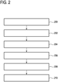
- Fig. 2 shows a flowchart which illustrates a method of operating the magnetic resonance imaging system 100 of Fig. 1 .
- the steps shown in Fig. 2 could for example be implemented by the machine-executable instructions 160.
- the magnetic resonance imaging system 100 is controlled with the pulse sequence commands 162 to acquire the measured magnetic resonance data.
- the measured magnetic resonance data is acquired as measured data portions 164. Each of the measured data portions is acquired during a time period.
- step 202 the intermediate magnetic resonance image 168 is reconstructed using the measured magnetic resonance data 164 according to a compressed sensing magnetic resonance imaging protocol.
- step 204 the predicted data portion 170 is calculated for each of the measured data portions 164 using the intermediate magnetic resonance image. As was mentioned before, this may be done using an inverse Fourier transform to predict the measured data.
- step 206 a residual 172 is calculated for each of the measured data portions 164 using the predicted data portion 170.
- step 208 one or more outlier data portions 176 are identified from measured data portions that have a residual 172 that is above a predetermined threshold 174.
- step 210 the corrected magnetic resonance image 178 is reconstructed using the measured magnetic resonance data 164 according to the compressed sensing magnetic resonance imaging protocol. This is done by excluding the outlier data portions 176 from the image reconstruction. As was also mentioned above, this may be performed in several different ways.
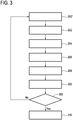
- Fig. 3 shows a flowchart which illustrates a further method of operating the magnetic resonance imaging system 100 of Fig. 1 .
- the method begins with step 200'.
- step 200' the magnetic resonance imaging system 100 is controlled with the pulse sequence commands 162 such that only a portion of the measured magnetic resonance data is acquired. This may be one or more measured data portions 164.
- Steps 202-208 are identical with those as are shown in Fig. 2 except the method is performed with all of the measured data portions that have been acquired as opposed to the complete measured magnetic resonance data.
- step 300 is performed.
- step 300 the scheduling of the reacquisition of measured data portions that were identified as outlier data portions is performed.
- the method then proceeds to decision box 302.
- Decision box 302 may answer several different questions. A first question would be have all measured data portions been acquired. If the answer is yes then the method proceeds to step 210 and a corrected magnetic resonance image is constructed.
- step 200 the method returns back to step 200' and another group of one or more measured data portions is acquired.
- the decision box 302 may also ask the question has sufficient magnetic resonance data been acquired. If the answer is yes then the method again proceeds to step 210. If the answer is no then the method proceeds back to step 200' also.
- the compressed sensing magnetic resonance imaging protocols are able to reconstruct a magnetic resonance image without the complete acquisition of k-space. In some cases it may be sufficient to simply drop or ignore the data which was identified as outlier data portions. However, if too many outlier data portions are identified then it may be beneficial to go through and acquire one or more of the identified outlier data portions.
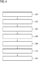
- Fig. 4 shows a further method of operating the magnetic resonance imaging system 100 of Fig. 1 .
- the method in Fig. 4 is similar to that shown in Fig. 2 with an additional step performed after step 210.
- Step 400 is performed after step 210.
- one or more outlier magnetic resonance images is constructed from the one or more outlier data portions.
- the outlier data portions may be treated as separately acquired magnetic resonance data and used separately to construct an intermediate magnetic resonance image and residual and corrected magnetic resonance image as is illustrated in Fig. 2 .
- the residual could be due to transient motion by the subject 118.
- the residual being above the predetermined threshold may be due to periodic or involuntary periodic motion of the subject 118.
- the outlier data portions could be used to make a series of images that represent different phases of the subject's 118 periodic motion.
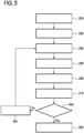
- Fig. 5 shows a flowchart which illustrates a further method of operating the magnetic resonance imaging system of Fig. 1 .
- the method shown in Fig. 5 is similar to the method illustrated in Fig. 2 .
- the steps 200-210 are performed as they are in Fig. 2 .
- the method proceeds to step 500.
- Step 500 is a decision box where a test of convergence between the corrected magnetic resonance image and the intermediate magnetic resonance image is performed. If the corrected magnetic resonance image has converged to the intermediate magnetic resonance image within a predetermined measure or statistical test then the method proceeds to step 502 and the method ends. If the two images have not converged sufficiently then the method proceeds to step 504.
- step 504 the corrected magnetic resonance image is set as being the intermediate magnetic resonance image and the method returns to step 204.
- the predicted data portion is recalculated for each of the measured data portions using what was previously the corrected magnetic resonance image.
- the method then proceeds normally through steps 206, 208 and step 210 again.
- step 208 as the intermediate magnetic resonance image has changed there may be more data portions that are identified as outlier data portions.
- the measured data portions which were previously identified as outlier data portions in other iterations may continue to be identified as outlier data portions.
- the steps of calculating residual and a predicted data portion might be repeated for every single measured data portion for every iteration.
- the method then proceeds to step 500. If the images have converged then the method ends in step 502 and if not the iterations may continue by proceeding back to step 504. In some examples a maximum number of iterations may be set.
- CS reconstruction is an iterative reconstruction where the difference between measured data and re-projected image is used to calculate an image update (which is subject to a given sparsity constraint). Motion during data acquisition will cause image artifacts because the reconstruction algorithm tries to merge conflicting data into a single image. Because CS typically uses fewer data than standard reconstruction its sensitivity towards motion artifacts is higher than that of standard reconstruction.
- Examples may improve the stability of CS reconstruction against motion artifacts by introducing a simple model for motion.
- the intermediate magnetic resonance image represents the static temporal phase of the object, and in this way this model distinguishes a static temporal phase of the object, in which the object is at rest, from a possible motion phase: It is possible that data points from the static phase are taken into account in the calculation of the image update and data points acquired during the motion phase are ignored.
- the measured data portions are acquired during distinct time periods.
- the measured data portions identified as the outlier data portions may represent times when the object is possibly in motion.
- CS Compressed sensing
- Examples may reduce ghosting artifacts caused by motion during data acquisition during CS.
- examples may improve the stability of CS reconstruction against motion artifacts by introducing a simple model for motion.
- This model distinguishes a static temporal phase of the object from a possible motion phase: Preferably data points from the static phase / period are taken into account in the calculation of the image update and data points acquired during the motion phase are ignored.
- the segmentation of the data set into static and motion temporal phase can be done in several ways:
- An MRI scan is performed using variable density sampling and appropriate subsampling. Based on these data a CS reconstruction is started.
- CSM coil sensitivity maps
- a further ingredient is the proper choice of the temporal footprint of the corresponding profiles.
- the sampling pattern in k-space is important but also the temporal order of data acquisition:
- the individual samples After the generation of the variable density sampling pattern for CS the individual samples have to be distributed in time appropriately.
- considerations about the desired image contrast are important as well as appropriate sampling diversity. This means that subsequently acquired k-space samples, which potentially could be grouped into individual temporal footprints, are largely distributed over k-space to avoid too large under sampling resulting in local holes of large extent in k-space making reconstruction very unstable.
- a computer program may be stored/distributed on a suitable medium, such as an optical storage medium or a solid-state medium supplied together with or as part of other hardware, but may also be distributed in other forms, such as via the Internet or other wired or wireless telecommunication systems. Any reference signs in the claims should not be construed as limiting the scope.
Landscapes
- Physics & Mathematics (AREA)
- General Physics & Mathematics (AREA)
- Condensed Matter Physics & Semiconductors (AREA)
- Engineering & Computer Science (AREA)
- High Energy & Nuclear Physics (AREA)
- Health & Medical Sciences (AREA)
- Radiology & Medical Imaging (AREA)
- Signal Processing (AREA)
- Nuclear Medicine, Radiotherapy & Molecular Imaging (AREA)
- General Health & Medical Sciences (AREA)
- Computer Vision & Pattern Recognition (AREA)
- Artificial Intelligence (AREA)
- Magnetic Resonance Imaging Apparatus (AREA)
Applications Claiming Priority (2)
| Application Number | Priority Date | Filing Date | Title |
|---|---|---|---|
| EP16180581 | 2016-07-21 | ||
| PCT/EP2017/067866 WO2018015298A1 (en) | 2016-07-21 | 2017-07-14 | Motion-corrected compressed sensing magnetic resonance imaging |
Publications (2)
| Publication Number | Publication Date |
|---|---|
| EP3500869A1 EP3500869A1 (en) | 2019-06-26 |
| EP3500869B1 true EP3500869B1 (en) | 2021-02-24 |
Family
ID=56507473
Family Applications (1)
| Application Number | Title | Priority Date | Filing Date |
|---|---|---|---|
| EP17737301.6A Active EP3500869B1 (en) | 2016-07-21 | 2017-07-14 | Corrected compressed sensing magnetic resonance imaging |
Country Status (6)
Families Citing this family (9)
| Publication number | Priority date | Publication date | Assignee | Title |
|---|---|---|---|---|
| EP3543725A1 (en) | 2018-03-22 | 2019-09-25 | Koninklijke Philips N.V. | Self-navigation in three-dimensional magnetic resonance imaging |
| CN109040757B (zh) * | 2018-07-20 | 2020-11-10 | 西安交通大学 | 一种压缩感知多层残差图像编码方法 |
| JP7221681B2 (ja) * | 2018-12-28 | 2023-02-14 | キヤノンメディカルシステムズ株式会社 | 画像再構成方法、再構成装置、及び磁気共鳴イメージング装置 |
| US10950014B2 (en) | 2019-03-01 | 2021-03-16 | Canon Medical Systems Corporation | Method and apparatus for adaptive compressed sensing (CS) to correct motion artifacts in magnetic resonance imaging (MRI) |
| EP3719525A1 (en) * | 2019-04-01 | 2020-10-07 | Koninklijke Philips N.V. | Correction of magnetic resonance images using simulated magnetic resonance images |
| US10806370B1 (en) * | 2019-04-25 | 2020-10-20 | General Electric Company | MRI system and method for detection and correction of patient motion |
| US11280868B2 (en) * | 2019-06-19 | 2022-03-22 | GE Precision Healthcare LLC | Image enhancement with variable number of excitation (NEX) acquisitions accelerated using compressed sensing |
| EP3824814A1 (en) * | 2019-11-22 | 2021-05-26 | Koninklijke Philips N.V. | Assessment of measured tomographic data |
| EP3865892A1 (en) * | 2020-02-17 | 2021-08-18 | Koninklijke Philips N.V. | Iterative reconstruction of gradient echo magnetic resonance images |
Family Cites Families (20)
| Publication number | Priority date | Publication date | Assignee | Title |
|---|---|---|---|---|
| ATE528733T1 (de) * | 2007-12-20 | 2011-10-15 | Wisconsin Alumni Res Found | Verfahren für dynamische eingeschränkte bildrekonstruktion mit vorhergehendem bild |
| US8472688B2 (en) * | 2008-04-17 | 2013-06-25 | Wisconsin Alumni Research Foundation | Method for image reconstruction employing sparsity-constrained iterative correction |
| WO2009134820A2 (en) * | 2008-04-28 | 2009-11-05 | Cornell University | Tool for accurate quantification in molecular mri |
| CN110659443A (zh) * | 2010-06-11 | 2020-01-07 | Abb瑞士股份有限公司 | 检测状态估计网络模型数据误差 |
| GB2504642B (en) | 2011-04-21 | 2018-02-21 | Koninl Philips Electronics Nv | Magnetic resonance imaging of object inmotion |
| US8768034B2 (en) | 2012-03-20 | 2014-07-01 | Siemens Medical Solutions Usa, Inc. | Motion compensated MR imaging system |
| EP2839433B1 (en) | 2012-04-19 | 2024-03-13 | New York University | System, method and computer-accessible medium for highly-accelerated dynamic magnetic resonance imaging using golden-angle radial sampling |
| EP2728371B1 (en) | 2012-11-02 | 2022-07-27 | Universitätsklinikum Freiburg | Segmented 3D Cartesian MR data acquisition using a randomized sampling pattern for compressed sensing image reconstruction |
| US9797974B2 (en) | 2013-01-30 | 2017-10-24 | The Board Of Trustees Of The Leland Stanford Junior University | Nonrigid motion correction in 3D using autofocusing with localized linear translations |
| CN103424420A (zh) * | 2013-01-31 | 2013-12-04 | 上海理工大学 | 一种基于拟合的核磁共振信号处理方法 |
| US9476712B2 (en) | 2013-07-31 | 2016-10-25 | Honeywell International Inc. | MEMS device mechanism enhancement for robust operation through severe shock and acceleration |
| CN105874346B (zh) * | 2013-12-10 | 2019-10-18 | 皇家飞利浦有限公司 | 使用在放大视场中的内插计算mri rf线圈灵敏度 |
| EP3080633A1 (en) | 2013-12-12 | 2016-10-19 | Koninklijke Philips N.V. | Mr imaging using multi-echo segmented k-space acquisition |
| JP6494986B2 (ja) | 2014-01-16 | 2019-04-03 | キヤノンメディカルシステムズ株式会社 | 磁気共鳴イメージング装置及び画像処理装置 |
| JP6072723B2 (ja) * | 2014-04-21 | 2017-02-01 | 株式会社日立製作所 | 磁気共鳴イメージング装置、及び画像撮像方法 |
| US9770223B2 (en) * | 2014-09-09 | 2017-09-26 | Wisconsin Alumni Research Foundation | System and method for accelerated, time-resolved imaging |
| US9846214B2 (en) * | 2014-12-29 | 2017-12-19 | Toshiba Medical Systems Corporation | Magnetic resonance image reconstruction for undersampled data acquisitions |
| CN105005012B (zh) * | 2015-06-05 | 2017-09-26 | 北京大学 | 基于压缩感知的腹部器官动态对比增强磁共振成像方法 |
| US10310047B2 (en) * | 2016-04-21 | 2019-06-04 | University Of Virginia Patent Foundation | Systems and methods for free-breathing cine DENSE MRI using self-navigation |
| JP6545887B2 (ja) * | 2017-11-24 | 2019-07-17 | キヤノンメディカルシステムズ株式会社 | 医用データ処理装置、磁気共鳴イメージング装置及び学習済みモデル生成方法 |
-
2017
- 2017-07-14 WO PCT/EP2017/067866 patent/WO2018015298A1/en unknown
- 2017-07-14 EP EP17737301.6A patent/EP3500869B1/en active Active
- 2017-07-14 CN CN201780045151.0A patent/CN109477878B/zh active Active
- 2017-07-14 US US16/317,592 patent/US10698064B2/en active Active
- 2017-07-14 RU RU2019104619A patent/RU2019104619A/ru not_active Application Discontinuation
- 2017-07-14 JP JP2019502637A patent/JP6932181B2/ja active Active
Non-Patent Citations (1)
| Title |
|---|
| None * |
Also Published As
| Publication number | Publication date |
|---|---|
| CN109477878A (zh) | 2019-03-15 |
| CN109477878B (zh) | 2021-08-24 |
| JP6932181B2 (ja) | 2021-09-08 |
| EP3500869A1 (en) | 2019-06-26 |
| WO2018015298A1 (en) | 2018-01-25 |
| US10698064B2 (en) | 2020-06-30 |
| US20190242965A1 (en) | 2019-08-08 |
| JP2019530486A (ja) | 2019-10-24 |
| RU2019104619A (ru) | 2020-08-21 |
| BR112019001002A2 (pt) | 2019-05-14 |
Similar Documents
| Publication | Publication Date | Title |
|---|---|---|
| EP3500869B1 (en) | Corrected compressed sensing magnetic resonance imaging | |
| EP3055706B1 (en) | Corrected multiple-slice magnetic resonance imaging | |
| JP7191105B2 (ja) | 運動補償磁気共鳴イメージング | |
| EP3543722A1 (en) | Magnetic resonance imaging using corrected k-space trajectories calculated from current sensor data | |
| US11519991B2 (en) | Motion estimation and correction in magnetic resonance imaging | |
| EP3916417A1 (en) | Correction of magnetic resonance images using multiple magnetic resonance imaging system configurations | |
| EP4127756B1 (en) | Reduction of off-resonance effects in magnetic resonance imaging | |
| EP3123191B1 (en) | Propeller magnetic resonance imaging | |
| WO2015197366A1 (en) | Motion correction in magnetic resonance imaging | |
| WO2018036986A1 (en) | Bo-corrected sensitivity encoding magnetic resonance imaging | |
| US11609294B2 (en) | Acquisition of four dimensional magnetic resonance data during subject motion | |
| EP3769103B1 (en) | Self-navigation in three-dimensional magnetic resonance imaging | |
| CN115667969A (zh) | 对欠采样因子的自动调整 | |
| EP3256871B1 (en) | Metal artifact correction in magnetic resonance imaging | |
| WO2018134445A1 (en) | Acquisition of four dimensional magnetic resonance data during subject motion | |
| EP4614178A1 (en) | Motion correction using navigators between sequential groups of k-space data | |
| WO2018115179A1 (en) | Mri method for t1 mapping of the heart using a maximum likelihood reconstruction in k-space | |
| EP3524996A1 (en) | Reduction of artifacts in parallel magnetic resonance imaging | |
| WO2025186081A1 (en) | Motion correction using navigators between sequential groups of k-space data | |
| BR112019001002B1 (pt) | Sistema de imageamento por ressonância magnética, mídia legível por computador e método para controlar um sistema de imageamento por ressonância magnética |
Legal Events
| Date | Code | Title | Description |
|---|---|---|---|
| STAA | Information on the status of an ep patent application or granted ep patent |
Free format text: STATUS: UNKNOWN |
|
| STAA | Information on the status of an ep patent application or granted ep patent |
Free format text: STATUS: THE INTERNATIONAL PUBLICATION HAS BEEN MADE |
|
| PUAI | Public reference made under article 153(3) epc to a published international application that has entered the european phase |
Free format text: ORIGINAL CODE: 0009012 |
|
| STAA | Information on the status of an ep patent application or granted ep patent |
Free format text: STATUS: REQUEST FOR EXAMINATION WAS MADE |
|
| 17P | Request for examination filed |
Effective date: 20190221 |
|
| AK | Designated contracting states |
Kind code of ref document: A1 Designated state(s): AL AT BE BG CH CY CZ DE DK EE ES FI FR GB GR HR HU IE IS IT LI LT LU LV MC MK MT NL NO PL PT RO RS SE SI SK SM TR |
|
| AX | Request for extension of the european patent |
Extension state: BA ME |
|
| DAV | Request for validation of the european patent (deleted) | ||
| DAX | Request for extension of the european patent (deleted) | ||
| RAP1 | Party data changed (applicant data changed or rights of an application transferred) |
Owner name: KONINKLIJKE PHILIPS N.V. |
|
| GRAP | Despatch of communication of intention to grant a patent |
Free format text: ORIGINAL CODE: EPIDOSNIGR1 |
|
| STAA | Information on the status of an ep patent application or granted ep patent |
Free format text: STATUS: GRANT OF PATENT IS INTENDED |
|
| INTG | Intention to grant announced |
Effective date: 20200916 |
|
| GRAS | Grant fee paid |
Free format text: ORIGINAL CODE: EPIDOSNIGR3 |
|
| GRAA | (expected) grant |
Free format text: ORIGINAL CODE: 0009210 |
|
| STAA | Information on the status of an ep patent application or granted ep patent |
Free format text: STATUS: THE PATENT HAS BEEN GRANTED |
|
| AK | Designated contracting states |
Kind code of ref document: B1 Designated state(s): AL AT BE BG CH CY CZ DE DK EE ES FI FR GB GR HR HU IE IS IT LI LT LU LV MC MK MT NL NO PL PT RO RS SE SI SK SM TR |
|
| REG | Reference to a national code |
Ref country code: CH Ref legal event code: EP |
|
| REG | Reference to a national code |
Ref country code: DE Ref legal event code: R096 Ref document number: 602017033275 Country of ref document: DE |
|
| REG | Reference to a national code |
Ref country code: AT Ref legal event code: REF Ref document number: 1365165 Country of ref document: AT Kind code of ref document: T Effective date: 20210315 |
|
| REG | Reference to a national code |
Ref country code: IE Ref legal event code: FG4D |
|
| REG | Reference to a national code |
Ref country code: DE Ref legal event code: R084 Ref document number: 602017033275 Country of ref document: DE |
|
| REG | Reference to a national code |
Ref country code: GB Ref legal event code: 746 Effective date: 20210422 |
|
| REG | Reference to a national code |
Ref country code: LT Ref legal event code: MG9D |
|
| REG | Reference to a national code |
Ref country code: NL Ref legal event code: MP Effective date: 20210224 |
|
| PG25 | Lapsed in a contracting state [announced via postgrant information from national office to epo] |
Ref country code: BG Free format text: LAPSE BECAUSE OF FAILURE TO SUBMIT A TRANSLATION OF THE DESCRIPTION OR TO PAY THE FEE WITHIN THE PRESCRIBED TIME-LIMIT Effective date: 20210524 Ref country code: LT Free format text: LAPSE BECAUSE OF FAILURE TO SUBMIT A TRANSLATION OF THE DESCRIPTION OR TO PAY THE FEE WITHIN THE PRESCRIBED TIME-LIMIT Effective date: 20210224 Ref country code: FI Free format text: LAPSE BECAUSE OF FAILURE TO SUBMIT A TRANSLATION OF THE DESCRIPTION OR TO PAY THE FEE WITHIN THE PRESCRIBED TIME-LIMIT Effective date: 20210224 Ref country code: GR Free format text: LAPSE BECAUSE OF FAILURE TO SUBMIT A TRANSLATION OF THE DESCRIPTION OR TO PAY THE FEE WITHIN THE PRESCRIBED TIME-LIMIT Effective date: 20210525 Ref country code: HR Free format text: LAPSE BECAUSE OF FAILURE TO SUBMIT A TRANSLATION OF THE DESCRIPTION OR TO PAY THE FEE WITHIN THE PRESCRIBED TIME-LIMIT Effective date: 20210224 Ref country code: NO Free format text: LAPSE BECAUSE OF FAILURE TO SUBMIT A TRANSLATION OF THE DESCRIPTION OR TO PAY THE FEE WITHIN THE PRESCRIBED TIME-LIMIT Effective date: 20210524 Ref country code: PT Free format text: LAPSE BECAUSE OF FAILURE TO SUBMIT A TRANSLATION OF THE DESCRIPTION OR TO PAY THE FEE WITHIN THE PRESCRIBED TIME-LIMIT Effective date: 20210624 |
|
| REG | Reference to a national code |
Ref country code: AT Ref legal event code: MK05 Ref document number: 1365165 Country of ref document: AT Kind code of ref document: T Effective date: 20210224 |
|
| PG25 | Lapsed in a contracting state [announced via postgrant information from national office to epo] |
Ref country code: NL Free format text: LAPSE BECAUSE OF FAILURE TO SUBMIT A TRANSLATION OF THE DESCRIPTION OR TO PAY THE FEE WITHIN THE PRESCRIBED TIME-LIMIT Effective date: 20210224 Ref country code: RS Free format text: LAPSE BECAUSE OF FAILURE TO SUBMIT A TRANSLATION OF THE DESCRIPTION OR TO PAY THE FEE WITHIN THE PRESCRIBED TIME-LIMIT Effective date: 20210224 Ref country code: LV Free format text: LAPSE BECAUSE OF FAILURE TO SUBMIT A TRANSLATION OF THE DESCRIPTION OR TO PAY THE FEE WITHIN THE PRESCRIBED TIME-LIMIT Effective date: 20210224 Ref country code: PL Free format text: LAPSE BECAUSE OF FAILURE TO SUBMIT A TRANSLATION OF THE DESCRIPTION OR TO PAY THE FEE WITHIN THE PRESCRIBED TIME-LIMIT Effective date: 20210224 Ref country code: SE Free format text: LAPSE BECAUSE OF FAILURE TO SUBMIT A TRANSLATION OF THE DESCRIPTION OR TO PAY THE FEE WITHIN THE PRESCRIBED TIME-LIMIT Effective date: 20210224 |
|
| PG25 | Lapsed in a contracting state [announced via postgrant information from national office to epo] |
Ref country code: IS Free format text: LAPSE BECAUSE OF FAILURE TO SUBMIT A TRANSLATION OF THE DESCRIPTION OR TO PAY THE FEE WITHIN THE PRESCRIBED TIME-LIMIT Effective date: 20210624 |
|
| PG25 | Lapsed in a contracting state [announced via postgrant information from national office to epo] |
Ref country code: AT Free format text: LAPSE BECAUSE OF FAILURE TO SUBMIT A TRANSLATION OF THE DESCRIPTION OR TO PAY THE FEE WITHIN THE PRESCRIBED TIME-LIMIT Effective date: 20210224 Ref country code: SM Free format text: LAPSE BECAUSE OF FAILURE TO SUBMIT A TRANSLATION OF THE DESCRIPTION OR TO PAY THE FEE WITHIN THE PRESCRIBED TIME-LIMIT Effective date: 20210224 Ref country code: CZ Free format text: LAPSE BECAUSE OF FAILURE TO SUBMIT A TRANSLATION OF THE DESCRIPTION OR TO PAY THE FEE WITHIN THE PRESCRIBED TIME-LIMIT Effective date: 20210224 Ref country code: EE Free format text: LAPSE BECAUSE OF FAILURE TO SUBMIT A TRANSLATION OF THE DESCRIPTION OR TO PAY THE FEE WITHIN THE PRESCRIBED TIME-LIMIT Effective date: 20210224 |
|
| REG | Reference to a national code |
Ref country code: DE Ref legal event code: R097 Ref document number: 602017033275 Country of ref document: DE |
|
| PG25 | Lapsed in a contracting state [announced via postgrant information from national office to epo] |
Ref country code: RO Free format text: LAPSE BECAUSE OF FAILURE TO SUBMIT A TRANSLATION OF THE DESCRIPTION OR TO PAY THE FEE WITHIN THE PRESCRIBED TIME-LIMIT Effective date: 20210224 Ref country code: DK Free format text: LAPSE BECAUSE OF FAILURE TO SUBMIT A TRANSLATION OF THE DESCRIPTION OR TO PAY THE FEE WITHIN THE PRESCRIBED TIME-LIMIT Effective date: 20210224 Ref country code: SK Free format text: LAPSE BECAUSE OF FAILURE TO SUBMIT A TRANSLATION OF THE DESCRIPTION OR TO PAY THE FEE WITHIN THE PRESCRIBED TIME-LIMIT Effective date: 20210224 |
|
| PLBE | No opposition filed within time limit |
Free format text: ORIGINAL CODE: 0009261 |
|
| STAA | Information on the status of an ep patent application or granted ep patent |
Free format text: STATUS: NO OPPOSITION FILED WITHIN TIME LIMIT |
|
| PG25 | Lapsed in a contracting state [announced via postgrant information from national office to epo] |
Ref country code: AL Free format text: LAPSE BECAUSE OF FAILURE TO SUBMIT A TRANSLATION OF THE DESCRIPTION OR TO PAY THE FEE WITHIN THE PRESCRIBED TIME-LIMIT Effective date: 20210224 Ref country code: ES Free format text: LAPSE BECAUSE OF FAILURE TO SUBMIT A TRANSLATION OF THE DESCRIPTION OR TO PAY THE FEE WITHIN THE PRESCRIBED TIME-LIMIT Effective date: 20210224 |
|
| 26N | No opposition filed |
Effective date: 20211125 |
|
| PG25 | Lapsed in a contracting state [announced via postgrant information from national office to epo] |
Ref country code: SI Free format text: LAPSE BECAUSE OF FAILURE TO SUBMIT A TRANSLATION OF THE DESCRIPTION OR TO PAY THE FEE WITHIN THE PRESCRIBED TIME-LIMIT Effective date: 20210224 |
|
| REG | Reference to a national code |
Ref country code: CH Ref legal event code: PL |
|
| PG25 | Lapsed in a contracting state [announced via postgrant information from national office to epo] |
Ref country code: MC Free format text: LAPSE BECAUSE OF FAILURE TO SUBMIT A TRANSLATION OF THE DESCRIPTION OR TO PAY THE FEE WITHIN THE PRESCRIBED TIME-LIMIT Effective date: 20210224 |
|
| REG | Reference to a national code |
Ref country code: BE Ref legal event code: MM Effective date: 20210731 |
|
| PG25 | Lapsed in a contracting state [announced via postgrant information from national office to epo] |
Ref country code: LI Free format text: LAPSE BECAUSE OF NON-PAYMENT OF DUE FEES Effective date: 20210731 Ref country code: IT Free format text: LAPSE BECAUSE OF FAILURE TO SUBMIT A TRANSLATION OF THE DESCRIPTION OR TO PAY THE FEE WITHIN THE PRESCRIBED TIME-LIMIT Effective date: 20210224 Ref country code: CH Free format text: LAPSE BECAUSE OF NON-PAYMENT OF DUE FEES Effective date: 20210731 |
|
| PG25 | Lapsed in a contracting state [announced via postgrant information from national office to epo] |
Ref country code: IS Free format text: LAPSE BECAUSE OF FAILURE TO SUBMIT A TRANSLATION OF THE DESCRIPTION OR TO PAY THE FEE WITHIN THE PRESCRIBED TIME-LIMIT Effective date: 20210624 Ref country code: LU Free format text: LAPSE BECAUSE OF NON-PAYMENT OF DUE FEES Effective date: 20210714 |
|
| PG25 | Lapsed in a contracting state [announced via postgrant information from national office to epo] |
Ref country code: IE Free format text: LAPSE BECAUSE OF NON-PAYMENT OF DUE FEES Effective date: 20210714 Ref country code: BE Free format text: LAPSE BECAUSE OF NON-PAYMENT OF DUE FEES Effective date: 20210731 |
|
| PG25 | Lapsed in a contracting state [announced via postgrant information from national office to epo] |
Ref country code: CY Free format text: LAPSE BECAUSE OF FAILURE TO SUBMIT A TRANSLATION OF THE DESCRIPTION OR TO PAY THE FEE WITHIN THE PRESCRIBED TIME-LIMIT Effective date: 20210224 |
|
| PG25 | Lapsed in a contracting state [announced via postgrant information from national office to epo] |
Ref country code: HU Free format text: LAPSE BECAUSE OF FAILURE TO SUBMIT A TRANSLATION OF THE DESCRIPTION OR TO PAY THE FEE WITHIN THE PRESCRIBED TIME-LIMIT; INVALID AB INITIO Effective date: 20170714 |
|
| PG25 | Lapsed in a contracting state [announced via postgrant information from national office to epo] |
Ref country code: MK Free format text: LAPSE BECAUSE OF FAILURE TO SUBMIT A TRANSLATION OF THE DESCRIPTION OR TO PAY THE FEE WITHIN THE PRESCRIBED TIME-LIMIT Effective date: 20210224 |
|
| PG25 | Lapsed in a contracting state [announced via postgrant information from national office to epo] |
Ref country code: TR Free format text: LAPSE BECAUSE OF FAILURE TO SUBMIT A TRANSLATION OF THE DESCRIPTION OR TO PAY THE FEE WITHIN THE PRESCRIBED TIME-LIMIT Effective date: 20210224 |
|
| PG25 | Lapsed in a contracting state [announced via postgrant information from national office to epo] |
Ref country code: MT Free format text: LAPSE BECAUSE OF FAILURE TO SUBMIT A TRANSLATION OF THE DESCRIPTION OR TO PAY THE FEE WITHIN THE PRESCRIBED TIME-LIMIT Effective date: 20210224 |
|
| PGFP | Annual fee paid to national office [announced via postgrant information from national office to epo] |
Ref country code: DE Payment date: 20240730 Year of fee payment: 8 |
|
| PGFP | Annual fee paid to national office [announced via postgrant information from national office to epo] |
Ref country code: GB Payment date: 20240724 Year of fee payment: 8 |
|
| PGFP | Annual fee paid to national office [announced via postgrant information from national office to epo] |
Ref country code: FR Payment date: 20240725 Year of fee payment: 8 |