EP3500680B1 - Doppelsträngige kreissonden - Google Patents
Doppelsträngige kreissonden Download PDFInfo
- Publication number
- EP3500680B1 EP3500680B1 EP17754692.6A EP17754692A EP3500680B1 EP 3500680 B1 EP3500680 B1 EP 3500680B1 EP 17754692 A EP17754692 A EP 17754692A EP 3500680 B1 EP3500680 B1 EP 3500680B1
- Authority
- EP
- European Patent Office
- Prior art keywords
- nucleic acid
- probe
- strand
- target
- molecule
- Prior art date
- Legal status (The legal status is an assumption and is not a legal conclusion. Google has not performed a legal analysis and makes no representation as to the accuracy of the status listed.)
- Active
Links
Images










Classifications
-
- C—CHEMISTRY; METALLURGY
- C12—BIOCHEMISTRY; BEER; SPIRITS; WINE; VINEGAR; MICROBIOLOGY; ENZYMOLOGY; MUTATION OR GENETIC ENGINEERING
- C12Q—MEASURING OR TESTING PROCESSES INVOLVING ENZYMES, NUCLEIC ACIDS OR MICROORGANISMS; COMPOSITIONS OR TEST PAPERS THEREFOR; PROCESSES OF PREPARING SUCH COMPOSITIONS; CONDITION-RESPONSIVE CONTROL IN MICROBIOLOGICAL OR ENZYMOLOGICAL PROCESSES
- C12Q1/00—Measuring or testing processes involving enzymes, nucleic acids or microorganisms; Compositions therefor; Processes of preparing such compositions
- C12Q1/68—Measuring or testing processes involving enzymes, nucleic acids or microorganisms; Compositions therefor; Processes of preparing such compositions involving nucleic acids
- C12Q1/6844—Nucleic acid amplification reactions
-
- C—CHEMISTRY; METALLURGY
- C12—BIOCHEMISTRY; BEER; SPIRITS; WINE; VINEGAR; MICROBIOLOGY; ENZYMOLOGY; MUTATION OR GENETIC ENGINEERING
- C12Q—MEASURING OR TESTING PROCESSES INVOLVING ENZYMES, NUCLEIC ACIDS OR MICROORGANISMS; COMPOSITIONS OR TEST PAPERS THEREFOR; PROCESSES OF PREPARING SUCH COMPOSITIONS; CONDITION-RESPONSIVE CONTROL IN MICROBIOLOGICAL OR ENZYMOLOGICAL PROCESSES
- C12Q1/00—Measuring or testing processes involving enzymes, nucleic acids or microorganisms; Compositions therefor; Processes of preparing such compositions
- C12Q1/68—Measuring or testing processes involving enzymes, nucleic acids or microorganisms; Compositions therefor; Processes of preparing such compositions involving nucleic acids
- C12Q1/6804—Nucleic acid analysis using immunogens
-
- C—CHEMISTRY; METALLURGY
- C12—BIOCHEMISTRY; BEER; SPIRITS; WINE; VINEGAR; MICROBIOLOGY; ENZYMOLOGY; MUTATION OR GENETIC ENGINEERING
- C12Q—MEASURING OR TESTING PROCESSES INVOLVING ENZYMES, NUCLEIC ACIDS OR MICROORGANISMS; COMPOSITIONS OR TEST PAPERS THEREFOR; PROCESSES OF PREPARING SUCH COMPOSITIONS; CONDITION-RESPONSIVE CONTROL IN MICROBIOLOGICAL OR ENZYMOLOGICAL PROCESSES
- C12Q1/00—Measuring or testing processes involving enzymes, nucleic acids or microorganisms; Compositions therefor; Processes of preparing such compositions
- C12Q1/68—Measuring or testing processes involving enzymes, nucleic acids or microorganisms; Compositions therefor; Processes of preparing such compositions involving nucleic acids
- C12Q1/6813—Hybridisation assays
- C12Q1/6816—Hybridisation assays characterised by the detection means
-
- C—CHEMISTRY; METALLURGY
- C12—BIOCHEMISTRY; BEER; SPIRITS; WINE; VINEGAR; MICROBIOLOGY; ENZYMOLOGY; MUTATION OR GENETIC ENGINEERING
- C12Q—MEASURING OR TESTING PROCESSES INVOLVING ENZYMES, NUCLEIC ACIDS OR MICROORGANISMS; COMPOSITIONS OR TEST PAPERS THEREFOR; PROCESSES OF PREPARING SUCH COMPOSITIONS; CONDITION-RESPONSIVE CONTROL IN MICROBIOLOGICAL OR ENZYMOLOGICAL PROCESSES
- C12Q1/00—Measuring or testing processes involving enzymes, nucleic acids or microorganisms; Compositions therefor; Processes of preparing such compositions
- C12Q1/68—Measuring or testing processes involving enzymes, nucleic acids or microorganisms; Compositions therefor; Processes of preparing such compositions involving nucleic acids
- C12Q1/6813—Hybridisation assays
- C12Q1/6827—Hybridisation assays for detection of mutation or polymorphism
-
- C—CHEMISTRY; METALLURGY
- C12—BIOCHEMISTRY; BEER; SPIRITS; WINE; VINEGAR; MICROBIOLOGY; ENZYMOLOGY; MUTATION OR GENETIC ENGINEERING
- C12Q—MEASURING OR TESTING PROCESSES INVOLVING ENZYMES, NUCLEIC ACIDS OR MICROORGANISMS; COMPOSITIONS OR TEST PAPERS THEREFOR; PROCESSES OF PREPARING SUCH COMPOSITIONS; CONDITION-RESPONSIVE CONTROL IN MICROBIOLOGICAL OR ENZYMOLOGICAL PROCESSES
- C12Q1/00—Measuring or testing processes involving enzymes, nucleic acids or microorganisms; Compositions therefor; Processes of preparing such compositions
- C12Q1/68—Measuring or testing processes involving enzymes, nucleic acids or microorganisms; Compositions therefor; Processes of preparing such compositions involving nucleic acids
- C12Q1/6813—Hybridisation assays
- C12Q1/6832—Enhancement of hybridisation reaction
-
- C—CHEMISTRY; METALLURGY
- C12—BIOCHEMISTRY; BEER; SPIRITS; WINE; VINEGAR; MICROBIOLOGY; ENZYMOLOGY; MUTATION OR GENETIC ENGINEERING
- C12Q—MEASURING OR TESTING PROCESSES INVOLVING ENZYMES, NUCLEIC ACIDS OR MICROORGANISMS; COMPOSITIONS OR TEST PAPERS THEREFOR; PROCESSES OF PREPARING SUCH COMPOSITIONS; CONDITION-RESPONSIVE CONTROL IN MICROBIOLOGICAL OR ENZYMOLOGICAL PROCESSES
- C12Q1/00—Measuring or testing processes involving enzymes, nucleic acids or microorganisms; Compositions therefor; Processes of preparing such compositions
- C12Q1/68—Measuring or testing processes involving enzymes, nucleic acids or microorganisms; Compositions therefor; Processes of preparing such compositions involving nucleic acids
- C12Q1/6844—Nucleic acid amplification reactions
- C12Q1/6848—Nucleic acid amplification reactions characterised by the means for preventing contamination or increasing the specificity or sensitivity of an amplification reaction
-
- C—CHEMISTRY; METALLURGY
- C12—BIOCHEMISTRY; BEER; SPIRITS; WINE; VINEGAR; MICROBIOLOGY; ENZYMOLOGY; MUTATION OR GENETIC ENGINEERING
- C12Q—MEASURING OR TESTING PROCESSES INVOLVING ENZYMES, NUCLEIC ACIDS OR MICROORGANISMS; COMPOSITIONS OR TEST PAPERS THEREFOR; PROCESSES OF PREPARING SUCH COMPOSITIONS; CONDITION-RESPONSIVE CONTROL IN MICROBIOLOGICAL OR ENZYMOLOGICAL PROCESSES
- C12Q1/00—Measuring or testing processes involving enzymes, nucleic acids or microorganisms; Compositions therefor; Processes of preparing such compositions
- C12Q1/68—Measuring or testing processes involving enzymes, nucleic acids or microorganisms; Compositions therefor; Processes of preparing such compositions involving nucleic acids
- C12Q1/6844—Nucleic acid amplification reactions
- C12Q1/6853—Nucleic acid amplification reactions using modified primers or templates
-
- C—CHEMISTRY; METALLURGY
- C12—BIOCHEMISTRY; BEER; SPIRITS; WINE; VINEGAR; MICROBIOLOGY; ENZYMOLOGY; MUTATION OR GENETIC ENGINEERING
- C12Q—MEASURING OR TESTING PROCESSES INVOLVING ENZYMES, NUCLEIC ACIDS OR MICROORGANISMS; COMPOSITIONS OR TEST PAPERS THEREFOR; PROCESSES OF PREPARING SUCH COMPOSITIONS; CONDITION-RESPONSIVE CONTROL IN MICROBIOLOGICAL OR ENZYMOLOGICAL PROCESSES
- C12Q1/00—Measuring or testing processes involving enzymes, nucleic acids or microorganisms; Compositions therefor; Processes of preparing such compositions
- C12Q1/68—Measuring or testing processes involving enzymes, nucleic acids or microorganisms; Compositions therefor; Processes of preparing such compositions involving nucleic acids
- C12Q1/6876—Nucleic acid products used in the analysis of nucleic acids, e.g. primers or probes
-
- C—CHEMISTRY; METALLURGY
- C12—BIOCHEMISTRY; BEER; SPIRITS; WINE; VINEGAR; MICROBIOLOGY; ENZYMOLOGY; MUTATION OR GENETIC ENGINEERING
- C12Q—MEASURING OR TESTING PROCESSES INVOLVING ENZYMES, NUCLEIC ACIDS OR MICROORGANISMS; COMPOSITIONS OR TEST PAPERS THEREFOR; PROCESSES OF PREPARING SUCH COMPOSITIONS; CONDITION-RESPONSIVE CONTROL IN MICROBIOLOGICAL OR ENZYMOLOGICAL PROCESSES
- C12Q2525/00—Reactions involving modified oligonucleotides, nucleic acids, or nucleotides
- C12Q2525/10—Modifications characterised by
- C12Q2525/131—Modifications characterised by incorporating a restriction site
-
- C—CHEMISTRY; METALLURGY
- C12—BIOCHEMISTRY; BEER; SPIRITS; WINE; VINEGAR; MICROBIOLOGY; ENZYMOLOGY; MUTATION OR GENETIC ENGINEERING
- C12Q—MEASURING OR TESTING PROCESSES INVOLVING ENZYMES, NUCLEIC ACIDS OR MICROORGANISMS; COMPOSITIONS OR TEST PAPERS THEREFOR; PROCESSES OF PREPARING SUCH COMPOSITIONS; CONDITION-RESPONSIVE CONTROL IN MICROBIOLOGICAL OR ENZYMOLOGICAL PROCESSES
- C12Q2525/00—Reactions involving modified oligonucleotides, nucleic acids, or nucleotides
- C12Q2525/10—Modifications characterised by
- C12Q2525/161—Modifications characterised by incorporating target specific and non-target specific sites
-
- C—CHEMISTRY; METALLURGY
- C12—BIOCHEMISTRY; BEER; SPIRITS; WINE; VINEGAR; MICROBIOLOGY; ENZYMOLOGY; MUTATION OR GENETIC ENGINEERING
- C12Q—MEASURING OR TESTING PROCESSES INVOLVING ENZYMES, NUCLEIC ACIDS OR MICROORGANISMS; COMPOSITIONS OR TEST PAPERS THEREFOR; PROCESSES OF PREPARING SUCH COMPOSITIONS; CONDITION-RESPONSIVE CONTROL IN MICROBIOLOGICAL OR ENZYMOLOGICAL PROCESSES
- C12Q2525/00—Reactions involving modified oligonucleotides, nucleic acids, or nucleotides
- C12Q2525/30—Oligonucleotides characterised by their secondary structure
- C12Q2525/301—Hairpin oligonucleotides
-
- C—CHEMISTRY; METALLURGY
- C12—BIOCHEMISTRY; BEER; SPIRITS; WINE; VINEGAR; MICROBIOLOGY; ENZYMOLOGY; MUTATION OR GENETIC ENGINEERING
- C12Q—MEASURING OR TESTING PROCESSES INVOLVING ENZYMES, NUCLEIC ACIDS OR MICROORGANISMS; COMPOSITIONS OR TEST PAPERS THEREFOR; PROCESSES OF PREPARING SUCH COMPOSITIONS; CONDITION-RESPONSIVE CONTROL IN MICROBIOLOGICAL OR ENZYMOLOGICAL PROCESSES
- C12Q2525/00—Reactions involving modified oligonucleotides, nucleic acids, or nucleotides
- C12Q2525/30—Oligonucleotides characterised by their secondary structure
- C12Q2525/307—Circular oligonucleotides
-
- C—CHEMISTRY; METALLURGY
- C12—BIOCHEMISTRY; BEER; SPIRITS; WINE; VINEGAR; MICROBIOLOGY; ENZYMOLOGY; MUTATION OR GENETIC ENGINEERING
- C12Q—MEASURING OR TESTING PROCESSES INVOLVING ENZYMES, NUCLEIC ACIDS OR MICROORGANISMS; COMPOSITIONS OR TEST PAPERS THEREFOR; PROCESSES OF PREPARING SUCH COMPOSITIONS; CONDITION-RESPONSIVE CONTROL IN MICROBIOLOGICAL OR ENZYMOLOGICAL PROCESSES
- C12Q2531/00—Reactions of nucleic acids characterised by
- C12Q2531/10—Reactions of nucleic acids characterised by the purpose being amplify/increase the copy number of target nucleic acid
- C12Q2531/125—Rolling circle
-
- C—CHEMISTRY; METALLURGY
- C12—BIOCHEMISTRY; BEER; SPIRITS; WINE; VINEGAR; MICROBIOLOGY; ENZYMOLOGY; MUTATION OR GENETIC ENGINEERING
- C12Q—MEASURING OR TESTING PROCESSES INVOLVING ENZYMES, NUCLEIC ACIDS OR MICROORGANISMS; COMPOSITIONS OR TEST PAPERS THEREFOR; PROCESSES OF PREPARING SUCH COMPOSITIONS; CONDITION-RESPONSIVE CONTROL IN MICROBIOLOGICAL OR ENZYMOLOGICAL PROCESSES
- C12Q2537/00—Reactions characterised by the reaction format or use of a specific feature
- C12Q2537/10—Reactions characterised by the reaction format or use of a specific feature the purpose or use of
- C12Q2537/163—Reactions characterised by the reaction format or use of a specific feature the purpose or use of blocking probe
Definitions
- the present invention relates generally to the detection of an analyte in a sample by rolling circle amplification (RCA), and in particular to the detection of a nucleic acid molecule (a target nucleic acid) in a sample, which may be the analyte for detection or may be indicative of, or a reporter for, the presence of the analyte in the sample.
- the invention concerns the provision of a new nucleic acid probe for use in rolling circle amplification assays.
- the probe comprises a double-stranded circular structure which comprises a circular RCA template and which, in the absence of the target nucleic acid molecule, renders the template unavailable for a RCA reaction, and in particular unavailable for the binding of any molecule capable of priming a RCA reaction. This protects the circular template from an RCA reaction until the probe has bound to the target, thereby improving the specificity of the assay.
- the present invention also provides methods for using such probes in the detection of an analyte, and kits for the same.
- Rolling circle replication (RCR) reactions are well described in the art and have been demonstrated to be useful in a variety of assays for the detection of target nucleic acid molecules in a sample, i.e. for the replication or amplification of a target nucleic acid molecule, wherein the replicated or amplified nucleic acid molecule is detected. Accordingly, rolling circle replication commonly is referred to as rolling circle amplification (RCA), and these terms are used interchangeably herein.
- RCA methods have been developed in which the rolling circle product is further amplified by a hyperbranch or DNA cascade reaction, as described in WO 97/19193 and WO 97/20948 , or by a further RCA reaction.
- Various methods of amplifying a rolling circle product are also described in WO2014/076214 , WO2014/076209 and WO2015/071445 .
- RCA relates to the synthesis of nucleic acid molecules using a circular single stranded nucleic acid molecule, e.g. a circle oligonucleotide, as rolling circle template (a RCA template) and a strand-displacing polymerase to extend a primer which is hybridised to the template.
- the primer may in certain assays be provided by a target RNA molecule or a target DNA molecule, but can be any nucleic acid molecule which can hybridise to the circular template.
- the addition of a polymerase and nucleotides starts the synthesis reaction, i.e. polymerisation.
- the rolling circle template is endless, the resultant product is a long single stranded nucleic acid molecule composed of tandem repeats that are complementary to the rolling circle template.
- RCA reactions often utilise linear nucleic acid molecules, e.g. oligonucleotides such as padlock probes as described in more detail below, which are manipulated to generate circular nucleic acid molecules, typically by ligating the ends of the nucleic acid molecule together, e.g. using a ligase enzyme.
- the ends of the nucleic acid molecule may be brought into proximity to each other by their hybridisation to adjacent sequences on a target nucleic acid molecule which acts as a ligation template.
- the formation of the circular nucleic acid molecule allows it to be copied in a RCA reaction. This reaction may be initiated by adding a primer to the closed circle or a primer may be generated from the target nucleic acid molecule, i.e.
- the RCA product therefore forms part of the initial primer. This can be particularly advantageous because it may allow localised detection of the target nucleic acid, i.e. in embodiments where the nucleic acid molecule used to prime RCA is immobilised the RCA product will also be immobilised.
- the RCA product comprises a string of tandemly repeated complementary copies of the circular template nucleic acid molecule.
- the product may remain attached to a target analyte, and thus rolling circle amplification is of particular utility in the detection of analytes in an in situ context (though homogeneous detection is possible for "in solution” assays).
- An RCA reaction may result in a 1000-fold amplification of the circle in an hour, based on a circle consisting of approximately 100 nucleotides (though smaller circles may be amplified more quickly).
- RCA of a circular oligonucleotide may result in an RCA product that forms a bundle or "blob" of DNA that can be about 1 ⁇ m in diameter.
- the product i.e.
- blob may be detected by the hybridisation of nucleic acid probes (detection probes) conjugated to fluorescent labels which allows the blob to be visualised by fluorescence microscopy (in heterogeneous assays, i.e. in situ) or flow cytometry (in homogeneous assays).
- the RCA products may be reduced to monomers by digestion with a restriction enzyme or a ribozyme, e.g. WO 98/38300 and Dahl F et al., Proc Natl Acad Sci USA. 101(13), 4548-53 (2004 ), which are then detected.
- RCA may be used for the detection of any nucleic acid molecule in a sample and typically has been used to detect directly target nucleic acids in cell and tissue samples, e.g. in situ, i.e. localised, detection of nucleic acid molecules, which is of significant interest both for research and diagnostic purposes.
- RCA assays are not limited to use in heterogeneous formats and may also find utility in homogeneous assays (see e.g. WO 2009/037659 ).
- RCA has also been utilised in methods for the detection of other analytes, i.e. analytes other than nucleic acid molecules, such as cells, viruses, proteins, peptides, small molecules etc.
- analytes i.e. analytes other than nucleic acid molecules
- assays have been developed in which a nucleic acid molecule may be used to directly or indirectly tag or label a target analyte in a sample and detection of the nucleic acid molecule serves to indicate the presence of the analyte in the sample.
- a new nucleic acid molecule may be generated in a sample (i.e.
- nucleic acid molecule that was not present in the original sample and was not one of the components added to the sample), for example through the interaction of one or more analyte-binding molecules when they have bound to the target analyte (e.g. the analyte-binding molecules may carry nucleic acid molecule(s) that interact to generate a new nucleic acid molecule).
- the detection of the generated nucleic acid molecule is indicative of the analyte in a sample, i.e. the generated nucleic acid molecule acts as a marker or proxy for the target analyte.
- nucleic acid molecule as a marker or proxy for the target analyte, e.g. protein
- a nucleic acid molecule as a marker or proxy for the target analyte, e.g. protein
- immuno-PCR, immuno-RCA and proximity probe assays may rely on the detection of a nucleic acid molecule as a substitute for detecting the target analyte directly.
- Immuno-PCR involves labelling an antibody for a specific target analyte with a nucleic acid molecule.
- the target analyte is captured on a substrate, e.g. with a first antibody and contacted with the antibody:nucleic acid complex.
- Excess antibody may be washed away before the sample is subjected to a polymerase chain reaction (PCR), which is used to amplify the nucleic acid molecule conjugated to the antibody.
- PCR polymerase chain reaction
- Detection of the PCR product is indicative of the presence of the analyte in the sample and proportional to the amount of analyte.
- Immuno-RCA relies on the same principles as immuno-PCR. The difference is that a circular (or circularisable) oligonucleotide (RCA template) is hybridised to the nucleic acid molecule conjugated to the antibody (the RCA template may be pre-hybridized or added after the antibody has been allowed to interact with the target analyte).
- the nucleic acid molecule conjugated to the antibody is used as the primer for RCA.
- the RCA product is tethered to the antibody that is interacting with the target analyte, thereby allowing localised detection of the analyte in the sample, e.g. in a cell or tissue sample.
- a proximity assay relies on the principle of "proximity probing", wherein an analyte is detected by the binding of multiple (i.e. two or more, generally two or three) probes, which when brought into proximity by binding to the analyte (hence “proximity probes") allow a signal to be generated.
- the proximity probes comprises a nucleic acid domain linked to the analyte-binding domain of the probe, and generation of the signal involves an interaction between the nucleic acid moieties and/or a further functional moiety which is carried by the other probe(s).
- proximity probing has been developed in recent years and many assays based on this principle are now well known in the art.
- proximity ligation assays rely on proximal binding of proximity probes to an analyte to generate a signal from a ligation reaction involving or mediated by (e.g. between and/or templated by) the nucleic acid domains of the proximity probes.
- Proximity probes of the art are generally used in pairs, and individually consist of an analyte-binding domain with specificity to the target analyte, and a functional domain, e.g. a nucleic acid domain coupled thereto.
- the analyte-binding domain can be for example a nucleic acid "aptamer” ( Fredriksson et al (2002) Nat Biotech 20:473-477 ) or can be proteinaceous, such as a monoclonal or polyclonal antibody ( Gullberg et al (2004) Proc Natl Acad Sci USA 101:8420-8424 ).
- the respective analyte-binding domains of each proximity probe pair may have specificity for different binding sites on the analyte, which analyte may consist of a single molecule or a complex of interacting molecules, or may have identical specificities, for example in the event that the target analyte exists as a multimer.
- the functional domains e.g.
- nucleic acid domains are able to interact, for example, nucleic acid domains may be joined to form a new nucleic acid sequence generally by means of a ligation reaction, which may be templated by a splint oligonucleotide added to the reaction, said splint oligonucleotide containing regions of complementarity for the ends of the respective nucleic acid domains of the proximity probe pair.
- the new nucleic acid sequence thereby generated serves to report the presence or amount of analyte in a sample, and can be qualitatively or quantitatively detected, for example by real-time, quantitative PCR (q-PCR).
- the nucleic acid domains of the proximity probes when in proximity may template the ligation of one or more added oligonucleotides to each other, including an intramolecular ligation, to circularise one or more added linear oligonucleotides, for example based on the so-called padlock probe principle, wherein the ends of the added linear oligonucleotide(s) are brought into juxtaposition for ligation by hybridising to a template, here one or more nucleic acid domains of the proximity probes (in the case of a padlock probe the target nucleic acid for the probe).
- Various such assay formats are described in WO 01/61037 .
- WO 97/00446 and US 6,511,809 disclose a heterogeneous format for proximity ligation assays, i.e. the analyte is first immobilised to a solid substrate by means of a specific analyte-binding reagent.
- Homogeneous proximity ligation assays are disclosed in WO 01/61037 , WO 03/044231 , WO 2005/123963 , Fredriksson et al (2002) Nat Biotech 20:473-477 and Gullberg et al (2004) Proc Natl Acad Sci USA 101:8420-8424 .
- nucleic acid domains of proximity probes may comprise an extension reaction - for example the nucleic acid domains may hybridise to one another and one or both of the domains may template the extension of the other.
- the extension product may then be detected, for example by means of a padlock probe which hybridises to the extension product and which may itself be detected by an RCA reaction.
- RCA may be of utility in the specific detection of any nucleic acid molecule in a sample, regardless of whether it is the "original" target analyte in a sample or it is a "proxy” or “marker” for the target analyte generated by the interaction of specific detection molecules, e.g. proximity probes, with the target analyte, e.g. protein.
- specific detection molecules e.g. proximity probes
- RCA may be useful in the detection of amplified nucleic acid molecules. For instance, in samples in which the target nucleic acid molecule is present in particularly low amounts, e.g. rare transcripts, rare viral genomes etc., RCA can be used to enhance the detection of the nucleic acid molecule for detection.
- the nucleic acid molecule for detection may itself be a RCA product.
- a RCA reaction may be useful to amplify a primary RCA product, i.e. to produce secondary RCA products.
- Secondary RCA products may also be the target for further RCA reactions, i.e. to produce tertiary RCA products, and so on.
- Circular nucleic acid molecules generated by circularisation of a linear oligonucleotide or provided as a pre-formed circle (pre-circularised), which act as the amplification template for rolling circle amplification, are known to bind in a non-specific manner to single-stranded oligonucleotides present in a sample.
- a bound oligonucleotide comprising a free 3' end can act as a primer for the initiation of rolling circle amplification, resulting in non-specific initiation of rolling circle amplification.
- an RCA product may be generated from the non-specific initiation (priming) of an RCA reaction, which is indistinguishable save for a small region at its 5' end from an RCA product formed as an indicator for the presence of a target nucleic acid in a sample, and this can contribute to the background signal generated during an RCA-based detection assay.
- This is a particular problem where a pre-circularised nucleic acid molecule is provided in an RCA-based detection assay, as the lack of the requirement for a target-dependent circularisation step allows comparatively higher levels of non-specific initiation of rolling circle amplification to take place.
- ligation of a linear (padlock) probe in RCA-based detection methods is also known to be a source of background signal, as ligation may be templated to a degree by non-specific (i.e. non-target) nucleic acid molecules present in a sample.
- detection is dependent on the formation of a circular nucleic acid template for RCA (i.e. where the formation of a circular nucleic acid template is indicative of the presence of a target analyte in a sample)
- templating of circularisation by a non-specific nucleic acid molecule may lead to the target-independent generation of a detectable signal, thus increasing background signal generated in the detection method.
- Probes providing the components necessary for an RCA reaction, which require activation by target-dependent cleaving and/or unfolding before the RCA reaction can take place are provided in WO2014/076214 .
- the present invention accordingly provides new probes, which are designed to reduce the complexity of a RCA-based assay and which reduce non-specific (target-independent) generation of a RCA product (RCP) in the detection of a target nucleic acid when a probe comprising a pre-formed circular RCA template is used.
- the new probes comprise a pre-formed circular nucleic acid molecule (the RCA template) and protect it, within a double-stranded structure, from being able to template a RCA reaction until the circle can be released from the double-stranded structure by (or in) the presence of the target nucleic acid molecule.
- the double-stranded circular structure prevents the binding of oligonucleotides to the circular RCA template, or the priming of an RCA reaction, until the probe is activated by a strand displacement reaction which involves the binding of the probe to its target molecule (i.e. a target-dependent activation of the probe).
- a target-dependent activation of the probe i.e. a target-dependent activation of the probe.
- non-specific (i.e. target-independent) RCA is reduced, thereby reducing the background signal (target-independent signal) generated during the course of an RCA-based detection reaction, and increasing both the specificity and sensitivity of the assay.
- the present invention accordingly and advantageously allows a pre-formed circular nucleic acid molecule to be used as the template for an RCA reaction, by providing a probe in which the circular nucleic acid molecule is protected from non-specific binding of oligonucleotides present in a sample by at least one 'protector' nucleic acid strand which is hybridised to the circular nucleic acid molecule, and thus which is prevented from giving rise to an unwanted RCA product.
- the probe itself is not able to undergo a RCA reaction until it has bound to its target. Binding of the probe to a target nucleic acid releases (i.e.
- the present invention allows a template for an RCA reaction to be added to a sample as a preformed circular nucleic acid molecule, whilst preventing the accidental, non-specific initiation of RCA by an oligonucleotide present in the sample. This is achieved by providing the circular nucleic acid molecule as a double-stranded circular structure in a probe which also comprises a target binding site.
- the circle thus only becomes available to act as a template for RCA in the presence of the correct target nucleic acid molecule, and thus non-specific initiation of RCA may be avoided. Furthermore, the present invention allows a target-dependent ligation step (a circularisation step) to generate a RCA template to be dispensed with in the course of a detection assay, further improving the sensitivity of RCA-based detection methods.
- a target-dependent ligation step a circularisation step
- the present invention provides a nucleic acid probe for detection of a target nucleic acid molecule by an RCA reaction, wherein in the presence of the target nucleic acid molecule, said probe is activatable by the target nucleic acid molecule or by a separate activator molecule which binds to the target molecule, to be able to undergo a RCA reaction, said probe comprising:
- the probe comprises one or more protector strands (or alternatively put, at least one protector strand), and therefore comprises at least two strands, the first template strand and a second protector strand, and optionally one or more further protector strands. At least one of the second, or where present further, protector strands comprises the target binding site.
- a probe comprises two or more protector strands which each comprise a target binding site
- the target binding sites in the two or more protector strands may be the same (i.e. for the same sequence within a target nucleic acid molecule) or may be different (i.e. for different sequences within the same target nucleic acid molecule or for different target nucleic acid molecules).
- a probe comprises two or more protector strands, each comprising a different target binding site
- the respective target binding sites in the two or more protector strands may each be complementary to the separate regions in the same target molecule, and may bind to such regions (i.e. cognate, complementary sequences) in the target molecule.
- Such regions in the target nucleic acid molecule may, in certain embodiments be located adjacent to one another in the target nucleic acid molecule, or may be near to one another in the target nucleic acid molecule.
- the different regions in the target nucleic acid molecule may be spatially separate. Therefore, in such embodiments, the separate regions may be viewed as each forming a part of the binding site, or each being partial binding sites within the target nucleic acid molecule, for the nucleic acid probe of the present invention.
- a probe comprises two or more protector strands, each comprising a different target binding site, wherein each target binding site is for a different target nucleic acid molecule
- the different target nucleic acid molecules may be located in proximity to one another.
- the probe comprises two or more protector strands (i.e. a second and one or more further protector strands) they each comprise a region of complementarity to the first template strand.
- Such a region of complementarity within a second or further protector strand is hybridised to the first template strand, thereby rendering it inaccessible to the binding of other nucleic acid molecules and may thus be viewed as a "protecting region".
- the second protector strand and/or, where present, one or more of the further protector strands comprises a target binding site (or more particularly comprises at least a first single-stranded region which comprises at least an accessible part of a target binding site, which allows the probe to bind to a complementary binding site in the target nucleic acid molecule).
- the protector strand(s) may thus be seen to form, singly (where there is one protector strand) or together (where there are two or more protector strands), an envelope which is complementary to the first template strand, and which is hybridised thereto to form a double-stranded circular structure containing the first template strand inside the envelope.
- the protector strand(s) provide (or together provide) a "circle" around the template in order to prevent non-specific amplification.
- the envelope may be seen as a loop.
- the second protector strand forms a loop which is complementary to the first template strand and is hybridised thereto to form a double-stranded circular structure containing the first template strand inside the loop.
- the loop may be seen as, or to represent, the "protecting region".
- the second and third or more (or second and further) protector strands when hybridised to the first circular template strand, together form the envelope (i.e. the circle around the template) which protects the first template strand (i.e. within the double-stranded structure).
- the second and further protector strands together form a double-stranded circular structure when hybridised to the first circular template strand.
- the bases of the first template strand need be based-paired to the protector strand(s) in the double-stranded structure, nor must the regions of complementarity of the protector strand(s) be completely (or absolutely) complementary to the first template strand, as long as there is sufficient hybridisation between the strands for a substantially double-stranded circular structure to form, which is sufficient to render the first circular template strand unable to participate in a RCA reaction.
- At least one second protector strand may be hybridised to the first circular template strand to prevent the non-specific (target-independent) activation of RCA.
- this provides a "circle" around the template which prevents non-specific amplification.
- the first circular template strand lies (or is provided) inside a double-stranded circular structure.
- the second protector strand may comprise a region of complementarity to the entire first circular template strand. In this way, the second protector strand may form a loop which is complementary to the first template strand, and is hybridised thereto to form a double-stranded circular structure containing the first template strand inside the loop.
- the probe may comprise two or more (i.e. second and further) protector strands, which combine to form the envelope which protects the first template strand, and thus together may provide the circle around the template.
- the envelope may comprise two or more protector strands.
- the second and further protector strands may each comprise a region of complementarity to a portion (a part or a region) of the first circular template strand, but the two or more protector strands between them comprise regions of complementarity to the entire first circular template strand (or more particularly, substantially to the entire first circular template strand).
- the first template strand is hybridised to the second protector strand or strands, and is thus protected from giving rise to an unwanted RCA product in the absence of a target nucleic acid molecule.
- the probe of the present invention may comprise a single, second, protector strand, and thus a probe of the invention may comprise:
- the probe may comprise two or more (i.e. second and third or more) protector strands.
- the probe may comprise:
- a probe comprising two protector strands is shown in Figure 21 and in Figure 26 .
- the circular nucleic acid molecule (first circular template strand) provided in the probe of the present invention is not capable of interacting with oligonucleotides present in the sample in the absence of the target nucleic acid molecule. Binding of the target nucleic acid molecule "activates" the nucleic acid probe, or allows the probe to be activated, in the sense of allowing or permitting it to undergo a RCA reaction. This means that the probe is able to provide a template, which is accessible for a RCA reaction. Whilst in some embodiments a primer-template complex may be provided by the probe when activated (i.e.
- the probe provides the primer
- the term "able to undergo a RCA reaction" does not require the probe to provide the primer for RCA, merely a template; the primer can be separately provided.
- the activator may provide the primer.
- binding of the target molecule directly or indirectly induces the probe to undergo a conformational change (in the form of a strand displacement reaction).
- the probe may therefore be seen to be an "activatable" probe, that is, to be capable of being activated in the presence of a target nucleic acid, and the specificity of the probe (i.e. the target nucleic acid sequence detectable by the probe) is determined by the protector strand, specifically by the target binding site within the protector strand. Whilst activation may be direct i.e. mediated by the target nucleic acid molecule, or indirect i.e.
- activation may only take place upon binding of the target nucleic acid probe to the target molecule, and is thus dependent upon and indicative of the presence of a target nucleic acid molecule in a sample.
- the present invention may be seen to provide a nucleic acid probe which is activatable in the presence of the target nucleic acid molecule to undergo a RCA reaction, said activation being achieved by an activator (i.e. an activator for the RCA reaction) being either the target nucleic acid molecule or a separate activator molecule binding to the target nucleic acid, said probe comprising:
- both first and second single-stranded regions will be on the same strand.
- the same (second or further) protector strand may comprise both the first and second single-stranded regions, e.g. a second protector strand may provide both the first and second single-stranded region (see for example Figure 26 ).
- a different second or further protecting strand may comprise the (optional) second single-stranded region.
- each (second and/or further) protecting strand may comprise a first and a second single-stranded region, i.e. each strand may comprise a target binding site as hereinbefore described, and may further comprise a binding site for the separate activator molecule.
- the present invention accordingly provides a nucleic acid probe which is activatable in the presence of the target nucleic acid molecule to undergo a RCA reaction, said activation being achieved by an activator (i.e. an activator for the RCA reaction) being either the target nucleic acid molecule or a separate activator molecule binding to the target nucleic acid, said probe comprising:
- nucleic acid probe comprising:
- Activation of the probe is required to enable a RCA reaction to proceed, e.g. to provide a template for RCA, and may be performed by the target nucleic acid molecule or by a separate activator molecule binding thereto.
- the target nucleic acid molecule may function as the activator of the probe (and hence of the RCA reaction), or the probe activator may be a separate activator molecule which is provided together with the probe, and which in use is recruited by or to the target nucleic acid molecule to induce the strand displacement reaction which activates the probe.
- Activation by a separate activator molecule requires it to bind to the target molecule before activation can take place. Accordingly, in either case, in the absence of the target nucleic acid molecule, the probe will not bind to its activator, and thus will not be activated or provide a template, or primer/template complex, for a RCA reaction.
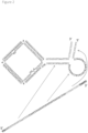
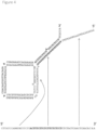
- Figures 1 and 2 , and 3 and 4 respectively depict two different probe designs in which the probe is activated by its target nucleic acid molecule.
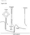
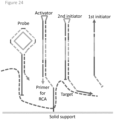
- Figures 14 , 18 , 23 , 24 , 25 and 26 depict probe designs in which the probe is activated by a separate activator molecule, which is itself recruited by binding to the target molecule. The target molecule thus binds both the probe and the separate activator molecule, and brings them together in proximity, to allow the separate activator molecule to interact with and activate the probe. This is described in more detail below.
- the term “recruits” is used herein to mean that the target nucleic acid molecule binds to the separate activator molecule, and thereby brings it into proximity with the probe (which is also bound to the target molecule), allowing it to interact with (i.e. bind to) the probe, and to activate the probe.
- the target molecule is capable of binding simultaneously to both the probe and the separate activator molecule, bringing them into proximity and thereby facilitating or enabling their interaction.
- the probe is capable of binding simultaneously to both the target molecule and to the separate activator molecule (i.e.
- the probe has separate and distinct binding sites for the target and separate activator molecules, specifically binding sites which are at least partially accessible for binding, and conversely the target molecule has separate and distinct binding sites for the probe and separate activator molecule). In this manner, activation of the probe by the separate activator molecule is target-dependent.
- Binding of the target nucleic acid molecule to the separate activator molecule may be direct (i.e. the target nucleic acid molecule binds to a portion of the separate activator molecule), or be indirect (i.e. the target nucleic acid molecule binds to the separate activator molecule by an intermediary molecule). Indirect binding mediated by an intermediary molecule may take place where the intermediary molecule is bound to the target nucleic acid molecule, or may take place where the intermediary molecule is situated in proximity to the target nucleic acid molecule, e.g.
- nucleic acid probe and separate activator molecule may thus bind to different molecules, which binding may still result in the activation of the nucleic acid probe as described herein.
- the activator i.e. the target nucleic acid or a separate activator molecule
- the activator be it the target molecule or a separate activator molecule, comprises a binding site for the probe (more particularly for a sequence in a single-stranded region of the probe).
- the activator is a separate activator molecule it also comprises a binding site for the target molecule (i.e. a region of complementarity to a sequence in the target molecule, which target sequence is distinct to the sequence (binding site) in the target molecule which binds to the probe), or as noted above, for an intermediary molecule situated in proximity thereto.
- the probe comprises at least a first single-stranded region comprising at least an accessible part of the target binding site.
- the probe may also comprise a second single-stranded region comprising at least an accessible part of a binding site for the separate activator molecule (a "separate activator binding site").
- the first and second single-stranded regions may be part of a single, or the same, single-stranded part of the probe (i.e. adjacent in the same protector strand). However, as described further below, the first and second single-stranded regions may be spatially separated within a protector strand.
- the first and second single-stranded regions may be located in the same protector strand, or may be situated in different protector strands, and/or each of the second and/or further protector strands may comprise a first and/or a second single-stranded region.
- the single-stranded region(s) of the probe are, or comprise, a part or a portion of the binding site for the activator (i.e. a part or portion of the activator binding site), and the binding site for the activator (be it the target nucleic acid molecule or the separate activator molecule) extends beyond the single-stranded region, i.e. into a region which is at least partially double-stranded or hybridised to a complementary sequence within the probe (e.g. into the double stranded circular structure).
- part of the binding site in the probe for the activator target or separate activator molecule
- part of the binding site in the probe for the activator is accessible for binding by the activator when it is in contact with the probe, but part is not accessible (i.e.
- the binding site for the activator i.e. the target nucleic acid or the separate activator molecule
- the binding site for the activator is thus partially single-stranded, and further comprises a double-stranded portion, in which the "activator" binding site portion is hybridised to another part of the probe (a complementary sequence in the first circular template strand and/or a complementary sequence in the same (e.g. second) protector strand or a further protector strand).
- the activator target nucleic acid molecule or separate activator molecule
- a strand displacement occurs, as the "activator” invades the double-stranded structure (which contains the "inaccessible” part(s) of the "activator” binding site), causing the double-stranded (duplex) part to be opened up, or unfolded, at least in part.
- binding of the "activator” to the accessible part of its binding site allows the "activator” to bind to a further part or portion of the activator binding site (target binding site or binding site for a separate activator molecule), wherein the binding of the activator to the activator binding site displaces a complementary sequence therefrom and activates the probe.
- binding of the activator for the RCA reaction i.e. the activator of the probe, be it the target or the separate activator molecule
- binding of the activator for the RCA reaction causes a strand displacement reaction to activate the probe, which in turn allows RCA of the first template strand to take place.
- the probe Upon binding of the probe to the target molecule or separate activator molecule, the probe undergoes a conformational change comprising at least partially interrupting the Watson-Crick base pairing between a pair of antiparallel nucleotide sequences within the probe, mediated by strand invasion by the target nucleic acid molecule or separate activator molecule (i.e. the activator for the probe).
- the activator binds to the target nucleic acid molecule via a single-stranded region in a (e.g. second) protector strand and induces strand displacement of a nucleotide sequence hybridised thereto (or to a further protector strand), thereby activating the probe.
- accessible as used herein (e.g. in the context of a binding site for the target or separate activator molecule) thus means simply that that part of the binding site is available for binding when the probe is contacted with the target nucleic acid and/or separate activator molecule. Thus, this part of the binding site is exposed (i.e. single-stranded) and (immediately) available for binding. Conversely, an "inaccessible” part of the binding site means that the activator or target cannot bind to that part until it is rendered available for binding by a strand displacement reaction, e.g. exposed, or released. Accordingly, an inaccessible part of the binding site is protected from binding until the probe is activated.
- An accessible part of a binding site may thus be seen as a "toehold", which allows binding to take place.
- the different, accessible and inaccessible, parts of a binding site may be seen as different domains of a binding site (e.g. a binding site in the probe for the target or separate activator molecule).
- the accessible part (i.e. the toehold) of a target binding site or separate activator binding site may be viewed as a first domain (i.e. a first accessible domain) and the binding site may contain one or more further domains which lie in double-stranded part(s) of the probe, including but not necessarily, in the double-stranded circular structure.
- the probe may comprise a stem-loop structure (more particularly the double-stranded circular structure may be part of a stem-loop structure of the probe), e.g. with the second protector strand forming a stem (alternatively expressed, the loop of the stem-loop structure may be part of the double-stranded structure).
- the probe may comprise one or more stems in addition to the envelope (or loop) structure, with the envelope (or loop) being part of the double-stranded circular structure.
- the second or other domains of the binding site may lie in a double-stranded region (duplex) of the envelope (i.e. the double-stranded circular structure) and/or the stem of the probe (or of another (i.e. a separate) stem-loop structure within the probe).
- duplex double-stranded region of the envelope
- stem of the probe or of another (i.e. a separate) stem-loop structure within the probe.
- the target binding site in the first single stranded region may simply be designed to be fully accessible i.e. it may have a single accessible domain lying in the (or a) first single-stranded region. This is shown in Figure 23 . More particularly, the first single-stranded region may constitute the target binding site.
- target binding serves simply to recruit the separate activator molecule and does not in itself cause a strand displacement in the probe.
- the (or a) target binding site may only be partially accessible (the first single-stranded region), and part of the target binding site (i.e. further binding domains) may be hybridised to a complementary sequence in another part of the probe, wherein the complementary sequence may be a portion of the binding site in the probe for the separate activator molecule.
- the complementary sequence may be a portion of the binding site in the probe for the separate activator molecule.
- binding of the target nucleic acid molecule to the accessible part (domain) of its binding site may allow the target nucleic acid molecule to bind to a further part or portion (domain) of the target binding site, wherein the binding of the target nucleic acid molecule to its binding site displaces a complementary sequence therefrom and exposes, or makes accessible, the first domain of the separate activator binding site.
- the first domain of the (or a) separate activator binding site may therefore be inaccessible in the absence of the target nucleic acid molecule, and be exposed or become accessible only after the binding of the target nucleic acid molecule to the probe.
- the first domain of the separate activator binding site may be situated within a metastable secondary structure (e.g. a hairpin or a stem loop structure), and only becomes accessible (e.g. through unfolding or opening of the metastable secondary structure) upon binding of the nucleic acid probe to the target nucleic acid molecule.
- the first domain of the separate activator binding site may therefore be complementary to at least a portion of the target binding site, or in other words, may have homology to at least a portion of the target nucleic acid molecule.
- At least one protector strand is hybridised to the first circular template strand, thereby blocking the binding of nucleic acid molecules to the first circular template strand, and thus preventing initiation of an RCA reaction.
- the probe structure is also unable itself to prime a RCA reaction in the absence of the target molecule. This is of particular utility as it may prevent the non-specific binding of nucleic acid molecules present in a sample to the RCA template, thereby preventing the non-target specific initiation of RCA.
- the conformational changes which occur upon binding of the probe to the target nuclei acid molecule i.e.
- the activation of the probe via a strand displacement as hereinbefore described may be seen as releasing the first circular template strand such that RCA using the first template strand may be initiated.
- binding of the activator for the RCA reaction may release the amplification template from its circle (from within the double-stranded circular structure).
- such releasing may comprise a strand displacement in the double-stranded circular structure, but as shown in Figures 1 and 2 this is not necessary, and the first circular template strand may be released for RCA by a primer/template complex being allowed to form, comprising the double-stranded circular structure. This is described in more detail below.
- the present invention provides a nucleic acid probe which is activatable by the target nucleic acid molecule to undergo a RCA reaction, said probe comprising:
- the activator for the probe is a separate activator molecule
- the probe comprises a second and/or further protector strand
- at least one of the second and/or further protector strands comprises a first single-stranded region which forms or comprises part of a target binding site and at least one of the second and/or further protector strands (optionally the same protector strand) comprises a second single-stranded region which forms or comprises part of the binding site for the separate activator molecule.
- the nucleic acid probe binds to the target nucleic acid molecule by the or a first single-stranded region or regions, and binding of a separate activator molecule (recruited by the target nucleic acid molecule) to the second single-stranded region of the probe displaces a complementary sequence hybridised to the binding site for the separate activator molecule, thereby activating the probe.
- the present invention thus provides a nucleic acid probe which, in the presence of the target nucleic acid molecule, is activatable to undergo a RCA reaction, the activator for said RCA reaction being a separate activator molecule binding to the target nucleic acid, said probe comprising:
- a probe of the invention may comprise a primer, or more particularly a primer sequence, for the RCA reaction.
- the primer sequence may be a region, or stretch of nucleotides, in the second protector strand which is either hybridised, or capable of hybridising, to the first circular template strand, and which is able to prime a RCA reaction when the probe is activated.
- the primer sequence may lie at the 3' end of a protector strand (e.g. of the second protector strand) and may be capable of being extended by a polymerase enzyme when it becomes hybridised to the first circular template strand, which occurs upon activation of the probe.
- the second protector strand is released by the strand displacement, allowing it to be cleaved to leave a 3' end (which contains the primer sequence) hybridised to the first circular template strand and available for extension in a RCA reaction. This is described in more detail below.
- the strand displacement reaction displaces part of a (e.g. the second) protector strand from the double-stranded circular structure, thereby exposing a part of the first circular template strand to allow binding of a primer for the RCA reaction.
- a (e.g. the second) protector strand from the double-stranded circular structure, thereby exposing a part of the first circular template strand to allow binding of a primer for the RCA reaction.
- the probe may comprise within the 3' end region of a (e.g. the second) protector strand a sequence that is complementary to a sequence within the first circular template strand, in particular to the part of the first circular template strand that is exposed following the strand displacement reaction (i.e. a sequence that is homologous to a portion of the a protector strand hybridised to the first circular template strand (e.g.
- a first single-stranded region of the nucleic acid probe may in particular embodiments be situated at an end of a (e.g. the second) protector strand.
- a first single-stranded region may thus be situated at the 5' end or at the 3' end of a (e.g. the second) protector strand.
- the first single-stranded region may be situated at an intermediate position within a (e.g. the second) protector strand.
- the activator for the RCA reaction (the activator for the probe) is a separate activator molecule and wherein the nucleic acid probe comprises a second single-stranded region which comprises at least an accessible part of the activator binding site
- the second single-stranded region may be situated at an end of a (e.g. the second) protector strand.
- the second single-stranded region may thus be situated at the 5' end or at the 3' end of a (e.g. the second) protector strand.
- the second single-stranded region may be situated at an intermediate position within a (e.g. the second) protector strand.
- first single-stranded region may be situated in the 5' end region and the second single-stranded region may be situated in the 3' end region of a (e.g. the second) protector strand.
- a first single-stranded region may be situated in the 3' end region or the 5' end region and the second single-stranded region may be situated at an intermediate position (i.e. an intermediate region) within a (e.g. the second) protector strand, or the first single-stranded region may be situated in an intermediate region within the second protector strand, and the second single-stranded region may be situated in the 3' end region or the 5' end region of a (e.g. the second) protector strand.
- both the first and second single-stranded regions may be situated in (separate) intermediate regions within a (e.g. the second) protector strand.
- first and second single-stranded regions may alternatively be situated within different protector strands.
- a first single-stranded region may be situated within one (e.g. the second) protector strand (i.e. at the 5' or 3' end, or an intermediate position thereof), and the second single-stranded region may be situated within another (i.e. a further) protector strand (i.e. at the 5' or 3' end, or an intermediate position thereof).
- the nucleic acid probe may provide the primer capable of acting as or providing the primer for RCA of the template strand once the probe has been activated.
- the probe may comprise in the 3' end region of a (e.g. the second) protector strand a sequence capable of binding to the template strand following displacement of second protector strand therefrom, thereby providing a primer for rolling circle amplification.
- a protector strand (e.g. the second protector strand) may therefore comprise a primer sequence in the 3' end region thereof or in an intermediate region thereof, which is capable of acting as or providing a primer for RCA of the template strand once the probe has been activated (or, in other words, when the probe has bound to the target molecule, and optionally to the separate activator molecule, if used).
- a second protector strand comprises a primer sequence in an intermediate region thereof
- the primer may be generated through cleavage (e.g. degradation) of the (e.g. second) protector strand in an activation-dependent manner.
- the cleavage may conveniently be, or comprise, enzymatic cleavage.
- Activation of the probe by strand displacement may therefore comprise the probe undergoing a conformational change which results in the generation of a substrate suitable for enzymatic cleavage.
- cleavage may be 3' exonuclease degradation of the 3' end of the protector strand following the strand displacement reaction by a component having 3' exonuclease activity.
- the component having 3' exonuclease activity will degrade any single-stranded nucleic acid sequence present within the nucleic acid probe having a free 3' end following activation of the probe, and thus will degrade the 3' end region of a (e.g. the second) protector strand back to the point at which it is hybridised to the first circular template strand.
- degradation of the 3' end of a (e.g. the second) protector strand of the probe will provide a primer to allow RCA of the first template strand.
- the primer may be generated through restriction enzyme cleavage or by nickase cleavage.
- the nucleic acid probe as defined herein may, in certain embodiments (where the probe comprises a single protector strand), comprise a second protector strand which comprises 5' and 3' end regions (i.e. regions which do not form part of the double-stranded circular structure comprising the loop of the second protector strand) which comprise mutually complementary regions, which are hybridised to each other to form a partially double stranded stem region comprising a duplex between the mutually complementary regions in the 5' and 3' end regions.
- a second protector strand which comprises 5' and 3' end regions (i.e. regions which do not form part of the double-stranded circular structure comprising the loop of the second protector strand) which comprise mutually complementary regions, which are hybridised to each other to form a partially double stranded stem region comprising a duplex between the mutually complementary regions in the 5' and 3' end regions.
- the nucleic acid probe may therefore in certain embodiments form a stem loop structure comprising a loop which is complementary to the first template strand and is hybridised thereto to form a double-stranded circular structure containing the first template strand inside the loop, and a partially double-stranded stem region comprising a duplex between the mutually complementary regions in the 5' and 3' end regions, and at least the first single-stranded region.
- the strand displacement reaction which occurs upon binding of the probe to the target (and optionally to the separate activator molecule) results in at least partial opening of the stem of the stem-loop structure.
- there is also at least partial opening of the loop i.e. the double stranded circular structure comprising the loop).
- the nucleic acid probe may comprise protector strands which comprise 5' and 3' end regions (i.e. regions which do not form part of the double-stranded circular structure) which comprise complementary regions to end regions of another protector strand, and which hybridise thereto to form partially double-stranded stem structures, i.e. stem regions comprising a duplex between the 5' and 3' end regions of the respective protector strands.
- the probe may comprise second and third protector strands, as depicted for Example in Figures 21 and 26 , wherein stem structures (which may be partially double stranded) are formed between the 5' and 3' end regions of the two respective strands.
- the probe may be seen to comprise a double-stranded circle structure comprising portions of the second protector strands complementary to the first template strand (i.e. the "protecting regions” or envelope) and one or more stem regions (comprising duplexes formed between complementary regions in the 5' and 3' end regions of separate second protector strands).
- the stem regions may be partially double stranded.
- at least one such stem region is partially double-stranded and comprises a first and/or second single-stranded region.
- the strand displacement reaction which occurs upon binding of the probe to the target (and optionally to the separate activator molecule) results in at least partial opening of the region of duplex of a stem region (i.e. of a stem arm structure), analogously to opening the stem region of a stem-loop structure.
- at least partial opening of the envelope may also occur, analogously to opening the loop region of a stem loop structure.
- a 3' end region of one of the protector strands may comprise, or provide, the primer for RCA.
- the primer may be provided by the protector strand which does not comprise the first single-stranded region (comprising the target binding site; see e.g. Figure 21 ) or which does not comprise the first and second single-stranded regions (comprising the target binding site and binding site for the separate activator molecule respectively; see e.g. Figure 26 ).
- the probe comprises a second protector strand which comprises the first single-stranded region, and optionally the second single-stranded region (where present) and a third protector strand.
- the third protector strand comprises a third single-stranded region which comprises or provides a primer for RCA.
- an intermediate region of the second or further protector strand is a region which is in between the 5' and 3' end regions.
- An intermediate region thus lies in the part of the second or further protector strand which provides, or constitutes, the loop or which contributes to the envelope, in the double-stranded circular structure, i.e. the part of the second or further protector strand which is hybridised to the first circular template strand.
- the mutually complementary regions which form the duplex in the partially double-stranded stem region of the stem or stem-loop structure need not lie at the very 5' and/or 3' end of the second protector strand or further protector strand(s).
- the first and/or second single-stranded regions may lie at the 5' and/or 3' ends of the second protector strand or further protector strand(s), and hence of the stem region.
- the duplex of the stem region may comprise one or two single-stranded "extensions", which may constitute, or comprise, first and/or second single-stranded regions, or the primer for RCA (e.g. as a third single-stranded region).
- a first and/or the second single stranded region may lie in the 5' end region 5' to the duplex, and/or in the 3' end region 3' to the duplex. In particular, (but not necessarily) they may lie at the 5' and/or 3' ends.
- a further region (or sequence) lying at the end of single-stranded region for example a capture or immobilisation sequence, e.g.
- an affinity binding probe e.g. a proximity probe
- the 5' end region of a (e.g. the second) protector strand provides the 5' strand of the duplex (i.e. the duplex of a stem), and the 3' end region provides the 3' strand of the duplex.
- the 5' and/or 3' end regions may comprise further or additional single-stranded regions i.e. additional sequences within the 5' and/or 3' end regions which do not hybridise to a complementary sequence in the opposite strand (e.g. sequences within the 5' and/or 3' strands of the duplex and/or 5' to the 5' strand of the duplex and/or 3' to the 3' strand of the duplex).
- a first, and if present, the second single-stranded regions of the probe may thus be situated within or at the end of one or other of the strands of the duplex of the stem region.
- a first single-stranded region and, if present, the second single-stranded region may thus be provided as single-stranded (i.e. unpaired regions) within the partially double-stranded stem region.
- the partially double-stranded stem region may, in certain embodiments, comprise a duplex between the mutually complementary regions in the 5' and 3' end regions (or complementary regions in the 5' and 3' end regions of two (e.g. second and third) protector strands), and may further comprise first and/or second single-stranded regions.
- the single stranded region or regions may be situated at an end of one of the strands of the duplex, or may be situated within one of the strands of the duplex as an unpaired and accessible "bulge" region (that is an unpaired single-stranded region within a strand of the duplex, or a loop-type or blister structure).
- the probe comprises a first and second single-stranded region, wherein the first single-stranded region is situated at the end of one of the strands of the duplex, and the second single-stranded region is situated within one of the strands of the duplex as a bulge (an unpaired loop).
- the first single-stranded region is situated within one of the strands of the duplex as a bulge (an unpaired loop) and the second single-stranded region is situated at the end of one of the strands of the duplex.
- the target binding site (or optionally sites) may be situated at least partially within the 5' or 3' end region of a (e.g. the second protector) strand, and thus within the partially double-stranded stem region.
- the binding site for the separate activator molecule may be situated at least partially within the 5' or 3' end region of a (e.g. the second) protector strand, and thus within the partially double-stranded stem region.
- the target binding site (or, where present, the binding site for the separate activator molecule) may thus be at least partially hybridised to a complementary sequence within the probe (a sequence from the opposite strand of the duplex, and, in certain embodiments, a sequence from the first circular template strand).
- binding of the target nucleic acid molecule, or, where present, the separate activator molecule (i.e. the activator for the RCA reaction) to the nucleic acid probe results in a strand displacement reaction.
- the probe comprises a stem loop structure
- the activator displaces the nucleotide sequence hybridised to the (inaccessible part(s) of the) activator binding site (the target binding site, or where present, the binding site for the separate activator molecule) and causes the stem region duplex to at least partially open.
- Binding of the target nucleic acid molecule or separate activator molecule may further result in the at least partial opening of the loop (i.e. the at least partial displacement of a (e.g. the second) protector strand from the first circular template strand).
- Such displacement(s) activate the probe, allowing initiation of an RCA reaction.
- a single-stranded region (the first, or if present, the second, single-stranded region), may be situated within a bulge in a strand in the stem region duplex.
- the first accessible domain of an activator (target or separate activator molecule) binding site is situated within a bulge.
- the single-stranded region i.e. the bulge
- the activator binding site may further comprise second and third domains, which are situated in the duplex of the stem region, either side of the bulge, and are hybridised to complementary sequences within the opposite strand of the duplex.
- binding of the activator to the first domain of the binding site is followed by binding of the activator to the second and third domains of the binding site, which binding displaces the sequences hybridised thereto and opens at least the stem of the stem loop structure.
- the first single-stranded region is situated within the bulge, and the bulge (and thus the target binding site) is situated within the 5' end region of a (e.g. the second) protector strand (the 5' strand of the duplex).
- a (e.g. the second) protector strand the 5' strand of the duplex.
- binding of the target nucleic acid molecule to the target binding site via the first single-stranded region situated within the bulge opens at least the stem of the stem loop structure and releases the 3' end region of a (e.g. the second) protector strand.
- the 3' end region of a (e.g. the) second protector strand is displaced from its complementary sequence in the 5' end region of a (e.g.
- the second) protector strand i.e. either of the same protector strand, or of another protector strand
- the second) protector strand is thus rendered single-stranded following unfolding of the duplex.
- This may render the 3' end of the (e.g. second) protector strand susceptible to cleavage (e.g. 3' exonuclease cleavage), wherein the 3' end of the strand is cleaved (e.g. degraded) to provide a primer for RCA of the first template strand.
- the present invention provides a nucleic acid probe for detection of a target nucleic acid molecule, wherein said probe is activatable by the target nucleic acid molecule to undergo a RCA reaction, said probe comprising:
- the present invention provides a nucleic acid probe for detection of a target nucleic acid molecule, wherein said probe is activatable by the target nucleic acid molecule to undergo a RCA reaction, said probe comprising:
- the 3' end region may be removed by cleavage, leaving an intermediate region of the second protector strand hybridised to the first circular template strand having a hybridised 3' end which may be extended in a RCA reaction.
- the single stranded region (the first or if present, the second, single-stranded region) may be situated at an end of a strand in the duplex.
- the single-stranded region may thus be situated at the 5' end or the 3' end of a (e.g. the second) protector strand, and thus at the 5' or 3' end of the duplex structure.
- the single-stranded region is a portion (e.g. a first accessible domain) of the target binding site, and is situated at the 5' end of a (e.g. the second) protector strand.
- the single-stranded region is a portion (e.g. a first accessible domain) of a binding site for a separate activator molecule and is situated at the 3' end of a (e.g. the second) protector strand.
- the single-stranded region (i.e. comprising or constituting a binding site for the target nucleic acid molecule or for the separate activator molecule) may be seen to represent a first domain of the activator binding site.
- the activator binding site further comprises a second and third domain, wherein the second domain is situated within the stem of the duplex structure (i.e. adjacent to the first domain and within the duplex structure) and is hybridised to a complementary sequence within the opposite strand of the duplex structure, and the third domain is situated at least partially within the loop region of the second protector strand and is hybridised to a complementary sequence within the first circular template strand.
- binding of the activator to the first domain of the activator binding site is followed by binding of the activator to the second and third domains of the activator binding site, which binding displaces the sequences hybridised to the activator binding site and causes the duplex to open, and a (e.g. the second) protector strand to dissociate at least partially from the first circular template strand, exposing a single-stranded region of the first circular template strand.
- a primer for RCA may bind to said single-stranded region, thereby allowing the initiation of RCA.
- the primer is provided from the 3' end of a protector strand (e.g. the second protector strand, if there is one protector strand, or a third protector strand if there are two protector strands etc.). In another embodiment, the primer is provided from the 3' end of the separate activator molecule.
- the present invention provides a nucleic acid probe for detection of a target nucleic acid molecule, wherein said probe is activatable by the target nucleic acid molecule to undergo a RCA reaction, said probe comprising:
- the present invention also provides a nucleic acid probe for detection of a target nucleic acid molecule, wherein said probe is activatable by the target nucleic acid molecule to undergo a RCA reaction, said probe comprising:
- the present invention provides a nucleic acid probe for detection of a target nucleic acid molecule, wherein in the presence of the target nucleic acid molecule said probe is activatable by a separate activator molecule, which also binds to the target nucleic acid molecule, to undergo a RCA reaction, said probe comprising:
- the ends of the probe may be hybridised to one another, i.e. the duplex region may extend to the ends of the probe.
- the 3' end, the 5' end, or both the 3' and 5' ends of the probe may be single-stranded, i.e. the ends of the duplex structure may be single-stranded and hence not complementary to one another or capable of hybridisation to one another.
- the nucleic acid probe does not comprise a partially double-stranded stem region, and instead comprises a double-stranded circular structure, a single-stranded 5' end region and a single-stranded 3' end region.
- the probe may comprise only a single single-stranded region (i.e. a single-stranded 5' end region or a single-stranded 3' end region.)
- the 5' single-stranded end region of the second protector strand comprises an accessible part of the target binding site
- the single-stranded 3' end region of the second protector strand comprises a primer for the RCA reaction. This is depicted in Figure 20 .
- the 3' single-stranded end region of the second protector strand may comprise an accessible part of the target binding site (the primer for the RCA reaction may be provided separately or from another molecule (e.g. the target nucleic acid molecule) present in the sample).
- the primer for the RCA reaction may be provided separately or from another molecule (e.g. the target nucleic acid molecule) present in the sample).
- one single-stranded end region e.g. the 5' end region
- the other single-stranded end region e.g. the 3' end region
- the other part of the target or (where present) separate activator binding site lies in the loop of the double-stranded circular structure and is hybridised to the first circular template strand, such that it may be displaced from the double-stranded circular structure upon binding of the target or separate activator molecule to the accessible part of the binding site.
- the present invention provides a nucleic acid probe for detection of a target nucleic acid molecule, wherein said probe is activatable by the target nucleic acid molecule to undergo a RCA reaction, said probe comprising:
- two or more second protector strands may similarly bind to (and protect) a first circular template strand, and not comprise regions of complementarity at their 5' and/or 3' ends.
- Production of the nucleic acid probes of the present invention typically comprises contacting a linear first nucleic acid molecule (which will serve as the first circular template strand of the probe) with a linear second nucleic acid molecule (which will serve as the second protector strand) which comprises a region of complementarity (the loop region) to the first template strand and may hybridise thereto to bring the 5' and 3' ends of the first nucleic acid molecule into direct juxtaposition for ligation.
- Addition of a ligase molecule e.g. T4 ligase
- T4 ligase may circularise the first template strand, thereby forming a probe of the invention comprising a double-stranded circular structure.
- first first nucleic acid molecule may hybridise to a first second nucleic acid molecule via its 5' end and to a second second nucleic acid molecule via its 3' end
- second first nucleic acid molecule may hybridise to a first second nucleic acid molecule via its 3' end and to a second second nucleic acid molecule via its 5' end.
- Intermolecular (rather than intramolecular) ligation of the at least two first nucleic acid molecules may result in the formation of a multimeric nucleic acid probe comprising a circular nucleic acid molecule comprising at least two first nucleic acid molecules, and at least two second protector strands hybridised thereto, which strands protect the circular nucleic acid molecule from RCA in the absence of the target nucleic acid molecule (i.e. they form an envelope which is complementary to the first template strand and is hybridised thereto to form a double-stranded circular structure) .
- the probe will be in monomeric form comprising one first circular template strand and one second protector strand, it may be provided in multimeric (e.g. oligomeric) form , comprising two or more first circular template strand which are ligated together into a circle (an enlarged circle) and two or more protector strands. This is depicted in Figure 6 .
- the probe comprises a stem-loop structure (i.e. in particular wherein the 5' and 3' end regions of the probe are complementary and capable of forming a duplex structure), as two (or more) second nucleic acid molecules may be capable of interacting via their respective 5' and 3' ends to form intermolecular, rather than intramolecular, duplex structures, resulting in a nucleic acid multimer.
- Said multimer will comprise the loop region of the second nucleic acid strand between said duplex structures, and will thus contain "n" loop regions, where "n” is the number of second nucleic acid molecules present in said multimer.
- the 5' and 3' ends of two (or more) first nucleic acid molecules may be brought into juxtaposition for ligation, templated by the multimer structure formed by the interaction of the 5' and 3' ends of the (at least) two second nucleic acid molecules.
- Addition of a ligase molecule will, in these embodiments, result in the formation of a multimeric circular strand comprising "n" first nucleic acid molecules.
- the present invention further provides a multimeric nucleic acid probe, comprising a multimeric first circular template strand comprising at least two first template strands, and at least two second protector strands which protect the multimeric first template strand from RCA in the absence of the target nucleic acid molecule.
- Said multimeric nucleic acid probe will be activatable in the presence of the target nucleic acid molecule in an analogous way to the probes described above.
- the present invention provides a nucleic acid probe for detection of a target nucleic acid molecule, wherein in the presence of the target nucleic acid molecule said probe is activatable by a separate activator molecule to undergo a RCA reaction, said probe comprising:
- Such a probe may be used together with a separate activator molecule which is also designed to bind to the target molecule, and which itself may be "activated” by target binding to interact with the probe.
- a separate activator molecule may comprise at least an accessible part (domain) of a target binding site and a probe binding site which is at least partially contained within a metastable secondary structure (such as a hairpin, or stem-loop structure) and which is inaccessible for binding to the probe until the separate activator molecule has bound to the target molecule, and a target molecule-mediated strand displacement causes the metastable secondary structure to open to release, or render accessible, the probe binding site within the separate activator molecule.
- a metastable secondary structure such as a hairpin, or stem-loop structure
- Binding of the separate activator molecule to the first domain of the separate activator binding site in the probe then causes the probe to be activated by a strand displacement reaction by the separate activator molecule, as described above.
- a reaction is depicted in Figure 25 .
- the separate activator molecule for use with such a probe may comprise single stranded 5' and 3' end regions, and an intermediate sequence therebetween comprising mutually complementary regions which hybridise together to form a stem-loop structure, wherein one of said single-stranded regions comprises an accessible domain of a target binding site, and the stem-loop structure comprises a further domain of the target binding site and at least one domain of a probe binding site.
- the other single-stranded region may comprise a further domain of the probe binding site.
- the 3' end region of the separate activator molecule comprises a first domain of the target binding site which is complementary to a cognate activator binding site (or domain thereof) in the target molecule;
- the duplex of the stem of the stem loop structure comprises a second domain of the target binding site hybridised to the second domain of the probe binding site (which is complementary to the second domain of the separate activator binding site of the probe);
- the loop of stem-loop structure comprises the first domain of the probe binding site (which is complementary to the first domain of the separate activator binding site of the probe);
- the 5' end region comprises a third domain of the probe binding site (which is complementary to the third domain of the separate activator binding site in the probe).
- two sequences need not have perfect homology to be “complementary” under the invention.
- two sequences are sufficiently complementary when at least about 85% (preferably at least about 90%, and most preferably at least about 95%) of the nucleotides share base pair organization over a defined length of the molecule.
- two complementary sequences within the probe may not comprise an absolute (i.e. 100%) degree of complementarity.
- the interaction between the probe and the target nucleic acid or separate activator molecule is sufficient to displace a sequence within the probe (i.e. strand displacement) that is hybridised to the target binding site or binding site for the separate activator molecule, and thus preferably is a stronger interaction that the interaction between the two complementary sequences within the probe.
- complementary sequences within the probe may comprise one or more mismatches (unpaired bases) relative to one another, provided that in the absence of the target nucleic acid molecule or separate activator molecule the sequences are able to remain hybridised to each other.
- the rate of amplification of a target sequence by RCA is dependent on the size of the template nucleic acid molecule used in the amplification reaction.
- a template for RCA will be of a size that allows copies of it to be generated (i.e. for it to be amplified) at an acceptable rate.
- a smaller template may be less likely to undergo non-target specific amplification.
- the first circular template strand may be 100, 200, 300, 400 or 500 nucleotides or more in size (particularly when in multimeric form)
- the first circular template strand may preferably comprise up to 500 nucleotides, up to 400 nucleotides, up to 300 nucleotides, up to 200 nucleotides or up to 100 nucleotides, or more particularly, up to 90, 80, 70 or 60 nucleotides.
- RCA is known to be sterically hindered if the circular nucleic acid molecule is too small.
- double-stranded circular structures may have difficulty in forming (i.e.
- the second protectors strand may not be able to bind to the first template strand) if the first circular template strand is too small, due to the additional steric constrains imposed when a double-helical structure is formed upon binding of the second protector strand to the first circular template strand.
- the first circular template strand in certain embodiments comprises at least 20 nucleotides, at least 30 nucleotides, at least 40 nucleotides, at least 50 nucleotides, at least 60 nucleotides, at least 70 nucleotides, at least 80 nucleotides, or at least 90 nucleotides.
- double-stranded circular nucleic acid molecules of a smaller size than was previously thought possible may be produced where the two strands contain one or more mismatches (i.e. one or more insertions, deletions or mismatches) relative to one-another.
- mismatches i.e. one or more insertions, deletions or mismatches
- a mismatch may provide a 'hinge' region comprising a higher degree of flexibility and/or rotational freedom, and thus that this may relieve stress within the phosphodiester backbones of the nucleic acid molecules in the double-stranded structure.
- the first and second or further strands of the probe of the present invention may comprise one or more mismatches (insertions, deletions and/or substitutions) relative to one another, within the double-stranded circular structure (e.g. within the loop region, or the envelope region) of the probe.
- the protecting regions of the second or further (subsequent) protector strands need not be 100% complementary and/or not all bases of the first template strand need be hybridised to a protecting region of a protector strand. It is believed that this may allow a smaller first circular template strand to be provided in the probes of the present invention.
- a second or further protector strand may contain one or more base insertions relative to the first template strand.
- the first template strand may contain one or more base insertions relative to the second template strand.
- the one or more base insertions may be an adenosine (A) nucleotide.
- the first and second strands may comprise one or more base substitutions, i.e. bases which do not undergo canonical Watson-Crick base-pairing.
- the second or further protector strand or strands must maintain a sufficient degree of homology to the first template strand to remain hybridised thereto. Furthermore, it is preferred that extended stretches (i.e. more than two or three nucleotides) of mismatch between the respective strands is avoided, as this may allow non-specific binding of an oligonucleotide present in the sample to the first template strand, which may initiate RCA in the absence of a target nucleotide.
- the first and second strands do not contain a contiguous stretch of 4 or more, or more particularly 3 or more, mismatched bases, and preferably do not contain a contiguous stretch of 2 or more mismatched bases.
- a mismatch may comprise 1 to 3 contiguous mismatched bases, more preferably 1 or 2 contiguous mismatched bases.
- an accessible part of a target or separate activator binding site may, for example, comprise 6 to 50 nucleotides (e.g. any one of 7, 8, 9 or 10 to any one of 20, 22, 25, 27, 30, 35, 40 or 45 nucleotides).
- a primer sequence may comprise 6 to 30 nucleotides, e.g. any one of 6, 7, 8, 9 or 10 to any one of 12, 14, 16, 18, 20, 22, 24 or 25 nucleotides.
- a duplex in a partially double-stranded stem region may comprise, for example 6 to 50 base pairs, e.g. any one of 7, 8, 9 or 10 to any one of 15, 20, 25, 30, 35, 40, or 45 base pairs.
- a duplex which comprises a single stranded region in a bulge in one of the strands may for example have shorter duplex regions either side of the bulge, than a single duplex in a stem region.
- a duplex in a stem region may comprise one or more mismatches, e.g. as discussed above, for example to aid or facilitate a strand displacement reaction.
- the first circular template strand may comprise a reporter domain, which is a sequence that can be used to detect and/or identify the RCA product produced from the probe of the invention. This may be particularly advantageous in multiplex embodiments of the invention, i.e. where more than one analyte, e.g. nucleic acid analyte, is detected in a single assay.
- the RCA template provided by each probe (each probe is specific for a target analyte, as determined by the target binding domain, or, if present, the binding domain for the separate activator molecule), may comprise a unique "marker" or identification sequence (e.g. a bar-code sequence, such as a site comprising the sequence of a specific detection probe, i.e.
- the RCA product is complementary to the RCA template and as such detection probes that hybridise to the RCA product will comprise a sequence that is identical to part of the RCA template) to allow the separate detection and/or quantification of each analyte in the sample.
- each probe may comprise a different reporter domain and the detection of the interaction of the probe and the target analyte, i.e. the detection of each analyte, may be detected in parallel (i.e. at the same time), e.g. using oligonucleotides tagged with distinct labels (e.g. fluorophores or sequence tags) that may hybridise to the complement of the reporter domain.
- each marker (and therefore each analyte) may be detected using sequential visualisation reactions, wherein each reaction is separated by, e.g. stripping or bleaching steps.
- sequential visualisation reactions suitable for using the methods of the invention are known in the art, see e.g. Göransson et al., 2009 (A single molecule array for digital targeted molecular analyses. Nucleic Acids Res. 2009 Jan;37(1):e7 ), and Wahlby et al., 2002 (Sequential immunofluorescence staining and image analysis for detection of large numbers of antigens in individual cell nuclei. Cytometry, 47(1):32-41, 2002 ).
- multiple analytes may be detected in parallel.
- multiple analytes may be detected sequentially.
- Combinatorial methods of labelling e.g. ratio labelling, using different combinations and/or ratios of different labels are known in the art and may be used to increase the number of different molecules, and hence different analytes which may detected at one time, or in the same reaction.
- combinations using different coloured and/or fluorescent labels and/or different ratios of different coloured and/or fluorescent labels may be used.
- a second or further protector strand of the nucleic acid probe and/or of a separate activator molecule (if present), and/or of a separate primer (if present) may be provided in a modified form such that they are resistant to 3' exonuclease activity.
- Modifications to nucleic acid molecules or to nucleotides contained within a nucleic acid molecule, to prevent degradation by exonucleases are well known in the art and generally utilise the modification of one or more residues at the protected end, e.g. the 3' end. Any modification that is suitable for the protection against 3' exonuclease activity may be utilised in the methods of the invention.
- the primer sequence of a probe or of a separate activator molecule preferably comprises at least one modified nucleotide at the 3' end.
- the modifications may be selected from any one or more of the list comprising a thiophosphate-modified nucleotide, phosphorothioate nucleotide a locked nucleic acid nucleotide (inaccessible RNA nucleotide), a 2'-OMe-CE Phosphoramidite modified nucleotide, or a peptide nucleic acid nucleotide.
- the second or further protector strand of the probe may comprise a modification or block at or near the 3' end which acts to inhibit extension (e.g. a "polymerase-block” or "extension block”).
- extension e.g. a "polymerase-block” or "extension block”
- This may be useful to protect from extension a 3' end of a second protector strand which is not intended to provide a primer for RCA, for example in an embodiment where the probe is activated by a separate activator molecule which also provides the primer for RCA, e.g. as depicted in Figures 23 and 24 .
- the 3' end of the second protector strand of the probe is protected from both degradation and extension.
- the 3' end of the activator molecule provides the RCA primer and is protected from degradation but not extension.
- the present invention provides use of a nucleic acid probe as defined herein in the detection of a target nucleic acid molecule, wherein said probe is activatable in the presence of the target nucleic acid to generate an RCA product (i.e. wherein upon binding of the probe to the target nucleic acid, the probe is activated to allow RCA of the template strand of the probe).
- the present invention may thus also be seen to provide methods for the detection of a target nucleic acid molecule, using the nucleic acid probes as described herein.
- the present invention provides a method for detecting a target nucleic acid molecule by an RCA reaction, said method comprising:
- Components of the detection reaction may be added to, or contacted with, a sample containing the target nucleic acid molecule sequentially or simultaneously.
- the components of the detection reaction may be pre-mixed prior to addition to a sample.
- a probe and other reagents for a RCA reaction e.g. polymerase and/or nucleotides and/or a separate primer if necessary
- various components may be pre-mixed, but this will not include the probe and separate activator molecule.
- the sample may be contacted with the probe and separate activator molecule at the same, or substantially the same time.
- the probe and the separate activator may be added to the sample, or a sample and a probe may be brought into contact with (e.g. added to) a separate activator molecule (e.g. an immobilised separate activator molecule as depicted in Figure 23 ).
- any one or more of the components may be contacted, e.g. pre-incubated, with the sample prior to the addition of other components required to activate RCA and detect the target.
- the sample containing the target molecule may be contacted with the separate activator molecule, and then with the probe.
- the target binding site(s) or, if present, the binding site for the separate activator molecule in a second or further protector strand may lie at least partially within the loop or envelope structure, and thus may be hybridised to the first circular template strand to form a double-stranded circular structure.
- binding of the target nucleic acid molecule or of the activator molecule to the probe displaces at least a portion of a second or further protecting strand from the first circular template strand, thereby exposing a region of the first circular template strand, allowing an RCA primer to bind to the exposed region to initiate rolling circle amplification.
- a separate activator molecule which binds to and is recruited by the target nucleic acid, may be used to activate the nucleic acid probe, and thus in certain embodiments a separate activator molecule may be provided to activate the nucleic acid probe.
- the separate activator molecule where used, comprises a binding site for the target nucleic acid molecule, and binds (hybridises) thereto and is thereby recruited by the target nucleic acid molecule.
- the nucleic acid probe and the separate activator molecule may both bind to the target nucleic acid, wherein the binding site for the separate activator molecule within the target nucleic acid molecule is distinct from, or more particularly, spatially separate from, the binding site for the nucleic acid probe within the target nucleic acid molecule.
- the target nucleic acid molecule thus may comprise two separate binding sites for the nucleic acid probe and separate activator molecule, respectively, and conversely the separate activator molecule comprises a target binding site and a separate and distinct probe binding site, such that it may simultaneously bind to both the target and the probe.
- the target binding site in the separate activator molecule may be situated at an end of the separate activator molecule, i.e.
- the separate activator molecule further comprises a region of complementarity for the binding site for the separate activator molecule in the nucleic acid probe (i.e. a probe binding site), and, when present, binds to the activator binding site (e.g. to the second single-stranded region of certain probe embodiments as set out above) of the nucleic acid probe in the course of activating the probe of the invention.
- said region of complementarity will be situated at an end of the separate activator molecule, such that the end of the molecule hybridises to a (e.g. the second) protector strand of the nucleic acid probe during the strand displacement/activation step of the invention.
- the target nucleic acid is contacted (bound) by both the nucleic acid probe of the invention and the separate activator molecule, and activation of the nucleic acid probe is dependent upon the binding of the separate activator molecule to the target nucleic acid molecule.
- an RCA product may, in certain embodiments where a separate activator molecule is required and provided, be dependent upon the binding of the nucleic acid probe of the invention, and of a separate activator molecule, to a target nucleic acid molecule.
- Such binding may be seen to take place in proximity, i.e. both molecules may bind to distinct positions nearby within the target nucleic acid molecule.
- this may alternatively be indirect binding, i.e. binding of a separate activator molecule to an intermediary molecule in proximity to the target molecule.
- Both the nucleic acid probe and the separate activator molecule may thus comprise target binding sites (nucleic acid sequences complementary to, and capable of hybridising to the target nucleic acid molecule), and regions capable of interacting with one another (i.e. a second single-stranded region and binding site for a separate activator molecule within the nucleic acid probe, and a region of complementarity to the nucleic acid probe within the separate activator molecule).
- the separate activator molecule may comprise a binding site to an intermediary molecule, in proximity to the target molecule. Accordingly, where the activator for the probe (the activator for the RCA reaction) is a separate activator molecule, the detection assay may be viewed as a proximity-based detection assay comprising the use of a nucleic acid probe of the invention.
- the primer for RCA may be provided by or from the 3' end region of a (e.g. the second) protector strand, optionally wherein said 3' end region is released by the strand displacement reaction, or from an intermediate region of a (e.g. the second) protector strand (e.g. where the 3' end region is removed by cleavage).
- the primer for RCA may alternatively be provided by the target nucleic acid molecule, or, where present, by the separate activator molecule.
- the target nucleic acid molecule or separate activator molecule may comprise a free 3' end which may bind to the exposed region of the first circular template strand following activation of the probe, thereby to initiate rolling circle amplification.
- a primer may be generated from the target nucleic acid molecule or activator molecule, i.e. a 3' end may be generated by cleavage (e.g. degradation) of the target nucleic acid or activator molecule.
- the primer may be provided by the 3' end region of the activator molecule, or provided by or from the 3' end of the target nucleic acid molecule.
- the nucleic acid probe comprises a stem-loop structure comprising a partially double-stranded stem region comprising a duplex between the 5' and 3' end regions of the second protector strand
- binding of the target nucleic acid molecule, or if present, the separate activator molecule, to the probe displaces the 3' end region from the 5' end region of the second protector strand, thereby opening at least the stem of the stem-loop structure to activate the probe.
- the nucleic acid probe comprises two or more protector strands having regions of complementarity at their 5' and 3' end regions for another protector strand and forming partially double-stranded stem regions comprising a duplex between the respective ends of two protector strands, binding of the target nucleic acid molecule, or if present, the separate activator molecule, to the probe displaces the 3' end region of one protector strand from the 5' end region of another protector strand, thereby opening at least the stem to activate the probe.
- the opening of a stem results in the release of a 3' end region of a (e.g. the second) protector strand, which may render the 3' end region susceptible to cleavage (e.g. enzymatic cleavage), to generate a primer for the RCA reaction.
- the method of the present invention may thus further comprise a step of cleavage of a released 3' end region to generate a primer for the RCA reaction.
- Said cleavage may be enzymatic cleavage and may in certain embodiments be performed by a component having 3' exonuclease activity.
- the component having 3' exonuclease activity may be a polymerase enzyme.
- the present invention thus provides a method for detecting a target nucleic acid molecule by an RCA reaction, said method comprising:
- such a cleavage step to generate the primer is not necessary, and the strand displacement reaction which activates the probe results in displacement of at least a portion of a (e.g. the second) protecting strand from the first circular template strand, thereby exposing a region of the first circular template strand, allowing an RCA primer to bind to the exposed region to initiate rolling circle amplification.
- the primer may be a primer sequence lying at the 3' end of a (e.g. the second) protector strand or at the 3' end of a separate activator molecule. Alternatively it may be a separately added primer molecule.
- the method may further comprise a step of allowing a primer to bind to the exposed region of the first circular template strand.
- the probe used in such methods may, in certain embodiments, comprise a single-stranded region at the 5' end of a (e.g. the second) protector strand, wherein said single-stranded region comprises at least an accessible part of the target binding site. Binding of the target nucleic acid molecule to the probe may thereby disrupt a duplex region and displace a portion of a (e.g. the second) protector strand from the first template strand, thereby exposing a region of the first circular template strand and allowing an RCA primer to bind to the exposed region.
- the probe may comprise a region at the 3' end of a (e.g. the second) protector strand which is complementary to the exposed region of the first circular template strand, and is thus able to bind to the exposed region and act as a primer for RCA.
- the probe may comprise a first single-stranded region at the 5' end of a (e.g. the second) protector strand comprising at least an accessible part of the target binding site, and a second single-stranded region at the 3' end of a (e.g. the second or further) protector strand comprising at least an accessible part of a binding site for a separate activator molecule. Binding of the separate activator molecule to the probe disrupts the duplex region and displaces a portion of a (e.g.
- the second) protector strand from the first template strand, thereby exposing a region of the first circular template strand and allowing an RCA primer to bind to the exposed region
- the separate activator molecule may comprise a region at its 3' end which is complementary to the exposed region of the first circular template strand, and is thus able to bind to the exposed region and act as a primer for RCA.
- the target nucleic acid detected by the probes of the present invention may be a target analyte of interest, i.e. an analyte present in a sample, the detection of which may be of interest, or it may be a reporter or marker nucleic acid molecule which is indicative of the presence of a target analyte in a sample (i.e. it may be a "surrogate" for a target analyte).
- the target nucleic acid molecule may therefore be any kind of nucleic acid molecule. Thus it may be DNA or RNA, or a modified variant thereof.
- the nucleic acid may be made up of ribonucleotides and/or deoxyribonucleotides as well as synthetic nucleotides that are capable of participating in Watson-Crick type or analogous base pair interactions.
- the target nucleic acid may be or may comprise, e.g. bi-sulphite converted DNA, LNA, PNA or any other derivative containing a non-nucleotide backbone.
- the target nucleic acid molecule is a target analyte.
- the target nucleic acid molecule may be coding or non-coding DNA, for example genomic DNA or a sub-fraction thereof, or may be derived from genomic DNA, e.g. a copy or amplicon thereof, or it may be cDNA or a sub-fraction thereof, or an amplicon or copy thereof etc.
- the target molecule may be or may be derived from coding (i.e. pre-mRNA or mRNA) or non-coding RNA sequences (such as tRNA, rRNA, snoRNA, miRNA, siRNA, snRNA, exRNA, piRNA and long ncRNA).
- the target nucleic acid may be a reporter oligonucleotide or nucleic acid molecule indicative of the presence of a target analyte.
- the target nucleic acid may therefore be the product of a previous amplification reaction, e.g. a previous PCR reaction, RCA reaction or LCR reaction, or may be a nucleic acid molecule formed as a result of a previous ligation or extension reaction, e.g. in a proximity dependent manner (the nucleic acid molecule may be the product of a proximity ligation assay or a proximity extension assay).
- the target nucleic acid molecule may be the product of a previous RCA reaction (a RCA product), which product may in certain embodiments be indicative of the presence of a target analyte in a sample e.g. formed as a result of a proximity-based detection assay.
- the target nucleic acid molecule is the product of a previous RCA reaction (i.e. the amplification product formed from a previous circular template)
- the target binding site may be seen to be complementary to at least a portion of the circle used as a template for the previous RCA reaction.
- the target nucleic acid molecule may alternatively be the nucleic acid domain of a probe binding to a target analyte, for example a proximity probe or any antibody-based probe. Activation of the probe may therefore be accomplished by a nucleic acid molecule provided in a sample which is indicative of the presence of a target analyte, rather than the target analyte itself.
- the target nucleic acid molecule may be the nucleic acid domain of a probe binding to a target analyte
- the nucleic acid probe may be the nucleic acid domain of a further probe which also binds to the target analyte.
- Such probes may accordingly be viewed as a pair of proximity probes for a target analyte.
- the nucleic acid probe may ⁇ detect' the target nucleic acid molecule in a manner hereinbefore described, leading to the generation of a detectable RCA product.
- the target nucleic acid molecule may be the nucleic acid domain of a probe binding to a target analyte
- the intermediary molecule may be the nucleic acid domain of a further probe which also binds to the target analyte.
- probes may similarly be viewed as a pair of proximity probes for a target analyte.
- a nucleic acid probe may bind to the target nucleic acid molecule and a separate activator molecule may bind to the intermediary molecule (i.e. in proximity to the nucleic acid probe).
- the separate activator molecule may activate the nucleic acid probe, thereby leading to the generation of a detectable RCA product.
- the target nucleic acid molecule may be the nucleic acid domain of a probe binding to the target analyte
- the separate activator molecule may be the nucleic acid domain of a further probe which also binds to the target analyte, and such probes may also be viewed as a pair of proximity probes for a target analyte.
- a nucleic acid probe may bind to the target nucleic acid molecule and the separate activator molecule.
- the nucleic acid probe may be activated, thereby leading to the generation of a detectable RCA product.
- the present invention thus provides proximity-based detection methods for the detection of a target analyte in a sample in which the nucleic acid probe as disclosed herein is provided as the nucleic acid domain of a first proximity probe which binds to a target analyte, and the target nucleic acid molecule is provided as the nucleic acid domain of a second proximity probe which binds to a target analyte, wherein the first and second proximity probes bind to the target in proximity, and wherein when said first and second proximity probes are bound to the target analyte in proximity, the nucleic acid domain of the second proximity probe (the target nucleic acid molecule) is able to interact with the nucleic acid domain of the first proximity probe (the nucleic acid probe of the invention)
- the present invention thus provides a method for detecting a target analyte in a sample, said method comprising:
- the present invention also provides a method for detecting a target analyte in a sample, said method comprising:
- the present invention also provides a method for detecting a target analyte in a sample, said method comprising:
- the proximity probes for use in such methods are in effect detection probes which bind to the analyte (via the analyte-binding domain), the binding of which may be detected (to detect the analyte) by means of detecting the interaction which occurs between the nucleic acid domains thereof upon such binding.
- the probes may be viewed as nucleic acid-tagged affinity ligands or binding partners for the analyte, the analyte-binding domain being the affinity binding partner, and the nucleic acid domain the nucleic acid tag.
- the nucleic acid domain is coupled to the analyte-binding domain and this "coupling" or connection may be by any means known in the art, and which may be desired or convenient and may be direct or indirect e.g. via a linking group.
- the domains may be associated with one another by covalent linkage (e.g. chemical crosslinking) or by non-covalent association e.g., via streptavidin-biotin based coupling (biotin being provided on one domain and streptavidin on the other).
- the analyte binding domain may be any binding partner for the target analyte, and it may be a direct or indirect binding partner therefor. Thus it may bind to the target analyte directly, or indirectly via an intermediary molecule or binding partner which binds to the target analyte, the analyte binding domain binding to said intermediary molecule (binding partner).
- the analyte-binding domain or the intermediary binding partner is a specific binding partner for the analyte.
- a binding partner is any molecule or entity capable of binding to its target, e.g. target analyte, and a specific binding partner is one which is capable of binding specifically to its target (e.g.
- binding partner binds to the target (e.g. analyte) with greater affinity and/or specificity than to other components in the sample.
- binding to the target analyte may be distinguished from non-target analytes; the specific binding partner either does not bind to non-target analytes or does so negligibly or non-detectably or any such non-specific binding, if it occurs, may be distinguished.
- the binding between the target analyte and its binding partner is typically non-covalent.
- the analyte binding domain may be selected to have a high binding affinity for a target analyte.
- high binding affinity is meant a binding affinity of at least about 10 -4 M, usually at least about 10 -6 M or higher, e.g., 10 -9 M or higher.
- the analyte binding domain may be any of a variety of different types of molecules, so long as it exhibits the requisite binding affinity for the target protein when present as part of the proximity probe.
- the analyte binding domain may be a ligand that has medium or even low affinity for its target analyte, e.g., less than about 10 -4 M.
- the analyte binding domain may be a small molecule or large molecule ligand.
- small molecule ligand is meant a ligand ranging in size from about 50 to about 10,000 Daltons, usually from about 50 to about 5,000 Daltons and more usually from about 100 to about 1000 Daltons.
- large molecule is meant a ligand ranging in size from about 10,000 Daltons or greater in molecular weight.
- the small molecule may be any molecule, as well as a binding portion or fragment thereof, that is capable of binding with the requisite affinity to the target analyte. Generally, the small molecule is a small organic molecule that is capable of binding to the target analyte of interest.
- the small molecule will include one or more functional groups necessary for structural interaction with the target analyte, e.g. groups necessary for hydrophobic, hydrophilic, electrostatic or even covalent interactions.
- the target analyte is a protein
- the small molecule ligand will include functional groups necessary for structural interaction with proteins, such as hydrogen bonding, hydrophobic-hydrophobic interactions, electrostatic interactions, etc., and will typically include at least an amine, amide, sulfhydryl, carbonyl, hydroxyl or carboxyl group, preferably at least two of the functional chemical groups.
- the small molecule may also comprise a region that may be modified and/or participate in covalent linkage to the nucleic acid domain of the proximity probe, without substantially adversely affecting the small molecule's ability to bind to its target analyte.
- Small molecule affinity ligands often comprise cyclical carbon or heterocyclic structures and/or aromatic or polyaromatic structures substituted with one or more of the above functional groups.
- structures found among biomolecules including peptides, saccharides, fatty acids, steroids, purines, pyrimidines, derivatives, structural analogs or combinations thereof. Such compounds may be screened to identify those of interest, where a variety of different screening protocols are known in the art.
- the small molecule may be derived from a naturally occurring or synthetic compound that may be obtained from a wide variety of sources, including libraries of synthetic or natural compounds. For example, numerous means are available for random and directed synthesis of a wide variety of organic compounds and biomolecules, including the preparation of randomized oligonucleotides and oligopeptides. Alternatively, libraries of natural compounds in the form of bacterial, fungal, plant and animal extracts are available or readily produced. Additionally, natural or synthetically produced libraries and compounds are readily modified through conventional chemical, physical and biochemical means, and may be used to produce combinatorial libraries. Known small molecules may be subjected to directed or random chemical modifications, such as acylation, alkylation, esterification, amidification, etc., to produce structural analogs.
- the small molecule may be obtained from a library of naturally occurring or synthetic molecules, including a library of compounds produced through combinatorial means, i.e. a compound diversity combinatorial library.
- a library of compounds produced through combinatorial means i.e. a compound diversity combinatorial library.
- the small molecule employed will have demonstrated some desirable affinity for the protein target in a convenient binding affinity assay.
- Combinatorial libraries as well as methods for their production and screening, are known in the art and described in: US 5,741,713 ; 5,734,018 ; 5,731,423 ; 5,721,099 ; 5,708,153 ; 5,698,673 ; 5,688,997 ; 5,688,696 ; 5,684,711 ; 5,641,862 ; 5,639,603 ; 5,593,853 ; 5,574,656 ; 5,571,698 ; 5,565,324 ; 5,549,974 ; 5,545,568 ; 5,541,061 ; 5,525,735 ; 5,463,564 ; 5,440,016 ; 5,438,119 ; 5,223,409 .
- the analyte binding domain may also be a large molecule.
- analyte binding domains are antibodies, as well as binding fragments and derivatives or mimetics thereof.
- antibodies are the analyte binding domain, they may be derived from polyclonal compositions, such that a heterogeneous population of antibodies differing by specificity are each "tagged" with the same tag nucleic acid (nucleic acid domain) or monoclonal compositions, in which a homogeneous population of identical antibodies that have the same specificity for the target analyte are each tagged with the same tag nucleic acid.
- the analyte binding domain may be either a monoclonal or polyclonal antibody.
- the affinity ligand is an antibody binding fragment or derivative or mimetic thereof, where these fragments, derivatives and mimetics have the requisite binding affinity for the target analyte.
- antibody fragments such as Fv, F(ab)2 and Fab may be prepared by cleavage of the intact protein, e.g. by protease or chemical cleavage.
- recombinantly or synthetically produced antibody fragments or derivatives such as single chain antibodies or scFvs, or other antibody derivatives such as chimeric antibodies or CDR-grafted antibodies, where such recombinantly or synthetically produced antibody fragments retain the binding characteristics of the above antibodies.
- Such antibody fragments or derivatives generally include at least the VH and VL domains of the subject antibodies, so as to retain the binding characteristics of the subject antibodies.
- Such antibody fragments, derivatives or mimetics may be readily prepared using any convenient methodology, such as the methodology disclosed in U.S. Patent Nos. 5,851,829 and 5,965,371 .
- antibodies, fragments, derivatives and mimetics thereof may be obtained from commercial sources and/or prepared using any convenient technology, where methods of producing polyclonal antibodies, monoclonal antibodies, fragments, derivatives and mimetics thereof, including recombinant derivatives thereof, are known to those of the skill in the art.
- Polynucleic acid aptamers may be RNA oligonucleotides which may act to selectively bind proteins, much in the same manner as a receptor or antibody ( Conrad et al., Methods Enzymol. (1996), 267(Combinatorial Chemistry), 336-367 ).
- the target analyte binding domain is a nucleic acid, e.g., an aptamer
- the target analyte is not a nucleic acid.
- the analyte binding domain may in other circumstances be an oligonucleotide probe, i.e. a probe being complementary to a sequence within a target nucleic acid molecule, and capable of hybridising thereto.
- the analyte binding domain will be one that includes a moiety that can be covalently attached to the nucleic acid domain without substantially abolishing the binding affinity of the analyte binding domain to its target analyte.
- analyte binding domain may also be a lectin, a soluble cell-surface receptor or derivative thereof, an affibody or any combinatorially derived protein or peptide from phage display or ribosome display or any type of combinatorial peptide or protein library. Combinations of any analyte-binding domain may be used.
- the binding sites on the analyte for the respective analyte-binding domains of the proximity probes in a set may be the same or different.
- the analyte-binding domains of two or more probes may be the same.
- the analyte is a single molecule or comprises different sub-units or constituents (e.g. a heteromeric complex or an aggregate of different proteins)
- the analyte binding domains will be different.
- Binding sites on the analyte for the analyte binding domain need not be on the same molecule. They may be on separate, but closely positioned, molecules.
- the multiple binding domains of an organism such as a bacterium or cell, or a virus, can be targeted by the proximity detection methods of the present invention.
- the target analyte e.g. a target nucleic acid molecule or a target analyte which is a target for a detection probe such as proximity probe
- the target analyte may be immobilised on a solid support or surface e.g. in an array, or may be fixed or immobilised within a biological sample, e.g. a fixed cell or tissue sample.
- the present invention may thereby thus provide in certain embodiments an in situ method for the detection of a target analyte in a sample.
- Immobilisation of the analyte on a solid phase may be achieved in various ways. Accordingly, several embodiments of solid phase assays are contemplated.
- the analyte can first be captured by an immobilised (or immobilisable) capture probe and then bound by subsequently added probe(s).
- the immobilised capture probe may be immobilised, i.e. bound to the support, in any convenient way.
- the manner or means of immobilisation and the solid support may be selected, according to choice, from any number of immobilisation means and solid supports as are widely known in the art and described in the literature.
- the capture probe may be directly bound to the support (e.g.
- a capture probe may be provided with means for immobilisation (e.g. an affinity binding partner, e.g. biotin or a hapten or a nucleic acid molecule, capable of binding to its binding partner, i.e. a cognate binding partner, e.g. streptavidin or an antibody or a nucleic acid molecule) provided on the support.
- an affinity binding partner e.g. biotin or a hapten or a nucleic acid molecule
- a cognate binding partner e.g. streptavidin or an antibody or a nucleic acid molecule
- the probe may be immobilised before or after binding to the analyte. Further, such an "immobilisable" capture probe may be contacted with the sample together with the support.
- Figure 14 shows a representative depiction of a target analyte (in this case a protein) immobilised on a solid support by means of an antibody-based capture probe, wherein the capture probe comprises an antibody conjugated to an oligonucleotide which comprises a sequence complementary to an oligonucleotide immobilised on the solid support.
- the capture probe may be, for example, an antibody or nucleic acid molecule that is capable of binding to the target analyte specifically.
- the capture probe may be an immobilised (or immobilisable) analyte-specific probe comprising an analyte binding domain (i.e. an analyte capture probe).
- analyte capture probe the analyte is first captured by the immobilised or immobilisable capture probe which serves only to immobilise the analyte on the solid phase, and subsequently the immobilised analyte is incubated with the probe of the invention.
- the capture probe may be any binding partner capable of binding the analyte, directly or indirectly, as discussed above in related to intermediary binding partners. More particularly, such a capture probe binds specifically to the analyte.
- the solid support may be any of the well-known supports or matrices which are currently widely used or proposed for immobilisation, separation etc. These may take the form of particles (e.g. beads which may be magnetic or non-magnetic), sheets, gels, filters, membranes, fibres, capillaries, or microtitre strips, tubes, plates or wells etc.
- the support may be made of glass, silica, latex or a polymeric material. Suitable are materials presenting a high surface area for binding of the analyte.
- Such supports may have an irregular surface and may be for example porous or particulate e.g. particles, fibres, webs, sinters or sieves. Particulate materials e.g.
- a particulate solid support used according to the invention will comprise spherical beads.
- the size of the beads is not critical, but they may for example be of the order of diameter of at least 1 and preferably at least 2 ⁇ m, and have a maximum diameter of preferably not more than 10, and e.g. not more than 6 ⁇ m.
- Monodisperse particles that is those which are substantially uniform in size (e.g. size having a diameter standard deviation of less than 5%) have the advantage that they provide very uniform reproducibility of reaction.
- Representative monodisperse polymer particles may be produced by the technique described in US-A-4336173 .
- magnetic beads are advantageous.
- the term "magnetic” as used herein means that the support is capable of having a magnetic moment imparted to it when placed in a magnetic field, i.e. paramagnetic, and thus is displaceable under the action of that field.
- a support comprising magnetic particles may readily be removed by magnetic aggregation, which provides a quick, simple and efficient way of separating the particles following the analyte binding steps.
- the analyte itself may be immobilised (or immobilisable) on the solid phase e.g. by non-specific absorption.
- the analyte may be present within cells, being optionally fixed and/or permeabilised, which are (capable of being) attached to a solid support, e.g. a tissue sample comprising analyte may be immobilised on a microscope slide.
- the nucleic acid probe, and/or, if present, the separate activator molecule may be immobilised on a solid surface. Immobilisation may be direct, i.e. by attaching a nucleic acid molecule directly to a solid surface, preferably at one of its termini, or may also be indirect, i.e. by allowing a nucleic acid molecule to hybridise to a further nucleic acid molecule that is directly attached to the solid surface. Attachment of a nucleic acid molecule to a surface may be performed by any convenient method, e.g. a method as described herein.
- the separate activator molecule may be immobilised directly to a solid support (e.g. an array) by an end thereof e.g. by its 5' end.
- the target nucleic acid molecule binds to the nucleic acid probe and the separate activator molecule, bringing the nucleic acid probe into proximity with the activator molecule, and localising the nucleic acid probe and target nucleic acid molecule to the solid support.
- the primer for the RCA reaction may be provided by the 3' end of the separate activator molecule, and thus the RCA product formed upon the detection of the target nucleic acid molecule may remain bound to (i.e. immobilised to) the solid support.
- Immobilising the separate activator molecule has the advantage that unbound molecules (including unbound probes and/or other unbound non-target nucleic acid molecules present in a sample) may be removed, e.g. by washing the support or by separating it from the sample or reaction mixture.
- the nucleic acid probe of the invention may be immobilised on a solid support and may be designed to be released from the solid support upon target recognition.
- an end region (e.g. a 5' end region) of the probe which comprises the accessible part (domain) of the target binding site may comprise a further (second) inaccessible part (domain) of the target binding site which is hybridised to an oligonucleotide immobilised on a solid support.
- this second domain is complementary to a region of the immobilised oligonucleotide which is identical to a sequence in the target molecule which is adjacent to the sequence in the target molecule which hybridises to the first accessible domain of the target binding site.
- target binding may both activate and release the nucleic acid probe from the solid support.
- FIG 22 Such an arrangement is depicted in Figure 22 .
- This may be advantageous in that only released probes may be detected, thereby improving sensitivity (false starters may be eliminated).
- use of a homogeneous detection system in combination with such an embodiment may enhance the sensitivity of the detection method, as only those probe molecules which have bound to the target will be dissociate from the solid surface, and thus detectable in solution, i.e. away from the solid support, whereas the RCA product formed from probes which undergo non-specific or spontaneous activation will remain and only be detectable on the solid support.
- a homogenous assay may be performed, which is conducted in solution (i.e. without a solid phase) using a dilution step, for example analogously to the protein detection assay as described in Example 5.
- a dilution step for example analogously to the protein detection assay as described in Example 5.
- a method comprising a dilution step may be used in the context of detecting proteins using proximity probes (as described in Example 5) or to detect a nucleic acid target analyte (e.g. DNA or RNA).
- the dilution step may be performed after contacting the target analyte (e.g.
- the reaction mixture may be diluted from 5 to 50X, or 10 to 30X. Dilution may be performed in step of adding reagents for the RCA reaction, e.g. a buffer containing a polymerase enzyme and/or nucleotides, and/or reagents or conditions for activating a separate activator molecule (see below).
- reagents for the RCA reaction e.g. a buffer containing a polymerase enzyme and/or nucleotides, and/or reagents or conditions for activating a separate activator molecule (see below).
- the separate activator molecule or the target nucleic acid molecule may comprise a metastable secondary structure such that it is unable to bind to the nucleic acid probe until released from said metastable secondary structure.
- step (d), which comprises allowing the target nucleic acid molecule and, if present the separate activator molecule, to bind to the nucleic acid probe comprises a step of introducing permissive conditions to allow the separate activator molecule or the target nucleic acid molecule to bind to the nucleic acid probe.
- the metastable secondary structure comprises a stem loop structure, or a hairpin structure, or the target nucleic acid molecule or separate activator molecule is hybridised to a blocking oligonucleotide to form a double-stranded region.
- the region complementary to the probe binding site i.e. a region identical to a probe sequence
- the blocking oligonucleotide or strand of the metastable secondary structure which blocks the target or separate activator molecule from binding to the probe may thus be seen as an "activation protector" strand.
- the region complementary to the probe binding site may be a portion of the target-binding site in the separate activator molecule (i.e. the binding site for the target molecule).
- the binding site complementary and capable of binding to the activator binding site in the nucleic acid probe may be contained within the secondary structure, whereby it is prevented from binding to the probe by a portion (domain) of the target binding site of the separate activator molecule.
- a strand displacement may occur, as the target nucleic acid molecule invades the metastable secondary structure (which contains at least a portion (domain) of the probe binding site of the separate activator molecule), causing the double-stranded part to be opened up, or unfolded, at least in part, thereby exposing the probe binding site in the separate activator molecule.
- the present invention thus provides a method for detecting a target nucleic acid molecule by an RCA reaction, said method comprising:
- a blocking oligonucleotide it will be pre-hybridised to the target nucleic acid molecule or separate activator molecule before the target nucleic acid molecule or separate activator molecule is contacted with the nucleic acid probe of the invention.
- transmissive conditions is used broadly herein to refer to any state or condition in which the interacting (i.e. mutually complementary, hybridisable) regions of the nucleic acid probe and target or separate activator molecule (in other words the respective cognate binding sites of the probe and target/separate activator molecule) are able to interact.
- Non-permissive conditions means conversely that the interacting regions are not able to interact, and this includes that an interacting region is protected (or shielded or masked), for example by a separately added blocking oligonucleotide or other molecule that hybridises or binds to it, or by a region of secondary structure in the target or separate activator molecule.
- the target or separate activator molecule may contain a region of self-complementarity.
- the self-complementary regions may hybridise to one another to form a region of metastable secondary structure.
- the region of metastable secondary structure will contain a loop of single stranded nucleic acid, more particularly a stem-loop or hairpin structure comprising a double-stranded "stem" region and a single stranded loop.
- Permissive conditions may be introduced by removing the blocking oligonucleotide or disrupting or unfolding the metastable secondary structure, e.g. disrupting or displacing or invading the double-stranded stem of a stem-loop or hairpin structure.
- This may include adding a further reagent or assay component, e.g. an oligonucleotide (an initiator oligonucleotide, as discussed below) which is able to bind to the target nucleic acid or activator molecule to "open up" the metastable secondary structure e.g.
- nucleic acid probe to invade or otherwise displace the stem structure or blocking oligonucleotide, or more broadly to induce a conformational change which exposes, and renders accessible for binding, the region of the target nucleic acid molecule or separate activator molecule complementary to a binding site within the nucleic acid probe.
- the target nucleic acid molecule or separate activator molecule may comprise one or more cleavable or degradable groups or nucleotides.
- a reagent or component may thereby be added, or conditions introduced, which cause or result in the degradation or cleavage of the metastable secondary structure, such that said degradation results in the target nucleic acid molecule or separate activator molecule being able to interact with its complementary sequence within the nucleic acid probe.
- the target nucleic acid molecule or separate activator molecule may comprise one or more cleavable or degradable groups or nucleotides, for example ribonucleotides such that it may be digested by addition of a ribonuclease, e.g.
- uracil residues which may be cleaved from the oligonucleotide using an uracil-DNA glycosylase (UNG) enzyme.
- UNG uracil-DNA glycosylase
- Removal of one or more uracil bases may destabilise the hybridisation with the blocking oligonucleotide (e.g. the opposite strand of a hairpin) causing it to become displaced or released.
- the blocking oligonucleotide may comprise one or more cleavage recognition sites for a nickase enzyme. Cleavage of the blocking oligonucleotide by the nickase may cause destabilise its hybridisation causing it to be displaced or released.
- step (d) may comprise cleavage of the activator molecule or of the target molecule, wherein said cleavage exposes a region of complementarity for the activator binding site or the target binding site in the nucleic acid probe.
- the activator molecule or the target nucleic acid molecule may comprise one or more uracil residues
- step (d) may comprise introducing a uracil-DNA glycosylase enzyme and said cleavage comprises removal or one or more uracil bases.
- the activator molecule or target nucleic acid molecule comprises one or more nickase or restriction enzyme recognition sites, wherein step (d) comprises introducing a nickase enzyme or restriction enzyme and said cleavage comprises nicking or restriction digestion of the activation molecule or target nucleic acid molecule.
- the introduction of permissive conditions may comprise contacting the target nucleic acid molecule, or, when present, the separate activator molecule, with an initiator oligonucleotide.
- An initiator is able to hybridise to an accessible initiator binding site (i.e. a toehold region for the initiator). This may be situated in the activation protector strand adjacent to the metastable secondary structure present in a target nucleic acid molecule or a separate activator molecule.
- the initiator thus comprises a region of complementarity to the initiator binding site in the target/separate activator molecule (and the initiator binding region may accordingly be viewed as a cognate region of complementarity which is accessible in the target or separate activator molecule).
- Binding to the accessible initiator binding site allows the initiator to invade the metastable secondary structure, displacing a complementary sequence hybridised to the target nucleic acid molecule or separate activator molecule (i.e. displacing the activation protector strand) and causing the metastable secondary structure to open up allowing the target or separate activator molecule to interact with (i.e. hybridise to) its complementary binding site within the nucleic acid probe.
- the hybridisation and invasion by the initiator thus triggers the activation of the nucleic acid probe, and thus the RCA reaction.
- step (d) comprises introducing an initiator oligonucleotide comprising a region of complementarity to a cognate region of complementarity which is accessible in the separate activator molecule or in the target nucleic acid molecule, wherein the initiator oligonucleotide hybridises to the activator molecule or to the target nucleic acid molecule, unfolding the metastable secondary structure and exposing a region of complementarity for the activator binding site or the target binding site in the nucleic acid probe.
- the initiator molecule may itself comprise a metastable secondary structure, such that it may not itself induce the unfolding of the target nucleic acid molecule or separate nucleic acid molecule in the absence of permissive conditions.
- the initiator molecule may therefore comprise one or more cleavable or degradable groups or nucleotides as described herein, or may require a further (e.g. a second) initiator oligonucleotide to hybridise to a secondary structure present therein and invade it, thereby exposing a region of complementarity to a cognate region of complementarity in the target nucleic acid molecule or separate activator molecule.
- Two (or more) initiator molecules i.e.
- a first initiator molecule which activates a second initiator molecule, which in turn activates a separate activator molecule may therefore be used to activate the nucleic acid probe in the methods of the present invention.
- One or more initiator molecules may also bind to, or be recruited by, the target nucleic acid molecule, i.e. may comprise binding sites or regions of complementarity for the target nucleic acid molecule. Such a system is depicted in Figure 24 .
- An initiator molecule may in certain embodiments be provided in a modified form such that it is resistant to 3' exonuclease activity and may therefore comprise at its 3' end any modification as hereinbefore described for this purpose. Furthermore, in certain embodiments an initiator molecule may comprise a modification or block at or near the 3' end which acts to inhibit extension (e.g. a "polymerase-block” or “extension block”). Initiators comprising such modifications are depicted in Figures 23 and 24 .
- the present invention provides a method of detecting a target nucleic acid molecule using a pair of nucleic acid probes, wherein each probe of the pair is capable of being activated by the RCA product formed as result of the activation of the other probe.
- the probes may thus be viewed as cognate partners, wherein each probe is activatable following the activation of the other.
- Figure 19 Such a system is depicted in Figure 19 .
- the present invention provides a method of detecting a target nucleic acid molecule, said method comprising:
- the present invention thus provides a method for the exponential amplification of a signal generated upon the detection of a target nucleic acid sequence, as each successive RCA product will activate multiple further nucleic acid probes.
- the above method is not, however, limited to requiring a pair of nucleic acid probes, as through appropriate probe design, it may be possible to design an exponential amplification system in which three or four or more alternating RCA reactions take place, wherein each RCA product is capable of activating the next nucleic acid probe in the sequence (e.g. the RCA product formed from a first probe may activate a second probe, the RCA product formed from a second probe may activate a third probe and so on).
- an exponential amplification system in which a single species of nucleic acid probe is used is also contemplated, i.e. where an RCA product is capable of activating the nucleic acid probe used to generate said RCA product.
- the first and second probes described above may be seen to be the same.
- the reaction mixture for the RCA reaction of the subject methods typically includes a polymerase and deoxyribonucleoside triphosphates (dNTPs).
- the step of performing the RCA reaction using a first circular template strand of the probe as template for extension of the RCA primer in a polymerase reaction may be performed according to principles well known in the art. Rolling-circle amplification (RCA) is well known in the art, being described in Dean et al., 2001 Genome Research, 11, pp. 1095-1099 .
- Any polymerase suitable for a RCA reaction may be used. Specifically this will be a strand-displacing polymerase, typically Phi29 polymerase.
- the desired polymerase activity may be provided by one or more distinct polymerase enzymes.
- the polymerase has exonuclease activity, e.g. 5' and/or 3' exonuclease activity.
- the various constituent components may be combined in any convenient order. For example, all of the various constituent components may be combined at the same time to produce the reaction mixture.
- the polymerase and other reagents for the RCA step e.g. a RCA reaction mix
- the RCA reaction mix is added after a washing step (e.g. when an embodiment with an immobilised activator target or separate activator molecule is used), or when an immobilised probe has been released from a solid support upon activation etc., or after a probe has been activated.
- the reaction mixture for RCA will include four different types of dNTPs corresponding to the four naturally occurring bases present, i.e. dATP, dTTP, dCTP and dGTP.
- each dNTP will typically be present in an amount ranging from about 10 to 5000 ⁇ M, usually from about 20 to 1000 ⁇ M.
- the amplified products of an RCA reaction may be detected using any convenient protocol, where the particular protocol employed may detect the RCA products non-specifically or specifically, as described in greater detail below.
- the RCA product may be detected directly, e.g. the concatemer may be cleaved to generate monomers which may be detect using gel electrophoresis or by hybridizing labelled detection nucleotides that hybridize to the reporter domain in the RCA product.
- the RCA product may be detected indirectly, e.g. the product may be amplified by PCR and the amplification products may be detected.
- Representative non-specific detection protocols of interest include protocols that employ signal producing systems that selectively detect single or double stranded DNA products, e.g., via intercalation.
- Representative detectable molecules that find use in such embodiments include fluorescent nucleic acid stains, such as phenanthridinium dyes, including monomers or homo- or heterodimers thereof, that give an enhanced fluorescence when complexed with nucleic acids.
- fluorescent nucleic acid stains such as phenanthridinium dyes, including monomers or homo- or heterodimers thereof, that give an enhanced fluorescence when complexed with nucleic acids.
- phenanthridinium dyes include ethidium homodimer, ethidium bromide, propidium iodide, and other alkyl-substituted phenanthridinium dyes.
- the nucleic acid stain is or incorporates an acridine dye, or a homo or heterodimer thereof, such as acridine orange, acridine homodimer, ethidiumacridine heterodimer, or 9-amino-6-chloro-2-methoxyacridine.
- the nucleic acid stain is an indole or imidazole dye, such as Hoechst 33258, Hoechst 33342, Hoechst 34580 ( BIOPROBES 34, Molecular Probes, Inc.
- nucleic acid stains include, but are not limited to, 7-aminoactinomycin D, hydroxystilbamidine, LDS 751, selected psoralens (furocoumarins), styryl dyes, metal complexes such as ruthenium complexes, and transition metal complexes (incorporating Tb3+ and Eu3+, for example).
- the nucleic acid stain is a cyanine dye or a homo- or heterodimer of a cyanine dye that gives an enhanced fluorescence when associated with nucleic acids.
- a cyanine dye or a homo- or heterodimer of a cyanine dye that gives an enhanced fluorescence when associated with nucleic acids.
- nucleic acid stains commercially available under the trademarks TOTO, BOBO, POPO, YOYO, TO-PRO, BO-PRO, PO-PRO and YO-PRO from Molecular Probes, Inc., Eugene, Oreg. Any of the dyes described in U.S. Pat. No. 5,436,134 to Haugland et al. (1995 ), U.S. Pat. No. 5,658,751 to Yue et al. (1997 ), and U.S. Pat. No. 5,863,753 to Haugland et al.
- nucleic acid stains commercially available under the trademarks SYBR Green, EvaGreen, SYTO, SYTOX, PICOGREEN, OLIGREEN, and RIBOGREEN from Molecular Probes, Inc., Eugene, Oreg.
- the nucleic acid stain is a monomeric, homodimeric or heterodimeric cyanine dye that incorporates an aza- or polyazabenzazolium heterocycle, such as an azabenzoxazole, azabenzimidazole, or azabenzothiazole, that gives an enhanced fluorescence when associated with nucleic acids, including nucleic acid stains commercially available under the trademarks SYTO, SYTOX, JOJO, JO-PRO, LOLO, LO-PRO from Molecular Probes, Inc., Eugene, Oreg.
- an aza- or polyazabenzazolium heterocycle such as an azabenzoxazole, azabenzimidazole, or azabenzothiazole
- a signal producing system that is specific for the amplification product, as opposed to nucleic acid molecules in general, may be employed to detect the amplification.
- the signal producing system may include a detection probe that specifically binds to a sequence found in the amplification product (i.e. a reporter domain sequence), where the detection probe may be labelled with a directly or indirectly detectable label.
- a directly detectable label is one that can be directly detected without the use of additional reagents
- an indirectly detectable label is one that is detectable by employing one or more additional reagents, e.g., where the label is a member of a signal producing system made up of two or more components.
- the label is a directly detectable label, where directly detectable labels of interest include, but are not limited to: fluorescent labels, radioisotopic labels, chemiluminescent labels, and the like.
- the label is a fluorescent label, where the labelling reagent employed in such embodiments is a fluorescently tagged nucleotide(s), e.g. fluorescently tagged CTP (such as Cy3-CTP, Cy5-CTP) etc. Fluorescent moieties which may be used to tag nucleotides for producing labelled probe nucleic acids (i.e.
- detection probes include, but are not limited to: fluorescein, the cyanine dyes, such as Cy3, Cy5, Alexa 555, Bodipy 630/650, and the like. Other labels, such as those described above, may also be employed as are known in the art.
- the specifically labelled detection probes are labelled with "energy transfer” labels.
- energy transfer refers to the process by which the fluorescence emission of a fluorescent group is altered by a fluorescence-modifying group.
- Energy transfer labels are well known in the art, and such labelled oligonucleotide probes include the TaqMan ® type probes, as described in U.S. Patent No. 6,248,526 (as well as Held et al., Genome Res. (1996) 6:986-994 ; Holland et al., Proc. Natl Acad. Sci. USA (1991) 88:7276-7280 ; and Lee et al., Nuc. Acids Res.
- detection probes include: Scorpion probes (as described in Whitcombe et al., Nature Biotechnology (1999) 17:804-807 ; U.S. Patent No. 6,326,145 ), Sunrise probes (as described in Nazarenko et al., Nuc. Acids Res. (1997) 25:2516-2521 ; U.S. Patent No. 6,117,635 ), Molecular Beacons ( Tyagi et al., Nature Biotechnology (1996) 14:303-308 ; U.S. Patent No. 5,989,823 ), and conformationally assisted probes (as described in provisional application serial no. 60/138,376 , which corresponds to WO 2000/75378 ).
- determining the presence of the RCA product may be achieved using any convenient protocol in order to detect the target nucleic acid (or target analyte) in the sample.
- the reaction mixture is screened etc. (i.e., assayed, assessed, evaluated, tested, etc.) for the presence of any resultant RCA products in order to detect the presence of the target nucleic acid molecule in the sample being assayed.
- the particular detection protocol may vary depending on the sensitivity desired and the application in which the method is being practiced.
- the RCA product may be directly detected without any further amplification, while in other embodiments the detection protocol may include an amplification component, in which the copy number of the RCA product is increased or the RCA product is used as a template for further RCA reactions, e.g., to enhance sensitivity of the particular assay.
- the next step in the subject methods is signal detection from the labelled RCA or amplification products of interest, where signal detection may vary depending on the particular signal producing system employed. In certain embodiments, merely the presence or absence of detectable signal, e.g., fluorescence, is determined and used in the subject assays, e.g., to determine or identify the presence or absence of the target nucleic acid molecule. Depending on the particular label employed, detection of a signal may indicate the presence or absence of the target nucleic acid molecule.
- detectable signal e.g., fluorescence
- signal detection typically includes detecting a change in a fluorescent signal from the reaction mixture to obtain an assay result.
- any modulation in the fluorescent signal generated by the reaction mixture is assessed.
- the change may be an increase or decrease in fluorescence, depending on the nature of the label employed, but in certain embodiments is an increase in fluorescence.
- the sample may be screened for an increase in fluorescence using any convenient means, e.g., a suitable fluorimeter, such as a thermostable-cuvette or plate-reader fluorimeter, or where the sample is a tissue sample on a microscope slide, fluorescence may be detected using a fluorescence microscope. Fluorescence is suitably monitored using a known fluorimeter.
- the signals from these devices are sent to a data processor board and converted into a spectrum associated with each sample tube.
- Multiple tubes for example 96 tubes, can be assessed at the same time.
- multiple analytes may be detected in parallel, whereas in other embodiments multiple analytes may be detected sequentially, e.g. one analyte at a time or one group of analytes at a time.
- Kits comprising a nucleic acid probe of the present invention, and suitable for performing the methods described herein, are also provided.
- Said kits may comprise:
- the subject probes and methods may be used to screen a sample for the presence or absence of one or more target analytes in a sample.
- the invention provides probes and methods for detecting the presence or quantifying the amount of one or more target analytes in a sample.
- the subject probes and methods can be employed to detect the presence of one or more target analytes in a variety of different types of samples, including complex samples having large amounts of non-target entities, where the probe of the invention allows for superior detection of the target analyte(s) over equivalent methods that utilise RCA probes.
- the subject probes provide methods that are highly sensitive for detecting one or more target analytes in a simple or complex sample.
- the sample that is assayed in the subject methods is, in many embodiments, from a physiological source, as discussed in more detail above.
- probes and methods of the invention have numerous advantages over existing methods.
- the use of probes that provide a pre-formed RCA template renders the probes and methods of the invention particularly useful for the simultaneous detection of multiple analytes in a sample, i.e. multiplex assays.
- each probe may result in a RCA product that is unique, which allows multiple analytes to be detected in parallel.
- detect e.g. visualise
- the RCA products sequentially, e.g. one at a time or one group at a time.
- the probe of the invention enables other reagents to be added to the assay at the same time as the probes.
- the RCA nucleic acid components provided by the probes cannot initiate a RCA reaction until the probe bound to its target nucleic acid molecule, thereby minimising RCA products that arise from non-specific interactions and/or false priming of the an RCA reaction. Reducing the number of steps in the assay minimises potential errors and renders the protocol more suitable for automation.
- Nucleic acid probes were pre-fabricated by ligation of the circular template strand inside the protector strand with the protector strand acting as template for the ligation (guidance for the ligase). This was performed with a 2-fold excess of the protector strand, and the excess was subsequently removed by use of a biotinylated capture oligo and magnetic beads coated with streptavidin. When the fabrication results were analysed with denaturing PAGE it was found that a fraction of the formed reporters were not monomeric (dimer, trimer and tetramer bands formed in lanes 5-8 and 10 and 11 in Figure 5 ).
- a fraction of the protector strand can be captured by the capture oligo immobilised on magnetic beads, while the circle oligo is barely affected.
- the template strand is ligated (circularised), without and with use of the capture oligo for excess protector strand removal. In this case the ligation was performed at a high concentration (1 ⁇ M) template strand and 2 ⁇ M protector strand).
- Lanes 7 and 8 correspond to lanes 5 and 6, except that the ligation was performed at a lower concentration (100nM template strand and 200nM protector strand). It is clearly seen that there is a tendency for smaller ligation products when lowering the ligation concentration
- Example 2 detection of a target nucleic acid molecule using nucleic acid probes
- molecular beacon probes TET-fluorophore and BHQ1 quencher
- RCP rolling circle product
- Amplification was monitored using 5nM nucleic acid probe seeded with different amounts of target nucleic acid molecule in real time for 120 min at 37°C. A clear dose response was seen for both 'design 1' ( Figures 7 and 8 ) and 'design 2' ( Figure 9 ) probes. Slightly lower background was observed for 'design 2' probes. The nucleic acid probes were activatable in the presence of 100pM target nucleic acid molecule (design 2), and signal can be clearly detected over the background amplification from 5nM probes.
- One nucleic acid probe can in theory be activated for each copy of an original template nucleic acid molecule that is present in a concatemeric RCA product. Detection of an RCA product thus can give rise to second generation products when detected using nucleic acid probes. Amplification starts immediately upon probe activation, which can occur once amplification of the first template takes place. Without any additional steps, except for the addition of the nucleic acid probes to the initial RCA mix, a second generation 'super-RCA' product may be generated.
- the integrity of the second generation 'super-RCA' DNA product formed was found to be maintained when stained with SYBR gold and visualised with fluorescence microscopy ( Figure 11 ). RCA products were grown in solution and then added and allowed to settle down on positively charged microscopy slides. DNA was stained with SYBR Gold. The RCA was performed for 90 minutes at 37°C with and without the nucleic acid probes (5nM) and first RCA template (circle with primer, 10pM). The integrity of the second generation RCA product was very good (lower right image). The number of blobs (RCPs) was counted with imaged function find intensity maxima. Different thresholds were used for first generation RCPs (threshold 1) and the second generation RCPs (threshold 140).
- Second generation RCPs were also detected by flow cytometry ( Figure 12 ). Second generation RCPs were stained with detection oligonucleotides containing Texas Red fluorophores. Different concentrations of templates for a first generation of RCA were amplified further with 5nM nucleic acid probes ( ⁇ design 2'). The background from the nucleic acid probes is seen in the upper left graph of Figure 12A . A peak to the right of the negative control peak appears when the concentration of template for the first RCA reaction is increased ( Figure 12A , top right, bottom left and bottom right). The number of events in the bar graph in Figure 12B is the number of counts in the peak, indicating the presence of second generation (super-RCA) products.
- a very simple interference experiment was performed using 90nM random DNA fragments mixed with 1nM target nucleic acid molecule for the nucleic acid probe ( Figure 13 ). Detection was performed in the presence and absence of the random DNA fragments. In the presence of the target nucleic acid, sensitivity - the ability of the nucleic acid probes to detect the target nucleic acid molecule was not affected by the random DNA fragments. In the absence of the target nucleic acid, selectivity - the ability of the nucleic acid probes to not be activated by oligonucleotides present in the sample, was not affected by the random DNA fragments.
- Example 5 proximity-based detection of a target analyte
- Proteins may be detected using the nucleic acid probes described herein in a proximity-dependent manner, by conjugating the nucleic acid probe and an activator nucleotide to a pair of proximity probes as shown in Figure 14A .
- the proximity-based activation of the nucleic acid probe was assessed in a series of proof of concept experiments ( Figure 15-17 ), and shown to be effective in the detection of a protein complex on the surface of A549 cells in an in situ detection assay ( Figure 18 ).
- the activator nucleotide (the target nucleic acid molecule for the nucleic acid probe used in a proximity-dependent detection assay) may be modified to be protected, i.e. that it cannot activate the nucleic acid probe until it is, itself, activated.
- One such way of doing this is using a hairpin structure, in which the strand complementary to the target nucleic acid sequence contains Uracil residues instead of Thymine.
- a Uracil-DNA glycosylate (UNG) can then de-protect the target nucleic acid and allow it to activate the nucleic acid probe (bound in proximity).
- the nucleic acid probe is attached to a first antibody forming a first proximity probe and the target nucleic acid molecule attached to a second antibody forming a second proximity probe ( Figure 14A ).
- the nucleic acid probe (5nM) was incubated with different concentration of the activator molecules and an RCA mix with and without UNG. The amplification was monitored at 37°C for 3 hours. Some background amplification from the nucleic acid probes was seen without activator present. Addition of 1nM activator without UNG did not increase the signal above background. Addition of 10nM activator without UNG resulted in a slight increase in signal.
- a model system was prepared to investigate whether proximity-based detection could be performed for a solid phase detection assay ( Figure 15 ).
- a biotinylated nucleic acid hybridisation target with sequences capable of hybridising to the nucleic acid probe and the activator molecule was immobilized to a streptavidin coated surface at two different concentrations ( Figure 15A ).
- Nucleic acid probes (5nM) and activators (5nM) were incubated on the surface for 30 min at 37°C. The surface was washed twice with PBS with 0.05% Tween-20 before addition of RCA mix containing Texas red labelled detection oligos and with or without UNG.
- the RCA reaction was run for 30 min at 37°C and then the slides were analysed by fluorescence microscopy. The same exposure time was used for all images. The individual blobs were counted with imaged using the intensity maxima function with the same threshold for all images.
- FIG. 15A A close-up image of the blobs in Figure 15A reveals that they are diffuse and not easily digitally counted.
- a compaction oligo that contains two copies of a complementary sequence to the RCP with a short spacer in between was added to the sample during the RCA reaction (in the RCA mix) ( Figure 15B ). Detection of the target was performed at 100 and 50 pM ( Figure 15B left and right conditions) and detection was performed in the presence and absence of the compaction oligo ( Figure 15B top and bottom conditions)
- the amplification was monitored in real time at 37°C ( Figure 16 , top row) for samples with and without a target nucleic acid molecule.
- the gradients were calculated ( Figure 16 , middle row) and the signal over background ratio (fold change) was plotted for each concentration of blocking oligo ( Figure 16 , bottom row).
- a 5x excess of blocking oligonucleotide (25nM) over the nucleic acid probe (5nM) ( Figure 16C ) was sufficient to reach the highest signal to background ratio. Increasing the amount of blocking oligonucleotide further reduced the signal and the background equally and thus did not improve the signal to noise ratio.
- a homogeneous proximity-based detection assay was performed using nucleic acid probe and activator attached to antibodies.
- a batch of polyclonal anti-mouse IgG was split in two aliquots and conjugated with different oligonucleotides (Frw. and Rev.).
- the nucleic acid probe was hybridised to the Frw. probes and activators to the Rev. probes.
- a monoclonal mouse anti-human-IL6 antibody was used as the target analyte for the probes.
- Proximity probes 2.5nM were incubated with different concentrations of target at 4°C overnight.
- a proximity-based detection assay was performed to detect a protein-protein complex in situ. Frozen A549 cells fixed on a microscopy slide were thawed and incubated with PBS + 0.2% Triton X-100 for 5 minutes to permeabilise the cell membranes. Slides were incubated with 30 ⁇ l Olink blocking buffer for 60 minutes at 37°C in a humidity chamber. Primary antibodies were diluted in Olink Antibody diluent (mouse IgG anti E-cadherin 1:100, rabbit IgG anti ⁇ -catenin 1:200). Blocking buffer was removed and 15 ⁇ l of each antibody solution was added and incubated overnight at 4°C.
- Slides were washed, and incubated with 30 ⁇ l 20 nM secondary antibodies conjugated to oligonucleotides (nucleic acid probe and activator molecule) for 60 minutes at 37°C in a humidity chamber. Following incubation, slides were washed and incubated with 30 ⁇ l RCA mix (Phi29 buffer, BSA, PolyA, dNTPs, UNG, Hoechst, Texas Red labelled detection oligonucleotide and Phi29 polymerase) for 100 minutes at 37°C in a humidity chamber. Slides were washed at room temperature and allowed to try, before being contacted with SlowFade mounting media and a coverslip applied and incubated for 15 minutes in the dark.
- oligonucleotides nucleic acid probe and activator molecule
- the nucleic acid probe was immobilised by hybridisation to a sequence complementary to a portion of the target binding site ( Figure 21 ) so that target binding releases the probe from the solid surface.
- the probe was immobilized by hybridisation to biotinylated oligos coupled to magnetic beads coated with streptavidin. Different amounts of target nucleic acid were incubated with the immobilised probes at 37°C for 45 min. Following incubation the supernatants were transferred to tubes with RCA mix containing molecular beacons and the amplifications were monitored in a qPCR machine at 37°C for 120 min ( Figure 21B ).
- Sequence name SEQ ID NO Sequence Protector strand 1 1
- Protector strand 2 Template strand 3
- TGGGTGTTGTGTTAATTTGTGGTTTGGTGTGTGTGTTGGTACTGGCCTAGCTGTTTGGTT Target 2 CTTATCCAGGCGCTTCTTACTTTCTTCTCTTCTCCTCTCTACTCAACTCAACACAA
Landscapes
- Chemical & Material Sciences (AREA)
- Life Sciences & Earth Sciences (AREA)
- Organic Chemistry (AREA)
- Health & Medical Sciences (AREA)
- Proteomics, Peptides & Aminoacids (AREA)
- Zoology (AREA)
- Wood Science & Technology (AREA)
- Engineering & Computer Science (AREA)
- Analytical Chemistry (AREA)
- Immunology (AREA)
- Molecular Biology (AREA)
- General Health & Medical Sciences (AREA)
- Microbiology (AREA)
- Biochemistry (AREA)
- Bioinformatics & Cheminformatics (AREA)
- General Engineering & Computer Science (AREA)
- Biotechnology (AREA)
- Genetics & Genomics (AREA)
- Biophysics (AREA)
- Physics & Mathematics (AREA)
- Chemical Kinetics & Catalysis (AREA)
- Pathology (AREA)
- Measuring Or Testing Involving Enzymes Or Micro-Organisms (AREA)
Claims (19)
- Nukleinsäuresonde zum Nachweis eines Zielnukleinsäuremoleküls durch eine RCA-Reaktion, wobei die Sonde in Gegenwart des Zielnukleinsäuremoleküls durch das Zielnukleinsäuremolekül oder durch ein separates Aktivatormolekül, welches an das Zielmolekül bindet, aktivierbar ist, um eine RCA-Reaktion eingehen zu können, wobei die Sonde umfasst:(i) einen ersten zirkulären Matrizenstrang, welcher als eine Matrize für eine RCA dienen kann; und(ii) zumindest einen zweiten Schutzstrang, welcher den ersten Strang in Abwesenheit des Zielnukleinsäuremoleküls vor eine RCA schützt, wobei zumindest einer des zweiten und/oder aller weiteren Schutzstränge eine Zielbindungsstelle umfasst, und wobei optional zumindest einer des zweiten oder weiteren Schutzstränge eine Bindungsstelle für das separate Aktivatormolekül umfasst;wobei der zweite und alle weiteren Schutzstränge eine komplementäre Region zu dem ersten Matrizenstrang bereitstellen und mit dem gesamten ersten zirkulären Matrizenstrang hybridisiert sind, um eine doppelstrangige zirkuläre Struktur zu bilden, welche den ersten Matrizenstrang innerhalb des Schutzstrangs/der Schutzstränge enthält, wodurch die RCA des ersten Matrizenstrang gehemmt wird, und wobei ein zweiter und/oder weiterer Schutzstrang weiter zumindest eine erste einzelstrangige Region umfasst, welche zumindest einen zugänglichen Teil der Zielbindungsstelle umfasst, welcher es der Sonde erlaubt, an eine komplementäre Bindungsstelle in dem Zielnukleinsäuremolekül zu binden, und optional derselbe oder ein anderer zweiter und/oder weiterer Schutzstrang eine zweite einzelstrangige Region umfasst, welche zumindest einen zugänglichen Teil der Bindungsstelle für das separate Aktivatormolekül umfasst, sodass bei Binden der Sonde an das Zielnukleinsäuremolekül, oder wenn die Sonde durch das separate Aktivatormolekül aktivierbar ist, bei Binden auch des separaten Aktivatormoleküls an das Zielnukleinsäuremolekül die Sonde eine durch das Zielnukleinsäuremolekül oder durch das gebundene separate Aktivatormolekül vermittelte Strangverdrängungsreaktion eingehen kann, welche eine RCA des ersten Matrizenstrangs erlaubt,wobei, wenn die Sonde einen einzelnen zweiten Schutzstrang umfasst, der zweite Schutzstrang eine Schleife bildet, welche komplementär zu dem ersten Matrizenstrang ist und mit diesem hybridisiert ist, um eine doppelstrangige zirkuläre Struktur zu bilden, welche den ersten Matrizenstrang innerhalb der Schleife enthält, und der zweite Schutzstrang weiter zumindest die erste einzelstrangige Region umfasst; undwobei, wenn die Sonde zwei oder mehr Schutzstränge umfasst, die zwei oder mehr Schutzstränge eine Hülle bilden, welche komplementär zu dem ersten Matrizenstrang ist und mit diesem hybridisiert ist, um eine doppelstrangige zirkuläre Struktur zu bilden, welche den ersten Matrizenstrang innerhalb der Hülle enthält, und zumindest einer der Schutzstränge weiter zumindest die erste einzelstrangige Region umfasst.
- Nukleinsäuresonde nach Anspruch 1, wobei:(A) die Strangverdrängungsreaktion einen Teil eines zweiten Schutzstrangs aus der doppelstrangigen zirkulären Struktur verdrängt, wodurch ein Teil des ersten zirkulären Matrizenstrangs freigelegt wird, um Binden eines Primers für die RCA-Reaktion zu erlauben, wobei optional die 3'-Endregion eines zweiten Schutzstrangs den RCA-Primer umfasst; und/oder(B) wobei sich die erste einzelstrangige Region an einem Ende eines zweiten Schutzstrangs befindet, wobei sich die erste einzelstrangige Region optional an dem 5'-Ende eines zweiten Schutzstrangs befindet oder sich die erste einzelstrangige Region an einer Zwischenposition innerhalb des zweiten Schutzstrangs befindet.
- Nukleinsäuresonde nach Anspruch 1 oder 2, wobei der Aktivator für die RCA-Reaktion ein separates Aktivatormolekül ist, und wobei die erste und die zweite einzelstrangige Region räumlich getrennt sind.
- Nukleinsäuresonde nach Anspruch 3, wobei:(A) die erste und die zweite einzelstrangige Region sich in demselben Schutzstrang befinden, wobei die erste und die zweite einzelstrangige Region sich optional an unterschiedlichen Enden des zweiten Schutzstrangs befinden; oder(B) wobei die Sonde zwei oder mehr Schutzstränge umfasst, und wobei sich die erste und die zweite einzelstrangige Region in unterschiedlichen Schutzsträngen befinden; und/oderwobei sich die zweite einzelstrangige Region an dem 3'-Ende eines zweiten Schutzstrangs befindet.
- Nukleinsäuresonde nach einem der Ansprüche 1 oder 2, wobei ein zweiter Schutzstrang eine Primersequenz in der 3'-Endregion davon oder in einer Zwischenregion davon umfasst, welche für die RCA des Matrizenstrangs als Primer dienen oder einen solchen bereitstellen kann, wenn die Sonde aktiviert wurde.
- Nukleinsäuresonde nach einem der Ansprüche 1 bis 5, wobei:wenn die Sonde einen einzelnen zweiten Schutzstrang umfasst, die 5'- und die 3'-Endregion des zweiten Schutzstrangs gegenseitig komplementäre Regionen umfassen, welche miteinander hybridisiert sind, um eine Stammschleifenstruktur zu bilden, welche eine Schleife umfasst, welche die komplementäre Region zu dem ersten Matrizenstrang bereitstellt und mit diesem hybridisiert ist, um eine doppelstrangige zirkuläre Struktur, welche den ersten Matrizenstrang innerhalb der Schleife enthält, und eine teilweise doppelstrangige Stammregion zu bilden, welcher einen Duplex zwischen den gegenseitig komplementären Regionen in der 5'- und der 3'-Endregion und zumindest der ersten einzelstrangigen Region umfasst; undwenn die Sonde zwei oder mehr Schutzstränge umfasst, die Schutzstränge 5'- und 3'-Endregionen umfassen, welche komplementäre Regionen zu Endregionen eines anderen Schutzstrangs umfassen und welche damit hybridisiert sind, um eine doppelstrangige Kreisstruktur zu bilden, welche Abschnitte der zweiten Schutzstränge umfasst, welche die komplementäre Region zu dem ersten Matrizenstrang bereitstellen, und einen oder mehrere teilweise doppelstrangige Stammregionen, welche Duplexregionen zwischen den gegenseitig komplementären Regionen in den 5'- und 3'-Endregionen umfassen, und wobei eine der Duplexregionen zumindest die erste einzelstrangige Region umfasst.
- Nukleinsäuresonde nach Anspruch 6, wobei die erste einzelstrangige Region:(i) sich in einer Wölbung in einem Strang eines besagten Duplex befindet; oder (ii) sich in einer Wölbung in dem Strang eines besagten Duplex befindet, wobei der Strang die 5'-Endregion umfasst; oder (iii) an einem Ende eines Strangs eines besagten Duplex liegt; oder (iv) an dem 5'-Ende des zweiten Schutzstrangs eines besagten Duplex liegt;und/oder wobei die zweite einzelstrangige Region an dem Ende des anderen Strangs des Duplex liegt, wobei der andere Strang die erste einzelstrangige Region nicht enthält.
- Nukleinsäuresonde nach einem der Ansprüche 1 bis 7, wobei die Zielbindungsstelle oder, falls vorhanden, die Bindungsstelle für das separate Aktivatormolekül zumindest teilweise innerhalb der Schleifenregion des zweiten Schutzstrangs liegt und zumindest teilweise mit dem ersten zirkulären Matrizenstrang hybridisiert ist.
- Nukleinsäuresonde nach Anspruch 7 oder Anspruch 8, wobei eine erste zugängliche Domäne der Zielbindungsstelle oder, falls vorhanden, der Bindungsstelle für das separate Aktivatormolekül sich in einer Wölbung in einem Strang eines besagten Duplex befindet und weitere Zielbindungsdomänen der Zielbindungsstelle oder, falls vorhanden, weitere Bindungsdomänen für das separate Aktivatormolekül in der Stammregion vorhanden sind, sodass Binden des Zielnukleinsäuremoleküls oder separaten Aktivatormoleküls eine Strangverdrängungsreaktion verursacht, welche zumindest die Stammregion öffnet und das 3'- oder 5'-Ende eines zweiten Schutzstrangs freigibt, wobei optional, wenn das 3'-Ende des zweiten Schutzstrangs freigegeben wird, das freigegebene 3'-Ende des zweiten Schutzstrangs (i) spaltbar ist, um einen Primer für eine RCA des ersten Matrizenstrangs bereitzustellen, oder (ii) im Anschluss an die Strangverdrängungsreaktion an den ersten zirkulären Matrizenstrang binden kann, um eine RCA des ersten Matrizenstrangs zu primen.
- Nukleinsäuresonde nach einem der Ansprüche 1 bis 9, wobei sich die erste einzelstrangige Region optional an dem 3'-Ende eines zweiten Schutzstrangs befindet, und/oder wobei die Sonde die doppelstrangige zirkuläre Struktur, eine einzelstrangige 5'-Endregion des zweiten Schutzstrangs, welcher einen zugänglichen Teil der Zielbindungsstelle umfasst, und eine einzelstrangige 3'-Endregion des zweiten Schutzstrangs umfasst, welcher einen Primer für die RCA-Reaktion umfasst.
- Nukleinsäuresonde nach einem der Ansprüche 1 bis 3 oder 5 bis 7, wobei die Sonde einen einzelnen zweiten Schutzstrang umfasst, welcher (a) in der 5'-Endregion davon die Zielbindungsstelle und eine Bindungsstelle für das separate Aktivatormolekül umfasst, wobei die Zielbindungsstelle und die separate Aktivatorbindungsstelle jeweils eine erste, zweite und dritte Domäne umfassen, wobei die erste Domäne der Zielbindungsstelle für Binden durch das Zielmolekül zugänglich ist, wodurch der Sonde erlaubt wird, an eine komplementäre Stelle in dem Zielmolekül zu binden, und (b) in der 3'-Endregion davon eine Primerdomäne umfasst, welche mit dem ersten Matrizenstrang hybridisieren kann, um eine RCA davon zu primen;
wobei die 5'- und die 3'-Endregion des zweiten Schutzstrangs gegenseitig komplementäre Regionen umfassen, welche miteinander hybridisiert sind, um eine erste Stammschleifenstruktur zu bilden, welche eine erste Schleife umfasst, welche komplementär zu dem ersten Matrizenstrang ist und mit diesem hybridisiert ist, um eine doppelstrangige zirkuläre Struktur zu bilden, welche den ersten Matrizenstrang innerhalb der Schleife enthält, und eine teilweise doppelstrangige Stammregion umfassen, welche(i) einen ersten Duplex zwischen den gegenseitig komplementären Regionen,(ii) eine 5'-Endregion, welche eine erste einzelstrangige Region, welche die zugängliche erste Domäne der Zielbindungsstelle am Ende des 5'-Strangs des ersten Duplex enthält, und eine zweite Stammschleifenstruktur umfasst, welche eine zweite Schleife und einen zweiten Duplex umfasst, und(iii) eine einzelstrangige 3'-Endregion umfassen, welche die Primerdomäne am Ende des 3'-Strangs des ersten Duplex umfasst; und wobeidie zweite und die dritte Domäne der Zielbindungsstelle in dem zweiten Duplex bzw. in der zweiten Schleife enthalten sind und die erste, zweite und dritte Domäne der separaten Aktivatorbindungsstelle in dem zweiten Duplex, ersten Duplex bzw. der ersten Schleife enthalten sind, wobei die zweite Domäne der Zielbindungsstelle komplementär zu der ersten Domäne der Aktivatorbindungsstelle innerhalb des zweiten Duplex und mit dieser hybridisiert ist, und die dritte Domäne der Aktivatorbindungsstelle mit dem ersten zirkulären Matrizenstrang hybridisiert ist;sodass beim Binden der ersten zugänglichen Domäne der Zielbindungsstelle an ihre komplementäre Stelle in dem Zielmolekül Strangverdrängung durch das Zielmolekül dazu führt, dass sich der zweite Duplex öffnet, um zu erlauben, dass die entsprechenden komplementären Stellen des Zielmoleküls an die zweite und dritte Domäne der Zielbindungsstelle binden, und dadurch die erste Domäne der separaten Aktivatorbindungsstelle zugänglich zu machen, woraufhin Binden des separaten Aktivatormoleküls an die erste Domäne seiner Bindungsstelle die an die zweite und dritte Domäne der separaten Aktivatorbindungsstelle hybridisierten Sequenzen verdrängt und dazu führt, dass sich der erste Duplex öffnet und der zweite Schutzstrang sich zumindest teilweise von dem ersten zirkulären Matrizenstrang löst, wodurch dem einzelstrangigen 3'-Ende des zweiten Schutzstrangs erlaubt wird, an den ersten Matrizenstrang zu binden, um einen Primer für die RCA des ersten Matrizenstrangs bereitzustellen. - Nukleinsäuresonde nach einem der Ansprüche 1 bis 11, wobei:(i) der erste zirkuläre Matrizenstrang kleiner als 100 Nukleotide lang ist; und/oder(ii) der erste zirkuläre Matrizenstrang zumindest 20 Nukleotide lang ist; und/oder(iii) der erste Matrizenstrang und der zweite und/oder weitere Schutzstränge eine oder mehrere Fehlpaarungen innerhalb der komplementären Region umfassen; und/oder(iv) wobei der zweite und/oder weitere Schutzstränge eine oder mehrere Baseneinfügungen relativ zu dem ersten Matrizenstrang enthält, oder wobei der erste Matrizenstrang eine oder mehrere Baseneinfügungen relativ zu dem zweiten und/oder weiteren Schutzsträngen enthält.
- Verfahren zum Nachweis eines Zielnukleinsäuremoleküls durch eine RCA-Reaktion, wobei das Verfahren umfasst:a. Inkontaktbringen des Zielnukleinsäuremoleküls mit einer Nukleinsäuresonde wie in einem der Ansprüche 1 bis 12 definiert;b. wenn die Sonde durch ein separates Aktivatormolekül aktiviert wird, gleichzeitig oder separat vor oder nach Schritt (a), Inkontaktbringen des Zielnukleinsäuremoleküls mit einem separaten Aktivatormolekül, wobei das Aktivatormolekül eine Bindungsstelle für das Zielmolekül und eine Bindungsstelle umfasst, welche komplementär zu der Aktivatorbindungsstelle in der Nukleinsäuresonde ist und an diese binden kann;c. Erlauben, dass das Zielnukleinsäuremolekül an das separate Aktivatormolekül bindet, falls vorhanden;d. Erlauben, dass das Zielnukleinsäuremolekül und, falls vorhanden, separat oder gleichzeitig das separate Aktivatormolekül an die Nukleinsäuresonde bindet, wobei Binden des Zielmoleküls oder, falls vorhanden, des Aktivatormoleküls an die Sonde eine Strangverdrängungsreaktion verursacht, welche die Sonde aktiviert, um eine RCA des ersten Matrizenstrangs der Sonde zu erlauben;e. Durchführen einer RCA-Reaktion unter Verwendung des ersten Matrizenstrangs als RCA-Matrize;f. Nachweisen des RCA-Produkts aus Schritt (e), um dadurch die Zielnukleinsäuresequenz nachzuweisen;wobei optional:(A) Binden des Zielnukleinsäuremoleküls oder des Aktivatormoleküls an die Sonde zumindest einen Abschnitt des zweiten Schutzstrangs aus dem ersten zirkulären Matrizenstrang verdrängt, wodurch eine Region des ersten zirkulären Matrizenstrangs freigelegt wird, was einem RCA-Primer erlaubt, an die freigelegte Region zu binden, um Rolling Circle Amplification zu initiieren; und/oder(B) der Primer für RCA:(i) durch oder aus der 3'-Endregion der Nukleinsäuresonde bereitgestellt ist, wobei optional die 3'-Endregion durch die Strangverdrängungsreaktion freigegeben wird; oder(ii) durch die 3'-Endregion des Aktivatormoleküls bereitgestellt ist; oder(iii) durch oder aus dem 3'-Ende des Zielnukleinsäuremoleküls bereitgestellt ist; oder(iv) als separates Nukleinsäuremolekül bereitgestellt ist; und/oder(C) die Nukleinsäuresonde eine Stammschleifenstruktur umfasst, welche eine teilweise doppelstrangige Stammregion umfasst, welche einen Duplex zwischen der 5'- und der 3'-Endregion des zweiten Schutzstrangs umfasst, wobei Binden des Zielnukleinsäuremoleküls oder, falls vorhanden, des Aktivatormoleküls an die Sonde die 3'-Endregion aus der 5'-Endregion des zweiten Schutzstrangs verdrängt, wodurch der Stamm geöffnet und die 3'-Endregion des zweiten Schutzstrangs freigelegt und einer Spaltung ausgesetzt wird, und wobei das Verfahren weiter Spalten der freigegebenen 3'-Endregion umfasst, um einen Primer für die RCA-Reaktion zu erzeugen, wobei Spalten optional (a) eine enzymatische Spaltung; oder (b) ein Abbau durch 3'-Exonuklease durch eine Komponente mit Exonukleaseaktivität; oder (c) eine Spaltung durch ein Polymeraseenzym mit 3'-Exonukleaseaktivität ist.
- Verfahren nach Anspruch 13, wobei:(A)a) zumindest ein Abschnitt der Zielbindungsstelle in der Nukleinsäuresonde und zumindest ein Abschnitt der Bindungsstelle in der Nukleinsäuresonde für das separate Aktivatormolekül komplementär sind und miteinander hybridisieren, um einen Duplex innerhalb einer metastabilen Sekundärstruktur zu bilden, wobei beim Binden des Zielnukleinsäuremoleküls an die Nukleinsäuresonde das Zielnukleinsäuremolekül in die metastabile Sekundärstruktur eindringt, was dazu führt, dass der Duplex zumindest teilweise entfaltet wird, wodurch zumindest der zugängliche Teil oder Abschnitt der Bindungsstelle für das separate Aktivatormolekül freigelegt wird; und/oder(b) zumindest ein Abschnitt der Bindungsstelle in dem separaten Aktivatormolekül, welcher zu der Aktivatorbindungsstelle in der Nukleinsäuresonde komplementär ist und an diese binden kann, und zumindest ein Abschnitt der Bindungsstelle für das Zielmolekül in dem separaten Aktivatormolekül komplementär sind und miteinander hybridisieren, um einen Duplex innerhalb einer metastabilen Sekundärstruktur zu bilden, und wobei beim Binden des Zielnukleinsäuremoleküls an das separate Aktivatormolekül das Zielnukleinsäuremolekül in die metastabile Sekundärstruktur eindringt, was dazu führt, dass der Duplex zumindest teilweise entfaltet wird, wodurch die Sondenbindungsstelle freigelegt wird, was dem separaten Aktivatormolekül erlaubt an die Sonde zu binden; und/oder(B) das Zielnukleinsäuremolekül:(i) ein Zielanalyt ist;(ii) ein Reporter-Oligonukleotid oder Nukleinsäuremolekül ist, welches auf die Anwesenheit eines Zielanalyten hinweist;iii) ein Rolling-Circle-Amplification-Produkt ist; oder(iv) die Nukleinsäuredomäne einer Sonde ist, welche an einen Zielanalyten bindet (beispielsweise eine Näherungssonde); und/oder(C) das Zielnukleinsäuremolekül und die Nukleinsäuresonde, und/oder, falls vorhanden, das separate Aktivatormolekül auf einer festen Oberfläche immobilisiert wird; und/oder(D) die Nukleinsäuresonde die Nukleinsäuredomäne einer ersten Näherungssonde ist, welche eine Analytbindungsdomäne mit Spezifität für einen Zielanalyten umfasst, und wobei die Zielnukleinsäure die Nukleinsäuredomäne einer zweiten Näherungssonde ist, welche eine Analytbindungsdomäne mit Spezifität für den Zielanalyten umfasst, wobei der Zielanalyt optional auf einer festen Oberfläche immobilisiert ist, und/oder wobei die Analytbindungsdomänen proteinhaltig sind, bevorzugt wobei die Analytbindungsdomänen Antikörper oder Antikörperfragmente sind.
- Verfahren nach Anspruch 13 oder 14, wobei das separate Aktivatormolekül oder das Zielnukleinsäuremolekül eine metastabile Sekundärstruktur umfasst, sodass es nicht an die Nukleinsäuresonde binden kann, bis es von der metastabilen Sekundärstruktur freigegeben wird, wobei Schritt (d) Einführen permissiver Bedingungen umfasst, um dem separaten Aktivatormolekül oder dem Zielnukleinsäuremolekül zu erlauben, an die Nukleinsäuresonde zu binden, wobei, falls vorhanden, das separate Aktivatormolekül nur dann an die Sonde binden kann, wenn sowohl die Nukleinsäuresonde als auch das separate Aktivatormolekül an das Zielnukleinsäuremolekül gebunden haben, wobei optional die metastabile Sekundärstruktur eine Haarnadelstruktur ist, und/oder wobei Schritt (d) Einführen eines Initiator-Oligonukleotids umfasst, welches eine komplementäre Region zu einer verwandten komplementären Region umfasst, welche in dem separaten Aktivatormolekül oder in dem Zielnukleinsäuremolekül zugänglich ist, wobei das Initiator-Oligonukleotid mit dem Aktivatormolekül oder dem Zielnukleinsäuremolekül hybridisiert, wodurch die Sekundärstruktur entfaltet und eine komplementäre Region für die Aktivatorbindungsstelle oder die Zielbindungsstelle in der Nukleinsäuresonde freigelegt wird und/oder wobei Schritt (d) Spaltung des Aktivatormoleküls oder des Zielmoleküls umfasst, wobei die Spaltung eine komplementäre Region für die Aktivatorbindungsstelle oder die Zielbindungsstelle in der Nukleinsäuresonde freilegt, wobei optional das Aktivatormolekül oder das Zielmolekül einen oder mehrere Uracilreste enthält, wobei Schritt (d) Einführen eines Uracil-DNA-Glycosylaseenzyms umfasst und die Spaltung Entfernung einer oder mehrerer Uracilbasen umfasst, wobei optional das Aktivatormolekül oder das Zielmolekül eine oder mehrere Nickase- oder Restriktionsenzym-Erkennungsstellen umfasst, wobei Schritt (d) Einführen eines Nickaseenzyms oder Restriktionsenzyms umfasst und die Spaltung Strangbruch oder Restriktionsverdauung des Aktivatormoleküls oder Zielmoleküls umfasst.
- Satz, umfassend:a) eine Nukleinsäuresonde wie in einem der Ansprüche 1 bis 12 definiert; und eine oder mehrere weitere Komponenten, ausgewählt aus:b) einem Polymeraseenzym für Rolling Circle Amplification;c) einem Primer für eine RCAd) einem separaten Aktivatormolekül;e) einem oder mehreren Reagenzien zur Durchführung einer RCA-Reaktion;f) Mitteln zum Nachweisen eines RCA-Produkts; undg) einem Initiator-Oligonukleotid oder anderen Mitteln zum Einführen permissiver Bedingungen, um dem Zielmolekül oder einem separaten Aktivatormoleküls zu erlauben, an die Sonde zu binden, wobei optional das separate Aktivatormolekül wie in einem der Ansprüche 13 bis 15 definiert ist.
- Verfahren zum Nachweisen eines Zielanalyten in einer Probe, wobei das Verfahren umfasst:(i) Inkontaktbringen des Zielanalyten mit zumindest einer ersten und zweiten Näherungssonde, wobei die Sonden jeweils eine Analytbindungsdomäne und eine Nukleinsäuredomäne umfassen und gleichzeitig an den Zielanalyten binden können, wobei die Nukleinsäuredomäne der ersten Näherungssonde eine Nukleinsäuresonde wie in einem der Ansprüche 1, 2, 5, 8 oder 10 definiert ist, und wobei die Nukleinsäuredomäne der zweiten Näherungssonde ein Zielnukleinsäuremolekül ist, welches eine Bindungsstelle umfasst, welche komplementär zu der Zielbindungsstelle in der Nukleinsäuresonde ist und an diese binden kann;(ii) Erlauben, dass die Nukleinsäuredomänen der Näherungssonden miteinander interagieren, wenn die Näherungssonden an den Analyten binden, wobei die Interaktion eine Strangverdrängungsreaktion verursacht, welche die Sonde aktiviert, um eine RCA des ersten Matrizenstrangs der Sonde zu erlauben;(iii) Durchführen einer RCA-Reaktion unter Verwendung des ersten Matrizenstrangs als RCA-Matrize(iv) Nachweisen des RCA-Produkts aus Schritt (c), um dadurch den Zielanalyten in der Probe nachzuweisen.
- Verfahren zum Nachweisen eines Zielanalyten in einer Probe, wobei das Verfahren umfasst:(i) Inkontaktbringen des Zielanalyten mita. einer Nukleinsäuresonde und einem separaten Aktivatormolekül für die Sonde; wobei die Nukleinsäuresonde umfasst:einen ersten zirkulären Matrizenstrang, welcher als eine Matrize für eine RCA dienen kann; undzumindest einen zweiten Schutzstrang, welcher den ersten Strang in Abwesenheit des Zielnukleinsäuremoleküls vor einer RCA schützt, wobei zumindest einer des zweiten und/oder aller weiteren Schutzstränge eine Zielbindungsstelle umfasst, und wobei zumindest einer des zweiten oder der weiteren Schutzstränge eine Bindungsstelle für das separate Aktivatormolekül umfasst;
wobei der zweite und alle weiteren Schutzstränge eine komplementäre Region zu dem ersten Matrizenstrang bereitstellen und mit dem gesamten ersten zirkulären Matrizenstrang hybridisiert sind, um eine doppelstrangige zirkuläre Struktur zu bilden, welche den ersten Matrizenstrang innerhalb des Schutzstrangs/der Schutzstränge enthält, wodurch eine RCA des ersten Matrizenstrangs gehemmt wird, und wobei ein zweiter und/oder weiterer Schutzstrang weiter zumindest eine erste einzelstrangige Region umfasst, welche zumindest einen zugänglichen Teil der Zielbindungsstelle umfasst, welcher es der Sonde erlaubt, an eine komplementäre Bindungsstelle in dem Zielnukleinsäuremolekül zu binden, und derselbe oder ein anderer zweiter und/oder weiterer Schutzstrang eine zweite einzelstrangige Region umfasst, welche zumindest einen zugänglichen Teil der Bindungsstelle für das separate Aktivatormolekül umfasst; undb. zumindest einer ersten und zweiten Näherungssonde, wobei die Sonden jeweils eine Analytbindungsdomäne und eine Nukleinsäuredomäne umfassen und gleichzeitig an den Zielanalyten binden können, wobei die Nukleinsäuredomäne der ersten Näherungssonde ein Zielnukleinsäuremolekül ist, welches eine Bindungsstelle umfasst, welche komplementär zu der Zielbindungsstelle in der Nukleinsäuresonde ist und an diese binden kann, und wobei die Nukleinsäuredomäne der zweiten Näherungssonde ein Zwischenmolekül ist, welches eine Bindungsstelle umfasst, welche komplementär zu einer Bindungsstelle in einem separatem Aktivatormolekül ist und an diese binden kann;
wobei die Nukleinsäuresonde und das separate Aktivatormolekül gleichzeitig mit oder nach der zumindest ersten und zweiten Näherungssonde mit dem Zielanalyten in Kontakt kommen;(ii) Erlauben, dass die Nukleinsäuresonde und das separate Aktivatormolekül an die Nukleinsäuredomänen der Näherungssonde binden, wobei die Nukleinsäuresonde und das separate Aktivatormolekül beim Binden der Näherungssonden an den Analyten miteinander interagieren, wobei die Interaktion eine Strangverdrängungsreaktion verursacht, welche die Sonde aktiviert, um eine RCA des ersten Matrizenstrangs der Sonde zu erlauben;(iii) Durchführen einer RCA-Reaktion unter Verwendung des ersten Matrizenstrangs als RCA-Matrize; und(iv) Nachweisen des RCA-Produkts aus Schritt (iii), um dadurch den Zielanalyten in der Probe nachzuweisen. - Verfahren zum Nachweisen eines Zielanalyten in einer Probe, wobei das Verfahren umfasst:(i) Inkontaktbringen des Zielanalyten mita. einer Nukleinsäuresonde, wobei die Nukleinsäuresonde umfasst:einen ersten zirkulären Matrizenstrang, welcher als eine Matrize für eine RCA dienen kann; undzumindest einen zweiten Schutzstrang, welcher den ersten Strang in Abwesenheit des Zielnukleinsäuremoleküls vor einer RCA schützt, wobei zumindest einer des zweiten und/oder aller weiteren Schutzstränge eine Zielbindungsstelle umfasst, und wobei zumindest einer des zweiten oder der weiteren Schutzstränge eine Bindungsstelle für ein separates Aktivatormolekül umfasst;
wobei der zweite und alle weiteren Schutzstränge eine komplementäre Region zu dem ersten Matrizenstrang bereitstellen und mit dem gesamten ersten zirkulären Matrizenstrang hybridisiert sind, um eine doppelstrangige zirkuläre Struktur zu bilden, welche den ersten Matrizenstrang innerhalb des Schutzstrangs/der Schutzstränge enthält, wodurch eine RCA des ersten Matrizenstrangs gehemmt wird, und wobei ein zweiter und/oder weiterer Schutzstrang weiter zumindest eine erste einzelstrangige Region umfasst, welche zumindest einen zugänglichen Teil der Zielbindungsstelle umfasst, welcher es der Sonde erlaubt, an eine komplementäre Bindungsstelle in dem Zielnukleinsäuremolekül zu binden, und derselbe oder ein anderer zweiter und/oder weiterer Schutzstrang eine zweite einzelstrangige Region umfasst, welche zumindest einen zugänglichen Teil der Bindungsstelle für das separate Aktivatormolekül umfasst; undb. zumindest einer ersten und zweiten Näherungssonde, wobei die Sonden jeweils eine Analytbindungsdomäne und eine Nukleinsäuredomäne umfassen und gleichzeitig an den Zielanalyten binden können, wobei die Nukleinsäuredomäne der ersten Näherungssonde ein Zielnukleinsäuremolekül ist, welches eine Bindungsstelle umfasst, welche komplementär zu der Zielbindungsstelle in der Nukleinsäuresonde ist und an diese binden kann, und wobei die Nukleinsäuredomäne der zweiten Näherungssonde ein separates Aktivatormolekül ist, wobei das Aktivatormolekül eine Bindungsstelle umfasst, welche komplementär zu der Aktivatorbindungsstelle in der Nukleinsäuresonde ist und an diese binden kann;wobei die Nukleinsäuresonde gleichzeitig mit oder nach der zumindest ersten und zweiten Näherungssonde mit dem Zielanalyten in Kontakt kommt;(ii) Erlauben, dass die Nukleinsäuresonde an die Nukleinsäuredomänen der Näherungssonde bindet, wobei die Nukleinsäuresonde und das separate Aktivatormolekül beim Binden der Näherungssonden an den Analyten miteinander interagieren, wobei die Interaktion eine Strangverdrängungsreaktion verursacht, welche die Sonde aktiviert, um eine RCA des ersten Matrizenstrangs der Sonde zu erlauben;(iii) Durchführen einer RCA-Reaktion unter Verwendung des ersten Matrizenstrangs als RCA-Matrize; und(iv) Nachweisen des RCA-Produkts aus Schritt (iii), um dadurch den Zielanalyten in der Probe nachzuweisen.
Applications Claiming Priority (2)
| Application Number | Priority Date | Filing Date | Title |
|---|---|---|---|
| GBGB1614023.8A GB201614023D0 (en) | 2016-08-16 | 2016-08-16 | Double-stranded circle probes |
| PCT/EP2017/070654 WO2018033528A1 (en) | 2016-08-16 | 2017-08-15 | Double-stranded circle probes |
Publications (2)
| Publication Number | Publication Date |
|---|---|
| EP3500680A1 EP3500680A1 (de) | 2019-06-26 |
| EP3500680B1 true EP3500680B1 (de) | 2024-10-02 |
Family
ID=56985867
Family Applications (1)
| Application Number | Title | Priority Date | Filing Date |
|---|---|---|---|
| EP17754692.6A Active EP3500680B1 (de) | 2016-08-16 | 2017-08-15 | Doppelsträngige kreissonden |
Country Status (5)
| Country | Link |
|---|---|
| US (1) | US11649486B2 (de) |
| EP (1) | EP3500680B1 (de) |
| CN (1) | CN109906273B (de) |
| GB (1) | GB201614023D0 (de) |
| WO (1) | WO2018033528A1 (de) |
Families Citing this family (10)
| Publication number | Priority date | Publication date | Assignee | Title |
|---|---|---|---|---|
| EP3592768B1 (de) | 2017-03-08 | 2024-12-25 | The Regents of The University of Michigan | Analytnachweis |
| US12359253B2 (en) | 2018-04-09 | 2025-07-15 | The Board Of Trustees Of The Leland Stanford Junior University | Method of in situ gene sequencing |
| CN108983476B (zh) * | 2018-08-31 | 2021-09-07 | 上海天马微电子有限公司 | 液晶显示面板与液晶显示装置 |
| US20230109070A1 (en) * | 2019-10-18 | 2023-04-06 | The Board of Trustees og the Leland Standford Junior University | Clinical- and industrial-scale intact-tissue sequencing |
| GB202004469D0 (en) | 2020-03-27 | 2020-05-13 | Olink Proteomics Ab | Controls for proximity detection assays |
| CN118251506A (zh) * | 2021-07-23 | 2024-06-25 | 密执安大学评议会 | 用于检测和定量单分子分析物的脱保护计数探针 |
| US20230031305A1 (en) * | 2021-07-30 | 2023-02-02 | 10X Genomics, Inc. | Compositions and methods for analysis using nucleic acid probes and blocking sequences |
| CA3224093A1 (en) | 2021-07-30 | 2023-02-02 | Jorge Ivan Hernandez Neuta | Methods and compositions for synchronizing reactions in situ |
| CN115873927B (zh) * | 2022-08-04 | 2023-12-26 | 迈杰转化医学研究(苏州)有限公司 | 一种不依赖连接的核酸恒温滚环扩增方法及其应用 |
| GB202213930D0 (en) * | 2022-09-23 | 2022-11-09 | Xu Bo | Padlock blocking oligonucleotide |
Family Cites Families (45)
| Publication number | Priority date | Publication date | Assignee | Title |
|---|---|---|---|---|
| JPH06500014A (ja) | 1990-07-25 | 1994-01-06 | シンジーン,インコーポレイテッド | 多数の核酸相補体を生成させる環状伸長法 |
| US6096880A (en) | 1993-04-15 | 2000-08-01 | University Of Rochester | Circular DNA vectors for synthesis of RNA and DNA |
| SE504798C2 (sv) | 1995-06-16 | 1997-04-28 | Ulf Landegren | Immunanalys och testkit med två reagens som kan tvärbindas om de adsorberats till analyten |
| US5854033A (en) | 1995-11-21 | 1998-12-29 | Yale University | Rolling circle replication reporter systems |
| US6143495A (en) | 1995-11-21 | 2000-11-07 | Yale University | Unimolecular segment amplification and sequencing |
| ATE238434T1 (de) | 1995-12-05 | 2003-05-15 | Jorn Erland Koch | Eine in kaskaden verlaufende vervielfältigungsreaktion von nukleinsäuren |
| US6558928B1 (en) | 1998-03-25 | 2003-05-06 | Ulf Landegren | Rolling circle replication of padlock probes |
| AU3671099A (en) | 1998-04-29 | 1999-11-16 | Trustees Of Boston University | Methods and compositions pertaining to pd-loops |
| GB9822777D0 (en) | 1998-10-20 | 1998-12-16 | Tepnel Medical Ltd | Template chain reaction |
| SE516272C2 (sv) | 2000-02-18 | 2001-12-10 | Ulf Landegren | Metoder och kit för analytdetektion mha proximitets-probning |
| US6291187B1 (en) | 2000-05-12 | 2001-09-18 | Molecular Staging, Inc. | Poly-primed amplification of nucleic acid sequences |
| US6511809B2 (en) | 2000-06-13 | 2003-01-28 | E. I. Du Pont De Nemours And Company | Method for the detection of an analyte by means of a nucleic acid reporter |
| US6686157B2 (en) | 2000-06-30 | 2004-02-03 | Molecular Staging Inc. | Signal amplification with lollipop probes |
| GB0018120D0 (en) | 2000-07-24 | 2000-09-13 | Fermentas Ab | Nuclease |
| US6861222B2 (en) | 2000-11-09 | 2005-03-01 | Yale University | Nucleic acid detection using structured probes |
| GB0101397D0 (en) | 2001-01-19 | 2001-03-07 | Amersham Pharm Biotech Uk Ltd | Suppression of non-specific nucleic acid amplication |
| GB2378245A (en) | 2001-08-03 | 2003-02-05 | Mats Nilsson | Nucleic acid amplification method |
| GB2382137A (en) | 2001-11-20 | 2003-05-21 | Mats Gullberg | Nucleic acid enrichment |
| WO2003044231A1 (en) | 2001-11-23 | 2003-05-30 | Simon Fredriksson | Method and kit for proximity probing with multivalent proximity probes |
| GB0207063D0 (en) | 2002-03-26 | 2002-05-08 | Amersham Biosciences Uk Ltd | Immobilised probes |
| US20050118616A1 (en) | 2002-08-16 | 2005-06-02 | Kawashima Tadashi R. | Amplification of target nucleotide sequence without polymerase chain reaction |
| CN101410530B (zh) | 2003-04-18 | 2013-03-27 | 贝克顿·迪金森公司 | 免疫-扩增 |
| US20040248103A1 (en) * | 2003-06-04 | 2004-12-09 | Feaver William John | Proximity-mediated rolling circle amplification |
| EP1664328A2 (de) | 2003-09-26 | 2006-06-07 | Circleamp, Inc. | Amplifikation von polynukleotiden mittels rolling-circle-amplifikation |
| US20050186590A1 (en) | 2003-11-10 | 2005-08-25 | Crothers Donald M. | Nucleic acid detection method having increased sensitivity |
| US20050155478A1 (en) | 2004-01-21 | 2005-07-21 | Ab Sandvik Materials Technology, | Nicked cutting rule |
| WO2005123963A2 (en) | 2004-06-14 | 2005-12-29 | The Board Of Trustees Of The Leland Stanford Junior University | Methods and compositions for use in analyte detection using proximity probes |
| JP5068175B2 (ja) | 2005-01-04 | 2012-11-07 | 日立化成工業株式会社 | プライマー生成ローリングサークル型増幅 |
| JP2008527979A (ja) | 2005-01-12 | 2008-07-31 | アプレラ コーポレイション | 核酸の選択的増幅のための組成物、方法およびキット |
| US7989166B2 (en) | 2005-04-12 | 2011-08-02 | In Situ Rcp A/S | Circle probes and their use in the identification of biomolecules |
| US20070026430A1 (en) | 2005-06-30 | 2007-02-01 | Applera Corporation | Proximity probing of target proteins comprising restriction and/or extension |
| EP1945813A4 (de) | 2005-10-11 | 2010-04-21 | Stratagene California | Binäre signalnachweistests |
| US8043810B2 (en) | 2007-05-02 | 2011-10-25 | Eagle Eye Research, Inc. | Analyte detection using autocatalytic chain reactions |
| JP2010533867A (ja) | 2007-07-17 | 2010-10-28 | アジレント テクノロジーズ インコーポレイテッド | 分析物検出アッセイ |
| US8709727B2 (en) | 2007-09-17 | 2014-04-29 | Mattias Strömberg | Magnetic detection of small entities |
| US20110294125A1 (en) | 2008-12-18 | 2011-12-01 | Mcmaster University | Colorimetric biosensor with allosteric dnazyme activation and rolling circle signal amplification |
| GB201004292D0 (en) | 2010-03-15 | 2010-04-28 | Olink Ab | Assay for localised detection of analytes |
| WO2012049316A1 (en) | 2010-10-15 | 2012-04-19 | Olink Ab | Dynamic range methods |
| GB201101621D0 (en) | 2011-01-31 | 2011-03-16 | Olink Ab | Method and product |
| GB201107863D0 (en) | 2011-05-11 | 2011-06-22 | Olink Ab | Method and product |
| EP2920324B1 (de) | 2012-11-14 | 2017-12-27 | Olink Bioscience AB | Lokalisiertes rca-basiertes verstärkungsverfahren |
| EP2920320B1 (de) | 2012-11-14 | 2016-12-28 | Olink Bioscience AB | Rca-reportersonden und deren verwendung zur detektion von nukleinsäuremolekülen |
| GB201320145D0 (en) | 2013-11-14 | 2014-01-01 | Olink Ab | Localised RCA-based amplification method using a padlock-probe |
| GB201413717D0 (en) | 2014-08-01 | 2014-09-17 | Olink Ab | Method for selecting a target nucleic acid sequence |
| GB201413718D0 (en) | 2014-08-01 | 2014-09-17 | Olink Ab | Method for selecting a target nucleic acid sequence |
-
2016
- 2016-08-16 GB GBGB1614023.8A patent/GB201614023D0/en not_active Ceased
-
2017
- 2017-08-15 WO PCT/EP2017/070654 patent/WO2018033528A1/en not_active Ceased
- 2017-08-15 EP EP17754692.6A patent/EP3500680B1/de active Active
- 2017-08-15 US US16/325,247 patent/US11649486B2/en active Active
- 2017-08-15 CN CN201780049836.2A patent/CN109906273B/zh active Active
Also Published As
| Publication number | Publication date |
|---|---|
| WO2018033528A1 (en) | 2018-02-22 |
| GB201614023D0 (en) | 2016-09-28 |
| US20190203279A1 (en) | 2019-07-04 |
| CN109906273A (zh) | 2019-06-18 |
| CN109906273B (zh) | 2024-02-06 |
| EP3500680A1 (de) | 2019-06-26 |
| US11649486B2 (en) | 2023-05-16 |
Similar Documents
| Publication | Publication Date | Title |
|---|---|---|
| US11649486B2 (en) | Double-stranded circle probes | |
| US20250263779A1 (en) | Methods of detecting an analyte | |
| EP2920320B1 (de) | Rca-reportersonden und deren verwendung zur detektion von nukleinsäuremolekülen | |
| US10227632B2 (en) | Rolling circle amplification method | |
| CN104919057B (zh) | 基于rca的局部扩增方法 | |
| EP2707502B1 (de) | Entfaltende näherungssensoren und verfahren zu ihrer verwendung | |
| CA2945358C (en) | Systems and methods for clonal replication and amplification of nucleic acid molecules for genomic and therapeutic applications | |
| EP3174997B1 (de) | Verfahren zur auswahl einer zielnukleinsäuresequenz | |
| US20160289750A1 (en) | Localised rca-based amplification method using a padlock-probe | |
| CN110088297A (zh) | 锁式探针检测方法 | |
| WO2016016452A1 (en) | Method for selecting a target nucleic acid sequence | |
| JP2022501005A (ja) | アッセイを改善する為の化合物、組成物、及び方法 | |
| WO2024062116A1 (en) | Padlock blocking oligonucleotide | |
| EP4590848A1 (de) | Nachweis und profilierung molekularer wechselwirkungen | |
| JP2005110664A (ja) | 核酸シグナルの増幅による標的dnaの存在を検出する方法、及び免疫学的配位子の検出又は定量する方法 |
Legal Events
| Date | Code | Title | Description |
|---|---|---|---|
| STAA | Information on the status of an ep patent application or granted ep patent |
Free format text: STATUS: UNKNOWN |
|
| STAA | Information on the status of an ep patent application or granted ep patent |
Free format text: STATUS: THE INTERNATIONAL PUBLICATION HAS BEEN MADE |
|
| PUAI | Public reference made under article 153(3) epc to a published international application that has entered the european phase |
Free format text: ORIGINAL CODE: 0009012 |
|
| STAA | Information on the status of an ep patent application or granted ep patent |
Free format text: STATUS: REQUEST FOR EXAMINATION WAS MADE |
|
| 17P | Request for examination filed |
Effective date: 20190228 |
|
| AK | Designated contracting states |
Kind code of ref document: A1 Designated state(s): AL AT BE BG CH CY CZ DE DK EE ES FI FR GB GR HR HU IE IS IT LI LT LU LV MC MK MT NL NO PL PT RO RS SE SI SK SM TR |
|
| AX | Request for extension of the european patent |
Extension state: BA ME |
|
| DAV | Request for validation of the european patent (deleted) | ||
| DAX | Request for extension of the european patent (deleted) | ||
| STAA | Information on the status of an ep patent application or granted ep patent |
Free format text: STATUS: EXAMINATION IS IN PROGRESS |
|
| 17Q | First examination report despatched |
Effective date: 20200507 |
|
| REG | Reference to a national code |
Ref legal event code: R079 Ipc: C12Q0001683200 Ref country code: DE Ref legal event code: R079 Ref document number: 602017085212 Country of ref document: DE Free format text: PREVIOUS MAIN CLASS: C12Q0001680000 Ipc: C12Q0001683200 |
|
| GRAP | Despatch of communication of intention to grant a patent |
Free format text: ORIGINAL CODE: EPIDOSNIGR1 |
|
| STAA | Information on the status of an ep patent application or granted ep patent |
Free format text: STATUS: GRANT OF PATENT IS INTENDED |
|
| RIC1 | Information provided on ipc code assigned before grant |
Ipc: C12Q 1/6804 20180101ALI20230818BHEP Ipc: C12Q 1/6832 20180101AFI20230818BHEP |
|
| INTG | Intention to grant announced |
Effective date: 20230914 |
|
| GRAJ | Information related to disapproval of communication of intention to grant by the applicant or resumption of examination proceedings by the epo deleted |
Free format text: ORIGINAL CODE: EPIDOSDIGR1 |
|
| STAA | Information on the status of an ep patent application or granted ep patent |
Free format text: STATUS: EXAMINATION IS IN PROGRESS |
|
| INTC | Intention to grant announced (deleted) | ||
| RAP3 | Party data changed (applicant data changed or rights of an application transferred) |
Owner name: NAVINCI DIAGNOSTICS AB |
|
| GRAP | Despatch of communication of intention to grant a patent |
Free format text: ORIGINAL CODE: EPIDOSNIGR1 |
|
| STAA | Information on the status of an ep patent application or granted ep patent |
Free format text: STATUS: GRANT OF PATENT IS INTENDED |
|
| INTG | Intention to grant announced |
Effective date: 20240628 |
|
| GRAS | Grant fee paid |
Free format text: ORIGINAL CODE: EPIDOSNIGR3 |
|
| GRAA | (expected) grant |
Free format text: ORIGINAL CODE: 0009210 |
|
| STAA | Information on the status of an ep patent application or granted ep patent |
Free format text: STATUS: THE PATENT HAS BEEN GRANTED |
|
| RAP3 | Party data changed (applicant data changed or rights of an application transferred) |
Owner name: NAVINCI DIAGNOSTICS AB |
|
| AK | Designated contracting states |
Kind code of ref document: B1 Designated state(s): AL AT BE BG CH CY CZ DE DK EE ES FI FR GB GR HR HU IE IS IT LI LT LU LV MC MK MT NL NO PL PT RO RS SE SI SK SM TR |
|
| REG | Reference to a national code |
Ref country code: GB Ref legal event code: FG4D |
|
| REG | Reference to a national code |
Ref country code: CH Ref legal event code: EP |
|
| REG | Reference to a national code |
Ref country code: DE Ref legal event code: R096 Ref document number: 602017085212 Country of ref document: DE |
|
| REG | Reference to a national code |
Ref country code: IE Ref legal event code: FG4D |
|
| P01 | Opt-out of the competence of the unified patent court (upc) registered |
Free format text: CASE NUMBER: APP_52958/2024 Effective date: 20240923 |
|
| REG | Reference to a national code |
Ref country code: LT Ref legal event code: MG9D |
|
| REG | Reference to a national code |
Ref country code: NL Ref legal event code: MP Effective date: 20241002 |
|
| REG | Reference to a national code |
Ref country code: AT Ref legal event code: MK05 Ref document number: 1728533 Country of ref document: AT Kind code of ref document: T Effective date: 20241002 |
|
| PG25 | Lapsed in a contracting state [announced via postgrant information from national office to epo] |
Ref country code: NL Free format text: LAPSE BECAUSE OF FAILURE TO SUBMIT A TRANSLATION OF THE DESCRIPTION OR TO PAY THE FEE WITHIN THE PRESCRIBED TIME-LIMIT Effective date: 20241002 |
|
| PG25 | Lapsed in a contracting state [announced via postgrant information from national office to epo] |
Ref country code: NL Free format text: LAPSE BECAUSE OF FAILURE TO SUBMIT A TRANSLATION OF THE DESCRIPTION OR TO PAY THE FEE WITHIN THE PRESCRIBED TIME-LIMIT Effective date: 20241002 |
|
| PG25 | Lapsed in a contracting state [announced via postgrant information from national office to epo] |
Ref country code: IS Free format text: LAPSE BECAUSE OF FAILURE TO SUBMIT A TRANSLATION OF THE DESCRIPTION OR TO PAY THE FEE WITHIN THE PRESCRIBED TIME-LIMIT Effective date: 20250202 Ref country code: PT Free format text: LAPSE BECAUSE OF FAILURE TO SUBMIT A TRANSLATION OF THE DESCRIPTION OR TO PAY THE FEE WITHIN THE PRESCRIBED TIME-LIMIT Effective date: 20250203 Ref country code: HR Free format text: LAPSE BECAUSE OF FAILURE TO SUBMIT A TRANSLATION OF THE DESCRIPTION OR TO PAY THE FEE WITHIN THE PRESCRIBED TIME-LIMIT Effective date: 20241002 |
|
| PG25 | Lapsed in a contracting state [announced via postgrant information from national office to epo] |
Ref country code: FI Free format text: LAPSE BECAUSE OF FAILURE TO SUBMIT A TRANSLATION OF THE DESCRIPTION OR TO PAY THE FEE WITHIN THE PRESCRIBED TIME-LIMIT Effective date: 20241002 |
|
| PG25 | Lapsed in a contracting state [announced via postgrant information from national office to epo] |
Ref country code: BG Free format text: LAPSE BECAUSE OF FAILURE TO SUBMIT A TRANSLATION OF THE DESCRIPTION OR TO PAY THE FEE WITHIN THE PRESCRIBED TIME-LIMIT Effective date: 20241002 |
|
| PG25 | Lapsed in a contracting state [announced via postgrant information from national office to epo] |
Ref country code: ES Free format text: LAPSE BECAUSE OF FAILURE TO SUBMIT A TRANSLATION OF THE DESCRIPTION OR TO PAY THE FEE WITHIN THE PRESCRIBED TIME-LIMIT Effective date: 20241002 |
|
| PG25 | Lapsed in a contracting state [announced via postgrant information from national office to epo] |
Ref country code: NO Free format text: LAPSE BECAUSE OF FAILURE TO SUBMIT A TRANSLATION OF THE DESCRIPTION OR TO PAY THE FEE WITHIN THE PRESCRIBED TIME-LIMIT Effective date: 20250102 |
|
| PG25 | Lapsed in a contracting state [announced via postgrant information from national office to epo] |
Ref country code: AT Free format text: LAPSE BECAUSE OF FAILURE TO SUBMIT A TRANSLATION OF THE DESCRIPTION OR TO PAY THE FEE WITHIN THE PRESCRIBED TIME-LIMIT Effective date: 20241002 Ref country code: LV Free format text: LAPSE BECAUSE OF FAILURE TO SUBMIT A TRANSLATION OF THE DESCRIPTION OR TO PAY THE FEE WITHIN THE PRESCRIBED TIME-LIMIT Effective date: 20241002 Ref country code: GR Free format text: LAPSE BECAUSE OF FAILURE TO SUBMIT A TRANSLATION OF THE DESCRIPTION OR TO PAY THE FEE WITHIN THE PRESCRIBED TIME-LIMIT Effective date: 20250103 |
|
| PG25 | Lapsed in a contracting state [announced via postgrant information from national office to epo] |
Ref country code: CZ Free format text: LAPSE BECAUSE OF FAILURE TO SUBMIT A TRANSLATION OF THE DESCRIPTION OR TO PAY THE FEE WITHIN THE PRESCRIBED TIME-LIMIT Effective date: 20241002 Ref country code: PL Free format text: LAPSE BECAUSE OF FAILURE TO SUBMIT A TRANSLATION OF THE DESCRIPTION OR TO PAY THE FEE WITHIN THE PRESCRIBED TIME-LIMIT Effective date: 20241002 |
|
| PG25 | Lapsed in a contracting state [announced via postgrant information from national office to epo] |
Ref country code: RS Free format text: LAPSE BECAUSE OF FAILURE TO SUBMIT A TRANSLATION OF THE DESCRIPTION OR TO PAY THE FEE WITHIN THE PRESCRIBED TIME-LIMIT Effective date: 20250102 |
|
| PG25 | Lapsed in a contracting state [announced via postgrant information from national office to epo] |
Ref country code: SM Free format text: LAPSE BECAUSE OF FAILURE TO SUBMIT A TRANSLATION OF THE DESCRIPTION OR TO PAY THE FEE WITHIN THE PRESCRIBED TIME-LIMIT Effective date: 20241002 |
|
| REG | Reference to a national code |
Ref country code: DE Ref legal event code: R097 Ref document number: 602017085212 Country of ref document: DE |
|
| PG25 | Lapsed in a contracting state [announced via postgrant information from national office to epo] |
Ref country code: DK Free format text: LAPSE BECAUSE OF FAILURE TO SUBMIT A TRANSLATION OF THE DESCRIPTION OR TO PAY THE FEE WITHIN THE PRESCRIBED TIME-LIMIT Effective date: 20241002 |
|
| PG25 | Lapsed in a contracting state [announced via postgrant information from national office to epo] |
Ref country code: EE Free format text: LAPSE BECAUSE OF FAILURE TO SUBMIT A TRANSLATION OF THE DESCRIPTION OR TO PAY THE FEE WITHIN THE PRESCRIBED TIME-LIMIT Effective date: 20241002 |
|
| PG25 | Lapsed in a contracting state [announced via postgrant information from national office to epo] |
Ref country code: RO Free format text: LAPSE BECAUSE OF FAILURE TO SUBMIT A TRANSLATION OF THE DESCRIPTION OR TO PAY THE FEE WITHIN THE PRESCRIBED TIME-LIMIT Effective date: 20241002 |
|
| PG25 | Lapsed in a contracting state [announced via postgrant information from national office to epo] |
Ref country code: SK Free format text: LAPSE BECAUSE OF FAILURE TO SUBMIT A TRANSLATION OF THE DESCRIPTION OR TO PAY THE FEE WITHIN THE PRESCRIBED TIME-LIMIT Effective date: 20241002 |
|
| PG25 | Lapsed in a contracting state [announced via postgrant information from national office to epo] |
Ref country code: IT Free format text: LAPSE BECAUSE OF FAILURE TO SUBMIT A TRANSLATION OF THE DESCRIPTION OR TO PAY THE FEE WITHIN THE PRESCRIBED TIME-LIMIT Effective date: 20241002 |
|
| PLBE | No opposition filed within time limit |
Free format text: ORIGINAL CODE: 0009261 |
|
| STAA | Information on the status of an ep patent application or granted ep patent |
Free format text: STATUS: NO OPPOSITION FILED WITHIN TIME LIMIT |
|
| PG25 | Lapsed in a contracting state [announced via postgrant information from national office to epo] |
Ref country code: SE Free format text: LAPSE BECAUSE OF FAILURE TO SUBMIT A TRANSLATION OF THE DESCRIPTION OR TO PAY THE FEE WITHIN THE PRESCRIBED TIME-LIMIT Effective date: 20241002 |
|
| 26N | No opposition filed |
Effective date: 20250703 |
|
| PGFP | Annual fee paid to national office [announced via postgrant information from national office to epo] |
Ref country code: DE Payment date: 20250819 Year of fee payment: 9 |
|
| PGFP | Annual fee paid to national office [announced via postgrant information from national office to epo] |
Ref country code: GB Payment date: 20250818 Year of fee payment: 9 |
|
| PGFP | Annual fee paid to national office [announced via postgrant information from national office to epo] |
Ref country code: FR Payment date: 20250818 Year of fee payment: 9 |
|
| REG | Reference to a national code |
Ref country code: CH Ref legal event code: H13 Free format text: ST27 STATUS EVENT CODE: U-0-0-H10-H13 (AS PROVIDED BY THE NATIONAL OFFICE) Effective date: 20260324 |
|
| PG25 | Lapsed in a contracting state [announced via postgrant information from national office to epo] |
Ref country code: MC Free format text: LAPSE BECAUSE OF FAILURE TO SUBMIT A TRANSLATION OF THE DESCRIPTION OR TO PAY THE FEE WITHIN THE PRESCRIBED TIME-LIMIT Effective date: 20241002 |
|
| PG25 | Lapsed in a contracting state [announced via postgrant information from national office to epo] |
Ref country code: LU Free format text: LAPSE BECAUSE OF NON-PAYMENT OF DUE FEES Effective date: 20250815 |
|
| PG25 | Lapsed in a contracting state [announced via postgrant information from national office to epo] |
Ref country code: CH Free format text: LAPSE BECAUSE OF NON-PAYMENT OF DUE FEES Effective date: 20250831 |