EP3497449B1 - Bestimmung der tk1-protein-spiegel von nichtmenschlichen säugetieren - Google Patents
Bestimmung der tk1-protein-spiegel von nichtmenschlichen säugetieren Download PDFInfo
- Publication number
- EP3497449B1 EP3497449B1 EP17839927.5A EP17839927A EP3497449B1 EP 3497449 B1 EP3497449 B1 EP 3497449B1 EP 17839927 A EP17839927 A EP 17839927A EP 3497449 B1 EP3497449 B1 EP 3497449B1
- Authority
- EP
- European Patent Office
- Prior art keywords
- canine
- feline
- antibody
- equine
- protein
- Prior art date
- Legal status (The legal status is an assumption and is not a legal conclusion. Google has not performed a legal analysis and makes no representation as to the accuracy of the status listed.)
- Active
Links
Images


Classifications
-
- C—CHEMISTRY; METALLURGY
- C07—ORGANIC CHEMISTRY
- C07K—PEPTIDES
- C07K16/00—Immunoglobulins [IG], e.g. monoclonal or polyclonal antibodies
- C07K16/40—Immunoglobulins [IG], e.g. monoclonal or polyclonal antibodies against enzymes
-
- C—CHEMISTRY; METALLURGY
- C07—ORGANIC CHEMISTRY
- C07K—PEPTIDES
- C07K16/00—Immunoglobulins [IG], e.g. monoclonal or polyclonal antibodies
- C07K16/18—Immunoglobulins [IG], e.g. monoclonal or polyclonal antibodies against material from animals or humans
-
- G—PHYSICS
- G01—MEASURING; TESTING
- G01N—INVESTIGATING OR ANALYSING MATERIALS BY DETERMINING THEIR CHEMICAL OR PHYSICAL PROPERTIES
- G01N33/00—Investigating or analysing materials by specific methods not covered by groups G01N1/00 - G01N31/00
- G01N33/48—Biological material, e.g. blood, urine; Haemocytometers
- G01N33/50—Chemical analysis of biological material, e.g. blood, urine; Testing involving biospecific ligand binding methods; Immunological testing
- G01N33/53—Immunoassay; Biospecific binding assay; Materials therefor
- G01N33/536—Immunoassay; Biospecific binding assay; Materials therefor with immune complex formed in liquid phase
- G01N33/537—Immunoassay; Biospecific binding assay; Materials therefor with immune complex formed in liquid phase with separation of immune complex from unbound antigen or antibody
- G01N33/539—Immunoassay; Biospecific binding assay; Materials therefor with immune complex formed in liquid phase with separation of immune complex from unbound antigen or antibody involving precipitating reagent, e.g. ammonium sulfate
- G01N33/541—Double or second antibody, i.e. precipitating antibody
-
- G—PHYSICS
- G01—MEASURING; TESTING
- G01N—INVESTIGATING OR ANALYSING MATERIALS BY DETERMINING THEIR CHEMICAL OR PHYSICAL PROPERTIES
- G01N33/00—Investigating or analysing materials by specific methods not covered by groups G01N1/00 - G01N31/00
- G01N33/48—Biological material, e.g. blood, urine; Haemocytometers
- G01N33/50—Chemical analysis of biological material, e.g. blood, urine; Testing involving biospecific ligand binding methods; Immunological testing
- G01N33/53—Immunoassay; Biospecific binding assay; Materials therefor
- G01N33/543—Immunoassay; Biospecific binding assay; Materials therefor with an insoluble carrier for immobilising immunochemicals
- G01N33/54313—Immunoassay; Biospecific binding assay; Materials therefor with an insoluble carrier for immobilising immunochemicals the carrier being characterised by its particulate form
- G01N33/54326—Magnetic particles
- G01N33/5434—Magnetic particles using magnetic particle immunoreagent carriers which constitute new materials per se
-
- G—PHYSICS
- G01—MEASURING; TESTING
- G01N—INVESTIGATING OR ANALYSING MATERIALS BY DETERMINING THEIR CHEMICAL OR PHYSICAL PROPERTIES
- G01N33/00—Investigating or analysing materials by specific methods not covered by groups G01N1/00 - G01N31/00
- G01N33/48—Biological material, e.g. blood, urine; Haemocytometers
- G01N33/50—Chemical analysis of biological material, e.g. blood, urine; Testing involving biospecific ligand binding methods; Immunological testing
- G01N33/53—Immunoassay; Biospecific binding assay; Materials therefor
- G01N33/573—Immunoassay; Biospecific binding assay; Materials therefor for enzymes or isoenzymes
-
- G—PHYSICS
- G01—MEASURING; TESTING
- G01N—INVESTIGATING OR ANALYSING MATERIALS BY DETERMINING THEIR CHEMICAL OR PHYSICAL PROPERTIES
- G01N33/00—Investigating or analysing materials by specific methods not covered by groups G01N1/00 - G01N31/00
- G01N33/48—Biological material, e.g. blood, urine; Haemocytometers
- G01N33/50—Chemical analysis of biological material, e.g. blood, urine; Testing involving biospecific ligand binding methods; Immunological testing
- G01N33/53—Immunoassay; Biospecific binding assay; Materials therefor
- G01N33/575—Immunoassay; Biospecific binding assay; Materials therefor for cancer
-
- C—CHEMISTRY; METALLURGY
- C07—ORGANIC CHEMISTRY
- C07K—PEPTIDES
- C07K2317/00—Immunoglobulins specific features
- C07K2317/20—Immunoglobulins specific features characterized by taxonomic origin
-
- C—CHEMISTRY; METALLURGY
- C07—ORGANIC CHEMISTRY
- C07K—PEPTIDES
- C07K2317/00—Immunoglobulins specific features
- C07K2317/30—Immunoglobulins specific features characterized by aspects of specificity or valency
- C07K2317/34—Identification of a linear epitope shorter than 20 amino acid residues or of a conformational epitope defined by amino acid residues
-
- G—PHYSICS
- G01—MEASURING; TESTING
- G01N—INVESTIGATING OR ANALYSING MATERIALS BY DETERMINING THEIR CHEMICAL OR PHYSICAL PROPERTIES
- G01N2800/00—Detection or diagnosis of diseases
- G01N2800/52—Predicting or monitoring the response to treatment, e.g. for selection of therapy based on assay results in personalised medicine; Prognosis
-
- G—PHYSICS
- G01—MEASURING; TESTING
- G01N—INVESTIGATING OR ANALYSING MATERIALS BY DETERMINING THEIR CHEMICAL OR PHYSICAL PROPERTIES
- G01N2800/00—Detection or diagnosis of diseases
- G01N2800/54—Determining the risk of relapse
Definitions
- the disclosure relates to determination of TK1 protein levels, and in particular to a kit and methods involving the use of anti-TK1 antibodies for determining canine, feline or equine TK1 protein levels.
- Lymphomas are the most common form of hematological tumors and accounts for 5 % of all cancers in dog. The annual incidence has been estimated to 13 to 40 cases per 100,000 dogs.
- Canine lymphoma is similar to human non-Hodgkin's lymphoma in terms of genetic and environmental factors that contribute to disease progression. Early stage diagnosis in combination with effective chemotherapy can control the malignancy.
- proliferation markers including argyrophilic nucleolar organizing regions (AgNORs), proliferation cell nuclear antigen (PCNA) and Ki-67 have been investigated as prognostic markers in canine lymphoma, but their usage is limited to immunohistochemistry.
- Serum lactate dehydrogenase (LDH) was also investigated as a marker for monitoring canine lymphomas, but LDH is up regulated in diseases other than malignancies and thus has a limited clinical value (von Euler et al., 2006).
- Thymidine kinase 1 is one of the biomarkers that is released into the blood during uncontrolled cell growth. TK1 converts deoxythymidine (dT) to deoxythymidine monophosphate (dTMP), which is eventually incorporated into DNA in proliferating cells. TK1 activity is tightly associated with the cell cycle and reaches a peak in S-phase, declines rapidly in G2, and is degraded by specific mechanisms in M phase.
- Serum TK1 activity measurements is an established tool for diagnosis and monitoring of lymphomas and leukemia's in human medicine.
- Serum TK1 activity is measured by using several enzymatic assays e.g., TK radioenzymatic assay (TK-RIA), such as PROLIFIGENO, or TK chemiluminescent immune assay (TK-CLIA), such as LIAISON ® TK, where a thymidine substrate analogue, like 125 I-iododeoxyuridine or azidothymidine (AZT), is phosphorylated into the corresponding monophosphate.
- TK-RIA TK radioenzymatic assay
- PROLIFIGENO PROLIFIGENO
- TK-CLIA TK chemiluminescent immune assay
- LIAISON ® TK where a thymidine substrate analogue, like 125 I-iododeoxyuridine or azidothymidine (AZT), is phosphorylated into
- TK-REA and TK-CLIA assays provide valuable information for prognosis and treatment monitoring of canine hematological tumors.
- TK1 protein The development of antibodies against different regions of the human TK1 protein has extended the clinical utility of TK1 determinations further.
- TK-ELISA TK Enzyme-Linked Immunosorbent Assay
- WO 2015/094106 discloses monoclonal antibodies or fragments capable of binding to a serum form of human TK1 and to kits and methods involving the use of such monoclonal antibodies or fragments.
- TK1 thymidine kinase 1
- ALL acute lymphocytic leukemia
- CMTs canine mammary tumors
- BMC Veterminary Research 10: 228 discloses that serum TK1 protein and activity levels were significantly higher in CMT than in healthy dogs. Size exclusion chromatography demonstrated major differences in the molecular forms of sTK1 in ALL, healthy, and CMT dogs, with a large fraction of inactive TK1 protein in CMT.
- US 2010/266495 discloses methods of diagnosing and treating cancers that overexpress thymidine kinase 1 on their cell surfaces. These methods utilize monoclonal antibodies that bind specifically to thymidine kinase 1 to inhibit or prevent proliferation of the cancer cells.
- EP 1627230 discloses early prediction of progression of a cancer disease in patients treated for cancer.
- the binding response level of an immunoreactive material (IM) comprising thymidine kinase 1 (TK1) protein is determined in a body fluid sample of a patient and the likelihood of progress of the cancer disease is estimated based directly on this determined IM binding response level.
- IM immunoreactive material
- TK1 thymidine kinase 1
- kits according to claim 1 for determining a level of non-human mammal TK1 protein in a sample.
- the kit comprises a first antibody immobilized to a support or intended to be immobilized to the support and a second antibody.
- One of the first antibody and the second antibody has specificity for a peptide consisting of an amino acid sequence from a C-terminal region of non-human mammal TK1.
- the other of the first antibody and second antibody has specificity for a peptide consisting of a first amino acid sequence from an active site of TK1.
- the non-human mammal TK1 is selected from the group consisting of canine TK1 protein, feline TK1 protein and equine TK1 protein.
- the one of the first antibody and the second antibody has specificity for a peptide consisting of an amino acid sequence of SEQ ID NO: 11 and the other of the first antibody and second antibody has specificity for a peptide consisting of an amino acid sequence of SEQ ID NO: 2.
- Another aspect of the embodiments relates to an in vitro method for determining a level of canine, feline or equine TK1 protein in a sample according to the claims.
- the method comprises contacting the sample with a first antibody and a second antibody of the kit as defined above.
- the method also comprises detecting an amount of bound second antibody.
- a further aspect of the embodiments relates to an in vitro method for estimating the likelihood of recurrence of a tumor disease in a canine, feline or equine subject according to the claims.
- the method comprises determining a level of canine, feline or equine TK1 protein in a body sample from the canine, feline or equine subject using the in vitro a method or the kit according to above.
- the method also comprises comparing the level of canine, feline or equine TK1 protein in the body sample with a level of canine, feline or equine TK1 protein representative of a population of healthy canine, feline or equine subjects or with a level of canine, feline or equine TK1 protein previously determined in the canine, feline or equine subject.
- the method further comprises estimating the likelihood of recurrence of the tumor disease in the non-human mammal subject based on the comparison.
- Yet another aspect of the embodiments relates to an in vitro method for determining cell proliferation in a canine, feline or equine subject according to the claims.
- the method comprises determining a level of canine, feline or equine TK1 protein in a body sample from the canine, feline or equine subject using the in vitro a method or the kit according to above.
- the method also comprises determining the cell proliferation based on the level of canine, feline or equine TK1 protein in the body sample.
- a further aspect of the embodiments relates to an in vitro method for determining a proliferation process response in a canine, feline or equine subject suffering from a malignant disease according to the claims.
- the method comprises determining a level of canine, feline or equine TK1 protein in a body sample from the canine, feline or equine subject using the in vitro a method or the kit according to above.
- the method also comprises determining the proliferation process response based on the level of canine, feline or equine TK1 protein in the body sample.
- Yet another aspect of the embodiments relates to a method for determining a level of inflammation, infection, or tumor cell proliferation in a canine, feline or equine subject according to the claims.
- the method comprises determining a level of canine, feline or equine TK1 protein in a body sample from the canine, feline or equine subject using the in vitro a method or the kit according to above.
- the method also comprises determining the level of inflammation, infection or tumor cell proliferation based on the level of canine, feline or equine TK1 protein in the body sample.
- a further aspect of the embodiments relates to an in vitro method for evaluating efficiency of a treatment of a malignant disease in a canine, feline or equine subject according to the claims.
- the method comprises determining a level of canine, feline or equine TK1 protein in a body sample from the canine, feline or equine subject using a method or a kit according to the embodiments prior to or in connection with start of the treatment of the malignant disease.
- the method also comprises determining a level of canine, feline or equine TK1 protein in a body sample from the canine, feline or equine subject using the in vitro method or kit according to the embodiments during or after the treatment of the malignant disease.
- the method further comprises evaluating efficiency of the treatment of the malignant disease based on a comparison of the level of canine, feline or equine TK1 protein determined in the body sample prior to or in connection with start of the treatment of the malignant disease and the level of canine, feline or equine TK1 protein determined in the body sample during or after the treatment of the malignant disease.
- the embodiments enable determining TK1 protein levels in canine, feline or equine subject.
- the kit and methods of the embodiments are easy to perform, fast and as sensitive and specific as existing TK1 activity assays.
- the kit and methods are furthermore far more sensitive than TK1 activity assays in differentiating healthy dogs, cats or horses from dogs, cats or horses suffering from solid tumors.
- the present disclosure relates to determination of canine, feline or equine TK1 protein levels, and in particular to a kit and methods involving the use of anti-TK1 antibodies for determining canine, feline or equine TK1 protein levels.
- references to non-human mammalian TK1 protein level and non-human mammalian subject herein are reference to canine (dog), feline (cat) or equine (horse) TK1 protein level and canine, feline or equine subject.
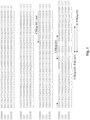
- human TK1 The crystal structure of human TK1 has been resolved and the major part of the enzyme, including the N-terminal region, is conserved in humans, dogs, cats and horses except for a few residues, see Fig. 1 and SEQ ID NO: 3 representing dog TK1 (canine TK1), SEQ ID NO: 4 representing human TK1, SEQ ID NO: 9 representing cat TK1 (feline TK1) and SEQ ID NO: 10 representing horse TK1 (equine TK1).
- human TK1 sequence shows 88.5 % similarity with dog TK1 but a significant sequence diversity (9.1 %) is found in the C-terminal region.
- the kit comprises a first antibody immobilized to a support or intended to be immobilized to the support and a second antibody.
- One of the first antibody and the second antibody has specificity for a peptide consisting of an amino acid sequence from a C-terminal region of non-human mammal TK1.
- the other of the first antibody and second antibody has specificity for a peptide consisting of a first amino acid sequence from an active site of TK1.
- the non-human mammal TK1 is selected from a group consisting of canine TK1 protein, feline TK1 protein and equine TK1 protein.
- the kit is in particular suitable for determining the level of non-human mammal serum TK1 (STK1) protein in sample, preferably a body sample from a non-human mammal subject.
- STK1 non-human mammal serum TK1
- the peptide consisting of a first amino acid sequence or a second amino acid sequence from the C-terminal regional is preferably a peptide selected from a portion of the non-human mammal TK1 ranging from amino acid position 200 to the end of the non-human mammal TK1 (amino acid position 242 in dog, amino acid position 237 in cat and amino acid position 237 in horse).
- the peptide is selected from a portion of the TK1 protein ranging from amino acid position 205, preferably 210 and more preferably 211, to amino acid position 240, preferably 235 and more preferably 230.
- the peptide is preferably an N-mer, wherein N is an integer within a range of 10 and 25, preferably 10 to 20 and more preferably 15, 16 or 17.
- the peptide preferably consists of N consecutive amino acids in the C-terminal region of the non-human mammal TK1 protein.
- At least one additional amino acid such as a cysteine residue, may be added to the N-terminal or C-terminal, preferably the N-terminal, of the peptide for use as coupling to other molecules, such as carrier proteins.
- the peptide consisting of a first amino acid sequence or a second amino acid sequence from the C-terminal region has an amino acid sequence corresponding to amino acid positions 215 to 230 in canine TK1, see Fig. 1 , i.e., has amino acid sequence of GKPGEGKEATGVRKLF (SEQ ID NO: 1).
- the corresponding amino acid sequence with the added N-terminal cysteine residue is CGKPGEGKEATGVRKLF (SEQ ID NO: 5).
- the peptide consisting of a first amino acid sequence or a second amino acid sequence from the C-terminal region has an amino acid sequence corresponding to amino acid positions 211 to 225 in canine TK1, see Fig. 1 , i.e., has amino acid sequence of VLVPGKPGEGKEATG (SEQ ID NO: 11).
- the corresponding amino acid sequence with the added N-terminal cysteine residue is CVLVPGKPGEGKEATG (SEQ ID NO: 12).
- the peptide consisting of a first amino acid sequence or a second amino acid sequence from the C-terminal region has an amino acid sequence corresponding to amino acid positions 213 to 227 in feline TK1, see Fig. 1 , i.e., has amino acid sequence of GKPGEASGARKLFAP (SEQ ID NO: 13).
- the corresponding amino acid sequence with the added N-terminal cysteine residue is CGKPGEASGARKLFAP (SEQ ID NO: 14).
- the peptide consisting of an amino acid sequence from an active site of TK1 is preferably a peptide selected from a portion of TK1 ranging from amino acid position 150 to amino acid position 190.
- the peptide is selected from a portion of TK1 ranging from amino acid position 155, preferably 160 and more preferably 161, to amino acid position 185, preferably 183.
- the peptide is preferably an M-mer, wherein M is an integer within a range of 10 and 40, preferably 20 to 30 and more preferably 23 or 24.
- the peptide preferably consists of M consecutive amino acids in the active site of the TK1 protein.
- This portion representing the active site of TK1 shows very high levels of homology between different species, such as between human, canine, feline and equine TK1 as shown in Fig. 1 .
- a single amino acid position between human TK1 and canine TK1 and feline TK1 within the portion extending from amino acid position 150 to amino acid position 190.
- the peptide could consist of an amino acid sequence from an active site of human TK1, from canine TK1, from feline TK1, from equine TK1 or from another non-human mammal TK1.
- Experimental data as presented herein shows that an antibody that has specificity for a peptide consisting of an amino acid sequence from the active site of human TK1 binds specifically also to canine TK1.
- At least one additional amino acid such as a cysteine residue, may be added to the N-terminal or C-terminal, preferably the N-terminal, of the peptide for use as coupling to other molecules, such as carrier proteins.
- the peptide consisting of an amino acid sequence from the active site of TK1 has an amino acid sequence corresponding to amino acid positions 161 to 183 in human TK1 (and equine TK1), see Fig. 1 , i.e., has amino acid sequence of AYTKRLGTEKEVEVIGGADKYHS (SEQ ID NO: 2).
- the corresponding amino acid sequence with the added N-terminal cysteine residue is CAYTKRLGTEKEVEVIGGADKYHS (SEQ ID NO: 6).
- the peptide consisting of an amino acid sequence from the active site of TK1 has an amino acid sequence corresponding to amino acid positions 161 to 183 in canine TK1, see Fig. 1 , i.e., has amino acid sequence of AYTKRLGSEKEVEVIGGADKYHS (SEQ ID NO: 7).
- the corresponding amino acid sequence with the added N-terminal cysteine residue is CAYTKRLGSEKEVEVIGGADKYHS (SEQ ID NO: 8).
- the peptide consisting of an amino acid sequence from the active site of TK1 has an amino acid sequence corresponding to amino acid positions 161 to 183 in feline TK1, see Fig. 1 , i.e., has amino acid sequence of AYTKRLGAEKEVEVIGGADKYHS (SEQ ID NO: 15).
- the corresponding amino acid sequence with the added N-terminal cysteine residue is CAYTKRLGAEKEVEVIGGADKYHS (SEQ ID NO: 16).
- a kit for determining the level of non-human mammal TK1 protein in a sample comprises the first antibody immobilized to a support or intended to be immobilized to the support and the second antibody.
- one of the first and the second antibody has specificity for a peptide consisting of an amino acid sequence from the active site of TK1 and the other of the first antibody and the second antibody has specificity for a peptide consisting of an amino acid sequence from the C-terminal region of TK1.
- one of the first antibody and the second antibody has specificity for a peptide consisting of an amino acid sequence selected from a group consisting of SEQ ID NO: 11 and SEQ ID NO: 12 and the other of the first antibody and the second antibody has specificity for a peptide consisting of an amino acid sequence selected from a group consisting of SEQ ID NO: 2, SEQ ID NO: 6, SEQ ID NO: 7 and SEQ ID NO: 8.
- the combination of the first and second antibodies according to the invention thus, involves an antibody having specificity for a peptide consisting of an amino acid sequence consisting of SEQ ID NO: 11 and an antibody having specificity for a peptide consisting of an amino acid sequence consisting of SEQ ID NO: 2.
- first and second antibodies involve, an antibody having specificity for a peptide consisting of an amino acid sequence consisting of SEQ ID NO: 11 and an antibody having specificity for a peptide consisting of an amino acid sequence consisting of SEQ ID NO: 6, an antibody having specificity for a peptide consisting of an amino acid sequence consisting of SEQ ID NO: 11 and an antibody having specificity for a peptide consisting of an amino acid sequence consisting of SEQ ID NO: 7, an antibody having specificity for a peptide consisting of an amino acid sequence consisting of SEQ ID NO: 11 and an antibody having specificity for a peptide consisting of an amino acid sequence consisting of SEQ ID NO: 8, an antibody having specificity for a peptide consisting of an amino acid sequence consisting of SEQ ID NO: 12 and an antibody having specificity for a peptide consisting of an amino acid sequence consisting of SEQ ID NO: 2, an antibody having specificity for a peptide consisting of an amino acid sequence
- one of the first antibody and the second antibody is the CTK1p-211 antibody and the other of the first antibody and the second antibody is the CTK1p-161 antibody.
- one of the first antibody and the second antibody is the CTK1p-211 antibody and the other of the first antibody and the second antibody is the Arv4 antibody.
- one of the first antibody and the second antibody has specificity for a peptide consisting of an amino acid sequence selected from a group consisting of SEQ ID NO: 1 and SEQ ID NO: 5 and the other of the first antibody and the second antibody has specificity for a peptide consisting of an amino acid sequence selected from a group consisting of SEQ ID NO: 2, SEQ ID NO: 6, SEQ ID NO: 7 and SEQ ID NO: 8.
- first and second antibodies involve an antibody having specificity for a peptide consisting of an amino acid sequence consisting of SEQ ID NO: 1 and an antibody having specificity for a peptide consisting of an amino acid sequence consisting of SEQ ID NO: 2, an antibody having specificity for a peptide consisting of an amino acid sequence consisting of SEQ ID NO: 1 and an antibody having specificity for a peptide consisting of an amino acid sequence consisting of SEQ ID NO: 6, an antibody having specificity for a peptide consisting of an amino acid sequence consisting of SEQ ID NO: 1 and an antibody having specificity for a peptide consisting of an amino acid sequence consisting of SEQ ID NO: 7, an antibody having specificity for a peptide consisting of an amino acid sequence consisting of SEQ ID NO: 1 and an antibody having specificity for a peptide consisting of an amino acid sequence consisting of SEQ ID NO: 8, an antibody having specificity for a peptide consisting of a peptide consisting of an amino acid sequence consisting of
- one of the first antibody and the second antibody is the CTK1p-215 antibody and the other of the first antibody and the second antibody is the CTK1p-161 antibody.
- one of the first antibody and the second antibody is the CTK1p-215 antibody and the other of the first antibody and the second antibody is the Arv4 antibody.
- one of the first antibody and the second antibody is the PAb-Arv1 antibody and the other of the first antibody and the second antibody is the CTK1p-161 antibody.
- one of the first antibody and the second antibody is the PAb-Arv1 antibody and the other of the first antibody and the second antibody is the Arv4 antibody.
- the kit for determining the level of non-human mammal TK1 protein in a sample comprises the first antibody immobilized to a support or intended to be immobilized to the support and the second antibody.
- one of the first and the second antibody has specificity for a peptide consisting of a first amino acid sequence from the C-terminal region of TK1 and the other of the first antibody and the second antibody has specificity for a peptide consisting of a second amino acid sequence from the C-terminal region of TK1.
- the first and second amino acid sequences are different amino acid sequences from the C-terminal region of TK1.
- the two amino acid sequences may, however, be partially overlapping as shown in Fig. 1 when comparing, for instance, the amino acid sequence for CTK1p-211 with the amino acid sequence for CTK1p-215.
- the two amino acid sequences could be overlapping with P amino acids.
- P is equal to or larger than 0 but smaller than the largest of N 1 and N 2 , i.e., 0 ⁇ P ⁇ max( N 1 , N 2 ).
- TK1 is polyvalent and has several antibody binding sites both on recombinant TK1 and serum TK1.
- TK1 is polyvalent and has several antibody binding sites both on recombinant TK1 and serum TK1.
- one of the first antibody and the second antibody has specificity for a peptide consisting of an amino acid sequence selected from a group consisting of SEQ ID NO: 11 and SEQ ID NO: 12 and the other of the first antibody and the second antibody has specificity for a peptide consisting of an amino acid sequence selected from a group consisting of SEQ ID NO: 1 and SEQ ID NO: 5.
- first and second antibodies involve an antibody having specificity for a peptide consisting of an amino acid sequence consisting of SEQ ID NO: 11 and an antibody having specificity for a peptide consisting of an amino acid sequence consisting of SEQ ID NO: 1, an antibody having specificity for a peptide consisting of an amino acid sequence consisting of SEQ ID NO: 11 and an antibody having specificity for a peptide consisting of an amino acid sequence consisting of SEQ ID NO: 5, an antibody having specificity for a peptide consisting of an amino acid sequence consisting of SEQ ID NO: 12 and an antibody having specificity for a peptide consisting of an amino acid sequence consisting of SEQ ID NO: 1 and an antibody having specificity for a peptide consisting of an amino acid sequence consisting of SEQ ID NO: 12 and an antibody having specificity for a peptide consisting of an amino acid sequence consisting of SEQ ID NO: 5.
- one of the first antibody and the second antibody is the CTK1p-211 antibody and the other of the first antibody and the second antibody is the CTK1p-215 antibody.
- one of the first antibody and the second antibody is the CTK1p-211 antibody and the other of the first antibody and the second antibody is the PAb-Arv1 antibody.
- the antibodies may be polyclonal antibodies, monoclonal antibodies or one them may be a polyclonal antibody with the other as a monoclonal antibody.
- the antibody that has specificity for the peptide consisting of an amino acid sequence from the C-terminal region of non-human mammal TK1 may be a polyclonal antibody and the antibody that has specificity for the peptide consisting of an amino acid sequence from the active site of TK1 may be a monoclonal antibody.
- One or both of the antibodies may be an antibody fragment having specificity for the relevant peptide.
- the fragment can be selected from a group consisting of a single chain antibody, a Fv fragment, a scFv fragment, a Fab fragment, a F(ab')2 fragment, a Fab' fragment, a Fd fragment, a singledomain antibody (sdAb), a scFv-Fc fragment, a di-scFv fragment and a CDR region.
- the specificity of an antibody can be determined based on affinity and/or avidity.
- the affinity represented by the equilibrium constant for the dissociation of an antigen with the antibody ( K d ), is a measure for the binding strength between an antigenic determinant and an antigen-binding site on the antibody. The lesser the value of K d , the stronger the binding strength between the antigenic determinant and the antibody.
- the affinity can also be expressed as the affinity constant ( K a ), which is 1/ K d .
- affinity can be determined in a manner known per se, depending on the specific antigen of interest.
- Avidity is the measure of the strength of binding between an antibody and the pertinent antigen. Avidity is related to both the affinity between an antigenic determinant and its antigen binding site on the antibody and the number of pertinent binding sites present on the monoclonal antibody.
- antibodies will bind to their antigen with a dissociation constant ( K d ) of 10 -5 to 10 -12 moles/liter (M) or less, and preferably 10 -7 to 10 -12 M or less and more preferably 10 -8 to 10 -12 M, i.e. with an association constant ( K a ) of 10 5 to 10 -12 M -1 or more, and preferably 10 7 to 10 -12 M -1 or more and more preferably 10 8 to 10 -12 M -1 .
- K d dissociation constant
- M moles/liter
- K a association constant
- any K d value greater than 10 -4 M (or any K a value lower than 10 4 M -1 ) is generally considered to indicate non-specific binding.
- an antibody or fragment will bind to the serum form and/or recombinant form of non-human mammal TK1 with an affinity less than 500 nM, preferably less than 200 nM, more preferably less than 10 nM, such as less than 5 nM.
- the kit is a sandwich assay kit. This means that the kit uses antibodies binding to different epitopes of non-human mammal TK1 protein so that both the first and second antibodies can simultaneously bind to the same non-human mammal TK1 molecule or complex.
- the kit is an Enzyme-Linked Immunosorbent Assay (ELISA) kit and preferably a sandwich ELISA.
- ELISA Enzyme-Linked Immunosorbent Assay
- a sandwich ELISA can be used to detect cellular and/or serum TK1 protein in a sample by preparing a surface of a support, such as a solid support, to which the first antibody is bound as so-called capture antibody.
- a known quantity of the first antibody is bound to the surface of the support. Any nonspecific binding sites on the surface are optionally but preferably blocked.
- the sample is then applied to the surface so that any non-human mammal TK1 protein present therein will be captured by the immobilized first antibodies. Unbound material is preferably removed by one or multiple washing steps.
- the second antibody typically denoted detection antibody, is then added and is allowed to bind to any non-human mammal TK1 protein captured by the first antibody.
- the amount of bound second antibody is then determined by direct or indirect detection methods.
- a label or enzyme can be attached directly to the second antibody or indirectly via a link, such as a biotin-streptavidin or a biotin-avidin link. It is, alternatively, possible to use a secondary antibody that is labeled or connected to an enzyme and binds specifically to the second antibody.
- the second antibody has a covalently attached biotin.
- the second antibody has a covalently attached streptavidin or avidin.
- the kit preferably also comprises a horseradish peroxidase (HRP) labeled streptavidin or a HRP labeled avidin.
- HRP horseradish peroxidase
- the kit also comprises a HRP labeled biotin.
- the kit also comprises a HRP substrate, such as a 3,3',5,5'-tetramethylbenzidine (TMB) substrate, a 3,3'-diaminobenzidine (DAB) substrate or a 2,2'-azino-bis(3-ethylbenzothiazoline-6-sulphonic acid (ABTS) substrate.
- TMB 3,3',5,5'-tetramethylbenzidine
- DABTS 2,2'-azino-bis(3-ethylbenzothiazoline-6-sulphonic acid
- the level of non-human mammal TK1 protein in the sample can be determined by spectrophotometric methods that detect the conversion of the chromogenic substrate by HRP into a colored
- the kit also comprises a microtiter plate (MCP) as the support to which the first antibody is immobilized or is intended to be immobilized.
- MCP microtiter plate
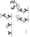
- Fig. 2 is a schematic overview of the concept of a sandwich ELISA using the first and second antibodies according to an embodiment.
- the kit does not necessarily have to be an ELISA kit.
- the kit uses affinity chromatography where the first antibody is bound to the stationary phase, such as to a gel matrix or beads in a column.
- the gel matrix or beads could be made of agarose, such as SEPHAROSE ® .
- non-human mammal TK1 protein present in a sample will be entrapped in the column through binding to the immobilized first antibodies.
- the bound non-human mammal TK1 protein can be eluted and detected using the second antibody.
- the amount of eluted non-human mammal TK1 protein can be determined using Western blotting and with the second antibody for TK1 detection using direct or indirect detection methods.
- the support could alternatively be magnetic beads, such as DYNABEADS ® magnetic beads.
- the non-human mammal TK1 protein determined can be non-human mammal cellular and/or serum TK1 protein, preferably non-human mammal serum TK1 (STK1) protein or molecules.
- STK1 non-human mammal serum TK1
- the non-human mammal TK1 protein is selected from a group consisting of canine TK1 protein, feline TK1 protein and equine TK1 protein.
- the TK1 protein can be from dogs, cats or horses.
- the non-human mammal TK1 protein is canine TK1 protein.
- the kit of the present embodiments can be used in an in vitro method for determining the level of non-human mammal TK1 protein in a sample.
- Another aspect relates to an in vitro method for determining a level of canine, feline or equine TK1 protein in a sample.
- the method comprises contacting the sample with a first antibody and a second antibody of the kit according to the embodiments.
- the method also comprises detecting an amount of bound second antibody.
- the level of canine, feline or equine TK1 protein in the sample is then determined based on the detected amount of bound second antibody.
- the detection can be direct or indicted, and may generate a fluorescent or chromogenic signal.
- Direct detection typically involves the use of an antibody that is conjugated to a label.
- Indirect detection utilizes a labeled secondary antibody raised against the host species of the antibody.
- Commonly used labels for visualization of binding of antibody to epitope includes fluorophores and enzymes that convert soluble substrates into chromogenic end products.
- the sample is a body sample and is more preferably selected from a group consisting of a cell sample, a tissue sample, a blood sample, a serum sample, a cerebrospinal fluid sample, a pleural fluid sample, a synovial fluid sample and a peritoneal cavity fluid sample.
- the body sample is a body fluid sample and preferably selected from a group consisting of a blood sample, a serum sample, a cerebrospinal fluid sample, a pleural fluid sample, a synovial fluid sample and a peritoneal cavity fluid sample.
- the body (fluid) sample is a blood sample or a serum sample.
- the antibodies can be used to determine a level, i.e., an amount, of non-human mammal TK1 protein in a sample.
- the antibodies are believed to be able to bind to and thereby enable determination of the level of non-human mammal cellular or serum TK1 in its various forms, such as dimers, tetramers, and oligomers, including potentially bound to further proteins, cofactors or molecules.
- non-human mammal cellular and/or serum TK1 protein thereby includes the non-human mammal cellular and/or serum TK1 in its various forms.
- the method may be a method for determining a level of non-human mammal STK1 protein in a body sample.
- the method may be a method for determining a level of non-human mammal cellular TK1 protein in a body sample.
- the method may be a method for determining a level of non-human mammal cellular TK1 protein and non-human mammal STK1 protein in a body sample.
- a further aspect of the embodiments relates to an in vitro method for estimating the likelihood of recurrence of a tumor disease in a canine, feline or equine subject, i.e., a dog, a cat or a horse according to the claims.
- the method comprises determining a level of canine, feline or equine TK1 protein in a body sample from the canine, feline or equine subject using the in vitro a method or the kit according to the embodiments.
- the level of canine, feline or equine TK1 protein in the body sample is then compared with a level of canine, feline or equine TK1 protein representative of a population of healthy canine, feline or equine subjects or with a level of canine, feline or equine TK1 protein previously determined in the canine, feline or equine subject.
- the method further comprises estimating the likelihood of recurrence of the tumor disease in the non-human mammal subject based on the comparison.
- a determined level that is higher than a level associated with a population of healthy non-human mammals indicates an increased likelihood of recurrence of a tumor disease in the non-human mammal subject.
- a determined level that is higher than a level associated with the non-human mammal subject subsequent to previous therapy indicates an increased likelihood of recurrence of a tumor disease in the non-human mammal subject.
- Yet another aspect of the embodiments relates to a method for determining cell proliferation in a canine, feline or equine subject according to the claims.
- the method comprises determining a level of canine, feline or equine TK1 protein in a body sample from the canine, feline or equine subject using the in vitro a method or the kit according to the embodiments.
- the method also comprises determining the cell proliferation based on the level of canine, feline or equine TK1 protein in the body sample.
- a level of normal or tumor cell proliferation is determined and compared with the determined level of non-human mammal TK1 protein to determine whether the non-human mammal subject has normal or baseline cell proliferation or an elevated cell proliferation.
- the present method can be used as a tool in monitoring various therapies applied to non-human mammal subjects.
- the method can be used to monitor anti-proliferation or anti-tumor therapy in the non-human mammal subject.
- the method can be used to verify whether a selected anti-proliferation or anti-tumor therapy has the desired effect in reducing cell proliferation in the non-human mammal subject. If the therapy does not have the desired effect, i.e., no significant decrease in cell proliferation is detected, then another or a modified anti-proliferation or anti-tumor therapy can be applied to the non-human mammal subject.
- a further aspect of the embodiments relates to a method for determining a proliferation process response in a canine, feline or equine subject suffering from a malignant disease according to the claims.
- the method comprises determining a level of canine, feline or equine TK1 protein in a body sample from the canine, feline or equine subject using the in vitro a method or the kit according to the embodiments.
- the method also comprises determining the proliferation process response based on the level of canine, feline or equine TK1 protein in the body sample.
- An example of such a proliferation process response could be an immune reaction or immune reaction response.
- At least one other biomarker for the proliferation process response may also be usec in the determination.
- Yet another aspect of the embodiments relates to a method for determining a level of inflammation, infection or tumor cell proliferation in a canine, feline or equine subject according to the claims.
- the method comprises determining a level of canine, feline or equine TK1 protein in a body sample from the canine, feline or equine subject using the in vitro a method or the kit according to the embodiments.
- the method also comprises determining the level of inflammation, infection or tumor cell proliferation based on the level of canine, feline or equine TK1 protein in the body sample.
- at least one other biomarker for inflammation, infector or tumor cell proliferation may also be used in the determination.
- a further aspect of the embodiments relates to a method for evaluating efficiency of a treatment of a malignant disease in a canine, feline or equine subject according to the claims.
- the method comprises determining a level of canine, feline or equine TK1 protein in a body sample from the canine, feline or equine subject using the in vitro a method or the kit according to the embodiments prior to or in connection with start of the treatment of the malignant disease.
- the method also comprises determining a level of canine, feline or equine TK1 protein in a body sample from the canine, feline or equine subject using the method or kit according to the embodiments during or after the treatment of the malignant disease.
- the method further comprises evaluating efficiency of the treatment of the malignant disease based on a comparison of the level of canine, feline or equine TK1 protein determined in the body sample prior to or in connection with start of the treatment of the malignant disease and the level of canine, feline or equine TK1 protein determined in the body sample during or after the treatment of the malignant disease.
- the in vitro method and kit can be used to evaluate or determine efficiency of a treatment of a malignant disease in dogs, cats or horses.
- a reduction in TK1 protein level in a treated non-human mammal is determined to correlate with a treatment that has effect with regard to the malignant disease, i.e., an efficient treatment.
- the particular treatment is not efficient and does not have the desired effect with regard to the malignant disease.
- the tumor is preferably a hematological tumor, such as lymphoma or leukemia, or a solid tumor, such as mammary tumor, histiocytic sarcoma, mastocytoma, melanoma, hemangiosarcoma or adenocarcinoma.
- Kiran Kumar 2010 disclosed production of anti-dog TK1 antibodies produced by immunizing rabbits with a 28 amino acid long peptide corresponding to amino acids 196 to 223 in dog TK1.
- the anti-dog TK1 antibodies however, showed poor performance when used in an ELISA assay and had high background. Production of polyclonal antibodies against the long (28 amino acids) peptide showed large batch-to-batch variation. This is in clear contrast to present embodiments and the experimental data presented herein.
- the antibodies of the present embodiments can successfully be used in an ELISA assay with high specificity and capable of discriminating between healthy subject and subjects suffering from hematological or solid tumors.
- TK1-ELISA for determination of serum TK1 (STK1) protein levels in dogs with hematological and solid tumors was developed. Both STK1 activity and STK1 protein levels were measured in sera from healthy dogs, dogs with leukemia and lymphomas, and solid tumors. Furthermore, serum samples from six dogs with lymphoma were followed during therapy to test the monitoring efficiency of both these TK1 assays.
- Serum samples from healthy dogs, dogs with hematological tumors and solid tumors were collected from the University Animal Hospital at the Swedish University of Agricultural Sciences, Uppsala, Sweden, and stored at -20°C until analysis. The project was approved by the Swedish Animal Ethics Committee.
- the mean and median age was 6 years (range 3-10 years) for the healthy group, 8.5 and 8 years (range 3-13 years) for hematological tumors, and 9 and 8 years (range 3-14 years) for dogs with solid tumors.
- Polyclonal antibodies were produced against a peptide in the C-terminal region of dog TK1.
- the 16-mer antibody was produced using a 16 amino acid synthetic peptide (amino acids 215-230, GKPGEGKEATGVRKLF, SEQ ID NO: 1; PAb-Arv1) ( Fig. 1 ) with a cysteine added to the N-terminus.
- This 16-mer peptide was coupled to keyhole limpet hemocyanin (KLH) and mixed with Freund's complete adjuvant as antigen for immunization of rabbits (GenScript, Piscataway, NJ, USA).
- the antisera were collected after the 3 rd and 4 th immunizations and purified on peptide coupled Sepharose 4B columns as previously described (Wu et al., 2003).
- the 24-mer antibody was produced as previously described (Gasparri et al., 2009) against the long lasso shaped loop in the active site of human TK1 (amino acids 161-183, AYTKRLGTEKEVEVIGGADKYHS, SEQ ID NO: 2; Ar-4) with a cysteine added to the N-terminus.
- a 24-mer peptide was also coupled to KLH and mice were immunized and monoclonal antibodies were prepared as previously described (Gasparri et al., 2009).
- the Ar-4 antibodies were provided by AroCell AB, Uppsala, Sweden.
- the Ar-4 antibodies were biotinylated using the ChromaLink TM Biotin Antibody Labeling Kit (Solulink, California, USA) according to manufacturer instructions.
- the biotinylated Ar-4 antibodies were analyzed by Western blotting using streptavidin-HRP.
- TK1 activity in serum samples was determined using the optimized [ 3 H]-dThd phosphorylation assay, as described previously (Sharif et al., 2012 a; Kiran Kumar et al., 2013).
- 10 ⁇ L of serum was incubated with the reaction mixture containing 10 mM Tris-HCl pH 7.6, 2 mM dithiothreitol (DTT), 5 mM MgCl 2 , 5 mM NaF, 5 mM ATP, and 5 ⁇ M [ 3 H]-deoxy thymidine for 1 h at 37°C and applied to the DE-81 filter paper discs.
- the filters were then washed twice with 1 mM ammonium formate for 5 min/each and the bound products were eluted for 45 min in 0.1 M HCl and 0.2 M KCI.
- the radioactivity was determined by ⁇ -scintillation counting as previously described (Sharif et al., 2012 a).
- the TK1 activity was expressed as pmol/min/mL.
- ELISA plates of the type Maxisorp, NUNC (Denmark) were coated with 100 ⁇ L of the PAb-Arv1 anti-TK1 polyclonal antibody (4 ⁇ g/mL) in carbonate buffer (pH 9.6) and incubated overnight at 4°C. Plates were washed, using a Tecan Hydro flex microplate washer, four times with wash buffer (Tris 0.1 M, pH 7.4, NaCl 0.3 M, Tween 0.05 % and BSA 1 %). Thereafter, all wells were blocked with 5 % nonfat dry milk powder diluted in TBST for 1 h at room temperature.
- Dog serum samples of 50 ⁇ L were diluted in 50 ⁇ L serum dilution buffer (AroCell AB, Uppsala) and pre-incubated at room temperature for 1 h. Wells were again washed and pre-incubated serum samples were added. Then plates were incubated at room temperature for 2 h on a rocking platform. Plates were washed as described above and the biotinlayted Ar-4 antibody (4 ⁇ g/mL in wash buffer) was added. After incubation at room temperature for 1 h, the plates were washed as above and horse radish peroxidase (HRP) diluted 1:32,000 in wash buffer was added, and incubated for 1 h.
- HRP horse radish peroxidase
- TMB Thermo Fisher Scientific
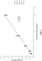
- Dog recombinant TK1 was purified as described previously (Sharif et al., 2012 b) and a standard curve was established with different concentrations of recombinant dog TK1 (0.6 - 10 ng /ml diluted in sample dilution buffer). By using the standard curve, based on average absorbance of different serum samples, the concentrations of TK1 in the serum samples were calculated. The detection limit was estimated as the minimum analyte concentration giving a value significantly different from that of the zero calibrator. The inter-assay variation was also determined from the mean and SD of all serum samples which were independently assayed as duplicates in two different experiments.
- TK1 activity and TK1 protein distribution in healthy, hematological tumors and solid tumors were tested for normality using the D'Agostino and Pearson omnibus normality test.
- STK1 activity and protein levels in healthy dogs as well as dogs with hematological and solid tumors showed non-Gaussian distribution.
- Spermann correlation coefficient (rs) was used to determine the correlation between TK1 activities and TK1 protein levels.
- Mann Whitney t-test was used to evaluate the difference between the groups.
- receiver operating characteristic (ROC) curves were constructed.
- Statistical analyses were performed using Graph Pad Prism 5.0 (Graph Pad Software, La Jolla, CA, USA). The level of significance was set at P ⁇ 0.05.
- the ELISA principle is based on a sandwich immunoenzymatic system as shown in Fig. 2 .
- the first step is a coating of micro titer plates with purified polyclonal anti dog TK1 specific antibody produced against a peptide in the C-terminal region of dog TK1 (PAb-Arv1, Fig. 1 ). This part of TK1 is an exposed region of the TK1 protein complexes formed in the blood. After blocking the wells with milk powder (5 %), pre-incubated serum samples from healthy and tumor dogs were allowed to bind to the antibodies on the plates. Proteins not specifically bound are removed by the washing procedure.
- the second antibody Ar-4 made against a highly conserved and exposed active site region of TK1 were biotinylated and allowed to react with the TK1 bound to the wells.
- the detection of antigen-antibody bound complex by a streptavidin-peroxidase (HRP) complex which is visualized by the addition of a chromogenic substrate (TMB).
- the intensity of the color reaction is proportional to the quantity of TK1 present in the serum samples.
- Dog recombinant TK1 was used as a calibrator to generate the dose-response curve of the TK1- ELISA.
- a typical calibration curve from 5 different runs is shown in Fig. 3 .
- Intra assay variation at all non-zero calibration points CVs were ⁇ 10 %.
- the limit of detection (LOD) of the assay was 0.46 ng/mL and between-run imprecision (CV) was ⁇ 20 % at concentrations down to 0.63 ng/mL.
- LOD limit of detection
- CV was ⁇ 20 % at concentrations down to 0.63 ng/mL.
- the inter-assay variation ranged from 5-15 % and intra assay variation was 5 %.
- Table 1 The median serum TK1 activities and TK1 protein levels from the healthy dogs, dogs with hematological malignancies and dogs with solid tumors are summarized in Table 1 below.
- Table 1 - TK1 activity and protein levels in sera from healthy dogs, dogs with hematological malignancies and dogs with solid tumors No of samples STK1 activity (pmol/min/mL) STK1-ELISA (ng/mL) Group (N) Range Median Range Median Healthy 30 0.7 - 1.4 1.02 ⁇ 0.46 - 0.47 ⁇ 0.46 Lymphoma 31 0.54 -29.8 2.43 ⁇ 0.46 - 4.38 0.78 Leukemia 5 1.33 - 58.7 38.3 ⁇ 0.46 - 4.19 3.92 Mammary tumors 16 0.62 - 1.83 1.07 ⁇ 0.46 -1.51 0.50 Mastocytoma 8 0.51 - 1.52 0.85 ⁇ 0.46 - 1.1 0.50 Malignant melanoma 11 0.52 - 1.52 1.15 ⁇ 0.46
- STK1 activities were in the range of 0.7 - 1.4 pmol/min/mL (Table 1) with a median of 1.02 pmol/min/mL.
- the STK1 activity levels in sera from dogs with hematological tumors ranged from 0.5 - 59 pmol/min/mL (Table 1).
- STK1 activity levels between males and females In healthy dogs as well as in dogs with hematological tumors, no significant difference was found in STK1 activity levels between males and females.
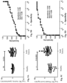
- Statistical analysis using the Mann-Whitney U test showed that STK1 activity was significantly higher in the hematologic tumors group compared to in healthy dogs (P ⁇ 0.0001, Fig. 4A ).
- the ROC curve analysis of STK1 activity assay results showed an area under curve (AUC) of 0.83 (P ⁇ 0.0001, 95 % Cl, 0.72-0.94) ( Fig. 4B ).
- AUC area under curve
- the sensitivity was 75 % (95 % Cl, 0.578-0.878) and the specificity 96 % (95 % Cl, 0.82-0.99).
- TK1 protein levels in clinical samples were determined by using different concentrations of recombinant dog TK1 as standard.
- STK1 protein levels were ⁇ 0.46 ng/mL and 1 out of 30 sera had protein value of 0.47 ng/mL.
- the estimated upper limit of the normal reference based on the 30 healthy dogs was 0.46 ng/mL.
- Sera from dogs with lymphoma or leukemia had in general higher STK1 protein levels, ranging from ⁇ 0.46 to 4.4 ng/mL.
- Significant differences were found in the median STK1 protein levels between healthy and hematological tumors (P ⁇ 0.0001, Fig. 4C ).
- the ROC curve analysis showed an AUC of 0.94 (P ⁇ 0.0001, 95 % Cl, 0.89-0.99).
- the true positive rate was 78 % (95 % Cl, 0.60-0.898) and the false positive rate was 4 % (95 % Cl, 0.82-0.99), using a cutoff value of 0.46 ng/mL (
- TK1 protein levels were significantly higher in sera from dogs with solid tumors (range of ⁇ 0.46 to 3.4 ng/mL) compared to healthy (P ⁇ 0.0001, Fig. 5C ).
- ROC curve analysis had an AUC of 0.88 (P ⁇ 0.0001, 95 % Cl, 0.80-0.95) ( Fig. 5D ).
- the sensitivity was 62 % (95 % Cl, 0.458-0.772) and the specificity 96 % (95 % Cl, 0.82-0.99).
- Sera from dogs with hematological and solid tumors were further sub-classified as lymphomas, leukemias and mammary tumors, mastocytomas and melanomas.
- Figs. 6A and 6B illustrate the log STK1 activity distribution in the different sub-classes
- Figs. 6C and 6D illustrate the log STK1 protein levels in the different sub-classes.
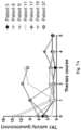
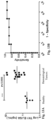
- Dogs with lymphoma are often treated using a doxorubicin based multi-agent protocol (ADRIA-Plus). Serum samples were collected from six dogs before and after each dose of ADRIA-plus. Initially, high STK1 activity and STK1 protein values were found in five dogs and four out of the six dogs showed significant decline in STK1 activity and STK1 protein levels after the first treatment, leading to levels similar or lower than the cut-off value ( Figs. 7A , 7B ). In two patients (no. 8, 9) there was an increase in STK1 activity and STK1 protein during 3 rd treatment but the levels decreased after the 4 th treatment. In two patients (patient no.
- STK1 activity and STK1 protein levels increased after the 1 st and decreased after 2 nd treatment but increased again after the 3 rd and 4 th treatment followed by a decline after the 5 th treatment.
- One patient (patient no. 19) was apparently in complete remission after the 2 nd treatment.
- Mean STK1 activity and STK1 protein in dogs with lymphoma that were in complete remission were not significantly different from the STK1 in healthy controls.
- two patients (patient no. 11, 37) showed a different pattern of response to chemotherapy.
- Patient no. 11 had significant reduction in STK1 activity and STK1 protein levels after 1 st treatment.
- TK1-ELISA was developed as disclosed herein, which can overcome the limitations of traditional TK1 radioisotope assays.
- ROC curve analysis for hematological tumors demonstrated that both the TK1-ELISA and TK1 activity assays have a sensitivity of 75 % and a false positive rate of 4 %. This indicates that both assays may be accurate and sensitive enough for clinical routine practice.
- TK1 activity assay could not distinguish healthy from solid tumor patients, which were demonstrated by the ROC curve with a much lower sensitivity (27 %).
- the TK1-ELISA could differentiate these groups with two fold higher sensitivity (62 %) than the TK1 activity assay.
- TK1 protein levels return to normal (baseline level) after chemotherapy leading to tumor remission and increased to higher levels in relapsed patients, similar to TK1 activity levels in dogs with lymphoma.
- a transient increase in TK1 protein levels were found in a few patients during therapy, which could be due to release of TK1 from cells that died because of drug toxicity.
- the present study describes a new TK1-ELISA for determining TK1 protein levels, which can be a potential marker for diagnosis and monitoring of canine hematological tumors.
- the TK1-ELISA is easy to perform, fast and as sensitive and specific as the existing TK1 activity assays.
- CTK1p-211, CTK1p-215 Two different polyclonal rabbit antisera were produced using peptides from the C-terminal region of dog TK1 (CTK1p-211, CTK1p-215) and one antisera produced from the active site region of dog TK1 (CTK1p-161).
- the first canine TK1 peptide sequence (CTK1p-215) antiserum was against a 16 amino acid synthetic peptide (amino acids 215-230 in dog TK1; Fig. 1 ) to which a N-terminal cysteine was added (CGKPGEGKEATGVRKLF, SEQ ID NO: 5).
- the second antiserum (CTK1p-211) was produced using a 15 amino acid peptide (amino acids 211-225 in dog TK1; Fig.
- CTK1p-161 antisera was produced using a highly conserved active site sequence of dog TK1 (amino acids 161-183 in dog TK1; Fig. 1 ) to which a N-terminal cysteine was added (CAYTKRLGTEKEVEVIGGADKYHS, SEQ ID NO: 8).
- FTK1p-213 antisera was produced against the C-terminal region of cat TK1 (amino acids 213-227 in cat TK1; Fig. 1 ) to which a N-terminal cysteine was added (CGKPGEASGARKLFAP, SEQ ID NO: 14).
- peptides had a cysteine added to the N-terminus for coupling to the carrier protein KLH.
- the peptide carrier complexes were used as antigens for immunization of the rabbits (GenScript, Piscataway, NJ, USA). The tested antisera were collected after the 3 rd and 4 th immunizations. Cloning and expression of the cat and the horse recombinant TK1 protein was done as described for the dog TK1 protein (Jagarlamudi et al., 2015).
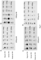
- TK1 antisera All four different TK1 antisera were tested for their reactivity with recombinant dog, cat and horse TK1 by using a dot blot assay.
- 3 ⁇ L of different dilutions of the recombinant TK1 preparations were applied on nitrocellulose membrane (Bio-Rad).
- Membranes were allowed to dry and blocked with 5 % non-fat dry milk powder diluted in TBST (mixture of tris-buffered saline (TBS) and Polysorbate 20 (also known as Tween 20)) for 1 hour at room temperature.
- TST tris-buffered saline
- Polysorbate 20 also known as Tween 20
- Figs. 8A-8D illustrate the results of the dot blot immunoassay with dog, cat and horse recombinant TK1 with the four different TK1 antisera.
- Figs. 9A-9C illustrate dot intensities in arbitrary units with different concentrations of dog, cat and horse recombinant TK1 with the four different TK1 antisera.
- CTK1p-215, CTK1p-211 and FTK1p-213 C-terminal peptides
- CTK1p-211 gave antibodies with high reactivity with all tested recombinant TK1 proteins, but with a clear preference for dog TK1.
- CTK1p-215 gave antisera against dog and horse TK1, while FTK1p-213 only produced antibodies reactive with horse TK1.
- CTK1p-161 resulted in reacting antibodies with all three types of TK1 protein and was superior for cat TK1.
- Example 1 The procedure in Example 1 was repeated but with the following differences.
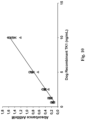
- Figs. 10 and 11A-11D illustrate the results from this Example 3 .
- Fig. 10 illustrates a standard curve with different concentrations of recombinant TK1 with CTK1p-215 coating and CTK1p-161 as detection antibodies.
- Fig. 11A illustrates the log STK1 ELISA protein levels in sera from healthy dogs and dogs with hematological malignancies.
- Figs. 11B illustrates the corresponding ROC curve for discrimination of hematological tumor dogs from healthy dogs.
- Fig. 11C illustrates the log STK1 ELISA protein levels in sera from healthy dogs and dogs with solid tumors.
- Figs. 11D illustrates the corresponding ROC curve for discrimination of solid tumor dogs from healthy dogs.
- CTK1p-211 anti-TK1 polyclonal antibody was used for coating and biotinylated CTK1p-215 was used for detection.
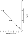
- Figs. 12 and 13A-13B The results from these antibodies are presented in Figs. 12 and 13A-13B .
- Fig. 12 illustrates a standard curve with different concentrations of recombinant TK1 with CTK1p-211 coating and CTK1p-215 as detection antibodies.
- Fig. 13A illustrates the log STK1 ELISA protein levels in sera from healthy dogs and dogs with hematological malignancies.
- Figs. 13B illustrates the corresponding ROC curve for discrimination of hematological tumor dogs from healthy dogs.
Landscapes
- Health & Medical Sciences (AREA)
- Life Sciences & Earth Sciences (AREA)
- Immunology (AREA)
- Chemical & Material Sciences (AREA)
- Engineering & Computer Science (AREA)
- Molecular Biology (AREA)
- Hematology (AREA)
- Urology & Nephrology (AREA)
- Biomedical Technology (AREA)
- General Health & Medical Sciences (AREA)
- Biochemistry (AREA)
- Medicinal Chemistry (AREA)
- Organic Chemistry (AREA)
- Physics & Mathematics (AREA)
- Biotechnology (AREA)
- Analytical Chemistry (AREA)
- Microbiology (AREA)
- Cell Biology (AREA)
- General Physics & Mathematics (AREA)
- Pathology (AREA)
- Food Science & Technology (AREA)
- Genetics & Genomics (AREA)
- Proteomics, Peptides & Aminoacids (AREA)
- Biophysics (AREA)
- Peptides Or Proteins (AREA)
- Enzymes And Modification Thereof (AREA)
- Hospice & Palliative Care (AREA)
- Oncology (AREA)
Claims (14)
- Kit zum Bestimmen eines Spiegels von nichtmenschlichem Thymidinkinase-1(TK1)-Protein in einer Probe, Folgendes umfassend:einen ersten Antikörper, der auf einem Träger immobilisiert ist oder dazu bestimmt ist, auf dem Träger immobilisiert zu werden; undeinen zweiten Antikörper, wobei einer von dem ersten Antikörper und dem zweiten Antikörper eine Spezifität für ein Peptid aufweist, das aus einer ersten Aminosäuresequenz aus einer C-terminalen Region von nichtmenschlicher Säugetier-TK1 besteht, und der andere von dem ersten Antikörper und dem zweiten Antikörper eine Spezifität für ein Peptid aufweist, das aus einer Aminosäuresequenz von einer aktiven Stelle von TK1 besteht,wobei die nichtmenschliche Säugetier-TK1 ausgewählt ist aus einer Gruppe bestehend aus Hunde-TK1-Protein, Katzen-TK1-Protein und Pferde-TK1-Protein; undder andere von dem ersten Antikörper und dem zweiten Antikörper eine Spezifität für ein Peptid aufweist, das aus einer Aminosäuresequenz der SEQ ID NO: 2 besteht, dadurch gekennzeichnet, dassder eine von dem ersten Antikörper und dem zweiten Antikörper eine Spezifität für ein Peptid aufweist, das aus einer Aminosäuresequenz der SEQ ID NO: 11 besteht.
- Kit nach Anspruch 1, wobei der Kit ein Sandwich-Assay-Kit ist.
- Kit nach Anspruch 1 oder 2, wobei der Kit ein Enzyme-Linked-Immunosorbent-Assay(ELISA)-Kit ist.
- Kit nach einem der Ansprüche 1 bis 3, wobei der zweite Antikörper ein kovalent gebundenes Biotin oder ein kovalent gebundenes Streptavidin oder Avidin aufweist.
- Kit nach Anspruch 4, ferner Folgendes umfassend:ein Meerrettichperoxidase(horseradish peroxidase - HRP)-markiertes Streptavidin oder Avidin oder ein HRP-markiertes Biotin; undein HRP-Substrat, ausgewählt aus einer Gruppe bestehend aus einem 3,3',5,5'-Tetramethylbenzidin(TMB)-Substrat, einem 3,3'-Diaminobenzidin(DAB)-Substrat und einem 2,2'-Azino-bis(3-ethylbenzothiazolin-6-sulfonsäure) (ABTS)-Substrat.
- Kit nach einem der Ansprüche 1 bis 5, ferner umfassend eine Mikrotiterplatte als Träger.
- Kit nach einem der Ansprüche 1 bis 5, ferner umfassend Agarosekügelchen oder Magnetkügelchen als Träger.
- In-vitro-Verfahren zum Bestimmen eines Spiegels von Hunde-, Katzen- oder Pferde-Thymidinkinase-1(TK1)-Protein in einer Probe, Folgendes umfassend:Inkontaktbringen der Probe mit einem ersten Antikörper und einem zweiten Antikörper des Kits nach einem der Ansprüche 1 bis 8; undNachweisen einer Menge an gebundenem zweitem Antikörper.
- Verfahren nach Anspruch 9, ferner umfassend das Bestimmen des Spiegels von Hunde-, Katzen- oder Pferde-TK1-Protein in der Probe basierend auf der nachgewiesenen Menge an gebundenem zweitem Antikörper.
- In-vitro-Verfahren zum Schätzen der Wahrscheinlichkeit eines Wiederauftretens einer Tumorerkrankung bei einem Hunde-, Katzenoder Pferde-Subjekt, Folgendes umfassend:Bestimmen eines Spiegels von Hunde-, Katzen- oder Pferde-Thymidinkinase-1(TK1)-Protein in einer Körperprobe von dem Hunde-, Katzen- oder Pferde-Subjekt unter Verwendung des Kits nach einem der Ansprüche 1 bis 8 oder des In-vitro-Verfahrens nach Anspruch 9 oder 10;Vergleichen des Spiegels von Hunde-, Katzen- oder Pferde-TK1-Protein in der Körperprobe mit einem Spiegel von Hunde-, Katzenoder Pferde-TK1-Protein, das repräsentativ für eine Population von gesunden Hunde-, Katzen- oder Pferde-Subjekten ist, oder mit einem Spiegel von Hunde-, Katzen- oder Pferde-TK1-Protein, das zuvor in dem Hunde-, Katze- oder Pferde-Subjekt bestimmt wurde; undSchätzen der Wahrscheinlichkeit eines erneuten Auftretens der Tumorerkrankung bei dem Hunde-, Katzen- oder Pferde-Subjekt basierend auf dem Vergleich.
- In-vitro-Verfahren zum Bestimmen der Zellproliferation bei einem Hunde-, Katzen- oder Pferde-Subjekt, Folgendes umfassend:Bestimmen eines Spiegels von Hunde-, Katzen- oder Pferde-Thymidinkinase-1(TK1)-Protein in einer Körperprobe von dem Hunde-, Katzen- oder Pferde-Subjekt unter Verwendung des Kits nach einem der Ansprüche 1 bis 8 oder des In-vitro-Verfahrens nach Anspruch 9 oder 10; undBestimmen der Zellproliferation basierend auf dem Spiegel von Hunde-, Katzen- oder Pferde-TK1-Protein in der Körperprobe.
- In-vitro-Verfahren zum Bestimmen einer Proliferationsprozessantwort bei einem an einer malignen Erkrankung leidenden Hunde-, Katzen- oder Pferde-Subjekt, Folgendes umfassend:Bestimmen eines Spiegels von Hunde-, Katzen- oder Pferde-Thymidinkinase-1(TK1)-Protein in einer Körperprobe von dem Hunde-, Katzen- oder Pferde-Subjekt unter Verwendung des Kits nach einem der Ansprüche 1 bis 8 oder des In-vitro-Verfahrens nach Anspruch 9 oder 10; undBestimmen der Proliferationsprozessantwort basierend auf dem Spiegel von Hunde-, Katzen- oder Pferde-TK1-Protein in der Körperprobe.
- In-vitro-Verfahren zum Bestimmen eines Grades an Entzündung, Infektion oder Tumorzellproliferation bei einem Hunde-, Katzenoder Pferde-Subjekt, Folgendes umfassend:Bestimmen eines Spiegels von Hunde-, Katzen- oder Pferde-Thymidinkinase(TK1)-Protein in einer Körperprobe von dem Hunde, Katzen- oder Pferde-Subjekt unter Verwendung des Kits nach einem der Ansprüche 1 bis 8 oder des In-vitro-Verfahrens nach Anspruch 9 oder 10; undBestimmen des Grades an Entzündung, Infektion oder Tumorzellproliferation basierend auf dem Spiegel von Hunde-, Katzen- oder Pferde-TK1-Protein in der Körperprobe.
- In-vitro-Verfahren zum Bewerten der Wirksamkeit der Behandlung einer malignen Erkrankung bei einem Hunde-, Katzenoder Pferde-Subjekt, Folgendes umfassend:Bestimmen eines Spiegels von Hunde-, Katzen- oder Pferde-Thymidinkinase-1(TK1)-Protein in einer Körperprobe von dem Hunde-, Katzen- oder Pferde-Subjekt unter Verwendung des Kits nach einem der Ansprüche 1 bis 8 oder des In-vitro-Verfahrens nach Anspruch 9 oder 10 vor oder in Verbindung mit dem Beginn der Behandlung der malignen Erkrankung;Bestimmen eines Spiegels von Hunde-, Katzen- oder Pferde-TK1-Protein in einer Körperprobe von dem Hunde-, Katzen- oder Pferde-Subjekt unter Verwendung des Kits nach einem der Ansprüche 1 bis 8 oder des In-vitro-Verfahrens nach Anspruch 9 oder 10 während oder nach der Behandlung der malignen Erkrankung; undBewerten der Wirksamkeit der Behandlung der malignen Erkrankung basierend auf einem Vergleich des in der Körperprobe vor oder in Verbindung mit dem Beginn der Behandlung der malignen Erkrankung bestimmten Spiegels von Hunde-, Katzen- oder Pferde-TK1-Protein und des in der Körperprobe während oder nach der Behandlung der malignen Erkrankung bestimmten Spiegels von Hunde-, Katzen- oder Pferde-TK1-Protein.
Applications Claiming Priority (2)
| Application Number | Priority Date | Filing Date | Title |
|---|---|---|---|
| US201662373262P | 2016-08-10 | 2016-08-10 | |
| PCT/SE2017/050806 WO2018030946A1 (en) | 2016-08-10 | 2017-08-09 | Determination of non-human mammal tk1 protein levels |
Publications (4)
| Publication Number | Publication Date |
|---|---|
| EP3497449A1 EP3497449A1 (de) | 2019-06-19 |
| EP3497449A4 EP3497449A4 (de) | 2020-07-29 |
| EP3497449C0 EP3497449C0 (de) | 2024-08-21 |
| EP3497449B1 true EP3497449B1 (de) | 2024-08-21 |
Family
ID=61163110
Family Applications (1)
| Application Number | Title | Priority Date | Filing Date |
|---|---|---|---|
| EP17839927.5A Active EP3497449B1 (de) | 2016-08-10 | 2017-08-09 | Bestimmung der tk1-protein-spiegel von nichtmenschlichen säugetieren |
Country Status (7)
| Country | Link |
|---|---|
| US (1) | US20190185582A1 (de) |
| EP (1) | EP3497449B1 (de) |
| JP (1) | JP7015294B2 (de) |
| CN (1) | CN109416358A (de) |
| AU (1) | AU2017310218B2 (de) |
| CA (1) | CA3031645A1 (de) |
| WO (1) | WO2018030946A1 (de) |
Families Citing this family (6)
| Publication number | Priority date | Publication date | Assignee | Title |
|---|---|---|---|---|
| WO2019201901A1 (en) | 2018-04-18 | 2019-10-24 | F. Hoffmann-La Roche Ag | Novel anti-thymidine kinase antibodies |
| CN110873711B (zh) * | 2018-09-04 | 2022-02-22 | 华瑞同康生物技术(深圳)有限公司 | 一种基于全自动化学发光分析仪的血清tk1检测试剂盒 |
| CN110346569A (zh) * | 2019-06-28 | 2019-10-18 | 安徽恩禾生物技术有限公司 | 一种胸苷激酶化学发光法检测试剂盒及其制备方法 |
| CN112940131A (zh) * | 2021-02-04 | 2021-06-11 | 福建亿彤生物科技有限公司 | 一种针对人血清中tk1的兔单克隆抗体及应用 |
| GB2619885B (en) * | 2021-03-11 | 2026-03-18 | Vetica Labs Inc | Detection of biomarkers useful in diagnosing chronic enteropathies in cats |
| US12578340B2 (en) | 2022-06-03 | 2026-03-17 | Alertix Veterinary Diagnostics Ab | Determination of canine TK1 protein levels |
Family Cites Families (8)
| Publication number | Priority date | Publication date | Assignee | Title |
|---|---|---|---|---|
| ATE239784T1 (de) * | 1993-08-06 | 2003-05-15 | Univ Brigham Young | Monoklonale antikörper gegen isozyme der thymidinkinase |
| EP1627230B1 (de) * | 2003-05-16 | 2010-09-15 | AroCell AB | Prognose des verlaufs einer krebserkrankung |
| US7837998B2 (en) * | 2004-05-21 | 2010-11-23 | Nathaniel Lallatin | Anti-cancer activity of an anti-thymidine kinase monoclonal antibody |
| US20100266495A1 (en) * | 2004-05-21 | 2010-10-21 | Brigham Young University | Anti-Cancer Activity of an Anti-Thymidine Kinase Monoclonal Antibody |
| US8501419B2 (en) * | 2007-05-23 | 2013-08-06 | Arocell Ab | Exposed proliferation-related peptides, ligands and methods employing the same |
| CN102432683B (zh) * | 2011-10-28 | 2013-10-30 | 周际 | 一种多表位tk1抗体的制备及其在肿瘤患者早期复发风险及预后评估中的应用 |
| CN103728456B (zh) * | 2013-09-16 | 2015-09-30 | 长沙赢润生物技术有限公司 | 胸腺嘧啶核苷激酶的elisa诊断试剂盒 |
| EP3770178A1 (de) | 2013-12-19 | 2021-01-27 | Arocell AB | Monoklonale anti-tk1-antikörper |
-
2017
- 2017-08-09 CA CA3031645A patent/CA3031645A1/en active Pending
- 2017-08-09 WO PCT/SE2017/050806 patent/WO2018030946A1/en not_active Ceased
- 2017-08-09 EP EP17839927.5A patent/EP3497449B1/de active Active
- 2017-08-09 JP JP2019505376A patent/JP7015294B2/ja active Active
- 2017-08-09 US US16/323,224 patent/US20190185582A1/en not_active Abandoned
- 2017-08-09 CN CN201780039279.6A patent/CN109416358A/zh active Pending
- 2017-08-09 AU AU2017310218A patent/AU2017310218B2/en active Active
Also Published As
| Publication number | Publication date |
|---|---|
| EP3497449A1 (de) | 2019-06-19 |
| EP3497449C0 (de) | 2024-08-21 |
| AU2017310218A1 (en) | 2018-12-20 |
| JP2019532260A (ja) | 2019-11-07 |
| JP7015294B2 (ja) | 2022-03-04 |
| CN109416358A (zh) | 2019-03-01 |
| US20190185582A1 (en) | 2019-06-20 |
| WO2018030946A1 (en) | 2018-02-15 |
| AU2017310218B2 (en) | 2023-03-09 |
| CA3031645A1 (en) | 2018-02-15 |
| EP3497449A4 (de) | 2020-07-29 |
Similar Documents
| Publication | Publication Date | Title |
|---|---|---|
| EP3497449B1 (de) | Bestimmung der tk1-protein-spiegel von nichtmenschlichen säugetieren | |
| CN111337678B (zh) | 与肿瘤免疫治疗效果相关的生物标志物及其应用 | |
| EP3536713B1 (de) | Monoklonale anti-tk1-antikörper | |
| Ghosh et al. | A specific and sensitive assay for blood levels of glycated CD59: a novel biomarker for diabetes | |
| CN101523213B (zh) | 天然形式人自分泌运动因子特异的抗体、其筛选方法、以及通过测定自分泌运动因子而检测恶性淋巴瘤的方法和检测试剂 | |
| Jagarlamudi et al. | Breast and prostate cancer patients differ significantly in their serum Thymidine kinase 1 (TK1) specific activities compared with those hematological malignancies and blood donors: implications of using serum TK1 as a biomarker | |
| US20130225442A1 (en) | Lung Cancer Tests | |
| Jagarlamudi et al. | A new sandwich ELISA for quantification of thymidine kinase 1 protein levels in sera from dogs with different malignancies can aid in disease management | |
| EP3304086B1 (de) | Methode zur vorhersage des ansprechens auf eine kombinationstherapie mit lenvatinib und everolimus | |
| KR20150129932A (ko) | 보체인자 b 단백질에 특이적으로 결합하는 항체를 포함하는 췌장암 진단용 키트 | |
| US12578340B2 (en) | Determination of canine TK1 protein levels | |
| Ye et al. | Development of a gold nanoparticle-based lateral flow immunochromatographic assay for the rapid and quantitative detection of thymidine kinase 1 in human serum | |
| KR20230068378A (ko) | 폐암에서 자가항체를 검출하기 위한 항원 조합의 용도 | |
| WO2006084018A2 (en) | Methods for determining responsiveness to cancer therapy | |
| CN115038969A (zh) | 预测患者生存 | |
| Qi et al. | Development of a highly specific HER2 monoclonal antibody for immunohistochemistry using protein microarray chips | |
| KR102128251B1 (ko) | 아르기닌이 메틸화된 drd2에 특이적으로 결합하는 대장암 진단용 바이오마커 조성물 | |
| CN112639475A (zh) | Dlbcl的预后指数中的胸苷激酶(tk-1) | |
| EP4359800A1 (de) | Vorhersage eines krebsrezidivs | |
| Wang et al. | Monoclonal antibody selection for interleukin-4 quantification using suspension arrays and forward-phase protein microarrays | |
| Sharif et al. | canine Thymidine kinase 1 protein | |
| Chourb et al. | Improved detection of the MUC1 cancer antigen CA 15-3 by ALYGNSA fluorimmunoassay | |
| Ma et al. | Protein microarrays for quantitative detection of PAI-1 in serum | |
| HK40077809A (zh) | 预测患者生存 | |
| Ejaz et al. | Indirect Back-Titration ELISA: A New Format for Estimation of Human Tissue Kallikreins |
Legal Events
| Date | Code | Title | Description |
|---|---|---|---|
| STAA | Information on the status of an ep patent application or granted ep patent |
Free format text: STATUS: THE INTERNATIONAL PUBLICATION HAS BEEN MADE |
|
| PUAI | Public reference made under article 153(3) epc to a published international application that has entered the european phase |
Free format text: ORIGINAL CODE: 0009012 |
|
| STAA | Information on the status of an ep patent application or granted ep patent |
Free format text: STATUS: REQUEST FOR EXAMINATION WAS MADE |
|
| 17P | Request for examination filed |
Effective date: 20190311 |
|
| AK | Designated contracting states |
Kind code of ref document: A1 Designated state(s): AL AT BE BG CH CY CZ DE DK EE ES FI FR GB GR HR HU IE IS IT LI LT LU LV MC MK MT NL NO PL PT RO RS SE SI SK SM TR |
|
| AX | Request for extension of the european patent |
Extension state: BA ME |
|
| RIN1 | Information on inventor provided before grant (corrected) |
Inventor name: ROENNBERG, HENRIK Inventor name: ERIKSSON, STAFFAN Inventor name: JAGARLAMUDI, KIRAN KUMAR |
|
| DAV | Request for validation of the european patent (deleted) | ||
| DAX | Request for extension of the european patent (deleted) | ||
| RIC1 | Information provided on ipc code assigned before grant |
Ipc: G01N 33/573 20060101ALI20200224BHEP Ipc: C07K 16/40 20060101ALI20200224BHEP Ipc: G01N 33/574 20060101AFI20200224BHEP Ipc: C12Q 1/48 20060101ALI20200224BHEP |
|
| A4 | Supplementary search report drawn up and despatched |
Effective date: 20200603 |
|
| RIC1 | Information provided on ipc code assigned before grant |
Ipc: G01N 33/573 20060101ALI20200527BHEP Ipc: G01N 33/574 20060101AFI20200527BHEP Ipc: C07K 16/40 20060101ALI20200527BHEP Ipc: C12Q 1/48 20060101ALI20200527BHEP |
|
| DA4 | Supplementary search report drawn up and despatched (deleted) | ||
| RA4 | Supplementary search report drawn up and despatched (corrected) |
Effective date: 20200629 |
|
| RIC1 | Information provided on ipc code assigned before grant |
Ipc: G01N 33/574 20060101AFI20200622BHEP Ipc: C12Q 1/48 20060101ALI20200622BHEP Ipc: G01N 33/573 20060101ALI20200622BHEP Ipc: C07K 16/40 20060101ALI20200622BHEP |
|
| STAA | Information on the status of an ep patent application or granted ep patent |
Free format text: STATUS: EXAMINATION IS IN PROGRESS |
|
| 17Q | First examination report despatched |
Effective date: 20210409 |
|
| GRAP | Despatch of communication of intention to grant a patent |
Free format text: ORIGINAL CODE: EPIDOSNIGR1 |
|
| STAA | Information on the status of an ep patent application or granted ep patent |
Free format text: STATUS: GRANT OF PATENT IS INTENDED |
|
| INTG | Intention to grant announced |
Effective date: 20240415 |
|
| GRAS | Grant fee paid |
Free format text: ORIGINAL CODE: EPIDOSNIGR3 |
|
| GRAA | (expected) grant |
Free format text: ORIGINAL CODE: 0009210 |
|
| STAA | Information on the status of an ep patent application or granted ep patent |
Free format text: STATUS: THE PATENT HAS BEEN GRANTED |
|
| RIN1 | Information on inventor provided before grant (corrected) |
Inventor name: JAGARLAMUDI, KIRAN KUMAR Inventor name: ROENNBERG, HENRIK Inventor name: ERIKSSON, STAFFAN |
|
| RAP3 | Party data changed (applicant data changed or rights of an application transferred) |
Owner name: ALERTIX VETERINARY DIAGNOSTICS AB |
|
| AK | Designated contracting states |
Kind code of ref document: B1 Designated state(s): AL AT BE BG CH CY CZ DE DK EE ES FI FR GB GR HR HU IE IS IT LI LT LU LV MC MK MT NL NO PL PT RO RS SE SI SK SM TR |
|
| REG | Reference to a national code |
Ref country code: GB Ref legal event code: FG4D |
|
| REG | Reference to a national code |
Ref country code: CH Ref legal event code: EP |
|
| REG | Reference to a national code |
Ref country code: DE Ref legal event code: R096 Ref document number: 602017084306 Country of ref document: DE |
|
| REG | Reference to a national code |
Ref country code: IE Ref legal event code: FG4D |
|
| U01 | Request for unitary effect filed |
Effective date: 20240821 |
|
| U07 | Unitary effect registered |
Designated state(s): AT BE BG DE DK EE FI FR IT LT LU LV MT NL PT SE SI Effective date: 20240827 |
|
| PG25 | Lapsed in a contracting state [announced via postgrant information from national office to epo] |
Ref country code: NO Free format text: LAPSE BECAUSE OF FAILURE TO SUBMIT A TRANSLATION OF THE DESCRIPTION OR TO PAY THE FEE WITHIN THE PRESCRIBED TIME-LIMIT Effective date: 20241121 |
|
| PG25 | Lapsed in a contracting state [announced via postgrant information from national office to epo] |
Ref country code: GR Free format text: LAPSE BECAUSE OF FAILURE TO SUBMIT A TRANSLATION OF THE DESCRIPTION OR TO PAY THE FEE WITHIN THE PRESCRIBED TIME-LIMIT Effective date: 20241122 Ref country code: PL Free format text: LAPSE BECAUSE OF FAILURE TO SUBMIT A TRANSLATION OF THE DESCRIPTION OR TO PAY THE FEE WITHIN THE PRESCRIBED TIME-LIMIT Effective date: 20240821 |
|
| PG25 | Lapsed in a contracting state [announced via postgrant information from national office to epo] |
Ref country code: IS Free format text: LAPSE BECAUSE OF FAILURE TO SUBMIT A TRANSLATION OF THE DESCRIPTION OR TO PAY THE FEE WITHIN THE PRESCRIBED TIME-LIMIT Effective date: 20241221 |
|
| PG25 | Lapsed in a contracting state [announced via postgrant information from national office to epo] |
Ref country code: HR Free format text: LAPSE BECAUSE OF FAILURE TO SUBMIT A TRANSLATION OF THE DESCRIPTION OR TO PAY THE FEE WITHIN THE PRESCRIBED TIME-LIMIT Effective date: 20240821 |
|
| PG25 | Lapsed in a contracting state [announced via postgrant information from national office to epo] |
Ref country code: RS Free format text: LAPSE BECAUSE OF FAILURE TO SUBMIT A TRANSLATION OF THE DESCRIPTION OR TO PAY THE FEE WITHIN THE PRESCRIBED TIME-LIMIT Effective date: 20241121 Ref country code: ES Free format text: LAPSE BECAUSE OF FAILURE TO SUBMIT A TRANSLATION OF THE DESCRIPTION OR TO PAY THE FEE WITHIN THE PRESCRIBED TIME-LIMIT Effective date: 20240821 |
|
| PG25 | Lapsed in a contracting state [announced via postgrant information from national office to epo] |
Ref country code: RS Free format text: LAPSE BECAUSE OF FAILURE TO SUBMIT A TRANSLATION OF THE DESCRIPTION OR TO PAY THE FEE WITHIN THE PRESCRIBED TIME-LIMIT Effective date: 20241121 Ref country code: PL Free format text: LAPSE BECAUSE OF FAILURE TO SUBMIT A TRANSLATION OF THE DESCRIPTION OR TO PAY THE FEE WITHIN THE PRESCRIBED TIME-LIMIT Effective date: 20240821 Ref country code: NO Free format text: LAPSE BECAUSE OF FAILURE TO SUBMIT A TRANSLATION OF THE DESCRIPTION OR TO PAY THE FEE WITHIN THE PRESCRIBED TIME-LIMIT Effective date: 20241121 Ref country code: IS Free format text: LAPSE BECAUSE OF FAILURE TO SUBMIT A TRANSLATION OF THE DESCRIPTION OR TO PAY THE FEE WITHIN THE PRESCRIBED TIME-LIMIT Effective date: 20241221 Ref country code: HR Free format text: LAPSE BECAUSE OF FAILURE TO SUBMIT A TRANSLATION OF THE DESCRIPTION OR TO PAY THE FEE WITHIN THE PRESCRIBED TIME-LIMIT Effective date: 20240821 Ref country code: GR Free format text: LAPSE BECAUSE OF FAILURE TO SUBMIT A TRANSLATION OF THE DESCRIPTION OR TO PAY THE FEE WITHIN THE PRESCRIBED TIME-LIMIT Effective date: 20241122 Ref country code: ES Free format text: LAPSE BECAUSE OF FAILURE TO SUBMIT A TRANSLATION OF THE DESCRIPTION OR TO PAY THE FEE WITHIN THE PRESCRIBED TIME-LIMIT Effective date: 20240821 |
|
| PG25 | Lapsed in a contracting state [announced via postgrant information from national office to epo] |
Ref country code: SM Free format text: LAPSE BECAUSE OF FAILURE TO SUBMIT A TRANSLATION OF THE DESCRIPTION OR TO PAY THE FEE WITHIN THE PRESCRIBED TIME-LIMIT Effective date: 20240821 |
|
| PG25 | Lapsed in a contracting state [announced via postgrant information from national office to epo] |
Ref country code: CZ Free format text: LAPSE BECAUSE OF FAILURE TO SUBMIT A TRANSLATION OF THE DESCRIPTION OR TO PAY THE FEE WITHIN THE PRESCRIBED TIME-LIMIT Effective date: 20240821 |
|
| PG25 | Lapsed in a contracting state [announced via postgrant information from national office to epo] |
Ref country code: SK Free format text: LAPSE BECAUSE OF FAILURE TO SUBMIT A TRANSLATION OF THE DESCRIPTION OR TO PAY THE FEE WITHIN THE PRESCRIBED TIME-LIMIT Effective date: 20240821 |
|
| PLBE | No opposition filed within time limit |
Free format text: ORIGINAL CODE: 0009261 |
|
| STAA | Information on the status of an ep patent application or granted ep patent |
Free format text: STATUS: NO OPPOSITION FILED WITHIN TIME LIMIT |
|
| 26N | No opposition filed |
Effective date: 20250522 |
|
| U20 | Renewal fee for the european patent with unitary effect paid |
Year of fee payment: 9 Effective date: 20250715 |
|
| PGFP | Annual fee paid to national office [announced via postgrant information from national office to epo] |
Ref country code: GB Payment date: 20250715 Year of fee payment: 9 |
|
| REG | Reference to a national code |
Ref country code: CH Ref legal event code: H13 Free format text: ST27 STATUS EVENT CODE: U-0-0-H10-H13 (AS PROVIDED BY THE NATIONAL OFFICE) Effective date: 20260324 |