EP3491145B1 - Marker und verfahren zur beurteilung der zusammensetzung oder reinheit einer zellkultur - Google Patents
Marker und verfahren zur beurteilung der zusammensetzung oder reinheit einer zellkultur Download PDFInfo
- Publication number
- EP3491145B1 EP3491145B1 EP17748696.6A EP17748696A EP3491145B1 EP 3491145 B1 EP3491145 B1 EP 3491145B1 EP 17748696 A EP17748696 A EP 17748696A EP 3491145 B1 EP3491145 B1 EP 3491145B1
- Authority
- EP
- European Patent Office
- Prior art keywords
- marker
- ace
- mmp1
- cell culture
- cell
- Prior art date
- Legal status (The legal status is an assumption and is not a legal conclusion. Google has not performed a legal analysis and makes no representation as to the accuracy of the status listed.)
- Active
Links
Images






Classifications
-
- C—CHEMISTRY; METALLURGY
- C12—BIOCHEMISTRY; BEER; SPIRITS; WINE; VINEGAR; MICROBIOLOGY; ENZYMOLOGY; MUTATION OR GENETIC ENGINEERING
- C12Q—MEASURING OR TESTING PROCESSES INVOLVING ENZYMES, NUCLEIC ACIDS OR MICROORGANISMS; COMPOSITIONS OR TEST PAPERS THEREFOR; PROCESSES OF PREPARING SUCH COMPOSITIONS; CONDITION-RESPONSIVE CONTROL IN MICROBIOLOGICAL OR ENZYMOLOGICAL PROCESSES
- C12Q1/00—Measuring or testing processes involving enzymes, nucleic acids or microorganisms; Compositions therefor; Processes of preparing such compositions
- C12Q1/68—Measuring or testing processes involving enzymes, nucleic acids or microorganisms; Compositions therefor; Processes of preparing such compositions involving nucleic acids
- C12Q1/6876—Nucleic acid products used in the analysis of nucleic acids, e.g. primers or probes
- C12Q1/6881—Nucleic acid products used in the analysis of nucleic acids, e.g. primers or probes for tissue or cell typing, e.g. human leukocyte antigen [HLA] probes
-
- G—PHYSICS
- G01—MEASURING; TESTING
- G01N—INVESTIGATING OR ANALYSING MATERIALS BY DETERMINING THEIR CHEMICAL OR PHYSICAL PROPERTIES
- G01N33/00—Investigating or analysing materials by specific methods not covered by groups G01N1/00 - G01N31/00
- G01N33/48—Biological material, e.g. blood, urine; Haemocytometers
- G01N33/50—Chemical analysis of biological material, e.g. blood, urine; Testing involving biospecific ligand binding methods; Immunological testing
- G01N33/5005—Chemical analysis of biological material, e.g. blood, urine; Testing involving biospecific ligand binding methods; Immunological testing involving human or animal cells
-
- G—PHYSICS
- G01—MEASURING; TESTING
- G01N—INVESTIGATING OR ANALYSING MATERIALS BY DETERMINING THEIR CHEMICAL OR PHYSICAL PROPERTIES
- G01N33/00—Investigating or analysing materials by specific methods not covered by groups G01N1/00 - G01N31/00
- G01N33/48—Biological material, e.g. blood, urine; Haemocytometers
- G01N33/50—Chemical analysis of biological material, e.g. blood, urine; Testing involving biospecific ligand binding methods; Immunological testing
- G01N33/68—Chemical analysis of biological material, e.g. blood, urine; Testing involving biospecific ligand binding methods; Immunological testing involving proteins, peptides or amino acids
- G01N33/6887—Chemical analysis of biological material, e.g. blood, urine; Testing involving biospecific ligand binding methods; Immunological testing involving proteins, peptides or amino acids from muscle, cartilage or connective tissue
-
- C—CHEMISTRY; METALLURGY
- C12—BIOCHEMISTRY; BEER; SPIRITS; WINE; VINEGAR; MICROBIOLOGY; ENZYMOLOGY; MUTATION OR GENETIC ENGINEERING
- C12Q—MEASURING OR TESTING PROCESSES INVOLVING ENZYMES, NUCLEIC ACIDS OR MICROORGANISMS; COMPOSITIONS OR TEST PAPERS THEREFOR; PROCESSES OF PREPARING SUCH COMPOSITIONS; CONDITION-RESPONSIVE CONTROL IN MICROBIOLOGICAL OR ENZYMOLOGICAL PROCESSES
- C12Q2600/00—Oligonucleotides characterized by their use
- C12Q2600/158—Expression markers
-
- G—PHYSICS
- G01—MEASURING; TESTING
- G01N—INVESTIGATING OR ANALYSING MATERIALS BY DETERMINING THEIR CHEMICAL OR PHYSICAL PROPERTIES
- G01N2333/00—Assays involving biological materials from specific organisms or of a specific nature
- G01N2333/435—Assays involving biological materials from specific organisms or of a specific nature from animals; from humans
- G01N2333/46—Assays involving biological materials from specific organisms or of a specific nature from animals; from humans from vertebrates
- G01N2333/47—Assays involving proteins of known structure or function as defined in the subgroups
- G01N2333/4701—Details
- G01N2333/4722—Proteoglycans, e.g. aggreccan
-
- G—PHYSICS
- G01—MEASURING; TESTING
- G01N—INVESTIGATING OR ANALYSING MATERIALS BY DETERMINING THEIR CHEMICAL OR PHYSICAL PROPERTIES
- G01N2400/00—Assays, e.g. immunoassays or enzyme assays, involving carbohydrates
- G01N2400/10—Polysaccharides, i.e. having more than five saccharide radicals attached to each other by glycosidic linkages; Derivatives thereof, e.g. ethers, esters
- G01N2400/38—Heteroglycans, i.e. polysaccharides having more than one sugar residue in the main chain in either alternating or less regular sequence, e.g. gluco- or galactomannans, Konjac gum, Locust bean gum or Guar gum
- G01N2400/40—Glycosaminoglycans, i.e. GAG or mucopolysaccharides, e.g. chondroitin sulfate, dermatan sulfate, hyaluronic acid, heparin, heparan sulfate, and related sulfated polysaccharides
-
- G—PHYSICS
- G01—MEASURING; TESTING
- G01N—INVESTIGATING OR ANALYSING MATERIALS BY DETERMINING THEIR CHEMICAL OR PHYSICAL PROPERTIES
- G01N2800/00—Detection or diagnosis of diseases
- G01N2800/10—Musculoskeletal or connective tissue disorders
Definitions
- the invention relates to markers for assessing the composition or purity of a cell culture, a method for assessing the composition or purity of a cell culture, the use of a cell culture for the production of an advanced therapy medicinal product and the use of a kit for carrying out the above-mentioned method.
- cell-based medicinal products in particular cell-based tissue engineering products. This applies in particular with regard to the purity and identity of cells used to manufacture cell-based medicinal products.
- Such cultures are often prepared using autologous cartilage cells and are used after a certain cultivation time in the context of matrix-associated autologous cartilage cell transplantation.
- the particular problem is that the various cell types of mesenchymal origin, such as osteoblasts, synovial cells and mesenchymal stem cells, do not differ very much and are therefore practically indistinguishable using conventional markers.
- endothelial cells, osteoblasts and synovial cells can generally be regarded as significant as contaminating cell types.
- synovial cells are particularly critical because, in contrast to endothelial cells and osteoblasts, they often behave similarly to cartilage cells under normal culture conditions.
- the object of the invention is therefore to provide markers which make it possible to check the purity of cartilage cell cultures.
- the object of the invention is to provide the use of a cell culture for the production of a pharmaceutical for advanced therapies.
- the invention relates to the use of a marker for assessing, in particular for determining, the composition or purity of a cell culture.
- the marker is particularly characterized in that it is at least one marker from the group consisting of MMP1, ACE, POSTN, SYNPO, SULF1, ARHGEF28, IGF2BP1, BAIAP2L1, LIMCH1, SCIN, DCLK1, PPL, CRABP2, NES , XPNPEP2 and SLIT3 is selected, wherein the expression level of the at least one marker is determined.
- At least one marker can be one of the above markers or a combination of two or more markers, ie a combination of two, three, four, five, six, seven, eight, nine, ten, eleven , twelve, thirteen, fourteen, fifteen, or sixteen of the above markers or a combination of any of the above markers.
- the expression "at least one marker” in the context of the present invention mean a ratio of two of the above markers.
- the expression "marker” in the context of the present invention can generally mean a gene product, i.e. the result of the expression, such as in particular the messenger RNA (mRNA), also referred to as messenger RNA, or the protein, one or more of the markers mentioned above .
- mRNA messenger RNA
- messenger RNA also referred to as messenger RNA
- protein one or more of the markers mentioned above .
- expression level is to be understood as meaning an expression level based on RNA, in particular messenger RNA (mRNA-based), and/or an expression level on a protein basis.
- RNA messenger RNA
- the term “determine” used in the present invention in connection with the expression “expression level” is intended to include any biochemical and/or biotechnological method in the context of the present invention, by means of which the expression level of the at least one marker, in particular on an RNA or protein basis , recorded, in particular recorded quantitatively, can be.
- cartilage cell markers i.e. chondrocyte markers
- synovial cell markers i.e. synoviocyte markers
- the markers identified within the scope of the present invention allow in particular the detection of synovial cell contamination in cartilage cell cultures and/or the detection of the presence of cartilage cells in a synovial cell culture and/or the discrimination between cartilage cells and synovial cells.
- the markers proposed according to the invention can therefore also be used as cartilage cell or chondrocyte markers, i.e. as purity or quality markers and/or identity markers for cartilage cells or chondrocytes, and/or as synovial cell or synoviocyte markers, i.e. as purity or quality markers and/or or identity markers for synovial cells or synoviocytes.
- the level of expression of the at least one marker is determined in a cell sample obtained from the cell culture.
- cell sample should be understood to mean a sample containing cells or a sample consisting of cells, which is taken from the cell culture.
- POSTN is also referred to as "periostin” or "osteoblast-specific factor OSF-2".
- Periostin is a ligand for alpha-V/beta-3 and alpha-V/beta-5 integrins to promote adhesion and migration of epithelial cells.
- SYNPO is also referred to as "synaptopodin".
- Synaptopodin is a cytoplasmic actin-associated protein with a molecular mass of approximately 73 kDa. Synaptopodin is found in both the podocytes of kidneys and the cerebrum.
- MMP1 is also referred to as "matrix metalloproteinase-1", “interstitial collagenase” or “fibroblast collagenase”.
- Sulfatase 1 is an enzyme that selectively removes 6-O-sulfate groups from heparan sulfate.
- ARHGEF28 is also referred to as "Rho Guanine Nucleotide Exchange Factor 28".
- IGF2BP1 is also known as "Insulin-Like Growth Factor 2 mRNA Binding Protein 1".
- BAIAP2L1 is also referred to as "brain-specific angiogenesis inhibitor 1-associated protein 2-like protein 1 (BAI1-associated protein 2-like protein 1)".
- LIMCH1 is also referred to as "LIM and Calponin Homology Domains 1".
- SCIN As a protein, SCIN is also referred to as "scinderin” or “adseverin”.
- DCLK1 is also referred to as “doublecortin-like kinase-1”.
- PPL As a protein, PPL is also referred to as "periplakin".
- CRABP2 is also referred to as "cellular retinoic acid-binding protein 2".
- NES As a protein, NES is also referred to as "nestin”.
- XPNPEP2 As a protein, XPNPEP2 is also referred to as "X-prolyl aminopeptidase”.
- SLIT3 is also referred to as "Slit Guidance Ligand 3".
- the at least one marker is ACE.
- ACE is also known as "angiotensin converting enzyme”.
- the at least one marker is a combination of ACE and at least one other marker selected from the group consisting of MMP1, POSTN, SYNPO, SULF1, ARHGEF28, IGF2BP1, BAIAP2L1, LIMCH1, SCIN, DCLK1, PPL, CRABP2, NES , XPNPEP2 and SLIT3.
- the at least one marker is particularly preferably a combination of ACE and MMP1.
- the at least one marker can in particular be a combination of ACE and at least one further marker selected from the group consisting of DCLK1, PPL, CRABP2, NES, XPNPEP2 and SLIT3.
- the cell culture is a cartilage cell culture or chondrocyte culture.
- cartilage cell culture or "chondrocyte culture” is to be understood in the context of the present invention as a culture which, starting from cartilage tissue, preferably starting from a cartilage biopsy, in particular cartilage-bone punch biopsy, by means of enzymatic digestion and subsequent cultivation of the after digestion cells obtained is produced or can be produced.
- cartilage tissue can mean healthy cartilage tissue, diseased or pathologically altered cartilage tissue or a regenerated cartilage tissue, i.e. a preferably resulting from medical treatment, in particular an autologous chondrocyte transplantation (ACT) or matrix-associated autologous chondrocyte transplantation (MACT), newly formed cartilage tissue.
- ACT autologous chondrocyte transplantation
- MACT matrix-associated autologous chondrocyte transplantation
- the term "cartilage biopsy” can mean a biopsy which contains healthy cartilage tissue, diseased or pathologically altered cartilage tissue or a regenerated cartilage tissue or consists of one of the aforementioned tissues.
- the term “cartilage-bone punch biopsy” is intended to mean a biopsy in the context of the present invention, which contains healthy bone and cartilage tissue, diseased bone and cartilage tissue or diseased bone and cartilage tissue or a bone and cartilage tissue regenerate or from a of the aforementioned tissue.
- synovial cell culture or “synovocyte culture” is to be understood in the context of the present invention as meaning a culture which is produced or can be produced starting from synovial tissue by means of enzymatic digestion and subsequent cultivation of the cells obtained after the digestion.
- the cell culture has cells which originate from a cartilage biopsy.
- the cell culture can have cells which originate from a cartilage-bone punch biopsy, in particular a cartilage tissue fragment thereof.
- the biopsy in particular cartilage biopsy or cartilage-bone punch biopsy, is preferably a biopsy from a joint, in particular a knee joint.
- pure synovial cell culture should be understood to mean a synovial cell culture which has cells exclusively in the form of synovial cells or synoviocytes.
- the term "pure cartilage cell culture” should be understood to mean a cartilage cell culture which has cells exclusively in the form of cartilage cells or chondrocytes.
- the at least one marker is the ratio of ACE to MMP1 (ACE/MMP1 ratio) or the ratio of MMP1 to ACE (MMP1/ACE ratio).
- the marker for assessing the composition or purity of a cell culture is the ratio of ACE to MMP1 or the ratio of MMP1 to ACE.
- the ratio of ACE to MMP1 is preferably formed to assess the composition or purity of the cell culture, ie the ACE/MMP1 ratio is used as a marker to assess the composition or purity of a cell culture.
- a value of the ACE/MMP1 ratio above a defined threshold value preferably indicates that the cell culture has synovial cells (synovocytes).
- the threshold is equal to or less than the value of the ratio of ACE to MMP1 in a pure synovial cell culture.
- the expression level of the at least one marker is determined on an RNA basis, preferably on a messenger RNA basis (mRNA basis).
- RNA basis preferably on a messenger RNA basis (mRNA basis).
- the level of expression of the at least one protein-based marker is determined.
- the expression level of the at least one marker is determined by means of a polymerase chain reaction such as reverse transcriptase polymerase chain reaction or real-time quantitative polymerase chain reaction, an electrophoresis assay, a gel electrophoresis assay such as Southern blot, Northern blot or Western blot, protein -Chips, antibodies, an immunoassay such as ELISAs, a LUMINEX immunoassay or ELISpot assay, an immunofluorescence assay, an immunohistochemical assay, a radioimmunological assay, proteome analysis, a chromatography-based assay such as HPLC or gel chromatography, a mass spectroscopic assay or a flow cytometry assays.
- a polymerase chain reaction such as reverse transcriptase polymerase chain reaction or real-time quantitative polymerase chain reaction
- an electrophoresis assay such as Southern blot, Northern blot or Western blot, protein -Chips
- the invention relates to a method for assessing, in particular for determining, the composition or purity of a cell culture.
- the method is particularly characterized in that the at least one marker ACE or a combination of ACE and at least one other marker selected from the group consisting of MMP1, POSTN, SYNPO, SULF1, ARHGEF28, IGF2BP1, BAIAP2L1, LIMCH1, SCIN, DCLK1 , PPL, CRABP2, NES, XPNPEP2 and SLIT3.
- the at least one marker ACE or a combination of ACE and at least one other marker selected from the group consisting of MMP1, POSTN, SYNPO, SULF1, ARHGEF28, IGF2BP1, BAIAP2L1, LIMCH1, SCIN, DCLK1 , PPL, CRABP2, NES, XPNPEP2 and SLIT3.
- the cell culture has cells which originate from a cartilage biopsy.
- the cell culture can have cells which originate from a cartilage-bone punch biopsy, in particular a cartilage tissue fragment thereof.
- the biopsy in particular cartilage biopsy or cartilage-bone punch biopsy, is preferably a biopsy from a joint, in particular a knee joint.
- the at least one marker is the ratio of ACE to MMP1 (ACE/MMP1 ratio) or the ratio of MMP1 to ACE (MMP1/ACE ratio).
- the at least one marker is preferably the ratio of ACE to MMP1.
- a value of the ratio of ACE to MMP1 above a defined threshold indicates that the cell culture has synovial cells (synoviocytes).
- the threshold is equal to or less than the value of the ratio of ACE to MMP1 in a pure synovial cell culture.
- the invention relates to the use of a kit for carrying out a method according to the second aspect of the invention.
- the kit has at least one component for detecting at least one marker according to the first aspect of the invention.
- the component can in particular be an antibody, such as capture and/or detection antibody, dye such as a fluorescent dye, beads, buffer solution, nutrient solution, washing solution, primer (oligonucleotides that serve as a starting point for DNA-replicating enzymes such as DNA polymerase) or be a mixture/combination of at least two of the components mentioned.
- Figure 12 shows a volcano plot visualization of the mean values of log10-transformed synoviocyte/chondrocyte LFQ intensities (Mean LFQ Ratios) versus the standard deviations of the ratios of all proteins quantified in at least 3 out of 5 replicate experiments.
- the dashed lines indicate a 2- and 10-fold difference in protein expression, respectively (corresponding to ⁇ 0.3 and ⁇ 1 on the logarithmic scale).
- Figure 12 shows a volcano plot visualizing the differences in peptide count between synoviocytes and chondrocytes and the associated differences in q-value for all proteins not used in the mean rank test. Peptide count differences greater than 1 mean higher expression in synoviocytes and peptide count differences smaller than 1 mean higher expression in chondrocytes. The proteins already classified as biomarker candidates in study 1 are identified by gene names.
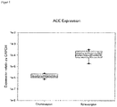
- FIG. 4 shows graphically the mRNA expression of the synoviocyte marker ACE.
- the boxplots used in the graph show the 25th/75th percentiles (boxes), 10/90th percentiles (brackets), 5/95th percentiles (dots), the medians (solid lines) and the means (dotted lines).
- the 4 Figure 12 illustrates that ACE mRNA expression is significantly higher in synoviocytes than in chondrocytes. ACE is therefore particularly suitable for use as a synoviocyte marker.
- the boxplots used in the graph show the 25th/75th percentiles (boxes), 10/90th percentiles (brackets), 5/95th percentiles (dots), the medians (solid lines) and the means (dotted lines).
- 6 illustrates that the ratio of ACE to MMP1 (ACE / MMP1 ratio) as a marker within the meaning of the present invention, in particular for assessing the composition or purity of a cell culture and / or for in vitro determination of the identity of a chondrocyte and / or a synoviocyte suitable.
- the boxplots used in the graph show the 25th/75th percentiles (boxes), 10/90th percentiles (brackets), 5/95th percentiles (dots), the medians (solid lines) and the means (dotted lines).
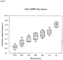
- the 7 illustrates that the value of the ratio of ACE to MMP1 correlates with the degree of purity of a chondrocyte-containing cell mixture, in such a way that a low value of the ratio of ACE to MMP1 correlates with a high degree of purity of a chondrocyte-containing cell mixture and a high value of the ratio of ACE to MMP1 correlates with a low degree of purity of a chondrocyte-containing cell mixture.
- 7 shows in particular that the degree of purity of a chondrocyte-containing cell mixture is higher, the lower the value of the ratio of ACE to MMP1.
- the cell mixtures were prepared as follows: Chondrocytes and synoviocytes were cultivated separately from one another—as described below under 1. Material and methods—and trypsinized and centrifuged at the end of the culture. The resulting cell pellet was resuspended. The cell number of the cell suspensions was determined and then the appropriate volumes of each cell suspension were used to prepare mixtures containing 100%, 90%, 75%, 50%, 25%, 10% and 0% chondrocytes. The resulting cell mixtures were centrifuged off again and the resulting cell pellet was lysed and used for gene expression analysis.
- the boxplots used in the graph show the 25th/75th percentiles (boxes), 10/90th percentiles (brackets), 5/95th percentiles (dots), the medians (solid lines) and the means (dotted lines).
- the boxplots used in the graph show the 25th/75th percentiles (boxes), 10/90th percentiles (brackets), 5/95th percentiles (dots), the medians (solid lines) and the means (dotted lines).
- the chondrocytes and synoviocytes used came from the knee joint of deceased donors in the USA.
- the tissue was very finely minced using two scalpels and digested with collagenase for 24 h, 5% CO2 and 37°C.
- the synovial tissue was treated identically to the cartilage tissue throughout the cultivation. After digestion, incubation was carried out in chondrocyte medium for 48 h, 5% CO2 and 37°C.
- the cells were then passed through a cell strainer, centrifuged and expanded in fresh chondrocyte medium. The cells were cultured for 19 to a maximum of 23 days with 2-3 media changes, depending on growth.
- the cells were detached from the culture surface with trypsin, washed with PBS, frozen and shipped on dry ice to Evotec Kunststoff.
- the proteins in the cell lysates were reduced with 10 mM TCEP (tris(2-carboxyethyl)phosphine) and alkylated in the presence of 40 mM CAA (2-chloroacetamide). After centrifugation, a 150 ⁇ g aliquot of the clear lysate was diluted 1:2 with H2O and a 1:1 mixture of endopeptidase Lys-C (Wako) and trypsin (Promega) in an enzyme to protein ratio of 1:50 was added and left overnight incubated.
- TCEP tris(2-carboxyethyl)phosphine
- CAA 2-chloroacetamide
- the resulting peptide mixture was acidified by the addition of 99% ethyl acetate and 1% TFA and then desalted using a Strata-XC column (100 mg sorbent, Phenomenex).
- the peptides were eluted with 80% acetonitrile and 5% v/v ammonium hydroxide, snap frozen in liquid nitrogen and lyophilized.
- the peptides were then fractionated using reversed-phase chromatography at high pH ( Wang Y, Yang F, Gritsenko MA, Clauss T, Liu T, Shen Y, Monroe ME, Lopez-Ferrer D, Reno T, Moore RJ et al. (2011).
- the pooled fractions of the eluted peptides were then pooled in a non-linear fashion for a total of 12 samples.
- the samples were then desalted using a home-made C18 STAGEtip columns ( Rappsilber J, Mann M, and Ishihama Y (2007). Protocol for micro-purification, enrichment, pre-fractionation and storage of peptides for proteomics using StageTips. Nat Protoc 2, 1896-1906 ).
- the peptides were reconstituted in 0.5% formic acid.
- Proteins with ratios in less than 3 replicates were subjected to a non-parametric "PeptideCount” test, which uses the individual peptide counts for a given protein. For each protein, the mean difference in PeptideCounts between chondrocytes and synoviocytes was determined. Group labels were then permuted and occurrences of major differences scored across all proteins. This was repeated for all possible permutations, using the average number of false discoveries to estimate the false discovery (FD) of a given protein.
- PeptideCount Proteins with ratios in less than 3 replicates were subjected to a non-parametric "PeptideCount” test, which uses the individual peptide counts for a given protein. For each protein, the mean difference in PeptideCounts between chondrocytes and synoviocytes was determined. Group labels were then permuted and occurrences of major differences scored across all proteins. This was repeated for all possible permutations, using the average number of false discoveries to estimate the false discovery (FD) of a given protein.
- RNA isolation is carried out using the RNeasy mini kit 250 (Qiagen).
- the cDNA synthesis takes place using Advantage® RT for PCR kit ( Clontech ).
- Gene-specific primer sequences were designed at NCBI with PrimerBlast and synthesized by Biotez. The primers were used in a concentration of 100nM or 200nM/well and the expression of the mRNA was analyzed using the real-time PCR method on the Roche LC480. The fluorescent dye SYBR Green was used to detect the amplified sequences.
- a total of 10,702 different proteins could be identified with a fixed FDR of ⁇ 1%.
- a similar number of proteins were identified for chondrocytes and synoviocytes (10,550 and 10,462, respectively). The identifications are based on more than 2,300,000 peptide evidences and on 162,299 identified peptides.
- a comparable number of proteins could be identified across all experiments, which speaks for the fundamental robustness of the workflow used for proteome analysis.
- the applied FDR of 10% is an arbitrarily chosen value that appeared suitable for the further bioinformatic analyzes of the study, such as gene ontology enrichment and interaction-network analysis.
- the distribution of the proteins with significantly different protein expression between the two cell types can be visualized using so-called "volcano plots".
- the mean values of the log10-transformed synoviocyte/chondrocyte LFQ intensities were plotted against the standard deviation of the ratios of all proteins. All proteins with differential expression between the two cell types are shown in red. Proteins classified as "biomarker candidates" were tagged by name.
Landscapes
- Life Sciences & Earth Sciences (AREA)
- Health & Medical Sciences (AREA)
- Chemical & Material Sciences (AREA)
- Engineering & Computer Science (AREA)
- Immunology (AREA)
- Proteomics, Peptides & Aminoacids (AREA)
- Molecular Biology (AREA)
- Analytical Chemistry (AREA)
- Organic Chemistry (AREA)
- Biomedical Technology (AREA)
- Hematology (AREA)
- Urology & Nephrology (AREA)
- Microbiology (AREA)
- Cell Biology (AREA)
- Biotechnology (AREA)
- Physics & Mathematics (AREA)
- Biochemistry (AREA)
- General Health & Medical Sciences (AREA)
- Zoology (AREA)
- Wood Science & Technology (AREA)
- Pathology (AREA)
- General Physics & Mathematics (AREA)
- Medicinal Chemistry (AREA)
- Food Science & Technology (AREA)
- Biophysics (AREA)
- Bioinformatics & Cheminformatics (AREA)
- General Engineering & Computer Science (AREA)
- Genetics & Genomics (AREA)
- Tropical Medicine & Parasitology (AREA)
- Measuring Or Testing Involving Enzymes Or Micro-Organisms (AREA)
- Investigating Or Analysing Biological Materials (AREA)
- Micro-Organisms Or Cultivation Processes Thereof (AREA)
Description
- Die Erfindung betrifft Marker zur Beurteilung der Zusammensetzung oder Reinheit einer Zellkultur, ein Verfahren zur Beurteilung der Zusammensetzung oder Reinheit einer Zellkultur, die Verwendung einer Zellkultur zur Herstellung eines Arzneimittels für neuartige Therapien sowie die Verwendung eines Kits zur Durchführung des vorstehend genannten Verfahrens.
- An die Qualität von zellbasierten Arzneimitteln, insbesondere zellbasierten Tissue-Engineering-Produkten, werden zunehmend hohe Anforderungen gestellt. Dies gilt insbesondere im Hinblick auf die Reinheit sowie Identität von zur Herstellung von zellbasierten Arzneimitteln verwendeten Zellen.
- Dies stellt insbesondere für Knorpelzellkulturen ein großes Problem dar. Derartige Kulturen werden häufig unter Verwendung von autologen Knorpelzellen angesetzt und kommen nach einer bestimmten Kultivierungszeit im Rahmen einer Matrix-assoziierten autologen Knorpelzelltransplantation zum Einsatz.
- Die besondere Problematik besteht darin, dass sich die verschiedenen Zelltypen mesenchymalen Ursprungs, wie beispielsweise Osteoblasten, Synovialzellen sowie mesenchymale Stammzellen, nicht sehr unterscheiden und daher mittels herkömmlicher Marker praktisch nicht zu unterscheiden sind.
- Als kontaminierende Zelltypen können bei Knorpelzellkulturen grundsätzlich Endothelzellen, Osteoblasten sowie Synovialzellen als bedeutsam angesehen werden. Besonders kritisch sind jedoch Synovialzellen, da sie sich - im Gegensatz zu Endothelzellen und Osteoblasten - unter üblichen Kulturbedingungen häufig ähnlich wie Knorpelzellen verhalten.
- Verfahren zur Beurteilung von Zellen und Zellkulturen sind bereits aus der
EP 2 132 562 B9 bekannt. - Stephen Rapko et al. (Identification of the Chondrocyte Lineage Using Microfibril-Associated Glycoprotein-2, A Novel Marker That Distinguishes Chrondocytes from Synovial Cells, TISSUE ENGINEERING: Part C, V. 16, Nr. 6, 2010, p. 1367-1375) offenbart CRTL1 sowie MAGP2 als Marker zur Unterscheidung zwischen Knorpelzellkulturen und Synovialzellkulturen. Gegenstand der
WO 2011/148025 A1 ist die Verwendung des Biomarkers SUSD2 zur Identifizierung von Knorpelzellen gegenüber mesenchymalen Zellen in einer Zellkultur. - Gleichwohl besteht weiterhin ein Bedarf an alternativen oder verbesserten Möglichkeiten zur Durchführung von Reinheitskontrollen bei Zellkulturen sowie zur Identitätsbestimmung von Zellen.
- Die Erfindung stellt sich daher die Aufgabe, Marker bereitzustellen, welche eine Reinheitskontrolle von Knorpelzellkulturen ermöglichen.
- Darüber hinaus stellt sich die Erfindung die Aufgabe, die Verwendung einer Zellkultur zur Herstellung eines Arzneimittels für neuartige Therapien bereitzustellen.
- Diese Aufgaben werden erfindungsgemäß gelöst durch die Verwendung eines Markers gemäß dem unabhängigen Anspruch 1, durch ein Verfahren gemäß dem Anspruch 8 sowie durch die Verwendung eines Kits gemäß Anspruch 9. Bevorzugte Ausführungsformen sind in den abhängigen Ansprüchen definiert.
- Gemäß einem ersten Aspekt betrifft die Erfindung die Verwendung eines Markers zur Beurteilung, insbesondere zur Bestimmung, der Zusammensetzung oder Reinheit einer Zellkultur.
- Der Marker zeichnet sich besonders dadurch aus, dass es sich um wenigstens einen Marker handelt, welcher aus der Gruppe bestehend aus MMP1, ACE, POSTN, SYNPO, SULF1, ARHGEF28, IGF2BP1, BAIAP2L1, LIMCH1, SCIN, DCLK1, PPL, CRABP2, NES, XPNPEP2 und SLIT3 ausgewählt ist, wobei das Expressionsniveau des wenigstens einen Markers bestimmt wird.
- Der Ausdruck "wenigstens ein Marker" kann im Sinne der vorliegenden Erfindung einen der oben genannten Marker oder eine Kombination von zwei oder mehr Markern, d.h. eine Kombination aus zwei, drei, vier, fünf, sechs, sieben, acht, neun, zehn, elf, zwölf, dreizehn, vierzehn, fünfzehn, oder sechzehn, der oben genannten Marker oder eine Kombination von allen der oben genannten Marker bedeuten. Insbesondere kann der Ausdruck "wenigstens ein Marker" im Sinne der vorliegenden Erfindung ein Verhältnis aus zwei der oben genannten Marker bedeuten.
- Weiterhin kann der Ausdruck "Marker" im Sinne der vorliegenden Erfindung allgemein ein Genprodukt, d.h. das Ergebnis der Expression, wie insbesondere die messenger RNA (mRNA), auch als Boten-RNA bezeichnet, oder das Protein, eines oder mehrerer der oben genannten Marker bedeuten.
- Unter dem Ausdruck "Expressionsniveau" soll im Sinne der vorliegenden Erfindung ein Expressionsniveau auf RNA-Basis, insbesondere messenger RNA-Basis (mRNA-Basis), und/oder ein Expressionsniveau auf Protein-Basis verstanden werden.
- Der in der vorliegenden Erfindung im Zusammenhang mit dem Ausdruck "Expressionsniveau" verwendete Ausdruck "bestimmen" soll im Sinne der vorliegenden Erfindung jedes biochemische und/oder biotechnologische Verfahren umfassen, mittels dessen das Expressionsniveau des wenigstens einen Markers, insbesondere auf RNA- oder Protein-Basis, erfasst, insbesondere quantitativ erfasst, werden kann.
- Den Erfindern ist es im Rahmen einer auf massenspektroskopischen Untersuchungen basierenden Proteom-Analyse mit nachgeschalteter bioinformatischer Auswertung gelungen, Knorpelzellmarker, d.h. Chondrozytenmarker, sowie Synovialzellmarker, d.h. Synoviozytenmarker, zu identifizieren, welche dazu geeignet sind, eine Reinheitskontrolle von Knorpelzellkulturen, und/oder einen Nachweis der Identität von Knorpelzellen, d.h. Chondrozyten, und/oder Synovialzellen, d.h. Synoviozyten bzw. Synovialfibroblasten, zu ermöglichen. Die im Rahmen der vorliegenden Erfindung identifizierten Marker erlauben insbesondere den Nachweis einer Synovialzellverunreinigung in Knorpelzellkulturen und/oder den Nachweis der Anwesenheit von Knorpelzellen in einer Synovialzellkultur und/oder die Diskriminierung zwischen Knorpelzellen und Synovialzellen.
- Die erfindungsgemäß vorgeschlagenen Marker können daher auch als Knorpellzell- bzw. Chondrozytenmarker, d.h. als Reinheits- bzw. Qualitätsmarker und/oder Identitätsmarker für Knorpelzellen bzw. Chondrozyten, und/oder als Synovialzell- bzw. Synoviozytenmarker, d.h. als Reinheits- bzw. Qualitätsmarker und/oder Identitätsmarker für Synovialzellen bzw. Synoviozyten, bezeichnet werden.
- In einer bevorzugten Ausführungsform wird das Expressionsniveau des wenigstens einen Markers in einer aus der Zellkultur gewonnenen Zellprobe bestimmt.
- Unter dem Ausdruck "Zellprobe" soll im Sinne der vorliegenden Erfindung eine zellhaltige oder eine aus Zellen bestehende Probe verstanden werden, welche der Zellkultur entnommen wird.
- POSTN wird als Protein auch als "Periostin" oder "Osteoblast-spezifischer Faktor OSF-2" bezeichnet. Periostin ist ein Ligand für alpha-V/beta-3 sowie alpha-V/beta-5 Integrine zur Unterstützung der Adhäsion und Migration von Epithelzellen.
- Als Protein wird SYNPO auch als "Synaptopodin" bezeichnet. Synaptopodin ist ein cytoplasmatisches aktinassoziiertes Protein mit einer Molekülmasse von etwa 73 kDa. Synaptopodin kommt sowohl in den Podocyten von Nieren als auch im Großhirn vor.
- Als Protein wird MMP1 auch als "Matrix-Metalloproteinase-1", "Interstitiale Collagenase" oder "Fibroblast Kollagenase" bezeichnet.
- Als Protein wird SULF1 auch als "Sulfatase 1" bezeichnet. Sulfatase 1 ist ein Enzym, welches selektiv 6-O-Sulfatgruppen von Heparansulfat entfernt.
- Als Protein wird ARHGEF28 auch als "Rho Guanine Nucleotide Exchange Factor 28" bezeichnet.
- Als Protein wird IGF2BP1 auch als "Insulin-Like Growth Factor 2 mRNA Binding Protein 1" bezeichnet.
- Als Protein wird BAIAP2L1 auch als "Brain-specific angiogenesis inhibitor 1-associated protein 2-like protein 1 (BAI1-associated protein 2-like protein 1)" bezeichnet.
- Als Protein wird LIMCH1 auch als "LIM and Calponin Homology Domains 1" bezeichnet.
- Als Protein wird SCIN auch als "Scinderin" oder "Adseverin" bezeichnet.
- Als Protein wird DCLK1 auch als "doublecortin like kinase-1" bezeichnet.
- Als Protein wird PPL auch als "Periplakin" bezeichnet.
- Als Protein wird CRABP2 auch als "cellular retinoic acid-binding protein 2" bezeichnet.
- Als Protein wird NES auch als "Nestin" bezeichnet.
- Als Protein wird XPNPEP2 auch als "X-Prolyl Aminopeptidase" bezeichnet.
- Als Protein wird SLIT3 auch als "Slit Guidance Ligand 3" bezeichnet.
- Bei dem wenigstens einen Marker handelt es sich um ACE. Als Protein wird ACE auch als "Angiotensin converting enzyme (Angiotensin-konvertierendes Enzym)" bezeichnet.
- Bei dem wenigstens einen Marker handelt es sich um eine Kombination von ACE und wenigstens einem weiteren Marker, ausgewählt aus der Gruppe bestehend aus MMP1, POSTN, SYNPO, SULF1, ARHGEF28, IGF2BP1, BAIAP2L1, LIMCH1, SCIN, DCLK1, PPL, CRABP2, NES, XPNPEP2 und SLIT3. Besonders bevorzugt handelt es sich bei dem wenigstens einen Marker um eine Kombination von ACE und MMP1.
- Weiterhin kann es sich bei dem wenigstens einen Marker insbesondere um eine Kombination von ACE und wenigstens einem weiteren Marker, ausgewählt aus der Gruppe bestehend aus DCLK1, PPL, CRABP2, NES, XPNPEP2 und SLIT3, handeln.
- Bei der Zellkultur handelt es sich um eine Knorpelzellkultur bzw. Chondrozytenkultur.
- Unter dem Ausdruck "Knorpelzellkultur" bzw. "Chondrozytenkultur" soll im Sinne der vorliegenden Erfindung eine Kultur verstanden werden, welche ausgehend von Knorpelgewebe, bevorzugt ausgehend von einem Knorpelbiopsat, insbesondere Knorpel-Knochen-Stanzbiopsat, mittels enzymatischem Verdau und anschließender Kultivierung der nach dem Verdau erhaltenen Zellen hergestellt bzw. herstellbar ist.
- Der Ausdruck "Knorpelgewebe" kann im Sinne der vorliegenden Erfindung gesundes Knorpelgewebe, erkranktes oder krankhaft verändertes Knorpelgewebe oder ein Knorpelgewebe-Regenerat, das heißt ein vorzugsweise infolge medizinischer Behandlung, insbesondere einer autologen Chondrozyten-Transplantation (ACT) oder Matrix-assoziierten autologen Chondrozyten-Transplantation (MACT), neu entstandenes Knorpelgewebe, bedeuten.
- Der Ausdruck "Knorpelbiopsat" kann im Sinne der vorliegenden Erfindung ein Biopsat bedeuten, welches gesundes Knorpelgewebe, erkranktes oder krankhaft verändertes Knorpelgewebe oder ein Knorpelgewebe-Regenerat enthält oder aus einem der vorgenannten Gewebe besteht. Der Ausdruck "Knorpel-Knochen-Stanzbiopsat" soll im Sinne der vorliegenden Erfindung ein Biopsat bedeuten, welches gesundes Knochen- und Knorpelgewebe, erkranktes Knochen- und Knorpelgewebe oder krankhaft verändertes Knochen- und Knorpelgewebe oder ein Knochen- und Knorpelgewebe-Regenerat enthält oder aus einem der vorgenannten Gewebe besteht.
- Unter dem Ausdruck "Synovialzellkultur" bzw. "Synoviozytenkultur" soll im Sinne der vorliegenden Erfindung eine Kultur verstanden werden, welche ausgehend von Synovialgewebe mittels enzymatischem Verdau und anschließender Kultivierung der nach dem Verdau erhaltenen Zellen hergestellt bzw. herstellbar ist.
- Die Zellkultur weist in einer weiteren Ausführungsform Zellen auf, welche aus einem Knorpelbiopsat stammen. Insbesondere kann die Zellkultur Zellen aufweisen, welche aus einem Knorpel-Knochen-Stanzbiopsat, insbesondere einem Knorpelgewebefragment davon, stammen.
- Bei dem Biopsat, insbesondere Knorpelbiopsat oder Knorpel-Knochen-Stanzbiopsat, handelt es sich vorzugsweise um ein Biopsat aus einem Gelenk, insbesondere Kniegelenk.
- Unter dem Ausdruck "reine Synovialzellkultur" soll im Sinne der vorliegenden Erfindung eine Synovialzellkultur verstanden werden, welche Zellen ausschließlich in Form von Synovialzellen bzw. Synoviozyten aufweist.
- Unter dem Ausdruck "reine Knorpelzellkultur" soll im Sinne der vorliegenden Erfindung eine Knorpelzellkultur verstanden werden, welche Zellen ausschließlich in Form von Knorpelzellen bzw. Chondrozyten aufweist.
- In einer weiteren Ausführungsform handelt es sich bei dem wenigstens einen Marker um das Verhältnis von ACE zu MMP1 (ACE/MMP1 Verhältnis) oder um das Verhältnis von MMP1 zu ACE (MMP1/ACE Verhältnis). Mit anderen Worten handelt es sich bei dem Marker zur Beurteilung der Zusammensetzung oder Reinheit einer Zellkultur gemäß einer weiteren Ausführungsform um das Verhältnis von ACE zu MMP1 oder um das Verhältnis von MMP1 zu ACE. Bevorzugt wird zur Beurteilung der Zusammensetzung oder Reinheit der Zellkultur das Verhältnis von ACE zu MMP1 gebildet, das heißt als Marker zur Beurteilung der Zusammensetzung oder Reinheit einer Zellkultur das ACE/MMP1 Verhältnis verwendet. Vorzugsweise zeigt ein Wert des ACE/MMP1 Verhältnisses oberhalb eines definierten Schwellenwertes an, dass die Zellkultur Synovialzellen (Synoviozyten) aufweist. Bevorzugt ist der Schwellenwert gleich oder kleiner als der Wert des Verhältnisses von ACE zu MMP1 in einer reinen Synovialzellkultur.
- Das Expressionsniveau des wenigstens einen Markers wird in einer weiteren Ausführungsform auf RNA-Basis, vorzugsweise messenger-RNA-Basis (mRNA-Basis), bestimmt.
- In einer weiteren Ausführungsform wird das Expressionsniveau des wenigstens einen Markers auf Protein-Basis bestimmt.
- In einer weiteren Ausführungsform wird das Expressionsniveau des wenigstens einen Markers mittels Polymerase-Kettenreaktion wie Reverse Transkriptase-Polymerase-Kettenreaktion oder quantitative Echtzeit-Polymerase-Kettenreaktion, eines Elektrophorese-Assays, eines Gelelektrophorese-Assays wie Southern Blot, Northern Blot oder Western Blot, Protein-Chips, Antikörper, eines Immunassays wie ELISAs, eines LUMINEX-Immunassays oder ELISpot Assays, eines Immunfluoreszenz-Assays, eines immunhistochemischen Assays, eines radioimmunologischen Assays, Proteom-Analyse, eines Chromatographie-basierten Assays wie HPLC oder Gelchromatographie, eines massenspektroskopischen Assays oder eines Durchflusszytometrie-Assays bestimmt.
- Gemäß einem zweiten Aspekt betrifft die Erfindung ein Verfahren zur Beurteilung, insbesondere zur Bestimmung, der Zusammensetzung oder Reinheit einer Zellkultur.
- Das Verfahren weist die folgenden Schritte auf:
- a) Gewinnen einer Zellprobe aus einer Zellkultur,
- b) Bestimmen des Expressionsniveaus wenigstens eines Markers in der Zellprobe und
- c) Bestimmen der Zusammensetzung oder Reinheit der Zellkultur basierend auf dem Expressionsniveau des wenigstens einen Markers.
- Das Verfahren zeichnet sich besonders dadurch aus, dass der wenigstens eine Marker ACE oder eine Kombination von ACE und wenigstens einem weiteren Marker, ausgewählt aus der Gruppe bestehend aus MMP1, POSTN, SYNPO, SULF1, ARHGEF28, IGF2BP1, BAIAP2L1, LIMCH1, SCIN, DCLK1, PPL, CRABP2, NES, XPNPEP2 und SLIT3 ist.
- Die Zellkultur weist in einer weiteren Ausführungsform Zellen auf, welche aus einem Knorpelbiopsat stammen. Insbesondere kann die Zellkultur Zellen aufweisen, welche aus einem Knorpel-Knochen-Stanzbiopsat, insbesondere einem Knorpelgewebefragment davon, stammen.
- Bei dem Biopsat, insbesondere Knorpelbiopsat oder Knorpel-Knochen-Stanzbiopsat, handelt es sich vorzugsweise um ein Biopsat aus einem Gelenk, insbesondere Kniegelenk.
- In einer weiteren Ausführungsform handelt es sich bei dem wenigstens einen Marker um das Verhältnis von ACE zu MMP1 (ACE/MMP1 Verhältnis) oder um das Verhältnis von MMP1 zu ACE (MMP1/ACE Verhältnis). Bevorzugt handelt es sich bei dem wenigstens einen Marker um das Verhältnis von ACE zu MMP1. Vorzugsweise zeigt ein Wert des Verhältnisses von ACE zu MMP1 oberhalb eines definierten Schwellenwertes an, dass die Zellkultur Synovialzellen (Synoviozyten) aufweist. Bevorzugt ist der Schwellenwert gleich oder kleiner als der Wert des Verhältnisses von ACE zu MMP1 in einer reinen Synovialzellkultur.
- Bezüglich weiterer Merkmale und Vorteile des Verfahrens, insbesondere des wenigstens einen Markers, wird zur Vermeidung von Wiederholungen vollständig auf die im Rahmen des ersten Erfindungsaspekts gemachten Ausführungen Bezug genommen. Die dort beschriebenen Ausführungsformen gelten (sinngemäß) auch für das Verfahren gemäß zweitem Erfindungsaspekt.
- Gemäß einem dritten Aspekt betrifft die Erfindung die Verwendung eines Kits zur Durchführung eines Verfahrens gemäß zweitem Erfindungsaspekt.
- Das Kit weist wenigstens eine Komponente zum Nachweis wenigstens eines Markers gemäß erstem Erfindungsaspekt auf.
- Bei der Komponente kann es sich insbesondere um einen Antikörper, wie beispielsweise Abfang- und/oder Detektionsantikörper, Farbstoff wie Fluoreszenzfarbstoff, Beads, Pufferlösung, Nährlösung, Waschlösung, Primer (Oligonukleotide, welche als Startpunkt für DNAreplizierende Enzyme wie die DNA-Polymerase dienen) oder um eine Mischung/Kombination von wenigstens zwei der genannten Komponenten handeln.
- Bezüglich weiterer Merkmale und Vorteile des Kits, insbesondere des wenigstens einen Markers, wird zur Vermeidung von Wiederholungen vollständig auf die im Rahmen der bisherigen Beschreibung gemachten Ausführungen Bezug genommen. Die dort beschriebenen Ausführungsformen gelten (sinngemäß) auch für das Kit gemäß drittem Erfindungsaspekt.
- Weitere Merkmale und Vorteile der Erfindung ergeben sich aus der nachfolgenden Beschreibung von bevorzugten Ausführungsformen in Form von Ausführungsbeispielen. Hierbei können einzelne Merkmale jeweils für sich alleine oder in Kombination miteinander verwirklicht sein. Die nachstehend beschriebenen Ausführungsbeispiele dienen lediglich der weiteren Erläuterung der Erfindung, ohne diese hierauf zu beschränken.
- In den Figuren ist Folgendes gezeigt:
- Figur 1:
- Scatterplot-Vergleich von log10-transformierten Synoviozyten/Chondrozyten LQF-Intensitäten,
- Figur 2:
- Vulkanplot-Darstellung zur Visualisierung der Mittelwerte von log10-transformierten Synoviozyten/Chondrozyten LFQ-Intensitäten(mean LFQ ratios),,
- Figur 3:
- Vulkan-Plot zur Visualisierung der Unterschiede im PeptideCount zwischen Synoviozyten und Chondrozyten und den zugehörigen Unterschieden im q-Wert für alle Proteine, die nicht im MeanRank-Test verwendet wurden,
- Figur 4:
- mRNA-Expression des Synoviozyten-Markers ACE in Chondrozyten und Synoviozyten,
- Figur 5:
- mRNA-Expression des Chondrozyten-Markers MMP1 in Chondrozyten und Synoviozyten,
- Figur 6:
- Verhältnis der mRNA-Expression des Synoviozyten-Markers ACE und des Chondrozyten-Markers MMP1 in Chondrozyten und Synoviozyten,
- Figur 7:
- Verhältnis der mRNA-Expression von ACE/MMP1 in Zellmischungen,
- Fig. 8:
- mRNA-Expression von ACE in Zellmischungen und
- Fig. 9:
- mRNA-Expression von MMP1 in Zellmischungen.
-
Fig. 1 zeigt einen Scatterplot-Vergleich der log10-transformierten Synoviozyten/Chondrozyten LQF-Intensitäten von Donor 1 (x-Achse) zu den anderen vier Donoren. Die Ursprungsgerade mit der Steigung 1 entspricht einer 100%igen Korrelation zwischen den Wiederholungsexperimenten. Proteine, die im Mean Rank-Test als in einem Zelltyp höher exprimiert eingestuft wurden, sind dunkler dargestellt. -
Fig. 2 zeigt eine Vulkanplot-Darstellung zur Visualisierung der Mittelwerte von log10-transformierten Synoviozyten/Chondrozyten LFQ-Intensitäten (Mean LFQ Ratios) gegenüber den Standardabweichungen der Verhältnisse aller Proteine, die in mindestens 3 von 5 Wiederholungsexperimenten quantifiziert wurden. Die schwarze vertikale Linie zeigt die Grenze zwischen positiven (= höhere Expression in Synoviozyten) und negativen (= höhere Expression in Chondrozyten) LFQ-Verhältnissen an. Die gestrichelten Linien zeigen jeweils einen 2- bzw. 10-fachen Unterschied in der Proteinexpression an (entspricht ± 0,3 und ± 1 auf der logarithmischen Skala). -
Fig. 3 zeigt ein Vulkan-Plot zur Visualisierung der Unterschiede im Peptid Count zwischen Synoviozyten und Chondrozyten und den zugehörigen Unterschieden im q-Wert für alle Proteine, die nicht im Mean Rank-Test verwendet wurden. Peptid Count-Unterschiede größer 1 bedeuten eine höhere Expression in Synoviozyten und Peptid Count-Unterschiede kleiner 1 bedeuten eine höhere Expression in Chondrozyten. Die in der Studie 1 schon als Biomarkerkandidaten eingestuften Proteine sind mittels Gennamen gekennzeichnet. -
Fig. 4 zeigt graphisch die mRNA-Expression des Synoviozyten-Markers ACE. Chondrozyten und Synoviozyten desselben Donors wurden - wie nachfolgend unter 1. Material und Methoden beschrieben - kultiviert. Am Ende der Kultivierung wurde die mRNA-Expression von ACE analysiert. Die Ergebnisse sind relativ zur Expression des Referenzgenes GAPDH (Glycerinaldehyd-3-phosphat-Dehydrogenase) dargestellt (n = 10). Die in der Graphik verwendeten Boxplots zeigen die 25/75 Perzentilen (Boxen), 10/90 Perzentilen (Klammern), 5/95 Perzentilen (Punkte), die Mediane (durchgezogene Linien) und die Mittelwerte (gepunktete Linien). DieFig. 4 veranschaulicht, dass die mRNA-Expression von ACE in Synoviozyten signifikant höher ist als in Chondrozyten. ACE ist daher in besonderer Weise zur Verwendung als Synoviozyten-Marker geeignet. -
Fig. 5 zeigt graphisch die mRNA-Expression des Chondrozyten-Markers MMP1. Chondrozyten und Synoviozyten desselben Donors wurden - wie nachfolgend unter 1. Material und Methoden beschrieben - kultiviert. Am Ende der Kultivierung wurde die mRNA-Expression von MMP1 analysiert. Die Ergebnisse sind relativ zur Expression des Referenzgenes GAPDH (Glycerinaldehyd-3-phosphat-Dehydrogenase) dargestellt (n = 10). Die in der Graphik verwendeten Boxplots zeigen die 25/75 Perzentilen (Boxen), 10/90 Perzentilen (Klammern), 5/95 Perzentilen (Punkte), die Mediane (durchgezogene Linien) und die Mittelwerte (gepunktete Linien). DieFig. 5 macht deutlich, dass die mRNA-Expression von MMP1 in Chondrozyten signifikant höher ist als in Synoviozyten. MMP1 ist daher in besonderer Weise zur Verwendung als ChondrozytenMarker geeignet. -
Fig. 6 zeigt graphisch das Verhältnis der Expression des Synoviozyten-Markers ACE und des Chondrozyten-Markers MMP1 in Chondrozyten und Synoviozyten (n = 10). Die in der Graphik verwendeten Boxplots zeigen die 25/75 Perzentilen (Boxen), 10/90 Perzentilen (Klammern), 5/95 Perzentilen (Punkte), die Mediane (durchgezogene Linien) und die Mittelwerte (gepunktete Linien).Fig. 6 veranschaulicht, dass sich auch das Verhältnis von ACE zu MMP1 (ACE/MMP1 Verhältnis) als Marker im Sinne der vorliegenden Erfindung, insbesondere zur Beurteilung der Zusammensetzung oder Reinheit einer Zellkultur und/oder zur in vitro Bestimmung der Identität eines Chondrozyten und/oder eines Synoviozyten eignet. -
Fig. 7 zeigt graphisch das Verhältnis der mRNA-Expression von ACE zu MMP1 in Zellmischungen, welche unterschiedliche Anteile an Chondrozyten und Synoviozyten enthalten (n = 7). Die in der Graphik verwendeten Boxplots zeigen die 25/75 Perzentilen (Boxen), 10/90 Perzentilen (Klammern), 5/95 Perzentilen (Punkte), die Mediane (durchgezogene Linien) und die Mittelwerte (gepunktete Linien). DieFig. 7 veranschaulicht, dass der Wert des Verhältnisses von ACE zu MMP1 mit dem Reinheitsgrad einer Chondrozyten-haltigen Zellmischung korreliert, und zwar in der Weise, dass ein niedriger Wert des Verhältnisses von ACE zu MMP1 mit einem hohen Reinheitsgrad einer Chondrozyten-haltigen Zellmischung und ein hoher Wert des Verhältnisses von ACE zu MMP1 mit einem niedrigen Reinheitsgrad einer Chondrozyten-haltigen Zellmischung korreliert.Fig. 7 zeigt insbesondere, dass der Reinheitsgrad einer Chondrozyten-haltigen Zellmischung umso höher ist, je niedriger der Wert des Verhältnisses von ACE zu MMP1 ist. - Die Zellmischungen wurden folgendermaßen hergestellt:
Chondrozyten und Synoviozyten wurden - wie nachfolgend unter 1. Material und Methoden beschrieben - getrennt voneinander kultiviert und am Ende der Kultur abtrypsiniert und abzentrifugiert. Das erhaltene Zellpellet wurde resuspendiert. Die Zellzahl der Zellsuspensionen wurde bestimmt und anschließend wurden die entsprechenden Volumina jeder Zellsuspension verwendet, um Mischungen mit 100%, 90%, 75%, 50%, 25%, 10% und 0% Chondrozyten herzustellen. Die so entstandenen Zellmischungen wurden erneut abzentrifugiert und das entstandene Zellpellet lysiert und für die Genexpressionsanalyse verwendet. -
Fig. 8 zeigt graphisch die mRNA-Expression von ACE in Zellmischungen, die unterschiedliche Anteile an Chondrozyten und Synoviozyten enthalten (n = 7). Die in der Graphik verwendeten Boxplots zeigen die 25/75 Perzentilen (Boxen), 10/90 Perzentilen (Klammern), 5/95 Perzentilen (Punkte), die Mediane (durchgezogene Linien) und die Mittelwerte (gepunktete Linien). - Bezüglich der Herstellung der Zellmischungen wird auf die in Bezug auf
Fig. 7 gemachten Ausführungen verwiesen. -
Fig. 9 zeigt graphisch die mRNA-Expression von MMP1 in Zellmischungen, die unterschiedliche Anteile an Chondrozyten und Synoviozyten enthalten (n = 7). Die in der Graphik verwendeten Boxplots zeigen die 25/75 Perzentilen (Boxen), 10/90 Perzentilen (Klammern), 5/95 Perzentilen (Punkte), die Mediane (durchgezogene Linien) und die Mittelwerte (gepunktete Linien). - Bezüglich der Herstellung der Zellmischungen wird ebenfalls auf die im Zusammenhang der
Fig. 7 gemachten Ausführungen Bezug genommen. - Die verwendeten Chondrozyten und Synoviozyten stammten aus dem Kniegelenk verstorbener Spender aus den USA. Das Gewebe wurde mittels zweier Skalpelle sehr fein zerkleinert und mit Kollagenase für 24h, 5% CO2 und 37°C, verdaut. Das Synovialgewebe wurde während der ganzen Kultivierung identisch zum Knorpelgewebe behandelt. Nach dem Verdau erfolgte eine Inkubation in Chondrozytenmedium für 48 h, 5% CO2 und 37°C stehend. Anschließend wurden die Zellen durch einen Zellsieb gegeben, abzentrifugiert und in frischem Chondrozytenmedium expandiert. Die Zellen wurden bei 2-3 maligem Medienwechsel je nach Wachstum 19 bis maximal 23 Tage kultiviert.
- Nach der Kultivierung wurden die Zellen mit Trypsin von der Kulturoberfläche abgelöst, mit PBS gewaschen, eingefroren und auf Trockeneis zu Evotec München versendet.
- Die Zelllyse und die enzymatische Spaltung der Proteine wurde gemäß eines kürzlich publizierten Protokolls durchgeführt (Kulak, N.A., Pichler, G., Paron, I., Nagaraj, N., and Mann, M. (2014). Minimal, encapsulated proteomic-sample processing applied to copy-number estimation in eukaryotic cells. Nat Methods 11, 319-324). Die Zellen wurden zunächst bei -80 °C gelagert, dann bevor der Lyse auf Eis gestellt. Die Lyse wurde mit einem kalten Lyse-Puffer (1% w/v SDC, 10 mM TCEP, 40 mM CAA, 100 mM Tris pH 8,5) durchgeführt. Zellextrakte wurden sofort bei 99°C für 10 min. inkubiert und anschließend auf Eis dreimal für 30 Sekunden mit Ultraschall behandelt. Danach wurde der Zelldebris durch Zentrifugation entfernt. Der Überstand wurde in eine neues Reaktionsgefäß überführt und die Proteinkonzentration bestimmt (BCA Assay, Pierce). Für die anschließende enzymatische Proteinspaltung wurden von jeden Zelllysat 150 µg verwendet. Insgesamt wurden je 10 Chondrozyten- und 10 Synoviozytenkulturen für die Analyse verwendet.
- Die Proteine in den Zelllysaten wurden mit 10 MM TCEP (Tris(2-Carboxyethyl)phosphin) reduziert und in Gegenwart von 40 mM CAA (2-Chloroacetamid) alkyliert. Nach Zentrifugation wurde jeweils ein 150 µg Aliquot des klaren Lysates 1: 2 mit H2O verdünnt und eine 1 : 1 Mischung von Endopeptidase Lys-C (Wako) und Trypsin (Promega) in einem Enzym zu Protein Verhältnis von 1 : 50 zugegeben und über Nacht inkubiert. Die entstandene Peptid-Mischung wurde durch die Zugabe von 99% Ethylacetat und 1 % TFA angesäuert und anschließend mittels einer Strata-X-C Säule (100 mg Sorptionsmittel, Phenomenex) entsalzt. Die Peptide wurden mit 80 % Acetonitril und 5 % v/v Ammoniumhydroxid eluiert, in flüssigem Stickstoff schockgefroren und lyophilisiert. Anschließend wurden die Peptide mittels Reversed Phase Chromatografie bei hohem pH fraktioniert (Wang, Y., Yang, F., Gritsenko, M.A., Clauss, T., Liu, T., Shen, Y., Monroe, M.E., Lopez-Ferrer, D., Reno, T., Moore, R.J., et al. (2011). Reversed-phase chromatography with multiple fraction concatenation strategy for proteome profiling of human MCF10A cells. Proteomics 11, 2019-2026). Dazu wurden die Peptide in 20 mM Ammoniumformiat (pH 10, Puffer A) rekonstituiert, dann auf eine XBridge C18, 200 x 4,6 mm analytische Säule (Waters) eines Äkta Explorer System (GE Healthcare) geladen und mittels eines segmentierten Gradienten mit aufsteigender Acetonitril Konzentration von 7 % bis 30 % Puffer B (Puffer A mit 80 % Acetronitril) über 15 min. und anschließend für 5 min. mit einem Gradienten bis 55 % aufgetrennt. Die gesammelten Fraktionen der eluierten Peptide wurden dann in nicht-linearer Weise so vereint, dass insgesamt 12 Proben entstanden. Die Proben wurden dann mit einer selbst hergestellten C18 STAGEtip Säulen entsalzt (Rappsilber, J., Mann, M., and Ishihama, Y. (2007). Protocol for micro-purification, enrichment, pre-fractionation and storage of peptides for proteomics using StageTips. Nat Protoc 2, 1896-1906). Für die massenspektrometrische Analyse wurden die Peptide in 0,5 % Ameisensäure rekonstituiert.
- Alle LC-MS/MS Analysen wurden auf einem Q Exactive Plus Massenspektrometer (Thermo Fisher Scientific) mit Easy n-LC 1000 UPLC System (Thermo Fisher Scientific) durchgeführt. Die Proben wurden mit einem Autosampler auf eine 40 cm Quarzglas-Emmissionsquelle (New Objective) geladen, die mit reverse phase Material (Reprusil-Pur C18-AQ, 3 µm, Dr. Maisch GmbH) bis zu einem maximalen Druck von 800 bar gefüllt wurde. Die gebundenen Peptide wurden in einer Laufzeit von 125 min. für die Analyse aller fraktionierten Proben eluiert. Peptide wurden mittels einer Nanoelektrospray-Ionenquelle (Proxeon Biosystems) direkt in das Massenspektrometer gesprüht. Das Massenspektrometer wurde in einem Daten abhängigen Modus betrieben, um automatisch zwischen MS Gesamtscans in einer Auflösung von R = 70.000 (bei m/z = 200) mit einem Targetwert von 3.000.000 Counts (max. Injektionszeit = 45 ms) und MS/MS Fragmentationsscans bei R = 17.500 und einem Targetwert von 100.000 Ionen wechseln zu können. Die 10 intensivsten Peptid-Ionen wurden mittels HCD (higher-energy collisional dissociation) ausgewählt.
- Alle Rohdaten, die im Rahmen der Untersuchungen erhalten wurden, wurden mit der MaxQuant Software Suite (Version 1.5.3.2) für Peptid und Protein-Identifizierung und Quantifizierung unter Nutzung der humanen SwissProt Datenbank (Version 03 2014) prozessiert (Cox, J., and Mann, M. (2008). MaxQuant enables high peptide identification rates, individualized p.p.b.-range mass accuracies and proteome-wide protein quantification. Nat Biotechnol 26, 1367-1372; Olsen, J.V., Vermeulen, M., Santamaria, A., Kumar, C., Miller, M.L., Jensen, L.J., Gnad, F., Cox, J., Jensen, T.S., Nigg, E.A., et al. (2010). Quantitative phosphoproteomics reveals widespread full phosphorylation site occupancy during mitosis. Sci Signal 3, ra3). Die markierungsfreie Quantifizierungsmöglichkeit wurde aktiviert, während die "match between runs" Funktion abgeschaltet wurde. Die maximal zulässige Abweichung der Massen war 4,5 ppm für MS und 20 ppm für MS2 Peaks. Carbamidomethylierung von Cysteinen wurde als feste Modifikation eingegebenen, Oxidation von Methionin und N-terminale Acetylierung wurden als variable Modifikationen festgelegt. Alle Peptide mussten eine minimale Länge von 7 Aminosäuren aufweisen und es wurde ein Maximum von zwei nicht gespaltenen tryptischen Schnittstellen (missed cleavages) zugelassen. Die FDR (false discovery rate) für Protein- und Peptid-Identifizierung wurde auf 1 % gesetzt.
- Die Proteinintensitäten der markierungsfreien Quantifizierung (LFQ, label-free quantification) wurden für die Kalkulation der Verhältnis der Synoviozyten und der Chondrozyten desselben Donors verwendet. Dieses Paaren resultierte in insgesamt 5 Replikaten. Die erhaltenen Verhältnisse wurden dann log10-transformiert und für die weitere Datenanalyse verwendet. Proteine, die als Kontaminanten oder reverse-hits in der Decoy-Datenbank markiert waren, wurden aus der modifizierten MaxQuant Proteingruppen-Tabelle entfernt. Signifikante Expressionsunterschiede von Proteinen mit LFQ-Verhältnissen in drei oder mehr Replikaten wurden mittels dem nicht-parametrischen "one-sample MeanRank"-Test (Klammer M., Dybowski J.N., Hoffmann D., and Schaab C. (2014). Identification of significant features by the Global Mean Rank test. PLoS One. 2014, Aug 13;9(8):e104504. doi: 10.1371) bestimmt. Der MeanRank-Test ist ein globaler Rang-basierter Test, der die FDR (false discovery rate) zuverlässig kontrolliert.
- Proteine mit Verhältnissen in weniger als 3 Replikaten wurden einem nicht-parametrischen "PeptideCount"-Test unterzogen, der die individuellen Peptidcounts für ein gegebenes Protein benützt. Für jedes Protein wurde der mittlere Unterschied in den PeptideCounts zwischen Chondrozyten und Synoviozyten bestimmt. Gruppen-Labels wurden dann permutiert und das Auftreten von größeren Unterschieden wurde über alle Proteine hinweg gezählt. Das wurde für alle möglichen Permutationen wiederholt, indem die durchschnittliche Zahl von falsch entdeckten benutzt wurde, um die false discovery (FD) eines gegebenen Proteins abzuschätzen.
- Für die kombinierte Analyse der beiden Studien wurden insgesamt 10 Donoren mit zusammengehörigen Chondrozyten und Synoviozyten verwendet. Signifikante Expressionsunterschiede in Proteinen mit LFQ Verhältnissen in mindestens 7 Replikaten wurden mittels MeanRank-Test bestimmt und alle anderen Proteine wurden dem nicht-parametrischen "PeptideCount"-Test unterworfen.
- Um die Ergebnisse der beiden Einzelstudien mit je 5 Donoren vergleichen zu können, wurden die Daten entweder über die UniProtKB Identifier und/oder über den Genname abgeglichen. Basierend auf der Qualität des Abgleiches wurden diese in die folgenden vier Kategorien eingeteilt: (++) identische UniProtKB Identifier; (+) teilweise identische UniProtKB Identifier und identische Gennamen; (+/-) teilweise identische UniProtKB Identifier und teilweise passender Genname; (-) nichts zutreffendes.
- Am Ende der Kultivierung wurde von den abgelösten Zellen ein Aliquot von 0,5 Mio. für die Analyse der Genexpression abgenommen, abzentrifugiert und das Zellpellet mit Lysepuffer lysiert. Die anschließende RNA Isolation erfolgt mittels RNeasy mini kit 250 (Qiagen). Die cDNA Synthese erfolgt mittels Advantage® RT for PCR kit (Clontech).
- Genspezifische Primersequenzen wurden am NCBI mit PrimerBlast designed und von Biotez synthetisiert. Die Primer wurden in einer Konzentration von 100nM bzw. 200nM/Well verwendet und die Expression der mRNA mittels des real-time PCR Verfahren am LC480 von Roche analysiert. Zur Detektion der amplifizierten Sequenzen wurde der Fluoreszenzfarbstoff SYBR Green verwendet.
- Insgesamt konnten bei einer festgelegten FDR von < 1 % 10.702 verschiedene Proteine identifiziert werden. Für Chondrozten und Synoviozyten wurde eine ähnliche Anzahl von Proteinen identifiziert (10.550 bzw. 10.462). Die Identifizierungen basieren auf mehr als 2.300.000 Peptid-Evidenzen und auf 162.299 identifizierten Peptiden. Über alle Experimente hinweg konnte eine vergleichbare Anzahl von Proteinen identifiziert werden, was für die grundsätzliche Robustheit des angewendeten Workflows für die Proteomanalyse spricht.
- Um die Reproduzierbarkeit der Proteinquantifizierung über biologische Replikate von 5 Donoren mit zusammengehörigen Chondrozyten- und Synoviozyten-Präparationen hinweg zu zeigen, wurden Scatterplot-Analysen der markierungsfreien Quantifizierungsverhältnisse (LFQ ratio) durchgeführt. Dazu wurden die Intensitäten der LFQ in log10-transformierte Verhältnisse umgewandelt, die das Verhältnis der Synoviozyten LFQ-Intensität zur LFQ-Intensität der Chondrozyten darstellen.
- Exemplarisch sind die Ergebnisse für Donor 1 in
Figur 1 dargestellt. Neben einer großen Wolke von log10-transformierten Proteinverhältnissen um Null herum (= um ein lineares Verhältnis von etwa 1 herum), was bedeutet, dass die Proteine in beiden Zelltypen gleich exprimiert sind, findet man einige Proteine, die eine unterschiedliche Häufigkeit in den beiden Zelltypen besitzen. Die Scatterplot-Vergleiche zeigen eine hohe Korrelation der Donoren, da die überwiegende Mehrheit der Proteine auf einer Ursprungsgerade mit der Steigung 1 liegen. Die erhaltenen Ergebnisse waren für beide Studien vergleichbar. - Im nächsten Schritt wurden weitere Datenanalysen durchgeführt, um Proteine mit signifikant unterschiedlichen Expressionsniveaus zwischen den beiden Zelltypen zu identifizieren. Dafür wurden alle Proteine, die in mindestens 3 der 5 biologischen Replikaten quantifiziert wurden, einer statistischen Analyse unterworfen. Insgesamt erfüllten 7.361 Proteine in Studie 2 und 5.378 Proteine in Studie 1 diese Anforderungen. Signifikante und reproduzierbare Veränderungen wurden durch Anwendung des MeanRank-Test bei einer FDR von 10 % (q-Werte ≤ 0,1) (Klammer M., Dybowski J.N., Hoffmann D., and Schaab C. (2014). Identification of significant features by the Global Mean Rank test. PLoS One. 2014, Aug 13;9(8):e104504. doi: 10.1371) identifiziert. Die angewandte FDR von 10 % ist dabei ein willkürlich gewählter Wert, der als geeignet für die weiteren bioinformatischen Analysen der Studie, wie gene ontology enrichment und interaction-network analysis, erschien. Die Verteilung der Proteine mit signifikant unterschiedlicher Proteinexpression zwischen den beiden Zelltypen kann mit Hilfe von sogenannten "Vulkanplots" visualisiert werden. Hierfür wurden die Mittelwerte der log10-transformierten Synoviozyten/Chondrozyten LFQ Intensitäten (mean LFQ ratio) gegen die Standardabweichung der Verhältnisse aller Proteine aufgetragen. Alle Proteine mit differentieller Expression zwischen den beiden Zelltypen sind in rot dargestellt. Proteine, die als "Biomarker-Kandidaten" eingestuft wurden, wurden namentlich markiert.
- Nach Anwendung des MeanRank-Test erwiesen sich ca. 15 % aller Proteine, die im Test analysiert wurden, als signifikant unterschiedlich in Chondrozyten und Synoviozyten (insgesamt 1.135). Um Markerproteine zu identifizieren, die möglichst exklusiv nur in einem der beiden Zelltypen exprimiert wurden, wurde im nächsten Schritt ein "PeptideCount" durchgeführt. Dieser Test vergleicht die durchschnittliche Anzahl von Peptiden, mit denen ein Protein in den beiden Zelltypen identifiziert wurde. Insgesamt wurden für diesen Test 3.340 Proteine verwendet, von denen 16 Proteine differentiell exprimiert waren (10 % FDR, q-Werte ≤ 0,1). Von diesen 16 Proteinen waren 8 jeweils nur in einem Zelltyp exprimiert bzw. in einem Zelltyp signifikant höher exprimiert.
- Ausgewählte Proteine mit spezifischer Proteinexpression in Chondroyzten oder Synoviozyten, die anhand des "PeptideCount"-Test in beiden Studien gefunden wurden, sind in Tabelle 1 dargestellt.

- Proteine, die mittels bioinformatischer Analyse als signifikant unterschiedlich exprimiert ermittelt wurden, sind in Tabelle 2 zusammengestellt.
Tabelle 2: Marker für Chondrozyten und Synoviozyten. Zelltyp ID (Proteingruppe, EVT03802) ID (Proteingruppe, EVT03867) Proteinname Genname Match Typ angew. Test (EVT03802) q-Wert (EVT03802) angew. Test (EVT03867) q-Wert (EVT03867) Chondrozyten 4057 4524 Periostin POSTN ++ MeanRank 0,009 MeanRank 0,009 5679 6480 Synaptopodin SYNPO + PeptideCount 0,068 MeanRank 0,023 1317 1522 Interstitial collagenase MMP1 ++ PeptideCount 0,069 PeptideCount 0,100 5512 6273 Extracellular sulfatase Sulf-1 SULF1 ++ MeanRank 0,009 PeptideCount 0,162 5645 6432 Rho guanine nucleotide exchange factor 28 ARHGEF28 + PeptideCount 0,098 PeptideCount 0,003 8360 9665 Insulin-like growth factor 2 mRNA-binding protein 1 IGF2BP1 + PeptideCount 0,098 PeptideCount 0,134 8638 9976 Brain-specific angiogenesis inhibitor 1-associated protein 2-like protein 1 BAIAP2L1 ++ PeptideCount 0,098 PeptideCount 0,091 8901 10270 LIM and calponin homology domains-containing protein 1 LIMCH1 + PeptideCount 0,068 PeptideCount 0,014 9320 10748 Adseverin SCIN + PeptideCount 0,069 PeptideCount 0,107 Synoviozyten 367 440 Serine/threonine-protein kinase DCLK1 DCLK1 + MeanRank 0,000 MeanRank 0,000 640 763 Periplakin PPL + MeanRank 0,000 MeanRank 0,004 2164 2455 Cellular retinoic acid-binding protein 2 CRABP2 ++ MeanRank 0,000 MeanRank 0,000 2604 2927 Nestin NES ++ PeptideCount 0,000 MeanRank 0,000 589 703 Xaa-Pro aminopeptidase 2 XPNPEP2 ++ PeptideCount 0,098 PeptideCount 0,060 735 876 Slit homolog 3 protein SLIT3 ++ PeptideCount 0,062 PeptideCount 0,000 1687 1928 Angiotensin-converting enzyme ACE ++ PeptideCount 0,069 PeptideCount 0,000 - Für die als "Biomarker-Kandidaten" eingestuften Proteine wurde im Folgenden zusätzlich der Unterschied in der Proteinexpression zwischen den beiden Zelltypen (fold-difference) und die mittleren PeptideCounts zusammengefasst (Tabelle 3).
Tabelle 3: Unterschiede in der Expression der Marker zwischen Chondroyzten und Synoviozyten sowie die Mittelwerte der PeptideCounts (jeweils getrennt nach Studie 1 und 2 aufgelistet). Zelltyp Genname angew. Test (EVT03802) angew. Test (EVT03867) EVT03802_fach Unterschied in Protein-expression EVT03867_fach Unterschied in Protein-expression EVT03802_Mittel wert PeptideCount Synoviozyten (#1-#5) EVT03802_Mittel wert PeptideCount Chondrozyten (#1-#5) EVT03867_Mittel wert PeptideCount Synoviozyten (#1-#5) EVT03867_Mittel wert PeptideCount Chondrozyten (#1-#5) POSTN MeanRank MeanRank 46,7 15,2 7 54,6 12,4 30,6 SYNPO PeptideCount MeanRank 17,3 6,0 0,6 13,2 4,6 17 MMP1 PeptideCount PeptideCount 0,2 9,6 0 11,2 SULF1 MeanRank PeptideCount 46,1 2,8 1,8 17,8 3,2 9,2 Chondrozyten ARHGEF28 PeptideCount PeptideCount 0 7,8 0,8 25,4 IGF2BP1 PeptideCount PeptideCount 0 7,6 0,6 7,8 BAIAP2L1 PeptideCount PeptideCount 3,7 7,5 0,6 8,4 1,4 14,2 LIMCH1 PeptideCount PeptideCount 6,7 0,2 12,8 1,2 22,6 SCIN PeptideCount PeptideCount 0,2 10,4 0 10,4 DCLK1 MeanRank MeanRank 67,7 19,0 29,8 2,6 23,2 10,6 PPL MeanRank MeanRank 116,5 4,7 131,6 10,8 89,6 56,6 CRABP2 MeanRank MeanRank 71,7 269,8 14,8 2,8 8 2 Synoviozyten NES PeptideCount MeanRank 26,6 114,3 60,6 5,8 55,2 15,8 XPNPEP2 PeptideCount PeptideCount 8,2 0,2 17,8 0,2 SLIT3 PeptideCount PeptideCount 23,2 81,4 19,4 0,4 30 1,2 ACE PeptideCount PeptideCount 207,7 9,6 0 32 0,8
Claims (9)
- Verwendung eines Markers zur Beurteilung der Zusammensetzung oder Reinheit einer Zellkultur, wobei es sich bei dem Marker um wenigstens einen Marker handelt, welcher aus der Gruppe bestehend aus ACE, MMP1, POSTN, SYNPO, SULF1, ARHGEF28, IGF2BP1, BAIAP2L1, LIMCH1, SCIN, DCLK1, PPL, CRABP2, NES, XPNPEP2 und SLIT3 ausgewählt ist, wobei das Expressionsniveau des wenigstens einen Markers bestimmt wird, dadurch gekennzeichnet, dass es sich bei dem wenigstens einen Marker um ACE oder um eine Kombination von ACE und wenigstens einem weiteren Marker, ausgewählt aus der Gruppe bestehend aus MMP1, POSTN, SYNPO, SULF1, ARHGEF28, IGF2BP1, BAIAP2L1, LIMCH1, SCIN, DCLK1, PPL, CRABP2, NES, XPNPEP2 und SLIT3, und bei der Zellkultur um eine Knorpelzellkultur handelt.
- Verwendung nach Anspruch 1, dadurch gekennzeichnet, dass das Expressionsniveau des wenigstens einen Markers in einer aus der Zellkultur gewonnenen Zellprobe bestimmt wird.
- Verwendung nach Anspruch 1 oder Anspruch 2, dadurch gekennzeichnet, dass die Zellkultur Zellen aufweist, welche aus einem Knorpelbiopsat, bevorzugt Knorpel-Knochen-Stanzbiopsat, insbesondere einem Knorpelgewebefragment davon, stammen.
- Verwendung nach Anspruch 3, dadurch gekennzeichnet, dass es sich bei dem Biopsat um ein Biopsat aus einem Gelenk, insbesondere Kniegelenk, handelt.
- Verwendung nach einem der vorhergehenden Ansprüche, dadurch gekennzeichnet, dass es sich bei dem wenigstens einen Marker um das Verhältnis von ACE zu MMP1 oder um das umgekehrte Verhältnis davon handelt.
- Verwendung nach Anspruch 5, dadurch gekennzeichnet, dass der Wert des Verhältnisses von ACE zu MMP1 oberhalb eines definierten Schwellenwertes anzeigt, dass die Zellkultur Synovialzellen aufweist, wobei der Schwellenwert vorzugsweise gleich oder kleiner ist als der Wert des Verhältnisses von ACE zu MMP1 in einer reinen Synovialzellkultur.
- Verwendung nach einem der vorhergehenden Ansprüche, dadurch gekennzeichnet, dass das Expressionsniveau auf RNA-Basis, vorzugsweise mRNA-Basis, oder auf Protein-Basis bestimmt wird.
- Verfahren zur Beurteilung der Zusammensetzung oder Reinheit einer Zellkultur, aufweisend die folgenden Schritte:a) Gewinnen einer Zellprobe aus einer Zellkultur,b) Bestimmen des Expressionsniveaus wenigstens eines Markers in der Zellprobe undc) Bestimmen der Zusammensetzung oder Reinheit der Zellkultur basierend auf dem Expressionsniveau des wenigstens einen Markers,dadurch gekennzeichnet, dass der wenigstens eine Marker ACE oder eine Kombination von ACE und wenigstens einem weiteren Marker, ausgewählt aus der Gruppe bestehend aus MMP1, POSTN, SYNPO, SULF1, ARHGEF28, IGF2BP1, BAIAP2L1, LIMCH1, SCIN, DCLK1, PPL, CRABP2, NES, XPNPEP2 und SLIT3, ist, und es sich bei der Zellkultur um eine Knorpelzellkultur handelt.
- Verwendung eines Kits zur Durchführung eines Verfahrens nach Anspruch 8, wobei das Kit wenigstens eine Komponente zum Nachweis wenigstens eines Markers, ausgewählt aus der Gruppe bestehend aus MMP1, ACE, POSTN, SYNPO, SULF1, ARHGEF28, IGF2BP1, BAIAP2L1, LIMCH1, SCIN, DCLK1, PPL, CRABP2, NES, XPNPEP2 und SLIT3, aufweist, dadurch gekennzeichnet, dass es sich bei dem wenigstens einen Marker um ACE oder um eine Kombination von ACE und wenigstens einem weiteren Marker, ausgewählt aus der Gruppe bestehend aus MMP1, POSTN, SYNPO, SULF1, ARHGEF28, IGF2BP1, BAIAP2L1, LIMCH1, SCIN, DCLK1, PPL, CRABP2, NES, XPNPEP2 und SLIT3 handelt.
Applications Claiming Priority (2)
| Application Number | Priority Date | Filing Date | Title |
|---|---|---|---|
| DE102016213684.4A DE102016213684A1 (de) | 2016-07-26 | 2016-07-26 | Marker und Verfahren zur Beurteilung der Zusammensetzung oder Reinheit einer Zellkultur sowie zur in vitro Bestimmung der Identität einer Knorpelzelle oder einer Synovialzelle |
| PCT/EP2017/068781 WO2018019839A1 (de) | 2016-07-26 | 2017-07-25 | Marker und verfahren zur beurteilung der zusammensetzung oder reinheit einer zellkultur sowie zur in vitro bestimmung der identität einer knorpelzelle oder einer synovialzelle |
Publications (2)
| Publication Number | Publication Date |
|---|---|
| EP3491145A1 EP3491145A1 (de) | 2019-06-05 |
| EP3491145B1 true EP3491145B1 (de) | 2022-04-20 |
Family
ID=59523089
Family Applications (1)
| Application Number | Title | Priority Date | Filing Date |
|---|---|---|---|
| EP17748696.6A Active EP3491145B1 (de) | 2016-07-26 | 2017-07-25 | Marker und verfahren zur beurteilung der zusammensetzung oder reinheit einer zellkultur |
Country Status (6)
| Country | Link |
|---|---|
| US (1) | US20190161799A1 (de) |
| EP (1) | EP3491145B1 (de) |
| JP (1) | JP7001674B2 (de) |
| DE (1) | DE102016213684A1 (de) |
| ES (1) | ES2922286T3 (de) |
| WO (1) | WO2018019839A1 (de) |
Family Cites Families (7)
| Publication number | Priority date | Publication date | Assignee | Title |
|---|---|---|---|---|
| MX2009010694A (es) * | 2007-04-06 | 2009-10-22 | Genzyme Corp | Metodo para evaluar celulas y cultivos celulares. |
| JP5591119B2 (ja) * | 2008-12-18 | 2014-09-17 | iPSアカデミアジャパン株式会社 | 軟骨細胞様細胞、及びその製造方法 |
| ES2370794B1 (es) * | 2010-05-25 | 2012-10-31 | Servicio Andaluz De Salud | Biomarcador de células cartilaginosas humanas. |
| DE102010033565B4 (de) * | 2010-07-27 | 2012-06-21 | Tetec Tissue Engineering Technologies Ag | Marker zur Bestimmung von Chondrozyten |
| DE102014201528A1 (de) * | 2013-03-27 | 2014-10-02 | Tetec Tissue Engineering Technologies Ag | In vitro-Verfahren zur prognostischen Beurteilung der Erfolgsaussichten einer Implantation und/oder Transplantation |
| JP2014230493A (ja) * | 2013-05-28 | 2014-12-11 | 三菱レイヨン株式会社 | 間葉系幹細胞からの分化状態を識別する遺伝子群及び分化状態の評価方法 |
| WO2016104574A1 (ja) * | 2014-12-24 | 2016-06-30 | 国立大学法人京都大学 | 異所性骨化の予防・治療剤及びそのスクリーニング方法 |
-
2016
- 2016-07-26 DE DE102016213684.4A patent/DE102016213684A1/de not_active Withdrawn
-
2017
- 2017-07-25 ES ES17748696T patent/ES2922286T3/es active Active
- 2017-07-25 WO PCT/EP2017/068781 patent/WO2018019839A1/de not_active Ceased
- 2017-07-25 US US16/321,151 patent/US20190161799A1/en not_active Abandoned
- 2017-07-25 EP EP17748696.6A patent/EP3491145B1/de active Active
- 2017-07-25 JP JP2019504055A patent/JP7001674B2/ja active Active
Also Published As
| Publication number | Publication date |
|---|---|
| EP3491145A1 (de) | 2019-06-05 |
| CA3031842A1 (en) | 2018-02-01 |
| JP2019531053A (ja) | 2019-10-31 |
| DE102016213684A1 (de) | 2018-02-01 |
| US20190161799A1 (en) | 2019-05-30 |
| JP7001674B2 (ja) | 2022-02-04 |
| ES2922286T3 (es) | 2022-09-12 |
| WO2018019839A1 (de) | 2018-02-01 |
Similar Documents
| Publication | Publication Date | Title |
|---|---|---|
| DE112011102286B4 (de) | Massenspektrometrische Quantifizierung von Analyten unter Verwendung eines Universalreporters | |
| Gong et al. | Human ESC-sEVs alleviate age-related bone loss by rejuvenating senescent bone marrow-derived mesenchymal stem cells | |
| DE102011017084B4 (de) | Massenspektrometriedaten-Erfassungsmodus zur Erzielung einer zuverlässigeren Proteinquantifizierung | |
| Wang et al. | Unique molecular profile of exosomes derived from primary human proximal tubular epithelial cells under diseased conditions | |
| DE102014000646B4 (de) | Massenspektrometrische Resistenzbestimmung durch Metabolismus-Messung | |
| DE19802576B4 (de) | Verfahren zur gleichzeitigen Identifizierung von Proteinen und ihren Bindungspartnern | |
| Farias et al. | Axon microdissection and transcriptome profiling reveals the in vivo RNA content of fully differentiated myelinated motor axons | |
| DE102017219909A1 (de) | Proteogenomisches Analysesystem und -verfahren | |
| EP1381628B1 (de) | Entzündungsspezifische peptide und deren verwendungen | |
| Schroll et al. | Nutrient restriction of glucose or serum results in similar proteomic expression changes in 3D colon cancer cell cultures | |
| EP2052254A2 (de) | Biomarker für leberentzündung | |
| Kierszniowska et al. | Ratio‐dependent significance thresholds in reciprocal 15N‐labeling experiments as a robust tool in detection of candidate proteins responding to biological treatment | |
| Lambeth et al. | Differentiation of peptide isomers and epimers by radical-directed dissociation | |
| Yong et al. | Identification of a functional nuclear translocation sequence in hPPIP5K2 | |
| EP3205722A1 (de) | Vorrichtung und verfahren zur isolierung von nukleinsäuren aus vollblut | |
| EP3491145A1 (de) | Marker und verfahren zur beurteilung der zusammensetzung oder reinheit einer zellkultur sowie zur in vitro bestimmung der identität einer knorpelzelle oder einer synovialzelle | |
| Van den Bogaerdt et al. | Differential expression of CRABP-II in fibroblasts derived from dermis and subcutaneous fat | |
| Park et al. | Two-dimensional electrophoresis analyses of atopic dermatitis and the chances to detect new candidate proteins by the variations in immobilized pH gradient strips | |
| WO2003098182A2 (de) | Verfahren zur quantifizierung von molekülen | |
| DE102008043241B3 (de) | Verfahren zur massenspektrometrischen relativen Quantifizierung von Peptiden markiert mit 36S mittels metabolischer Markierung | |
| EP1430301B1 (de) | Verfahren zur herstellung einer normalisierten genbank aus nukleinsäure-exktrakten von bodenproben und deren verwendung | |
| WO2012123527A1 (de) | Verfahren und marker zur diagnose subklinischer und klinischer formen der t-zellvermittelten tubulointerstitiellen rejektion nach nierentransplantation | |
| JP2012213391A (ja) | 融合プロテオミクスによるnf1特異的タンパク質の同定方法、nf1特異的タンパク質発現抑制方法、nf1特異的タンパク質の腫瘍マーカー及び治療ターゲットとしての使用方法 | |
| CN119410785A (zh) | 标志物在制备检测细胞质量或衰老程度的产品中的用途 | |
| DE102007032258A1 (de) | Proteomik mit hohem linearem Durchsatz |
Legal Events
| Date | Code | Title | Description |
|---|---|---|---|
| STAA | Information on the status of an ep patent application or granted ep patent |
Free format text: STATUS: UNKNOWN |
|
| STAA | Information on the status of an ep patent application or granted ep patent |
Free format text: STATUS: THE INTERNATIONAL PUBLICATION HAS BEEN MADE |
|
| PUAI | Public reference made under article 153(3) epc to a published international application that has entered the european phase |
Free format text: ORIGINAL CODE: 0009012 |
|
| STAA | Information on the status of an ep patent application or granted ep patent |
Free format text: STATUS: REQUEST FOR EXAMINATION WAS MADE |
|
| 17P | Request for examination filed |
Effective date: 20190107 |
|
| AK | Designated contracting states |
Kind code of ref document: A1 Designated state(s): AL AT BE BG CH CY CZ DE DK EE ES FI FR GB GR HR HU IE IS IT LI LT LU LV MC MK MT NL NO PL PT RO RS SE SI SK SM TR |
|
| AX | Request for extension of the european patent |
Extension state: BA ME |
|
| DAV | Request for validation of the european patent (deleted) | ||
| DAX | Request for extension of the european patent (deleted) | ||
| REG | Reference to a national code |
Ref country code: DE Ref legal event code: R079 Ref document number: 502017013035 Country of ref document: DE Free format text: PREVIOUS MAIN CLASS: C12Q0001680000 Ipc: C12Q0001688100 |
|
| RIC1 | Information provided on ipc code assigned before grant |
Ipc: C12Q 1/6881 20180101AFI20210428BHEP |
|
| GRAP | Despatch of communication of intention to grant a patent |
Free format text: ORIGINAL CODE: EPIDOSNIGR1 |
|
| STAA | Information on the status of an ep patent application or granted ep patent |
Free format text: STATUS: GRANT OF PATENT IS INTENDED |
|
| INTG | Intention to grant announced |
Effective date: 20210707 |
|
| GRAJ | Information related to disapproval of communication of intention to grant by the applicant or resumption of examination proceedings by the epo deleted |
Free format text: ORIGINAL CODE: EPIDOSDIGR1 |
|
| STAA | Information on the status of an ep patent application or granted ep patent |
Free format text: STATUS: REQUEST FOR EXAMINATION WAS MADE |
|
| INTC | Intention to grant announced (deleted) | ||
| GRAP | Despatch of communication of intention to grant a patent |
Free format text: ORIGINAL CODE: EPIDOSNIGR1 |
|
| STAA | Information on the status of an ep patent application or granted ep patent |
Free format text: STATUS: GRANT OF PATENT IS INTENDED |
|
| INTG | Intention to grant announced |
Effective date: 20211109 |
|
| GRAS | Grant fee paid |
Free format text: ORIGINAL CODE: EPIDOSNIGR3 |
|
| GRAA | (expected) grant |
Free format text: ORIGINAL CODE: 0009210 |
|
| STAA | Information on the status of an ep patent application or granted ep patent |
Free format text: STATUS: THE PATENT HAS BEEN GRANTED |
|
| AK | Designated contracting states |
Kind code of ref document: B1 Designated state(s): AL AT BE BG CH CY CZ DE DK EE ES FI FR GB GR HR HU IE IS IT LI LT LU LV MC MK MT NL NO PL PT RO RS SE SI SK SM TR |
|
| REG | Reference to a national code |
Ref country code: GB Ref legal event code: FG4D Free format text: NOT ENGLISH |
|
| REG | Reference to a national code |
Ref country code: CH Ref legal event code: EP |
|
| REG | Reference to a national code |
Ref country code: RO Ref legal event code: EPE |
|
| REG | Reference to a national code |
Ref country code: IE Ref legal event code: FG4D Free format text: LANGUAGE OF EP DOCUMENT: GERMAN |
|
| REG | Reference to a national code |
Ref country code: DE Ref legal event code: R096 Ref document number: 502017013035 Country of ref document: DE |
|
| REG | Reference to a national code |
Ref country code: AT Ref legal event code: REF Ref document number: 1485189 Country of ref document: AT Kind code of ref document: T Effective date: 20220515 |
|
| REG | Reference to a national code |
Ref country code: LT Ref legal event code: MG9D |
|
| REG | Reference to a national code |
Ref country code: NL Ref legal event code: MP Effective date: 20220420 |
|
| REG | Reference to a national code |
Ref country code: ES Ref legal event code: FG2A Ref document number: 2922286 Country of ref document: ES Kind code of ref document: T3 Effective date: 20220912 |
|
| PG25 | Lapsed in a contracting state [announced via postgrant information from national office to epo] |
Ref country code: NL Free format text: LAPSE BECAUSE OF FAILURE TO SUBMIT A TRANSLATION OF THE DESCRIPTION OR TO PAY THE FEE WITHIN THE PRESCRIBED TIME-LIMIT Effective date: 20220420 |
|
| PG25 | Lapsed in a contracting state [announced via postgrant information from national office to epo] |
Ref country code: SE Free format text: LAPSE BECAUSE OF FAILURE TO SUBMIT A TRANSLATION OF THE DESCRIPTION OR TO PAY THE FEE WITHIN THE PRESCRIBED TIME-LIMIT Effective date: 20220420 Ref country code: PT Free format text: LAPSE BECAUSE OF FAILURE TO SUBMIT A TRANSLATION OF THE DESCRIPTION OR TO PAY THE FEE WITHIN THE PRESCRIBED TIME-LIMIT Effective date: 20220822 Ref country code: NO Free format text: LAPSE BECAUSE OF FAILURE TO SUBMIT A TRANSLATION OF THE DESCRIPTION OR TO PAY THE FEE WITHIN THE PRESCRIBED TIME-LIMIT Effective date: 20220720 Ref country code: LT Free format text: LAPSE BECAUSE OF FAILURE TO SUBMIT A TRANSLATION OF THE DESCRIPTION OR TO PAY THE FEE WITHIN THE PRESCRIBED TIME-LIMIT Effective date: 20220420 Ref country code: HR Free format text: LAPSE BECAUSE OF FAILURE TO SUBMIT A TRANSLATION OF THE DESCRIPTION OR TO PAY THE FEE WITHIN THE PRESCRIBED TIME-LIMIT Effective date: 20220420 Ref country code: GR Free format text: LAPSE BECAUSE OF FAILURE TO SUBMIT A TRANSLATION OF THE DESCRIPTION OR TO PAY THE FEE WITHIN THE PRESCRIBED TIME-LIMIT Effective date: 20220721 Ref country code: FI Free format text: LAPSE BECAUSE OF FAILURE TO SUBMIT A TRANSLATION OF THE DESCRIPTION OR TO PAY THE FEE WITHIN THE PRESCRIBED TIME-LIMIT Effective date: 20220420 Ref country code: BG Free format text: LAPSE BECAUSE OF FAILURE TO SUBMIT A TRANSLATION OF THE DESCRIPTION OR TO PAY THE FEE WITHIN THE PRESCRIBED TIME-LIMIT Effective date: 20220720 |
|
| PG25 | Lapsed in a contracting state [announced via postgrant information from national office to epo] |
Ref country code: RS Free format text: LAPSE BECAUSE OF FAILURE TO SUBMIT A TRANSLATION OF THE DESCRIPTION OR TO PAY THE FEE WITHIN THE PRESCRIBED TIME-LIMIT Effective date: 20220420 Ref country code: PL Free format text: LAPSE BECAUSE OF FAILURE TO SUBMIT A TRANSLATION OF THE DESCRIPTION OR TO PAY THE FEE WITHIN THE PRESCRIBED TIME-LIMIT Effective date: 20220420 Ref country code: LV Free format text: LAPSE BECAUSE OF FAILURE TO SUBMIT A TRANSLATION OF THE DESCRIPTION OR TO PAY THE FEE WITHIN THE PRESCRIBED TIME-LIMIT Effective date: 20220420 Ref country code: IS Free format text: LAPSE BECAUSE OF FAILURE TO SUBMIT A TRANSLATION OF THE DESCRIPTION OR TO PAY THE FEE WITHIN THE PRESCRIBED TIME-LIMIT Effective date: 20220820 |
|
| REG | Reference to a national code |
Ref country code: DE Ref legal event code: R097 Ref document number: 502017013035 Country of ref document: DE |
|
| PG25 | Lapsed in a contracting state [announced via postgrant information from national office to epo] |
Ref country code: SM Free format text: LAPSE BECAUSE OF FAILURE TO SUBMIT A TRANSLATION OF THE DESCRIPTION OR TO PAY THE FEE WITHIN THE PRESCRIBED TIME-LIMIT Effective date: 20220420 Ref country code: SK Free format text: LAPSE BECAUSE OF FAILURE TO SUBMIT A TRANSLATION OF THE DESCRIPTION OR TO PAY THE FEE WITHIN THE PRESCRIBED TIME-LIMIT Effective date: 20220420 Ref country code: EE Free format text: LAPSE BECAUSE OF FAILURE TO SUBMIT A TRANSLATION OF THE DESCRIPTION OR TO PAY THE FEE WITHIN THE PRESCRIBED TIME-LIMIT Effective date: 20220420 Ref country code: DK Free format text: LAPSE BECAUSE OF FAILURE TO SUBMIT A TRANSLATION OF THE DESCRIPTION OR TO PAY THE FEE WITHIN THE PRESCRIBED TIME-LIMIT Effective date: 20220420 Ref country code: CZ Free format text: LAPSE BECAUSE OF FAILURE TO SUBMIT A TRANSLATION OF THE DESCRIPTION OR TO PAY THE FEE WITHIN THE PRESCRIBED TIME-LIMIT Effective date: 20220420 |
|
| PLBE | No opposition filed within time limit |
Free format text: ORIGINAL CODE: 0009261 |
|
| STAA | Information on the status of an ep patent application or granted ep patent |
Free format text: STATUS: NO OPPOSITION FILED WITHIN TIME LIMIT |
|
| PG25 | Lapsed in a contracting state [announced via postgrant information from national office to epo] |
Ref country code: MC Free format text: LAPSE BECAUSE OF FAILURE TO SUBMIT A TRANSLATION OF THE DESCRIPTION OR TO PAY THE FEE WITHIN THE PRESCRIBED TIME-LIMIT Effective date: 20220420 |
|
| 26N | No opposition filed |
Effective date: 20230123 |
|
| REG | Reference to a national code |
Ref country code: BE Ref legal event code: MM Effective date: 20220731 |
|
| PG25 | Lapsed in a contracting state [announced via postgrant information from national office to epo] |
Ref country code: AL Free format text: LAPSE BECAUSE OF FAILURE TO SUBMIT A TRANSLATION OF THE DESCRIPTION OR TO PAY THE FEE WITHIN THE PRESCRIBED TIME-LIMIT Effective date: 20220420 |
|
| PG25 | Lapsed in a contracting state [announced via postgrant information from national office to epo] |
Ref country code: LU Free format text: LAPSE BECAUSE OF NON-PAYMENT OF DUE FEES Effective date: 20220725 |
|
| PG25 | Lapsed in a contracting state [announced via postgrant information from national office to epo] |
Ref country code: SI Free format text: LAPSE BECAUSE OF FAILURE TO SUBMIT A TRANSLATION OF THE DESCRIPTION OR TO PAY THE FEE WITHIN THE PRESCRIBED TIME-LIMIT Effective date: 20220420 Ref country code: BE Free format text: LAPSE BECAUSE OF NON-PAYMENT OF DUE FEES Effective date: 20220731 |
|
| PG25 | Lapsed in a contracting state [announced via postgrant information from national office to epo] |
Ref country code: IE Free format text: LAPSE BECAUSE OF NON-PAYMENT OF DUE FEES Effective date: 20220725 |
|
| P01 | Opt-out of the competence of the unified patent court (upc) registered |
Effective date: 20230726 |
|
| PG25 | Lapsed in a contracting state [announced via postgrant information from national office to epo] |
Ref country code: IT Free format text: LAPSE BECAUSE OF FAILURE TO SUBMIT A TRANSLATION OF THE DESCRIPTION OR TO PAY THE FEE WITHIN THE PRESCRIBED TIME-LIMIT Effective date: 20220420 |
|
| PG25 | Lapsed in a contracting state [announced via postgrant information from national office to epo] |
Ref country code: HU Free format text: LAPSE BECAUSE OF FAILURE TO SUBMIT A TRANSLATION OF THE DESCRIPTION OR TO PAY THE FEE WITHIN THE PRESCRIBED TIME-LIMIT; INVALID AB INITIO Effective date: 20170725 |
|
| PG25 | Lapsed in a contracting state [announced via postgrant information from national office to epo] |
Ref country code: MK Free format text: LAPSE BECAUSE OF FAILURE TO SUBMIT A TRANSLATION OF THE DESCRIPTION OR TO PAY THE FEE WITHIN THE PRESCRIBED TIME-LIMIT Effective date: 20220420 Ref country code: CY Free format text: LAPSE BECAUSE OF FAILURE TO SUBMIT A TRANSLATION OF THE DESCRIPTION OR TO PAY THE FEE WITHIN THE PRESCRIBED TIME-LIMIT Effective date: 20220420 |
|
| PG25 | Lapsed in a contracting state [announced via postgrant information from national office to epo] |
Ref country code: MT Free format text: LAPSE BECAUSE OF FAILURE TO SUBMIT A TRANSLATION OF THE DESCRIPTION OR TO PAY THE FEE WITHIN THE PRESCRIBED TIME-LIMIT Effective date: 20220420 |
|
| PG25 | Lapsed in a contracting state [announced via postgrant information from national office to epo] |
Ref country code: BG Free format text: LAPSE BECAUSE OF FAILURE TO SUBMIT A TRANSLATION OF THE DESCRIPTION OR TO PAY THE FEE WITHIN THE PRESCRIBED TIME-LIMIT Effective date: 20220420 |
|
| PG25 | Lapsed in a contracting state [announced via postgrant information from national office to epo] |
Ref country code: BG Free format text: LAPSE BECAUSE OF FAILURE TO SUBMIT A TRANSLATION OF THE DESCRIPTION OR TO PAY THE FEE WITHIN THE PRESCRIBED TIME-LIMIT Effective date: 20220420 |
|
| PGFP | Annual fee paid to national office [announced via postgrant information from national office to epo] |
Ref country code: ES Payment date: 20250803 Year of fee payment: 9 |
|
| PGFP | Annual fee paid to national office [announced via postgrant information from national office to epo] |
Ref country code: DE Payment date: 20250720 Year of fee payment: 9 |
|
| PGFP | Annual fee paid to national office [announced via postgrant information from national office to epo] |
Ref country code: GB Payment date: 20250723 Year of fee payment: 9 |
|
| PGFP | Annual fee paid to national office [announced via postgrant information from national office to epo] |
Ref country code: AT Payment date: 20250721 Year of fee payment: 9 Ref country code: FR Payment date: 20250725 Year of fee payment: 9 |
|
| PGFP | Annual fee paid to national office [announced via postgrant information from national office to epo] |
Ref country code: CH Payment date: 20250801 Year of fee payment: 9 |
|
| PGFP | Annual fee paid to national office [announced via postgrant information from national office to epo] |
Ref country code: RO Payment date: 20250721 Year of fee payment: 9 |
|
| PG25 | Lapsed in a contracting state [announced via postgrant information from national office to epo] |
Ref country code: TR Free format text: LAPSE BECAUSE OF FAILURE TO SUBMIT A TRANSLATION OF THE DESCRIPTION OR TO PAY THE FEE WITHIN THE PRESCRIBED TIME-LIMIT Effective date: 20220420 |