EP3470531A1 - Procédés et kits de criblage de patients atteints d'un cancer - Google Patents
Procédés et kits de criblage de patients atteints d'un cancer Download PDFInfo
- Publication number
- EP3470531A1 EP3470531A1 EP18196198.8A EP18196198A EP3470531A1 EP 3470531 A1 EP3470531 A1 EP 3470531A1 EP 18196198 A EP18196198 A EP 18196198A EP 3470531 A1 EP3470531 A1 EP 3470531A1
- Authority
- EP
- European Patent Office
- Prior art keywords
- patients
- patient
- cancer
- genes
- ren
- Prior art date
- Legal status (The legal status is an assumption and is not a legal conclusion. Google has not performed a legal analysis and makes no representation as to the accuracy of the status listed.)
- Withdrawn
Links
- 206010028980 Neoplasm Diseases 0.000 title claims abstract description 152
- 201000011510 cancer Diseases 0.000 title claims abstract description 87
- 238000000034 method Methods 0.000 title claims abstract description 87
- 238000012216 screening Methods 0.000 title claims abstract description 13
- 108090000623 proteins and genes Proteins 0.000 claims abstract description 260
- 230000014509 gene expression Effects 0.000 claims abstract description 167
- 238000011282 treatment Methods 0.000 claims abstract description 129
- 230000004044 response Effects 0.000 claims abstract description 119
- 230000001506 immunosuppresive effect Effects 0.000 claims abstract description 85
- 241000282414 Homo sapiens Species 0.000 claims abstract description 81
- 230000004083 survival effect Effects 0.000 claims abstract description 72
- 230000033289 adaptive immune response Effects 0.000 claims abstract description 55
- 230000000259 anti-tumor effect Effects 0.000 claims abstract description 49
- 238000004458 analytical method Methods 0.000 claims abstract description 21
- 239000002955 immunomodulating agent Substances 0.000 claims abstract description 17
- 239000002246 antineoplastic agent Substances 0.000 claims abstract description 10
- 229940127089 cytotoxic agent Drugs 0.000 claims abstract description 10
- 239000003795 chemical substances by application Substances 0.000 claims abstract description 9
- 230000003439 radiotherapeutic effect Effects 0.000 claims abstract description 6
- 239000000523 sample Substances 0.000 claims description 78
- 101000611936 Homo sapiens Programmed cell death protein 1 Proteins 0.000 claims description 61
- 102100040678 Programmed cell death protein 1 Human genes 0.000 claims description 61
- 101000738413 Homo sapiens T-cell surface glycoprotein CD3 gamma chain Proteins 0.000 claims description 56
- 102100037911 T-cell surface glycoprotein CD3 gamma chain Human genes 0.000 claims description 49
- 101000889276 Homo sapiens Cytotoxic T-lymphocyte protein 4 Proteins 0.000 claims description 44
- 102100039498 Cytotoxic T-lymphocyte protein 4 Human genes 0.000 claims description 43
- 101000946843 Homo sapiens T-cell surface glycoprotein CD8 alpha chain Proteins 0.000 claims description 32
- 102100031151 C-C chemokine receptor type 2 Human genes 0.000 claims description 30
- 101710149815 C-C chemokine receptor type 2 Proteins 0.000 claims description 30
- 101000738335 Homo sapiens T-cell surface glycoprotein CD3 zeta chain Proteins 0.000 claims description 30
- 102100037906 T-cell surface glycoprotein CD3 zeta chain Human genes 0.000 claims description 30
- 150000007523 nucleic acids Chemical class 0.000 claims description 30
- 101000998146 Homo sapiens Interleukin-17A Proteins 0.000 claims description 29
- 102100033461 Interleukin-17A Human genes 0.000 claims description 29
- 101001009599 Homo sapiens Granzyme A Proteins 0.000 claims description 25
- 102100034922 T-cell surface glycoprotein CD8 alpha chain Human genes 0.000 claims description 25
- 102000039446 nucleic acids Human genes 0.000 claims description 25
- 108020004707 nucleic acids Proteins 0.000 claims description 25
- 102100030386 Granzyme A Human genes 0.000 claims description 24
- 108090000172 Interleukin-15 Proteins 0.000 claims description 22
- 102000003812 Interleukin-15 Human genes 0.000 claims description 22
- 101001033000 Homo sapiens Granzyme H Proteins 0.000 claims description 21
- 102100038393 Granzyme H Human genes 0.000 claims description 20
- 102100032367 C-C motif chemokine 5 Human genes 0.000 claims description 19
- 101000797762 Homo sapiens C-C motif chemokine 5 Proteins 0.000 claims description 19
- 101001046687 Homo sapiens Integrin alpha-E Proteins 0.000 claims description 18
- 101001117317 Homo sapiens Programmed cell death 1 ligand 1 Proteins 0.000 claims description 18
- 102100024216 Programmed cell death 1 ligand 1 Human genes 0.000 claims description 18
- 108010044012 STAT1 Transcription Factor Proteins 0.000 claims description 18
- 102100029904 Signal transducer and activator of transcription 1-alpha/beta Human genes 0.000 claims description 18
- 101000987581 Homo sapiens Perforin-1 Proteins 0.000 claims description 17
- 102100022341 Integrin alpha-E Human genes 0.000 claims description 17
- 102100028467 Perforin-1 Human genes 0.000 claims description 17
- 101000946860 Homo sapiens T-cell surface glycoprotein CD3 epsilon chain Proteins 0.000 claims description 15
- 102100035794 T-cell surface glycoprotein CD3 epsilon chain Human genes 0.000 claims description 15
- 101000713602 Homo sapiens T-box transcription factor TBX21 Proteins 0.000 claims description 14
- 102100037850 Interferon gamma Human genes 0.000 claims description 14
- 102100036840 T-box transcription factor TBX21 Human genes 0.000 claims description 14
- 102100020997 Fractalkine Human genes 0.000 claims description 13
- 102100030385 Granzyme B Human genes 0.000 claims description 13
- 101000854520 Homo sapiens Fractalkine Proteins 0.000 claims description 13
- 101001009603 Homo sapiens Granzyme B Proteins 0.000 claims description 13
- 101000599940 Homo sapiens Interferon gamma Proteins 0.000 claims description 13
- 101000610551 Homo sapiens Prominin-1 Proteins 0.000 claims description 13
- 101000845170 Homo sapiens Thymic stromal lymphopoietin Proteins 0.000 claims description 13
- 102100040120 Prominin-1 Human genes 0.000 claims description 13
- 102100025279 C-X-C motif chemokine 11 Human genes 0.000 claims description 12
- 101000858060 Homo sapiens C-X-C motif chemokine 11 Proteins 0.000 claims description 12
- 102100031294 Thymic stromal lymphopoietin Human genes 0.000 claims description 12
- 102100038395 Granzyme K Human genes 0.000 claims description 10
- 101001033007 Homo sapiens Granzyme K Proteins 0.000 claims description 10
- 108010019530 Vascular Endothelial Growth Factors Proteins 0.000 claims description 9
- 102000005789 Vascular Endothelial Growth Factors Human genes 0.000 claims description 9
- 101000598002 Homo sapiens Interferon regulatory factor 1 Proteins 0.000 claims description 7
- 102100036981 Interferon regulatory factor 1 Human genes 0.000 claims description 7
- 238000011394 anticancer treatment Methods 0.000 claims description 4
- 101100172190 Mus musculus Elavl1 gene Proteins 0.000 abstract description 3
- 101000974815 Homo sapiens BTB/POZ domain-containing protein KCTD11 Proteins 0.000 description 52
- 101000579218 Homo sapiens Renin Proteins 0.000 description 52
- 238000002512 chemotherapy Methods 0.000 description 48
- 238000004393 prognosis Methods 0.000 description 30
- 210000004027 cell Anatomy 0.000 description 28
- 201000010099 disease Diseases 0.000 description 22
- 208000037265 diseases, disorders, signs and symptoms Diseases 0.000 description 22
- 210000001519 tissue Anatomy 0.000 description 20
- 206010009944 Colon cancer Diseases 0.000 description 19
- 208000001333 Colorectal Neoplasms Diseases 0.000 description 18
- 230000008649 adaptation response Effects 0.000 description 16
- 230000003321 amplification Effects 0.000 description 16
- 238000003199 nucleic acid amplification method Methods 0.000 description 16
- 102100039037 Vascular endothelial growth factor A Human genes 0.000 description 15
- 230000002349 favourable effect Effects 0.000 description 15
- 238000009396 hybridization Methods 0.000 description 15
- 230000028993 immune response Effects 0.000 description 15
- 101000808011 Homo sapiens Vascular endothelial growth factor A Proteins 0.000 description 14
- 102100030304 Platelet factor 4 Human genes 0.000 description 14
- 238000001514 detection method Methods 0.000 description 14
- 239000013615 primer Substances 0.000 description 13
- 230000009286 beneficial effect Effects 0.000 description 12
- 238000009169 immunotherapy Methods 0.000 description 12
- 206010058467 Lung neoplasm malignant Diseases 0.000 description 11
- 201000005202 lung cancer Diseases 0.000 description 11
- 208000020816 lung neoplasm Diseases 0.000 description 11
- 238000011002 quantification Methods 0.000 description 11
- YBJHBAHKTGYVGT-ZKWXMUAHSA-N (+)-Biotin Chemical compound N1C(=O)N[C@@H]2[C@H](CCCCC(=O)O)SC[C@@H]21 YBJHBAHKTGYVGT-ZKWXMUAHSA-N 0.000 description 10
- 239000003153 chemical reaction reagent Substances 0.000 description 10
- 238000002474 experimental method Methods 0.000 description 10
- 201000001441 melanoma Diseases 0.000 description 10
- 230000002035 prolonged effect Effects 0.000 description 10
- 238000002560 therapeutic procedure Methods 0.000 description 10
- 102000004127 Cytokines Human genes 0.000 description 9
- 108090000695 Cytokines Proteins 0.000 description 9
- -1 LTK Proteins 0.000 description 9
- 206010033128 Ovarian cancer Diseases 0.000 description 9
- 206010061535 Ovarian neoplasm Diseases 0.000 description 9
- 230000000694 effects Effects 0.000 description 9
- 210000000987 immune system Anatomy 0.000 description 9
- 238000003753 real-time PCR Methods 0.000 description 9
- 108091032973 (ribonucleotides)n+m Proteins 0.000 description 8
- 108700039887 Essential Genes Proteins 0.000 description 8
- 206010038389 Renal cancer Diseases 0.000 description 8
- 230000008901 benefit Effects 0.000 description 8
- 230000000973 chemotherapeutic effect Effects 0.000 description 8
- 238000007901 in situ hybridization Methods 0.000 description 8
- 102100025248 C-X-C motif chemokine 10 Human genes 0.000 description 7
- 101000858088 Homo sapiens C-X-C motif chemokine 10 Proteins 0.000 description 7
- 208000008839 Kidney Neoplasms Diseases 0.000 description 7
- 239000002671 adjuvant Substances 0.000 description 7
- 239000011230 binding agent Substances 0.000 description 7
- 201000010982 kidney cancer Diseases 0.000 description 7
- 239000002987 primer (paints) Substances 0.000 description 7
- 206010008342 Cervix carcinoma Diseases 0.000 description 6
- 102000007644 Colony-Stimulating Factors Human genes 0.000 description 6
- 108010071942 Colony-Stimulating Factors Proteins 0.000 description 6
- 102000004190 Enzymes Human genes 0.000 description 6
- 108090000790 Enzymes Proteins 0.000 description 6
- 108010050904 Interferons Proteins 0.000 description 6
- 102000014150 Interferons Human genes 0.000 description 6
- 102000015696 Interleukins Human genes 0.000 description 6
- 108010063738 Interleukins Proteins 0.000 description 6
- 208000006105 Uterine Cervical Neoplasms Diseases 0.000 description 6
- 229940022399 cancer vaccine Drugs 0.000 description 6
- 238000009566 cancer vaccine Methods 0.000 description 6
- 201000010881 cervical cancer Diseases 0.000 description 6
- 229940047120 colony stimulating factors Drugs 0.000 description 6
- 230000034994 death Effects 0.000 description 6
- 230000001024 immunotherapeutic effect Effects 0.000 description 6
- 229940047124 interferons Drugs 0.000 description 6
- 108020004999 messenger RNA Proteins 0.000 description 6
- 239000002773 nucleotide Substances 0.000 description 6
- 125000003729 nucleotide group Chemical group 0.000 description 6
- 238000010837 poor prognosis Methods 0.000 description 6
- 238000003757 reverse transcription PCR Methods 0.000 description 6
- 230000009870 specific binding Effects 0.000 description 6
- 108090001008 Avidin Proteins 0.000 description 5
- 206010006187 Breast cancer Diseases 0.000 description 5
- 208000026310 Breast neoplasm Diseases 0.000 description 5
- GHASVSINZRGABV-UHFFFAOYSA-N Fluorouracil Chemical compound FC1=CNC(=O)NC1=O GHASVSINZRGABV-UHFFFAOYSA-N 0.000 description 5
- 101000980827 Homo sapiens T-cell surface glycoprotein CD1a Proteins 0.000 description 5
- 108010047761 Interferon-alpha Proteins 0.000 description 5
- 102000006992 Interferon-alpha Human genes 0.000 description 5
- 108091028043 Nucleic acid sequence Proteins 0.000 description 5
- 102100024219 T-cell surface glycoprotein CD1a Human genes 0.000 description 5
- 239000000427 antigen Substances 0.000 description 5
- 108091007433 antigens Proteins 0.000 description 5
- 102000036639 antigens Human genes 0.000 description 5
- 229960002685 biotin Drugs 0.000 description 5
- 235000020958 biotin Nutrition 0.000 description 5
- 239000011616 biotin Substances 0.000 description 5
- 210000004443 dendritic cell Anatomy 0.000 description 5
- 229960002949 fluorouracil Drugs 0.000 description 5
- 230000006870 function Effects 0.000 description 5
- 230000006872 improvement Effects 0.000 description 5
- 230000001965 increasing effect Effects 0.000 description 5
- 229940047122 interleukins Drugs 0.000 description 5
- 238000002493 microarray Methods 0.000 description 5
- OHDXDNUPVVYWOV-UHFFFAOYSA-N n-methyl-1-(2-naphthalen-1-ylsulfanylphenyl)methanamine Chemical compound CNCC1=CC=CC=C1SC1=CC=CC2=CC=CC=C12 OHDXDNUPVVYWOV-UHFFFAOYSA-N 0.000 description 5
- 238000002360 preparation method Methods 0.000 description 5
- 238000001356 surgical procedure Methods 0.000 description 5
- 238000012360 testing method Methods 0.000 description 5
- 102000040650 (ribonucleotides)n+m Human genes 0.000 description 4
- IJGRMHOSHXDMSA-UHFFFAOYSA-N Atomic nitrogen Chemical compound N#N IJGRMHOSHXDMSA-UHFFFAOYSA-N 0.000 description 4
- 102100022790 BTB/POZ domain-containing protein KCTD11 Human genes 0.000 description 4
- 102100021943 C-C motif chemokine 2 Human genes 0.000 description 4
- 102100036170 C-X-C motif chemokine 9 Human genes 0.000 description 4
- 206010010034 Colorectal cancer stage III Diseases 0.000 description 4
- ZHNUHDYFZUAESO-UHFFFAOYSA-N Formamide Chemical compound NC=O ZHNUHDYFZUAESO-UHFFFAOYSA-N 0.000 description 4
- 102100039620 Granulocyte-macrophage colony-stimulating factor Human genes 0.000 description 4
- 101000897480 Homo sapiens C-C motif chemokine 2 Proteins 0.000 description 4
- 101000947172 Homo sapiens C-X-C motif chemokine 9 Proteins 0.000 description 4
- 101000884279 Homo sapiens CD276 antigen Proteins 0.000 description 4
- 101000955999 Homo sapiens V-set domain-containing T-cell activation inhibitor 1 Proteins 0.000 description 4
- 206010027476 Metastases Diseases 0.000 description 4
- 206010060862 Prostate cancer Diseases 0.000 description 4
- 208000000236 Prostatic Neoplasms Diseases 0.000 description 4
- VYPSYNLAJGMNEJ-UHFFFAOYSA-N Silicium dioxide Chemical compound O=[Si]=O VYPSYNLAJGMNEJ-UHFFFAOYSA-N 0.000 description 4
- FAPWRFPIFSIZLT-UHFFFAOYSA-M Sodium chloride Chemical compound [Na+].[Cl-] FAPWRFPIFSIZLT-UHFFFAOYSA-M 0.000 description 4
- 210000001744 T-lymphocyte Anatomy 0.000 description 4
- 102100038929 V-set domain-containing T-cell activation inhibitor 1 Human genes 0.000 description 4
- 239000000872 buffer Substances 0.000 description 4
- 150000001875 compounds Chemical class 0.000 description 4
- MHMNJMPURVTYEJ-UHFFFAOYSA-N fluorescein-5-isothiocyanate Chemical compound O1C(=O)C2=CC(N=C=S)=CC=C2C21C1=CC=C(O)C=C1OC1=CC(O)=CC=C21 MHMNJMPURVTYEJ-UHFFFAOYSA-N 0.000 description 4
- 229960005386 ipilimumab Drugs 0.000 description 4
- 210000001165 lymph node Anatomy 0.000 description 4
- 210000004698 lymphocyte Anatomy 0.000 description 4
- 239000000463 material Substances 0.000 description 4
- 230000001394 metastastic effect Effects 0.000 description 4
- 206010061289 metastatic neoplasm Diseases 0.000 description 4
- 239000002096 quantum dot Substances 0.000 description 4
- 238000002271 resection Methods 0.000 description 4
- WYWHKKSPHMUBEB-UHFFFAOYSA-N tioguanine Chemical compound N1C(N)=NC(=S)C2=C1N=CN2 WYWHKKSPHMUBEB-UHFFFAOYSA-N 0.000 description 4
- 102000002260 Alkaline Phosphatase Human genes 0.000 description 3
- 108020004774 Alkaline Phosphatase Proteins 0.000 description 3
- 102100038078 CD276 antigen Human genes 0.000 description 3
- 208000005024 Castleman disease Diseases 0.000 description 3
- 108010074604 Epoetin Alfa Proteins 0.000 description 3
- 102000003951 Erythropoietin Human genes 0.000 description 3
- 108090000394 Erythropoietin Proteins 0.000 description 3
- 108010017080 Granulocyte Colony-Stimulating Factor Proteins 0.000 description 3
- 102000004269 Granulocyte Colony-Stimulating Factor Human genes 0.000 description 3
- 108010017213 Granulocyte-Macrophage Colony-Stimulating Factor Proteins 0.000 description 3
- 108010002350 Interleukin-2 Proteins 0.000 description 3
- 102000000588 Interleukin-2 Human genes 0.000 description 3
- FBOZXECLQNJBKD-ZDUSSCGKSA-N L-methotrexate Chemical compound C=1N=C2N=C(N)N=C(N)C2=NC=1CN(C)C1=CC=C(C(=O)N[C@@H](CCC(O)=O)C(O)=O)C=C1 FBOZXECLQNJBKD-ZDUSSCGKSA-N 0.000 description 3
- 239000002253 acid Substances 0.000 description 3
- 230000003044 adaptive effect Effects 0.000 description 3
- 238000011226 adjuvant chemotherapy Methods 0.000 description 3
- 229940120638 avastin Drugs 0.000 description 3
- 229930195731 calicheamicin Natural products 0.000 description 3
- HXCHCVDVKSCDHU-LULTVBGHSA-N calicheamicin Chemical compound C1[C@H](OC)[C@@H](NCC)CO[C@H]1O[C@H]1[C@H](O[C@@H]2C\3=C(NC(=O)OC)C(=O)C[C@](C/3=C/CSSSC)(O)C#C\C=C/C#C2)O[C@H](C)[C@@H](NO[C@@H]2O[C@H](C)[C@@H](SC(=O)C=3C(=C(OC)C(O[C@H]4[C@@H]([C@H](OC)[C@@H](O)[C@H](C)O4)O)=C(I)C=3C)OC)[C@@H](O)C2)[C@@H]1O HXCHCVDVKSCDHU-LULTVBGHSA-N 0.000 description 3
- 210000000349 chromosome Anatomy 0.000 description 3
- 230000003247 decreasing effect Effects 0.000 description 3
- 239000003814 drug Substances 0.000 description 3
- 229940105423 erythropoietin Drugs 0.000 description 3
- 239000003102 growth factor Substances 0.000 description 3
- 230000007938 immune gene expression Effects 0.000 description 3
- 238000000338 in vitro Methods 0.000 description 3
- 230000002779 inactivation Effects 0.000 description 3
- 238000002372 labelling Methods 0.000 description 3
- 239000003446 ligand Substances 0.000 description 3
- 210000002540 macrophage Anatomy 0.000 description 3
- GLVAUDGFNGKCSF-UHFFFAOYSA-N mercaptopurine Chemical compound S=C1NC=NC2=C1NC=N2 GLVAUDGFNGKCSF-UHFFFAOYSA-N 0.000 description 3
- 229960000485 methotrexate Drugs 0.000 description 3
- KKZJGLLVHKMTCM-UHFFFAOYSA-N mitoxantrone Chemical compound O=C1C2=C(O)C=CC(O)=C2C(=O)C2=C1C(NCCNCCO)=CC=C2NCCNCCO KKZJGLLVHKMTCM-UHFFFAOYSA-N 0.000 description 3
- OXCMYAYHXIHQOA-UHFFFAOYSA-N potassium;[2-butyl-5-chloro-3-[[4-[2-(1,2,4-triaza-3-azanidacyclopenta-1,4-dien-5-yl)phenyl]phenyl]methyl]imidazol-4-yl]methanol Chemical compound [K+].CCCCC1=NC(Cl)=C(CO)N1CC1=CC=C(C=2C(=CC=CC=2)C2=N[N-]N=N2)C=C1 OXCMYAYHXIHQOA-UHFFFAOYSA-N 0.000 description 3
- 230000002285 radioactive effect Effects 0.000 description 3
- 238000012552 review Methods 0.000 description 3
- 108020004463 18S ribosomal RNA Proteins 0.000 description 2
- RTQWWZBSTRGEAV-PKHIMPSTSA-N 2-[[(2s)-2-[bis(carboxymethyl)amino]-3-[4-(methylcarbamoylamino)phenyl]propyl]-[2-[bis(carboxymethyl)amino]propyl]amino]acetic acid Chemical compound CNC(=O)NC1=CC=C(C[C@@H](CN(CC(C)N(CC(O)=O)CC(O)=O)CC(O)=O)N(CC(O)=O)CC(O)=O)C=C1 RTQWWZBSTRGEAV-PKHIMPSTSA-N 0.000 description 2
- STQGQHZAVUOBTE-UHFFFAOYSA-N 7-Cyan-hept-2t-en-4,6-diinsaeure Natural products C1=2C(O)=C3C(=O)C=4C(OC)=CC=CC=4C(=O)C3=C(O)C=2CC(O)(C(C)=O)CC1OC1CC(N)C(O)C(C)O1 STQGQHZAVUOBTE-UHFFFAOYSA-N 0.000 description 2
- 101100166845 Arabidopsis thaliana CESA3 gene Proteins 0.000 description 2
- 101100298284 Arabidopsis thaliana ELI1 gene Proteins 0.000 description 2
- 206010005003 Bladder cancer Diseases 0.000 description 2
- 208000003174 Brain Neoplasms Diseases 0.000 description 2
- 102100028990 C-X-C chemokine receptor type 3 Human genes 0.000 description 2
- 102100025277 C-X-C motif chemokine 13 Human genes 0.000 description 2
- GAGWJHPBXLXJQN-UORFTKCHSA-N Capecitabine Chemical compound C1=C(F)C(NC(=O)OCCCCC)=NC(=O)N1[C@H]1[C@H](O)[C@H](O)[C@@H](C)O1 GAGWJHPBXLXJQN-UORFTKCHSA-N 0.000 description 2
- 201000009030 Carcinoma Diseases 0.000 description 2
- 102000009410 Chemokine receptor Human genes 0.000 description 2
- 108050000299 Chemokine receptor Proteins 0.000 description 2
- 102000019034 Chemokines Human genes 0.000 description 2
- 108010012236 Chemokines Proteins 0.000 description 2
- 102100035371 Chymotrypsin-like elastase family member 1 Human genes 0.000 description 2
- 206010052358 Colorectal cancer metastatic Diseases 0.000 description 2
- 206010010032 Colorectal cancer stage I Diseases 0.000 description 2
- 206010010033 Colorectal cancer stage II Diseases 0.000 description 2
- UHDGCWIWMRVCDJ-CCXZUQQUSA-N Cytarabine Chemical compound O=C1N=C(N)C=CN1[C@H]1[C@@H](O)[C@H](O)[C@@H](CO)O1 UHDGCWIWMRVCDJ-CCXZUQQUSA-N 0.000 description 2
- 238000000018 DNA microarray Methods 0.000 description 2
- 108010092160 Dactinomycin Proteins 0.000 description 2
- AOJJSUZBOXZQNB-TZSSRYMLSA-N Doxorubicin Chemical compound O([C@H]1C[C@@](O)(CC=2C(O)=C3C(=O)C=4C=CC=C(C=4C(=O)C3=C(O)C=21)OC)C(=O)CO)[C@H]1C[C@H](N)[C@H](O)[C@H](C)O1 AOJJSUZBOXZQNB-TZSSRYMLSA-N 0.000 description 2
- 102100025137 Early activation antigen CD69 Human genes 0.000 description 2
- 108010029961 Filgrastim Proteins 0.000 description 2
- 238000000729 Fisher's exact test Methods 0.000 description 2
- WSFSSNUMVMOOMR-UHFFFAOYSA-N Formaldehyde Chemical compound O=C WSFSSNUMVMOOMR-UHFFFAOYSA-N 0.000 description 2
- 102100021186 Granulysin Human genes 0.000 description 2
- 102100022087 Granzyme M Human genes 0.000 description 2
- 101000916050 Homo sapiens C-X-C chemokine receptor type 3 Proteins 0.000 description 2
- 101000858064 Homo sapiens C-X-C motif chemokine 13 Proteins 0.000 description 2
- 101000737684 Homo sapiens Chymotrypsin-like elastase family member 1 Proteins 0.000 description 2
- 101000934374 Homo sapiens Early activation antigen CD69 Proteins 0.000 description 2
- 101001040751 Homo sapiens Granulysin Proteins 0.000 description 2
- 101000900697 Homo sapiens Granzyme M Proteins 0.000 description 2
- 101000946863 Homo sapiens T-cell surface glycoprotein CD3 delta chain Proteins 0.000 description 2
- 102100026720 Interferon beta Human genes 0.000 description 2
- 108090000467 Interferon-beta Proteins 0.000 description 2
- 102000013462 Interleukin-12 Human genes 0.000 description 2
- 108010065805 Interleukin-12 Proteins 0.000 description 2
- 206010027480 Metastatic malignant melanoma Diseases 0.000 description 2
- NWIBSHFKIJFRCO-WUDYKRTCSA-N Mytomycin Chemical compound C1N2C(C(C(C)=C(N)C3=O)=O)=C3[C@@H](COC(N)=O)[C@@]2(OC)[C@@H]2[C@H]1N2 NWIBSHFKIJFRCO-WUDYKRTCSA-N 0.000 description 2
- 239000013614 RNA sample Substances 0.000 description 2
- 208000006265 Renal cell carcinoma Diseases 0.000 description 2
- 102000005886 STAT4 Transcription Factor Human genes 0.000 description 2
- 108010019992 STAT4 Transcription Factor Proteins 0.000 description 2
- 102100035891 T-cell surface glycoprotein CD3 delta chain Human genes 0.000 description 2
- NKANXQFJJICGDU-QPLCGJKRSA-N Tamoxifen Chemical compound C=1C=CC=CC=1C(/CC)=C(C=1C=CC(OCCN(C)C)=CC=1)/C1=CC=CC=C1 NKANXQFJJICGDU-QPLCGJKRSA-N 0.000 description 2
- 210000000447 Th1 cell Anatomy 0.000 description 2
- FOCVUCIESVLUNU-UHFFFAOYSA-N Thiotepa Chemical compound C1CN1P(N1CC1)(=S)N1CC1 FOCVUCIESVLUNU-UHFFFAOYSA-N 0.000 description 2
- 208000007097 Urinary Bladder Neoplasms Diseases 0.000 description 2
- 108010073929 Vascular Endothelial Growth Factor A Proteins 0.000 description 2
- 150000007513 acids Chemical class 0.000 description 2
- RJURFGZVJUQBHK-UHFFFAOYSA-N actinomycin D Natural products CC1OC(=O)C(C(C)C)N(C)C(=O)CN(C)C(=O)C2CCCN2C(=O)C(C(C)C)NC(=O)C1NC(=O)C1=C(N)C(=O)C(C)=C2OC(C(C)=CC=C3C(=O)NC4C(=O)NC(C(N5CCCC5C(=O)N(C)CC(=O)N(C)C(C(C)C)C(=O)OC4C)=O)C(C)C)=C3N=C21 RJURFGZVJUQBHK-UHFFFAOYSA-N 0.000 description 2
- 230000009471 action Effects 0.000 description 2
- 229940037003 alum Drugs 0.000 description 2
- 230000033115 angiogenesis Effects 0.000 description 2
- 239000003242 anti bacterial agent Substances 0.000 description 2
- 229940088710 antibiotic agent Drugs 0.000 description 2
- 238000003556 assay Methods 0.000 description 2
- 229960000397 bevacizumab Drugs 0.000 description 2
- 230000027455 binding Effects 0.000 description 2
- 238000001574 biopsy Methods 0.000 description 2
- 238000001815 biotherapy Methods 0.000 description 2
- 230000010261 cell growth Effects 0.000 description 2
- 238000006243 chemical reaction Methods 0.000 description 2
- 229960004630 chlorambucil Drugs 0.000 description 2
- JCKYGMPEJWAADB-UHFFFAOYSA-N chlorambucil Chemical compound OC(=O)CCCC1=CC=C(N(CCCl)CCCl)C=C1 JCKYGMPEJWAADB-UHFFFAOYSA-N 0.000 description 2
- 208000006990 cholangiocarcinoma Diseases 0.000 description 2
- 210000001771 cumulus cell Anatomy 0.000 description 2
- STQGQHZAVUOBTE-VGBVRHCVSA-N daunorubicin Chemical compound O([C@H]1C[C@@](O)(CC=2C(O)=C3C(=O)C=4C=CC=C(C=4C(=O)C3=C(O)C=21)OC)C(C)=O)[C@H]1C[C@H](N)[C@H](O)[C@H](C)O1 STQGQHZAVUOBTE-VGBVRHCVSA-N 0.000 description 2
- 230000001419 dependent effect Effects 0.000 description 2
- 238000003745 diagnosis Methods 0.000 description 2
- BFMYDTVEBKDAKJ-UHFFFAOYSA-L disodium;(2',7'-dibromo-3',6'-dioxido-3-oxospiro[2-benzofuran-1,9'-xanthene]-4'-yl)mercury;hydrate Chemical compound O.[Na+].[Na+].O1C(=O)C2=CC=CC=C2C21C1=CC(Br)=C([O-])C([Hg])=C1OC1=C2C=C(Br)C([O-])=C1 BFMYDTVEBKDAKJ-UHFFFAOYSA-L 0.000 description 2
- VLCYCQAOQCDTCN-UHFFFAOYSA-N eflornithine Chemical compound NCCCC(N)(C(F)F)C(O)=O VLCYCQAOQCDTCN-UHFFFAOYSA-N 0.000 description 2
- 238000005516 engineering process Methods 0.000 description 2
- YJGVMLPVUAXIQN-UHFFFAOYSA-N epipodophyllotoxin Natural products COC1=C(OC)C(OC)=CC(C2C3=CC=4OCOC=4C=C3C(O)C3C2C(OC3)=O)=C1 YJGVMLPVUAXIQN-UHFFFAOYSA-N 0.000 description 2
- 239000007850 fluorescent dye Substances 0.000 description 2
- OVBPIULPVIDEAO-LBPRGKRZSA-N folic acid Chemical compound C=1N=C2NC(N)=NC(=O)C2=NC=1CNC1=CC=C(C(=O)N[C@@H](CCC(O)=O)C(O)=O)C=C1 OVBPIULPVIDEAO-LBPRGKRZSA-N 0.000 description 2
- CHPZKNULDCNCBW-UHFFFAOYSA-N gallium nitrate Chemical compound [Ga+3].[O-][N+]([O-])=O.[O-][N+]([O-])=O.[O-][N+]([O-])=O CHPZKNULDCNCBW-UHFFFAOYSA-N 0.000 description 2
- 229960003297 gemtuzumab ozogamicin Drugs 0.000 description 2
- 201000003115 germ cell cancer Diseases 0.000 description 2
- 239000011521 glass Substances 0.000 description 2
- 230000012010 growth Effects 0.000 description 2
- 201000011066 hemangioma Diseases 0.000 description 2
- 229960001001 ibritumomab tiuxetan Drugs 0.000 description 2
- 229960001101 ifosfamide Drugs 0.000 description 2
- HOMGKSMUEGBAAB-UHFFFAOYSA-N ifosfamide Chemical compound ClCCNP1(=O)OCCCN1CCCl HOMGKSMUEGBAAB-UHFFFAOYSA-N 0.000 description 2
- 210000002865 immune cell Anatomy 0.000 description 2
- 238000011534 incubation Methods 0.000 description 2
- 230000002401 inhibitory effect Effects 0.000 description 2
- 108010045069 keyhole-limpet hemocyanin Proteins 0.000 description 2
- 238000007834 ligase chain reaction Methods 0.000 description 2
- 230000000670 limiting effect Effects 0.000 description 2
- 238000001325 log-rank test Methods 0.000 description 2
- 238000004519 manufacturing process Methods 0.000 description 2
- 230000008018 melting Effects 0.000 description 2
- 238000002844 melting Methods 0.000 description 2
- 229960001428 mercaptopurine Drugs 0.000 description 2
- 230000009401 metastasis Effects 0.000 description 2
- 208000021039 metastatic melanoma Diseases 0.000 description 2
- 229960001156 mitoxantrone Drugs 0.000 description 2
- 239000000203 mixture Substances 0.000 description 2
- 239000003068 molecular probe Substances 0.000 description 2
- 210000000822 natural killer cell Anatomy 0.000 description 2
- QZGIWPZCWHMVQL-UIYAJPBUSA-N neocarzinostatin chromophore Chemical compound O1[C@H](C)[C@H](O)[C@H](O)[C@@H](NC)[C@H]1O[C@@H]1C/2=C/C#C[C@H]3O[C@@]3([C@@H]3OC(=O)OC3)C#CC\2=C[C@H]1OC(=O)C1=C(O)C=CC2=C(C)C=C(OC)C=C12 QZGIWPZCWHMVQL-UIYAJPBUSA-N 0.000 description 2
- 229910052757 nitrogen Inorganic materials 0.000 description 2
- 238000010606 normalization Methods 0.000 description 2
- 238000011275 oncology therapy Methods 0.000 description 2
- 229940127084 other anti-cancer agent Drugs 0.000 description 2
- 239000012188 paraffin wax Substances 0.000 description 2
- BASFCYQUMIYNBI-UHFFFAOYSA-N platinum Chemical compound [Pt] BASFCYQUMIYNBI-UHFFFAOYSA-N 0.000 description 2
- YJGVMLPVUAXIQN-XVVDYKMHSA-N podophyllotoxin Chemical compound COC1=C(OC)C(OC)=CC([C@@H]2C3=CC=4OCOC=4C=C3[C@H](O)[C@@H]3[C@@H]2C(OC3)=O)=C1 YJGVMLPVUAXIQN-XVVDYKMHSA-N 0.000 description 2
- 229960001237 podophyllotoxin Drugs 0.000 description 2
- YVCVYCSAAZQOJI-UHFFFAOYSA-N podophyllotoxin Natural products COC1=C(O)C(OC)=CC(C2C3=CC=4OCOC=4C=C3C(O)C3C2C(OC3)=O)=C1 YVCVYCSAAZQOJI-UHFFFAOYSA-N 0.000 description 2
- 239000013641 positive control Substances 0.000 description 2
- 102000004169 proteins and genes Human genes 0.000 description 2
- RXWNCPJZOCPEPQ-NVWDDTSBSA-N puromycin Chemical compound C1=CC(OC)=CC=C1C[C@H](N)C(=O)N[C@H]1[C@@H](O)[C@H](N2C3=NC=NC(=C3N=C2)N(C)C)O[C@@H]1CO RXWNCPJZOCPEPQ-NVWDDTSBSA-N 0.000 description 2
- 238000001959 radiotherapy Methods 0.000 description 2
- 229960004622 raloxifene Drugs 0.000 description 2
- GZUITABIAKMVPG-UHFFFAOYSA-N raloxifene Chemical compound C1=CC(O)=CC=C1C1=C(C(=O)C=2C=CC(OCCN3CCCCC3)=CC=2)C2=CC=C(O)C=C2S1 GZUITABIAKMVPG-UHFFFAOYSA-N 0.000 description 2
- 102000005962 receptors Human genes 0.000 description 2
- 108020003175 receptors Proteins 0.000 description 2
- 210000003289 regulatory T cell Anatomy 0.000 description 2
- 229920005989 resin Polymers 0.000 description 2
- 239000011347 resin Substances 0.000 description 2
- 229960004641 rituximab Drugs 0.000 description 2
- 150000003839 salts Chemical class 0.000 description 2
- 108010038379 sargramostim Proteins 0.000 description 2
- 239000000377 silicon dioxide Substances 0.000 description 2
- 239000011780 sodium chloride Substances 0.000 description 2
- 238000010561 standard procedure Methods 0.000 description 2
- 230000000638 stimulation Effects 0.000 description 2
- 239000000126 substance Substances 0.000 description 2
- 229960001196 thiotepa Drugs 0.000 description 2
- 229960003087 tioguanine Drugs 0.000 description 2
- 229960005267 tositumomab Drugs 0.000 description 2
- 201000005112 urinary bladder cancer Diseases 0.000 description 2
- NNJPGOLRFBJNIW-HNNXBMFYSA-N (-)-demecolcine Chemical compound C1=C(OC)C(=O)C=C2[C@@H](NC)CCC3=CC(OC)=C(OC)C(OC)=C3C2=C1 NNJPGOLRFBJNIW-HNNXBMFYSA-N 0.000 description 1
- WDQLRUYAYXDIFW-RWKIJVEZSA-N (2r,3r,4s,5r,6r)-4-[(2s,3r,4s,5r,6r)-3,5-dihydroxy-4-[(2r,3r,4s,5s,6r)-3,4,5-trihydroxy-6-(hydroxymethyl)oxan-2-yl]oxy-6-[[(2r,3r,4s,5s,6r)-3,4,5-trihydroxy-6-(hydroxymethyl)oxan-2-yl]oxymethyl]oxan-2-yl]oxy-6-(hydroxymethyl)oxane-2,3,5-triol Chemical compound O[C@@H]1[C@@H](CO)O[C@@H](O)[C@H](O)[C@H]1O[C@H]1[C@H](O)[C@@H](O[C@H]2[C@@H]([C@@H](O)[C@H](O)[C@@H](CO)O2)O)[C@H](O)[C@@H](CO[C@H]2[C@@H]([C@@H](O)[C@H](O)[C@@H](CO)O2)O)O1 WDQLRUYAYXDIFW-RWKIJVEZSA-N 0.000 description 1
- FLWWDYNPWOSLEO-HQVZTVAUSA-N (2s)-2-[[4-[1-(2-amino-4-oxo-1h-pteridin-6-yl)ethyl-methylamino]benzoyl]amino]pentanedioic acid Chemical compound C=1N=C2NC(N)=NC(=O)C2=NC=1C(C)N(C)C1=CC=C(C(=O)N[C@@H](CCC(O)=O)C(O)=O)C=C1 FLWWDYNPWOSLEO-HQVZTVAUSA-N 0.000 description 1
- CGMTUJFWROPELF-YPAAEMCBSA-N (3E,5S)-5-[(2S)-butan-2-yl]-3-(1-hydroxyethylidene)pyrrolidine-2,4-dione Chemical compound CC[C@H](C)[C@@H]1NC(=O)\C(=C(/C)O)C1=O CGMTUJFWROPELF-YPAAEMCBSA-N 0.000 description 1
- TVIRNGFXQVMMGB-OFWIHYRESA-N (3s,6r,10r,13e,16s)-16-[(2r,3r,4s)-4-chloro-3-hydroxy-4-phenylbutan-2-yl]-10-[(3-chloro-4-methoxyphenyl)methyl]-6-methyl-3-(2-methylpropyl)-1,4-dioxa-8,11-diazacyclohexadec-13-ene-2,5,9,12-tetrone Chemical compound C1=C(Cl)C(OC)=CC=C1C[C@@H]1C(=O)NC[C@@H](C)C(=O)O[C@@H](CC(C)C)C(=O)O[C@H]([C@H](C)[C@@H](O)[C@@H](Cl)C=2C=CC=CC=2)C/C=C/C(=O)N1 TVIRNGFXQVMMGB-OFWIHYRESA-N 0.000 description 1
- XRBSKUSTLXISAB-XVVDYKMHSA-N (5r,6r,7r,8r)-8-hydroxy-7-(hydroxymethyl)-5-(3,4,5-trimethoxyphenyl)-5,6,7,8-tetrahydrobenzo[f][1,3]benzodioxole-6-carboxylic acid Chemical compound COC1=C(OC)C(OC)=CC([C@@H]2C3=CC=4OCOC=4C=C3[C@H](O)[C@@H](CO)[C@@H]2C(O)=O)=C1 XRBSKUSTLXISAB-XVVDYKMHSA-N 0.000 description 1
- XRBSKUSTLXISAB-UHFFFAOYSA-N (7R,7'R,8R,8'R)-form-Podophyllic acid Natural products COC1=C(OC)C(OC)=CC(C2C3=CC=4OCOC=4C=C3C(O)C(CO)C2C(O)=O)=C1 XRBSKUSTLXISAB-UHFFFAOYSA-N 0.000 description 1
- AESVUZLWRXEGEX-DKCAWCKPSA-N (7S,9R)-7-[(2S,4R,5R,6R)-4-amino-5-hydroxy-6-methyloxan-2-yl]oxy-6,9,11-trihydroxy-9-(2-hydroxyacetyl)-4-methoxy-8,10-dihydro-7H-tetracene-5,12-dione iron(3+) Chemical compound [Fe+3].COc1cccc2C(=O)c3c(O)c4C[C@@](O)(C[C@H](O[C@@H]5C[C@@H](N)[C@@H](O)[C@@H](C)O5)c4c(O)c3C(=O)c12)C(=O)CO AESVUZLWRXEGEX-DKCAWCKPSA-N 0.000 description 1
- JXVAMODRWBNUSF-KZQKBALLSA-N (7s,9r,10r)-7-[(2r,4s,5s,6s)-5-[[(2s,4as,5as,7s,9s,9ar,10ar)-2,9-dimethyl-3-oxo-4,4a,5a,6,7,9,9a,10a-octahydrodipyrano[4,2-a:4',3'-e][1,4]dioxin-7-yl]oxy]-4-(dimethylamino)-6-methyloxan-2-yl]oxy-10-[(2s,4s,5s,6s)-4-(dimethylamino)-5-hydroxy-6-methyloxan-2 Chemical compound O([C@@H]1C2=C(O)C=3C(=O)C4=CC=CC(O)=C4C(=O)C=3C(O)=C2[C@@H](O[C@@H]2O[C@@H](C)[C@@H](O[C@@H]3O[C@@H](C)[C@H]4O[C@@H]5O[C@@H](C)C(=O)C[C@@H]5O[C@H]4C3)[C@H](C2)N(C)C)C[C@]1(O)CC)[C@H]1C[C@H](N(C)C)[C@H](O)[C@H](C)O1 JXVAMODRWBNUSF-KZQKBALLSA-N 0.000 description 1
- INAUWOVKEZHHDM-PEDBPRJASA-N (7s,9s)-6,9,11-trihydroxy-9-(2-hydroxyacetyl)-7-[(2r,4s,5s,6s)-5-hydroxy-6-methyl-4-morpholin-4-yloxan-2-yl]oxy-4-methoxy-8,10-dihydro-7h-tetracene-5,12-dione;hydrochloride Chemical compound Cl.N1([C@H]2C[C@@H](O[C@@H](C)[C@H]2O)O[C@H]2C[C@@](O)(CC=3C(O)=C4C(=O)C=5C=CC=C(C=5C(=O)C4=C(O)C=32)OC)C(=O)CO)CCOCC1 INAUWOVKEZHHDM-PEDBPRJASA-N 0.000 description 1
- RCFNNLSZHVHCEK-IMHLAKCZSA-N (7s,9s)-7-(4-amino-6-methyloxan-2-yl)oxy-6,9,11-trihydroxy-9-(2-hydroxyacetyl)-4-methoxy-8,10-dihydro-7h-tetracene-5,12-dione;hydrochloride Chemical compound [Cl-].O([C@H]1C[C@@](O)(CC=2C(O)=C3C(=O)C=4C=CC=C(C=4C(=O)C3=C(O)C=21)OC)C(=O)CO)C1CC([NH3+])CC(C)O1 RCFNNLSZHVHCEK-IMHLAKCZSA-N 0.000 description 1
- NOPNWHSMQOXAEI-PUCKCBAPSA-N (7s,9s)-7-[(2r,4s,5s,6s)-4-(2,3-dihydropyrrol-1-yl)-5-hydroxy-6-methyloxan-2-yl]oxy-6,9,11-trihydroxy-9-(2-hydroxyacetyl)-4-methoxy-8,10-dihydro-7h-tetracene-5,12-dione Chemical compound N1([C@H]2C[C@@H](O[C@@H](C)[C@H]2O)O[C@H]2C[C@@](O)(CC=3C(O)=C4C(=O)C=5C=CC=C(C=5C(=O)C4=C(O)C=32)OC)C(=O)CO)CCC=C1 NOPNWHSMQOXAEI-PUCKCBAPSA-N 0.000 description 1
- FPVKHBSQESCIEP-UHFFFAOYSA-N (8S)-3-(2-deoxy-beta-D-erythro-pentofuranosyl)-3,6,7,8-tetrahydroimidazo[4,5-d][1,3]diazepin-8-ol Natural products C1C(O)C(CO)OC1N1C(NC=NCC2O)=C2N=C1 FPVKHBSQESCIEP-UHFFFAOYSA-N 0.000 description 1
- IEXUMDBQLIVNHZ-YOUGDJEHSA-N (8s,11r,13r,14s,17s)-11-[4-(dimethylamino)phenyl]-17-hydroxy-17-(3-hydroxypropyl)-13-methyl-1,2,6,7,8,11,12,14,15,16-decahydrocyclopenta[a]phenanthren-3-one Chemical compound C1=CC(N(C)C)=CC=C1[C@@H]1C2=C3CCC(=O)C=C3CC[C@H]2[C@H](CC[C@]2(O)CCCO)[C@@]2(C)C1 IEXUMDBQLIVNHZ-YOUGDJEHSA-N 0.000 description 1
- FDKXTQMXEQVLRF-ZHACJKMWSA-N (E)-dacarbazine Chemical compound CN(C)\N=N\c1[nH]cnc1C(N)=O FDKXTQMXEQVLRF-ZHACJKMWSA-N 0.000 description 1
- LKJPYSCBVHEWIU-KRWDZBQOSA-N (R)-bicalutamide Chemical compound C([C@@](O)(C)C(=O)NC=1C=C(C(C#N)=CC=1)C(F)(F)F)S(=O)(=O)C1=CC=C(F)C=C1 LKJPYSCBVHEWIU-KRWDZBQOSA-N 0.000 description 1
- AGNGYMCLFWQVGX-AGFFZDDWSA-N (e)-1-[(2s)-2-amino-2-carboxyethoxy]-2-diazonioethenolate Chemical compound OC(=O)[C@@H](N)CO\C([O-])=C\[N+]#N AGNGYMCLFWQVGX-AGFFZDDWSA-N 0.000 description 1
- FONKWHRXTPJODV-DNQXCXABSA-N 1,3-bis[2-[(8s)-8-(chloromethyl)-4-hydroxy-1-methyl-7,8-dihydro-3h-pyrrolo[3,2-e]indole-6-carbonyl]-1h-indol-5-yl]urea Chemical compound C1([C@H](CCl)CN2C(=O)C=3NC4=CC=C(C=C4C=3)NC(=O)NC=3C=C4C=C(NC4=CC=3)C(=O)N3C4=CC(O)=C5NC=C(C5=C4[C@H](CCl)C3)C)=C2C=C(O)C2=C1C(C)=CN2 FONKWHRXTPJODV-DNQXCXABSA-N 0.000 description 1
- BTOTXLJHDSNXMW-POYBYMJQSA-N 2,3-dideoxyuridine Chemical compound O1[C@H](CO)CC[C@@H]1N1C(=O)NC(=O)C=C1 BTOTXLJHDSNXMW-POYBYMJQSA-N 0.000 description 1
- BOMZMNZEXMAQQW-UHFFFAOYSA-N 2,5,11-trimethyl-6h-pyrido[4,3-b]carbazol-2-ium-9-ol;acetate Chemical compound CC([O-])=O.C[N+]1=CC=C2C(C)=C(NC=3C4=CC(O)=CC=3)C4=C(C)C2=C1 BOMZMNZEXMAQQW-UHFFFAOYSA-N 0.000 description 1
- QCXJFISCRQIYID-IAEPZHFASA-N 2-amino-1-n-[(3s,6s,7r,10s,16s)-3-[(2s)-butan-2-yl]-7,11,14-trimethyl-2,5,9,12,15-pentaoxo-10-propan-2-yl-8-oxa-1,4,11,14-tetrazabicyclo[14.3.0]nonadecan-6-yl]-4,6-dimethyl-3-oxo-9-n-[(3s,6s,7r,10s,16s)-7,11,14-trimethyl-2,5,9,12,15-pentaoxo-3,10-di(propa Chemical compound C[C@H]1OC(=O)[C@H](C(C)C)N(C)C(=O)CN(C)C(=O)[C@@H]2CCCN2C(=O)[C@H](C(C)C)NC(=O)[C@H]1NC(=O)C1=C(N=C2C(C(=O)N[C@@H]3C(=O)N[C@H](C(N4CCC[C@H]4C(=O)N(C)CC(=O)N(C)[C@@H](C(C)C)C(=O)O[C@@H]3C)=O)[C@@H](C)CC)=C(N)C(=O)C(C)=C2O2)C2=C(C)C=C1 QCXJFISCRQIYID-IAEPZHFASA-N 0.000 description 1
- VNBAOSVONFJBKP-UHFFFAOYSA-N 2-chloro-n,n-bis(2-chloroethyl)propan-1-amine;hydrochloride Chemical compound Cl.CC(Cl)CN(CCCl)CCCl VNBAOSVONFJBKP-UHFFFAOYSA-N 0.000 description 1
- YIMDLWDNDGKDTJ-QLKYHASDSA-N 3'-deamino-3'-(3-cyanomorpholin-4-yl)doxorubicin Chemical compound N1([C@H]2C[C@@H](O[C@@H](C)[C@H]2O)O[C@H]2C[C@@](O)(CC=3C(O)=C4C(=O)C=5C=CC=C(C=5C(=O)C4=C(O)C=32)OC)C(=O)CO)CCOCC1C#N YIMDLWDNDGKDTJ-QLKYHASDSA-N 0.000 description 1
- NDMPLJNOPCLANR-UHFFFAOYSA-N 3,4-dihydroxy-15-(4-hydroxy-18-methoxycarbonyl-5,18-seco-ibogamin-18-yl)-16-methoxy-1-methyl-6,7-didehydro-aspidospermidine-3-carboxylic acid methyl ester Natural products C1C(CC)(O)CC(CC2(C(=O)OC)C=3C(=CC4=C(C56C(C(C(O)C7(CC)C=CCN(C67)CC5)(O)C(=O)OC)N4C)C=3)OC)CN1CCC1=C2NC2=CC=CC=C12 NDMPLJNOPCLANR-UHFFFAOYSA-N 0.000 description 1
- GOLORTLGFDVFDW-UHFFFAOYSA-N 3-(1h-benzimidazol-2-yl)-7-(diethylamino)chromen-2-one Chemical compound C1=CC=C2NC(C3=CC4=CC=C(C=C4OC3=O)N(CC)CC)=NC2=C1 GOLORTLGFDVFDW-UHFFFAOYSA-N 0.000 description 1
- GNFTZDOKVXKIBK-UHFFFAOYSA-N 3-(2-methoxyethoxy)benzohydrazide Chemical compound COCCOC1=CC=CC(C(=O)NN)=C1 GNFTZDOKVXKIBK-UHFFFAOYSA-N 0.000 description 1
- AOJJSUZBOXZQNB-VTZDEGQISA-N 4'-epidoxorubicin Chemical compound O([C@H]1C[C@@](O)(CC=2C(O)=C3C(=O)C=4C=CC=C(C=4C(=O)C3=C(O)C=21)OC)C(=O)CO)[C@H]1C[C@H](N)[C@@H](O)[C@H](C)O1 AOJJSUZBOXZQNB-VTZDEGQISA-N 0.000 description 1
- DODQJNMQWMSYGS-QPLCGJKRSA-N 4-[(z)-1-[4-[2-(dimethylamino)ethoxy]phenyl]-1-phenylbut-1-en-2-yl]phenol Chemical compound C=1C=C(O)C=CC=1C(/CC)=C(C=1C=CC(OCCN(C)C)=CC=1)/C1=CC=CC=C1 DODQJNMQWMSYGS-QPLCGJKRSA-N 0.000 description 1
- TVZGACDUOSZQKY-LBPRGKRZSA-N 4-aminofolic acid Chemical compound C1=NC2=NC(N)=NC(N)=C2N=C1CNC1=CC=C(C(=O)N[C@@H](CCC(O)=O)C(O)=O)C=C1 TVZGACDUOSZQKY-LBPRGKRZSA-N 0.000 description 1
- IDPUKCWIGUEADI-UHFFFAOYSA-N 5-[bis(2-chloroethyl)amino]uracil Chemical compound ClCCN(CCCl)C1=CNC(=O)NC1=O IDPUKCWIGUEADI-UHFFFAOYSA-N 0.000 description 1
- NMUSYJAQQFHJEW-KVTDHHQDSA-N 5-azacytidine Chemical compound O=C1N=C(N)N=CN1[C@H]1[C@H](O)[C@H](O)[C@@H](CO)O1 NMUSYJAQQFHJEW-KVTDHHQDSA-N 0.000 description 1
- WYXSYVWAUAUWLD-SHUUEZRQSA-N 6-azauridine Chemical compound O[C@@H]1[C@H](O)[C@@H](CO)O[C@H]1N1C(=O)NC(=O)C=N1 WYXSYVWAUAUWLD-SHUUEZRQSA-N 0.000 description 1
- 229960005538 6-diazo-5-oxo-L-norleucine Drugs 0.000 description 1
- YCWQAMGASJSUIP-YFKPBYRVSA-N 6-diazo-5-oxo-L-norleucine Chemical compound OC(=O)[C@@H](N)CCC(=O)C=[N+]=[N-] YCWQAMGASJSUIP-YFKPBYRVSA-N 0.000 description 1
- UFGQWTWQNIGAEB-UHFFFAOYSA-N 7-chloroquinoline-3-carboxylic acid Chemical compound C1=C(Cl)C=CC2=CC(C(=O)O)=CN=C21 UFGQWTWQNIGAEB-UHFFFAOYSA-N 0.000 description 1
- ZGXJTSGNIOSYLO-UHFFFAOYSA-N 88755TAZ87 Chemical compound NCC(=O)CCC(O)=O ZGXJTSGNIOSYLO-UHFFFAOYSA-N 0.000 description 1
- HDZZVAMISRMYHH-UHFFFAOYSA-N 9beta-Ribofuranosyl-7-deazaadenin Natural products C1=CC=2C(N)=NC=NC=2N1C1OC(CO)C(O)C1O HDZZVAMISRMYHH-UHFFFAOYSA-N 0.000 description 1
- 108010085238 Actins Proteins 0.000 description 1
- CEIZFXOZIQNICU-UHFFFAOYSA-N Alternaria alternata Crofton-weed toxin Natural products CCC(C)C1NC(=O)C(C(C)=O)=C1O CEIZFXOZIQNICU-UHFFFAOYSA-N 0.000 description 1
- 206010061424 Anal cancer Diseases 0.000 description 1
- 208000007860 Anus Neoplasms Diseases 0.000 description 1
- 102000014654 Aromatase Human genes 0.000 description 1
- 108010078554 Aromatase Proteins 0.000 description 1
- NOWKCMXCCJGMRR-UHFFFAOYSA-N Aziridine Chemical class C1CN1 NOWKCMXCCJGMRR-UHFFFAOYSA-N 0.000 description 1
- 230000003844 B-cell-activation Effects 0.000 description 1
- 241000193830 Bacillus <bacterium> Species 0.000 description 1
- VGGGPCQERPFHOB-MCIONIFRSA-N Bestatin Chemical compound CC(C)C[C@H](C(O)=O)NC(=O)[C@@H](O)[C@H](N)CC1=CC=CC=C1 VGGGPCQERPFHOB-MCIONIFRSA-N 0.000 description 1
- 102100026031 Beta-glucuronidase Human genes 0.000 description 1
- 206010004593 Bile duct cancer Diseases 0.000 description 1
- 108010006654 Bleomycin Proteins 0.000 description 1
- 206010005949 Bone cancer Diseases 0.000 description 1
- 208000018084 Bone neoplasm Diseases 0.000 description 1
- 206010055113 Breast cancer metastatic Diseases 0.000 description 1
- MBABCNBNDNGODA-LTGLSHGVSA-N Bullatacin Natural products O=C1C(C[C@H](O)CCCCCCCCCC[C@@H](O)[C@@H]2O[C@@H]([C@@H]3O[C@H]([C@@H](O)CCCCCCCCCC)CC3)CC2)=C[C@H](C)O1 MBABCNBNDNGODA-LTGLSHGVSA-N 0.000 description 1
- KGGVWMAPBXIMEM-ZRTAFWODSA-N Bullatacinone Chemical compound O1[C@@H]([C@@H](O)CCCCCCCCCC)CC[C@@H]1[C@@H]1O[C@@H]([C@H](O)CCCCCCCCCC[C@H]2OC(=O)[C@H](CC(C)=O)C2)CC1 KGGVWMAPBXIMEM-ZRTAFWODSA-N 0.000 description 1
- KGGVWMAPBXIMEM-JQFCFGFHSA-N Bullatacinone Natural products O=C(C[C@H]1C(=O)O[C@H](CCCCCCCCCC[C@H](O)[C@@H]2O[C@@H]([C@@H]3O[C@@H]([C@@H](O)CCCCCCCCCC)CC3)CC2)C1)C KGGVWMAPBXIMEM-JQFCFGFHSA-N 0.000 description 1
- COVZYZSDYWQREU-UHFFFAOYSA-N Busulfan Chemical compound CS(=O)(=O)OCCCCOS(C)(=O)=O COVZYZSDYWQREU-UHFFFAOYSA-N 0.000 description 1
- GAGWJHPBXLXJQN-UHFFFAOYSA-N Capecitabine Natural products C1=C(F)C(NC(=O)OCCCCC)=NC(=O)N1C1C(O)C(O)C(C)O1 GAGWJHPBXLXJQN-UHFFFAOYSA-N 0.000 description 1
- 241000283707 Capra Species 0.000 description 1
- OKTJSMMVPCPJKN-UHFFFAOYSA-N Carbon Chemical compound [C] OKTJSMMVPCPJKN-UHFFFAOYSA-N 0.000 description 1
- SHHKQEUPHAENFK-UHFFFAOYSA-N Carboquone Chemical compound O=C1C(C)=C(N2CC2)C(=O)C(C(COC(N)=O)OC)=C1N1CC1 SHHKQEUPHAENFK-UHFFFAOYSA-N 0.000 description 1
- 208000005623 Carcinogenesis Diseases 0.000 description 1
- 206010007279 Carcinoid tumour of the gastrointestinal tract Diseases 0.000 description 1
- AOCCBINRVIKJHY-UHFFFAOYSA-N Carmofur Chemical compound CCCCCCNC(=O)N1C=C(F)C(=O)NC1=O AOCCBINRVIKJHY-UHFFFAOYSA-N 0.000 description 1
- DLGOEMSEDOSKAD-UHFFFAOYSA-N Carmustine Chemical compound ClCCNC(=O)N(N=O)CCCl DLGOEMSEDOSKAD-UHFFFAOYSA-N 0.000 description 1
- 241001227713 Chiron Species 0.000 description 1
- JWBOIMRXGHLCPP-UHFFFAOYSA-N Chloditan Chemical compound C=1C=CC=C(Cl)C=1C(C(Cl)Cl)C1=CC=C(Cl)C=C1 JWBOIMRXGHLCPP-UHFFFAOYSA-N 0.000 description 1
- XCDXSSFOJZZGQC-UHFFFAOYSA-N Chlornaphazine Chemical compound C1=CC=CC2=CC(N(CCCl)CCCl)=CC=C21 XCDXSSFOJZZGQC-UHFFFAOYSA-N 0.000 description 1
- MKQWTWSXVILIKJ-LXGUWJNJSA-N Chlorozotocin Chemical compound OC[C@@H](O)[C@@H](O)[C@H](O)[C@H](C=O)NC(=O)N(N=O)CCCl MKQWTWSXVILIKJ-LXGUWJNJSA-N 0.000 description 1
- 208000010126 Chondromatosis Diseases 0.000 description 1
- 208000019591 Chondromyxoid fibroma Diseases 0.000 description 1
- 208000005243 Chondrosarcoma Diseases 0.000 description 1
- 201000009047 Chordoma Diseases 0.000 description 1
- 208000006332 Choriocarcinoma Diseases 0.000 description 1
- 206010010035 Colorectal cancer stage IV Diseases 0.000 description 1
- 108020004635 Complementary DNA Proteins 0.000 description 1
- 101150073133 Cpt1a gene Proteins 0.000 description 1
- 208000009798 Craniopharyngioma Diseases 0.000 description 1
- 229930188224 Cryptophycin Natural products 0.000 description 1
- CMSMOCZEIVJLDB-UHFFFAOYSA-N Cyclophosphamide Chemical compound ClCCN(CCCl)P1(=O)NCCCO1 CMSMOCZEIVJLDB-UHFFFAOYSA-N 0.000 description 1
- YVGGHNCTFXOJCH-UHFFFAOYSA-N DDT Chemical compound C1=CC(Cl)=CC=C1C(C(Cl)(Cl)Cl)C1=CC=C(Cl)C=C1 YVGGHNCTFXOJCH-UHFFFAOYSA-N 0.000 description 1
- 108020004414 DNA Proteins 0.000 description 1
- WEAHRLBPCANXCN-UHFFFAOYSA-N Daunomycin Natural products CCC1(O)CC(OC2CC(N)C(O)C(C)O2)c3cc4C(=O)c5c(OC)cccc5C(=O)c4c(O)c3C1 WEAHRLBPCANXCN-UHFFFAOYSA-N 0.000 description 1
- NNJPGOLRFBJNIW-UHFFFAOYSA-N Demecolcine Natural products C1=C(OC)C(=O)C=C2C(NC)CCC3=CC(OC)=C(OC)C(OC)=C3C2=C1 NNJPGOLRFBJNIW-UHFFFAOYSA-N 0.000 description 1
- 108010002156 Depsipeptides Proteins 0.000 description 1
- AUGQEEXBDZWUJY-ZLJUKNTDSA-N Diacetoxyscirpenol Chemical compound C([C@]12[C@]3(C)[C@H](OC(C)=O)[C@@H](O)[C@H]1O[C@@H]1C=C(C)CC[C@@]13COC(=O)C)O2 AUGQEEXBDZWUJY-ZLJUKNTDSA-N 0.000 description 1
- AUGQEEXBDZWUJY-UHFFFAOYSA-N Diacetoxyscirpenol Natural products CC(=O)OCC12CCC(C)=CC1OC1C(O)C(OC(C)=O)C2(C)C11CO1 AUGQEEXBDZWUJY-UHFFFAOYSA-N 0.000 description 1
- SHIBSTMRCDJXLN-UHFFFAOYSA-N Digoxigenin Natural products C1CC(C2C(C3(C)CCC(O)CC3CC2)CC2O)(O)C2(C)C1C1=CC(=O)OC1 SHIBSTMRCDJXLN-UHFFFAOYSA-N 0.000 description 1
- 229930193152 Dynemicin Natural products 0.000 description 1
- 206010014733 Endometrial cancer Diseases 0.000 description 1
- 206010014759 Endometrial neoplasm Diseases 0.000 description 1
- AFMYMMXSQGUCBK-UHFFFAOYSA-N Endynamicin A Natural products C1#CC=CC#CC2NC(C=3C(=O)C4=C(O)C=CC(O)=C4C(=O)C=3C(O)=C3)=C3C34OC32C(C)C(C(O)=O)=C(OC)C41 AFMYMMXSQGUCBK-UHFFFAOYSA-N 0.000 description 1
- SAMRUMKYXPVKPA-VFKOLLTISA-N Enocitabine Chemical compound O=C1N=C(NC(=O)CCCCCCCCCCCCCCCCCCCCC)C=CN1[C@H]1[C@@H](O)[C@H](O)[C@@H](CO)O1 SAMRUMKYXPVKPA-VFKOLLTISA-N 0.000 description 1
- 206010014967 Ependymoma Diseases 0.000 description 1
- HTIJFSOGRVMCQR-UHFFFAOYSA-N Epirubicin Natural products COc1cccc2C(=O)c3c(O)c4CC(O)(CC(OC5CC(N)C(=O)C(C)O5)c4c(O)c3C(=O)c12)C(=O)CO HTIJFSOGRVMCQR-UHFFFAOYSA-N 0.000 description 1
- OBMLHUPNRURLOK-XGRAFVIBSA-N Epitiostanol Chemical compound C1[C@@H]2S[C@@H]2C[C@]2(C)[C@H]3CC[C@](C)([C@H](CC4)O)[C@@H]4[C@@H]3CC[C@H]21 OBMLHUPNRURLOK-XGRAFVIBSA-N 0.000 description 1
- 208000000461 Esophageal Neoplasms Diseases 0.000 description 1
- 229930189413 Esperamicin Natural products 0.000 description 1
- JOYRKODLDBILNP-UHFFFAOYSA-N Ethyl urethane Chemical compound CCOC(N)=O JOYRKODLDBILNP-UHFFFAOYSA-N 0.000 description 1
- 201000008808 Fibrosarcoma Diseases 0.000 description 1
- 208000004057 Focal Nodular Hyperplasia Diseases 0.000 description 1
- 208000022072 Gallbladder Neoplasms Diseases 0.000 description 1
- 201000004066 Ganglioglioma Diseases 0.000 description 1
- 101001070329 Geobacillus stearothermophilus 50S ribosomal protein L18 Proteins 0.000 description 1
- 208000000527 Germinoma Diseases 0.000 description 1
- 206010018338 Glioma Diseases 0.000 description 1
- BLCLNMBMMGCOAS-URPVMXJPSA-N Goserelin Chemical compound C([C@@H](C(=O)N[C@H](COC(C)(C)C)C(=O)N[C@@H](CC(C)C)C(=O)N[C@@H](CCCN=C(N)N)C(=O)N1[C@@H](CCC1)C(=O)NNC(N)=O)NC(=O)[C@H](CO)NC(=O)[C@H](CC=1C2=CC=CC=C2NC=1)NC(=O)[C@H](CC=1NC=NC=1)NC(=O)[C@H]1NC(=O)CC1)C1=CC=C(O)C=C1 BLCLNMBMMGCOAS-URPVMXJPSA-N 0.000 description 1
- 108010069236 Goserelin Proteins 0.000 description 1
- 102100039619 Granulocyte colony-stimulating factor Human genes 0.000 description 1
- 206010018691 Granuloma Diseases 0.000 description 1
- 206010019629 Hepatic adenoma Diseases 0.000 description 1
- 208000017604 Hodgkin disease Diseases 0.000 description 1
- 208000010747 Hodgkins lymphoma Diseases 0.000 description 1
- 241000282412 Homo Species 0.000 description 1
- 101000933465 Homo sapiens Beta-glucuronidase Proteins 0.000 description 1
- 101000579123 Homo sapiens Phosphoglycerate kinase 1 Proteins 0.000 description 1
- 101000582950 Homo sapiens Platelet factor 4 Proteins 0.000 description 1
- 101000835093 Homo sapiens Transferrin receptor protein 1 Proteins 0.000 description 1
- VSNHCAURESNICA-UHFFFAOYSA-N Hydroxyurea Chemical compound NC(=O)NO VSNHCAURESNICA-UHFFFAOYSA-N 0.000 description 1
- 206010021042 Hypopharyngeal cancer Diseases 0.000 description 1
- 206010056305 Hypopharyngeal neoplasm Diseases 0.000 description 1
- MPBVHIBUJCELCL-UHFFFAOYSA-N Ibandronate Chemical compound CCCCCN(C)CCC(O)(P(O)(O)=O)P(O)(O)=O MPBVHIBUJCELCL-UHFFFAOYSA-N 0.000 description 1
- 101150098378 Il17a gene Proteins 0.000 description 1
- 108060003951 Immunoglobulin Proteins 0.000 description 1
- 102000008394 Immunoglobulin Fragments Human genes 0.000 description 1
- 108010021625 Immunoglobulin Fragments Proteins 0.000 description 1
- 102100026214 Indian hedgehog protein Human genes 0.000 description 1
- 101710139099 Indian hedgehog protein Proteins 0.000 description 1
- 108010074328 Interferon-gamma Proteins 0.000 description 1
- 108090000177 Interleukin-11 Proteins 0.000 description 1
- 102000003815 Interleukin-11 Human genes 0.000 description 1
- 108090000978 Interleukin-4 Proteins 0.000 description 1
- 102000004388 Interleukin-4 Human genes 0.000 description 1
- 208000037396 Intraductal Noninfiltrating Carcinoma Diseases 0.000 description 1
- 206010073094 Intraductal proliferative breast lesion Diseases 0.000 description 1
- 208000005125 Invasive Hydatidiform Mole Diseases 0.000 description 1
- 208000007766 Kaposi sarcoma Diseases 0.000 description 1
- JLERVPBPJHKRBJ-UHFFFAOYSA-N LY 117018 Chemical compound C1=CC(O)=CC=C1C1=C(C(=O)C=2C=CC(OCCN3CCCC3)=CC=2)C2=CC=C(O)C=C2S1 JLERVPBPJHKRBJ-UHFFFAOYSA-N 0.000 description 1
- 206010023825 Laryngeal cancer Diseases 0.000 description 1
- 229920001491 Lentinan Polymers 0.000 description 1
- 108010000817 Leuprolide Proteins 0.000 description 1
- HLFSDGLLUJUHTE-SNVBAGLBSA-N Levamisole Chemical compound C1([C@H]2CN3CCSC3=N2)=CC=CC=C1 HLFSDGLLUJUHTE-SNVBAGLBSA-N 0.000 description 1
- 206010073099 Lobular breast carcinoma in situ Diseases 0.000 description 1
- GQYIWUVLTXOXAJ-UHFFFAOYSA-N Lomustine Chemical compound ClCCN(N=O)C(=O)NC1CCCCC1 GQYIWUVLTXOXAJ-UHFFFAOYSA-N 0.000 description 1
- 108010074338 Lymphokines Proteins 0.000 description 1
- 102000008072 Lymphokines Human genes 0.000 description 1
- 208000006644 Malignant Fibrous Histiocytoma Diseases 0.000 description 1
- 208000032271 Malignant tumor of penis Diseases 0.000 description 1
- VJRAUFKOOPNFIQ-UHFFFAOYSA-N Marcellomycin Natural products C12=C(O)C=3C(=O)C4=C(O)C=CC(O)=C4C(=O)C=3C=C2C(C(=O)OC)C(CC)(O)CC1OC(OC1C)CC(N(C)C)C1OC(OC1C)CC(O)C1OC1CC(O)C(O)C(C)O1 VJRAUFKOOPNFIQ-UHFFFAOYSA-N 0.000 description 1
- 229930126263 Maytansine Natural products 0.000 description 1
- 208000037196 Medullary thyroid carcinoma Diseases 0.000 description 1
- 208000000172 Medulloblastoma Diseases 0.000 description 1
- IVDYZAAPOLNZKG-KWHRADDSSA-N Mepitiostane Chemical compound O([C@@H]1[C@]2(CC[C@@H]3[C@@]4(C)C[C@H]5S[C@H]5C[C@@H]4CC[C@H]3[C@@H]2CC1)C)C1(OC)CCCC1 IVDYZAAPOLNZKG-KWHRADDSSA-N 0.000 description 1
- 206010027406 Mesothelioma Diseases 0.000 description 1
- 229930192392 Mitomycin Natural products 0.000 description 1
- OVBPIULPVIDEAO-UHFFFAOYSA-N N-Pteroyl-L-glutaminsaeure Natural products C=1N=C2NC(N)=NC(=O)C2=NC=1CNC1=CC=C(C(=O)NC(CCC(O)=O)C(O)=O)C=C1 OVBPIULPVIDEAO-UHFFFAOYSA-N 0.000 description 1
- ZDZOTLJHXYCWBA-VCVYQWHSSA-N N-debenzoyl-N-(tert-butoxycarbonyl)-10-deacetyltaxol Chemical compound O([C@H]1[C@H]2[C@@](C([C@H](O)C3=C(C)[C@@H](OC(=O)[C@H](O)[C@@H](NC(=O)OC(C)(C)C)C=4C=CC=CC=4)C[C@]1(O)C3(C)C)=O)(C)[C@@H](O)C[C@H]1OC[C@]12OC(=O)C)C(=O)C1=CC=CC=C1 ZDZOTLJHXYCWBA-VCVYQWHSSA-N 0.000 description 1
- 208000001894 Nasopharyngeal Neoplasms Diseases 0.000 description 1
- 206010061306 Nasopharyngeal cancer Diseases 0.000 description 1
- 208000034176 Neoplasms, Germ Cell and Embryonal Diseases 0.000 description 1
- 206010029260 Neuroblastoma Diseases 0.000 description 1
- 239000000020 Nitrocellulose Substances 0.000 description 1
- SYNHCENRCUAUNM-UHFFFAOYSA-N Nitrogen mustard N-oxide hydrochloride Chemical compound Cl.ClCC[N+]([O-])(C)CCCl SYNHCENRCUAUNM-UHFFFAOYSA-N 0.000 description 1
- 238000000636 Northern blotting Methods 0.000 description 1
- 108020004711 Nucleic Acid Probes Proteins 0.000 description 1
- 208000000160 Olfactory Esthesioneuroblastoma Diseases 0.000 description 1
- 201000010133 Oligodendroglioma Diseases 0.000 description 1
- 108091034117 Oligonucleotide Proteins 0.000 description 1
- 229930187135 Olivomycin Natural products 0.000 description 1
- 206010031096 Oropharyngeal cancer Diseases 0.000 description 1
- 206010057444 Oropharyngeal neoplasm Diseases 0.000 description 1
- 208000001715 Osteoblastoma Diseases 0.000 description 1
- KJWZYMMLVHIVSU-IYCNHOCDSA-N PGK1 Chemical compound CCCCC[C@H](O)\C=C\[C@@H]1[C@@H](CCCCCCC(O)=O)C(=O)CC1=O KJWZYMMLVHIVSU-IYCNHOCDSA-N 0.000 description 1
- 229930012538 Paclitaxel Natural products 0.000 description 1
- VREZDOWOLGNDPW-ALTGWBOUSA-N Pancratistatin Chemical compound C1=C2[C@H]3[C@@H](O)[C@H](O)[C@@H](O)[C@@H](O)[C@@H]3NC(=O)C2=C(O)C2=C1OCO2 VREZDOWOLGNDPW-ALTGWBOUSA-N 0.000 description 1
- VREZDOWOLGNDPW-MYVCAWNPSA-N Pancratistatin Natural products O=C1N[C@H]2[C@H](O)[C@H](O)[C@H](O)[C@H](O)[C@@H]2c2c1c(O)c1OCOc1c2 VREZDOWOLGNDPW-MYVCAWNPSA-N 0.000 description 1
- 206010061902 Pancreatic neoplasm Diseases 0.000 description 1
- 208000002471 Penile Neoplasms Diseases 0.000 description 1
- 206010034299 Penile cancer Diseases 0.000 description 1
- 108010057150 Peplomycin Proteins 0.000 description 1
- 102100028251 Phosphoglycerate kinase 1 Human genes 0.000 description 1
- 102000004160 Phosphoric Monoester Hydrolases Human genes 0.000 description 1
- 108090000608 Phosphoric Monoester Hydrolases Proteins 0.000 description 1
- 108091000080 Phosphotransferase Proteins 0.000 description 1
- KMSKQZKKOZQFFG-HSUXVGOQSA-N Pirarubicin Chemical compound O([C@H]1[C@@H](N)C[C@@H](O[C@H]1C)O[C@H]1C[C@@](O)(CC=2C(O)=C3C(=O)C=4C=CC=C(C=4C(=O)C3=C(O)C=21)OC)C(=O)CO)[C@H]1CCCCO1 KMSKQZKKOZQFFG-HSUXVGOQSA-N 0.000 description 1
- 208000007913 Pituitary Neoplasms Diseases 0.000 description 1
- 208000007452 Plasmacytoma Diseases 0.000 description 1
- HFVNWDWLWUCIHC-GUPDPFMOSA-N Prednimustine Chemical compound O=C([C@@]1(O)CC[C@H]2[C@H]3[C@@H]([C@]4(C=CC(=O)C=C4CC3)C)[C@@H](O)C[C@@]21C)COC(=O)CCCC1=CC=C(N(CCCl)CCCl)C=C1 HFVNWDWLWUCIHC-GUPDPFMOSA-N 0.000 description 1
- 101710089372 Programmed cell death protein 1 Proteins 0.000 description 1
- 239000013616 RNA primer Substances 0.000 description 1
- AHHFEZNOXOZZQA-ZEBDFXRSSA-N Ranimustine Chemical compound CO[C@H]1O[C@H](CNC(=O)N(CCCl)N=O)[C@@H](O)[C@H](O)[C@H]1O AHHFEZNOXOZZQA-ZEBDFXRSSA-N 0.000 description 1
- 201000000582 Retinoblastoma Diseases 0.000 description 1
- OWPCHSCAPHNHAV-UHFFFAOYSA-N Rhizoxin Natural products C1C(O)C2(C)OC2C=CC(C)C(OC(=O)C2)CC2CC2OC2C(=O)OC1C(C)C(OC)C(C)=CC=CC(C)=CC1=COC(C)=N1 OWPCHSCAPHNHAV-UHFFFAOYSA-N 0.000 description 1
- 208000004337 Salivary Gland Neoplasms Diseases 0.000 description 1
- 206010061934 Salivary gland cancer Diseases 0.000 description 1
- 201000010208 Seminoma Diseases 0.000 description 1
- 229920000519 Sizofiran Polymers 0.000 description 1
- 208000000453 Skin Neoplasms Diseases 0.000 description 1
- 206010041067 Small cell lung cancer Diseases 0.000 description 1
- 208000005718 Stomach Neoplasms Diseases 0.000 description 1
- 108010090804 Streptavidin Proteins 0.000 description 1
- 230000005867 T cell response Effects 0.000 description 1
- BXFOFFBJRFZBQZ-QYWOHJEZSA-N T-2 toxin Chemical compound C([C@@]12[C@]3(C)[C@H](OC(C)=O)[C@@H](O)[C@H]1O[C@H]1[C@]3(COC(C)=O)C[C@@H](C(=C1)C)OC(=O)CC(C)C)O2 BXFOFFBJRFZBQZ-QYWOHJEZSA-N 0.000 description 1
- CGMTUJFWROPELF-UHFFFAOYSA-N Tenuazonic acid Natural products CCC(C)C1NC(=O)C(=C(C)/O)C1=O CGMTUJFWROPELF-UHFFFAOYSA-N 0.000 description 1
- 208000024313 Testicular Neoplasms Diseases 0.000 description 1
- 206010057644 Testis cancer Diseases 0.000 description 1
- 210000000068 Th17 cell Anatomy 0.000 description 1
- 210000004241 Th2 cell Anatomy 0.000 description 1
- 208000000728 Thymus Neoplasms Diseases 0.000 description 1
- 208000024770 Thyroid neoplasm Diseases 0.000 description 1
- IWEQQRMGNVVKQW-OQKDUQJOSA-N Toremifene citrate Chemical compound OC(=O)CC(O)(C(O)=O)CC(O)=O.C1=CC(OCCN(C)C)=CC=C1C(\C=1C=CC=CC=1)=C(\CCCl)C1=CC=CC=C1 IWEQQRMGNVVKQW-OQKDUQJOSA-N 0.000 description 1
- 102100026144 Transferrin receptor protein 1 Human genes 0.000 description 1
- UMILHIMHKXVDGH-UHFFFAOYSA-N Triethylene glycol diglycidyl ether Chemical compound C1OC1COCCOCCOCCOCC1CO1 UMILHIMHKXVDGH-UHFFFAOYSA-N 0.000 description 1
- 206010054094 Tumour necrosis Diseases 0.000 description 1
- 208000015778 Undifferentiated pleomorphic sarcoma Diseases 0.000 description 1
- 208000002495 Uterine Neoplasms Diseases 0.000 description 1
- 206010046799 Uterine leiomyosarcoma Diseases 0.000 description 1
- JXLYSJRDGCGARV-WWYNWVTFSA-N Vinblastine Natural products O=C(O[C@H]1[C@](O)(C(=O)OC)[C@@H]2N(C)c3c(cc(c(OC)c3)[C@]3(C(=O)OC)c4[nH]c5c(c4CCN4C[C@](O)(CC)C[C@H](C3)C4)cccc5)[C@@]32[C@H]2[C@@]1(CC)C=CCN2CC3)C JXLYSJRDGCGARV-WWYNWVTFSA-N 0.000 description 1
- 206010047741 Vulval cancer Diseases 0.000 description 1
- 208000004354 Vulvar Neoplasms Diseases 0.000 description 1
- ZYVSOIYQKUDENJ-ASUJBHBQSA-N [(2R,3R,4R,6R)-6-[[(6S,7S)-6-[(2S,4R,5R,6R)-4-[(2R,4R,5R,6R)-4-[(2S,4S,5S,6S)-5-acetyloxy-4-hydroxy-4,6-dimethyloxan-2-yl]oxy-5-hydroxy-6-methyloxan-2-yl]oxy-5-hydroxy-6-methyloxan-2-yl]oxy-7-[(3S,4R)-3,4-dihydroxy-1-methoxy-2-oxopentyl]-4,10-dihydroxy-3-methyl-5-oxo-7,8-dihydro-6H-anthracen-2-yl]oxy]-4-[(2R,4R,5R,6R)-4-hydroxy-5-methoxy-6-methyloxan-2-yl]oxy-2-methyloxan-3-yl] acetate Chemical class COC([C@@H]1Cc2cc3cc(O[C@@H]4C[C@@H](O[C@@H]5C[C@@H](O)[C@@H](OC)[C@@H](C)O5)[C@H](OC(C)=O)[C@@H](C)O4)c(C)c(O)c3c(O)c2C(=O)[C@H]1O[C@H]1C[C@@H](O[C@@H]2C[C@@H](O[C@H]3C[C@](C)(O)[C@@H](OC(C)=O)[C@H](C)O3)[C@H](O)[C@@H](C)O2)[C@H](O)[C@@H](C)O1)C(=O)[C@@H](O)[C@@H](C)O ZYVSOIYQKUDENJ-ASUJBHBQSA-N 0.000 description 1
- SPJCRMJCFSJKDE-ZWBUGVOYSA-N [(3s,8s,9s,10r,13r,14s,17r)-10,13-dimethyl-17-[(2r)-6-methylheptan-2-yl]-2,3,4,7,8,9,11,12,14,15,16,17-dodecahydro-1h-cyclopenta[a]phenanthren-3-yl] 2-[4-[bis(2-chloroethyl)amino]phenyl]acetate Chemical compound O([C@@H]1CC2=CC[C@H]3[C@@H]4CC[C@@H]([C@]4(CC[C@@H]3[C@@]2(C)CC1)C)[C@H](C)CCCC(C)C)C(=O)CC1=CC=C(N(CCCl)CCCl)C=C1 SPJCRMJCFSJKDE-ZWBUGVOYSA-N 0.000 description 1
- IFJUINDAXYAPTO-UUBSBJJBSA-N [(8r,9s,13s,14s,17s)-17-[2-[4-[4-[bis(2-chloroethyl)amino]phenyl]butanoyloxy]acetyl]oxy-13-methyl-6,7,8,9,11,12,14,15,16,17-decahydrocyclopenta[a]phenanthren-3-yl] benzoate Chemical compound C([C@@H]1[C@@H](C2=CC=3)CC[C@]4([C@H]1CC[C@@H]4OC(=O)COC(=O)CCCC=1C=CC(=CC=1)N(CCCl)CCCl)C)CC2=CC=3OC(=O)C1=CC=CC=C1 IFJUINDAXYAPTO-UUBSBJJBSA-N 0.000 description 1
- IHGLINDYFMDHJG-UHFFFAOYSA-N [2-(4-methoxyphenyl)-3,4-dihydronaphthalen-1-yl]-[4-(2-pyrrolidin-1-ylethoxy)phenyl]methanone Chemical compound C1=CC(OC)=CC=C1C(CCC1=CC=CC=C11)=C1C(=O)C(C=C1)=CC=C1OCCN1CCCC1 IHGLINDYFMDHJG-UHFFFAOYSA-N 0.000 description 1
- XZSRRNFBEIOBDA-CFNBKWCHSA-N [2-[(2s,4s)-4-[(2r,4s,5s,6s)-4-amino-5-hydroxy-6-methyloxan-2-yl]oxy-2,5,12-trihydroxy-7-methoxy-6,11-dioxo-3,4-dihydro-1h-tetracen-2-yl]-2-oxoethyl] 2,2-diethoxyacetate Chemical compound O([C@H]1C[C@](CC2=C(O)C=3C(=O)C4=CC=CC(OC)=C4C(=O)C=3C(O)=C21)(O)C(=O)COC(=O)C(OCC)OCC)[C@H]1C[C@H](N)[C@H](O)[C@H](C)O1 XZSRRNFBEIOBDA-CFNBKWCHSA-N 0.000 description 1
- JLCPHMBAVCMARE-UHFFFAOYSA-N [3-[[3-[[3-[[3-[[3-[[3-[[3-[[3-[[3-[[3-[[3-[[5-(2-amino-6-oxo-1H-purin-9-yl)-3-[[3-[[3-[[3-[[3-[[3-[[5-(2-amino-6-oxo-1H-purin-9-yl)-3-[[5-(2-amino-6-oxo-1H-purin-9-yl)-3-hydroxyoxolan-2-yl]methoxy-hydroxyphosphoryl]oxyoxolan-2-yl]methoxy-hydroxyphosphoryl]oxy-5-(5-methyl-2,4-dioxopyrimidin-1-yl)oxolan-2-yl]methoxy-hydroxyphosphoryl]oxy-5-(6-aminopurin-9-yl)oxolan-2-yl]methoxy-hydroxyphosphoryl]oxy-5-(6-aminopurin-9-yl)oxolan-2-yl]methoxy-hydroxyphosphoryl]oxy-5-(6-aminopurin-9-yl)oxolan-2-yl]methoxy-hydroxyphosphoryl]oxy-5-(6-aminopurin-9-yl)oxolan-2-yl]methoxy-hydroxyphosphoryl]oxyoxolan-2-yl]methoxy-hydroxyphosphoryl]oxy-5-(5-methyl-2,4-dioxopyrimidin-1-yl)oxolan-2-yl]methoxy-hydroxyphosphoryl]oxy-5-(4-amino-2-oxopyrimidin-1-yl)oxolan-2-yl]methoxy-hydroxyphosphoryl]oxy-5-(5-methyl-2,4-dioxopyrimidin-1-yl)oxolan-2-yl]methoxy-hydroxyphosphoryl]oxy-5-(5-methyl-2,4-dioxopyrimidin-1-yl)oxolan-2-yl]methoxy-hydroxyphosphoryl]oxy-5-(6-aminopurin-9-yl)oxolan-2-yl]methoxy-hydroxyphosphoryl]oxy-5-(6-aminopurin-9-yl)oxolan-2-yl]methoxy-hydroxyphosphoryl]oxy-5-(4-amino-2-oxopyrimidin-1-yl)oxolan-2-yl]methoxy-hydroxyphosphoryl]oxy-5-(4-amino-2-oxopyrimidin-1-yl)oxolan-2-yl]methoxy-hydroxyphosphoryl]oxy-5-(4-amino-2-oxopyrimidin-1-yl)oxolan-2-yl]methoxy-hydroxyphosphoryl]oxy-5-(6-aminopurin-9-yl)oxolan-2-yl]methoxy-hydroxyphosphoryl]oxy-5-(4-amino-2-oxopyrimidin-1-yl)oxolan-2-yl]methyl [5-(6-aminopurin-9-yl)-2-(hydroxymethyl)oxolan-3-yl] hydrogen phosphate Polymers Cc1cn(C2CC(OP(O)(=O)OCC3OC(CC3OP(O)(=O)OCC3OC(CC3O)n3cnc4c3nc(N)[nH]c4=O)n3cnc4c3nc(N)[nH]c4=O)C(COP(O)(=O)OC3CC(OC3COP(O)(=O)OC3CC(OC3COP(O)(=O)OC3CC(OC3COP(O)(=O)OC3CC(OC3COP(O)(=O)OC3CC(OC3COP(O)(=O)OC3CC(OC3COP(O)(=O)OC3CC(OC3COP(O)(=O)OC3CC(OC3COP(O)(=O)OC3CC(OC3COP(O)(=O)OC3CC(OC3COP(O)(=O)OC3CC(OC3COP(O)(=O)OC3CC(OC3COP(O)(=O)OC3CC(OC3COP(O)(=O)OC3CC(OC3COP(O)(=O)OC3CC(OC3COP(O)(=O)OC3CC(OC3COP(O)(=O)OC3CC(OC3CO)n3cnc4c(N)ncnc34)n3ccc(N)nc3=O)n3cnc4c(N)ncnc34)n3ccc(N)nc3=O)n3ccc(N)nc3=O)n3ccc(N)nc3=O)n3cnc4c(N)ncnc34)n3cnc4c(N)ncnc34)n3cc(C)c(=O)[nH]c3=O)n3cc(C)c(=O)[nH]c3=O)n3ccc(N)nc3=O)n3cc(C)c(=O)[nH]c3=O)n3cnc4c3nc(N)[nH]c4=O)n3cnc4c(N)ncnc34)n3cnc4c(N)ncnc34)n3cnc4c(N)ncnc34)n3cnc4c(N)ncnc34)O2)c(=O)[nH]c1=O JLCPHMBAVCMARE-UHFFFAOYSA-N 0.000 description 1
- ZOZKYEHVNDEUCO-XUTVFYLZSA-N aceglatone Chemical compound O1C(=O)[C@H](OC(C)=O)[C@@H]2OC(=O)[C@@H](OC(=O)C)[C@@H]21 ZOZKYEHVNDEUCO-XUTVFYLZSA-N 0.000 description 1
- 229950002684 aceglatone Drugs 0.000 description 1
- 229930183665 actinomycin Natural products 0.000 description 1
- RJURFGZVJUQBHK-IIXSONLDSA-N actinomycin D Chemical compound C[C@H]1OC(=O)[C@H](C(C)C)N(C)C(=O)CN(C)C(=O)[C@@H]2CCCN2C(=O)[C@@H](C(C)C)NC(=O)[C@H]1NC(=O)C1=C(N)C(=O)C(C)=C2OC(C(C)=CC=C3C(=O)N[C@@H]4C(=O)N[C@@H](C(N5CCC[C@H]5C(=O)N(C)CC(=O)N(C)[C@@H](C(C)C)C(=O)O[C@@H]4C)=O)C(C)C)=C3N=C21 RJURFGZVJUQBHK-IIXSONLDSA-N 0.000 description 1
- 230000003213 activating effect Effects 0.000 description 1
- 239000012190 activator Substances 0.000 description 1
- 230000004721 adaptive immunity Effects 0.000 description 1
- 238000009098 adjuvant therapy Methods 0.000 description 1
- 229950004955 adozelesin Drugs 0.000 description 1
- BYRVKDUQDLJUBX-JJCDCTGGSA-N adozelesin Chemical compound C1=CC=C2OC(C(=O)NC=3C=C4C=C(NC4=CC=3)C(=O)N3C[C@H]4C[C@]44C5=C(C(C=C43)=O)NC=C5C)=CC2=C1 BYRVKDUQDLJUBX-JJCDCTGGSA-N 0.000 description 1
- 230000001919 adrenal effect Effects 0.000 description 1
- 238000001261 affinity purification Methods 0.000 description 1
- 108700025316 aldesleukin Proteins 0.000 description 1
- 229960000548 alemtuzumab Drugs 0.000 description 1
- 229940045714 alkyl sulfonate alkylating agent Drugs 0.000 description 1
- 150000008052 alkyl sulfonates Chemical class 0.000 description 1
- 229940100198 alkylating agent Drugs 0.000 description 1
- 239000002168 alkylating agent Substances 0.000 description 1
- SHGAZHPCJJPHSC-YCNIQYBTSA-N all-trans-retinoic acid Chemical compound OC(=O)\C=C(/C)\C=C\C=C(/C)\C=C\C1=C(C)CCCC1(C)C SHGAZHPCJJPHSC-YCNIQYBTSA-N 0.000 description 1
- 229960000473 altretamine Drugs 0.000 description 1
- 206010065867 alveolar rhabdomyosarcoma Diseases 0.000 description 1
- 229960003437 aminoglutethimide Drugs 0.000 description 1
- ROBVIMPUHSLWNV-UHFFFAOYSA-N aminoglutethimide Chemical compound C=1C=C(N)C=CC=1C1(CC)CCC(=O)NC1=O ROBVIMPUHSLWNV-UHFFFAOYSA-N 0.000 description 1
- 229960002749 aminolevulinic acid Drugs 0.000 description 1
- 229960003896 aminopterin Drugs 0.000 description 1
- 229960001220 amsacrine Drugs 0.000 description 1
- XCPGHVQEEXUHNC-UHFFFAOYSA-N amsacrine Chemical compound COC1=CC(NS(C)(=O)=O)=CC=C1NC1=C(C=CC=C2)C2=NC2=CC=CC=C12 XCPGHVQEEXUHNC-UHFFFAOYSA-N 0.000 description 1
- BBDAGFIXKZCXAH-CCXZUQQUSA-N ancitabine Chemical compound N=C1C=CN2[C@@H]3O[C@H](CO)[C@@H](O)[C@@H]3OC2=N1 BBDAGFIXKZCXAH-CCXZUQQUSA-N 0.000 description 1
- 229950000242 ancitabine Drugs 0.000 description 1
- 239000003098 androgen Substances 0.000 description 1
- 229940030486 androgens Drugs 0.000 description 1
- 230000002280 anti-androgenic effect Effects 0.000 description 1
- 229940124650 anti-cancer therapies Drugs 0.000 description 1
- 229940046836 anti-estrogen Drugs 0.000 description 1
- 230000001833 anti-estrogenic effect Effects 0.000 description 1
- 230000000340 anti-metabolite Effects 0.000 description 1
- 239000000051 antiandrogen Substances 0.000 description 1
- 229940030495 antiandrogen sex hormone and modulator of the genital system Drugs 0.000 description 1
- 238000011319 anticancer therapy Methods 0.000 description 1
- 239000013059 antihormonal agent Substances 0.000 description 1
- 229940100197 antimetabolite Drugs 0.000 description 1
- 239000002256 antimetabolite Substances 0.000 description 1
- 229940045687 antimetabolites folic acid analogs Drugs 0.000 description 1
- 201000011165 anus cancer Diseases 0.000 description 1
- 150000008209 arabinosides Chemical class 0.000 description 1
- 238000003491 array Methods 0.000 description 1
- 229960002756 azacitidine Drugs 0.000 description 1
- VSRXQHXAPYXROS-UHFFFAOYSA-N azanide;cyclobutane-1,1-dicarboxylic acid;platinum(2+) Chemical compound [NH2-].[NH2-].[Pt+2].OC(=O)C1(C(O)=O)CCC1 VSRXQHXAPYXROS-UHFFFAOYSA-N 0.000 description 1
- 229950011321 azaserine Drugs 0.000 description 1
- 150000001541 aziridines Chemical class 0.000 description 1
- 210000003719 b-lymphocyte Anatomy 0.000 description 1
- 239000011324 bead Substances 0.000 description 1
- 230000006399 behavior Effects 0.000 description 1
- 229960000997 bicalutamide Drugs 0.000 description 1
- 208000026900 bile duct neoplasm Diseases 0.000 description 1
- 230000033228 biological regulation Effects 0.000 description 1
- 230000008512 biological response Effects 0.000 description 1
- 239000000090 biomarker Substances 0.000 description 1
- 230000015572 biosynthetic process Effects 0.000 description 1
- 229950008548 bisantrene Drugs 0.000 description 1
- 229950006844 bizelesin Drugs 0.000 description 1
- OYVAGSVQBOHSSS-UAPAGMARSA-O bleomycin A2 Chemical class N([C@H](C(=O)N[C@H](C)[C@@H](O)[C@H](C)C(=O)N[C@@H]([C@H](O)C)C(=O)NCCC=1SC=C(N=1)C=1SC=C(N=1)C(=O)NCCC[S+](C)C)[C@@H](O[C@H]1[C@H]([C@@H](O)[C@H](O)[C@H](CO)O1)O[C@@H]1[C@H]([C@@H](OC(N)=O)[C@H](O)[C@@H](CO)O1)O)C=1N=CNC=1)C(=O)C1=NC([C@H](CC(N)=O)NC[C@H](N)C(N)=O)=NC(N)=C1C OYVAGSVQBOHSSS-UAPAGMARSA-O 0.000 description 1
- 210000004369 blood Anatomy 0.000 description 1
- 239000008280 blood Substances 0.000 description 1
- 210000000601 blood cell Anatomy 0.000 description 1
- 201000011143 bone giant cell tumor Diseases 0.000 description 1
- 210000001185 bone marrow Anatomy 0.000 description 1
- 238000002725 brachytherapy Methods 0.000 description 1
- 201000000220 brain stem cancer Diseases 0.000 description 1
- 201000005389 breast carcinoma in situ Diseases 0.000 description 1
- 229960005520 bryostatin Drugs 0.000 description 1
- MJQUEDHRCUIRLF-TVIXENOKSA-N bryostatin 1 Chemical compound C([C@@H]1CC(/[C@@H]([C@@](C(C)(C)/C=C/2)(O)O1)OC(=O)/C=C/C=C/CCC)=C\C(=O)OC)[C@H]([C@@H](C)O)OC(=O)C[C@H](O)C[C@@H](O1)C[C@H](OC(C)=O)C(C)(C)[C@]1(O)C[C@@H]1C\C(=C\C(=O)OC)C[C@H]\2O1 MJQUEDHRCUIRLF-TVIXENOKSA-N 0.000 description 1
- MUIWQCKLQMOUAT-AKUNNTHJSA-N bryostatin 20 Natural products COC(=O)C=C1C[C@@]2(C)C[C@]3(O)O[C@](C)(C[C@@H](O)CC(=O)O[C@](C)(C[C@@]4(C)O[C@](O)(CC5=CC(=O)O[C@]45C)C(C)(C)C=C[C@@](C)(C1)O2)[C@@H](C)O)C[C@H](OC(=O)C(C)(C)C)C3(C)C MUIWQCKLQMOUAT-AKUNNTHJSA-N 0.000 description 1
- MBABCNBNDNGODA-LUVUIASKSA-N bullatacin Chemical compound O1[C@@H]([C@@H](O)CCCCCCCCCC)CC[C@@H]1[C@@H]1O[C@@H]([C@H](O)CCCCCCCCCC[C@@H](O)CC=2C(O[C@@H](C)C=2)=O)CC1 MBABCNBNDNGODA-LUVUIASKSA-N 0.000 description 1
- 229960002092 busulfan Drugs 0.000 description 1
- 108700002839 cactinomycin Proteins 0.000 description 1
- 229950009908 cactinomycin Drugs 0.000 description 1
- IVFYLRMMHVYGJH-PVPPCFLZSA-N calusterone Chemical compound C1C[C@]2(C)[C@](O)(C)CC[C@H]2[C@@H]2[C@@H](C)CC3=CC(=O)CC[C@]3(C)[C@H]21 IVFYLRMMHVYGJH-PVPPCFLZSA-N 0.000 description 1
- 229950009823 calusterone Drugs 0.000 description 1
- 229940112129 campath Drugs 0.000 description 1
- 230000036952 cancer formation Effects 0.000 description 1
- 230000005907 cancer growth Effects 0.000 description 1
- 229960004117 capecitabine Drugs 0.000 description 1
- 235000013877 carbamide Nutrition 0.000 description 1
- 229910052799 carbon Inorganic materials 0.000 description 1
- 229960004562 carboplatin Drugs 0.000 description 1
- 229960002115 carboquone Drugs 0.000 description 1
- 231100000504 carcinogenesis Toxicity 0.000 description 1
- 229960003261 carmofur Drugs 0.000 description 1
- 229960005243 carmustine Drugs 0.000 description 1
- 229950007509 carzelesin Drugs 0.000 description 1
- BBZDXMBRAFTCAA-AREMUKBSSA-N carzelesin Chemical compound C1=2NC=C(C)C=2C([C@H](CCl)CN2C(=O)C=3NC4=CC=C(C=C4C=3)NC(=O)C3=CC4=CC=C(C=C4O3)N(CC)CC)=C2C=C1OC(=O)NC1=CC=CC=C1 BBZDXMBRAFTCAA-AREMUKBSSA-N 0.000 description 1
- 108010047060 carzinophilin Proteins 0.000 description 1
- 239000002771 cell marker Substances 0.000 description 1
- 239000002458 cell surface marker Substances 0.000 description 1
- 201000007455 central nervous system cancer Diseases 0.000 description 1
- 201000010288 cervix melanoma Diseases 0.000 description 1
- 229960005395 cetuximab Drugs 0.000 description 1
- 229950008249 chlornaphazine Drugs 0.000 description 1
- 229960001480 chlorozotocin Drugs 0.000 description 1
- DQLATGHUWYMOKM-UHFFFAOYSA-L cisplatin Chemical compound N[Pt](N)(Cl)Cl DQLATGHUWYMOKM-UHFFFAOYSA-L 0.000 description 1
- 229960004316 cisplatin Drugs 0.000 description 1
- 208000029742 colonic neoplasm Diseases 0.000 description 1
- 230000000052 comparative effect Effects 0.000 description 1
- 230000000295 complement effect Effects 0.000 description 1
- 239000002131 composite material Substances 0.000 description 1
- 238000012937 correction Methods 0.000 description 1
- 230000001054 cortical effect Effects 0.000 description 1
- 108010089438 cryptophycin 1 Proteins 0.000 description 1
- PSNOPSMXOBPNNV-VVCTWANISA-N cryptophycin 1 Chemical compound C1=C(Cl)C(OC)=CC=C1C[C@@H]1C(=O)NC[C@@H](C)C(=O)O[C@@H](CC(C)C)C(=O)O[C@H]([C@H](C)[C@@H]2[C@H](O2)C=2C=CC=CC=2)C/C=C/C(=O)N1 PSNOPSMXOBPNNV-VVCTWANISA-N 0.000 description 1
- 108010090203 cryptophycin 8 Proteins 0.000 description 1
- PSNOPSMXOBPNNV-UHFFFAOYSA-N cryptophycin-327 Natural products C1=C(Cl)C(OC)=CC=C1CC1C(=O)NCC(C)C(=O)OC(CC(C)C)C(=O)OC(C(C)C2C(O2)C=2C=CC=CC=2)CC=CC(=O)N1 PSNOPSMXOBPNNV-UHFFFAOYSA-N 0.000 description 1
- 229960004397 cyclophosphamide Drugs 0.000 description 1
- 229960000684 cytarabine Drugs 0.000 description 1
- 231100000433 cytotoxic Toxicity 0.000 description 1
- 210000001151 cytotoxic T lymphocyte Anatomy 0.000 description 1
- 230000001472 cytotoxic effect Effects 0.000 description 1
- 230000007402 cytotoxic response Effects 0.000 description 1
- 229960003901 dacarbazine Drugs 0.000 description 1
- 229960000640 dactinomycin Drugs 0.000 description 1
- 229960000975 daunorubicin Drugs 0.000 description 1
- 230000007423 decrease Effects 0.000 description 1
- 229960005052 demecolcine Drugs 0.000 description 1
- 230000008021 deposition Effects 0.000 description 1
- 229950003913 detorubicin Drugs 0.000 description 1
- 238000011161 development Methods 0.000 description 1
- WVYXNIXAMZOZFK-UHFFFAOYSA-N diaziquone Chemical compound O=C1C(NC(=O)OCC)=C(N2CC2)C(=O)C(NC(=O)OCC)=C1N1CC1 WVYXNIXAMZOZFK-UHFFFAOYSA-N 0.000 description 1
- 229950002389 diaziquone Drugs 0.000 description 1
- 230000004069 differentiation Effects 0.000 description 1
- QONQRTHLHBTMGP-UHFFFAOYSA-N digitoxigenin Natural products CC12CCC(C3(CCC(O)CC3CC3)C)C3C11OC1CC2C1=CC(=O)OC1 QONQRTHLHBTMGP-UHFFFAOYSA-N 0.000 description 1
- SHIBSTMRCDJXLN-KCZCNTNESA-N digoxigenin Chemical compound C1([C@@H]2[C@@]3([C@@](CC2)(O)[C@H]2[C@@H]([C@@]4(C)CC[C@H](O)C[C@H]4CC2)C[C@H]3O)C)=CC(=O)OC1 SHIBSTMRCDJXLN-KCZCNTNESA-N 0.000 description 1
- 238000006073 displacement reaction Methods 0.000 description 1
- 201000009777 distal biliary tract carcinoma Diseases 0.000 description 1
- 239000003534 dna topoisomerase inhibitor Substances 0.000 description 1
- AMRJKAQTDDKMCE-UHFFFAOYSA-N dolastatin Chemical compound CC(C)C(N(C)C)C(=O)NC(C(C)C)C(=O)N(C)C(C(C)C)C(OC)CC(=O)N1CCCC1C(OC)C(C)C(=O)NC(C=1SC=CN=1)CC1=CC=CC=C1 AMRJKAQTDDKMCE-UHFFFAOYSA-N 0.000 description 1
- 229930188854 dolastatin Natural products 0.000 description 1
- ZWAOHEXOSAUJHY-ZIYNGMLESA-N doxifluridine Chemical compound O[C@@H]1[C@H](O)[C@@H](C)O[C@H]1N1C(=O)NC(=O)C(F)=C1 ZWAOHEXOSAUJHY-ZIYNGMLESA-N 0.000 description 1
- 229950005454 doxifluridine Drugs 0.000 description 1
- 229960004679 doxorubicin Drugs 0.000 description 1
- NOTIQUSPUUHHEH-UXOVVSIBSA-N dromostanolone propionate Chemical compound C([C@@H]1CC2)C(=O)[C@H](C)C[C@]1(C)[C@@H]1[C@@H]2[C@@H]2CC[C@H](OC(=O)CC)[C@@]2(C)CC1 NOTIQUSPUUHHEH-UXOVVSIBSA-N 0.000 description 1
- 229950004683 drostanolone propionate Drugs 0.000 description 1
- 229940079593 drug Drugs 0.000 description 1
- 208000028715 ductal breast carcinoma in situ Diseases 0.000 description 1
- 201000007273 ductal carcinoma in situ Diseases 0.000 description 1
- 229960005501 duocarmycin Drugs 0.000 description 1
- VQNATVDKACXKTF-XELLLNAOSA-N duocarmycin Chemical compound COC1=C(OC)C(OC)=C2NC(C(=O)N3C4=CC(=O)C5=C([C@@]64C[C@@H]6C3)C=C(N5)C(=O)OC)=CC2=C1 VQNATVDKACXKTF-XELLLNAOSA-N 0.000 description 1
- 229930184221 duocarmycin Natural products 0.000 description 1
- AFMYMMXSQGUCBK-AKMKHHNQSA-N dynemicin a Chemical compound C1#C\C=C/C#C[C@@H]2NC(C=3C(=O)C4=C(O)C=CC(O)=C4C(=O)C=3C(O)=C3)=C3[C@@]34O[C@]32[C@@H](C)C(C(O)=O)=C(OC)[C@H]41 AFMYMMXSQGUCBK-AKMKHHNQSA-N 0.000 description 1
- 229960002759 eflornithine Drugs 0.000 description 1
- XOPYFXBZMVTEJF-PDACKIITSA-N eleutherobin Chemical compound C(/[C@H]1[C@H](C(=CC[C@@H]1C(C)C)C)C[C@@H]([C@@]1(C)O[C@@]2(C=C1)OC)OC(=O)\C=C\C=1N=CN(C)C=1)=C2\CO[C@@H]1OC[C@@H](O)[C@@H](O)[C@@H]1OC(C)=O XOPYFXBZMVTEJF-PDACKIITSA-N 0.000 description 1
- XOPYFXBZMVTEJF-UHFFFAOYSA-N eleutherobin Natural products C1=CC2(OC)OC1(C)C(OC(=O)C=CC=1N=CN(C)C=1)CC(C(=CCC1C(C)C)C)C1C=C2COC1OCC(O)C(O)C1OC(C)=O XOPYFXBZMVTEJF-UHFFFAOYSA-N 0.000 description 1
- 229950000549 elliptinium acetate Drugs 0.000 description 1
- 201000009409 embryonal rhabdomyosarcoma Diseases 0.000 description 1
- 201000003908 endometrial adenocarcinoma Diseases 0.000 description 1
- 208000029382 endometrium adenocarcinoma Diseases 0.000 description 1
- 230000002708 enhancing effect Effects 0.000 description 1
- 229950011487 enocitabine Drugs 0.000 description 1
- 230000002255 enzymatic effect Effects 0.000 description 1
- 229960001904 epirubicin Drugs 0.000 description 1
- 229950002973 epitiostanol Drugs 0.000 description 1
- 229960003388 epoetin alfa Drugs 0.000 description 1
- 229940089118 epogen Drugs 0.000 description 1
- 229930013356 epothilone Natural products 0.000 description 1
- 150000003883 epothilone derivatives Chemical class 0.000 description 1
- 229940082789 erbitux Drugs 0.000 description 1
- 201000004101 esophageal cancer Diseases 0.000 description 1
- 229950002017 esorubicin Drugs 0.000 description 1
- ITSGNOIFAJAQHJ-BMFNZSJVSA-N esorubicin Chemical compound O([C@H]1C[C@@](O)(CC=2C(O)=C3C(=O)C=4C=CC=C(C=4C(=O)C3=C(O)C=21)OC)C(=O)CO)[C@H]1C[C@H](N)C[C@H](C)O1 ITSGNOIFAJAQHJ-BMFNZSJVSA-N 0.000 description 1
- LJQQFQHBKUKHIS-WJHRIEJJSA-N esperamicin Chemical compound O1CC(NC(C)C)C(OC)CC1OC1C(O)C(NOC2OC(C)C(SC)C(O)C2)C(C)OC1OC1C(\C2=C/CSSSC)=C(NC(=O)OC)C(=O)C(OC3OC(C)C(O)C(OC(=O)C=4C(=CC(OC)=C(OC)C=4)NC(=O)C(=C)OC)C3)C2(O)C#C\C=C/C#C1 LJQQFQHBKUKHIS-WJHRIEJJSA-N 0.000 description 1
- 208000032099 esthesioneuroblastoma Diseases 0.000 description 1
- 239000000328 estrogen antagonist Substances 0.000 description 1
- QSRLNKCNOLVZIR-KRWDZBQOSA-N ethyl (2s)-2-[[2-[4-[bis(2-chloroethyl)amino]phenyl]acetyl]amino]-4-methylsulfanylbutanoate Chemical compound CCOC(=O)[C@H](CCSC)NC(=O)CC1=CC=C(N(CCCl)CCCl)C=C1 QSRLNKCNOLVZIR-KRWDZBQOSA-N 0.000 description 1
- 229960005237 etoglucid Drugs 0.000 description 1
- VJJPUSNTGOMMGY-MRVIYFEKSA-N etoposide Chemical compound COC1=C(O)C(OC)=CC([C@@H]2C3=CC=4OCOC=4C=C3[C@@H](O[C@H]3[C@@H]([C@@H](O)[C@@H]4O[C@H](C)OC[C@H]4O3)O)[C@@H]3[C@@H]2C(OC3)=O)=C1 VJJPUSNTGOMMGY-MRVIYFEKSA-N 0.000 description 1
- 238000011156 evaluation Methods 0.000 description 1
- 230000001747 exhibiting effect Effects 0.000 description 1
- 238000010195 expression analysis Methods 0.000 description 1
- 229940043168 fareston Drugs 0.000 description 1
- 229960004177 filgrastim Drugs 0.000 description 1
- 229960000961 floxuridine Drugs 0.000 description 1
- ODKNJVUHOIMIIZ-RRKCRQDMSA-N floxuridine Chemical compound C1[C@H](O)[C@@H](CO)O[C@H]1N1C(=O)NC(=O)C(F)=C1 ODKNJVUHOIMIIZ-RRKCRQDMSA-N 0.000 description 1
- 229960000390 fludarabine Drugs 0.000 description 1
- GIUYCYHIANZCFB-FJFJXFQQSA-N fludarabine phosphate Chemical compound C1=NC=2C(N)=NC(F)=NC=2N1[C@@H]1O[C@H](COP(O)(O)=O)[C@@H](O)[C@@H]1O GIUYCYHIANZCFB-FJFJXFQQSA-N 0.000 description 1
- GNBHRKFJIUUOQI-UHFFFAOYSA-N fluorescein Chemical compound O1C(=O)C2=CC=CC=C2C21C1=CC=C(O)C=C1OC1=CC(O)=CC=C21 GNBHRKFJIUUOQI-UHFFFAOYSA-N 0.000 description 1
- 238000001215 fluorescent labelling Methods 0.000 description 1
- 229960002074 flutamide Drugs 0.000 description 1
- MKXKFYHWDHIYRV-UHFFFAOYSA-N flutamide Chemical compound CC(C)C(=O)NC1=CC=C([N+]([O-])=O)C(C(F)(F)F)=C1 MKXKFYHWDHIYRV-UHFFFAOYSA-N 0.000 description 1
- 229960000304 folic acid Drugs 0.000 description 1
- 235000019152 folic acid Nutrition 0.000 description 1
- 239000011724 folic acid Substances 0.000 description 1
- 150000002224 folic acids Chemical class 0.000 description 1
- 229960004783 fotemustine Drugs 0.000 description 1
- YAKWPXVTIGTRJH-UHFFFAOYSA-N fotemustine Chemical compound CCOP(=O)(OCC)C(C)NC(=O)N(CCCl)N=O YAKWPXVTIGTRJH-UHFFFAOYSA-N 0.000 description 1
- 201000010175 gallbladder cancer Diseases 0.000 description 1
- 229940044658 gallium nitrate Drugs 0.000 description 1
- 206010017758 gastric cancer Diseases 0.000 description 1
- 229960005277 gemcitabine Drugs 0.000 description 1
- SDUQYLNIPVEERB-QPPQHZFASA-N gemcitabine Chemical compound O=C1N=C(N)C=CN1[C@H]1C(F)(F)[C@H](O)[C@@H](CO)O1 SDUQYLNIPVEERB-QPPQHZFASA-N 0.000 description 1
- 108091008053 gene clusters Proteins 0.000 description 1
- 150000004676 glycans Chemical class 0.000 description 1
- 229930182470 glycoside Natural products 0.000 description 1
- 150000002338 glycosides Chemical class 0.000 description 1
- 229960002913 goserelin Drugs 0.000 description 1
- 201000000079 gynecomastia Diseases 0.000 description 1
- 238000010438 heat treatment Methods 0.000 description 1
- 206010073071 hepatocellular carcinoma Diseases 0.000 description 1
- 231100000844 hepatocellular carcinoma Toxicity 0.000 description 1
- 229940022353 herceptin Drugs 0.000 description 1
- UUVWYPNAQBNQJQ-UHFFFAOYSA-N hexamethylmelamine Chemical compound CN(C)C1=NC(N(C)C)=NC(N(C)C)=N1 UUVWYPNAQBNQJQ-UHFFFAOYSA-N 0.000 description 1
- 238000013090 high-throughput technology Methods 0.000 description 1
- 238000000265 homogenisation Methods 0.000 description 1
- XLYOFNOQVPJJNP-UHFFFAOYSA-M hydroxide Chemical compound [OH-] XLYOFNOQVPJJNP-UHFFFAOYSA-M 0.000 description 1
- 229960001330 hydroxycarbamide Drugs 0.000 description 1
- 201000006866 hypopharynx cancer Diseases 0.000 description 1
- 229940015872 ibandronate Drugs 0.000 description 1
- 230000036039 immunity Effects 0.000 description 1
- 102000018358 immunoglobulin Human genes 0.000 description 1
- 230000016784 immunoglobulin production Effects 0.000 description 1
- 229940072221 immunoglobulins Drugs 0.000 description 1
- DBIGHPPNXATHOF-UHFFFAOYSA-N improsulfan Chemical compound CS(=O)(=O)OCCCNCCCOS(C)(=O)=O DBIGHPPNXATHOF-UHFFFAOYSA-N 0.000 description 1
- 229950008097 improsulfan Drugs 0.000 description 1
- 230000006698 induction Effects 0.000 description 1
- 239000004615 ingredient Substances 0.000 description 1
- 239000003112 inhibitor Substances 0.000 description 1
- 102000002467 interleukin receptors Human genes 0.000 description 1
- 108010093036 interleukin receptors Proteins 0.000 description 1
- 230000016507 interphase Effects 0.000 description 1
- 208000014899 intrahepatic bile duct cancer Diseases 0.000 description 1
- 229940065638 intron a Drugs 0.000 description 1
- 206010073095 invasive ductal breast carcinoma Diseases 0.000 description 1
- 206010073096 invasive lobular breast carcinoma Diseases 0.000 description 1
- 206010023841 laryngeal neoplasm Diseases 0.000 description 1
- 229940115286 lentinan Drugs 0.000 description 1
- 229940087875 leukine Drugs 0.000 description 1
- GFIJNRVAKGFPGQ-LIJARHBVSA-N leuprolide Chemical compound CCNC(=O)[C@@H]1CCCN1C(=O)[C@H](CCCNC(N)=N)NC(=O)[C@H](CC(C)C)NC(=O)[C@@H](CC(C)C)NC(=O)[C@@H](NC(=O)[C@H](CO)NC(=O)[C@H](CC=1C2=CC=CC=C2NC=1)NC(=O)[C@H](CC=1N=CNC=1)NC(=O)[C@H]1NC(=O)CC1)CC1=CC=C(O)C=C1 GFIJNRVAKGFPGQ-LIJARHBVSA-N 0.000 description 1
- 229960004338 leuprorelin Drugs 0.000 description 1
- 229960001614 levamisole Drugs 0.000 description 1
- 239000007788 liquid Substances 0.000 description 1
- 201000007270 liver cancer Diseases 0.000 description 1
- 208000014018 liver neoplasm Diseases 0.000 description 1
- 201000011059 lobular neoplasia Diseases 0.000 description 1
- 229960002247 lomustine Drugs 0.000 description 1
- 229960003538 lonidamine Drugs 0.000 description 1
- WDRYRZXSPDWGEB-UHFFFAOYSA-N lonidamine Chemical compound C12=CC=CC=C2C(C(=O)O)=NN1CC1=CC=C(Cl)C=C1Cl WDRYRZXSPDWGEB-UHFFFAOYSA-N 0.000 description 1
- 230000002101 lytic effect Effects 0.000 description 1
- 208000015486 malignant pancreatic neoplasm Diseases 0.000 description 1
- MQXVYODZCMMZEM-ZYUZMQFOSA-N mannomustine Chemical compound ClCCNC[C@@H](O)[C@@H](O)[C@H](O)[C@H](O)CNCCCl MQXVYODZCMMZEM-ZYUZMQFOSA-N 0.000 description 1
- 229950008612 mannomustine Drugs 0.000 description 1
- WKPWGQKGSOKKOO-RSFHAFMBSA-N maytansine Chemical compound CO[C@@H]([C@@]1(O)C[C@](OC(=O)N1)([C@H]([C@@H]1O[C@@]1(C)[C@@H](OC(=O)[C@H](C)N(C)C(C)=O)CC(=O)N1C)C)[H])\C=C\C=C(C)\CC2=CC(OC)=C(Cl)C1=C2 WKPWGQKGSOKKOO-RSFHAFMBSA-N 0.000 description 1
- 229960004961 mechlorethamine Drugs 0.000 description 1
- HAWPXGHAZFHHAD-UHFFFAOYSA-N mechlorethamine Chemical compound ClCCN(C)CCCl HAWPXGHAZFHHAD-UHFFFAOYSA-N 0.000 description 1
- 230000001404 mediated effect Effects 0.000 description 1
- 208000023356 medullary thyroid gland carcinoma Diseases 0.000 description 1
- 229960001924 melphalan Drugs 0.000 description 1
- SGDBTWWWUNNDEQ-LBPRGKRZSA-N melphalan Chemical compound OC(=O)[C@@H](N)CC1=CC=C(N(CCCl)CCCl)C=C1 SGDBTWWWUNNDEQ-LBPRGKRZSA-N 0.000 description 1
- 102000006240 membrane receptors Human genes 0.000 description 1
- 108020004084 membrane receptors Proteins 0.000 description 1
- 210000003071 memory t lymphocyte Anatomy 0.000 description 1
- 206010027191 meningioma Diseases 0.000 description 1
- 229950009246 mepitiostane Drugs 0.000 description 1
- 229910052751 metal Inorganic materials 0.000 description 1
- 239000002184 metal Substances 0.000 description 1
- 239000002923 metal particle Substances 0.000 description 1
- 150000002739 metals Chemical class 0.000 description 1
- 230000031864 metaphase Effects 0.000 description 1
- VJRAUFKOOPNFIQ-TVEKBUMESA-N methyl (1r,2r,4s)-4-[(2r,4s,5s,6s)-5-[(2s,4s,5s,6s)-5-[(2s,4s,5s,6s)-4,5-dihydroxy-6-methyloxan-2-yl]oxy-4-hydroxy-6-methyloxan-2-yl]oxy-4-(dimethylamino)-6-methyloxan-2-yl]oxy-2-ethyl-2,5,7,10-tetrahydroxy-6,11-dioxo-3,4-dihydro-1h-tetracene-1-carboxylat Chemical compound O([C@H]1[C@@H](O)C[C@@H](O[C@H]1C)O[C@H]1[C@H](C[C@@H](O[C@H]1C)O[C@H]1C[C@]([C@@H](C2=CC=3C(=O)C4=C(O)C=CC(O)=C4C(=O)C=3C(O)=C21)C(=O)OC)(O)CC)N(C)C)[C@H]1C[C@H](O)[C@H](O)[C@H](C)O1 VJRAUFKOOPNFIQ-TVEKBUMESA-N 0.000 description 1
- HPNSFSBZBAHARI-UHFFFAOYSA-N micophenolic acid Natural products OC1=C(CC=C(C)CCC(O)=O)C(OC)=C(C)C2=C1C(=O)OC2 HPNSFSBZBAHARI-UHFFFAOYSA-N 0.000 description 1
- 229960003539 mitoguazone Drugs 0.000 description 1
- MXWHMTNPTTVWDM-NXOFHUPFSA-N mitoguazone Chemical compound NC(N)=N\N=C(/C)\C=N\N=C(N)N MXWHMTNPTTVWDM-NXOFHUPFSA-N 0.000 description 1
- VFKZTMPDYBFSTM-GUCUJZIJSA-N mitolactol Chemical compound BrC[C@H](O)[C@@H](O)[C@@H](O)[C@H](O)CBr VFKZTMPDYBFSTM-GUCUJZIJSA-N 0.000 description 1
- 229950010913 mitolactol Drugs 0.000 description 1
- 229960004857 mitomycin Drugs 0.000 description 1
- 229960000350 mitotane Drugs 0.000 description 1
- 239000003607 modifier Substances 0.000 description 1
- FOYWNSCCNCUEPU-UHFFFAOYSA-N mopidamol Chemical compound C12=NC(N(CCO)CCO)=NC=C2N=C(N(CCO)CCO)N=C1N1CCCCC1 FOYWNSCCNCUEPU-UHFFFAOYSA-N 0.000 description 1
- 229950010718 mopidamol Drugs 0.000 description 1
- 210000000214 mouth Anatomy 0.000 description 1
- 201000010879 mucinous adenocarcinoma Diseases 0.000 description 1
- 229960000951 mycophenolic acid Drugs 0.000 description 1
- HPNSFSBZBAHARI-RUDMXATFSA-N mycophenolic acid Chemical compound OC1=C(C\C=C(/C)CCC(O)=O)C(OC)=C(C)C2=C1C(=O)OC2 HPNSFSBZBAHARI-RUDMXATFSA-N 0.000 description 1
- NJSMWLQOCQIOPE-OCHFTUDZSA-N n-[(e)-[10-[(e)-(4,5-dihydro-1h-imidazol-2-ylhydrazinylidene)methyl]anthracen-9-yl]methylideneamino]-4,5-dihydro-1h-imidazol-2-amine Chemical compound N1CCN=C1N\N=C\C(C1=CC=CC=C11)=C(C=CC=C2)C2=C1\C=N\NC1=NCCN1 NJSMWLQOCQIOPE-OCHFTUDZSA-N 0.000 description 1
- 208000018795 nasal cavity and paranasal sinus carcinoma Diseases 0.000 description 1
- 210000000581 natural killer T-cell Anatomy 0.000 description 1
- 229940086322 navelbine Drugs 0.000 description 1
- 238000009099 neoadjuvant therapy Methods 0.000 description 1
- 229940071846 neulasta Drugs 0.000 description 1
- 229940082926 neumega Drugs 0.000 description 1
- 229940029345 neupogen Drugs 0.000 description 1
- 208000007538 neurilemmoma Diseases 0.000 description 1
- XWXYUMMDTVBTOU-UHFFFAOYSA-N nilutamide Chemical compound O=C1C(C)(C)NC(=O)N1C1=CC=C([N+]([O-])=O)C(C(F)(F)F)=C1 XWXYUMMDTVBTOU-UHFFFAOYSA-N 0.000 description 1
- 229960002653 nilutamide Drugs 0.000 description 1
- 229960001420 nimustine Drugs 0.000 description 1
- VFEDRRNHLBGPNN-UHFFFAOYSA-N nimustine Chemical compound CC1=NC=C(CNC(=O)N(CCCl)N=O)C(N)=N1 VFEDRRNHLBGPNN-UHFFFAOYSA-N 0.000 description 1
- YMVWGSQGCWCDGW-UHFFFAOYSA-N nitracrine Chemical compound C1=CC([N+]([O-])=O)=C2C(NCCCN(C)C)=C(C=CC=C3)C3=NC2=C1 YMVWGSQGCWCDGW-UHFFFAOYSA-N 0.000 description 1
- 229950008607 nitracrine Drugs 0.000 description 1
- 125000004999 nitroaryl group Chemical group 0.000 description 1
- 229920001220 nitrocellulos Polymers 0.000 description 1
- 229960003301 nivolumab Drugs 0.000 description 1
- 208000002154 non-small cell lung carcinoma Diseases 0.000 description 1
- 239000002853 nucleic acid probe Substances 0.000 description 1
- CZDBNBLGZNWKMC-MWQNXGTOSA-N olivomycin Chemical class O([C@@H]1C[C@@H](O[C@H](C)[C@@H]1O)OC=1C=C2C=C3C[C@H]([C@@H](C(=O)C3=C(O)C2=C(O)C=1)O[C@H]1O[C@@H](C)[C@H](O)[C@@H](OC2O[C@@H](C)[C@H](O)[C@@H](O)C2)C1)[C@H](OC)C(=O)[C@@H](O)[C@@H](C)O)[C@H]1C[C@H](O)[C@H](OC)[C@H](C)O1 CZDBNBLGZNWKMC-MWQNXGTOSA-N 0.000 description 1
- 229950011093 onapristone Drugs 0.000 description 1
- 108010046821 oprelvekin Proteins 0.000 description 1
- 201000005443 oral cavity cancer Diseases 0.000 description 1
- 201000006958 oropharynx cancer Diseases 0.000 description 1
- 201000008968 osteosarcoma Diseases 0.000 description 1
- 150000002916 oxazoles Chemical class 0.000 description 1
- 229960001592 paclitaxel Drugs 0.000 description 1
- VREZDOWOLGNDPW-UHFFFAOYSA-N pancratistatine Natural products C1=C2C3C(O)C(O)C(O)C(O)C3NC(=O)C2=C(O)C2=C1OCO2 VREZDOWOLGNDPW-UHFFFAOYSA-N 0.000 description 1
- 201000002528 pancreatic cancer Diseases 0.000 description 1
- 208000008443 pancreatic carcinoma Diseases 0.000 description 1
- 239000002245 particle Substances 0.000 description 1
- 239000013610 patient sample Substances 0.000 description 1
- 108010044644 pegfilgrastim Proteins 0.000 description 1
- 229960002340 pentostatin Drugs 0.000 description 1
- FPVKHBSQESCIEP-JQCXWYLXSA-N pentostatin Chemical compound C1[C@H](O)[C@@H](CO)O[C@H]1N1C(N=CNC[C@H]2O)=C2N=C1 FPVKHBSQESCIEP-JQCXWYLXSA-N 0.000 description 1
- QIMGFXOHTOXMQP-GFAGFCTOSA-N peplomycin Chemical compound N([C@H](C(=O)N[C@H](C)[C@@H](O)[C@H](C)C(=O)N[C@@H]([C@H](O)C)C(=O)NCCC=1SC=C(N=1)C=1SC=C(N=1)C(=O)NCCCN[C@@H](C)C=1C=CC=CC=1)[C@@H](O[C@H]1[C@H]([C@@H](O)[C@H](O)[C@H](CO)O1)O[C@@H]1[C@H]([C@@H](OC(N)=O)[C@H](O)[C@@H](CO)O1)O)C=1NC=NC=1)C(=O)C1=NC([C@H](CC(N)=O)NC[C@H](N)C(N)=O)=NC(N)=C1C QIMGFXOHTOXMQP-GFAGFCTOSA-N 0.000 description 1
- 229950003180 peplomycin Drugs 0.000 description 1
- 102000020233 phosphotransferase Human genes 0.000 description 1
- 229960000952 pipobroman Drugs 0.000 description 1
- NJBFOOCLYDNZJN-UHFFFAOYSA-N pipobroman Chemical compound BrCCC(=O)N1CCN(C(=O)CCBr)CC1 NJBFOOCLYDNZJN-UHFFFAOYSA-N 0.000 description 1
- NUKCGLDCWQXYOQ-UHFFFAOYSA-N piposulfan Chemical compound CS(=O)(=O)OCCC(=O)N1CCN(C(=O)CCOS(C)(=O)=O)CC1 NUKCGLDCWQXYOQ-UHFFFAOYSA-N 0.000 description 1
- 229950001100 piposulfan Drugs 0.000 description 1
- 229960001221 pirarubicin Drugs 0.000 description 1
- 201000002511 pituitary cancer Diseases 0.000 description 1
- 229920003023 plastic Polymers 0.000 description 1
- 239000004033 plastic Substances 0.000 description 1
- 229910052697 platinum Inorganic materials 0.000 description 1
- 150000003057 platinum Chemical class 0.000 description 1
- 201000009463 pleomorphic rhabdomyosarcoma Diseases 0.000 description 1
- 229920000642 polymer Polymers 0.000 description 1
- 102000040430 polynucleotide Human genes 0.000 description 1
- 108091033319 polynucleotide Proteins 0.000 description 1
- 239000002157 polynucleotide Substances 0.000 description 1
- 229920001282 polysaccharide Polymers 0.000 description 1
- 239000005017 polysaccharide Substances 0.000 description 1
- 230000008092 positive effect Effects 0.000 description 1
- 229960004694 prednimustine Drugs 0.000 description 1
- CPTBDICYNRMXFX-UHFFFAOYSA-N procarbazine Chemical compound CNNCC1=CC=C(C(=O)NC(C)C)C=C1 CPTBDICYNRMXFX-UHFFFAOYSA-N 0.000 description 1
- 229960000624 procarbazine Drugs 0.000 description 1
- 230000008569 process Effects 0.000 description 1
- 229940029359 procrit Drugs 0.000 description 1
- 229940087463 proleukin Drugs 0.000 description 1
- 230000001737 promoting effect Effects 0.000 description 1
- WOLQREOUPKZMEX-UHFFFAOYSA-N pteroyltriglutamic acid Chemical compound C=1N=C2NC(N)=NC(=O)C2=NC=1CNC1=CC=C(C(=O)NC(CCC(=O)NC(CCC(=O)NC(CCC(O)=O)C(O)=O)C(O)=O)C(O)=O)C=C1 WOLQREOUPKZMEX-UHFFFAOYSA-N 0.000 description 1
- 150000003212 purines Chemical class 0.000 description 1
- 229950010131 puromycin Drugs 0.000 description 1
- 150000003230 pyrimidines Chemical class 0.000 description 1
- UOWVMDUEMSNCAV-WYENRQIDSA-N rachelmycin Chemical compound C1([C@]23C[C@@H]2CN1C(=O)C=1NC=2C(OC)=C(O)C4=C(C=2C=1)CCN4C(=O)C1=CC=2C=4CCN(C=4C(O)=C(C=2N1)OC)C(N)=O)=CC(=O)C1=C3C(C)=CN1 UOWVMDUEMSNCAV-WYENRQIDSA-N 0.000 description 1
- 238000000163 radioactive labelling Methods 0.000 description 1
- 238000011362 radionuclide therapy Methods 0.000 description 1
- 229960002185 ranimustine Drugs 0.000 description 1
- BMKDZUISNHGIBY-UHFFFAOYSA-N razoxane Chemical compound C1C(=O)NC(=O)CN1C(C)CN1CC(=O)NC(=O)C1 BMKDZUISNHGIBY-UHFFFAOYSA-N 0.000 description 1
- 229960000460 razoxane Drugs 0.000 description 1
- 230000000306 recurrent effect Effects 0.000 description 1
- 230000014493 regulation of gene expression Effects 0.000 description 1
- 230000001105 regulatory effect Effects 0.000 description 1
- 201000010174 renal carcinoma Diseases 0.000 description 1
- 208000015347 renal cell adenocarcinoma Diseases 0.000 description 1
- 238000011160 research Methods 0.000 description 1
- 230000000717 retained effect Effects 0.000 description 1
- 229930002330 retinoic acid Natural products 0.000 description 1
- 238000010839 reverse transcription Methods 0.000 description 1
- 201000009410 rhabdomyosarcoma Diseases 0.000 description 1
- OWPCHSCAPHNHAV-LMONGJCWSA-N rhizoxin Chemical compound C/C([C@H](OC)[C@@H](C)[C@@H]1C[C@H](O)[C@]2(C)O[C@@H]2/C=C/[C@@H](C)[C@]2([H])OC(=O)C[C@@](C2)(C[C@@H]2O[C@H]2C(=O)O1)[H])=C\C=C\C(\C)=C\C1=COC(C)=N1 OWPCHSCAPHNHAV-LMONGJCWSA-N 0.000 description 1
- 210000003705 ribosome Anatomy 0.000 description 1
- 229950004892 rodorubicin Drugs 0.000 description 1
- MBABCNBNDNGODA-WPZDJQSSSA-N rolliniastatin 1 Natural products O1[C@@H]([C@@H](O)CCCCCCCCCC)CC[C@H]1[C@H]1O[C@@H]([C@H](O)CCCCCCCCCC[C@@H](O)CC=2C(O[C@@H](C)C=2)=O)CC1 MBABCNBNDNGODA-WPZDJQSSSA-N 0.000 description 1
- VHXNKPBCCMUMSW-FQEVSTJZSA-N rubitecan Chemical compound C1=CC([N+]([O-])=O)=C2C=C(CN3C4=CC5=C(C3=O)COC(=O)[C@]5(O)CC)C4=NC2=C1 VHXNKPBCCMUMSW-FQEVSTJZSA-N 0.000 description 1
- 229930182947 sarcodictyin Natural products 0.000 description 1
- 229960002530 sargramostim Drugs 0.000 description 1
- 206010039667 schwannoma Diseases 0.000 description 1
- 230000035945 sensitivity Effects 0.000 description 1
- 238000012163 sequencing technique Methods 0.000 description 1
- 229910052709 silver Inorganic materials 0.000 description 1
- 239000004332 silver Substances 0.000 description 1
- 229950001403 sizofiran Drugs 0.000 description 1
- 201000000849 skin cancer Diseases 0.000 description 1
- 201000008261 skin carcinoma Diseases 0.000 description 1
- 208000000649 small cell carcinoma Diseases 0.000 description 1
- 208000000587 small cell lung carcinoma Diseases 0.000 description 1
- 239000007787 solid Substances 0.000 description 1
- 239000007790 solid phase Substances 0.000 description 1
- VIDRYROWYFWGSY-UHFFFAOYSA-N sotalol hydrochloride Chemical compound Cl.CC(C)NCC(O)C1=CC=C(NS(C)(=O)=O)C=C1 VIDRYROWYFWGSY-UHFFFAOYSA-N 0.000 description 1
- ICXJVZHDZFXYQC-UHFFFAOYSA-N spongistatin 1 Natural products OC1C(O2)(O)CC(O)C(C)C2CCCC=CC(O2)CC(O)CC2(O2)CC(OC)CC2CC(=O)C(C)C(OC(C)=O)C(C)C(=C)CC(O2)CC(C)(O)CC2(O2)CC(OC(C)=O)CC2CC(=O)OC2C(O)C(CC(=C)CC(O)C=CC(Cl)=C)OC1C2C ICXJVZHDZFXYQC-UHFFFAOYSA-N 0.000 description 1
- 210000000130 stem cell Anatomy 0.000 description 1
- 230000004936 stimulating effect Effects 0.000 description 1
- 201000011549 stomach cancer Diseases 0.000 description 1
- 229960001052 streptozocin Drugs 0.000 description 1
- ZSJLQEPLLKMAKR-GKHCUFPYSA-N streptozocin Chemical compound O=NN(C)C(=O)N[C@H]1[C@@H](O)O[C@H](CO)[C@@H](O)[C@@H]1O ZSJLQEPLLKMAKR-GKHCUFPYSA-N 0.000 description 1
- 239000000758 substrate Substances 0.000 description 1
- 230000001629 suppression Effects 0.000 description 1
- 229960001603 tamoxifen Drugs 0.000 description 1
- 230000008685 targeting Effects 0.000 description 1
- RCINICONZNJXQF-MZXODVADSA-N taxol Chemical compound O([C@@H]1[C@@]2(C[C@@H](C(C)=C(C2(C)C)[C@H](C([C@]2(C)[C@@H](O)C[C@H]3OC[C@]3([C@H]21)OC(C)=O)=O)OC(=O)C)OC(=O)[C@H](O)[C@@H](NC(=O)C=1C=CC=CC=1)C=1C=CC=CC=1)O)C(=O)C1=CC=CC=C1 RCINICONZNJXQF-MZXODVADSA-N 0.000 description 1
- RCINICONZNJXQF-XAZOAEDWSA-N taxol® Chemical compound O([C@@H]1[C@@]2(CC(C(C)=C(C2(C)C)[C@H](C([C@]2(C)[C@@H](O)C[C@H]3OC[C@]3(C21)OC(C)=O)=O)OC(=O)C)OC(=O)[C@H](O)[C@@H](NC(=O)C=1C=CC=CC=1)C=1C=CC=CC=1)O)C(=O)C1=CC=CC=C1 RCINICONZNJXQF-XAZOAEDWSA-N 0.000 description 1
- 229940063683 taxotere Drugs 0.000 description 1
- NRUKOCRGYNPUPR-QBPJDGROSA-N teniposide Chemical compound COC1=C(O)C(OC)=CC([C@@H]2C3=CC=4OCOC=4C=C3[C@@H](O[C@H]3[C@@H]([C@@H](O)[C@@H]4O[C@@H](OC[C@H]4O3)C=3SC=CC=3)O)[C@@H]3[C@@H]2C(OC3)=O)=C1 NRUKOCRGYNPUPR-QBPJDGROSA-N 0.000 description 1
- 229960001278 teniposide Drugs 0.000 description 1
- 201000003120 testicular cancer Diseases 0.000 description 1
- BPEWUONYVDABNZ-DZBHQSCQSA-N testolactone Chemical compound O=C1C=C[C@]2(C)[C@H]3CC[C@](C)(OC(=O)CC4)[C@@H]4[C@@H]3CCC2=C1 BPEWUONYVDABNZ-DZBHQSCQSA-N 0.000 description 1
- 229960005353 testolactone Drugs 0.000 description 1
- 230000001225 therapeutic effect Effects 0.000 description 1
- 150000003557 thiazoles Chemical class 0.000 description 1
- 150000003585 thioureas Chemical class 0.000 description 1
- 201000009377 thymus cancer Diseases 0.000 description 1
- 201000002510 thyroid cancer Diseases 0.000 description 1
- 208000030901 thyroid gland follicular carcinoma Diseases 0.000 description 1
- 208000013818 thyroid gland medullary carcinoma Diseases 0.000 description 1
- YFTWHEBLORWGNI-UHFFFAOYSA-N tiamiprine Chemical compound CN1C=NC([N+]([O-])=O)=C1SC1=NC(N)=NC2=C1NC=N2 YFTWHEBLORWGNI-UHFFFAOYSA-N 0.000 description 1
- 229950011457 tiamiprine Drugs 0.000 description 1
- 229940044693 topoisomerase inhibitor Drugs 0.000 description 1
- 229960000303 topotecan Drugs 0.000 description 1
- UCFGDBYHRUNTLO-QHCPKHFHSA-N topotecan Chemical compound C1=C(O)C(CN(C)C)=C2C=C(CN3C4=CC5=C(C3=O)COC(=O)[C@]5(O)CC)C4=NC2=C1 UCFGDBYHRUNTLO-QHCPKHFHSA-N 0.000 description 1
- XFCLJVABOIYOMF-QPLCGJKRSA-N toremifene Chemical compound C1=CC(OCCN(C)C)=CC=C1C(\C=1C=CC=CC=1)=C(\CCCl)C1=CC=CC=C1 XFCLJVABOIYOMF-QPLCGJKRSA-N 0.000 description 1
- 229960005026 toremifene Drugs 0.000 description 1
- 239000003053 toxin Substances 0.000 description 1
- 231100000765 toxin Toxicity 0.000 description 1
- 108700012359 toxins Proteins 0.000 description 1
- 238000013518 transcription Methods 0.000 description 1
- 230000035897 transcription Effects 0.000 description 1
- 229960000575 trastuzumab Drugs 0.000 description 1
- 229950001353 tretamine Drugs 0.000 description 1
- IUCJMVBFZDHPDX-UHFFFAOYSA-N tretamine Chemical compound C1CN1C1=NC(N2CC2)=NC(N2CC2)=N1 IUCJMVBFZDHPDX-UHFFFAOYSA-N 0.000 description 1
- 229960001727 tretinoin Drugs 0.000 description 1
- 229960004560 triaziquone Drugs 0.000 description 1
- PXSOHRWMIRDKMP-UHFFFAOYSA-N triaziquone Chemical compound O=C1C(N2CC2)=C(N2CC2)C(=O)C=C1N1CC1 PXSOHRWMIRDKMP-UHFFFAOYSA-N 0.000 description 1
- 229930013292 trichothecene Natural products 0.000 description 1
- 150000003327 trichothecene derivatives Chemical class 0.000 description 1
- 229960001670 trilostane Drugs 0.000 description 1
- KVJXBPDAXMEYOA-CXANFOAXSA-N trilostane Chemical compound OC1=C(C#N)C[C@]2(C)[C@H]3CC[C@](C)([C@H](CC4)O)[C@@H]4[C@@H]3CC[C@@]32O[C@@H]31 KVJXBPDAXMEYOA-CXANFOAXSA-N 0.000 description 1
- NOYPYLRCIDNJJB-UHFFFAOYSA-N trimetrexate Chemical compound COC1=C(OC)C(OC)=CC(NCC=2C(=C3C(N)=NC(N)=NC3=CC=2)C)=C1 NOYPYLRCIDNJJB-UHFFFAOYSA-N 0.000 description 1
- 229960001099 trimetrexate Drugs 0.000 description 1
- 229950000212 trioxifene Drugs 0.000 description 1
- 150000003648 triterpenes Chemical class 0.000 description 1
- 229960000875 trofosfamide Drugs 0.000 description 1
- UMKFEPPTGMDVMI-UHFFFAOYSA-N trofosfamide Chemical compound ClCCN(CCCl)P1(=O)OCCCN1CCCl UMKFEPPTGMDVMI-UHFFFAOYSA-N 0.000 description 1
- HDZZVAMISRMYHH-LITAXDCLSA-N tubercidin Chemical compound C1=CC=2C(N)=NC=NC=2N1[C@@H]1O[C@@H](CO)[C@H](O)[C@H]1O HDZZVAMISRMYHH-LITAXDCLSA-N 0.000 description 1
- 230000004614 tumor growth Effects 0.000 description 1
- 208000029729 tumor suppressor gene on chromosome 11 Diseases 0.000 description 1
- 229950009811 ubenimex Drugs 0.000 description 1
- 208000010576 undifferentiated carcinoma Diseases 0.000 description 1
- 229960001055 uracil mustard Drugs 0.000 description 1
- 150000003672 ureas Chemical class 0.000 description 1
- 206010046766 uterine cancer Diseases 0.000 description 1
- 206010046885 vaginal cancer Diseases 0.000 description 1
- 208000013139 vaginal neoplasm Diseases 0.000 description 1
- 238000010200 validation analysis Methods 0.000 description 1
- 108700026220 vif Genes Proteins 0.000 description 1
- 229960003048 vinblastine Drugs 0.000 description 1
- JXLYSJRDGCGARV-XQKSVPLYSA-N vincaleukoblastine Chemical compound C([C@@H](C[C@]1(C(=O)OC)C=2C(=CC3=C([C@]45[C@H]([C@@]([C@H](OC(C)=O)[C@]6(CC)C=CCN([C@H]56)CC4)(O)C(=O)OC)N3C)C=2)OC)C[C@@](C2)(O)CC)N2CCC2=C1NC1=CC=CC=C21 JXLYSJRDGCGARV-XQKSVPLYSA-N 0.000 description 1
- 229960004528 vincristine Drugs 0.000 description 1
- OGWKCGZFUXNPDA-XQKSVPLYSA-N vincristine Chemical compound C([N@]1C[C@@H](C[C@]2(C(=O)OC)C=3C(=CC4=C([C@]56[C@H]([C@@]([C@H](OC(C)=O)[C@]7(CC)C=CCN([C@H]67)CC5)(O)C(=O)OC)N4C=O)C=3)OC)C[C@@](C1)(O)CC)CC1=C2NC2=CC=CC=C12 OGWKCGZFUXNPDA-XQKSVPLYSA-N 0.000 description 1
- OGWKCGZFUXNPDA-UHFFFAOYSA-N vincristine Natural products C1C(CC)(O)CC(CC2(C(=O)OC)C=3C(=CC4=C(C56C(C(C(OC(C)=O)C7(CC)C=CCN(C67)CC5)(O)C(=O)OC)N4C=O)C=3)OC)CN1CCC1=C2NC2=CC=CC=C12 OGWKCGZFUXNPDA-UHFFFAOYSA-N 0.000 description 1
- 229960004355 vindesine Drugs 0.000 description 1
- UGGWPQSBPIFKDZ-KOTLKJBCSA-N vindesine Chemical compound C([C@@H](C[C@]1(C(=O)OC)C=2C(=CC3=C([C@]45[C@H]([C@@]([C@H](O)[C@]6(CC)C=CCN([C@H]56)CC4)(O)C(N)=O)N3C)C=2)OC)C[C@@](C2)(O)CC)N2CCC2=C1N=C1[C]2C=CC=C1 UGGWPQSBPIFKDZ-KOTLKJBCSA-N 0.000 description 1
- GBABOYUKABKIAF-IELIFDKJSA-N vinorelbine Chemical compound C1N(CC=2C3=CC=CC=C3NC=22)CC(CC)=C[C@H]1C[C@]2(C(=O)OC)C1=CC([C@]23[C@H]([C@@]([C@H](OC(C)=O)[C@]4(CC)C=CCN([C@H]34)CC2)(O)C(=O)OC)N2C)=C2C=C1OC GBABOYUKABKIAF-IELIFDKJSA-N 0.000 description 1
- 229960002066 vinorelbine Drugs 0.000 description 1
- CILBMBUYJCWATM-PYGJLNRPSA-N vinorelbine ditartrate Chemical compound OC(=O)[C@H](O)[C@@H](O)C(O)=O.OC(=O)[C@H](O)[C@@H](O)C(O)=O.C1N(CC=2C3=CC=CC=C3NC=22)CC(CC)=C[C@H]1C[C@]2(C(=O)OC)C1=CC([C@]23[C@H]([C@@]([C@H](OC(C)=O)[C@]4(CC)C=CCN([C@H]34)CC2)(O)C(=O)OC)N2C)=C2C=C1OC CILBMBUYJCWATM-PYGJLNRPSA-N 0.000 description 1
- 201000005102 vulva cancer Diseases 0.000 description 1
- 238000005406 washing Methods 0.000 description 1
- 229940053867 xeloda Drugs 0.000 description 1
- 229940055760 yervoy Drugs 0.000 description 1
- 229950009268 zinostatin Drugs 0.000 description 1
- 229960000641 zorubicin Drugs 0.000 description 1
- FBTUMDXHSRTGRV-ALTNURHMSA-N zorubicin Chemical compound O([C@H]1C[C@@](O)(CC=2C(O)=C3C(=O)C=4C=CC=C(C=4C(=O)C3=C(O)C=21)OC)C(\C)=N\NC(=O)C=1C=CC=CC=1)[C@H]1C[C@H](N)[C@H](O)[C@H](C)O1 FBTUMDXHSRTGRV-ALTNURHMSA-N 0.000 description 1
Images
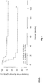

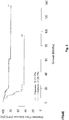


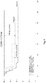
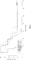
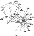
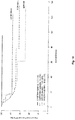

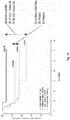


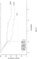

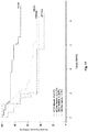

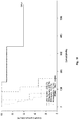

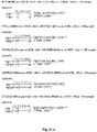
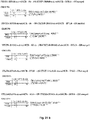
Classifications
-
- C—CHEMISTRY; METALLURGY
- C12—BIOCHEMISTRY; BEER; SPIRITS; WINE; VINEGAR; MICROBIOLOGY; ENZYMOLOGY; MUTATION OR GENETIC ENGINEERING
- C12Q—MEASURING OR TESTING PROCESSES INVOLVING ENZYMES, NUCLEIC ACIDS OR MICROORGANISMS; COMPOSITIONS OR TEST PAPERS THEREFOR; PROCESSES OF PREPARING SUCH COMPOSITIONS; CONDITION-RESPONSIVE CONTROL IN MICROBIOLOGICAL OR ENZYMOLOGICAL PROCESSES
- C12Q1/00—Measuring or testing processes involving enzymes, nucleic acids or microorganisms; Compositions therefor; Processes of preparing such compositions
- C12Q1/68—Measuring or testing processes involving enzymes, nucleic acids or microorganisms; Compositions therefor; Processes of preparing such compositions involving nucleic acids
- C12Q1/6876—Nucleic acid products used in the analysis of nucleic acids, e.g. primers or probes
- C12Q1/6883—Nucleic acid products used in the analysis of nucleic acids, e.g. primers or probes for diseases caused by alterations of genetic material
- C12Q1/6886—Nucleic acid products used in the analysis of nucleic acids, e.g. primers or probes for diseases caused by alterations of genetic material for cancer
-
- A—HUMAN NECESSITIES
- A61—MEDICAL OR VETERINARY SCIENCE; HYGIENE
- A61P—SPECIFIC THERAPEUTIC ACTIVITY OF CHEMICAL COMPOUNDS OR MEDICINAL PREPARATIONS
- A61P35/00—Antineoplastic agents
-
- C—CHEMISTRY; METALLURGY
- C12—BIOCHEMISTRY; BEER; SPIRITS; WINE; VINEGAR; MICROBIOLOGY; ENZYMOLOGY; MUTATION OR GENETIC ENGINEERING
- C12Q—MEASURING OR TESTING PROCESSES INVOLVING ENZYMES, NUCLEIC ACIDS OR MICROORGANISMS; COMPOSITIONS OR TEST PAPERS THEREFOR; PROCESSES OF PREPARING SUCH COMPOSITIONS; CONDITION-RESPONSIVE CONTROL IN MICROBIOLOGICAL OR ENZYMOLOGICAL PROCESSES
- C12Q2600/00—Oligonucleotides characterized by their use
- C12Q2600/106—Pharmacogenomics, i.e. genetic variability in individual responses to drugs and drug metabolism
-
- C—CHEMISTRY; METALLURGY
- C12—BIOCHEMISTRY; BEER; SPIRITS; WINE; VINEGAR; MICROBIOLOGY; ENZYMOLOGY; MUTATION OR GENETIC ENGINEERING
- C12Q—MEASURING OR TESTING PROCESSES INVOLVING ENZYMES, NUCLEIC ACIDS OR MICROORGANISMS; COMPOSITIONS OR TEST PAPERS THEREFOR; PROCESSES OF PREPARING SUCH COMPOSITIONS; CONDITION-RESPONSIVE CONTROL IN MICROBIOLOGICAL OR ENZYMOLOGICAL PROCESSES
- C12Q2600/00—Oligonucleotides characterized by their use
- C12Q2600/158—Expression markers
-
- C—CHEMISTRY; METALLURGY
- C12—BIOCHEMISTRY; BEER; SPIRITS; WINE; VINEGAR; MICROBIOLOGY; ENZYMOLOGY; MUTATION OR GENETIC ENGINEERING
- C12Q—MEASURING OR TESTING PROCESSES INVOLVING ENZYMES, NUCLEIC ACIDS OR MICROORGANISMS; COMPOSITIONS OR TEST PAPERS THEREFOR; PROCESSES OF PREPARING SUCH COMPOSITIONS; CONDITION-RESPONSIVE CONTROL IN MICROBIOLOGICAL OR ENZYMOLOGICAL PROCESSES
- C12Q2600/00—Oligonucleotides characterized by their use
- C12Q2600/16—Primer sets for multiplex assays
Definitions
- the present invention relates to methods and kits for screening patients with a cancer, particularly for determining whether a patient with a cancer would benefit of an antitumoral treatment.
- the TNM (for "Tumor-Node-Metastasis”) staging system uses the size of the tumor, the presence or absence of tumor in regional lymph nodes, and the presence or absence of distant metastases, to assign a stage and an outcome to the tumor.
- the TNM system developed from the observation that patients with small tumours have better prognosis than those with tumours of greater size at the primary site.
- patients with tumours confined to the primary site have better prognosis than those with regional lymph node involvement, which in turn is better than for those with distant spread of disease from one body part two another.
- cancers are usually staged into four levels.
- Stage I cancer is very localized cancer with no cancer in the lymph nodes.
- Stage II cancer has spread near to where the cancer started.
- Stage III cancer has spread to lymph nodes.
- Stage IV cancer has spread to a distant part of the body.
- the assigned stage is used as a basis for selection of appropriate therapy and for prognostic purposes. For example chemotherapy is always recommended for patients with stage IV cancers.
- regulation of gene expression refers to the control of the amount and timing of appearance of the functional product of a gene. Control of gene expression is vital to allow a cell to produce gene products when needed. The way that the information in genes is turned into gene products is regulated. In a short summary, regulation consists in a balance between actions of activators and inhibitors.
- EP 1 777 523 discloses methods for determining the outcome of a cancer in a patient, which are based on the quantification of one or several biological markers that are indicative of the presence of, or alternatively the level of, the adaptive immune response of said patient against said cancer.
- the two first groups of patients are to be considered as "bad responders” (i.e. the treatment will have a limited (or moderate) impact on their survival), whereas the third group of patients is to be considered as "good responders".
- a subject of the present invention is therefore a method and kits for screening patients with a cancer, particularly for determining whether a patient with a cancer would benefit of an antitumoral treatment.
- the present invention relates to a method for screening patients with a cancer comprising
- a patient who has a good adaptive immune response and a good immunosuppressive response could benefit of an anti tumoral treatment.
- the patient subjected to the above method suffers from a solid cancer selected from the group consisting of adrenal cortical cancer, anal cancer, bile duct cancer (e.g. periphilar cancer, distal bile duct cancer, intrahepatic bile duct cancer), bladder cancer, bone cancer (e.g. osteoblastoma, osteochrondroma, hemangioma, chondromyxoid fibroma, osteosarcoma, chondrosarcoma, fibrosarcoma, malignant fibrous histiocytoma, giant cell tumor of the bone, chordoma), brain and central nervous system cancer (e.g.
- a solid cancer selected from the group consisting of adrenal cortical cancer, anal cancer, bile duct cancer (e.g. periphilar cancer, distal bile duct cancer, intrahepatic bile duct cancer), bladder cancer, bone cancer (e.g. osteoblastoma, osteochrondroma, hemangio
- meningioma astocytoma, oligodendrogliomas, ependymoma, gliomas, medulloblastoma, ganglioglioma, Schwannoma, germinoma, craniopharyngioma), breast cancer (e.g. ductal carcinoma in situ, infiltrating ductal carcinoma, infiltrating lobular carcinoma, lobular carcinoma in situ, gynecomastia), Castleman disease (e.g. giant lymph node hyperplasia, angiofollicular lymph node hyperplasia), cervical cancer, colorectal cancer, endometrial cancer (e.g.
- lung cancer e.g. small cell lung cancer, non-small cell lung cancer
- mesothelioma plasmacytoma, nasal cavity and paranasal sinus cancer (e.g. esthesioneuroblastoma, midline granuloma), nasopharyngeal cancer, neuroblastoma, oral cavity and oropharyngeal cancer, ovarian cancer, pancreatic cancer, penile cancer, pituitary cancer, prostate cancer, retinoblastoma, rhabdomyosarcoma (e.g.
- rhabdomyosarcoma embryonal rhabdomyosarcoma, alveolar rhabdomyosarcoma, pleomorphic rhabdomyosarcoma), salivary gland cancer, skin cancer (e.g. melanoma, nonmelanoma skin cancer), stomach cancer, testicular cancer (e.g. seminoma, nonseminoma germ cell cancer), thymus cancer, thyroid cancer (e.g. follicular carcinoma, anaplastic carcinoma, poorly differentiated carcinoma, medullary thyroid carcinoma,), vaginal cancer, vulvar cancer, and uterine cancer (e.g. uterine leiomyosarcoma).
- skin cancer e.g. melanoma, nonmelanoma skin cancer
- stomach cancer testicular cancer (e.g. seminoma, nonseminoma germ cell cancer), thymus cancer, thyroid cancer (e.g. follicular carcinoma, anaplastic carcinoma, poorly differentiated carcinoma
- tissue sample means any tissue sample derived from the tumor of the patient.
- the tissue sample is obtained for the purpose of the in vitro evaluation.
- the sample can be fresh, frozen, fixed (e.g., formalin fixed), or embedded (e.g., paraffin embedded).
- the sample results from biopsy performed in a tumour sample of the patient.
- An example is an endoscopical biopsy performed in the bowel of the patient suffuring from colorectal cancer.
- an expression level EL 1 of a single gene representative of human adaptive immune response and of a single gene representative of human immunosuppressive response is assessed in the method of the invention.
- a limited number of genes of each kind provides good and reliable results and is easy to implement.
- Particularly a single reference value is sufficient for each of both genes. The higher the number of genes, the more sophisticated is the reference value. Examples of determination of reference values are given thereafter.
- genes than one or two pairs of genes are more difficult to implement and more expensive and time consuming but however provides other advantages. For example if the assessment of the expression level of one gene is erroneous, the overall result is compensated by the reserved of the other genes of the same kind (human adaptive immune response or immunosuppressive response).
- the expression “gene representative of the adaptive immune response” refers to any gene that is expressed by a cell that is an actor of the adaptive immune response in the tumor or that contributes to the settlement of the adaptive immune response in the tumor.
- the adaptive immune response also called “acquired immune response”
- cells of the adapative immune response include but are not limited to cytotoxic T cells, T memory T cells, Th1 and Th2 cells, activated macrophages and activated dendritic cells, NK cells and NKT cells.
- a gene representative of the adaptive immune response may be typically selected from the cluster of the co-modulated genes for the Th1 adaptive immunity, for the cytotoxic response, or for the memory response, and may encode for a Th1 cell surface marker, an interleukin (or an interleukin receptor), or a chemokine or (a chemokine receptor).
- the gene representative of the adaptative immune response is selected from the group consisting of
- the expression "gene representative of the immunosuppressive response” refers to any gene that is expressed by a cell that is an actor of the immunosuppressive response in the tumor or that contributes to the settlement of the immunosuppressive response in the tumor.
- the immunosuppressive response comprises
- cells of the immunosuppressive response include immature dendritic cells (CD1A), regulatory T cells (Treg cells) and Th17 cells expressing IL17A gene.
- a gene representative of the adaptive immune response may be typically selected from the group of the co-modulated adaptive immune genes, whereas the immunosuppressive genes, may be representative of the inactivation of immune cells (e.g. dendritic cells) and may contribute to induction of an immunosuppressive response.
- the immunosuppressive genes may be representative of the inactivation of immune cells (e.g. dendritic cells) and may contribute to induction of an immunosuppressive response.
- the gene representative of the immunosuppressive response is selected from the group consisting of genes reported in Table 2 hereunder: Table 2 CD274 CTLA4 IHH IL17A PDCD1 PF4 PROM1 REN TSLP VEGFA
- Said genes are preferred because they provide the best results for the response of a patient to the treatment as shown hereafter in table 5.
- a gene representative of the adaptative immune response is selected from the group consisting of GNLY, CXCL13, CX3CL1, CXCL9, ITGAE, CCL5, GZMH, IFNG, CCR2, CD3D, CD3E, CD3G, CD8A, CXCL10, CXCL11, GZMA, GZMB, GZMK, GZMM, IL15, IRF1, LTK, PRF1, STAT1, CD69, CD247, ICOS, CXCR3, STAT4, CCL2 and TBX21 and a gene representative of the immunosuppressive response is selected from the group consisting of PF4, REN, VEGFA, TSLP, IL17A, PROM1, IHH, CD1A, CTLA4, PDCD1, CD276, CD274, and VTCN1 (B7H4).
- a gene representative of the adaptative immune response and a gene representative of the immunosuppressive response are selected respectively from the groups consisting of the genes of Tables 1 and 2 above.
- a pair of genes is selected from the combinations of genes of table 5 hereafter. Combinations of genes of both types of figure 9 linked by thick lines are more preferred.
- Combinations Nr 2, 3, 7, 8, 9, 10, 15, 41, 43, 44, 55, 56, and 66 of table 5 hereafter are preferred.
- Other preferred pairs of genes are combinations Nr 2, 3, 7, 8, 9, 10, 15, 41, 43, 44, 55, 56, and 66 of table 5 hereafter and CD3G-VEGF, CD3E-VEGF and CD8A-VEGF.
- genes selected for use in the present process may depend on the type of treatment contemplated for the patient.
- genes selected from the group consisting of CX3CL1 IL15, CD247, CD3G, CD8A, PRF1, CCL5 and TBX21 for the immunosuppressive response preferably CX3CL1 and IL15 and gene CTLA4 for the adaptative immune response will be preferred when a treatment using a drug such as a monoclonal antibody working by activating the immune system such as Ipilimumab, also known as MDX-010 or MDX-101, marketed as Yervoy®, is contemplated for a patient.
- Genes selected from the group consisting of IL15 and GZMA for the adaptative immune response, and gene VEGFA for the immunosuppressive response will be preferred when a treatment such as an antibody that inhibits vascular endothelial growth factor A (VEGF-A) such as bevacizumab marketed as Avastin®, is contemplated for a patient.
- VEGF-A vascular endothelial growth factor A
- GZMA - PDCD1 also designated as CD279
- a treatment such as an antibody that targets PD-1 such as BMS-936558, is contemplated for a patient.
- the name of each of the genes of interest refers to the internationally recognised name of the corresponding gene, as found in internationally recognised gene sequences and protein sequences databases, including the database from the HUGO Gene Nomenclature Committee that is available notably at the following Internet address: http://www.gene.ucl.ac.uk/nomenclature/index.html.
- the name of each of the genes of interest may also refer to the internationally recognised name of the corresponding gene, as found in the internationally recognised gene sequences database Genbank. Through these internationally recognised sequence databases, the nucleic acid to each of the gene of interest described herein may be retrieved by one skilled in the art.
- the cancer prognosis method of the invention may be performed with a combination of genes provided that the combination comprises at least one one gene representative of the adaptive immune response and at least one gene representative of the immunosuppressive response.
- the number of genes that may be used in the present method is only limited by the number of distinct biological genes of interest that are practically available at the time of carrying out the method.
- a combination of 2, 3, 4, 5, 6, 7, 8, 9, 10, 11, 12, 13, 14, 15, 16, 17, 18, 19, 20, 21, 22, 23, 24, 25, 26, 27, 28, 29, 30, 31, 32, 33, 34, 35, 36, 37, 38, 39, 40, 41, 42, 43, 44, 45, 46, 47, 48, 49 and 50 distinct genes are quantified, preferably a combination of 2, 3, 4, 5, 6, 7, 8, 9, or 10 genes and more preferably a combination of 2, 3, 4, 5, or 6, genes.
- the number of combined genes that are required for reaching a high statistical relevance e.g. P value lower than 10 -3
- the number of genes used as genes representative of the adaptive immune response and the number of genes used as genes representative of the immunosuppressive response may be the same or different.
- an about balanced number of genes of each kind is used, for example 2 of each, or three of each, or 5 of one kind and 6 of the other kind.
- Determining an expression level of a gene in a tumor sample obtained from a patient can be implemented by a panel of techniques well known in the art.
- an expression level of a gene is assessed by determining the quantity of mRNA produced by this gene.
- a subject of the present application is therefore a a method for screening patients with a cancer defined above comprising determining an expression level ELA of one or several genes representative of human adaptive immune response or the expression level ELI of one or several genes representative of human immunosuppressive response by determining the quantity of mRNA corresponding to said genes.
- nucleic acid contained in the samples e.g., cell or tissue prepared from the patient
- the thus extracted mRNA is then detected by hybridization (e. g., Northern blot analysis) and/or amplification (e.g., RT-PCR).
- hybridization e. g., Northern blot analysis
- amplification e.g., RT-PCR
- quantitative or semi-quantitative RT-PCR is preferred.
- Real-time quantitative or semi-quantitative RT-PCR is particularly advantageous.
- LCR ligase chain reaction
- TMA transcription-mediated amplification
- SDA strand displacement amplification
- NASBA nucleic acid sequence based amplification
- NGS quantitative new generation sequencing of RNA
- Nucleic acids comprising at least 10 nucleotides and exhibiting sequence complementarity or homology to the mRNA of interest herein find utility as hybridization probes or amplification primers. It is understood that such nucleic acids need not be completely identical, but are typically at least about 80% identical to the homologous region of comparable size, more preferably 85% identical and even more preferably 90-95% identical. In certain embodiments, it will be advantageous to use nucleic acids in combination with appropriate means, such as a detectable label, for detecting hybridization. A wide variety of appropriate indicators are known in the art including, fluorescent, radioactive, enzymatic or other ligands (e. g. avidin/biotin).
- Probes typically comprise single-stranded nucleic acids of between 10 to 1000 nucleotides in length, for instance of between 10 and 800, more preferably of between 15 and 700, typically of between 20 and 500 nucleotides.
- Primers typically are shorter single-stranded nucleic acids, of between 10 to 25 nucleotides in length, designed to perfectly or almost perfectly match a nucleic acid of interest, to be amplified.
- the probes and primers are "specific" to the nucleic acids they hybridize to, i.e. they preferably hybridize under high stringency hybridization conditions (corresponding to the highest melting temperature Tm, e.g., 50 % formamide, 5x or 6x SCC.
- SCC is a 0.15 M NaCl, 0.015 M Na-citrate).
- Nucleic acids which may be used as primers or probes in the above amplification and detection method may be assembled as a kit.
- a kit includes consensus primers and molecular probes.
- a preferred kit also includes the components necessary to determine if amplification has occurred.
- a kit may also include, for example, PCR buffers and enzymes; positive control sequences, reaction control primers; and instructions for amplifying and detecting the specific sequences.
- the methods of the invention comprise the steps of providing total RNAs extracted from cumulus cells and subjecting the RNAs to amplification and hybridization to specific probes, more particularly by means of a quantitative or semi-quantitative RT-PCR.
- Probes made using the disclosed methods can be used for nucleic acid detection, such as in situ hybridization (ISH) procedures (for example, fluorescence in situ hybridization (FISH), chromogenic in situ hybridization (CISH) and silver in situ hybridization (SISH)) or comparative genomic hybridization (CGH).
- ISH in situ hybridization
- FISH fluorescence in situ hybridization
- CISH chromogenic in situ hybridization
- SISH silver in situ hybridization
- CGH comparative genomic hybridization
- ISH In situ hybridization
- a sample containing target nucleic acid sequence e.g., genomic target nucleic acid sequence
- a metaphase or interphase chromosome preparation such as a cell or tissue sample mounted on a slide
- a labeled probe specifically hybridizable or specific for the target nucleic acid sequence (e.g., genomic target nucleic acid sequence).
- the slides are optionally pretreated, e.g., to remove paraffin or other materials that can interfere with uniform hybridization.
- the sample and the probe are both treated, for example by heating to denature the double stranded nucleic acids.
- the probe (formulated in a suitable hybridization buffer) and the sample are combined, under conditions and for sufficient time to permit hybridization to occur (typically to reach equilibrium).
- the chromosome preparation is washed to remove excess probe, and detection of specific labeling of the chromosome target is performed using standard techniques.
- a biotinylated probe can be detected using fluorescein-labeled avidin or avidin-alkaline phosphatase.
- fluorescein-labeled avidin or avidin-alkaline phosphatase For fluorochrome detection, the fluorochrome can be detected directly, or the samples can be incubated, for example, with fluorescein isothiocyanate (FITC)-conjugated avidin. Amplification of the FITC signal can be effected, if necessary, by incubation with biotin-conjugated goat antiavidin antibodies, washing and a second incubation with FITC-conjugated avidin.
- FITC fluorescein isothiocyanate
- samples can be incubated, for example, with streptavidin, washed, incubated with biotin-conjugated alkaline phosphatase, washed again and pre-equilibrated (e.g., in alkaline phosphatase (AP) buffer).
- AP alkaline phosphatase
- Numerous reagents and detection schemes can be employed in conjunction with FISH, CISH, and SISH procedures to improve sensitivity, resolution, or other desirable properties.
- probes labeled with fluorophores including fluorescent dyes and QUANTUM DOTS®
- fluorophores including fluorescent dyes and QUANTUM DOTS®
- the probe can be labeled with a nonfluorescent molecule, such as a hapten (such as the following non-limiting examples: biotin, digoxigenin, DNP, and various oxazoles, pyrrazoles, thiazoles, nitroaryls, benzofurazans, triterpenes, ureas, thioureas, rotenones, coumarin, courmarin-based compounds, Podophyllotoxin, Podophyllotoxin-based compounds, and combinations thereof), ligand or other indirectly detectable moiety.
- a hapten such as the following non-limiting examples: biotin, digoxigenin, DNP, and various oxazoles, pyrrazoles, thiazoles, nitroaryls, benzofurazans, triterpenes, ureas, thioureas, rotenones, coumarin, courmarin-based compounds, Podophyllotoxin, Podo
- Probes labeled with such non-fluorescent molecules (and the target nucleic acid sequences to which they bind) can then be detected by contacting the sample (e.g., the cell or tissue sample to which the probe is bound) with a labeled detection reagent, such as an antibody (or receptor, or other specific binding partner) specific for the chosen hapten or ligand.
- a labeled detection reagent such as an antibody (or receptor, or other specific binding partner) specific for the chosen hapten or ligand.
- the detection reagent can be labeled with a fluorophore (e.g., QUANTUM DOT®) or with another indirectly detectable moiety, or can be contacted with one or more additional specific binding agents (e.g., secondary or specific antibodies), which can be labeled with a fluorophore.
- the probe, or specific binding agent (such as an antibody, e.g., a primary antibody, receptor or other binding agent) is labeled with an enzyme that is capable of converting a fluorogenic or chromogenic composition into a detectable fluorescent, colored or otherwise detectable signal (e.g., as in deposition of detectable metal particles in SISH).
- the enzyme can be attached directly or indirectly via a linker to the relevant probe or detection reagent. Examples of suitable reagents (e.g., binding reagents) and chemistries (e.g., linker and attachment chemistries) are described in U.S. Patent Application Publications Nos. 2006/0246524 ; 2006/0246523 , and 2007/0117153 .
- multiplex detection schemes can be produced to facilitate detection of multiple target nucleic acid sequences (e.g., genomic target nucleic acid sequences) in a single assay (e.g., on a single cell or tissue sample or on more than one cell or tissue sample).
- a first probe that corresponds to a first target sequence can be labelled with a first hapten, such as biotin, while a second probe that corresponds to a second target sequence can be labelled with a second hapten, such as DNP.
- the bound probes can be detected by contacting the sample with a first specific binding agent (in this case avidin labelled with a first fluorophore, for example, a first spectrally distinct QUANTUM DOT®, e.g., that emits at 585 mn) and a second specific binding agent (in this case an anti-DNP antibody, or antibody fragment, labelled with a second fluorophore (for example, a second spectrally distinct QUANTUM DOT®, e.g., that emits at 705 mn).
- a first specific binding agent in this case avidin labelled with a first fluorophore, for example, a first spectrally distinct QUANTUM DOT®, e.g., that emits at 585 mn
- a second specific binding agent in this case an anti-DNP antibody, or antibody fragment, labelled with a second fluorophore (for example, a second spectrally distinct QUANTUM DOT®,
- Probes typically comprise single-stranded nucleic acids of between 10 to 1000 nucleotides in length, for instance of between 10 and 800, more preferably of between 15 and 700, typically of between 20 and 500.
- Primers typically are shorter single-stranded nucleic acids, of between 10 to 25 nucleotides in length, designed to perfectly or almost perfectly match a nucleic acid of interest, to be amplified.
- the probes and primers are "specific" to the nucleic acids they hybridize to, i.e. they preferably hybridize under high stringency hybridization conditions (corresponding to the highest melting temperature Tm, e.g., 50 % formamide, 5x or 6x SCC.
- SCC is a 0.15 M NaCl, 0.015 M Na-citrate).
- the nucleic acid primers or probes used in the above amplification and detection method may be assembled as a kit.
- a kit includes consensus primers and molecular probes.
- a preferred kit also includes the components necessary to determine if amplification has occurred.
- the kit may also include, for example, PCR buffers and enzymes; positive control sequences, reaction control primers; and instructions for amplifying and detecting the specific sequences.
- the methods of the invention comprise the steps of providing total RNAs extracted from cumulus cells and subjecting the RNAs to amplification and hybridization to specific probes, more particularly by means of a quantitative or semi-quantitative RT-PCR.
- the expression level is determined by DNA chip analysis.
- DNA chip or nucleic acid microarray consists of different nucleic acid probes that are chemically attached to a substrate, which can be a microchip, a glass slide or a microsphere-sized bead.
- a microchip may be constituted of polymers, plastics, resins, polysaccharides, silica or silica-based materials, carbon, metals, inorganic glasses, or nitrocellulose.
- Probes comprise nucleic acids such as cDNAs or oligonucleotides that may be about 10 to about 60 base pairs.
- a sample from a test subject optionally first subjected to a reverse transcription, is labelled and contacted with the microarray in hybridization conditions, leading to the formation of complexes between target nucleic acids that are complementary to probe sequences attached to the microarray surface.
- the labelled hybridized complexes are then detected and can be quantified or semi-quantified. Labelling may be achieved by various methods, e.g. by using radioactive or fluorescent labelling.
- Many variants of the microarray hybridization technology are available to the man skilled in the art (see e.g. the review by Hoheisel, Nature Reviews, Genetics, 2006, 7:200-210 ).
- the expression level of a gene may be expressed as absolute expression level or normalized expression level. Both types of values may be used in the present method.
- the expression level of a gene is preferably expressed as normalized expression level when quantitative PCR is used as method of assessment of the expression level because small differences at the beginning of an experiment could provide huge differences after a number of cycles.
- expression levels are normalized by correcting the absolute expression level of a gene by comparing its expression to the expression of a gene that is not relevant for determining the cancer stage of the patient, e.g., a housekeeping gene that is constitutively expressed.
- Suitable genes for normalization include housekeeping genes such as the actin gene ACTB, ribosomal 18S gene, GUSB, PGK1 and TFRC. This normalization allows comparing the expression level of one sample, e.g., a patient sample, with the expression level of another sample, or comparing samples from different sources.
- the present method includes comparing the expression levels ELA 1 - ELA n and ELI 1 - ELI n determined at step i) with predetermined reference values ELRA 1 - ELRA n and ELRI 1 - ELRI n (collectively named ELR).
- the predetermined reference values ELRA 1 - ELRA n and ELRI 1 - ELRI n may be a definite value or a range of values.
- ELA 1 of a tumour sample of a patient for the genes considered is higher than the corresponding predetermined reference value (or range of values) ELRA 1 , the patient will be considered as "good responder” ("High” or any such assessment such as “positive”, etc.) and will be considered as “bad responder” ("Low”or any such assessment such as "negative”, etc.), if ELA 1 is lower than ELRA 1 . Similar considerations apply to ELI 1 - ELI n and ELRI 1 - ELRI n .
- Predetermined reference values used for comparison may consist of "cut-off" values that may be determined as described in WO2007045996 .
- Predetermined reference values used for comparison may consist of "cut-off" values that may be determined as described hereunder.
- Each reference (“cut-off") value ELR for each gene may be determined by carrying out a method comprising the steps of:
- a confidence interval may be constructed around the value of expression level thus obtained, for example ELR ⁇ 5 or 10%.
- the expression level of a gene G1 has been assessed for 100 cancer samples of 100 patients.
- the 100 samples are ranked according to the expression level of gene G1.
- Sample 1 has the highest expression level and sample 100 has the lowest expression level.
- a first grouping provides two subsets: on one side sample Nr 1 and on the other side the 99 other samples.
- the next grouping provides on one side samples 1 and 2 and on the other side the 98 remaining samples etc., until the last grouping: on one side samples 1 to 99 and on the other side sample Nr 100.
- Kaplan Meier curves are prepared for each of the 99 groups of two subsets. Also for each of the 99 groups, the p value between both subsets was calculated.
- the reference value ELR is selected such as the discrimination based on the criterion of the minimum p value is the strongest. In other terms, the expression level corresponding to the boundary between both subsets for which the p value is minimum is considered as the reference value. It should be noted that according to the experiments made by the inventors, the reference value ELR is not necessarily the median value of expression levels.
- the reference value ELR (cut-off value) may be used in the present method to discriminate tumour samples and therefore the corresponding patients.
- Kaplan-Meier curves of percentage of survival as a function of time are commonly used to measure the fraction of patients living for a certain amount of time after treatment and are well known by the man skilled in the art. P value is conventionally used in statistical significance testing.
- Such predetermined reference values of expression level may be determined for any gene defined above and may be used in the method of the invention for screening patients with a cancer.
- a reference value ELRA for one or for each of several genes representative of human adaptive immune response and a reference value ELRI for one or each of several genes representative of human immunosuppressive response are necessary for implementing the method of the invention.
- a tumor tissue sample from a patient suffering of cancer may be subjected to determination of the expression levels ELA 1 - ELA n and ELI 1 - ELI n respectively of genes GA 1 - GA n and G1 1 - GI n 1 by using the same technique as the technique used for obtaining reference values ELRA 1 - ELRA n and ELRI 1 - ELRI n , for example by determining the amount of mRNA (for example using Quantitative PCR) produced by the relevant gene.
- Different techniques may be used for obtaining the relevant data for two different genes or several different genes. However, preferably a same technique is implemented, preferably selected among those previously cited or used in the examples hereafter.
- ELA1 is higher than ELRA1
- the patient is considered as a good responder as regards human adaptive immune response and as a bad responder if ELA1 is lower than ELRA1.
- ELI1 is higher than ELRI1
- the patient is considered as a good responder as regards human immunosuppressive response and as a bad responder if ELI1 is lower than ELRI1.
- a numerical value lower than the reference value may actually mean that the expression level is higher than the reference level.
- a dCt value lower than the relevant reference value means that the signal was detected earlier, i.e.: the expression level of the gene is higher than the reference level.
- the method for screening patients which is the subject matter of the present invention has very advantageous properties and qualities.
- the method allows in particular, from the knowledge of the response of the patient concerning human adaptive immune response and human immunosuppressive response, to make a good assessment of prognosis with respect to DFS and OS of a patient, independently of the cancer type, origin or stage as evidenced in the experimental section..
- a minimal statistical significance value (minimal threshold of significance, e.g. maximal threshold P value) is arbitrarily set and a range of a plurality of arbitrary quantification values for which the statistical significance value calculated at step g) is higher (more significant, e.g. lower P value) are retained, so that a range of quantification values is provided.
- This range of quantification values includes a "cut-off" value as described above. According to this specific embodiment of a "cut-off" value, poor or good clinical outcome prognosis can be determined by comparing the expression level with the range of values which are identified.
- a cut-off value thus consists of a range of quantification values, e.g.
- a suitable (exemplary) range may be from 4-6.
- a patient may be assessed by comparing values obtained by measuring the expression level of a gene representative of the immune adaptive response and a gene representative of the immunosuppressive response, where values greater than 5 reveal a good prognosis (a contrario a bad prognosis when the gene is representative of immunosuppressive response) and values less than 5 reveal a poor prognosis; (a contrario a good prognosis when the gene is representative of immunosuppressive response).
- a patient may be assessed by comparing values obtained by measuring the expression level of a gene representative of the immune adaptive response and a gene representative of the immunosuppressive response and comparing the values on a scale, where values above the range of 4-6 indicate a good prognosis (a contrario a bad prognosis when the gene is representative of immunosuppressive response) and values below the range of 4-6 indicate a poor prognosis (a contrario a good prognosis when the gene is representative of immunosuppressive response), with values falling within the range of 4-6 indicating an intermediate prognosis.
- the method for screening patients with a cancer of the invention further comprises the step of concluding that a patient would advantageously receive an antitumoral treatment if the patient is a good responder for each of human adaptive immune response and human immunosuppressive response.
- the method for screening patients with a cancer of the invention further comprises the step of concluding that a patient would not advantageously receive an antitumoral treatment if the patient is not a good responder for both of human adaptive immune response and human immunosuppressive response.
- the method of the invention comprises comparison steps which include a classification of the quantification values measured for the expression level of a gene into two possibilities, respectively: (i) a first possibility when the quantification value for the expression level is higher than the predetermined corresponding reference value (the first possibility is named "Hi” for example) and (ii) a second possibility when the quantification value for the expression level is lower than the predetermined corresponding reference value (the second possibility is named " Lo " for example).
- the result of the comparison step consists of a "Hi" value for the gene representative of the immune adaptive response and a "Lo” value for the gene representative of the immunosuppressive response
- a good prognosis is provided and the treatment will provide a limited (or moderate) impact on the survival of the patient (i.e. a "bad responder to antitumoral treatment”).
- the result of the comparison step consists of a "Lo” value for the gene representative of the immune adaptive response and a "Hi” value for the gene representative of the immunosuppressive response then a poor prognosis is provided and the treatment will provide a limited (or moderate) impact on the survival of the patient (i.e.
- a score which is a composite of the classifications may also be calculated. For example a positive coefficient (e.g. +1) is allocated when expression level of the gene representative of the immune adaptive response is higher than the predetermined reference value and a negative coefficient (e.g. -1) is allocated when expression level of the gene representative of the immune adaptive response is lower than the predetermined reference value. Conversely, a positive coefficient (e.g. +1) is allocated when expression level of the gene representative of the immunosupressive response is lower than the predetermined reference value and a negative coefficient (e.g. -1) is allocated when expression level of the gene representative of the immunosupressive response is higher than the predetermined reference value.
- a positive coefficient e.g. +1
- a negative coefficient e.g. -1
- Calculating a score is particularly suitable to make easier to understand the results of the comparison step when a combination of more than 2 genes is used. Typically the more such a score is close to the value 0, the more the treatment will have a positive effect on the survival of the patient (i.e. the patient would benefit of the treatment).
- the present method therefore allows defining inter alia a new group of patients which had never been identified until now, i.e. patients whose cancer will be successfully treated by an anti-cancer treatment.
- An anti-cancer treatment may consist of radiotherapy, chemotherapy or immunotherapy.
- the treatment may consist of an adjuvant therapy (i.e. treatment after chirurgical resection of the primary tumor) of a neoadjuvant therapy (i.e. treatment before chirurgical resection of the primary tumor).
- the present invention therefore relates to a chemotherapeutic agent, a radiotherapeutic agent, or an immunotherapeutic agent, preferably the latter, for use in the treatment of a stage I-III cancer patient who has been considered as a good responder to antitumoral treatment according to the above method of the invention.
- chemotherapeutic agent refers to chemical compounds that are effective in inhibiting tumor growth.
- examples of chemotherapeutic agents include alkylating agents such as thiotepa and cyclosphosphamide; alkyl sulfonates such as busulfan, improsulfan and piposulfan; aziridines such as benzodopa, carboquone, meturedopa, and uredopa; ethylenimines and methylamelamines including altretamine, triethylenemelamine, trietylenephosphoramide, triethylenethiophosphaorarnide and trimethylolomelamine; acetogenins (especially bullatacin and bullatacinone); a carnptothecin (including the synthetic analogue topotecan); bryostatin; callystatin; CC-1065 (including its adozelesin, carzelesin and bizelesin synthetic analogues); cryptophycins
- calicheamicin especially calicheamicin (11 and calicheamicin 211, see, e.g., Agnew Chem Intl. Ed. Engl. 33:183-186 (1994 ); dynemicin, including dynemicin A; an esperamicin; as well as neocarzinostatin chromophore and related chromoprotein enediyne antiobiotic chromomophores), aclacinomysins, actinomycin, authramycin, azaserine, bleomycins, cactinomycin, carabicin, canninomycin, carzinophilin, chromomycins, dactinomycin, daunorubicin, detorubicin, 6-diazo-5-oxo-L-norleucine, doxorubicin (including morpholino-doxorubicin, cyanomorpholino-doxorubicin, 2-pyrrolin
- paclitaxel (TAXOL®, Bristol-Myers Squibb Oncology, Princeton, N.].) and doxetaxel (TAXOTERE®, Rhone-Poulenc Rorer, Antony, France); chlorambucil; gemcitabine; 6-thioguanine; mercaptopurine; methotrexate; platinum analogs such as cisplatin and carboplatin; vinblastine; platinum; etoposide (VP-16); ifosfamide; mitomycin C; mitoxantrone; vincristine; vinorelbine; navelbine; novantrone; teniposide; daunomycin; aminopterin; xeloda; ibandronate; CPT-1 1 ; topoisomerase inhibitor RFS 2000; difluoromethylornithine (DMFO); retinoic acid; capecitabine; and phannaceutically acceptable salts, acids or derivatives of any of the above.
- antihormonal agents that act to regulate or inhibit honnone action on tumors
- anti-estrogens including for example tamoxifen, raloxifene, aromatase inhibiting 4(5)-imidazoles, 4-hydroxytamoxifen, trioxifene, keoxifene, LY117018, onapristone, and toremifene (Fareston); and anti-androgens such as flutamide, nilutamide, bicalutamide, leuprolide, and goserelin; and phannaceutically acceptable salts, acids or derivatives of any of the above.
- immunotherapeutic agent refers to a compound, composition or treatment that indirectly or directly enhances, stimulates or increases the body's immune response against cancer cells and/or that decreases the side effects of other anticancer therapies. Immunotherapy is thus a therapy that directly or indirectly stimulates or enhances the immune system's responses to cancer cells and/or lessens the side effects that may have been caused by other anti-cancer agents. Immunotherapy is also referred to in the art as immunologic therapy, biological therapy biological response modifier therapy and biotherapy. Examples of common immunotherapeutic agents known in the art include, but are not limited to, cytokines, cancer vaccines, monoclonal antibodies and non-cytokine adjuvants. Alternatively the immunotherapeutic treatment may consist of administering the patient with an amount of immune cells (T cells, NK, cells, dendritic cells, B cells).
- Immunotherapeutic agents can be non-specific, i.e. boost the immune system generally so that the human body becomes more effective in fighting the growth and/or spread of cancer cells, or they can be specific, i.e. targeted to the cancer cells themselves immunotherapy regimens may combine the use of non-specific and specific immunotherapeutic agents.
- Non-specific immunotherapeutic agents are substances that stimulate or indirectly improve the immune system.
- Non-specific immunotherapeutic agents have been used alone as a main therapy for the treatment of cancer, as well as in addition to a main therapy, in which case the non-specific immunotherapeutic agent functions as an adjuvant to enhance the effectiveness of other therapies (e.g. cancer vaccines).
- Non-specific immunotherapeutic agents can also function in this latter context to reduce the side effects of other therapies, for example, bone marrow suppression induced by certain chemotherapeutic agents.
- Non-specific immunotherapeutic agents can act on key immune system cells and cause secondary responses, such as increased production of cytokines and immunoglobulins. Alternatively, the agents can themselves comprise cytokines.
- Non-specific immunotherapeutic agents are generally classified as cytokines or non-cytokine adjuvants.
- cytokines have found application in the treatment of cancer either as general non-specific immunotherapies designed to boost the immune system, or as adjuvants provided with other therapies.
- Suitable cytokines include, but are not limited to, interferons, interleukins and colony-stimulating factors.
- Interferons contemplated by the present invention include the common types of IFNs, IFN-alpha (IFN-a), IFN-beta (IFN-beta) and IFN-gamma (IFN-y).
- IFNs can act directly on cancer cells, for example, by slowing their growth, promoting their development into cells with more normal behaviour and/or increasing their production of antigens thus making the cancer cells easier for the immune system to recognise and destroy.
- IFNs can also act indirectly on cancer cells, for example, by slowing down angiogenesis, boosting the immune system and/or stimulating natural killer (NK) cells, T cells and macrophages.
- NK natural killer
- IFN-alpha Recombinant IFN-alpha is available commercially as Roferon (Roche Pharmaceuticals) and Intron A (Schering Corporation).
- Roferon Roche Pharmaceuticals
- Intron A Strecombinant IFN-alpha
- Interleukins contemplated by the present invention include IL-2, IL-4, IL-11 and IL-12.
- Examples of commercially available recombinant interleukins include Proleukin® (IL-2; Chiron Corporation) and Neumega® (IL-12; Wyeth Pharmaceuticals).
- Zymogenetics, Inc. (Seattle, Wash.) is currently testing a recombinant form of IL-21, which is also contemplated for use in the combinations of the present invention.
- Interleukins alone or in combination with other immunotherapeutics or with chemotherapeutics, have shown efficacy in the treatment of various cancers including renal cancer (including metastatic renal cancer), melanoma (including metastatic melanoma), ovarian cancer (including recurrent ovarian cancer), cervical cancer (including metastatic cervical cancer), breast cancer, colorectal cancer, lung cancer, brain cancer, and prostate cancer.
- renal cancer including metastatic renal cancer
- melanoma including metastatic melanoma
- ovarian cancer including recurrent ovarian cancer
- cervical cancer including metastatic cervical cancer
- breast cancer including metastatic cervical cancer
- colorectal cancer lung cancer
- brain cancer and prostate cancer.
- Interleukins have also shown good activity in combination with IFN-alpha in the treatment of various cancers ( Negrier et al., Ann Oncol. 2002 13(9): 1460-8 ; Touranietal, J. Clin. Oncol. 2003 21(21):398794 ).
- Colony-stimulating factors contemplated by the present invention include granulocyte colony stimulating factor (G-CSF or filgrastim), granulocyte-macrophage colony stimulating factor (GM-CSF or sargramostim) and erythropoietin (epoetin alfa, darbepoietin).
- G-CSF or filgrastim granulocyte colony stimulating factor
- GM-CSF or sargramostim granulocyte-macrophage colony stimulating factor
- erythropoietin epoetin alfa, darbepoietin
- colony stimulating factors are available commercially, for example, Neupogen® (G-CSF; Amgen), Neulasta (pelfilgrastim; Amgen), Leukine (GM-CSF; Berlex), Procrit (erythropoietin; Ortho Biotech), Epogen (erythropoietin; Amgen), Arnesp (erytropoietin).
- Colony stimulating factors have shown efficacy in the treatment of cancer, including melanoma, colorectal cancer (including metastatic colorectal cancer), and lung cancer.
- Non-cytokine adjuvants suitable for use in the combinations of the present invention include, but are not limited to, Levamisole, alum hydroxide (alum), Calmette-Guerin bacillus (ACG), incomplete Freund's Adjuvant (IFA), QS-21, DETOX, Keyhole limpet hemocyanin (KLH) and dinitrophenyl (DNP).
- Non-cytokine adjuvants in combination with other immuno- and/or chemotherapeutics have demonstrated efficacy against various cancers including, for example, colon cancer and colorectal cancer (Levimasole); melanoma (BCG and QS-21); renal cancer and bladder cancer (BCG).
- immunotherapeutic agents can be active, i.e. stimulate the body's own immune response, or they can be passive, i.e. comprise immune system components that were generated external to the body.
- Passive specific immunotherapy typically involves the use of one or more monoclonal antibodies that are specific for a particular antigen found on the surface of a cancer cell or that are specific for a particular cell growth factor.
- Monoclonal antibodies may be used in the treatment of cancer in a number of ways, for example, to enhance a subject's immune response to a specific type of cancer, to interfere with the growth of cancer cells by targeting specific cell growth factors, such as those involved in angiogenesis, or by enhancing the delivery of other anticancer agents to cancer cells when linked or conjugated to agents such as chemotherapeutic agents, radioactive particles or toxins.
- Monoclonal antibodies currently used as cancer immunotherapeutic agents that are suitable for inclusion in the combinations of the present invention include, but are not limited to, rituximab (Rituxan®), trastuzumab (Herceptin®), ibritumomab tiuxetan (Zevalin®), tositumomab (Bexxar®), cetuximab (C-225, Erbitux®), bevacizumab (Avastin®), gemtuzumab ozogamicin (Mylotarg®), alemtuzumab (Campath®), and BL22.
- Monoclonal antibodies are used in the treatment of a wide range of cancers including breast cancer (including advanced metastatic breast cancer), colorectal cancer (including advanced and/or metastatic colorectal cancer), ovarian cancer, lung cancer, prostate cancer, cervical cancer, melanoma and brain tumours.
- Other examples include anti-CTLA4 antibodies (e.g. Ipilimumab), anti-PD1 antibodies, anti-PDL1 antibodies, anti-TIMP3 antibodies, anti-LAG3 antibodies, anti-B7H3 antibodies, anti-B7H4 antibodies or anti-B7H6 antibodies.
- Monoclonal antibodies can be used alone or in combination with other immunotherapeutic agents or chemotherapeutic agents.
- Cancer vaccines have been developed that comprise whole cancer cells, parts of cancer cells or one or more antigens derived from cancer cells. Cancer vaccines, alone or in combination with one or more immuno- or chemotherapeutic agents are being investigated in the treatment of several types of cancer including melanoma, renal cancer, ovarian cancer, breast cancer, colorectal cancer, and lung cancer. Non-specific immunotherapeutics are useful in combination with cancer vaccines in order to enhance the body's immune response.
- the immunotherapeutic treatment may consist of an adoptive immunotherapy as described by Nicholas P. Restifo, Mark E. Dudley and Steven A. Rosenberg "Adoptive immunotherapy for cancer: harnessing the T cell response, Nature Reviews Immunology, Volume 12, April 2012 ).
- adoptive immunotherapy the patient's circulating lymphocytes, or tumor infiltrated lymphocytes, are isolated in vitro, activated by lymphokines such as IL-2 or transuded with genes for tumor necrosis, and readministered (Rosenberg et al., 1988; 1989).
- the activated lymphocytes are most preferably be the patient's own cells that were earlier isolated from a blood or tumor sample and activated (or "expanded") in vitro.
- This form of immunotherapy has produced several cases of regression of melanoma and renal carcinoma.
- radiotherapeutic agent as used herein, is intended to refer to any radiotherapeutic agent known to one of skill in the art to be effective to treat or ameliorate cancer, without limitation.
- the radiotherapeutic agent can be an agent such as those administered in brachytherapy or radionuclide therapy.
- Such methods can optionally further comprise the administration of one or more additional cancer therapies, such as, but not limited to, chemotherapies, and/or another radiotherapy.
- kits for performing the methods of the invention comprise means for measuring the expression level of the gene clusters of the invention in the sample obtained from the patient.
- a kit may include probes, primers macroarrays or microarrays as above described.
- the kit may comprise a set of probes as above defined, usually made of DNA, and that may be pre-labelled.
- probes may be unlabelled and the ingredients for labelling may be included in the kit in separate containers.
- a kit may further comprise hybridization reagents or other suitably packaged reagents and materials needed for the particular hybridization protocol, including solid-phase matrices, if applicable, and standards.
- the kit of the invention may comprise amplification primers that may be pre-labelled or may contain an affinity purification or attachment moiety.
- the kit may further comprise amplification reagents and also other suitably packaged reagents and materials needed for the particular amplification protocol.
- kits composed of the 3 adaptive genes, CD3G + CCR2 + GZMA and of the 3 suppressive genes REN + PDCD1 + VEGF, will be particularly suitable for predicting response to therapy, including anti-VEGF treatment (Avastin) in particular with the combination GZMA/VEGF, anti-PD1 treatment in particular with the combinations CD3G/PDCD1, CCR2/PDCD1, GZMA/PDCD1, or adjuvant chemotherapy as illustrated by the P-values in Table 5.
- anti-VEGF treatment Avastin
- anti-PD1 treatment in particular with the combinations CD3G/PDCD1, CCR2/PDCD1, GZMA/PDCD1, or adjuvant chemotherapy as illustrated by the P-values in Table 5.
- a further object of the present invention is a method of treatment of cancer comprising the steps consisting in
- FIGURES are a diagrammatic representation of FIGURES.
- genes CD3G genes representative of the immune adaptive response
- REN genes representative of the immunosuppressive response
- RNA of the tumour was isolated by homogenization with RNeasy isolation-kit (Qiagen, Valencia, CA). The integrity and the quantity of the RNA were evaluated on a bioanalyzer-2100 (Agilent Technologies, Palo Alto, CA). RT-PCR experiments were performed according to the manufacturer's instructions (Applied-Biosystems, Foster City, CA).
- Quantitative real-time TaqMan-PCR was performed using Low-Density-Arrays and the 7900 robotic real-time PCR-system (Applied-Biosystems). 18S ribosomal RNA primers and probe were used as internal control.
- a dCt value lower than the relevant reference value means that the signal was detected earlier, i.e.: the expression of the gene is High.
- a dCt value higher than the relevant reference value means that the signal was detected later, i.e.: the expression of the gene is Low.
- the patient is considered as a good responder for human adaptive immune response and as a good responder for human immunosuppressive response.
- the result of the comparison step consists of a "Hi" value for CD3G and a "Hi” value for REN, then an intermediate prognosis is provided and the contemplated treatment with chemotherapy or anti-PDL1 mAb treatment or any other efficient treatment will likely provide a significant impact on the survival of the patient.
- genes CD3G and REN by a sample of colorectal tumour of a second patient has been assessed according to the procedure of example 1: The patient had a Stage II colorectal cancer.
- the patient is considered as a bad responder for human adaptive immune response and as a bad responder for human immunosuppressive response.
- the result of the comparison step consists of a "Lo" value for CD3G and a "Lo” value for REN, then a bad prognosis is provided and the contemplated antitumoral treatment with chemotherapy or anti-PDL1 mAb treatment would provide no significant impact on the survival of the patient.
- Reference values used for comparison consisting of "cut-off" values may for example be predetermined as described hereunder.
- RNA samples analyzed were from 108 different patients. These patients were used for gene expression experiments (Taqman cohort). The observation time in the cohorts was the interval between diagnosis and last contact (death or last follow-up). Data were censored at the last follow-up for patients without relapse, or death. The min:max values until progression/death or last follow-up were (0 to 136) months, respectively. Three patients for whom follow-up data were unavailable were excluded from survival analysis. Time to recurrence or disease-free time was defined as the interval from the date of surgery to confirmed tumor relapse date for relapsed patients and from the date of surgery to the date of last follow-up for disease-free patients.
- Figures 1 , 2 and 3 represent Kaplan Meier curves (percentage of disease free survival versus time in months) concerning 73 pairs of subsets such as mentioned in preparation 1, for genes CD3G, GZMH and ITGAE and for the corresponding optimal reference value. Significance among patient groups was calculated using the log-rank test. P-values were corrected applying the method proposed by Altman et al ( Altman DG, et al J Natl Cancer Inst 86:829-35, 1994 ). Hazard ratios (HR) were corrected as suggested by Holators et al ( Hollander N, et al Stat Med 23:1701-13, 2004 ).
- Reference values ELR were obtained according to the above method for various combinations of genes and panels of 73 patients suffering of Stage II-III cancers and are reported in Table 4 hereunder.
- the first gene is representative of human adaptive immune response and the second gene is representative of human immunosuppressive response.
- each gene is followed by its corresponding reference value as obtained according to the above procedure.
- Table 5 hereunder summarises the results of a comparison for a same pair of genes between the following groups of patients:
- logrank is a well known test statistic which compares estimates of the hazard functions of the two groups at each observed event time. Logrank is constructed by computing the observed and expected number of events in one of the groups at each observed event time and then adding these to obtain an overall summary across all time points where there is an event.
- results obtained with other pairs of genes such as CD3G-VEGF, CD3E-VEGF and CD8A-VEGF are provided hereafter.
- a logrank P value below 0.05 is significative.
- a corrected hazard ratio whose value is infinite reveals the absence of tumour recurrence.
- the gap between the two curves is infinite and the P-value is very significative.
- Figure 7 represents a Kaplan Meier curve for DFS for four subsets of cancer stage II/III patients.
- Each pair comprises a subset of "Hi-Hi" patients and the other pair is constituted by the three other groups of patients (Others).
- the comparison of expression levels of one or several genes representative of human adaptive immune response and the expression level of one or several genes representative of human immunosuppressive response to predetermined reference values is a reliable criterion for determining whether a patient could be successfully treated against cancer or not, in the sense that the risk-benefit balance is favourable to the patient. This illustrates the value of stratifying patients with such combination of genes to predict their response to treatment.
- Absolute reference values are quite specific of a given method for assessing the level of expression of a gene. For comparison purposes of course the same units should preferably be used. When the technique used for determining expression levels of genes implies an exponential phenomenon such as quantitative PCR, a corrected reference value should preferably be used. In the present experiments, dCt normalized values compared to housekeeping gene (18S) were used.
- Figure 8 represents a Kaplan Meier curve for OS for four subsets of cancer stage IV patients, of the kind of the curve of Figure 7 .
- the comparison of expression levels of one or several genes representative of human adaptive immune response and the expression level of one or several genes representative of human immunosuppressive response to predetermined reference values is a reliable criterion for determining whether a patient could be successfully treated against cancer or not, in the sense that the risk-benefit balance is favourable to the patient. This illustrates the value of stratifying patients with such combination of genes to predict their response to treatment.
- Absolute reference values are quite specific of a given method for assessing the level of expression of a gene. For comparison purposes of course the same units should preferably be used. When the technique used for determining expression levels of genes implies an exponential phenomenon such as quantitative PCR, a corrected reference value should preferably be used. In the present experiments, dCt normalized values compared to housekeeping gene (18S) were used.
- Figure 9 illustrates some combinations of genes of both types.
- White dots represent genes representative of an adaptative immune response whereas black dots represent genes representative of an immunosuppressive response.
- the lines between genes of both types represent significant correlations of gene combinations. Thick lines are the most significant.
- a gene comprising many connecting lines is more universal than a gene comprising only a few lines or a single line.
- gene CD3G representative of an adaptative immune response is linked by thick lines to five different genes representative of an immunosuppressive response. Therefore, gene CD3G is a quite universal gene representative of an adaptative immune response.
- PDCD1 is linked by numerous thick lines to different genes representative of an adaptative immune response. Therefore, gene PDCD1 is a quite universal gene representative of an immunosuppressive response.
- Figure 10 represents a Kaplan Meier curve for four subsets of colorectal cancer stage I/III patients.
- Each pair comprises a subset of "Hi-Hi" patients and the other pair is constituted by the three other groups of patients (Others).
- the comparison of expression levels of one or several genes representative of human adaptive immune response and the expression level of one or several genes representative of human immunosuppressive response to predetermined reference values is a reliable criterion for determining whether a patient could be successfully treated against cancer or not, in the sense that the risk-benefit balance is favourable to the patient. This illustrates the value of stratifying patients with such combination of genes to predict their response to treatment.
- Absolute reference values are quite specific of a given method for assessing the level of expression of a gene. For comparison purposes of course the same units should preferably be used. When the technique used for determining expression levels of genes implies an exponential phenomenon such as quantitative PCR, a corrected reference value should preferably be used. In the present experiments, dCt normalized values compared to housekeeping gene (18S) were used.
- Figure 11 represents a Kaplan Meier curve for four subsets of colorectal cancer stage II/III patients.
- Each pair comprises a subset of "Hi-Hi" patients and the other pair is constituted by the three other groups of patients (Others).
- the comparison of expression levels of one or several genes representative of human adaptive immune response and the expression level of one or several genes representative of human immunosuppressive response to predetermined reference values is a reliable criterion for determining whether a patient could be successfully treated against cancer or not, in the sense that the risk-benefit balance is favourable to the patient. This illustrates the value of stratifying patients with such combination of genes to predict their response to treatment.
- Absolute reference values are quite specific of a given method for assessing the level of expression of a gene. For comparison purposes of course the same units should preferably be used. When the technique used for determining expression levels of genes implies an exponential phenomenon such as quantitative PCR, a corrected reference value should preferably be used. In the present experiments, dCt normalized values compared to housekeeping gene (18S) were used.
- Figure 12 represents a Kaplan Meier curve for four subsets of colorectal cancer stage II/III patients.
- Each pair comprises a subset of "Hi-Hi" patients and the other pair is constituted by the three other groups of patients (Others).
- the hazard ratio for DFS between untreated and treated patients is 6.36 (p ⁇ 0.05) and the hazard ratio for OS between untreated and treated patients is 4.81 (p ⁇ 0.05).
- the comparison of expression levels of one or several genes representative of human adaptive immune response and the expression level of one or several genes representative of human immunosuppressive response to predetermined reference values is a reliable criterion for determining whether a patient could be successfully treated against cancer or not, in the sense that the risk-benefit balance is favourable to the patient. This illustrates the value of stratifying patients with such combination of genes to predict their response to treatment.
- Absolute reference values are quite specific of a given method for assessing the level of expression of a gene. For comparison purposes of course the same units should preferably be used. When the technique used for determining expression levels of genes implies an exponential phenomenon such as quantitative PCR, a corrected reference value should preferably be used. In the present experiments, dCt normalized values compared to housekeeping gene (18S) were used.
- Figure 13 represents a Kaplan Meier curve for four subsets of colorectal cancer stage I/III patients.
- Each pair comprises a subset of "Hi-Hi" patients and the other pair is constituted by the three other groups of patients (Others).
- the comparison of expression levels of one or several genes representative of human adaptive immune response and the expression level of one or several genes representative of human immunosuppressive response to predetermined reference values is a reliable criterion for determining whether a patient could be successfully treated against cancer or not, in the sense that the risk-benefit balance is favourable to the patient. This illustrates the value of stratifying patients with such combination of genes to predict their response to treatment.
- Absolute reference values are quite specific of a given method for assessing the level of expression of a gene. For comparison purposes of course the same units should preferably be used. When the technique used for determining expression levels of genes implies an exponential phenomenon such as quantitative PCR, a corrected reference value should preferably be used. In the present experiments, dCt normalized values compared to housekeeping gene (18S) were used.
- Figure 14 represents a Kaplan Meier curve for four subsets of lung cancer early stage (I/II) patients.
- Each pair comprises a subset of "Hi-Hi” patients and the other pair is constituted by the three other groups of patients (Others).
- the comparison of expression levels of one or several genes representative of human adaptive immune response and the expression level of one or several genes representative of human immunosuppressive response to predetermined reference values is a reliable criterion for determining whether a patient could be successfully treated against cancer or not, in the sense that the risk-benefit balance is favourable to the patient. This illustrates the value of stratifying patients with such combination of genes to predict their response to treatment.
- Figure 15 represents a Kaplan Meier curve for four subsets of lung cancer early stage (I/II) patients.
- Each pair comprises a subset of "Hi-Hi” patients and the other pair is constituted by the three other groups of patients (Others).
- the comparison of expression levels of one or several genes representative of human adaptive immune response and the expression level of one or several genes representative of human immunosuppressive response to predetermined reference values is a reliable criterion for determining whether a patient could be successfully treated against cancer or not, in the sense that the risk-benefit balance is favourable to the patient. This illustrates the value of stratifying patients with such combination of genes to predict their response to treatment.
- Figure 16 represents a Kaplan Meier curve for four subsets of lung cancer early stage (I/II) patients.
- Each pair comprises a subset of "Hi-Hi” patients and the other pair is constituted by the three other groups of patients (Others).
- the comparison of expression levels of one or several genes representative of human adaptive immune response and the expression level of one or several genes representative of human immunosuppressive response to predetermined reference values is a reliable criterion for determining whether a patient could be successfully treated against cancer or not, in the sense that the risk-benefit balance is favourable to the patient. This illustrates the value of stratifying patients with such combination of genes to predict their response to treatment.
- Figure 17 represents a Kaplan Meier curve for four subsets of lung cancer early stage (I/II) patients.
- Each pair comprises a subset of "Hi-Hi” patients and the other pair is constituted by the three other groups of patients (Others).
- the comparison of expression levels of one or several genes representative of human adaptive immune response and the expression level of one or several genes representative of human immunosuppressive response to predetermined reference values is a reliable criterion for determining whether a patient could be successfully treated against cancer or not, in the sense that the risk-benefit balance is favourable to the patient. This illustrates the value of stratifying patients with such combination of genes to predict their response to treatment.
- Figure 18 represents a Kaplan Meier curve for four subsets of lung cancer early stage (I/II) patients.
- Each pair comprises a subset of "Hi-Hi” patients and the other pair is constituted by the three other groups of patients (Others).
- the comparison of expression levels of one or several genes representative of human adaptive immune response and the expression level of one or several genes representative of human immunosuppressive response to predetermined reference values is a reliable criterion for determining whether a patient could be successfully treated against cancer or not, in the sense that the risk-benefit balance is favourable to the patient. This illustrates the value of stratifying patients with such combination of genes to predict their response to treatment.
- Figure 19 represents a Kaplan Meier curve for OS for four subsets of advanced ovarian cancer patients. Patients were submitted to chemotherapies and four subsets of patients were categorized. Patients with complete response (CR) and with no complete response (IR). Each pair comprises a subset of "Hi-Hi" patients and the other pair is constituted by the three other groups of patients (Others).
- the comparison of expression levels of one or several genes representative of human adaptive immune response and the expression level of one or several genes representative of human immunosuppressive response to predetermined reference values is a reliable criterion for determining whether a patient could be successfully treated against cancer or not, in the sense that the risk-benefit balance is favourable to the patient. This illustrates the value of stratifying patients with such combination of genes to predict their response to treatment.
- Figure 20 represents a Kaplan Meier curve for OS for four subsets of advanced ovarian cancer patients. Patients were submitted to chemotherapies and four subsets of patients were categorized. Patients with complete response (CR) and with no complete response (IR). Each pair comprises a subset of "Hi-Hi" patients and the other pair is constituted by the three other groups of patients (Others).
- the comparison of expression levels of one or several genes representative of human adaptive immune response and the expression level of one or several genes representative of human immunosuppressive response to predetermined reference values is a reliable criterion for determining whether a patient could be successfully treated against cancer or not, in the sense that the risk-benefit balance is favourable to the patient. This illustrates the value of stratifying patients with such combination of genes to predict their response to treatment.
- Figure 21 represents a Fisher-Exact-Test contingency table of late stage (III-IV) melanoma patients. All patients were submitted to chemotherapy. Four groups are represented, two subsets of patients with complete response (CR) and the two other subsets with no complete response (PR, SD, PD). Patients HiHi have a high expression of both genes compared to all other patients ("Others"). Representative examples of patients HiHi are illustrated such as: REN/CCL5, CTLA4/CCL5, VEGFA/CD3E, CD276/CD8A, CTLA4/CD8A, PDCD1/STAT1, PDCD1/CXCL10, VEGFA/CXCL10, CD274/CXCL11, CTLA4/CXCL9. Significant Fisher Test illustrates the fact that patients with gene combinations HiHi vs Others have different disease outcome and different response to treatment. Patients with HiHi have an increased frequency of complete response to treatment.
- the comparison of expression levels of one or several genes representative of human adaptive immune response and the expression level of one or several genes representative of human immunosuppressive response to predetermined reference values is a reliable criterion for determining whether a patient could be successfully treated against cancer or not, in the sense that the risk-benefit balance is favourable to the patient. This illustrates the value of stratifying patients with such combination of genes to predict their response to treatment.
Landscapes
- Chemical & Material Sciences (AREA)
- Health & Medical Sciences (AREA)
- Life Sciences & Earth Sciences (AREA)
- Organic Chemistry (AREA)
- Proteomics, Peptides & Aminoacids (AREA)
- Analytical Chemistry (AREA)
- Wood Science & Technology (AREA)
- Pathology (AREA)
- Immunology (AREA)
- Genetics & Genomics (AREA)
- Zoology (AREA)
- Engineering & Computer Science (AREA)
- General Health & Medical Sciences (AREA)
- Bioinformatics & Cheminformatics (AREA)
- Microbiology (AREA)
- Molecular Biology (AREA)
- Hospice & Palliative Care (AREA)
- Biotechnology (AREA)
- Biophysics (AREA)
- Biochemistry (AREA)
- Physics & Mathematics (AREA)
- General Engineering & Computer Science (AREA)
- Oncology (AREA)
- General Chemical & Material Sciences (AREA)
- Chemical Kinetics & Catalysis (AREA)
- Medicinal Chemistry (AREA)
- Nuclear Medicine, Radiotherapy & Molecular Imaging (AREA)
- Pharmacology & Pharmacy (AREA)
- Animal Behavior & Ethology (AREA)
- Public Health (AREA)
- Veterinary Medicine (AREA)
- Measuring Or Testing Involving Enzymes Or Micro-Organisms (AREA)
- Medicines That Contain Protein Lipid Enzymes And Other Medicines (AREA)
- Apparatus Associated With Microorganisms And Enzymes (AREA)
Applications Claiming Priority (3)
| Application Number | Priority Date | Filing Date | Title |
|---|---|---|---|
| EP12305975 | 2012-08-06 | ||
| EP13745117.5A EP2880180B1 (fr) | 2012-08-06 | 2013-08-05 | Procédés et kits de criblage de patients atteints d'un cancer |
| PCT/EP2013/066425 WO2014023706A1 (fr) | 2012-08-06 | 2013-08-05 | Procédés et trousses pour le criblage de patients atteints d'un cancer |
Related Parent Applications (2)
| Application Number | Title | Priority Date | Filing Date |
|---|---|---|---|
| EP13745117.5A Division EP2880180B1 (fr) | 2012-08-06 | 2013-08-05 | Procédés et kits de criblage de patients atteints d'un cancer |
| PCT/EP2013/066425 Previously-Filed-Application WO2014023706A1 (fr) | 2012-08-06 | 2013-08-05 | Procédés et trousses pour le criblage de patients atteints d'un cancer |
Publications (1)
| Publication Number | Publication Date |
|---|---|
| EP3470531A1 true EP3470531A1 (fr) | 2019-04-17 |
Family
ID=46762994
Family Applications (2)
| Application Number | Title | Priority Date | Filing Date |
|---|---|---|---|
| EP13745117.5A Active EP2880180B1 (fr) | 2012-08-06 | 2013-08-05 | Procédés et kits de criblage de patients atteints d'un cancer |
| EP18196198.8A Withdrawn EP3470531A1 (fr) | 2012-08-06 | 2013-08-05 | Procédés et kits de criblage de patients atteints d'un cancer |
Family Applications Before (1)
| Application Number | Title | Priority Date | Filing Date |
|---|---|---|---|
| EP13745117.5A Active EP2880180B1 (fr) | 2012-08-06 | 2013-08-05 | Procédés et kits de criblage de patients atteints d'un cancer |
Country Status (13)
| Country | Link |
|---|---|
| US (2) | US20150218650A1 (fr) |
| EP (2) | EP2880180B1 (fr) |
| JP (2) | JP6559566B2 (fr) |
| AU (1) | AU2013301609B2 (fr) |
| CA (1) | CA2881389C (fr) |
| DK (1) | DK2880180T3 (fr) |
| ES (1) | ES2702722T3 (fr) |
| LT (1) | LT2880180T (fr) |
| PL (1) | PL2880180T3 (fr) |
| PT (1) | PT2880180T (fr) |
| SG (1) | SG11201500922RA (fr) |
| TR (1) | TR201819211T4 (fr) |
| WO (1) | WO2014023706A1 (fr) |
Families Citing this family (12)
| Publication number | Priority date | Publication date | Assignee | Title |
|---|---|---|---|---|
| UA109108C2 (uk) | 2008-12-09 | 2015-07-27 | Дженентек, Інк. | Антитіло до pd-l1 та його застосування для посилення функції t-клітин |
| US20170231929A1 (en) * | 2014-08-19 | 2017-08-17 | National University Corporation Okayama University | Method for enhancing immune cell function and method for assessing immune cell multifunctionality |
| EP3240908A2 (fr) | 2014-12-30 | 2017-11-08 | F. Hoffmann-La Roche AG | Procédés et compositions de pronostic et de traitement du cancer |
| AU2017261685B2 (en) * | 2016-05-09 | 2023-09-07 | Assistance Publique - Hôpitaux De Paris | Methods for classifying patients with a solid cancer |
| CA3052670A1 (fr) | 2017-03-01 | 2018-09-07 | Genentech, Inc. | Procedes diagnostiques et therapeutiques relatifs au cancer |
| US11311945B2 (en) | 2018-03-22 | 2022-04-26 | Sumitomo Electric Hardmetal Corp. | Surface-coated cutting tool and method for manufacturing same |
| BR112021024374A8 (pt) | 2019-06-03 | 2022-12-06 | Inst Nat Sante Rech Med | Método para modular um regime de tratamento |
| CA3151629A1 (fr) | 2019-11-07 | 2021-05-14 | Laura E. BENJAMIN | Classification de microenvironnements tumoraux |
| GB202007099D0 (en) | 2020-05-14 | 2020-07-01 | Kymab Ltd | Tumour biomarkers for immunotherapy |
| WO2022002873A1 (fr) | 2020-06-30 | 2022-01-06 | INSERM (Institut National de la Santé et de la Recherche Médicale) | Procédés pour prédire le risque de récidive et/ou de mort de patients souffrant d'un cancer solide après des traitements adjuvants pré-opératoires |
| WO2022002874A1 (fr) | 2020-06-30 | 2022-01-06 | INSERM (Institut National de la Santé et de la Recherche Médicale) | Procédés pour prédire le risque de récidive et/ou de mort de patients souffrant d'un cancer solide après un traitement adjuvant préopératoire et une chirurgie radicale |
| WO2023175366A1 (fr) | 2022-03-17 | 2023-09-21 | Veracyte | Méthodes de prédiction de la réponse à un traitement immunothérapeutique chez un patient atteint d'un cancer |
Citations (3)
| Publication number | Priority date | Publication date | Assignee | Title |
|---|---|---|---|---|
| WO2005024043A2 (fr) * | 2003-09-03 | 2005-03-17 | Government Of The United States Of America, As Represented By Secretary, Department Of Health And Human Services | Procedes d'identification, de diagnostic et de prediction pour la survie des lymphomes |
| EP1777523A1 (fr) * | 2005-10-19 | 2007-04-25 | INSERM (Institut National de la Santé et de la Recherche Médicale) | Méthode pour pronostiquer la progression d'un cancer et les résultats du patient et les moyens pour performer la méthode |
| WO2012038068A2 (fr) * | 2010-09-24 | 2012-03-29 | Niels Grabe | Moyens et procédés pour la prévision de la réponse à un traitement d'un patient cancéreux |
Family Cites Families (13)
| Publication number | Priority date | Publication date | Assignee | Title |
|---|---|---|---|---|
| US4888278A (en) | 1985-10-22 | 1989-12-19 | University Of Massachusetts Medical Center | In-situ hybridization to detect nucleic acid sequences in morphologically intact cells |
| US5447841A (en) | 1986-01-16 | 1995-09-05 | The Regents Of The Univ. Of California | Methods for chromosome-specific staining |
| US6280929B1 (en) | 1986-01-16 | 2001-08-28 | The Regents Of The University Of California | Method of detecting genetic translocations identified with chromosomal abnormalities |
| US5427932A (en) | 1991-04-09 | 1995-06-27 | Reagents Of The University Of California | Repeat sequence chromosome specific nucleic acid probes and methods of preparing and using |
| US5472842A (en) | 1993-10-06 | 1995-12-05 | The Regents Of The University Of California | Detection of amplified or deleted chromosomal regions |
| US6942970B2 (en) | 2000-09-14 | 2005-09-13 | Zymed Laboratories, Inc. | Identifying subjects suitable for topoisomerase II inhibitor treatment |
| JP2002363169A (ja) * | 2001-06-08 | 2002-12-18 | Fujiyakuhin Co Ltd | 抗腫瘍活性を有する複素環化合物 |
| US20110152115A1 (en) * | 2003-09-03 | 2011-06-23 | The United States of America, as represented by the Secretary, Department of Health and | Methods for identifying, diagnosing, and predicting survival of lymphomas |
| AU2006239154A1 (en) | 2005-04-28 | 2006-11-02 | Ventana Medical Systems, Inc | Nanoparticle conjugates |
| EP1877101B1 (fr) | 2005-04-28 | 2016-11-16 | Ventana Medical Systems, Inc. | Enzymes conjuguées à des anticorps par un lieur peg hétérobifonctionnel |
| DK1951316T3 (en) | 2005-11-23 | 2015-08-31 | Ventana Med Syst Inc | molecular conjugate |
| US20110070582A1 (en) * | 2008-11-03 | 2011-03-24 | Source Precision Medicine, Inc. d/b/d Source MDX | Gene Expression Profiling for Predicting the Response to Immunotherapy and/or the Survivability of Melanoma Subjects |
| DK3141617T3 (en) * | 2011-01-11 | 2019-02-25 | Inst Nat Sante Rech Med | PROCEDURE FOR PREVENTING THE CANCER OF A CANCER ON A PATIENT BY ANALYZING GENEPRESSION |
-
2013
- 2013-08-05 SG SG11201500922RA patent/SG11201500922RA/en unknown
- 2013-08-05 LT LTEP13745117.5T patent/LT2880180T/lt unknown
- 2013-08-05 US US14/420,053 patent/US20150218650A1/en not_active Abandoned
- 2013-08-05 PT PT13745117T patent/PT2880180T/pt unknown
- 2013-08-05 AU AU2013301609A patent/AU2013301609B2/en active Active
- 2013-08-05 CA CA2881389A patent/CA2881389C/fr active Active
- 2013-08-05 WO PCT/EP2013/066425 patent/WO2014023706A1/fr active Application Filing
- 2013-08-05 EP EP13745117.5A patent/EP2880180B1/fr active Active
- 2013-08-05 DK DK13745117.5T patent/DK2880180T3/en active
- 2013-08-05 TR TR2018/19211T patent/TR201819211T4/tr unknown
- 2013-08-05 EP EP18196198.8A patent/EP3470531A1/fr not_active Withdrawn
- 2013-08-05 ES ES13745117T patent/ES2702722T3/es active Active
- 2013-08-05 PL PL13745117T patent/PL2880180T3/pl unknown
- 2013-08-05 JP JP2015525857A patent/JP6559566B2/ja active Active
-
2019
- 2019-07-17 JP JP2019131862A patent/JP6995091B2/ja active Active
-
2020
- 2020-06-09 US US16/896,736 patent/US20200299784A1/en active Pending
Patent Citations (3)
| Publication number | Priority date | Publication date | Assignee | Title |
|---|---|---|---|---|
| WO2005024043A2 (fr) * | 2003-09-03 | 2005-03-17 | Government Of The United States Of America, As Represented By Secretary, Department Of Health And Human Services | Procedes d'identification, de diagnostic et de prediction pour la survie des lymphomes |
| EP1777523A1 (fr) * | 2005-10-19 | 2007-04-25 | INSERM (Institut National de la Santé et de la Recherche Médicale) | Méthode pour pronostiquer la progression d'un cancer et les résultats du patient et les moyens pour performer la méthode |
| WO2012038068A2 (fr) * | 2010-09-24 | 2012-03-29 | Niels Grabe | Moyens et procédés pour la prévision de la réponse à un traitement d'un patient cancéreux |
Non-Patent Citations (4)
| Title |
|---|
| F PAGÈS ET AL: "Immune infiltration in human tumors: a prognostic factor that should not be ignored", ONCOGENE, vol. 29, no. 8, 25 February 2010 (2010-02-25), pages 1093 - 1102, XP055025840, ISSN: 0950-9232, DOI: 10.1038/onc.2009.416 * |
| FRANCK PAGÉS ET AL: "Prognostic impact of anticancer immune responses: an introduction", SEMINARS IN IMMUNOPATHOLOGY, vol. 33, no. 4, 1 July 2011 (2011-07-01), pages 317 - 319, XP055042377, ISSN: 1863-2297, DOI: 10.1007/s00281-011-0278-4 * |
| KRISTOFFER WATTEN BRUDVIK ET AL: "Regulatory T-cell-mediated inhibition of antitumor immune responses is associated with clinical outcome in patients with liver metastasis from colorectal cancer", CANCER IMMUNOLOGY, IMMUNOTHERAPY, SPRINGER, BERLIN, DE, vol. 61, no. 7, 11 December 2011 (2011-12-11), pages 1045 - 1053, XP035074367, ISSN: 1432-0851, DOI: 10.1007/S00262-011-1174-4 * |
| VANESSA DESCHOOLMEESTER ET AL: "Immune Cells in Colorectal Cancer: Prognostic Relevance and Role of MSI", CANCER MICROENVIRONMENT ; OFFICIAL JOURNAL OF THE INTERNATIONAL CANCER MICROENVIRONMENT SOCIETY, SPRINGER NETHERLANDS, DORDRECHT, vol. 4, no. 3, 27 May 2011 (2011-05-27), pages 377 - 392, XP019988885, ISSN: 1875-2284, DOI: 10.1007/S12307-011-0068-5 * |
Also Published As
| Publication number | Publication date |
|---|---|
| PT2880180T (pt) | 2018-12-27 |
| JP6995091B2 (ja) | 2022-01-14 |
| CA2881389A1 (fr) | 2014-02-13 |
| SG11201500922RA (en) | 2015-04-29 |
| LT2880180T (lt) | 2019-01-25 |
| AU2013301609A1 (en) | 2015-02-26 |
| US20150218650A1 (en) | 2015-08-06 |
| EP2880180B1 (fr) | 2018-10-03 |
| ES2702722T3 (es) | 2019-03-05 |
| CA2881389C (fr) | 2022-01-04 |
| AU2013301609B2 (en) | 2019-03-14 |
| JP6559566B2 (ja) | 2019-08-14 |
| DK2880180T3 (en) | 2019-01-14 |
| TR201819211T4 (tr) | 2019-01-21 |
| JP2019176880A (ja) | 2019-10-17 |
| US20200299784A1 (en) | 2020-09-24 |
| WO2014023706A1 (fr) | 2014-02-13 |
| JP2015530877A (ja) | 2015-10-29 |
| EP2880180A1 (fr) | 2015-06-10 |
| PL2880180T3 (pl) | 2019-04-30 |
Similar Documents
| Publication | Publication Date | Title |
|---|---|---|
| US20200299784A1 (en) | Methods and kits for screening patients with cancer | |
| US20220259665A1 (en) | Methods for predicting the survival time and treatment responsiveness of a patient suffering from a solid cancer with a signature of at least 7 genes | |
| AU2017261685B2 (en) | Methods for classifying patients with a solid cancer | |
| JP2022174309A (ja) | がんの重症度を評価するためのtim-3 | |
| WO2017055322A1 (fr) | Procédés de quantification de la population de neutrophiles dans un prélèvement de tissu | |
| WO2017055327A1 (fr) | Procédés de quantification de la population de cellules endothéliales dans un échantillon de tissu | |
| KR102431271B1 (ko) | 항암제 반응성 예측용 바이오마커 및 이의 용도 | |
| WO2017055319A1 (fr) | Procédés de quantification de la population de cellules b dans un prélèvement de tissu | |
| WO2017055325A1 (fr) | Procédés de quantification de la population de cellules nk dans un prélèvement de tissu | |
| WO2017055324A1 (fr) | Procédés de quantification de la population de cellules d'origine monocytaire dans un prélèvement de tissu |
Legal Events
| Date | Code | Title | Description |
|---|---|---|---|
| PUAI | Public reference made under article 153(3) epc to a published international application that has entered the european phase |
Free format text: ORIGINAL CODE: 0009012 |
|
| AC | Divisional application: reference to earlier application |
Ref document number: 2880180 Country of ref document: EP Kind code of ref document: P |
|
| AK | Designated contracting states |
Kind code of ref document: A1 Designated state(s): AL AT BE BG CH CY CZ DE DK EE ES FI FR GB GR HR HU IE IS IT LI LT LU LV MC MK MT NL NO PL PT RO RS SE SI SK SM TR |
|
| STAA | Information on the status of an ep patent application or granted ep patent |
Free format text: STATUS: THE APPLICATION IS DEEMED TO BE WITHDRAWN |
|
| 18D | Application deemed to be withdrawn |
Effective date: 20191018 |