EP3465207B1 - Ligand identification by co-fractionation - Google Patents
Ligand identification by co-fractionation Download PDFInfo
- Publication number
- EP3465207B1 EP3465207B1 EP17725620.3A EP17725620A EP3465207B1 EP 3465207 B1 EP3465207 B1 EP 3465207B1 EP 17725620 A EP17725620 A EP 17725620A EP 3465207 B1 EP3465207 B1 EP 3465207B1
- Authority
- EP
- European Patent Office
- Prior art keywords
- macromolecules
- ligands
- ligand
- macromolecule
- camp
- Prior art date
- Legal status (The legal status is an assumption and is not a legal conclusion. Google has not performed a legal analysis and makes no representation as to the accuracy of the status listed.)
- Active
Links
Images


Classifications
-
- G—PHYSICS
- G01—MEASURING; TESTING
- G01N—INVESTIGATING OR ANALYSING MATERIALS BY DETERMINING THEIR CHEMICAL OR PHYSICAL PROPERTIES
- G01N33/00—Investigating or analysing materials by specific methods not covered by groups G01N1/00 - G01N31/00
- G01N33/48—Biological material, e.g. blood, urine; Haemocytometers
- G01N33/50—Chemical analysis of biological material, e.g. blood, urine; Testing involving biospecific ligand binding methods; Immunological testing
- G01N33/53—Immunoassay; Biospecific binding assay; Materials therefor
- G01N33/536—Immunoassay; Biospecific binding assay; Materials therefor with immune complex formed in liquid phase
- G01N33/537—Immunoassay; Biospecific binding assay; Materials therefor with immune complex formed in liquid phase with separation of immune complex from unbound antigen or antibody
-
- G—PHYSICS
- G01—MEASURING; TESTING
- G01N—INVESTIGATING OR ANALYSING MATERIALS BY DETERMINING THEIR CHEMICAL OR PHYSICAL PROPERTIES
- G01N1/00—Sampling; Preparing specimens for investigation
- G01N1/28—Preparing specimens for investigation including physical details of (bio-)chemical methods covered elsewhere, e.g. G01N33/50, C12Q
- G01N1/34—Purifying; Cleaning
-
- G—PHYSICS
- G01—MEASURING; TESTING
- G01N—INVESTIGATING OR ANALYSING MATERIALS BY DETERMINING THEIR CHEMICAL OR PHYSICAL PROPERTIES
- G01N24/00—Investigating or analyzing materials by the use of nuclear magnetic resonance, electron paramagnetic resonance or other spin effects
- G01N24/08—Investigating or analyzing materials by the use of nuclear magnetic resonance, electron paramagnetic resonance or other spin effects by using nuclear magnetic resonance
- G01N24/087—Structure determination of a chemical compound, e.g. of a biomolecule such as a protein
-
- G—PHYSICS
- G01—MEASURING; TESTING
- G01N—INVESTIGATING OR ANALYSING MATERIALS BY DETERMINING THEIR CHEMICAL OR PHYSICAL PROPERTIES
- G01N24/00—Investigating or analyzing materials by the use of nuclear magnetic resonance, electron paramagnetic resonance or other spin effects
- G01N24/08—Investigating or analyzing materials by the use of nuclear magnetic resonance, electron paramagnetic resonance or other spin effects by using nuclear magnetic resonance
- G01N24/088—Assessment or manipulation of a chemical or biochemical reaction, e.g. verification whether a chemical reaction occurred or whether a ligand binds to a receptor in drug screening or assessing reaction kinetics
-
- G—PHYSICS
- G01—MEASURING; TESTING
- G01N—INVESTIGATING OR ANALYSING MATERIALS BY DETERMINING THEIR CHEMICAL OR PHYSICAL PROPERTIES
- G01N27/00—Investigating or analysing materials by the use of electric, electrochemical, or magnetic means
- G01N27/62—Investigating or analysing materials by the use of electric, electrochemical, or magnetic means by investigating the ionisation of gases, e.g. aerosols; by investigating electric discharges, e.g. emission of cathode
-
- G—PHYSICS
- G01—MEASURING; TESTING
- G01N—INVESTIGATING OR ANALYSING MATERIALS BY DETERMINING THEIR CHEMICAL OR PHYSICAL PROPERTIES
- G01N33/00—Investigating or analysing materials by specific methods not covered by groups G01N1/00 - G01N31/00
- G01N33/48—Biological material, e.g. blood, urine; Haemocytometers
- G01N33/50—Chemical analysis of biological material, e.g. blood, urine; Testing involving biospecific ligand binding methods; Immunological testing
- G01N33/53—Immunoassay; Biospecific binding assay; Materials therefor
- G01N33/564—Immunoassay; Biospecific binding assay; Materials therefor for pre-existing immune complex or autoimmune disease, i.e. systemic lupus erythematosus, rheumatoid arthritis, multiple sclerosis, rheumatoid factors or complement components C1-C9
-
- G—PHYSICS
- G01—MEASURING; TESTING
- G01N—INVESTIGATING OR ANALYSING MATERIALS BY DETERMINING THEIR CHEMICAL OR PHYSICAL PROPERTIES
- G01N33/00—Investigating or analysing materials by specific methods not covered by groups G01N1/00 - G01N31/00
- G01N33/48—Biological material, e.g. blood, urine; Haemocytometers
- G01N33/50—Chemical analysis of biological material, e.g. blood, urine; Testing involving biospecific ligand binding methods; Immunological testing
- G01N33/68—Chemical analysis of biological material, e.g. blood, urine; Testing involving biospecific ligand binding methods; Immunological testing involving proteins, peptides or amino acids
- G01N33/6803—General methods of protein analysis not limited to specific proteins or families of proteins
- G01N33/6845—Methods of identifying protein-protein interactions in protein mixtures
-
- G—PHYSICS
- G01—MEASURING; TESTING
- G01N—INVESTIGATING OR ANALYSING MATERIALS BY DETERMINING THEIR CHEMICAL OR PHYSICAL PROPERTIES
- G01N33/00—Investigating or analysing materials by specific methods not covered by groups G01N1/00 - G01N31/00
- G01N33/48—Biological material, e.g. blood, urine; Haemocytometers
- G01N33/50—Chemical analysis of biological material, e.g. blood, urine; Testing involving biospecific ligand binding methods; Immunological testing
- G01N33/68—Chemical analysis of biological material, e.g. blood, urine; Testing involving biospecific ligand binding methods; Immunological testing involving proteins, peptides or amino acids
- G01N33/6803—General methods of protein analysis not limited to specific proteins or families of proteins
- G01N33/6848—Methods of protein analysis involving mass spectrometry
Definitions
- This invention relates to a method of determining ligands of macromolecules in a sample, wherein said sample is a cell-free cell extract, said method comprising or consisting of
- Bioactive compounds are small molecules which exert a desired function in a given biological system by modulating the state of the system into a desired direction. They represent the key ingredients in both pharmaceutical, agrochemical and bioengineering industries (life science industry) with their turnover exceeding 1 trillion Euro annually.
- WO2014/082083 describes a method for selecting aptamers which bind to a given target molecule.
- WO 2006/015796 describes a high-throughput screening method of test molecules binding to a predetermined target molecule.
- US 2015/105280 describes a method (i.e. an immunoassay) for detecting a multitude of predetermined analytes, in particular antigens, in a sample by using for the detection of the analytes (antigens) antibodies.
- the present invention in a first aspect, relates to a method of determining ligands of macromolecules in a sample, wherein said sample is a cell-free cell extract, said method comprising or consisting of
- ligand in accordance with the invention designates a molecule which is capable of binding to a macromolecule of the invention, macromolecules being defined further below. Binding of ligands to macromolecules is direct in the sense that a chemical and/or physicochemical interaction between ligand and macromolecule occurs. Said chemical and physicochemical interactions are preferably selected from dipole-dipole interactions, dipole-charge interactions, charge-charge interactions, van-der-Waals interactions, hydrophobic interactions, stacking interactions and covalent interactions. Generally speaking, the interaction between ligand and macromolecule may be covalent or non-covalent. Preference is given to non-covalent interactions.
- a ligand is smaller, preferably at least an order of magnitude smaller, than a macromolecule in terms of its molecular mass.
- Ligands may be cognate ligands.
- the term "cognate ligand” designates a ligand which interacts under physiological or in vivo conditions with a macromolecule, the ligand generally being formed by the biosynthetic machinery of the given biological system.
- Ligands in accordance with the invention may be cognate ligands, but do not have to be so.
- a xenobiotic compound i.e. a compound which does not occur in nature, especially not in an in vivo setting, may be found to be a ligand of a macromolecule.
- the dissociation constant K D of the ligand-macromolecule complex is in or below the two digit micromolar range, i.e. less than 100 ⁇ M.
- K D is less than 10 ⁇ M, less than 1 ⁇ M, less than 100 nM, less than 10 nM, less than 1 nM, less than 100 pM, or less than 10 pM.
- unbound ligand refers to ligands which are not bound to macromolecules.
- the term comprises molecules which are not capable of binding to any macromolecule present in the sample subjected to the method in accordance with the first aspect.
- the term in its broadest sense, extends to those molecules which are capable of binding a macromolecule, but do not occur in bound form, for example for thermodynamic reasons such as presence of the respective ligand in excess.
- unbound ligand designates only those molecules which are not capable of binding any macromolecule present in the sample, regardless of the amounts of macromolecules and ligand. These are molecules, generally smaller than macromolecules (as noted above), which do not interact with macromolecules comprised in the sample.
- a macromolecule in accordance with the invention is a molecule which is greater than the above defined ligand in terms of its molecule mass.
- the macromolecule is significantly larger than the ligand, preferably by one, two or more orders of magnitude.
- the term "order of magnitude" corresponds to a factor 10.
- Preference is given to biological macromolecules.
- Biological macromolecules are large molecules as they occur in biological systems.
- Biological macromolecules are generally polymers or polycondensates of smaller building blocks.
- a particularly preferred type of macromolecule is a polypeptide.
- nucleic acids include both DNA and RNA, RNA being preferred.
- macromolecule designates a single molecule.
- more complex molecular architectures are embraced by the term “macromolecule”.
- more complex architectures include those molecules or molecular assemblies where more than one polymer or polycondensate are bound to each other.
- the manner in which two or more polymers or polycondensates are connected to each other may be covalent and/or non-covalent.
- An example of a covalent molecular assembly which also falls under the term "macromolecule” in accordance with the present invention is insulin.
- insulin comprises two polypeptide chains which are linked by disulfide bridges.
- a non-covalent molecular assembly which also meets the requirements of the term "macromolecule” in accordance with the present invention is hemoglobin which is an ⁇ 2 ⁇ 2 non-covalent heterotetramer. Accordingly, it is understood that the term “macromolecule” includes dimers, trimers, tetramers, pentamers, hexamers, and higher order oligomers of polymers or polycondensates which are either identical to each other or different from each other, thereby giving rise to homooligomers or heterooligomers, respectively. Also in this particular context, preference is given to those polycondensates which are polypeptides.
- micromolecule also extends to organelles, especially to those organelles which are sometimes referred to as "minor organelles". Minor organelles include proteasomes and ribosomes. Accordingly, it is understood that the term “macromolecule” extends to those macromolecular assemblies which comprise both polypeptides and nucleic acids (in the case of the ribosome RNA). The term also includes major organelles such as mitochondria and chloroplasts.
- ligand and macromolecule may be of the same compound class however preferably they differ in molecular weight by at least a factor of at least 10, at least 20, at least 30, at least 40, at least 50, more preferably at least 100.
- the ligand may be a peptide and the macromolecule a polypeptide. Having said that, it is also envisaged to investigate ligand-macromolecule interactions where ligand and macromolecule are exclusively of different molecular architecture. This would apply to a scenario where small organic molecule ligands which are not peptidic in nature are investigated for their binding capability to a proteinaceous macromolecule.
- sample employed in the method according to the present invention is a cell-free cell extract. Owing to the definition of the term “macromolecule” as given above, the sample does not have to be a clear liquid, but may be so. Also, it may be a suspension of macromolecular assemblies and/or organelles.
- Said sample may comprise, in addition to complexes and ligands, ligand-free macromolecules, also referred to as unbound macromolecules.
- Step (a) of the method of the first aspect defines an analytical method.
- Step (a) may be, but does not have to be performed in columns.
- the method of step (a) provides for separating unbound ligands from bound ligands, bound ligands being present in the form of the recited complexes.
- step (a) may yield, upon removal of said ligands, a mixture of complexes and unbound macromolecules, to the extent the latter are present.
- the method of the invention in accordance with the first aspect proceeds to releasing ligands from macromolecules.
- step (b) Given that unbound ligands were already removed in step (a), it follows that the mixture obtained in step (b) contains only those ligands which initially were present in the form of complexes, i.e. bound to macromolecules.
- Step (c) of the method of the first aspect provides for determining said ligands.
- Preferred methods of chemical analysis are disclosed below.
- Especially preferred is mass spectrometry (MS).
- the method in accordance with the first aspect is neither limited to a single ligand or a small number of ligands, nor is it limited to a single macromolecule or a small number of macromolecules.
- complex ligand-macromolecule interaction networks such as protein-metabolite interactomes (PMIs) can be conveniently elucidated and mapped with the method of the first aspect. Deviant from the prior art, and quite surprisingly, this is done without the need for specific protein baits or ligand baits.
- step (a) of the method of the invention exploits the observation that small molecules, to the extent they are ligands of macromolecules, co-elute with the macromolecule when separated according to size.
- conditions for step (a) of the method of the invention are chosen such that said complexes remain stable. Stability will generally be given for the preferred dissociation constant values given above. Suitable conditions include aqueous solutions such as buffered aqueous solutions.
- the ligands (i) are ligands which occur naturally in a biological system such as metabolites, peptides, lipids and nucleic acids including small RNAs and oligonucleotides; (ii) have a molecular mass between about 50 Da and 2000 Da; and/or (iii) are small organic molecules.
- Exemplary metabolites include glycylproline, FAD, cAMP, riboflavin, FMN and NAD.
- cAMP includes 2',3' cAMP and 3',5' cAMP.
- Example 8 illustrates the use of the present invention for identifying 2',3' cAMP as a ligand of a biological macromolecule.
- Small RNAs in accordance with item (i) include microRNAs and small interfering RNAs (siRNAs). Typical siRNA molecules are described in WO02/44321 and references cited therein.
- a preferred molecule weight range in accordance with item (ii) is between 100 Da and 1000 Da.
- small organic molecule has its art-established meaning and refers to molecules comprising carbon atoms and furthermore one or more of the following atoms: hydrogen, oxygen, nitrogen, sulphur, phosphorus and halogens such as F, Cl and Br.
- "Small” designates molecular masses in accordance with item (ii).
- said biological system is a cell, a tissue, an organism, a sample taken from an organism, a composition secreted by an organism, or an environmental sample.
- the mentioned samples are also preferred samples in accordance with step (a) of the method of the first aspect of the invention.
- said biological system is the source the sample originates from.
- said cell is an isolated cell, an in vitro cell, an ex vivo cell or a cell in culture.
- said tissue is an ex vivo tissue, a tissue sample previously taken from an organism or an artificial tissue.
- Said organism may be a mammal including human. It may also be a non-human organism.
- the recited biological system as such is not being processed by the method of the invention. Rather, the biological system is used to define a category of ligands in the sense that said category of ligands are those ligands which originate from biological systems.
- said environmental sample is a sample comprising biological material such as a sample from a stretch of water comprising organisms living therein or a soil sample comprising organisms living therein.
- a ligand determined in step (c) is a candidate lead compound for developing a modulator, e.g. inhibitor or activator, of a macromolecule.
- a molecule once known to be capable of binding to a macromolecule which is considered as a therapeutic target molecule, can be developed or optimized in order to eventually yield a medicament.
- the starting compound for such process of development or optimization is also referred to as lead compound.
- said macromolecules are (1) proteins, nucleic acids, membranes and/or macromolecular assemblies such as organelles; and/or (2) (i) a proteome or RNAome.
- Said macromolecules are comprised in a cell-free cell extract, said cell extract preferably being a cell lysate.
- said cell lysate is cell-free. E.g., it has been subjected to centrifugation to remove insoluble material. Preferably, it has been subjected to no further purification.
- protein includes polypeptides, but is not confined thereto.
- a polypeptide is a single continuous chain of amino acids. Proteins may have more complex structures such as homo- or heterooligomers of the same or different polypeptides (for details see further above).
- Amino acids may be derivatized. This may either be in the form of the naturally occurring post-translational modifications such as phosphorylation or glycosylation, or may be artificially achieved in synthetically prepared amino acids or polypeptides. Examples of the latter include O-methyl-serine.
- the C-terminus of a polypeptide chain may be esterified, e.g. with C 1 to C 4 alkanols and/or the N-terminus amidated, e.g. with C 1 to C 4 primary alkanamines.
- nucleic acid includes DNA and RNA.
- nucleic acid also extends to molecules with a backbone which is not the canonical sugar-phosphate backbone. Examples are peptide nucleic acids (PNAs).
- PNAs peptide nucleic acids
- the nucleobases may be modified.
- the sugar may be modified, especially in ribonucleotides, for example at the 2'-position, e.g. with O-methyl or fluoro. Also locked nucleotides (LNAs) may be used.
- macromolecule includes macromolecular assemblies. These in turn may comprise one or more of proteins, nucleic acids and lipids.
- the mentioned membranes may be closed vesicles.
- Membranes and vesicles may comprise one or more proteins, in particular transmembrane and/or membrane associated proteins.
- proteome has its art-established meaning and refers to the complete set of expressed proteins in a given organelle, cell, cell type, tissue or organism. As such, quite complex mixtures may be analyzed and complex interaction networks can be elucidated with the method of the present invention. Complete proteomes have already been analyzed and this is shown in the examples enclosed herewith.
- RNAome has its art-established meaning and refers to the complete set of RNA in a given cell, cell type, tissue or organism.
- complex samples may be conveniently analyzed.
- An example of a complex sample is a cell extract.
- the cell extract may be obtained by breaking up cells and merely removing insoluble material, e.g. by centrifugation, and thereafter subjecting the remaining cell free cellular extract, preferably directly, to analysis with the method of the first aspect. This is convenient because any further purification is generally dispensable. This is advantageous because any bias or loss of material which could be caused by additional steps is avoided. It is surprising that the method in accordance with the first aspect can conveniently and successfully handle such rather crude and complex samples.
- fractions cover a molecular mass range from about 10 kDa to about 10000 kDa; and/or (ii) 2, 3, 4, 5, 6, 7, 8, 9, 10 or more fractions are collected.
- the method according to the present invention yields fractions. This can be the result of a size cut-off in accordance with step (a), wherein the separating properties of ligands, macromolecules and complexes formed by ligands and macromolecules, are a continuous function of their size.
- macromolecules when not comprised in a complex, are ligand-free.
- the molecular mass range is preferably adjusted to the type of application.
- entire proteomes, especially unknown or partially unknown proteomes are to be characterized with regard to their interaction partners, the recited range from 10 kDa to 10000 kDa is a useful starting point.
- Other preferred ranges are from 10 kDa to 1000 kDa and from 10 kDa to 600 kDa.
- an elution profile of said given ligand is obtained.
- An elution profile is a data set which may be shown as vector, table or as a two-dimensional diagram where the amount of ligands is presented in dependency of the separation property of the complexes comprised in the sample being analyzed, the separation property being size. Elution profiles are described in the examples and shown in the figures. The observation of multimodal distributions in elution profiles is generally indicative of a plurality of macromolecules binding to a given ligand. The terms "elution profile” and "fractionation profile” are used equivalently herein.
- one or more macromolecules are determined by a chemical analysis method as described herein, in particular MS, proteomic analysis, NMR spectrometry, sequencing (nucleic acid and/or protein sequencing) or detection by antibodies.
- identification can be done on digested proteins with MS coupled to LC or MALDI, or on intact proteins with a suitable mass spectrometry instrument or other protein identification methods like detection by antibodies, NMR or protein sequencing.
- MS/MS analysis for either (i) de novo sequencing in case of modified proteins or proteomes from organisms whose genome is not sequenced, or (ii) identification by search against the available databases.
- Determining macromolecules preferably includes determining both macromolecules which were bound to a ligand and macromolecules which were not bound to a ligand.
- a macromolecule binds a ligand
- the macromolecule and the ligand will co-elute or co-fractionate, these two terms being used equivalently herein.
- Statistical methods may be used for that purpose.
- Determining refers to elucidating chemical structure and/or composition and may include quantitation.
- step (e) of the method of the present invention it is determined which ligand binds which macromolecule, preferably by determining the amount of a given macromolecule in each of the fractions in accordance with the invention and comparing fractionation profiles or elution profiles of ligands with such obtained fractionation profiles or elution profiles of macromolecules.
- This provides for the elucidation of one or more macromolecule-ligand interactions in the sense that the identity of the interacting molecules is determined.
- macromolecules may closely co-elute along several fractions in a given separation method, for example SEC, due to similar hydrodynamic volume of the macromolecular complexes.
- SEC separation method
- the most likely macromolecular interaction partner of a released small molecule of a particular fraction in step (c) is the one having the most similar elution profile to the ligand.
- the elution profiles can be visually compared.
- they are subjected to mathematical analysis, more specifically statistical analysis, preferably after appropriate and art-established transformation of the data, like scaling and/or centering.
- Mathematical analysis of elution profiles generally embraces identifying causal relationships with correlation or similar profiles by applying various distance measures between vectors representing elution profiles, like Euclidean distance, Manhattan distance and the like.
- the results of such co-elution (or co-fractionation) analysis maybe displayed as lists, two dimensional diagrams, or networks (see Figure 4 ).
- the macromolecule having the highest correlation coefficient or smallest distance to a particular ligand represents the most likely interaction partner.
- Step (e) of the method namely the determination of which ligand binds which macromolecule directly provides information about which macromolecule in said sample was originally bound to a ligand.
- Those macromolecules which were bound to ligands preferably low molecular weight ligands, i.e. ligands which have a molecular weight of less or equal one tenth of the molecular weight of the respective macromolecule, are macromolecules which have an elevated probability of being druggable.
- “Elevated” in this context means a statistically significant difference when compared to all macromolecules present in a given sample.
- a key question in developing drugs for a macromolecule such as a protein is the question of "druggability" of the target, i.e. its general accessibility to modulation of its activity by a small molecule.
- the method of the present invention can also be used for identifying druggable macromolecules.
- a macromolecule binding a ligand is identified as a druggable macromolecule.
- ligands recited in such a method of identifying druggable macromolecules are ligands as they occur in nature.
- the above disclosed analysis of co-elution behavior may be extended.
- Such more complex methods will generally involve one or more further analytical methods in addition to the analytical method in accordance with step (a) of the main embodiment.
- the methods of the invention in accordance with this preferred embodiment, may comprise at least 2, at least 3, at least 4, at least 5, at least 10, at least 15, or at least 20 different size exclusion chromatography separation methods.
- possible implementations of this preferred embodiment include repeatedly performing the method in accordance with the first embodiment, wherein for each repetition a different size exclusion chromatography method and/or different size exclusion chromatography material is used.
- step (a) of the method of the main embodiment of the present invention may be implemented such that at least 2, at least 3, at least 4, at least 5, at least 10, at least 15, or at least 20 different size exclusion chromatography methods are comprised.
- this does not amount to parallel performing of different size exclusion chromatographies, but subsequent performing of different chromatographies, wherein the result of a first size exclusion chromatography is fed into a second chromatography.
- the two approaches may be combined.
- more than one sample may be analyzed.
- Such different samples may be taken from different cell types or different tissue types of one given organism, or may be extracts of different parts of a given plant. Those ligand-macromolecule interactions which appear in a plurality of different samples are those which have a higher likelihood of factually occurring.
- the method of the first aspect provides for obtaining a protein-metabolite interactome (PMI).
- PMI protein-metabolite interactome
- Determining the PMI of a given biological system provides a number of advantages over existing approaches for lead identification and drug development.
- knowing the PMI defines both the druggable proteins of that biological system as well as the chemical space of ligands binding to the druggable proteins. This helps in selecting both better targets for drug development as well as providing lead compounds for drug development.
- comparing the PMIs of different tissues such as diseased versus healthy tissue is performed. This allows to identify interactions which occur only in diseased cells. These provide direct access to drug development specifically addressing druggable targets in diseased cells.
- An exemplary disease is cancer.
- PMI of chloroplasts are analyzed and compared to other PMIs. Chloroplast-specific PMIs allow to develop plant-specific herbicides.
- protein-metabolite interactome designates the entirety of interactions occurring between proteins and small molecules naturally occurring in a biological system. This is an art-established term.
- metabolism is a generic term designating small molecules occurring in biological systems and made by the action of enzymes in said systems.
- Small molecules include, but are not confined to amino acids, peptides, nucleosides, nucleotides, oligonucleotides, monosaccharides, disaccharides, oligosaccharides, fatty acids, monoacylglycerols, diacylglycerols, triacylglycerols, phospholipids and intermediates of the metabolism such as C6 and C3 molecules occurring in glycolysis or gluconeogenesis, tricarbocylic acids, pyruvate, lactate; and furthermore coenzymes, co-factors and prosthetic groups.
- step (a) separates complexes and unbound ligands according to size by size exclusion chromatography (SEC).
- SEC size exclusion chromatography
- said chemical analysis method is mass spectrometry (MS), nuclear magnetic resonance (NMR), sequencing and/or detection by antibodies.
- step (a) of the method of the first aspect provides for a separation of analytes according to size.
- Size exclusion chromatography provides for a higher degree of retention of analytes when they are smaller.
- Preferred devices and matrices for performing size exclusion are well-known in the art. Exemplary or preferred materials and devices are mentioned in the examples enclosed herewith.
- size in one embodiment, is the molecular mass. In other embodiments, "size" is an apparent size, apparent size being the parameter according to which separation in a given analytical method occurs. Apparent size may be the hydrodynamic volume. Apparent size and molecular mass will correlate, correlation preferably being governed by a monotonous function. Apparent size and molecular mass may coincide. Preferably, size is molecular mass.
- said ligand is a non-covalent ligand.
- said releasing in step (b) is effected by denaturation of said complexes.
- step (a) of the method of the first aspect has been completed, unbound ligands are removed from the mixture. Ligands are still present, however, only to the extent they are bound to macromolecules. In order to eventually determine said ligands, it is necessary to release them from the complexes. A preferred means is denaturation. Denaturation interferes with the 3-dimensional structure of the macromolecules, including their capability to bind a given ligand.
- said denaturation is effected by (ba) heating, preferably to 100°C; (bb) adding denaturing chemicals such as chaotropic compounds including urea and guanidinium hydrochloride and detergents including SDS; (bc) adding organic solvents interfering with the ligand-macromolecule interaction such as acetone and acetonitrile; and/or cleaving said macromolecules enzymatically and/or chemically. Enzymatic cleavage can be done; e.g., with trypsin.
- said method further comprises one, more or all of the following further steps: (aa) prior to step (a), breaking up cells comprising said macromolecules and optionally said ligands, followed by removal of insoluble material; (ab) after step (a) and prior to step (b), washing; (ca) after step (b) and prior to step (c), removing macromolecules, extracting ligands and/or performing liquid chromatography (LC) or gas chromatography (GC) of the ligands, or, if applicable, of the extracted ligands, wherein preferably said LC or GC, to the extent it is performed, is effected in an online LC/MS device, an online LC/NMR device, and online GC/MS device or an online GC/NMR device; (da) after step (b) and prior to step (d), to the extent step (d) is performed, extracting macromolecules and optionally performing LC of the extracted macromolecules, wherein preferably LC is effected in
- Step (aa) provides for a step preceding step (a) which uses cells as starting material.
- These cells may be isolated cells or cells comprised in the tissue or biological sample.
- insoluble material may be removed, for example by centrifugation. Further purification steps are less preferred and generally dispensable. Accordingly, step (aa), while providing for removal of insoluble material, preferably excludes any further processing, especially a purification step, prior to feeding the cell extract into step (a) of the method of the first aspect.
- Said cell extract is a preferred sample in accordance with step (a).
- Washing in accordance with step (ab) is preferred because it provides for removal of any unbound material.
- Unbound materials are large analytes in case of size exclusion and small analytes in case of size filtration.
- Step (b) of the method of the first aspect provides for releasing bound ligands from macromolecules.
- a step (ca) Prior to subjecting the released ligands to analysis in step (c) it is preferable to remove macromolecules in a step (ca). This can be done, for example by centrifugation. In particular, if denaturation has been used for the purpose of releasing ligands, denatured macromolecules are easily removed by means of centrifugation.
- Extracting ligands can be done as described in Giavalisco, et al. (Plant J. 68, 364-76 (2011 )). Generally speaking, mixtures of water with polar solvents can be used for extracting.
- a particularly preferred solvent mixture for extraction is the ternary mixture of methyl tertiary butyl ether (MTBE), methanol and water. This is particularly useful for extracting semi-polar ligands. This is also useful for simultaneous precipitatation of macromolecules which are subsequently pelleted.
- MTBE methyl tertiary butyl ether
- Extracting macromolecules, especially proteins can be done in parallel to ligand extraction using the MTBE method (see above) or using other extraction methods like aceton, methanol/chloroform, or proteins can be directly solubilized in a urea/thiourea mixture or detergents prior to chemical analysis.
- the present invention also provides a method of determining ligands of proteins, said method comprising or consisting of (a) subjecting complexes formed by said proteins and said ligands to size filtration or size exclusion chromatography; (b) denaturing, for one, more or all fractions obtained in step (a), said complexes; (c) subjecting the released ligands obtained in step (b) to mass spectrometry (MS), thereby determining said ligands of said macromolecules; (d) subjecting the released macromolecules obtained in step (b) to MS, thereby determining said macromolecules; and (e) comparing elution profiles of ligands with elution profiles macromolecules, thereby determining which ligand binds which macromolecule.
- MS mass spectrometry
- the present invention provides a method of identifying, out of a plurality of test compounds, (a) ligand(s) of a plurality of macromolecules, said method comprising or consisting of:
- Complexes and ligands in accordance with the second aspect are non-covalent complexes and non-covalent ligands, respectively.
- test compound in accordance with the invention is a functional designation. It means that, prior to performing the method of the invention, it is not known whether said compound binds to any macromolecule.
- a test compound may be of synthetic origin. It may be a xenobiotic compound, but does not have to be. Accordingly, the method in accordance with the second aspect can be used for screening purposes, in particular for the identification of previously unknown ligands.
- the method of the second aspect is a screening method.
- the present invention provides for the concomitant assaying of a plurality of ligands.
- the number of ligands is not particularly limited in that respect and may be up to 1000, up to 10000, up to 100000, up to 1000000 or more.
- several macromolecules may be assayed concomitantly in the same mixture. Accordingly, multiplexing with regard to ligands is conveniently affordable. Moreover, even two-dimensional multiplexing, namely with regard to both ligands and macromolecules is achievable.
- said macromolecule(s) is/are (a) protein(s) or (a) nucleic acid(s), preferably RNAs; (ii) said ligand(s) is/are as defined in relation to the first aspect and preferred embodiments thereof; and/or (iii) said plurality of macromolecules are 2, 3, 4, 5, 6, 7, 8, 9 or 10 macromolecules.
- said chemical analysis method of step (d) is mass spectrometry (MS) or nuclear magnetic resonance (NMR).
- said dissociating in step (c) is effected by denaturation of said complexes, said denaturation preferably being effected by (ca) heating, preferably to 100°C; (cb) adding denaturing chemicals such as chaotropic compounds including urea and guanidinium hydrochloride and detergents including SDS; and/or (cc) adding organic solvents interfering with the ligand-macromolecule interaction such as acetone and acetonitrile; and/or (cd) cleaving said macromolecules enzymatically and/or chemically.
- denaturation preferably being effected by (ca) heating, preferably to 100°C; (cb) adding denaturing chemicals such as chaotropic compounds including urea and guanidinium hydrochloride and detergents including SDS; and/or (cc) adding organic solvents interfering with the ligand-macromolecule interaction such as acetone and acetonitrile; and/or (cd)
- said method further comprises one, more or all of the following further steps: (ba) after step (b) and prior to step (c), washing; (da) after step (c) and prior to step (d), removing said macromolecule(s), and extracting ligand(s), if any.
- each embodiment mentioned in a dependent claim is combined with each embodiment of each claim (independent or dependent) said dependent claim depends from.
- a dependent claim 2 reciting 3 alternatives D, E and F and a claim 3 depending from claims 1 and 2 and reciting 3 alternatives G, H and I
- the specification unambiguously discloses embodiments corresponding to combinations A, D, G; A, D, H; A, D, I; A, E, G; A, E, H; A, E, I; A, F, G; A, F, H; A, F, I; B, D, G; B, D, H; B, D, I; B, E, G; B, E, H; B, E, I; B, F, G; B, F, H; B, F, I; C, D, G; C, D, H; C, D, I; C,
- MM2d Arabidopsis cells cultures ( encourages & Murray, Plant J. 30, 203-12 (2002 )) were grown in MSMO medium supplemented with 3% sucrose, 0.05mg/L kinetin and 0.5mg/L 1-Naphthaleneacetic acid on orbital shaker at 130rpm in the light. Cells were passaged weekly to fresh medium and harvested during logarithmic growth using rapid filtration and liquid nitrogen snap freezing.
- Frozen cells were grinded with mortar and pestle or a Retsch mill (Retsch GmbH, Haan, Germany) for 4 times 1 min at 30 rps. 1.5mL (for size filtration) or 0.7mL (for size exclusion chromatography) of lysis buffer (50mM Tris-HCI pH 7.5, 500mM NaCl, 1.5mM MgCl 2 , 5mM DTT, 1mM PMSF, 1xProtease Inhibitor Cocktail (Sigma-Aldrich), 0.1mM Na 3 VO 4 and 1mM NaF) were added per 1g of cells. In SEC experiments, 50 mM Ammonium bicarbonate-HCI pH7.5 was used instead of Tris as buffering agent.
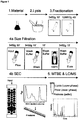
- Native Arabidopsis MM2d cell culture lysate (later referred to as input) was loaded on size filtration spin columns with a 10kDa cutoff to separate the protein fraction from the free metabolite fraction (flow through). Subsequently the protein fraction was washed thoroughly in order to remove any non-bound metabolites (wash). In a final step heat denaturation was applied to denature the proteins and release non-covalently bound metabolites from the proteins (elution). All samples were analyzed by applying our LC/MS metabolomics platform for semi-polar compounds (Giavalisco (2011), loc.cit. ) ( Fig. 1 , step 5).
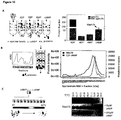
- LC/MS analysis of input, flow, wash and eluate samples resulted in approximately 8892 metabolic features (as defined by molecular mass (m/z) and retention time), of which approximately 150 could be putatively annotated to a metabolite.
- the flow through contained many metabolites and the metabolite content decreased in the washing. Strikingly, after heat treatment many metabolites (approximately 50% of all detected metabolic features), while being absent in wash samples, were again detectable in the eluate, thereby showing that this large fraction of metabolites is indeed forming stable complexes with proteins.
- ligands such as cyclic nucleotides (cGMP, cAMP, cCMP), co-factors (FAD, NAD, FMN) and peptides ( Fig. 2a-b ). Proteins were digested on the cutoff filter and peptides were released for further analysis.
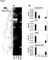
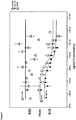
- Figure 4(a) shows a hierarchical cluster dendrogram of proteins and putatively annotated small molecules based on Pearson correlation. Grey boxes indicate clusters that have a correlation coefficient greater than 0.7.
- Figure 4(b) shows cluster no 5 from (a) as network where edge weights correspond to correlation strength.
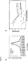
- Figure 4(c) shows individual two dimensional plots of elution profiles from selected small molecules (solid lines) and two proteins (dashed lines) with highly similar profiles. Correlation plots are shown in insets.
- N or C beads N terminal group of glycine or C terminal group of proline
- Both resins were incubated with the native Arabidopsis lysate and proteins captured on the beads were eluated with high concentration of Gly-Pro. Affinity experiments suffer from a high rate of false positives related to the unspecific binding.
- To counteract this and prior to Gly-Pro elution we introduced an extra step where beads were incubated with a mix of glycine and proline. This extra step reduced the number of hits from hundreds to 34 proteins pulled with N and C beads.
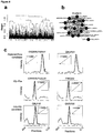
- cytoplasmic fructose bisphosphate aldolases FBA8 and FBA8 ( Fig 5 ). They also co-elute with Gly-Pro in the SEC experiment ( Fig 5 ).
- Aldolases are enzymes in the glycolysis and gluconeogenesis pathway. They catalyse the reversible reaction in which fructose 1,6 bisphosphate (Fru1,6bP) is cleaved into two three carbon products, namely 3-phosphate glyceraldehyde (G3P) and dihydroxyacetone phosphate (DHAP).
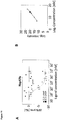
- FBA6 protein was expressed and purified from E.coli. MST uses the intristic tendency of all the biological molecules to move in the temperature gradient. This movement is dependent on the molecule size, charge and hydration shell. Complex formation affects at least one of the three parameters changing the movement and indicating binding event. Gly-Pro, but not Pro-Gly, binds FBA6 with the Kd of approximately 200nM, which is comparable to the substrate (DHAP). The results obtained point to both strong and specific interaction.
- PMIs Protein-metabolite interactions
- 2',3'-cAMP Extracellular 2',3'-cAMP is a source of adenosine. J Biol Chem 284: 33097-33106 ; Verrier et al. (2012) The brain in vivo expresses the 2',3'-cAMP-adenosine pathway. J Neurochem 122: 115-125 ). 2',3'-cAMP is formed during mRNA degradation, when hydrolysis of the P-O5' bond mediated by RNases is accompanied by transphosphorylation of mRNA to form 2',3'-cyclic nucleotides ( Thompson et al. (1994) Energetics of catalysis by ribonucleases: fate of the 2',3'-cyclic phosphodiester intermediate.
- 2', 3' cAMP is the most abundant of all the 2',3'-cyclic nucleotides. Similar to its metabolism, also the role of 2', 3'-cAMP is not well understood. In rat brain mitochondria, 2',3'-cAMP, was shown to activate mitochondrial transition pores ( Azarashvili et al. (2009) Ca2+-dependent permeability transition regulation in rat brain mitochondria by 2',3'-cyclic nucleotides and 2',3'-cyclic nucleotide 3'-phosphodiesterase.
- RNA-binding proteins TIA-1 and TIAR link the phosphorylation of eIF-2 alpha to the assembly of mammalian stress granules. Journal of Cell Biology 147: 1431-1441 ; Gilks et al. (2004) Stress granule assembly is mediated by prion-like aggregation of TIA-1. Mol Biol Cell 15: 5383-5398 ).
- Rbp47b/TIA1 localizes to the nucleus, acting as a component of the pre-mRNA splicing machinery (Gilks (2004) (loc. cit.); Lorkovic et al. (2000) RBP45 and RBP47, two oligouridylate-specific hnRNP-like proteins interacting with poly(A)+ RNA in nuclei of plant cells. RNA 6: 1610-1624 ).
- Rbp47b/TIA1 re-localizes to the cytoplasm, where its aggregation marks the formation of stress granules (Kedersha (1999) (loc. cit.); Weber et al.
- SGs are mRNP particles composed of large aggregates of stalled translation pre-initiation complexes, which contain mRNA, 40S ribosomal subunits, translation initiation factors and RNA-binding proteins (RBPs) (SGs) (Kedersha (1999) (loc. cit.), Weber (2008) (loc. cit.); Kedersha et al. (2005) Stress granules and processing bodies are dynamically linked sites of mRNP remodeling.
- SGs sequester housekeeping mRNAs and apoptosis regulatory factors, whilst exclude mRNAs encoding proteins involved in stress tolerance. Defects in SG assembly and disassembly can contribute to neurodegeneration ( Wolozin (2012) Regulated protein aggregation: stress granules and neurodegeneration. Mol Neurodegener 7: 56 ).
- SG assembly is also a target in cancer research, as presence of SG in cancer cells makes them more resistant to treatment and prone to metastasis (Mahboubi and Stochaj (2017) (loc. cit.)).
- SG integrity is important for viral resistance, by confining viral RNA and proteins ( Yoneyama et al. (2016) Regulation of antiviral innate immune signaling by stress-induced RNA granules. J Biochem 159: 279-286 ).
- RNA-binding proteins such as Rbp47b and TIA1
- Rbp47b and TIA1 RNA-binding proteins
- the self-assembly of the Rbp47b/ TIA1 proteins depends on RRM RNA binding motives, known to recruit mRNAs, and on the prion-like PRD domain that supports protein-protein interactions (Gilks (2004) (loc. cit.); Weber (2008) (loc. cit.)).
- the overexpression of TIA-1 induces SG assembly, even in the absence of stress (Gilks (2004) (loc. cit.)).
- Post-translational modifications (PTMs) of SG proteins can affect granule dynamics. For example oxidation of TIA1 protein inhibits SG formation sensitizing cells to apoptosis ( Arimoto-Matsuzaki et al. (2016) TIA1 oxidation inhibits stress granule assembly and sensitizes cells to stress-induced apoptosis. Nat Commun 7: 10252 ).
- CESTA cellular thermal shift assay
- MST micro-scale thermophoresis
- Rbp47b was expressed in and purified from E. coli.
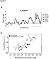
- the MST results show clear binding, with a Kd value of 1 ⁇ M.
- the measured Kd value also corresponds to the ⁇ 20 ⁇ M concentration of 2',3'-cAMP measured in the native Arabidopsis lysate ( Fig. 11B ).
- 2',3'-cAMP accumulates in low light and darkness, but most notably under heat stress ( Fig. 12A ), conditions characterized by increased RNA degradation ( Merret et al. (2013) XRN4 and LARP1 are required for a heat-triggered mRNA decay pathway involved in plant acclimation and survival during thermal stress. Cell Rep 5: 1279-1293 ; Baginsky and Gruissem (2002) Endonucleolytic activation directs dark-induced chloroplast mRNA degradation. Nucleic Acids Res 30: 4527-4533 ) and formation of the stress granules (SGs) (Kedersha (1999) (loc. cit.); Weber (2008) (loc. cit.)).
- SGs stress granules
- TIA1 the human functional homolog of Rbp47b.
- TIA1 was labeled and its interaction with 2',3'-cAMP was determined using MST.
- RNA decay is associated with cytoplasmic mRNP foci, referred to as processing bodies (P bodies, PBs).
- RNA decay related to stalled translation is induced under stress conditions in both animals and in plants, e.g.
- cytoplasmic mRNP foci playing a role in translational repression, by selective stabilization and storage of the mRNAs (Kedersha (1999) (loc. cit.); Weber (2008) (loc. cit.); Kedersha (2005) (loc. cit.); Anderson (2009) (loc. cit.)).
- Rbp47b/TIA1 aggregation is a key even is SG formation and we could further demonstrate that the self-assembly is facilitated by 2', 3'-cAMP binding. In that way and secondly, we provide evidence for the existence of small molecule regulation during stress granule formation, in addition to already reported PTMs of SGs proteins e.g. Arimoto-Matsuzaki (2016) (loc. cit.). An RNA degradation product, 2', 3'-cAMP is highly suitable for such a role, providing a means of negative-feedback regulation between RNA degradation and storage. We speculate that under control conditions nuclear localization of Rbp47b/TIA1 prevents the interaction from taking place.
- Arabidopsis cells cultures ( Menges and Murray (2002) Synchronous Arabidopsis suspension cultures for analysis of cellcycle gene activity. Plant J 30: 203-212 ) were grown in MSMO medium supplemented with 3% sucrose, 0.05 mg/L kinetin, and 0.5 mg/L 1-naphthaleneacetic acid on an orbital shaker at 130 RPM in the light. Cells were passaged weekly to fresh medium and harvested during logarithmic growth using rapid filtration and liquid-nitrogen snap freezing.
- Plant-cell material was collected as described above and pulverized to homogeneity in liquid nitrogen with mortar and pestle, followed by resuspension in 1 mL lysis buffer (25 mM Tris-HCl, pH 7.5; 0.5 M NaCl; 15 mM MgCl2; 0.5 mM DTT; 1 mM NaF; 1 mM Na3VO4; 1 ⁇ Protease Inhibitor Cocktail, Sigma-Aldrich P9599, Steinheim, Germany) per 1 g of plant material. Cellular debris was separated by 10 min centrifugation at 4 °C, 14,000 RPM. Crude lysate was subjected to ultra-centrifugation (45 min, 4 °C, 35,000 RPM) to obtain a soluble fraction referred to as the native Arabidopsis lysate.
- 1 lysis buffer 25 mM Tris-HCl, pH 7.5; 0.5 M NaCl; 15 mM MgCl2; 0.5 mM
- Custom 2',3'-cAMP agarose beads were purchased from Cube Biotech (Monheim, Germany). 2',3'-cAMP was coupled to the beads using the amine (NH2) group of the purine ring and a 14-carbon spacer arm. Before use, beads were equilibrated with lysis buffer. 3 mL native lysate (approximately 90 mg of total protein) was combined with 150 ⁇ L agarose resin (see above), incubated for 1 h on a rotating wheel at 4 °C (binding), transferred to a Mobicol "Classic" (35 ⁇ M pore size filter) column and washed with 10 mL wash buffer (0.025 M Tris-HCI, pH 7.5; 0.5 M NaCl).
- TAP-tagged Rbp47b and empty vector lines were prepared as described by ( Van Leene et al. (2015) An improved toolbox to unravel the plant cellular machinery by tandem affinity purification of Arabidopsis protein complexes. Nat Protoc 10: 169-187 ) using the pKCS binary vector and standard Agrobacterium transformation.
- Native Arabidopsis lysate was incubated with 10 or 100 ⁇ M 2',3'-cAMP and with DMSO (used for cAMP solution preparation) as control for 30 min at room temperature with mixing. Further steps were adapted from Franken (2015) (loc. cit.).
- 2',3'-cAMP reference compound (Sigma A9376) was spiked into cellular extract (soluble fraction; see above) in concentrations ranging from 100 pM to 1 mM.
- lysate was obtained from cells labeled with 15N ( Kierszniowska et al. (2009) Ratio-dependent significance thresholds in reciprocal 15N-labeling experiments as a robust tool in detection of candidate proteins responding to biological treatment. Proteomics 9: 1916-1924 ).
- TIA1 protein was purchased from Origene (Herford, Germany).
- Rbp47b was cloned as a C-terminal GFP fusion (to increase protein solubility) into the E . coli expression vector pDEST14 containing His6-tag at the N-terminal of the Gateway (Karlsruhe, Germany) cassette. Rosetta cells expressing His6-Rbp47b-GFP were grown at 28°C overnight and next day were moved to Terrific Broth medium supplied with 1% sucrose and relevant antibiotics. Cultures at OD 0.4 were induced by addition of 0.1 mM IPTG and transferred to 16 °C for overnight incubation.
- MST measurements were performed using a Monolith NT.115 instrument (NanoTemper, Ober, Germany). Capillaries were loaded into the instrument as sets of 13-16 point ligand titrations. Proteins (Rb47b and TIA1) were labeled in phosphate buffer (PBS) using MonolithTM protein labeling kit RED-NHS (amine reactive; MO-L001) according to the manufacturer's instruction. Excitation was optimized by varying the LED power to yield emission intensities above 200 AU, corresponding to 10-50 nM labeled protein. Monolith power was set to 60%. Ligands [2',3'-cAMP (Sigma A9376)] and [3',5'-cAMP (Sigma A6885)] were dissolved in PBS.
- Tween and premium coated capillaries were used to prevent sticking.
- Non-labeled TIA1 and Rbp47b were used as ligands in the self-assembly experiments.
- MO Affinity Analysis software was used to analyze (Kd calculation) and visualize the data. Presented data are from 2-3 technical replicates.
Landscapes
- Health & Medical Sciences (AREA)
- Life Sciences & Earth Sciences (AREA)
- Engineering & Computer Science (AREA)
- Immunology (AREA)
- Physics & Mathematics (AREA)
- Molecular Biology (AREA)
- Chemical & Material Sciences (AREA)
- Hematology (AREA)
- Biomedical Technology (AREA)
- Urology & Nephrology (AREA)
- Biochemistry (AREA)
- Pathology (AREA)
- Analytical Chemistry (AREA)
- General Health & Medical Sciences (AREA)
- General Physics & Mathematics (AREA)
- Medicinal Chemistry (AREA)
- Biotechnology (AREA)
- Food Science & Technology (AREA)
- Microbiology (AREA)
- Cell Biology (AREA)
- Bioinformatics & Cheminformatics (AREA)
- High Energy & Nuclear Physics (AREA)
- Bioinformatics & Computational Biology (AREA)
- Proteomics, Peptides & Aminoacids (AREA)
- Biophysics (AREA)
- Spectroscopy & Molecular Physics (AREA)
- Chemical Kinetics & Catalysis (AREA)
- Rehabilitation Therapy (AREA)
- Rheumatology (AREA)
- Crystallography & Structural Chemistry (AREA)
- Electrochemistry (AREA)
- Investigating Or Analysing Biological Materials (AREA)
- Other Investigation Or Analysis Of Materials By Electrical Means (AREA)
Priority Applications (2)
| Application Number | Priority Date | Filing Date | Title |
|---|---|---|---|
| PL17725620T PL3465207T3 (pl) | 2016-05-30 | 2017-05-29 | Identyfikacja ligandów przez wspólne frakcjonowanie |
| SI201730986T SI3465207T1 (sl) | 2016-05-30 | 2017-05-29 | Identifikacija ligandov s KO-frakcionacijo |
Applications Claiming Priority (2)
| Application Number | Priority Date | Filing Date | Title |
|---|---|---|---|
| EP16172000 | 2016-05-30 | ||
| PCT/EP2017/062841 WO2017207460A1 (en) | 2016-05-30 | 2017-05-29 | Ligand identification by co-fractionation |
Publications (2)
| Publication Number | Publication Date |
|---|---|
| EP3465207A1 EP3465207A1 (en) | 2019-04-10 |
| EP3465207B1 true EP3465207B1 (en) | 2021-09-01 |
Family
ID=56119308
Family Applications (1)
| Application Number | Title | Priority Date | Filing Date |
|---|---|---|---|
| EP17725620.3A Active EP3465207B1 (en) | 2016-05-30 | 2017-05-29 | Ligand identification by co-fractionation |
Country Status (15)
| Country | Link |
|---|---|
| US (1) | US10976310B2 (pl) |
| EP (1) | EP3465207B1 (pl) |
| KR (1) | KR102346785B1 (pl) |
| CN (1) | CN109219750B (pl) |
| AU (1) | AU2017272493B2 (pl) |
| BR (1) | BR112018074856A2 (pl) |
| DK (1) | DK3465207T3 (pl) |
| ES (1) | ES2899110T3 (pl) |
| HU (1) | HUE056359T2 (pl) |
| LT (1) | LT3465207T (pl) |
| PL (1) | PL3465207T3 (pl) |
| PT (1) | PT3465207T (pl) |
| SG (1) | SG11201810531TA (pl) |
| SI (1) | SI3465207T1 (pl) |
| WO (1) | WO2017207460A1 (pl) |
Families Citing this family (8)
| Publication number | Priority date | Publication date | Assignee | Title |
|---|---|---|---|---|
| CA3115315A1 (en) | 2018-10-15 | 2020-04-23 | Anthony A. HYMAN | Compounds for treatment of diseases and methods of screening therefor |
| US11493519B2 (en) | 2019-02-08 | 2022-11-08 | Dewpoint Therapeutics, Inc. | Methods of characterizing condensate-associated characteristics of compounds and uses thereof |
| JP2022548695A (ja) | 2019-09-18 | 2022-11-21 | デューポイント セラピューティクス, インコーポレイテッド | 凝縮体関連特異性のスクリーニング方法及びその使用 |
| JP7327096B2 (ja) * | 2019-11-13 | 2023-08-16 | 株式会社島津製作所 | 分子の分離方法 |
| KR20220137672A (ko) * | 2020-02-13 | 2022-10-12 | 에프. 호프만-라 로슈 아게 | 핵 자기 공명 이완법으로 aav 입자의 부하 상태를 결정하는 방법 |
| CN113528497B (zh) * | 2021-07-22 | 2022-06-24 | 中国林业科学研究院林业研究所 | Fba8基因或fba8蛋白在制备具有磷酸转移活性和/或蛋白水解活性的试剂中的应用 |
| WO2024112940A1 (en) * | 2022-11-26 | 2024-05-30 | Enveda Therapeutics, Inc. | Methods and systems for identifying compounds for forming, stabilizing or disrupting molecular complexes |
| CN118685492A (zh) * | 2024-03-08 | 2024-09-24 | 西湖实验室(生命科学和生物医学浙江省实验室) | 微量新鲜样本六组学连续提取方法 |
Family Cites Families (6)
| Publication number | Priority date | Publication date | Assignee | Title |
|---|---|---|---|---|
| US4459359A (en) | 1981-11-19 | 1984-07-10 | New York Blood Center, Inc. | Sensitive immunoassays of antigens or antibodies sequestered within immune complexes |
| CA2369868A1 (en) * | 1999-04-15 | 2000-10-26 | The University Of Virginia Patent Foundation | Proteome mining |
| JP2005535727A (ja) * | 2002-08-14 | 2005-11-24 | ブランシェット・ロックフェラー・ニューロサイエンスィズ・インスティテュート | タンパク質−タンパク質相互作用に関与するタンパク質亜集団の単離方法 |
| WO2006015796A1 (en) * | 2004-08-09 | 2006-02-16 | MAX-PLANCK-Gesellschaft zur Förderung der Wissenschaften e.V. | Method for high-throughput screening of test molecules binding to target molecules |
| US20150105280A1 (en) | 2010-03-10 | 2015-04-16 | Perfinity Biosciences, Inc. | Selector based recognition and quantification system and method for multiple analytes in a single analysis |
| AU2013347838A1 (en) | 2012-11-26 | 2015-06-11 | Caris Life Sciences Switzerland Holdings Gmbh | Biomarker compositions and methods |
-
2017
- 2017-05-29 KR KR1020187038204A patent/KR102346785B1/ko active Active
- 2017-05-29 HU HUE17725620A patent/HUE056359T2/hu unknown
- 2017-05-29 WO PCT/EP2017/062841 patent/WO2017207460A1/en not_active Ceased
- 2017-05-29 LT LTEPPCT/EP2017/062841T patent/LT3465207T/lt unknown
- 2017-05-29 DK DK17725620.3T patent/DK3465207T3/da active
- 2017-05-29 ES ES17725620T patent/ES2899110T3/es active Active
- 2017-05-29 EP EP17725620.3A patent/EP3465207B1/en active Active
- 2017-05-29 PL PL17725620T patent/PL3465207T3/pl unknown
- 2017-05-29 PT PT177256203T patent/PT3465207T/pt unknown
- 2017-05-29 AU AU2017272493A patent/AU2017272493B2/en active Active
- 2017-05-29 BR BR112018074856-0A patent/BR112018074856A2/pt not_active Application Discontinuation
- 2017-05-29 SG SG11201810531TA patent/SG11201810531TA/en unknown
- 2017-05-29 US US16/304,182 patent/US10976310B2/en active Active
- 2017-05-29 SI SI201730986T patent/SI3465207T1/sl unknown
- 2017-05-29 CN CN201780034328.7A patent/CN109219750B/zh active Active
Also Published As
| Publication number | Publication date |
|---|---|
| CA3025711A1 (en) | 2017-12-07 |
| BR112018074856A2 (pt) | 2019-03-06 |
| KR20190013999A (ko) | 2019-02-11 |
| SG11201810531TA (en) | 2018-12-28 |
| SI3465207T1 (sl) | 2022-01-31 |
| PT3465207T (pt) | 2021-11-30 |
| US10976310B2 (en) | 2021-04-13 |
| LT3465207T (lt) | 2021-12-10 |
| AU2017272493B2 (en) | 2021-07-22 |
| EP3465207A1 (en) | 2019-04-10 |
| AU2017272493A1 (en) | 2018-12-20 |
| HUE056359T2 (hu) | 2022-02-28 |
| WO2017207460A1 (en) | 2017-12-07 |
| KR102346785B1 (ko) | 2022-01-04 |
| PL3465207T3 (pl) | 2022-01-17 |
| DK3465207T3 (da) | 2021-11-15 |
| CN109219750B (zh) | 2022-05-31 |
| CN109219750A (zh) | 2019-01-15 |
| US20190361012A1 (en) | 2019-11-28 |
| ES2899110T3 (es) | 2022-03-10 |
Similar Documents
| Publication | Publication Date | Title |
|---|---|---|
| EP3465207B1 (en) | Ligand identification by co-fractionation | |
| Li et al. | Profiling PRMT methylome reveals roles of hnRNPA1 arginine methylation in RNA splicing and cell growth | |
| Reichel et al. | In planta determination of the mRNA-binding proteome of Arabidopsis etiolated seedlings | |
| Roitinger et al. | Quantitative phosphoproteomics of the ataxia telangiectasia-mutated (ATM) and ataxia telangiectasia-mutated and Rad3-related (ATR) dependent DNA damage response in Arabidopsis thaliana*[S] | |
| Lin et al. | Examining histone posttranslational modification patterns by high-resolution mass spectrometry | |
| Vertegaal | Uncovering ubiquitin and ubiquitin-like signaling networks | |
| Kosmacz et al. | Interaction of 2′, 3′-cAMP with Rbp47b plays a role in stress granule formation | |
| Duncan et al. | Multiple lines of evidence localize signaling, morphology, and lipid biosynthesis machinery to the mitochondrial outer membrane of Arabidopsis | |
| JP2003534768A (ja) | 環状ペプチド | |
| Schmitt et al. | Asc1p/RACK1 connects ribosomes to eukaryotic phosphosignaling | |
| Xu et al. | Spatial and temporal proteomics reveals the distinct distributions and dynamics of O-GlcNAcylated proteins | |
| Gilmore et al. | Characterization of a highly conserved histone related protein, Ydl156w, and its functional associations using quantitative proteomic analyses | |
| Foyn et al. | Protein N-terminal acetyltransferases act as N-terminal propionyltransferases in vitro and in vivo | |
| Rodríguez-Celma et al. | Systems-wide analysis of manganese deficiency-induced changes in gene activity of Arabidopsis roots | |
| Ge et al. | Proteomic analyses of apoplastic proteins from germinating Arabidopsis thaliana pollen | |
| Chen et al. | Comparative acetylomic analysis reveals differentially acetylated proteins regulating anther and pollen development in kenaf cytoplasmic male sterility line | |
| Doroshenk et al. | Proteomic analysis of cytoskeleton-associated RNA binding proteins in developing rice seed | |
| CA3025711C (en) | Ligand identification by co-fractionation | |
| Qi et al. | Genome-wide mapping of RNA-protein associations through sequencing | |
| Solis et al. | Simplified high yield TAILS terminomics using a new HPG-ALD 800K-2000 polymer with precipitation | |
| Zhang et al. | Towards the N-terminal acetylome: an N-terminal acetylated peptide enrichment method using CNBr-activated sepharose resin | |
| Kraus et al. | The structural scaffold of the TPLATE complex deforms the membrane during plant endocytosis | |
| Imami | Proteome analysis of puromycin-labeled nascent polypeptides | |
| Zhang et al. | GABPA Recruits the Integrator Endonuclease Complex to Promote Transcription Elongation | |
| Braus | Asc1p/RACK1 Connects Ribosomes to Eukaryotic Phosphosignaling |
Legal Events
| Date | Code | Title | Description |
|---|---|---|---|
| STAA | Information on the status of an ep patent application or granted ep patent |
Free format text: STATUS: UNKNOWN |
|
| STAA | Information on the status of an ep patent application or granted ep patent |
Free format text: STATUS: THE INTERNATIONAL PUBLICATION HAS BEEN MADE |
|
| PUAI | Public reference made under article 153(3) epc to a published international application that has entered the european phase |
Free format text: ORIGINAL CODE: 0009012 |
|
| STAA | Information on the status of an ep patent application or granted ep patent |
Free format text: STATUS: REQUEST FOR EXAMINATION WAS MADE |
|
| 17P | Request for examination filed |
Effective date: 20181221 |
|
| AK | Designated contracting states |
Kind code of ref document: A1 Designated state(s): AL AT BE BG CH CY CZ DE DK EE ES FI FR GB GR HR HU IE IS IT LI LT LU LV MC MK MT NL NO PL PT RO RS SE SI SK SM TR |
|
| AX | Request for extension of the european patent |
Extension state: BA ME |
|
| RIN1 | Information on inventor provided before grant (corrected) |
Inventor name: KIERSZNIOWSKA, SYLWIA Inventor name: CHODASIEWICZ, MONIKA Inventor name: VEYEL, DANIEL Inventor name: WILLMITZER, LOTHAR Inventor name: SKIRYCZ, ALEKSANDRA |
|
| DAV | Request for validation of the european patent (deleted) | ||
| DAX | Request for extension of the european patent (deleted) | ||
| RIN1 | Information on inventor provided before grant (corrected) |
Inventor name: CHODASIEWICZ, MONIKA Inventor name: VEYEL, DANIEL Inventor name: WILLMITZER, LOTHAR Inventor name: SKIRYCZ, ALEKSANDRA Inventor name: KIERSZNIOWSKA, SYLWIA |
|
| STAA | Information on the status of an ep patent application or granted ep patent |
Free format text: STATUS: EXAMINATION IS IN PROGRESS |
|
| 17Q | First examination report despatched |
Effective date: 20191028 |
|
| GRAP | Despatch of communication of intention to grant a patent |
Free format text: ORIGINAL CODE: EPIDOSNIGR1 |
|
| STAA | Information on the status of an ep patent application or granted ep patent |
Free format text: STATUS: GRANT OF PATENT IS INTENDED |
|
| INTG | Intention to grant announced |
Effective date: 20210317 |
|
| GRAS | Grant fee paid |
Free format text: ORIGINAL CODE: EPIDOSNIGR3 |
|
| GRAA | (expected) grant |
Free format text: ORIGINAL CODE: 0009210 |
|
| STAA | Information on the status of an ep patent application or granted ep patent |
Free format text: STATUS: THE PATENT HAS BEEN GRANTED |
|
| AK | Designated contracting states |
Kind code of ref document: B1 Designated state(s): AL AT BE BG CH CY CZ DE DK EE ES FI FR GB GR HR HU IE IS IT LI LT LU LV MC MK MT NL NO PL PT RO RS SE SI SK SM TR |
|
| REG | Reference to a national code |
Ref country code: GB Ref legal event code: FG4D |
|
| REG | Reference to a national code |
Ref country code: CH Ref legal event code: EP Ref country code: AT Ref legal event code: REF Ref document number: 1426752 Country of ref document: AT Kind code of ref document: T Effective date: 20210915 |
|
| REG | Reference to a national code |
Ref country code: DE Ref legal event code: R096 Ref document number: 602017045149 Country of ref document: DE |
|
| REG | Reference to a national code |
Ref country code: IE Ref legal event code: FG4D |
|
| REG | Reference to a national code |
Ref country code: DK Ref legal event code: T3 Effective date: 20211111 |
|
| REG | Reference to a national code |
Ref country code: PT Ref legal event code: SC4A Ref document number: 3465207 Country of ref document: PT Date of ref document: 20211130 Kind code of ref document: T Free format text: AVAILABILITY OF NATIONAL TRANSLATION Effective date: 20211124 |
|
| REG | Reference to a national code |
Ref country code: SE Ref legal event code: TRGR |
|
| REG | Reference to a national code |
Ref country code: NL Ref legal event code: FP |
|
| REG | Reference to a national code |
Ref country code: EE Ref legal event code: FG4A Ref document number: E021714 Country of ref document: EE Effective date: 20211119 |
|
| REG | Reference to a national code |
Ref country code: SK Ref legal event code: T3 Ref document number: E 38692 Country of ref document: SK |
|
| PG25 | Lapsed in a contracting state [announced via postgrant information from national office to epo] |
Ref country code: FI Free format text: LAPSE BECAUSE OF FAILURE TO SUBMIT A TRANSLATION OF THE DESCRIPTION OR TO PAY THE FEE WITHIN THE PRESCRIBED TIME-LIMIT Effective date: 20210901 Ref country code: NO Free format text: LAPSE BECAUSE OF FAILURE TO SUBMIT A TRANSLATION OF THE DESCRIPTION OR TO PAY THE FEE WITHIN THE PRESCRIBED TIME-LIMIT Effective date: 20211201 Ref country code: BG Free format text: LAPSE BECAUSE OF FAILURE TO SUBMIT A TRANSLATION OF THE DESCRIPTION OR TO PAY THE FEE WITHIN THE PRESCRIBED TIME-LIMIT Effective date: 20211201 Ref country code: RS Free format text: LAPSE BECAUSE OF FAILURE TO SUBMIT A TRANSLATION OF THE DESCRIPTION OR TO PAY THE FEE WITHIN THE PRESCRIBED TIME-LIMIT Effective date: 20210901 Ref country code: HR Free format text: LAPSE BECAUSE OF FAILURE TO SUBMIT A TRANSLATION OF THE DESCRIPTION OR TO PAY THE FEE WITHIN THE PRESCRIBED TIME-LIMIT Effective date: 20210901 |
|
| PG25 | Lapsed in a contracting state [announced via postgrant information from national office to epo] |
Ref country code: GR Free format text: LAPSE BECAUSE OF FAILURE TO SUBMIT A TRANSLATION OF THE DESCRIPTION OR TO PAY THE FEE WITHIN THE PRESCRIBED TIME-LIMIT Effective date: 20211202 |
|
| REG | Reference to a national code |
Ref country code: HU Ref legal event code: AG4A Ref document number: E056359 Country of ref document: HU |
|
| REG | Reference to a national code |
Ref country code: ES Ref legal event code: FG2A Ref document number: 2899110 Country of ref document: ES Kind code of ref document: T3 Effective date: 20220310 |
|
| PG25 | Lapsed in a contracting state [announced via postgrant information from national office to epo] |
Ref country code: IS Free format text: LAPSE BECAUSE OF FAILURE TO SUBMIT A TRANSLATION OF THE DESCRIPTION OR TO PAY THE FEE WITHIN THE PRESCRIBED TIME-LIMIT Effective date: 20220101 Ref country code: SM Free format text: LAPSE BECAUSE OF FAILURE TO SUBMIT A TRANSLATION OF THE DESCRIPTION OR TO PAY THE FEE WITHIN THE PRESCRIBED TIME-LIMIT Effective date: 20210901 Ref country code: RO Free format text: LAPSE BECAUSE OF FAILURE TO SUBMIT A TRANSLATION OF THE DESCRIPTION OR TO PAY THE FEE WITHIN THE PRESCRIBED TIME-LIMIT Effective date: 20210901 Ref country code: AL Free format text: LAPSE BECAUSE OF FAILURE TO SUBMIT A TRANSLATION OF THE DESCRIPTION OR TO PAY THE FEE WITHIN THE PRESCRIBED TIME-LIMIT Effective date: 20210901 |
|
| REG | Reference to a national code |
Ref country code: DE Ref legal event code: R097 Ref document number: 602017045149 Country of ref document: DE |
|
| PLBE | No opposition filed within time limit |
Free format text: ORIGINAL CODE: 0009261 |
|
| STAA | Information on the status of an ep patent application or granted ep patent |
Free format text: STATUS: NO OPPOSITION FILED WITHIN TIME LIMIT |
|
| 26N | No opposition filed |
Effective date: 20220602 |
|
| REG | Reference to a national code |
Ref country code: AT Ref legal event code: UEP Ref document number: 1426752 Country of ref document: AT Kind code of ref document: T Effective date: 20210901 |
|
| PG25 | Lapsed in a contracting state [announced via postgrant information from national office to epo] |
Ref country code: MC Free format text: LAPSE BECAUSE OF FAILURE TO SUBMIT A TRANSLATION OF THE DESCRIPTION OR TO PAY THE FEE WITHIN THE PRESCRIBED TIME-LIMIT Effective date: 20210901 Ref country code: LU Free format text: LAPSE BECAUSE OF NON-PAYMENT OF DUE FEES Effective date: 20220529 |
|
| P01 | Opt-out of the competence of the unified patent court (upc) registered |
Effective date: 20230425 |
|
| PG25 | Lapsed in a contracting state [announced via postgrant information from national office to epo] |
Ref country code: MK Free format text: LAPSE BECAUSE OF FAILURE TO SUBMIT A TRANSLATION OF THE DESCRIPTION OR TO PAY THE FEE WITHIN THE PRESCRIBED TIME-LIMIT Effective date: 20210901 Ref country code: CY Free format text: LAPSE BECAUSE OF FAILURE TO SUBMIT A TRANSLATION OF THE DESCRIPTION OR TO PAY THE FEE WITHIN THE PRESCRIBED TIME-LIMIT Effective date: 20210901 |
|
| PG25 | Lapsed in a contracting state [announced via postgrant information from national office to epo] |
Ref country code: TR Free format text: LAPSE BECAUSE OF FAILURE TO SUBMIT A TRANSLATION OF THE DESCRIPTION OR TO PAY THE FEE WITHIN THE PRESCRIBED TIME-LIMIT Effective date: 20210901 |
|
| PG25 | Lapsed in a contracting state [announced via postgrant information from national office to epo] |
Ref country code: MT Free format text: LAPSE BECAUSE OF FAILURE TO SUBMIT A TRANSLATION OF THE DESCRIPTION OR TO PAY THE FEE WITHIN THE PRESCRIBED TIME-LIMIT Effective date: 20210901 |
|
| PGFP | Annual fee paid to national office [announced via postgrant information from national office to epo] |
Ref country code: NL Payment date: 20250526 Year of fee payment: 9 |
|
| PGFP | Annual fee paid to national office [announced via postgrant information from national office to epo] |
Ref country code: DE Payment date: 20250530 Year of fee payment: 9 Ref country code: PL Payment date: 20250509 Year of fee payment: 9 |
|
| PGFP | Annual fee paid to national office [announced via postgrant information from national office to epo] |
Ref country code: GB Payment date: 20250528 Year of fee payment: 9 Ref country code: ES Payment date: 20250606 Year of fee payment: 9 Ref country code: DK Payment date: 20250526 Year of fee payment: 9 |
|
| PGFP | Annual fee paid to national office [announced via postgrant information from national office to epo] |
Ref country code: LT Payment date: 20250509 Year of fee payment: 9 |
|
| PGFP | Annual fee paid to national office [announced via postgrant information from national office to epo] |
Ref country code: HU Payment date: 20250514 Year of fee payment: 9 |
|
| PGFP | Annual fee paid to national office [announced via postgrant information from national office to epo] |
Ref country code: IT Payment date: 20250522 Year of fee payment: 9 Ref country code: BE Payment date: 20250526 Year of fee payment: 9 |
|
| PGFP | Annual fee paid to national office [announced via postgrant information from national office to epo] |
Ref country code: PT Payment date: 20250509 Year of fee payment: 9 Ref country code: LV Payment date: 20250526 Year of fee payment: 9 |
|
| PGFP | Annual fee paid to national office [announced via postgrant information from national office to epo] |
Ref country code: EE Payment date: 20250527 Year of fee payment: 9 Ref country code: FR Payment date: 20250527 Year of fee payment: 9 |
|
| PGFP | Annual fee paid to national office [announced via postgrant information from national office to epo] |
Ref country code: CH Payment date: 20250601 Year of fee payment: 9 |
|
| PGFP | Annual fee paid to national office [announced via postgrant information from national office to epo] |
Ref country code: AT Payment date: 20250521 Year of fee payment: 9 |
|
| PGFP | Annual fee paid to national office [announced via postgrant information from national office to epo] |
Ref country code: SK Payment date: 20250520 Year of fee payment: 9 |
|
| PGFP | Annual fee paid to national office [announced via postgrant information from national office to epo] |
Ref country code: CZ Payment date: 20250514 Year of fee payment: 9 |
|
| PGFP | Annual fee paid to national office [announced via postgrant information from national office to epo] |
Ref country code: IE Payment date: 20250522 Year of fee payment: 9 |
|
| PGFP | Annual fee paid to national office [announced via postgrant information from national office to epo] |
Ref country code: SI Payment date: 20250523 Year of fee payment: 9 Ref country code: SE Payment date: 20250526 Year of fee payment: 9 |