EP3443122B1 - Signaux optiques mixtes dans une analyse de polymère à l'aide de nanopores - Google Patents
Signaux optiques mixtes dans une analyse de polymère à l'aide de nanopores Download PDFInfo
- Publication number
- EP3443122B1 EP3443122B1 EP17782828.2A EP17782828A EP3443122B1 EP 3443122 B1 EP3443122 B1 EP 3443122B1 EP 17782828 A EP17782828 A EP 17782828A EP 3443122 B1 EP3443122 B1 EP 3443122B1
- Authority
- EP
- European Patent Office
- Prior art keywords
- nanopore
- fluorescent
- labels
- polynucleotide
- nanopores
- Prior art date
- Legal status (The legal status is an assumption and is not a legal conclusion. Google has not performed a legal analysis and makes no representation as to the accuracy of the status listed.)
- Active
Links
- 230000003287 optical effect Effects 0.000 title claims description 101
- 229920000642 polymer Polymers 0.000 title claims description 50
- 238000004458 analytical method Methods 0.000 title description 12
- 102000040430 polynucleotide Human genes 0.000 claims description 133
- 108091033319 polynucleotide Proteins 0.000 claims description 133
- 239000002157 polynucleotide Substances 0.000 claims description 133
- 239000000178 monomer Substances 0.000 claims description 114
- 238000010791 quenching Methods 0.000 claims description 107
- 230000000171 quenching effect Effects 0.000 claims description 97
- 125000003729 nucleotide group Chemical group 0.000 claims description 77
- 239000002773 nucleotide Substances 0.000 claims description 73
- 102000004169 proteins and genes Human genes 0.000 claims description 63
- 108090000623 proteins and genes Proteins 0.000 claims description 63
- 230000005284 excitation Effects 0.000 claims description 54
- 238000000034 method Methods 0.000 claims description 50
- 102000039446 nucleic acids Human genes 0.000 claims description 37
- 108020004707 nucleic acids Proteins 0.000 claims description 37
- 150000007523 nucleic acids Chemical class 0.000 claims description 37
- 239000003795 chemical substances by application Substances 0.000 claims description 31
- -1 nucleoside triphosphates Chemical class 0.000 claims description 13
- 239000002777 nucleoside Substances 0.000 claims description 6
- 239000001226 triphosphate Substances 0.000 claims 3
- 235000011178 triphosphate Nutrition 0.000 claims 3
- 239000007850 fluorescent dye Substances 0.000 description 64
- 235000018102 proteins Nutrition 0.000 description 62
- 238000001327 Förster resonance energy transfer Methods 0.000 description 44
- 230000005945 translocation Effects 0.000 description 41
- 108091034117 Oligonucleotide Proteins 0.000 description 40
- 238000005259 measurement Methods 0.000 description 40
- 239000012528 membrane Substances 0.000 description 37
- 239000010410 layer Substances 0.000 description 35
- 238000001514 detection method Methods 0.000 description 33
- 239000000975 dye Substances 0.000 description 28
- 238000006243 chemical reaction Methods 0.000 description 25
- 230000007704 transition Effects 0.000 description 23
- 239000007787 solid Substances 0.000 description 20
- 239000000758 substrate Substances 0.000 description 20
- 239000000370 acceptor Substances 0.000 description 19
- 108020004414 DNA Proteins 0.000 description 16
- 108090000765 processed proteins & peptides Proteins 0.000 description 16
- JLCPHMBAVCMARE-UHFFFAOYSA-N [3-[[3-[[3-[[3-[[3-[[3-[[3-[[3-[[3-[[3-[[3-[[5-(2-amino-6-oxo-1H-purin-9-yl)-3-[[3-[[3-[[3-[[3-[[3-[[5-(2-amino-6-oxo-1H-purin-9-yl)-3-[[5-(2-amino-6-oxo-1H-purin-9-yl)-3-hydroxyoxolan-2-yl]methoxy-hydroxyphosphoryl]oxyoxolan-2-yl]methoxy-hydroxyphosphoryl]oxy-5-(5-methyl-2,4-dioxopyrimidin-1-yl)oxolan-2-yl]methoxy-hydroxyphosphoryl]oxy-5-(6-aminopurin-9-yl)oxolan-2-yl]methoxy-hydroxyphosphoryl]oxy-5-(6-aminopurin-9-yl)oxolan-2-yl]methoxy-hydroxyphosphoryl]oxy-5-(6-aminopurin-9-yl)oxolan-2-yl]methoxy-hydroxyphosphoryl]oxy-5-(6-aminopurin-9-yl)oxolan-2-yl]methoxy-hydroxyphosphoryl]oxyoxolan-2-yl]methoxy-hydroxyphosphoryl]oxy-5-(5-methyl-2,4-dioxopyrimidin-1-yl)oxolan-2-yl]methoxy-hydroxyphosphoryl]oxy-5-(4-amino-2-oxopyrimidin-1-yl)oxolan-2-yl]methoxy-hydroxyphosphoryl]oxy-5-(5-methyl-2,4-dioxopyrimidin-1-yl)oxolan-2-yl]methoxy-hydroxyphosphoryl]oxy-5-(5-methyl-2,4-dioxopyrimidin-1-yl)oxolan-2-yl]methoxy-hydroxyphosphoryl]oxy-5-(6-aminopurin-9-yl)oxolan-2-yl]methoxy-hydroxyphosphoryl]oxy-5-(6-aminopurin-9-yl)oxolan-2-yl]methoxy-hydroxyphosphoryl]oxy-5-(4-amino-2-oxopyrimidin-1-yl)oxolan-2-yl]methoxy-hydroxyphosphoryl]oxy-5-(4-amino-2-oxopyrimidin-1-yl)oxolan-2-yl]methoxy-hydroxyphosphoryl]oxy-5-(4-amino-2-oxopyrimidin-1-yl)oxolan-2-yl]methoxy-hydroxyphosphoryl]oxy-5-(6-aminopurin-9-yl)oxolan-2-yl]methoxy-hydroxyphosphoryl]oxy-5-(4-amino-2-oxopyrimidin-1-yl)oxolan-2-yl]methyl [5-(6-aminopurin-9-yl)-2-(hydroxymethyl)oxolan-3-yl] hydrogen phosphate Polymers Cc1cn(C2CC(OP(O)(=O)OCC3OC(CC3OP(O)(=O)OCC3OC(CC3O)n3cnc4c3nc(N)[nH]c4=O)n3cnc4c3nc(N)[nH]c4=O)C(COP(O)(=O)OC3CC(OC3COP(O)(=O)OC3CC(OC3COP(O)(=O)OC3CC(OC3COP(O)(=O)OC3CC(OC3COP(O)(=O)OC3CC(OC3COP(O)(=O)OC3CC(OC3COP(O)(=O)OC3CC(OC3COP(O)(=O)OC3CC(OC3COP(O)(=O)OC3CC(OC3COP(O)(=O)OC3CC(OC3COP(O)(=O)OC3CC(OC3COP(O)(=O)OC3CC(OC3COP(O)(=O)OC3CC(OC3COP(O)(=O)OC3CC(OC3COP(O)(=O)OC3CC(OC3COP(O)(=O)OC3CC(OC3COP(O)(=O)OC3CC(OC3CO)n3cnc4c(N)ncnc34)n3ccc(N)nc3=O)n3cnc4c(N)ncnc34)n3ccc(N)nc3=O)n3ccc(N)nc3=O)n3ccc(N)nc3=O)n3cnc4c(N)ncnc34)n3cnc4c(N)ncnc34)n3cc(C)c(=O)[nH]c3=O)n3cc(C)c(=O)[nH]c3=O)n3ccc(N)nc3=O)n3cc(C)c(=O)[nH]c3=O)n3cnc4c3nc(N)[nH]c4=O)n3cnc4c(N)ncnc34)n3cnc4c(N)ncnc34)n3cnc4c(N)ncnc34)n3cnc4c(N)ncnc34)O2)c(=O)[nH]c1=O JLCPHMBAVCMARE-UHFFFAOYSA-N 0.000 description 15
- 239000007790 solid phase Substances 0.000 description 15
- 229910052770 Uranium Inorganic materials 0.000 description 14
- 238000009739 binding Methods 0.000 description 13
- 239000000463 material Substances 0.000 description 13
- 108091006146 Channels Proteins 0.000 description 12
- 239000000232 Lipid Bilayer Substances 0.000 description 12
- 230000027455 binding Effects 0.000 description 12
- 150000001875 compounds Chemical class 0.000 description 12
- 229910052581 Si3N4 Inorganic materials 0.000 description 11
- VYPSYNLAJGMNEJ-UHFFFAOYSA-N Silicium dioxide Chemical compound O=[Si]=O VYPSYNLAJGMNEJ-UHFFFAOYSA-N 0.000 description 11
- 102000004196 processed proteins & peptides Human genes 0.000 description 11
- HQVNEWCFYHHQES-UHFFFAOYSA-N silicon nitride Chemical compound N12[Si]34N5[Si]62N3[Si]51N64 HQVNEWCFYHHQES-UHFFFAOYSA-N 0.000 description 11
- 238000003491 array Methods 0.000 description 10
- 238000005286 illumination Methods 0.000 description 10
- 239000002086 nanomaterial Substances 0.000 description 10
- 239000002096 quantum dot Substances 0.000 description 10
- 125000000539 amino acid group Chemical group 0.000 description 9
- 238000007672 fourth generation sequencing Methods 0.000 description 9
- 230000007274 generation of a signal involved in cell-cell signaling Effects 0.000 description 9
- 230000003993 interaction Effects 0.000 description 9
- 238000002372 labelling Methods 0.000 description 9
- 239000000243 solution Substances 0.000 description 9
- 239000000126 substance Substances 0.000 description 9
- 150000003573 thiols Chemical class 0.000 description 9
- 108010013381 Porins Proteins 0.000 description 8
- 102000017033 Porins Human genes 0.000 description 8
- 230000005684 electric field Effects 0.000 description 8
- 238000004519 manufacturing process Methods 0.000 description 8
- 239000000523 sample Substances 0.000 description 8
- 101710092462 Alpha-hemolysin Proteins 0.000 description 7
- 102000053602 DNA Human genes 0.000 description 7
- 108020004682 Single-Stranded DNA Proteins 0.000 description 7
- 230000015572 biosynthetic process Effects 0.000 description 7
- 238000000151 deposition Methods 0.000 description 7
- 238000009826 distribution Methods 0.000 description 7
- 229910052751 metal Inorganic materials 0.000 description 7
- 239000002184 metal Substances 0.000 description 7
- 229920001184 polypeptide Polymers 0.000 description 7
- 239000011148 porous material Substances 0.000 description 7
- 239000011535 reaction buffer Substances 0.000 description 7
- ANRHNWWPFJCPAZ-UHFFFAOYSA-M thionine Chemical compound [Cl-].C1=CC(N)=CC2=[S+]C3=CC(N)=CC=C3N=C21 ANRHNWWPFJCPAZ-UHFFFAOYSA-M 0.000 description 7
- 238000012546 transfer Methods 0.000 description 7
- 108091032973 (ribonucleotides)n+m Proteins 0.000 description 6
- OKTJSMMVPCPJKN-UHFFFAOYSA-N Carbon Chemical compound [C] OKTJSMMVPCPJKN-UHFFFAOYSA-N 0.000 description 6
- 238000013459 approach Methods 0.000 description 6
- 230000008021 deposition Effects 0.000 description 6
- GNBHRKFJIUUOQI-UHFFFAOYSA-N fluorescein Chemical compound O1C(=O)C2=CC=CC=C2C21C1=CC=C(O)C=C1OC1=CC(O)=CC=C21 GNBHRKFJIUUOQI-UHFFFAOYSA-N 0.000 description 6
- 238000012986 modification Methods 0.000 description 6
- 230000004048 modification Effects 0.000 description 6
- 150000003141 primary amines Chemical class 0.000 description 6
- 235000001014 amino acid Nutrition 0.000 description 5
- 150000001413 amino acids Chemical class 0.000 description 5
- 239000003792 electrolyte Substances 0.000 description 5
- 238000005516 engineering process Methods 0.000 description 5
- 230000002829 reductive effect Effects 0.000 description 5
- 238000012163 sequencing technique Methods 0.000 description 5
- 239000000377 silicon dioxide Substances 0.000 description 5
- 229910004613 CdTe Inorganic materials 0.000 description 4
- 238000001712 DNA sequencing Methods 0.000 description 4
- LFQSCWFLJHTTHZ-UHFFFAOYSA-N Ethanol Chemical compound CCO LFQSCWFLJHTTHZ-UHFFFAOYSA-N 0.000 description 4
- IQFYYKKMVGJFEH-XLPZGREQSA-N Thymidine Chemical compound O=C1NC(=O)C(C)=CN1[C@@H]1O[C@H](CO)[C@@H](O)C1 IQFYYKKMVGJFEH-XLPZGREQSA-N 0.000 description 4
- OIRDTQYFTABQOQ-KQYNXXCUSA-N adenosine Chemical compound C1=NC=2C(N)=NC=NC=2N1[C@@H]1O[C@H](CO)[C@@H](O)[C@H]1O OIRDTQYFTABQOQ-KQYNXXCUSA-N 0.000 description 4
- 229910052782 aluminium Inorganic materials 0.000 description 4
- 238000003556 assay Methods 0.000 description 4
- 239000002041 carbon nanotube Substances 0.000 description 4
- 229910021393 carbon nanotube Inorganic materials 0.000 description 4
- 239000003153 chemical reaction reagent Substances 0.000 description 4
- 239000011521 glass Substances 0.000 description 4
- 150000002500 ions Chemical class 0.000 description 4
- 230000000670 limiting effect Effects 0.000 description 4
- 239000000203 mixture Substances 0.000 description 4
- 239000012038 nucleophile Substances 0.000 description 4
- PYWVYCXTNDRMGF-UHFFFAOYSA-N rhodamine B Chemical compound [Cl-].C=12C=CC(=[N+](CC)CC)C=C2OC2=CC(N(CC)CC)=CC=C2C=1C1=CC=CC=C1C(O)=O PYWVYCXTNDRMGF-UHFFFAOYSA-N 0.000 description 4
- 238000000926 separation method Methods 0.000 description 4
- 235000012239 silicon dioxide Nutrition 0.000 description 4
- 230000003595 spectral effect Effects 0.000 description 4
- 239000001018 xanthene dye Substances 0.000 description 4
- QTBSBXVTEAMEQO-UHFFFAOYSA-N Acetic acid Chemical compound CC(O)=O QTBSBXVTEAMEQO-UHFFFAOYSA-N 0.000 description 3
- 102000004190 Enzymes Human genes 0.000 description 3
- 108090000790 Enzymes Proteins 0.000 description 3
- PEDCQBHIVMGVHV-UHFFFAOYSA-N Glycerine Chemical compound OCC(O)CO PEDCQBHIVMGVHV-UHFFFAOYSA-N 0.000 description 3
- 108091093037 Peptide nucleic acid Proteins 0.000 description 3
- BLRPTPMANUNPDV-UHFFFAOYSA-N Silane Chemical compound [SiH4] BLRPTPMANUNPDV-UHFFFAOYSA-N 0.000 description 3
- XUIMIQQOPSSXEZ-UHFFFAOYSA-N Silicon Chemical compound [Si] XUIMIQQOPSSXEZ-UHFFFAOYSA-N 0.000 description 3
- 230000003321 amplification Effects 0.000 description 3
- 239000012491 analyte Substances 0.000 description 3
- 239000007864 aqueous solution Substances 0.000 description 3
- 230000008901 benefit Effects 0.000 description 3
- 229910052799 carbon Inorganic materials 0.000 description 3
- 235000018417 cysteine Nutrition 0.000 description 3
- XUJNEKJLAYXESH-UHFFFAOYSA-N cysteine Natural products SCC(N)C(O)=O XUJNEKJLAYXESH-UHFFFAOYSA-N 0.000 description 3
- 230000001419 dependent effect Effects 0.000 description 3
- 238000001962 electrophoresis Methods 0.000 description 3
- 230000002255 enzymatic effect Effects 0.000 description 3
- 150000002148 esters Chemical group 0.000 description 3
- 229910052737 gold Inorganic materials 0.000 description 3
- 239000010931 gold Substances 0.000 description 3
- 238000009396 hybridization Methods 0.000 description 3
- 238000010884 ion-beam technique Methods 0.000 description 3
- 239000003068 molecular probe Substances 0.000 description 3
- 238000003199 nucleic acid amplification method Methods 0.000 description 3
- 230000008569 process Effects 0.000 description 3
- 238000012545 processing Methods 0.000 description 3
- 238000011160 research Methods 0.000 description 3
- 239000001022 rhodamine dye Substances 0.000 description 3
- 229910000077 silane Inorganic materials 0.000 description 3
- 229910052710 silicon Inorganic materials 0.000 description 3
- 239000010703 silicon Substances 0.000 description 3
- 235000000346 sugar Nutrition 0.000 description 3
- 238000003786 synthesis reaction Methods 0.000 description 3
- 239000000979 synthetic dye Substances 0.000 description 3
- 150000003732 xanthenes Chemical class 0.000 description 3
- YBJHBAHKTGYVGT-ZKWXMUAHSA-N (+)-Biotin Chemical compound N1C(=O)N[C@@H]2[C@H](CCCCC(=O)O)SC[C@@H]21 YBJHBAHKTGYVGT-ZKWXMUAHSA-N 0.000 description 2
- YKBGVTZYEHREMT-KVQBGUIXSA-N 2'-deoxyguanosine Chemical compound C1=NC=2C(=O)NC(N)=NC=2N1[C@H]1C[C@H](O)[C@@H](CO)O1 YKBGVTZYEHREMT-KVQBGUIXSA-N 0.000 description 2
- VGONTNSXDCQUGY-RRKCRQDMSA-N 2'-deoxyinosine Chemical compound C1[C@H](O)[C@@H](CO)O[C@H]1N1C(N=CNC2=O)=C2N=C1 VGONTNSXDCQUGY-RRKCRQDMSA-N 0.000 description 2
- 150000003923 2,5-pyrrolediones Chemical group 0.000 description 2
- IOOMXAQUNPWDLL-UHFFFAOYSA-N 2-[6-(diethylamino)-3-(diethyliminiumyl)-3h-xanthen-9-yl]-5-sulfobenzene-1-sulfonate Chemical compound C=12C=CC(=[N+](CC)CC)C=C2OC2=CC(N(CC)CC)=CC=C2C=1C1=CC=C(S(O)(=O)=O)C=C1S([O-])(=O)=O IOOMXAQUNPWDLL-UHFFFAOYSA-N 0.000 description 2
- CKTSBUTUHBMZGZ-ULQXZJNLSA-N 4-amino-1-[(2r,4s,5r)-4-hydroxy-5-(hydroxymethyl)oxolan-2-yl]-5-tritiopyrimidin-2-one Chemical compound O=C1N=C(N)C([3H])=CN1[C@@H]1O[C@H](CO)[C@@H](O)C1 CKTSBUTUHBMZGZ-ULQXZJNLSA-N 0.000 description 2
- HJCUTNIGJHJGCF-UHFFFAOYSA-N 9,10-dihydroacridine Chemical compound C1=CC=C2CC3=CC=CC=C3NC2=C1 HJCUTNIGJHJGCF-UHFFFAOYSA-N 0.000 description 2
- GJCOSYZMQJWQCA-UHFFFAOYSA-N 9H-xanthene Chemical compound C1=CC=C2CC3=CC=CC=C3OC2=C1 GJCOSYZMQJWQCA-UHFFFAOYSA-N 0.000 description 2
- IKYJCHYORFJFRR-UHFFFAOYSA-N Alexa Fluor 350 Chemical compound O=C1OC=2C=C(N)C(S(O)(=O)=O)=CC=2C(C)=C1CC(=O)ON1C(=O)CCC1=O IKYJCHYORFJFRR-UHFFFAOYSA-N 0.000 description 2
- 239000004971 Cross linker Substances 0.000 description 2
- 108700020911 DNA-Binding Proteins Proteins 0.000 description 2
- 102000016928 DNA-directed DNA polymerase Human genes 0.000 description 2
- 108010014303 DNA-directed DNA polymerase Proteins 0.000 description 2
- 239000004593 Epoxy Chemical group 0.000 description 2
- 206010056740 Genital discharge Diseases 0.000 description 2
- SIKJAQJRHWYJAI-UHFFFAOYSA-N Indole Chemical compound C1=CC=C2NC=CC2=C1 SIKJAQJRHWYJAI-UHFFFAOYSA-N 0.000 description 2
- 235000019687 Lamb Nutrition 0.000 description 2
- PEEHTFAAVSWFBL-UHFFFAOYSA-N Maleimide Chemical compound O=C1NC(=O)C=C1 PEEHTFAAVSWFBL-UHFFFAOYSA-N 0.000 description 2
- 101100153331 Mus musculus Timp1 gene Proteins 0.000 description 2
- UFWIBTONFRDIAS-UHFFFAOYSA-N Naphthalene Chemical compound C1=CC=CC2=CC=CC=C21 UFWIBTONFRDIAS-UHFFFAOYSA-N 0.000 description 2
- 108700028353 OmpC Proteins 0.000 description 2
- 108700006385 OmpF Proteins 0.000 description 2
- 101710129178 Outer plastidial membrane protein porin Proteins 0.000 description 2
- JUJWROOIHBZHMG-UHFFFAOYSA-N Pyridine Chemical compound C1=CC=NC=C1 JUJWROOIHBZHMG-UHFFFAOYSA-N 0.000 description 2
- SMWDFEZZVXVKRB-UHFFFAOYSA-N Quinoline Chemical compound N1=CC=CC2=CC=CC=C21 SMWDFEZZVXVKRB-UHFFFAOYSA-N 0.000 description 2
- FAPWRFPIFSIZLT-UHFFFAOYSA-M Sodium chloride Chemical compound [Na+].[Cl-] FAPWRFPIFSIZLT-UHFFFAOYSA-M 0.000 description 2
- DRTQHJPVMGBUCF-XVFCMESISA-N Uridine Chemical compound O[C@@H]1[C@H](O)[C@@H](CO)O[C@H]1N1C(=O)NC(=O)C=C1 DRTQHJPVMGBUCF-XVFCMESISA-N 0.000 description 2
- 102100037820 Voltage-dependent anion-selective channel protein 1 Human genes 0.000 description 2
- 238000010521 absorption reaction Methods 0.000 description 2
- DZBUGLKDJFMEHC-UHFFFAOYSA-N acridine Chemical compound C1=CC=CC2=CC3=CC=CC=C3N=C21 DZBUGLKDJFMEHC-UHFFFAOYSA-N 0.000 description 2
- XAGFODPZIPBFFR-UHFFFAOYSA-N aluminium Chemical compound [Al] XAGFODPZIPBFFR-UHFFFAOYSA-N 0.000 description 2
- 150000001412 amines Chemical class 0.000 description 2
- MWPLVEDNUUSJAV-UHFFFAOYSA-N anthracene Chemical compound C1=CC=CC2=CC3=CC=CC=C3C=C21 MWPLVEDNUUSJAV-UHFFFAOYSA-N 0.000 description 2
- 125000003118 aryl group Chemical group 0.000 description 2
- 125000004429 atom Chemical group 0.000 description 2
- 125000000751 azo group Chemical group [*]N=N[*] 0.000 description 2
- CUFNKYGDVFVPHO-UHFFFAOYSA-N azulene Chemical compound C1=CC=CC2=CC=CC2=C1 CUFNKYGDVFVPHO-UHFFFAOYSA-N 0.000 description 2
- IOJUPLGTWVMSFF-UHFFFAOYSA-N benzothiazole Chemical compound C1=CC=C2SC=NC2=C1 IOJUPLGTWVMSFF-UHFFFAOYSA-N 0.000 description 2
- 230000003139 buffering effect Effects 0.000 description 2
- DEGAKNSWVGKMLS-UHFFFAOYSA-N calcein Chemical compound O1C(=O)C2=CC=CC=C2C21C1=CC(CN(CC(O)=O)CC(O)=O)=C(O)C=C1OC1=C2C=C(CN(CC(O)=O)CC(=O)O)C(O)=C1 DEGAKNSWVGKMLS-UHFFFAOYSA-N 0.000 description 2
- 238000005229 chemical vapour deposition Methods 0.000 description 2
- 229910052802 copper Inorganic materials 0.000 description 2
- ZYGHJZDHTFUPRJ-UHFFFAOYSA-N coumarin Chemical compound C1=CC=C2OC(=O)C=CC2=C1 ZYGHJZDHTFUPRJ-UHFFFAOYSA-N 0.000 description 2
- 238000006352 cycloaddition reaction Methods 0.000 description 2
- 125000000151 cysteine group Chemical group N[C@@H](CS)C(=O)* 0.000 description 2
- VGONTNSXDCQUGY-UHFFFAOYSA-N desoxyinosine Natural products C1C(O)C(CO)OC1N1C(NC=NC2=O)=C2N=C1 VGONTNSXDCQUGY-UHFFFAOYSA-N 0.000 description 2
- 230000000694 effects Effects 0.000 description 2
- 230000005672 electromagnetic field Effects 0.000 description 2
- 238000000295 emission spectrum Methods 0.000 description 2
- 238000012407 engineering method Methods 0.000 description 2
- 238000001704 evaporation Methods 0.000 description 2
- 230000008020 evaporation Effects 0.000 description 2
- 229960002143 fluorescein Drugs 0.000 description 2
- MHMNJMPURVTYEJ-UHFFFAOYSA-N fluorescein-5-isothiocyanate Chemical compound O1C(=O)C2=CC(N=C=S)=CC=C2C21C1=CC=C(O)C=C1OC1=CC(O)=CC=C21 MHMNJMPURVTYEJ-UHFFFAOYSA-N 0.000 description 2
- 238000001506 fluorescence spectroscopy Methods 0.000 description 2
- 239000012634 fragment Substances 0.000 description 2
- 239000001307 helium Substances 0.000 description 2
- 229910052734 helium Inorganic materials 0.000 description 2
- 239000003228 hemolysin Substances 0.000 description 2
- 238000010348 incorporation Methods 0.000 description 2
- 238000003780 insertion Methods 0.000 description 2
- 230000037431 insertion Effects 0.000 description 2
- 239000000138 intercalating agent Substances 0.000 description 2
- QDLAGTHXVHQKRE-UHFFFAOYSA-N lichenxanthone Natural products COC1=CC(O)=C2C(=O)C3=C(C)C=C(OC)C=C3OC2=C1 QDLAGTHXVHQKRE-UHFFFAOYSA-N 0.000 description 2
- 125000005647 linker group Chemical group 0.000 description 2
- KWGKDLIKAYFUFQ-UHFFFAOYSA-M lithium chloride Chemical compound [Li+].[Cl-] KWGKDLIKAYFUFQ-UHFFFAOYSA-M 0.000 description 2
- 108040007791 maltose transporting porin activity proteins Proteins 0.000 description 2
- 230000007246 mechanism Effects 0.000 description 2
- 230000002438 mitochondrial effect Effects 0.000 description 2
- 239000002105 nanoparticle Substances 0.000 description 2
- 239000002071 nanotube Substances 0.000 description 2
- 125000003835 nucleoside group Chemical group 0.000 description 2
- 229960002378 oftasceine Drugs 0.000 description 2
- BRJCLSQFZSHLRL-UHFFFAOYSA-N oregon green 488 Chemical compound OC(=O)C1=CC(C(=O)O)=CC=C1C1=C2C=C(F)C(=O)C=C2OC2=CC(O)=C(F)C=C21 BRJCLSQFZSHLRL-UHFFFAOYSA-N 0.000 description 2
- 230000036961 partial effect Effects 0.000 description 2
- 150000004713 phosphodiesters Chemical class 0.000 description 2
- 238000002360 preparation method Methods 0.000 description 2
- 230000001902 propagating effect Effects 0.000 description 2
- BBEAQIROQSPTKN-UHFFFAOYSA-N pyrene Chemical compound C1=CC=C2C=CC3=CC=CC4=CC=C1C2=C43 BBEAQIROQSPTKN-UHFFFAOYSA-N 0.000 description 2
- 238000006862 quantum yield reaction Methods 0.000 description 2
- 239000011541 reaction mixture Substances 0.000 description 2
- 230000009467 reduction Effects 0.000 description 2
- 230000002441 reversible effect Effects 0.000 description 2
- XLXOKMFKGASILN-UHFFFAOYSA-N rhodamine red-X Chemical compound C=12C=CC(=[N+](CC)CC)C=C2OC2=CC(N(CC)CC)=CC=C2C=1C1=CC=C(S(=O)(=O)NCCCCCC(O)=O)C=C1S([O-])(=O)=O XLXOKMFKGASILN-UHFFFAOYSA-N 0.000 description 2
- 150000003839 salts Chemical class 0.000 description 2
- 239000004065 semiconductor Substances 0.000 description 2
- 229910052709 silver Inorganic materials 0.000 description 2
- 239000002356 single layer Substances 0.000 description 2
- MPLHNVLQVRSVEE-UHFFFAOYSA-N texas red Chemical compound [O-]S(=O)(=O)C1=CC(S(Cl)(=O)=O)=CC=C1C(C1=CC=2CCCN3CCCC(C=23)=C1O1)=C2C1=C(CCC1)C3=[N+]1CCCC3=C2 MPLHNVLQVRSVEE-UHFFFAOYSA-N 0.000 description 2
- RYYWUUFWQRZTIU-UHFFFAOYSA-K thiophosphate Chemical compound [O-]P([O-])([O-])=S RYYWUUFWQRZTIU-UHFFFAOYSA-K 0.000 description 2
- 238000001429 visible spectrum Methods 0.000 description 2
- BCMCBBGGLRIHSE-UHFFFAOYSA-N 1,3-benzoxazole Chemical compound C1=CC=C2OC=NC2=C1 BCMCBBGGLRIHSE-UHFFFAOYSA-N 0.000 description 1
- HIYWOHBEPVGIQN-UHFFFAOYSA-N 1h-benzo[g]indole Chemical compound C1=CC=CC2=C(NC=C3)C3=CC=C21 HIYWOHBEPVGIQN-UHFFFAOYSA-N 0.000 description 1
- GVJXGCIPWAVXJP-UHFFFAOYSA-N 2,5-dioxo-1-oxoniopyrrolidine-3-sulfonate Chemical compound ON1C(=O)CC(S(O)(=O)=O)C1=O GVJXGCIPWAVXJP-UHFFFAOYSA-N 0.000 description 1
- CMLFRMDBDNHMRA-UHFFFAOYSA-N 2h-1,2-benzoxazine Chemical compound C1=CC=C2C=CNOC2=C1 CMLFRMDBDNHMRA-UHFFFAOYSA-N 0.000 description 1
- BCHZICNRHXRCHY-UHFFFAOYSA-N 2h-oxazine Chemical compound N1OC=CC=C1 BCHZICNRHXRCHY-UHFFFAOYSA-N 0.000 description 1
- WCICUBWFIOLNSV-UHFFFAOYSA-N 2h-oxazine-3,4-diamine Chemical class NC1=C(N)C=CON1 WCICUBWFIOLNSV-UHFFFAOYSA-N 0.000 description 1
- OALHHIHQOFIMEF-UHFFFAOYSA-N 3',6'-dihydroxy-2',4',5',7'-tetraiodo-3h-spiro[2-benzofuran-1,9'-xanthene]-3-one Chemical class O1C(=O)C2=CC=CC=C2C21C1=CC(I)=C(O)C(I)=C1OC1=C(I)C(O)=C(I)C=C21 OALHHIHQOFIMEF-UHFFFAOYSA-N 0.000 description 1
- FPQQSJJWHUJYPU-UHFFFAOYSA-N 3-(dimethylamino)propyliminomethylidene-ethylazanium;chloride Chemical compound Cl.CCN=C=NCCCN(C)C FPQQSJJWHUJYPU-UHFFFAOYSA-N 0.000 description 1
- QWZHDKGQKYEBKK-UHFFFAOYSA-N 3-aminochromen-2-one Chemical class C1=CC=C2OC(=O)C(N)=CC2=C1 QWZHDKGQKYEBKK-UHFFFAOYSA-N 0.000 description 1
- MJKVTPMWOKAVMS-UHFFFAOYSA-N 3-hydroxy-1-benzopyran-2-one Chemical class C1=CC=C2OC(=O)C(O)=CC2=C1 MJKVTPMWOKAVMS-UHFFFAOYSA-N 0.000 description 1
- DKIDEFUBRARXTE-UHFFFAOYSA-N 3-mercaptopropanoic acid Chemical compound OC(=O)CCS DKIDEFUBRARXTE-UHFFFAOYSA-N 0.000 description 1
- 150000005244 3-nitropyrroles Chemical class 0.000 description 1
- WCKQPPQRFNHPRJ-UHFFFAOYSA-N 4-[[4-(dimethylamino)phenyl]diazenyl]benzoic acid Chemical compound C1=CC(N(C)C)=CC=C1N=NC1=CC=C(C(O)=O)C=C1 WCKQPPQRFNHPRJ-UHFFFAOYSA-N 0.000 description 1
- BHOXPDZNQUGRRH-UHFFFAOYSA-N 4-amino-4h-oxazin-3-one Chemical class NC1C=CONC1=O BHOXPDZNQUGRRH-UHFFFAOYSA-N 0.000 description 1
- OZFPSOBLQZPIAV-UHFFFAOYSA-N 5-nitro-1h-indole Chemical class [O-][N+](=O)C1=CC=C2NC=CC2=C1 OZFPSOBLQZPIAV-UHFFFAOYSA-N 0.000 description 1
- QFFLRMDXYQOYKO-KVQBGUIXSA-N 7-[(2r,4s,5r)-4-hydroxy-5-(hydroxymethyl)oxolan-2-yl]-1h-imidazo[4,5-d]triazin-4-one Chemical compound C1[C@H](O)[C@@H](CO)O[C@H]1N1C2=NN=NC(O)=C2N=C1 QFFLRMDXYQOYKO-KVQBGUIXSA-N 0.000 description 1
- ZCYVEMRRCGMTRW-UHFFFAOYSA-N 7553-56-2 Chemical compound [I] ZCYVEMRRCGMTRW-UHFFFAOYSA-N 0.000 description 1
- SBJSOZIDESYQAY-UHFFFAOYSA-N 9h-xanthen-3-amine Chemical compound C1=CC=C2OC3=CC(N)=CC=C3CC2=C1 SBJSOZIDESYQAY-UHFFFAOYSA-N 0.000 description 1
- JLDSMZIBHYTPPR-UHFFFAOYSA-N Alexa Fluor 405 Substances CC[NH+](CC)CC.CC[NH+](CC)CC.CC[NH+](CC)CC.C12=C3C=4C=CC2=C(S([O-])(=O)=O)C=C(S([O-])(=O)=O)C1=CC=C3C(S(=O)(=O)[O-])=CC=4OCC(=O)N(CC1)CCC1C(=O)ON1C(=O)CCC1=O JLDSMZIBHYTPPR-UHFFFAOYSA-N 0.000 description 1
- WEJVZSAYICGDCK-UHFFFAOYSA-N Alexa Fluor 430 Substances CC[NH+](CC)CC.CC1(C)C=C(CS([O-])(=O)=O)C2=CC=3C(C(F)(F)F)=CC(=O)OC=3C=C2N1CCCCCC(=O)ON1C(=O)CCC1=O WEJVZSAYICGDCK-UHFFFAOYSA-N 0.000 description 1
- 239000012103 Alexa Fluor 488 Substances 0.000 description 1
- 239000012104 Alexa Fluor 500 Substances 0.000 description 1
- 239000012105 Alexa Fluor 514 Substances 0.000 description 1
- WHVNXSBKJGAXKU-UHFFFAOYSA-N Alexa Fluor 532 Substances [H+].[H+].CC1(C)C(C)NC(C(=C2OC3=C(C=4C(C(C(C)N=4)(C)C)=CC3=3)S([O-])(=O)=O)S([O-])(=O)=O)=C1C=C2C=3C(C=C1)=CC=C1C(=O)ON1C(=O)CCC1=O WHVNXSBKJGAXKU-UHFFFAOYSA-N 0.000 description 1
- ZAINTDRBUHCDPZ-UHFFFAOYSA-M Alexa Fluor 546 Substances [H+].[Na+].CC1CC(C)(C)NC(C(=C2OC3=C(C4=NC(C)(C)CC(C)C4=CC3=3)S([O-])(=O)=O)S([O-])(=O)=O)=C1C=C2C=3C(C(=C(Cl)C=1Cl)C(O)=O)=C(Cl)C=1SCC(=O)NCCCCCC(=O)ON1C(=O)CCC1=O ZAINTDRBUHCDPZ-UHFFFAOYSA-M 0.000 description 1
- IGAZHQIYONOHQN-UHFFFAOYSA-N Alexa Fluor 555 Substances C=12C=CC(=N)C(S(O)(=O)=O)=C2OC2=C(S(O)(=O)=O)C(N)=CC=C2C=1C1=CC=C(C(O)=O)C=C1C(O)=O IGAZHQIYONOHQN-UHFFFAOYSA-N 0.000 description 1
- 239000012109 Alexa Fluor 568 Substances 0.000 description 1
- 239000012110 Alexa Fluor 594 Substances 0.000 description 1
- 239000012111 Alexa Fluor 610 Substances 0.000 description 1
- 239000012112 Alexa Fluor 633 Substances 0.000 description 1
- 239000012113 Alexa Fluor 635 Substances 0.000 description 1
- 239000012114 Alexa Fluor 647 Substances 0.000 description 1
- 239000012115 Alexa Fluor 660 Substances 0.000 description 1
- 239000012116 Alexa Fluor 680 Substances 0.000 description 1
- 239000012117 Alexa Fluor 700 Substances 0.000 description 1
- 239000012118 Alexa Fluor 750 Substances 0.000 description 1
- 239000012119 Alexa Fluor 790 Substances 0.000 description 1
- 241000193738 Bacillus anthracis Species 0.000 description 1
- DWRXFEITVBNRMK-UHFFFAOYSA-N Beta-D-1-Arabinofuranosylthymine Natural products O=C1NC(=O)C(C)=CN1C1C(O)C(O)C(CO)O1 DWRXFEITVBNRMK-UHFFFAOYSA-N 0.000 description 1
- WKBOTKDWSSQWDR-UHFFFAOYSA-N Bromine atom Chemical compound [Br] WKBOTKDWSSQWDR-UHFFFAOYSA-N 0.000 description 1
- HMFHBZSHGGEWLO-SOOFDHNKSA-N D-ribofuranose Chemical compound OC[C@H]1OC(O)[C@H](O)[C@@H]1O HMFHBZSHGGEWLO-SOOFDHNKSA-N 0.000 description 1
- 230000006820 DNA synthesis Effects 0.000 description 1
- 108090000626 DNA-directed RNA polymerases Proteins 0.000 description 1
- 102000004163 DNA-directed RNA polymerases Human genes 0.000 description 1
- 238000006117 Diels-Alder cycloaddition reaction Methods 0.000 description 1
- 102000003960 Ligases Human genes 0.000 description 1
- 108090000364 Ligases Proteins 0.000 description 1
- 238000006845 Michael addition reaction Methods 0.000 description 1
- 102000008300 Mutant Proteins Human genes 0.000 description 1
- 108010021466 Mutant Proteins Proteins 0.000 description 1
- 101710163270 Nuclease Proteins 0.000 description 1
- 108091028043 Nucleic acid sequence Proteins 0.000 description 1
- AWZJFZMWSUBJAJ-UHFFFAOYSA-N OG-514 dye Chemical compound OC(=O)CSC1=C(F)C(F)=C(C(O)=O)C(C2=C3C=C(F)C(=O)C=C3OC3=CC(O)=C(F)C=C32)=C1F AWZJFZMWSUBJAJ-UHFFFAOYSA-N 0.000 description 1
- 102000015636 Oligopeptides Human genes 0.000 description 1
- 108010038807 Oligopeptides Proteins 0.000 description 1
- ZCQWOFVYLHDMMC-UHFFFAOYSA-N Oxazole Chemical compound C1=COC=N1 ZCQWOFVYLHDMMC-UHFFFAOYSA-N 0.000 description 1
- 102000007079 Peptide Fragments Human genes 0.000 description 1
- 108010033276 Peptide Fragments Proteins 0.000 description 1
- BELBBZDIHDAJOR-UHFFFAOYSA-N Phenolsulfonephthalein Chemical compound C1=CC(O)=CC=C1C1(C=2C=CC(O)=CC=2)C2=CC=CC=C2S(=O)(=O)O1 BELBBZDIHDAJOR-UHFFFAOYSA-N 0.000 description 1
- 239000002202 Polyethylene glycol Substances 0.000 description 1
- PYMYPHUHKUWMLA-LMVFSUKVSA-N Ribose Natural products OC[C@@H](O)[C@@H](O)[C@@H](O)C=O PYMYPHUHKUWMLA-LMVFSUKVSA-N 0.000 description 1
- 235000014548 Rubus moluccanus Nutrition 0.000 description 1
- PJANXHGTPQOBST-VAWYXSNFSA-N Stilbene Natural products C=1C=CC=CC=1/C=C/C1=CC=CC=C1 PJANXHGTPQOBST-VAWYXSNFSA-N 0.000 description 1
- FZWLAAWBMGSTSO-UHFFFAOYSA-N Thiazole Chemical compound C1=CSC=N1 FZWLAAWBMGSTSO-UHFFFAOYSA-N 0.000 description 1
- AYFVYJQAPQTCCC-UHFFFAOYSA-N Threonine Natural products CC(O)C(N)C(O)=O AYFVYJQAPQTCCC-UHFFFAOYSA-N 0.000 description 1
- 239000004473 Threonine Substances 0.000 description 1
- KFDLHDGFDLHFRW-UHFFFAOYSA-N [O-][N+](Br)=O Chemical compound [O-][N+](Br)=O KFDLHDGFDLHFRW-UHFFFAOYSA-N 0.000 description 1
- 238000002835 absorbance Methods 0.000 description 1
- 238000000862 absorption spectrum Methods 0.000 description 1
- 230000009471 action Effects 0.000 description 1
- 150000001336 alkenes Chemical class 0.000 description 1
- HMFHBZSHGGEWLO-UHFFFAOYSA-N alpha-D-Furanose-Ribose Natural products OCC1OC(O)C(O)C1O HMFHBZSHGGEWLO-UHFFFAOYSA-N 0.000 description 1
- PNEYBMLMFCGWSK-UHFFFAOYSA-N aluminium oxide Inorganic materials [O-2].[O-2].[O-2].[Al+3].[Al+3] PNEYBMLMFCGWSK-UHFFFAOYSA-N 0.000 description 1
- 230000001668 ameliorated effect Effects 0.000 description 1
- 238000010640 amide synthesis reaction Methods 0.000 description 1
- 229940093740 amino acid and derivative Drugs 0.000 description 1
- RWZYAGGXGHYGMB-UHFFFAOYSA-N anthranilic acid Chemical compound NC1=CC=CC=C1C(O)=O RWZYAGGXGHYGMB-UHFFFAOYSA-N 0.000 description 1
- 230000003466 anti-cipated effect Effects 0.000 description 1
- 239000012062 aqueous buffer Substances 0.000 description 1
- 238000000231 atomic layer deposition Methods 0.000 description 1
- CREXVNNSNOKDHW-UHFFFAOYSA-N azaniumylideneazanide Chemical group N[N] CREXVNNSNOKDHW-UHFFFAOYSA-N 0.000 description 1
- 239000000987 azo dye Substances 0.000 description 1
- 230000004888 barrier function Effects 0.000 description 1
- SJCPQBRQOOJBFM-UHFFFAOYSA-N benzo[a]phenalen-1-one Chemical compound C1=CC=C2C(C(=O)C=C3)=C4C3=CC=CC4=CC2=C1 SJCPQBRQOOJBFM-UHFFFAOYSA-N 0.000 description 1
- IQFYYKKMVGJFEH-UHFFFAOYSA-N beta-L-thymidine Natural products O=C1NC(=O)C(C)=CN1C1OC(CO)C(O)C1 IQFYYKKMVGJFEH-UHFFFAOYSA-N 0.000 description 1
- DRTQHJPVMGBUCF-PSQAKQOGSA-N beta-L-uridine Natural products O[C@H]1[C@@H](O)[C@H](CO)O[C@@H]1N1C(=O)NC(=O)C=C1 DRTQHJPVMGBUCF-PSQAKQOGSA-N 0.000 description 1
- 230000005540 biological transmission Effects 0.000 description 1
- 239000011616 biotin Substances 0.000 description 1
- 229960002685 biotin Drugs 0.000 description 1
- 235000020958 biotin Nutrition 0.000 description 1
- GDTBXPJZTBHREO-UHFFFAOYSA-N bromine Substances BrBr GDTBXPJZTBHREO-UHFFFAOYSA-N 0.000 description 1
- 229910052794 bromium Inorganic materials 0.000 description 1
- 239000000872 buffer Substances 0.000 description 1
- 239000000298 carbocyanine Substances 0.000 description 1
- 125000002915 carbonyl group Chemical group [*:2]C([*:1])=O 0.000 description 1
- 125000005606 carbostyryl group Chemical group 0.000 description 1
- 125000003178 carboxy group Chemical group [H]OC(*)=O 0.000 description 1
- 150000001732 carboxylic acid derivatives Chemical group 0.000 description 1
- 150000001735 carboxylic acids Chemical group 0.000 description 1
- 238000005266 casting Methods 0.000 description 1
- 238000006555 catalytic reaction Methods 0.000 description 1
- 238000000576 coating method Methods 0.000 description 1
- 238000004891 communication Methods 0.000 description 1
- 230000000295 complement effect Effects 0.000 description 1
- 229910052593 corundum Inorganic materials 0.000 description 1
- 229960000956 coumarin Drugs 0.000 description 1
- 235000001671 coumarin Nutrition 0.000 description 1
- 230000008878 coupling Effects 0.000 description 1
- 238000010168 coupling process Methods 0.000 description 1
- 238000005859 coupling reaction Methods 0.000 description 1
- 238000004132 cross linking Methods 0.000 description 1
- 239000003431 cross linking reagent Substances 0.000 description 1
- 230000007423 decrease Effects 0.000 description 1
- 238000013461 design Methods 0.000 description 1
- 230000000368 destabilizing effect Effects 0.000 description 1
- 238000007598 dipping method Methods 0.000 description 1
- 238000002848 electrochemical method Methods 0.000 description 1
- 230000005518 electrochemistry Effects 0.000 description 1
- 238000004070 electrodeposition Methods 0.000 description 1
- 238000006911 enzymatic reaction Methods 0.000 description 1
- 238000000407 epitaxy Methods 0.000 description 1
- GVEPBJHOBDJJJI-UHFFFAOYSA-N fluoranthrene Natural products C1=CC(C2=CC=CC=C22)=C3C2=CC=CC3=C1 GVEPBJHOBDJJJI-UHFFFAOYSA-N 0.000 description 1
- 238000002866 fluorescence resonance energy transfer Methods 0.000 description 1
- 102000034287 fluorescent proteins Human genes 0.000 description 1
- 108091006047 fluorescent proteins Proteins 0.000 description 1
- 125000000524 functional group Chemical group 0.000 description 1
- 238000007306 functionalization reaction Methods 0.000 description 1
- 238000013412 genome amplification Methods 0.000 description 1
- PCHJSUWPFVWCPO-UHFFFAOYSA-N gold Chemical compound [Au] PCHJSUWPFVWCPO-UHFFFAOYSA-N 0.000 description 1
- 229910021389 graphene Inorganic materials 0.000 description 1
- 230000005283 ground state Effects 0.000 description 1
- 230000007062 hydrolysis Effects 0.000 description 1
- 238000006460 hydrolysis reaction Methods 0.000 description 1
- 230000003301 hydrolyzing effect Effects 0.000 description 1
- RAXXELZNTBOGNW-UHFFFAOYSA-N imidazole Substances C1=CNC=N1 RAXXELZNTBOGNW-UHFFFAOYSA-N 0.000 description 1
- PZOUSPYUWWUPPK-UHFFFAOYSA-N indole Natural products CC1=CC=CC2=C1C=CN2 PZOUSPYUWWUPPK-UHFFFAOYSA-N 0.000 description 1
- RKJUIXBNRJVNHR-UHFFFAOYSA-N indolenine Natural products C1=CC=C2CC=NC2=C1 RKJUIXBNRJVNHR-UHFFFAOYSA-N 0.000 description 1
- 230000000977 initiatory effect Effects 0.000 description 1
- 230000010354 integration Effects 0.000 description 1
- PNDPGZBMCMUPRI-UHFFFAOYSA-N iodine Chemical compound II PNDPGZBMCMUPRI-UHFFFAOYSA-N 0.000 description 1
- 229910052740 iodine Inorganic materials 0.000 description 1
- 239000011630 iodine Substances 0.000 description 1
- 239000012948 isocyanate Substances 0.000 description 1
- 150000002513 isocyanates Chemical class 0.000 description 1
- 238000010380 label transfer Methods 0.000 description 1
- 239000003446 ligand Substances 0.000 description 1
- 150000002632 lipids Chemical class 0.000 description 1
- 238000001459 lithography Methods 0.000 description 1
- 125000003588 lysine group Chemical group [H]N([H])C([H])([H])C([H])([H])C([H])([H])C([H])([H])C([H])(N([H])[H])C(*)=O 0.000 description 1
- 229940107698 malachite green Drugs 0.000 description 1
- FDZZZRQASAIRJF-UHFFFAOYSA-M malachite green Chemical compound [Cl-].C1=CC(N(C)C)=CC=C1C(C=1C=CC=CC=1)=C1C=CC(=[N+](C)C)C=C1 FDZZZRQASAIRJF-UHFFFAOYSA-M 0.000 description 1
- 238000000691 measurement method Methods 0.000 description 1
- 230000001404 mediated effect Effects 0.000 description 1
- 229910044991 metal oxide Inorganic materials 0.000 description 1
- 150000004706 metal oxides Chemical class 0.000 description 1
- 150000002739 metals Chemical class 0.000 description 1
- 238000000386 microscopy Methods 0.000 description 1
- 238000003801 milling Methods 0.000 description 1
- 238000010369 molecular cloning Methods 0.000 description 1
- 229910052750 molybdenum Inorganic materials 0.000 description 1
- 229910052759 nickel Inorganic materials 0.000 description 1
- 150000002825 nitriles Chemical class 0.000 description 1
- 125000000449 nitro group Chemical group [O-][N+](*)=O 0.000 description 1
- 238000001668 nucleic acid synthesis Methods 0.000 description 1
- 230000000269 nucleophilic effect Effects 0.000 description 1
- 150000003833 nucleoside derivatives Chemical class 0.000 description 1
- 150000001282 organosilanes Chemical class 0.000 description 1
- 150000004893 oxazines Chemical class 0.000 description 1
- 230000003647 oxidation Effects 0.000 description 1
- 238000007254 oxidation reaction Methods 0.000 description 1
- TWNQGVIAIRXVLR-UHFFFAOYSA-N oxo(oxoalumanyloxy)alumane Chemical compound O=[Al]O[Al]=O TWNQGVIAIRXVLR-UHFFFAOYSA-N 0.000 description 1
- 125000002080 perylenyl group Chemical group C1(=CC=C2C=CC=C3C4=CC=CC5=CC=CC(C1=C23)=C45)* 0.000 description 1
- CSHWQDPOILHKBI-UHFFFAOYSA-N peryrene Natural products C1=CC(C2=CC=CC=3C2=C2C=CC=3)=C3C2=CC=CC3=C1 CSHWQDPOILHKBI-UHFFFAOYSA-N 0.000 description 1
- WWBGWPHHLRSTFI-UHFFFAOYSA-N phenalen-1-one Chemical compound C1=CC(C(=O)C=C2)=C3C2=CC=CC3=C1 WWBGWPHHLRSTFI-UHFFFAOYSA-N 0.000 description 1
- 229960003531 phenolsulfonphthalein Drugs 0.000 description 1
- 238000005240 physical vapour deposition Methods 0.000 description 1
- QWYZFXLSWMXLDM-UHFFFAOYSA-M pinacyanol iodide Chemical compound [I-].C1=CC2=CC=CC=C2N(CC)C1=CC=CC1=CC=C(C=CC=C2)C2=[N+]1CC QWYZFXLSWMXLDM-UHFFFAOYSA-M 0.000 description 1
- 239000004033 plastic Substances 0.000 description 1
- 229920003023 plastic Polymers 0.000 description 1
- 229920001223 polyethylene glycol Polymers 0.000 description 1
- 238000003752 polymerase chain reaction Methods 0.000 description 1
- 238000006116 polymerization reaction Methods 0.000 description 1
- 229920001296 polysiloxane Polymers 0.000 description 1
- 150000004032 porphyrins Chemical class 0.000 description 1
- UMJSCPRVCHMLSP-UHFFFAOYSA-N pyridine Natural products COC1=CC=CN=C1 UMJSCPRVCHMLSP-UHFFFAOYSA-N 0.000 description 1
- 238000012552 review Methods 0.000 description 1
- 238000005096 rolling process Methods 0.000 description 1
- YGSDEFSMJLZEOE-UHFFFAOYSA-M salicylate Chemical compound OC1=CC=CC=C1C([O-])=O YGSDEFSMJLZEOE-UHFFFAOYSA-M 0.000 description 1
- 229960001860 salicylate Drugs 0.000 description 1
- 230000011664 signaling Effects 0.000 description 1
- 150000004756 silanes Chemical class 0.000 description 1
- 125000005372 silanol group Chemical group 0.000 description 1
- 229910052814 silicon oxide Inorganic materials 0.000 description 1
- 239000011780 sodium chloride Substances 0.000 description 1
- 230000009870 specific binding Effects 0.000 description 1
- 238000004544 sputter deposition Methods 0.000 description 1
- 230000003068 static effect Effects 0.000 description 1
- PJANXHGTPQOBST-UHFFFAOYSA-N stilbene Chemical compound C=1C=CC=CC=1C=CC1=CC=CC=C1 PJANXHGTPQOBST-UHFFFAOYSA-N 0.000 description 1
- 235000021286 stilbenes Nutrition 0.000 description 1
- 238000003756 stirring Methods 0.000 description 1
- 238000003860 storage Methods 0.000 description 1
- 150000008163 sugars Chemical class 0.000 description 1
- 230000001629 suppression Effects 0.000 description 1
- 229910052715 tantalum Inorganic materials 0.000 description 1
- 238000012360 testing method Methods 0.000 description 1
- ABZLKHKQJHEPAX-UHFFFAOYSA-N tetramethylrhodamine Chemical compound C=12C=CC(N(C)C)=CC2=[O+]C2=CC(N(C)C)=CC=C2C=1C1=CC=CC=C1C([O-])=O ABZLKHKQJHEPAX-UHFFFAOYSA-N 0.000 description 1
- 239000010409 thin film Substances 0.000 description 1
- 125000003396 thiol group Chemical group [H]S* 0.000 description 1
- 229940104230 thymidine Drugs 0.000 description 1
- 229910052718 tin Inorganic materials 0.000 description 1
- 229910052719 titanium Inorganic materials 0.000 description 1
- 239000001003 triarylmethane dye Substances 0.000 description 1
- 229910052721 tungsten Inorganic materials 0.000 description 1
- DRTQHJPVMGBUCF-UHFFFAOYSA-N uracil arabinoside Natural products OC1C(O)C(CO)OC1N1C(=O)NC(=O)C=C1 DRTQHJPVMGBUCF-UHFFFAOYSA-N 0.000 description 1
- 229940045145 uridine Drugs 0.000 description 1
- 229910052720 vanadium Inorganic materials 0.000 description 1
- 238000001947 vapour-phase growth Methods 0.000 description 1
- XLYOFNOQVPJJNP-UHFFFAOYSA-N water Substances O XLYOFNOQVPJJNP-UHFFFAOYSA-N 0.000 description 1
- 229910001845 yogo sapphire Inorganic materials 0.000 description 1
Images


Classifications
-
- C—CHEMISTRY; METALLURGY
- C12—BIOCHEMISTRY; BEER; SPIRITS; WINE; VINEGAR; MICROBIOLOGY; ENZYMOLOGY; MUTATION OR GENETIC ENGINEERING
- C12Q—MEASURING OR TESTING PROCESSES INVOLVING ENZYMES, NUCLEIC ACIDS OR MICROORGANISMS; COMPOSITIONS OR TEST PAPERS THEREFOR; PROCESSES OF PREPARING SUCH COMPOSITIONS; CONDITION-RESPONSIVE CONTROL IN MICROBIOLOGICAL OR ENZYMOLOGICAL PROCESSES
- C12Q1/00—Measuring or testing processes involving enzymes, nucleic acids or microorganisms; Compositions therefor; Processes of preparing such compositions
- C12Q1/68—Measuring or testing processes involving enzymes, nucleic acids or microorganisms; Compositions therefor; Processes of preparing such compositions involving nucleic acids
- C12Q1/6869—Methods for sequencing
-
- G—PHYSICS
- G01—MEASURING; TESTING
- G01N—INVESTIGATING OR ANALYSING MATERIALS BY DETERMINING THEIR CHEMICAL OR PHYSICAL PROPERTIES
- G01N21/00—Investigating or analysing materials by the use of optical means, i.e. using sub-millimetre waves, infrared, visible or ultraviolet light
- G01N21/62—Systems in which the material investigated is excited whereby it emits light or causes a change in wavelength of the incident light
- G01N21/63—Systems in which the material investigated is excited whereby it emits light or causes a change in wavelength of the incident light optically excited
- G01N21/64—Fluorescence; Phosphorescence
- G01N21/6428—Measuring fluorescence of fluorescent products of reactions or of fluorochrome labelled reactive substances, e.g. measuring quenching effects, using measuring "optrodes"
-
- B—PERFORMING OPERATIONS; TRANSPORTING
- B82—NANOTECHNOLOGY
- B82Y—SPECIFIC USES OR APPLICATIONS OF NANOSTRUCTURES; MEASUREMENT OR ANALYSIS OF NANOSTRUCTURES; MANUFACTURE OR TREATMENT OF NANOSTRUCTURES
- B82Y15/00—Nanotechnology for interacting, sensing or actuating, e.g. quantum dots as markers in protein assays or molecular motors
-
- C—CHEMISTRY; METALLURGY
- C12—BIOCHEMISTRY; BEER; SPIRITS; WINE; VINEGAR; MICROBIOLOGY; ENZYMOLOGY; MUTATION OR GENETIC ENGINEERING
- C12Q—MEASURING OR TESTING PROCESSES INVOLVING ENZYMES, NUCLEIC ACIDS OR MICROORGANISMS; COMPOSITIONS OR TEST PAPERS THEREFOR; PROCESSES OF PREPARING SUCH COMPOSITIONS; CONDITION-RESPONSIVE CONTROL IN MICROBIOLOGICAL OR ENZYMOLOGICAL PROCESSES
- C12Q2561/00—Nucleic acid detection characterised by assay method
- C12Q2561/113—Real time assay
-
- C—CHEMISTRY; METALLURGY
- C12—BIOCHEMISTRY; BEER; SPIRITS; WINE; VINEGAR; MICROBIOLOGY; ENZYMOLOGY; MUTATION OR GENETIC ENGINEERING
- C12Q—MEASURING OR TESTING PROCESSES INVOLVING ENZYMES, NUCLEIC ACIDS OR MICROORGANISMS; COMPOSITIONS OR TEST PAPERS THEREFOR; PROCESSES OF PREPARING SUCH COMPOSITIONS; CONDITION-RESPONSIVE CONTROL IN MICROBIOLOGICAL OR ENZYMOLOGICAL PROCESSES
- C12Q2563/00—Nucleic acid detection characterized by the use of physical, structural and functional properties
- C12Q2563/107—Nucleic acid detection characterized by the use of physical, structural and functional properties fluorescence
-
- G—PHYSICS
- G01—MEASURING; TESTING
- G01N—INVESTIGATING OR ANALYSING MATERIALS BY DETERMINING THEIR CHEMICAL OR PHYSICAL PROPERTIES
- G01N21/00—Investigating or analysing materials by the use of optical means, i.e. using sub-millimetre waves, infrared, visible or ultraviolet light
- G01N21/62—Systems in which the material investigated is excited whereby it emits light or causes a change in wavelength of the incident light
- G01N21/63—Systems in which the material investigated is excited whereby it emits light or causes a change in wavelength of the incident light optically excited
- G01N21/64—Fluorescence; Phosphorescence
- G01N21/6428—Measuring fluorescence of fluorescent products of reactions or of fluorochrome labelled reactive substances, e.g. measuring quenching effects, using measuring "optrodes"
- G01N2021/6432—Quenching
Definitions
- Nanopore sequencing has been proposed as an approach to overcome a host of challenges in current DNA sequencing technologies, including reduction of per-run sequencing cost, simplification of sample preparation, reduction of run times, increasing sequence read lengths, providing real-time sample analysis, and the like.
- polymer analysis such as DNA analysis
- nanopores has its own set of technical difficulties, such as, reliable nanostructure fabrication, control of DNA translocation rates, unambiguous nucleotide discrimination, detection and processing of signals from large arrays of nanoscale sensors, and so on, e.g. Branton et al, Nature Biotechnology, 26(10): 1146-1153 (2008 ).
- Optical detection of nucleotides has been proposed as a potential solution to some of the technical difficulties in the field of nanopore sequencing, for example, the difficulty of collecting independent signals from large arrays of nanopores.
- fluorescence-based signals overcoming background noise in the optical detection of single molecules remains a significant challenge, which has led to the frequent use of microscopy systems, such as total internal reflection fluorescence (TIRF) systems, which minimize background excitation by limiting the spatial region of excitation.
- TIRF total internal reflection fluorescence
- collected signals at any instant may comprise contributions from multiple optical labels within the same resolution limited area and excitation volume, which greatly complicates base calling.
- nanopore sequencing technology and its particular applications, such as optically-based nanopore sequencing, if methods and devices were available that would permit unambiguous base-calling despite detected optical signals comprising light from multiple spatially indistinguishable labels.
- the present invention is directed to methods as defined in the appended claims for polymer analysis, especially polynucleotide analysis, using optical labels and nanopores.
- the invention is directed to a method of analyzing a polymer comprising the steps of (a) translocating a polymer through a nanopore, wherein different kinds of monomers of the polymer are labeled with different optical labels that generate distinguishable optical signals and wherein the nanopore constrains the monomers to move single file through an excitation zone that encompasses a plurality of monomers; (b) detecting a time-ordered set of optical signals from the monomers as the polymer passes through the excitation zone; (c) separating optical signals from different kinds of monomers to form monomer-specific time-ordered sets of optical signals; and (d) determining a sequence of monomers from the monomer-specific time-ordered sets of optical signals from the polymer.
- a method of analyzing a polynucleotide comprising the steps of (a) translocating a polynucleotide through a nanopore, nucleotides of the polynucleotide being labeled with fluorescent labels and the nanopore having a bore that spatially constrains the fluorescent labels to prevent emission of fluorescent signals during translocation thereof; (b) exciting the fluorescent labels; (c) detecting a time series of fluorescent signals from the fluorescent labels as the polynucleotide translocates through the bore; and (d) determining a sequence of fluorescent labels attached to nucleotides of the polynucleotide from the time series of fluorescent signals.
- the present invention advantageously overcomes the problem of optical measurements containing contributions of more than one optical label in optically-based nanopore analysis.
- Guidance for aspects of the invention is found in many available references and treatises well known to those with ordinary skill in the art, including, for example, Cao, Nanostructures & Nanomaterials (Imperial College Press, 2004 ); Levinson, Principles of Lithography, Second Edition (SPIE Press, 2005 ); Doering and Nishi, Editors, Handbook of Semiconductor Manufacturing Technology, Second Edition (CRC Press, 2007 ); Sawyer et al, Electrochemistry for Chemists, 2nd edition (Wiley Interscience, 1995 ); Bard and Faulkner, Electrochemical Methods: Fundamentals and Applications, 2nd edition (Wiley, 2000 ); Lakowicz, Principles of Fluorescence Spectroscopy, 3rd edition (Springer, 2006 ); Hermanson, Bioconjugate Techniques, Second Edition (Academic Press, 2008 ); and the like.
- the invention is based on a recognition and appreciation that in some configurations, optically-based nanopore analysis of polymers (i) generates a time series of optical measurements that comprise overlapping contributions from sequences of more than one labeled monomer, thereby making it difficult, if not impossible, to determine an ordering of the monomers from a single measurement, and (ii) by selecting optical labels for monomers which generate distinguishable signals, the optical measurements can be separated into contributions from different labels on different kinds of monomers, which allows overlapping measurements to be converted into sequence information.
- a method comprising the following steps: (a) translocating a polymer through a nanopore, wherein different kinds of monomers of the polymer are labeled with different optical labels that generate distinguishable optical signals and wherein the nanopore constrains the monomers to move single file through an excitation zone or signal generation zone that encompasses a plurality of monomers; (b) detecting a time series of optical signals from the monomers as the polymer passes through the excitation zone or signal generation zone; (c) separating optical signals from different kinds of monomers; and (d) determining a sequence of monomers from time series of the separated optical signals from the excitation zone or the signal generation zone.
- excitation zone means a spatial region or volume adjacent to a nanopore from which optical signals are generated or collected.
- a labeled monomer may transit such zones during a "transition interval".
- regions or volumes may be determined by several factors known to those of skill in the art, including, but not limited to, the manner in which excitation energy is delivered to optical labels (e.g. FRET, TIRF, or the like), the nature of the optical labels (e.g. whether there is self-and/or mutual quenching), the nanopore configuration employed (e.g.
- excitation or signal generation zones may be determined empirically by measuring optical signals generated by test or calibration polynucleotides which have known sequences of nucleotides, known labels and/or known concentrations of quenching agents.
- excitation zones or signal generation zones adjacent to nanopores each has a volume and geometry so that a number of nucleotides occupying such volume is in the range of from 1 to 4, or in the range of from 1 to 3, or in still other embodiments, in the range of from 1 to 2.
- such zones comprise a single contiguous volume adjacent to the trans opening of a nanopore.
- the invention relates to the use of nanopores, fluorescent quenching, and fluorescent signaling to sequentially identify nucleotides of fluorescently labeled polynucleotide analytes.
- Such analysis of polynucleotide analytes may be carried out on single polynucleotides or on pluralities of polynucleotides in parallel at the same time, for example, by using an array of nanopores.
- nucleotides are labeled with fluorescent labels that are capable of at least three states while attached to a polynucleotide: (i) A substantially quenched state wherein fluorescence of an attached fluorescent label is quenched by a fluorescent label on an immediately adjacent monomer or by interaction with a quenching agent; for example, a fluorescent label attached to a polynucleotide in accordance with the invention is substantially quenched when the labeled polynucleotide is free in conventional aqueous solutions or buffers for studying and manipulating the polynucleotide.
- Some embodiments of the invention are (in part) applications of the discovery that during the transition interval a fluorescent label (on an otherwise substantially fully labeled and self-quenched or quenched polynucleotide) is capable of generating a detectable fluorescent signal and that the number of exiting labels contributing to a measured signal may be (at least in part) controlled by controlling the translocation speed of the labeled polynucleotide. If translocation speed (e.g. nucleotides exiting a nanopore per msec) is higher than the transition rate (from signal-capable to quenched, i.e. the quenching rate), then measured fluorescent signals, or signal samples, may contain contributions from more than one label. This circumstance makes signal analysis more difficult and possibly less accurate. In accordance with some embodiments, this problem may be ameliorated by adjusting translocation speed, for example by reducing translocation speed, so that substantially only one fluorescent label at a time contributes fluorescence to a measured fluorescent signal.
- translocation speed e.g. nucleot
- the fluorescent signal generated during the transition interval is due to the presence of one or more freely rotatable dipoles of the fluorescent labels that emerged from a nanopore, which renders the fluorescent labels capable of generating a fluorescent signal, for example, after direct excitation or via excitation via FRET.
- the polynucleotide is a single stranded polynucleotide, such as, DNA or RNA, but especially a single stranded DNA.
- the invention includes a method for determining a nucleotide sequence of a polynucleotide by recording signals generated by fluorescent labels as they exit a nanopore one at a time as a polynucleotide translocates through the nanopore.
- a translocation speed may be selected to minimize the number of fluorescent labels that contribute to measured fluorescent signals. Such selection may be made either by real-time adjustment of parameters controllable during operation (such as the voltage across the nanopores, temperature, or the like) or by predetermined instrument set-up (e.g. reaction buffer viscosity, ion concentration, or the like).
- each attached fluorescent label transitions during a transition interval from a constrained state in the nanopore to a quenched state on the polynucleotide in free solution.
- the label is capable of generating a fluorescent signal which can be measured.
- a step of the method of the invention comprises exciting each fluorescent label as it is transitioning from a constrained state in the nanopore to a quenched state on the polymer in free solution.
- a fluorescent label is capable of emitting a detectable fluorescent signal indicative of the nucleotide to which it is attached.
- substantially quenched as used above means a fluorescent label generates a fluorescent signal at least thirty percent reduced from a signal generated under the same conditions, but without adjacent mutually quenching labels. In some embodiments, “substantially quenched” as used above means a fluorescent label generates a fluorescent signal at least fifty percent reduced from a signal generated under the same conditions, but without adjacent mutually quenching labels.
- optical signals may be FRET signals or they may be fluorescent emissions from directly excited fluorescent labels attached to monomers.
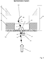
- Fig. 1 illustrates components of one embodiment in which a protein nanopore (100) is disposed in a lipid bilayer (102) disposed (in turn) across aperture (104) of solid state membrane (106), which comprises opaque layer (108) (such as a metal layer), silicon nitride layer (110) and silicon support layer (112). Opaque layer (108) prevents or reduces transmission of excitation beam (114) through solid state membrane (106) where it could excite undesired background fluorescence.
- nanopore (100) As polymer (120) with differently labeled monomers (illustrated as black (122) and white (124)) pass through nanopore (100), at each measurement interval a plurality of monomers (such as, 141, 142 and 143) are present in excitation zones (126) and (128) within the same resolution limited area.
- optical measurements are made with an epi-illumination system and it is assumed that nanopore (100) has been selected so that optical signals from monomers interior to nanopore (100) are suppressed and do not contribute to measured optical signals.
- Excitation zone (128) is a FRET zone adjacent to FRET donor (130); that is, excitation zone (128) defines a distance from FRET donor (130) within which FRET can occur between FRET donor (130) and an optical label attached to a monomer, which may also be referred to as an acceptor label, or FRET acceptor label.
- Excitation zone (126) is a non-propagating protrusion of a component of excitation beam (114) into aperture (104) which occurs whenever the dimensions of aperture (104) are selected to be sufficiently below the wavelength of excitation beam (114).
- a plurality of monomers (141, 142 and 143) would contribute to an optical signal measured at the instant, or interval, during which monomers (141, 142 and 143) are in the excitation zones (126) and (128).
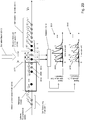
- Fig. 2 illustrates an embodiment in which optical measurements are made with total internal reflection fluorescence (TIRF) excitation in a system such as described in Soni et al, Review of Scientific Instruments, 81: 014301 (2010 ); and in U.S. patent publication 2012/0135410 .
- protein nanopore (200) with attached FRET donor (202) is inserted into lipid bilayer (204) disposed on solid state membrane (206) with aperture (208).
- Total internal reflection (TIR) is made possible by selecting electrolytes on cis (205) and trans (207) sides of solid state membrane (206) with different indices of refraction.
- TIR boundary (210) is created at or near the plane that solid state membrane (206) is disposed in, so that an evanescent field is created on the cis (205) side of solid state membrane (206).
- the evanescent field may excite optical labels prior to their entry into nanopore (200).
- FRET donor (202) is excited directly by light reflected at the TIR boundary (210), so that FRET can take place between FRET donor (202) and labels on monomers (219) within FRET zone (220).
- nanopore (200) may be selected so that fluorescent emissions by labels are suppressed when labeled monomers are in the bore of nanopore (200).
- a plurality of monomers, such as 225, 226 and 227, contribute to an optical measurement recorded at the indicated configuration in the figure.
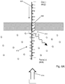
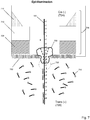
- labels on monomers may be excited by an evanescence field alone using an apparatus similar to that shown in Fig. 4A .
- a very narrow second chamber on the trans side of a nanopore or nanopore array permits an evanescent field to extend from a surface of an underlying glass slide to establish excitation zones both at entrances and exits of the nanopores, so that each optical measurement associated with a nanopore contains contributions from a plurality of labeled monomers.
- Array of apertures (400) (which may include protein nanopores inserted in a lipid bilayer), may be formed in silicon nitride layer (402), which may have a thickness in the range of from 20-100 nm.
- Silicon nitride layer (402) may be formed on a silicon support layer (403).
- Second chamber (406) may be formed by silicon nitride layer (402), silicon dioxide layer (404) which determines the height of second chamber (406), and surface (408) of glass slide (410).
- Silicon dioxide layer (404) may have a thickness in the range of from 50-100 nm.
- a desired evanescent field (407) extending from surface (408) across silicon nitride layer (402) may be established by directing light beam (412) at an appropriate angle relative to glass slide (410) so that TIR occurs.
- cis(-) conditions may be established in first chamber (416) and trans(+) conditions may be established in second chamber (406) with electrodes operationally connected to first and second chambers (406 and 421).
- Fig. 4B is a close-up of a particular embodiment of an aperture in array (400) which diagrammatically shows protein nanopore (420) inserted in lipid bilayer (422) that is disposed on a surface of silicon nitride layer (402).
- Polymer (425) with labeled monomers (for example, 427) is shown translocating through nanopore (420), which has been selected with bore (421) having dimensions that cause suppression of fluorescent emissions of labels interior to bore (421).
- a measured optical signal at a particular time point, t, or interval, from a resolution limited area containing aperture (400) may comprise contributions from a plurality of labels on monomers in the excitation regions, for example, monomers n 1 -n 4 and n 13 -n 15 .
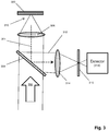
- Fig. 2B provides a further illustration of collecting fluorescent signals that comprise fluorescence generated by two labeled monomers.
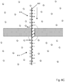
- Fig. 2B shows labeled polynucleotide (2000) translocating through nanopore (2002), wherein labeled polynucleotide (2000) comprises two labels "a” and "b" (for example, which may correspond to dC being labeled with "a” and dA, dG and dT being labeled with "b”, or the like). Labels of nucleotides free of nanopore (2002) are quenched, either by interaction with other labels (2011) or by action of quenching agents (not shown).
- Labels of nucleotides inside of nanopore (2002) are constrained and/or oriented (2014) so that they produce no detectable signal during all or part of their transit through the nanopore.
- nucleotides of labeled polynucleotide (2000) emerge from exit (2015) of nanopore (2002) they become capable of being excited by excitation beam (2010) and generating a detectable signal for an interval prior to being quenched. If translocation speed V 1 is high then the distance (2008) traveled by a nucleotide prior to quenching may exceed the inter-nucleotide distance of polynucleotide (2000) so that more than one label contributes fluorescence to a fluorescent signal collected by detector (2016), i.e. a measured fluorescent signal.
- the signal intensities for two channels e.g. corresponding to emission maxima of two fluorescent labels
- Fig. 2B (2031 and 2032) where two fluorescent labels contribute to a measured signal.
- Intensity values represented by solid lines, e.g. 2033, are from label "a”
- intensity values represented by dashed lines, e.g. 2036, are from label "b”.
- the presence of solid and dashed lines in both channels of Fig. 2B reflects overlapping emission bands of the fluorescent labels, which when collected together complicates analysis because amounts of a measured intensity are from both labels.
- a time-ordered set of optical measurements are recorded.
- Optical measurements at adjacent time points are overlapping in the sense that each optical measurement contains contributions from labels of adjacent monomers.
- three monomers generate signals at each time point (for example, B, C and D of polymer ...-A-(B-C-D)-...
- sequence information may be determined from the time-ordered set of optical signal measurements when it is separated into a plurality of time-ordered sets of monomer-specific signals.
- Algorithms similar to those used in sequencing-by-hybridization (SBH) to reconstruct target polynucleotide sequences from hybridization data may be used to reconstruct target polynucleotides here, e.g. U.S.
- Fig. 5 illustrates one embodiment of a step for determining monomer sequence information from a time-ordered set of overlapping optical signals based on a simple model of nanopore translocation.
- the simple model assumes that optical measurements at each time step (except at the entry and exit of a polymer from a nanopore) each contain signal contributions from the same number of monomers (referred to in Fig. 5 as an "n-tuple" to indicate that a measurement would contain contributions from n monomers). It is understood that more complex models may allow for differing numbers of contributing monomers in each measurement, for local variations in translocation speed, deviations in linear movement of monomers, and other like phenomena. That is, in some embodiments, optical measurements at different times may have contributions from different numbers of nucleotides.
- the differing number of nucleotides are ordered along a segment of the target polynucleotide.
- the step of determining illustrated by Fig. 5 assumes that a labeled polymer has passed through a nanopore and that a time ordered set of optical measurements has been made, including separation of optical signals into monomer-specific signals (500). The entry and exit of a polymer are treated differently since there are necessarily different numbers of monomers in the excitation zone(s) upon entry and exit. In this embodiment, it is assumed that initial and final optical measurements under these conditions permits the initial and final monomers to be determined directly from their monomer-specific signal.
- preparation of labeled polymers for analysis may include insertion of a plurality of predetermined labeled nucleotides at one or both ends of such labeled polymers for the purpose of generating a known sequence of optical signals to aid in a sequence determination step.
- predetermined labeled nucleotides would be similar to key sequences in Ion Torrent or 454 sequencing, e.g. U.S. patent 7,575,865 .
- time index, i is set to zero; the index, j, for candidate sequences at the current time, i, is set to 1 (502); and the initial n-tuple of the set of monomer-specific time-ordered optical signals is examined (504).
- Such examination comprises first determining from the measurement at time i all possible n-tuples of monomers that are consistent with the measurement, then determining from those n-tuples which ones that properly overlap candidate sequence Si. New candidate sequences Si+1 are formed (and a sequence Si is extended) by each properly overlapping n-tuple for the set consistent with the measurement (506).
- New extended candidate sequences, Si+1, are stored and the index giving the number of candidate sequences at time i+1, Ji+1, is updated (508). This step is repeated until every candidate sequence, Si, has been examined (510), and a similar examination is carried out at each time, i, until each optical measurement in the time-ordered set has been examined.
- Nanopores used with the invention may be solid-state nanopores, protein nanopores, or hybrid nanopores comprising protein nanopores or organic nanotubes such as carbon or graphene nanotubes, configured in a solid-state membrane, or like framework.
- Important features of nanopores include constraining polymer analytes, such as polynucleotides, so that their monomers pass through a signal generation region (or excitation zone, or the like) in sequence, That is, so that monomers, such as nucleotides, pass through a detection zone (or excitation region or like region) in single file.
- additional features of nanopores include passing single stranded nucleic acids while not passing double stranded nucleic acids, or equivalently bulky molecules.
- nanopores, especially protein nanopores may be selected so that their bores are sized so that labels of monomers are sterically constrained so that FRET signals, or even fluorescent signals, are suppressed.
- nanopores used in connection with the methods of the invention are provided in the form of arrays, such as an array of clusters of nanopores, which may be disposed regularly on a planar surface.
- clusters are each in a separate resolution limited area so that optical signals from nanopores of different clusters are distinguishable by the optical detection system employed, but optical signals from nanopores within the same cluster cannot necessarily be assigned to a specific nanopore within such cluster by the optical detection system employed.
- Solid state nanopores may be fabricated in a variety of materials including but not limited to, silicon nitride (Si 3 N 4 ), silicon dioxide (SiO 2 ), and the like.
- the fabrication and operation of nanopores for analytical applications, such as DNA sequencing, are disclosed in the following exemplary references: Ling, U.S. patent 7,678,562 ; Hu et al, U.S. patent 7,397,232 ; Golovchenko et al, U.S. patent 6,464,842 ; Chu et al, U.S. patent 5,798,042 ; Sauer et al, U.S. patent 7,001,792 ; Su et al, U.S.
- patent publication 2009/0029477 Howorka et al, International patent publication WO2009/007743 ; Brown et al, International patent publication WO2011/067559 ; Meller et al, International patent publication WO2009/020682 ; Polonsky et al, International patent publication WO2008/092760 ; Van der Zaag et al, International patent publication WO2010/007537 ; Yan et al, Nano Letters, 5(6): 1129-1134 (2005 ); Iqbal et al, Nature Nanotechnology, 2: 243-248 (2007 ); Wanunu et al, Nano Letters, 7(6): 1580-1585 (2007 ); Dekker, Nature Nanotechnology, 2: 209-215 (2007 ); Storm et al, Nature Materials, 2: 537-540 (2003 ); Wu et al, Electrophoresis, 29(13): 2754-2759 (2008 ); Nakane et al, Electrophoresis, 23: 25
- nanopore arrays with one or more light-blocking layers, that is, one or more opaque layers.
- nanopore arrays are fabricated in thin sheets of material, such as, silicon, silicon nitride, silicon oxide, aluminum oxide, or the like, which readily transmit light, particularly at the thicknesses used, e.g. less than 50-100 nm. For electrical detection of analytes this is not a problem.
- light transmitted through an array invariably excites materials outside of intended reaction sites, thus generates optical noise, for example, from nonspecific background fluorescence, fluorescence from labels of molecules that have not yet entered a nanopore, or the like.
- an opaque layer may be a metal layer.
- Such metal layer may comprise Sn, Al, V, Ti, Ni, Mo, Ta, W, Au, Ag or Cu.
- such metal layer may comprise Al, Au, Ag or Cu.
- such metal layer may comprise aluminum or gold, or may comprise solely aluminum.
- the thickness of an opaque layer may vary widely and depends on the physical and chemical properties of material composing the layer.
- the thickness of an opaque layer may be at least 5 nm, or at least 10 nm, or at least 40 nm. In other embodiments, the thickness of an opaque layer may be in the range of from 5-100 nm; in other embodiments, the thickness of an opaque layer may be in the range of from 10-80 nm.
- An opaque layer need not block (i.e. reflect or absorb) 100 percent of the light from an excitation beam. In some embodiments, an opaque layer may block at least 10 percent of incident light from an excitation beam; in other embodiments, an opaque layer may block at least 50 percent of incident light from an excitation beam.
- Opaque layers or coatings may be fabricated on solid state membranes by a variety of techniques known in the art. Material deposition techniques may be used including chemical vapor deposition, electrodeposition, epitaxy, thermal oxidation, physical vapor deposition, including evaporation and sputtering, casting, and the like. In some embodiments, atomic layer deposition may be used, e.g. U.S. patent 6,464,842 ; Wei et al, Small, 6(13): 1406-1414 (2010 ).
- a 1-100 nm channel or aperture may be formed through a solid substrate, usually a planar substrate, such as a membrane, through which an analyte, such as single stranded DNA, is induced to translocate.
- a 2-50 nm channel or aperture is formed through a substrate; and in still other embodiments, a 2-30 nm, or a 2-20 nm, or a 3-30 nm, or a 3-20 nm, or a 3-10 nm channel or aperture if formed through a substrate.
- Biological nanopores provide reproducible narrow bores, or lumens, especially in the 1-10 nanometer range, as well as techniques for tailoring the physical and/or chemical properties of the nanopore and for directly or indirectly attaching groups or elements, such as fluorescent labels, which may be FRET donors or acceptors, by conventional protein engineering methods.
- Solid-state nanopores may be combined with a biological nanopore to form a so-called “hybrid" nanopore that overcomes some of these shortcomings, thereby providing the precision of a biological pore protein with the stability of a solid state nanopore.
- hybrid nanopore provides a precise location of the nanopore which simplifies the data acquisition greatly.
- clusters may also be formed by disposing protein nanopores in lipid bilayers supported by solid phase membrane containing an array of apertures.
- an array may comprise apertures fabricated (e.g. drilled, etched, or the like) in solid phase support.
- the geometry of such apertures may vary depending on the fabrication techniques employed.
- each such aperture is associated with, or encompassed by, a separate resolution limited area; however, in other embodiments, multiple apertures may be within the same resolution limited area.
- the cross-sectional area of the apertures may vary widely and may or may not be the same as between different clusters, although such areas are usually substantially the same as a result of conventional fabrication approaches.
- apertures have a minimal linear dimension (e.g.
- diameter in the case of circular apertures in the range of from 10 to 200 nm, or have areas in the range of from about 100 to 3x10 4 nm 2 .
- Across the apertures may be disposed a lipid bilayer.
- the distribution of protein nanopores per aperture may be varied, for example, by controlling the concentration of protein nanopores during inserting step.
- clusters of nanopores may comprise a random number of nanopores.
- clusters containing one or more apertures on average have a number of protein nanopores that is greater than zero; in other embodiments, such clusters have a number of protein nanopores that is greater than 0.25; in other embodiments, such clusters have a number of protein nanopores that is greater than 0.5; in other embodiments, such clusters have a number of protein nanopores that is greater than 0.75; in other embodiments, such clusters have a number of protein nanopores that is greater than 1.0.
- a solid phase membrane such as a SiN membrane, having an array of apertures therethrough providing communication between a first chamber and a second chamber (also sometimes referred to as a "cis chamber” and a “trans chamber”) and supporting a lipid bilayer on a surface facing the second, or trans, chamber.
- diameters of the aperture in such a solid phase membrane may be in the range of 10 to 200 nm, or in the range of 20 to 100 nm.
- such solid phase membranes further include protein nanopores inserted into the lipid bilayer in regions where such bilayer spans the apertures on the surface facing the trans chamber.
- such protein nanopores are inserted from the cis side of the solid phase membrane using techniques described herein.
- such protein nanopores have a structure identical to, or similar to, ⁇ -hemolysin in that it comprises a barrel, or bore, along an axis and at one end has a "cap” structure and at the other end has a “stem” structure (using the terminology from Song et al, Science, 274: 1859-1866 (1996 )).
- insertion into the lipid bilayer results in the protein nanopore being oriented so that its cap structure is exposed to the cis chamber and its stem structure is exposed to the trans chamber.
- the present invention may employ hybrid nanopores in clusters, particularly for optical-based nanopore sequencing of polynucleotides.
- Such nanopores comprise a solid-state orifice, or aperture, into which a protein biosensor, such as a protein nanopore, is stably inserted.
- a charged polymer may be attached to a protein nanopore (e.g. alpha hemolysin) by conventional protein engineering techniques after which an applied electric field may be used to guide a protein nanopore into an aperture in a solid-state membrane.
- the aperture in the solid-state substrate is selected to be slightly smaller than the protein, thereby preventing it from translocating through the aperture. Instead, the protein will be embedded into the solid-state orifice.
- a donor fluorophore is attached to the protein nanopore.
- This complex is then inserted into a solid-state aperture or nanohole (for example, 3-10 nm in diameter) by applying an electric field across the solid state nanohole, or aperture, until the protein nanopore is transported into the solid-state nanohole to form a hybrid nanopore.
- the formation of the hybrid nanopore can be verified by (a) the inserted protein nanopore causing a drop in current based on a partial blockage of the solid-state nanohole and by (b) the optical detection of the donor fluorophore.
- Solid state, or synthetic, nanopores may be preprared in a variety of ways, as exemplified in the references cited above.
- a helium ion microscope may be used to drill the synthetic nanopores in a variety of materials, e.g. as disclosed by Yang et al, Nanotechnolgy, 22: 285310 (2011 ).
- a chip that supports one or more regions of a thin-film material, e.g. silicon nitride, that has been processed to be a free-standing membrane is introduced to the helium ion microscope (HIM) chamber.
- HIM motor controls are used to bring a free-standing membrane into the path of the ion beam while the microscope is set for low magnification.
- Beam parameters including focus and stigmation are adjusted at a region adjacent to the free-standing membrane, but on the solid substrate.
- the chip position is moved such that the free-standing membrane region is centered on the ion beam scan region and the beam is blanked.
- the HIM field of view is set to a dimension (in ⁇ m) that is sufficient to contain the entire anticipated nanopore pattern and sufficient to be useful in future optical readout (i.e. dependent on optical magnification, camera resolution, etc.).
- the ion beam is then rastered once through the entire field of view at a pixel dwell time that results in a total ion dose sufficient to remove all or most of the membrane autofluorescence.
- the field of view is then set to the proper value (smaller than that used above) to perform lithographically-defined milling of either a single nanopore or an array of nanopores.
- the pixel dwell time of the pattern is set to result in nanopores of one or more predetermined diameters, determined through the use of a calibration sample prior to sample processing. This entire process is repeated for each desired region on a single chip and/or for each chip introduced into the HIM chamber.
- a nanopore may have one or more labels attached for use in optically-based nanopore sequencing methods.
- the label may be a member of a Forster Resonance Energy Transfer (FRET) pair.
- FRET Forster Resonance Energy Transfer
- Such labels may comprise organic fluorophores, chemiluminescent labels, quantum dots, metallic nanoparticles and/or fluorescent proteins.
- Target nucleic acids may have one distinct label per nucleotide.
- the labels attached to the nucleotides may be selected from the group consisting of organic fluorophores.
- the label attachment site in the pore protein can be generated by conventional protein engineering methods, e.g. a mutant protein can be constructed that will allow the specific binding of the label.
- a cysteine residue may be inserted at the desired position of the protein which inserts a thiol (SH) group that can be used to attach a label.
- the cysteine can either replace a natural occurring amino acid or can be incorporated as an addition amino acid.
- a maleimide-activated label is then covalently attached to the thiol residue of the protein nanopore.
- the attachment of the label to the protein nanopore or the label on the nucleic acid is reversible.
- an easily breakable chemical bond e.g. an S-S bond or a pH labile bond
- the label may be removed when the corresponding conditions are met.
- an epi-illumination system in which excitation beam delivery and optical signal collection occurs through a single objective, may be used for direct illumination of labels on a polymer analyte or donors on nanopores.
- the basic components of a confocal epi-illumination system for use with the invention is illustrated in Fig. 3 .
- Excitation beam (302) passes through dichroic (304) and onto objective lens (306) which focuses (310) excitation beam (302) onto layered membrane (300), in which labels are excited directly to emit an optical signal, such as a fluorescent signal, of are excited indirectly via a FRET interaction to emit an optical signal.
- Such optical signal is collected by objective lens (306) and directed to dichroic (304), which is selected so that it passes light of excitation beam (302) but reflects light of optical signals (311).
- Reflected optical signals (311) passes through lens (314) which focuses it through pinhole (316) and onto detector (318).
- a device for implementing the above methods for analyzing polymers typically includes a set of electrodes for establishing an electric field across the layered membrane and nanopores.
- Single stranded nucleic acids are exposed to nanopores by placing them in an electrolyte in a first chamber, which is configured as the "cis" side of the layered membrane by placement of a negative electrode in the chamber.
- the negatively single stranded nucleic acids are captured by nanopores and translocated to a second chamber on the other side of the layered membrane, which is configured as the "trans" side of membrane by placement of a positive electrode in the chamber.
- the speed of translocation depends in part on the ionic strength of the electrolytes in the first and second chambers and the applied voltage across the nanopores.
- a translocation speed may be selected by preliminary calibration measurements, for example, using predetermined standards of labeled single stranded nucleic acids that generate signals at different expected rates per nanopore for different voltages.
- a translocation speed may be selected based on the signal rates from such calibration measurements. Consequently, from such measurements a voltage may be selected that permits, or maximizes, reliable nucleotide identifications, for example, over an array of nanopores.
- such calibrations may be made using nucleic acids from the sample of templates being analyzed (instead of, or in addition to, predetermined standard sequences). In some embodiments, such calibrations may be carried out in real time during a sequencing run and the applied voltage may be modified in real time based on such measurements, for example, to maximize the acquisition of nucleotide-specific signals.
- translocation speed of polynucleotides through nanopores and the need for its control have been appreciated in the field of nanopore technology wherein changes in electric current are use to identify translocating analytes.
- a wide variety of methods have been used to control translocation speed, which include both methods that can be adjusted in real-time without significant difficulty (e.g. voltage potential across nanopores, temperature, and the like) and methods that can be adjusted during operation only with difficulty (reaction buffer viscosity, presence or absence of charged side chains in the bore of a protein nanopore, ionic composition and concentration of the reaction buffer, velocity-retarding groups attached or hybridized to polynucleotide analytes, molecular motors, and the like), e.g.
- a step or steps are included for active control of translocation speed while a method of the invention is being implemented, e.g.
- a step or steps are included that determine a translocation speed that is not actively controlled or changed while a method of the invention is being implemented, e.g. reaction buffer viscosity, ionic concentration, and the like.
- a translocation speed is selected by providing a reaction buffer having a concentration of glycerol, or equivalent reagent, in the range of from 1 to 60 percent.
- a measure of whether one or more than one label is contributing fluorescence to measured signals may be based on the distribution of fluorescence intensity among a plurality of channels over which fluorescence is collected.
- the plurality of channels include 2, 3, or 4 channels corresponding to the emission bands of the fluorescent labels used.
- the relative distribution of signal intensity among the different channels e.g. 4 channels
- the relative distribution of signal intensity among the different channels could be represented ideally as (1,0,0,0); (0,1,0,0); (0,0,1,0) or (0,0,0,1).
- a measure which would vary monotonically between a maximum value corresponding to relative intensity distributions (1,0,0,0); (0,1,0,0); (0,0,1,0) or (0,0,0,1) and a minimum value corresponding to a relative intensity distribution of (.25,.25,.25,.25) may be used for controlling in real-time a translocation speed. For example, an initial translocation speed could be lowered based on the value of such a measure that was near its minimum. Such lowering may be implemented, for example, by lowering a potential voltage across the nanopores by a predetermined amount, after which the measure could be re-calculated. Such steps could be repeated until the process was optimized.
- translocation speeds depend in part on the voltage difference (or electrical field strength) across a nanopore and conditions in the reaction mixture, or buffer, of a first chamber where polynucleotides are exposed to the nanopores (e.g. disposed in a solid phase membrane making up one wall of the first chamber).
- Polynucleotide capture rates by nanopores depend on concentration of such polynucleotides.
- conventional reaction mixture conditions for nanopore sequencing may be employed with the invention (for controlling translocatin speed by varying voltage potential across nanopores), for example, 1M KCl (or equivalent salt, such as NaCl, LiCl, or the like) and a pH buffering system (which, for example, ensures that proteins being used, e.g.
- a pH buffering system may be used to keep the pH substantially constant at a value in the range of 6.8 to 8.8.
- a voltage difference across the nanopores may be in the range of from 70 to 200 mV. In other embodiments, a voltage difference across the nanopores may be in the range of from 80 to 150 mV.
- An appropriate voltage for operation may be selected using conventional measurement techniques. Current (or voltage) across a nanopore may readily be measured using commercially available instruments. A voltage difference may be selected so that translocation speed is within a desired range. In some embodiments, a range of translocation speeds comprises those speeds less than 1000 nucleotides per second.
- a range of translocation speeds is from 10 to 800 nucleotides per second; in other embodiments, a range of translocation speeds is from 10 to 600 nucleotides per second; in other embodiments, a range of translocation speeds is from 200 to 800 nucleotides per second; in other embodiments, a range of translocation speeds is from 200 to 500 nucleotides per second.
- other factors affecting translocation speed e.g. temperature, viscosity, ion concentration, charged side chains in the bore of a protein nanopore, and the like, may be selected to obtain translocation speeds in the ranges cited above.
- a device for implementing the above methods for single stranded nucleic acids typically includes providing a set of electrodes for establishing an electric field across the nanopores (which may comprise an array).
- Single stranded nucleic acids are exposed to nanopores by placing them in an electrolyte (i.e. reaction buffer) in a first chamber, which is configured as the "cis" side of the layered membrane by placement of a negative electrode in the chamber.
- an electric field the negatively single stranded nucleic acids are captured by nanopores and translocated to a second chamber on the other side of the layered membrane, which is configured as the "trans" side of membrane by placement of a positive electrode in the chamber.
- a translocation speed may be selected by preliminary calibration measurements, for example, using predetermined standards of labeled single stranded nucleic acids that generate signals at different expected rates per nanopore for different voltages.
- an initial translocation speed may be selected based on the signal rates from such calibration measurements, as well as the measure based on relative signal intensity distribution discussed above. Consequently, from such measurements a voltage may be selected that permits, or maximizes, reliable nucleotide identifications, for example, over an array of nanopores.
- such calibrations may be made using nucleic acids from the sample of templates being analyzed (instead of, or in addition to, predetermined standard sequences). In some embodiments, such calibrations may be carried out in real time during a sequencing run and the applied voltage may be modified in real time based on such measurements, for example, to maximize the acquisition of nucleotide-specific signals.
- self- and mutually quenching fluorescent labels may be used in addition to quenching agents in order to reduce fluorescent emissions outside of those from labels on nucleotides exiting nanopores, i.e. to restrict the spatial extent of a signal generation zone.
- Use of such fluorescent labels is disclosed in U.S. patent publication 2016/0122812 .
- monomers are labeled with fluorescent labels that are capable of at least three states while attached to a target polynucleotide: (i) A substantially quenched state wherein fluorescence of an attached fluorescent label is quenched by a fluorescent label on an immediately adjacent monomer; for example, a fluorescent label attached to a polynucleotide is substantially quenched when the labeled polynucleotide is free in conventional aqueous solution for studying and manipulating the polynucleotide.
- a sterically constrained state wherein a labeled polynucleotide is translocating through a nanopore such that the free-solution movements or alignments of an attached fluorescent label is disrupted or limited so that there is little or no detectable fluorescent signal generated from the fluorescent label.
- a transition state wherein a fluorescent label attached to a polynucleotide transitions from the sterically constrained state to the quenched state as the fluorescent label exits the nanopore (during a "transition interval") while the polynucleotide translocates through the nanopore.
- this example is an application of the discovery that during the transition interval a fluorescent label (on an otherwise substantially fully labeled and self-quenched polynucleotide) is capable of generating a detectable fluorescent signal.
- a fluorescent label on an otherwise substantially fully labeled and self-quenched polynucleotide
- the fluorescent signal generated during the transition interval is due to the presence of a freely rotatable dipole in the fluorescent label emerging from the nanopore, which renders the fluorescent label temporarily capable of generating a fluorescent signal, for example, after direct excitation or via FRET.
- the dipoles are limited in their rotational freedom thereby reducing or limiting the number of emitted photons.
- the polynucleotide is a polynucleotide, usually a single stranded polynucleotide, such as, DNA or RNA, but especially single stranded DNA. Further disclosed is a method for determining a nucleotide sequence of a polynucleotide by recording signals generated by attached fluorescent labels as they exit a nanopore one at a time as a polynucleotide translocates through the nanopore. Upon exit, each attached fluorescent label transitions during a transition interval from a constrained state in the nanopore to a quenched state on the polynucleotide in free solution.
- a step of the method comprises exciting each fluorescent label as it is transitioning from a constrained state in the nanopore to a quenched state on the polynucleotide in free solution.
- the fluorescent label is capable of emitting a detectable fluorescent signal indicative of the nucleotide it is attached to.
- the invention includes an application of the discovery that fluorescent labels and nanopores may be selected so that during translocation of a polynucleotide through a nanopore fluorescent labels attached to monomers are forced into a constrained state in which they are incapable (or substantially incapable) of producing a detectable fluorescent signal.
- nanopores are selected that have a bore, or lumen, with a diameter in the range of from 1 to 4 nm; in other embodiments, nanopores are selected that have a bore or lumen with a diameter in the range of from 2 to 3 nm. In some embodiments, such bore diameters are provided by a protein nanopore.
- such nanopores are used to force fluorescent labels into a constrained state, so that whenever a fluorescent label exits a nanopore, it transitions from being substantially incapable of generating a fluorescent signal to being detectable and identifiable by a fluorescent signal it can be induced to emit.
- fluorescent labels attached to each of a sequence of monomers of a polynucleotide may be detected in sequence as they suddenly generate a fluorescent signal in a region immediately adjacent to a nanopore exit (a "transition zone” or “transition volume” or “detection zone”).
- organic fluorescent dyes are used as fluorescent labels with nanopores of the above diameters.
- At least one such organic fluorescent dye is selected from the set consisting of xanthene dyes, rhodamine dyes and cyanine dyes.
- Some embodiments for determining a monomer sequence of a polynucleotide may be carried out with the following steps: (a) translocating a polynucleotide through a nanopore, wherein monomers of the polynucleotide are labeled with fluorescent labels wherein the nanopore constrains fluorescent labels within its bore into a constrained state such that substantially no detectable fluorescent signal is generated therein; (b) exciting the fluorescent label of each monomer upon exiting the nanopore; (c) measuring a fluorescent signal in a detection zone generated by the exiting fluorescent label to identify the monomer to which the fluorescent label is attached; (d) quenching fluorescent signals from excited fluorescent labels outside of the detection zone, and (d) determining a monomer sequence of the polynucleotide from a sequence of fluorescent signals.
- fluorescent labels are acceptors of a
- substantially quenched as used above means a fluorescent label generates a fluorescent signal at least thirty percent reduced from a signal generated under the same conditions, but without adjacent mutually quenching labels. In some embodiments, “substantially quenched” as used above means a fluorescent label generates a fluorescent signal at least fifty percent reduced from a signal generated under the same conditions, but without adjacent mutually quenching labels.
- a nucleotide sequence of a target polynucleotide is determined by carrying out four separate reactions in which copies of the target polynucleotide have each of its four different kinds of nucleotide (A, C, G and T) labeled with a single fluorescent label.
- a nucleotide sequence of a target polynucleotide is determined by carrying out four separate reactions in which copies of the target polynucleotide have each of its four different kinds of nucleotide (A, C, G and T) labeled with one fluorescent label while at the same time the other nucleotides on the same target polynucleotide are labeled with a second fluorescent label.
- a first fluorescent label is attached to A's of the target polynucleotide in a first reaction
- a second fluorescent label is attached to C's, G's and T's (i.e. to the "not-A" nucleotides) of the target polynucleotides in the first reaction.
- the first label is attached to C's of the target polynucleotide and the second fluorescent label is attached to A's, G's and T's (i.e. to the "not-C" nucleotides) of the target polynucleotide.
- G and T for nucleotides G and T.
- the same labeling scheme may be expressed in terms of conventional terminology for subsets of nucleotide types; thus, in the above example, in a first reaction, a first fluorescent label is attached to A's and a second fluorescent label is attached to B's; in a second reaction, a first fluorescent label is attached to C's and a second fluorescent label is attached to D's; in a third reaction, a first fluorescent label is attached to G's and a second fluorescent label is attached to H's; and in a fourth reaction, a first fluorescent label is attached to T's and a second fluorescent label is attached to V's.
- a polymer such as a polynucleotide or peptide
- a polymer may be labeled with a single fluorescent label attached to a single kind of monomer, for example, every T (or substantially every T) of a polynucleotide is labeled with a fluorescent label, e.g. a cyanine dye.
- a collection, or sequence, of fluorescent signals from the polynucleotide may form a signature or fingerprint for the particular polynucleotide. In some such embodiments, such fingerprints may or may not provide enough information for a sequence of monomers to be determined.
- substantially all monomers of a polynucleotide analyte with fluorescent dyes or labels that are members of a mutually quenching set.
- the use of the term “substantially all” in reference to labeling polynucleotide analytes is to acknowledge that chemical and enzymatic labeling techniques are typically less than 100 percent efficient. In some embodiments, “substantially all” means at least 80 percent of all monomer have fluorescent labels attached. In other embodiments, “substantially all” means at least 90 percent of all monomer have fluorescent labels attached. In other embodiments, “substantially all” means at least 95 percent of all monomer have fluorescent labels attached.
- Mutually quenching sets of fluorescent dyes have the following properties: (i) each member quenches fluorescence of every member (for example, by FRET or by static or contact mechanisms), and (ii) each member generates a distinct fluorescent signal when excited and when in a non-quenched state. That is, if a mutually quenching set consists of two dyes, D1 and D2, then (i) D1 is self-quenched (e.g. by contact quenching with another D1 molecule) and it is quenched by D2 (e.g. by contact quenching) and (ii) D2 is self-quenched (e.g. by contact quenching with another D2 molecule) and it is quenched by D1 (e.g.
- members of a mutually quenching set comprise organic fluorescent dyes that components or moieties capable of stacking interactions, such as aromatic ring structures.
- Exemplary mutually quenching sets of fluorescent dyes, or labels may be selected from rhodamine dyes, fluorescein dyes and cyanine dyes.
- a mutually quenching set may comprise the rhodamine dye, TAMRA, and the fluorescein dye, FAM.
- mutually quenching sets of fluorescent dyes may be formed by selecting two or more dyes from the group consisting of Oregon Green 488, Fluorescein-EX, fluorescein isothiocyanate, Rhodamine Red-X, Lissamine rhodamine B, Calcein, Fluorescein, Rhodamine, one or more BODIPY dyes, Texas Red, Oregon Green 514, and one or more Alexa Fluors.
- Respresentative BODIPY dyes include BODIPY FL, BODIPY R6G, BODIPY TMR, BODIPY 581/591, BODIPY TR, BODIPY 630/650 and BODIPY 650/665.
- Representative Alexa Fluors include Alexa Fluor 350, 405, 430, 488, 500, 514, 532, 546, 555, 568, 594, 610, 633, 635, 647, 660, 680, 700, 750 and 790.
- a monomer sequence of a target polynucleotide is determined by carrying out separate reactions (one for each kind of monomer) in which copies of the target polynucleotide have each different kind of monomer labeled with a mutually- or self-quenching fluorescent label.
- a monomer sequence of a target polynucleotide is determined by carrying out separate reactions (one for each kind of monomer) in which copies of the target polynucleotide have each different kind of monomer labeled with a different mutually quenching fluorescent label selected from the same mutually quenching set.
- a selected monomer (say, monomer X) is labeled with a first mutually quenching dye and every other kind of monomer (i.e., not-monomer X) is labeled with a second mutually quenching dye from the same set.
- steps of the embodiment generate a sequence of two different fluorescent signals, one indicating monomer X and another indicating not-monomer X.
- a single fluorescent label (for example, attached to a single kind of monomer in a polynucleotide comprising multiple kinds of monomers) may be used that is self-quenching when attached to adjacent monomers (of the same kind) on a polynucleotide, such as adjacent nucleotides of a polynucleotide.
- Exemplary self-quenching fluorescent labels include, but are not limited to, Oregon Green 488, fluorescein-EX, FITC, Rhodamine Red-X, Lissamine rhodamine B, calcein, fluorescein, rhodamine, BODIPYS, and Texas Red, e.g. which are disclosed in Molecular Probes Handbook, 11th Edition (2010).
- Figs. 6A-6C illustrate different embodiments corresponding to where quenching agents are applied in a nanopore device: trans chamber only ( Fig. 6A ), cis chamber only ( Fig. 6B ), or both cis and trans chambers ( Fig. 6C ).
- labeled polynucleotide (600) is illustrated translocating nanopore (606) of solid phase membrane (608) from cis chamber (602) to trans chamber (604).
- Immersed in trans chamber (604) are non-fluorescent quenching agents (605) designated by "Q”.
- Quenching agents are soluble under translocation conditions for labeled polynucleotide (600), and under the same conditions, quenching agents bind to single stranded polynucleotides, such as (600), without substantial sequence specificity.
- quenching agents include derivatives of many well-known organic dyes, such as asymmetric cyanine dyes, as well as conjugates of such compounds and oligonucleotides and/or analogs thereof.
- selection of the type and concentration of quenching agent and the translocation speed define detection zone (610).
- "detection zone” means a region or volume (which may be contiguous or non-contiguous) from which fluorescent signals are collected to form the raw data from which information, such as sequence information, about a labeled polynucleotide is determined.
- Fluorescent labels in trans chamber (604) outside of detection zone (610) are substantially quenched by quenching agents (605) bound to the portion of labeled polynucleotide (600) in trans chamber (604).
- quenching agents comprise an oligonucleotide or analog conjugated to one or more quenching moieties based on organic dyes as described more fully below.
- Embodiments of Fig. 6A may be employed when, for example, solid phase membrane (608) is or comprises an opaque layer so that fluorescent labels in cis chamber (602) are substantially non-excited.
- Fig. 6B shows substantially the same elements as those in Fig. 6A with the exception that quenching agents (605) are disposed in cis chamber (602).
- This configuration may be desirable under circumstances where undesired evanescent waves, or like non-radiative light energy, extend to cis chamber (602) and excite fluorescent labels which generate fluorescent signals that are collected.
- Quenching agents (605) that bind to labeled polynucleotide (600) in cis chamber (602) reduce or eliminate such fluorescent signals.
- quenching agents (605) and cross-section of nanopore (606) are selected so that quenching agents (605) are excluded from translocating through nanopore (606). In some embodiments, this may be achieved by using protein nanopore ⁇ -hemolysin and quenching agents comprising conjugates of oligonucleotides or analogs thereof and one or more quenching compounds, as described more fully below.
- Fig. 6C illustrates an embodiment where quenching agents (605) are present in both cis chamber (602) and trans chamber (604), which provides the advantages described for the embodiments of both Figs. 6A and 6B .
- Fig. 7 illustrates an embodiment which includes the following elements: protein nanopore (700) disposed in lipid bilayer (702); epi-illumination of fluorescent labels with opaque layer (708) in solid phase membrane (706) to prevent or reduce background fluorescence; and quenching agents (710) disposed in trans chamber (726).
- polynucleotide (720) with fluorescently labeled nucleotides labels being indicated by "f", as with (722)
- Oligonucleotide quenchers (710) are disposed in trans chamber (726) under conditions (e.g.
- Nanopore (700) may be selected so that signals from fluorescent labels are suppressed during transit of the nanopore as described in Huber et al, U.S. patent publication US 2016/0076091 .
- labeled nucleotides emerge from nanopore (700) in region (728) they become unsuppressed and capable of generating a signal.
- direct illumination e.g. non-FRET
- such emerged labels would continue to emit fluorescence as they travel further into trans chamber (726), thereby contributing greatly to a collected signal.
- a fluorescent signal from a single fluorescent label is detected from detection zone (728) during a detection period as the labeled polynucleotide moves through the detection zone.
- a plurality of fluorescent signals is collected from a plurality of fluorescent labels in detection zone (728) during a predetermined time period.
- detection period is less than 1 msec, or less than 0.1 msec, or less than 0.01 msec.
- such detection perior is at least 0.01 msec, or at least 0.1 msec, or at least 0.5 msec.
- Quenching agents disclosed herein comprise any compound (or set of compounds) that under nanopore sequencing conditions is (i) substantially non-fluorescent, (ii) binds to single stranded nucleic acids, particularly single stranded DNA, and (iii) absorbs excitation energy from other molecules non-radiatively and releases it non-radiatively.
- quenching agents further bind non-covalently to single stranded DNA.
- a large variety of quenching compounds are available for use with the invention including, but not limited to, non-fluorescent derivatives of common synthetic dyes such as cyanine and xanthene dyes, as described more fully below. Guidance in selecting quenching compounds may be found in U.S. patents 6,323,337 ; 6,750,024 and like references.
- a quenching agent may be a single stranded DNA binding dye that has been covalently modified with a heavy atom that is known to quench fluorescence (such as bromine or iodine), or covalently modified with other groups known to quench fluorescence, such as a nitro group or a azo group.
- a heavy atom that is known to quench fluorescence (such as bromine or iodine)
- other groups known to quench fluorescence such as a nitro group or a azo group.
- An example of dye that is known to bind single stranded DNA is Sybr Green ( Zipper et al, (2004), Nucleic Acids Research. 32 (12 )).
- quenching agents comprise a binding moiety and one or more quenching moieties.
- Binding moieties may include any compound that binds to single stranded nucleic acids without substantial sequence specificity. Binding moieties may comprise peptides or oligonucleotides or analogs of either having modified linkages and/or monomers. Oligonucleotides and their analogs may provide binding to polynucleotides via duplex formation or via non-base paired aptameric binding. In some embodiments, binding moieties comprise an oligonucleotide or analog thereof having a length in the range of from 6 to 60 nucleotides.
- Such oligonucleotides or analogs may be conjugated to one quenching moiety or to a plurality of quenching moieties.
- the plurality of quenching moieties conjugated to each oligonucleotide or analog is 2 or 3.
- Quenching moieties conjugated to a binding moiety may be the same or different.
- a binding moiety is an oligonucleotide or analog
- two quenching moieties are conjugated thereto, one at a 5' end and one at a 3' end of the oligonucleotide.
- Oligonucleotides or analogs having from 2 to 3 quenching moieties may be synthesized using conventional linkage and synthetic chemistries, for example, as disclosed in the references cited herein.
- Oligonucleotides or analogs may be provided as a single species or they may be provided as mixtures of a plurality of oligonucleotides or analogs with different sequences, and therefore, different binding specificities.
- oligonucleotides or analogs are random sequence polymers; that is, they are provided as mixtures of every possible sequence of a given length.
- such oligonucleotides or analogs may be represented by the formulas, "NNNNNN” for 6-mers, or "NNNNNNNN” for 8-mers, wherein N may be A, C, G or T, or an analog thereof.
- oligonucleotides in reference to oligonucleotides means an oligonucleotide that contains one or more nucleotide analogs.
- a "nucleotide analog” is a nucleotide that may have a modified linkage moiety, sugar moiety or base moiety.
- Exemplary oligonucleotide analogs that may be used with the invention include, but are not limited to, peptide nucleic acids (PNAs), locked nucleic acids (LNAs)(2'-O-methyl RNA), phosphorothioate oligonucleotides, bridged nucleic acids (BNAs), or the like.
- oligonucleotide binding moieties comprise universal bases; that is, they contain one or more nucleotide analogs that can replace any of the four natural nucleotides without destabilizing base-pair interactions. Nucleotide analogs having universal base properties are described in Loakes, Nucleic Acids Research, 29(12): 2437-2447 (2001 ). In some embodiments, oligonucleotide binding moieties comprise 2'-deoxyinosine, 7-deaza-2'-deoxyinosine, 2-aza-2'-deoxyinosine, 3-nitropyrrole nucleotides, 5-nitroindole nucleotides, or the like.
- quenching agents may comprise a combination of two or more compounds that act together to quench undesired fluorescent signals of a single stranded labeled polynucleotide.
- a quenching agent may comprise an oligonucleotide (e.g., polydeoxyinosine) that may form a duplex with the labeled polynucleotide and separately a double stranded intercalator that is a quencher.
- the quenching intercalator binds to the resulting duplex and quenches fluorescent signals from the polynucleotide.
- any synthetic dye that can detectably quench fluorescent signals of the fluorescent labels of a labeled polynucleotide is an acceptable quenching moiety for the purposes of the invention.
- the quenching moieties possess an absorption band that exhibits at least some spectral overlap with an emission band of the fluorescent labels on a labeled polynucleotide. This overlap may occur with emission of the fluorescent label (donor) occurring at a lower or even higher wavelength emission maximum than the maximal absorbance wavelength of the quenching moiety (acceptor), provided that sufficient spectral overlap exists. Energy transfer may also occur through transfer of emission of the donor to higher electronic states of the acceptor.
- One of ordinary skill in the art determines the utility of a given quenching moiety by examination of that dye's excitation bands with respect to the emission spectrum of the fluorescent labels being used.
- fluorescence quenching occurs through Fluorescence Resonance Energy Transfer (FRET or through the formation of charge transfer complexes) between a fluorescent label and a quenching moiety.
- FRET Fluorescence Resonance Energy Transfer
- the spectral and electronic properties of the donor and acceptor compounds have a strong effect on the degree of energy transfer observed, as does the separation distance between the fluorescent labels on the labeled polynucleotide and the quenching moiety. As the separation distance increases, the degree of fluorescence quenching decreases.
- a quenching moiety may be optionally fluorescent, provided that the maximal emission wavelength of the dye is well separated from the maximal emission wavelength of the fluorescent labels when bound to labeled polynucleotides.
- the quenching moiety is only dimly fluorescent, or is substantially non-fluorescent, when covalently conjugated to a oligonucleotide or analog.
- substantially non-fluorescent indicates that the fluorescence efficiency of the quenching moiety in an assay solution as described for any of the methods herein is less than or equal to 5 percent, preferably less than or equal to 1 percent.
- the covalently bound quenching moiety exhibits a quantum yield of less than about 0.1, more preferably less than about 0.01.
- the fluorescence of fluorescent labels associated with a quenching oligonucleotide is quenched more than 50% relative to the same oligonucleotide associated with the same fluorescent labels in the absence of the covalently bound quenching moiety.
- the fluorescent labels are quenched more than 90% relative to the unlabeled oligonucleotide.
- the nucleic acid stains are quenched more than 95% relative to the unlabeled oligonucleotide.
- a quenching moiety may be a pyrene, an anthracene, a naphthalene, an acridine, a stilbene, an indole or benzindole, an oxazole or benzoxazole, a thiazole or benzothiazole, a 4-amino-7-nitrobenz-2-oxa-1,3-diazole (NBD), a cyanine, a carbocyanine, a carbostyryl, a porphyrin, a salicylate, an anthranilate, an azulene, a perylene, a pyridine, a quinoline, a coumarin (including hydroxycoumarins and aminocoumarins and fluorinated and sulfonated derivatives thereof (as described in U.S.
- quenching moieties that are substantially non-fluorescent dyes include in particular azo dyes (such as DABCYL or DABSYL dyes and their structural analogs), triarylmethane dyes such as malachite green or phenol red, 4',5z-diether substituted fluoresceins ( U.S. Pat. No. 4,318,846 (1982 )), or asymmetric cyanine dye quenchers ( PCT Int. App. WO 99 37,717 (1999 )).
- azo dyes such as DABCYL or DABSYL dyes and their structural analogs
- triarylmethane dyes such as malachite green or phenol red
- 4',5z-diether substituted fluoresceins U.S. Pat. No. 4,318,846 (1982 )
- asymmetric cyanine dye quenchers PCT Int. App. WO 99 37,717 (1999 )
- the synthetic dye is optionally a fluorescein, a rhodol ( U.S. Pat. No. 5,227,487 to Haugland, et al. (1993 )), or a rhodamine.
- fluorescein includes benzo- or dibenzofluoresceins, seminaphthofluoresceins, or naphthofluoresceins.
- rhodol includes seminaphthorhodafluors ( U.S. Pat. No. 4,945,171 to Haugland, et al. (1990 )).
- Xanthenes include fluorinated derivatives of xanthene dyes (Int. Publ. No. WO 97/39064 , Molecular Probes, Inc. (1997)), and sulfonated derivatives of xanthene dyes (Int. Publ. No. WO 99/15517 , Molecular Probes, Inc. (1999)).
- oxazines include resorufms, aminooxazinones, diaminooxazines, and their benzo-substituted analogs.
- the quenching moiety is an substantially nonfluorescent derivative of 3- and/or 6-amino xanthene that is substituted at one or more amino nitrogen atoms by an aromatic or heteroaromatic ring system, e.g. as described in U.S. patent 6,399392 .
- These quenching dyes typically have absorption maxima above 530 nm, have little or no observable fluorescence and efficiently quench a broad spectrum of luminescent emission, such as is emitted by chemilumiphores, phosphors, or fluorophores.
- the quenching dye is a substituted rhodamine.
- the quenching compound is a substituted rhodol.
- a quenching moiety may comprise one or more non-fluorescent quenchers known as Black Hole QuenchersTM compounds (BHQs) described in the following patents: 7,019,129 ; 7,109,312 ; 7,582,432 ; 8,410,025 ; 8,440,399 ; 8,633,307 ; 8,946,404 ; 9,018,369 ; or 9,139,610 . Additional quenching moieties are disclosed in the following: U.S. patents 6,699,975 ; 6,790,945 ; and 8,114,979 .
- BHQs Black Hole QuenchersTM compounds
- a nanopore may be labeled with one or more quantum dots.
- one or more quantum dots may be attached to a nanopore, or attached to a solid phase support adjacent to (and within a FRET distance of an entrance or exit of a nanopore), and employed as donors in FRET reactions with acceptors on analytes.
- quantum dots are well known and are described widely in the scientific and patent literature, such as, in U.S. patents 6,252,303 ; 6,855,551 ; 7,235,361 ; and the like.
- a Quantum dot which may be utilized as a pore label is a CdTe quantum dot which can be synthesized in an aqueous solution.
- a CdTe quantum dot may be functionalized with a nucleophilic group such as primary amines, thiols or functional groups such as carboxylic acids.
- a CdTe quantum dot may include a mercaptopropionic acid capping ligand, which has a carboxylic acid functional group that may be utilized to covalently link a quantum dot to a primary amine on the exterior of a protein pore.
- the cross-linking reaction may be accomplished using standard cross-linking reagents (homo-bifunctional as well as hetero-bifunctional) which are known to those having ordinary skill in the art of bioconjugation. Care may be taken to ensure that the modifications do not impair or substantially impair the translocation of a nucleic acid through the nanopore. This may be achieved by varying the length of the employed crosslinker molecule used to attach the donor label to the nanopore.
- the primary amine of the lysine residue 131 of the natural alpha hemolysin protein may be used to covalently bind carboxy modified CdTe Quantum dots via 1-Ethyl-3-[3-dimethylaminopropyl]carbodiimide hydrochloride/ N-hydroxysulfosuccinimide (EDC/NHS) coupling chemistry.
- EDC/NHS N-hydroxysulfosuccinimide
- a biological polymer e.g., a nucleic acid molecule or polymer
- each of the four nucleotides or building blocks of a nucleic acid molecule may be labeled with an acceptor label thereby creating a labeled (e.g., fluorescent) counterpart to each naturally occurring nucleotide.
- the acceptor label may be in the form of an energy accepting molecule which can be attached to one or more nucleotides on a portion or on the entire strand of a converted nucleic acid.
- a variety of methods may be utilized to label the monomers or nucleotides of a nucleic acid molecule or polymer.
- a labeled nucleotide may be incorporated into a nucleic acid during synthesis of a new nucleic acid using the original sample as a template ("labeling by synthesis").
- labeling by synthesis For example, the labeling of nucleic acid may be achieved via PCR, whole genome amplification, rolling circle amplification, primer extension or the like or via various combinations and extensions of the above methods known to persons having ordinary skill in the art.
- a label may comprise a reactive group such as a nucleophile (amines, thiols etc.). Such nucleophiles, which are not present in natural nucleic acids, can then be used to attach fluorescent labels via amine or thiol reactive chemistry such as NHS esters, maleimides, epoxy rings, isocyanates etc. Such nucleophile reactive fluorescent dyes (i.e. NHS-dyes) are readily commercially available from different sources.
- An advantage of labeling a nucleic acid with small nucleophiles lies in the high efficiency of incorporation of such labeled nucleotides when a "labeling by synthesis" approach is used. Bulky fluorescently labeled nucleic acid building blocks may be poorly incorporated by polymerases due to steric hindrance of the labels during the polymerization process into newly synthesized DNA.
- NHS esters can be used to react very specifically with primary amines or maleimides will react with thiol groups.
- Either primary amines (NH2) or thiol (SH) modified nucleotides are commercially available. These relatively small modifications are readily incorporated in a polymerase mediated DNA synthesis and can be used for subsequent labeling reactions using either NHS or maleimide modified dyes.
- Guidance for selecting and using such orthogonal linker chemistries may be found in Hermanson (cited above).
- Additional orthogonal attachment chemistries for typical attachment positions include Huisgen-type cycloaddition for a copper-catalyzed reaction and an uncatalyzed reaction; alkene plus nitrile oxide cycloaddition, e.g. as disclosed in Gutsmiedl et al, Org. Lett., 11: 2405-2408 (2009 ); Diels-Alder cycloaddition, e.g. disclosed in Seelig et al, Tetrahedron Lett., 38: 7729-7732 (1997 ); carbonyl ligation, e.g. as disclosed in Casi et al, J. Am. Chem.
- Electrode field means a non-propagating electromagnetic field; that is, it is an electromagnetic field in which the average value of the Poynting vector is zero.
- FRET Fluorescence, resonant energy transfer
- FRET means a non-radiative dipole-dipole energy transfer mechanism from an excited donor fluorophore to an acceptor fluorophore in a ground state.
- the rate of energy transfer in a FRET interaction depends on the extent of spectral overlap of the emission spectrum of the donor with the absorption spectrum of the acceptor, the quantum yield of the donor, the relative orientation of the donor and acceptor transition dipoles, and the distance between the donor and acceptor molecules, Lakowitz, Principles of Fluorescence Spectroscopy, Third Edition (Springer, 2006 ).
- FRET interactions of particular interest are those which result a portion of the energy being transferred to an acceptor, in turn, being emitted by the acceptor as a photon, with a frequency lower than that of the light exciting its donor (i.e. a "FRET signal”).
- FRET distance means a distance between a FRET donor and a FRET acceptor over which a FRET interaction can take place and a detectable FRET signal produced by the FRET acceptor.
- Kit refers to any delivery system for delivering materials or reagents for carrying out a method of the invention.
- delivery systems include systems that allow for the storage, transport, or delivery of reaction reagents (e.g., fluorescent labels, such as mutually quenching fluorescent labels, fluorescent label linking agents, enzymes, etc. in the appropriate containers) and/or supporting materials (e.g., buffers, written instructions for performing the assay etc.) from one location to another.
- reaction reagents e.g., fluorescent labels, such as mutually quenching fluorescent labels, fluorescent label linking agents, enzymes, etc. in the appropriate containers
- supporting materials e.g., buffers, written instructions for performing the assay etc.
- kits include one or more enclosures (e.g., boxes) containing the relevant reaction reagents and/or supporting materials.
- Such contents may be delivered to the intended recipient together or separately.
- a first container may contain an enzyme for use in an assay, while a second or more containers contain mutually quenching fluorescent labels.
- Nanopore means any opening positioned in a substrate that allows the passage of analytes through the substrate in a predetermined or discernable order, or in the case of polymer analytes, passage of their monomeric units through the substrate in a pretermined or discernible order. In the latter case, a predetermined or discernible order may be the primary sequence of monomeric units in the polymer.
- nanopores include proteinaceous or protein based nanopores, synthetic or solid state nanopores, and hybrid nanopores comprising a solid state nanopore having a protein nanopore embedded therein.
- a nanopore may have an inner diameter of 1-10 nm or 1-5 nm or 1-3 nm.
- protein nanopores include but are not limited to, alpha-hemolysin, voltage-dependent mitochondrial porin (VDAC), OmpF, OmpC, MspA and LamB (maltoporin), e.g. disclosed in Rhee, M. et al., Trends in Biotechnology, 25(4) (2007): 174-181 ; Bayley et al (cited above); Gundlach et al, U.S. patent publication 2012/0055792 ; and the like. Any protein pore that allows the translocation of single nucleic acid molecules may be employed.
- a nanopore protein may be labeled at a specific site on the exterior of the pore, or at a specific site on the exterior of one or more monomer units making up the pore forming protein.
- Pore proteins are chosen from a group of proteins such as, but not limited to, alpha-hemolysin, MspA, voltage-dependent mitochondrial porin (VDAC), Anthrax porin, OmpF, OmpC and LamB (maltoporin). Integration of the pore protein into the solid state hole is accomplished by attaching a charged polymer to the pore protein. After applying an electric field the charged complex is electrophoretically pulled into the solid state hole.
- a synthetic nanopore, or solid-state nanopore may be created in various forms of solid substrates, examples of which include but are not limited to silicones (e.g. Si3N4, SiO2), metals, metal oxides (e.g. Al2O3) plastics, glass, semiconductor material, and combinations thereof.
- a synthetic nanopore may be more stable than a biological protein pore positioned in a lipid bilayer membrane.
- a synthetic nanopore may also be created by using a carbon nanotube embedded in a suitable substrate such as but not limited to polymerized epoxy. Carbon nanotubes can have uniform and well-defined chemical and structural properties. Various sized carbon nanotubes can be obtained, ranging from one to hundreds of nanometers.
- the surface charge of a carbon nanotube is known to be about zero, and as a result, electrophoretic transport of a nucleic acid through the nanopore becomes simple and predictable ( Ito, T. et al., Chem. Commun. 12 (2003): 1482-83 ).
- the substrate surface of a synthetic nanopore may be chemically modified to allow for covalent attachment of the protein pore or to render the surface properties suitable for optical nanopore sequencing. Such surface modifications can be covalent or non-covalent. Most covalent modification include an organosilane deposition for which the most common protocols are described: 1) Deposition from aqueous alcohol. This is the most facile method for preparing silylated surfaces. A 95% ethanol-5% water solution is adjusted to pH 4.5-5.5 with acetic acid.
- Silane is added with stirring to yield a 2% final concentration. After hydrolysis and silanol group formation the substrate is added for 2-5min. After rinsed free of excess materials by dipping briefly in ethanol. Cure of the silane layer is for 5-10min at 110 degrees Celsius.
- Vapor Phase Deposition Silanes can be applied to substrates under dry aprotic conditions by chemical vapor deposition methods. These methods favor monolayer deposition. In closed chamber designs, substrates are heated to sufficient temperature to achieve 5mm vapor pressure. Alternatively, vacuum can be applied until silane evaporation is observed.
- Spin-on deposition Spin-on applications can be made under hydrolytic conditions which favor maximum functionalization and polylayer deposition or dry conditions which favor monolayer deposition.
- single nanopores are employed with methods of the invention.
- a plurality of nanopores are employed.
- a plurality of nanopores is employed as an array of nanopores, usually disposed in a planar substrate, such as a solid phase membrane.
- Nanopores of a nanopore array may be spaced regularly, for example, in a rectilinear pattern, or may be spaced randomly. In a preferred embodiment, nanopores are spaced regularly in a rectilinear pattern in a planar solid phase substrate.
- Nanostructure (used interchangeably with “nanoscale structure” and “nanoscale feature”) means a structure that has at least one dimension within a range of a few nanometers to several hundred nanometers, for example, from 1 to 1000 nanometers. In some applications, such range is from 2 to 500 nanometers; in other applications, such range is from 3 to 500 nanometers.
- the shape and geometry of nanostructures may vary widely and include, but are not limited to, nanopores, nanowells, nanoparticles, and any other convenient shapes particularly suitable for carrying out sequences of reactions.
- nanostructures may be protein nanopores operationally associated with a solid phase membrane.
- Nanostructures such as, nanopores and nanowells, may be formed in a larger common substrate, such as a solid phase membrane, or other solid, to form arrays of nanopores or nanowells.
- Nanostructures of particular interest are those capable of supporting or containing a chemical, physical (e.g. FRET), enzymatic and/or binding reaction or a sequence of such reactions.
- a nanostructure, such as a nanowell encloses a volume that is less than one nanoliter (10 ⁇ -9 liter), less than one picoliter, or less than one femtoliter.
- each of the individual nanowells provides a volume that is less than 1000 zeptoliters, 100 zeptoliters, 80 zeptoliters, or less than 50 zeptoliters, or less than 1 zeptoliter, or even less than 100 yactoliters.
- nanowells comprise zero mode waveguides.
- Protein peptide fragment
- polypeptide polypeptide
- oligopeptide fragment in reference to a peptide
- amino acids in a peptide or polypeptide may be derivatized with various moieties, including but not limited to, polyethylene glycol, dyes, biotin, haptens, or like moieties.
- protein or polypeptides or peptides referred to herein may have 2 from to 70 amino acid residues; and in other embodiments, they may have from 2 to 50 amino acid residues.
- proteins or polypeptides or peptides referred to herein may have from a few tens of amino acid residues, e.g. 20, to up to a thousand or more amino acid residues, e.g. 1200.
- proteins, polypeptides, peptides, or fragments thereof may have from 10 to 1000 amino acid residues; or they may have from 20 to 500 amino acid residues; or they may have from 20 to 200 amino acid residues.
- Polymer means a plurality of monomers connected into a linear chain. Usually, polymers comprise more than one type of monomer, for example, as a polynucleotide comprising A's, C's, G's and T's, or a polypeptide comprising more than one kind of amino acid. Monomers may include without limitation nucleosides and derivatives or analogs thereof and amino acids and derivatives and analogs thereof. In some embodiments, polymers are polynucleotides, whereby nucleoside monomers are connected by phosphodiester linkages, or analogs thereof.
- Polynucleotide or “oligonucleotide” are used interchangeably and each mean a linear polymer of nucleotide monomers.
- Monomers making up polynucleotides and oligonucleotides are capable of specifically binding to a natural polynucleotide by way of a regular pattern of monomer-to-monomer interactions, such as Watson-Crick type of base pairing, base stacking, Hoogsteen or reverse Hoogsteen types of base pairing, or the like.
- Such monomers and their internucleosidic linkages may be naturally occurring or may be analogs thereof, e.g. naturally occurring or non-naturally occurring analogs.
- Non-naturally occurring analogs may include PNAs, phosphorothioate internucleosidic linkages, bases containing linking groups permitting the attachment of labels, such as fluorophores, or haptens, and the like.
- PNAs phosphorothioate internucleosidic linkages
- bases containing linking groups permitting the attachment of labels such as fluorophores, or haptens, and the like.
- labels such as fluorophores, or haptens, and the like.
- oligonucleotide or polynucleotide requires enzymatic processing, such as extension by a polymerase, ligation by a ligase, or the like, one of ordinary skill would understand that oligonucleotides or polynucleotides in those instances would not contain certain analogs of internucleosidic linkages, sugar moieties, or bases at any or some positions.
- Polynucleotides typically range in size from a few monomeric units
- oligonucleotides when they are usually referred to as "oligonucleotides,” to several thousand monomeric units.
- A denotes deoxyadenosine
- C denotes deoxycytidine
- G denotes deoxyguanosine
- T denotes thymidine
- I denotes deoxyinosine
- U denotes uridine, unless otherwise indicated or obvious from context.
- polynucleotides comprise the four natural nucleosides (e.g. deoxyadenosine, deoxycytidine, deoxyguanosine, deoxythymidine for DNA or their ribose counterparts for RNA) linked by phosphodiester linkages; however, they may also comprise non-natural nucleotide analogs, e.g. including modified bases, sugars, or internucleosidic linkages.
- nucleosides e.g. deoxyadenosine, deoxycytidine, deoxyguanosine, deoxythymidine for DNA or their ribose counterparts for RNA
- non-natural nucleotide analogs e.g. including modified bases, sugars, or internucleosidic linkages.
- oligonucleotide or polynucleotide substrate requirements for activity e.g. single stranded DNA, RNA/DNA duplex, or the like
- selection of appropriate composition for the oligonucleotide or polynucleotide substrates is well within the knowledge of one of ordinary skill, especially with guidance from treatises, such as Sambrook et al, Molecular Cloning, Second Edition (Cold Spring Harbor Laboratory, New York, 1989 ), and like references.
- the oligonucleotide and polynucleotide may refer to either a single stranded form or a double stranded form (i.e. duplexes of an oligonucleotide or polynucleotide and its respective complement). It will be clear to one of ordinary skill which form or whether both forms are intended from the context of the terms usage.
- Primer means an oligonucleotide, either natural or synthetic that is capable, upon forming a duplex with a polynucleotide template, of acting as a point of initiation of nucleic acid synthesis and being extended from its 3' end along the template so that an extended duplex is formed.
- Extension of a primer is usually carried out with a nucleic acid polymerase, such as a DNA or RNA polymerase.
- the sequence of nucleotides added in the extension process is determined by the sequence of the template polynucleotide.
- primers are extended by a DNA polymerase. Primers usually have a length in the range of from 14 to 40 nucleotides, or in the range of from 18 to 36 nucleotides.
- Primers are employed in a variety of nucleic amplification reactions, for example, linear amplification reactions using a single primer, or polymerase chain reactions, employing two or more primers.
- Guidance for selecting the lengths and sequences of primers for particular applications is well known to those of ordinary skill in the art, as evidenced by the following references: Dieffenbach, editor, PCR Primer: A Laboratory Manual, 2nd Edition (Cold Spring Harbor Press, New York, 2003 ).
- a surface of a nanopore array may be partitioned, or subdivided, into non-overlapping regions, or substantially non-overlapping regions, corresponding to resolution limited areas. The size of such subdivisions corresponding to resolution limited areas may depend on a particular optical detection system employed.
- a resolution limited area is in the range of from 300 nm 2 to 3.0 ⁇ m 2 ; in other embodiments, a resolution limited area is in the range of from 1200 nm 2 to 0.7 ⁇ m 2 ; in other embodiments, a resolution limited area is in the range of from 3x10 4 nm 2 to 0.7 ⁇ m 2 , wherein the foregoing ranges of areas are in reference to a surface of a nanopore or nanowell array.
- the visible spectrum means wavelengths in the range of from about 380 nm to about 700 nm.
- Sequence determination includes determination of partial as well as full sequence information of the polynucleotide. That is, the terms include sequences of subsets of the full set of four natural nucleotides, A, C, G and T, such as, for example, a sequence of just A's and C's of a target polynucleotide. That is, the terms include the determination of the identities, ordering, and locations of one, two, three or all of the four types of nucleotides within a target polynucleotide.
- the terms include the determination of the identities, ordering, and locations of two, three or all of the four types of nucleotides within a target polynucleotide.
- sequence determination may be accomplished by identifying the ordering and locations of a single type of nucleotide, e.g. cytosines, within the target polynucleotide "catcgc ... " so that its sequence is represented as a binary code, e.g. "100101 ... " representing "c-(not c)(not c)c-(not c)-c" and the like.
- the terms may also include subsequences of a target polynucleotide that serve as a fingerprint for the target polynucleotide; that is, subsequences that uniquely identify a target polynucleotide, or a class of target polynucleotides, within a set of polynucleotides, e.g. all different RNA sequences expressed by a cell.
Claims (9)
- Procédé d'analyse d'un polymère comprenant les étapes consistant à :transloquer un polymère à travers un nanopore, dans lequel différents types de monomères du polymère sont marqués avec différents marqueurs optiques qui génèrent des signaux optiques distincts et dans lequel le nanopore contraint les monomères à se déplacer suivant une rangée unique à travers une zone d'excitation qui englobe une pluralité de monomères ;détecter un ensemble ordonné dans le temps de signaux optiques provenant des monomères lorsqu'ils traversent la zone d'excitation ;séparer des signaux optiques provenant de différents types de monomères pour former des ensembles ordonnés dans le temps spécifiques à un monomère de signaux optiques ; etdéterminer une séquence de monomères à partir des ensembles ordonnés dans le temps spécifiques à un monomère de signaux optiques provenant du polymère.
- Procédé selon la revendication 1, dans lequel ledit polymère est un polynucléotide.
- Procédé selon la revendication 2, dans lequel ledit polymère est un polynucléotide marqué en étendant une amorce fusionnée à une molécule d'acide nucléique matrice en présence de nucléoside triphosphates marqués.
- Procédé selon la revendication 3, dans lequel ladite amorce contient une séquence clé avec des nucléotides marqués qui génèrent un signal optique initial lorsque ledit polynucléotide est transloqué à travers ledit nanopore et passe à travers ladite zone d'excitation.
- Procédé selon la revendication 3, dans lequel lesdits nucléoside triphosphates marqués comprennent au moins deux marqueurs fluorescents distincts attachés à au moins deux types différents de nucléoside triphosphates de sorte qu'au moins deux types différents de nucléotide dans ladite amorce étendue peuvent être identifiés par des signaux fluorescents générés par les marqueurs fluorescents distincts.
- Procédé selon la revendication 2, dans lequel ladite étape de détermination comprend la formation de séquences candidates à partir de segments chevauchants de nucléotides déterminés à partir desdits signaux optiques.
- Procédé selon la revendication 1, dans lequel ladite étape de détection comprend en outre l'excitation desdits marqueurs optiques dans ladite zone d'excitation pour générer lesdits signaux optiques.
- Procédé selon la revendication 7, dans lequel lesdits marqueurs optiques sont des marqueurs fluorescents et dans lequel ladite zone d'excitation a un volume et une géométrie qui sont déterminés par ledit nanopore, une extinction mutuelle de marqueurs fluorescents adjacents et/ou des agents d'extinction.
- Procédé selon l'une quelconque des revendications 1 à 8, dans lequel ledit nanopore est un nanopore protéinique.
Applications Claiming Priority (2)
| Application Number | Priority Date | Filing Date | Title |
|---|---|---|---|
| US201662322343P | 2016-04-14 | 2016-04-14 | |
| PCT/US2017/024314 WO2017180319A1 (fr) | 2016-04-14 | 2017-03-27 | Signaux optiques mixtes dans une analyse de polymère à l'aide de nanopores |
Publications (3)
| Publication Number | Publication Date |
|---|---|
| EP3443122A1 EP3443122A1 (fr) | 2019-02-20 |
| EP3443122A4 EP3443122A4 (fr) | 2019-12-04 |
| EP3443122B1 true EP3443122B1 (fr) | 2021-07-07 |
Family
ID=60042715
Family Applications (1)
| Application Number | Title | Priority Date | Filing Date |
|---|---|---|---|
| EP17782828.2A Active EP3443122B1 (fr) | 2016-04-14 | 2017-03-27 | Signaux optiques mixtes dans une analyse de polymère à l'aide de nanopores |
Country Status (5)
| Country | Link |
|---|---|
| US (2) | US20190078158A1 (fr) |
| EP (1) | EP3443122B1 (fr) |
| JP (1) | JP2019513387A (fr) |
| CN (1) | CN109415765A (fr) |
| WO (1) | WO2017180319A1 (fr) |
Families Citing this family (3)
| Publication number | Priority date | Publication date | Assignee | Title |
|---|---|---|---|---|
| CA3031812A1 (fr) * | 2016-08-19 | 2018-02-22 | Quantapore, Inc. | Sequencage par nanopores a base optique a l'aide d'agents d'extinction |
| GB201902630D0 (en) * | 2019-02-27 | 2019-04-10 | Imperial Innovations Ltd | Analyte detection method |
| WO2023029044A1 (fr) * | 2021-09-06 | 2023-03-09 | 百图生科(北京)智能技术有限公司 | Méthode et appareil de séquençage de cellule unique, dispositif, support et produit-programme |
Family Cites Families (10)
| Publication number | Priority date | Publication date | Assignee | Title |
|---|---|---|---|---|
| US7575865B2 (en) * | 2003-01-29 | 2009-08-18 | 454 Life Sciences Corporation | Methods of amplifying and sequencing nucleic acids |
| EP2483680A4 (fr) * | 2009-09-30 | 2014-01-01 | Quantapore Inc | Séquençage ultrarapide de polymères biologiques au moyen de nanopores marqués |
| WO2012121756A1 (fr) * | 2011-03-04 | 2012-09-13 | Quantapore, Inc. | Appareil et procédé pour effectuer la détection ou le séquençage optique de nanopores |
| US20130040827A1 (en) * | 2011-08-14 | 2013-02-14 | Stephen C. Macevicz | Method and compositions for detecting and sequencing nucleic acids |
| WO2014066905A2 (fr) * | 2012-10-28 | 2014-05-01 | Quantapore, Inc. | Réduction de fluorescence d'arrière-plan dans des matériaux mems par traitement par faisceau d'ions de faible énergie |
| CN105283560B (zh) * | 2013-05-24 | 2018-11-30 | 昆塔波尔公司 | 基于纳米孔的通过混合的fret检测的核酸分析 |
| CN107109472B (zh) * | 2014-10-10 | 2021-05-11 | 昆塔波尔公司 | 利用互相猝灭的荧光标记物的基于纳米孔的聚合物分析 |
| US20170132361A1 (en) * | 2015-10-28 | 2017-05-11 | Pacific Biosciences Of California, Inc. | Sequence assembly method |
| CN108463560B (zh) * | 2016-01-15 | 2022-09-20 | 昆塔波尔公司 | 具有降低的背景的基于光学的纳米孔分析 |
| US11453910B2 (en) * | 2016-10-10 | 2022-09-27 | Quantapore, Inc. | Controlling fluorescent signal composition in optically-based nanopore sequencing |
-
2017
- 2017-03-27 CN CN201780023542.2A patent/CN109415765A/zh active Pending
- 2017-03-27 US US16/085,974 patent/US20190078158A1/en not_active Abandoned
- 2017-03-27 EP EP17782828.2A patent/EP3443122B1/fr active Active
- 2017-03-27 JP JP2018553370A patent/JP2019513387A/ja active Pending
- 2017-03-27 WO PCT/US2017/024314 patent/WO2017180319A1/fr active Application Filing
-
2021
- 2021-09-08 US US17/447,143 patent/US20220170088A1/en not_active Abandoned
Non-Patent Citations (1)
| Title |
|---|
| None * |
Also Published As
| Publication number | Publication date |
|---|---|
| US20190078158A1 (en) | 2019-03-14 |
| EP3443122A1 (fr) | 2019-02-20 |
| CN109415765A (zh) | 2019-03-01 |
| WO2017180319A1 (fr) | 2017-10-19 |
| EP3443122A4 (fr) | 2019-12-04 |
| US20220170088A1 (en) | 2022-06-02 |
| JP2019513387A (ja) | 2019-05-30 |
Similar Documents
| Publication | Publication Date | Title |
|---|---|---|
| US20220112551A1 (en) | Optically-based nanopore analysis with reduced background | |
| EP3482196B1 (fr) | Séquencement de nanopores à base optique | |
| US20170175186A1 (en) | Efficient optical analysis of polymers using arrays of nanostructures | |
| US20220170088A1 (en) | Mixed optical signals in polymer analysis with nanopores | |
| US20220106629A1 (en) | Method of translocating nucleic acids through nanopores | |
| US11453910B2 (en) | Controlling fluorescent signal composition in optically-based nanopore sequencing | |
| US20190033286A1 (en) | Redundant polymer analysis by translocation reversals | |
| CA3031812A1 (fr) | Sequencage par nanopores a base optique a l'aide d'agents d'extinction | |
| US20220112550A1 (en) | Two-color nanopore sequencing | |
| US20190277829A1 (en) | Hybrid nanopores with annular dna nanostructures |
Legal Events
| Date | Code | Title | Description |
|---|---|---|---|
| STAA | Information on the status of an ep patent application or granted ep patent |
Free format text: STATUS: THE INTERNATIONAL PUBLICATION HAS BEEN MADE |
|
| PUAI | Public reference made under article 153(3) epc to a published international application that has entered the european phase |
Free format text: ORIGINAL CODE: 0009012 |
|
| STAA | Information on the status of an ep patent application or granted ep patent |
Free format text: STATUS: REQUEST FOR EXAMINATION WAS MADE |
|
| 17P | Request for examination filed |
Effective date: 20181019 |
|
| AK | Designated contracting states |
Kind code of ref document: A1 Designated state(s): AL AT BE BG CH CY CZ DE DK EE ES FI FR GB GR HR HU IE IS IT LI LT LU LV MC MK MT NL NO PL PT RO RS SE SI SK SM TR |
|
| AX | Request for extension of the european patent |
Extension state: BA ME |
|
| DAV | Request for validation of the european patent (deleted) | ||
| DAX | Request for extension of the european patent (deleted) | ||
| RIN1 | Information on inventor provided before grant (corrected) |
Inventor name: HUBER, MARTIN Inventor name: MACEVICZ, STEPHEN C. Inventor name: ANDERSON, BRETT N. |
|
| RAP1 | Party data changed (applicant data changed or rights of an application transferred) |
Owner name: QUANTAPORE, INC. |
|
| A4 | Supplementary search report drawn up and despatched |
Effective date: 20191104 |
|
| RIC1 | Information provided on ipc code assigned before grant |
Ipc: C12Q 1/6869 20180101AFI20191028BHEP Ipc: C40B 20/00 20060101ALI20191028BHEP |
|
| REG | Reference to a national code |
Ref country code: DE Ref legal event code: R079 Ref document number: 602017041725 Country of ref document: DE Free format text: PREVIOUS MAIN CLASS: C12Q0001680000 Ipc: C12Q0001686900 |
|
| GRAP | Despatch of communication of intention to grant a patent |
Free format text: ORIGINAL CODE: EPIDOSNIGR1 |
|
| STAA | Information on the status of an ep patent application or granted ep patent |
Free format text: STATUS: GRANT OF PATENT IS INTENDED |
|
| RIC1 | Information provided on ipc code assigned before grant |
Ipc: C12Q 1/6869 20180101AFI20201006BHEP |
|
| INTG | Intention to grant announced |
Effective date: 20201023 |
|
| GRAS | Grant fee paid |
Free format text: ORIGINAL CODE: EPIDOSNIGR3 |
|
| GRAA | (expected) grant |
Free format text: ORIGINAL CODE: 0009210 |
|
| STAA | Information on the status of an ep patent application or granted ep patent |
Free format text: STATUS: THE PATENT HAS BEEN GRANTED |
|
| AK | Designated contracting states |
Kind code of ref document: B1 Designated state(s): AL AT BE BG CH CY CZ DE DK EE ES FI FR GB GR HR HU IE IS IT LI LT LU LV MC MK MT NL NO PL PT RO RS SE SI SK SM TR |
|
| REG | Reference to a national code |
Ref country code: GB Ref legal event code: FG4D |
|
| REG | Reference to a national code |
Ref country code: AT Ref legal event code: REF Ref document number: 1408664 Country of ref document: AT Kind code of ref document: T Effective date: 20210715 |
|
| REG | Reference to a national code |
Ref country code: DE Ref legal event code: R096 Ref document number: 602017041725 Country of ref document: DE |
|
| REG | Reference to a national code |
Ref country code: IE Ref legal event code: FG4D |
|
| REG | Reference to a national code |
Ref country code: LT Ref legal event code: MG9D |
|
| REG | Reference to a national code |
Ref country code: NL Ref legal event code: MP Effective date: 20210707 |
|
| REG | Reference to a national code |
Ref country code: AT Ref legal event code: MK05 Ref document number: 1408664 Country of ref document: AT Kind code of ref document: T Effective date: 20210707 |
|
| PG25 | Lapsed in a contracting state [announced via postgrant information from national office to epo] |
Ref country code: HR Free format text: LAPSE BECAUSE OF FAILURE TO SUBMIT A TRANSLATION OF THE DESCRIPTION OR TO PAY THE FEE WITHIN THE PRESCRIBED TIME-LIMIT Effective date: 20210707 Ref country code: RS Free format text: LAPSE BECAUSE OF FAILURE TO SUBMIT A TRANSLATION OF THE DESCRIPTION OR TO PAY THE FEE WITHIN THE PRESCRIBED TIME-LIMIT Effective date: 20210707 Ref country code: SE Free format text: LAPSE BECAUSE OF FAILURE TO SUBMIT A TRANSLATION OF THE DESCRIPTION OR TO PAY THE FEE WITHIN THE PRESCRIBED TIME-LIMIT Effective date: 20210707 Ref country code: LT Free format text: LAPSE BECAUSE OF FAILURE TO SUBMIT A TRANSLATION OF THE DESCRIPTION OR TO PAY THE FEE WITHIN THE PRESCRIBED TIME-LIMIT Effective date: 20210707 Ref country code: BG Free format text: LAPSE BECAUSE OF FAILURE TO SUBMIT A TRANSLATION OF THE DESCRIPTION OR TO PAY THE FEE WITHIN THE PRESCRIBED TIME-LIMIT Effective date: 20211007 Ref country code: AT Free format text: LAPSE BECAUSE OF FAILURE TO SUBMIT A TRANSLATION OF THE DESCRIPTION OR TO PAY THE FEE WITHIN THE PRESCRIBED TIME-LIMIT Effective date: 20210707 Ref country code: NO Free format text: LAPSE BECAUSE OF FAILURE TO SUBMIT A TRANSLATION OF THE DESCRIPTION OR TO PAY THE FEE WITHIN THE PRESCRIBED TIME-LIMIT Effective date: 20211007 Ref country code: PT Free format text: LAPSE BECAUSE OF FAILURE TO SUBMIT A TRANSLATION OF THE DESCRIPTION OR TO PAY THE FEE WITHIN THE PRESCRIBED TIME-LIMIT Effective date: 20211108 Ref country code: NL Free format text: LAPSE BECAUSE OF FAILURE TO SUBMIT A TRANSLATION OF THE DESCRIPTION OR TO PAY THE FEE WITHIN THE PRESCRIBED TIME-LIMIT Effective date: 20210707 Ref country code: ES Free format text: LAPSE BECAUSE OF FAILURE TO SUBMIT A TRANSLATION OF THE DESCRIPTION OR TO PAY THE FEE WITHIN THE PRESCRIBED TIME-LIMIT Effective date: 20210707 Ref country code: FI Free format text: LAPSE BECAUSE OF FAILURE TO SUBMIT A TRANSLATION OF THE DESCRIPTION OR TO PAY THE FEE WITHIN THE PRESCRIBED TIME-LIMIT Effective date: 20210707 |
|
| PG25 | Lapsed in a contracting state [announced via postgrant information from national office to epo] |
Ref country code: PL Free format text: LAPSE BECAUSE OF FAILURE TO SUBMIT A TRANSLATION OF THE DESCRIPTION OR TO PAY THE FEE WITHIN THE PRESCRIBED TIME-LIMIT Effective date: 20210707 Ref country code: LV Free format text: LAPSE BECAUSE OF FAILURE TO SUBMIT A TRANSLATION OF THE DESCRIPTION OR TO PAY THE FEE WITHIN THE PRESCRIBED TIME-LIMIT Effective date: 20210707 Ref country code: GR Free format text: LAPSE BECAUSE OF FAILURE TO SUBMIT A TRANSLATION OF THE DESCRIPTION OR TO PAY THE FEE WITHIN THE PRESCRIBED TIME-LIMIT Effective date: 20211008 |
|
| REG | Reference to a national code |
Ref country code: DE Ref legal event code: R097 Ref document number: 602017041725 Country of ref document: DE |
|
| PG25 | Lapsed in a contracting state [announced via postgrant information from national office to epo] |
Ref country code: DK Free format text: LAPSE BECAUSE OF FAILURE TO SUBMIT A TRANSLATION OF THE DESCRIPTION OR TO PAY THE FEE WITHIN THE PRESCRIBED TIME-LIMIT Effective date: 20210707 |
|
| PLBE | No opposition filed within time limit |
Free format text: ORIGINAL CODE: 0009261 |
|
| STAA | Information on the status of an ep patent application or granted ep patent |
Free format text: STATUS: NO OPPOSITION FILED WITHIN TIME LIMIT |
|
| PG25 | Lapsed in a contracting state [announced via postgrant information from national office to epo] |
Ref country code: SM Free format text: LAPSE BECAUSE OF FAILURE TO SUBMIT A TRANSLATION OF THE DESCRIPTION OR TO PAY THE FEE WITHIN THE PRESCRIBED TIME-LIMIT Effective date: 20210707 Ref country code: SK Free format text: LAPSE BECAUSE OF FAILURE TO SUBMIT A TRANSLATION OF THE DESCRIPTION OR TO PAY THE FEE WITHIN THE PRESCRIBED TIME-LIMIT Effective date: 20210707 Ref country code: RO Free format text: LAPSE BECAUSE OF FAILURE TO SUBMIT A TRANSLATION OF THE DESCRIPTION OR TO PAY THE FEE WITHIN THE PRESCRIBED TIME-LIMIT Effective date: 20210707 Ref country code: EE Free format text: LAPSE BECAUSE OF FAILURE TO SUBMIT A TRANSLATION OF THE DESCRIPTION OR TO PAY THE FEE WITHIN THE PRESCRIBED TIME-LIMIT Effective date: 20210707 Ref country code: CZ Free format text: LAPSE BECAUSE OF FAILURE TO SUBMIT A TRANSLATION OF THE DESCRIPTION OR TO PAY THE FEE WITHIN THE PRESCRIBED TIME-LIMIT Effective date: 20210707 Ref country code: AL Free format text: LAPSE BECAUSE OF FAILURE TO SUBMIT A TRANSLATION OF THE DESCRIPTION OR TO PAY THE FEE WITHIN THE PRESCRIBED TIME-LIMIT Effective date: 20210707 |
|
| 26N | No opposition filed |
Effective date: 20220408 |
|
| PG25 | Lapsed in a contracting state [announced via postgrant information from national office to epo] |
Ref country code: IT Free format text: LAPSE BECAUSE OF FAILURE TO SUBMIT A TRANSLATION OF THE DESCRIPTION OR TO PAY THE FEE WITHIN THE PRESCRIBED TIME-LIMIT Effective date: 20210707 |
|
| PG25 | Lapsed in a contracting state [announced via postgrant information from national office to epo] |
Ref country code: MC Free format text: LAPSE BECAUSE OF FAILURE TO SUBMIT A TRANSLATION OF THE DESCRIPTION OR TO PAY THE FEE WITHIN THE PRESCRIBED TIME-LIMIT Effective date: 20210707 |
|
| REG | Reference to a national code |
Ref country code: CH Ref legal event code: PL |
|
| REG | Reference to a national code |
Ref country code: BE Ref legal event code: MM Effective date: 20220331 |
|
| PG25 | Lapsed in a contracting state [announced via postgrant information from national office to epo] |
Ref country code: LU Free format text: LAPSE BECAUSE OF NON-PAYMENT OF DUE FEES Effective date: 20220327 Ref country code: LI Free format text: LAPSE BECAUSE OF NON-PAYMENT OF DUE FEES Effective date: 20220331 Ref country code: IE Free format text: LAPSE BECAUSE OF NON-PAYMENT OF DUE FEES Effective date: 20220327 Ref country code: CH Free format text: LAPSE BECAUSE OF NON-PAYMENT OF DUE FEES Effective date: 20220331 |
|
| PG25 | Lapsed in a contracting state [announced via postgrant information from national office to epo] |
Ref country code: BE Free format text: LAPSE BECAUSE OF NON-PAYMENT OF DUE FEES Effective date: 20220331 |
|
| PGFP | Annual fee paid to national office [announced via postgrant information from national office to epo] |
Ref country code: FR Payment date: 20230327 Year of fee payment: 7 |
|
| PG25 | Lapsed in a contracting state [announced via postgrant information from national office to epo] |
Ref country code: HU Free format text: LAPSE BECAUSE OF FAILURE TO SUBMIT A TRANSLATION OF THE DESCRIPTION OR TO PAY THE FEE WITHIN THE PRESCRIBED TIME-LIMIT; INVALID AB INITIO Effective date: 20170327 |
|
| PG25 | Lapsed in a contracting state [announced via postgrant information from national office to epo] |
Ref country code: MK Free format text: LAPSE BECAUSE OF FAILURE TO SUBMIT A TRANSLATION OF THE DESCRIPTION OR TO PAY THE FEE WITHIN THE PRESCRIBED TIME-LIMIT Effective date: 20210707 Ref country code: CY Free format text: LAPSE BECAUSE OF FAILURE TO SUBMIT A TRANSLATION OF THE DESCRIPTION OR TO PAY THE FEE WITHIN THE PRESCRIBED TIME-LIMIT Effective date: 20210707 |
|
| PGFP | Annual fee paid to national office [announced via postgrant information from national office to epo] |
Ref country code: DE Payment date: 20240320 Year of fee payment: 8 Ref country code: GB Payment date: 20240320 Year of fee payment: 8 |