EP3440189B1 - Kugelförmiger gewebemikroarray und verfahren zur herstellung - Google Patents
Kugelförmiger gewebemikroarray und verfahren zur herstellung Download PDFInfo
- Publication number
- EP3440189B1 EP3440189B1 EP16831713.9A EP16831713A EP3440189B1 EP 3440189 B1 EP3440189 B1 EP 3440189B1 EP 16831713 A EP16831713 A EP 16831713A EP 3440189 B1 EP3440189 B1 EP 3440189B1
- Authority
- EP
- European Patent Office
- Prior art keywords
- mold
- spheroids
- spheroid
- tissue
- porous
- Prior art date
- Legal status (The legal status is an assumption and is not a legal conclusion. Google has not performed a legal analysis and makes no representation as to the accuracy of the status listed.)
- Active
Links
Images


Classifications
-
- G—PHYSICS
- G01—MEASURING; TESTING
- G01N—INVESTIGATING OR ANALYSING MATERIALS BY DETERMINING THEIR CHEMICAL OR PHYSICAL PROPERTIES
- G01N1/00—Sampling; Preparing specimens for investigation
- G01N1/28—Preparing specimens for investigation including physical details of (bio-)chemical methods covered elsewhere, e.g. G01N33/50, C12Q
- G01N1/36—Embedding or analogous mounting of samples
-
- C—CHEMISTRY; METALLURGY
- C12—BIOCHEMISTRY; BEER; SPIRITS; WINE; VINEGAR; MICROBIOLOGY; ENZYMOLOGY; MUTATION OR GENETIC ENGINEERING
- C12M—APPARATUS FOR ENZYMOLOGY OR MICROBIOLOGY; APPARATUS FOR CULTURING MICROORGANISMS FOR PRODUCING BIOMASS, FOR GROWING CELLS OR FOR OBTAINING FERMENTATION OR METABOLIC PRODUCTS, i.e. BIOREACTORS OR FERMENTERS
- C12M21/00—Bioreactors or fermenters specially adapted for specific uses
- C12M21/08—Bioreactors or fermenters specially adapted for specific uses for producing artificial tissue or for ex-vivo cultivation of tissue
-
- C—CHEMISTRY; METALLURGY
- C12—BIOCHEMISTRY; BEER; SPIRITS; WINE; VINEGAR; MICROBIOLOGY; ENZYMOLOGY; MUTATION OR GENETIC ENGINEERING
- C12M—APPARATUS FOR ENZYMOLOGY OR MICROBIOLOGY; APPARATUS FOR CULTURING MICROORGANISMS FOR PRODUCING BIOMASS, FOR GROWING CELLS OR FOR OBTAINING FERMENTATION OR METABOLIC PRODUCTS, i.e. BIOREACTORS OR FERMENTERS
- C12M23/00—Constructional details, e.g. recesses, hinges
- C12M23/02—Form or structure of the vessel
- C12M23/04—Flat or tray type, drawers
-
- C—CHEMISTRY; METALLURGY
- C12—BIOCHEMISTRY; BEER; SPIRITS; WINE; VINEGAR; MICROBIOLOGY; ENZYMOLOGY; MUTATION OR GENETIC ENGINEERING
- C12M—APPARATUS FOR ENZYMOLOGY OR MICROBIOLOGY; APPARATUS FOR CULTURING MICROORGANISMS FOR PRODUCING BIOMASS, FOR GROWING CELLS OR FOR OBTAINING FERMENTATION OR METABOLIC PRODUCTS, i.e. BIOREACTORS OR FERMENTERS
- C12M23/00—Constructional details, e.g. recesses, hinges
- C12M23/02—Form or structure of the vessel
- C12M23/12—Well or multiwell plates
-
- C—CHEMISTRY; METALLURGY
- C12—BIOCHEMISTRY; BEER; SPIRITS; WINE; VINEGAR; MICROBIOLOGY; ENZYMOLOGY; MUTATION OR GENETIC ENGINEERING
- C12N—MICROORGANISMS OR ENZYMES; COMPOSITIONS THEREOF; PROPAGATING, PRESERVING, OR MAINTAINING MICROORGANISMS; MUTATION OR GENETIC ENGINEERING; CULTURE MEDIA
- C12N5/00—Undifferentiated human, animal or plant cells, e.g. cell lines; Tissues; Cultivation or maintenance thereof; Culture media therefor
- C12N5/0062—General methods for three-dimensional culture
-
- C—CHEMISTRY; METALLURGY
- C12—BIOCHEMISTRY; BEER; SPIRITS; WINE; VINEGAR; MICROBIOLOGY; ENZYMOLOGY; MUTATION OR GENETIC ENGINEERING
- C12N—MICROORGANISMS OR ENZYMES; COMPOSITIONS THEREOF; PROPAGATING, PRESERVING, OR MAINTAINING MICROORGANISMS; MUTATION OR GENETIC ENGINEERING; CULTURE MEDIA
- C12N5/00—Undifferentiated human, animal or plant cells, e.g. cell lines; Tissues; Cultivation or maintenance thereof; Culture media therefor
- C12N5/0068—General culture methods using substrates
- C12N5/0075—General culture methods using substrates using microcarriers
-
- C—CHEMISTRY; METALLURGY
- C12—BIOCHEMISTRY; BEER; SPIRITS; WINE; VINEGAR; MICROBIOLOGY; ENZYMOLOGY; MUTATION OR GENETIC ENGINEERING
- C12N—MICROORGANISMS OR ENZYMES; COMPOSITIONS THEREOF; PROPAGATING, PRESERVING, OR MAINTAINING MICROORGANISMS; MUTATION OR GENETIC ENGINEERING; CULTURE MEDIA
- C12N5/00—Undifferentiated human, animal or plant cells, e.g. cell lines; Tissues; Cultivation or maintenance thereof; Culture media therefor
- C12N5/06—Animal cells or tissues; Human cells or tissues
- C12N5/0602—Vertebrate cells
- C12N5/0618—Cells of the nervous system
- C12N5/0619—Neurons
-
- C—CHEMISTRY; METALLURGY
- C12—BIOCHEMISTRY; BEER; SPIRITS; WINE; VINEGAR; MICROBIOLOGY; ENZYMOLOGY; MUTATION OR GENETIC ENGINEERING
- C12N—MICROORGANISMS OR ENZYMES; COMPOSITIONS THEREOF; PROPAGATING, PRESERVING, OR MAINTAINING MICROORGANISMS; MUTATION OR GENETIC ENGINEERING; CULTURE MEDIA
- C12N5/00—Undifferentiated human, animal or plant cells, e.g. cell lines; Tissues; Cultivation or maintenance thereof; Culture media therefor
- C12N5/06—Animal cells or tissues; Human cells or tissues
- C12N5/0602—Vertebrate cells
- C12N5/0625—Epidermal cells, skin cells; Cells of the oral mucosa
-
- C—CHEMISTRY; METALLURGY
- C12—BIOCHEMISTRY; BEER; SPIRITS; WINE; VINEGAR; MICROBIOLOGY; ENZYMOLOGY; MUTATION OR GENETIC ENGINEERING
- C12N—MICROORGANISMS OR ENZYMES; COMPOSITIONS THEREOF; PROPAGATING, PRESERVING, OR MAINTAINING MICROORGANISMS; MUTATION OR GENETIC ENGINEERING; CULTURE MEDIA
- C12N5/00—Undifferentiated human, animal or plant cells, e.g. cell lines; Tissues; Cultivation or maintenance thereof; Culture media therefor
- C12N5/06—Animal cells or tissues; Human cells or tissues
- C12N5/0602—Vertebrate cells
- C12N5/067—Hepatocytes
-
- C—CHEMISTRY; METALLURGY
- C12—BIOCHEMISTRY; BEER; SPIRITS; WINE; VINEGAR; MICROBIOLOGY; ENZYMOLOGY; MUTATION OR GENETIC ENGINEERING
- C12N—MICROORGANISMS OR ENZYMES; COMPOSITIONS THEREOF; PROPAGATING, PRESERVING, OR MAINTAINING MICROORGANISMS; MUTATION OR GENETIC ENGINEERING; CULTURE MEDIA
- C12N5/00—Undifferentiated human, animal or plant cells, e.g. cell lines; Tissues; Cultivation or maintenance thereof; Culture media therefor
- C12N5/06—Animal cells or tissues; Human cells or tissues
- C12N5/0602—Vertebrate cells
- C12N5/0693—Tumour cells; Cancer cells
-
- G—PHYSICS
- G01—MEASURING; TESTING
- G01N—INVESTIGATING OR ANALYSING MATERIALS BY DETERMINING THEIR CHEMICAL OR PHYSICAL PROPERTIES
- G01N1/00—Sampling; Preparing specimens for investigation
- G01N1/28—Preparing specimens for investigation including physical details of (bio-)chemical methods covered elsewhere, e.g. G01N33/50, C12Q
- G01N1/2806—Means for preparing replicas of specimens, e.g. for microscopal analysis
-
- G—PHYSICS
- G01—MEASURING; TESTING
- G01N—INVESTIGATING OR ANALYSING MATERIALS BY DETERMINING THEIR CHEMICAL OR PHYSICAL PROPERTIES
- G01N1/00—Sampling; Preparing specimens for investigation
- G01N1/28—Preparing specimens for investigation including physical details of (bio-)chemical methods covered elsewhere, e.g. G01N33/50, C12Q
- G01N1/30—Staining; Impregnating ; Fixation; Dehydration; Multistep processes for preparing samples of tissue, cell or nucleic acid material and the like for analysis
- G01N1/31—Apparatus therefor
- G01N1/312—Apparatus therefor for samples mounted on planar substrates
-
- C—CHEMISTRY; METALLURGY
- C12—BIOCHEMISTRY; BEER; SPIRITS; WINE; VINEGAR; MICROBIOLOGY; ENZYMOLOGY; MUTATION OR GENETIC ENGINEERING
- C12N—MICROORGANISMS OR ENZYMES; COMPOSITIONS THEREOF; PROPAGATING, PRESERVING, OR MAINTAINING MICROORGANISMS; MUTATION OR GENETIC ENGINEERING; CULTURE MEDIA
- C12N2513/00—3D culture
-
- C—CHEMISTRY; METALLURGY
- C12—BIOCHEMISTRY; BEER; SPIRITS; WINE; VINEGAR; MICROBIOLOGY; ENZYMOLOGY; MUTATION OR GENETIC ENGINEERING
- C12N—MICROORGANISMS OR ENZYMES; COMPOSITIONS THEREOF; PROPAGATING, PRESERVING, OR MAINTAINING MICROORGANISMS; MUTATION OR GENETIC ENGINEERING; CULTURE MEDIA
- C12N2533/00—Supports or coatings for cell culture, characterised by material
- C12N2533/70—Polysaccharides
- C12N2533/76—Agarose, agar-agar
-
- C—CHEMISTRY; METALLURGY
- C12—BIOCHEMISTRY; BEER; SPIRITS; WINE; VINEGAR; MICROBIOLOGY; ENZYMOLOGY; MUTATION OR GENETIC ENGINEERING
- C12N—MICROORGANISMS OR ENZYMES; COMPOSITIONS THEREOF; PROPAGATING, PRESERVING, OR MAINTAINING MICROORGANISMS; MUTATION OR GENETIC ENGINEERING; CULTURE MEDIA
- C12N2535/00—Supports or coatings for cell culture characterised by topography
-
- G—PHYSICS
- G01—MEASURING; TESTING
- G01N—INVESTIGATING OR ANALYSING MATERIALS BY DETERMINING THEIR CHEMICAL OR PHYSICAL PROPERTIES
- G01N1/00—Sampling; Preparing specimens for investigation
- G01N2001/002—Devices for supplying or distributing samples to an analysing apparatus
-
- G—PHYSICS
- G01—MEASURING; TESTING
- G01N—INVESTIGATING OR ANALYSING MATERIALS BY DETERMINING THEIR CHEMICAL OR PHYSICAL PROPERTIES
- G01N1/00—Sampling; Preparing specimens for investigation
- G01N1/28—Preparing specimens for investigation including physical details of (bio-)chemical methods covered elsewhere, e.g. G01N33/50, C12Q
- G01N1/36—Embedding or analogous mounting of samples
- G01N2001/362—Embedding or analogous mounting of samples using continuous plastic film to mount sample
-
- G—PHYSICS
- G01—MEASURING; TESTING
- G01N—INVESTIGATING OR ANALYSING MATERIALS BY DETERMINING THEIR CHEMICAL OR PHYSICAL PROPERTIES
- G01N1/00—Sampling; Preparing specimens for investigation
- G01N1/28—Preparing specimens for investigation including physical details of (bio-)chemical methods covered elsewhere, e.g. G01N33/50, C12Q
- G01N1/36—Embedding or analogous mounting of samples
- G01N2001/364—Embedding or analogous mounting of samples using resins, epoxy
-
- G—PHYSICS
- G01—MEASURING; TESTING
- G01N—INVESTIGATING OR ANALYSING MATERIALS BY DETERMINING THEIR CHEMICAL OR PHYSICAL PROPERTIES
- G01N1/00—Sampling; Preparing specimens for investigation
- G01N1/28—Preparing specimens for investigation including physical details of (bio-)chemical methods covered elsewhere, e.g. G01N33/50, C12Q
- G01N1/36—Embedding or analogous mounting of samples
- G01N2001/366—Moulds; Demoulding
-
- G—PHYSICS
- G01—MEASURING; TESTING
- G01N—INVESTIGATING OR ANALYSING MATERIALS BY DETERMINING THEIR CHEMICAL OR PHYSICAL PROPERTIES
- G01N1/00—Sampling; Preparing specimens for investigation
- G01N1/28—Preparing specimens for investigation including physical details of (bio-)chemical methods covered elsewhere, e.g. G01N33/50, C12Q
- G01N1/36—Embedding or analogous mounting of samples
- G01N2001/368—Mounting multiple samples in one block, e.g. TMA [Tissue Microarrays]
Definitions
- the present invention relates to a spheroid tissue microarray, and methods of manufacturing the same.
- a microarray is an array of a large number of biological materials on a solid substrate, for example a glass slide or thin film, by which the biological materials can be assayed using high throughput screening.
- Various different types of microarray are known, including DNA microarrays, protein and peptide microarrays, antibody microarrays and tissue microarrays.
- Tissue microarrays which comprise paraffin blocks in which a large number (e.g. up to 1000) of separate tissue samples can be assembled in an array to allow multiplex histological analysis.
- a hollow needle is used to remove small ( ⁇ 1 mm) tissue samples from materials of interest, which are inserted into a paraffin block in a precisely spaced array. Sections from the block may be cut, for example, using a microtome, and histologically analysed.
- Each microarray block can be cut into many sections (e.g. 100-500), which can be independently tested.
- WO 2015/069742 discloses preparing a mold having an array of wells, which are prefilled with water. Spheroids are pipetted into the water-filled wells and the water and spheroids within the wells are then infused with agarose. Once the agarose is set, it is removed from the mold. The implication is that the spheroids are at the base of downwardly extending agarose members, rather than embedded within the body of the agarose. The efficiency with which spheroids are retained at the base of the agarose members upon removal from the mold is unclear.
- the mold may be made from plastic or silicone. This document teaches that the spheroids are on the same plane so that they can be sectioned and stained on one slide. However, this document does not disclose precise horizontal positioning of the spheroids; instead it merely discloses that the spheroids are within separate compartments.
- the present invention seeks to provide an improved spheroid tissue microarray, and methods of manufacturing the same.
- spheroid is intended to mean agglomerates of cells, for example organoids, or spherical agglomerates of cells in a tissue-like structure. The skilled person will understand that such structures do not need to be strictly spherical; rather they have a three dimensionality reflective of in vivo properties.
- a spheroid tissue microarray suitable for sectioning comprising a regular array of tissue spheroids arranged in a grid pattern and embedded within a porous mold, wherein the spheroids are horizontally accurately positioned such that the centrepoint of each spheroid is no more than 100 micrometres from its regular grid location.
- the spheroid tissue microarray is suitable for use in high-throughput tissue histology.
- the microarray enables the substantially planar alignment, positioning and embedding of tissue spheroids in a geometric grid, allowing the simultaneous sectioning of a large number (for example, hundreds) or individual spheroids for subsequent histological analysis, for example staining, transcriptomic/proteomic profiling and image analysis.
- the spheroid tissue microarray is suitable for use in other fields, for example in tissue testing in the personal care and cosmetic industries, where animal testing may not be permitted.
- the spheroid tissue microarray of the present invention comprises a porous mold, i.e. a mold formed from porous material.
- a porous mold i.e. a mold formed from porous material.
- any suitable porous material may be used, and a preferred material is an agarose or agar gel.
- porosity is important and beneficial; one consequence is that the whole product in a subsequent step can be impregnated with a wax, and therefore can be conveniently sectioned.
- Porosity throughout the product, rather than just in the vicinity of each spheroid, provides further and inventive points of distinction over some prior art documents which for example use a porous matrix but only within separately divided wells.
- a mold is used which is itself porous.
- the present invention enables the formation of an ordered wax block which can be sectioned many times.
- WO 2015/069742 discloses a mold made from plastic or other material rather than a mold which is porous.
- the spheroids are surrounded by a porous matrix which results in greater strength and allows a higher density of spheroids per unit area.
- the porous mold is initially formed to contain a series of regularly positioned holes, arranged in a grid pattern.
- the holes are preferably between 0.2 and 3.0mm diameter, for example 0.5 to 1.5mm, preferably approximately 1mm.
- the holes may have a depth of between 2 and 5mm, for example 3 to 4mm, preferably approximately 4mm.
- the mold may contain, for example, from 100 to 300 holes, such as a 10 x 17 grid of 170 holes. However, any number of holes may be contained in the porous mold, for example 96 holes to reflect the industry standard 96 well plate.
- the spheroid tissue microarray of the present invention also comprises an array of tissue spheroids embedded within the porous mold.
- the spheroids can be positioned very accurately so that the distance between them is very uniform. This is because, during the preparation of the array, the holes on which the spheroids are to be located take the form of concave wells or "dimples", meaning that each spheroid is located at the centre of each well. This contrasts with flat bottomed wells in some arrays.
- spheroids are located at predetermined ideal positions, usually at regular intervals in a regular array.
- the spheroids have a mean diameter and are located horizontally such that their centres are no more than said mean diameter away from their predetermined ideal positions.
- they are no more than half of said mean diameter away, optionally no more than 0.1 of said mean diameter away.
- the accurate positioning of the spheroids means that it is possible to overlay a grid or template for, amongst other things, automated image analysis.
- the method may be automated by the use of robotics.
- each spheroid is no more than 100 micrometers, optionally no more than 50 micrometers, optionally no more than 20 micrometers, optionally no more than 10 micrometers, from the desired horizontal location.
- the spheroids are arranged substantially in a regular geometric (e.g. square) array.
- the standard deviation of spheroid positioning away from the ideal grid spheroid locations is no greater than 0.5x, optionally no greater than 0.25x, optionally no greater than 0.2x, optionally no greater than 0.1x, optionally no greater than 0.05x, or optionally no greater than 0.02x.
- the density of spheroids on the array can be very high, for example 100 spheroids per cm 2 or more, for example 200 spheroids per cm 2 or more, for example 400 spheroids per cm 2 or more, for example 500 spheroids per cm 2 or more, for example up to 1000 spheroids per cm 2 Robotic apparatus and/or process steps as described below may be used to achieve the highest levels of precision.
- the tissue spheroids may comprise any suitable tissue to be tested or assayed.
- the tissue may be derived from induced pluripotent stem cells, primary cells or cell lines such as those derived from cancerous tissues.
- the tissue spheroids may be made from a range of different cell types including, for example, neuronal cells, skin cells, liver cells, tumour cells, stromal cells (e.g. fibroblasts and macrophages), immune cells (e.g. lymphocytes and T-cells), and endothelial cells (e.g. vascular endothelial cells).
- the tissue spheroids may be formed from the in vitro 3D culture of cells.
- the spheroid tissue microarray may comprise tissue spheroids of different sizes.
- the tissue spheroids may have a spheroid diameter of 50 to 500 ⁇ m, for example 100 to 500 ⁇ m, such as 150 ⁇ m or/to 350 ⁇ m.
- the present invention provides a spheroid tissue microarray (as defined in accordance with the first aspect of the present invention) comprising an array of tissue spheroids embedded within a porous mold wherein the tissue spheroids comprise a substrate on which cells are grown prior to embedding.
- the substrate may be a bead formed from a suitable material, such as polystyrene or another suitable plastics material.
- Tissue spheroids comprising a substrate such as a polystyrene bead may have particular utility for the growth of skin cells, for example for use in the personal care or cosmetic industries. In Europe for example, animal testing is not permitted for cosmetics, and there is thus a need for human tissue tests. Growth of human skin on beads is useful because the skin cells tend to grow on the bead as they would in nature, i.e. (where present) epidermis outermost, dermis beneath the epidermis, and hypodermis below the dermis. Studies have found that if human skin spheroids are formed without a substrate, then the skin layers tend to grow "inside-out" compared to how they form in nature, making them unsuitable for testing.
- the spheroid tissue microarray may be impregnated with an additional structural agent, such as paraffin wax or resin, for additional structural stability.
- an additional structural agent such as paraffin wax or resin
- the tissue spheroids may be stained for histological analysis, for example the measurement of biomarkers (biological indicator molecules) by automated image analysis.
- biomarkers biological indicator molecules
- cellular responses biomarkers
- immunostaining microscope-based imaging of cells such as with a confocal scanner and automated image analysis.
- haematoxylin and eosin stain is one of the principal histological stains. Staining typically occurs after the spheroid tissue microarray has been cut into sections for analysis, for example by a microtome.
- a method of manufacturing a spheroid tissue microarray comprises the steps of: forming a mold of porous material from liquid mold material in a casting mold, and allowing the liquid mold material to set; removing the porous mold from the casting mold; topping up the porous mold with further liquid mold material, and allowing recesses to form in the surface of the mold by the drawing-in of liquid mold material through shrinkage as the liquid mold material sets; placing tissue spheroids into the recesses in the surface of the porous mold; and, sealing the tissue spheroids within the mold by topping off with liquid mold material and allowing the liquid mold material to set.
- This method of the present invention manufactures a spheroid tissue microarray according to the present invention.
- the method thus comprises the step of forming a mold of porous material from liquid mold material in a casting mold.
- the temperature at which the porous material will be liquid will depend upon the material being used.
- a preferred porous mold material is 0.5-4% agarose in water, preferably a 2% agarose/water mixture.
- a casting temperature of from 60 to 70°C is suitable.
- the casting mold is preferably pre-equilibrated to a suitable temperature, for example 70°C for liquid agarose gel.
- the casting mold may be topped off with liquid mold material, for example using a tissue cassette case.
- liquid mold material is allowed to set in the casting mold.
- a suitable time period and temperature for this will depend upon the material being used.
- suitable setting conditions are room temperature for 20 to 45 minutes, preferably approximately 30 minutes, followed by chilling at Oto 5°C (e.g. 4°C) for 20 to 45 minutes (e.g. 30 minutes).
- porous mold is then removed from the casting mold. This must be achieved without fracturing the porous mold, and may be assisted by stiffening the porous mold, for example by chilling.
- an agarose gel mold may be stiffened for removal from the casting mold by chilling at a temperature of, for example, -20°C for an appropriate time period (e.g. 5 to 15 minutes).
- the removal of the porous mold from the casting mold may also be facilitated by using a silicon, glycerol or polytetrafluoroethylene (teflon) material, or other hydrophobic material, on the casting mold, e.g. a silicon, glycerol or teflon spray.
- a silicon, glycerol or polytetrafluoroethylene (teflon) material or other hydrophobic material, on the casting mold, e.g. a silicon, glycerol or teflon spray.
- the porous mold is topped up with further liquid mold material, and recesses allowed to form in the surface of the mold by the drawing-in of liquid mold material through shrinkage as the liquid mold material sets.
- the porous mold may be heated to remove trapped gasses or other impurities, at a suitable temperature, for example 60 to 70°C, for a suitable period of time, for example 10 to 30 minutes (e.g. 20 minutes). This heating is preferably performed in a sealed environment to prevent evaporation of material.
- the porous mold is topped up with liquid mold material as described hereinabove.
- the liquid mold material may be the same or different to the material used to form the porous mold; for example, an agarose gel at 0.1 - 1.0% in water, optionally 0.4% in water or 0.7% in water, may be used.
- a suitable dye marker may be added to the topping-up liquid mold material, for example bromophenol blue or coomassie blue.
- Tissue spheroids are placed into the recesses in the surface of the porous mold, and sealed within the mold by topping off with further liquid mold material.
- liquid mold material for agarose gel in water, a temperature of from 60 to 70°C is suitable.
- the liquid mold material is then allowed to set, for example by cooling down to 0 to 5°C (e.g. 4°C) for 30 minutes.
- the casting mold can be made from a variety of materials such as silicone or polycarbonate.
- the casting mold is generally inert and non-porous. Protrusions of the casting mold are used to make complementary wells in the agarose/ porous mold and can be varied in their shape, dimension and spacing.
- porous (e.g. agarose) mold is released from the casting mold, further porous (e.g. agarose) material is added to wells in the porous (e.g. agarose) mold, and then recesses form on the further porous (e.g. agarose) material as it is drawn into the wells, and subsequently spheroids are placed in those recesses.
- further porous (e.g. agarose) material is added to wells in the porous (e.g. agarose) mold, and then recesses form on the further porous (e.g. agarose) material as it is drawn into the wells, and subsequently spheroids are placed in those recesses.
- the protrusions of the casting mold may have smaller protrusions (e.g. nipple-shaped protrusions) on their tops.
- the present invention provides a method of manufacturing a spheroid tissue microarray (as defined in accordance with the first aspect of the present invention), which method comprises the steps of: forming a mold of porous material from liquid mold material in a casting mold; allowing the liquid mold material to set; removing the porous mold from the casting mold; placing spheroids in recesses at the bases of wells in the mold of porous material; and sealing the spheroids within the porous mold by adding further porous material on top of the spheroids; wherein the recesses at the bases of the wells in the porous material are formed by protrusions of the casting mold carrying further, nipple-shaped, protrusions.
- the methods of the present invention thus result in spheroids being sandwiched between porous matrix components (e.g. between agarose layers). This is because the spheroids are placed onto a porous substrate (e.g. agarose) and then further porous substrate (e.g. a different agarose material) is placed on top. In contrast, in some prior art arrays, samples are placed at the bottom of non-porous wells, so that any porous matrix goes around but not underneath the samples.
- a porous substrate e.g. agarose
- further porous substrate e.g. a different agarose material
- the spheroid tissue microarray formed by these methods may then be impregnated with an additional structural agent, such as paraffin wax or resin (e.g. methacrylate resin), for additional structural stability, in one or more cycles under suitable conditions.
- an additional structural agent such as paraffin wax or resin (e.g. methacrylate resin)
- paraffin wax may be impregnated into a spheroid tissue microarray formed from agarose gel in cycles of one hour at 60°C, under atmospheric pressure or lower pressure (for example 0.5 atm).
- the impregnation of the mold with resin e.g. polymeric material, e.g. methacrylate polymer, allows for sectioning of the block on a microtome using for example a glass or diamond blade.
- Resin is more rigid than wax and allows for the cutting of thinner sections (for example, ⁇ 1uM).
- the sections are also more rigid and less likely than wax to distort when mounting for analysis. This allows further possibilities for achieving high levels of precision which in turn facilitates the manufacture of high density spheroid microarrays.
- the spheroid tissue microarray of the present invention may be sectioned, and used for analysis.
- the sections may be placed on a glass microscope slide, and may be stained with a histological staining material, for image analysis.
- the present invention provides a step forwards in terms of imaging.
- imaging methods are "whole mount” methods, i.e. they try to image whole spheroids, and suffer in terms of diffraction, and also are unable to assay multiple endpoints due to inability to apply heat induced retrieval (HIER) of the targets in whole mount preparations.
- HIER heat induced retrieval
- the present invention is advantageous in avoiding light scattering, providing higher resolution, and enabling more accurate analysis because it facilitates imaging throughout the spheroid.
- Novel applications include multiplex staining of liver spheroids with inflammatory cell markers and the quantification of drug induced liver injury using cytokine markers.
- tumour specific marker such as a green fluorescent (GF) tagged protein
- the porous mold is made by pouring liquid gel material into a casting mold.
- Any suitable porous material preferably an agarose or agar gel material, may be used to form the porous mold.
- a preferred agarose gel composition comprises 0.5 to 4% agarose in water, preferably a 2% agarose/water mixture.
- the liquid gel agarose is poured into the casting mold at a temperature of 60 to 70°C, the casting mold being pre-equilibrated at 70°C.
- a tissue cassette case (for example, of acetal polymer) is placed on top of the casting mold and topped up with liquid gel agarose.
- the liquid gel agarose is allowed to set at room temperature for a minimum of 20 to 45 minutes, preferably 30 minutes, and is then chilled at 4°C for 30 minutes. The gel is then chilled at a temperature of -20°C for 10 minutes to stiffen the gel, so it can be removed from the casting mold without fracturing.
- the porous mold may be removed from the casting mold by inserting two micro spatulas (width 4mm, depth 0.5mm, length 10cm) down each side of the base of the casting mold and levering the porous mold out of the casting mold.
- the porous agarose mold is then heated in an oven to 70°C in a sealed plastic bag to prevent evaporation for 20 minutes, and holes in the mold are then filled with liquid agarose gel at 0.1 to 1.0% in water, preferably 0.4%, containing coomassie blue dye marker, at a temperature from 60 to 70°C, preferably 70°C.
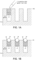
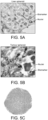
- the liquid agarose gel is drawn into the mold under surface tension and is allowed to set forming recessed chambers in the top 1mm of the mold that occur due to shrinkage of the gel volume as it sets (see Figure 1a ).
- NBF neutral buffered formaldehyde
- tissue spheroids are placed in the agarose mold recesses they are sealed in by topping off with liquid agarose gel at a temperature from 60 to 70°C (see Figure 1c ).
- the mold is then cooled at 4°C for 30 minutes to set the agarose gel seal, and placed in a solution of 70% ethanol for a minimum of 1 to 10 days, preferably 5 days.
- the spheroid tissue microarray is then impregnated with molten paraffin wax in a tissue processor ( Figure1 d) , using the following cycles: Time (hours) Solution Temp (°C) Vacuum (in Hg) 2 70% ethanol 25 - 2 95% ethanol 25 - 2 100% ethanol 25 - 2 100% ethanol 25 - 2 100% ethanol 25 - 2 100% ethanol 25 - 1 Xylene 25 - 2 Xylene 25 - 2 Xylene 25 - 3 Paraffin 60 - 3 Paraffin 60 15
- the spheroid tissue microarray may be sectioned and the sections placed on glass microscope slides (25mm x 75mm), prior to histological staining and/or matrix assisted laser desorption imaging mass spectrometric proteomic profiling and automated image analysis.
- Figure 1 illustrates one method of locating spheroids in an array
- Figure 6 illustrates an alternative method.
- casting mold 20 shown in cross-section, contains protrusions 21, shown in enlarged form, which are generally U-shaped but have smaller, nipple-shaped protrusions 22.
- protrusions 21 shown in enlarged form, which are generally U-shaped but have smaller, nipple-shaped protrusions 22.
- a porous mold e.g. an agarose mold
- dimples 24 at the base of the wells 23, for location of spheroids 25.
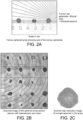
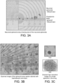
- the spheroids within the products of the present invention have been shown to maintain their integrity. This has been confirmed using cellular morphological analysis verified by a pathologist.
- the present invention can thus provide a spheroid tissue microarray and method of manufacturing the same, for use in high throughput histology.
- the spheroid tissue microarray provides the planar alignment and positioning of tissue spheroids on a geometric grid facilitating simultaneous sectioning of many (e.g. hundreds) of individual spheroids for subsequent analysis.
Landscapes
- Health & Medical Sciences (AREA)
- Engineering & Computer Science (AREA)
- Life Sciences & Earth Sciences (AREA)
- Biomedical Technology (AREA)
- Chemical & Material Sciences (AREA)
- Organic Chemistry (AREA)
- Bioinformatics & Cheminformatics (AREA)
- Wood Science & Technology (AREA)
- Zoology (AREA)
- Genetics & Genomics (AREA)
- Biotechnology (AREA)
- Biochemistry (AREA)
- General Health & Medical Sciences (AREA)
- General Engineering & Computer Science (AREA)
- Microbiology (AREA)
- Cell Biology (AREA)
- Sustainable Development (AREA)
- Neurology (AREA)
- Clinical Laboratory Science (AREA)
- Molecular Biology (AREA)
- Analytical Chemistry (AREA)
- Pathology (AREA)
- Physics & Mathematics (AREA)
- General Physics & Mathematics (AREA)
- Immunology (AREA)
- Oncology (AREA)
- Neurosurgery (AREA)
- Gastroenterology & Hepatology (AREA)
- Dermatology (AREA)
- Apparatus Associated With Microorganisms And Enzymes (AREA)
- Investigating Or Analysing Biological Materials (AREA)
- Measuring Or Testing Involving Enzymes Or Micro-Organisms (AREA)
- Immobilizing And Processing Of Enzymes And Microorganisms (AREA)
- Sampling And Sample Adjustment (AREA)
Claims (31)
- Sphäroid-Gewebemikroarray, das zum Schneiden geeignet ist, umfassend ein regelmäßiges Array von Gewebesphäroiden, die in einer Gitterstruktur angeordnet sind und in einen porösen Formkörper eingebettet sind, wobei die Sphäroide horizontal genau positioniert sind, so dass der Mittelpunkt jedes Sphäroids nicht mehr als 100 Mikrometer von seiner regulären Gitterposition entfernt ist.
- Sphäroid-Gewebemikroarray gemäß Anspruch 1, wobei der poröse Formkörper aus einem Agarose- oder Agar-Gel gebildet ist.
- Sphäroid-Gewebemikroarray gemäß Anspruch 1 oder Anspruch 2, enthaltend von 100 bis 300 Sphäroide.
- Sphäroid-Gewebemikroarray gemäß Anspruch 3, umfassend ein 10x17-Gitter von 170 Sphäroiden.
- Sphäroid-Gewebemikroarray gemäß Anspruch 1 oder Anspruch 2, enthaltend 96 Sphäroide.
- Sphäroid-Gewebemikroarray gemäß einem der vorstehenden Ansprüche, wobei die Dichte von Sphäroiden pro cm2 100 oder mehr beträgt.
- Sphäroid-Gewebemikroarray gemäß einem der vorstehenden Ansprüche, wobei die Gewebesphäroide Gewebe abgeleitet von einem oder mehreren von induzierten pluripotenten Stammzellen, primären Zellen, von kanzerösen Geweben abgeleiteten Zelllinien, neuronalen Zellen, Hautzellen, Leberzellen, Tumorzellen, Stromazellen, Immunzellen und Endothelzellen umfassen.
- Sphäroid-Gewebemikroarray gemäß einem der vorstehenden Ansprüche, wobei die Gewebesphäroide aus der In-vitro-3D-Kultur von Zellen gebildet sind.
- Sphäroid-Gewebemikroarray gemäß einem der vorstehenden Ansprüche, wobei die Gewebesphäroide einen Durchmesser von 50 bis 500 µm aufweisen.
- Sphäroid-Gewebemikroarray gemäß einem der vorstehenden Ansprüche, wobei die Gewebesphäroide ein Substrat umfassen, auf dem vor dem Einbetten Zellen wachsen gelassen werden.
- Sphäroid-Gewebemikroarray gemäß Anspruch 10, wobei das Substrat eine Perle ist, die aus Polystyrol (Amberlite) oder einem anderen porösen Kunststoffmaterial gebildet ist.
- Sphäroid-Gewebemikroarray gemäß Anspruch 10 oder 11, wobei die Gewebesphäroide menschliche Hautzellen umfassen.
- Sphäroid-Gewebemikroarray gemäß einem der vorstehenden Ansprüche, wobei die Gewebesphäroide zur histologischen Analyse gefärbt werden.
- Sphäroid-Gewebemikroarray gemäß einem der vorstehenden Ansprüche, das mit einem zusätzlichen Strukturmittel imprägniert ist.
- Sphäroid-Gewebemikroarray gemäß Anspruch 14, das mit Paraffinwachs oder einem Harz imprägniert ist.
- Verfahren zur Herstellung eines Sphäroid-Gewebemikroarrays, das ein regelmäßiges Array von Gewebesphäroiden umfasst, die in einer Gitterstruktur angeordnet sind, welches Verfahren die Schritte umfasst:Bilden eines Formkörpers aus porösem Material aus flüssigem Formmaterial in einer Gussform und Erstarrenlassen des flüssigen Formmaterials;Entnehmen des porösen Formkörpers aus der Gussform;Auffüllen des porösen Formkörpers mit weiterem flüssigem Formmaterial und erlauben, dass sich durch das Einziehen von flüssigem Formmaterial durch Schrumpfung während des Erstarrens des flüssigen Formmaterials Vertiefungen in der Oberfläche des Formkörpers bilden;Platzieren von Gewebesphäroiden in die Vertiefungen in der Oberfläche des porösen Formkörpers; undAbdichten der Gewebesphäroide innerhalb des Formkörpers durch Auffüllen mit flüssigem Formmaterial und Erstarrenlassen des flüssigen Formmaterials;wobei die Sphäroide horizontal genau positioniert werden, so dass der Mittelpunkt jedes Sphäroids nicht mehr als 100 Mikrometer von seiner regulären Gitterposition entfernt ist.
- Verfahren gemäß Anspruch 16 zur Herstellung eines Sphäroid-Gewebemikroarrays gemäß einem der Ansprüche 1 bis 15.
- Verfahren gemäß Anspruch 16 oder 17, wobei das flüssige Formmaterial 0,5 bis 4 % Agarose in Wasser ist.
- Verfahren gemäß einem der Ansprüche 16 bis 18, wobei der Schritt des Bildens des Formkörpers aus porösem Material aus flüssigen Formmaterial in einer Gussform bei einer Temperatur von 60 bis 70 °C durchgeführt wird.
- Verfahren gemäß einem der Ansprüche 16 bis 19, wobei das flüssige Formmaterial in der Gussform bei Raumtemperatur für 20 bis 45 Minuten, gefolgt von Kühlen auf 0 bis 5 °C für 20 bis 45 Minuten, erstarren gelassen wird.
- Verfahren gemäß einem der Ansprüche 16 bis 20, wobei vor dem Auffüllen der poröse Formkörper für 10 bis 30 Minuten in einer abgedichteten Umgebung auf 60 bis 70 °C erhitzt wird.
- Verfahren gemäß einem der Ansprüche 16 bis 21, wobei der poröse Formkörper mit flüssigem Formmaterial, das ein Agarosegel von 0,1 bis 1,0 % in Wasser umfasst, aufgefüllt wird.
- Verfahren gemäß einem der Ansprüche 16 bis 22, wobei die Vertiefungen innerhalb der oberen 1 mm des Formkörpers gebildet werden.
- Verfahren gemäß einem der Ansprüche 16 bis 23, wobei die Gewebesphäroide durch Auffüllen mit flüssigem Formmaterial bei einer Temperatur von 60 bis 70 °C innerhalb der Formkörpervertiefungen abgedichtet werden.
- Verfahren gemäß einem der Ansprüche 16 bis 24, wobei nach dem Abdichten der Gewebesphäroide in den Formkörpervertiefungen das flüssige Formmaterial durch Kühlen auf 0 bis 5 °C für etwa 30 Minuten erstarren gelassen wird.
- Verfahren gemäß einem der Ansprüche 16 bis 25, wobei das Sphäroid-Gewebemikroarray mit einem zusätzlichen Strukturmittel imprägniert wird.
- Verfahren gemäß Anspruch 26, wobei das Strukturmittel Paraffinwachs oder ein Harz ist.
- Verfahren zur Herstellung eines Sphäroid-Gewebemikroarrays, das ein regelmäßiges Array von Gewebesphäroiden umfasst, die in einer Gitterstruktur angeordnet sind, welches Verfahren die Schritte umfasst: Bilden eines Formkörpers aus porösem Material aus flüssigem Formmaterial in einer Gussform; Erstarrenlassen des flüssigen Formmaterials; Entnehmen des porösen Formkörpers aus der Gussform; Platzieren von Sphäroiden in Vertiefungen an der Basis von Näpfchen in dem Formkörper aus porösem Material; und Abdichten der Sphäroide innerhalb des porösen Formkörpers durch Zugeben von weiterem porösem Material auf die Sphäroide; wobei die Vertiefungen an der Basis der Näpfchen in dem porösen Material durch Vorsprünge der Gussform gebildet werden, die weitere noppenförmige Vorsprünge tragen; und wobei die Sphäroide horizontal genau positioniert werden, so dass der Mittelpunkt jedes Sphäroids nicht mehr als 100 Mikrometer von seiner regulären Gitterposition entfernt ist.
- Verfahren gemäß Anspruch 28, ferner umfassend einen nachfolgenden Schritt des Imprägnierens des Mikroarrays mit einem zusätzlichen Strukturmittel.
- Verfahren gemäß Anspruch 29, wobei das Strukturmittel Paraffinwachs oder ein Harz ist.
- Verwendung eines Sphäroid-Gewebemikroarrays gemäß einem der Ansprüche 1 bis 15 bei einem Analyseverfahren, umfassend Schneiden des Sphäroid-Gewebemikroarrays, um mehrere Sphäroide gleichzeitig zu schneiden, und Analysieren der Schnitte.
Applications Claiming Priority (3)
| Application Number | Priority Date | Filing Date | Title |
|---|---|---|---|
| US201662390660P | 2016-04-06 | 2016-04-06 | |
| GBGB1615517.8A GB201615517D0 (en) | 2016-09-13 | 2016-09-13 | Spheroid tissue miroarray and method of manufacture |
| PCT/GB2016/053907 WO2017174955A1 (en) | 2016-04-06 | 2016-12-09 | Spheroid tissue microarray and methods of manufacture |
Publications (3)
| Publication Number | Publication Date |
|---|---|
| EP3440189A1 EP3440189A1 (de) | 2019-02-13 |
| EP3440189C0 EP3440189C0 (de) | 2024-08-07 |
| EP3440189B1 true EP3440189B1 (de) | 2024-08-07 |
Family
ID=57234566
Family Applications (1)
| Application Number | Title | Priority Date | Filing Date |
|---|---|---|---|
| EP16831713.9A Active EP3440189B1 (de) | 2016-04-06 | 2016-12-09 | Kugelförmiger gewebemikroarray und verfahren zur herstellung |
Country Status (9)
| Country | Link |
|---|---|
| US (1) | US11320349B2 (de) |
| EP (1) | EP3440189B1 (de) |
| JP (1) | JP2019513419A (de) |
| KR (1) | KR20180132805A (de) |
| CN (1) | CN109312300B (de) |
| ES (1) | ES2985809T3 (de) |
| GB (1) | GB201615517D0 (de) |
| SG (1) | SG11201808787RA (de) |
| WO (1) | WO2017174955A1 (de) |
Families Citing this family (7)
| Publication number | Priority date | Publication date | Assignee | Title |
|---|---|---|---|---|
| US11300486B1 (en) * | 2016-11-23 | 2022-04-12 | Array Science, Llc | Apparatus for producing high yield cores for use in a microarray block, method for using same |
| CN108412487B (zh) * | 2018-03-07 | 2021-06-11 | 河南省科学院同位素研究所有限责任公司 | 一种耐高压放射性同位素示踪剂及其制备方法 |
| CN109366254B (zh) * | 2018-09-11 | 2021-05-28 | 中北大学 | 一种切屑金相试件自动制备装置 |
| CN109318080B (zh) * | 2018-09-11 | 2021-05-28 | 中北大学 | 一种切屑金相试件自动制备方法 |
| KR102873241B1 (ko) | 2020-05-11 | 2025-10-23 | 주식회사 에이엔케이 | 고밀도로 배열된 세포 스페로이드 형성용 기판 |
| CN115060570A (zh) * | 2022-06-30 | 2022-09-16 | 中国人民解放军东部战区总医院 | 一种适用于类器官等微小组织的包埋方法 |
| CN116008044A (zh) * | 2022-12-19 | 2023-04-25 | 复旦大学附属中山医院 | 一种内含植入材料血管硬组织病理切片树脂包埋的方法 |
Family Cites Families (11)
| Publication number | Priority date | Publication date | Assignee | Title |
|---|---|---|---|---|
| KR200327028Y1 (ko) * | 2003-06-17 | 2003-09-19 | 장시창 | 인체조직검사기구 |
| JP5039715B2 (ja) * | 2006-01-24 | 2012-10-03 | ブラウン ユニバーシティ | 細胞凝集及び封入デバイス及び方法 |
| WO2008054421A2 (en) * | 2006-10-27 | 2008-05-08 | Columbia University | Frozen cell and tissue microarrays |
| JP5261920B2 (ja) | 2006-11-10 | 2013-08-14 | 大日本印刷株式会社 | 細胞を用いた試験法および試験用キット |
| EP2271747B1 (de) | 2008-03-17 | 2016-09-07 | Agency for Science, Technology And Research | Mikroträger für pluripotente stammzellkultur |
| HUE037881T2 (hu) | 2008-06-20 | 2018-09-28 | Univ Maastricht | Önrendezõdõ szövetmodulok |
| US20120015440A1 (en) | 2008-09-08 | 2012-01-19 | Tokyo University Of Science Educational Foundation Administrative Org. | Spheroid composite, spheroid-containing hydrogel and processes for production of same |
| GB201105226D0 (en) | 2011-03-29 | 2011-05-11 | Univ Leiden | Methods |
| US9701938B2 (en) | 2012-10-12 | 2017-07-11 | Lena Biosciences, Inc. | Intra-culture perfusion methods and applications thereof |
| CN103060175B (zh) * | 2013-01-05 | 2014-07-02 | 太原理工大学 | 一种细胞微阵列芯片及其制备方法 |
| US20160281061A1 (en) * | 2013-11-05 | 2016-09-29 | The John Hopkins University | Tissue array for cell spheroids and methods of use |
-
2016
- 2016-09-13 GB GBGB1615517.8A patent/GB201615517D0/en not_active Ceased
- 2016-12-09 JP JP2019503793A patent/JP2019513419A/ja active Pending
- 2016-12-09 CN CN201680086317.9A patent/CN109312300B/zh active Active
- 2016-12-09 SG SG11201808787RA patent/SG11201808787RA/en unknown
- 2016-12-09 KR KR1020187031903A patent/KR20180132805A/ko not_active Withdrawn
- 2016-12-09 EP EP16831713.9A patent/EP3440189B1/de active Active
- 2016-12-09 WO PCT/GB2016/053907 patent/WO2017174955A1/en not_active Ceased
- 2016-12-09 ES ES16831713T patent/ES2985809T3/es active Active
- 2016-12-09 US US16/091,769 patent/US11320349B2/en active Active
Non-Patent Citations (1)
| Title |
|---|
| FUKUDA J ET AL: "Micromolding of photocrosslinkable chitosan hydrogel for spheroid microarray and co-cultures", BIOMATERIALS, ELSEVIER, AMSTERDAM, NL, vol. 27, no. 30, 1 October 2006 (2006-10-01), pages 5259 - 5267, XP027951182, ISSN: 0142-9612, [retrieved on 20061001] * |
Also Published As
| Publication number | Publication date |
|---|---|
| US20190162637A1 (en) | 2019-05-30 |
| SG11201808787RA (en) | 2018-11-29 |
| ES2985809T3 (es) | 2024-11-07 |
| GB201615517D0 (en) | 2016-10-26 |
| CN109312300B (zh) | 2023-01-10 |
| KR20180132805A (ko) | 2018-12-12 |
| WO2017174955A1 (en) | 2017-10-12 |
| CN109312300A (zh) | 2019-02-05 |
| US11320349B2 (en) | 2022-05-03 |
| JP2019513419A (ja) | 2019-05-30 |
| EP3440189C0 (de) | 2024-08-07 |
| EP3440189A1 (de) | 2019-02-13 |
Similar Documents
| Publication | Publication Date | Title |
|---|---|---|
| EP3440189B1 (de) | Kugelförmiger gewebemikroarray und verfahren zur herstellung | |
| ES2989523T3 (es) | Método de uso de un dispositivo modular de soporte para ensayos | |
| EP2917326B1 (de) | Zellkulturvorrichtung zur erzeugung und kultivierung von zellaggregaten, verfahren zur herstellung dieser vorrichtung und verwendung dieser vorrichtung | |
| ES2294321T3 (es) | Conjunto manual de construccion de micromatrices tisulares. | |
| US20110046017A1 (en) | Mold of recipient block and usage thereof | |
| ES2978142T3 (es) | Casete seccionable y bastidor de incrustación con tapa separable de inmovilización de tejido, y procedimientos para preparar muestras de tejido para biopsia | |
| WO2023229988A1 (en) | Tissue sample mold | |
| ES2917874T3 (es) | Cultivo de células | |
| JP2011503519A (ja) | 切片化可能な弾性発泡材料を備えた生検支持体 | |
| KR20140113139A (ko) | 세포 스페로이드 배양판 | |
| US12025541B2 (en) | Mass production manufacturing method for tissue chip | |
| Hess et al. | 3D versus 2D cell culture: implications for electron microscopy | |
| KR20210047268A (ko) | 세포 배양 장치 및 방법 | |
| KR20150051199A (ko) | 세포 스페로이드 배양판 | |
| US20120281208A1 (en) | Chamber for optical observation, method for optically observing sample, and method for manufacturing lower transparent plate | |
| CN115060570A (zh) | 一种适用于类器官等微小组织的包埋方法 | |
| US7256040B2 (en) | Method and apparatus for preparing monolayers of cells | |
| CN112326964A (zh) | 筛选靶细胞的方法、试剂盒及其用途 | |
| US20060046282A1 (en) | Template methods and devices for preparing sample arrays | |
| CN111979124A (zh) | 生物培养芯片及其制备和应用 | |
| EP4142944B1 (de) | Mikrotröpfchenschale | |
| CN216303865U (zh) | 生物培养芯片及其用于制备所述生物培养芯片的模板 | |
| CN113970472A (zh) | 组织切片方法以及用于组织切片的成型装置 | |
| WO2009149305A2 (en) | Method for creating distinct nitrocellulose-based pads on a substrate | |
| WO2018234163A1 (en) | Method for determining cell migration and invasion |
Legal Events
| Date | Code | Title | Description |
|---|---|---|---|
| STAA | Information on the status of an ep patent application or granted ep patent |
Free format text: STATUS: UNKNOWN |
|
| STAA | Information on the status of an ep patent application or granted ep patent |
Free format text: STATUS: THE INTERNATIONAL PUBLICATION HAS BEEN MADE |
|
| PUAI | Public reference made under article 153(3) epc to a published international application that has entered the european phase |
Free format text: ORIGINAL CODE: 0009012 |
|
| STAA | Information on the status of an ep patent application or granted ep patent |
Free format text: STATUS: REQUEST FOR EXAMINATION WAS MADE |
|
| 17P | Request for examination filed |
Effective date: 20181031 |
|
| AK | Designated contracting states |
Kind code of ref document: A1 Designated state(s): AL AT BE BG CH CY CZ DE DK EE ES FI FR GB GR HR HU IE IS IT LI LT LU LV MC MK MT NL NO PL PT RO RS SE SI SK SM TR |
|
| AX | Request for extension of the european patent |
Extension state: BA ME |
|
| DAV | Request for validation of the european patent (deleted) | ||
| DAX | Request for extension of the european patent (deleted) | ||
| STAA | Information on the status of an ep patent application or granted ep patent |
Free format text: STATUS: EXAMINATION IS IN PROGRESS |
|
| 17Q | First examination report despatched |
Effective date: 20191219 |
|
| GRAP | Despatch of communication of intention to grant a patent |
Free format text: ORIGINAL CODE: EPIDOSNIGR1 |
|
| STAA | Information on the status of an ep patent application or granted ep patent |
Free format text: STATUS: GRANT OF PATENT IS INTENDED |
|
| INTG | Intention to grant announced |
Effective date: 20240118 |
|
| GRAS | Grant fee paid |
Free format text: ORIGINAL CODE: EPIDOSNIGR3 |
|
| GRAJ | Information related to disapproval of communication of intention to grant by the applicant or resumption of examination proceedings by the epo deleted |
Free format text: ORIGINAL CODE: EPIDOSDIGR1 |
|
| GRAL | Information related to payment of fee for publishing/printing deleted |
Free format text: ORIGINAL CODE: EPIDOSDIGR3 |
|
| STAA | Information on the status of an ep patent application or granted ep patent |
Free format text: STATUS: EXAMINATION IS IN PROGRESS |
|
| GRAP | Despatch of communication of intention to grant a patent |
Free format text: ORIGINAL CODE: EPIDOSNIGR1 |
|
| STAA | Information on the status of an ep patent application or granted ep patent |
Free format text: STATUS: GRANT OF PATENT IS INTENDED |
|
| INTC | Intention to grant announced (deleted) | ||
| INTG | Intention to grant announced |
Effective date: 20240523 |
|
| GRAA | (expected) grant |
Free format text: ORIGINAL CODE: 0009210 |
|
| STAA | Information on the status of an ep patent application or granted ep patent |
Free format text: STATUS: THE PATENT HAS BEEN GRANTED |
|
| AK | Designated contracting states |
Kind code of ref document: B1 Designated state(s): AL AT BE BG CH CY CZ DE DK EE ES FI FR GB GR HR HU IE IS IT LI LT LU LV MC MK MT NL NO PL PT RO RS SE SI SK SM TR |
|
| REG | Reference to a national code |
Ref country code: GB Ref legal event code: FG4D |
|
| REG | Reference to a national code |
Ref country code: CH Ref legal event code: EP |
|
| REG | Reference to a national code |
Ref country code: IE Ref legal event code: FG4D |
|
| REG | Reference to a national code |
Ref country code: DE Ref legal event code: R096 Ref document number: 602016088808 Country of ref document: DE |
|
| U01 | Request for unitary effect filed |
Effective date: 20240904 |
|
| U07 | Unitary effect registered |
Designated state(s): AT BE BG DE DK EE FI FR IT LT LU LV MT NL PT RO SE SI Effective date: 20240919 |
|
| REG | Reference to a national code |
Ref country code: ES Ref legal event code: FG2A Ref document number: 2985809 Country of ref document: ES Kind code of ref document: T3 Effective date: 20241107 |
|
| U20 | Renewal fee for the european patent with unitary effect paid |
Year of fee payment: 9 Effective date: 20241113 |
|
| PG25 | Lapsed in a contracting state [announced via postgrant information from national office to epo] |
Ref country code: NO Free format text: LAPSE BECAUSE OF FAILURE TO SUBMIT A TRANSLATION OF THE DESCRIPTION OR TO PAY THE FEE WITHIN THE PRESCRIBED TIME-LIMIT Effective date: 20241107 |
|
| PG25 | Lapsed in a contracting state [announced via postgrant information from national office to epo] |
Ref country code: PL Free format text: LAPSE BECAUSE OF FAILURE TO SUBMIT A TRANSLATION OF THE DESCRIPTION OR TO PAY THE FEE WITHIN THE PRESCRIBED TIME-LIMIT Effective date: 20240807 Ref country code: GR Free format text: LAPSE BECAUSE OF FAILURE TO SUBMIT A TRANSLATION OF THE DESCRIPTION OR TO PAY THE FEE WITHIN THE PRESCRIBED TIME-LIMIT Effective date: 20241108 |
|
| PGFP | Annual fee paid to national office [announced via postgrant information from national office to epo] |
Ref country code: GB Payment date: 20241113 Year of fee payment: 9 |
|
| PG25 | Lapsed in a contracting state [announced via postgrant information from national office to epo] |
Ref country code: IS Free format text: LAPSE BECAUSE OF FAILURE TO SUBMIT A TRANSLATION OF THE DESCRIPTION OR TO PAY THE FEE WITHIN THE PRESCRIBED TIME-LIMIT Effective date: 20241207 |
|
| PG25 | Lapsed in a contracting state [announced via postgrant information from national office to epo] |
Ref country code: HR Free format text: LAPSE BECAUSE OF FAILURE TO SUBMIT A TRANSLATION OF THE DESCRIPTION OR TO PAY THE FEE WITHIN THE PRESCRIBED TIME-LIMIT Effective date: 20240807 |
|
| PG25 | Lapsed in a contracting state [announced via postgrant information from national office to epo] |
Ref country code: RS Free format text: LAPSE BECAUSE OF FAILURE TO SUBMIT A TRANSLATION OF THE DESCRIPTION OR TO PAY THE FEE WITHIN THE PRESCRIBED TIME-LIMIT Effective date: 20241107 |
|
| PG25 | Lapsed in a contracting state [announced via postgrant information from national office to epo] |
Ref country code: RS Free format text: LAPSE BECAUSE OF FAILURE TO SUBMIT A TRANSLATION OF THE DESCRIPTION OR TO PAY THE FEE WITHIN THE PRESCRIBED TIME-LIMIT Effective date: 20241107 Ref country code: PL Free format text: LAPSE BECAUSE OF FAILURE TO SUBMIT A TRANSLATION OF THE DESCRIPTION OR TO PAY THE FEE WITHIN THE PRESCRIBED TIME-LIMIT Effective date: 20240807 Ref country code: NO Free format text: LAPSE BECAUSE OF FAILURE TO SUBMIT A TRANSLATION OF THE DESCRIPTION OR TO PAY THE FEE WITHIN THE PRESCRIBED TIME-LIMIT Effective date: 20241107 Ref country code: IS Free format text: LAPSE BECAUSE OF FAILURE TO SUBMIT A TRANSLATION OF THE DESCRIPTION OR TO PAY THE FEE WITHIN THE PRESCRIBED TIME-LIMIT Effective date: 20241207 Ref country code: HR Free format text: LAPSE BECAUSE OF FAILURE TO SUBMIT A TRANSLATION OF THE DESCRIPTION OR TO PAY THE FEE WITHIN THE PRESCRIBED TIME-LIMIT Effective date: 20240807 Ref country code: GR Free format text: LAPSE BECAUSE OF FAILURE TO SUBMIT A TRANSLATION OF THE DESCRIPTION OR TO PAY THE FEE WITHIN THE PRESCRIBED TIME-LIMIT Effective date: 20241108 |
|
| PG25 | Lapsed in a contracting state [announced via postgrant information from national office to epo] |
Ref country code: SM Free format text: LAPSE BECAUSE OF FAILURE TO SUBMIT A TRANSLATION OF THE DESCRIPTION OR TO PAY THE FEE WITHIN THE PRESCRIBED TIME-LIMIT Effective date: 20240807 |
|
| PGFP | Annual fee paid to national office [announced via postgrant information from national office to epo] |
Ref country code: ES Payment date: 20250103 Year of fee payment: 9 |
|
| PGFP | Annual fee paid to national office [announced via postgrant information from national office to epo] |
Ref country code: CH Payment date: 20250101 Year of fee payment: 9 |
|
| PG25 | Lapsed in a contracting state [announced via postgrant information from national office to epo] |
Ref country code: CZ Free format text: LAPSE BECAUSE OF FAILURE TO SUBMIT A TRANSLATION OF THE DESCRIPTION OR TO PAY THE FEE WITHIN THE PRESCRIBED TIME-LIMIT Effective date: 20240807 |
|
| PG25 | Lapsed in a contracting state [announced via postgrant information from national office to epo] |
Ref country code: SK Free format text: LAPSE BECAUSE OF FAILURE TO SUBMIT A TRANSLATION OF THE DESCRIPTION OR TO PAY THE FEE WITHIN THE PRESCRIBED TIME-LIMIT Effective date: 20240807 |
|
| PLBE | No opposition filed within time limit |
Free format text: ORIGINAL CODE: 0009261 |
|
| STAA | Information on the status of an ep patent application or granted ep patent |
Free format text: STATUS: NO OPPOSITION FILED WITHIN TIME LIMIT |
|
| PG25 | Lapsed in a contracting state [announced via postgrant information from national office to epo] |
Ref country code: MC Free format text: LAPSE BECAUSE OF FAILURE TO SUBMIT A TRANSLATION OF THE DESCRIPTION OR TO PAY THE FEE WITHIN THE PRESCRIBED TIME-LIMIT Effective date: 20240807 |
|
| 26N | No opposition filed |
Effective date: 20250508 |
|
| PG25 | Lapsed in a contracting state [announced via postgrant information from national office to epo] |
Ref country code: IE Free format text: LAPSE BECAUSE OF NON-PAYMENT OF DUE FEES Effective date: 20241209 |
|
| U20 | Renewal fee for the european patent with unitary effect paid |
Year of fee payment: 10 Effective date: 20251001 |