EP3438251A1 - Cellule souche pluripotente spécifique stimulée par différenciation - Google Patents
Cellule souche pluripotente spécifique stimulée par différenciation Download PDFInfo
- Publication number
- EP3438251A1 EP3438251A1 EP17774865.4A EP17774865A EP3438251A1 EP 3438251 A1 EP3438251 A1 EP 3438251A1 EP 17774865 A EP17774865 A EP 17774865A EP 3438251 A1 EP3438251 A1 EP 3438251A1
- Authority
- EP
- European Patent Office
- Prior art keywords
- stem cell
- differentiation
- pluripotent stem
- cell
- marker
- Prior art date
- Legal status (The legal status is an assumption and is not a legal conclusion. Google has not performed a legal analysis and makes no representation as to the accuracy of the status listed.)
- Pending
Links
Images


Classifications
-
- C—CHEMISTRY; METALLURGY
- C12—BIOCHEMISTRY; BEER; SPIRITS; WINE; VINEGAR; MICROBIOLOGY; ENZYMOLOGY; MUTATION OR GENETIC ENGINEERING
- C12N—MICROORGANISMS OR ENZYMES; COMPOSITIONS THEREOF; PROPAGATING, PRESERVING, OR MAINTAINING MICROORGANISMS; MUTATION OR GENETIC ENGINEERING; CULTURE MEDIA
- C12N5/00—Undifferentiated human, animal or plant cells, e.g. cell lines; Tissues; Cultivation or maintenance thereof; Culture media therefor
- C12N5/06—Animal cells or tissues; Human cells or tissues
- C12N5/0602—Vertebrate cells
- C12N5/0618—Cells of the nervous system
- C12N5/0623—Stem cells
-
- C—CHEMISTRY; METALLURGY
- C12—BIOCHEMISTRY; BEER; SPIRITS; WINE; VINEGAR; MICROBIOLOGY; ENZYMOLOGY; MUTATION OR GENETIC ENGINEERING
- C12N—MICROORGANISMS OR ENZYMES; COMPOSITIONS THEREOF; PROPAGATING, PRESERVING, OR MAINTAINING MICROORGANISMS; MUTATION OR GENETIC ENGINEERING; CULTURE MEDIA
- C12N5/00—Undifferentiated human, animal or plant cells, e.g. cell lines; Tissues; Cultivation or maintenance thereof; Culture media therefor
- C12N5/10—Cells modified by introduction of foreign genetic material
-
- C—CHEMISTRY; METALLURGY
- C12—BIOCHEMISTRY; BEER; SPIRITS; WINE; VINEGAR; MICROBIOLOGY; ENZYMOLOGY; MUTATION OR GENETIC ENGINEERING
- C12N—MICROORGANISMS OR ENZYMES; COMPOSITIONS THEREOF; PROPAGATING, PRESERVING, OR MAINTAINING MICROORGANISMS; MUTATION OR GENETIC ENGINEERING; CULTURE MEDIA
- C12N2501/00—Active agents used in cell culture processes, e.g. differentation
- C12N2501/10—Growth factors
- C12N2501/115—Basic fibroblast growth factor (bFGF, FGF-2)
-
- C—CHEMISTRY; METALLURGY
- C12—BIOCHEMISTRY; BEER; SPIRITS; WINE; VINEGAR; MICROBIOLOGY; ENZYMOLOGY; MUTATION OR GENETIC ENGINEERING
- C12N—MICROORGANISMS OR ENZYMES; COMPOSITIONS THEREOF; PROPAGATING, PRESERVING, OR MAINTAINING MICROORGANISMS; MUTATION OR GENETIC ENGINEERING; CULTURE MEDIA
- C12N2501/00—Active agents used in cell culture processes, e.g. differentation
- C12N2501/10—Growth factors
- C12N2501/15—Transforming growth factor beta (TGF-β)
-
- C—CHEMISTRY; METALLURGY
- C12—BIOCHEMISTRY; BEER; SPIRITS; WINE; VINEGAR; MICROBIOLOGY; ENZYMOLOGY; MUTATION OR GENETIC ENGINEERING
- C12N—MICROORGANISMS OR ENZYMES; COMPOSITIONS THEREOF; PROPAGATING, PRESERVING, OR MAINTAINING MICROORGANISMS; MUTATION OR GENETIC ENGINEERING; CULTURE MEDIA
- C12N2501/00—Active agents used in cell culture processes, e.g. differentation
- C12N2501/10—Growth factors
- C12N2501/155—Bone morphogenic proteins [BMP]; Osteogenins; Osteogenic factor; Bone inducing factor
-
- C—CHEMISTRY; METALLURGY
- C12—BIOCHEMISTRY; BEER; SPIRITS; WINE; VINEGAR; MICROBIOLOGY; ENZYMOLOGY; MUTATION OR GENETIC ENGINEERING
- C12N—MICROORGANISMS OR ENZYMES; COMPOSITIONS THEREOF; PROPAGATING, PRESERVING, OR MAINTAINING MICROORGANISMS; MUTATION OR GENETIC ENGINEERING; CULTURE MEDIA
- C12N2501/00—Active agents used in cell culture processes, e.g. differentation
- C12N2501/20—Cytokines; Chemokines
- C12N2501/23—Interleukins [IL]
- C12N2501/235—Leukemia inhibitory factor [LIF]
-
- C—CHEMISTRY; METALLURGY
- C12—BIOCHEMISTRY; BEER; SPIRITS; WINE; VINEGAR; MICROBIOLOGY; ENZYMOLOGY; MUTATION OR GENETIC ENGINEERING
- C12N—MICROORGANISMS OR ENZYMES; COMPOSITIONS THEREOF; PROPAGATING, PRESERVING, OR MAINTAINING MICROORGANISMS; MUTATION OR GENETIC ENGINEERING; CULTURE MEDIA
- C12N2501/00—Active agents used in cell culture processes, e.g. differentation
- C12N2501/70—Enzymes
- C12N2501/72—Transferases (EC 2.)
- C12N2501/727—Kinases (EC 2.7.)
-
- C—CHEMISTRY; METALLURGY
- C12—BIOCHEMISTRY; BEER; SPIRITS; WINE; VINEGAR; MICROBIOLOGY; ENZYMOLOGY; MUTATION OR GENETIC ENGINEERING
- C12N—MICROORGANISMS OR ENZYMES; COMPOSITIONS THEREOF; PROPAGATING, PRESERVING, OR MAINTAINING MICROORGANISMS; MUTATION OR GENETIC ENGINEERING; CULTURE MEDIA
- C12N2506/00—Differentiation of animal cells from one lineage to another; Differentiation of pluripotent cells
- C12N2506/02—Differentiation of animal cells from one lineage to another; Differentiation of pluripotent cells from embryonic cells
-
- C—CHEMISTRY; METALLURGY
- C12—BIOCHEMISTRY; BEER; SPIRITS; WINE; VINEGAR; MICROBIOLOGY; ENZYMOLOGY; MUTATION OR GENETIC ENGINEERING
- C12N—MICROORGANISMS OR ENZYMES; COMPOSITIONS THEREOF; PROPAGATING, PRESERVING, OR MAINTAINING MICROORGANISMS; MUTATION OR GENETIC ENGINEERING; CULTURE MEDIA
- C12N2506/00—Differentiation of animal cells from one lineage to another; Differentiation of pluripotent cells
- C12N2506/45—Differentiation of animal cells from one lineage to another; Differentiation of pluripotent cells from artificially induced pluripotent stem cells
Definitions
- the present invention relates to a differentiation-promoted pluripotent stem cell (Differentiating-state Stem Cell, hereinafter referred to as "DiSC” in some cases) and use thereof.
- the present invention relates to a culture medium for inducing a pluripotent stem cell into a differentiation-promoted pluripotent stem cell, a method for manufacturing a differentiation-promoted pluripotent stem cell, a differentiation-promoted pluripotent stem cell, a method for manufacturing a neural stem cell mass, and a neural stem cell.
- Induced pluripotent stem cells have been produced from dermal fibroblasts in the related art.
- iPSC Induced pluripotent stem cells
- collection of skin is required in order to obtain a dermal fibroblast cell line, which is accompanied by problems such as bleeding, infection, and remaining scars.
- a technique of producing iPSC from peripheral blood cells which can be collected with a less-invasive method is under examination.
- CD3-positive T cells can be effectively reprogrammed into iPSC by using a Sendai virus vector (for example, refer to Non-Patent Document 1).
- iPSC characteristics of iPSC such as differentiation tendency greatly vary depending on types of cells, establishment methods, different donors, and the like which are used in establishing iPSC. Therefore, in the related art, it was necessary to select iPSC which easily differentiate into a desired cell among iPSC, requiring a lot of effort. In addition, the presence of an ES cell (embryonic stem cells, hereinafter referred to as "ESC" in some cases) line showing the resistance to differentiation has been known, and it was difficult to differentiate such ESC into a desired cell.
- ES cell embryonic stem cells
- Non-Patent Document 1 Seki T., et al., Generation of induced pluripotent stem cells from human terminally differentiated circulating T cells., Cell Stem Cell, 7 (1), 11-14, 2010 .
- An object of the present invention is to provide a technique in which even the ES cell line showing the resistance to differentiation, and an iPS cell line immediately after establishment, which has not been selected by selection of cell lines can be effectively differentiated into a desired cell.
- the present invention includes the following aspects.
- the present invention it is possible to provide a technique in which even the ES cell line showing the resistance to differentiation, and the iPS cell line immediately after establishment, which has not been selected by selection of cell lines can be effectively differentiated into a desired cell.
- the present invention provides a culture medium for inducing a pluripotent stem cell into a differentiation-promoted pluripotent stem cell, the medium including a GSK3 ⁇ inhibitor, a BMP signaling inhibitor, and a TGF- ⁇ inhibitor as active ingredients.
- the differentiation-promoted pluripotent stem cell referred herein is a cell newly found by the inventors of the present invention, in which expression levels of endoderm, mesoderm, and ectoderm markers increased while an undifferentiated state is maintained.
- the inventors of the present invention shed light on that the differentiation-promoted pluripotent stem cell can be induced from the pluripotent stem cell with the culture medium of the present embodiment.
- the inventors of the present invention shed light on that, by inducing the differentiation-promoted pluripotent stem cell from the pluripotent stem cell and differentiating the differentiation-promoted pluripotent stem cell into a desired cell, differentiation efficiency can be improved, and differentiation into a functional cell can be made in a short period of time for culturing compared with methods of the related art.
- the inventors of the present invention shed light on that, as the pluripotent stem cell, even an ES cell line showing the resistance to differentiation, and an iPS cell line immediately after establishment, which has not been selected by selection of cell lines can be induced into the differentiation-promoted pluripotent stem cell according to the culture medium of the present embodiment.
- examples of the GSK3 ⁇ inhibitor include CHIR99021 ( CAS No. 252917-06-9 ).
- examples of the BMP signaling inhibitor include Dorsomorphin ( CAS No. 866405-64-3 ), LDN-193189 ( CAS No. 1062368-24-4 ), Noggin, and the like.
- examples of the TGF- ⁇ inhibitor include SB431542 ( CAS No. 301836-41-9 ), A-83-01 ( CAS No. 909910-43-6 ), and the like.
- the term "differentiation-promoted pluripotent stem cell” means a pluripotent stem cell in which the differentiation is promoted while maintaining stemness.
- a level of differentiation potential into all germ layers of endoderm, mesoderm, and ectoderm (hereinafter referred to as “three germ layers” in some cases) is improved compared to pluripotent stem cells not underwent a method for manufacturing of the present embodiment.
- the concentration of the GSK3 ⁇ inhibitor is preferably about 3 ⁇ M.
- the concentration of the BMP signaling inhibitor is preferably about 3 to 6 ⁇ M.
- the concentration of the TGF- ⁇ inhibitor is preferably about 3 to 6 ⁇ M.
- the pluripotent stem cell of a target to be differentiated may be, for example, the ES cell, and may be, for example, the induced pluripotent stem cell (iPSC).
- the pluripotent stem cell of the target to be differentiated may be the ES cell line showing the resistance to differentiation, and may be the iPS cell line immediately after establishment, which has not been selected by selection of cell lines.
- it was difficult to differentiate these cell lines into a desired cell but by using the culture medium of the present embodiment, even these cell lines can be differentiated into a desired cell.
- the differentiation-promoted pluripotent stem cell having a high level of differentiation potential into the endoderm, the mesoderm, and the ectoderm can be obtained without selecting the pluripotent stem cell in advance based on origin, establishment methods, the level of differentiation potential into the endoderm, the mesoderm, or the ectoderm, and the like of the pluripotent stem cell.
- the culture medium of the present embodiment may be provided as a liquid or may be provided as a powder.
- an aspect in which the GSK3 ⁇ inhibitor, the BMP signaling inhibitor, or the TGF- ⁇ inhibitor are provided in a separate container and added to the culture medium when being used, may be adopted.
- the present invention provides a method for manufacturing the differentiation-promoted pluripotent stem cell, including a step of culturing the pluripotent stem cell in the culture medium described above.
- the method for manufacturing of the present embodiment can be said to be a method for manufacturing the differentiation-promoted pluripotent stem cell for differentiation into the endoderm or tissue derived from the endoderm.
- the method for manufacturing of the present embodiment can be said to be a method for manufacturing the differentiation-promoted pluripotent stem cell for differentiation into the mesoderm or tissue derived from the mesoderm.
- the method for manufacturing of the present embodiment can be said to be a method for manufacturing the differentiation-promoted pluripotent stem cell for differentiation into the ectoderm or tissue derived from the ectoderm.
- the method for manufacturing of the present embodiment can be said to a method for manufacturing the differentiation-promoted pluripotent stem cell for differentiation into the endoderm or tissue derived from the endoderm, or for differentiation into the mesoderm or tissue derived from the mesoderm.
- tissue derived from the endoderm examples include esophagus epithelium, gastric epithelium, gastrointestinal epithelium, liver, pancreas, bladder epithelium, posterior urethral epithelium, tonsil, pharyngeal epithelium, laryngeal epithelium, tracheal epithelium, lung, thyroid, parathyroid gland, thymus, eustachian tube, tympanic cavity, and the like.
- tissue derived from the mesoderm examples include microglia, bone, cartilage, heart, vascular endothelium, blood cells, spleen, bone marrow, dentine, peritoneal epithelium, kidney, urinary tract, pleural epithelium, adrenal cortex, muscle, ovary, uterus, testis, connective tissue, and the like.
- tissue derived from the ectoderm include central neuron, peripheral neuron, neuron cells, axis cylinders, myelin sheath, oral epithelium, tongue, tooth enamel, external rectal epithelium, external urethral epithelium, nasal epithelium, pituitary gland, pineal gland, adrenal medulla, pupillary sphincter, pupillary dilator, skin, cornea, retina, inner ear, outer ear, vaginal epithelium, and the like.
- the pluripotent stem cell of the target to be differentiated may be, for example, the ES cell, and may be, for example, the induced pluripotent stem cell (iPSC), and furthermore, may be the ES cell line showing the resistance to differentiation, and may be the iPS cell line immediately after establishment, which has not been selected by selection of cell lines.
- iPSC induced pluripotent stem cell
- the differentiation-promoted pluripotent stem cell having a high level of differentiation potential into all germ layers of the endoderm, the mesoderm, and the ectoderm can be obtained without selecting the pluripotent stem cell in advance based on origin, establishment methods, the level of differentiation potential into the endoderm, the mesoderm, or the ectoderm, and the like of the pluripotent stem cell.
- the method for manufacturing of the present embodiment can be a method by which the differentiation potential of the pluripotent stem cell into the endoderm, the mesoderm, and the ectoderm is improved, irrespective of origin of the pluripotent stem cell.
- the phrase "irrespective of origin” means that the differentiation potential is not affected by tissue from which the pluripotent stem cell is derived, a method for establishing the induced pluripotent stem cell, a vector used for establishing the induced pluripotent stem cell, a method for culturing the (induced) pluripotent stem cell, and the like.
- the differentiation-promoted pluripotent stem cell can be manufactured by culturing the pluripotent stem cell in the culture medium described above. As will be described later in the examples, by differentiating the differentiation-promoted pluripotent stem cell into a desired cell, the differentiation efficiency can be improved, or differentiation into a functional cell can be made in a short period of time for culturing compared to a case in which a normal pluripotent stem cell is differentiated into a desired cell.
- a period of time for culturing the pluripotent stem cell in the culture medium described above is preferably for 4 to 6 days, and more preferably for 5 days.
- the differentiation-promoted pluripotent stem cell in which the expression levels of the endoderm, mesoderm, and ectoderm markers increased while maintaining the undifferentiated state tends to be easily obtained with the period of time for culturing described above.
- the present invention provides the differentiation-promoted pluripotent stem cell, in which the expression levels of the endoderm, mesoderm, and ectoderm markers are increased compared with a control cell.
- the control cell include a pluripotent stem cell not underwent to the step of being cultured in the culture medium described above.
- the level of differentiation potential into all germ layers of the endoderm, the mesoderm, and the ectoderm is improved compared to the control cell.
- the differentiation-promoted pluripotent stem cell of the present embodiment is for the differentiation into the endoderm or the tissue derived from the endoderm.
- the differentiation-promoted pluripotent stem cell of the present embodiment is for the differentiation into the mesoderm or the tissue derived from the mesoderm.
- the differentiation-promoted pluripotent stem cell of the present embodiment is for the differentiation into the ectoderm or the tissue derived from the ectoderm.
- the differentiation-promoted pluripotent stem cell of the present embodiment is for differentiation into the endoderm or the tissue derived from the endoderm, or for differentiation into the mesoderm or the tissue derived from the mesoderm.
- a marker for undifferentiated cells in the differentiation-promoted pluripotent stem cell of the present embodiment, expression of a marker for undifferentiated cells is maintained.
- the marker for undifferentiated cells include octamer-binding transcription factor 4 (OCT4), NANOG, and the like.
- OCT4 octamer-binding transcription factor 4
- the endoderm marker examples include SOX17 and the like.
- examples of the mesoderm marker include BRACHYURY and the like.
- examples of the ectoderm marker include SOX1, PAX6, NESTIN, and the like. Expression levels of each marker may be measured in terms of mRNA levels, or may be measured in terms of protein levels.
- the present invention provides a method for manufacturing a neural stem cell mass, including a step of culturing the differentiation-promoted pluripotent stem cell described above in a culture medium containing the GSK3 ⁇ inhibitor, the TGF- ⁇ inhibitor, a Rho-associated protein kinase (ROCK) inhibitor, a Fibroblast Growth Factor 2 (FGF2), and a Leukemia Inhibitory Factor (LIF) as active ingredients.
- the method for manufacturing of the present embodiment can be said to be a method for manufacturing a neural stem cell.
- a differentiation induction method through formation of embryoid body (EB) of the related art requires about 1.5 months.
- differentiation of the pluripotent stem cell into the neural stem can be carried out within about 10 days according to the method for manufacturing of the present embodiment.
- the ES cell line showing the resistance to differentiation, and the iPS cell line immediately after establishment which has not been selected by selection of cell lines, which were difficult to be differentiated into the nervous system by methods of the related art, can be highly-efficiently differentiated into the neural stem cell mass.
- the GSK3 ⁇ inhibitor, the TGF- ⁇ inhibitor in a case where the pluripotent stem cell of the target to be differentiated is a cell derived from humans, FGF2 and LIF are preferably derived from humans, and the GSK3 ⁇ inhibitor, the TGF- ⁇ inhibitor, and the ROCK inhibitor are also preferably those intended for human ROCK.
- the pluripotent stem cell of the target to be differentiated is a cell derived from mice
- FGF2 and LIF are preferably derived from mice
- the GSK3 ⁇ inhibitor, the TGF- ⁇ inhibitor, and the ROCK inhibitor are also preferably those intended for mouse ROCK.
- RefSeq ID of human FGF2 protein is NP_001997
- RefSeq ID of mouse FGF2 protein is NP_032032.
- RefSeq ID of human LIF protein is NP_001244064
- RefSeq ID of mouse LIF protein is NP_001034626.
- the concentration of FGF2 in the culture medium described above is preferably about 20 ng/mL.
- the concentration of LIF in the culture medium described above is preferably about 10 ng/mL.
- ROCK inhibitor examples include Y27632 and the like.
- concentration of the ROCK inhibitor in the culture medium described above is preferably about 10 ⁇ M.
- the neural stem cells form the neural stem cell mass (neurosphere, hereinafter referred to as "NS" in some cases).
- the formed neural stem cell mass is dissociated into cells one by one, and the cells are cultured again in the culture medium containing the GSK3 ⁇ inhibitor, the TGF- ⁇ inhibitor, the ROCK inhibitor, FGF2, and LIF as active ingredients, and thus can be subcultured in the state of the neural stem cells.
- the step of culturing the differentiation-promoted pluripotent stem cell in the culture medium described above is preferably performed under a low oxygen environment.
- a low oxygen environment there is an environment in which an oxygen concentration is, for example, 1% to 10% (v/v), and is, for example, 1% to 5% (v/v). Accordingly, the number of neural stem cell manufactured can be increased.
- the method for manufacturing of the present embodiment preferably includes a step of dissociating the differentiation-promoted pluripotent stem cells into cells one by one, before the step of culturing the pluripotent stem cell in the culture medium described above. Accordingly, the number of neural stem cell manufactured can be increased.
- the present invention provides the neural stem cell mass, in which almost all neural stem cells constituting the neural stem cell mass do not substantially express the endoderm marker and the mesoderm marker.
- the neural stem cell mass of the present embodiment can be referred to as a neural stem cell mass which substantially does not contain cells expressing the endoderm and mesoderm markers (which substantially does not contain cells expressing the endoderm marker and cells expressing the mesoderm marker), or as a neural stem cell mass which substantially does not contain cells expressing the endoderm marker and cells expressing the mesoderm marker, and which is derived from the pluripotent stem cells.
- the neural stem cell mass of the present embodiment can be manufactured by the method for manufacturing the neural stem cell mass described above.
- the same as described above applies.
- the endoderm marker and the mesoderm marker are hardly expressed.
- the neural stem cell mass is a cluster of cells formed by self-proliferation of neural stem cells which are cells of the ectodermal system, and therefore, generally, the neural stem cell mass in vivo does not express the endoderm marker and the mesoderm marker.
- the neural stem cell mass obtained by differentiation induction of the pluripotent stem cell thereinto by the methods of the related art there is a case where the cells expressing the endoderm marker or the mesoderm marker are mixed therein.
- the neural stem cell mass obtained by differentiation induction of the pluripotent stem cell thereinto by the methods of the related art is constituted of a heterogeneous cell population such as (1) cells of the ectodermal system, (2) pluripotent cells that are committed to the ectodermal system, and (3) simply proliferated cells of the mesodermal system or cells of the endodermal system.
- a neural stem cell mass is difficult to be differentiated into functional neurons in some cases.
- the neural stem cell mass of the present embodiment is constituted almost only of a cell population such as (1) cells of the ectodermal system, and (2) pluripotent cells that are committed to the ectodermal system. Therefore, the neural stem cell mass of the present embodiment can be said to be a highly pure neuronal differentiation-inducing base.
- the neural stem cell mass which almost does not contain the cells expressing the endoderm marker or the mesoderm marker. Almost all cells constituting the neural stem cell mass do not substantially express the endoderm marker and the mesoderm marker.
- the phrase "do not substantially express” means that, for example, in a case where the neural stem cell mass is dissociated into cells one by one, and the analysis of cell populations is performed, the cells expressing the endoderm marker and the mesoderm marker can hardly be detected. More specifically, the phrase means that a proportion of the cells expressing the endoderm marker and the mesoderm marker is, for example, 1% or lower.
- the analysis of cell populations refers to an analysis method for analyzing the expression of a marker protein for each immunostained cell.
- T cell receptor gene may be rearranged.
- the rearrangement of the T cell receptor gene indicates that the neural stem cell is derived from mature T cells.
- the T cell receptor gene is rearranged.
- the present invention provides the method for manufacturing the germ layers or the tissue thereof, including a step of differentiating the differentiation-promoted pluripotent stem cell described above.
- the germ layers or the tissue thereof include the endoderm or the tissue derived from the endoderm, the mesoderm or the tissue derived from the mesoderm, and the ectoderm or the tissue derived from the ectoderm.
- the tissue derived from each germ layer include those described above.
- all the germ layers of the endoderm, the mesoderm, and the ectoderm, or the tissue derived therefrom can be efficiently manufactured from the pluripotent stem cell without selecting the pluripotent stem cell in advance based on origin, establishment methods, the level of differentiation potential into the endoderm, the mesoderm, or the ectoderm, and the like of the pluripotent stem cell.
- the step of differentiating the differentiation-promoted pluripotent stem cell is not particularly limited, and the step may be, for example, a method in which the embryoid body is formed and cultured, a method in which a predetermined gene is allowed to be expressed in the differentiation-promoted pluripotent stem cell, and the like.
- a 201B7 cell line which is human iPSC was cultured for 5 days in a normal culture medium (control), or in a culture medium containing the GSK3 ⁇ inhibitor, the BMP signaling inhibitor, and the TGF- ⁇ inhibitor in various combinations.
- the expression levels of the marker for undifferentiated cells, the endoderm marker, the mesoderm marker, and the ectoderm marker were measured by real time PCR.
- expression levels of each marker in EB formed by being cultured in a culture medium for embryoid body (EB) formation for 36 days were also measured in the same manner.
- CHIR99021 ( CAS No. 252917-06-9 ) was used as the GSK3 ⁇ inhibitor.
- Dorsomorphin CAS No. 866405-64-3
- SB431542 ( CAS No. 301836-41-9 ) was used as the TGF- ⁇ inhibitor.
- OCT4 octamer-binding transcription factor 4
- NANOG NANOG genes
- SOX1, PAX6, and NESTIN genes were examined as the ectoderm marker.
- a BRACHYURY gene was examined as the mesoderm marker.
- a SOX17 gene was examined as the endoderm marker.
- FIG. 1 shows the level of expression of OCT4, (b) of FIG. 1 shows the level of expression of NANOG, (c) of FIG. 1 shows the level of expression of SOX1, (d) of FIG. 1 shows the level of expression of PAX6, (e) of FIG. 1 shows the level of expression of NESTIN, (f) of FIG. 1 shows the level of expression of BRACHYURY, and (g) of FIG. 1 shows the level of expression of SOX17.
- SB represents SB431542
- DM represents Dorsomorphin
- CHIR represents CHIR99021.
- SB+DM represents that SB and DM were added to the culture medium
- SB+CHIR represents that SB and CHIR were added to the culture medium
- DM+CHIR represents that DM and CHIR were added to the culture medium
- SB+DM+CHIR represents that SB, DM, and CHIR were added to the culture medium.
- the concentration of SB in the culture medium was 3 ⁇ M
- the concentration of DM in the culture medium was 3 ⁇ M
- the concentration of CHIR in the culture medium was 3 ⁇ M.
- a condition under which the human pluripotent stem cell was induced into the differentiation-promoted pluripotent stem cell was examined. More specifically, the 201B7 cell line which is human iPSC was cultured in the culture medium added with SB, DM, and CHIR for 3, 4, 5, or 6 days of culturing days, and the differentiation-promoted pluripotent stem cell was induced from the 201B7 cell line. As a control, the 201B7 cell line cultured in a normal culture medium for 6 days was used.
- the expression levels of the marker for undifferentiated cells, the endoderm marker, the mesoderm marker, and the ectoderm marker were measured by real time PCR.
- the markers the same genes as those of Experimental Example 1 were examined.
- the form of the cells was observed.
- FIG. 2 shows the level of expression of OCT4, (b) of FIG. 2 shows the level of expression of NANOG, (c) of FIG. 2 shows the level of expression of SOX1, (d) of FIG. 2 shows the level of expression of PAX6, (e) of FIG. 2 shows the level of expression of NESTIN, (f) of FIG. 2 shows the level of expression of BRACHYURY, and (g) of FIG. 2 shows the level of expression of SOX17.
- FIG. 3 are optical photomicrographs of a human pluripotent stem cell colony (hPSC colony).
- a scale bar indicates 200 ⁇ m. It can be determined that, based on the form of the cell of the hPSC colony shown in (a) of FIG. 3 , the undifferentiated state is maintained in the cell.
- the cell of the hPSC colony shown in (b) of FIG. 3 has a form where a central part is recessed, and based on the form thereof, it can be determined that the cell excessively differentiated.
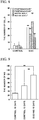
- FIG. 4 is a graph showing a proportion of hPSC colonies underwent the excessive differentiation among expressed hPSC colonies in a case where the 201B7 cell line was cultured in the culture medium added with SB, DM, and CHIR for 3, 4, 5, or 6 days to induce the differentiation-promoted pluripotent stem cell.
- the symbol "**" indicates that there is a significant difference at a risk ratio of less than 0.1%.
- the condition under which the culturing was carried out in the culture medium added with SB, DM, and CHIR for 5 days is suitable as the condition under which the human pluripotent stem cell was induced into the differentiation-promoted pluripotent stem cell.
- the inventors of the present invention have found that the colony of the differentiation-promoted pluripotent stem cells was smaller than a colony of a case in which a normal human pluripotent stem cell was cultured for the same period of time.
- the 201B7 cell line which is human iPSC was cultured in the culture medium added with SB, DM, and CHIR for 5 days to induce the differentiation-promoted pluripotent stem cell, and a change in diameter of the colony was measured.
- the 201B7 cell line was cultured in a normal culture medium for 5 days, and a change in diameter of the colony was measured. The experiments were independently carried out 3 times.
- FIG. 5 is a representative photograph of the colony of the 201B7 cells cultured in the normal culture medium for 5 days.
- (b) of FIG. 5 is a photograph of the colony of the differentiation-promoted pluripotent stem cell induced by culturing the 201B7 cell line in the culture medium added with SB, DM, and CHIR for 5 days. Magnifications of both (a) of FIG. 5 and (b) of FIG. 5 are the same, and a scale bar shows 200 ⁇ m.
- FIG. 6 is a graph showing the results of measuring changes in diameter of the differentiation-promoted pluripotent stem cell colony and a normal human pluripotent stem cell colony.
- the symbol "**" indicates that there is a significant difference at a risk ratio of less than 1%.
- the 201B7 cell line which is human iPSC was cultured in the culture medium added with SB, DM, and CHIR for 5 days, and therefore the differentiation-promoted pluripotent stem cell was formed.
- the 201B7 cell line cultured in a normal culture medium for 5 days was used.
- the expression levels of the endoderm marker, the mesoderm marker, and the ectoderm marker were measured by immunostaining.
- PAX6 and NESTIN proteins were detected as the ectoderm marker.
- BRACHYURY protein was detected as the mesoderm marker.
- SOX17 protein was examined as the endoderm marker.
- FIG. 7 is a graph showing an expression level of each marker protein.
- the symbol "**" indicates that there is a significant difference at a risk ratio of less than 0.1%.
- the above-described differentiation-promoted pluripotent stem cells and the control cells were dissociated into cells one by one, the same marker as that in the above-described immunostaining was immunostained, and a ratio of the cell population expressing each marker protein was analyzed.
- FIG. 8 is a graph showing the analysis results. As a result, it became clear that the number of PAX6 + BRACHYURY - cell, PAX6 - BRACHYURY + cell, NESTIN + SOX17 - cell, and NESTIN - SOX17 + cell was all increased in the differentiation-promoted pluripotent stem cell compared with the control.
- the 201B7 cell line which is human iPSC was cultured in the culture medium added with SB, DM, and CHIR for 5 days, and therefore the differentiation-promoted pluripotent stem cell was induced.
- the differentiation-promoted pluripotent stem cells which were induced were dissociated into cells one by one, and the cells were cultured in a serum-free culture medium containing FGF2, Y27632 ( CAS No. 331752-47-7 ), LIF, CHIR, and SB (hereinafter referred to as "NS culture medium” in some cases) for 5 days. Therefore, the neural stem cell mass (neurosphere, hereinafter referred to as "NS" in some cases) was formed (culturing days were a total of 10 days).
- the neural stem cell mass (NS) formed through the differentiation-promoted pluripotent stem cell (DiSC) is referred to as "DiSC-NS" in some cases.
- the composition of the NS culture medium is shown in Table 1.
- Table 1 Composition of NS culture medium 1:1 Mixture of DMEM culture medium and F-12 culture medium 0.6% Glucose 2 mM Glutamine 3 mM Sodium bicarbonate 5 mM HEPES 25 ⁇ g/mL Insulin 100 ⁇ g/mL Transferrin 20 nM Progesterone 30 nM Selenium chloride 60 ⁇ M Putrescine 2% B27 supplement (Thermo Fisher Scientific) 20 ng/mL FGF2 10 ⁇ M Y-27632 (Wako Pure Chemical Industries) 10 ng/mL hLIF 3 ⁇ M CHIR99021 2 ⁇ M SB431542
- the 201B7 cell line was cultured in a normal culture medium for 5 days, and then the cells were dissociated into cells one by one and cultured in the above-described NS culture medium for 5 days (culturing days were a total of 10 days) or for 10 days (culturing days was a total of 15 days), and the cells thus obtained were used as a control. Next, the number of neural stem cell mass formed was measured by microscopic observation. The experiments were independently carried out 3 times.
- FIG. 9 is a graph showing the number of neural stem cell mass formed by the culturing described above.
- the symbol "**" indicates that there is a significant difference at a risk ratio of less than 1 %.
- the expression levels of the marker for undifferentiated cells, the endoderm marker, the mesoderm marker, the ectoderm marker, and a neuron cell marker in the formed neural stem cell mass described above were measured by real time PCR.
- OCT4 and NANOG genes were examined as the marker for undifferentiated cells.
- PAX6, SOX1, and NESTIN genes were examined as the ectoderm marker.
- ⁇ III-tubulin (TUBB3) gene was examined as the neuron cell marker.
- BRACHYURY gene was examined as the mesoderm marker.
- SOX17 gene was examined as the endoderm marker.
- FIG. 10 shows the level of expression of OCT4, (b) of FIG. 10 shows the level of expression of NANOG, (c) of FIG. 10 shows the level of expression of PAX6, (d) of FIG. 10 shows the level of expression of SOX1, (e) of FIG. 10 shows the level of expression of NESTIN, (f) of FIG. 10 shows the level of expression of TUBB3, (g) of FIG. 10 shows the level of expression of BRACHYURY, and (h) of FIG. 10 shows the level of expression of SOX17.
- the symbol "**" indicates that there is a significant difference at a risk ratio of less than 1%.
- the above-described neural stem cell mass was dissociated into cells one by one, and the expression levels of the ectoderm marker, mesoderm marker, and endoderm marker proteins were examined by the analysis of cell populations.
- PAX6 and NESTIN proteins were detected as the ectoderm marker.
- BRACHYURY protein was detected as the mesoderm marker.
- SOX17 protein was detected as the endoderm marker.
- FIG. 11 is a graph showing percentages of PAX6 + BRACHYURY + cell, PAX6 + BRACHYURY - cell, PAX6 - BRACHYURY + cell, and PAX6 - BRACHYURY - cell in a neural stem cell mass formed not through the differentiation-promoted pluripotent stem cells.
- FIG. 11 is a graph showing percentages of PAX6 + BRACHYURY + cell, PAX6 + BRACHYURY - cell, PAX6 - BRACHYURY + cell, and PAX6 - BRACHYURY - cell in the neural stem cell mass formed through the differentiation-promoted pluripotent stem cells.
- FIG. 12 is a graph showing percentages of NESTTN + SOX17 + cell, NESTIN + SOX17 - cell, NESTIN - SOX17 + cell, and NESTIN - SOX17 - cell in the neural stem cell mass formed not through the differentiation-promoted pluripotent stem cells.
- (b) of FIG. 12 is a graph showing percentages of NESTIN + SOX17 + cell, NESTIN + SOX17 - cell, NESTIN - SOX17 + cell, and NESTIN - SOX17 - cell in the neural stem cell mass formed through the differentiation-promoted pluripotent stem cells.
- the neural stem cell mass formed not through the differentiation-promoted pluripotent stem cell a percentage of the cells (NESTIN + SOX17 + cells) expressing both the ectoderm marker and the endoderm marker was high compared with the neural stem cell mass formed through the differentiation-promoted pluripotent stem cell.
- the neural stem cell mass was formed from the 201B7 cell line and WD39 cell line which are human iPSC, and KhES1cell line which is ES cell line, through the differentiation-promoted pluripotent stem cell, or not through the differentiation-promoted pluripotent stem cell. Furthermore, these neural stem cell mass were differentiated into the neurons.
- the neuron obtained by differentiation of the neural stem cell mass thereinto, which is formed through the differentiation-promoted pluripotent stem cell is referred to as "DiSC neuron" in some cases.
- the neuron obtained by differentiation of the neural stem cell mass thereinto, which is formed not through the differentiation-promoted pluripotent stem cell was used as a control.
- the differentiation into the neuron was carried out by seeding each neural stem cell mass on laminin- and poly-L-ornithine-coated plates and culturing the cells in a neuronal differentiation-inducing culture medium.
- the composition of the neuronal differentiation-inducing culture medium is shown in Table 2.
- the expression levels of the neuron cell marker, a glutamatergic neuron marker, a GABAergic neuron marker, the marker for undifferentiated cells, the mesoderm marker, and the endoderm marker in the DiSC neuron and the control neuron were measured by real time PCR, 23 days after the culturing.
- the expression levels of the marker genes in the human neural stem cell (hNSC) were measured in the same manner.
- Microtubule-associated protein 2 (MAP2), synapsin-1 (SYN1), and ⁇ III-tubulin (TUBB3) gene were examined as the neuron cell marker.
- VGLUT1 Vesicular glutamate transporter 1
- Glutamic acid decarboxylase 2 (GAD65) and glutamic acid decarboxylase 1 (GAD67) genes were examined as the GABAergic neuron marker.
- NESTIN and SOX1 genes were examined as the ectoderm marker.
- OCT4 and NANOG genes were examined as the marker for undifferentiated cells.
- BRACHYURY gene was examined as the mesoderm marker.
- SOX17 gene was examined as the endoderm marker.
- FIG. 13 is a graph showing an expression level of each marker gene.
- DiSC neuron a tendency in which the expression levels of the neuron cell marker, the glutamatergic neuron marker, and the GABAergic neuron marker genes were high, and the expression levels of the ectoderm marker, the marker for undifferentiated cells, the mesoderm marker, and the endoderm marker genes were low, was recognized.
- the DiSC neuron and the control neuron produced in Experimental Example 6 were immunostained, and examination was performed on maturation of the neurons. As an indicator of mature neurons, the number of synapsin-1 + ⁇ III-tubulin + cells was measured. The experiments were independently carried out 3 times.
- FIG. 14 is a graph showing measurement results.
- the symbol "**" indicates that there is a significant difference at a risk ratio of less than 1%.
- the DiSC neuron and the control neuron produced in Experimental Example 6 were electrophysically analyzed. More specifically, neuron functionality was evaluated by measuring the number of firing neurons per minute.
- FIG. 15 is a graph showing the results of measuring the number of firing neurons in each neuron 11 to 23 days after the start of differentiation into neurons.
- the symbol "*" in FIG. 15 indicates that there is a significant difference at a risk rate of less than 5%, and the symbol "**" indicates that there is a significant difference at a risk rate of less than 1%.
- KhES2, KhES3, KhES4, and KhES5 which are human ES cell lines resistant to differentiation were differentiation-induced into the neural stem cell mass through the differentiation-promoted pluripotent stem cell.
- each cell line was cultured in the culture medium added with SB, DM, and CHIR for 5 days, and therefore the differentiation-promoted pluripotent stem cell was induced.
- the differentiation-promoted pluripotent stem cells which were induced were dissociated into cells one by one, and the cells were cultured in the above-described NS culture medium for 10 days.
- DdNS neural stem cell mass induced through the differentiation-promoted pluripotent stem cell
- FIG. 16 shows optical photomicrographs of each cell cultured in the NS culture medium for 10 days. Magnifications of the photographs are all the same, and a scale bar shows 200 ⁇ m.
- FIG. 17 is a graph showing the results of measuring the number of neural stem cell mass in each cell. In FIG. 17 , the symbol "**" indicates that there is a significant difference at a risk ratio of less than 1%.
- the neural stem cell mass can be efficiently induced even from the ES cells resistant to differentiation by being induced through the differentiation-promoted pluripotent stem cell.
- the differentiation into the neuron was carried out by seeding each neural stem cell mass induced in Experimental Example 9 on the laminin- and poly-L-ornithine-coated plates and culturing the cells in the above-described neuronal differentiation-inducing culture medium. Next, each neuron was immunostained on day 23 of culturing (on day 13 after culturing in the neuronal differentiation-inducing culture medium), and the number of ⁇ III-tubulin + MAP2 + neurons occupying the whole neurons was measured. The experiments were independently carried out 3 times.
- FIG. 18 is a graph showing measurement results.
- the symbol "*" in FIG. 18 indicates that there is a significant difference at a risk rate of less than 5%, and the symbol “**” indicates that there is a significant difference at a risk rate of less than 1%.
- each neuron was immunostained on day 23 of culturing (on day 13 after culturing in the neuronal differentiation-inducing culture medium), and the maturation of the neurons was examined. As an indicator of mature neurons, the number of synapsin-1 + ⁇ III-tubulin + cells was measured. The experiments were independently carried out 3 times.
- FIG. 19 is a graph showing measurement results.
- the symbol "**" indicates that there is a significant difference at a risk ratio of less than 1%.
- TiPSC line was established by introducing a Sendai virus vector (type "CytoTune (TM)" of DNAVEC) containing OCT4, SOX2, KLF4 and c-MYC in four vectors, respectively, into CD3-positive lymphocytes derived from healthy humans.
- TM Sendai virus vector
- FIG. 20 is a graph showing measurement results. As a result, it became clear that the number of ⁇ III-tubulin + MAP2 + neurons was large in the neurons after differentiation induction by the method through DiSC and DdNS compared with the control neuron.
- each neuron was immunostained on day 23 of culturing (on day 13 after culturing in the neuronal differentiation-inducing culture medium), and the maturation of the neurons was examined. As an indicator of mature neurons, the number of synapsin-1 + ⁇ III-tubulin + cells was measured.
- FIG. 21 is a graph showing measurement results. As a result, it became clear that the number of mature neurons was large in the neurons after differentiation induction by the method through DiSC and DdNS compared with the control neuron.
- Table 3 shows the number of TiPSC lines showing the formation of the neural stem cell mass and the differentiation into the neurons by the method not through DiSC and DdNS (control) and the method through DiSC and DdNS (shown as "DdNS” in Table 3).
- the TiPSC line immediately after establishment which has been known to be extremely difficult to differentiate into the neurons, could be differentiated into the neurons with high efficiency.
- Control DdNS The number of TiPSC lines which formed neural stem cell mass 9 29 The number of TiPSC differentiated into neurons 7 28 Percentage (%) of TiPSC lines differentiated into neurons 23.3 (7/30) 93.3 (28/30)
- the 201B7 cell line and the WD39 cell line which are human iPSC, and the KhES1cell line which is the ES cell line were differentiation-induced into the neurons by the method through DiSC and DdNS.
- the 201B7 cell line and the WD39 cell line which are human iPSC, and the KhES1cell line which is the ES cell line were differentiation-induced into the dopaminergic neurons.
- each cell was cultured in the culture medium added with SB, DM, and CHIR for 5 days, and therefore the differentiation-promoted pluripotent stem cell was induced.
- the differentiation-promoted pluripotent stem cells which were induced were dissociated into cells one by one, and the cells were cultured in the above-described NS culture medium for 3 days.
- the above-described culture medium was further added with CHIR (3 ⁇ M), Shh (100 ng/mL), Purmorpamine ( CAS No.
- TH tyrosine hydroxylase
- ⁇ III-tubulin which is a neuron cell marker
- FIG. 22 is a graph showing the results of measuring percentages of ⁇ III-tubulin + cell or TH + ⁇ III-tubulin + cell.
- DdNS shows results of the neurons after induction in the same manner as in Experimental Example 10.
- DdNS shows results of the dopaminergic neurons induced by the above-described method.
- the symbol "**" indicates that there is a significant difference at a risk ratio of less than 1%.
- the differentiation potential of the differentiation-promoted pluripotent stem cell (DiSC) induced from iPSC into the three germ layers was examined. Specifically, the 201B7 cell line which is human iPSC was cultured in the culture medium added with SB, DM, and CHIR for 5 days, and therefore the differentiation-promoted pluripotent stem cell was formed. Final concentrations of SB, DM, and CHIR were all 3 ⁇ M. As a control, the 201B7 cell line cultured in a normal culture medium for 5 days was used.
- the colony of the differentiation-promoted pluripotent stem cells was dissociated into cells one by one.
- a dissociation solution containing 0.25% trypsin, 100 ⁇ g/mL collagenase IV, 1 mM calcium chloride, 20% KnockOut Serum Replacement (KSR, Invitrogen) was used for cell dissociation.
- SNL feeder cells CELL BIOLABS
- FGF2 embryoid bodies
- the EB on day 8 was adhered as it was to Matrigel-coated 96-well plate without breaking, and was adhesive cultured for 10 days.
- the culture medium from which FGF2 was removed from the culture medium for normal human ES cells was used as a culture medium.
- the culture medium was exchanged every other day.
- the cells were fixed 10 days after the start of the adhesive culture (18 days after the formation of the differentiation-promoted pluripotent stem cell).
- the fixed cells were immunostained to detect AFP which is a differentiation marker of the endoderm, ⁇ SMA which is a differentiation marker of the mesoderm, and ⁇ III-tubulin which is a differentiation marker of the ectoderm. Therefore, the differentiation potential into the three germ layers was examined.
- IN Cell Analyzer 6000 GE Healthcare Bioscience
- FIG. 23 is a graph showing the results of quantifying an expression level of differentiation markers in each cell.
- the term "DiSC” indicates results of cells obtained by differentiation of the EB which was formed through DiSC.
- the symbol "**" indicates that there is a significant difference at a risk ratio of less than 0.1%.
- each ES cell line was cultured in the culture medium added with SB, DM, and CHIP for 5 days, and therefore the differentiation-promoted pluripotent stem cell was formed. Final concentrations of SB, DM, and CHIR were all 3 ⁇ M. As a control, each ES cell line cultured in a normal culture medium for 5 days was used.
- the expression levels of the endoderm marker, the mesoderm marker, and the ectoderm marker were measured by immunostaining.
- SOX1 protein was detected as the ectoderm marker.
- BRACHYURY protein was detected as the mesoderm marker.
- SOX17 protein was examined as the endoderm marker.
- FIG. 24 are graphs showing an expression level of each marker protein.
- (a) of FIG. 24 is a graph showing the expression level of SOX1 protein
- (b) of FIG. 24 is a graph showing the expression level of BRACHYURY protein
- (c) of FIG. 24 is a graph showing the expression level of SOX17 protein.
- the term "DiSC” indicates results of the differentiation-promoted pluripotent stem cells.
- the symbol "**" indicates that there is a significant difference at a risk ratio of less than 0.1%.
- each differentiation-promoted pluripotent stem cell was dissociated into cells one by one.
- the cells were cultured for 8 days, and therefore the EB was formed.
- the EB was adhered to Matrigel-coated 96-well plate, and was adhesive cultured for 10 days.
- the cells were fixed 10 days after the start of the adhesive culture (18 days after the formation of the differentiation-promoted pluripotent stem cell).
- the fixed cells were immunostained to detect AFP which is a differentiation marker of the endoderm, ⁇ SMA which is a differentiation marker of the mesoderm, and ⁇ III-tubulin which is a differentiation marker of the ectoderm. Therefore, the differentiation potential into the three germ layers was examined.
- IN Cell Analyzer 6000 GE Healthcare Bioscience
- FIG. 25 is a graph showing the results of quantifying an expression level of differentiation markers in each cell.
- the term "DiSC” indicates results of cells obtained by differentiation of the EB which was formed through DiSC.
- the differentiation potential of the differentiation-promoted pluripotent stem cell induced from TiPSC immediately after establishment, into the three germ layers was examined.
- TiPSC the human TiPSC 30 line immediately after establishment in the same manner as in Experimental Example 11 without colony screening was used.
- each TiPSC line was cultured in the culture medium added with SB, DM, and CHIR for 5 days, and therefore the differentiation-promoted pluripotent stem cell was formed.
- the final concentrations of SB, DM, and CHIR were all 3 ⁇ M.
- As a control each TiPSC line cultured in a normal culture medium for 5 days was used.
- the expression levels of the endoderm marker, the mesoderm marker, and the ectoderm marker were measured by immunostaining.
- SOX1 protein was detected as the ectoderm marker.
- BRACHYURY protein was detected as the mesoderm marker.
- SOX17 protein was examined as the endoderm marker.
- FIG. 26 are graphs showing an expression level of each marker protein.
- (a) of FIG. 26 is a graph showing the expression level of SOX1 protein
- (b) of FIG. 26 is a graph showing the expression level of BRACHYURY protein
- (c) of FIG. 26 is a graph showing the expression level of SOX17 protein.
- a lateral axis indicates numbers of TiPSC lines. The numbers in the graphs are relative to the expression level in the control cell where the expression level in the control cell is 1.
- the expression levels of the ectoderm marker, the mesoderm marker, and the endoderm marker were significantly increased in the differentiation-promoted pluripotent stem cell induced from TiPSC immediately after establishment compared with the control.
- the present invention it is possible to provide a technique in which even the ES cell line showing the resistance to differentiation, and the iPS cell line immediately after establishment, which has not been selected by selection of cell lines can be effectively differentiated into a desired cell.
Landscapes
- Health & Medical Sciences (AREA)
- Engineering & Computer Science (AREA)
- Life Sciences & Earth Sciences (AREA)
- Biomedical Technology (AREA)
- Genetics & Genomics (AREA)
- Zoology (AREA)
- Organic Chemistry (AREA)
- Bioinformatics & Cheminformatics (AREA)
- Chemical & Material Sciences (AREA)
- Wood Science & Technology (AREA)
- Biotechnology (AREA)
- Microbiology (AREA)
- Biochemistry (AREA)
- General Engineering & Computer Science (AREA)
- General Health & Medical Sciences (AREA)
- Cell Biology (AREA)
- Developmental Biology & Embryology (AREA)
- Neurology (AREA)
- Neurosurgery (AREA)
- Micro-Organisms Or Cultivation Processes Thereof (AREA)
Applications Claiming Priority (2)
| Application Number | Priority Date | Filing Date | Title |
|---|---|---|---|
| JP2016073739 | 2016-03-31 | ||
| PCT/JP2017/012254 WO2017170328A1 (fr) | 2016-03-31 | 2017-03-27 | Cellule souche pluripotente spécifique stimulée par différenciation |
Publications (2)
| Publication Number | Publication Date |
|---|---|
| EP3438251A1 true EP3438251A1 (fr) | 2019-02-06 |
| EP3438251A4 EP3438251A4 (fr) | 2020-02-26 |
Family
ID=59964435
Family Applications (1)
| Application Number | Title | Priority Date | Filing Date |
|---|---|---|---|
| EP17774865.4A Pending EP3438251A4 (fr) | 2016-03-31 | 2017-03-27 | Cellule souche pluripotente spécifique stimulée par différenciation |
Country Status (4)
| Country | Link |
|---|---|
| US (1) | US20200299643A1 (fr) |
| EP (1) | EP3438251A4 (fr) |
| JP (1) | JP7306667B2 (fr) |
| WO (1) | WO2017170328A1 (fr) |
Families Citing this family (2)
| Publication number | Priority date | Publication date | Assignee | Title |
|---|---|---|---|---|
| CN113416709A (zh) * | 2021-06-21 | 2021-09-21 | 香港再生医学有限公司 | 促使iPSC分化为外周神经元细胞的方法及培养基和系统 |
| CN114441413B (zh) * | 2022-01-24 | 2024-05-14 | 南京医科大学 | 一种iPSC技术诱导分化的多巴胺能神经元中单囊泡储存的分析方法 |
Family Cites Families (6)
| Publication number | Priority date | Publication date | Assignee | Title |
|---|---|---|---|---|
| CN102604894B (zh) * | 2012-02-29 | 2014-07-30 | 中国科学院广州生物医药与健康研究院 | 用于制备神经干细胞的培养基及其用途 |
| WO2013187416A1 (fr) * | 2012-06-12 | 2013-12-19 | 学校法人 慶應義塾 | PROCÉDÉ D'AMPLIFICATION D'UNE CELLULE iPS APPROPRIÉE POUR UNE DIFFÉRENCIATION NEURALE ET PROCÉDÉ DESTINÉ À L'INDUCTION D'UNE CELLULE SOUCHE NEURALE |
| US20150329821A1 (en) * | 2012-10-19 | 2015-11-19 | Agency For Science, Technology And Research | Methods of differentiating stem cells into one or more cell lineages |
| WO2014069479A1 (fr) * | 2012-10-29 | 2014-05-08 | 学校法人埼玉医科大学 | Procédé de fabrication de cellules souches pluripotentes différenciées |
| WO2014161075A1 (fr) * | 2013-04-05 | 2014-10-09 | University Health Network | Procédés et compositions permettant de générer des cellules de la lignée des chondrocytes et/ou un tissu de type cartilage |
| CA2923592A1 (fr) * | 2013-09-05 | 2015-03-12 | Kyoto University | Procede d'induction de cellules precurseurs neuronales produisant de la dopamine a partir de cellules souches pluripotentes |
-
2017
- 2017-03-27 JP JP2018509306A patent/JP7306667B2/ja active Active
- 2017-03-27 EP EP17774865.4A patent/EP3438251A4/fr active Pending
- 2017-03-27 WO PCT/JP2017/012254 patent/WO2017170328A1/fr active Application Filing
- 2017-03-27 US US16/086,206 patent/US20200299643A1/en active Pending
Also Published As
| Publication number | Publication date |
|---|---|
| US20200299643A1 (en) | 2020-09-24 |
| EP3438251A4 (fr) | 2020-02-26 |
| JP7306667B2 (ja) | 2023-07-12 |
| JPWO2017170328A1 (ja) | 2019-02-14 |
| WO2017170328A1 (fr) | 2017-10-05 |
Similar Documents
| Publication | Publication Date | Title |
|---|---|---|
| US20230220335A1 (en) | Methods for neural conversion of human embryonic stem cells | |
| US10487311B2 (en) | Methods of maintaining, expanding, and differentiating neuronal subtype specific progenitors | |
| Mica et al. | Modeling neural crest induction, melanocyte specification, and disease-related pigmentation defects in hESCs and patient-specific iPSCs | |
| JP2022120163A (ja) | 細胞の再プログラミングのための方法とその用途 | |
| US20240076612A1 (en) | Media compositions for neuronal cell culture | |
| WO2017160234A1 (fr) | Génération d'organoïdes spécifiques du mésencéphale à partir de cellules souches pluripotentes humaines | |
| Su et al. | Direct conversion of fibroblasts into neural progenitor-like cells by forced growth into 3D spheres on low attachment surfaces | |
| EP3683306A1 (fr) | Procédé de production de tissus rétiniens | |
| Kim et al. | Direct lineage reprogramming of mouse fibroblasts to functional midbrain dopaminergic neuronal progenitors | |
| Tian et al. | Reprogrammed mouse astrocytes retain a “memory” of tissue origin and possess more tendencies for neuronal differentiation than reprogrammed mouse embryonic fibroblasts | |
| Bell et al. | Differentiation of human induced pluripotent stem cells (iPSCs) into an effective model of forebrain neural progenitor cells and mature neurons | |
| EP3683304A1 (fr) | Procédé d'amplification de photorécepteurs de cônes ou photorécepteurs de bâtonnets à l'aide d'un transmetteur de signal de dorsalisation ou un transmetteur de signal de ventralisation | |
| WO2011144901A1 (fr) | Expansion et différenciation dirigée des cellules souches épidermiques de la crête neurale | |
| US20210123017A1 (en) | Method for producing dopaminergic neurons | |
| Zhu et al. | A robust single primate neuroepithelial cell clonal expansion system for neural tube development and disease studies | |
| US20200277567A1 (en) | Methods for chemically induced lineage reprogramming | |
| EP3438251A1 (fr) | Cellule souche pluripotente spécifique stimulée par différenciation | |
| JP7094567B2 (ja) | 神経堤細胞および交感神経細胞の製造方法 | |
| EP3950933A1 (fr) | Population de cellules comprenant des cellules souches pluripotentes et son procédé de production | |
| US20220396765A1 (en) | Method for producing pluripotent stem cell capable of differentiating into specific cell and application thereof | |
| EP3757208A1 (fr) | Agrégat cellulaire, mélange d'agrégats cellulaires et sa méthode de préparation | |
| Li et al. | Characterization of human-induced neural stem cells and derivatives following transplantation into the central nervous system of a nonhuman primate and rats | |
| Auletta et al. | Native extracellular matrix promotes human neuromuscular organoid morphogenesis and function | |
| Günther | Generation of early human neuroepithelial progenitors from primary cells for biomedical applications | |
| Shin et al. | The effect of physiological oxygen levels on GABAergic neuronal differentiation from mouse embryonic stem cells |
Legal Events
| Date | Code | Title | Description |
|---|---|---|---|
| STAA | Information on the status of an ep patent application or granted ep patent |
Free format text: STATUS: THE INTERNATIONAL PUBLICATION HAS BEEN MADE |
|
| PUAI | Public reference made under article 153(3) epc to a published international application that has entered the european phase |
Free format text: ORIGINAL CODE: 0009012 |
|
| STAA | Information on the status of an ep patent application or granted ep patent |
Free format text: STATUS: REQUEST FOR EXAMINATION WAS MADE |
|
| 17P | Request for examination filed |
Effective date: 20181026 |
|
| AK | Designated contracting states |
Kind code of ref document: A1 Designated state(s): AL AT BE BG CH CY CZ DE DK EE ES FI FR GB GR HR HU IE IS IT LI LT LU LV MC MK MT NL NO PL PT RO RS SE SI SK SM TR |
|
| AX | Request for extension of the european patent |
Extension state: BA ME |
|
| STAA | Information on the status of an ep patent application or granted ep patent |
Free format text: STATUS: REQUEST FOR EXAMINATION WAS MADE |
|
| DAV | Request for validation of the european patent (deleted) | ||
| DAX | Request for extension of the european patent (deleted) | ||
| A4 | Supplementary search report drawn up and despatched |
Effective date: 20200124 |
|
| RIC1 | Information provided on ipc code assigned before grant |
Ipc: C12N 5/0797 20100101ALI20200120BHEP Ipc: C12N 5/10 20060101AFI20200120BHEP |
|
| STAA | Information on the status of an ep patent application or granted ep patent |
Free format text: STATUS: EXAMINATION IS IN PROGRESS |
|
| 17Q | First examination report despatched |
Effective date: 20220211 |