EP3427696B1 - Anchored cardiovascular valve - Google Patents
Anchored cardiovascular valve Download PDFInfo
- Publication number
- EP3427696B1 EP3427696B1 EP18186664.1A EP18186664A EP3427696B1 EP 3427696 B1 EP3427696 B1 EP 3427696B1 EP 18186664 A EP18186664 A EP 18186664A EP 3427696 B1 EP3427696 B1 EP 3427696B1
- Authority
- EP
- European Patent Office
- Prior art keywords
- support member
- tissue
- ecm
- valve
- anchoring mechanisms
- Prior art date
- Legal status (The legal status is an assumption and is not a legal conclusion. Google has not performed a legal analysis and makes no representation as to the accuracy of the status listed.)
- Active
Links
Images


Classifications
-
- A—HUMAN NECESSITIES
- A61—MEDICAL OR VETERINARY SCIENCE; HYGIENE
- A61F—FILTERS IMPLANTABLE INTO BLOOD VESSELS; PROSTHESES; DEVICES PROVIDING PATENCY TO, OR PREVENTING COLLAPSING OF, TUBULAR STRUCTURES OF THE BODY, e.g. STENTS; ORTHOPAEDIC, NURSING OR CONTRACEPTIVE DEVICES; FOMENTATION; TREATMENT OR PROTECTION OF EYES OR EARS; BANDAGES, DRESSINGS OR ABSORBENT PADS; FIRST-AID KITS
- A61F2/00—Filters implantable into blood vessels; Prostheses, i.e. artificial substitutes or replacements for parts of the body; Appliances for connecting them with the body; Devices providing patency to, or preventing collapsing of, tubular structures of the body, e.g. stents
- A61F2/02—Prostheses implantable into the body
- A61F2/24—Heart valves ; Vascular valves, e.g. venous valves; Heart implants, e.g. passive devices for improving the function of the native valve or the heart muscle; Transmyocardial revascularisation [TMR] devices; Valves implantable in the body
- A61F2/2412—Heart valves ; Vascular valves, e.g. venous valves; Heart implants, e.g. passive devices for improving the function of the native valve or the heart muscle; Transmyocardial revascularisation [TMR] devices; Valves implantable in the body with soft flexible valve members, e.g. tissue valves shaped like natural valves
- A61F2/2418—Scaffolds therefor, e.g. support stents
-
- A—HUMAN NECESSITIES
- A61—MEDICAL OR VETERINARY SCIENCE; HYGIENE
- A61F—FILTERS IMPLANTABLE INTO BLOOD VESSELS; PROSTHESES; DEVICES PROVIDING PATENCY TO, OR PREVENTING COLLAPSING OF, TUBULAR STRUCTURES OF THE BODY, e.g. STENTS; ORTHOPAEDIC, NURSING OR CONTRACEPTIVE DEVICES; FOMENTATION; TREATMENT OR PROTECTION OF EYES OR EARS; BANDAGES, DRESSINGS OR ABSORBENT PADS; FIRST-AID KITS
- A61F2/00—Filters implantable into blood vessels; Prostheses, i.e. artificial substitutes or replacements for parts of the body; Appliances for connecting them with the body; Devices providing patency to, or preventing collapsing of, tubular structures of the body, e.g. stents
- A61F2/02—Prostheses implantable into the body
- A61F2/24—Heart valves ; Vascular valves, e.g. venous valves; Heart implants, e.g. passive devices for improving the function of the native valve or the heart muscle; Transmyocardial revascularisation [TMR] devices; Valves implantable in the body
- A61F2/2412—Heart valves ; Vascular valves, e.g. venous valves; Heart implants, e.g. passive devices for improving the function of the native valve or the heart muscle; Transmyocardial revascularisation [TMR] devices; Valves implantable in the body with soft flexible valve members, e.g. tissue valves shaped like natural valves
-
- A—HUMAN NECESSITIES
- A61—MEDICAL OR VETERINARY SCIENCE; HYGIENE
- A61P—SPECIFIC THERAPEUTIC ACTIVITY OF CHEMICAL COMPOUNDS OR MEDICINAL PREPARATIONS
- A61P29/00—Non-central analgesic, antipyretic or antiinflammatory agents, e.g. antirheumatic agents; Non-steroidal antiinflammatory drugs [NSAID]
-
- A—HUMAN NECESSITIES
- A61—MEDICAL OR VETERINARY SCIENCE; HYGIENE
- A61P—SPECIFIC THERAPEUTIC ACTIVITY OF CHEMICAL COMPOUNDS OR MEDICINAL PREPARATIONS
- A61P43/00—Drugs for specific purposes, not provided for in groups A61P1/00-A61P41/00
-
- A—HUMAN NECESSITIES
- A61—MEDICAL OR VETERINARY SCIENCE; HYGIENE
- A61P—SPECIFIC THERAPEUTIC ACTIVITY OF CHEMICAL COMPOUNDS OR MEDICINAL PREPARATIONS
- A61P9/00—Drugs for disorders of the cardiovascular system
- A61P9/06—Antiarrhythmics
-
- A—HUMAN NECESSITIES
- A61—MEDICAL OR VETERINARY SCIENCE; HYGIENE
- A61F—FILTERS IMPLANTABLE INTO BLOOD VESSELS; PROSTHESES; DEVICES PROVIDING PATENCY TO, OR PREVENTING COLLAPSING OF, TUBULAR STRUCTURES OF THE BODY, e.g. STENTS; ORTHOPAEDIC, NURSING OR CONTRACEPTIVE DEVICES; FOMENTATION; TREATMENT OR PROTECTION OF EYES OR EARS; BANDAGES, DRESSINGS OR ABSORBENT PADS; FIRST-AID KITS
- A61F2/00—Filters implantable into blood vessels; Prostheses, i.e. artificial substitutes or replacements for parts of the body; Appliances for connecting them with the body; Devices providing patency to, or preventing collapsing of, tubular structures of the body, e.g. stents
- A61F2/02—Prostheses implantable into the body
- A61F2/24—Heart valves ; Vascular valves, e.g. venous valves; Heart implants, e.g. passive devices for improving the function of the native valve or the heart muscle; Transmyocardial revascularisation [TMR] devices; Valves implantable in the body
- A61F2/2427—Devices for manipulating or deploying heart valves during implantation
- A61F2/2436—Deployment by retracting a sheath
-
- A—HUMAN NECESSITIES
- A61—MEDICAL OR VETERINARY SCIENCE; HYGIENE
- A61F—FILTERS IMPLANTABLE INTO BLOOD VESSELS; PROSTHESES; DEVICES PROVIDING PATENCY TO, OR PREVENTING COLLAPSING OF, TUBULAR STRUCTURES OF THE BODY, e.g. STENTS; ORTHOPAEDIC, NURSING OR CONTRACEPTIVE DEVICES; FOMENTATION; TREATMENT OR PROTECTION OF EYES OR EARS; BANDAGES, DRESSINGS OR ABSORBENT PADS; FIRST-AID KITS
- A61F2220/00—Fixations or connections for prostheses classified in groups A61F2/00 - A61F2/26 or A61F2/82 or A61F9/00 or A61F11/00 or subgroups thereof
- A61F2220/0008—Fixation appliances for connecting prostheses to the body
- A61F2220/0016—Fixation appliances for connecting prostheses to the body with sharp anchoring protrusions, e.g. barbs, pins, spikes
-
- A—HUMAN NECESSITIES
- A61—MEDICAL OR VETERINARY SCIENCE; HYGIENE
- A61F—FILTERS IMPLANTABLE INTO BLOOD VESSELS; PROSTHESES; DEVICES PROVIDING PATENCY TO, OR PREVENTING COLLAPSING OF, TUBULAR STRUCTURES OF THE BODY, e.g. STENTS; ORTHOPAEDIC, NURSING OR CONTRACEPTIVE DEVICES; FOMENTATION; TREATMENT OR PROTECTION OF EYES OR EARS; BANDAGES, DRESSINGS OR ABSORBENT PADS; FIRST-AID KITS
- A61F2230/00—Geometry of prostheses classified in groups A61F2/00 - A61F2/26 or A61F2/82 or A61F9/00 or A61F11/00 or subgroups thereof
- A61F2230/0063—Three-dimensional shapes
- A61F2230/0095—Saddle-shaped
Definitions
- the present invention generally relates to prosthetic valves for replacing defective cardiovascular valves. More particularly, the present invention relates to anchored tissue valves for replacing defective aortic, pulmonary, mitral, tricuspid and/or peripheral venous valves.
- the human heart has four valves that control blood flow circulating through the human body.
- the mitral valve located between the left atrium and the left ventricle
- the aortic valve located between the left ventricle and the aorta. Both of these valves direct oxygenated blood from the lungs into the aorta for distribution through the body.
- the tricuspid valve, located between the right atrium and the right ventricle, and the pulmonary valve, located between the right ventricle and the pulmonary artery, however, are situated on the right side of the heart and direct deoxygenated blood from the body to the lungs.
- the peripheral venous system also includes a number of valves that prevent retrograde blood flow. By preventing retrograde blood flow, the valves found throughout the venous system assist the flow of blood through the veins and returning to the heart.
- the mitral valve has two leaflets and the tricuspid valve has at least two, preferably three leaflets.
- the aortic and pulmonary valves have normally at least two, preferably three leaflets, also often referred to as "cusps" because of their half-moon like appearance.
- Venous valves are usually of the bicuspid type, with each cusp or leaflet forming a reservoir for blood, which, under pressure, forces the free edges of the cusps together to permit mostly antegrade blood flow to the heart.
- venous blood flow is against gravity while a person is standing, incompetent or destroyed venous valves can cause significant medical problems in the legs, ankles, and feet.
- Valve diseases are typically classified into two major categories; stenosis and insufficiency.
- stenosis the native valve does not open properly, whereby insufficiency represents the opposite effect showing deficient closing properties.
- Insufficiency of the inlet (atrioventricular) tricuspid valve to the right ventricle of the heart results in regurgitation of blood back into the right atrium, which, serving to receive blood flow returning in the veins from the entire body, then results in turn in suffusion and swelling (edema) of all the organs, most notably in the abdomen and extremities, insufficient forward conduction of blood flow from the right ventricle into the lungs causing compromise of pulmonary function, and ultimately pump failure of the right heart.
- right heart failure a condition that leads to incapacity and possibly to death if progressive and uncorrected.
- This condition can affect the deep veins of the body, commonly the lower extremities or pelvis, or the superficial veins of the lower extremities in particular, leading to progressive expansion of the veins and further valvular incompetence, a condition known as varicose veins.
- Heart valve dysfunctions typically include reparation of the diseased heart valve with preservation of the patient's own valve or replacement of the valve with a mechanical or bioprosthetic valve (i.e. "tissue” valve), i.e. a prosthetic valve.
- tissue i.e. a mechanical or bioprosthetic valve
- aortic heart valve it is frequently necessary to introduce a heart valve replacement.
- prosthetic heart valves have thus been developed for replacement of natural diseased or defective valves.
- Illustrative are the bioprosthetic "tissue" valves disclosed in Applicant's Co-Pending Application No. 13/560,573 .
- a prosthetic valve including mechanical valves and bioprosthetic valves
- US 2011/218619 A1 describes replacement heart valves, the frame of which is configured to expand to an expanded diameter sufficient to engage a native valve annulus, thereby anchoring the valve.
- US 2005/137682 A1 describes a prosthetic valve in the shape of a tube composed of a stent at one end and a membrane at the other end of the valve. The stent has to have a high radial strength once expanded to anchor the valve.
- WO 95/28899 A1 describes a bioprosthetic heart valve including a stent and mounted thereon a biological valve member.
- the valve also includes at one end a suturing cuff comprising a ring-shaped cushion, which suturing cuff is meant for anchoring the valve with the tissue.
- GB 2312485 A describes a method for making a bioprosthetic conduit taking a cylindrical mould, around which a layer of biocompatible sheet material is curved forming a tube, wherein at one end of the tube includes an O-shaped ring for attachment in expanded form of the conduit/tube to the patient's vascular system.
- US 7,153,324 B2 discloses an anchored cardiovascular valve according to the preamble of claim 1.
- the tissue valve includes a sewing ring that can be employed to suture the ends of the valve to the annulus of the cardiovascular vessel.
- peripheral venous valves Unfortunately, due to spatial constraints and the delicate nature of the venous system, to date, repair and/or replacement of peripheral venous valves have garnered limited success. Thus, insufficiency of vein function due to the incompetence or destruction of peripheral venous valves is typically treated conservatively with manual compression lymphatic massage therapy, skin lubrication, sequential compression pump, ankle pump, compression stockings, blood pressure medicine, and frequent periods of rest elevating the legs above the heart level.
- ranges can be expressed herein as from “about” or “approximately” one particular value, and/or to “about” or “approximately” another particular value. When such a range is expressed, another embodiment includes from the one particular value and/or to the other particular value. Similarly, when values are expressed as approximations, by use of the antecedent “about” or “approximately”, it will be understood that the particular value forms another embodiment. It will be further understood that the endpoints of each of the ranges are significant both in relation to the other endpoint, and independently of the other endpoint.
- anchored valve and “valve” are used interchangeably herein, and mean and include a structure, device or system of the invention that is configured for implantation in a cardiovascular vessel and selective restriction of fluid flow therethrough.
- anchoring mechanism and “anchor”, as used herein in connection with some embodiments of an anchored valve, mean a temporary structure that is configured and employed to "temporarily" position the valve proximate vessel tissue.
- the anchoring mechanisms are designed and configured to temporarily position tissue valves proximate a recipient's cardiovascular tissue for a predetermined period of time, which, in some embodiments, is preferably within the process of new tissue regeneration.
- extracellular matrix ECM
- ECM material ECM material
- SIS small intestine submucosa
- UBS urinary bladder submucosa
- SS stomach submucosa
- central nervous system tissue epithelium of mesodermal origin, i.e. mesothelial tissue, dermal extracellular matrix, subcutaneous extracellular matrix, gastrointestinal extracellular matrix, i.e.
- the ECM material can also comprise collagen from mammalian sources.
- UBS urinary bladder submucosa
- SIS small intestine submucosa
- SS stomach submucosa
- the ECM material can also be derived from basement membrane of mammalian tissue/organs, including, without limitation, urinary basement membrane (UBM), liver basement membrane (LBM), and amnion, chorion, allograft pericardium, allograft acellular dermis, amniotic membrane, Wharton's jelly, and combinations thereof.
- UBM urinary basement membrane
- LBM liver basement membrane
- amnion amnion
- chorion allograft pericardium
- allograft acellular dermis amniotic membrane
- Wharton's jelly and combinations thereof.
- mammalian basement membrane includes, without limitation, spleen, lymph nodes, salivary glands, prostate, pancreas and other secreting glands.
- the ECM material can also be derived from other sources, including, without limitation, collagen from plant sources and synthesized extracellular matrices, i.e. cell cultures.
- ECM material can comprise, in whole or in part, just the basement membrane (or transitional epithelial layer) with the subadjacent tunica intestinal, the tunica submucosa, tunica muscularis, and tunica serosa.
- the extracellular matrix component of the ECM material can thus contain any or all of these layers or only the basement membrane portion, excluding the submucosa.
- angiogenesis means a physiologic process involving the growth of new blood vessels from pre-existing blood vessels.
- Neovascularization means and includes the formation of functional vascular networks that can be perfused by blood or blood components. Neovascularization includes angiogenesis, budding angiogenesis, intussuceptive angiogenesis, sprouting angiogenesis, therapeutic angiogenesis and vasculogenesis.
- pharmacological agent means and include an agent, drug, compound, composition of matter or mixture thereof, including its formulation, which provides some therapeutic, often beneficial, effect.
- pharmaceutical agent thus mean and include, without limitation, antibiotics, anti-arrhythmic agents, anti-viral agents, analgesics, steroidal anti-inflammatories, non-steroidal anti-inflammatories, anti-neoplastics, anti-spasmodics, modulators of cell-extracellular matrix interactions, proteins, hormones, growth factors, matrix metalloproteinases (MMPS), enzymes and enzyme inhibitors, anticoagulants and/or antithrombic agents, DNA, RNA, modified DNA and RNA, NSAIDs, inhibitors of DNA, RNA or protein synthesis, polypeptides, oligonucleotides, polynucleotides, nucleoproteins, compounds modulating cell migration, compounds modulating proliferation and growth of tissue, and vasodilating agents.
- antibiotics antibiotics, anti-arrhythmic agents, anti-viral agents, analgesics, steroidal anti-inflammatories, non-steroidal anti-inflammatories, anti-neoplastics, anti-
- pharmacological agent thus include, without limitation, atropine, tropicamide, dexamethasone, dexamethasone phosphate, betamethasone, betamethasone phosphate, prednisolone, triamcinolone, triamcinolone acetonide, fluocinolone acetonide, anecortave acetate, budesonide, cyclosporine, FK-506, rapamycin, ruboxistaurin, midostaurin, flurbiprofen, suprofen, ketoprofen, diclofenac, ketorolac, nepafenac, lidocaine, neomycin, polymyxin b, bacitracin, gramicidin, gentamicin, oyxtetracycline, ciprofloxacin, ofloxacin, tobramycin, amikacin, vancomycin, cef
- the terms “pharmacological agent”, “active agent”, “drug” and “active agent formulation” can further include, without limitation, the following growth factors: platelet derived growth factor (PDGF), epidermal growth factor (EGF), transforming growth factor alpha (TGF-alpha), transforming growth factor beta (TGF-beta), fibroblast growth factor - 2 (FGF-2), basic fibroblast growth factor (bFGF), vascular epithelial growth factor (VEGF), hepatocyte growth factor (HGF), insulin-like growth factor (IGF), nerve growth factor (NGF), platlet derived growth factor (PDGF), tumor necrosis factor alpha (TNA-alpha), and placental growth factor (PLGF).
- PDGF platelet derived growth factor
- EGF epidermal growth factor
- TGF-alpha transforming growth factor alpha
- TGF-beta transforming growth factor beta
- FGF-2 fibroblast growth factor-2
- basic fibroblast growth factor bFGF
- VEGF vascular epitheli
- the terms "pharmacological agent”, “active agent”, “drug” and “active agent formulation” further mean and include the following Class I - Class V antiarrhythmic agents: (Class Ia) quinidine, procainamide and disopyramide; (Class Ib) lidocaine, phenytoin and mexiletine; (Class Ic) flecainide, propafenone and moricizine; (Class II) propranolol, esmolol, timolol, metoprolol and atenolol; (Class III) amiodarone, sotalol, ibutilide and dofetilide; (Class IV) verapamil and diltiazem) and (Class V) adenosine and digoxin.
- Class Ia quinidine, procainamide and disopyramide
- Class Ib lidocaine, phenytoin and mexiletine
- Class Ic flecainide, propafenone and mor
- pharmaceutical agent further mean and include, without limitation, the following antiobiotics: aminoglycosides, cephalosporins, chloramphenicol, clindamycin, erythromycins, fluoroquinolones, macrolides, azolides, metronidazole, penicillins, tetracyclines, trimethoprim-sulfamethoxazole and vancomycin.
- pharmacological agent further include, without limitation, the following steroids: andranes (e.g., testosterone), cholestanes, cholic acids, corticosteroids (e.g., dexamethasone), estraenes (e.g., estradiol) and pregnanes (e.g., progesterone).
- steroids e.g., testosterone
- cholestanes e.g., cholestanes
- cholic acids e.g., corticosteroids (e.g., dexamethasone)
- corticosteroids e.g., dexamethasone
- estraenes e.g., estradiol
- pregnanes e.g., progesterone
- narcotic analgesics including, without limitation, morphine, codeine, heroin, hydromorphone, levorphanol, meperidine, methadone, oxycodone, propoxyphene, fentanyl, methadone, naloxone, buprenorphine, butorphanol, nalbuphine and pentazocine.
- pharmaceutical agent can further include one or more classes of topical or local anesthetics, including, without limitation, esters, such as benzocaine, chloroprocaine, cocaine, cyclomethycaine, dimethocaine/larocaine, piperocaine, propoxycaine, procaine/novacaine, proparacaine, and tetracaine/amethocaine.
- esters such as benzocaine, chloroprocaine, cocaine, cyclomethycaine, dimethocaine/larocaine, piperocaine, propoxycaine, procaine/novacaine, proparacaine, and tetracaine/amethocaine.
- Local anesthetics can also include, without limitation, amides, such as articaine, bupivacaine, cinchocaine/dibucaine, etidocaine, levobupivacaine, lidocaine/lignocaine, mepivacaine, prilocaine, ropivacaine, and trimecaine. Local anesthetics can further include combinations of the above from either amides or esters.
- cytotoxic anti-neoplastic agents or chemotherapy agents including, without limitation, alkylating agents, cisplatin, carboplatin, oxaliplatin, mechlorethamine, cyclophosphamide, chlorambucil, and ifosfamide.
- Chemotherapy agents can also include, without limitation, antimetabolites, such as purine analogues, pyrimidine analogues, and antifolates.
- Chemotherapy drugs can also include, without limitation, plant alkaloids, such as vincristine, vinblastine, vinorelbine, vindesine, podophyllotoxin, etoposide, teniposide, taxanes, such as paclitaxel and docetaxel, topoisomerase inhibitors, such as irinotecan, topotecan, amsacrine, etoposide, etoposide phosphate and teniposide, cytotoxic antibiotics, such as actinomyocin, bleomycin, plicamycin, mytomycin and anthracyclines, such as doxorubicin, daunorubicin, valrubicin, idarubicin, epirubicin, and antibody treatments, such as abciximab, adamlimumab, alamtuzumab, basiliximab, belimumab, bevacizumab, brentuximab vedot
- anti-inflammatory and anti-inflammatory agent are also used interchangeably herein, and mean and include a “pharmacological agent” and/or “active agent formulation”, which, when a therapeutically effective amount is administered to a subject, prevents or treats bodily tissue inflammation i.e. the protective tissue response to injury or destruction of tissues, which serves to destroy, dilute, or wall off both the injurious agent and the injured tissues.
- Anti-inflammatory agents thus include, without limitation, alclofenac, alclometasone dipropionate, algestone acetonide, alpha amylase, amcinafal, amcinafide, amfenac sodium, amiprilose hydrochloride, anakinra, anirolac, anitrazafen, apazone, balsalazide disodium, bendazac, benoxaprofen, benzydamine hydrochloride, bromelains, broperamole, budesonide, carprofen, cicloprofen, cintazone, cliprofen, clobetasol propionate, clobetasone butyrate, clopirac, cloticasone propionate, cormethasone acetate, cortodoxone, decanoate, deflazacort, delatestryl, depo-testosterone, desonide, desoximetasone, dexamethasone dipropionate,
- stem cells are also used interchangeably herein, and mean and include an organism that has the potential to induce modulating proliferation, and/or growth and/or regeneration of tissue.
- Stem cells can thus include, without limitation, human embryonic stem cells, fetal cardiomyocytes, myofibroblasts, mesenchymal stem cells, autotransplated expanded cardiomyocytes, adipocytes, totipotent cells, pluripotent cells, blood stem cells, myoblasts, adult stem cells, bone marrow cells, mesenchymal cells, embryonic stem cells, parenchymal cells, epithelial cells, endothelial cells, mesothelial cells, fibroblasts, osteoblasts, chondrocytes, exogenous cells, endogenous cells, stem cells, hematopoietic stem cells, bone-marrow derived progenitor cells, myocardial cells, skeletal cells, fetal cells, undifferentiated cells, multi-potent progenitor cells, unipotent
- the terms “pharmacological agent”, “active agent”, “drug” and “active agent formulation” can further include the following active agents (referred to interchangeably herein as a “protein”, “peptide” and “polypeptide”): collagen (types I-V), proteoglycans, glycosaminoglycans (GAGs), glycoproteins, growth factors, cytokines, cell-surface associated proteins, cell adhesion molecules (CAM), angiogenic growth factors, endothelial ligands, matrikines, cadherins, immuoglobins, fibril collagens, non-fibrallar collagens, basement membrane collagens, multiplexins, small-leucine rich proteoglycans, decorins, biglycans, fibromodulins, keratocans, lumicans, epiphycans, heparin sulfate proteoglycans, perlecans, agrins, testicans, syndecans,
- active agents
- active agent formulation means and include an active agent optionally in combination with one or more pharmaceutically acceptable carriers and/or additional inert ingredients.
- the formulations can be either in solution or in suspension in the carrier.
- composition means and includes a composition comprising a "pharmacological agent” and/or a “pharmacological agent formulation” and/or any additional agent or component identified herein.
- terapéuticaally effective means that the amount of the "pharmacological composition” and/or “pharmacological agent” and/or “active agent formulation” administered is of sufficient quantity to ameliorate one or more causes, symptoms, or sequelae of a disease or disorder. Such amelioration only requires a reduction or alteration, not necessarily elimination, of the cause, symptom, or sequelae of a disease or disorder.
- delivery and “administration” are used interchangeably herein, and mean and include providing a “pharmacological composition” or “pharmacological agent” or “active agent formulation” to biological tissue.
- patient and “subject” are used interchangeably herein, and mean and include warm blooded mammals, humans and primates; avians; domestic household or farm animals, such as cats, dogs, sheep, goats, cattle, horses and pigs; laboratory animals, such as mice, rats and guinea pigs; fish; reptiles; zoo and wild animals; and the like.
- the present invention is directed to anchored valves which, in some embodiments, are formed from extracellular matrix materials.
- the anchored valves of the invention can be readily employed to replace native valves in the body including, without limitation, diseased or defective aortic, pulmonary, mitral, tricuspid and/or peripheral venous valves.
- the anchored valves include a support member having at least one internal leaflet that is sized and configured to selectively prevent undesired regurgitation of blood through the valve, and at least one anchoring mechanism.
- the anchored valves include two anchoring mechanisms.
- the anchoring mechanisms comprise expandable anchoring mechanisms, whereby the support member and expandable anchoring mechanisms, i.e. anchored valve, are capable of transitioning from a pre-deployment configuration, wherein the anchored valve is capable of being positioned within a cardiovascular vessel, to a post-deployment configuration, wherein the anchored valve is disposed proximate host tissue of the vessel.
- the support member which is employed to form the valve structure, can comprise various biocompatible materials, including, without limitation, Dacron, mammalian tissue, e.g., bovine tissue, and other polymeric materials.
- the support member comprises an extracellular matrix (ECM) material (anchored valves formed therefrom hereinafter referred to as “anchored tissue valves” or “tissue valves”).
- ECM extracellular matrix
- the ECM material can be derived from various mammalian tissue sources and methods for preparing same, such as disclosed in U.S. Pat. Nos. 7,550,004 , 7,244,444 , 6,379,710 , 6,358,284 , 6,206,931 , 5,733,337 and 4,902,508 and U.S. Application No. 12/707,427 .
- the mammalian tissue sources include, without limitation,
- the terms "extracellular matrix", “ECM” and “ECM material” are used interchangeably herein, and mean and include a collagen-rich substance that is found in between cells in mammalian tissue, and any material processed therefrom, e.g. decellularized ECM.
- the ECM material can be derived from a variety of mammalian tissue sources, including, without limitation, small intestine submucosa (SIS), urinary bladder submucosa (UBS), stomach submucosa (SS), central nervous system tissue, epithelium of mesodermal origin, i.e. mesothelial tissue, dermal extracellular matrix, subcutaneous extracellular matrix, gastrointestinal extracellular matrix, i.e. large and small intestines, tissue surrounding growing bone, placental extracellular matrix, ornamentum extracellular matrix, cardiac extracellular matrix, e.g., pericardium and/or myocardium, kidney extracellular matrix, pancreas extracellular matrix, lung extracellular matrix, and combinations thereof.
- the ECM material can also comprise collagen from mammalian sources.
- UBS urinary bladder submucosa
- SIS small intestine submucosa
- SS stomach submucosa
- the ECM material can also be derived from basement membrane of mammalian tissue/organs, including, without limitation, urinary basement membrane (UBM), liver basement membrane (LBM), and amnion, chorion, allograft pericardium, allograft acellular dermis, amniotic membrane, Wharton's jelly, and combinations thereof.
- UBM urinary basement membrane
- LBM liver basement membrane
- amnion amnion
- chorion allograft pericardium
- allograft acellular dermis amniotic membrane
- Wharton's jelly and combinations thereof.
- the pharmacological agent comprises a statin, i.e. a HMG-CoA reductase inhibitor.
- suitable statins include, without limitation, atorvastatin (Lipitor ® ), cerivastatin, fluvastatin (Lescol ® ), lovastatin (Mevacor ® , Altocor ® , Altoprev ® ), mevastatin, pitavastatin (Livalo ® , Pitava ® ), pravastatin (Pravachol ® , Selektine ® , Lipostat ® ), rosuvastatin (Crestor ® ), and simvastatin (Zocor ® , Lipex ® ).
- actives comprising a combination of a statin and another agent, such as ezetimbe/simvastatin (Vytorin ® ), are also suitable.
- statins exhibit numerous beneficial properties that provide several beneficial biochemical actions or activities. Several significant properties and beneficial actions resulting therefrom are discussed in detail below. Additional properties and beneficial actions are set forth in Co-Pending Application No. 13/373,569, filed on September 24, 2012 .
- Statins have numerous favorable effects on vascular wall cells and the cardiovascular system.
- One specific example is that statins facilitate the reduction of the G-Protein-Coupled Receptor, thromboxane A2 (TXA 2 ), which lowers the platelet activation and aggregation, and augmentation of adhesion molecules and chemokines.
- TXA 2 G-Protein-Coupled Receptor
- RhoA ras homilog gene family, member A
- Blocking RhoA activation further impacts numerous systems, such as macrophage growth, tissue plasminogen activators (t-PA), plasminogen activator inhibitor type 1 (PAI-1), smooth muscle cell (SMC) proliferation, nitric oxide (NO) production, endothelins, and angiotensin receptors.
- Macrophage growth reduced by blocking RhoA activation results in the reduction of matrix metalloprotinases (MMPs) and tissue factors (TF).
- MMPs matrix metalloprotinases
- TF tissue factors
- RhoA activation also affects the presence of tissue plasminogen activators (t-PA) and plasminogen activator inhibitor type 1 (PAI-1), which is the principal inhibitor of fibrinolysis.
- t-PA tissue plasminogen activators
- PAI-1 plasminogen activator inhibitor type 1
- NO Nitric Oxide
- statins can also enhance the presence of endothelins and angiotensin receptors. Endothelins and angiotensin receptors can also be affected by the subsequent blocking of RhoA activation associated with statin administration.
- ET-1 endothelins
- ET-2 endothelins
- ET-3 isoforms of endothelins
- ET-1 isoform primarily affected by statins and RhoA activation blocking.
- Secretion of ET-1 from the endothelium signals vasoconstriction and influences local cellular growth and survival.
- Angiotensin receptors are protein coupled receptors that are responsible for the signal transduction of the vasoconstricting stimulus of the main effector hormone angiotensin II.
- Angiotensin Receptor II Type I (AT-1) is the angiotensin receptor primarily affected by statin administration and RhoA activation blocking. AT-1 mediates vasocontraction, cardiac hypertrophy, vascular smooth muscle cell proliferation, inter alia.
- CRPs C-Reactive Proteins
- Statins also reduce the presence of adhesion molecules on the endothelium.
- Adhesion molecules are proteins that are located on the cell surface and are involved with inflammation and thrombin formation in vascular endothelial cells.
- Rh-1 is also reduced by statins.
- Rac-1 is a protein found in human cells, which plays a central role in endothelial cell migration, tubulogenesis, adhesion, and permeability.
- ROS reactive oxygen species
- the ECM support member (or material) can include 10 mg or greater of a statin to achieve a higher concentration of the statin within a desired tissue, or 10 ug or less to achieve a lower concentration of the statin within a desired tissue.
- the ECM support member includes chitosan or a derivative thereof.
- chitosan also exhibits numerous beneficial properties that provide several beneficial biochemical actions or activities.
- the ECM support member includes a cell.
- the cell can comprise, without limitation, a stem cell, such as, for example, a human embryonic stem cell, fetal cell, fetal cardiomyocyte, myofibroblast, mesenchymal stem cell, autotransplanted expanded cardiomyocyte, adipocyte, totipotent cell, pluripotent cell, blood stem cell, myoblast, adult stem cell, bone marrow cell, mesenchymal cell, embryonic stem cell, parenchymal cell, epithelial cell, endothelial cell, mesothelial cell, fibroblast, myofibroblast, osteoblast, chondrocyte, exogenous cell, endogenous cell, stem cell, hematopoetic stem cell, pluripotent stem cell, bone marrow-derived progenitor cell, progenitor cell, myocardial cell, skeletal cell, undifferentiated cell, multi-potent progenitor cell, unipot
- the ECM support member includes a protein.
- the protein can comprise, without limitation, a growth factor, collagen, proteoglycan, glycosaminoglycan (GAG) chain, glycoprotein, cytokine, cell-surface associated protein, cell adhesion molecule (CAM), angiogenic growth factor, endothelial ligand, matrikine, matrix metalloprotease, cadherin, immunoglobin, fibril collagen, non-fibrillar collagen, basement membrane collagen, multiplexin, small-leucine rich proteoglycan, decorin, biglycan, fibromodulin, keratocan, lumican, epiphycan, heparan sulfate proteoglycan, perlecan, agrin, testican, syndecan, glypican, serglycin, selectin, lectican, aggrecan, versican, nuerocan, brevican, cytoplasmic domain-44 (
- the anchored valves of the invention including anchored tissue valves, further include at least one, more preferably, at least two anchoring mechanisms.
- the anchoring mechanisms can comprise various forms and materials.
- the anchoring mechanisms comprise reinforcing rings or bands that are positioned and secured at desired positions, e.g. proximal and distal ends, on or in a valve.
- the rings and bands preferably comprise a biocompatible material, such as a biocompatible metal, e.g., Nitinol ® and stainless steel, and various polymeric materials.
- the rings and bands can also comprise various biodegradable materials, such as magnesium and ECM material.
- the anchoring mechanisms comprise expandable anchoring mechanisms.
- anchoring mechanism and “anchor”, as used in connection with some embodiments of anchored valves of the invention, including anchored tissue valves, mean a structure that is configured and employed to temporarily position a valve of the invention proximate host tissue of a vessel.
- the function of such an anchoring mechanism of the invention is thus to temporarily support and position a valve of the invention proximate host tissue of a vessel, i.e. vessel wall.
- such anchoring mechanisms temporarily position an anchored "tissue" valve proximate host tissue of a vessel, and maintain contact therewith for a predetermined anchor support period of time within the process of tissue regeneration.
- the anchoring mechanisms e.g. proximal and distal anchoring mechanisms, merely position the ECM support member and, hence, anchored tissue valve formed therefrom proximate host tissue of the vessel (or vessel wall) long enough to initiate blood vessel growth proximate the vessel wall.
- an endothelium layer or lining grows across the ECM support member and starts to remodel into healthy, native vascular wall cells and, thereby, creating a remodeled, natural vascular wall.
- the ECM support member will be completely enclosed in an endothelial lining in a minimum time duration of approximately 3 - 8 weeks and a maximum time duration of approximately 2 - 6 months.
- the anchoring mechanisms employed in anchored tissue valves are also completely enclosed in the endothelial lining during tissue remodeling, and remain encased in the endothelial lining for a defined period of time during and post healing.
- the anchoring mechanisms and ECM support member are enclosed in the endothelial lining, the ECM material begins to be reabsorbed during the tissue remodeling (or regeneration) process, and the anchoring mechanisms are no longer required for structural support.
- the function of the anchoring mechanisms transitions from a positioning and supporting function, wherein the anchoring mechanisms position and support the ECM support member and, hence, tissue valve proximate the host tissue of the vessel, to a reinforcing function, wherein the anchoring mechanisms merely reinforce the anchored tissue valve and/or remodeled tissue during (and after) the tissue regeneration process.
- the radial force exerted on the ECM support member and, hence, tissue valve by the anchoring mechanisms is highest at deployment.
- the radial force then preferably diminishes after approximately eight (8) weeks.
- the radial force provided by the anchoring mechanisms is minimal, more preferably, zero.
- the anchoring mechanisms could be completely absorbed or remain in place to reinforce the valve, i.e. function like a rebar in a matrix.
- the anchoring mechanisms comprise expandable anchoring mechanisms.
- a first or proximal expandable anchoring mechanism is disposed proximate the proximal end of the valve and a second or distal expandable anchoring mechanism is disposed proximate the distal end of the valve.
- the proximal and distal anchoring mechanisms are also capable of transitioning from a pre-deployment configuration, wherein a pre-deployment configuration of the support member and, hence, anchored valve formed therefrom is facilitated (or provided), i.e. a configuration that allows the valve to be positioned within a cardiovascular vessel, to a post-deployment configuration, wherein at least the proximal and distal ends of the valve are supported and positioned proximate the wall of the vessel (i.e. host tissue thereof) by the anchoring mechanisms.
- the proximal and distal anchoring mechanisms preferably comprise a biocompatible material. More preferably, the anchoring mechanisms comprise a biocompatible and biodegradable material.
- the anchoring mechanisms comprise stainless steel.
- the anchoring mechanisms comprise a cobalt-chrome nickel alloy.
- the anchoring mechanisms comprise magnesium or an alloy thereof.
- the anchoring mechanisms comprise Nitinol ® .
- the metal anchoring mechanisms include a coating of an immunomodulating compound that suppresses acute immune responses, while up regulating chronic immune response (i.e. tissue reconstruction).
- the immunomodulating compound comprises a polysaccharide, including, without limitation, GAGs, dextrans, alginate and chitosan.
- immunomodulating compound comprises a polymeric material, including, without limitation, high molecular weight hyaluronic acid (HMW-HA).
- HMW-HA high molecular weight hyaluronic acid
- the anchoring mechanisms can also comprise a polymeric material or a cross-linked ECM material.
- each anchoring mechanism comprises a single ring or band.
- the anchoring mechanisms comprise microneedle anchoring mechanisms having a plurality of preferably biodegradable microneedles or barbs that are adapted to maintain contact of the anchored valve against the wall of a vascular structure when disposed therein.
- the microneedles comprise drug-eluting members that facilitate the direct administration of a pharmacological agent to host tissue, e.g. host tissue of a vascular structure.
- the anchored valve 11 includes a support member 10 and two anchoring mechanisms (denoted “20a” and “20b” in Figs. 3 and 8 ), which, as discussed above, are capable of transitioning from a pre-deployment configuration to a post-deployment configuration.
- the support member 10 which is employed to form the valve structure, can comprise various materials, including, without limitation, Dacron and mammalian tissue, e.g., bovine tissue.
- the support member 10 comprises an expandable ECM material that is similarly capable of transitioning from a pre-deployment configuration to facilitate positioning of the support member and, hence, anchored tissue valve formed therefrom, within a cardiovascular vessel, to a post-deployment configuration, wherein the anchored valve is disposed proximate host tissue of the vessel.
- the support member 10 comprises a single layer of ECM material. In some embodiments, the support member 10 comprises multiple, layers of ECM material.
- the ECM material can be derived from various mammalian tissue sources including, without limitation, the small intestine, large intestine, stomach, lung, liver, kidney, pancreas, placenta, heart, bladder, prostate, tissue surrounding growing enamel, tissue surrounding growing bone, and any fetal tissue from any mammalian organ.
- the support member material 10 is preferably provided in sheet form (hereinafter "ECM sheet” or “ECM support member”).
- ECM sheet or “ECM support member”
- a first anchoring mechanism 20a is disposed on the top surface 12 of the ECM sheet 10 a first distance from a first end 16 of the sheet 10.
- a second anchoring mechanism 20b is disposed on the bottom surface 14 of the ECM sheet 10 a first distance from the second end 18 of the sheet 10.
- the first and second anchoring mechanisms 20a, 20b comprise an elongated material, i.e. a wire or strand.
- the anchoring mechanism material is capable of being formed into a substantially circular shape that conforms to the formed tubular shape of the anchored valve 11 (discussed below).
- the first and second anchoring mechanisms 20a, 20b can thus comprise various deformable materials.
- the first and second anchoring mechanisms 20a, 20b comprise a biocompatible material, such as a biocompatible metal, e.g., stainless steel, and various polymeric materials.
- the first and second anchoring mechanisms 20a, 20b comprise a biocompatible and biodegradable material, such as magnesium (or a magnesium alloy) wire (or strand) of cross-linked ECM material.
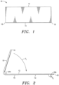
- the first end 16 of the sheet 10 is folded over (as denoted by Arrow “F 1 "), whereby the first anchoring mechanism 20a is encased in the folded over ECM sheet 10 (or the pocket 23 formed thereby).
- the sheet is sutured (shown in phantom and denoted "19a") proximate the first anchoring mechanism 20a to maintain the position of the anchoring mechanism 20a within the sheet pocket 23 (see Figs. 3 and 8 ).
- the second end 18 of the ECM sheet 10 is also folded over outwardly (as denoted by Arrow “F 2 ”) to encase the second anchoring mechanism 20b.
- the second end 18 is folded over "inwardly” to encase the second anchoring mechanism 20b.
- the first end 16 of the ECM sheet 10 is sutured or stitched 17 to the top surface 12 of the sheet 10.
- the first end 16 of the ECM sheet 10 is sutured 17 to the top surface 12 at three, preferably, equally spaced positions (denoted “S 1 ", "S 2 " and “S 3 ”) to, as discussed in detail below, form three (3) valve leaflets.
- the first end 16 of the ECM sheet 10 can alternatively be sutured to the top surface 12 at two positions to form two (2) valve leaflets, or one position to form one (1) leaflet.
- the suture positions S 1 , S 2 and S 3 are preferably disposed approximately 120° apart (see Fig. 4 ).
- the second end 18 of the ECM sheet 10 is sutured or stitched 19b to the bottom surface 14 of the sheet 10.
- the sutures 19b can comprise spaced (or intermittent) sutures or a continuous suture.
- the first side 13 of the ECM sheet 10 is rolled to meet the second side 15 of the sheet to form a substantially tubular structure, i.e. tubular anchored tissue valve 11.
- the second side 15 of the sheet can be rolled to meet the first side 13 of the sheet 10 to form the tubular valve structure.
- one of the sheet sides e.g., side 13 overlaps the other side, e.g., side 15.
- the first and second sides 13, 15 abut, as shown in Fig. 7 .
- the sutures 21 can similarly comprise spaced sutures or a continuous suture.
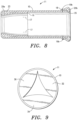
- Fig. 8 there is shown a side plan, sectional view of the anchored tissue valve 11 described above.
- the first anchoring mechanism (or anchor) 20a is disposed within the sheet pocket 23 that is formed by folding the first end 16 of the ECM sheet 10 over.
- the second anchoring mechanism (or anchor) 20b is disposed in the sheet pocket 25 that is formed by folding the second end 18 of the sheet 10 over.
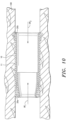
- FIG. 10 there is shown is a side plan, sectional view of the anchored tissue valve 11 in a post-deployment configuration in a cardiovascular vessel 100, wherein the valve 11 (and/or ECM support member 10) is disposed proximate host tissue of the vessel 100.
- the anchored tissue valve 11 can be deployed in a cardiovascular vessel by various traditional or minimally invasive means.
- the anchored tissue valve 11 When deployed in a cardiovascular vessel, the anchored tissue valve 11 allows normal blood flow therethrough in the direction denoted by Arrow “BF 1 " and selectively restricts regurgitation of blood in the direction denoted by Arrow BF 2 , i.e. the leaflets 30, 32 and 34 formed by suturing the first end 16 of the ECM sheet to the inner surface of the formed tubular valve structure (or member 10) expand and restrict fluid flow therethrough (see Fig. 9 ).
- the leaflets 30, 32, 34 can have various shapes and sizes, such as shown in U.S. Pat. No. 8,257,434 and Co-pending Application No. 13/560,573 .
- each leaflet 30, 32, 34 i.e. valve structure
- shape and length of each leaflet 30, 32, 34 is, of course, dependent upon the suturing points of the support member end 16 and the size, i.e. operative diameter, of the tubular support member and, hence, valve formed therefrom.
- the leaflets 30, 32, 34 have a substantially triangular shape.
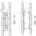
- the anchored tissue valve 40 similarly includes a support member 10 with at least one internal leaflet and at least one anchoring mechanism, more preferably, at least two anchoring mechanisms (denoted “42a" and "42b" in Fig. 15 ).
- the support member 10 similarly comprises an expandable ECM material that is capable of transitioning from a pre-deployment configuration, wherein the support member, i.e. ECM material, and, hence, anchored tissue valve formed therefrom, is capable of being positioned within a cardiovascular vessel, to a post-deployment configuration, wherein the anchored tissue valve is disposed proximate host tissue of the vessel.
- the anchoring mechanisms 42a, 42b comprise expandable anchoring mechanisms (or anchors) that facilitate the noted pre-deployment configuration of the support member 10 and are similarly capable of transitioning from a pre-deployment configuration to a post-deployment configuration, wherein the support member 10 and, hence anchored valve formed therefrom, is positioned proximate host tissue of the vessel.
- the anchoring mechanisms 42a, 42b comprise single ring members.
- the anchoring mechanisms 42a, 42b (or rings) preferably have a thickness in the range of approximately 0.25 - 1.0 mm. In some embodiments, the anchoring mechanisms 42a, 42b preferably have a thickness in the range of approximately 0.05 - 0.25 mm.
- the anchoring mechanisms 42a, 42b can similarly comprise various materials, preferably biocompatible materials, such as a biocompatible metal, e.g., stainless steel, and various polymeric materials.
- the anchoring mechanisms 42a, 42b can also comprise various biodegradable materials, such as magnesium.
- the anchoring mechanisms 42a, 42b comprise stainless steel.
- the anchoring mechanisms 42a, 42b comprise a cobalt-chrome nickel alloy.
- the anchoring mechanisms 42a, 42b comprise magnesium or an alloy thereof.
- the anchoring mechanisms 42a, 42b comprise a biocompatible shape memory alloy, including, without limitation, Nitinol ® .
- the metal anchoring mechanisms 42a, 42b include a coating of an immunomodulating compound that suppresses acute immune responses, while up-regulating chronic immune response (i.e. tissue reconstruction).
- the anchoring mechanisms 42a, 42b comprise a polymeric material, more preferably, a biocompatible and biodegradable polymeric material, such as, without limitation, polyesters, poly(amino acids), polyanhydrides, polyorthoesters, polyurethanes, polycarbonates, homopolymers and copolymers of poly(lactic acid) and poly(glycolic acid), copolyesters of e-caprolactone, trimethylene carbonate, and para-dioxanone, and like polymeric materials.
- a biocompatible and biodegradable polymeric material such as, without limitation, polyesters, poly(amino acids), polyanhydrides, polyorthoesters, polyurethanes, polycarbonates, homopolymers and copolymers of poly(lactic acid) and poly(glycolic acid), copolyesters of e-caprolactone, trimethylene carbonate, and para-dioxanone, and like polymeric materials.
- the anchoring mechanisms 42a, 42b can similarly comprise a cross-linked ECM material.
- the anchored tissue valve 40 (shown in Figs 14 and 15 ) is formed as follows: First, the sides of the support member or ECM sheet 10 are joined, as discussed above.
- first anchoring mechanism (denoted “42a") is then positioned in the pocket 23 formed by the folded over first sheet end 16.
- the first end 16 of the ECM sheet 10 is similarly sutured to the top surface at three, preferably, equally spaced positions to form the valve leaflets 30, 32, 34 (see Fig. 9 ).
- a second anchoring mechanism "42b" is then positioned proximate the second sheet end 18.
- the second sheet end 18 is then folded over to encase the second anchoring mechanism 42b in the pocket 25 formed by the folded over end 18.
- the second sheet end 18 is then sutured to the ECM sheet surface.
- the proximal ends of the anchoring mechanisms 42a, 42b are also sutured to the ECM sheet 10 to maintain the position of the anchoring mechanisms 42a, 42b in respective sheet pockets 23 and 25 (see Fig. 15 ).
- the anchoring mechanisms 42a, 42b are capable of transitioning from a pre-deployment configuration, wherein the pre-deployment configuration of the support member 10 and, hence, tissue valve 40 is similarly facilitated, to a post-deployment configuration, wherein at least the proximal and distal ends of the tissue valve 40 are supported and positioned proximate the wall of a vessel (i.e. host tissue thereof) by the anchoring mechanisms 42a, 42b.
- the primary function of the anchoring mechanisms 42a, 42b (and expandable anchoring mechanisms 52a, 52b, 62a, 62b, and 72a and 72b, discussed below) is to position the anchored tissue valve 40 (and tissue valves 50, 60 and 70) proximate host tissue of a cardiovascular vessel, and maintain contact therewith, for a predetermined temporary anchor support time period.
- the temporary anchor support time period is within the process of tissue regeneration.
- the anchor support time period is within the range of approximately 12 - 36 months. In some embodiments, the anchor support time period is within the range of approximately 3 - 12 months. In some embodiments, the anchor support time period is within the range of approximately 1 - 3 months.
- the anchor support time period can also be modulated.
- the anchoring mechanisms 42a, 42b (and expandable anchoring mechanisms 52a, 52b, 62a, 62b, and 72a and 72b) completely degrade after the anchor support time period.
- degradation of anchoring mechanisms 42a, 42b is further controlled, whereby substantially all of the anchoring mechanisms material is absorbed proximate the ECM support member 10.
- the noted controlled degradation is achieved by defined member thicknesses and supporting forces exerted on the ECM support member 10 by the anchoring mechanisms 42a, 42b (and expandable anchoring mechanisms 52a, 52b, 62a, 62b, and 72a and 72b), and the remodeling characteristics effectuated by the ECM support member 10 (or material thereof), whereby, when an anchored tissue valve is deployed in a vessel, i.e.
- the ECM support member 10 and anchoring mechanisms 42a, 42b (or expandable anchoring mechanisms 52a, 52b, 62a, 62b, and 72a and 72b) are in a post-deployment configuration), new tissue is generated and encases the anchoring mechanisms 42a, 42b (and expandable anchoring mechanisms 52a, 52b, 62a, 62b, and 72a and 72b).
- the anchoring mechanism material is then absorbed while encased by the new tissue, which substantially reduces or eliminates the possibility of anchoring mechanism fragments flowing into and obstructing a vessel.
- degradation of the anchoring mechanisms 42a, 42b is controlled, whereby the anchoring mechanisms 42a, 42b (and expandable anchoring mechanisms 52a, 52b, 62a, 62b, and 72a and 72b) are encased (or enclosed) in an endothelial lining after tissue remodeling commences for a defined period of time during and post healing, which similarly substantially reduces or eliminates the possibility of the anchor fragments flowing into and obstructing a vessel.
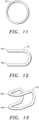
- each anchoring mechanism 42a, 42b preferably has a substantially circular "post-deployment" configuration or shape, i.e. shape after placement in a vessel.
- each anchoring mechanism 42a, 42b also has a predetermined initial or "pre-deployment" configuration or shape.
- each anchoring mechanism 42a, 42b is formed with a pre-deployment configuration, such as shown in Figs. 12 and 13 .
- each anchoring mechanism 42a, 42b is formed with a substantially circular shape, such as shown in Fig. 11 .
- the anchoring mechanisms 42a, 42b are then re-configured (via mechanical force means) to achieve the pre-deployment configuration shown in Figs. 12 and 13 .
- the anchoring mechanisms 42a, 42b comprise a biocompatible metal, including, without limitation, stainless steel and magnesium.
- the metal can also comprise a cobalt-chrome-nickel alloy.
- the anchoring mechanisms 42a, 42b are initially formed in a pre-deployment configuration.
- the pre-deployment configuration similarly comprises a substantially saddle shape, such as shown in Figs. 12 and 13 .
- the noted shape reduces the effective diameter of the valve 40, i.e. places the valve 40 in a pre-deployment configuration, which allows the anchored valve 40 to be easily placed in a vessel 100.
- the valve 40 is expanded, e.g. via a balloon, whereby anchoring mechanisms 42a, 42b are plastically deformed (i.e. re-configured or expanded) to achieve a substantially circular (and permanent) shape (see Fig. 15 ), and whereby anchoring mechanisms 42a, 42b position the ECM support member 10, and, hence, tissue valve 40 proximate host tissue of the vessel.
- the anchoring mechanisms 42a, 42b comprise a biocompatible shape memory alloy, including, without limitation, Nitinol ® .
- the anchoring mechanisms 42a, 42b are initially formed in a substantially circular pre-deployment configuration or shape and subsequently heat-treated at a first temperature (i.e. shape set heat treatment).
- the anchoring mechanisms 42a, 42b are then deformed or formed in a pre-deployment configuration or shape.
- the pre-deployment configuration or shape comprises a substantially saddle shape, such as shown in Figs. 12 and 13 .
- the anchoring mechanisms 42a, 42b when the temperature of the anchoring mechanisms 42a, 42b reach and exceed the Nitinol ® transition temperature (i.e. stress induced martensitic structure) - normally prior to deployment in a vessel - the anchoring mechanisms 42a, 42b recover (or expand to) their original circular shape, whereby each anchoring mechanism 42a, 42b exerts a supporting force on the ECM support member 10 to position the ECM support member 10, and, hence, tissue valve 40 proximate host tissue of the vessel.
- Nitinol ® transition temperature i.e. stress induced martensitic structure
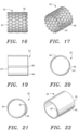
- the expandable anchoring mechanism 52 includes at least one helically arranged band element 54 forming a tubular configuration; the band element 54 comprising a plurality of uniformly shaped closed, interconnecting cells 56, and a plurality of connector elements extending between and interconnecting longitudinally spaced portions of the band over its tubular length.
- the cells 56 can comprise various shapes, such a rectangular and diamond shape. In the embodiment shown in Figs. 16 and 17 , the cells 56 are diamond shaped.
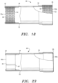
- the anchored tissue valve 50 (shown in Fig. 18 ) is similarly formed as follows: First, the sides of the ECM support member 10 are joined, as discussed above.
- first end 16 of the support member 10 is folded over inwardly.
- a first anchoring mechanism (denoted “52a") is then positioned in the pocket 23 formed by the folded over first support member end 16.
- the first end 16 of the ECM support member 10 is similarly preferably sutured to the top surface at three, preferably, equally spaced positions to form the valve leaflets 30, 32, 34 (see Fig. 9 ).
- the sheet is similarly sutured proximate the first anchoring mechanism 52a to maintain the position of the anchoring mechanism 52a within the sheet pocket 23 (see Fig. 18 ).
- a second anchoring mechanism (denoted “52b") is then positioned proximate the second support member end 18.
- the second support member end 18 is then folded over outwardly to encase the second anchoring mechanism 52b in the pocket 25 formed by the folded over end 18.
- the second support member end 18 is then sutured to the ECM sheet surface.
- the anchoring mechanisms 52a, 52b are similarly capable of transitioning from a first pre-deployment configuration, wherein a pre-deployment configuration of anchored tissue valve 50 is facilitated, i.e. a configuration that allows the valve 50 to be positioned within a cardiovascular vessel, to a first post-deployment configuration, wherein at least the proximal and distal ends of the tissue valve 50 are supported and positioned proximate the wall of the vessel (i.e. host tissue thereof) by the anchoring mechanisms 52a, 52b.
- the anchoring mechanisms 52a, 52b can similarly comprise various materials, preferably biocompatible materials, such as a biocompatible metal, e.g., stainless steel, and various polymeric materials.
- the anchoring mechanisms 52a, 52b can also comprise various biodegradable materials, such as magnesium or a magnesium alloy.
- the anchoring mechanisms 52a, 52b comprise a biocompatible shape memory alloy, including, without limitation, Nitinol ® .
- the metal anchoring mechanisms 52a, 52b include a coating of an immunomodulating compound that suppresses acute immune responses, while up-regulating chronic immune response (i.e. tissue reconstruction).
- the primary function of the anchoring mechanisms 52a, 52b is to similarly position the anchored tissue valve 50 proximate host tissue of a vessel, and maintain contact therewith, for a predetermined "temporary" anchor support time period.
- the anchor support time period is within the process of tissue regeneration.
- each anchoring mechanism 52a, 52b preferably has a substantially circular "post-deployment" configuration. To facilitate deployment of the anchored tissue valve 50 into a cardiovascular vessel, each anchoring mechanism 52a, 52b also has a pre-deployment configuration.
- the initial or pre-deployment configuration is similarly dependent upon the anchoring mechanism material.
- the anchoring mechanisms 52a, 52b comprise a biocompatible metal, including, without limitation, stainless steel and magnesium.
- the metal can also comprise a cobalt-chrome-nickel alloy.
- each anchoring mechanisms 52a, 52b is formed with a substantially circular "compressed" pre-deployment configuration.
- the valve 50 is expanded, e.g. via a balloon, whereby the anchoring mechanisms 52a, 52b are plastically deformed (i.e. re-configured or expanded) to achieve an "expanded" substantially circular, post-deployment configuration (see Fig. 18 ), and whereby anchoring mechanisms 52a, 52b position the ECM support member 10, and, hence, tissue valve 50 proximate host tissue of the vessel, and maintain contact therewith, for a predetermined anchor support time period.
- the anchoring mechanisms 52a, 52b comprise a biocompatible shape memory alloy, such as Nitinol ® .
- the anchoring mechanisms 52a, 52b are initially formed in a substantially circular "expanded" pre-deployment configuration or shape and subsequently heat-treated at a first temperature (i.e. shape set heat treatment).
- the anchoring mechanisms 52a, 52b are then similarly deformed or formed in a "compressed" pre-deployment configuration or shape.
- each anchoring mechanism 52a, 52b When the temperature of the anchoring mechanisms 52a, 52b reach and exceed the Nitinol ® transition temperature (i.e. stress induced martensitic structure) the anchoring mechanisms 52a, 52b recover (or expand to) their original circular shape, whereby each anchoring mechanism 52a, 52b similarly exerts a supporting force on the ECM support member 10 to position the ECM support member 10, and, hence, tissue valve 50 proximate host tissue of the vessel.
- Nitinol ® transition temperature i.e. stress induced martensitic structure
- the anchoring mechanism 62 comprises an expandable band 64.
- the anchored valve 60 includes a first expandable anchoring mechanism (or band) 62a disposed proximate the first end 16 of the support member 10 and a second expandable anchoring mechanism 62b disposed proximate the second end 18 of the member 10.
- the anchored valve 60 is formed in a manner similar to the formation of anchored tissue valve 50, discussed above.
- the expandable anchoring mechanisms or bands 62a, 62b are similarly capable of transitioning from a pre-deployment configuration (see Fig. 21 ), wherein the pre-deployment configuration of the support member 10 and, hence, anchored tissue valve 60 formed therefrom is facilitated, to a post-deployment configuration (see Fig. 20 ), wherein at least the proximal and distal ends of the tissue valve 60 are positioned proximate the wall of the vessel (i.e. host tissue thereof) by the anchoring mechanisms 62a, 62b.
- the anchoring mechanisms 62a, 62b can similarly comprise various materials, preferably biocompatible materials, such as a biocompatible metal, e.g., stainless steel, and various polymeric materials.
- the anchoring mechanisms 62a, 62b can also comprise various biodegradable materials, such as magnesium or a magnesium alloy.
- the anchoring mechanisms 62a, 62b comprise a biocompatible shape memory alloy, including, without limitation, Nitinol ® .
- the metal anchoring mechanisms 62a, 62b can also include a coating of an immunomodulating compound that suppresses acute immune responses, while up-regulating chronic immune response (i.e. tissue reconstruction).
- the anchoring mechanisms 62a, 62b comprise discontinuous bands. As illustrated in Figs. 21 and 22 , the discontinuous bands are designed and configured, whereby a first end of anchoring mechanism 65 over-laps the second end of the anchoring mechanism 67 to facilitate a "compressed" pre-deployment configuration.
- the pre-deployment configuration is similarly dependent upon the anchor material.
- the anchoring mechanisms 62a, 62b comprise a biocompatible metal, including, without limitation, stainless steel and magnesium.
- the anchoring mechanisms 62a, 62b can also comprise a cobalt-chrome-nickel alloy.
- each anchoring mechanism 62a, 62b is formed with a substantially circular "compressed" pre-deployment configuration or shape, such as shown in Figs. 21 and 22 .
- the valve 60 is expanded, whereby the anchoring mechanisms 62a, 62b expand to achieve the "expanded" substantially circular, post-deployment configuration shown in Figs. 20 and 23 , and whereby the anchoring mechanisms 62a, 62b position the ECM support member 10, and, hence, tissue valve 60 proximate host tissue of the vessel.
- anchoring mechanisms 62a, 62b can similarly comprise a biocompatible shape memory alloy, such as Nitinol ® .
- the anchoring mechanisms 62a, 62b are initially formed in the "expanded" pre-deployment configuration and subsequently heat-treated at a first temperature (i.e. shape set heat treatment).
- the anchoring mechanisms 62a, 62b are then similarly deformed or formed in a "compressed" pre-deployment configuration or shape.
- each anchoring mechanism 62a, 62b When the temperature of the anchoring mechanisms 62a, 62b reach and exceed the Nitinol ® transition temperature (i.e. stress induced martensitic structure) the anchoring mechanisms 62a, 62b recover (or expand to) their original circular shape, whereby each anchoring mechanism 62a, 62b similarly exerts a supporting force on the ECM support member 10 to position the ECM support member 10, and, hence, tissue valve 60 proximate host tissue of the vessel.
- the Nitinol ® transition temperature i.e. stress induced martensitic structure
- the anchoring mechanisms comprise microneedle anchoring mechanisms having plurality of biodegradable microneedles or barbs that are adapted to maintain contact of the ECM member 10 and, hence, anchored valve formed therefrom against the wall of a vascular structure when disposed therein.
- Suitable microneedle anchoring members are disclosed in Co-Pending Application No. 13/686,131, filed November 27, 2012 .
- microneedles or barbs can be employed within the scope of the invention; provided, the microneedle or barb has a head (or head region) that is able to pierce tissue and remain engaged to the tissue for a predetermined period of time.
- the microneedle anchoring member and, hence, microneedles comprise a biodegradable polymeric material, an ECM material or a pharmacological agent or composition (i.e. drug), e.g., Heparin ® , Plavix ® , etc., or a combination thereof.
- the microneedle anchoring member and, hence, microneedles comprise a biocompatible and bioabsorbable metal, such as magnesium.
- the microneedles comprise drug-eluting members that facilitate the direct administration of a pharmacological agent or composition to host tissue.
- the drug-eluting capability is facilitated by forming at least one, more preferably, each microneedle out of a pharmacological agent or composition, whereby upon engagement of the biodegradable microneedles to a recipient's tissue, the microneedles dissolve or degrade and the pharmacological agent or composition is administered to the recipient at the engagement site.
- the drug-eluting capability is facilitated by coating at least one, more preferably, each microneedle with a pharmacological agent or composition, whereby upon engagement of the microneedles to a recipient's tissue, the pharmacological agent or composition is absorbed and, hence, administered to the recipient.
- each microneedle has an internal reservoir that is adapted to receive and contain a pharmacological agent or composition therein. According to the invention, upon engagement of the biodegradable microneedles to a recipient's tissue, the microneedles dissolve or degrade and the pharmacological agent or composition contained in the reservoir is administered to the recipient.
- each microneedle has an internal reservoir that is adapted to receive and contain a pharmacological agent or composition therein and at least one, more preferably, a plurality of lumens in communication with the reservoir and, hence, pharmacological agent or composition contained therein.
- the microneedles also include a biodegradable or bioabsorbable coating (or sealing layer) on the outer surface to temporarily seal reservoir and inter-connected lumens. Upon engagement of the microneedles to a recipient's tissue, the coating dissolves or degrades and the pharmacological agent or composition contained in the reservoir is administered to the recipient via the microneedle lumens.
- the on-set and rate of administration of a pharmacological agent or composition can be determined and regulated by, among other things, the composition and/or properties of the base microneedle, e.g. dissolution rate, size of lumens, etc., and the composition and/or properties of the pharmacological agent or composition, and sealing coatings.
- the microneedle anchoring mechanism 72 includes a base 74 and a plurality of microneedles 76, which are designed and configured to pierce through and project out of a support member, in this embodiment, an ECM support member 10, when an anchored valve employing the microneedle anchoring mechanism 72 is deployed in a vessel.
- Fig. 27 there is shown one embodiment of an anchored valve 70 employing microneedle anchoring mechanisms 72 of the invention.
- the valve 70 includes a first microneedle anchoring mechanism 72a disposed proximate the first end 16 of the support member 10 and a second microneedle anchoring mechanism 72b disposed proximate the second end 18 of the member 10.
- the anchored tissue valve 70 is also formed in a manner similar to the formation of anchored valves 50 and 60, discussed above.
- the microneedle anchoring mechanisms 72a, 72b are similarly capable of transitioning from a pre-deployment configuration to facilitate a pre-deployment configuration of anchored tissue valve 70, to a post-deployment configuration, wherein at least the proximal and distal ends of the tissue valve 70 are supported and positioned proximate the wall of a vessel (i.e. host tissue thereof) by the microneedle anchoring mechanisms 72a, 72b.
- a first end 78a of each microneedle anchoring mechanism base 74 includes an elongated slot 77 that is designed and configured to receive the base projection 79 on the opposing end 78b of the base 74.
- Figs. 26A and 26B when the microneedle anchoring mechanisms 72a, 72b are in a pre-deployment configuration, i.e. a substantially circular shape having a first diameter (denoted "d 1 "), the elongated slot 77 slidably receives the base projection 79 therein.
- the base projection 79 transitions (or moves) within the elongated slot 77 to facilitate an expansion (or enlargement) of the circular shape to a second larger diameter (denoted "d 2 ").
- the microneedle anchoring mechanisms 72a, 72b upon deployment of an anchored tissue valve employing the microneedle anchoring mechanisms 72a, 72b in a vessel, the microneedle anchoring mechanisms 72a, 72b position and maintain at least the ends of the support member 10 and, hence, tissue valve 70 proximate host or vessel tissue when in a post-deployment configuration.
- the microneedles 76 also project through tissue valve 70 and engage the vessel tissue and secure the tissue valve 70 proximate the cardiovascular vessel tissue.
- the anchored valves of the invention can be readily employed to replace numerous valves in the body including, without limitation, diseased or defective aortic, pulmonary, mitral, tricuspid and/or peripheral venous valves.
- the size or diameter of the anchored valves of the invention can vary to accommodate placement in various cardiovascular vessels in pediatrics and adults.
- the present invention provides numerous advantages compared to prior art prosthetic valves. Among the advantages are the following:
Landscapes
- Health & Medical Sciences (AREA)
- Engineering & Computer Science (AREA)
- Life Sciences & Earth Sciences (AREA)
- Cardiology (AREA)
- Veterinary Medicine (AREA)
- Public Health (AREA)
- General Health & Medical Sciences (AREA)
- Animal Behavior & Ethology (AREA)
- Biomedical Technology (AREA)
- General Chemical & Material Sciences (AREA)
- Organic Chemistry (AREA)
- Pharmacology & Pharmacy (AREA)
- Nuclear Medicine, Radiotherapy & Molecular Imaging (AREA)
- Medicinal Chemistry (AREA)
- Chemical Kinetics & Catalysis (AREA)
- Chemical & Material Sciences (AREA)
- Heart & Thoracic Surgery (AREA)
- Vascular Medicine (AREA)
- Bioinformatics & Cheminformatics (AREA)
- Oral & Maxillofacial Surgery (AREA)
- Transplantation (AREA)
- Rheumatology (AREA)
- Pain & Pain Management (AREA)
- Prostheses (AREA)
- Pharmaceuticals Containing Other Organic And Inorganic Compounds (AREA)
- Medicines Containing Material From Animals Or Micro-Organisms (AREA)
- Acyclic And Carbocyclic Compounds In Medicinal Compositions (AREA)
- Materials For Medical Uses (AREA)
- Medicines That Contain Protein Lipid Enzymes And Other Medicines (AREA)
Priority Applications (1)
| Application Number | Priority Date | Filing Date | Title |
|---|---|---|---|
| EP18186664.1A EP3427696B1 (en) | 2013-03-01 | 2013-03-01 | Anchored cardiovascular valve |
Applications Claiming Priority (3)
| Application Number | Priority Date | Filing Date | Title |
|---|---|---|---|
| PCT/US2013/028561 WO2014133539A1 (en) | 2013-03-01 | 2013-03-01 | Anchored cardiovascular valve |
| EP13861484.7A EP2797556B1 (en) | 2013-03-01 | 2013-03-01 | Anchored cardiovascular valve |
| EP18186664.1A EP3427696B1 (en) | 2013-03-01 | 2013-03-01 | Anchored cardiovascular valve |
Related Parent Applications (1)
| Application Number | Title | Priority Date | Filing Date |
|---|---|---|---|
| EP13861484.7A Division EP2797556B1 (en) | 2013-03-01 | 2013-03-01 | Anchored cardiovascular valve |
Publications (2)
| Publication Number | Publication Date |
|---|---|
| EP3427696A1 EP3427696A1 (en) | 2019-01-16 |
| EP3427696B1 true EP3427696B1 (en) | 2025-07-02 |
Family
ID=51428653
Family Applications (2)
| Application Number | Title | Priority Date | Filing Date |
|---|---|---|---|
| EP18186664.1A Active EP3427696B1 (en) | 2013-03-01 | 2013-03-01 | Anchored cardiovascular valve |
| EP13861484.7A Not-in-force EP2797556B1 (en) | 2013-03-01 | 2013-03-01 | Anchored cardiovascular valve |
Family Applications After (1)
| Application Number | Title | Priority Date | Filing Date |
|---|---|---|---|
| EP13861484.7A Not-in-force EP2797556B1 (en) | 2013-03-01 | 2013-03-01 | Anchored cardiovascular valve |
Country Status (4)
| Country | Link |
|---|---|
| EP (2) | EP3427696B1 (enExample) |
| JP (1) | JP6205436B2 (enExample) |
| AU (1) | AU2013379744A1 (enExample) |
| WO (1) | WO2014133539A1 (enExample) |
Families Citing this family (29)
| Publication number | Priority date | Publication date | Assignee | Title |
|---|---|---|---|---|
| US8579964B2 (en) | 2010-05-05 | 2013-11-12 | Neovasc Inc. | Transcatheter mitral valve prosthesis |
| US9308087B2 (en) | 2011-04-28 | 2016-04-12 | Neovasc Tiara Inc. | Sequentially deployed transcatheter mitral valve prosthesis |
| US9554897B2 (en) | 2011-04-28 | 2017-01-31 | Neovasc Tiara Inc. | Methods and apparatus for engaging a valve prosthesis with tissue |
| US9345573B2 (en) | 2012-05-30 | 2016-05-24 | Neovasc Tiara Inc. | Methods and apparatus for loading a prosthesis onto a delivery system |
| US9572665B2 (en) | 2013-04-04 | 2017-02-21 | Neovasc Tiara Inc. | Methods and apparatus for delivering a prosthetic valve to a beating heart |
| US12343254B2 (en) | 2014-03-29 | 2025-07-01 | Corvivo Cardiovascular, Inc. | Prosthetic heart valves |
| US12350147B2 (en) | 2014-03-29 | 2025-07-08 | Corvivo Cardiovascular, Inc. | Adaptable prosthetic tissue valves |
| US12201518B2 (en) | 2014-03-29 | 2025-01-21 | Cormatrix Cardiovascular, Inc. | Reinforced prosthetic valves |
| CN105721190B (zh) * | 2014-12-04 | 2019-06-21 | 华为技术有限公司 | 数据传输路径的故障检测方法、装置及服务器 |
| US10631984B2 (en) | 2015-12-15 | 2020-04-28 | Neovasc Tiara Inc. | Transseptal delivery system |
| CA3007670C (en) | 2016-01-29 | 2024-09-17 | Neovasc Tiara Inc. | PROSTHETIC VALVE PREVENTING OBSTRUCTION THAT WOULD PREVENT FLOW |
| WO2018013361A1 (en) * | 2016-07-11 | 2018-01-18 | Cormatrix Cardiovascular, Inc. | Prosthetic tissue valves |
| AU2017361296B2 (en) | 2016-11-21 | 2022-09-29 | Neovasc Tiara Inc. | Methods and systems for rapid retraction of a transcatheter heart valve delivery system |
| WO2019036810A1 (en) | 2017-08-25 | 2019-02-28 | Neovasc Tiara Inc. | TRANSCATHETER MITRAL VALVULE PROSTHESIS WITH SEQUENTIAL DEPLOYMENT |
| EP3735290A2 (en) * | 2018-01-02 | 2020-11-11 | Evonik Corporation | Microneedle delivery system with anchor |
| CN110101486B (zh) * | 2018-02-01 | 2024-02-27 | 上海微创心通医疗科技有限公司 | 心脏瓣膜假体及其输送器 |
| AU2019233651B2 (en) | 2018-03-13 | 2022-01-27 | Institut Químic De Sarrià Cets Fundació Privada | Vascular repair patch |
| US11610660B1 (en) | 2021-08-20 | 2023-03-21 | AltaThera Pharmaceuticals LLC | Antiarrhythmic drug dosing methods, medical devices, and systems |
| US12396970B2 (en) | 2021-08-20 | 2025-08-26 | AltaThera Pharmaceuticals LLC | Anti-arrhythmic compositions and methods |
| US10512620B1 (en) | 2018-08-14 | 2019-12-24 | AltaThera Pharmaceuticals, LLC | Method of initiating and escalating sotalol hydrochloride dosing |
| US11696902B2 (en) | 2018-08-14 | 2023-07-11 | AltaThera Pharmaceuticals, LLC | Method of initiating and escalating sotalol hydrochloride dosing |
| US11344518B2 (en) | 2018-08-14 | 2022-05-31 | AltaThera Pharmaceuticals LLC | Method of converting atrial fibrillation to normal sinus rhythm and loading oral sotalol in a shortened time frame |
| AU2019374743B2 (en) | 2018-11-08 | 2022-03-03 | Neovasc Tiara Inc. | Ventricular deployment of a transcatheter mitral valve prosthesis |
| EP3934591A4 (en) | 2019-03-08 | 2022-11-23 | Neovasc Tiara Inc. | RETRIEVABLE PROSTHETIC RELEASE SYSTEM |
| EP3946163B1 (en) | 2019-04-01 | 2025-08-20 | Neovasc Tiara Inc. | Controllably deployable prosthetic valve |
| AU2020271896B2 (en) | 2019-04-10 | 2022-10-13 | Neovasc Tiara Inc. | Prosthetic valve with natural blood flow |
| CN114025813B (zh) | 2019-05-20 | 2024-05-14 | 内奥瓦斯克迪亚拉公司 | 具有止血机构的引入器 |
| AU2020295566B2 (en) | 2019-06-20 | 2023-07-20 | Neovasc Tiara Inc. | Low profile prosthetic mitral valve |
| CN114588500B (zh) * | 2022-02-10 | 2023-04-11 | 北京航空航天大学 | 胃肠道智能化自动弹射给药装置及其制备和给药方法 |
Family Cites Families (22)
| Publication number | Priority date | Publication date | Assignee | Title |
|---|---|---|---|---|
| US4902508A (en) | 1988-07-11 | 1990-02-20 | Purdue Research Foundation | Tissue graft composition |
| EP0705081B1 (en) * | 1994-04-22 | 2001-10-17 | Medtronic, Inc. | Stented bioprosthetic heart valve |
| US5522881A (en) * | 1994-06-28 | 1996-06-04 | Meadox Medicals, Inc. | Implantable tubular prosthesis having integral cuffs |
| US5733337A (en) | 1995-04-07 | 1998-03-31 | Organogenesis, Inc. | Tissue repair fabric |
| GB2312485B (en) * | 1996-04-24 | 1999-10-20 | Endre Bodnar | Bioprosthetic conduits |
| SK22499A3 (en) | 1996-08-23 | 1999-10-08 | Cook Biotech Inc | Graft prosthesis, materials and methods |
| WO1998025546A1 (en) | 1996-12-10 | 1998-06-18 | Cook Biotech, Inc. | Tubular grafts from purified submucosa |
| WO1998025637A1 (en) | 1996-12-10 | 1998-06-18 | Purdue Research Foundation | Biomaterial derived from vertebrate liver tissue |
| EP0850607A1 (en) * | 1996-12-31 | 1998-07-01 | Cordis Corporation | Valve prosthesis for implantation in body channels |
| US6074419A (en) * | 1996-12-31 | 2000-06-13 | St. Jude Medical, Inc. | Indicia for prosthetic device |
| US7125418B2 (en) * | 2002-04-16 | 2006-10-24 | The International Heart Institute Of Montana Foundation | Sigmoid valve and method for its percutaneous implantation |
| US7550004B2 (en) | 2002-08-20 | 2009-06-23 | Cook Biotech Incorporated | Endoluminal device with extracellular matrix material and methods |
| EP1659992B1 (en) * | 2003-07-31 | 2013-03-27 | Cook Medical Technologies LLC | Prosthetic valve devices and methods of making such devices |
| US7261732B2 (en) * | 2003-12-22 | 2007-08-28 | Henri Justino | Stent mounted valve |
| DE602005020754D1 (de) | 2004-03-31 | 2010-06-02 | Cook Inc | Transplantatmaterial und gefässprothese mit extrazellulärer kollagenmatrix und dessen herstellungsverfahren |
| EP2425801A3 (en) * | 2004-05-05 | 2013-01-02 | Direct Flow Medical, Inc. | Unstented heart valve with formed in place support structure |
| EP2025306B1 (en) * | 2007-04-23 | 2020-01-01 | Saint Joseph Research Institute | Methods of making a replacement heart valve |
| US8257434B2 (en) * | 2007-12-18 | 2012-09-04 | Cormatrix Cardiovascular, Inc. | Prosthetic tissue valve |
| AU2010216095B2 (en) * | 2009-02-18 | 2015-10-08 | Cormatrix Cardiovascular, Inc. | Compositions and methods for preventing cardiac arrhythmia |
| US8795354B2 (en) | 2010-03-05 | 2014-08-05 | Edwards Lifesciences Corporation | Low-profile heart valve and delivery system |
| EP2598044B1 (en) * | 2010-07-27 | 2019-03-13 | Incept, LLC | Apparatus for treating neurovascular venous outflow obstruction |
| EP2714099A4 (en) * | 2011-05-27 | 2014-11-19 | Cormatrix Cardiovascular Inc | STERILIZED, ACELLULAR EXTRACELLULAR MATRIX COMPOSITIONS AND METHODS OF MAKING SAME |
-
2013
- 2013-03-01 EP EP18186664.1A patent/EP3427696B1/en active Active
- 2013-03-01 EP EP13861484.7A patent/EP2797556B1/en not_active Not-in-force
- 2013-03-01 JP JP2015560149A patent/JP6205436B2/ja not_active Expired - Fee Related
- 2013-03-01 AU AU2013379744A patent/AU2013379744A1/en not_active Abandoned
- 2013-03-01 WO PCT/US2013/028561 patent/WO2014133539A1/en not_active Ceased
Also Published As
| Publication number | Publication date |
|---|---|
| JP6205436B2 (ja) | 2017-09-27 |
| EP2797556A4 (en) | 2015-06-03 |
| EP2797556B1 (en) | 2018-08-01 |
| JP2016509880A (ja) | 2016-04-04 |
| EP2797556A1 (en) | 2014-11-05 |
| AU2013379744A1 (en) | 2015-09-17 |
| EP3427696A1 (en) | 2019-01-16 |
| WO2014133539A1 (en) | 2014-09-04 |
Similar Documents
| Publication | Publication Date | Title |
|---|---|---|
| EP3427696B1 (en) | Anchored cardiovascular valve | |
| US9044319B2 (en) | Anchored cardiovascular valve | |
| US9011526B2 (en) | Tubular extracellular matrix prosthetic valve | |
| US9867696B2 (en) | Systems, apparatus and methods for sealing perivalvular leaks | |
| US8709076B1 (en) | Two-piece prosthetic valve | |
| US9308084B2 (en) | Prosthetic tissue valves and methods for anchoring same to cardiovascular structures | |
| CA2894844C (en) | A seamless tubular extracellular matrix prosthetic valve and method for forming same | |
| US10188510B2 (en) | Prosthetic tissue valves | |
| US20150289974A1 (en) | Reinforced Prosthetic Tissue Valves | |
| JP2016509880A5 (enExample) | ||
| US20160317296A1 (en) | Prosthetic Tissue Valves | |
| US12029643B2 (en) | Systems, apparatus and methods for sealing perivalvular leaks | |
| US9907649B2 (en) | Prosthetic tissue valves and methods for anchoring same to cardiovascular structures | |
| US10188509B2 (en) | Prosthetic tissue valves | |
| WO2014133543A1 (en) | Two-piece prosthetic valve |
Legal Events
| Date | Code | Title | Description |
|---|---|---|---|
| PUAI | Public reference made under article 153(3) epc to a published international application that has entered the european phase |
Free format text: ORIGINAL CODE: 0009012 |
|
| STAA | Information on the status of an ep patent application or granted ep patent |
Free format text: STATUS: THE APPLICATION HAS BEEN PUBLISHED |
|
| AC | Divisional application: reference to earlier application |
Ref document number: 2797556 Country of ref document: EP Kind code of ref document: P |
|
| AK | Designated contracting states |
Kind code of ref document: A1 Designated state(s): AL AT BE BG CH CY CZ DE DK EE ES FI FR GB GR HR HU IE IS IT LI LT LU LV MC MK MT NL NO PL PT RO RS SE SI SK SM TR |
|
| STAA | Information on the status of an ep patent application or granted ep patent |
Free format text: STATUS: REQUEST FOR EXAMINATION WAS MADE |
|
| 17P | Request for examination filed |
Effective date: 20190711 |
|
| RBV | Designated contracting states (corrected) |
Designated state(s): AL AT BE BG CH CY CZ DE DK EE ES FI FR GB GR HR HU IE IS IT LI LT LU LV MC MK MT NL NO PL PT RO RS SE SI SK SM TR |
|
| STAA | Information on the status of an ep patent application or granted ep patent |
Free format text: STATUS: EXAMINATION IS IN PROGRESS |
|
| 17Q | First examination report despatched |
Effective date: 20240228 |
|
| GRAP | Despatch of communication of intention to grant a patent |
Free format text: ORIGINAL CODE: EPIDOSNIGR1 |
|
| STAA | Information on the status of an ep patent application or granted ep patent |
Free format text: STATUS: GRANT OF PATENT IS INTENDED |
|
| INTG | Intention to grant announced |
Effective date: 20250130 |
|
| GRAS | Grant fee paid |
Free format text: ORIGINAL CODE: EPIDOSNIGR3 |
|
| GRAA | (expected) grant |
Free format text: ORIGINAL CODE: 0009210 |
|
| STAA | Information on the status of an ep patent application or granted ep patent |
Free format text: STATUS: THE PATENT HAS BEEN GRANTED |
|
| AC | Divisional application: reference to earlier application |
Ref document number: 2797556 Country of ref document: EP Kind code of ref document: P |
|
| AK | Designated contracting states |
Kind code of ref document: B1 Designated state(s): AL AT BE BG CH CY CZ DE DK EE ES FI FR GB GR HR HU IE IS IT LI LT LU LV MC MK MT NL NO PL PT RO RS SE SI SK SM TR |
|
| P01 | Opt-out of the competence of the unified patent court (upc) registered |
Free format text: CASE NUMBER: APP_24719/2025 Effective date: 20250523 |
|
| REG | Reference to a national code |
Ref country code: GB Ref legal event code: FG4D |
|
| REG | Reference to a national code |
Ref country code: CH Ref legal event code: EP |
|
| REG | Reference to a national code |
Ref country code: DE Ref legal event code: R096 Ref document number: 602013086871 Country of ref document: DE |
|
| REG | Reference to a national code |
Ref country code: IE Ref legal event code: FG4D |
|
| REG | Reference to a national code |
Ref country code: NL Ref legal event code: MP Effective date: 20250702 |
|
| PG25 | Lapsed in a contracting state [announced via postgrant information from national office to epo] |
Ref country code: PT Free format text: LAPSE BECAUSE OF FAILURE TO SUBMIT A TRANSLATION OF THE DESCRIPTION OR TO PAY THE FEE WITHIN THE PRESCRIBED TIME-LIMIT Effective date: 20251103 |
|
| PG25 | Lapsed in a contracting state [announced via postgrant information from national office to epo] |
Ref country code: NL Free format text: LAPSE BECAUSE OF FAILURE TO SUBMIT A TRANSLATION OF THE DESCRIPTION OR TO PAY THE FEE WITHIN THE PRESCRIBED TIME-LIMIT Effective date: 20250702 |
|
| REG | Reference to a national code |
Ref country code: AT Ref legal event code: MK05 Ref document number: 1808399 Country of ref document: AT Kind code of ref document: T Effective date: 20250702 |