EP3418397B1 - Système de détection d'une infection dans le liquide synovial - Google Patents
Système de détection d'une infection dans le liquide synovial Download PDFInfo
- Publication number
- EP3418397B1 EP3418397B1 EP18184143.8A EP18184143A EP3418397B1 EP 3418397 B1 EP3418397 B1 EP 3418397B1 EP 18184143 A EP18184143 A EP 18184143A EP 3418397 B1 EP3418397 B1 EP 3418397B1
- Authority
- EP
- European Patent Office
- Prior art keywords
- synovial fluid
- sample
- fluid sample
- joint
- interleukin
- Prior art date
- Legal status (The legal status is an assumption and is not a legal conclusion. Google has not performed a legal analysis and makes no representation as to the accuracy of the status listed.)
- Active
Links
Images


Classifications
-
- G—PHYSICS
- G01—MEASURING; TESTING
- G01N—INVESTIGATING OR ANALYSING MATERIALS BY DETERMINING THEIR CHEMICAL OR PHYSICAL PROPERTIES
- G01N33/00—Investigating or analysing materials by specific methods not covered by groups G01N1/00 - G01N31/00
- G01N33/48—Biological material, e.g. blood, urine; Haemocytometers
- G01N33/50—Chemical analysis of biological material, e.g. blood, urine; Testing involving biospecific ligand binding methods; Immunological testing
- G01N33/53—Immunoassay; Biospecific binding assay; Materials therefor
- G01N33/543—Immunoassay; Biospecific binding assay; Materials therefor with an insoluble carrier for immobilising immunochemicals
- G01N33/54366—Apparatus specially adapted for solid-phase testing
- G01N33/54386—Analytical elements
- G01N33/54387—Immunochromatographic test strips
- G01N33/54388—Immunochromatographic test strips based on lateral flow
-
- G—PHYSICS
- G01—MEASURING; TESTING
- G01N—INVESTIGATING OR ANALYSING MATERIALS BY DETERMINING THEIR CHEMICAL OR PHYSICAL PROPERTIES
- G01N33/00—Investigating or analysing materials by specific methods not covered by groups G01N1/00 - G01N31/00
- G01N33/48—Biological material, e.g. blood, urine; Haemocytometers
- G01N33/50—Chemical analysis of biological material, e.g. blood, urine; Testing involving biospecific ligand binding methods; Immunological testing
- G01N33/53—Immunoassay; Biospecific binding assay; Materials therefor
- G01N33/575—Immunoassay; Biospecific binding assay; Materials therefor for cancer
- G01N33/57575—Immunoassay; Biospecific binding assay; Materials therefor for cancer involving oncogenic proteins
-
- G—PHYSICS
- G01—MEASURING; TESTING
- G01N—INVESTIGATING OR ANALYSING MATERIALS BY DETERMINING THEIR CHEMICAL OR PHYSICAL PROPERTIES
- G01N33/00—Investigating or analysing materials by specific methods not covered by groups G01N1/00 - G01N31/00
- G01N33/48—Biological material, e.g. blood, urine; Haemocytometers
- G01N33/50—Chemical analysis of biological material, e.g. blood, urine; Testing involving biospecific ligand binding methods; Immunological testing
- G01N33/68—Chemical analysis of biological material, e.g. blood, urine; Testing involving biospecific ligand binding methods; Immunological testing involving proteins, peptides or amino acids
- G01N33/6863—Cytokines, i.e. immune system proteins modifying a biological response such as cell growth proliferation or differentiation, e.g. TNF, CNF, GM-CSF, lymphotoxin, MIF or their receptors
- G01N33/6869—Interleukin
-
- G—PHYSICS
- G01—MEASURING; TESTING
- G01N—INVESTIGATING OR ANALYSING MATERIALS BY DETERMINING THEIR CHEMICAL OR PHYSICAL PROPERTIES
- G01N33/00—Investigating or analysing materials by specific methods not covered by groups G01N1/00 - G01N31/00
- G01N33/48—Biological material, e.g. blood, urine; Haemocytometers
- G01N33/50—Chemical analysis of biological material, e.g. blood, urine; Testing involving biospecific ligand binding methods; Immunological testing
- G01N33/68—Chemical analysis of biological material, e.g. blood, urine; Testing involving biospecific ligand binding methods; Immunological testing involving proteins, peptides or amino acids
- G01N33/6893—Chemical analysis of biological material, e.g. blood, urine; Testing involving biospecific ligand binding methods; Immunological testing involving proteins, peptides or amino acids related to diseases not provided for elsewhere
-
- G—PHYSICS
- G01—MEASURING; TESTING
- G01N—INVESTIGATING OR ANALYSING MATERIALS BY DETERMINING THEIR CHEMICAL OR PHYSICAL PROPERTIES
- G01N2333/00—Assays involving biological materials from specific organisms or of a specific nature
- G01N2333/435—Assays involving biological materials from specific organisms or of a specific nature from animals; from humans
- G01N2333/475—Assays involving growth factors
- G01N2333/515—Angiogenesic factors; Angiogenin
-
- G—PHYSICS
- G01—MEASURING; TESTING
- G01N—INVESTIGATING OR ANALYSING MATERIALS BY DETERMINING THEIR CHEMICAL OR PHYSICAL PROPERTIES
- G01N2333/00—Assays involving biological materials from specific organisms or of a specific nature
- G01N2333/435—Assays involving biological materials from specific organisms or of a specific nature from animals; from humans
- G01N2333/52—Assays involving cytokines
- G01N2333/53—Colony-stimulating factor [CSF]
- G01N2333/535—Granulocyte CSF; Granulocyte-macrophage CSF
-
- G—PHYSICS
- G01—MEASURING; TESTING
- G01N—INVESTIGATING OR ANALYSING MATERIALS BY DETERMINING THEIR CHEMICAL OR PHYSICAL PROPERTIES
- G01N2333/00—Assays involving biological materials from specific organisms or of a specific nature
- G01N2333/435—Assays involving biological materials from specific organisms or of a specific nature from animals; from humans
- G01N2333/52—Assays involving cytokines
- G01N2333/54—Interleukins [IL]
- G01N2333/5412—IL-6
-
- G—PHYSICS
- G01—MEASURING; TESTING
- G01N—INVESTIGATING OR ANALYSING MATERIALS BY DETERMINING THEIR CHEMICAL OR PHYSICAL PROPERTIES
- G01N2333/00—Assays involving biological materials from specific organisms or of a specific nature
- G01N2333/435—Assays involving biological materials from specific organisms or of a specific nature from animals; from humans
- G01N2333/52—Assays involving cytokines
- G01N2333/54—Interleukins [IL]
- G01N2333/5421—IL-8
-
- G—PHYSICS
- G01—MEASURING; TESTING
- G01N—INVESTIGATING OR ANALYSING MATERIALS BY DETERMINING THEIR CHEMICAL OR PHYSICAL PROPERTIES
- G01N2333/00—Assays involving biological materials from specific organisms or of a specific nature
- G01N2333/435—Assays involving biological materials from specific organisms or of a specific nature from animals; from humans
- G01N2333/52—Assays involving cytokines
- G01N2333/54—Interleukins [IL]
- G01N2333/5428—IL-10
-
- G—PHYSICS
- G01—MEASURING; TESTING
- G01N—INVESTIGATING OR ANALYSING MATERIALS BY DETERMINING THEIR CHEMICAL OR PHYSICAL PROPERTIES
- G01N2333/00—Assays involving biological materials from specific organisms or of a specific nature
- G01N2333/435—Assays involving biological materials from specific organisms or of a specific nature from animals; from humans
- G01N2333/52—Assays involving cytokines
- G01N2333/54—Interleukins [IL]
- G01N2333/545—IL-1
-
- G—PHYSICS
- G01—MEASURING; TESTING
- G01N—INVESTIGATING OR ANALYSING MATERIALS BY DETERMINING THEIR CHEMICAL OR PHYSICAL PROPERTIES
- G01N2333/00—Assays involving biological materials from specific organisms or of a specific nature
- G01N2333/435—Assays involving biological materials from specific organisms or of a specific nature from animals; from humans
- G01N2333/705—Assays involving receptors, cell surface antigens or cell surface determinants
- G01N2333/715—Assays involving receptors, cell surface antigens or cell surface determinants for cytokines; for lymphokines; for interferons
- G01N2333/7158—Assays involving receptors, cell surface antigens or cell surface determinants for cytokines; for lymphokines; for interferons for chemokines
-
- G—PHYSICS
- G01—MEASURING; TESTING
- G01N—INVESTIGATING OR ANALYSING MATERIALS BY DETERMINING THEIR CHEMICAL OR PHYSICAL PROPERTIES
- G01N2333/00—Assays involving biological materials from specific organisms or of a specific nature
- G01N2333/435—Assays involving biological materials from specific organisms or of a specific nature from animals; from humans
- G01N2333/705—Assays involving receptors, cell surface antigens or cell surface determinants
- G01N2333/72—Assays involving receptors, cell surface antigens or cell surface determinants for hormones
-
- G—PHYSICS
- G01—MEASURING; TESTING
- G01N—INVESTIGATING OR ANALYSING MATERIALS BY DETERMINING THEIR CHEMICAL OR PHYSICAL PROPERTIES
- G01N2333/00—Assays involving biological materials from specific organisms or of a specific nature
- G01N2333/795—Porphyrin- or corrin-ring-containing peptides
-
- G—PHYSICS
- G01—MEASURING; TESTING
- G01N—INVESTIGATING OR ANALYSING MATERIALS BY DETERMINING THEIR CHEMICAL OR PHYSICAL PROPERTIES
- G01N2333/00—Assays involving biological materials from specific organisms or of a specific nature
- G01N2333/90—Enzymes; Proenzymes
- G01N2333/914—Hydrolases (3)
- G01N2333/948—Hydrolases (3) acting on peptide bonds (3.4)
- G01N2333/966—Elastase
-
- G—PHYSICS
- G01—MEASURING; TESTING
- G01N—INVESTIGATING OR ANALYSING MATERIALS BY DETERMINING THEIR CHEMICAL OR PHYSICAL PROPERTIES
- G01N2800/00—Detection or diagnosis of diseases
- G01N2800/10—Musculoskeletal or connective tissue disorders
-
- G—PHYSICS
- G01—MEASURING; TESTING
- G01N—INVESTIGATING OR ANALYSING MATERIALS BY DETERMINING THEIR CHEMICAL OR PHYSICAL PROPERTIES
- G01N2800/00—Detection or diagnosis of diseases
- G01N2800/26—Infectious diseases, e.g. generalised sepsis
-
- G—PHYSICS
- G01—MEASURING; TESTING
- G01N—INVESTIGATING OR ANALYSING MATERIALS BY DETERMINING THEIR CHEMICAL OR PHYSICAL PROPERTIES
- G01N2800/00—Detection or diagnosis of diseases
- G01N2800/60—Complex ways of combining multiple protein biomarkers for diagnosis
-
- G—PHYSICS
- G01—MEASURING; TESTING
- G01N—INVESTIGATING OR ANALYSING MATERIALS BY DETERMINING THEIR CHEMICAL OR PHYSICAL PROPERTIES
- G01N2800/00—Detection or diagnosis of diseases
- G01N2800/70—Mechanisms involved in disease identification
- G01N2800/7095—Inflammation
Definitions
- joint infection also known as septic arthritis. While fungus and viruses can be contributors, the most critical source of joint infections is bacterial pathogens due to their rapid growth and destruction of joints. Joint infection, if not properly diagnosed and treated, can be catastrophic to the joint and in some cases lead to sepsis and death.
- Total joint replacements present a particular challenge.
- the number of total joint replacements in the US is growing dramatically.
- the baby boomer population is rapidly progressing beyond the age of 50.
- the need for total joint replacement is increasing as this population remains active and is demanding treatments that will allow them to maintain their active lifestyles.
- Joint pain is frequently misdiagnosed and is a significant contributor to rising medical costs.
- the most common causes of joint pain are crystals (gout, pseudogout), injury, infection, and rheumatoid arthritis.
- crystals gout, pseudogout
- injury e.g., a sarcoma
- infection e.g., a sarcoma
- rheumatoid arthritis e.g., rheumatoid arthritis.
- few tests are available to accurately diagnose the cause of joint pain.
- a blood test is performed, which frequently yields vague and ambiguous results (sensitivity less than 80% and specificity less than 70%). Testing the joint fluid at the site of the pain is much more accurate because one is evaluating a specific response versus a general response.
- WO 2010/036930 discloses the use of synovial biomarkers for the diagnoses of joint infection.
- US 7,344,893 discloses a chromatographic lateral flow assay system detecting low levels of ligands in body fluid.
- Christina L. Jacovides et al., the Journal of Athroplasty, Volume 26, 2011, pages 99-103 discloses molecular markers for diagnoses of periprostetic joint infection.
- Dr. Friedrich Paulsen et al., Journal of Pathology, Volume 198, 2002, pages 369-377 discloses antimicrobial peptides expressed and produced in healthy and inflamed human synovial membranes.
- the present invention relates to a system for evaluating joint pain in a subject conveniently comprising detecting the presence or absence of a biomarker associated with inflammation, specifically human neutrophil ⁇ defensin, as well as determining variable levels of the biomarker in a synovial fluid sample.
- the disclosure provides a system for conveniently detecting the presence or absence of a biomarker associated with inflammation in a joint.
- the inflammation can be in a native joint or a replacement joint. In some instances, the inflammation is associated with an infection.
- the disclosure relates to an immunoassay device that can be used for detecting a biomarker in a specimen, and an immunoassay method using the same.
- the disclosed system may comprise any method known in the art to effectively detect a biomarker in a sample. Suitable methods include, but are not limited to, immunoassays, enzyme assays, mass spectrometry, biosensors, and chromatography. Thus, the disclosed system includes the use of any type of instrumentality to detect a desired biomarker.
- the disclosure relates to the discovery that one or more genes and corresponding polypeptides, wherein the polypeptides are human neutrophil ⁇ defensin, occurs in the synovial fluid of a patient afflicted with infection in a joint of the patient.
- These polypeptides bind specifically with antibodies that are raised against proteins of that family. Occurrence of these polypeptides in a patient's synovial fluid derived from the infected joint is a diagnostic that the patient is afflicted with infection in the joint. The amount of the polypeptides decreases with effective treatment of the infection of the joint.
- the polypeptides can also be used to assess the efficacy of any type of therapy directed to the infected joint.
- the disclosed system provides a new and convenient platform for monitoring pathology and response to a particular treatment.
- the disclosed system provides a platform for detecting a marker of infection in a joint, preferably periprosthetic joint infection, with at least 80% sensitivity, preferably at least 90%.
- the disclosed system provides a platform for detecting a marker of infection in a joint, preferably periprosthetic joint infection with at least 80% specificity, preferably at least 90%.
- the disclosed system allows for the detection of the desired marker with at least 80% sensitivity, preferably at least 90% and at least 80% specificity, preferably at least 90%.
- the disclosed system allows for the detection of the desired marker with at least 80% accuracy, preferably at least 90%.
- the disclosed system can be used to diagnose joint pain, preferably diagnosing the source of inflammation that is associated with the joint pain.
- the disclosed system can be used to diagnose joint pain associated with inflammation.
- inflammation in the joint can be caused by bacterial infection.
- the inflammation is associated with periprosthetic joint infection.
- joint infection is diagnosed by detecting the presence of the markers of the invention in a sample, such as synovial fluid.
- the disclosed system may take the form of a user-friendly point-of-use or point-of-care platform, for example a lateral flow device, having a sample application region and a readable detection region to indicate the presence or absence of the biomarker or variable levels of the biomarker.
- the readable detection region includes a test line and a control line, wherein the test line detects the biomarker associated with the disease or disorder, and the control line detects the presence or absence of a marker associated with the fluid being tested.
- the fluid being tested is synovial fluid and the marker for synovial fluid includes, but is not limited to, hyaluronic acid (HA), mucopolysaccharide, glucosamine, chondroitin sulfate cartilage oligomeric matrix protein, lumican, lubricin, and the like.
- HA is detected in synovial fluid using an agent that binds to HA.
- the agent that binds to HA is an anti-HA antibody, more preferably, the agent that binds HA is aggrecan.
- a comparison of the control line to the test line yields the test result from the diagnostic system.
- a valid result occurs when the control line is detected at a higher intensity level than the test line.
- a valid result occurs when the control line is darker than the test line. That is, the control line represents an internal control for the diagnostic system for verifying that the sample being evaluated is synovial fluid.
- control line is a reference line that insures that the test has been run correctly.
- the control line is also used as a reference when the reader determines if the result is positive or negative.
- the disclosed system is useful for the diagnosis of infection in a joint when the control line is detected at a higher intensity than the test line. In some instances, if the test line is darker than the control line then the test is said to have an invalid result. If the test line is lighter than the control line then the test is said to have a valid result.
- the disclosed system detects a biomarker by way of a lateral flow immunoassay that utilizes strips of cellulose membrane onto which antibodies and other reagents are applied.
- a lateral flow immunoassay that utilizes strips of cellulose membrane onto which antibodies and other reagents are applied.
- the test sample moves along the strip due to capillary action and reacts with the reagents at different points along the strip.
- the end result is the appearance or absence of a detectable line or spot.
- the lateral flow device can be in the form of a cartridge that can be read by a machine.
- the machine is automated.
- the biomarkers of the invention can be detected in a system that takes the form of a laboratory test, for example a type of numbered well plate (e.g., 96 well plate).
- a system that takes the form of a laboratory test, for example a type of numbered well plate (e.g., 96 well plate).
- an element means one element or more than one element.
- “About” as used herein when referring to a measurable value such as an amount, a temporal duration, and the like, is meant to encompass variations of ⁇ 20% or ⁇ 10%, more preferably ⁇ 5%, even more preferably ⁇ 1%, and still more preferably ⁇ 0.1% from the specified value, as such variations are appropriate to perform the disclosed methods.
- abnormal when used in the context of organisms, tissues, cells or components thereof, refers to those organisms, tissues, cells or components thereof that differ in at least one observable or detectable characteristic (e.g., age, treatment, time of day, etc.) from those organisms, tissues, cells or components thereof that display the "normal” (expected) respective characteristic. Characteristics which are normal or expected for one cell or tissue type, might be abnormal for a different cell or tissue type.
- alteration refers to a mutation in a gene in a cell that affects the function, activity, expression (transcription or translation) or conformation of the polypeptide that it encodes.
- Mutations encompassed by the present invention can be any mutation of a gene in a cell that results in the enhancement or disruption of the function, activity, expression or conformation of the encoded polypeptide, including the complete absence of expression of the encoded protein and can include, for example, missense and nonsense mutations, insertions, deletions, frameshifts and premature terminations.
- mutations encompassed by the present invention may alter splicing the mRNA (splice site mutation) or cause a shift in the reading frame (frameshift).
- amplification refers to the operation by which the number of copies of a target nucleotide sequence present in a sample is multiplied.
- antibody refers to an immunoglobulin molecule which specifically binds with an antigen.
- Antibodies can be intact immunoglobulins derived from natural sources or from recombinant sources and can be immunoreactive portions of intact immunoglobulins. Antibodies are typically tetramers of immunoglobulin molecules.
- the antibodies in the present invention may exist in a variety of forms including, for example, polyclonal antibodies, monoclonal antibodies, Fv, Fab and F(ab) 2 , as well as single chain antibodies and humanized antibodies ( Harlow et al., 1999, In: Using Antibodies: A Laboratory Manual, Cold Spring Harbor Laboratory Press, NY ; Harlow et al., 1989, In: Antibodies: A Laboratory Manual, Cold Spring Harbor, New York ; Houston et al., 1988, Proc. Natl. Acad. Sci. USA 85:5879-5883 ; Bird et al., 1988, Science 242:423-426 ).
- antibody heavy chain refers to the larger of the two types of polypeptide chains present in all antibody molecules in their naturally occurring conformations.
- antibody light chain refers to the smaller of the two types of polypeptide chains present in all antibody molecules in their naturally occurring conformations.
- ⁇ and ⁇ light chains refer to the two major antibody light chain isotypes.
- synthetic antibody an antibody which is generated using recombinant DNA technology, such as, for example, an antibody expressed by a bacteriophage as described herein.
- the term should also be construed to mean an antibody which has been generated by the synthesis of a DNA molecule encoding the antibody and which DNA molecule expresses an antibody protein, or an amino acid sequence specifying the antibody, wherein the DNA or amino acid sequence has been obtained using synthetic DNA or amino acid sequence technology which is available and well known in the art.
- an antibody which recognizes a specific antigen, but does not substantially recognize or bind other molecules in a sample.
- an antibody that specifically binds to an antigen from one species may also bind to that antigen from one or more species. But, such cross-species reactivity does not itself alter the classification of an antibody as specific.
- an antibody that specifically binds to an antigen may also bind to different allelic forms of the antigen. However, such cross reactivity does not itself alter the classification of an antibody as specific.
- the terms “specific binding” or “specifically binding,” can be used in reference to the interaction of an antibody, a protein, or a peptide with a second chemical species, to mean that the interaction is dependent upon the presence of a particular structure (e.g., an antigenic determinant or epitope) on the chemical species; for example, an antibody recognizes and binds to a specific protein structure rather than to proteins generally. If an antibody is specific for epitope "A”, the presence of a molecule containing epitope A (or free, unlabeled A), in a reaction containing labeled "A” and the antibody, will reduce the amount of labeled A bound to the antibody.
- a particular structure e.g., an antigenic determinant or epitope
- apper any device including, but not limited to, a hypodermic syringe, a pipette, an iontophoresis device, a patch, and the like, for administering the compositions of the invention to a subject.
- Aggregation means a massing together or clustering of independent but similar units, such as particles, parts, or bodies.
- antigen or "Ag” as used herein is defined as a molecule that provokes an immune response. This immune response may involve either antibody production, or the activation of specific immunologically-competent cells, or both.
- antigens can be derived from recombinant or genomic DNA. A skilled artisan will understand that any DNA, which comprises a nucleotide sequence or a partial nucleotide sequence encoding a protein that elicits an immune response, therefore encodes an "antigen" as that term is used herein.
- an antigen need not be encoded solely by a full length nucleotide sequence of a gene. It is readily apparent that the present invention includes, but is not limited to, the use of partial nucleotide sequences of more than one gene and that these nucleotide sequences are arranged in various combinations to elicit the desired immune response. Moreover, a skilled artisan will understand that an antigen need not be encoded by a "gene” at all. It is readily apparent that an antigen can be generated synthesized or can be derived from a biological sample. Such a biological sample can include, but is not limited to a tissue sample, a tumor sample, a cell or a biological fluid.
- auto-antigen means, in accordance with the present invention, any self-antigen which is mistakenly recognized by the immune system as being foreign.
- Auto-antigens comprise, but are not limited to, cellular proteins, phosphoproteins, cellular surface proteins, cellular lipids, nucleic acids, glycoproteins, including cell surface receptors.
- biomarker in the context of the present invention encompasses, without limitation, proteins, nucleic acids, and metabolites, together with their polymorphisms, mutations, variants, modifications, subunits, fragments, protein-ligand complexes, and degradation products, protein-ligand complexes, elements, related metabolites, and other analytes or sample-derived measures. Biomarkers can also include mutated proteins or mutated nucleic acids. Biomarkers also encompass non-blood borne factors or non-analyte physiological markers of health status, such as clinical parameters, as well as traditional laboratory risk factors. Biomarkers also include any calculated indices created mathematically or combinations of any one or more of the foregoing measurements, including temporal trends and differences.
- Biosensor is an analytical device for the detection of an analyte in a sample.
- Biosensors can comprise a recognition element, which can recognize or capture a specific analyte, and a transducer, which transmits the presence or absence of an analyte into a detectable signal.
- the term “data” in relation to one or more biomarkers, or the term “biomarker data” generally refers to data reflective of the absolute and/or relative abundance (level) of a product of a biomarker in a sample.
- the term “dataset” in relation to one or more biomarkers refers to a set of data representing levels of each of one or more biomarker products of a panel of biomarkers in a reference population of subjects. A dataset can be used to generate a formula/classifier. The dataset need not comprise data for each biomarker product of the panel for each individual of the reference population.
- the "dataset" when used in the context of a dataset to be applied to a formula can refer to data representing levels of products of each biomarker for each individual in one or more reference populations, but as would be understood can also refer to data representing levels of products of each biomarker for 99%, 95%, 90%, 85%, 80%, 75%, 70% or less of the individuals in each of said one or more reference populations and can still be useful for purposes of applying to a formula.
- control or reference standard describes a material comprising none, or a normal, low, or high level of one of more of the marker (or biomarker) expression products of one or more of the disclosed markers (or biomarkers), such that the control or reference standard may serve as a comparator against which a sample can be compared.
- a "detector molecule” is a molecule that may be used to detect a compound of interest.
- Non-limiting examples of a detector molecule are molecules that bind specifically to a compound of interest, such as, but not limited to, an antibody, a cognate receptor, and a small molecule.
- determining the level of marker (or biomarker) expression is meant an assessment of the degree of expression of a marker in a sample at the nucleic acid or protein level, using technology available to the skilled artisan to detect a sufficient portion of any marker expression product.
- “Differentially increased expression” or “up regulation” refers to biomarker product levels which are at least 10% or more, for example, 20%, 30%, 40%, or 50%, 60%, 70%, 80%, 90% higher or more, and/or 1.1 fold, 1.2 fold, 1.4 fold, 1.6 fold, 1.8 fold, 2.0 fold higher or more, and any and all whole or partial increments therebetween than a control.
- “Differentially decreased expression” or “down regulation” refers to biomarker product levels which are at least 10% or more, for example, 20%, 30%, 40%, or 50%, 60%, 70%, 80%, 90% lower or less, and/or 2.0 fold, 1.8 fold, 1.6 fold, 1.4 fold, 1.2 fold, 1.1 fold or less lower, and any and all whole or partial increments therebetween than a control.
- a “disease” is a state of health of an animal wherein the animal cannot maintain homeostasis, and wherein if the disease is not ameliorated then the animal's health continues to deteriorate.
- an “immunoassay” refers to a biochemical test that measures the presence or concentration of a substance in a sample, such as a biological sample, using the reaction of an antibody to its cognate antigen, for example the specific binding of an antibody to a protein. Both the presence of the antigen or the amount of the antigen present can be measured.
- an "instructional material” includes a publication, a recording, a diagram, or any other medium of expression which can be used to communicate the usefulness of a component in a kit for detecting biomarkers disclosed herein.
- the instructional material of the kit can, for example, be affixed to a container which contains the component or be shipped together with a container which contains the component. Alternatively, the instructional material can be shipped separately from the container with the intention that the instructional material and the component be used cooperatively by the recipient.
- label when used herein refers to a detectable compound or composition that is conjugated directly or indirectly to a probe to generate a "labeled" probe.
- the label may be detectable by itself (e.g. radioisotope labels or fluorescent labels) or, in the case of an enzymatic label, may catalyze chemical alteration of a substrate compound or composition that is detectable (e.g., avidin-biotin).
- primers can be labeled to detect a PCR product.
- the "level" of one or more biomarkers means the absolute or relative amount or concentration of the biomarker in the sample.
- a “marker” as used herein can also mean a “biomarker.”
- a “marker” can be detected using any means known in the art or by a previously unknown means that only becomes apparent upon consideration of the marker by the skilled artisan.
- a marker may be detected using a direct means, or by a method including multiple steps of intermediate processing and/or detection.
- the term “tag” is also used interchangeably with the term “marker,” but the term “tag” may also be used, in certain aspects, to include markers that are associated with one or more other molecules.
- marker expression encompasses the transcription, translation, post-translation modification, and phenotypic manifestation of a gene, including all aspects of the transformation of information encoded in a gene into RNA or protein.
- marker expression includes transcription into messenger RNA (mRNA) and translation into protein, as well as transcription into types of RNA such as transfer RNA (tRNA) and ribosomal RNA (rRNA) that are not translated into protein.
- mRNA messenger RNA
- tRNA transfer RNA
- rRNA ribosomal RNA
- microarray and “array” refers broadly to both “DNA microarrays” and “DNA chip(s),” and encompasses all art-recognized solid supports, and all art-recognized methods for affixing nucleic acid molecules thereto or for synthesis of nucleic acids thereon.
- Preferred arrays typically comprise a plurality of different nucleic acid probes that are coupled to a surface of a substrate in different, known locations.
- These arrays also described as “microarrays” or colloquially “chips” have been generally described in the art, for example, U.S. Pat. Nos.
- Arrays may generally be produced using a variety of techniques, such as mechanical synthesis methods or light directed synthesis methods that incorporate a combination of photolithographic methods and solid phase synthesis methods. Techniques for the synthesis of these arrays using mechanical synthesis methods are described in, e.g., U.S. Pat. Nos. 5,384,261 , and 6,040,193 .
- arrays may be nucleic acids on beads, gels, polymeric surfaces, fibers such as fiber optics, glass or any other appropriate substrate. (See U.S. Pat. Nos. 5,770,358 , 5,789,162 , 5,708,153 , 6,040,193 and 5,800,992 .) Arrays may be packaged in such a manner as to allow for diagnostic use or can be an all-inclusive device; e.g., U.S. Pat. Nos. 5,856,174 and 5,922,591 .
- Arrays are commercially available from, for example, Affymetrix (Santa Clara, Calif.) and Applied Biosystems (Foster City, Calif), and are directed to a variety of purposes, including genotyping, diagnostics, mutation analysis, marker expression, and gene expression monitoring for a variety of eukaryotic and prokaryotic organisms.
- the number of probes on a solid support may be varied by changing the size of the individual features. In one embodiment the feature size is 20 by 25 microns square, in other embodiments features may be, for example, 8 by 8, 5 by 5 or 3 by 3 microns square, resulting in about 2,600,000, 6,600,000 or 18,000,000 individual probe features.
- Measurement or “measurement,” or alternatively “detecting” or “detection,” means assessing the presence, absence, quantity or amount (which can be an effective amount) of either a given substance within a clinical or subject-derived sample, including the derivation of qualitative or quantitative concentration levels of such substances, or otherwise evaluating the values or categorization of a subject's clinical parameters.
- patient refers to any animal, or cells thereof whether in vitro or in situ, amenable to the methods described herein.
- the patient, subject or individual is a human.
- Polypeptide refers to a polymer in which the monomers are amino acid residues which are joined together through amide bonds. When the amino acids are alpha-amino acids, either the L-optical isomer or the D- optical isomer can be used, the L-isomers being preferred.
- the terms “polypeptide” or “protein” or “peptide” as used herein are intended to encompass any amino acid sequence and include modified sequences such as glycoproteins.
- the term “polypeptide” or “protein” or “peptide” is specifically intended to cover naturally occurring proteins, as well as those which are recombinantly or synthetically produced. It should be noted that the term “polypeptide” or “protein” includes naturally occurring modified forms of the proteins, such as glycosylated forms.
- a “reference level” of a biomarker means a level of the biomarker that is indicative of a particular disease state, phenotype, or lack thereof, as well as combinations of disease states, phenotypes, or lack thereof.
- a “positive" reference level of a biomarker means a level that is indicative of a particular disease state or phenotype.
- a “negative” reference level of a biomarker means a level that is indicative of a lack of a particular disease state or phenotype.
- sample or “biological sample” as used herein means a biological material isolated from an individual.
- the biological sample may contain any biological material suitable for detecting the desired biomarkers, and may comprise cellular and/or non-cellular material obtained from the individual.
- solid support refers to a material or group of materials having a rigid or semi-rigid surface or surfaces.
- at least one surface of the solid support will be substantially flat, although in some embodiments it may be desirable to physically separate synthesis regions for different compounds with, for example, wells, raised regions, pins, etched trenches, or the like.
- the solid support(s) will take the form of beads, resins, gels, microspheres, or other geometric configurations. See U.S. Pat. No. 5,744,305 for exemplary substrates.
- wild-type refers to a gene or gene product isolated from a naturally occurring source.
- a wild-type gene is that which is most frequently observed in a population and is thus arbitrarily designed the "normal” or “wild-type” form of the gene.
- modified or mutant refers to a gene or gene product that displays modifications in sequence and/or functional properties (i.e., altered characteristics) when compared to the wild-type gene or gene product. It is noted that naturally occurring mutants can be isolated; these are identified by the fact that they have altered characteristics (including altered nucleic acid sequences) when compared to the wild-type gene or gene product.
- ranges throughout this disclosure, various aspects of the invention can be presented in a range format. It should be understood that the description in range format is merely for convenience and brevity and should not be construed as an inflexible limitation on the scope of the invention. Accordingly, the description of a range should be considered to have specifically disclosed all the possible subranges as well as individual numerical values within that range. For example, description of a range such as from 1 to 6 should be considered to have specifically disclosed subranges such as from 1 to 3, from 1 to 4, from 1 to 5, from 2 to 4, from 2 to 6, from 3 to 6 etc., as well as individual numbers within that range, for example, 1, 2, 2.7, 3, 4, 5, 5.3, and 6. This applies regardless of the breadth of the range.
- the present disclosure relates to systems and methods for conveniently monitoring the presence or absence of a biomarker in a joint fluid, as well as determining variable levels of the biomarker in synovial fluid.
- the disclosure provides methods and systems for detecting a biomarker in a synovial fluid wherein the system also includes a control in order to ensure that the test sample is indeed synovial fluid.
- the biomarkers and the control for synovial fluid may be identified by any suitable assay.
- a suitable assay may include one or more of an enzyme assay, an immunoassay, mass spectrometry, chromatography, electrophoresis, a biosensor, an antibody microarray, or any combination thereof.
- an immunoassay may be an enzyme-linked immunosorbant immunoassay (ELISA), a sandwich assay, a competitive assay, a radioimmunoassay (RIA), a lateral flow immunoassay, a Western Blot, an immunoassay using a biosensor, an immunoprecipitation assay, an agglutination assay, a turbidity assay or a nephelometric assay.
- ELISA enzyme-linked immunosorbant immunoassay
- sandwich assay a sandwich assay
- a competitive assay a radioimmunoassay (RIA)
- RIA radioimmunoassay
- a lateral flow immunoassay a Western Blot
- an immunoassay using a biosensor an immunoprecipitation assay, an agglutination assay, a turbidity assay or a nephelometric assay.
- the disclosure includes any platform for detecting a desired biomarker in a biological sample such as synovial fluid.
- the system provides a convenient point-of-care device which can quickly detect the presence or absence of a biomarker in an at home or clinical setting.
- a point of care device is a lateral flow immunoassay, which utilizes strips of a membrane, preferably a cellulose membrane, onto which antibodies and other reagents are applied. The sample moves along the strip due to capillary action and reacts with the reagents at different points along the strip. The end result is the appearance or absence of a colored line or spot, which can be compared to a control line.
- control line is useful for the detection of a marker of synovial fluid (e.g., hyaluronic acid) in order to ensure that the sample tested is indeed synovial fluid.
- a marker of synovial fluid e.g., hyaluronic acid
- the marker of synovial fluid is present at a concentration significantly different in synovial fluid compared to the amount in other common matrices (i.e. blood) so as to validate that the sample tested is synovial fluid.
- the system may include a base or support layer and an absorbent matrix comprising at least one absorbent layer through which a liquid sample can flow along a flow path by force or by capillary action.
- the base layer may also be absorbent and be in fluid communication with the absorbent matrix, such that the flow path of liquid sample passes through both the absorbent matrix and the base layer.
- the flow path includes at least two regions, where the first region is a sample application region, and the second region is a detection region.
- the biomarker can be detected in a system that takes the form of a laboratory test, for example a type of numbered well plate (e.g., 96 well plate).
- the lateral flow device can be in the form of a cartridge that can be read by a machine. Preferably, the machine is automated.
- Synovial fluid is a biological fluid that is found in the synovial cavity of the joints (e.g., knee, hip, shoulder) of the human body between the cartilage and synovium of facing articulating surfaces. Synovial fluid provides nourishment to the cartilage and also serves as a lubricant for the joints. The cells of the cartilage and synovium secrete fluid and the fluid lubricates and reduces friction between the articulating surfaces.
- Human synovial fluid is comprised of approximately 85% water. It is derived from the dialysate of blood plasma, which itself is made up of water, dissolved proteins, glucose, clotting factors, mineral ions, hormones, etc. The proteins, albumin and globulins, are present in synovial fluid and are believed to play an important role in the lubrication of the joint area. Other proteins are also found in human synovial fluid, including the glycoproteins such as alpha-1-acid glycoprotein (AGP), alpha-1-antitrypsin (A1AT) and lubricin.
- AGP alpha-1-acid glycoprotein
- A1AT alpha-1-antitrypsin
- hyaluronic acid Another compound that is present in human synovial fluid is hyaluronic acid. Hyaluronic acid is also believed to play a role in lubrication.
- Human synovial fluid further includes other compounds, such as polysaccharides and phospholipids.
- the phospholipid, dipalmitoylphosphatidylcholine (DPPC) is also present in human synovial fluid.
- DPPC dipalmitoylphosphatidylcholine
- DPPC dipalmitoylphosphatidylcholine
- Synovial fluid can be withdrawn from a desired joint for use in the disclosed diagnostic system.
- the synovial fluid withdrawn can be analyzed in order to obtain clues for the local condition and to receive information about the disease present in the joint.
- Physical and chemical properties, inflammation markers, presence of leukocytes, antibodies, and the likes can be investigated to diagnose infection in the joint.
- control marker for synovial fluid includes, but is not limited to, hyaluronic acid (HA), mucopolysaccharide, glucosamine, chondroitin sulfate cartilage oligomeric matrix protein, lumican, lubricin, and the like.
- the concentration of these synovial fluid markers can be used as a normalizing factor in the disclosed systems and assays.
- synovial fluid is inherently viscous and presents significant issues when the sample is aspirated or pipetted.
- the ideal diluent for synovial fluid contains a buffer capable of maintaining a pH in the range of 6-8.
- the buffer contains saline as a base (i.e. phosphate, Tris).
- the buffer contains a detergent that is capable of lysing the cellular material in the synovial fluid sample.
- Detergents are amphipathic molecules, meaning they contain both a nonpolar "tail” having aliphatic or aromatic character and a polar "head.” Ionic character of the polar head group forms the basis for broad classification of detergents; they may be ionic (charged, either anionic or cationic), non-ionic (uncharged) or zwitterionic (having both positively and negatively charged groups but with a net charge of zero). Detergent molecules allow the dispersion (miscibility) of water-insoluble, hydrophobic compounds into aqueous media, including the extraction and solubilization of membrane proteins.
- the buffer of the invention comprises one or more non-ionic detergents, including, but not limited to, N-octyl- ⁇ -D-glucopyranside, N-octyl- ⁇ -D-maltoside, ZWITTERGENT 3.14, deoxycholate; n-Dodecanoylsucrose; n-Dodecyl- ⁇ -D-glucopyranoside; n-Dodecyl- ⁇ -D-maltoside; n-Octyl- ⁇ -D-glucopyranoside; n-Octyl- ⁇ -D-maltopyranoside; n-Octyl- ⁇ -D-thioglucopyranoside; n-Decanoylsucrose; n-Decyl- ⁇ -D-maltopyranoside; n-Decyl- ⁇ -D-thiomaltoside; n-Heptyl-p-D-glucopyranoside; n-
- an ionic detergent can be used with the methods of the invention, including, but not limited to BATC, Cetyltrimethylammonium Bromide, Chenodeoxycholic Acid, Cholic Acid, Deoxycholic Acid, Glycocholic Acid, Glycodeoxycholic Acid, Glycolithocholic Acid, Lauroylsarcosine, Taurochenodeoxycholic Acid, Taurocholic Acid, Taurodehydrocholic Acid, Taurolithocholic Acid, Tauroursodeoxycholic Acid, and TOPPA.
- Zwitterionic detergents can also be used with the compositions and methods of the invention, including, but not limited to, amidosulfobetaines, CHAPS, CHAPSO, carboxybetaines, and methylbetaines.
- Anionic detergents can also be used with the compositions and methods of the invention, including, but not limited to, e.g. SDS, N-lauryl sarcosine, sodium deoxycholate, alkyl-aryl sulphonates, long chain (fatty) alcohol sulphates, olefine sulphates and sulphonates, alpha olefine sulphates and sulphonates, sulphated monoglycerides, sulphated ethers, sulphosuccinates, alkane sulphonates, phosphate esters, alkyl isethionates, and sucrose esters.
- SDS SDS
- N-lauryl sarcosine sodium deoxycholate
- alkyl-aryl sulphonates long chain (fatty) alcohol sulphates
- olefine sulphates and sulphonates alpha olefine sulphates and sulphonates
- any suitable liquid may be used as a solvent in the buffer of the present invention.
- the liquid may be organic or inorganic and may be a pure liquid, a mixture of liquids or a solution of substances in the liquid and may contain additional substances to enhance the properties of the solvent.
- Any liquid that is suitable for solubilizing the cellular components of body samples in total or in parts may be regarded as a lysis buffer as used herein.
- the solvent is designed, so that cells, cell debris, nucleic acids, polypeptides, lipids and other biomolecules potentially present in the sample are dissolved.
- the solvent may be designed to assure differential lysis of specific components of the body sample, leaving other components undissolved.
- the lysis buffer of the invention comprises one or more agents that prevent the degradation of components within the sample.
- Such components may for example comprise enzyme inhibitors such as proteinase inhibitors, RNAse inhibitors, DNAse inhibitors, nuclease (e.g. endonucleases and exonucleases) inhibitors, etc.
- Proteinase inhibitors may e.g. comprise inhibitors of serine proteinases, inhibitors of cysteine proteinases, inhibitors of aspartic proteinases, inhibitors of acidic proteinases, inhibitors of alkaline proteinases or inhibitors of neutral proteinases.
- the ideal diluent for processing synovial fluid contains a buffer capable of maintaining a pH in the range of about 5 to about 9, preferably about 6 to about 8, more preferably about 6.5 to about 7.5.
- buffers include HEPES, PIPES, Tris-Hydrochloride (Tris-HCl), and MOPS.
- Optional components for the diluent may be included as part of the composition or as an adjuvant to be added separately, depending on what subsequent purification procedures are performed.
- Optional components include a defoaming agent at a concentration of about 1%; enzymes such as lysozyme, lyticase, zymolyase, neuraminidase, streptolysin, cellulysin, mutanolysin, chitinase, glucalase or lysostaphin may be used, at a concentration of about 0.1 to 5 mg/ml; one or more inorganic salts such as sodium chloride, potassium chloride, magnesium chloride, calcium chloride, lithium chloride, or praseodymium chloride at a concentration of about 1 mM to 5M; protease inhibitors (e.g., phenylmethylsulfonyl fluoride, trypsin inhibitor, aprotinin, pepstatin A),
- the diluent may also include the addition of heterophilic and Rf factor blocking agents to remove the impact of anti-species antibodies and Rf factor that may exist in the clinical sample.
- Reagents and methods of the present disclosure generally inhibit interferents from interfering with analysis for a particular analyte. Therefore, it is desirable to substantially suppress a false positive or a false negative signal caused by an interferent, if present, in a sample.
- such interferents may be, e.g., a heterophilic antibody, a rheumatoid factor, a lipoprotein, a fibrin, a clotting factor, an IgE, a human antibody to allergens, a human anti-mouse immunoglobulin, a human anti-goat immunoglobulin, a human anti-bovine immunoglobulin, a human anti-dog immunoglobulin and a human anti-rabbit immunoglobulin, etc.
- a heterophilic antibody e.g., a rheumatoid factor, a lipoprotein, a fibrin, a clotting factor, an IgE, a human antibody to allergens, a human anti-mouse immunoglobulin, a human anti-goat immunoglobulin, a human anti-bovine immunoglobulin, a human anti-dog immunoglobulin and a human anti-rabbit immunoglobulin, etc.
- interfering factors such as heterophilic antibodies can arise from iatrogenic and noniatrogenic causes.
- the former may result from the normal response of the human immune system to an administered "foreign" protein antigen.
- diagnostic or pharmaceutical reagents may lead to the introduction of such proteins and subsequent generation of such antibodies.
- mouse monoclonal antibodies are foreign proteins in humans and in vivo they may trigger an immune response to produce human anti-mouse antibodies. In many circumstances where mouse monoclonal antibodies have been administered to subjects, those subjects have developed human anti-mouse antibody response.
- synovial fluid sample it is desirable to process synovial fluid and to arrive at an assay buffer that: 1) dilutes the synovial fluid sample to enhance the ability to pipette/transfer the sample, 2) lyses all of the cellular components in the synovial fluid sample, 3) preserves the synovial fluid sample and stabilizes the biomarkers therefrom, and 4) complexes/removes interfering substances from the synovial fluid sample.
- centrifuge e.g., spin
- the synovial fluid sample prior to assaying the sample.
- spin e.g., if there is some contamination of the synovial fluid with blood, it is desirable to spin the sample prior to processing in the assay.
- spinning the synovial fluid is an option whether or not the synovial fluid is contaminated. This is because the results presented herein demonstrate that the differences in the measured level of a biomarker in some instances is not great enough to impact clinical decisions such as where the cutoff is set or whether or not a sample is deemed to be positive or negative for an infection.
- the disclosure includes methods for the detection of differentially expressed human neutrophil ⁇ defensin between samples of non-infected joint and infected joint.
- the joint can be a native joint (e.g., RA, Gout, Pseudogout) or a replacement joint.
- the disclosure provides a method for detecting periprosthetic infection, also known as "septic failure.”
- the disclosure contemplates using methods known to those skilled in the art to detect and to measure the level of differentially expressed marker expression products, such as RNA and protein, to measure the level of one or more differentially expressed marker expression products.
- the disclosure includes a gene signature differential analysis method designed to detect genes present in one sample set, and absent in another. Genes with differential expression in cells from sites of infection or inflammation versus normal tissue are better diagnostic and therapeutic targets than genes that do not change in expression.
- differential gene expression may be focused on a variety of tissues and fluids, and may also be used to detect or measure a number of different molecular targets.
- a cell When a cell expresses a gene, it transcribes the appropriate RNA, which is ultimately translated into a protein.
- the relevant protein may then be localized to a variety of intracellular or extracellular locations.
- Methods of detecting or measuring gene expression may utilize methods that focus on cellular components (cellular examination), or methods that focus on examining extracellular components (fluid examination). Because gene expression involves the ordered production of a number of different molecules, a cellular or fluid examination may be used to detect or measure a variety of molecules including RNA, protein, and a number of molecules that may be modified as a result of the protein's function.
- Typical diagnostic methods focusing on nucleic acids include amplification techniques such as PCR and RT-PCR (including quantitative variants), and hybridization techniques such as in situ hybridization, microarrays, blots, and others.
- Typical diagnostic methods focusing on proteins include binding techniques such as ELISA, imunohistochemistry, microarray and functional techniques such as enzymatic assays.
- Computer software products typically include computer readable medium having computer-executable instructions for performing the logic steps of the disclosed method.
- Suitable computer readable medium include floppy disk, CD-ROM/DVD/DVD-ROM, hard-disk drive, flash memory, ROM/RAM, magnetic tapes and etc.
- the computer executable instructions may be written in a suitable computer language or combination of several languages.
- the genes identified as being differentially expressed may be assessed in a variety of nucleic acid detection assays to detect or quantify the expression level of a gene or multiple genes in a given sample.
- nucleic acid detection assays For example, traditional Northern blotting, nuclease protection, RT-PCR, microarray, and differential display methods may be used for detecting gene expression levels.
- Methods for assaying for mRNA include Northern blots, slot blots, dot blots, and hybridization to an ordered array of oligonucleotides. Any method for specifically and quantitatively measuring a specific protein or mRNA or DNA product can be used. However, methods and assays are most efficiently designed with array or chip hybridization-based methods for detecting the expression of a large number of genes. Any hybridization assay format may be used, including solution-based and solid support-based assay formats.
- the protein products of the genes identified herein can also be assayed to determine the amount of expression.
- Methods for assaying for a protein include Western blot, immunoprecipitation, and radioimmunoassay.
- the proteins analyzed may be localized intracellularly (most commonly an application of immunohistochemistry) or extracellularly (most commonly an application of immunoassays such as ELISA).
- Biological samples may be of any biological tissue or fluid containing leukocytes. Frequently the sample will be a "clinical sample” which is a sample derived from a patient. Typical clinical samples include, but are not limited to, synovial fluid, sputum, blood, blood-cells (e.g., white cells), tissue or fine needle biopsy samples, urine, peritoneal fluid, cerebrospinal fluid, abscesses, and pleural fluid, or cells therefrom. Biological samples may also include sections of tissues, such as frozen sections or formalin fixed sections taken for histological purposes. Periprosthetic tissues are often analyzed for evidence of infection.
- Controls groups may either be normal (i.e., not infected or not- inflamed tissue, for example) or samples from known types or stages of infection or inflammation or other disease. As described below, comparison of the expression patterns of the sample to be tested with those of the controls can be used to diagnose infection in the joint.
- the system disclosed herein includes application of synovial fluid obtained from a test sample to a system for the detection of one or more biomarkers that are upregulated in inflammation in a joint, preferably by at least two- fold increase compared to a normal joint.
- the inflammation is associated with an infection (e.g., joint infection).
- Joint infection can be in a native joint or a replacement joint.
- the joint infection is a periprosthetic joint infection.
- Such biomarkers include human neutrophil ⁇ defensin, and optionally also one or more of, IL-la (Interleukin 1-alpha), IL-I ⁇ (Interleukin 1-beta), IL-lra (Interleukin 1 receptor antagonist), IL-4 (Interleukin 4), IL-5 (Interleukin 5), IL-6 (Interleukin 6), IL-8 (Interleukin 8), IL-10 (Interleukin 10), IL-17 (Interleukin 17), ENA-78 (Epithelial cell-derived neutrophil-activating peptide 78), FGF-Basic (Fibroblast growth factor basic), G-CSF (Granulocyte colony-stimulating factor), GM-CSF (Granulocyte monocyte colony-stimulating factor), IFN-g (Interferon gamma), MCP-1 (Monocyte chemotactic protein 1), MIP-la (Macrophage inflammatory protein 1-alpha), MIP-1B (Macrophage
- the system disclosed herein includes application of a synovial fluid from a test sample to a system for the detection of one or more biomarker that is upregulated in joint infection.
- the joint infection can be in a native joint or a replacement joint.
- the joint infection is a periprosthetic joint infection.
- Such biomarkers include human neutrophil ⁇ defensin, and optionally also one or more of IL-I ⁇ , IL-6, IL-8, TNFa, G-CSF, IL-la, VEGF, IP- 10, BFGF (aka FGF2), CRP, a2M, SKALP, HNE Enzyme assay, Lactoferrin, Lipocalin-2/NGAL, Neutrophil Elastase-2 (ELA2), Resistin, Thrombospondin-1 (TSP-1), and BPI.
- the system disclosed herein includes application of synovial fluid obtained from a test sample to system for the detection of one or more biomarkers that are down regulated in joint infection, preferably by at least two-fold increase compared to a normal joint.
- the joint infection can be in a native joint or a replacement joint.
- the joint infection is a periprosthetic joint infection.
- Exemplary biomarkers that are down regulated in septic as compared to aseptic inflammation include those listed in U.S. Patent No. 7,598,080 .
- biomarkers that are downregulated in septic inflammation include biomarkers that are upregulated in gout.
- biomarkers include, but are not limited to, fatty acid binding protein 5 (psoriasis-associated), CD36 antigen (collagen type I receptor, thrombospondin receptor), CD9 antigen (p24), lipase A (lysosomal acid, cholesterol esterase (Wolman disease)), glycoprotein (transmembrane) nmb, Fc fragment of IgE (high affinity I, receptor for; alpha polypeptide), potassium channel tetramerisation domain containing 12, membrane-spanning 4-domains (subfamily A, member 4), legumain, fibronectin 1 , V-set and immunoglobulin domain containing 4, v-maf musculoaponeurotic fibrosarcoma oncogene homolog B (avian), chondroitin sulfate proteoglycan 2 (versican), histamine N-methyltransferase, disabled homolog 2, mitogen-responsive phosphoprotein (Drosophila), cytochrome P450 (family 1, subfamily B
- a diagnostic gene expression signature and corresponding protein expression signature can be obtained by comparing results in cells present in synovial fluid from patients with confirmed bacterial infection as compared to patients with aseptic loosening or patients with inflammation that is not caused by infection (e.g., gout).
- Controls may include normal synovial fluid (i.e., not infected or not- inflamed synovial fluid, for example) or synovial fluid previously obtained from joints having known types or stages of infection or inflammation.
- comparison of the expression patterns of the sample to be tested with those of the controls is used to establish initial cutoffs for the disclosed systems.
- the disclosed system can be used to detect at least one, two, three, four, five, or at least ten different biomarkers.
- the system includes determining a proteomic profile.
- the disclosed system includes detecting a proteomic profile including at least 1, 2, 3, 4, 5, 6, 7, 8, 9, 10, or all of these proteins, including any of the proteins set forth in herein or those listed in U.S. Patent No. 7,598,080 .
- the system can detect nucleic acids that encode the disclosed protein biomarker or biomarkers.
- the disclosure provides a system for detecting a biomarker of infection in a joint, preferably periprosthetic joint infection, with at least 90%, 91%, 92%, 93%, 94%, 95%, 96%, 97%, 98%, 99%, 100% sensitivity; at least 90%, 91%, 92%, 93%, 94%, 95%, 96%, 97%, 98%, 99%, 100% specificity; or both at least 90%, 91%, 92%, 93%, 94%, 95%, 96%, 97%, 98%, 99%, 100% sensitivity and specificity.
- the disclosure provides a system for detecting a biomarker of infection in joint with at least 90%, 91%, 92%, 93%, 94%, 95%, 96%, 97%, 98%, 99%, 100% accuracy.
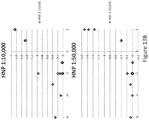
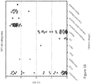
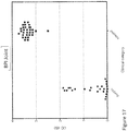
- the system is able to detect human neutrophil ⁇ defensin (also known as HNP 1-3) as a marker for periprosthetic joint infection with at least 90% sensitivity and specificity.
- the cutoff value (derived by ROC analysis) for HNP 1-3 as a marker is about 5000 ng/ml, preferably about 6000 ng/ml, preferably about 7000 ng/ml, preferably about 8000 ng/ml, preferably about 9000 ng/ml, preferably about 10000 ng/ml, most preferably about 7720 ng/ml.
- the cut-off range for HNP1-3 as a marker for periprosthetic joint infection with at least 90% sensitivity and specificity is about 1000 ng/ml -19000 ng/ml, preferably about 2000 ng/ml - 16000 ng/ml, preferably about 3000 ng/ml - 13000 ng/ml, most preferably about 3334 ng/ml - 10946 ng/ml.
- the present disclosure relates to immunoassays for assessing (e.g., detecting or quantifying) at least one biomarker of interest in a test sample, where the sensitivity of the immunoassay is improved relative to, specifically is from about three to about fifteen times, especially, from about two to about ten times, higher than, the sensitivity of conventional immunoassays known in the art.
- Controls with respect to the presence or absence of the biomarker or differential expression of the biomarker may either be normal (i.e., not infected or not-inflamed tissue, for example) or samples from known types or stages of infection or inflammation or other disease.
- comparison of the expression patterns of the sample to be tested with those of the controls can be used to diagnose the disease or disorder.
- the control or control group is used for purposes of establishing initial cutoffs for the systems and assay of the invention. Therefore, mere detection of a biomarker without the requirement of comparison to a control group can diagnose the disease or disorder in the joint.
- the disclosed system may be used for qualitative (yes/no answer); semi-quantitative (-/+/++/+++/++++) or quantitative answer.
- Changes in the levels of expression of the biomarker are associated with pathogenesis.
- changes in the expression levels of particular biomarkers serve as signposts for the presence of and progression of infection in the joint.
- prosthetic joint infection is higher after a revision arthroplasty which may be due to the long operation time, scar formation, or recrudescence of unrecognized infection present at the initial surgery.
- antibiotic treatment may mean removing the implant outright, and cleaning the implant and infected area before replacing the joint, which is costly, both in terms of expenses, time and the patient's condition.
- the procedure involves a surgical incision, drainage of the area, hardware removal and debridement of all devitalized tissue in conjunction with long term bed rest, pharmacological treatment followed by replacement of the joint.
- detection of a marker in a sample identifies a subject from which the sample was obtained, as having or not having a particular pathology.
- the invention provides the ability to detect a marker in a synovial fluid sample, wherein detection of the marker identifies whether inflammation in the joint of the subject is caused by an infection not.
- the disclosure provides a system for quickly diagnosing whether the inflammation in the joint is caused by an infection or not. Determination of the source of inflammation enables the physician to apply the appropriate therapy to ameliorate the inflammation. For example, if the subject has a bacterial infection in the joint, the patient is treated with anti-bacterial. Alternatively, if the diagnosis indicates that the inflammation is not caused by a bacterial infection, the physician can apply the appropriate type of therapy to treat aseptic inflammation.
- the concentration of the biomarker in a sample may be determined by any suitable assay.
- a suitable assay may include one or more of the following methods, an enzyme assay, an immunoassay, mass spectrometry, chromatography, electrophoresis or an antibody microarray, or any combination thereof.
- the system and methods of the invention may include any method known in the art to detect a biomarker in a sample.
- the disclosure also relates to methods for a multiplex analysis platform.
- the method comprises an analytical method for multiplexing analytical measurements of markers.
- the method comprises a set of compatible analytical strategies for multiplex measurements of markers and/or metabolites in synovial fluid.
- the sample of the invention is a biological sample, specifically synovial fluid.
- the disclosed systems and methods can be performed in the form of various immunoassay formats, which are well known in the art.
- Immunoassays in their most simple and direct sense, are binding assays involving binding between antibodies and antigen. Many types and formats of immunoassays are known and all are suitable for detecting the disclosed biomarkers.
- immunoassays are enzyme linked immunosorbent assays (ELISAs), enzyme linked immunospot assay (ELISPOT), radioimmunoassays (RIA), radioimmune precipitation assays (RIP A), immunobead capture assays, Western blotting, dot blotting, gel-shift assays, Flow cytometry, protein arrays, multiplexed bead arrays, magnetic capture, in vivo imaging, fluorescence resonance energy transfer (FRET), fluorescence recovery/localization after photobleaching (FRAP/FLAP), a sandwich assay, a competitive assay, an immunoassay using a biosensor, an immunoprecipitation assay, an agglutination assay, a turbidity assay, a nephelometric assay, etc.
- ELISAs enzyme linked immunosorbent assays
- ELISPOT enzyme linked immunospot assay
- RIA radioimmunoassays
- RIP A radioimm
- immunoassays involve contacting a sample suspected of containing a molecule of interest (such as the disclosed biomarkers) with an antibody to the molecule of interest or contacting an antibody to a molecule of interest (such as antibodies to the disclosed biomarkers) with a molecule that can be bound by the antibody, as the case may be, under conditions effective to allow the formation of immunocomplexes.
- a molecule of interest such as the disclosed biomarkers
- an antibody to a molecule of interest such as antibodies to the disclosed biomarkers
- the sample-antibody composition such as a tissue section, ELISA plate, dot blot or Western blot, can then be washed to remove any non-specifically bound antibody species, allowing only those antibodies specifically bound within the primary immune complexes to be detected.
- Immunoassays can include methods for detecting or quantifying the amount of a molecule of interest (such as the disclosed biomarkers or their antibodies) in a sample, which methods generally involve the detection or quantitation of any immune complexes formed during the binding process.
- a molecule of interest such as the disclosed biomarkers or their antibodies
- the detection of immunocomplex formation is well known in the art and can be achieved through the application of numerous approaches. These methods are generally based upon the detection of a label or marker, such as any radioactive, fluorescent, biological or enzymatic tags or any other known label. See, for example, U.S. Pat. Nos. 3,817,837 ; 3,850,752 ; 3,939,350 ; 3,996,345 ; 4,277,437 ; 4,275,149 and 4,366,241 for teachings regarding immunodetection methods and labels.
- a label can include a fluorescent dye, a member of a binding pair, such as biotin/streptavidin, a metal (e.g., gold), or an epitope tag that can specifically interact with a molecule that can be detected, such as by producing a colored substrate or fluorescence.
- a fluorescent dye also known herein as fluorochromes and fluorophores
- enzymes that react with colorometric substrates (e.g., horseradish peroxidase).
- colorometric substrates e.g., horseradish peroxidase
- each antigen can be labeled with a distinct fluorescent compound for simultaneous detection. Labeled spots on the array are detected using a fluorimeter, the presence of a signal indicating an antigen bound to a specific antibody.
- Fluorophores are compounds or molecules that luminesce. Typically fluorophores absorb electromagnetic energy at one wavelength and emit electromagnetic energy at a second wavelength.
- immunoassays There are two main types of immunoassays, homogeneous and heterogeneous. In homogeneous immunoassays, both the immunological reaction between an antigen and an antibody and the detection are carried out in a homogeneous reaction. Heterogeneous immunoassays include at least one separation step, which allows the differentiation of reaction products from unreacted reagents. A variety of immunoassays can be used to detect one or more of the proteins disclosed herein.
- ELISA is a heterogeneous immunoassay, which can be used in the methods disclosed herein.
- the assay can be used to detect protein antigens in various formats. In the "sandwich" format the antigen being assayed is held between two different antibodies.
- a solid surface is first coated with a solid phase antibody.
- the test sample, containing the antigen (e.g., a diagnostic protein), or a composition containing the antigen, such as a synovial fluid sample from a subject of interest, is then added and the antigen is allowed to react with the bound antibody. Any unbound antigen is washed away. A known amount of enzyme-labeled antibody is then allowed to react with the bound antigen.
- Any excess unbound enzyme-linked antibody is washed away after the reaction.
- the substrate for the enzyme used in the assay is then added and the reaction between the substrate and the enzyme produces a color change.
- the amount of visual color change is a direct measurement of specific enzyme-conjugated bound antibody, and consequently the antigen present in the sample tested.
- ELISA can also be used as a competitive assay.
- the test specimen containing the antigen to be determined is mixed with a precise amount of enzyme-labeled antigen and both compete for binding to an anti-antigen antibody attached to a solid surface. Excess free enzyme-labeled antigen is washed off before the substrate for the enzyme is added. The amount of color intensity resulting from the enzyme-substrate interaction is a measure of the amount of antigen in the sample tested.
- a heterogeneous immunoassay such as an ELISA, can be used to detect any of the proteins disclosed herein.
- Homogeneous immunoassays include, for example, the Enzyme Multiplied Immunoassay Technique (EMIT), which typically includes a biological sample comprising the biomarkers to be measured, enzyme-labeled molecules of the biomarkers to be measured, specific antibody or antibodies binding the biomarkers to be measured, and a specific enzyme chromogenic substrate.
- EMIT Enzyme Multiplied Immunoassay Technique
- excess of specific antibodies is added to a biological sample. If the biological sample contains the proteins to be detected, such proteins bind to the antibodies. A measured amount of the corresponding enzyme-labeled proteins is then added to the mixture. Antibody binding sites not occupied by molecules of the protein in the sample are occupied with molecules of the added enzyme-labeled protein.
- enzyme activity is reduced because only free enzyme-labeled protein can act on the substrate.
- the amount of substrate converted from a colorless to a colored form determines the amount of free enzyme left in the mixture.
- a high concentration of the protein to be detected in the sample causes higher absorbance readings. Less protein in the sample results in less enzyme activity and consequently lower absorbance readings.
- EMIT Inactivation of the enzyme label when the antigen-enzyme complex is antibody-bound makes the EMIT a useful system, enabling the test to be performed without a separation of bound from unbound compounds as is necessary with other immunoassay methods.
- a homogenous immunoassay such as an EMIT, can be used to detect any of the proteins disclosed herein.
- detection of antigen is made with the use of antigens specific antibodies as detector molecules.
- immunoassays and the systems and methods of the present invention are not limited to the use of antibodies as detector molecules. Any substance that can bind or capture the antigen within a given sample may be used. Aside from antibodies, suitable substances that can also be used as detector molecules include but are not limited to enzymes, peptides, proteins, and nucleic acids. Further, there are many detection methods known in the art in which the captured antigen may be detected. In some assays, enzyme-linked antibodies produce a color change. In other assays, detection of the captured antigen is made through detecting fluorescent, luminescent, chemiluminescent, or radioactive signals. The system and methods of the disclosure is not limited to the particular types of detectable signals produced in an immunoassay.
- Immunoassay kits are also included in the disclosure. These kits include, in separate containers (a) monoclonal antibodies having binding specificity for the polypeptides used in the diagnosis of inflammation or the source of inflammation; and (b) and anti-antibody immunoglobulins.
- This immunoassay kit may be utilized for the practice of the various methods provided herein.
- the monoclonal antibodies and the anti-antibody immunoglobulins can be provided in an amount of about 0.001 mg to 100 grams, and more preferably about 0.01 mg to 1 gram.
- the anti-antibody immunoglobulin may be a polyclonal immunoglobulin, protein A or protein G or functional fragments thereof, which may be labeled prior to use by methods known in the art.
- the immunoassay kit includes two, three or four of: antibodies that specifically bind a protein herein.
- the immunoassay kit of the disclosure can comprise (a) a sample pad, (b) a conjugated label pad, the conjugated label pad having a detectable label, a portion of the conjugated label pad and a portion of the sample pad forming a first interface, (c) a lateral-flow assay comprising a membrane, a portion of the membrane and a portion of the conjugated label pad forming a second interface, and (d) at least one antibody bound to the membrane, the first interface allowing fluid to flow from the sample pad to the conjugated label pad and contact the detectable label wherein the biomarker present in the sample forms an biomarker- conjugated label complex, the second interface allowing fluid to flow from the conjugated label pad to the membrane and to contact the at least one membrane-bound antibody to form to an biomarker-antibody complex and cause the detectable label to form a detectable signal.
- the biomarkers of the disclosure are detected using biosensors, e.g. with sensor systems with amperometric, electrochemical, potentiometric, conductimetric, impedance, magnetic, optical, acoustic or thermal transducers.
- biosensors include a biosensor recognition element which can include proteins, nucleic acids, antibodies, etc. that bind to a particular biomarker and a transducer which converts a molecular signal (i.e. binding of biomarker to recognition element) into an electric or digital signal that can be quantified, displayed, and analyzed.
- Biosensors may also include a reader device which translates the signal into a user- friendly display of the results. Examples of potential components that comprise an exemplary biosensor are described in Bohunicky et al. (2011, Nanotechnology Science and Applications, 4: 1-10 ).
- a biosensor may incorporate a physical, chemical or biological detection system.
- a biosensor is a sensor with a biological recognition system, e.g. based on a nucleic acid, such as an oligonucleotide probe or aptamer, or a protein such as an enzyme, binding protein, receptor protein, transporter protein or antibody.
- the biological recognition system may comprise traditional immunoassays described elsewhere herein.
- the recognition element e.g. protein, nucleic acid, antibody, etc.
- the recognition element may be unlabeled and binding of the biomarker to the element is directly observed and converted into a signal by the transducer.
- the method for detection of the biomarker in a biosensor may comprise immunological, electrical, thermal, magnetic, optical (e.g. hologram) or acoustic technologies. Using such biosensors, it is possible to detect the target biomarker at the anticipated concentrations found in biological samples.
- the biosensor may incorporate detection methods and systems as described herein for detection of the biomarker.
- Biosensors may employ electrical (e.g. amperometric, potentiometric, conductimetric, or impedance detection systems), calorimetric (e.g. thermal), magnetic, optical (e.g. hologram, luminescence, fluorescence, colorimetry), or mass change (e.g. piezoelectric, acoustic wave) technologies.
- the level of one, two, three, or more biomarkers can be detected by one or more methods selected from: direct, indirect or coupled enzymatic, spectrophotometric, fluorimetric, luminometric, spectrometric, polarimetric and chromatographic techniques.
- biosensors comprise one or more enzymes used directly or indirectly via a mediator, or using a binding, receptor or transporter protein, coupled to an electrical, optical, acoustic, magnetic or thermal transducer. Using such biosensors, it is possible to detect the level of target biomarkers at the anticipated concentrations found in biological samples.
- a biomarker in one embodiment, can be detected using a biosensor incorporating technologies based on "smart" holograms, or high frequency acoustic systems, such systems are particularly amenable to "bar code” or array configurations.
- smart hologram sensors Smart Holograms Ltd, Cambridge, UK
- a holographic image is stored in a thin polymer film that is sensitized to react specifically with the biomarker.
- the biomarker reacts with the polymer leading to an alteration in the image displayed by the hologram.
- the test result read-out can be a change in the optical brightness, image, color and/or position of the image.
- a sensor hologram can be read by eye, thus removing the need for detection equipment.
- a simple color sensor can be used to read the signal when quantitative measurements are required. Opacity or color of the sample does not interfere with operation of the sensor.
- the format of the sensor allows multiplexing for simultaneous detection of several substances. Reversible and irreversible sensors can be designed to meet different requirements, and continuous monitoring of a particular biomarker of interest is feasible.
- Biosensors to detect the biomarker of the disclosure may include acoustic, surface plasmon resonance, holographic and microengineered sensors. Imprinted recognition elements, thin film transistor technology, magnetic acoustic resonator devices and other novel acousto-electrical systems may be employed in biosensors for detection of the biomarkers of the disclosure.
- biosensors for detection of the biomarker of the disclosure are coupled, i.e. they combine biomolecular recognition with appropriate means to convert detection of the presence, or quantitation, of the biomarker in the sample into a signal.
- Biosensors can be adapted for "alternate site” diagnostic testing, e.g. in the ward, outpatients' department, surgery, home, field and workplace.
- Methods involving detection and/or quantification of the biomarker of the disclosure can be performed using bench-top instruments, or can be incorporated onto disposable, diagnostic or monitoring platforms that can be used in a non-laboratory environment, e.g. in the physician's office or at the patient's bedside.
- the systems and methods of the disclosure can be performed in the form of various mass spectrometry (MS) or chromatography formats, which are well known in the art.
- MS mass spectrometry
- the levels of biomarkers present in a sample can be determined by mass spectrometry.
- any mass spectrometric techniques that can obtain precise information on the mass of peptides, and preferably also on fragmentation and/or (partial) amino acid sequence of selected peptides, are useful herein.
- Suitable peptide MS techniques and systems are well-known per se (see, e.g., Methods in Molecular Biology, vol.
- mass spectrometry or “MS” as used herein refer to methods of filtering, detecting, and measuring ions based on their mass-to-charge ratio, or "m/z.”
- mass spectrometry or “MS” as used herein refer to methods of filtering, detecting, and measuring ions based on their mass-to-charge ratio, or "m/z.”
- MS mass-to-charge ratio
- one or more molecules of interest are ionized, and the ions are subsequently introduced into a mass spectrographic instrument where, due to a combination of magnetic and electric fields, the ions follow a path in space that is dependent upon mass (“m”) and charge (“z”).
- m mass-to-charge ratio
- Atmospheric Pressure Chemical Ionization Chemical Ionization
- Chemical Ionization Chemical Ionization
- EI Electron Impact
- EI Electrospray Ionization
- FAB Fast Atom Bombardment
- FD/FI Field Desorption/Field Ionization
- MALDI Matrix Assisted Laser Desorption Ionization
- TSP Thermospray Ionization
- the levels of biomarkers present in a sample can be determined by MS such as matrix-assisted laser desorption/ionization time-of-flight (MALDI-TOF) MS; MALDI-TOF post-source-decay (PSD); MALDI-TOF/TOF; surface-enhanced laser desorption/ionization time-of-flight mass spectrometry (SELDI-TOF) MS; tandem mass spectrometry (e.g., MS/MS, MS/MS/MS etc.); electrospray ionization mass spectrometry (ESI-MS); ESI-MS/MS; ESI-MS/(MS)n (n is an integer greater than zero); ESI 3D or linear (2D) ion trap MS; ESI triple quadrupole MS; ESI quadrupole orthogonal TOF (Q-TOF); ESI Fourier transform MS systems; desorption/ionization on silicon (DIOS); secondary ion mass spectrometry (S
- Peptide ion fragmentation in tandem MS (MS/MS) arrangements may be achieved using manners established in the art, such as, e.g., collision induced dissociation (CID). See for example, U.S. Patent Application Nos: 20030199001 , 20030134304 , 20030077616 .
- CID collision induced dissociation
- Such techniques may be used for relative and absolute quantification and also to assess the ratio of the biomarker according to the invention with other biomarkers that may be present. These methods are also suitable for clinical screening, prognosis, monitoring the results of therapy, identifying patients most likely to respond to a particular therapeutic treatment, for drug screening and development, and identification of new targets for drug treatment.
- a gas phase ion spectrophotometer is used.
- laser-desorption/ionization mass spectrometry is used to analyze the sample.
- Modern laser desorption/ionization mass spectrometry (“LDI-MS”) can be practiced in two main variations: matrix assisted laser desorption/ionization (“MALDI”) mass spectrometry and surface-enhanced laser desorption/ionization (“SELDI”).
- MALDI matrix assisted laser desorption/ionization
- SELDI surface-enhanced laser desorption/ionization
- MALDI matrix assisted laser desorption/ionization
- the analyte is mixed with a solution containing a matrix, and a drop of the liquid is placed on the surface of a substrate.
- the matrix solution then co-crystallizes with the biological molecules.
- the substrate is inserted into the mass spectrometer.
- Laser energy is directed to the substrate surface where it desorbs and ionizes the biological molecules without significantly fragmenting them. See, e.g., U.S. Pat. No. 5, 118,937 , and U.S. Pat. No. 5,045,694 .
- the substrate surface is modified so that it is an active participant in the desorption process.
- the surface is derivatized with adsorbent and/or capture reagents that selectively bind the biomarker of interest.
- the surface is derivatized with energy absorbing molecules that are not desorbed when struck with the laser.
- the surface is derivatized with molecules that bind the protein of interest and that contain a photolytic bond that is broken upon application of the laser.
- SELDI is a powerful tool for identifying a characteristic "fingerprint" of proteins and peptides in body fluids and tissues for a given condition, e.g. drug treatments and diseases.
- This technology utilizes protein chips to capture proteins/peptides and a time-of-flight mass spectrometer (tof-MS) to quantitate and calculate the mass of compounds ranging from small molecules and peptides of less than 1,000 Da up to proteins of 500 kDa.
- Quantifiable differences in protein/peptide patterns can be statistically evaluated using automated computer programs which represent each protein/peptide measured in the biofluid spectrum as a coordinate in multi-dimensional space.
- the SELDI system also has a capability of running hundreds of samples in a single experiment.
- all the signals from SELDI mass spectrometry are derived from native proteins/peptides (unlike some other proteomics technologies which require protease digestion), thus directly reflecting the underlying physiology of a given condition.
- the derivatizing agent generally is localized to a specific location on the substrate surface where the sample is applied. See, e.g., U.S. Pat. No. 5,719,060 and WO 98/59361 .
- the two methods can be combined by, for example, using a SELDI affinity surface to capture an analyte and adding matrix-containing liquid to the captured analyte to provide the energy absorbing material.
- mass spectrometers see, e.g., Principles of Instrumental Analysis, 3rd edition., Skoog, Saunders College Publishing, Philadelphia, 1985 ; and Kirk-Othmer Encyclopedia of Chemical Technology, 4th ed. Vol.
- Detection and quantification of the biomarker will typically depend on the detection of signal intensity. For example, in certain embodiments, the signal strength of peak values from spectra of a first sample and a second sample can be compared (e.g., visually, by computer analysis etc.), to determine the relative amounts of particular biomarker.
- Software programs such as the Biomarker Wizard program (Ciphergen Biosystems, Inc., Fremont, Calif.) can be used to aid in analyzing mass spectra. The mass spectrometers and their techniques are well known to those of skill in the art.
- detection and quantification of biomarkers by mass spectrometry may involve multiple reaction monitoring (MRM), such as described among others by Kuhn et al. 2004 (Proteomics 4: 1175-86 ).
- MRM multiple reaction monitoring
- MS peptide analysis methods may be advantageously combined with upstream peptide or protein separation or fractionation methods, such as for example with the chromatographic and other methods described herein below.
- Chromatography can also be used for measuring biomarkers.
- the term "chromatography” encompasses methods for separating chemical substances, referred to as such and vastly available in the art.
- chromatography refers to a process in which a mixture of chemical substances (analytes) carried by a moving stream of liquid or gas ("mobile phase") is separated into components as a result of differential distribution of the analytes, as they flow around or over a stationary liquid or solid phase (“stationary phase”), between said mobile phase and said stationary phase.
- the stationary phase may be usually a finely divided solid, a sheet of filter material, or a thin film of a liquid on the surface of a solid, or the like.
- Chromatography is also widely applicable for the separation of chemical compounds of biological origin, such as, e.g., amino acids, proteins, fragments of proteins or peptides, etc.
- Chromatography as used herein may be preferably columnar (i.e., wherein the stationary phase is deposited or packed in a column), preferably liquid chromatography, and yet more preferably high-performance liquid chromatograph7 (HPLC). While particulars of chromatography are well known in the art, for further guidance see, e.g., Meyer M., 1998, ISBN: 047198373X, and " Practical HPLC Methodology and Applications", Bidlingmeyer, B. A., John Wiley & Sons Inc., 1993 .
- Exemplary types of chromatography include, without limitation, HPLC, normal phase HPLC (NP-HPLC), reversed phase HPLC (RP-HPLC), ion exchange chromatography (IEC), such as cation or anion exchange chromatography, hydrophilic interaction chromatography (HILIC), hydrophobic interaction chromatography (HIC), size exclusion chromatography (SEC) including gel filtration chromatography or gel permeation chromatography, chromatofocusing, affinity chromatography such as immuno-affinity, immobilized metal affinity chromatography, and the like.
- chromatography including single-, two- or more-dimensional chromatography, may be used as a peptide fractionation method in conjunction with a further peptide analysis method, such as for example, with a downstream mass spectrometry analysis as described elsewhere in this specification.
- peptide or polypeptide separation, identification or quantification methods may be used, optionally in conjunction with any of the above described analysis methods, for measuring biomarkers in the present disclosure.
- Such methods include, without limitation, chemical extraction partitioning, isoelectric focusing (IEF) including capillary isoelectric focusing (CIEF), capillary isotachophoresis (CITP), capillary electrochromatography (CEC), and the like, one-dimensional polyacrylamide gel electrophoresis (PAGE), two-dimensional polyacrylamide gel electrophoresis (2D-PAGE), capillary gel electrophoresis (CGE), capillary zone electrophoresis (CZE), micellar electrokinetic chromatography (MEKC), free flow electrophoresis (FFE), etc.
- IEF isoelectric focusing
- CITP capillary isotachophoresis
- CEC capillary electrochromatography
- PAGE polyacrylamide gel electrophoresis
- 2D-PAGE two-dimensional polyacrylamide gel electrophore
- Point-of-use analytical tests have been developed for the routine identification or monitoring of health-related conditions (such as pregnancy, cancer, endocrine disorders, infectious diseases or drug abuse) using a variety of biological samples (such as urine, serum, plasma, blood, saliva).
- Some of the point-of-use assays are based on highly specific interactions between specific binding pairs, such as antigen/antibody, hapten/antibody, lectin/carbohydrate, apoprotein/cofactor and biotin/(strept)avidin.
- assays are performed with test strips in which a specific binding pair member is attached to a mobilizable material (such as a metal sol or beads made of latex or glass) or an immobile substrate (such as glass fibers, cellulose strips or nitrocellulose membranes).
- a mobilizable material such as a metal sol or beads made of latex or glass
- an immobile substrate such as glass fibers, cellulose strips or nitrocellulose membranes.
- Other point-of use devices may comprise optical biosensors, photometric biosensors, electrochemical biosensor, or other types of biosensors.
- Suitable biosensors in point-of-use devices for performing methods of the invention include "cards" or "chips” with optical or acoustic readers. Biosensors can be configured to allow the data collected to be electronically transmitted to the physician for interpretation and thus can form the basis for e-medicine, where diagnosis and monitoring can be done without the need for the patient to be in proximity to a physician or a clinic.
- Detection of a biomarker in a synovial fluid can be carried out using a sample capture device, such as a lateral flow device (for example a lateral flow test strip) that allows detection of one or more biomarkers, such as those described herein.
- a sample capture device such as a lateral flow device (for example a lateral flow test strip) that allows detection of one or more biomarkers, such as those described herein.
- the test strips of the present disclosure include a flow path from an upstream sample application area to a test site.
- the flow path can be from a sample application area through a mobilization zone to a capture zone.
- the mobilization zone may contain a mobilizable marker that interacts with an analyte or analyte analog
- the capture zone contains a reagent that binds the analyte or analyte analog to detect the presence of an analyte in the sample.
- U.S. Pat. No. 6,656,744 discloses a lateral flow test strip in which a label binds to an antibody through a streptavidin-biotin interaction.
- Flow-through type assay devices were designed, in part, to obviate the need for incubation and washing steps associated with dipstick assays.
- Flow-through immunoassay devices involve a capture reagent (such as one or more antibodies) bound to a porous membrane or filter to which a liquid sample is added.
- target analyte such as protein
- detector reagent such as labeled antibody (e.g., gold-conjugated or colored latex particle-conjugated protein).
- the detector reagent may be placed on the membrane in a manner that permits the detector to mix with the sample and thereby label the analyte.
- the visual detection of detector reagent provides an indication of the presence of target analyte in the sample.
- Representative flow-through assay devices are described in U.S. Pat. Nos. 4,246,339 ; 4,277,560 ; 4,632,901 ; 4,812,293 ; 4,920,046 ; and 5,279,935 ; U.S. Patent Application Publication Nos. 20030049857 and 20040241876 ; and WO 08/030,546 .
- Migration assay devices usually incorporate within them reagents that have been attached to colored labels, thereby permitting visible detection of the assay results without addition of further substances. See, for example, U.S. Pat. No. 4,770,853 ; PCT Publication No. WO 88/08534 .
- U.S. Pat. No. 5,229,073 describes a semiquantitative competitive immunoassay lateral flow method for measuring plasma lipoprotein levels. This method utilizes a plurality of capture zones or lines containing immobilized antibodies to bind both the labeled and free lipoprotein to give a semi-quantitative result.
- U.S. Pat. No. 5,591,645 provides a chromatographic test strip with at least two portions. The first portion includes a movable tracer and the second portion includes an immobilized binder capable of binding to the analyte.
- Devices described herein generally include a strip of absorbent material (such as a microporous membrane), which, in some instances, can be made of different substances each joined to the other in zones, which may be abutted and/or overlapped.
- the absorbent strip can be fixed on a supporting non-interactive material (such as nonwoven polyester), for example, to provide increased rigidity to the strip.
- Zones within each strip may differentially contain the specific binding partner(s) and/or other reagents required for the detection and/or quantification of the particular analyte being tested for, for example, one or more proteins disclosed herein. Thus these zones can be viewed as functional sectors or functional regions within the test device.
- a fluid sample is introduced to the strip at the proximal end of the strip, for instance by dipping or spotting.
- a sample is collected or obtained using methods well known to those skilled in the art.
- the sample containing the particular proteins to be detected may be obtained from any biological source.
- the biological source is synovial fluid.
- the sample may be diluted, purified, concentrated, filtered, dissolved, suspended or otherwise manipulated prior to assay to optimize the immunoassay results.
- the fluid migrates distally through all the functional regions of the strip. The final distribution of the fluid in the individual functional regions depends on the adsorptive capacity and the dimensions of the materials used.
- porous solid supports such as nitrocellulose, described elsewhere herein are preferably in the form of sheets or strips.
- the thickness of such sheets or strips may vary within wide limits, for example, from about 0.01 to 0.5 mm, from about 0.02 to 0.45 mm, from about 0.05 to 0.3 mm, from about 0.075 to 0.25 mm, from about 0.1 to 0.2 mm, or from about 0.11 to 0.15 mm.
- the pore size of such sheets or strips may similarly vary within wide limits, for example from about 0.025 to 15 microns, or more specifically from about 0.1 to 3 microns; however, pore size is not intended to be a limiting factor in selection of the solid support.
- the flow rate of a solid support can also vary within wide limits, for example from about 12.5 to 90 sec/cm (i.e., 50 to 300 sec/4 cm), about 22.5 to 62.5 sec/cm (i.e., 90 to 250 sec/4 cm), about 25 to 62.5 sec/cm (i.e., 100 to 250 sec/4 cm), about 37.5 to 62.5 sec/cm (i.e., 150 to 250 sec/4 cm), or about 50 to 62.5 sec/cm (i.e., 200 to 250 sec/4 cm).
- a detector serves this purpose.
- a detector may be integrated into an assay device (for example includes in a conjugate pad), or may be applied to the device from an external source.
- a detector may be a single reagent or a series of reagents that collectively serve the detection purpose.
- a detector reagent is a labeled binding partner specific for the analyte (such as a gold-conjugated antibody for a particular protein of interest).
- a detector reagent collectively includes an unlabeled first binding partner specific for the analyte and a labeled second binding partner specific for the first binding partner and so forth.
- the detector can be a labeled antibody specific for a protein described herein.
- the detector can also be an unlabeled first antibody specific for the protein of interest and a labeled second antibody that specifically binds the unlabeled first antibody.
- a detector reagent specifically detects bound analyte of an analyte-capture reagent complex and, therefore, a detector reagent preferably does not substantially bind to or react with the capture reagent or other components localized in the analyte capture area.
- a detector reagent can specifically recognize a positive control molecule (such as a non-specific human IgG for a labeled Protein A detector, or a labeled Protein G detector, or a labeled anti-human Ab(Fc)) that is present in a secondary capture area.
- a positive control molecule such as a non-specific human IgG for a labeled Protein A detector, or a labeled Protein G detector, or a labeled anti-human Ab(Fc)
- a flow-through device involves a capture reagent (such as one or more antibodies) immobilized on a solid support, typically, microtiter plate or a membrane (such as, nitrocellulose, nylon, or PVDF).
- a capture reagent such as one or more antibodies
- a membrane such as, nitrocellulose, nylon, or PVDF.
- the membrane of a flow-through device is placed in functional or physical contact with an absorbent layer, which acts as a reservoir to draw a fluid sample through the membrane.
- an absorbent layer which acts as a reservoir to draw a fluid sample through the membrane.
- any remaining protein-binding sites on the membrane can be blocked (either before or concurrent with sample administration) to minimize nonspecific interactions.
- a fluid sample is placed in contact with the membrane.
- a flow-through device also includes a sample application area (or reservoir) to receive and temporarily retain a fluid sample of a desired volume.
- the sample passes through the membrane matrix.
- an analyte in the sample such as one or more protein, for example, one or more proteins described herein
- the immobilized capture reagent such as one or more antibodies.
- a detector reagent such as labeled antibodies that specifically bind one or more proteins
- a detector reagent can be added with the sample or a solution containing a detector reagent can be added subsequent to application of the sample.
- an analyte is specifically bound by capture reagent, a characteristic attributable to the particular detector reagent can be observed on the surface of the membrane.
- Optional wash steps can be added at any time in the process, for instance, following application of the sample, and/or following application of a detector reagent.
- a lateral flow device is an analytical device having as its essence a test strip, through which flows a test sample fluid that is suspected of containing an analyte of interest.
- the test fluid and any suspended analyte can flow along the strip to a detection zone in which the analyte (if present) interacts with a capture agent and a detection agent to indicate a presence, absence and/or quantity of the analyte.
- lateral flow devices are one-step lateral flow assays in which a biological fluid is placed in a sample area on a bibulous strip (though non-bibulous materials can be used, and rendered bibulous, e.g., by applying a surfactant to the material), and allowed to migrate along the strip until the liquid comes into contact with a specific binding partner (such as an antibody) that interacts with an analyte (such as one or more proteins) in the liquid. Once the analyte interacts with the binding partner, a signal (such as a fluorescent or otherwise visible dye) indicates that the interaction has occurred.
- a specific binding partner such as an antibody
- analyte such as one or more proteins
- test strips can also incorporate control indicators, which provide a signal that the test has adequately been performed, even if a positive signal indicating the presence (or absence) of an analyte is not seen on the strip.
- Lateral flow devices have a wide variety of physical formats that are equally well known in the art. Any physical format that supports and/or houses the basic components of a lateral flow device in the proper function relationship is contemplated by this disclosure.
- Figures 1 and 2 The basic components of a particular embodiment of a lateral flow device are illustrated in Figures 1 and 2 which comprise a sample pad, a conjugate pad, a migration membrane, and an absorbent pad.
- the sample pad (such as the sample pad in Figures 1 and 2 ) is a component of a lateral flow device that initially receives the sample, and may serve to remove particulates from the sample.
- a sample pad such as glass fiber, woven fibers, screen, nonwoven fibers, cellosic fibers or paper
- a cellulose sample pad may be beneficial if a large bed volume is a factor in a particular application.
- Sample pads may be treated with one or more release agents, such as buffers, salts, proteins, detergents, and surfactants.
- release agents may be useful, for example, to promote resolubilization of conjugate-pad constituents, and to block non-specific binding sites in other components of a lateral flow device, such as a nitrocellulose membrane.
- Representative release agents include, for example, trehalose or glucose (1%-5%), PVP or PVA (0.5%-2%), Tween 20 or Triton X-100 (0.1%-1%), casein (1%-2%), SDS (0.02%-5%), and PEG (0.02%-5%).
- the types of membranes useful in a lateral flow device include but are not limited to nitrocellulose (including pure nitrocellulose and modified nitrocellulose) and nitrocellulose direct cast on polyester support, polyvinylidene fluoride, or nylon).
- the conjugate pad (such as conjugate pad in Figures 1 and 2 ) serves to, among other things, hold a detector reagent.
- Suitable materials for the conjugate pad include glass fiber, polyester, paper, or surface modified polypropylene.
- Detector reagent(s) contained in a conjugate pad is typically released into solution upon application of the test sample.
- a conjugate pad may be treated with various substances to influence release of the detector reagent into solution.
- the conjugate pad may be treated with PVA or PVP (0.5% to 2%) and/or Triton X-100 (0.5%).
- Other release agents include, without limitation, hydroxypropylmethyl cellulose, SDS, Brij and ⁇ -lactose. A mixture of two or more release agents may be used in any given application.
- an absorbent pad acts to increase the total volume of sample that enters the device. This increased volume can be useful, for example, to wash away unbound analyte from the membrane.
- Any of a variety of materials is useful to prepare an absorbent pad, for example, cellulosic filters or paper.
- an absorbent pad can be paper (i.e., cellulosic fibers).
- One of skill in the art may select a paper absorbent pad on the basis of, for example, its thickness, compressibility, manufacturability, and uniformity of bed volume. The volume uptake of an absorbent made may be adjusted by changing the dimensions (usually the length) of an absorbent pad.
- a fluid sample containing an analyte of interest such as one or more proteins described herein, is applied to the sample pad.
- the sample may be applied to the sample pad by dipping the end of the device containing the sample pad into the sample (such as synovial) or by applying the sample directly onto the sample pad.
- the sample passes, for instance by capillary action, to the conjugate pad.
- the analyte of interest such as a protein of interest
- a conjugate pad may bind (or be bound by) a mobilized or mobilizable detector reagent, such as an antibody (such as antibody that recognizes one or more of the proteins described herein).
- a protein analyte may bind to a labeled (e.g., gold-conjugated or colored latex particle-conjugated) antibody contained in the conjugate pad.
- the analyte complexed with the detector reagent may subsequently flow to the test line where the complex may further interact with an analyte-specific binding partner (such as an antibody that binds a particular protein, an anti-hapten antibody, or streptavidin), which is immobilized at the proximal test line.
- an analyte-specific binding partner such as an antibody that binds a particular protein, an anti-hapten antibody, or streptavidin
- a protein complexed with a detector reagent such as gold-conjugated antibody
- the formation of a complex which results from the accumulation of the label (e.g., gold or colored latex) in the localized region of the proximal test line, is detected.
- the control line may contain an immobilized, detector-reagent-specific binding partner, which can bind the detector reagent in the presence or absence of the analyte. Such binding at the control line indicates proper performance of the test, even in the absence of the analyte of interest.
- control line detects the presence of hyaluronic acid which is a marker for synovial fluid in order to ensure that the test sample is indeed synovial fluid. Therefore, in this situation, the control line is formed by using antibodies to hyaluronic acid ( Figure 2 ). If the sample is obtained from anything other than the joint (i.e. blood), no line is formed and the test is deemed to be invalid.
- test results may be visualized directly, or may be measured using a reader (such as a scanner).
- the reader device may detect color, fluorescence, luminescence, radioactivity, or any other detectable marker derived from the labeled reagent from the readout area (for example, the test line and/or control line).
- test line there may be a second (or third, fourth, or more) test line located parallel or perpendicular (or in any other spatial relationship) to test line in test result zone (for example test lines to biomarker 1 and biomarker 2 in Figure 2 ).
- test result zone for example test lines to biomarker 1 and biomarker 2 in Figure 2 .
- a second detector reagent specific for a second analyte such as another antibody
- the second test line will contain a second specific binding partner having affinity for a second analyte, such as a second protein in the sample.
- the test line will contain a third (or more) specific binding partner having affinity for a third (or more) analyte.
- a comparison of the control line to the test line yields the test result from the disclosed diagnostic system.
- a valid result occurs when the control fine is detected at a higher intensity level than the test line.
- a valid result occurs when the control line is at least 5% or more, for example, 10%, 20%, 30%, 40%, 50%, 60%, 70%, 80%, 90%, 100% or more darker than the test line.
- a valid result occurs when the control line is at least 0.5 fold or more, for example, 1 fold, 2 fold, 3 fold, 4 fold, 5 fold, 6 fold, 7 fold, 8 fold, 9 fold, 10 fold or more darker than the test line.
- control line is a reference line that insures that the test has been run correctly and that the tested sample is not obtained from anything other than the joint (i.e. blood).
- the disclosed system is useful in the diagnosis of infection in a joint when the control line is detected at an at least equal intensity than the test line.
- the control line is detected at higher intensity than the test line. In some instances, if the test fine is not at least equal in darkness or intensity as the control line then the test is said to have an invalid result. If the test line is at least equal or lighter than the control line then the test is said to have a valid result.
- control line is a reference line that detects HA, wherein HA is a valid marker for distinguishing synovial fluid from other bodily fluids (e.g., blood).
- HA is detected by using aggrecan to detect the difference between blood and synovial fluid. This is because aggrecan can effectively bind to HA.
- the systems are designed for use on site at the point of care, where patients are examined and tested, as well as for operation remote from the site.
- the systems are designed to accept input in the form of patient data, including, but not limited to biochemical test data, physical test data, historical data and other such data, and to process and output information, such as data relating to a medical diagnosis or a disease risk indicator.
- patient data may be contained within the system, such as medical records or history, or may be input as a signal or image from a medical test or procedure, for example, immunoassay test data, blood pressure reading, ultrasound, X-ray or MRI, or introduced in any other form.
- Specific test data can be digitized, processed and input into the medical diagnosis expert system, where it may be integrated with other patient information.
- the output from the system is a disease risk index or medical diagnosis.
- Point of care testing refers to real time diagnostic testing that can be done in a rapid time frame so that the resulting test is performed faster than comparable tests that do not employ this system.
- the exemplified immunoassay disclosed and described herein can be performed in significantly less time than the corresponding ELISA assay, e.g., in less than half an hour.
- point of care testing refers to testing that can be performed rapidly and on site, such as in a doctor's office, at a bedside, in a stat laboratory, emergency room or other such locales, particularly where rapid and accurate results are required.
- a point of care diagnostic and risk assessment system includes a reader for reading patient data, a test device designed to be read in the reader, and software for analysis of the data.
- a test strip device in a plastic housing is designed for use with the reader, optionally including a symbology, such as an alphanumeric character bar code or other machine-readable code, and software designed for analysis of the data generated from the test strip are also provided.
- a reader refers to an instrument for detecting and/or quantitating data, such as on test strips. The data may be visible to the naked eye, but does not need to be visible. Such readers are disclosed and described in U.S. Pat. Nos. 6,267,722 , 6,394,952 and 6,867,051 .
- a reflectance reader refers to an instrument adapted to read a test strip using reflected light, including fluorescence, or electromagnetic radiation of any wavelength. Reflectance can be detected using a photodetector or other detector, such as charge coupled diodes (CCD).
- CCD charge coupled diodes
- An exemplary reflectance reader includes a cassette slot adapted to receive a test-strip, light-emitting diodes, optical fibers, a sensing head, including means for positioning the sensing head along the test strip, a control circuit to read the photodetector output and control the on and off operation of the light-emitting diodes, a memory circuit for storing raw and/or processed data, and a photodetector, such as a silicon photodiode detector.
- a color change refers to a change in intensity or hue of color or may be the appearance of color where no color existed or the disappearance of color.
- a sample is applied to a diagnostic immunoassay test strip, and colored or dark bands are produced.
- the intensity of the color reflected by the colored label in the test region (or detection zone) of the test strip is, for concentration ranges of interest, directly proportional or otherwise correlated with an amount of analyte present in the sample being tested.
- the color intensity produced is read, in accordance with the present embodiment, using a reader device, for example, a reflectance reader, adapted to read the test strip.
- the intensity of the color reflected by the colored label in the test region (or detection zone) of the test strip is directly proportional to the amount of analyte present in the sample being tested.
- a darker colored line in the test region indicates a greater amount of analyte
- a lighter colored line in the test region indicates a smaller amount of analyte.
- the color intensity produced i.e., the darkness or lightness of the colored line, is read using a reader device, for example, a reflectance reader, adapted to read the test strip.
- a reflectance measurement obtained by the reader device is correlated to the presence and/or quantity of analyte present in the sample.
- the reader takes a plurality of readings along the strip, and obtains data that are used to generate results that are an indication of the presence and/or quantity of analyte present in the sample.
- the system may correlate such data with the presence of a disorder, condition or risk thereof.
- the reader may (optionally) be adapted to read a symbology, such as a bar code, which is present on the test strip or housing and encodes information relating to the test strip device and/or test result and/or patient, and/or reagent or other desired information.
- a symbology such as a bar code
- the associated information is stored in a remote computer database, but can be manually stored.
- the symbology can be imprinted when the device is used and the information encoded therein.
- Example 1 System for detecting biomarkers in joint fluid
- joint fluid e.g., synovial fluid
- the system of the disclosure useful for analyzing joint fluid can be based on a lateral flow system.
- the lateral flow immunoassay utilizes strips of cellulose membrane onto which antibodies and other reagents can be coated. The sample moves along the strip due to capillary action and reacts with the reagents at different points along the strip. The end result is usually the appearance or absence of a colored line or spot ( Figure 1 ). Because a lateral flow assay for biomarkers can be in the form of a sandwich assay, the signal results from the formation of an immunocomplex on the membrane.
- lateral flow assay The advantages of a lateral flow assay include quick results following application of the sample, no instrumentation is required, able to execute the test and read the result in the field itself, and allows for room temperature storage.
- Biomarkers are proteins or other cellular components that relate specifically to injury or to disease and that can be found in body fluids such as synovial fluid.
- a biomarker designates any indicatory (nucleic acids, enzymes, metabolites and other types of molecules: histamines, hormones, proteins, etc.) present in the body as a biological response to disease.
- the identification and development of biomarkers make it possible to perform faster, more accurate disease diagnostics and help physicians prescribe new treatments for those patients who are likely to benefit from them. Tests bases on biomarkers often contribute to a faster cure and avoid time-consuming and costly analyses.
- synovial fluid provides an optimum medium for the use of biomarkers in diagnosing, for example, the source of joint pain because joint fluid is a "closed" system.
- concentrations of IL-6 in serum versus synovial fluid from patients with an infected knee were compared, a 1000x amplication in the synovial fluid was observed.
- the presence of a biomarker in synovial fluid is a result from a local response.
- control line is formed by using monoclonal antibodies to hyaluronic acid ( Figure 2 ). If the sample is from anything other than the joint (i.e. serum), no line is formed and the test is deemed invalid.
- HNP 1-3 is a biomarker for infection in joint, preferably a replacement joint or periprosthetic joint.
- Dilution buffer was prepared by mixing 10 ml of the 10x dilution buffer with 90 ml of deionized H 2 O. As per the manufactures suggestion, the buffer mixture was allowed to sit for 10 minutes and inspected for crystals/particulate before being used. To make the standard/sample buffer, 90 ml of this dilution buffer was mixed with 10 ml of plasma diluent.
- Synovial fluid and serum samples were diluted in sample buffer at both 1:100 and 1:1000. 100 ul of each standard or sample was then added to the assay plate as discussed elsewhere herein. The plate was covered and incubated at RT for 1 hour. The plate was then washed 3x using the biotek automated plate washer program "flat nunc plate” with in-house wash buffer of DPBS with 0.05% tween. This automated washer and wash buffer were used for all wash steps.
- HNP1-3 showed a clear separation between periprosthetic joint infection positive and negative samples ( Figure 12 ). These negative samples were sourced from multiple patient types and fluids, including serum, psuedogout, metal on metal (MOM), osteoarthritis (OA), and inflamed but not infected.
- MOM metal on metal
- OA osteoarthritis
- HNP1-3 is a clinically significant biomarker .
- the next set of experiments was designed to use an accurate standard curve in order to interpolate dose concentrations and determine cutoff points via ROC analysis. For example, experiments were performed to seek an optimal dilution at which to run synovial fluid samples with the Hycult HNP1-3 Elisa kit.
- the standard was reconstituted with 0.5 ml of di H 2 O, yielding a stock concentration of 46 ng/ml (this value was stated on the certificate of analysis provided with this lot of kit).
- 150 ul of the reconstituted standard was diluted with 540 ul standard diluent to produce the first point in the standard curve, 10,000 pg/ml. The remaining points were then prepared using 1:2 dilutions in sample buffer.
- the commercially available Human HNP1-3 ELISA kit from Hycult Biotech was used for determining human HNP 1-3 concentrations in synovial fluid. Experiments were performed to correlate HNP 1-3 levels with the presence of Periprosthetic Joint Infection (PJI) in synovial fluid synovial fluid from human patients.
- the human HNP 1-3 ELISA is a ready-to-use sandwich type, solid-phase, enzyme-linked immunosorbant assay. Samples, controls and standards were incubated in microplate wells coated with antibodies recognizing human HNP1-3. Biotinylated tracer antibodies provided with the kit bound to the captured human HNP1-3.
- Streptavidin-peroxidase conjugate then binds to the Biotinylated tracer antibody and reacted with the TMB substrate.
- the enzyme reaction was stopped by the addition of oxalic acid.
- the absorbance at 450nm was measured by a spectrophotometer.
- Human HNP1-3 concentrations in synovial fluid samples were measured by plotting the absorbance of a standard curve versus the corresponding concentrations of the human HNP1-3 standards.
- Table 10 Sample Cohort Descriptions Category Number of samples Description Classification Pseudo gout 2 Challenging native group -5 Gout 3 Challenging native group -4 Mom 1 Challenging Joint group -3 OA 15 Challenging native group -2 Aseptic 22 Negative cohort -1 Infected-culture positive 20 Positive cohort 1 Infected-culture nega tive 3 Positive cohort 2 Equivocal infected 3 Positive samples that lack sufficient results to make an unambiguous classification as infected. For information only and not included in final analysis 3 Native-infected 1 Non-joint positive group 4 Total 70
- foil pouch around the plate was vacuum-sealed and unpunctured. Any unused strips were immediately returned to the foil pouch containing the desiccant pack and resealed along the entire edge of the zip-seal. Quality guaranteed for 1 month if stored at 2-8°C.
- Synovial fluid samples were processed by centrifugation at 2,200 RPM for 10 minutes to remove cell debris and other contaminants. Samples are aliquoted and stored at -80°C until used in assay. Samples were thawed to room temperature and mixed gently before assay was initiated. Samples were diluted in 1.5ml polypropylene tubes using the dilution buffer supplied by Hycult kit HK317-02. The specified dilution was accomplished for each sample using a vortex to ensure dispersion of synovial fluid. Samples were not diluted according to product insert. Diluted samples were transferred to a round bottom polypropylene 96-well microplate in the order of the experimental plate layout.
- Wash buffer was prepared by mixing 20 ml of 40x wash buffer supplied with the HK317-02 kit and 780 ml of diH2O.
- Dilution buffer was prepared by mixing 20 ml of 10x dilution buffer with 180 ml of diH2O. No crystals were observed in the concentrated dilution buffer.
- Standard dilution buffer was prepared by mixing 10 ml of the 10x plasma diluent with 90 ml of the prepared dilution buffer.
- Standard solution was prepared by reconstituting lyophilized standard with 0.5 ml of diH20 to get a 46 ng/ml stock concentration.
- a standard curve was prepared in polypropylene tubes by a serial dilution of the reconstituted standard solution.
- Tracer solution was prepared by reconstituting lyophilized Biotinylated tracer antibody with 1 ml of diH20. One part reconstituted tracer was diluted with 11 parts of dilution buffer.
- Streptavidin-peroxidase solution was prepared by reconstituting the lyophilized conjugate with 1 ml of diH2O. Required volume of conjugate solution was prepared by diluting 1 part reconstituted conjugate with 23 parts of dilution buffer.
- Standard solution was prepared by reconstituting lyophilized standard with 0.5 ml of diH20 to get a 46 ng/ml stock concentration. Further dilutions were made using standard dilution buffer. A standard curve was prepared in polypropylene tubes using a 1:2 serial dilution of the reconstituted standard. The first standard dilution of the reconstituted standard was calculated to achieve a 10,000 pg/ml concentration.
- 100 ul in duplicate of standard, samples, and controls were transferred into appropriate wells, preferably without touching the side or bottom of the wells.
- the tray was covered and air bubbles were eliminated by tapping the tray without splashing the liquid onto the cover.
- the strips or plate were incubated for 1 hour at room temperature. Then the plates were washed with wash buffer. After washing, 100 ul of diluted tracer was added to each well with an incubation time of 1 hour at room temperature. The plates are then washed to remove the tracer solution. After washing, 100 ul of diluted streptavidin-peroxidase was added to each well with an incubation time of 1 hour at room temperature.
- streptavidin-peroxidase was removed by washing the plates following by the addition of 100 ul of TMB substrate to each well with an incubation time of 15 minutes at room temperature (avoiding exposing the micro well strips to direct sunlight). The reaction was stopped by adding 100 ul of stop solution and the plate was read within 30 minutes after addition of stop solution at 450 nm using a plate reader, following the instructions provided by the instrument's manufacturer.
- Human neutrophil alpha-defensins also called Human Neutrophil Peptides, HNP
- HNP Human Neutrophil Peptides
- HNP-1, HNP-2 and HNP-3 are encoded by two genes DEFA1 and DEFA3 localized to chromosome 8.
- DEFA1 and DEFA3 encode identical peptides except the conversion of the first amino acid from alanine in HNP-1 to aspartic acid in HNP-3; HNP-2 represent N-terminally truncated iso-form lacking the first amino acid.
- HNP Activation of neutrophils leads to rapid release of HNP.
- HNP can be measured in plasma during infection and inflammation. Micromolar concentrations of HNP are described in septic blood, while in normal plasma very low levels of HNP are present. The results presented herein correlate high HNP levels with septic synovial fluid.
- HNP 1-3 was found to be a desired biomarker for use in diagnosing periprosthetic joint infection. The ROC analysis of the data derived a cut-off at 7720 ng/ml. The results demonstrate an AUC of 1.0 with 100% specificity and 100% sensitivity with prosthetic joint samples (Table 11).
- Figure 14 demonstrates a clinical plot showing that HNP 1-3 can be a biomarker for periprosthetic joint infection in synovial fluid from joint samples.
- Figure 15 shows the results from samples tested including synovial fluid from native joints.
- Table 11 Assay performance data Biomarker AUC Cut-off (derived by ROC analysis on joint samples) Specificity Joint Sensitivity Joint Specificity ALL Sensitivity ALL HNP 1-3 1.000 7720 ng/mL 100.0 100.0 97.67 100.0
- the commercially available Human BPI ELISA kit from Hycult Biotech was used for determining human BPI concentrations in synovial fluid. Experiments were performed to correlate BPI levels with the presence of Periprosthetic Joint Infection (PJI) in synovial fluid synovial fluid from human patients.
- the human BPI ELISA is a ready-to-use sandwich type, solid-phase, enzyme-linked immunosorbant assay. Samples, controls and standards were incubated in microplate wells coated with antibodies recognizing human BPI. Biotinylated tracer antibodies provided with the kit bound to the captured human BPI. Streptavidin-peroxidase conjugate bound to the Biotinylated tracer antibody and was able to react with the TMB substrate.
- the enzyme reaction was stopped by the addition of oxalic acid.
- the absorbance at 450nm was measured by a spectrophotometer.
- Human BPI concentrations in synovial fluid samples were measured by plotting the absorbance of a standard curve versus the corresponding concentrations of the human BPI standards.
- Table 12 Sample Cohort Descriptions Category Number of samples Description Classification Unclassified 55 Insufficient data accompanied with sample to classify -7 Pseudo gout 3 Challenging native group -5 Gout 0 Challenging native group -4 Mom 1 Challenging Joint group -3 OA 3 Challenging native group -2 Aseptic 24 Negative cohort -1 Infected-culture positive 15 Positive cohort 1 Infected-culture negative 1 Positive cohort 2 Equivocal infected 8 Positive samples that lack sufficient results to make an unambiguous classification as infected. For information only and not included in final analysis 3 Native-infected 1 Non-joint positive group 4 Total 111
- Wash/dilution buffer B was prepared by mixing 20 ml of the 40x wash/dilution buffer A supplied with the HK314-02 kit with 380 ml of diH2O.
- Wash/Dilution buffer B was prepared by mixing 40 ml of 20x wash/dilution buffer B with 360 ml of diH2O.
- Final wash/dilution buffer was prepared by mixing buffer A and buffer B together.
- Standard solution was prepared by reconstituting lyophilized standard with 0.5 ml of diH20 to get a 219 ng/ml stock concentration.
- a standard curve was prepared in polypropylene tubes by a serial dilution of the reconstituted standard solution.
- Tracer solution was prepared by reconstituting lyophilized Biotinylated tracer antibody with 1 ml of diH20. 1 part reconstituted tracer was diluted with 11 parts of dilution buffer.
- Streptavidin-peroxidase solution was prepared by reconstituting the lyophilized conjugate with 1 ml of diH2O. Required volume of conjugate solution was prepared by diluting 1 part reconstituted conjugate with 23 parts of dilution buffer.
- Standard solution was prepared by reconstituting lyophilized standard with 0.5 ml of diH20 to get a 219 ng/ml stock concentration. Further dilutions were made using wash/dilution buffer.
- a standard curve was prepared in polypropylene tubes using a 1:2.5 serial dilution of the reconstituted standard.
- the first standard dilution of the reconstituted standard was calculated to achieve a 25,000 pg/ml concentration.
- the antimicrobial protein BPI Bacterial Permeability Increasing protein
- BPI Bacterial Permeability Increasing protein
- LPS lipopolysaccharides
- Binding of BPI to the lipid A moiety of LPS exerts multiple anti-infective activities against gram-negative bacteria: 1) cytotoxicity via sequential damage to bacterial outer and inner lipid membranes, 2) neutralization of gram-negative bacterial LPS, 3) opsonization of bacteria to enhance phagocytosis by neutrophils.
- Airway epithelial cells constitutively express the BPI gene and produce the BPI protein and, therefore, BPI may be a critical determinant in the development of LPS-triggered airway disease. Inflammation induced by LPS possibly contributes to the development of rapid airflow decline, a serious and often fatal complication of hematopoietic cell transplantation. In plasma of healthy individuals BPI is present at levels of ⁇ 0.5 ng/ml, which increases approximately 10-fold during acute phase responses.
- Millipore Sepsis Panel 3 (HSP3MAG-63k) was used to source all materials for this experiment. After allowing the kit contents to reach room temperature, the magnetic beads were prepared. Each individual bead vial was sonicated for 30 seconds, than vortexed for 1 minute. 150 ul of each bead solution was combined in the bead mixing vial, and the mixture was brought to a total volume of 3.0 ml using the bead diluent.
- the standard was reconstituted with 250 ul of diH2O and allowed to sit for 15 minutes at room temperature. Standards were then prepared using a 6-step 1:4 serial dilution, using the assay buffer as a diluent.
- the plate was washed using the standard Luminex wash protocol (DPBS w/ 0.05% Tween as wash buffer, Biotek automated washer for all steps: 1 minute incubation on magnet, aspiration and dispense of wash buffer, 30 seconds shaking on the jitterbug, repeated 3x). 25 ul/well of detection antibody cocktail was then added to the plate. After one hour of incubation at 25* at shake setting 5 on the Jitterbug plate shaker, 25 ul/well of Strep-PE detection molecule was added to each well. The plate was incubated for 30 min at 25* at shake setting 5 on the Jitterbug plate shaker.
- markers include IL-1 ⁇ , IL-6, IL-8, TNF ⁇ , G-CSF, IL-1a, VEGF, IP-10, BFGF (aka FGF2), CRP, a2M, SKALP, HNE Enzyme assay, LE Strip, Lactoferrin, Lipocalin-2/NGAL, Neutrophil Elastase-2 (ELA2), Resistin, Thrombospondin-1 (TSP-1), HNP1-3, and BPI.
- FGF2M BFGF
- CRP CRP
- a2M BFGF
- SKALP HNE Enzyme assay
- LE Strip Lactoferrin
- Lipocalin-2/NGAL Neutrophil Elastase-2
- ESA Neutrophil Elastase-2
- TSP-1 Thrombospondin-1
- HNP1-3 HNP1-3
- BPI BPI
- Table 14 The overall assay performance is listed in Table 14.
- Table 14 ROC Analysis Biomarker AUC Cut-off (derived by ROC analysis on joint samples) Cut-off Range (same unit as cutoff) Specificity Joint Sensitivity Joint Specificity ALL Sensitivity ALL HNP1-3 1.000 7720 ng/mL 3334 - 10946 100.0 100.0 97.67 100.0 ELA-2 1.000 942 ng/mL 721 - 19000 100.0 100.0 97.67 100.0 NGAL 0.998 1,644 ng/mL 1100 - 3200 95.65 100.0 93.02 100.0 Resistin 0.996 82.9 ng/mL 53 - 112 95.65 100.0 93.02 100.0 Thrombospondin 0.996 136 ng/mL 131 - 141 95.65 100.0 92.86 100.0 Lactoferrin 0.996 2,993 ng/mL 1200 - 4700 95.65 100.0 93.02 100.0 IL-1 ⁇
- Hyaluronic acid is a large carbohydrate polymer that is a major component of synovial fluid. HA increases the viscosity of the synovial fluid and thereby increases its quality as a lubricant. While HA is found in a variety of tissues (including connective, epithelial, and neural), the concentration of hyaluronic acid is greatly increased in synovial fluid relative to other tissues, including serum. As such, the quantitation of HA in biological samples could serve as a means to distinguish synovial fluid from contaminating serum.
- synovial fluid samples were diluted well beyond 1: 100. Even at 1: 100,000, raw signal for synovial samples were detectable above background, though a 1:50,000 dilution was adequate to bring the samples into range. Synovial fluid samples were found to have HA concentrations ranging from 0.529 ug/ml to 2.575 ug/ml. Even at a 1:1,000 dilution, HA levels in serum were not detectable. For both a "high" and "low” SF sample, dilution with serum produces equal and expected changes in the concentration of HA.
- HA is a valid marker for distinguishing synovial fluid from serum.
- the sensitivity of this assay prefers that synovial fluid be diluted 1:50,000 any yet still allows for very clear detection of whether or not any synovial fluid is present in a sample.
Landscapes
- Health & Medical Sciences (AREA)
- Life Sciences & Earth Sciences (AREA)
- Immunology (AREA)
- Engineering & Computer Science (AREA)
- Molecular Biology (AREA)
- Urology & Nephrology (AREA)
- Biomedical Technology (AREA)
- Hematology (AREA)
- Chemical & Material Sciences (AREA)
- Cell Biology (AREA)
- Food Science & Technology (AREA)
- General Health & Medical Sciences (AREA)
- Biotechnology (AREA)
- Medicinal Chemistry (AREA)
- Physics & Mathematics (AREA)
- Analytical Chemistry (AREA)
- Biochemistry (AREA)
- Microbiology (AREA)
- General Physics & Mathematics (AREA)
- Pathology (AREA)
- Proteomics, Peptides & Aminoacids (AREA)
- Investigating Or Analysing Biological Materials (AREA)
- Measuring Or Testing Involving Enzymes Or Micro-Organisms (AREA)
- Oncology (AREA)
- Apparatus Associated With Microorganisms And Enzymes (AREA)
Claims (15)
- Système d'évaluation des douleurs articulaires chez un sujet, le système comprenant :a) un premier réactif conçu pour se lier spécifiquement à la défensine α de neutrophile humain ; etb) un indicateur de contrôle conçu pour indiquer qu'un essai utilisant le système a été correctement exécuté ;dans lequel la détection d'au moins 3,000 ng/mL de défensine α de neutrophile humain dans un échantillon de liquide synovial à partir d'une articulation détecte une infection dans l'articulation.
- Système selon la revendication 1, comprenant en outre un tampon d'échantillon conçu pour éliminer la matière particulaire provenant d'un échantillon de liquide synovial.
- Système selon la revendication 1 ou la revendication 2, dans lequel la détection d'au moins 5 000 ng/mL de HNP1-3 diagnostique une infection articulaire avec au moins 90 % de spécificité et au moins 90 % de sensibilité.
- Système selon la revendication 3, comprenant en outre un tampon d'essai comprenant :a) un détergent pour lyser les composants cellulaires d'un échantillon de liquide synovial ;b) un inhibiteur pour empêcher la dégradation d'un composant présent dans un échantillon de liquide synovial ;c) un conservateur pour préserver l'échantillon de liquide synovial contre la dégradation ;d) un échantillon pour maintenir l'échantillon de liquide synovial à un pH compris entre 6 et 8 ; oue) un détergent non ionique.
- Système selon l'une quelconque des revendications 1 à 4, comprenant en outre un dispositif comprenant une voie d'écoulement pour permettre l'écoulement d'un échantillon de liquide synovial.
- Système selon l'une quelconque des revendications 1 à 5, comprenant en outre un troisième réactif conçu pour se lier spécifiquement à au moins l'un parmi une élastase 2 (ELA-2) neutrophile humaine, une protéine bactéricide / augmentant la perméabilité (BPI), une lipocaline / lipocaline 2 associée à la gélatinase neutrophile (NGAL), la résistine, la thrombospondine, la lactoferrine, une protéine C-réactive (CRP), un facteur de nécrose tumorale α (TNFa), l'alpha-2 macroglobuline (a2M), le facteur de croissance de l'endothélium vasculaire (VEGF), le facteur 2 de croissance de fibroblaste (FGF2), une anti-leukoprotéinase d'origine cutanée (SKALP), la protéine à interféron gamma inducible 10 (IP-10), un facteur de stimulation de colonie granulocytaire (G-CSF), l'interleukine-1 β (IL-1 β), l'interleukine-6 (IL-6), l'interleukine-8 (IL-8), l'interleukine-10 (IL-10), ou l'interleukine-17 (IL-17).
- Procédé d'évaluation d'un sujet éprouvant des douleurs articulaires, le procédé comprenant ;
l'application d'un échantillon liquide synovial qui a été obtenu à partir d'une articulation douloureuse chez le sujet sur un système comprenant un premier réactif de détection conçu pour se lier spécifiquement à une défensine α de neutrophile humain, et un indicateur de contrôle conçu pour indiquer qu'un essai utilisant le système a été correctement exécuté ;
dans lequel la détection d'au moins 3 000 ng/mL de défensine α de neutrophile humain dans l'échantillon de liquide synovial diagnostique une infection articulaire chez le sujet. - Procédé selon la revendication 7, comprenant en outre le traitement du sujet avec un agent antibactérien si plus de 3 000 ng/mL de défensine α de neutrophile humain est détectée dans l'échantillon de liquide synovial ou le traitement du sujet pour une inflammation aseptique si moins de 3 000 ng/mL de défensine α de neutrophile humain est détectée dans l'échantillon de liquide synovial.
- Procédé selon la revendication 7 ou la revendication 8, dans lequel le système comprend un dispositif, une cartouche, ou une bande comprenant une voie d'écoulement pour l'échantillon de liquide synovial.
- Procédé selon l'une quelconque des revendications 7 à 10, dans lequel la détection d'au moins 5 000 ng/mL de défensine α de neutrophile humain diagnostique une infection articulaire chez le sujet avec une sensibilité d'au moins 90 % et une spécificité d'au moins 90 %.
- Procédé selon l'une quelconque des revendications 7 à 10, comprenant en outre la mise en contact de l'échantillon de liquide synovial avec un tampon d'essai comprenant :a) un détergent pour lyser les composants cellulaires présents dans l'échantillon de liquide synovial ;b) un conservateur pour préserver l'échantillon de liquide synovial contre la dégradation ;c) un inhibiteur pour empêcher la dégradation d'un composant présent dans l'échantillon de liquide synovial ;d) un détergent non ionique ; oue) un tampon conçu pour maintenir l'échantillon de liquide synovial à un pH compris entre 6 et 8.
- Procédé selon la revendication 11, dans lequel le tampon d'essai est en quantité suffisante pour diluer l'échantillon de liquide synovial par un facteur de dilution compris entre environ 5 000 et environ 10 000.
- Procédé selon l'une quelconque des revendications 7 à 12, comprenant en outre la filtration de l'échantillon de liquide synovial avant l'application de l'échantillon de liquide synovial au système.
- Procédé selon la revendication 13, dans lequel l'échantillon de liquide synovial est appliqué sur un tampon d'échantillon du système afin d'éliminer la matière particulaire provenant de l'échantillon de liquide synovial.
- Procédé selon l'une quelconque des revendications 7 à 14, dans lequel le système comprend en outre un troisième réactif conçu pour se lier à au moins l'un parmi une élastase 2 (ELA-2) neutrophile humaine, une protéine bactéricide / augmentant la perméabilité (BPI), une lipocaline / lipocaline 2 associée à la gélatinase neutrophile (NGAL), la résistine, la thrombospondine, la lactoferrine, une protéine C-réactive (CRP), un facteur de nécrose tumorale α (TNFa), l'alpha-2 macroglobuline (a2M), le facteur de croissance de l'endothélium vasculaire (VEGF), le facteur 2 de croissance de fibroblaste (FGF2), une anti-leukoprotéinase d'origine cutanée (SKALP), la protéine à interféron gamma inducible 10 (IP-10), un facteur de stimulation de colonie granulocytaire (G-CSF), l'interleukine-1 β (IL-1 β), l'interleukine-6 (IL-6), l'interleukine-8 (IL-8), l'interleukine-10 (IL-10), ou l'interleukine-17 (IL-17).
Applications Claiming Priority (3)
| Application Number | Priority Date | Filing Date | Title |
|---|---|---|---|
| US201261590234P | 2012-01-24 | 2012-01-24 | |
| PCT/US2012/061350 WO2013112216A1 (fr) | 2012-01-24 | 2012-10-22 | Système de détection d'une infection dans le liquide synovial |
| EP12866897.7A EP2807271B1 (fr) | 2012-01-24 | 2012-10-22 | Système de détection d'une infection dans le liquide synovial |
Related Parent Applications (2)
| Application Number | Title | Priority Date | Filing Date |
|---|---|---|---|
| EP12866897.7A Division EP2807271B1 (fr) | 2012-01-24 | 2012-10-22 | Système de détection d'une infection dans le liquide synovial |
| EP12866897.7A Division-Into EP2807271B1 (fr) | 2012-01-24 | 2012-10-22 | Système de détection d'une infection dans le liquide synovial |
Publications (2)
| Publication Number | Publication Date |
|---|---|
| EP3418397A1 EP3418397A1 (fr) | 2018-12-26 |
| EP3418397B1 true EP3418397B1 (fr) | 2020-10-07 |
Family
ID=48873797
Family Applications (2)
| Application Number | Title | Priority Date | Filing Date |
|---|---|---|---|
| EP12866897.7A Active EP2807271B1 (fr) | 2012-01-24 | 2012-10-22 | Système de détection d'une infection dans le liquide synovial |
| EP18184143.8A Active EP3418397B1 (fr) | 2012-01-24 | 2012-10-22 | Système de détection d'une infection dans le liquide synovial |
Family Applications Before (1)
| Application Number | Title | Priority Date | Filing Date |
|---|---|---|---|
| EP12866897.7A Active EP2807271B1 (fr) | 2012-01-24 | 2012-10-22 | Système de détection d'une infection dans le liquide synovial |
Country Status (5)
| Country | Link |
|---|---|
| US (4) | US10139405B2 (fr) |
| EP (2) | EP2807271B1 (fr) |
| AU (1) | AU2012367247B2 (fr) |
| CA (1) | CA2862499C (fr) |
| WO (1) | WO2013112216A1 (fr) |
Families Citing this family (26)
| Publication number | Priority date | Publication date | Assignee | Title |
|---|---|---|---|---|
| EP2807271B1 (fr) | 2012-01-24 | 2018-08-22 | CD Diagnostics, Inc. | Système de détection d'une infection dans le liquide synovial |
| EP3420354A1 (fr) * | 2016-02-23 | 2019-01-02 | Lynxon AB | Procédé de diagnostic de l'arthrite ou d'une autre maladie dégradant les articulations |
| WO2017151794A1 (fr) * | 2016-03-01 | 2017-09-08 | Rowan University | Méthodes utilisant un d-dimère pour le diagnostic d'une infection articulaire périprothétique |
| US11104933B1 (en) | 2016-03-22 | 2021-08-31 | Cleu Diagnostics, Llc | Compositions and methods for determining the presence of active leukocyte cells using an electrochemical assay |
| US10192144B2 (en) * | 2016-04-14 | 2019-01-29 | Research International, Inc. | Coupon reader |
| US20190170749A1 (en) * | 2016-07-28 | 2019-06-06 | Macfarlane Burnet Institute For Medical Research And Publi C Health Limited | Estimating cellular populations |
| WO2018049412A1 (fr) | 2016-09-12 | 2018-03-15 | Graftworx, Inc. | Dispositif portable avec diagnostics multimodaux |
| WO2018085679A1 (fr) * | 2016-11-04 | 2018-05-11 | Stave James W | Détection directe de micro-organismes dans des échantillons de patient par dosage immunologique |
| CN106324260B (zh) * | 2016-11-10 | 2018-06-22 | 中国科学院成都生物研究所 | 一种牛血清白蛋白检测探针及其制备方法和应用 |
| US11612888B2 (en) * | 2017-01-04 | 2023-03-28 | The Research Foundation For The State University Of New York | Biomarker detection device |
| EP3600020A4 (fr) | 2017-03-29 | 2021-05-05 | GraftWorx, Inc. | Dispositif à porter sur soi à diagnostic multimodal |
| EP3682239B1 (fr) * | 2017-09-13 | 2023-03-22 | Alio, Inc. | Systèmes et procédés de détection d'une infection périprothétique |
| WO2019147850A1 (fr) * | 2018-01-27 | 2019-08-01 | Becton, Dickinson And Company | Dosage à écoulement latéral multiplex pour différencier des infections bactériennes d'infections virales |
| DE102018102804A1 (de) | 2018-02-08 | 2019-08-08 | Stefan Eggli | Nachweis von Infektionen in Gelenken |
| CN108593923A (zh) * | 2018-03-15 | 2018-09-28 | 福建医科大学附属第医院 | 关节液中性粒细胞明胶酶相关脂质运载蛋白在人工关节感染的检测及诊断试剂盒中的应用 |
| RU2691142C1 (ru) * | 2018-08-01 | 2019-06-11 | Сергей Александрович Ошкуков | Способ выбора тактики ревизионного эндопротезирования крупных суставов |
| CN110412266A (zh) * | 2019-08-02 | 2019-11-05 | 吉林大学 | 用于同时检测IL-1β、IL-1Ra和IL-1Ra/IL-1β比值的胶体金试纸条 |
| JP2021051056A (ja) * | 2019-09-21 | 2021-04-01 | 信介 池田 | インプラント周囲感染の診断方法または病態判定方法 |
| US11567071B2 (en) | 2020-10-16 | 2023-01-31 | Fresenius Medical Care Holdings, Inc. | Devices and methods for lateral flow tests of bodily fluids |
| CN218099204U (zh) * | 2021-02-14 | 2022-12-20 | 贝克顿·迪金森公司 | 免疫测定测试条 |
| CN113049837A (zh) * | 2021-04-01 | 2021-06-29 | 捷和泰(北京)生物科技有限公司 | 全血pct免疫比浊试剂 |
| JP2024516677A (ja) * | 2021-04-30 | 2024-04-16 | エフ. ホフマン-ラ ロシュ アーゲー | 敗血症の早期検出用のngalマーカーパネル |
| CN115290900A (zh) * | 2022-07-07 | 2022-11-04 | 浙江爱康生物科技有限公司 | 一种降钙素原测定试剂盒及其制备方法 |
| US12582985B2 (en) | 2022-08-02 | 2026-03-24 | City University Of Hong Kong | Sample-in-result-out closed microfluidic device for nucleic acid molecular point-of-care testing detection |
| WO2024108005A2 (fr) * | 2022-11-16 | 2024-05-23 | The Board Of Trustees Of The University Of Arkansas | Plate-forme de detection optique pour l'analyse de liquide synovial |
| US12416633B2 (en) | 2022-12-27 | 2025-09-16 | Princeton Biochemicals | Method and system for exposing hidden or masked antigenic sites of viral specimens present in biosamples using a home-based COVID-19 rapid lateral flow immunoassay test |
Family Cites Families (116)
| Publication number | Priority date | Publication date | Assignee | Title |
|---|---|---|---|---|
| NL154598B (nl) | 1970-11-10 | 1977-09-15 | Organon Nv | Werkwijze voor het aantonen en bepalen van laagmoleculire verbindingen en van eiwitten die deze verbindingen specifiek kunnen binden, alsmede testverpakking. |
| US3817837A (en) | 1971-05-14 | 1974-06-18 | Syva Corp | Enzyme amplification assay |
| US3939350A (en) | 1974-04-29 | 1976-02-17 | Board Of Trustees Of The Leland Stanford Junior University | Fluorescent immunoassay employing total reflection for activation |
| US3996345A (en) | 1974-08-12 | 1976-12-07 | Syva Company | Fluorescence quenching with immunological pairs in immunoassays |
| SE388694B (sv) | 1975-01-27 | 1976-10-11 | Kabi Ab | Sett att pavisa ett antigen exv i prov av kroppvetskor, med utnyttjande av till porost berarmaterial bundna eller adsorberande antikroppar |
| US4277437A (en) | 1978-04-05 | 1981-07-07 | Syva Company | Kit for carrying out chemically induced fluorescence immunoassay |
| US4275149A (en) | 1978-11-24 | 1981-06-23 | Syva Company | Macromolecular environment control in specific receptor assays |
| NL7807532A (nl) | 1978-07-13 | 1980-01-15 | Akzo Nv | Metaal-immunotest. |
| US4277560A (en) | 1978-10-24 | 1981-07-07 | Technicon Instruments Corporation | Enzyme immunoassays using immobilized reagents in a flowing stream |
| US4246339A (en) | 1978-11-01 | 1981-01-20 | Millipore Corporation | Test device |
| US4376110A (en) | 1980-08-04 | 1983-03-08 | Hybritech, Incorporated | Immunometric assays using monoclonal antibodies |
| US4366241A (en) | 1980-08-07 | 1982-12-28 | Syva Company | Concentrating zone method in heterogeneous immunoassays |
| US4517288A (en) | 1981-01-23 | 1985-05-14 | American Hospital Supply Corp. | Solid phase system for ligand assay |
| US4435504A (en) | 1982-07-15 | 1984-03-06 | Syva Company | Immunochromatographic assay with support having bound "MIP" and second enzyme |
| US4496654A (en) | 1983-04-08 | 1985-01-29 | Quidel | Detection of HCG with solid phase support having avidin coating |
| GB8331514D0 (en) | 1983-11-25 | 1984-01-04 | Janssen Pharmaceutica Nv | Visualization method |
| US4703017C1 (en) | 1984-02-14 | 2001-12-04 | Becton Dickinson Co | Solid phase assay with visual readout |
| US4632901A (en) | 1984-05-11 | 1986-12-30 | Hybritech Incorporated | Method and apparatus for immunoassays |
| DE3445816C1 (de) | 1984-12-15 | 1986-06-12 | Behringwerke Ag, 3550 Marburg | Flaechenfoermiges diagnostisches Mittel |
| US4740468A (en) | 1985-02-14 | 1988-04-26 | Syntex (U.S.A.) Inc. | Concentrating immunochemical test device and method |
| US4806311A (en) | 1985-08-28 | 1989-02-21 | Miles Inc. | Multizone analytical element having labeled reagent concentration zone |
| US4806312A (en) | 1985-08-28 | 1989-02-21 | Miles Inc. | Multizone analytical element having detectable signal concentrating zone |
| CA1291031C (fr) | 1985-12-23 | 1991-10-22 | Nikolaas C.J. De Jaeger | Methode pour la detection de liants specifiques et des substances liables par ceux-ci |
| AU7385687A (en) * | 1986-06-09 | 1987-12-10 | Ortho Diagnostic Systems Inc. | Immunoglobulin immunoassay with internalized control assays |
| US4812293A (en) | 1986-06-30 | 1989-03-14 | Becton, Dickinson And Company | Vacuum actuated assay device and method of using same |
| US4770853A (en) | 1986-12-03 | 1988-09-13 | New Horizons Diagnostics Corporation | Device for self contained solid phase immunodiffusion assay |
| US4920046A (en) | 1987-02-20 | 1990-04-24 | Becton, Dickinson And Company | Process, test device, and test kit for a rapid assay having a visible readout |
| DE3705686C2 (de) | 1987-02-23 | 1995-11-30 | Boehringer Mannheim Gmbh | Verfahren zur Bestimmung von Antikörpern |
| CA1303983C (fr) | 1987-03-27 | 1992-06-23 | Robert W. Rosenstein | Essai en phase solide |
| US4857453A (en) | 1987-04-07 | 1989-08-15 | Syntex (U.S.A.) Inc. | Immunoassay device |
| GB2204398B (en) | 1987-04-27 | 1991-08-28 | Unilever Plc | Analytical test device for use in assays |
| US4855240A (en) | 1987-05-13 | 1989-08-08 | Becton Dickinson And Company | Solid phase assay employing capillary flow |
| US4943522A (en) | 1987-06-01 | 1990-07-24 | Quidel | Lateral flow, non-bibulous membrane assay protocols |
| US5120643A (en) | 1987-07-13 | 1992-06-09 | Abbott Laboratories | Process for immunochromatography with colloidal particles |
| US5334513A (en) | 1988-05-17 | 1994-08-02 | Syntex (U.S.A.) Inc. | Method for immunochromatographic analysis |
| DE3834766A1 (de) | 1988-10-12 | 1990-04-26 | Boehringer Mannheim Gmbh | Verfahren zur bestimmung einer spezifisch bindefaehigen substanz |
| DE3901857A1 (de) | 1989-01-23 | 1990-07-26 | Boehringer Mannheim Gmbh | Verfahren zur bestimmung von hiv 2 antikoerpern |
| US5550061A (en) * | 1989-02-02 | 1996-08-27 | Hybrivet Systems, Inc. | Test swab and method of using same |
| US5800992A (en) | 1989-06-07 | 1998-09-01 | Fodor; Stephen P.A. | Method of detecting nucleic acids |
| US5744101A (en) | 1989-06-07 | 1998-04-28 | Affymax Technologies N.V. | Photolabile nucleoside protecting groups |
| US5424186A (en) | 1989-06-07 | 1995-06-13 | Affymax Technologies N.V. | Very large scale immobilized polymer synthesis |
| US5143854A (en) | 1989-06-07 | 1992-09-01 | Affymax Technologies N.V. | Large scale photolithographic solid phase synthesis of polypeptides and receptor binding screening thereof |
| GB2236186B (en) | 1989-08-22 | 1994-01-05 | Finnigan Mat Gmbh | Process and device for laser desorption of analyte molecular ions, especially of biomolecules |
| US5045694A (en) | 1989-09-27 | 1991-09-03 | The Rockefeller University | Instrument and method for the laser desorption of ions in mass spectrometry |
| US5075078A (en) | 1989-10-05 | 1991-12-24 | Abbott Laboratories | Self-performing immunochromatographic device |
| US5279935A (en) | 1990-03-01 | 1994-01-18 | Becton, Dickinson And Company | Method of immunossay including deactivation of endogenous alkaline phosphatase |
| US5451504A (en) | 1991-07-29 | 1995-09-19 | Serex, Inc. | Method and device for detecting the presence of analyte in a sample |
| ATE148889T1 (de) | 1991-09-18 | 1997-02-15 | Affymax Tech Nv | Verfahren zur synthese der verschiedenen sammlungen von oligomeren |
| EP1588761A3 (fr) | 1991-11-22 | 2005-11-23 | Affymetrix, Inc. | Procédé de préparation de matrices de polymères |
| US5384261A (en) | 1991-11-22 | 1995-01-24 | Affymax Technologies N.V. | Very large scale immobilized polymer synthesis using mechanically directed flow paths |
| US5229073A (en) | 1992-02-27 | 1993-07-20 | Abbott Laboratories | One-step competitive immunoassay for the semiquantitative determination of plasma lipoprotein(a) |
| US5605798A (en) | 1993-01-07 | 1997-02-25 | Sequenom, Inc. | DNA diagnostic based on mass spectrometry |
| US5424193A (en) | 1993-02-25 | 1995-06-13 | Quidel Corporation | Assays employing dyed microorganism labels |
| CA2512290C (fr) | 1993-05-28 | 2010-02-02 | Baylor College Of Medicine | Methode et appareil pour la desorption et l'ionisation d'analytes |
| US5837832A (en) | 1993-06-25 | 1998-11-17 | Affymetrix, Inc. | Arrays of nucleic acid probes on biological chips |
| US6090555A (en) | 1997-12-11 | 2000-07-18 | Affymetrix, Inc. | Scanned image alignment systems and methods |
| JP3694894B2 (ja) | 1994-02-19 | 2005-09-14 | 生化学工業株式会社 | 正常アグリカンの測定法および測定用キット |
| US5571639A (en) | 1994-05-24 | 1996-11-05 | Affymax Technologies N.V. | Computer-aided engineering system for design of sequence arrays and lithographic masks |
| US5795716A (en) | 1994-10-21 | 1998-08-18 | Chee; Mark S. | Computer-aided visualization and analysis system for sequence evaluation |
| US5712172A (en) | 1995-05-18 | 1998-01-27 | Wyntek Diagnostics, Inc. | One step immunochromatographic device and method of use |
| US5856174A (en) | 1995-06-29 | 1999-01-05 | Affymetrix, Inc. | Integrated nucleic acid diagnostic device |
| AU6720096A (en) | 1995-08-09 | 1997-03-05 | Quidel Corporation | Test strip and method for one step lateral flow assay |
| US5733729A (en) | 1995-09-14 | 1998-03-31 | Affymetrix, Inc. | Computer-aided probability base calling for arrays of nucleic acid probes on chips |
| AUPO071396A0 (en) | 1996-06-28 | 1996-07-25 | Chandler, Howard Milne | Chromatographic assay or test device |
| US6140064A (en) | 1996-09-10 | 2000-10-31 | Theodor-Kocher Institute | Method of detecting or identifying ligands, inhibitors or promoters of CXC chemokine receptor 3 |
| US5798273A (en) | 1996-09-25 | 1998-08-25 | Becton Dickinson And Company | Direct read lateral flow assay for small analytes |
| US5953727A (en) | 1996-10-10 | 1999-09-14 | Incyte Pharmaceuticals, Inc. | Project-based full-length biomolecular sequence database |
| US5972594A (en) * | 1997-02-05 | 1999-10-26 | University Of Pittsburgh | Method for screening for reproductive tract inflammation and preeclampsia using neutrophil defensins |
| AU6277498A (en) | 1997-02-15 | 1998-09-08 | Beth Israel Deaconess Medical Center | Multiple-site antibody capture immunoassays and kits |
| US6258548B1 (en) | 1997-06-05 | 2001-07-10 | A-Fem Medical Corporation | Single or multiple analyte semi-quantitative/quantitative rapid diagnostic lateral flow test system for large molecules |
| NZ516848A (en) | 1997-06-20 | 2004-03-26 | Ciphergen Biosystems Inc | Retentate chromatography apparatus with applications in biology and medicine |
| WO1999005574A1 (fr) | 1997-07-25 | 1999-02-04 | Affymetrix, Inc. | PROCEDE ET SYSTEME D'OBTENTION D'UNE BASE DE DONNEES DE CONCEPTION DE PUCES A ENSEMBLES DE SONDES$i() |
| US6420108B2 (en) | 1998-02-09 | 2002-07-16 | Affymetrix, Inc. | Computer-aided display for comparative gene expression |
| JP2001514907A (ja) | 1997-08-15 | 2001-09-18 | アフィメトリックス・インコーポレーテッド | クラスタ解析を利用した多形検出 |
| GB9717926D0 (en) | 1997-08-22 | 1997-10-29 | Micromass Ltd | Methods and apparatus for tandem mass spectrometry |
| WO1999010536A1 (fr) | 1997-08-22 | 1999-03-04 | Yale University | Procede d'etude des modifications de l'expression des genes dans les granulocytes |
| WO1999032660A1 (fr) | 1997-12-19 | 1999-07-01 | Affymetrix | Exploitation de genomes pour la recherche de nouveaux medicaments |
| WO1999038194A1 (fr) | 1998-01-23 | 1999-07-29 | Analytica Of Branford, Inc. | Spectrometrie de masse depuis des surfaces |
| US6394952B1 (en) | 1998-02-03 | 2002-05-28 | Adeza Biomedical Corporation | Point of care diagnostic systems |
| US6267722B1 (en) | 1998-02-03 | 2001-07-31 | Adeza Biomedical Corporation | Point of care diagnostic systems |
| US6433012B1 (en) | 1998-03-25 | 2002-08-13 | Large Scale Biology Corp. | Method for inhibiting inflammatory disease |
| US6185561B1 (en) | 1998-09-17 | 2001-02-06 | Affymetrix, Inc. | Method and apparatus for providing and expression data mining database |
| US6248598B1 (en) * | 1998-09-17 | 2001-06-19 | Stuart C. Bogema | Immunoassay that provides for both collection of saliva and assay of saliva for one or more analytes with visual readout |
| GB9827411D0 (en) * | 1998-12-11 | 1999-02-03 | Axis Biochemicals Asa | Dipstick assay |
| US6699722B2 (en) | 2000-04-14 | 2004-03-02 | A-Fem Medical Corporation | Positive detection lateral-flow apparatus and method for small and large analytes |
| US20040241876A1 (en) | 2000-12-22 | 2004-12-02 | France Fannes | Flow through assay device, diagnostic kit comprising said assay device and use of said assay device in the detection of an analyte present in a sample |
| US20020183936A1 (en) | 2001-01-24 | 2002-12-05 | Affymetrix, Inc. | Method, system, and computer software for providing a genomic web portal |
| WO2002086168A1 (fr) | 2001-04-19 | 2002-10-31 | Ciphergen Biosystems, Inc. | Caracterisation de biomolecule par spectrometrie de masse et a l'aide d'etiquettes d'affinite |
| US7531362B2 (en) | 2001-06-07 | 2009-05-12 | Medmira Inc. | Rapid diagnostic assay |
| US7026121B1 (en) | 2001-06-08 | 2006-04-11 | Expression Diagnostics, Inc. | Methods and compositions for diagnosing and monitoring transplant rejection |
| US20040175769A1 (en) * | 2001-06-12 | 2004-09-09 | Taizo Hara | Assay method for hyaluronic acid |
| DK2327985T3 (en) | 2001-06-26 | 2016-09-05 | Senomyx Inc | H1 Oligomeric T1R Taste Receptors and Cell Lines Expressing the Receptors, and Their Use to Identify Taste Compounds |
| JP2005500543A (ja) | 2001-08-13 | 2005-01-06 | ビヨンド ジェノミクス, インコーポレイテッド | 生物学的系をプロファイリングするための方法およびシステム |
| US6654120B2 (en) | 2001-08-23 | 2003-11-25 | Quantimetrix Corporation | Synovial fluid control |
| US20030077611A1 (en) | 2001-10-24 | 2003-04-24 | Sention | Methods and systems for dynamic gene expression profiling |
| DE10155600B4 (de) | 2001-11-09 | 2009-08-27 | Oligene Gmbh | Nukleinsäure-Array |
| JP2003304888A (ja) | 2002-03-14 | 2003-10-28 | F Hoffmann La Roche Ag | 化合物の毒性予測のための方法 |
| WO2003091735A1 (fr) | 2002-04-23 | 2003-11-06 | Millipore Corporation | Preparation d'echantillons de fluides biologiques pour des applications proteomiques |
| US8563476B2 (en) | 2002-11-15 | 2013-10-22 | Morehouse School Of Medicine | Anti-CXCL9, anti-CXCL10, anti-CXCL11, anti-CXCL13, anti-CXCR3 and anti-CXCR5 agents for inflammatory disorders |
| EP1599573B1 (fr) | 2003-02-17 | 2013-06-19 | Cold Spring Harbor Laboratory | Modele d'etude du role de genes dans la resistance de tumeur a la chimiotherapie |
| WO2005000331A2 (fr) * | 2003-06-04 | 2005-01-06 | Mucosal Therapeutics, Inc. | Methodes et compositions permettant de traiter et de prevenir des troubles degeneratifs des articulations |
| ITPD20040053A1 (it) * | 2004-02-27 | 2004-05-27 | Fidia Advanced Biopolymers Srl | Biomateriali costituiti da derivati dell'acido ialuronico come nuova terapia di cura per la protezione e la la riparazione della cartilagine articolare danneggiata per osteoartrosi |
| US20050250141A1 (en) * | 2004-03-30 | 2005-11-10 | Lambert James L | Diagnostic assays including multiplexed lateral flow immunoassays with quantum dots |
| US7598080B2 (en) * | 2004-08-20 | 2009-10-06 | Carl Deirmengian | Diagnostic assay for source of inflammation |
| CN101223445A (zh) * | 2005-05-26 | 2008-07-16 | 约翰·霍普金斯大学 | 乳腺癌的生物标志 |
| US20100292154A1 (en) * | 2005-06-17 | 2010-11-18 | The Brigham And Women's Hospital, Inc. | Protein profile for osteoarthritis |
| US7344893B2 (en) * | 2005-10-13 | 2008-03-18 | Auric Enterprises, Llc | Immuno-gold lateral flow assay |
| JP2009512844A (ja) * | 2005-10-20 | 2009-03-26 | ノルディック・バイオサイエンス・ダイアグノスティクス・アクティーゼルスカブ | アグリカンおよびそのフラグメントの検出または定量 |
| EP2122356A2 (fr) | 2006-09-06 | 2009-11-25 | QuantRX Biomedical Corporation | Bandelette réactive pour chromatographie avec marqueur mobile |
| WO2009103123A1 (fr) | 2008-02-22 | 2009-08-27 | Starpharma Pty Limited | Inhibiteurs dendrimères d'enzymes |
| WO2010036930A1 (fr) * | 2008-09-26 | 2010-04-01 | Javad Parvizi | Méthodes et trousses de détection d'une infection articulaire |
| US20110244597A1 (en) * | 2008-11-28 | 2011-10-06 | Konica Minolta Medical & Graphic, Inc. | Immunochromatographic medium and immunochromatographic method |
| US8372594B2 (en) * | 2009-01-23 | 2013-02-12 | Case Western Reserve University | Secreted phospholipase A2 biomarkers for arthritis |
| KR20120105444A (ko) * | 2009-10-15 | 2012-09-25 | 오클라호마 메디컬 리써치 화운데이션 | 염증성 질병의 활성 측정 및 모니터링을 위한 바이오마커 및 방법 |
| EP3202919A1 (fr) * | 2010-03-05 | 2017-08-09 | The Curators of the University of Missouri | Biomarqueurs de l'arthrose |
| EP2807271B1 (fr) | 2012-01-24 | 2018-08-22 | CD Diagnostics, Inc. | Système de détection d'une infection dans le liquide synovial |
-
2012
- 2012-10-22 EP EP12866897.7A patent/EP2807271B1/fr active Active
- 2012-10-22 EP EP18184143.8A patent/EP3418397B1/fr active Active
- 2012-10-22 CA CA2862499A patent/CA2862499C/fr active Active
- 2012-10-22 AU AU2012367247A patent/AU2012367247B2/en active Active
- 2012-10-22 WO PCT/US2012/061350 patent/WO2013112216A1/fr not_active Ceased
- 2012-10-22 US US14/371,965 patent/US10139405B2/en active Active
-
2018
- 2018-09-26 US US16/142,550 patent/US11499970B2/en active Active
-
2022
- 2022-10-07 US US17/962,125 patent/US12385909B2/en active Active
-
2025
- 2025-07-14 US US19/268,358 patent/US20250341519A1/en active Pending
Non-Patent Citations (2)
| Title |
|---|
| M I BOKAREWA ET AL: "Intraarticular release and accumulation of defensins and bactericidal/permeability-increasing protein in patients with rheumatoid arthritis.", JOURNAL OF RHEUMATOLOGY, vol. 30, no. 8, 1 August 2003 (2003-08-01), CA, pages 1719 - 1724, XP055396032, ISSN: 0315-162X * |
| RIEPL BIRGIT ET AL: "Tumor necrosis factor and norepinephrine lower the levels of human neutrophil peptides 1-3 secretion by mixed synovial tissue cultures in osteoarthritis and rheumatoid arthritis", ARTHRITIS RESEARCH AND THERAPY, BIOMED CENTRAL, LONDON, GB, vol. 12, no. 3, 4 June 2010 (2010-06-04), pages R110, XP021085238, ISSN: 1478-6354, DOI: 10.1186/AR3044 * |
Also Published As
| Publication number | Publication date |
|---|---|
| CA2862499C (fr) | 2020-10-27 |
| EP2807271A4 (fr) | 2016-01-06 |
| US11499970B2 (en) | 2022-11-15 |
| WO2013112216A1 (fr) | 2013-08-01 |
| US20150011412A1 (en) | 2015-01-08 |
| EP2807271A1 (fr) | 2014-12-03 |
| US10139405B2 (en) | 2018-11-27 |
| EP3418397A1 (fr) | 2018-12-26 |
| AU2012367247B2 (en) | 2018-04-05 |
| EP2807271B1 (fr) | 2018-08-22 |
| US20230058794A1 (en) | 2023-02-23 |
| US20190056391A1 (en) | 2019-02-21 |
| AU2012367247A1 (en) | 2014-08-07 |
| US20250341519A1 (en) | 2025-11-06 |
| US12385909B2 (en) | 2025-08-12 |
| CA2862499A1 (fr) | 2013-08-01 |
Similar Documents
| Publication | Publication Date | Title |
|---|---|---|
| US12385909B2 (en) | System for detecting infection in synovial fluid | |
| US20180045737A1 (en) | Methods for detecting adverse local tissue reaction (altr) necrosis | |
| US8129128B2 (en) | Immunoassay reagent compositions for diagnosing multiple sclerosis | |
| US8697370B2 (en) | Biomarker for diagnosis, prediction and/or prognosis of sepsis and uses thereof | |
| Kjölvmark et al. | Elevated urine levels of heparin-binding protein in children with urinary tract infection | |
| WO2017064555A1 (fr) | Procédés et dispositifs pour diagnostiquer une inflammation de la surface oculaire et la sécheresse oculaire | |
| Wai-Hoe et al. | Proteomics and detection of uromodulin in first-time renal calculi patients and recurrent renal calculi patients | |
| Wan et al. | Serum immunoglobulin G4 has limited diagnostic value in immunoglobulin G4-related chronic rhinosinusitis | |
| US20250327815A1 (en) | System and methods for diagnosing acute interstitial nephritis | |
| JP2022082068A (ja) | 乳幼児アトピー性皮膚炎の検出方法 | |
| WO2012019031A2 (fr) | Biomarqueurs utilisables en cas d'affections associées à l'hormone de croissance |
Legal Events
| Date | Code | Title | Description |
|---|---|---|---|
| PUAI | Public reference made under article 153(3) epc to a published international application that has entered the european phase |
Free format text: ORIGINAL CODE: 0009012 |
|
| STAA | Information on the status of an ep patent application or granted ep patent |
Free format text: STATUS: REQUEST FOR EXAMINATION WAS MADE |
|
| 17P | Request for examination filed |
Effective date: 20180718 |
|
| AC | Divisional application: reference to earlier application |
Ref document number: 2807271 Country of ref document: EP Kind code of ref document: P |
|
| AK | Designated contracting states |
Kind code of ref document: A1 Designated state(s): AL AT BE BG CH CY CZ DE DK EE ES FI FR GB GR HR HU IE IS IT LI LT LU LV MC MK MT NL NO PL PT RO RS SE SI SK SM TR |
|
| AX | Request for extension of the european patent |
Extension state: BA ME |
|
| STAA | Information on the status of an ep patent application or granted ep patent |
Free format text: STATUS: EXAMINATION IS IN PROGRESS |
|
| 17Q | First examination report despatched |
Effective date: 20191009 |
|
| REG | Reference to a national code |
Ref country code: DE Ref legal event code: R079 Ref document number: 602012072751 Country of ref document: DE Free format text: PREVIOUS MAIN CLASS: C12Q0001680000 Ipc: G01N0033558000 |
|
| GRAP | Despatch of communication of intention to grant a patent |
Free format text: ORIGINAL CODE: EPIDOSNIGR1 |
|
| STAA | Information on the status of an ep patent application or granted ep patent |
Free format text: STATUS: GRANT OF PATENT IS INTENDED |
|
| RIC1 | Information provided on ipc code assigned before grant |
Ipc: G01N 33/558 20060101AFI20200402BHEP |
|
| INTG | Intention to grant announced |
Effective date: 20200417 |
|
| GRAS | Grant fee paid |
Free format text: ORIGINAL CODE: EPIDOSNIGR3 |
|
| GRAA | (expected) grant |
Free format text: ORIGINAL CODE: 0009210 |
|
| STAA | Information on the status of an ep patent application or granted ep patent |
Free format text: STATUS: THE PATENT HAS BEEN GRANTED |
|
| AC | Divisional application: reference to earlier application |
Ref document number: 2807271 Country of ref document: EP Kind code of ref document: P |
|
| AK | Designated contracting states |
Kind code of ref document: B1 Designated state(s): AL AT BE BG CH CY CZ DE DK EE ES FI FR GB GR HR HU IE IS IT LI LT LU LV MC MK MT NL NO PL PT RO RS SE SI SK SM TR |
|
| REG | Reference to a national code |
Ref country code: GB Ref legal event code: FG4D |
|
| REG | Reference to a national code |
Ref country code: CH Ref legal event code: EP Ref country code: AT Ref legal event code: REF Ref document number: 1321716 Country of ref document: AT Kind code of ref document: T Effective date: 20201015 |
|
| REG | Reference to a national code |
Ref country code: IE Ref legal event code: FG4D |
|
| REG | Reference to a national code |
Ref country code: DE Ref legal event code: R096 Ref document number: 602012072751 Country of ref document: DE |
|
| REG | Reference to a national code |
Ref country code: CH Ref legal event code: NV Representative=s name: TR-IP CONSULTING LLC, CH |
|
| REG | Reference to a national code |
Ref country code: NL Ref legal event code: MP Effective date: 20201007 |
|
| REG | Reference to a national code |
Ref country code: AT Ref legal event code: MK05 Ref document number: 1321716 Country of ref document: AT Kind code of ref document: T Effective date: 20201007 |
|
| PG25 | Lapsed in a contracting state [announced via postgrant information from national office to epo] |
Ref country code: FI Free format text: LAPSE BECAUSE OF FAILURE TO SUBMIT A TRANSLATION OF THE DESCRIPTION OR TO PAY THE FEE WITHIN THE PRESCRIBED TIME-LIMIT Effective date: 20201007 Ref country code: PT Free format text: LAPSE BECAUSE OF FAILURE TO SUBMIT A TRANSLATION OF THE DESCRIPTION OR TO PAY THE FEE WITHIN THE PRESCRIBED TIME-LIMIT Effective date: 20210208 Ref country code: RS Free format text: LAPSE BECAUSE OF FAILURE TO SUBMIT A TRANSLATION OF THE DESCRIPTION OR TO PAY THE FEE WITHIN THE PRESCRIBED TIME-LIMIT Effective date: 20201007 Ref country code: NO Free format text: LAPSE BECAUSE OF FAILURE TO SUBMIT A TRANSLATION OF THE DESCRIPTION OR TO PAY THE FEE WITHIN THE PRESCRIBED TIME-LIMIT Effective date: 20210107 Ref country code: GR Free format text: LAPSE BECAUSE OF FAILURE TO SUBMIT A TRANSLATION OF THE DESCRIPTION OR TO PAY THE FEE WITHIN THE PRESCRIBED TIME-LIMIT Effective date: 20210108 |
|
| REG | Reference to a national code |
Ref country code: LT Ref legal event code: MG4D |
|
| PG25 | Lapsed in a contracting state [announced via postgrant information from national office to epo] |
Ref country code: AT Free format text: LAPSE BECAUSE OF FAILURE TO SUBMIT A TRANSLATION OF THE DESCRIPTION OR TO PAY THE FEE WITHIN THE PRESCRIBED TIME-LIMIT Effective date: 20201007 Ref country code: ES Free format text: LAPSE BECAUSE OF FAILURE TO SUBMIT A TRANSLATION OF THE DESCRIPTION OR TO PAY THE FEE WITHIN THE PRESCRIBED TIME-LIMIT Effective date: 20201007 Ref country code: SE Free format text: LAPSE BECAUSE OF FAILURE TO SUBMIT A TRANSLATION OF THE DESCRIPTION OR TO PAY THE FEE WITHIN THE PRESCRIBED TIME-LIMIT Effective date: 20201007 Ref country code: IS Free format text: LAPSE BECAUSE OF FAILURE TO SUBMIT A TRANSLATION OF THE DESCRIPTION OR TO PAY THE FEE WITHIN THE PRESCRIBED TIME-LIMIT Effective date: 20210207 Ref country code: PL Free format text: LAPSE BECAUSE OF FAILURE TO SUBMIT A TRANSLATION OF THE DESCRIPTION OR TO PAY THE FEE WITHIN THE PRESCRIBED TIME-LIMIT Effective date: 20201007 Ref country code: LV Free format text: LAPSE BECAUSE OF FAILURE TO SUBMIT A TRANSLATION OF THE DESCRIPTION OR TO PAY THE FEE WITHIN THE PRESCRIBED TIME-LIMIT Effective date: 20201007 Ref country code: BG Free format text: LAPSE BECAUSE OF FAILURE TO SUBMIT A TRANSLATION OF THE DESCRIPTION OR TO PAY THE FEE WITHIN THE PRESCRIBED TIME-LIMIT Effective date: 20210107 |
|
| PG25 | Lapsed in a contracting state [announced via postgrant information from national office to epo] |
Ref country code: HR Free format text: LAPSE BECAUSE OF FAILURE TO SUBMIT A TRANSLATION OF THE DESCRIPTION OR TO PAY THE FEE WITHIN THE PRESCRIBED TIME-LIMIT Effective date: 20201007 Ref country code: NL Free format text: LAPSE BECAUSE OF FAILURE TO SUBMIT A TRANSLATION OF THE DESCRIPTION OR TO PAY THE FEE WITHIN THE PRESCRIBED TIME-LIMIT Effective date: 20201007 Ref country code: LU Free format text: LAPSE BECAUSE OF NON-PAYMENT OF DUE FEES Effective date: 20201022 |
|
| REG | Reference to a national code |
Ref country code: DE Ref legal event code: R097 Ref document number: 602012072751 Country of ref document: DE |
|
| REG | Reference to a national code |
Ref country code: BE Ref legal event code: MM Effective date: 20201031 |
|
| PG25 | Lapsed in a contracting state [announced via postgrant information from national office to epo] |
Ref country code: RO Free format text: LAPSE BECAUSE OF FAILURE TO SUBMIT A TRANSLATION OF THE DESCRIPTION OR TO PAY THE FEE WITHIN THE PRESCRIBED TIME-LIMIT Effective date: 20201007 Ref country code: SK Free format text: LAPSE BECAUSE OF FAILURE TO SUBMIT A TRANSLATION OF THE DESCRIPTION OR TO PAY THE FEE WITHIN THE PRESCRIBED TIME-LIMIT Effective date: 20201007 Ref country code: LT Free format text: LAPSE BECAUSE OF FAILURE TO SUBMIT A TRANSLATION OF THE DESCRIPTION OR TO PAY THE FEE WITHIN THE PRESCRIBED TIME-LIMIT Effective date: 20201007 Ref country code: MC Free format text: LAPSE BECAUSE OF FAILURE TO SUBMIT A TRANSLATION OF THE DESCRIPTION OR TO PAY THE FEE WITHIN THE PRESCRIBED TIME-LIMIT Effective date: 20201007 Ref country code: EE Free format text: LAPSE BECAUSE OF FAILURE TO SUBMIT A TRANSLATION OF THE DESCRIPTION OR TO PAY THE FEE WITHIN THE PRESCRIBED TIME-LIMIT Effective date: 20201007 Ref country code: CZ Free format text: LAPSE BECAUSE OF FAILURE TO SUBMIT A TRANSLATION OF THE DESCRIPTION OR TO PAY THE FEE WITHIN THE PRESCRIBED TIME-LIMIT Effective date: 20201007 Ref country code: SM Free format text: LAPSE BECAUSE OF FAILURE TO SUBMIT A TRANSLATION OF THE DESCRIPTION OR TO PAY THE FEE WITHIN THE PRESCRIBED TIME-LIMIT Effective date: 20201007 |
|
| PLBE | No opposition filed within time limit |
Free format text: ORIGINAL CODE: 0009261 |
|
| STAA | Information on the status of an ep patent application or granted ep patent |
Free format text: STATUS: NO OPPOSITION FILED WITHIN TIME LIMIT |
|
| PG25 | Lapsed in a contracting state [announced via postgrant information from national office to epo] |
Ref country code: BE Free format text: LAPSE BECAUSE OF NON-PAYMENT OF DUE FEES Effective date: 20201031 Ref country code: DK Free format text: LAPSE BECAUSE OF FAILURE TO SUBMIT A TRANSLATION OF THE DESCRIPTION OR TO PAY THE FEE WITHIN THE PRESCRIBED TIME-LIMIT Effective date: 20201007 |
|
| 26N | No opposition filed |
Effective date: 20210708 |
|
| PG25 | Lapsed in a contracting state [announced via postgrant information from national office to epo] |
Ref country code: IT Free format text: LAPSE BECAUSE OF FAILURE TO SUBMIT A TRANSLATION OF THE DESCRIPTION OR TO PAY THE FEE WITHIN THE PRESCRIBED TIME-LIMIT Effective date: 20201007 Ref country code: AL Free format text: LAPSE BECAUSE OF FAILURE TO SUBMIT A TRANSLATION OF THE DESCRIPTION OR TO PAY THE FEE WITHIN THE PRESCRIBED TIME-LIMIT Effective date: 20201007 |
|
| PG25 | Lapsed in a contracting state [announced via postgrant information from national office to epo] |
Ref country code: SI Free format text: LAPSE BECAUSE OF FAILURE TO SUBMIT A TRANSLATION OF THE DESCRIPTION OR TO PAY THE FEE WITHIN THE PRESCRIBED TIME-LIMIT Effective date: 20201007 |
|
| PG25 | Lapsed in a contracting state [announced via postgrant information from national office to epo] |
Ref country code: IS Free format text: LAPSE BECAUSE OF FAILURE TO SUBMIT A TRANSLATION OF THE DESCRIPTION OR TO PAY THE FEE WITHIN THE PRESCRIBED TIME-LIMIT Effective date: 20210207 Ref country code: TR Free format text: LAPSE BECAUSE OF FAILURE TO SUBMIT A TRANSLATION OF THE DESCRIPTION OR TO PAY THE FEE WITHIN THE PRESCRIBED TIME-LIMIT Effective date: 20201007 Ref country code: MT Free format text: LAPSE BECAUSE OF FAILURE TO SUBMIT A TRANSLATION OF THE DESCRIPTION OR TO PAY THE FEE WITHIN THE PRESCRIBED TIME-LIMIT Effective date: 20201007 Ref country code: CY Free format text: LAPSE BECAUSE OF FAILURE TO SUBMIT A TRANSLATION OF THE DESCRIPTION OR TO PAY THE FEE WITHIN THE PRESCRIBED TIME-LIMIT Effective date: 20201007 |
|
| PG25 | Lapsed in a contracting state [announced via postgrant information from national office to epo] |
Ref country code: MK Free format text: LAPSE BECAUSE OF FAILURE TO SUBMIT A TRANSLATION OF THE DESCRIPTION OR TO PAY THE FEE WITHIN THE PRESCRIBED TIME-LIMIT Effective date: 20201007 |
|
| P01 | Opt-out of the competence of the unified patent court (upc) registered |
Effective date: 20230527 |
|
| PGFP | Annual fee paid to national office [announced via postgrant information from national office to epo] |
Ref country code: IE Payment date: 20250923 Year of fee payment: 14 |
|
| REG | Reference to a national code |
Ref country code: CH Ref legal event code: U11 Free format text: ST27 STATUS EVENT CODE: U-0-0-U10-U11 (AS PROVIDED BY THE NATIONAL OFFICE) Effective date: 20251101 |
|
| PGFP | Annual fee paid to national office [announced via postgrant information from national office to epo] |
Ref country code: DE Payment date: 20251010 Year of fee payment: 14 |
|
| PGFP | Annual fee paid to national office [announced via postgrant information from national office to epo] |
Ref country code: GB Payment date: 20251013 Year of fee payment: 14 |
|
| PGFP | Annual fee paid to national office [announced via postgrant information from national office to epo] |
Ref country code: FR Payment date: 20251014 Year of fee payment: 14 |
|
| PGFP | Annual fee paid to national office [announced via postgrant information from national office to epo] |
Ref country code: CH Payment date: 20251101 Year of fee payment: 14 |
|
| REG | Reference to a national code |
Ref country code: CH Ref legal event code: R17 Free format text: ST27 STATUS EVENT CODE: U-0-0-R10-R17 (AS PROVIDED BY THE NATIONAL OFFICE) Effective date: 20260223 |