EP3409793A1 - Mitochondrial dna deletion between about residues 12317-16254 for use in the detection of cancer - Google Patents
Mitochondrial dna deletion between about residues 12317-16254 for use in the detection of cancer Download PDFInfo
- Publication number
- EP3409793A1 EP3409793A1 EP18160726.8A EP18160726A EP3409793A1 EP 3409793 A1 EP3409793 A1 EP 3409793A1 EP 18160726 A EP18160726 A EP 18160726A EP 3409793 A1 EP3409793 A1 EP 3409793A1
- Authority
- EP
- European Patent Office
- Prior art keywords
- cancer
- mtdna
- deletion
- seq
- amount
- Prior art date
- Legal status (The legal status is an assumption and is not a legal conclusion. Google has not performed a legal analysis and makes no representation as to the accuracy of the status listed.)
- Withdrawn
Links
Images

Classifications
-
- C—CHEMISTRY; METALLURGY
- C12—BIOCHEMISTRY; BEER; SPIRITS; WINE; VINEGAR; MICROBIOLOGY; ENZYMOLOGY; MUTATION OR GENETIC ENGINEERING
- C12Q—MEASURING OR TESTING PROCESSES INVOLVING ENZYMES, NUCLEIC ACIDS OR MICROORGANISMS; COMPOSITIONS OR TEST PAPERS THEREFOR; PROCESSES OF PREPARING SUCH COMPOSITIONS; CONDITION-RESPONSIVE CONTROL IN MICROBIOLOGICAL OR ENZYMOLOGICAL PROCESSES
- C12Q1/00—Measuring or testing processes involving enzymes, nucleic acids or microorganisms; Compositions therefor; Processes of preparing such compositions
- C12Q1/68—Measuring or testing processes involving enzymes, nucleic acids or microorganisms; Compositions therefor; Processes of preparing such compositions involving nucleic acids
- C12Q1/6876—Nucleic acid products used in the analysis of nucleic acids, e.g. primers or probes
- C12Q1/6883—Nucleic acid products used in the analysis of nucleic acids, e.g. primers or probes for diseases caused by alterations of genetic material
- C12Q1/6886—Nucleic acid products used in the analysis of nucleic acids, e.g. primers or probes for diseases caused by alterations of genetic material for cancer
-
- C—CHEMISTRY; METALLURGY
- C12—BIOCHEMISTRY; BEER; SPIRITS; WINE; VINEGAR; MICROBIOLOGY; ENZYMOLOGY; MUTATION OR GENETIC ENGINEERING
- C12Q—MEASURING OR TESTING PROCESSES INVOLVING ENZYMES, NUCLEIC ACIDS OR MICROORGANISMS; COMPOSITIONS OR TEST PAPERS THEREFOR; PROCESSES OF PREPARING SUCH COMPOSITIONS; CONDITION-RESPONSIVE CONTROL IN MICROBIOLOGICAL OR ENZYMOLOGICAL PROCESSES
- C12Q1/00—Measuring or testing processes involving enzymes, nucleic acids or microorganisms; Compositions therefor; Processes of preparing such compositions
- C12Q1/68—Measuring or testing processes involving enzymes, nucleic acids or microorganisms; Compositions therefor; Processes of preparing such compositions involving nucleic acids
- C12Q1/6844—Nucleic acid amplification reactions
- C12Q1/6851—Quantitative amplification
-
- C—CHEMISTRY; METALLURGY
- C12—BIOCHEMISTRY; BEER; SPIRITS; WINE; VINEGAR; MICROBIOLOGY; ENZYMOLOGY; MUTATION OR GENETIC ENGINEERING
- C12Q—MEASURING OR TESTING PROCESSES INVOLVING ENZYMES, NUCLEIC ACIDS OR MICROORGANISMS; COMPOSITIONS OR TEST PAPERS THEREFOR; PROCESSES OF PREPARING SUCH COMPOSITIONS; CONDITION-RESPONSIVE CONTROL IN MICROBIOLOGICAL OR ENZYMOLOGICAL PROCESSES
- C12Q2600/00—Oligonucleotides characterized by their use
- C12Q2600/156—Polymorphic or mutational markers
-
- C—CHEMISTRY; METALLURGY
- C12—BIOCHEMISTRY; BEER; SPIRITS; WINE; VINEGAR; MICROBIOLOGY; ENZYMOLOGY; MUTATION OR GENETIC ENGINEERING
- C12Q—MEASURING OR TESTING PROCESSES INVOLVING ENZYMES, NUCLEIC ACIDS OR MICROORGANISMS; COMPOSITIONS OR TEST PAPERS THEREFOR; PROCESSES OF PREPARING SUCH COMPOSITIONS; CONDITION-RESPONSIVE CONTROL IN MICROBIOLOGICAL OR ENZYMOLOGICAL PROCESSES
- C12Q2600/00—Oligonucleotides characterized by their use
- C12Q2600/158—Expression markers
Definitions
- the present invention pertains to the field of mitochondrial genomics. In particular it is related to the detection of human mitochondrial genome mutations and their utility as an indicators of cancer.
- Mitochondrial DNA (mtDNA) sequence dynamics are important diagnostic tools. Mutations in mtDNA are often preliminary indicators of developing disease, often associated with nuclear mutations, and act as biomarkers specifically related to: disease, such as but not limited to, tissue damage and cancer from smoking and exposure to second hand tobacco smoke (Lee et al., 1998; Wei, 1998); longevity, based on accumulation of mitochondrial genome mutations beginning around 20 years of age and increasing thereafter (von Wurmb, 1998); metastatic disease caused by mutation or exposure to carcinogens, mutagens, ultraviolet radiation (Birch-Machin, 2000); osteoarthritis; cardiovascular, Alzheimer, Parkinson disease (Shoffner et al., 1993; Sherratt et al., 1997;Zhang et al, 1998); age associated hearing loss (Seidman et al., 1997); optic nerve degeneration and cardiac dysrhythmia (Brown et al., 1997; Wallace et al., 1988); chronic progressive external exophthalmoplegia (T
- Mutations at specific sites of the mitochondrial genome can be associated with certain diseases. For example, mutations at positions 4216, 4217 and 4917 are associated with Leber's Hereditary Optic Neuropathy (LHON) (Mitochondrial Research Society; Huoponen (2001); MitoMap). A mutation at 15452 was found in 5/5 patients to be associated with ubiquinol cytochrome c reductase (complex III) deficiency (Valnot et al. 1999).
- these mutations or alterations include point mutations (transitions, transversions), deletions (one base to thousands of bases), inversions, duplications, (one base to thousands of bases), recombinations and insertions (one base to thousands of bases).
- specific base pair alterations, deletions, or combinations thereof have been found to be associated with early onset of prostate, skin, and lung cancer, as well as aging (e.g. Polyak et al., 1998), premature aging, exposure to carcinogens (Lee et al., 1998), etc.
- Prostate cancer is a frequently diagnosed solid tumour that most likely originates in the prostate epithelium (Huang et al. 1999). In 1997, nearly 10 million American men were screened for prostate specific antigen (PSA), the presence of which suggests prostate cancer (Woodwell, 1999). Indeed, this indicates an even higher number of men screened by an initial digital rectal exam (DRE). In the same year, 31 million men had a DRE (Woodwell, 1999). Moreover, the annual number of newly diagnosed cases of prostate cancer in the United States is estimated at 179,000 (Landis et al., 1999). It is the second most commonly diagnosed cancer and second leading cause of cancer mortality in Canadian men.
- PSA prostate specific antigen
- DRE digital rectal exam
- prostate cancer accounted for 19,800 of newly diagnosed cancers in Canadian men (28%) (National Cancer Institute of Canada). It is estimated that 30% to 40% of all men over the age of forty-nine (49) have some cancerous prostate cells, yet only 20% to 25% of these men have a clinically significant form of prostate cancer (SpringNet - CE Connection, internet, www.springnet.com/ce/j803a.htm). Prostate cancer exhibits a wide variety of histological behaviour involving both endogenous and exogenous factors, i.e. socio-economic situations, diet, geography, hormonal imbalance, family history and genetic constitution (Konishi et al. 1997; Hayward et al. 1998). Although certain mtDNA alterations have been previously associated with prostate cancer, the need exists for further markers for the detection of prostate cancer.
- Breast cancer is a cancer of the glandular breast tissue and is the fifth most common cause of cancer death. In 2005, breast cancer caused 502,000 deaths (7% of cancer deaths; almost 1 % of all deaths) worldwide (World Health Organization Cancer Fact Sheet No. 297). Among women worldwide, breast cancer is the most common cancer and the most common cause of cancer death (World Health Organization Cancer Fact Sheet No. 297). Although certain mtDNA alterations have been previously associated with breast cancer, for example in Parrella et al. (Cancer Research: 61, 2001 ), the need exists for further markers for the detection of breast cancer.
- the present invention pertains to mitochondrial DNA mutations for use in the detection of cancer.
- a method of detecting a cancer in an individual comprising:
- a method of monitoring an individual for the development of a cancer comprising:
- a method of detecting a cancer in an individual comprising:
- a diagnostic kit for carrying out the method of the invention comprising:
- the present invention provides methods of predicting, diagnosing and monitoring cancer.
- the methods comprise obtaining one or more biological samples, extracting mitochondrial DNA (mtDNA) from the samples, quantifying the amount of a mitochondrial mutation in the samples and comparing the quantity of the mutation in a sample with a reference value.
- mtDNA mitochondrial DNA
- the methods provide a comprehensive tool for determining disease onset and for assessing the predisposition of an individual to cancer.
- the methods also allow for the monitoring of an individual's risk factors over time and/or for monitoring a patient's response to therapeutic agents and treatment regimes.
- biological sample refers to a tissue or bodily fluid containing cells from which mtDNA can be obtained.
- the biological sample can be derived from tissue such as breast or prostate tissue, or from blood, saliva, cerebral spinal fluid, sputa, urine, mucous, synovial fluid, peritoneal fluid, amniotic fluid and the like.
- the biological sample may be a surgical specimen or a biopsy specimen.
- the biological sample can be used either directly as obtained from the source or following a pre-treatment to modify the character of the sample.
- the biological sample can be pre-treated prior to use by, for example, preparing plasma or serum from blood, disrupting cells, preparing liquids from solid materials, diluting viscous fluids, filtering liquids, distilling liquids, concentrating liquids, inactivating interfering components, adding reagents, and the like.
- cycle threshold is the point at which target amplification using real-time PCR rises above background, as indicated by a signal such as a fluorescence signal.
- the C T is inversely related to the quantity of the sequence being investigated.
- diagnosis means using the presence or absence of a mutation or combination of mutations as a factor in disease diagnosis or management.
- the detection of the mutation(s) can be a step in the diagnosis of a disease.
- deletion means removal of a region of mtDNA from a contiguous sequence of mtDNA. Deletions can range in size from one base to thousands of bases or larger.
- mitochondria DNA As used herein, “mitochondrial DNA” or “mtDNA” is DNA present in mitochondria.
- mutation encompasses any modification or change in mitochondrial DNA from the wild type sequence, including without limitation point mutations, transitions, insertions, transversions, translocations, deletions, inversions, duplications, recombinations or combinations thereof.
- the modification or change of the sequence can extend from a single base change to the addition or elimination of an entire DNA fragment.
- sensitivity refers to the fraction of true positives (true positive rate) results obtained using the method of the present invention.
- therapy and treatment refer to an intervention performed with the intention of improving a subject's status.
- the improvement can be subjective or objective and is related to ameliorating the symptoms associated with, preventing the development of, or altering the pathology of a disease.
- therapy and treatment are used in the broadest sense, and include the prevention (prophylaxis), moderation, reduction, and curing of a disease, at various stages. Preventing deterioration of a subject's status is also encompassed by the term.
- Subjects in need of therapy/treatment thus include those already having the disease, as well as those prone to, or at risk of developing, the disease, and those in whom the disease is to be prevented.
- Mitochondrial DNA (mtDNA) dynamics are an important diagnostic tool. Mutations in mtDNA are often preliminary indicators of developing disease and may act as biomarkers indicative of risk factors associated with disease onset. As discussed herein, measuring the level of mitochondrial DNA aberration in a biological sample can determine the presence of one or more cancers and identify the potential risk or predisposition of a patient to one or more cancers. Furthermore, measurement of mtDNA at regular intervals can provide health care professionals with a real-time, quantitative monitoring tool for measuring the progression of a patient over time and/or as an assessment for treatment recommendations in order to determine their effectiveness in preventing or treating cancer.
- the present invention therefore, provides methods for predicting, diagnosing or monitoring cancer, comprising obtaining one or more biological samples, extracting mitochondrial DNA (mtDNA) from the samples, and assaying the samples for mitochondrial mutation by: quantifying the amount of an mtDNA aberration in the sample and comparing the level of the aberration with a reference value.
- mtDNA mitochondrial DNA
- the reference value is based on whether the method seeks to predict, diagnose or monitor cancer. Accordingly, the reference value may relate to mtDNA data collected from one or more known non-cancerous biological samples, from one or more known cancerous biological samples, and/or from one or more biological samples taken over time.
- reference values are used for comparison with the mtDNA data collected from the one or more biological samples wherein, for example, a similar or elevated amount of deletion in the biological sample compared to the reference sample is indicative of a predisposition to or the onset of cancer, or wherein an increasing level of the deletion over time is indicative of cancer onset.
- the methods for predicting, monitoring and diagnosing cancer comprise an assay for detecting and quantifying one or more mitochondrial mutations.
- the mutation is an mtDNA deletion.
- the mutation is an mtDNA deletion of 3926bp of mtDNA (referred to herein as "the 4 kb deletion" or "4 kb sequence”).
- the mutation is an mtDNA deletion having the sequence as set forth in SEQ ID NO:1 or SEQ ID NO:2, there being no difference between SEQ ID NO: 1 and SEQ ID NO: 2 when in circular form.
- the 4 kb deletion spans approximately nucleotides 12317 and 16254 of the human mtDNA genome.
- the human mtDNA genome is listed herein as SEQ ID NO:3 (Genbank accession no. AC_000021).
- the 4 kb deletion is characterized by direct flanking repeats 12 bp in size, with the repeats located at positions 12317-12328 and 16243 to 16254.
- the repeat sequence is 5'-TGCAACTCCAAA-3'.
- the mutation is an mtDNA deletion of between about residue 12317 and about residue 16254 of the human mtDNA genome.
- this deletion is associated with cancer and in particular prostate and breast cancer. Therefore, such deletion provides an accurate biomarker and, therefore, a valuable tool for the detection, diagnosis, or monitoring of cancer in at least these tissues.
- the deletion results in the creation of two deletion monomers, one of 4 kb in size (small sublimon) and one of approximately 12.5 kb in size (large sublimon).
- the occurrence of the deletion may be detected by either identifying the presence of the small sublimon or the large sublimon, the 4 kb or 12.5 kb sequence respectively.
- Exemplary methods for assaying the mitochondrial mutation are provided in the Example section. Extraction of mtDNA from a sample may be undertaken using any suitable known method. MtDNA extraction is followed by amplification of all or a region of the mitochondrial genome, and may include sequencing of the mitochondrial genome, as is known in the art and described, for example, in Current Protocols in Molecular Biology (Ausubel et al., John Wiley & Sons, New York, 2007 ). Likewise, methods for detecting the presence of mutations in the mtDNA can be selected from suitable techniques known to those skilled in the art.
- analyzing mtDNA can comprise sequencing the mtDNA, amplifying mtDNA by PCR, Southern, Northern, Western South-Western blot hybridizations, denaturing HPLC, hybridization to microarrays, biochips or gene chips, molecular marker analysis, biosensors, melting temperature profiling or a combination of any of the above.
- mtDNA is amplified by PCR prior to sequencing.
- the method of PCR is well known in the art and may be performed as described in Mullis and Faloona, 1987, Methods Enzymol., 155: 335 .
- PCR products can be sequenced directly or cloned into a vector which is then placed into a bacterial host. Examples of DNA sequencing methods are found in Brumley, R. L. Jr. and Smith, L.M., 1991, Rapid DNA sequencing by horizontal ultrathin gel electrophoresis, Nucleic Acids Res.
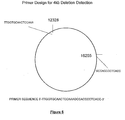
- primer sequences are examples of primers that may be used for the detection of the 4 kb deletion:
- a pair of amplification primers are used to amplify a target region indicative of the presence of the 4 kb deletion.
- one of the pair of amplification primers overlaps a spliced region of mtDNA after deletion of the 4 kb sequence has occurred and the mtDNA has reformed as a circular mtDNA molecule (eg. a splice at a position between 12328 and 16255 of the mtDNA genome). Therefore, extension of the overlapping primer can only occur if the 4 kb section is deleted.
- Figure 5 is a schematic diagram showing the design and sequence of the primer (ie. SEQ ID NO: 4).
- a pair of amplification primers are used to amplify a target region associated with the deleted 4 kb sequence.
- the deleted 4 kb sequence upon deletion, may reform as a circular mtDNA molecule.
- one of the pair of amplification primers overlaps the rejoining site of the ends of the 4 kb sequence.
- an increase in the amount of the 4 kb molecule detected in a sample is indicative of cancer.
- the breakpoint of the deletion is unknown thereby resulting in two possibilities for primer location.
- two separate forward primers may be designed to amplify the target region associated with the deleted 4 kb sequence.
- the following primer sequences are examples of those that may be used for the detection of the 4 kb deletion in this scenario:
- the forward primers A or B can be used with reverse primer C to create PCR products that are useful in qPCR assays.
- biological sample refers to a tissue or bodily fluid containing cells from which mtDNA can be obtained.
- the biological sample can be derived from tissue including, but not limited to, breast, prostate, nervous, muscle, heart, stomach, colon tissue and the like; or from blood, saliva, cerebral spinal fluid, sputa, urine, mucous, synovial fluid, peritoneal fluid, amniotic fluid and the like.
- the biological sample may be obtained from a cancerous or non-cancerous tissue and may be a surgical specimen or a biopsy specimen.
- the biological sample can be used either directly as obtained from the source or following a pre-treatment to modify the character of the sample.
- the biological sample can be pre-treated prior to use by, for example, preparing plasma or serum from blood, disrupting cells, preparing liquids from solid materials, diluting viscous fluids, filtering liquids, distilling liquids, concentrating liquids, inactivating interfering components, adding reagents, and the like.
- sample type may be assayed at a single time (i.e. for the detection of more than one cancer).
- a course of collections are required, for example, for the monitoring of risk factors or cancer over time, a given sample may be diagnosed alone or together with other sample taken throughout the test period.
- biological samples may be taken once only, or at regular intervals such as biweekly, monthly, semi-annually or annually.
- mitochondrial DNA targets are in much greater abundance (approximately 1000 fold greater) than nucleic acid targets and as such sample sizes comprising extremely low yields of nucleic acids would be suitable for use with the present invention.
- the system and method of the present invention may be used to detect cancer at an early stage, and before any histological abnormalities.
- sample testing at regular intervals such as biweekly, monthly, semi-annually or annually (or any other suitable interval) can provide health care professionals with a real-time, quantitative monitoring tool to compare against treatment recommendations to determine their effectiveness in preventing or treating the disease.
- the present invention may be used for detecting the presence of pre-neoplasia, neoplasia and progression towards potential malignancy of prostate cancer and breast cancer.
- the present invention involves the detection and quantification of the 4 kb mtDNA deletion for the detection, diagnosis, and/or monitoring of cancer.
- mtDNA is extracted from a biological sample (for example body tissue, or body fluids such as urine, prostate massage fluid).
- the extracted mtDNA is then tested in order to determine the levels (ie. quantity) of the 4 kb deletion in the sample.
- the levels of the deletion were found to be elevated in samples obtained from subjects with cancer when compared to samples obtained from subjects without cancer. Based on the information and data supplied below, the inventors have concluded that elevated levels of the 4 kb deletion in human mtDNA is indicative of cancer.
- samples of, for instance prostate tissue, prostate massage fluid, urine or breast tissue are obtained from an individual and tested over a period of time (eg. years) in order to monitor the genesis or progression of cancer.
- a period of time eg. years
- Increasing levels of the 4 kb deletion over time could be indicative of the beginning or progression of cancer.
- Another aspect of the invention provides methods for confirming or refuting the results of a cancer biopsy test from a biopsy sample (eg. prostate or breast cancer), comprising: obtaining non-cancerous tissue from a biopsy sample; and detecting and quantifying the amount of the 4 kb mtDNA deletion in the non-diseased tissue.
- a biopsy sample eg. prostate or breast cancer
- the various examples provided below illustrate a difference in the amount of mtDNA having the 4 kb deletion between samples obtained from subjects having cancer, and subjects without cancer.
- the amount of the 4 kb deletion was found to be higher in the samples obtained from subjects having cancer. This determination was made by comparing the amount of the 4 kb deletion in the samples from known cancer cells and/or known non-cancer cells.
- a method for screening individuals for cancer from one or more biological samples comprising: obtaining the one or more samples, and detecting and quantifying the level of the 4 kb mtDNA deletion in the samples.
- a method for screening individuals for prostate or breast cancer from a body fluid or tissue sample comprising; obtaining the body fluid or tissue sample, and detecting and quantifying the level of the 4 kb mtDNA deletion in the body fluid or tissue sample.
- Age related accumulation of the 4 kb mtDNA deletion may also predispose an individual to, for example, prostate cancer or breast cancer, which is prevalent in middle aged and older men, and middle aged and older women, respectively.
- an accumulation of the 4 kb mtDNA deletion may be associated with a particular lifestyle based on an individual's diet, exercise habits, and exposure to known carcinogens.
- regular cancer screening may take place by monitoring over time the amount of the 4 kb deletion in one or more biological samples, non-limiting examples of which include breast and prostate tissues or body fluids such as prostate massage fluid, or urine.
- the method of the present invention may also be used for screening potential therapeutic agents for use in cancer treatment or for monitoring the therapeutic effect of such agents.
- the method of the present invention may be used to measure various biomarkers associated with the cancers identified herein.
- the ability to assess the level of DNA damage in any biological sample at any time point provides the foundation for a unique and informative screening test for an individual's health and to assess the safety and efficacy of existing and new therapeutic agents and treatment regimes.
- identifying the specific genetic changes underlying a subject's state of health it may be readily determined whether and to what extent a patient will respond to a particular therapeutic agent or regime.
- kits for use in a clinical environment. Such kits could not only include one or more sampling means, but other materials necessary for the identification of mtDNA mutations.
- kits can optionally include reagents required to conduct a diagnostic assay, such as buffers, salts, detection reagents, and the like.
- Other components such as buffers and solutions for the isolation and/or treatment of a biological sample, may also be included in the kit.
- One or more of the components of the kit may be lyophilised and the kit may further comprise reagents suitable for the reconstitution of the lyophilised components.
- the kit may also contain reaction vessels, mixing vessels and other components that facilitate the preparation of the test sample.
- the kit may also optionally include instructions for use, which may be provided in paper form or in computer-readable form, such as a disc, CD, DVD or the like.
- kits for diagnosing cancer comprising means for extraction of mtDNA, primers, reagents and instructions.
- kits for diagnosing cancer for example prostate or breast cancer, comprising means for extraction of mtDNA, primers having the nucleic acid sequences recited in SEQ ID NOs: 4 and 5, reagents and instructions.
- kits for diagnosing cancer for example prostate or breast cancer, comprising means for extraction of mtDNA, primers having the nucleic acid sequences recited in SEQ ID NOs: 6 and 5, reagents and instructions.
- Urine samples were collected from five patients who had been diagnosed with prostate cancer and five who had a needle biopsy procedure which was unable to detect prostate malignancy. These samples were collected following a digital rectal exam (DRE) to facilitate the collection of prostate cells.
- DRE digital rectal exam
- Pellets were resuspended in 200ul phosphate buffered saline solution. Both the resuspended pellet and the whole urine sample were subjected to a DNA extraction procedure using the QiaAMP DNA Mini Kit (Qiagen P/N 51304) according to the manufacturer's directions. The resulting DNA extracts were then quantified using a NanoDrop ND-1000 Spectrophotometer and normalized to a concentration of 0.1ng/ul.
- Results from the urine pellet did not yield significant differences in the mean cycle threshold observed or a useful cutoff point. However, the results from the whole urine sample did yield significant differences as provided below.
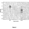
- Tables 1 and 2 and Figure 1 show the difference in the mean C T scores for urine samples from subjects having prostate malignant tissue and benign tissue at the 0.04 significance level.
- Table 1 Mean Values for C T scores: Urine Samples N Mean Std. Deviation Std. Error Mean Benign 7 38.0357 3.40974 1.288876 Malignant 7 31.9300 6.12583 2.31534
- Table 2 Significance Test for Mean C T scores Independent Samples Test CTt40 fluid Levene's Test for Equality of Variances Test for Equality Means F Sig. t df Sig. (2-tailed) Mean Diff. Std. Error Diff.
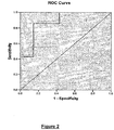
- Tables 3 and 4, and Figure 2 illustrate that when using a cut-off cycle threshold of 36.255 the sensitivity of the assay for prostate cancer is 86% and the specificity is 86%.
- FIG. 2 is a Receiver Operating Characteristic (ROC) curve illustrating the specificity and sensitivity of the 4 kb mtDNA deletion as a marker for prostate cancer when testing urine. These results were obtained using a cutoff C T of 36.255. The sensitivity of the marker at this C T is 86%, while the specificity is 86%.
- ROC Receiver Operating Characteristic
- the accuracy of the test depends on how well the test separates the group being tested into those with and without the prostate cancer. Accuracy is measured by the area under the ROC curve. Table 4 shows the calculation of the area under the curve for the present example. Table 3 Determination of Specificity and Sensitivity Positive if ⁇ a Sensitivity 1 - specificity 19.86 .000 .000 24.87 .143 .000 29.48 .286 .000 30.54 .429 .000 32.235 .429 .143 33.77 .571 .143 35.11 .714 .143 36.255 .857 .143 37.415 .857 .286 39.23 .857 .429 39.995 1.000 .429 40.21 1.000 .857 41.42 1.000 1.000 a - the smallest cutoff value is the minimum observed test value minus 1 and the largest cutoff value is the maximum observed test value plus 1.
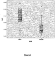
- Tables 5 and 6, and Figure 3 show the difference in the mean C T scores for breast tissue samples from subjects having malignant breast tissue and benign breast tissue at the 0.065 level.
- Table 5 Mean Values for C T scores: Breast Tissue Samples Group N Mean Std. Dev. Std. Error Mean Normal 9 21.5278 2.71939 .90646 Malignant 9 18.9089 2.89126 .96375 Table 6 Significance Test for Mean C T scores CTt40 fluid Levene's Test for Equality of Variances Test for Equality Means F Sig. t df Sig. (2-tailed) Mean Diff. Std. Error Diff.
- Tables 7 and 8, and Figure 4 illustrate that when using a cut-off cycle threshold of 19.845 the sensitivity of the assay for breast cancer is 78% and the specificity is 78%.
- Figure 4 is an ROC curve illustrating the specificity and sensitivity of the 4 kb mtDNA deletion as a marker for breast cancer when testing breast tissue. These results were obtained using a cutoff C T of 19.845. The sensitivity of the marker at this C T is 78%, while the specificity is 78%.
- Prostate needle biopsy specimens were obtained from 19 individuals, 9 without prostate cancer and 10 with prostate cancer. Needle biopsy tissues were formalin-fixed paraffin embedded (FFPE) as is standard in the clinical diagnostic setting. 10 micron sections of each biopsy were deposited directly into centrifuge tubes and the DNA was extracted using the QiaAMP DNA Mini Kit ( Qiagen, p/n 51306). DNA extracts were quantified by absorbance at 260nm using a NanoDrop ND-1000 Spectrophotometer. Yields ranged from 347ng to 750ng.
- FFPE formalin-fixed paraffin embedded
- Table 9 Reagents and Concentrations for Amplification Reaction Reagent Final Concentration iQ SYBR Green Supermix 1X (Bio-Rad Laboratories, p/n 170-8882) Forward Primer 12303-12316/16243-16259F 175nmol 5'- CCCAAAAATTTTGGTGCAACTCCAAAGCCAC - 3' (SEQ ID NO: 6) Reverse Primer 16410R 175nmol 5'-AGGATGGTGGTCAAGGGAC-3' (SEQ ID NO: 5) DNA extract 0.8ng/ul
- Results demonstrate that those individuals with prostate cancer have a lower C T value and therefore higher levels of the 4 kb deletion in prostate tissue than do those without prostate cancer.
- Patients with prostate cancer have an average C T value of 30.7 while the patients without prostate cancer have an average C T value of 36.4. This difference of 5.7 C T corresponds to nearly 100 fold greater 4 kb deletion levels in the group with prostate malignancy than in the group without.
- Table 10 Patient Diagnosis and Associated C T Score Patient Number and Diagnosis C(t) CUG 1301 Malignant 25.7 CUG 1268 Malignant 27.7 CUG RN 345 Normal 28.3 CUG 1272 Malignant 28.8 CUG 1375 Malignant 29.1 CUG 1259 Malignant 29.1 CUG 1381 Malignant 30.2 CUG RN 82 Normal 30.5 CUG 1372 Malignant 30.9 CUG 1085 C T1 Normal 31.5 CUG 1317 Malignant 31.7 CUG 1377 F Normal 33.6 CUG 1365 B Normal 34.6 CUG 1370 Malignant 35.9 CUG RN 405 Normal 37.5 CUG 1366 Malignant 37.9 CUG RN 701 Normal 41.7 CUG RN 420 Normal 45 CUG RN 373 Normal 45
- Tables 11 and 12 show the difference in the mean C T scores for prostate tissue samples from subjects having normal and malignant prostate tissue.
- Table 11 Mean Values for C T Score: Prostate Needle Biopsy Tissue Group N Mean Std. Dev. Std. Error Mean Normal 9 36.4111 6.25229 2.08410 Malignant 10 30.7 3.69534 1.16857
- Table 12 Significance Test for C T Scores CTt40 fluid Levene's Test for Equality of Variances Test for Equality Means F Sig. t df Sig. (2-tailed) Mean Diff. Std. Error Diff.
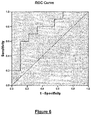
- Table 13 and Figure 6 illustrate that when using a cutoff of C T 32.65 the sensitivity and specificity of correctly diagnosing these patients is 80% and 67% respectively.
- Table 13 Determination of Specificity and Sensitivity Positive if ⁇ a Sensitivity 1 - specificity 24.7 .000 .000 26.7 .100 .000 28.0 .200 .000 28.55 .200 .111 28.95 .300 .111 29.65 .500 .111 30.35 .600 .111 30.7 .600 .222 31.2 .700 .222 31.6 .700 .333 32.65 .800 .333 34.1 .800 .444 32.25 .800 .556 36.7 .900 .556 37.7 .900 .667 39.8 1.000 .667 43.35 1.000 .778 46.0 1.000 1.000 1.000
Landscapes
- Chemical & Material Sciences (AREA)
- Life Sciences & Earth Sciences (AREA)
- Health & Medical Sciences (AREA)
- Organic Chemistry (AREA)
- Proteomics, Peptides & Aminoacids (AREA)
- Engineering & Computer Science (AREA)
- Zoology (AREA)
- Wood Science & Technology (AREA)
- Immunology (AREA)
- Analytical Chemistry (AREA)
- Genetics & Genomics (AREA)
- Pathology (AREA)
- General Engineering & Computer Science (AREA)
- General Health & Medical Sciences (AREA)
- Microbiology (AREA)
- Molecular Biology (AREA)
- Biotechnology (AREA)
- Biophysics (AREA)
- Physics & Mathematics (AREA)
- Biochemistry (AREA)
- Bioinformatics & Cheminformatics (AREA)
- Oncology (AREA)
- Hospice & Palliative Care (AREA)
- Chemical Kinetics & Catalysis (AREA)
- Measuring Or Testing Involving Enzymes Or Micro-Organisms (AREA)
- Investigating Or Analysing Biological Materials (AREA)
Applications Claiming Priority (3)
| Application Number | Priority Date | Filing Date | Title |
|---|---|---|---|
| US263707P | 2007-11-09 | 2007-11-09 | |
| PCT/CA2008/001956 WO2009059414A1 (en) | 2007-11-09 | 2008-11-10 | Mitochondrial dna deletion between about residues 12317-16254 for use in the detection of cancer |
| EP08846547.1A EP2220252B1 (en) | 2007-11-09 | 2008-11-10 | Mitochondrial dna deletion between about residues 12317-16254 for use in the detection of cancer |
Related Parent Applications (1)
| Application Number | Title | Priority Date | Filing Date |
|---|---|---|---|
| EP08846547.1A Division EP2220252B1 (en) | 2007-11-09 | 2008-11-10 | Mitochondrial dna deletion between about residues 12317-16254 for use in the detection of cancer |
Publications (1)
| Publication Number | Publication Date |
|---|---|
| EP3409793A1 true EP3409793A1 (en) | 2018-12-05 |
Family
ID=40625325
Family Applications (2)
| Application Number | Title | Priority Date | Filing Date |
|---|---|---|---|
| EP18160726.8A Withdrawn EP3409793A1 (en) | 2007-11-09 | 2008-11-10 | Mitochondrial dna deletion between about residues 12317-16254 for use in the detection of cancer |
| EP08846547.1A Not-in-force EP2220252B1 (en) | 2007-11-09 | 2008-11-10 | Mitochondrial dna deletion between about residues 12317-16254 for use in the detection of cancer |
Family Applications After (1)
| Application Number | Title | Priority Date | Filing Date |
|---|---|---|---|
| EP08846547.1A Not-in-force EP2220252B1 (en) | 2007-11-09 | 2008-11-10 | Mitochondrial dna deletion between about residues 12317-16254 for use in the detection of cancer |
Country Status (9)
Families Citing this family (3)
| Publication number | Priority date | Publication date | Assignee | Title |
|---|---|---|---|---|
| US9410188B2 (en) * | 2010-05-28 | 2016-08-09 | Biomerieux | Method and kit for discriminating between breast cancer and benign breast disease |
| CN107604061B (zh) * | 2017-08-31 | 2021-02-02 | 中国科学院北京基因组研究所 | 线粒体-细胞核dna甲基化联合位点的筛选方法和应用 |
| WO2024130647A1 (zh) * | 2022-12-22 | 2024-06-27 | 中国科学院广州生物医药与健康研究院 | 分离的环状rna及其在预防和治疗肺癌中的用途 |
Citations (4)
| Publication number | Priority date | Publication date | Assignee | Title |
|---|---|---|---|---|
| EP0849364A1 (en) * | 1996-12-20 | 1998-06-24 | Roche Diagnostics GmbH | Method for the direct, exponential amplification and sequencing of DNA molecules and its application |
| WO2005056573A1 (en) * | 2002-06-10 | 2005-06-23 | 1304854 Ontario Ltd. | Complete mitochondrial genome sequences as a diagnostic tool for the health sciences |
| WO2006111029A1 (en) * | 2005-04-18 | 2006-10-26 | Genesis Genomics Inc. | Mitochondrial mutations and rearrangements as a diagnostic tool for the detection of sun exposure, prostate cancer and other cancers |
| WO2009039601A1 (en) * | 2007-09-26 | 2009-04-02 | Genesis Genomics Inc. | 3.4 kb mitochondrial dna deletion for use in the detection of cancer |
Family Cites Families (5)
| Publication number | Priority date | Publication date | Assignee | Title |
|---|---|---|---|---|
| US5800992A (en) * | 1989-06-07 | 1998-09-01 | Fodor; Stephen P.A. | Method of detecting nucleic acids |
| US6582908B2 (en) * | 1990-12-06 | 2003-06-24 | Affymetrix, Inc. | Oligonucleotides |
| US6472378B2 (en) * | 1998-08-31 | 2002-10-29 | Pro-Neuron, Inc. | Compositions and methods for treatment of mitochondrial diseases |
| CA2450403A1 (en) * | 2001-06-11 | 2002-12-19 | 1304854 Ontario Ltd. | Complete mitochondrial genome sequences as a diagnostic tool for the health sciences |
| US20050026167A1 (en) * | 2001-06-11 | 2005-02-03 | Mark Birch-Machin | Complete mitochondrial genome sequences as a diagnostic tool for the health sciences |
-
2008
- 2008-11-10 US US12/742,032 patent/US20100311057A1/en not_active Abandoned
- 2008-11-10 AU AU2008324675A patent/AU2008324675B2/en not_active Ceased
- 2008-11-10 CN CN200880115243.2A patent/CN101883864B/zh not_active Expired - Fee Related
- 2008-11-10 EP EP18160726.8A patent/EP3409793A1/en not_active Withdrawn
- 2008-11-10 EP EP08846547.1A patent/EP2220252B1/en not_active Not-in-force
- 2008-11-10 WO PCT/CA2008/001956 patent/WO2009059414A1/en active Application Filing
- 2008-11-10 KR KR1020107012456A patent/KR101644661B1/ko not_active Expired - Fee Related
- 2008-11-10 NZ NZ584656A patent/NZ584656A/xx not_active IP Right Cessation
- 2008-11-10 CA CA2704361A patent/CA2704361C/en active Active
- 2008-11-10 JP JP2010532390A patent/JP5481383B2/ja not_active Expired - Fee Related
-
2013
- 2013-01-18 US US13/745,204 patent/US20130288243A1/en not_active Abandoned
-
2014
- 2014-09-17 US US14/489,119 patent/US20150004619A1/en not_active Abandoned
-
2016
- 2016-06-21 US US15/188,604 patent/US20160289772A1/en not_active Abandoned
-
2018
- 2018-04-06 US US15/947,192 patent/US10400290B2/en active Active
Patent Citations (4)
| Publication number | Priority date | Publication date | Assignee | Title |
|---|---|---|---|---|
| EP0849364A1 (en) * | 1996-12-20 | 1998-06-24 | Roche Diagnostics GmbH | Method for the direct, exponential amplification and sequencing of DNA molecules and its application |
| WO2005056573A1 (en) * | 2002-06-10 | 2005-06-23 | 1304854 Ontario Ltd. | Complete mitochondrial genome sequences as a diagnostic tool for the health sciences |
| WO2006111029A1 (en) * | 2005-04-18 | 2006-10-26 | Genesis Genomics Inc. | Mitochondrial mutations and rearrangements as a diagnostic tool for the detection of sun exposure, prostate cancer and other cancers |
| WO2009039601A1 (en) * | 2007-09-26 | 2009-04-02 | Genesis Genomics Inc. | 3.4 kb mitochondrial dna deletion for use in the detection of cancer |
Non-Patent Citations (40)
| Title |
|---|
| AUSUBEL ET AL.: "Current Protocols in Molecular Biology", 2007, JOHN WILEY & SONS |
| BIRCH-MACHIN MA: "Online Conference Report (Sunburnt DNA), International Congress of Biochemistry and Molecular Biology", NEW SCIENTIST, 2000 |
| BIRCH-MACHIN MA; TAYLOR RW; COCHRAN B; ACKRELL BAC; TUMBULL DM, ANN NEUROL, vol. 48, 2000, pages 330 - 335 |
| BIRCH-MACHIN, M.A.: "Mitochondria and skin disease", CLIN EXP DERMATOL, vol. 25, 2000, pages 141 - 6 |
| BOGLIOLO, M ET AL., MUTAGENESIS, vol. 14, 1999, pages 77 - 82 |
| BROWN, M.D. ET AL., AM J. HUMN GENET, vol. 60, 1997, pages 381 - 387 |
| BRUMLEY, R. L. JR.; SMITH, L.M.: "Rapid DNA sequencing by horizontal ultrathin gel electrophoresis", NUCLEIC ACIDS RES., vol. 19, 1991, pages 4121 - 4126 |
| CELIA H TENGAN ET AL: "Detection and Analysis of Mitochondrial DNA Deletions by Whole Genome PCR", BIOCHEMICAL AND MOLECULAR MEDICINE, 1 June 1996 (1996-06-01), pages 130 - 134, XP055223831, Retrieved from the Internet <URL:http://ac.els-cdn.com/S107731509690040X/1-s2.0-S107731509690040X-main.pdf?_tid=52372172-7cac-11e5-bbb4-00000aab0f6c&acdnat=1445951706_7e4fdbacdfe9e83f7033bae7d543ce5a> [retrieved on 20151027], DOI: 10.1006/bmme.1996.0040 * |
| CHABI B ET AL: "Quantification of Mitochondrial DNA Deletion, Depletion, and Overreplication: Application to Diagnosis", CLINICAL CHEMISTRY, AMERICAN ASSOCIATION FOR CLINICAL CHEMISTRY, WASHINGTON, DC, vol. 49, no. 8, 1 August 2003 (2003-08-01), pages 1309 - 1317, XP003003516, ISSN: 0009-9147, DOI: 10.1373/49.8.1309 * |
| CHATTERJEE A ET AL: "Mitochondrial DNA mutations in human cancer", ONCOGENE, NATURE PUBLISHING GROUP UK, LONDON, vol. 25, 1 August 2006 (2006-08-01), pages 4663 - 4674, XP008136288, ISSN: 0950-9232, DOI: 10.1038/SJ.ONC.1209604 * |
| CHINNERY PF; TURNBULL DM., LANCET, vol. 354, no. 1, 1999, pages 17 - 21 |
| HAYWARD SW; GROSSFELD GD; TLSTY TD; CUNHA GR, INT J ONCOL, vol. 13, 1998, pages 35 - 47 |
| HOPGOOD, R. ET AL.: "Strategies for automated sequencing of human mtDNA directly from PCR products", BIOTECHNIQUES, vol. 13, 1992, pages 82 - 92 |
| HUANG GM; NG WL; FARKAS J; HE L; LIANG HA; GORDON D; HOOD R, GENOMICS, vol. 59, no. 2, 1999, pages 178 - 86 |
| HUOPONEN, KIRSI: "Leberhereditary optic neuropathy: clinical and molecular genetic findings", NEUROGENETICS, vol. 3, 2001, pages 119 - 125 |
| KONISHI N; CHO M; YAMAMOTO K; HIASA Y, PATHOL. INT., vol. 47, 1997, pages 735 - 747 |
| KRISHNAN K J ET AL: "The Use of a 3895 by Mitochondrial DNA Deletion as a Marker for Sunlight Exposure in Human Skin", JOURNAL OF INVESTIGATIVE DERMATOLOGY, ELSEVIER, NL, vol. 123, no. 6, 1 December 2004 (2004-12-01), pages 1020 - 1024, XP003003514, ISSN: 0022-202X, DOI: 10.1111/J.0022-202X.2004.23457.X * |
| LANDIS SH; MURRAY T; BOLDEN S; WINGO PA, CANCER J. CLIN., vol. 49, pages 8 - 31 |
| LEE HC; LU CY; FAHN HJ; WEI YHU, FEDERATION OF EUROPEAN BIOCHEMICAL SOCIETIES, vol. 441, 1998, pages 292 - 296 |
| LUCKEY, J.A. ET AL.: "High speed DNA sequencing by capillary gel electrophoresis", METHODS ENZYMOL., vol. 218, 1993, pages 154 - 172 |
| MULLIS; FALOONA, METHODS ENZYMOL., vol. 155, 1987, pages 335 |
| NAVIAUX, RK.: "Mitochondrial Disease- Primary Care Physican's Guide", PSY-ED. CORP D/B/A EXCEPTIONAL PARENTS GUIDE, 1997, pages 3 - 10 |
| PARRELLA ET AL., CANCER RESEARCH, vol. 61, 2001 |
| PARRELLA P; XIAO Y; FLISS M; SANCHEZ-CESPEDES M; MAZZARELLI P; RINALDI M; NICOL T; GABRIELSON E; CUOMO C; COHEN D: "Detection of mitochondrial DNA mutations in primary breast cancer and fine-needle aspirates", CANCER RES, vol. 61, 2001, pages 7623 - 7626, XP002244378 |
| POLYAK Y ET AL., NATURE GENET, vol. 20, no. 3, 1998, pages 291 - 293 |
| SEIDMAN, M.D. ET AL., ARCH. OTOLARYNGOF HEAD NECK SURG., vol. 123, 1997, pages 1039 - 1045 |
| SHERRAT EJ; THOMAS AW; ALCOLADO JC, CLIN. SCI., vol. 92, 1997, pages 225 - 235 |
| SHOFFNER JM; BROWN MD; TORRONI A; LOTT MT; CABELL MF; MIRRA SS; BEAL MF; YANG C; GEARING M; SALVO R, GENOMICS, vol. 17, 1993, pages 171 - 184 |
| TANAKA, M. ET AL.: "Automated sequencing of mtDNA", METHODS ENZYMOL., vol. 264, 1996, pages 407 - 421 |
| TANIIKE, M., BIOCHEM BIOPHYS RES COMUN, vol. 186, 1992, pages 47 - 53 |
| VALDEMAR MÁXIMO ET AL: "Mitochondrial DNA Somatic Mutations (Point Mutations and Large Deletions) and Mitochondrial DNA Variants in Human Thyroid Pathology", THE AMERICAN JOURNAL OF PATHOLOGY, vol. 160, no. 5, 1 May 2002 (2002-05-01), pages 1857 - 1865, XP055032294, ISSN: 0002-9440, DOI: 10.1016/S0002-9440(10)61132-7 * |
| VALNOT, ISABELLE ET AL.: "A mitochondrial cytochrome b mutation but no mutations of nuclearly encoded subunits in ubiquinol cytochrome c reductase (complex III) deficiency", HUMAN GENETICS, vol. 104, 1999, pages 460 - 466 |
| WALLACE ET AL.: "Mitochondiral DNA MUtatio Assoicated with Leber's Hereditary Optic Neuropathy", SCIENCE, pages 1427 - 1429 |
| WEI YH, PROCEEDINGS OF THE NAT. SCI. COUNCIL OF THE REPUBLIC OF CHINA, vol. 22, no. 2, April 1998 (1998-04-01), pages 5567 |
| WOODWELL DA: "Advance data from vital and health statistics", 1999, MARYLAND: NATIONAL CENTER FOR HEALTH STATISTICS, article "National Ambulatory Medical Care Survey: 1997 Summary" |
| WURMB, N; OEHMICHEN, M; MEISSNER, C, MUTAT RES., vol. 422, 1998, pages 247 - 254 |
| YEH, J.J. ET AL., ONCOGENE JOURNAL, vol. 19, 2000, pages 2060 - 2066 |
| ZHANG ET AL.: "Multiple mitochondiral DNA deletions in an elderly human individual", FEBS LETT, vol. 297, 1992, pages 34 - 38 |
| ZHANG, C. ET AL., BIOCHEM. BIOPHYS. RES. COMUN., vol. 195, 1993, pages 1104 - 1110 |
| ZHU WEIZHU ET AL: "Large-scale mitochondrial DNA deletion mutations and nuclear genome instability in human breast cancer", CANCER DETECTION AND PREVENTION, ELSEVIER SCIENCE, vol. 28, no. 2, 1 January 2004 (2004-01-01), pages 119 - 126, XP002524596, ISSN: 0361-090X, DOI: 10.1016/J.CDP.2003.11.008 * |
Also Published As
| Publication number | Publication date |
|---|---|
| CN101883864A (zh) | 2010-11-10 |
| CN101883864B (zh) | 2014-02-12 |
| US20130288243A1 (en) | 2013-10-31 |
| US20150004619A1 (en) | 2015-01-01 |
| WO2009059414A1 (en) | 2009-05-14 |
| US10400290B2 (en) | 2019-09-03 |
| KR101644661B1 (ko) | 2016-08-01 |
| JP2011502487A (ja) | 2011-01-27 |
| CA2704361C (en) | 2023-04-04 |
| AU2008324675A1 (en) | 2009-05-14 |
| EP2220252A4 (en) | 2012-08-22 |
| KR20100090702A (ko) | 2010-08-16 |
| EP2220252A1 (en) | 2010-08-25 |
| US20160289772A1 (en) | 2016-10-06 |
| AU2008324675B2 (en) | 2015-04-23 |
| JP5481383B2 (ja) | 2014-04-23 |
| NZ584656A (en) | 2012-08-31 |
| CA2704361A1 (en) | 2009-05-14 |
| US20100311057A1 (en) | 2010-12-09 |
| EP2220252B1 (en) | 2018-03-14 |
| US20180230549A1 (en) | 2018-08-16 |
Similar Documents
| Publication | Publication Date | Title |
|---|---|---|
| JP2008545418A (ja) | 癌の診断、予後診断、および治療のための遊離循環dnaの使用 | |
| US11111546B2 (en) | 3.4 KB mitochondrial DNA deletion for use in the detection of cancer | |
| US10400290B2 (en) | Mitochondrial DNA deletion between about residues 12317-16254 for use in the detection of cancer | |
| EP2634267B1 (en) | 3.4 kb mitochondrial DNA deletion for use in the detection of cancer | |
| US20190382852A1 (en) | Mitochondrial DNA deletion between about residues 12317-16254 for use in the detection of cancer | |
| HK1145407B (en) | Mitochondrial dna deletion between about residues 12317-16254 for use in the detection of cancer | |
| HK1145407A (en) | Mitochondrial dna deletion between about residues 12317-16254 for use in the detection of cancer | |
| Class et al. | Patent application title: Mitochondrial DNA deletion between about residues 12317-16254 for use in the detection of cancer | |
| HK1188813A (en) | 3.4 kb mitochondrial dna deletion for use in the detection of cancer | |
| HK1145343B (en) | 3.4 kb mitochondrial dna deletion for use in the detection of cancer | |
| CN115896275A (zh) | 一种肿瘤检测方法及应用 | |
| CN116814790A (zh) | Pitx2基因作为标志物在检测肺癌中的应用 | |
| CN115896276A (zh) | 一种肿瘤检测方法及应用 |
Legal Events
| Date | Code | Title | Description |
|---|---|---|---|
| PUAI | Public reference made under article 153(3) epc to a published international application that has entered the european phase |
Free format text: ORIGINAL CODE: 0009012 |
|
| AC | Divisional application: reference to earlier application |
Ref document number: 2220252 Country of ref document: EP Kind code of ref document: P |
|
| AK | Designated contracting states |
Kind code of ref document: A1 Designated state(s): AT BE BG CH CY CZ DE DK EE ES FI FR GB GR HR HU IE IS IT LI LT LU LV MC MT NL NO PL PT RO SE SI SK TR |
|
| 17P | Request for examination filed |
Effective date: 20190604 |
|
| RBV | Designated contracting states (corrected) |
Designated state(s): AT BE BG CH CY CZ DE DK EE ES FI FR GB GR HR HU IE IS IT LI LT LU LV MC MT NL NO PL PT RO SE SI SK TR |
|
| STAA | Information on the status of an ep patent application or granted ep patent |
Free format text: STATUS: THE APPLICATION IS DEEMED TO BE WITHDRAWN |
|
| 18D | Application deemed to be withdrawn |
Effective date: 20200603 |