EP3400877A1 - Procédé d'imagerie d'une zone d'examen au moyen d'un premier agent de contraste et d'un second agent de contraste - Google Patents
Procédé d'imagerie d'une zone d'examen au moyen d'un premier agent de contraste et d'un second agent de contraste Download PDFInfo
- Publication number
- EP3400877A1 EP3400877A1 EP18152125.3A EP18152125A EP3400877A1 EP 3400877 A1 EP3400877 A1 EP 3400877A1 EP 18152125 A EP18152125 A EP 18152125A EP 3400877 A1 EP3400877 A1 EP 3400877A1
- Authority
- EP
- European Patent Office
- Prior art keywords
- contrast agent
- image
- measurement data
- information
- projection measurement
- Prior art date
- Legal status (The legal status is an assumption and is not a legal conclusion. Google has not performed a legal analysis and makes no representation as to the accuracy of the status listed.)
- Withdrawn
Links
- 239000002872 contrast media Substances 0.000 title claims abstract description 244
- 238000000034 method Methods 0.000 title claims abstract description 67
- 238000003384 imaging method Methods 0.000 title claims abstract description 17
- 238000005259 measurement Methods 0.000 claims abstract description 82
- 238000002591 computed tomography Methods 0.000 claims abstract description 39
- 239000000463 material Substances 0.000 claims description 28
- 238000002083 X-ray spectrum Methods 0.000 claims description 19
- 238000007306 functionalization reaction Methods 0.000 claims description 17
- 239000000126 substance Substances 0.000 claims description 17
- 239000004005 microsphere Substances 0.000 claims description 16
- 239000002105 nanoparticle Substances 0.000 claims description 16
- 238000000354 decomposition reaction Methods 0.000 claims description 15
- 230000017531 blood circulation Effects 0.000 claims description 11
- 238000004590 computer program Methods 0.000 claims description 10
- 230000010412 perfusion Effects 0.000 claims description 9
- 239000000941 radioactive substance Substances 0.000 claims description 8
- 230000003595 spectral effect Effects 0.000 description 20
- 229910052688 Gadolinium Inorganic materials 0.000 description 13
- UIWYJDYFSGRHKR-UHFFFAOYSA-N gadolinium atom Chemical compound [Gd] UIWYJDYFSGRHKR-UHFFFAOYSA-N 0.000 description 13
- 210000001519 tissue Anatomy 0.000 description 13
- 239000007924 injection Substances 0.000 description 12
- 238000002347 injection Methods 0.000 description 12
- XEEYBQQBJWHFJM-UHFFFAOYSA-N Iron Chemical compound [Fe] XEEYBQQBJWHFJM-UHFFFAOYSA-N 0.000 description 8
- 239000003814 drug Substances 0.000 description 8
- PCHJSUWPFVWCPO-UHFFFAOYSA-N gold Chemical compound [Au] PCHJSUWPFVWCPO-UHFFFAOYSA-N 0.000 description 8
- 229910052737 gold Inorganic materials 0.000 description 8
- 239000010931 gold Substances 0.000 description 8
- 239000002245 particle Substances 0.000 description 8
- 206010028980 Neoplasm Diseases 0.000 description 7
- 210000000988 bone and bone Anatomy 0.000 description 7
- 230000005855 radiation Effects 0.000 description 7
- 238000000926 separation method Methods 0.000 description 7
- 230000001225 therapeutic effect Effects 0.000 description 7
- ZCYVEMRRCGMTRW-UHFFFAOYSA-N 7553-56-2 Chemical compound [I] ZCYVEMRRCGMTRW-UHFFFAOYSA-N 0.000 description 6
- 229940079593 drug Drugs 0.000 description 6
- 239000011630 iodine Substances 0.000 description 6
- PNDPGZBMCMUPRI-UHFFFAOYSA-N iodine Chemical compound II PNDPGZBMCMUPRI-UHFFFAOYSA-N 0.000 description 6
- 229910052740 iodine Inorganic materials 0.000 description 6
- 229910052689 Holmium Inorganic materials 0.000 description 5
- KJZYNXUDTRRSPN-UHFFFAOYSA-N holmium atom Chemical compound [Ho] KJZYNXUDTRRSPN-UHFFFAOYSA-N 0.000 description 5
- 230000002792 vascular Effects 0.000 description 5
- 210000000577 adipose tissue Anatomy 0.000 description 4
- 229910052742 iron Inorganic materials 0.000 description 4
- 230000010110 radioembolization Effects 0.000 description 4
- 238000002560 therapeutic procedure Methods 0.000 description 4
- XLYOFNOQVPJJNP-UHFFFAOYSA-N water Substances O XLYOFNOQVPJJNP-UHFFFAOYSA-N 0.000 description 4
- 238000010521 absorption reaction Methods 0.000 description 3
- 210000004204 blood vessel Anatomy 0.000 description 3
- 230000010109 chemoembolization Effects 0.000 description 3
- 230000008021 deposition Effects 0.000 description 3
- 238000001514 detection method Methods 0.000 description 3
- 230000000694 effects Effects 0.000 description 3
- 238000001914 filtration Methods 0.000 description 3
- 230000035699 permeability Effects 0.000 description 3
- 238000011002 quantification Methods 0.000 description 3
- 210000004872 soft tissue Anatomy 0.000 description 3
- WFKWXMTUELFFGS-UHFFFAOYSA-N tungsten Chemical compound [W] WFKWXMTUELFFGS-UHFFFAOYSA-N 0.000 description 3
- 229910052721 tungsten Inorganic materials 0.000 description 3
- 239000010937 tungsten Substances 0.000 description 3
- 239000011324 bead Substances 0.000 description 2
- 229940039231 contrast media Drugs 0.000 description 2
- 238000003745 diagnosis Methods 0.000 description 2
- 230000004069 differentiation Effects 0.000 description 2
- 230000003902 lesion Effects 0.000 description 2
- 238000005316 response function Methods 0.000 description 2
- 230000011218 segmentation Effects 0.000 description 2
- 229940124597 therapeutic agent Drugs 0.000 description 2
- 238000012795 verification Methods 0.000 description 2
- 230000000007 visual effect Effects 0.000 description 2
- 229910004613 CdTe Inorganic materials 0.000 description 1
- 229910001218 Gallium arsenide Inorganic materials 0.000 description 1
- 206010019695 Hepatic neoplasm Diseases 0.000 description 1
- VWQVUPCCIRVNHF-OUBTZVSYSA-N Yttrium-90 Chemical compound [90Y] VWQVUPCCIRVNHF-OUBTZVSYSA-N 0.000 description 1
- 239000013543 active substance Substances 0.000 description 1
- 238000004458 analytical method Methods 0.000 description 1
- 238000013459 approach Methods 0.000 description 1
- 239000000090 biomarker Substances 0.000 description 1
- 230000015572 biosynthetic process Effects 0.000 description 1
- 150000001875 compounds Chemical class 0.000 description 1
- 238000013170 computed tomography imaging Methods 0.000 description 1
- 238000012937 correction Methods 0.000 description 1
- 230000007423 decrease Effects 0.000 description 1
- 230000001419 dependent effect Effects 0.000 description 1
- 230000001627 detrimental effect Effects 0.000 description 1
- 238000011156 evaluation Methods 0.000 description 1
- -1 for example Substances 0.000 description 1
- 210000002767 hepatic artery Anatomy 0.000 description 1
- 238000013152 interventional procedure Methods 0.000 description 1
- 238000001361 intraarterial administration Methods 0.000 description 1
- 238000001990 intravenous administration Methods 0.000 description 1
- 239000011133 lead Substances 0.000 description 1
- 210000004185 liver Anatomy 0.000 description 1
- 208000014018 liver neoplasm Diseases 0.000 description 1
- 230000008338 local blood flow Effects 0.000 description 1
- 230000014759 maintenance of location Effects 0.000 description 1
- 239000003550 marker Substances 0.000 description 1
- YFDLHELOZYVNJE-UHFFFAOYSA-L mercury diiodide Chemical compound I[Hg]I YFDLHELOZYVNJE-UHFFFAOYSA-L 0.000 description 1
- 238000011275 oncology therapy Methods 0.000 description 1
- 230000002285 radioactive effect Effects 0.000 description 1
- 239000002331 radioactive microsphere Substances 0.000 description 1
- 238000001959 radiotherapy Methods 0.000 description 1
- 238000001228 spectrum Methods 0.000 description 1
- 238000012360 testing method Methods 0.000 description 1
- 231100000331 toxic Toxicity 0.000 description 1
- 230000002588 toxic effect Effects 0.000 description 1
- 230000001960 triggered effect Effects 0.000 description 1
- 210000003556 vascular endothelial cell Anatomy 0.000 description 1
- 238000012800 visualization Methods 0.000 description 1
Images

Classifications
-
- A—HUMAN NECESSITIES
- A61—MEDICAL OR VETERINARY SCIENCE; HYGIENE
- A61B—DIAGNOSIS; SURGERY; IDENTIFICATION
- A61B6/00—Apparatus or devices for radiation diagnosis; Apparatus or devices for radiation diagnosis combined with radiation therapy equipment
- A61B6/48—Diagnostic techniques
- A61B6/481—Diagnostic techniques involving the use of contrast agents
-
- A—HUMAN NECESSITIES
- A61—MEDICAL OR VETERINARY SCIENCE; HYGIENE
- A61B—DIAGNOSIS; SURGERY; IDENTIFICATION
- A61B6/00—Apparatus or devices for radiation diagnosis; Apparatus or devices for radiation diagnosis combined with radiation therapy equipment
- A61B6/02—Arrangements for diagnosis sequentially in different planes; Stereoscopic radiation diagnosis
- A61B6/03—Computed tomography [CT]
- A61B6/032—Transmission computed tomography [CT]
-
- A—HUMAN NECESSITIES
- A61—MEDICAL OR VETERINARY SCIENCE; HYGIENE
- A61B—DIAGNOSIS; SURGERY; IDENTIFICATION
- A61B6/00—Apparatus or devices for radiation diagnosis; Apparatus or devices for radiation diagnosis combined with radiation therapy equipment
- A61B6/48—Diagnostic techniques
- A61B6/482—Diagnostic techniques involving multiple energy imaging
-
- A—HUMAN NECESSITIES
- A61—MEDICAL OR VETERINARY SCIENCE; HYGIENE
- A61B—DIAGNOSIS; SURGERY; IDENTIFICATION
- A61B6/00—Apparatus or devices for radiation diagnosis; Apparatus or devices for radiation diagnosis combined with radiation therapy equipment
- A61B6/50—Apparatus or devices for radiation diagnosis; Apparatus or devices for radiation diagnosis combined with radiation therapy equipment specially adapted for specific body parts; specially adapted for specific clinical applications
- A61B6/507—Apparatus or devices for radiation diagnosis; Apparatus or devices for radiation diagnosis combined with radiation therapy equipment specially adapted for specific body parts; specially adapted for specific clinical applications for determination of haemodynamic parameters, e.g. perfusion CT
-
- A—HUMAN NECESSITIES
- A61—MEDICAL OR VETERINARY SCIENCE; HYGIENE
- A61K—PREPARATIONS FOR MEDICAL, DENTAL OR TOILETRY PURPOSES
- A61K49/00—Preparations for testing in vivo
- A61K49/04—X-ray contrast preparations
-
- A—HUMAN NECESSITIES
- A61—MEDICAL OR VETERINARY SCIENCE; HYGIENE
- A61K—PREPARATIONS FOR MEDICAL, DENTAL OR TOILETRY PURPOSES
- A61K49/00—Preparations for testing in vivo
- A61K49/04—X-ray contrast preparations
- A61K49/0409—Physical forms of mixtures of two different X-ray contrast-enhancing agents, containing at least one X-ray contrast-enhancing agent which is not a halogenated organic compound
- A61K49/0414—Particles, beads, capsules or spheres
- A61K49/0419—Microparticles, microbeads, microcapsules, microspheres, i.e. having a size or diameter higher or equal to 1 micrometer
-
- A—HUMAN NECESSITIES
- A61—MEDICAL OR VETERINARY SCIENCE; HYGIENE
- A61K—PREPARATIONS FOR MEDICAL, DENTAL OR TOILETRY PURPOSES
- A61K49/00—Preparations for testing in vivo
- A61K49/04—X-ray contrast preparations
- A61K49/0409—Physical forms of mixtures of two different X-ray contrast-enhancing agents, containing at least one X-ray contrast-enhancing agent which is not a halogenated organic compound
- A61K49/0414—Particles, beads, capsules or spheres
- A61K49/0423—Nanoparticles, nanobeads, nanospheres, nanocapsules, i.e. having a size or diameter smaller than 1 micrometer
-
- A—HUMAN NECESSITIES
- A61—MEDICAL OR VETERINARY SCIENCE; HYGIENE
- A61K—PREPARATIONS FOR MEDICAL, DENTAL OR TOILETRY PURPOSES
- A61K9/00—Medicinal preparations characterised by special physical form
- A61K9/0012—Galenical forms characterised by the site of application
- A61K9/0019—Injectable compositions; Intramuscular, intravenous, arterial, subcutaneous administration; Compositions to be administered through the skin in an invasive manner
-
- A—HUMAN NECESSITIES
- A61—MEDICAL OR VETERINARY SCIENCE; HYGIENE
- A61B—DIAGNOSIS; SURGERY; IDENTIFICATION
- A61B6/00—Apparatus or devices for radiation diagnosis; Apparatus or devices for radiation diagnosis combined with radiation therapy equipment
- A61B6/52—Devices using data or image processing specially adapted for radiation diagnosis
- A61B6/5211—Devices using data or image processing specially adapted for radiation diagnosis involving processing of medical diagnostic data
- A61B6/5229—Devices using data or image processing specially adapted for radiation diagnosis involving processing of medical diagnostic data combining image data of a patient, e.g. combining a functional image with an anatomical image
- A61B6/5235—Devices using data or image processing specially adapted for radiation diagnosis involving processing of medical diagnostic data combining image data of a patient, e.g. combining a functional image with an anatomical image combining images from the same or different ionising radiation imaging techniques, e.g. PET and CT
Definitions
- the invention relates to a method and to an image generation device, a computer tomography system, a computer program product and a computer-readable medium for distinguishing information from a first contrast agent and a second contrast agent in a first image.
- energy-resolved image data or images can be generated.
- counting or energy-resolving X-ray detectors or / and several X-ray spectra can be used.
- Energy-dissolving X-ray detectors can provide energy-resolved attenuation data in multiple energy ranges from a single X-ray spectrum or multiple X-ray spectra.
- the energy ranges can represent the energy deposited in the converter material or, for example after a step of deployment of the energy deposition spectrum by means of a detector response function, the energy of the X-ray photons.
- the examination volume can be recorded using a plurality of X-ray spectra, for example in conjunction with at least one integrating X-ray detector.
- a plurality of X-ray spectra for example in conjunction with at least one integrating X-ray detector.
- at least two X-ray source-detector systems or at least two different X-ray spectra of a single X-ray source can be used.
- the X-radiation or photons can be converted into electrical pulses in direct-converting X-ray detectors by a suitable converter material.
- the converter material for example, CdTe, CZT, CdZnTeSe, CdTeSe, CdMnTe, InP, TlBr2, HgI2, GaAs or others can be used.
- the electrical pulses are from an evaluation, such as an integrated circuit (Application Specific Integrated Circuit, ASIC).
- ASIC Application Specific Integrated Circuit
- Computed tomography is an imaging procedure that is used primarily for medical diagnostics and materials testing.
- a radiation source for example an X-ray source
- the projection measurement data are recorded within an angular sector.
- the projection measurement data are a plurality of projections which contain information about the attenuation of the radiation through the examination subject from different projection angles. From these projections, a two-dimensional sectional image or a three-dimensional volume image of the examination object can be calculated.
- the projection measurement data are also referred to as raw data or the projection measurement data can already be preprocessed so that, for example, detector-related intensity differences of the attenuation are reduced. From this projection measurement data, image data can then be reconstructed, for example by means of the so-called filtered backprojection or by means of an iterative reconstruction method, and thus an image is generated.
- WO 2016/146214 A1 is a system for transarterial chemoembolization of a region of interest comprising a tumor known.
- the system includes an injection device arranged to elute first drug eluting into the region of interest Microsphere beads containing at least a first drug and a first contrast agent and introducing into the region of interest second drug eluting microsphere beads containing at least a second drug and a second contrast agent.
- An imaging system is arranged to obtain a first image data set of the region of interest having at least a first X-ray energy and a second image data set of the region of interest with at least one second X-ray energy.
- a concentration determination device is arranged to determine a first drug concentration from the first image data set and a second drug concentration from the second image data set.
- a method comprising determining a permeability metric of the vascular tissue of interest based on a first time gain curve and a second time gain curve corresponding to a first contrast material and a second contrast material flowing through the vascular tissue of interest and generating a signal indicative thereof.
- a computing system includes a time gain waveform generator that receives first dynamic contrast enhanced imaging data representing the vascular tissue of interest and a first low-penetration particle contrast material, and that receives second dynamically contrast-enhanced imaging data representing the vascular tissue of interest and a second high-pervasion particle contrast material.
- the computing system generates a first time gain curve for the first contrast material and a second time gain curve for the second contrast material.
- a permeability metric determining means determines a permeability metric for the vascular tissue of interest by determining an effective difference between the first and second time gain curves.
- a medical image diagnostic apparatus for example, a medical image diagnostic apparatus, an imaging unit, an image generation unit, and a display unit are known.
- the image forming unit images a subject into which blood vessel contrast enhancement particles and diseased tissue contrast enhancement particles have been injected.
- the blood vessel contrast enhancement particles have the first particle size that is greater than the vascular endothelial cell vacancy under the EPR effect.
- the diseased tissue contrast enhancement particles have the second particle size smaller than the gap.
- the image generation unit generates a medical image associated with an imaging area of the subject based on output data from the imaging unit.
- the display unit displays the medical image.
- the invention is based on the problem that there is hitherto no possibility of quantifying contrast media present in the body at the same time, for example in conjunction with nanoparticles or microspheres, in a recording or a picture.
- no first contrast agent can be used, since the second contrast agent can not be distinguished from the first contrast agent in the image.
- the object is achieved according to the invention by a method according to claim 1, an image generation device according to claim 20, a computer tomography system according to claim 21, a computer program product according to claim 22 and a computer-readable medium according to claim 23.
- the invention relates to a method for imaging an examination region of an object to be examined, the examination region having a first contrast agent and a second contrast agent different from the first contrast agent, comprising the steps of recording first projection measurement data and second projection measurement data and generating a first image using a computer tomography system from the first projection measurement data and the second projection measurement data.
- the recording comprises the recording of the first projection measurement data with a first energy range and the second projection measurement data different from the first projection measurement data with a second energy range different from the first energy range in an examination region.
- Generation includes generating the first image based on the first projection measurement data and based on the second projection measurement data.
- the first image provides a first image having only isolated first information of the first contrast agent, only isolated second information of the second contrast agent, or isolated first information of the first contrast agent along with isolated second information of the second contrast agent.
- the first image may, for example, additionally comprise a weakened image of the examination region.
- the attenuation by the first contrast agent and / or the second contrast agent can be disregarded or represented by the isolated first information or the isolated second information in the first image.
- the attenuation image can serve, for example, for anatomical orientation.
- the first image may additionally include a marker of a region of interest, in which therapy is performed by means of the therapeutically effective functionalization of the first contrast agent.
- the isolated first information or isolated second information can be displayed in the first image, for example as grayscale or colored shades, or as a numerical value.
- the first projection measurement data and the second projection measurement data are preferably recorded by means of a computer tomography system (CT).
- CT computer tomography system
- the recording may include a sequential recording or a spiral recording of the examination area.
- the first projection measurement data and the second projection measurement data indicate the attenuation of the X-ray radiation by the object.
- the first projection measurement data and the second projection measurement data essentially indicate a measure of the X-rays irradiated through the examination region.
- the first projection measurement data and the second projection measurement data can be preprocessed, so that, for example, corrections of the detector response can be taken into account.
- the first projection measurement data and the second projection measurement data preferably include images of the same examination area.
- the recording may in particular comprise the recording of the first projection measurement data and of the second projection measurement data of the substantially identical examination region.
- the recording of the first projection measurement data or the recording of the second projection measurement data may additionally comprise an area around the examination area.
- the first projection measurement data and the second projection measurement data may preferably be recorded at the same time.
- the first projection measurement data and the second projection measurement data can be recorded at different times.
- the first projection measurement data and the second projection measurement data may be used for one step of the reconstruction.
- the reconstruction may include, for example, a filtered backprojection and / or an iterative reconstruction.
- the first image may be either one of first information or second information, or first information and second information in common, and thereby distinguishable.
- the first image comprises in particular the complete examination area.
- a second, For example, iodine-containing, contrast agent usually administered intravenously. Due to its high atomic number, this leads to a good contrast in CT images.
- the contrast can be increased, in particular, with respect to surrounding soft tissue, which essentially has no first contrast agent or no second contrast agent. Enriched structures, lesions or vessels can be better distinguished from each other.
- contrast media can also be administered, whereby, for example, only the representation of vessels can be in the foreground.
- the examination area comprises, in particular, a region of the examination subject which is relevant, for example, anatomically or physiologically, for the type of examination or for the subsequent diagnosis or success check of a treatment based, for example, on the first image.
- contrast enhancement in CT images may lead to other methods or techniques.
- a therapeutic agent such as radioactive microspheres or TARE, can be delivered intra-arterially within the scope of an intervention directly into the area of the tumor.
- this therapeutic can be marked in advance with a high atomic number element, for example holmium, iron or other, or a compound having a high atomic number. So the appropriate distribution of the Therapeutic in the CT image or first image are made visible.
- a high atomic number element for example holmium, iron or other, or a compound having a high atomic number. So the appropriate distribution of the Therapeutic in the CT image or first image are made visible.
- the method may further comprise the steps of controlling injection devices of the first contrast agent and the second contrast agent, wherein each control unit uses a particular patient-specific injection protocol.
- the computational unit of the computed tomography system may provide and / or receive parameters, such as an amount of the first contrast agent or the second contrast agent or injection timing, of the injection protocols.
- Both the first contrast agent and also the second contrast agent lead to increased X-ray absorption and thus to higher CT values (in Hounsfield units, HU) in the first image or in the CT images.
- the method according to the invention makes it possible for the first contrast agent or the second contrast agent to be able to be displayed on their own and to be quantified.
- the inventors propose to use a spectral method of computed tomography for the differentiation of a second contrast agent, for example arterially or intravenously administered, from a first contrast agent, for example comprising microspheres or nanoparticles.
- a second contrast agent for example arterially or intravenously administered
- a first contrast agent for example comprising microspheres or nanoparticles.
- the data acquisition with dual-energy techniques in which the x-ray-sensitive material of microspheres or nanoparticles would behave like "dense" water, or with multi-energy techniques.
- multiple recordings can be recorded in dual-energy mode or by using counting detectors, which allow the simultaneous recording of multiple energy ranges.
- the recording can be particularly advantageous with counting detectors, it can have a perfect match of a plurality of images, for example the first image, the second image, the third image, anatomical CT image, contrast agent image or nanoparticle image, in particular by simultaneously recording the first projection measurement data and the second projection measurement data.
- the first contrast agent can advantageously be separated from the second contrast agent and the first contrast agent quantify the second contrast agent.
- Enriched structures, lesions or vessels can be better separated from one another by the second contrast agent, in particular also in the presence of a first contrast agent in the examination area.
- a waiting time for reducing a previously administered, in particular first, contrast agent can be avoided.
- the recording comprises the recording of at least the first projection measurement data with a first energy range and the second projection measurement data different from the first projection measurement data with a second energy range different from the first energy range in an examination region.
- further projection measurement data in particular with additional energy ranges, can be recorded.
- the first, second and possibly further energy ranges are at least partially different.
- at least two different X-ray spectra can be used as at least two different energy ranges.
- at least two different detector energy ranges may be used as at least two different energy ranges.
- At least two projection measurement data, in particular at least two projection measurement data records can be recorded.
- the number of energy ranges or projection measurement data (sets) may, in particular, be in the range from 2 to 10, preferably in the range from 2 to 4.
- the number of energy ranges or projection measurement data (sets) can be 2, 3 or 4.
- the number of energy ranges or projection measurement data (sets) may, for example, be in the range of 2 to 8 or in the range of 2 to 6.
- the number of energy ranges or projection measurement data (sets) can be an integer.
- a use of at least two energy ranges increase the spectral resolution of the recording.
- the spectral resolution of the recording can be increased by the use of more than two energy ranges.
- the inventors propose a contrast agent administered during an intervention, bound to microspheres or nanoparticles as the first contrast agent from a second contrast agent administered intraarterially or intravenously during or after the intervention by recording CT data sets at at least two different X-ray energies to separate.
- spectral computed tomography for example, two images can be calculated, namely a first image which contains only the first contrast agent and which can be used to determine the range of the therapy, and a second image which contains only the second contrast agent and which one It is a measure of the local blood flow that can be used to determine the effectiveness of the therapy.
- Spectral computed tomography can use a dual-energy approach with two different ones X-ray spectra or a spectrally resolving or (photon-) counting X-ray detector.
- a separation and quantification of a, for example, radioactive, deposited therapeutic, for example in the context of an intervention (TARE) in the form of microspheres, and one, for example, during or after the intervention administered intravenously second contrast agent based on spectral information can be obtained.
- the separations and the quantification of nanoparticles, for example administered as part of an intervention as the first contrast agent, and intravenously administered second contrast agent can be obtained on the basis of spectral information.
- the representation of the isolated first information and the isolated second information may not be affected by movement between CT examinations.
- the first contrast agent has a therapeutically effective functionalization.
- a therapeutically active substance can be coupled to the first contrast agent.
- the first contrast agent may have a therapeutically effective functionalization.
- the first contrast agent may be in the form of nanoparticles, microspheres, or the like, and may be therapeutically active, either chemically or by radiation.
- the first contrast agent can be introduced during the intervention, for example in the liver, and remains there.
- the first contrast agent can simultaneously have a therapeutically effective functionalization and a contrast agent substance.
- the second contrast agent is indicative of a blood flow characteristic.
- the second contrast agent may indicate a blood flow characteristic in an area or volume of the examination area.
- the second contrast agent may preferably be a known contrast agent, for example comprising iodine, gadolinium or others, with which the blood circulation can be examined or represented.
- the second contrast agent may be given at the same time or in a follow-up after the intervention.
- the treatment success of the therapeutically effective functionalization can be represented or determined.
- a global blood flow characteristic may be displayed.
- the first image has local information about a local distribution of the first contrast agent and / or second contrast agent.
- the distribution in the first image may indicate whether or not the first contrast agent and / or the second contrast agent are present in local volumes of the examination region.
- the first contrast agent and the second contrast agent and their amount or local distribution can be displayed separately.
- the local distribution or the local information can be represented for example by superposition over an anatomical representation in the first image, in the second image or in the third image.
- the local information or the local distribution can be represented, for example, in color and / or with a texture or shade.
- a global parameter can be displayed via the local distribution, for example as the sum of the local information.
- the first image has local information about an amount of the first contrast agent and / or the second contrast agent.
- the amount can be an absolute amount, a density or a concentration.
- local information about the quantity can also be displayed.
- the local information can, for example, be displayed for the entire examination area, a partial area of the examination area up to a voxel of the first image.
- a measure of the deposition of the first contrast agent, in particular with therapeutically effective functionalization can be determined and displayed in the examination area or treatment area.
- a measure for the deposition of the second contrast agent, in particular for the representation of the blood flow, in the examination area or treatment area can be determined and displayed. It is also possible to display a global parameter over the set, for example as the sum of the local information.
- the first image comprises local information about an amount or distribution of the first contrast agent having a therapeutically effective functionalization.
- the distribution and local density or local information of the first contrast agent can be used as a measure of the distribution and local amount or density of chemical or radioactive substances administered during the intervention and bound to the microspheres or nanoparticles, for example transarterial chemoembolization (TACE) or transarterial Radioembolization (TARE), and thus advantageously serve as a measure of the scope of the intervention.
- TACE transarterial chemoembolization
- TARE transarterial Radioembolization
- the first information or the distribution and the local density of the second contrast agent administered intraarterially or intravenously during or after the intervention can advantageously serve as a measure of the local perfusion and thus of the therapeutic success. It is also possible to display a global parameter over the set, for example as the sum of the local information.
- the therapeutically effective functionalization comprises bound to the first contrast agent chemical and / or radioactive substances or a chemical or / and radioactive substance.
- the first contrast agent may include chemical substances, for example, for trans-lateral chemoembolization (TACE).
- the first contrast agent may comprise radioactive substances, for example for trans-lateral radioembolization (TARE).
- TACE trans-lateral chemoembolization
- TARE trans-lateral radioembolization
- the first contrast agent can have several functions such as therapeutically effective function and contrast enhancement.
- radioembolization or selective internal radio therapy can be used for primary and secondary liver tumors.
- Radioembolization is accomplished by injection of the first contrast agent via, for example, the hepatic artery.
- the first contrast agent may comprise a radioactive substance, for example yttrium-90.
- a particularly high concentration or density of the first contrast agent can be advantageously achieved.
- the tumor tissue can be killed by the radioactive substance, so that the tumor can at least partially decline.
- the first image has local information about a local distribution of the first contrast agent and / or the second contrast agent in a predetermined range.
- the predetermined area may be a treatment area.
- the predetermined area may designate the volume to be treated with the first contrast agent or its therapeutically effective functionalization in the examination area.
- the predetermined area may be marked in the first image, for example by a border or area marking.
- the range of the first contrast agent or the success of the introduction in the predetermined range can be determined by visual comparison or automatic comparison of the predetermined range with the local information via a local distribution.
- the first image has local information based on the second contrast agent about a local perfusion in a predetermined area.
- the local perfusion in the predetermined area can provide information about the therapeutic success.
- the therapeutic success in the predetermined region can be determined via local perfusion. For example, a tumor can be treated as a predetermined area by the therapeutically effective functionalization. If the predetermined area has little or no perfusion, therapy success can be concluded.
- the assessment of the therapeutic outcome may further include biomarkers or texture analysis, for example, the distribution of the first contrast agent or the second contrast agent.
- a second image is generated with isolated second information of the second contrast agent and removed isolated first information of the first contrast agent.
- a second image can be generated.
- the second image may correspond to the first image.
- the second image may only have isolated second information of the second contrast agent.
- the first image or the second image can be generated by means of spectral techniques such that the first contrast agent is removed by spectral techniques and only the second contrast agent is displayed.
- the removal of the first contrast agent can be achieved, for example, by (multi) material decomposition.
- the verification of the blood flow by means of the second contrast agent can be carried out in a similar quality compared to an examination in the absence of the first contrast agent.
- a second image is provided with isolated first information of the first contrast agent and generate isolated second information of the second contrast agent.
- the second image may only have isolated first information of the first contrast agent.
- the first image or the second image may be generated by means of spectral techniques such that the second contrast agent is removed by spectral techniques and only the first contrast agent is displayed.
- the removal of the second contrast agent can be achieved, for example, by means of (multi) material decomposition.
- the verification of the range of the first contrast agent can be carried out in a similar quality compared to an examination in the absence of the second contrast agent.
- a third image is generated with the isolated first information of the first contrast agent and isolated second information of the second contrast agent removed.
- the first information of the first contrast agent and the second information of the second contrast agent can be removed by spectral techniques.
- only anatomical information without the influence of contrast agents can be shown in the third image.
- a more precise diagnosis for example with regard to the anatomical environment around a region marked with the first contrast agent or the second contrast agent, can be carried out in the predetermined region.
- the first contrast agent has a contrast substance bound to microspheres or nanoparticles.
- the microspheres or nanoparticles can serve as a carrier for the contrast substance, so that they form the first contrast agent together with the therapeutically effective functionalization.
- the first contrast agent comprises at least one of holmium, iron, gold, tungsten or gadolinium.
- the contrast substance bound to the first contrast agent may be holmium or iron or gold or tungsten or gadolinium.
- the first contrast agent may have, in particular, an element with an atomic number greater than 20 as the contrast substance.
- the first contrast agent can have, in particular, an element with an atomic number of less than 80 as the contrast substance.
- the contrast substance fulfills the condition that it is not detrimental to the examination subject in the dosage required for the examination or treatment.
- the first contrast agent can be distinguished from bone.
- the second contrast agent comprises at least one of iodine or gadolinium.
- iodine or gadolinium can be used. If the first contrast agent has iodine, the second contrast agent has, for example, gadolinium. If the first contrast agent has gadolinium, the second contrast agent has, for example, iodine.
- the second contrast agent can be a known contrast agent suitable for the examination of the blood circulation, which is not toxic, for example, in the required dose.
- the first contrast agent and the second contrast agent can be distinguished by their diversity by means of a spectral technique.
- the diversity of the two contrast agents can be formed, for example, by different cross sections or attenuation coefficients with respect to the photo or Compton effect.
- the diversity of the two contrast agents may be formed, for example, in terms of increased absorption due to a K-edge of the first contrast agent or the second contrast agent.
- the two contrast agents may be designed differently so that they can be distinguished by means of spectral computed tomography methods.
- the two contrast agents can have a different energy-dependent attenuation coefficient.
- the first contrast agent or the second contrast agent has an element which has a K edge in the range of 30 to 100 keV. For example Iodine, gadolinium and gold have a K-edge.
- the achievable contrast in, for example, the first image or the second image can advantageously be increased by the K-edge of the first contrast agent or of the second contrast agent.
- the first contrast agent or the second contrast agent has a K-edge in the range of 30 to 100keV.
- the first energy range or the second energy range may preferably be adapted to the photon energy at which the energy value of the K edge is reached or exceeded. It is first possible to generate a first intermediate image for the first energy range and a second intermediate image for the second energy range. Subsequently, a difference image can be generated from the first intermediate image and the second intermediate image.
- the different dependency of the attenuation coefficients on the photon energy of the X-ray radiation of the first contrast agent and the second contrast agent can be advantageously used, wherein the dependency is particularly pronounced in the region of the K edge.
- Particularly advantageous is the choice of the first energy range and the second energy range, if in each case one of the two energy ranges is formed above or below the energy value of the K edge.
- the difference image may be the first image or the second image.
- the differential image may only have isolated information of the contrast agent having an element with a K-edge.
- the first energy range comprises a first X-ray spectrum and the second energy range comprises a second X-ray spectrum.
- the first energy range and the second energy range can be adjusted by means of two different tube voltages or filters.
- a dual-energy or dual-source computed tomography system may be used.
- kV switching can be used.
- the first projection measurement data and the second projection measurement data can be recorded temporally and / or spatially offset, for example due to the switching of the tube voltage or filtering or the arrangement of the two x-ray tube detector systems.
- two different energy ranges for the recording can be provided by simple means, for example the switching of the tube voltage or filtering.
- the first X-ray spectrum may, for example, have a tube voltage between 70 kV and 100 kV.
- the second X-ray spectrum may have, for example, a tube voltage between 120 kV and 150 kV.
- the first energy range and the second energy range may be partially overlapping, in particular in an energy range below the maximum photon energy of the first X-ray spectrum, wherein the tube voltage of the second X-ray spectrum is higher than the tube voltage of the first X-ray spectrum.
- the first energy range comprises a first detector energy range and the second energy range comprises a second detector energy range.
- the first detector energy range and the second detector energy range may preferably be formed in a direct-converting or counting X-ray detector.
- the first detector energy range or the second detector energy range can be defined by predetermined threshold values.
- the first detector energy range or the second detector energy range can be determined by unilateral discrimination by means of a threshold value or by two-sided discrimination, a so-called window discrimination, by means of two different threshold values.
- the first detector energy range and the second detector energy range have separate registers for counting the events above the threshold value or between the two threshold values. In the case of one-sided discrimination, the first detector energy range and the second detector energy range may at least partially overlap.
- the first detector energy range and the second detector energy range may be substantially separated from each other or formed substantially non-overlapping.
- the substantially separate detector energy ranges may be separated from each other within the accuracy of discriminating the electrical signals.
- the separation relates in particular to the differentiation of different electrical signals based on the different deposited energy in the converter material.
- the substantial separation of the detector energy ranges for example, due to the detector response function, may have a greater inaccuracy over the separation purely based on the different electrical signals.
- the first projection measurement data and the second projection measurement data can be recorded simultaneously.
- the X-ray detector may preferably have 2, 3 or 4 detector energy ranges, in particular per detector element or (sub-) pixel.
- adjacent detector element or adjacent (sub-) pixels may have different detector energy ranges, each detector element or each (sub-) pixel having at least one detector energy range.
- a multi-material decomposition step is included in the method of the invention.
- the multi-material decomposition can be raw-data-based or image-data-based.
- the (multi) material decomposition can, for example, exploit the different cross sections of different elements for the photo and Compton effect.
- Two base materials can be selected.
- a segmentation-based material quantification with a plurality of material decompositions may be carried out, wherein the first projection measurement data and the second projection measurement data may be segmented by means of spectral information, and prior knowledge and heuristics may be included.
- the first projection measurement data and the second projection measurement data can be subdivided into fatty tissue, tissue, dense tissue, bone and air.
- the first projection measurement data and the second projection measurement data may preferably be divided into two of adipose tissue, tissue, dense tissue, bone and air, and first contrast agent and second contrast agent.
- the first contrast agent and the second contrast agent can be distinguished from two of adipose tissue, tissue, dense tissue, bone and air.
- the first contrast agent can be distinguished from the second contrast agent.
- the first contrast agent and the second contrast agent of two can be distinguished from adipose tissue, tissue, dense tissue, bone and air.
- the detection of bone structures with low density can be improved, since some algorithms can distinguish them only very limited from blood vessels with contrast agent.
- the volume fractions of the base materials for each region can be output separately.
- a method for multi-material decomposition is for example from the document DE102009017615 A1 known.
- more than two materials or material pairs can be separated from one another, for example water or soft tissue, the first contrast agent and the second contrast agent.
- base materials for example, water, various types of tissue, bone, first contrast agent or second contrast agent can be selected.
- the invention further relates to an image generating device for carrying out the method according to the invention, comprising an input interface for acquiring projection measurement data obtained by means of a computer tomography system from an examination region of an object to be examined, a reconstruction unit for reconstructing a first image on the basis of the acquired first projection measurement data and second projection measurement data, and an image data interface for outputting the first image.
- an image generating device for carrying out the method according to the invention, comprising an input interface for acquiring projection measurement data obtained by means of a computer tomography system from an examination region of an object to be examined, a reconstruction unit for reconstructing a first image on the basis of the acquired first projection measurement data and second projection measurement data, and an image data interface for outputting the first image.
- the invention further relates to a computed tomography system, comprising a projection data acquisition unit, comprising an X-ray source and a detector device for recording projection measurement data of an examination region of an object to be examined, a control device for controlling the projection data acquisition unit, and an image generation device according to the invention.
- a computed tomography system comprising a projection data acquisition unit, comprising an X-ray source and a detector device for recording projection measurement data of an examination region of an object to be examined, a control device for controlling the projection data acquisition unit, and an image generation device according to the invention.
- the computed tomography system has all the means for carrying out the method.
- the invention further relates to a computer program product with a computer program which can be loaded directly into a memory device of a control device of a computer tomography system, with program sections in order to carry out all steps of the method according to the invention when the computer program is executed in the control device of the computer tomography system.
- the computer program product can be used to enable a computed tomography system to carry out the method according to the invention.
- the invention further relates to a computer-readable medium on which readable and executable program sections are stored by a computer unit in order to execute all the steps of the inventive method when the program sections are executed by the computer unit.
- the computer-readable medium can be used to enable a computed tomography system to carry out the method according to the invention.
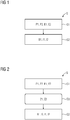
- the Fig. 1 shows an exemplary embodiment of the inventive method S for imaging an examination area of an object to be examined with a computed tomography system according to a first embodiment.
- the examination area has a first contrast agent and a second contrast agent different from the first contrast agent.
- the method S has at least the steps of recording S1 and generating S2.
- first projection measured data P1 are recorded with a first energy range E1 and second projection measured data P2 different from the first projection measured data P1 with a second energy range E2 different from the first energy range E1 in an examination region.
- the first energy range E1 comprises a first X-ray spectrum and the second energy range E2 comprises a second X-ray spectrum.
- the first energy range E1 comprises a first detector energy range and the second energy range E2 comprises a second detector energy range.
- Spectral projection measurement data P1, P2 are recorded, so that a spectral technique for generating S2 of the first image B1 can be used.
- a first image B1 is generated on the basis of the first projection measurement data P1 and the second projection measurement data P2.
- the first image B1 also has only isolated first information I1 of the first contrast agent, only isolated second information 12 of the second contrast agent, or isolated first information I1 of the first contrast agent together with isolated second information 12 of the second contrast agent.
- the first contrast agent has a therapeutically effective functionalization.
- the therapeutically effective functionalization comprises chemical and / or radioactive substances bound to the first contrast agent.
- the first contrast agent has a contrast substance bound to microspheres or nanoparticles.
- the first contrast agent has at least one, preferably exactly one, of the elements holmium, iron, gold, tungsten or gadolinium.
- the second contrast agent is indicative of a blood flow characteristic, for example a blood circulation.
- the second contrast agent has at least one, preferably exactly one, of the elements iodine or gadolinium.
- the first image B1 may have local information about a local distribution of the first contrast agent and / or second contrast agent.
- the first image B1 may have local information about a quantity of the first contrast agent and / or the second contrast agent.
- the first image B1 may have local information about a quantity or distribution of the first contrast agent having a therapeutically effective functionalization.
- the first image B1 may have local information about a local distribution of the first contrast agent and / or the second contrast agent in a predetermined range.
- the first image B1 may have local information based on the second contrast agent about a local perfusion in a predetermined range.
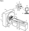
- the Fig. 2 shows an exemplary embodiment of the inventive method S for imaging an examination area of an object to be examined with a computed tomography system according to a second embodiment.
- the method further comprises a step of subtracting S3.
- the first contrast agent or the second contrast agent has an element which has a K-edge in the range of 30 to 100 keV.
- iodine, gadolinium and gold have a K-edge in this range.
- iodine, gadolinium and gold are suitable as contrast agents.
- the achievable contrast in, for example, the first image or the second image can advantageously be increased by the K-edge of the first contrast agent or of the second contrast agent.
- the first energy range E1 or the second energy range E2 can preferably be adapted to the photon energy at which the energy value of the K edge is reached or exceeded. It is initially possible to generate a first intermediate image Z1 for the first energy range E1 in the intermediate image generation S3 step and a second intermediate image Z2 for the second energy range E2. Subsequently, a difference image D from the first intermediate image Z1 and the second intermediate image Z2 can be generated.
- the different dependency of the attenuation coefficients on the photon energy of the X-ray radiation of the first contrast agent and the second contrast agent can be advantageously used, wherein the dependency is particularly pronounced in the region of the K edge.
- an energy threshold or discriminator threshold between the first detector energy range and the second detector energy range can be adapted to the K edge of the first contrast agent or of the second contrast agent.
- the energy threshold may substantially correspond to the energy of the K-edge.
- the difference image D may be the first image B1 or the second image B2. The difference image D can only have isolated information of the contrast agent, which has an element with a K edge.
- the Fig. 3 shows an exemplary embodiment of the inventive method S for imaging an examination area of an object to be examined with a computed tomography system according to a third embodiment.
- the method S further comprises a step of multi-material decomposition S4.
- the multi-material decomposition S4 can be raw-data-based or image-data-based.
- a method for multi-material decomposition S4 is for example from the document DE102009017615 A1 known.
- more than two materials or material pairs can be separated from one another, for example water or soft tissue, the first contrast agent and the second contrast agent.
- the Fig. 4 shows an exemplary embodiment of the inventive method S for imaging an examination area of an object to be examined with a computed tomography system according to a fourth embodiment.
- the methods S according to the first, second and third embodiments may further generate the second image B2 and / or the third image B3 in the step of generating S2.
- the second image B2 has isolated second information 12 of the second contrast agent, the isolated first information I1 of the first contrast agent being removed.
- the second image B2 comprises isolated first information I1 of the first contrast agent, the isolated second information 12 of the second contrast agent being removed.
- the isolated first information I1 of the first contrast agent and the isolated second information 12 of the second contrast agent are removed.
- the Fig. 5 shows an exemplary embodiment of the computer tomography system 31 according to the invention according to a first embodiment for carrying out the method according to the invention.
- the computed tomography system 31 includes a projection data acquisition unit 33 having a rotor 35.
- the rotor 35 includes an X-ray source 37 and the detector device 29.
- the object 39 is mounted on the patient couch 41 and is movable along the rotation axis z 43 by the projection data acquisition unit 33.
- a computer unit 45 is used for controlling and calculating the sectional images.
- the computer unit 45 comprises a control device 50 with a memory device 51.
- the computer unit 45 further comprises an image generation device 52 having an input interface 53, a reconstruction unit 54, an image data interface 55 and a material decomposition unit 56.
- the material decomposition unit 56 can be used to perform the multimaterial decomposition and / or the difference formation be suitable.
- An input device 47 and an output device 49 are connected to the computer unit 45.
- the computed tomography system 31 further comprises a first injection device 58 for injecting the first contrast agent, wherein the first injection device 58 comprises a control device for executing a patient-specific injection protocol.
- the computed tomography system 31 further comprises a second injection device 60 for injecting the second contrast agent, wherein the second injection device 60 comprises a control device for executing a patient-specific injection protocol.
- the projection data acquisition unit 33 has a direct-converting X-ray detector in the detector device 29 or means for changing the X-ray spectrum or for adjusting the first energy range and the second energy range, for example by means of filtering or kV-switching.
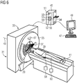
- the Fig. 6 shows an exemplary embodiment of the computer tomography system 31 according to the invention according to a second embodiment for carrying out the method according to the invention.
- the projection data acquisition unit 33 has two X-ray sources 37 and two detector devices 29 (second detector device not shown for clarity).
- Computed Tomography System 31 is a dual-source CT.
Landscapes
- Health & Medical Sciences (AREA)
- Life Sciences & Earth Sciences (AREA)
- Engineering & Computer Science (AREA)
- Medical Informatics (AREA)
- Animal Behavior & Ethology (AREA)
- General Health & Medical Sciences (AREA)
- Public Health (AREA)
- Veterinary Medicine (AREA)
- Pathology (AREA)
- Surgery (AREA)
- High Energy & Nuclear Physics (AREA)
- Physics & Mathematics (AREA)
- Nuclear Medicine, Radiotherapy & Molecular Imaging (AREA)
- Optics & Photonics (AREA)
- Biophysics (AREA)
- Radiology & Medical Imaging (AREA)
- Biomedical Technology (AREA)
- Heart & Thoracic Surgery (AREA)
- Molecular Biology (AREA)
- Epidemiology (AREA)
- Chemical & Material Sciences (AREA)
- Pulmonology (AREA)
- Theoretical Computer Science (AREA)
- Dentistry (AREA)
- Oral & Maxillofacial Surgery (AREA)
- Dermatology (AREA)
- Medicinal Chemistry (AREA)
- Pharmacology & Pharmacy (AREA)
- Nanotechnology (AREA)
- Apparatus For Radiation Diagnosis (AREA)
Priority Applications (1)
| Application Number | Priority Date | Filing Date | Title |
|---|---|---|---|
| US15/973,637 US20180325479A1 (en) | 2017-05-10 | 2018-05-08 | Method for imaging an examination region having first contrast medium and second contrast medium |
Applications Claiming Priority (1)
| Application Number | Priority Date | Filing Date | Title |
|---|---|---|---|
| EP17170450 | 2017-05-10 |
Publications (1)
| Publication Number | Publication Date |
|---|---|
| EP3400877A1 true EP3400877A1 (fr) | 2018-11-14 |
Family
ID=58707322
Family Applications (1)
| Application Number | Title | Priority Date | Filing Date |
|---|---|---|---|
| EP18152125.3A Withdrawn EP3400877A1 (fr) | 2017-05-10 | 2018-01-17 | Procédé d'imagerie d'une zone d'examen au moyen d'un premier agent de contraste et d'un second agent de contraste |
Country Status (2)
| Country | Link |
|---|---|
| US (1) | US20180325479A1 (fr) |
| EP (1) | EP3400877A1 (fr) |
Families Citing this family (1)
| Publication number | Priority date | Publication date | Assignee | Title |
|---|---|---|---|---|
| DE102021210860A1 (de) | 2021-09-28 | 2023-03-30 | Siemens Healthcare Gmbh | Computerimplementiertes Verfahren zur Auswertung von Bilddaten eines Patienten, Eingriffsanordnung, Computerprogramm und elektronisch lesbarer Datenträger |
Citations (6)
| Publication number | Priority date | Publication date | Assignee | Title |
|---|---|---|---|---|
| DE102007024158A1 (de) * | 2007-05-24 | 2008-11-27 | Bayer Schering Pharma Aktiengesellschaft | Auswahlverfahren für zwei Kontrastmittel zur Verwendung in einer Dual-Energy-CT-Untersuchung, Kontrastmittelkombination und Erzeugung von CT-Aufnahmen mit einer Kontrastmittelkombination mit unterschiedlichen Energiespektren |
| DE102007050438A1 (de) * | 2007-10-22 | 2009-04-23 | Siemens Ag | Verfahren zur simultanen Darstellung der Durchblutung von Muskelgewebe und Gefäßen |
| DE102009017615A1 (de) | 2009-04-16 | 2010-12-30 | Siemens Aktiengesellschaft | Verfahren und Bildrekonstruktionseinrichtung zur Erzeugung von radiologischen Bilddaten |
| US20150038827A1 (en) | 2013-07-31 | 2015-02-05 | Kabushiki Kaisha Toshiba | Medical image diagnostic apparatus and ultrasonic diagnostic apparatus |
| US20150221082A1 (en) | 2012-08-06 | 2015-08-06 | Koninklijke Philips N.V. | Dynamic contrast-enhanced imaging based permeability metric |
| WO2016146214A1 (fr) | 2015-03-18 | 2016-09-22 | Koninklijke Philips N.V. | Détermination de la concentration en médicament après une chimio-embolisation par voie transartérielle avec des billes microsphériques d'élution de médicament de tailles différentes |
-
2018
- 2018-01-17 EP EP18152125.3A patent/EP3400877A1/fr not_active Withdrawn
- 2018-05-08 US US15/973,637 patent/US20180325479A1/en not_active Abandoned
Patent Citations (6)
| Publication number | Priority date | Publication date | Assignee | Title |
|---|---|---|---|---|
| DE102007024158A1 (de) * | 2007-05-24 | 2008-11-27 | Bayer Schering Pharma Aktiengesellschaft | Auswahlverfahren für zwei Kontrastmittel zur Verwendung in einer Dual-Energy-CT-Untersuchung, Kontrastmittelkombination und Erzeugung von CT-Aufnahmen mit einer Kontrastmittelkombination mit unterschiedlichen Energiespektren |
| DE102007050438A1 (de) * | 2007-10-22 | 2009-04-23 | Siemens Ag | Verfahren zur simultanen Darstellung der Durchblutung von Muskelgewebe und Gefäßen |
| DE102009017615A1 (de) | 2009-04-16 | 2010-12-30 | Siemens Aktiengesellschaft | Verfahren und Bildrekonstruktionseinrichtung zur Erzeugung von radiologischen Bilddaten |
| US20150221082A1 (en) | 2012-08-06 | 2015-08-06 | Koninklijke Philips N.V. | Dynamic contrast-enhanced imaging based permeability metric |
| US20150038827A1 (en) | 2013-07-31 | 2015-02-05 | Kabushiki Kaisha Toshiba | Medical image diagnostic apparatus and ultrasonic diagnostic apparatus |
| WO2016146214A1 (fr) | 2015-03-18 | 2016-09-22 | Koninklijke Philips N.V. | Détermination de la concentration en médicament après une chimio-embolisation par voie transartérielle avec des billes microsphériques d'élution de médicament de tailles différentes |
Also Published As
| Publication number | Publication date |
|---|---|
| US20180325479A1 (en) | 2018-11-15 |
Similar Documents
| Publication | Publication Date | Title |
|---|---|---|
| EP2150179B1 (fr) | Procédé de sélection de deux agents de contraste destinés à être utilisés lors d'un examen par ct à double énergie, combinaison d'agents de contraste et production d'images de ct à l'aide d'une combinaison d'agents de contraste et de spectres d'énergie différents | |
| DE102010027227B4 (de) | Verfahren und Computertomographiegerät zur Durchführung einer angiographischen Untersuchung | |
| DE102016203257B4 (de) | Erzeugen von kontrastverstärkten Bilddaten auf Basis einer Multi-Energie-Röntgenbildgebung | |
| DE102013200337B4 (de) | Verfahren, Computertomopraph und Computerprogrammprodukt zum Bestimmen von Intensitätswerten einer Röntgenstrahlung zur Dosismodulation | |
| DE102012211892B4 (de) | Verfahren zur Extraktion eines Datensatzes aus einem medizinischen Bilddatensatz sowie medizinische Bildaufnahmeeinrichtung und Computerprogramm | |
| DE10355383A1 (de) | Verfahren und Vorrichtung zum Erfassen von Perfusionsdaten | |
| WO2012097801A1 (fr) | Procédé de production d'une représentation radiographique assistée par agent de contraste et système radiographique approprié | |
| DE102016207437B4 (de) | Spektralunabhängige Ermittlung von Kalkablagerungen in Blutgefäßen | |
| DE102011053762A1 (de) | System und Verfahren zum Bandpassfiltern für Dualenergie-CT | |
| DE10355094A1 (de) | Verfahren und Vorrichtung zur Feststellung von Struktur-Perfusions- und Funktionsabnormitäten | |
| DE102015207107A1 (de) | Verfahren zur Erzeugung einer virtuellen Röntgenprojektion anhand eines mittels Röntgenbildaufnahmevorrichtung erhaltenen Bilddatensatzes, Computerprogramm, Datenträger sowie Röntgenbildaufnahmevorrichtung | |
| DE10350532A1 (de) | Verfahren und Vorrichtung zur Feststellung von Struktur-, Perfusions- und Funktionsabnormitäten | |
| DE102015217141A1 (de) | Erzeugen von kontrastverstärkten Bilddaten von zu untersuchendem Brustgewebe | |
| DE102016222093A1 (de) | Simultaner Einsatz von unterschiedlichen Kontrastmitteln bei CT-Bildgebungsverfahren | |
| DE102012222714A1 (de) | Ermittlung eines Mehrfachenergie-Bildes | |
| DE102015218928B4 (de) | Verfahren zur Erzeugung von Röntgenbilddaten eines Untersuchungsobjektes mit unterdrücktem Calcium-Signal | |
| EP3628226A1 (fr) | Procédé de surveillance d'un prélèvement de tissu au moyen d'un système d'imagerie à rayons x | |
| DE102015217617A1 (de) | Verfahren zum Korrigieren von Röntgenbilddaten umfassend Information bezüglich eines Zerfallsprozesses eines radioaktiven Materials | |
| EP3795082B1 (fr) | Procédé et dispositif de génération d'un ensemble de données d'image de tomodensitométrie | |
| EP3400877A1 (fr) | Procédé d'imagerie d'une zone d'examen au moyen d'un premier agent de contraste et d'un second agent de contraste | |
| DE102016224717B4 (de) | Verfahren zum Ermitteln von Gewebeeigenschaften von Tumoren, sowie Bildanalyseeinrichtung, Computertomographiesystem, Computerprogrammprodukt und computerlesbares Medium | |
| DE102015212369A1 (de) | Verfahren und Vorrichtung zur selektiven Detektierung und Quantifizierung von Kontrastmitteln | |
| DE102008045633A1 (de) | Verfahren zur verbesserten Darstellung von Mehr-Energie-CT-Aufnahmen | |
| DE102006001655A1 (de) | Verfahren und Vorrichtung zur virtuellen Darmreinigung | |
| DE102007050438B4 (de) | Verfahren und CT-System zur simultanen Darstellung der Durchblutung von Muskelgewebe und Gefäßen |
Legal Events
| Date | Code | Title | Description |
|---|---|---|---|
| PUAI | Public reference made under article 153(3) epc to a published international application that has entered the european phase |
Free format text: ORIGINAL CODE: 0009012 |
|
| STAA | Information on the status of an ep patent application or granted ep patent |
Free format text: STATUS: THE APPLICATION HAS BEEN PUBLISHED |
|
| AK | Designated contracting states |
Kind code of ref document: A1 Designated state(s): AL AT BE BG CH CY CZ DE DK EE ES FI FR GB GR HR HU IE IS IT LI LT LU LV MC MK MT NL NO PL PT RO RS SE SI SK SM TR |
|
| AX | Request for extension of the european patent |
Extension state: BA ME |
|
| STAA | Information on the status of an ep patent application or granted ep patent |
Free format text: STATUS: REQUEST FOR EXAMINATION WAS MADE |
|
| 17P | Request for examination filed |
Effective date: 20190503 |
|
| RBV | Designated contracting states (corrected) |
Designated state(s): AL AT BE BG CH CY CZ DE DK EE ES FI FR GB GR HR HU IE IS IT LI LT LU LV MC MK MT NL NO PL PT RO RS SE SI SK SM TR |
|
| STAA | Information on the status of an ep patent application or granted ep patent |
Free format text: STATUS: EXAMINATION IS IN PROGRESS |
|
| 17Q | First examination report despatched |
Effective date: 20200714 |
|
| STAA | Information on the status of an ep patent application or granted ep patent |
Free format text: STATUS: EXAMINATION IS IN PROGRESS |
|
| STAA | Information on the status of an ep patent application or granted ep patent |
Free format text: STATUS: THE APPLICATION IS DEEMED TO BE WITHDRAWN |
|
| 18D | Application deemed to be withdrawn |
Effective date: 20210126 |