EP3400032B1 - System for the treatment of wounds with dressing having closed cells - Google Patents
System for the treatment of wounds with dressing having closed cells Download PDFInfo
- Publication number
- EP3400032B1 EP3400032B1 EP16820492.3A EP16820492A EP3400032B1 EP 3400032 B1 EP3400032 B1 EP 3400032B1 EP 16820492 A EP16820492 A EP 16820492A EP 3400032 B1 EP3400032 B1 EP 3400032B1
- Authority
- EP
- European Patent Office
- Prior art keywords
- closed cells
- manifold
- pressure
- tissue site
- sheets
- Prior art date
- Legal status (The legal status is an assumption and is not a legal conclusion. Google has not performed a legal analysis and makes no representation as to the accuracy of the status listed.)
- Active
Links
- 206010052428 Wound Diseases 0.000 title description 75
- 208000027418 Wounds and injury Diseases 0.000 title description 74
- 238000011282 treatment Methods 0.000 title description 25
- 239000012530 fluid Substances 0.000 claims description 101
- 230000001225 therapeutic effect Effects 0.000 claims description 21
- 238000004891 communication Methods 0.000 claims description 11
- 238000010168 coupling process Methods 0.000 claims description 11
- 238000005859 coupling reaction Methods 0.000 claims description 11
- 230000008878 coupling Effects 0.000 claims description 10
- 210000001519 tissue Anatomy 0.000 description 223
- 238000002560 therapeutic procedure Methods 0.000 description 91
- 239000000243 solution Substances 0.000 description 39
- 239000000463 material Substances 0.000 description 35
- 238000000034 method Methods 0.000 description 31
- 238000005469 granulation Methods 0.000 description 21
- 230000003179 granulation Effects 0.000 description 21
- 239000000945 filler Substances 0.000 description 8
- 239000010410 layer Substances 0.000 description 8
- 230000037361 pathway Effects 0.000 description 8
- 230000000845 anti-microbial effect Effects 0.000 description 7
- 210000000416 exudates and transudate Anatomy 0.000 description 7
- 230000006870 function Effects 0.000 description 7
- 239000007788 liquid Substances 0.000 description 7
- 239000004599 antimicrobial Substances 0.000 description 6
- 238000009826 distribution Methods 0.000 description 6
- 230000008901 benefit Effects 0.000 description 5
- 238000005516 engineering process Methods 0.000 description 5
- 229920001903 high density polyethylene Polymers 0.000 description 5
- 239000004700 high-density polyethylene Substances 0.000 description 5
- 229920001684 low density polyethylene Polymers 0.000 description 5
- 239000004702 low-density polyethylene Substances 0.000 description 5
- 229920000728 polyester Polymers 0.000 description 5
- 230000008569 process Effects 0.000 description 5
- 238000003860 storage Methods 0.000 description 5
- VGGSQFUCUMXWEO-UHFFFAOYSA-N Ethene Chemical compound C=C VGGSQFUCUMXWEO-UHFFFAOYSA-N 0.000 description 4
- 239000005977 Ethylene Substances 0.000 description 4
- 239000000853 adhesive Substances 0.000 description 4
- 230000001070 adhesive effect Effects 0.000 description 4
- 238000009530 blood pressure measurement Methods 0.000 description 4
- 239000007789 gas Substances 0.000 description 4
- 230000001788 irregular Effects 0.000 description 4
- 239000004711 α-olefin Substances 0.000 description 4
- 229920000089 Cyclic olefin copolymer Polymers 0.000 description 3
- 230000015572 biosynthetic process Effects 0.000 description 3
- 229920001577 copolymer Polymers 0.000 description 3
- 230000006837 decompression Effects 0.000 description 3
- 230000007423 decrease Effects 0.000 description 3
- 238000010586 diagram Methods 0.000 description 3
- 210000002615 epidermis Anatomy 0.000 description 3
- 239000000835 fiber Substances 0.000 description 3
- 239000006260 foam Substances 0.000 description 3
- 208000015181 infectious disease Diseases 0.000 description 3
- 208000014674 injury Diseases 0.000 description 3
- -1 polyethylene copolymers Polymers 0.000 description 3
- 229920000642 polymer Polymers 0.000 description 3
- 239000002356 single layer Substances 0.000 description 3
- VXNZUUAINFGPBY-UHFFFAOYSA-N 1-Butene Chemical compound CCC=C VXNZUUAINFGPBY-UHFFFAOYSA-N 0.000 description 2
- LIKMAJRDDDTEIG-UHFFFAOYSA-N 1-hexene Chemical compound CCCCC=C LIKMAJRDDDTEIG-UHFFFAOYSA-N 0.000 description 2
- KWKAKUADMBZCLK-UHFFFAOYSA-N 1-octene Chemical compound CCCCCCC=C KWKAKUADMBZCLK-UHFFFAOYSA-N 0.000 description 2
- IJGRMHOSHXDMSA-UHFFFAOYSA-N Atomic nitrogen Chemical compound N#N IJGRMHOSHXDMSA-UHFFFAOYSA-N 0.000 description 2
- CURLTUGMZLYLDI-UHFFFAOYSA-N Carbon dioxide Chemical compound O=C=O CURLTUGMZLYLDI-UHFFFAOYSA-N 0.000 description 2
- 229920010126 Linear Low Density Polyethylene (LLDPE) Polymers 0.000 description 2
- 229920002614 Polyether block amide Polymers 0.000 description 2
- 208000025865 Ulcer Diseases 0.000 description 2
- 210000000577 adipose tissue Anatomy 0.000 description 2
- 230000001580 bacterial effect Effects 0.000 description 2
- 210000000988 bone and bone Anatomy 0.000 description 2
- 230000009172 bursting Effects 0.000 description 2
- 210000000845 cartilage Anatomy 0.000 description 2
- 230000000295 complement effect Effects 0.000 description 2
- 239000004020 conductor Substances 0.000 description 2
- 210000002808 connective tissue Anatomy 0.000 description 2
- 239000000356 contaminant Substances 0.000 description 2
- 230000001351 cycling effect Effects 0.000 description 2
- 230000007547 defect Effects 0.000 description 2
- 230000002500 effect on skin Effects 0.000 description 2
- 229920001971 elastomer Polymers 0.000 description 2
- 230000002708 enhancing effect Effects 0.000 description 2
- 230000035876 healing Effects 0.000 description 2
- 239000012943 hotmelt Substances 0.000 description 2
- 230000002209 hydrophobic effect Effects 0.000 description 2
- WQYVRQLZKVEZGA-UHFFFAOYSA-N hypochlorite Chemical compound Cl[O-] WQYVRQLZKVEZGA-UHFFFAOYSA-N 0.000 description 2
- 238000007373 indentation Methods 0.000 description 2
- 210000003041 ligament Anatomy 0.000 description 2
- 239000006193 liquid solution Substances 0.000 description 2
- 238000004519 manufacturing process Methods 0.000 description 2
- 238000005259 measurement Methods 0.000 description 2
- 230000007246 mechanism Effects 0.000 description 2
- 238000002844 melting Methods 0.000 description 2
- 230000008018 melting Effects 0.000 description 2
- 210000003205 muscle Anatomy 0.000 description 2
- 238000009581 negative-pressure wound therapy Methods 0.000 description 2
- 238000012856 packing Methods 0.000 description 2
- 206010033675 panniculitis Diseases 0.000 description 2
- YWAKXRMUMFPDSH-UHFFFAOYSA-N pentene Chemical compound CCCC=C YWAKXRMUMFPDSH-UHFFFAOYSA-N 0.000 description 2
- 229920000573 polyethylene Polymers 0.000 description 2
- 229920005638 polyethylene monopolymer Polymers 0.000 description 2
- 229920005606 polypropylene copolymer Polymers 0.000 description 2
- 229920005629 polypropylene homopolymer Polymers 0.000 description 2
- 229920001296 polysiloxane Polymers 0.000 description 2
- 238000003825 pressing Methods 0.000 description 2
- 238000012545 processing Methods 0.000 description 2
- SQGYOTSLMSWVJD-UHFFFAOYSA-N silver(1+) nitrate Chemical compound [Ag+].[O-]N(=O)=O SQGYOTSLMSWVJD-UHFFFAOYSA-N 0.000 description 2
- 238000002791 soaking Methods 0.000 description 2
- 239000002904 solvent Substances 0.000 description 2
- 210000004304 subcutaneous tissue Anatomy 0.000 description 2
- 239000000126 substance Substances 0.000 description 2
- 210000002435 tendon Anatomy 0.000 description 2
- 230000000699 topical effect Effects 0.000 description 2
- 230000008733 trauma Effects 0.000 description 2
- 231100000397 ulcer Toxicity 0.000 description 2
- 229920001862 ultra low molecular weight polyethylene Polymers 0.000 description 2
- 230000002792 vascular Effects 0.000 description 2
- JMMZCWZIJXAGKW-UHFFFAOYSA-N 2-methylpent-2-ene Chemical compound CCC=C(C)C JMMZCWZIJXAGKW-UHFFFAOYSA-N 0.000 description 1
- 206010003445 Ascites Diseases 0.000 description 1
- 229940123208 Biguanide Drugs 0.000 description 1
- 206010063560 Excessive granulation tissue Diseases 0.000 description 1
- 239000004952 Polyamide Substances 0.000 description 1
- 239000004698 Polyethylene Substances 0.000 description 1
- 229920002367 Polyisobutene Polymers 0.000 description 1
- 239000004721 Polyphenylene oxide Substances 0.000 description 1
- 239000004793 Polystyrene Substances 0.000 description 1
- 239000004372 Polyvinyl alcohol Substances 0.000 description 1
- 239000004820 Pressure-sensitive adhesive Substances 0.000 description 1
- FAPWRFPIFSIZLT-UHFFFAOYSA-M Sodium chloride Chemical compound [Na+].[Cl-] FAPWRFPIFSIZLT-UHFFFAOYSA-M 0.000 description 1
- NINIDFKCEFEMDL-UHFFFAOYSA-N Sulfur Chemical compound [S] NINIDFKCEFEMDL-UHFFFAOYSA-N 0.000 description 1
- 208000002847 Surgical Wound Diseases 0.000 description 1
- 229920010346 Very Low Density Polyethylene (VLDPE) Polymers 0.000 description 1
- 239000003522 acrylic cement Substances 0.000 description 1
- 230000001154 acute effect Effects 0.000 description 1
- 230000004888 barrier function Effects 0.000 description 1
- 230000006399 behavior Effects 0.000 description 1
- 150000004283 biguanides Chemical class 0.000 description 1
- 230000005540 biological transmission Effects 0.000 description 1
- 230000017531 blood circulation Effects 0.000 description 1
- 229910002092 carbon dioxide Inorganic materials 0.000 description 1
- 239000001569 carbon dioxide Substances 0.000 description 1
- 125000002091 cationic group Chemical group 0.000 description 1
- 230000008859 change Effects 0.000 description 1
- 238000012412 chemical coupling Methods 0.000 description 1
- 230000001684 chronic effect Effects 0.000 description 1
- 239000011248 coating agent Substances 0.000 description 1
- 238000000576 coating method Methods 0.000 description 1
- 239000002131 composite material Substances 0.000 description 1
- 230000006835 compression Effects 0.000 description 1
- 238000007906 compression Methods 0.000 description 1
- 230000003750 conditioning effect Effects 0.000 description 1
- 230000006378 damage Effects 0.000 description 1
- 230000003247 decreasing effect Effects 0.000 description 1
- 230000002950 deficient Effects 0.000 description 1
- 230000001419 dependent effect Effects 0.000 description 1
- 210000004207 dermis Anatomy 0.000 description 1
- 238000011161 development Methods 0.000 description 1
- 206010012601 diabetes mellitus Diseases 0.000 description 1
- 230000000694 effects Effects 0.000 description 1
- 239000000806 elastomer Substances 0.000 description 1
- 230000003028 elevating effect Effects 0.000 description 1
- 238000004049 embossing Methods 0.000 description 1
- 210000000981 epithelium Anatomy 0.000 description 1
- 229920001038 ethylene copolymer Polymers 0.000 description 1
- 238000007667 floating Methods 0.000 description 1
- 239000000499 gel Substances 0.000 description 1
- 239000008187 granular material Substances 0.000 description 1
- 210000001126 granulation tissue Anatomy 0.000 description 1
- 230000005484 gravity Effects 0.000 description 1
- 239000000416 hydrocolloid Substances 0.000 description 1
- 239000000017 hydrogel Substances 0.000 description 1
- 230000002706 hydrostatic effect Effects 0.000 description 1
- 230000002458 infectious effect Effects 0.000 description 1
- 229920000554 ionomer Polymers 0.000 description 1
- 239000000644 isotonic solution Substances 0.000 description 1
- 229920001179 medium density polyethylene Polymers 0.000 description 1
- 239000004701 medium-density polyethylene Substances 0.000 description 1
- 239000012528 membrane Substances 0.000 description 1
- 230000005012 migration Effects 0.000 description 1
- 238000013508 migration Methods 0.000 description 1
- 239000000203 mixture Substances 0.000 description 1
- 238000012986 modification Methods 0.000 description 1
- 230000004048 modification Effects 0.000 description 1
- TVMXDCGIABBOFY-UHFFFAOYSA-N n-Octanol Natural products CCCCCCCC TVMXDCGIABBOFY-UHFFFAOYSA-N 0.000 description 1
- 230000001537 neural effect Effects 0.000 description 1
- 229910052757 nitrogen Inorganic materials 0.000 description 1
- 230000003287 optical effect Effects 0.000 description 1
- 239000002245 particle Substances 0.000 description 1
- 239000006072 paste Substances 0.000 description 1
- 230000002093 peripheral effect Effects 0.000 description 1
- 229940021222 peritoneal dialysis isotonic solution Drugs 0.000 description 1
- 230000035699 permeability Effects 0.000 description 1
- 229920001084 poly(chloroprene) Polymers 0.000 description 1
- 229920002647 polyamide Polymers 0.000 description 1
- 239000004417 polycarbonate Substances 0.000 description 1
- 229920000515 polycarbonate Polymers 0.000 description 1
- 229920000570 polyether Polymers 0.000 description 1
- 229920001195 polyisoprene Polymers 0.000 description 1
- 229920001155 polypropylene Polymers 0.000 description 1
- 229920002223 polystyrene Polymers 0.000 description 1
- 229920002635 polyurethane Polymers 0.000 description 1
- 239000004814 polyurethane Substances 0.000 description 1
- 229920006264 polyurethane film Polymers 0.000 description 1
- 229920002451 polyvinyl alcohol Polymers 0.000 description 1
- QQONPFPTGQHPMA-UHFFFAOYSA-N propylene Natural products CC=C QQONPFPTGQHPMA-UHFFFAOYSA-N 0.000 description 1
- 125000004805 propylene group Chemical group [H]C([H])([H])C([H])([*:1])C([H])([H])[*:2] 0.000 description 1
- 238000007789 sealing Methods 0.000 description 1
- 229910001961 silver nitrate Inorganic materials 0.000 description 1
- 210000003491 skin Anatomy 0.000 description 1
- 239000007787 solid Substances 0.000 description 1
- 230000004936 stimulating effect Effects 0.000 description 1
- 238000007920 subcutaneous administration Methods 0.000 description 1
- 229910052717 sulfur Inorganic materials 0.000 description 1
- 239000011593 sulfur Substances 0.000 description 1
- 238000001356 surgical procedure Methods 0.000 description 1
- 230000009182 swimming Effects 0.000 description 1
- 229920001169 thermoplastic Polymers 0.000 description 1
- 229920002725 thermoplastic elastomer Polymers 0.000 description 1
- 239000012815 thermoplastic material Substances 0.000 description 1
- 230000000472 traumatic effect Effects 0.000 description 1
- 238000011277 treatment modality Methods 0.000 description 1
- 238000011144 upstream manufacturing Methods 0.000 description 1
- 201000002282 venous insufficiency Diseases 0.000 description 1
- 238000013022 venting Methods 0.000 description 1
- 239000002699 waste material Substances 0.000 description 1
- XLYOFNOQVPJJNP-UHFFFAOYSA-N water Chemical compound O XLYOFNOQVPJJNP-UHFFFAOYSA-N 0.000 description 1
- 238000003466 welding Methods 0.000 description 1
- 230000029663 wound healing Effects 0.000 description 1
Images


Classifications
-
- A61F13/05—
-
- A—HUMAN NECESSITIES
- A61—MEDICAL OR VETERINARY SCIENCE; HYGIENE
- A61M—DEVICES FOR INTRODUCING MEDIA INTO, OR ONTO, THE BODY; DEVICES FOR TRANSDUCING BODY MEDIA OR FOR TAKING MEDIA FROM THE BODY; DEVICES FOR PRODUCING OR ENDING SLEEP OR STUPOR
- A61M1/00—Suction or pumping devices for medical purposes; Devices for carrying-off, for treatment of, or for carrying-over, body-liquids; Drainage systems
- A61M1/90—Negative pressure wound therapy devices, i.e. devices for applying suction to a wound to promote healing, e.g. including a vacuum dressing
- A61M1/91—Suction aspects of the dressing
- A61M1/915—Constructional details of the pressure distribution manifold
-
- A—HUMAN NECESSITIES
- A61—MEDICAL OR VETERINARY SCIENCE; HYGIENE
- A61M—DEVICES FOR INTRODUCING MEDIA INTO, OR ONTO, THE BODY; DEVICES FOR TRANSDUCING BODY MEDIA OR FOR TAKING MEDIA FROM THE BODY; DEVICES FOR PRODUCING OR ENDING SLEEP OR STUPOR
- A61M1/00—Suction or pumping devices for medical purposes; Devices for carrying-off, for treatment of, or for carrying-over, body-liquids; Drainage systems
- A61M1/90—Negative pressure wound therapy devices, i.e. devices for applying suction to a wound to promote healing, e.g. including a vacuum dressing
- A61M1/96—Suction control thereof
-
- A—HUMAN NECESSITIES
- A61—MEDICAL OR VETERINARY SCIENCE; HYGIENE
- A61M—DEVICES FOR INTRODUCING MEDIA INTO, OR ONTO, THE BODY; DEVICES FOR TRANSDUCING BODY MEDIA OR FOR TAKING MEDIA FROM THE BODY; DEVICES FOR PRODUCING OR ENDING SLEEP OR STUPOR
- A61M1/00—Suction or pumping devices for medical purposes; Devices for carrying-off, for treatment of, or for carrying-over, body-liquids; Drainage systems
- A61M1/71—Suction drainage systems
- A61M1/74—Suction control
-
- A—HUMAN NECESSITIES
- A61—MEDICAL OR VETERINARY SCIENCE; HYGIENE
- A61M—DEVICES FOR INTRODUCING MEDIA INTO, OR ONTO, THE BODY; DEVICES FOR TRANSDUCING BODY MEDIA OR FOR TAKING MEDIA FROM THE BODY; DEVICES FOR PRODUCING OR ENDING SLEEP OR STUPOR
- A61M1/00—Suction or pumping devices for medical purposes; Devices for carrying-off, for treatment of, or for carrying-over, body-liquids; Drainage systems
- A61M1/71—Suction drainage systems
- A61M1/77—Suction-irrigation systems
-
- A—HUMAN NECESSITIES
- A61—MEDICAL OR VETERINARY SCIENCE; HYGIENE
- A61M—DEVICES FOR INTRODUCING MEDIA INTO, OR ONTO, THE BODY; DEVICES FOR TRANSDUCING BODY MEDIA OR FOR TAKING MEDIA FROM THE BODY; DEVICES FOR PRODUCING OR ENDING SLEEP OR STUPOR
- A61M1/00—Suction or pumping devices for medical purposes; Devices for carrying-off, for treatment of, or for carrying-over, body-liquids; Drainage systems
- A61M1/90—Negative pressure wound therapy devices, i.e. devices for applying suction to a wound to promote healing, e.g. including a vacuum dressing
- A61M1/92—Negative pressure wound therapy devices, i.e. devices for applying suction to a wound to promote healing, e.g. including a vacuum dressing with liquid supply means
-
- A—HUMAN NECESSITIES
- A61—MEDICAL OR VETERINARY SCIENCE; HYGIENE
- A61M—DEVICES FOR INTRODUCING MEDIA INTO, OR ONTO, THE BODY; DEVICES FOR TRANSDUCING BODY MEDIA OR FOR TAKING MEDIA FROM THE BODY; DEVICES FOR PRODUCING OR ENDING SLEEP OR STUPOR
- A61M2205/00—General characteristics of the apparatus
- A61M2205/33—Controlling, regulating or measuring
- A61M2205/3331—Pressure; Flow
- A61M2205/3344—Measuring or controlling pressure at the body treatment site
-
- A—HUMAN NECESSITIES
- A61—MEDICAL OR VETERINARY SCIENCE; HYGIENE
- A61M—DEVICES FOR INTRODUCING MEDIA INTO, OR ONTO, THE BODY; DEVICES FOR TRANSDUCING BODY MEDIA OR FOR TAKING MEDIA FROM THE BODY; DEVICES FOR PRODUCING OR ENDING SLEEP OR STUPOR
- A61M2205/00—General characteristics of the apparatus
- A61M2205/50—General characteristics of the apparatus with microprocessors or computers
Definitions
- Negative-pressure therapy may provide a number of benefits, including migration of epithelial and subcutaneous tissues, improved blood flow, and micro-deformation of tissue at a wound site. Together, these benefits can increase development of granulation tissue and reduce healing times.
- “Instillation” is another practice that generally refers to a process of slowly introducing fluid to a tissue site and leaving the fluid for a prescribed period of time before removing the fluid.
- a wound can be washed out with a stream of liquid solution, or a cavity can be washed out using a liquid solution for therapeutic purposes.
- instillation of topical treatment solutions over a wound bed can be combined with negative-pressure therapy to further promote wound healing by loosening soluble contaminants in a wound bed and removing infectious material. As a result, soluble bacterial burden can be decreased, contaminants removed, and the wound cleansed.
- a system for treating a tissue site may comprise a manifold including a non-porous film having a plurality of closed cells defined by a sealed region perforated with apertures extending through the seals, wherein the manifold is adapted to contact the tissue site.
- the system may further comprise a cover adapted to provide a fluid seal between a therapeutic environment including the manifold proximate one side of the cover and a local external environment on the other side of the cover.
- the plurality of closed cells is adapted to form distal channels with the cover and the apertures are adapted to provide fluid communication between the distal channels and the tissue site.
- the system may further comprise a negative-pressure source fluidly coupled to the therapeutic environment and adapted to provide negative pressure through the distal channels and the apertures to the tissue site.
- a dressing for treating a tissue site comprising a manifold including a non-porous film having a plurality of closed cells defined by a sealed region perforated with apertures extending through the seals, wherein the manifold is adapted to contact the tissue site.
- the dressing may further comprise a cover adapted to provide a fluid seal between a therapeutic environment including the manifold proximate one side of the cover and a local external environment on the other side of the cover.
- the plurality of closed cells may be adapted to form distal channels with the cover and the apertures are adapted to provide fluid communication between the distal channels and the tissue site.
- the therapeutic environment is adapted to receive negative pressure that is applied through the distal channels and the apertures to the tissue site.
- a dressing may include a non-porous manifold covered by a drape that forms a seal over the tissue site so that the manifold can distribute negative pressure and instill fluids to the tissue site.
- the structure of such manifolds may also generate macro and micro strains at the tissue site to enhance granulation.
- Such manifolds may be left at the tissue site for several days or more and often must be removed when they become clogged with tissue ingrowth and exudates from the tissue site. It is desirable to reduce the amount of tissue ingrowth that may cause pain or discomfort when the manifold is removed from the tissue site after the negative pressure and instillation therapies have been applied for several days or more.
- the manifold is adapted to be positioned and sealed at the tissue site by a cover that forms distal channels with the closed cells and the sealed space for receiving and distributing fluids to the tissue site through the apertures.
- the manifold is a single component comprising a plurality of closed cells separated by a sealed region, and apertures extending through the sealed region such that the manifold provides both a manifold function and a filler function when positioned between the tissue site and the cover.
- the words "preferred” and “preferably” refer to embodiments of the technology that afford certain benefits, under certain circumstances. However, other embodiments may also be preferred, under the same or other circumstances. Furthermore, the recitation of one or more preferred embodiments does not imply that other embodiments are not useful and is not intended to exclude other embodiments from the scope of the technology.
- Components may be fluidly coupled to each other to provide a path for transferring fluids (i.e., liquid and/or gas) between the components.
- components may be fluidly coupled through a fluid conductor, such as a tube.
- a tube is an elongated, cylindrical structure with some flexibility, but the geometry and rigidity may vary.
- components may also be coupled by virtue of physical proximity, being integral to a single structure, or being formed from the same piece of material.
- some fluid conductors may be molded into or otherwise integrally combined with other components. Coupling may also include mechanical, thermal, electrical, or chemical coupling (such as a chemical bond) in some contexts.
- a tube may mechanically and fluidly couple the dressing 102 to the container 112 in some embodiments.
- the tissue interface 108 may be a manifold 140.
- a "manifold" in this context generally includes any substance or structure providing a plurality of pathways adapted to collect or distribute fluid across a tissue site under pressure.
- a manifold may be adapted to receive negative pressure from a source and distribute negative pressure through multiple apertures across a tissue site, which may have the effect of collecting fluid from across a tissue site and drawing the fluid toward the source.
- the fluid path may be reversed or a secondary fluid path may be provided to facilitate delivering fluid across a tissue site.
- the pathways of a manifold may be interconnected to improve distribution or collection of fluids across a tissue site.
- a manifold may additionally or alternatively comprise projections that form interconnected fluid pathways.
- a manifold may be molded to provide surface projections that define interconnected fluid pathways.
- the cover 106 may provide a bacterial barrier and protection from physical trauma.
- the cover 106 may also be constructed from a material that can reduce evaporative losses and provide a fluid seal between two components or two environments, such as between a therapeutic environment and a local external environment.
- the cover 106 may be, for example, an elastomeric film or membrane that can provide a seal adequate to maintain a negative pressure at a tissue site for a given negative-pressure source.
- the cover 106 may have a high moisture-vapor transmission rate (MVTR) in some applications.
- the MVTR may be at least 300 g/m 2 per twenty-four hours in some embodiments.
- An attachment device such as an attachment device 142, may be used to attach the cover 106 to an attachment surface, such as undamaged epidermis, a gasket, or another cover.
- the attachment device may take many forms.
- an attachment device may be a medically-acceptable, pressure-sensitive adhesive that extends about a periphery, a portion, or an entire sealing member.
- some or all of the cover 106 may be coated with an acrylic adhesive having a coating weight between 25-65 grams per square meter (g.s.m.). Thicker adhesives, or combinations of adhesives, may be applied in some embodiments to improve the seal and reduce leaks.
- Other example embodiments of an attachment device may include a double-sided tape, paste, hydrocolloid, hydrogel, silicone gel, or organogel.
- the fluid-delivery interface 148 may include an inlet pad.
- the inlet pad may be a material that is not sound-absorbing.
- the inlet pad may be an elastomer.
- the inlet pad may be an elastic polymer, such as polyurethane, thermoplastic elastomers, polyether block amide (PEBAX), polyisoprene, polychloroprene, chlorosulphonated polythene, and polyisobutylene, blends and copolymers.
- the fluid-delivery interface 148 and the negative-pressure interface 144 may be integrated into a single pad for the delivery and removal of solutions from the tissue site 150, such as a V.A.C. Vera T.R.A.C.TM Pad available from Kinetic Concepts, Inc. of San Antonio, Texas.
- a controller such as the controller 110, may be a microprocessor or computer programmed to operate one or more components of the therapy system 100, such as the negative-pressure source 104.
- the controller 110 may be a microcontroller, which generally comprises an integrated circuit containing a processor core and a memory programmed to directly or indirectly control one or more operating parameters of the therapy system 100. Operating parameters may include the power applied to the negative-pressure source 104, the pressure generated by the negative-pressure source 104, or the pressure distributed to the tissue interface 108, for example.
- the controller 110 is also preferably configured to receive one or more input signals, such as a feedback signal, and programmed to modify one or more operating parameters based on the input signals.
- the tissue site 150 may include, without limitation, any irregularity with a tissue, such as an open wound, surgical incision, or diseased tissue.
- the therapy system 100 is presented in the context of a tissue site that includes a wound 152, which is through the epidermis 154, or generally skin, and the dermis 156 and reaching into a hypodermis, or subcutaneous tissue 158.
- the therapy system 100 may be used to treat a wound of any depth, as well as many different types of wounds including open wounds, incisions, or other tissue sites.
- the tissue site 150 may be the bodily tissue of any human, animal, or other organism, including bone tissue, adipose tissue, muscle tissue, dermal tissue, vascular tissue, connective tissue, cartilage, tendons, ligaments, or any other tissue. Treatment of the tissue site 150 may include removal of fluids originating from the tissue site 150, such as exudates or ascites, or fluids instilled into the dressing to cleanse or treat the tissue site 150, such as antimicrobial solutions.
- the wound 152 may include undesirable tissue 160, biofilm 162 formed on any living or nonliving surface of the dressing 102 or the tissue site 150, and planktonic microbes 164 floating or swimming in liquid medium in and around the dressing 102.
- the therapy system 100 may be used in broader contexts, including with any type of tissue site including wounds, defects, or other treatment target located on or within living or nonliving tissue.
- FIG. 2A a graph illustrating an illustrative embodiment of pressure control modes 200 that may be used for the negative-pressure and instillation therapy system of Figures 1 and 1A is shown wherein the x-axis represents time in minutes (min) and/or seconds (sec) and the y-axis represents pressure generated by a pump in Torr (mmHg) that varies with time in a continuous pressure mode and an intermittent pressure mode that may be used for applying negative pressure in the therapy system.
- the target pressure (TP) may be set by the user in a continuous pressure mode as indicated by solid line 201 and dotted line 202 wherein the wound pressure (WP) is applied to the tissue site 150 until the user deactivates the negative-pressure source 104.
- the target pressure (TP) may also be set by the user in an intermittent pressure mode as indicated by solid lines 201, 203 and 205 wherein the wound pressure (WP) is cycled between the target pressure (TP) and atmospheric pressure.
- the target pressure (TP) may be set by the user at a value of 125 mmHg for a specified period of time (e.g., 5 min) followed by the therapy being turned off for a specified period of time (e.g., 2 min) as indicated by the gap between the solid lines 203 and 205 by venting the tissue site 150 to the atmosphere, and then repeating the cycle by turning the therapy back on as indicated by solid line 205 which consequently forms a square wave pattern between the target pressure (TP) level and atmospheric pressure.
- the decrease in the wound pressure (WP) at the tissue site 150 from ambient pressure to the target pressure (TP) is not instantaneous, but rather gradual depending on the type of therapy equipment and dressing being used for the particular therapy treatment.
- the negative-pressure source 104 and the dressing 102 may have an initial rise time as indicated by the dashed line 207 that may vary depending on the type of dressing and therapy equipment being used.
- the initial rise time for one therapy system may be in the range between about 20-30 mmHg/second or, more specifically, equal to about 25 mmHg/second, and in the range between about 5-10 mmHg/second for another therapy system.
- the repeating rise time as indicated by the solid line 205 may be a value substantially equal to the initial rise time as indicated by the dashed line 207.
- FIG. 3 is a flow chart illustrating an illustrative embodiment of a therapy method 300, not forming part of the invention, that may be used for providing negative-pressure and instillation therapy for delivering an antimicrobial solution or other treatment solution to a dressing at a tissue site.
- the controller 110 receives and processes data, such as data related to fluids provided to the tissue interface.
- data may include the type of instillation solution prescribed by a clinician, the volume of fluid or solution to be instilled to the tissue site (“fill volume”), and the amount of time needed to soak the tissue interface (“soak time”) before applying a negative pressure to the tissue site.
- the fill volume may be, for example, between 10 and 500 mL, and the soak time may be between one second to 30 minutes.
- the controller 110 may also control the operation of one or more components of the therapy system 100 to manage the fluids distributed from the solution source 114 for instillation to the tissue site 150 for application to the wound 152 as described in more detail above.
- fluid may be instilled to the tissue site 150 by applying a negative pressure from the negative-pressure source 104 to reduce the pressure at the tissue site 150 to draw the instillation fluid into the dressing 102 as indicated at 302.
- fluid may be instilled to the tissue site 150 by applying a positive pressure from the negative-pressure source 104 (not shown) or the positive-pressure source 116 to force the instillation fluid from the solution source 114 to the tissue interface 108 as indicated at 304.
- the application of negative pressure may be implemented to provide an intermittent mode of operation at 326 as described above to allow instillation fluid to soak into the tissue interface 108 as described above.
- a specific fill volume and the soak time may be provided depending, for example, on the type of wound 152 being treated and the type of dressing 102 being utilized to treat the wound 152.
- the therapy method 300 may begin may be utilized using any one of the three modes of operation at 330 as described above.
- the controller 110 may be utilized to select any one of these three modes of operation and the duration of the negative pressure therapy as described above before commencing another instillation cycle at 340 by instilling more fluid at 310.
- the sheets of non-porous, polymeric film may initially be separate sheets that are brought into superposition and sealed or they may be formed by folding a single sheet unto itself with a heat sealable surface facing inward.
- Each sheet of the non-porous, polymeric film also may be a monolayer or multilayer structure depending on the application or the desired structure of the closed cells.
- the sheets of non-porous, polymeric film may comprise any flexible material that can be manipulated to enclose closed cells, including various thermoplastic materials, e.g., polyethylene homopolymer or copolymer, polypropylene homopolymer or copolymer, etc.
- thermoplastic polymers include polyethylene homopolymers, such as low density polyethylene (LDPE) and high density polyethylene (HDPE), and polyethylene copolymers such as, e.g., ionomers, EVA, EMA, heterogeneous (Zeigler-Natta catalyzed) ethylene/alpha-olefin copolymers, and homogeneous (metallocene, single-cite catalyzed) ethylene/alpha-olefin copolymers.
- LDPE low density polyethylene
- HDPE high density polyethylene
- polyethylene copolymers such as, e.g., ionomers, EVA, EMA, heterogeneous (Zeigler-Natta catalyzed) ethylene/alpha-olefin copolymers, and homogeneous (metallocene, single-cite catalyzed) ethylene/alpha-olefin copolymers.
- polypropylene homopolymer or polypropylene copolymer e.g., propylene/ethylene copolymer
- polyesters e.g., polystyrenes, polyamides, polycarbonates, etc.
- sheets 402 and 403 may be adhesively bonded to each other.
- the closed cells 404 may be substantially airtight when formed and have an internal pressure that is substantially an ambient pressure.
- the closed cells 404 may be inflated with air or other suitable gases such as, for example, carbon dioxide or nitrogen.
- the closed cells 404 may be inflated to have an internal pressure greater than the atmospheric pressure to maintain their shape and resistance to collapsing under pressure.
- the closed cells 404 may be inflated to a pressure up to about 25 psi above the atmospheric pressure so that they do not collapse as described above.
- the sealed region 406 comprises sealed segments between the closed cells 404 that may be flexible enough so that the manifold 400 is sufficiently flexible to conform to the shape the tissue site.
- the sealed segments may be sufficiently flexible or sized so that the manifold 400 may be folded into two or more layers that are positioned at the tissue site to provide optimal negative pressure and instillation therapy to the tissue site as described in more detail below.
- the sealed segments of the sealed region 406 serve as common boundaries between adjacent closed cells 404.
- the sealed segments of the sealed region 406 may also be perforated to provide pathways for fluids to flow through the manifold 400.
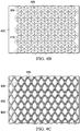
- the closed cells 404 that are generally hemispherical or spherical in shape may have a diameter between about 0.5 mm and 10 mm.
- the closed cells 404 also may have a pitch, i.e., the center to center distance between each of the closed cells 404, between about 1.5 mm and 15 mm. Because the sealed region 406 defines the base of the closed cells 404 including the diameter of a circular base and the pitch of adjacent closed cells 404, the surface area of the manifold 400 covered by the closed cells 404 may also be determined as a percentage, i.e., the cell coverage percentage.
- the cell coverage percentage is about 22% of the surface area of the manifold 400. In another example embodiment wherein the diameter of the closed cells 404 is about 2.0 mm and the pitch is about 5.0 mm, the cell coverage percentage is about 14% of the surface area of the manifold 400. In yet another example embodiment wherein the diameter of the closed cells 404 is about 1.0 mm and the pitch is about 1.5 mm, the cell coverage percentage is about 30% of the surface area of the manifold 400.
- the closed cells 414 may be formed on only one side of the sealed region 416 by using sheets of polymeric film having a different thickness or flexibility.
- the closed cells 414 may be formed in the sheet 413 by applying a vacuum to the sheet 413 where the sheet 412 is sufficiently thicker than the sheet 413 to withstand the vacuum being applied and retain a generally planar shape.
- closed cells 414 having other shapes may be formed to extend from only one side of the sealed region 406 and that such cells may be formed by using a variety of different methods.
- the closed cells 414 may be formed separately in the sheet 413 which is subsequently coupled to the sheet 412 that may have the same thickness as the sheet 413 so that the sealed region 416 remains thin and flexible.
- the closed cells in each sheet may not be aligned with each other, but rather overlap or aligned with the sealed portion of the opposite sheet.
- the closed cells 434 may be formed on both sides of the sealed region 436 by using sheets of polymeric film having a different thickness or flexibility.
- the shape of the closed cells 434 may be asymmetric when the sheets 432 and 433.
- the shape of the closed cells 434 may be substantially spherical as shown in Figure 4A3 .
- the manifold 400 may comprise a third sheet (not shown) forming a multi-sheet configuration wherein the third sheet is disposed between the sheets 402 and 403 to form closed cells 404 that may be generally spherical in shape formed by two hemispherical sections separated by portions of the third sheet of material.
- the manifold 400 may be a manifold 420 that comprises sheet 422 and sheet 423 of polymeric film having inner surfaces coupled or bonded to a third sheet 428 to form sealed region 426 defining a plurality of closed cells 424.
- the closed cells 424 are generally spherical in shape and formed by two hemispherical sections that are separated by portions of the third sheet 428.
- the third sheet 428 may also be formed from a polyester material to provide wicking within the closed cells 424, and may further include fibers flocked into the polyester material to provide additional wicking capability.
- the third sheet 428 also may include an antimicrobial layer or antimicrobials coated on the third sheet 428.
- each one may have one of its polymeric film sheets facing the tissue site and, more specifically, an outer surface of the tissue-facing sheet.
- the outer surface of the tissue-facing sheet may be textured with surface features, which may be protrusions or indentations, to enhance fluid flow through the manifolds and to increase micro-strains against the tissue site to enhance granulation.
- the outer side of the sheet facing the tissue site may further comprise a pattern of individual nodes or projections embossed on the outer surface of the sheet, a grid embossed on the outer surface of the sheet, a pattern or grid of grooves formed into the outer surface of the sheet, or any combination of the foregoing.
- a grid 449 may be embossed or extruded in a woven pattern on the outer surface of the tissue-facing sheet, e.g., sheet 402 or sheet 412, as shown in Figure 4C .
- the pattern of the grid 449 may have a variety of shapes like the diamond-shaped pattern shown. It should be understood that many types of protrusions or grids may be formed on the tissue-facing surface of a sheet of any one of the manifolds 400, 410, 420 and 430 to enhance fluid flow through the manifolds and/or enhance granulation of the tissue site. Moreover, it should be understood that any of such protrusions or grids may be formed by embossing, welding, or any other similar type of coupling mechanism.
- the cone shape may have a base diameter of about 0.4 mm and a vertical height of between 0.4 mm to 1.2 mm.
- the dome shape may have a spherical cap or parabolic shape with a base diameter ranging from about 0.4 mm to 1 mm.
- manifold 400 may further comprise chambers formed by interconnected closed cells to better distribute the apposition force applied to the manifold 400 as a result of the application of negative pressure because the volume of the chambers is greater than the volume of the individual closed cells.
- manifold 500 is similar in all respects to the manifold 400 comprising two sheets 502 and 503 of polymeric film having inner surfaces coupled to each other in a pattern defining a plurality of closed cells 504.
- the sheets 502 and 503 may be sealed to each other to form a sealed region 506 defining the closed cells 504.
- the sealed region 506 may also be perforated to provide pathways for fluid to flow through the manifold 500.
- the manifold 500 may be manifold 510 that is similar in all respects to the manifold 500 and in many respects to the manifold 410 as shown in Figure 5A1 . More specifically, the manifold 510 comprises two sheets of polymeric film, sheet 512 and sheet 513, having inner surfaces coupled to each other in a pattern defining a plurality of closed cells 514. The sheets 512 and 513 may be sealed to each other in a sealed region 516 that defines the closed cells 514 that are generally hemispherical in shape. The manifold 510 also may comprise a plurality of passageways 518 interconnecting the closed cells 514 to form a closed chamber 558. The closed chamber 558 may be formed in only one of the sheets 512 and 513 so that they extend from only one side of the sealed region 516 as shown in Figure 5A1 .
- the manifold 500 may be manifold 520 that is similar to the manifold 500 and in many respects to the manifold 420 as shown in Figure 5A2 . More specifically, the manifold 520 comprises two sheets of polymeric film, sheet 522 and sheet 523, having inner surfaces coupled or bonded to a third sheet 528 to form sealed region 526 defining a plurality of closed cells 524.
- the closed cells 524 are generally spherical in shape and formed by two hemispherical sections that are separated by portions of the third sheet 528.
- the manifolds 400 and 500 both comprise two sheets 402, 403 and 502, 503 of polymeric film having inner surfaces sealed to each other in a pattern defining a plurality of closed cells 404 and 504 in close proximity to one another.
- the sheets 402, 403 and 502, 503 may be sealed to each other in a sealed region 406 and 506 that defines the closed cells 404.
- the rows of the closed cells 404 and 504 are staggered so that the individual cells may be more closely nested together between the alternating rows to form a nested pattern of cells formed on the same plane as defined by the sealed regions 406, 506, respectively.
- the closed cells may be arranged in other patterns suitable for the particular therapy being utilized.
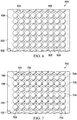
- a manifold 600 also comprises two sheets 602 and 603 of polymeric film having inner surfaces sealed to each other in a pattern defining a plurality of closed cells 604 in close proximity to one another.
- the rows and columns of closed cells 604 are not staggered, but rather arranged in an aligned pattern.
- the cell coverage percentage may range between about 10% and about 55% of the surface area of the manifold 600.
- the sheets 602 and 603 may be sealed to each other in a sealed region 606 that defines the closed cells 604.
- the rows and columns of the closed cells 604 are arranged in line to form an aligned pattern.
- the manifold 600 may also include a sealed region 606 that may be perforated as described above.
- the pattern of closed cells may have a variety of different arrangements.
- the closed cells 434 may flatten somewhat during the application of negative pressure to the manifold 430, the volume the closed cells 434 remains substantially constant so that the manifold 430 transmits the apposition force is to the proximal surfaces 912 of the hemispherical cells 454 while maintaining fluid flow through the proximal channels 916 and the distal channels 926 to continue providing negative pressure therapy to the wound 152. Consequently, the manifold 430 applies apposition forces to the proximal surfaces 912 and/or the nodes 429 (not shown in Figure 9 ) which enhance granulation while maintaining fluid communication with the tissue site 150 via the proximal channels 916 and the distal channels 926.
- the flexibility of the closed cells 434 also facilitates movement of the proximal surfaces 912 of the hemispherical portion 454 as the negative pressure increases during the decompression cycle and/or is varied during treatment so that the additional movement further enhances granulation.
- the manifold 410 and manifold 430 are a single component comprising closed cells 414 and 434, respectively, separated by sealed regions 416 and 436, respectively, and apertures 405 extending through the sealed regions such that the manifolds provide both a manifold function and a filler function.
- the manifold function provides fluid flow for both negative pressure and instillation liquids, while the filler function provides spacing between the tissue site 150 and the cover 106 with material having sufficient flexibility and tensile strength to prevent the manifold from collapsing in order to maintain fluid flow.
- the apposition force causes the cover 106 to collapse down on the manifold which is transmitted via the closed cells to increase micro-strains on the wound 152 thereby enhancing granulation.
- the method of dressing a wound is simpler and quicker because a caregiver places only one component in contact with the tissue site rather than two separate components, i.e., a separate manifold member and a separate filler member.
- a caregiver must first size and place a manifold member in contact with the tissue site and then position the filler member above the manifold member which requires more time and is more difficult.
- the filler member must be adjusted to substantially fill the space beneath the cover and be trimmed and resized to properly interface with the manifold member.
- the single-component manifolds described herein is less complicated and requires less time for dressing a wound than previous methods requiring the use of two members which can reduce the possibility of infection and other challenges inherent in previous methods of dressing wounds. Additionally, the single-component manifolds also provide better fluid flow because the fluids are not impeded by a separate filler member that might not be sufficiently porous or perforated to accommodate instillation fluids or negative pressure during the treatment process as described above.
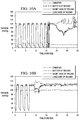
- Figures 10A and 10B illustrate these measurements for a manifold such as, for example, the manifold 430 having closed cells 434 with an average diameter of about 1 mm, wherein the manifold measured in Figure 10A had no apertures 405 while the manifold tested in Figure 10B included apertures 405.
- a manifold such as, for example, the manifold 430 having closed cells 434 with an average diameter of about 1 mm
- the manifold measured in Figure 10A had no apertures 405 while the manifold tested in Figure 10B included apertures 405.
- the pressures measured at both the short side edge and the long side edge of the wound 152 drops as low as 60 mmHg for a period of time while the manifold 430 that includes apertures 405 is measured fairly steadily at 120 mmHg except for a brief period of time where the pressure drops to about 100 mmHg.
- the pressure measurements at the bottom of the wound 152 are even more dramatic.
- the pressure measured at the bottom of the wound 152 is only 20 mmHg and stays there, whereas the pressure at the bottom of the wound 152 when utilizing the apertures 405 is measured fairly steadily at 100 mmHg.
- the example embodiments of the manifolds described above i.e., the manifolds 400, 500, 600 and 700, include apertures as described above including, for example, apertures 405, 505, 605 and 705.
- the closed cells 414 of the two manifolds 410 may be nested together so that the upper surfaces 814 of each one face the sealed region 416 of the other.
- the closed cells 414 of the two manifolds 410 may be nested together such that the sealed regions 416 of the two manifolds 410 are located in separate planes.
- diameter and pitch of the hemispherical surface of the closed cells 414 may be varied to fit within the tissue site 150 and to accommodate different type of wounds and therapies being provided.
- the closed cells 414 may have a diameter between about 0.5 mm and 10 mm and a pitch between about 1.5 mm and 15 mm depending on the depth of the wound 152 at the tissue site 150.
- the manifold 140 including the two manifolds 410 may increase the apposition force being applied to the wound 152 to further enhance granulation.
- the closed cells 434 also may be nested together so that the proximal surfaces 912 and the distal surfaces 922 of each one face the sealed region 436 of the other as shown for the stacked manifold 430(3) to form a block of closed cells 434 as the manifold 140.
- the closed cells 434 of the stacked manifolds 430(1) - 430(5) may be nested together such that the sealed regions 436 of the manifolds are located in separate planes.
- the proximal channels 916 and distal channels 926 are also formed as described above including those disposed between each of the stacked manifolds 430(1) - 430(5).
- a multilayer manifold may be necessary for deeper wounds 152 that may require greater apposition forces to further enhance granulation for more rapidly healing the wound 152.
- a chart is shown that illustrates two graphs, graph 1110 and graph 1120, of the load in newtons (N), i.e., the apposition force, being applied to a tissue site by negative pressure over time with the negative pressure cycling intermittently on and off, one minute on and one minute off, including three cycles each of 125 mmHg and 200 mmHg during the on cycles.
- the graph 1110 is the apposition force resulting from the application of negative pressure to the manifold 140 comprising a single layer that is the manifold 430 shown in Figure 9 .
- the apposition force for this single layer manifold varied between approximately 4.0 and 4.5 N when the negative pressure being applied was approximately 125 mmHg and varied between approximately 4.5 and 4.7 N when the negative pressure being applied was approximately 200 mmHg.
- the graph 1120 is the apposition force resulting from the application of negative pressure to the manifold 140 comprising a multilayer structure comprising five layers of the manifold 430 as shown in Figure 10 .
- the apposition force for this multilayer manifold varied between approximately 9 and 10 N when the negative pressure being applied was approximately 125 mmHg and varied between approximately 9 and 11 N when the negative pressure being applied was approximately 200 mmHg.
- the apposition force more than doubles when utilizing a multilayer manifold structure that further enhances granulation without sacrificing fluid communication of instillation fluids and negative pressure through the manifold structure as a result of the proximal channels 916 and the distal channels 926 formed among the closed cells 434.
Priority Applications (1)
| Application Number | Priority Date | Filing Date | Title |
|---|---|---|---|
| EP20163307.0A EP3693034B8 (en) | 2016-01-06 | 2016-12-13 | Systems for the treatment of wounds with dressing having closed cells |
Applications Claiming Priority (2)
| Application Number | Priority Date | Filing Date | Title |
|---|---|---|---|
| US201662275595P | 2016-01-06 | 2016-01-06 | |
| PCT/US2016/066392 WO2017119996A1 (en) | 2016-01-06 | 2016-12-13 | System and methods for the treatment of wounds with dressing having closed cells |
Related Child Applications (1)
| Application Number | Title | Priority Date | Filing Date |
|---|---|---|---|
| EP20163307.0A Division EP3693034B8 (en) | 2016-01-06 | 2016-12-13 | Systems for the treatment of wounds with dressing having closed cells |
Publications (2)
| Publication Number | Publication Date |
|---|---|
| EP3400032A1 EP3400032A1 (en) | 2018-11-14 |
| EP3400032B1 true EP3400032B1 (en) | 2020-03-18 |
Family
ID=57708825
Family Applications (2)
| Application Number | Title | Priority Date | Filing Date |
|---|---|---|---|
| EP20163307.0A Active EP3693034B8 (en) | 2016-01-06 | 2016-12-13 | Systems for the treatment of wounds with dressing having closed cells |
| EP16820492.3A Active EP3400032B1 (en) | 2016-01-06 | 2016-12-13 | System for the treatment of wounds with dressing having closed cells |
Family Applications Before (1)
| Application Number | Title | Priority Date | Filing Date |
|---|---|---|---|
| EP20163307.0A Active EP3693034B8 (en) | 2016-01-06 | 2016-12-13 | Systems for the treatment of wounds with dressing having closed cells |
Country Status (7)
| Country | Link |
|---|---|
| US (1) | US11318243B2 (ja) |
| EP (2) | EP3693034B8 (ja) |
| JP (1) | JP7082057B2 (ja) |
| CN (1) | CN108697832B (ja) |
| AU (1) | AU2016385411B2 (ja) |
| CA (1) | CA3010496A1 (ja) |
| WO (1) | WO2017119996A1 (ja) |
Families Citing this family (57)
| Publication number | Priority date | Publication date | Assignee | Title |
|---|---|---|---|---|
| GB0808376D0 (en) | 2008-05-08 | 2008-06-18 | Bristol Myers Squibb Co | Wound dressing |
| GB0817796D0 (en) | 2008-09-29 | 2008-11-05 | Convatec Inc | wound dressing |
| GB201020236D0 (en) | 2010-11-30 | 2011-01-12 | Convatec Technologies Inc | A composition for detecting biofilms on viable tissues |
| US9526816B2 (en) | 2010-12-08 | 2016-12-27 | Convatec Technologies Inc. | Wound exudate monitor accessory |
| WO2012078781A1 (en) | 2010-12-08 | 2012-06-14 | Convatec Technologies Inc. | Integrated system for assessing wound exudates |
| GB201115182D0 (en) | 2011-09-02 | 2011-10-19 | Trio Healthcare Ltd | Skin contact material |
| GB2497406A (en) | 2011-11-29 | 2013-06-12 | Webtec Converting Llc | Dressing with a perforated binder layer |
| KR20150099776A (ko) | 2012-12-20 | 2015-09-01 | 컨바텍 테크놀러지스 인크 | 화학적 개질된 셀룰로스 섬유의 처리 |
| KR20190008199A (ko) | 2016-03-30 | 2019-01-23 | 시노보 게엠베하 | 상처에서 미생물 감염의 검출 방법 |
| CN109310528B (zh) | 2016-03-30 | 2021-07-20 | 康沃特克科技公司 | 检测伤口中的微生物感染 |
| CN109069712A (zh) | 2016-05-13 | 2018-12-21 | 史密夫及内修公开有限公司 | 启用传感器的伤口监测和治疗装置 |
| WO2018009873A1 (en) | 2016-07-08 | 2018-01-11 | Convatec Technologies Inc. | Fluid collection apparatus |
| EP3481360B1 (en) | 2016-07-08 | 2022-03-09 | ConvaTec Technologies Inc. | Fluid flow sensing |
| ES2882336T3 (es) | 2016-07-08 | 2021-12-01 | Convatec Technologies Inc | Sistema flexible de presión negativa |
| US11324424B2 (en) | 2017-03-09 | 2022-05-10 | Smith & Nephew Plc | Apparatus and method for imaging blood in a target region of tissue |
| US11690570B2 (en) | 2017-03-09 | 2023-07-04 | Smith & Nephew Plc | Wound dressing, patch member and method of sensing one or more wound parameters |
| CA3059516A1 (en) | 2017-04-11 | 2018-10-18 | Smith & Nephew Plc | Component positioning and stress relief for sensor enabled wound dressings |
| US11791030B2 (en) | 2017-05-15 | 2023-10-17 | Smith & Nephew Plc | Wound analysis device and method |
| BR112019025031A2 (pt) | 2017-06-07 | 2020-08-18 | Kci Licensing, Inc | penso para tratar um sítio de tecido com pressão negativa e sistemas, aparelhos e métodos |
| US11819387B2 (en) | 2017-06-07 | 2023-11-21 | Kci Licensing, Inc. | Composite dressings for improved granulation and reduced maceration with negative-pressure treatment |
| AU2018282159A1 (en) | 2017-06-07 | 2019-12-19 | 3M Innovative Properties Company | Composite dressings for improved granulation and reduced maceration with negative-pressure treatment |
| EP3634337B1 (en) | 2017-06-07 | 2023-05-24 | 3M Innovative Properties Company | Methods for manufacturing and assembling dual material tissue interface for negative-pressure therapy |
| US10695227B2 (en) | 2017-06-07 | 2020-06-30 | Kci Licensing, Inc. | Methods for manufacturing and assembling dual material tissue interface for negative-pressure therapy |
| CN110799222B (zh) | 2017-06-07 | 2023-03-07 | 3M创新知识产权公司 | 用于减少组织向内生长的负压治疗的系统、设备以及方法 |
| US11607342B2 (en) | 2017-06-07 | 2023-03-21 | Kci Licensing, Inc. | Peel and place dressing for negative-pressure therapy |
| US11179512B2 (en) | 2017-06-07 | 2021-11-23 | Kci Licensing, Inc. | Multi-layer wound filler for extended wear time |
| RU2019139911A (ru) | 2017-06-07 | 2021-07-09 | Кейсиай ЛАЙСЕНСИНГ, ИНК. | Композитные перевязочные материалы для улучшенной грануляции и сниженной мацерации для лечения посредством отрицательного давления |
| JP7419072B2 (ja) | 2017-06-14 | 2024-01-22 | スミス アンド ネフュー ピーエルシー | 創傷閉鎖のための折り畳み可能シートおよび使用方法 |
| AU2018288530B2 (en) | 2017-06-23 | 2024-03-28 | Smith & Nephew Plc | Positioning of sensors for sensor enabled wound monitoring or therapy |
| GB201809007D0 (en) | 2018-06-01 | 2018-07-18 | Smith & Nephew | Restriction of sensor-monitored region for sensor-enabled wound dressings |
| GB201804502D0 (en) | 2018-03-21 | 2018-05-02 | Smith & Nephew | Biocompatible encapsulation and component stress relief for sensor enabled negative pressure wound therapy dressings |
| AU2018312883A1 (en) | 2017-08-10 | 2020-02-20 | Smith & Nephew Plc | Positioning of sensors for sensor enabled wound monitoring or therapy |
| GB201804971D0 (en) | 2018-03-28 | 2018-05-09 | Smith & Nephew | Electrostatic discharge protection for sensors in wound therapy |
| WO2019048624A1 (en) | 2017-09-10 | 2019-03-14 | Smith & Nephew Plc | ENCAPSULATION INSPECTION SYSTEMS AND METHODS AND COMPONENTS IN SENSOR EQUIPMENT DRESSINGS |
| GB201718870D0 (en) | 2017-11-15 | 2017-12-27 | Smith & Nephew Inc | Sensor enabled wound therapy dressings and systems |
| GB201718859D0 (en) | 2017-11-15 | 2017-12-27 | Smith & Nephew | Sensor positioning for sensor enabled wound therapy dressings and systems |
| WO2019063481A1 (en) | 2017-09-27 | 2019-04-04 | Smith & Nephew Plc | PH DETECTION FOR NEGATIVE PRESSURE WOUND THERAPY SURVEILLANCE AND THERAPY APPARATUS |
| EP3687396A1 (en) | 2017-09-28 | 2020-08-05 | Smith & Nephew plc | Neurostimulation and monitoring using sensor enabled wound monitoring and therapy apparatus |
| GB201718851D0 (en) * | 2017-11-15 | 2017-12-27 | Smith & Nephew | Flocked conformable circuit boards for sensor enabled wound therapy dressings and systems |
| EP4268775A3 (en) * | 2017-10-26 | 2023-12-27 | 3M Innovative Properties Company | Manifolding apparatus |
| CN111343950A (zh) | 2017-11-15 | 2020-06-26 | 史密夫及内修公开有限公司 | 实施传感器的集成伤口监测和/或治疗敷料和系统 |
| CN112105323A (zh) * | 2018-03-12 | 2020-12-18 | 凯希特许有限公司 | 用于舱室空间中的使用不同表面特征的敷料 |
| WO2020005535A1 (en) * | 2018-06-28 | 2020-01-02 | Kci Licensing, Inc. | A highly conformable wound dressing |
| US11534343B2 (en) | 2018-07-31 | 2022-12-27 | Kci Licensing, Inc. | Devices and methods for preventing localized pressure points in distribution components for tissue therapy |
| EP3840795A1 (en) | 2018-08-24 | 2021-06-30 | KCI Licensing, Inc. | Methods of managing moisture when using a low profile wound connection conduit |
| GB2592508B (en) | 2018-09-12 | 2022-08-31 | Smith & Nephew | Device, apparatus and method of determining skin perfusion pressure |
| EP3866871A1 (en) * | 2018-10-15 | 2021-08-25 | KCI Licensing, Inc. | Micro balloon-on-tube wound filler |
| EP3880143B1 (en) * | 2018-11-13 | 2023-09-20 | KCI Licensing, Inc. | Low profile distribution components for wound therapy |
| GB201820927D0 (en) | 2018-12-21 | 2019-02-06 | Smith & Nephew | Wound therapy systems and methods with supercapacitors |
| JP2022540272A (ja) * | 2019-03-27 | 2022-09-15 | ケーシーアイ ライセンシング インコーポレイテッド | 創傷体積推定を伴う創傷療法システム |
| US20220202620A1 (en) * | 2019-05-08 | 2022-06-30 | Kci Licensing, Inc. | Manifold With Biological Actives For Negative-Pressure Therapy |
| JP2022537356A (ja) * | 2019-06-20 | 2022-08-25 | ケーシーアイ ライセンシング インコーポレイテッド | 組織内部成長が低減し、装着時間が延長された陰圧治療のためのシステム、装置、及び方法 |
| WO2021059126A1 (en) * | 2019-09-24 | 2021-04-01 | Kci Licensing, Inc. | Dressing system for use on deep wounds with reduced in-growth and extended wear time |
| US11771819B2 (en) | 2019-12-27 | 2023-10-03 | Convatec Limited | Low profile filter devices suitable for use in negative pressure wound therapy systems |
| US11331221B2 (en) | 2019-12-27 | 2022-05-17 | Convatec Limited | Negative pressure wound dressing |
| US20230301835A1 (en) * | 2020-07-30 | 2023-09-28 | 3M Innovative Properties Company | Low-growth tissue interface |
| WO2023170508A1 (en) * | 2022-03-09 | 2023-09-14 | 3M Innovative Properties Company | Improved dressings and systems for negative-pressure wound therapy and instillation therapy having low-volume manifolds and bypass instillation passages |
Family Cites Families (155)
| Publication number | Priority date | Publication date | Assignee | Title |
|---|---|---|---|---|
| US1355846A (en) | 1920-02-06 | 1920-10-19 | David A Rannells | Medical appliance |
| US2547758A (en) | 1949-01-05 | 1951-04-03 | Wilmer B Keeling | Instrument for treating the male urethra |
| US2632443A (en) | 1949-04-18 | 1953-03-24 | Eleanor P Lesher | Surgical dressing |
| GB692578A (en) | 1949-09-13 | 1953-06-10 | Minnesota Mining & Mfg | Improvements in or relating to drape sheets for surgical use |
| US2682873A (en) | 1952-07-30 | 1954-07-06 | Johnson & Johnson | General purpose protective dressing |
| NL189176B (nl) | 1956-07-13 | 1900-01-01 | Hisamitsu Pharmaceutical Co | Pleister op basis van een synthetische rubber. |
| US2969057A (en) | 1957-11-04 | 1961-01-24 | Brady Co W H | Nematodic swab |
| US3066672A (en) | 1960-09-27 | 1962-12-04 | Jr William H Crosby | Method and apparatus for serial sampling of intestinal juice |
| US3367332A (en) | 1965-08-27 | 1968-02-06 | Gen Electric | Product and process for establishing a sterile area of skin |
| US3520300A (en) | 1967-03-15 | 1970-07-14 | Amp Inc | Surgical sponge and suction device |
| US3568675A (en) | 1968-08-30 | 1971-03-09 | Clyde B Harvey | Fistula and penetrating wound dressing |
| US3682180A (en) | 1970-06-08 | 1972-08-08 | Coilform Co Inc | Drain clip for surgical drain |
| BE789293Q (fr) | 1970-12-07 | 1973-01-15 | Parke Davis & Co | Pansement medico-chirugical pour brulures et lesions analogues |
| US3826254A (en) | 1973-02-26 | 1974-07-30 | Verco Ind | Needle or catheter retaining appliance |
| DE2527706A1 (de) | 1975-06-21 | 1976-12-30 | Hanfried Dr Med Weigand | Einrichtung zum einleiten von kontrastmittel in einen kuenstlichen darmausgang |
| DE2640413C3 (de) | 1976-09-08 | 1980-03-27 | Richard Wolf Gmbh, 7134 Knittlingen | Katheter-Überwachungsgerät |
| NL7710909A (nl) | 1976-10-08 | 1978-04-11 | Smith & Nephew | Samengestelde hechtstrook. |
| GB1562244A (en) | 1976-11-11 | 1980-03-05 | Lock P M | Wound dressing materials |
| US4080970A (en) | 1976-11-17 | 1978-03-28 | Miller Thomas J | Post-operative combination dressing and internal drain tube with external shield and tube connector |
| US4139004A (en) | 1977-02-17 | 1979-02-13 | Gonzalez Jr Harry | Bandage apparatus for treating burns |
| US4184510A (en) | 1977-03-15 | 1980-01-22 | Fibra-Sonics, Inc. | Valued device for controlling vacuum in surgery |
| US4165748A (en) | 1977-11-07 | 1979-08-28 | Johnson Melissa C | Catheter tube holder |
| US4245637A (en) | 1978-07-10 | 1981-01-20 | Nichols Robert L | Shutoff valve sleeve |
| SE414994B (sv) | 1978-11-28 | 1980-09-01 | Landstingens Inkopscentral | Venkateterforband |
| GB2047543B (en) | 1978-12-06 | 1983-04-20 | Svedman Paul | Device for treating tissues for example skin |
| US4266545A (en) | 1979-04-06 | 1981-05-12 | Moss James P | Portable suction device for collecting fluids from a closed wound |
| US4284079A (en) | 1979-06-28 | 1981-08-18 | Adair Edwin Lloyd | Method for applying a male incontinence device |
| US4261363A (en) | 1979-11-09 | 1981-04-14 | C. R. Bard, Inc. | Retention clips for body fluid drains |
| US4569348A (en) | 1980-02-22 | 1986-02-11 | Velcro Usa Inc. | Catheter tube holder strap |
| US4480638A (en) | 1980-03-11 | 1984-11-06 | Eduard Schmid | Cushion for holding an element of grafted skin |
| US4297995A (en) | 1980-06-03 | 1981-11-03 | Key Pharmaceuticals, Inc. | Bandage containing attachment post |
| US4333468A (en) | 1980-08-18 | 1982-06-08 | Geist Robert W | Mesentery tube holder apparatus |
| US4465485A (en) | 1981-03-06 | 1984-08-14 | Becton, Dickinson And Company | Suction canister with unitary shut-off valve and filter features |
| US4392853A (en) | 1981-03-16 | 1983-07-12 | Rudolph Muto | Sterile assembly for protecting and fastening an indwelling device |
| US4373519A (en) | 1981-06-26 | 1983-02-15 | Minnesota Mining And Manufacturing Company | Composite wound dressing |
| US4392858A (en) | 1981-07-16 | 1983-07-12 | Sherwood Medical Company | Wound drainage device |
| US4419097A (en) | 1981-07-31 | 1983-12-06 | Rexar Industries, Inc. | Attachment for catheter tube |
| AU550575B2 (en) | 1981-08-07 | 1986-03-27 | Richard Christian Wright | Wound drainage device |
| SE429197B (sv) | 1981-10-14 | 1983-08-22 | Frese Nielsen | Anordning for behandling av sar |
| DE3146266A1 (de) | 1981-11-21 | 1983-06-01 | B. Braun Melsungen Ag, 3508 Melsungen | Kombinierte vorrichtung fuer eine medizinische saugdrainage |
| US4551139A (en) | 1982-02-08 | 1985-11-05 | Marion Laboratories, Inc. | Method and apparatus for burn wound treatment |
| US4475909A (en) | 1982-05-06 | 1984-10-09 | Eisenberg Melvin I | Male urinary device and method for applying the device |
| DE3361779D1 (en) | 1982-07-06 | 1986-02-20 | Dow Corning | Medical-surgical dressing and a process for the production thereof |
| NZ206837A (en) | 1983-01-27 | 1986-08-08 | Johnson & Johnson Prod Inc | Thin film adhesive dressing:backing material in three sections |
| US4548202A (en) | 1983-06-20 | 1985-10-22 | Ethicon, Inc. | Mesh tissue fasteners |
| US4540412A (en) | 1983-07-14 | 1985-09-10 | The Kendall Company | Device for moist heat therapy |
| US4543100A (en) | 1983-11-01 | 1985-09-24 | Brodsky Stuart A | Catheter and drain tube retainer |
| US4525374A (en) | 1984-02-27 | 1985-06-25 | Manresa, Inc. | Treating hydrophobic filters to render them hydrophilic |
| GB2157958A (en) | 1984-05-03 | 1985-11-06 | Ernest Edward Austen Bedding | Ball game net support |
| US4897081A (en) | 1984-05-25 | 1990-01-30 | Thermedics Inc. | Percutaneous access device |
| US5215522A (en) | 1984-07-23 | 1993-06-01 | Ballard Medical Products | Single use medical aspirating device and method |
| GB8419745D0 (en) | 1984-08-02 | 1984-09-05 | Smith & Nephew Ass | Wound dressing |
| US4872450A (en) | 1984-08-17 | 1989-10-10 | Austad Eric D | Wound dressing and method of forming same |
| US4826494A (en) | 1984-11-09 | 1989-05-02 | Stryker Corporation | Vacuum wound drainage system |
| US4655754A (en) | 1984-11-09 | 1987-04-07 | Stryker Corporation | Vacuum wound drainage system and lipids baffle therefor |
| US4605399A (en) | 1984-12-04 | 1986-08-12 | Complex, Inc. | Transdermal infusion device |
| US5037397A (en) | 1985-05-03 | 1991-08-06 | Medical Distributors, Inc. | Universal clamp |
| US4640688A (en) | 1985-08-23 | 1987-02-03 | Mentor Corporation | Urine collection catheter |
| US4710165A (en) | 1985-09-16 | 1987-12-01 | Mcneil Charles B | Wearable, variable rate suction/collection device |
| US4758220A (en) | 1985-09-26 | 1988-07-19 | Alcon Laboratories, Inc. | Surgical cassette proximity sensing and latching apparatus |
| US4733659A (en) | 1986-01-17 | 1988-03-29 | Seton Company | Foam bandage |
| WO1987004626A1 (en) | 1986-01-31 | 1987-08-13 | Osmond, Roger, L., W. | Suction system for wound and gastro-intestinal drainage |
| US4838883A (en) | 1986-03-07 | 1989-06-13 | Nissho Corporation | Urine-collecting device |
| JPS62281965A (ja) | 1986-05-29 | 1987-12-07 | テルモ株式会社 | カテ−テルおよびカテ−テル用固定部材 |
| GB8621884D0 (en) | 1986-09-11 | 1986-10-15 | Bard Ltd | Catheter applicator |
| GB2195255B (en) | 1986-09-30 | 1991-05-01 | Vacutec Uk Limited | Apparatus for vacuum treatment of an epidermal surface |
| US4743232A (en) | 1986-10-06 | 1988-05-10 | The Clinipad Corporation | Package assembly for plastic film bandage |
| DE3634569A1 (de) | 1986-10-10 | 1988-04-21 | Sachse Hans E | Kondomkatheter, ein harnroehrenkatheter zur verhinderung von aufsteigenden infektionen |
| JPS63135179A (ja) | 1986-11-26 | 1988-06-07 | 立花 俊郎 | 薬物の経皮投与具 |
| GB8628564D0 (en) | 1986-11-28 | 1987-01-07 | Smiths Industries Plc | Anti-foaming agent suction apparatus |
| GB8706116D0 (en) | 1987-03-14 | 1987-04-15 | Smith & Nephew Ass | Adhesive dressings |
| US4787888A (en) | 1987-06-01 | 1988-11-29 | University Of Connecticut | Disposable piezoelectric polymer bandage for percutaneous delivery of drugs and method for such percutaneous delivery (a) |
| US4863449A (en) | 1987-07-06 | 1989-09-05 | Hollister Incorporated | Adhesive-lined elastic condom cathether |
| US5176663A (en) | 1987-12-02 | 1993-01-05 | Pal Svedman | Dressing having pad with compressibility limiting elements |
| US4906240A (en) | 1988-02-01 | 1990-03-06 | Matrix Medica, Inc. | Adhesive-faced porous absorbent sheet and method of making same |
| US4985019A (en) | 1988-03-11 | 1991-01-15 | Michelson Gary K | X-ray marker |
| GB8812803D0 (en) | 1988-05-28 | 1988-06-29 | Smiths Industries Plc | Medico-surgical containers |
| US4919654A (en) | 1988-08-03 | 1990-04-24 | Kalt Medical Corporation | IV clamp with membrane |
| US5000741A (en) | 1988-08-22 | 1991-03-19 | Kalt Medical Corporation | Transparent tracheostomy tube dressing |
| EP0379416B1 (fr) | 1989-01-16 | 1995-03-08 | Roussel-Uclaf | Dérivés d'azabicycloheptène et leurs sels, leur procédé de préparation, leur application comme médicaments et les compositions les renfermant |
| GB8906100D0 (en) | 1989-03-16 | 1989-04-26 | Smith & Nephew | Laminates |
| US4969880A (en) | 1989-04-03 | 1990-11-13 | Zamierowski David S | Wound dressing and treatment method |
| US5527293A (en) | 1989-04-03 | 1996-06-18 | Kinetic Concepts, Inc. | Fastening system and method |
| US5100396A (en) | 1989-04-03 | 1992-03-31 | Zamierowski David S | Fluidic connection system and method |
| US5261893A (en) | 1989-04-03 | 1993-11-16 | Zamierowski David S | Fastening system and method |
| US5358494A (en) | 1989-07-11 | 1994-10-25 | Svedman Paul | Irrigation dressing |
| JP2719671B2 (ja) | 1989-07-11 | 1998-02-25 | 日本ゼオン株式会社 | 創傷被覆材 |
| US5232453A (en) | 1989-07-14 | 1993-08-03 | E. R. Squibb & Sons, Inc. | Catheter holder |
| GB2235877A (en) | 1989-09-18 | 1991-03-20 | Antonio Talluri | Closed wound suction apparatus |
| US5134994A (en) | 1990-02-12 | 1992-08-04 | Say Sam L | Field aspirator in a soft pack with externally mounted container |
| US5092858A (en) | 1990-03-20 | 1992-03-03 | Becton, Dickinson And Company | Liquid gelling agent distributor device |
| US5149331A (en) | 1991-05-03 | 1992-09-22 | Ariel Ferdman | Method and device for wound closure |
| US5278100A (en) | 1991-11-08 | 1994-01-11 | Micron Technology, Inc. | Chemical vapor deposition technique for depositing titanium silicide on semiconductor wafers |
| US5645081A (en) | 1991-11-14 | 1997-07-08 | Wake Forest University | Method of treating tissue damage and apparatus for same |
| US5636643A (en) | 1991-11-14 | 1997-06-10 | Wake Forest University | Wound treatment employing reduced pressure |
| US5279550A (en) | 1991-12-19 | 1994-01-18 | Gish Biomedical, Inc. | Orthopedic autotransfusion system |
| US5167613A (en) | 1992-03-23 | 1992-12-01 | The Kendall Company | Composite vented wound dressing |
| FR2690617B1 (fr) | 1992-04-29 | 1994-06-24 | Cbh Textile | Pansement adhesif transparent. |
| DE4306478A1 (de) | 1993-03-02 | 1994-09-08 | Wolfgang Dr Wagner | Drainagevorrichtung, insbesondere Pleuradrainagevorrichtung, und Drainageverfahren |
| US5342376A (en) | 1993-05-03 | 1994-08-30 | Dermagraphics, Inc. | Inserting device for a barbed tissue connector |
| US6241747B1 (en) | 1993-05-03 | 2001-06-05 | Quill Medical, Inc. | Barbed Bodily tissue connector |
| US5344415A (en) | 1993-06-15 | 1994-09-06 | Deroyal Industries, Inc. | Sterile system for dressing vascular access site |
| US5437651A (en) | 1993-09-01 | 1995-08-01 | Research Medical, Inc. | Medical suction apparatus |
| US5549584A (en) | 1994-02-14 | 1996-08-27 | The Kendall Company | Apparatus for removing fluid from a wound |
| US5556375A (en) | 1994-06-16 | 1996-09-17 | Hercules Incorporated | Wound dressing having a fenestrated base layer |
| US5607388A (en) | 1994-06-16 | 1997-03-04 | Hercules Incorporated | Multi-purpose wound dressing |
| US5664270A (en) | 1994-07-19 | 1997-09-09 | Kinetic Concepts, Inc. | Patient interface system |
| WO1996005873A1 (en) | 1994-08-22 | 1996-02-29 | Kinetic Concepts Inc. | Wound drainage equipment |
| DE29504378U1 (de) | 1995-03-15 | 1995-09-14 | Mtg Medizinisch Tech Geraeteba | Elektronisch geregelte Niedervakuumpumpe für die Thorax- und Wunddrainage |
| GB9523253D0 (en) | 1995-11-14 | 1996-01-17 | Mediscus Prod Ltd | Portable wound treatment apparatus |
| US6135116A (en) | 1997-07-28 | 2000-10-24 | Kci Licensing, Inc. | Therapeutic method for treating ulcers |
| GB9719520D0 (en) | 1997-09-12 | 1997-11-19 | Kci Medical Ltd | Surgical drape and suction heads for wound treatment |
| AU755496B2 (en) | 1997-09-12 | 2002-12-12 | Kci Licensing, Inc. | Surgical drape and suction head for wound treatment |
| US6071267A (en) | 1998-02-06 | 2000-06-06 | Kinetic Concepts, Inc. | Medical patient fluid management interface system and method |
| US6488643B1 (en) | 1998-10-08 | 2002-12-03 | Kci Licensing, Inc. | Wound healing foot wrap |
| US6287316B1 (en) | 1999-03-26 | 2001-09-11 | Ethicon, Inc. | Knitted surgical mesh |
| US6856821B2 (en) | 2000-05-26 | 2005-02-15 | Kci Licensing, Inc. | System for combined transcutaneous blood gas monitoring and vacuum assisted wound closure |
| US7799004B2 (en) | 2001-03-05 | 2010-09-21 | Kci Licensing, Inc. | Negative pressure wound treatment apparatus and infection identification system and method |
| US6991643B2 (en) | 2000-12-20 | 2006-01-31 | Usgi Medical Inc. | Multi-barbed device for retaining tissue in apposition and methods of use |
| WO2001062328A1 (en) | 2000-02-24 | 2001-08-30 | Venetec International, Inc. | Universal catheter anchoring system |
| US6540705B2 (en) | 2001-02-22 | 2003-04-01 | Core Products International, Inc. | Ankle brace providing upper and lower ankle adjustment |
| US7645269B2 (en) | 2001-04-30 | 2010-01-12 | Kci Licensing, Inc. | Gradient wound treatment system and method |
| US7220476B2 (en) | 2001-05-10 | 2007-05-22 | Sealed Air Corporation (Us) | Apparatus and method for forming inflated chambers |
| US7846141B2 (en) | 2002-09-03 | 2010-12-07 | Bluesky Medical Group Incorporated | Reduced pressure treatment system |
| GB0224986D0 (en) | 2002-10-28 | 2002-12-04 | Smith & Nephew | Apparatus |
| GB0325120D0 (en) | 2003-10-28 | 2003-12-03 | Smith & Nephew | Apparatus with actives |
| GB0325126D0 (en) | 2003-10-28 | 2003-12-03 | Smith & Nephew | Apparatus with heat |
| US7790945B1 (en) * | 2004-04-05 | 2010-09-07 | Kci Licensing, Inc. | Wound dressing with absorption and suction capabilities |
| US7909805B2 (en) | 2004-04-05 | 2011-03-22 | Bluesky Medical Group Incorporated | Flexible reduced pressure treatment appliance |
| US7884258B2 (en) | 2004-04-13 | 2011-02-08 | Boehringer Technologies, L.P. | Wound contact device |
| US8529548B2 (en) | 2004-04-27 | 2013-09-10 | Smith & Nephew Plc | Wound treatment apparatus and method |
| RU2428209C2 (ru) * | 2006-01-23 | 2011-09-10 | КейСиАй ЛАЙСЕНЗИНГ, ИНК. | Система и способ обработки ран с использованием ультразвуковой хирургической очистки |
| AU2008248261A1 (en) * | 2007-05-01 | 2008-11-13 | The Brigham And Women's Hospital, Inc. | Wound healing device |
| EP2144643B8 (en) * | 2007-05-10 | 2020-12-30 | 3M Innovative Properties Company | Reduced pressure wound dressing having a wound contact surface with protrusions |
| WO2009021523A1 (en) * | 2007-08-14 | 2009-02-19 | Coloplast A/S | Pressure-distributing element of closed cell foam |
| WO2009089016A1 (en) * | 2008-01-08 | 2009-07-16 | Southeastern Medical Technologies, Llc | A methods and apparatuses for the treatment of wounds with pressures altered from atmospheric |
| US8021347B2 (en) | 2008-07-21 | 2011-09-20 | Tyco Healthcare Group Lp | Thin film wound dressing |
| US8007481B2 (en) | 2008-07-17 | 2011-08-30 | Tyco Healthcare Group Lp | Subatmospheric pressure mechanism for wound therapy system |
| WO2010017437A1 (en) * | 2008-08-08 | 2010-02-11 | Tyco Healthcare Group Lp | Wound dressing of continuous fibers |
| US8216198B2 (en) | 2009-01-09 | 2012-07-10 | Tyco Healthcare Group Lp | Canister for receiving wound exudate in a negative pressure therapy system |
| US8251979B2 (en) | 2009-05-11 | 2012-08-28 | Tyco Healthcare Group Lp | Orientation independent canister for a negative pressure wound therapy device |
| CA2741742C (en) * | 2008-10-29 | 2017-02-28 | Kci Licensing, Inc. | Modular, reduced-pressure, wound-closure systems and methods |
| US9327064B2 (en) * | 2008-12-24 | 2016-05-03 | Kci Licensing, Inc. | Membranes, systems, and methods for applying reduced pressure to a subcutaneous tissue site |
| BR112012008087A2 (pt) * | 2009-08-05 | 2016-03-01 | Tyco Healthcare | curativo de ferimento cirúrgico incorporando esferas de hidrogel conectadas tendo um eletrodo embutido naquele lugar |
| US8870837B2 (en) * | 2010-01-20 | 2014-10-28 | Kci Licensing, Inc. | Wound-connection pads for fluid instillation and negative pressure wound therapy, and systems and methods |
| US8791315B2 (en) * | 2010-02-26 | 2014-07-29 | Smith & Nephew, Inc. | Systems and methods for using negative pressure wound therapy to manage open abdominal wounds |
| US8430867B2 (en) * | 2010-03-12 | 2013-04-30 | Kci Licensing, Inc. | Reduced-pressure dressing connection pads, systems, and methods |
| US9061095B2 (en) * | 2010-04-27 | 2015-06-23 | Smith & Nephew Plc | Wound dressing and method of use |
| WO2012162098A2 (en) * | 2011-05-25 | 2012-11-29 | Kci Licensing, Inc. | Wound healing system using positive pressure to promote granulation at a tissue site |
| JP5967675B2 (ja) * | 2011-08-31 | 2016-08-10 | ケーシーアイ ライセンシング インコーポレイテッド | 減圧治療および創面切除用のシステムおよび方法 |
| EP2758086B1 (en) * | 2011-09-20 | 2018-05-23 | KCI Licensing, Inc. | Tissue treatment systems and methods having a non-tactile-stimulus-activated, macroscopically-deforming material |
| US9393354B2 (en) * | 2011-11-01 | 2016-07-19 | J&M Shuler Medical, Inc. | Mechanical wound therapy for sub-atmospheric wound care system |
| US10159771B2 (en) | 2013-03-14 | 2018-12-25 | Smith & Nephew Plc | Compressible wound fillers and systems and methods of use in treating wounds with negative pressure |
| CA2901882C (en) * | 2013-03-14 | 2021-04-13 | Kci Licensing, Inc. | Micro-porous conduit |
| EP2815731A1 (en) | 2013-06-18 | 2014-12-24 | Mölnlycke Health Care AB | Fluid transport dressing |
-
2016
- 2016-12-13 US US16/060,326 patent/US11318243B2/en active Active
- 2016-12-13 WO PCT/US2016/066392 patent/WO2017119996A1/en active Application Filing
- 2016-12-13 CN CN201680082489.9A patent/CN108697832B/zh active Active
- 2016-12-13 AU AU2016385411A patent/AU2016385411B2/en active Active
- 2016-12-13 EP EP20163307.0A patent/EP3693034B8/en active Active
- 2016-12-13 JP JP2018534979A patent/JP7082057B2/ja active Active
- 2016-12-13 EP EP16820492.3A patent/EP3400032B1/en active Active
- 2016-12-13 CA CA3010496A patent/CA3010496A1/en active Pending
Non-Patent Citations (1)
| Title |
|---|
| None * |
Also Published As
| Publication number | Publication date |
|---|---|
| US11318243B2 (en) | 2022-05-03 |
| WO2017119996A1 (en) | 2017-07-13 |
| CA3010496A1 (en) | 2017-07-13 |
| CN108697832B (zh) | 2022-10-11 |
| EP3400032A1 (en) | 2018-11-14 |
| US20200139025A1 (en) | 2020-05-07 |
| EP3693034B8 (en) | 2024-03-27 |
| EP3693034B1 (en) | 2024-02-21 |
| CN108697832A (zh) | 2018-10-23 |
| AU2016385411B2 (en) | 2021-10-14 |
| WO2017119996A8 (en) | 2017-08-31 |
| EP3693034A1 (en) | 2020-08-12 |
| JP7082057B2 (ja) | 2022-06-07 |
| AU2016385411A1 (en) | 2018-07-19 |
| JP2019500964A (ja) | 2019-01-17 |
Similar Documents
| Publication | Publication Date | Title |
|---|---|---|
| EP3400032B1 (en) | System for the treatment of wounds with dressing having closed cells | |
| US20240115789A1 (en) | Low profile distribution components for wound therapy | |
| US11432967B2 (en) | Fluid bridge for simultaneous application of negative pressure to multiple tissue sites | |
| US11278454B2 (en) | Methods of managing moisture when using a low profile wound connection conduit | |
| US11877911B2 (en) | Low profile distribution components for wound therapy | |
| AU2018282193A1 (en) | Multi-layer wound filler for extended wear time | |
| US20230049375A1 (en) | Dressing using differential surface features for use in compartment spaces | |
| US20220218892A1 (en) | Low-profile fluid conductors with moisture management features | |
| EP4096604B1 (en) | Multiple fluid pathway connector with foam supports | |
| WO2022238918A1 (en) | Low profile dual fluid instillation and removal bridge system with fluid delivery and pressure sensing capabilities | |
| EP4294341A1 (en) | Dressing interface having bubble supports with optional break-lines |
Legal Events
| Date | Code | Title | Description |
|---|---|---|---|
| STAA | Information on the status of an ep patent application or granted ep patent |
Free format text: STATUS: UNKNOWN |
|
| STAA | Information on the status of an ep patent application or granted ep patent |
Free format text: STATUS: THE INTERNATIONAL PUBLICATION HAS BEEN MADE |
|
| PUAI | Public reference made under article 153(3) epc to a published international application that has entered the european phase |
Free format text: ORIGINAL CODE: 0009012 |
|
| STAA | Information on the status of an ep patent application or granted ep patent |
Free format text: STATUS: REQUEST FOR EXAMINATION WAS MADE |
|
| 17P | Request for examination filed |
Effective date: 20180730 |
|
| AK | Designated contracting states |
Kind code of ref document: A1 Designated state(s): AL AT BE BG CH CY CZ DE DK EE ES FI FR GB GR HR HU IE IS IT LI LT LU LV MC MK MT NL NO PL PT RO RS SE SI SK SM TR |
|
| AX | Request for extension of the european patent |
Extension state: BA ME |
|
| DAV | Request for validation of the european patent (deleted) | ||
| DAX | Request for extension of the european patent (deleted) | ||
| GRAP | Despatch of communication of intention to grant a patent |
Free format text: ORIGINAL CODE: EPIDOSNIGR1 |
|
| STAA | Information on the status of an ep patent application or granted ep patent |
Free format text: STATUS: GRANT OF PATENT IS INTENDED |
|
| INTG | Intention to grant announced |
Effective date: 20191011 |
|
| GRAS | Grant fee paid |
Free format text: ORIGINAL CODE: EPIDOSNIGR3 |
|
| GRAA | (expected) grant |
Free format text: ORIGINAL CODE: 0009210 |
|
| STAA | Information on the status of an ep patent application or granted ep patent |
Free format text: STATUS: THE PATENT HAS BEEN GRANTED |
|
| AK | Designated contracting states |
Kind code of ref document: B1 Designated state(s): AL AT BE BG CH CY CZ DE DK EE ES FI FR GB GR HR HU IE IS IT LI LT LU LV MC MK MT NL NO PL PT RO RS SE SI SK SM TR |
|
| REG | Reference to a national code |
Ref country code: GB Ref legal event code: FG4D |
|
| REG | Reference to a national code |
Ref country code: DE Ref legal event code: R096 Ref document number: 602016032214 Country of ref document: DE |
|
| REG | Reference to a national code |
Ref country code: AT Ref legal event code: REF Ref document number: 1245183 Country of ref document: AT Kind code of ref document: T Effective date: 20200415 Ref country code: IE Ref legal event code: FG4D |
|
| PG25 | Lapsed in a contracting state [announced via postgrant information from national office to epo] |
Ref country code: FI Free format text: LAPSE BECAUSE OF FAILURE TO SUBMIT A TRANSLATION OF THE DESCRIPTION OR TO PAY THE FEE WITHIN THE PRESCRIBED TIME-LIMIT Effective date: 20200318 Ref country code: RS Free format text: LAPSE BECAUSE OF FAILURE TO SUBMIT A TRANSLATION OF THE DESCRIPTION OR TO PAY THE FEE WITHIN THE PRESCRIBED TIME-LIMIT Effective date: 20200318 Ref country code: NO Free format text: LAPSE BECAUSE OF FAILURE TO SUBMIT A TRANSLATION OF THE DESCRIPTION OR TO PAY THE FEE WITHIN THE PRESCRIBED TIME-LIMIT Effective date: 20200618 |
|
| REG | Reference to a national code |
Ref country code: NL Ref legal event code: MP Effective date: 20200318 |
|
| PG25 | Lapsed in a contracting state [announced via postgrant information from national office to epo] |
Ref country code: BG Free format text: LAPSE BECAUSE OF FAILURE TO SUBMIT A TRANSLATION OF THE DESCRIPTION OR TO PAY THE FEE WITHIN THE PRESCRIBED TIME-LIMIT Effective date: 20200618 Ref country code: GR Free format text: LAPSE BECAUSE OF FAILURE TO SUBMIT A TRANSLATION OF THE DESCRIPTION OR TO PAY THE FEE WITHIN THE PRESCRIBED TIME-LIMIT Effective date: 20200619 Ref country code: LV Free format text: LAPSE BECAUSE OF FAILURE TO SUBMIT A TRANSLATION OF THE DESCRIPTION OR TO PAY THE FEE WITHIN THE PRESCRIBED TIME-LIMIT Effective date: 20200318 Ref country code: SE Free format text: LAPSE BECAUSE OF FAILURE TO SUBMIT A TRANSLATION OF THE DESCRIPTION OR TO PAY THE FEE WITHIN THE PRESCRIBED TIME-LIMIT Effective date: 20200318 Ref country code: HR Free format text: LAPSE BECAUSE OF FAILURE TO SUBMIT A TRANSLATION OF THE DESCRIPTION OR TO PAY THE FEE WITHIN THE PRESCRIBED TIME-LIMIT Effective date: 20200318 |
|
| REG | Reference to a national code |
Ref country code: LT Ref legal event code: MG4D |
|
| PG25 | Lapsed in a contracting state [announced via postgrant information from national office to epo] |
Ref country code: NL Free format text: LAPSE BECAUSE OF FAILURE TO SUBMIT A TRANSLATION OF THE DESCRIPTION OR TO PAY THE FEE WITHIN THE PRESCRIBED TIME-LIMIT Effective date: 20200318 |
|
| PG25 | Lapsed in a contracting state [announced via postgrant information from national office to epo] |
Ref country code: SK Free format text: LAPSE BECAUSE OF FAILURE TO SUBMIT A TRANSLATION OF THE DESCRIPTION OR TO PAY THE FEE WITHIN THE PRESCRIBED TIME-LIMIT Effective date: 20200318 Ref country code: IS Free format text: LAPSE BECAUSE OF FAILURE TO SUBMIT A TRANSLATION OF THE DESCRIPTION OR TO PAY THE FEE WITHIN THE PRESCRIBED TIME-LIMIT Effective date: 20200718 Ref country code: SM Free format text: LAPSE BECAUSE OF FAILURE TO SUBMIT A TRANSLATION OF THE DESCRIPTION OR TO PAY THE FEE WITHIN THE PRESCRIBED TIME-LIMIT Effective date: 20200318 Ref country code: EE Free format text: LAPSE BECAUSE OF FAILURE TO SUBMIT A TRANSLATION OF THE DESCRIPTION OR TO PAY THE FEE WITHIN THE PRESCRIBED TIME-LIMIT Effective date: 20200318 Ref country code: LT Free format text: LAPSE BECAUSE OF FAILURE TO SUBMIT A TRANSLATION OF THE DESCRIPTION OR TO PAY THE FEE WITHIN THE PRESCRIBED TIME-LIMIT Effective date: 20200318 Ref country code: PT Free format text: LAPSE BECAUSE OF FAILURE TO SUBMIT A TRANSLATION OF THE DESCRIPTION OR TO PAY THE FEE WITHIN THE PRESCRIBED TIME-LIMIT Effective date: 20200812 Ref country code: RO Free format text: LAPSE BECAUSE OF FAILURE TO SUBMIT A TRANSLATION OF THE DESCRIPTION OR TO PAY THE FEE WITHIN THE PRESCRIBED TIME-LIMIT Effective date: 20200318 Ref country code: CZ Free format text: LAPSE BECAUSE OF FAILURE TO SUBMIT A TRANSLATION OF THE DESCRIPTION OR TO PAY THE FEE WITHIN THE PRESCRIBED TIME-LIMIT Effective date: 20200318 |
|
| REG | Reference to a national code |
Ref country code: AT Ref legal event code: MK05 Ref document number: 1245183 Country of ref document: AT Kind code of ref document: T Effective date: 20200318 |
|
| RAP2 | Party data changed (patent owner data changed or rights of a patent transferred) |
Owner name: 3M INNOVATIVE PROPERTIES COMPANY |
|
| REG | Reference to a national code |
Ref country code: DE Ref legal event code: R097 Ref document number: 602016032214 Country of ref document: DE |
|
| REG | Reference to a national code |
Ref country code: GB Ref legal event code: 732E Free format text: REGISTERED BETWEEN 20201203 AND 20201209 |
|
| REG | Reference to a national code |
Ref country code: DE Ref legal event code: R081 Ref document number: 602016032214 Country of ref document: DE Owner name: SOLVENTUM INTELLECTUAL PROPERTIES CO. (N.D.GES, US Free format text: FORMER OWNER: KCI LICENSING, INC., SAN ANTONIO, TEX., US Ref country code: DE Ref legal event code: R081 Ref document number: 602016032214 Country of ref document: DE Owner name: 3M INNOVATIVE PROPERTIES COMPANY, ST. PAUL, US Free format text: FORMER OWNER: KCI LICENSING, INC., SAN ANTONIO, TEX., US |
|
| PLBE | No opposition filed within time limit |
Free format text: ORIGINAL CODE: 0009261 |
|
| STAA | Information on the status of an ep patent application or granted ep patent |
Free format text: STATUS: NO OPPOSITION FILED WITHIN TIME LIMIT |
|
| PG25 | Lapsed in a contracting state [announced via postgrant information from national office to epo] |
Ref country code: IT Free format text: LAPSE BECAUSE OF FAILURE TO SUBMIT A TRANSLATION OF THE DESCRIPTION OR TO PAY THE FEE WITHIN THE PRESCRIBED TIME-LIMIT Effective date: 20200318 Ref country code: ES Free format text: LAPSE BECAUSE OF FAILURE TO SUBMIT A TRANSLATION OF THE DESCRIPTION OR TO PAY THE FEE WITHIN THE PRESCRIBED TIME-LIMIT Effective date: 20200318 Ref country code: AT Free format text: LAPSE BECAUSE OF FAILURE TO SUBMIT A TRANSLATION OF THE DESCRIPTION OR TO PAY THE FEE WITHIN THE PRESCRIBED TIME-LIMIT Effective date: 20200318 Ref country code: DK Free format text: LAPSE BECAUSE OF FAILURE TO SUBMIT A TRANSLATION OF THE DESCRIPTION OR TO PAY THE FEE WITHIN THE PRESCRIBED TIME-LIMIT Effective date: 20200318 |
|
| PGFP | Annual fee paid to national office [announced via postgrant information from national office to epo] |
Ref country code: IE Payment date: 20201123 Year of fee payment: 5 |
|
| 26N | No opposition filed |
Effective date: 20201221 |
|
| PG25 | Lapsed in a contracting state [announced via postgrant information from national office to epo] |
Ref country code: PL Free format text: LAPSE BECAUSE OF FAILURE TO SUBMIT A TRANSLATION OF THE DESCRIPTION OR TO PAY THE FEE WITHIN THE PRESCRIBED TIME-LIMIT Effective date: 20200318 |
|
| PG25 | Lapsed in a contracting state [announced via postgrant information from national office to epo] |
Ref country code: SI Free format text: LAPSE BECAUSE OF FAILURE TO SUBMIT A TRANSLATION OF THE DESCRIPTION OR TO PAY THE FEE WITHIN THE PRESCRIBED TIME-LIMIT Effective date: 20200318 |
|
| REG | Reference to a national code |
Ref country code: CH Ref legal event code: PL |
|
| PG25 | Lapsed in a contracting state [announced via postgrant information from national office to epo] |
Ref country code: MC Free format text: LAPSE BECAUSE OF FAILURE TO SUBMIT A TRANSLATION OF THE DESCRIPTION OR TO PAY THE FEE WITHIN THE PRESCRIBED TIME-LIMIT Effective date: 20200318 |
|
| REG | Reference to a national code |
Ref country code: BE Ref legal event code: MM Effective date: 20201231 |
|
| PG25 | Lapsed in a contracting state [announced via postgrant information from national office to epo] |
Ref country code: FR Free format text: LAPSE BECAUSE OF NON-PAYMENT OF DUE FEES Effective date: 20201231 Ref country code: LU Free format text: LAPSE BECAUSE OF NON-PAYMENT OF DUE FEES Effective date: 20201213 |
|
| PG25 | Lapsed in a contracting state [announced via postgrant information from national office to epo] |
Ref country code: CH Free format text: LAPSE BECAUSE OF NON-PAYMENT OF DUE FEES Effective date: 20201231 Ref country code: LI Free format text: LAPSE BECAUSE OF NON-PAYMENT OF DUE FEES Effective date: 20201231 |
|
| PG25 | Lapsed in a contracting state [announced via postgrant information from national office to epo] |
Ref country code: TR Free format text: LAPSE BECAUSE OF FAILURE TO SUBMIT A TRANSLATION OF THE DESCRIPTION OR TO PAY THE FEE WITHIN THE PRESCRIBED TIME-LIMIT Effective date: 20200318 Ref country code: MT Free format text: LAPSE BECAUSE OF FAILURE TO SUBMIT A TRANSLATION OF THE DESCRIPTION OR TO PAY THE FEE WITHIN THE PRESCRIBED TIME-LIMIT Effective date: 20200318 Ref country code: CY Free format text: LAPSE BECAUSE OF FAILURE TO SUBMIT A TRANSLATION OF THE DESCRIPTION OR TO PAY THE FEE WITHIN THE PRESCRIBED TIME-LIMIT Effective date: 20200318 |
|
| PG25 | Lapsed in a contracting state [announced via postgrant information from national office to epo] |
Ref country code: MK Free format text: LAPSE BECAUSE OF FAILURE TO SUBMIT A TRANSLATION OF THE DESCRIPTION OR TO PAY THE FEE WITHIN THE PRESCRIBED TIME-LIMIT Effective date: 20200318 Ref country code: AL Free format text: LAPSE BECAUSE OF FAILURE TO SUBMIT A TRANSLATION OF THE DESCRIPTION OR TO PAY THE FEE WITHIN THE PRESCRIBED TIME-LIMIT Effective date: 20200318 |
|
| PG25 | Lapsed in a contracting state [announced via postgrant information from national office to epo] |
Ref country code: BE Free format text: LAPSE BECAUSE OF NON-PAYMENT OF DUE FEES Effective date: 20201231 |
|
| PG25 | Lapsed in a contracting state [announced via postgrant information from national office to epo] |
Ref country code: IE Free format text: LAPSE BECAUSE OF NON-PAYMENT OF DUE FEES Effective date: 20211213 |
|
| P01 | Opt-out of the competence of the unified patent court (upc) registered |
Effective date: 20230530 |
|
| PGFP | Annual fee paid to national office [announced via postgrant information from national office to epo] |
Ref country code: GB Payment date: 20231121 Year of fee payment: 8 |
|
| PGFP | Annual fee paid to national office [announced via postgrant information from national office to epo] |
Ref country code: DE Payment date: 20231121 Year of fee payment: 8 |
|
| REG | Reference to a national code |
Ref country code: GB Ref legal event code: 732E Free format text: REGISTERED BETWEEN 20240208 AND 20240214 |
|
| REG | Reference to a national code |
Ref country code: DE Ref legal event code: R081 Ref document number: 602016032214 Country of ref document: DE Owner name: SOLVENTUM INTELLECTUAL PROPERTIES CO. (N.D.GES, US Free format text: FORMER OWNER: 3M INNOVATIVE PROPERTIES COMPANY, ST. PAUL, MN, US |