EP3374398B1 - Für asct2 spezifische bindemoleküle und verwendungen davon - Google Patents
Für asct2 spezifische bindemoleküle und verwendungen davon Download PDFInfo
- Publication number
- EP3374398B1 EP3374398B1 EP16802197.0A EP16802197A EP3374398B1 EP 3374398 B1 EP3374398 B1 EP 3374398B1 EP 16802197 A EP16802197 A EP 16802197A EP 3374398 B1 EP3374398 B1 EP 3374398B1
- Authority
- EP
- European Patent Office
- Prior art keywords
- antibody
- asct2
- amino acid
- seq
- acid sequence
- Prior art date
- Legal status (The legal status is an assumption and is not a legal conclusion. Google has not performed a legal analysis and makes no representation as to the accuracy of the status listed.)
- Active
Links
- 230000027455 binding Effects 0.000 title claims description 253
- 101000724418 Homo sapiens Neutral amino acid transporter B(0) Proteins 0.000 claims description 222
- 102100028267 Neutral amino acid transporter B(0) Human genes 0.000 claims description 208
- 210000004027 cell Anatomy 0.000 claims description 179
- 239000012634 fragment Substances 0.000 claims description 160
- 239000000427 antigen Substances 0.000 claims description 154
- 108091007433 antigens Proteins 0.000 claims description 154
- 102000036639 antigens Human genes 0.000 claims description 154
- 125000003275 alpha amino acid group Chemical group 0.000 claims description 128
- 241000282414 Homo sapiens Species 0.000 claims description 99
- 108090000765 processed proteins & peptides Proteins 0.000 claims description 73
- 206010028980 Neoplasm Diseases 0.000 claims description 61
- 108090000623 proteins and genes Proteins 0.000 claims description 59
- 108091033319 polynucleotide Proteins 0.000 claims description 57
- 102000040430 polynucleotide Human genes 0.000 claims description 57
- 239000002157 polynucleotide Substances 0.000 claims description 57
- NFGXHKASABOEEW-UHFFFAOYSA-N 1-methylethyl 11-methoxy-3,7,11-trimethyl-2,4-dodecadienoate Chemical compound COC(C)(C)CCCC(C)CC=CC(C)=CC(=O)OC(C)C NFGXHKASABOEEW-UHFFFAOYSA-N 0.000 claims description 51
- 102000004169 proteins and genes Human genes 0.000 claims description 46
- 235000018102 proteins Nutrition 0.000 claims description 44
- 201000011510 cancer Diseases 0.000 claims description 35
- 231100000599 cytotoxic agent Toxicity 0.000 claims description 32
- DLKUYSQUHXBYPB-NSSHGSRYSA-N (2s,4r)-4-[[2-[(1r,3r)-1-acetyloxy-4-methyl-3-[3-methylbutanoyloxymethyl-[(2s,3s)-3-methyl-2-[[(2r)-1-methylpiperidine-2-carbonyl]amino]pentanoyl]amino]pentyl]-1,3-thiazole-4-carbonyl]amino]-2-methyl-5-(4-methylphenyl)pentanoic acid Chemical class N([C@@H]([C@@H](C)CC)C(=O)N(COC(=O)CC(C)C)[C@H](C[C@@H](OC(C)=O)C=1SC=C(N=1)C(=O)N[C@H](C[C@H](C)C(O)=O)CC=1C=CC(C)=CC=1)C(C)C)C(=O)[C@H]1CCCCN1C DLKUYSQUHXBYPB-NSSHGSRYSA-N 0.000 claims description 31
- 108010047041 Complementarity Determining Regions Proteins 0.000 claims description 30
- YUOCYTRGANSSRY-UHFFFAOYSA-N pyrrolo[2,3-i][1,2]benzodiazepine Chemical compound C1=CN=NC2=C3C=CN=C3C=CC2=C1 YUOCYTRGANSSRY-UHFFFAOYSA-N 0.000 claims description 27
- 239000002619 cytotoxin Substances 0.000 claims description 23
- 239000003814 drug Substances 0.000 claims description 22
- 238000011282 treatment Methods 0.000 claims description 20
- DONIIAXVJUFSOY-FJTITLMZSA-N [H][C@](C)(CC)[C@H](NC(=O)[C@H]1C[C@H](C)CCN1C)C(=O)N(CC)[C@H](C[C@@H](OC(C)=O)C1=NC(=CS1)C(=O)N[C@H](C[C@H](C)C(O)=O)CC1=CC=C(NC(=O)[C@H](CCCCN)NC(=O)CCCCCN2C(=O)C=CC2=O)C=C1)C(C)C Chemical group [H][C@](C)(CC)[C@H](NC(=O)[C@H]1C[C@H](C)CCN1C)C(=O)N(CC)[C@H](C[C@@H](OC(C)=O)C1=NC(=CS1)C(=O)N[C@H](C[C@H](C)C(O)=O)CC1=CC=C(NC(=O)[C@H](CCCCN)NC(=O)CCCCCN2C(=O)C=CC2=O)C=C1)C(C)C DONIIAXVJUFSOY-FJTITLMZSA-N 0.000 claims description 19
- 101710112752 Cytotoxin Proteins 0.000 claims description 17
- 239000008194 pharmaceutical composition Substances 0.000 claims description 15
- 235000018417 cysteine Nutrition 0.000 claims description 14
- 102000044939 human SLC1A5 Human genes 0.000 claims description 13
- MTCFGRXMJLQNBG-UHFFFAOYSA-N Serine Natural products OCC(N)C(O)=O MTCFGRXMJLQNBG-UHFFFAOYSA-N 0.000 claims description 12
- XUJNEKJLAYXESH-UHFFFAOYSA-N cysteine Natural products SCC(N)C(O)=O XUJNEKJLAYXESH-UHFFFAOYSA-N 0.000 claims description 12
- 101000655246 Homo sapiens Neutral amino acid transporter A Proteins 0.000 claims description 11
- 241000282567 Macaca fascicularis Species 0.000 claims description 10
- 238000004519 manufacturing process Methods 0.000 claims description 10
- 230000002018 overexpression Effects 0.000 claims description 10
- 239000002202 Polyethylene glycol Substances 0.000 claims description 8
- 238000003780 insertion Methods 0.000 claims description 8
- 230000037431 insertion Effects 0.000 claims description 8
- 229920001223 polyethylene glycol Polymers 0.000 claims description 8
- 239000003937 drug carrier Substances 0.000 claims description 7
- FWMNVWWHGCHHJJ-SKKKGAJSSA-N 4-amino-1-[(2r)-6-amino-2-[[(2r)-2-[[(2r)-2-[[(2r)-2-amino-3-phenylpropanoyl]amino]-3-phenylpropanoyl]amino]-4-methylpentanoyl]amino]hexanoyl]piperidine-4-carboxylic acid Chemical compound C([C@H](C(=O)N[C@H](CC(C)C)C(=O)N[C@H](CCCCN)C(=O)N1CCC(N)(CC1)C(O)=O)NC(=O)[C@H](N)CC=1C=CC=CC=1)C1=CC=CC=C1 FWMNVWWHGCHHJJ-SKKKGAJSSA-N 0.000 claims description 6
- 230000008512 biological response Effects 0.000 claims description 5
- 229940124597 therapeutic agent Drugs 0.000 claims description 5
- 102000004190 Enzymes Human genes 0.000 claims description 4
- 108090000790 Enzymes Proteins 0.000 claims description 4
- KZSNJWFQEVHDMF-BYPYZUCNSA-N L-valine Chemical compound CC(C)[C@H](N)C(O)=O KZSNJWFQEVHDMF-BYPYZUCNSA-N 0.000 claims description 4
- KZSNJWFQEVHDMF-UHFFFAOYSA-N Valine Natural products CC(C)C(N)C(O)=O KZSNJWFQEVHDMF-UHFFFAOYSA-N 0.000 claims description 4
- 150000002632 lipids Chemical class 0.000 claims description 4
- 239000000651 prodrug Substances 0.000 claims description 4
- 229940002612 prodrug Drugs 0.000 claims description 4
- 239000004474 valine Substances 0.000 claims description 4
- 239000004599 antimicrobial Substances 0.000 claims description 3
- 238000012258 culturing Methods 0.000 claims description 3
- 102000057818 human SLC1A4 Human genes 0.000 claims description 3
- 239000003607 modifier Substances 0.000 claims description 3
- 239000008177 pharmaceutical agent Substances 0.000 claims description 3
- 238000000034 method Methods 0.000 description 83
- 229940049595 antibody-drug conjugate Drugs 0.000 description 76
- 235000001014 amino acid Nutrition 0.000 description 63
- 230000014509 gene expression Effects 0.000 description 61
- 239000000611 antibody drug conjugate Substances 0.000 description 59
- 229940024606 amino acid Drugs 0.000 description 55
- 229920001184 polypeptide Polymers 0.000 description 48
- 102000004196 processed proteins & peptides Human genes 0.000 description 48
- 150000001413 amino acids Chemical class 0.000 description 43
- 208000037265 diseases, disorders, signs and symptoms Diseases 0.000 description 38
- 238000006467 substitution reaction Methods 0.000 description 37
- 229930184737 tubulysin Natural products 0.000 description 31
- 230000021615 conjugation Effects 0.000 description 30
- 108020004414 DNA Proteins 0.000 description 26
- IAZDPXIOMUYVGZ-UHFFFAOYSA-N Dimethylsulphoxide Chemical compound CS(C)=O IAZDPXIOMUYVGZ-UHFFFAOYSA-N 0.000 description 25
- 108060003951 Immunoglobulin Proteins 0.000 description 23
- 206010035226 Plasma cell myeloma Diseases 0.000 description 23
- 102000018358 immunoglobulin Human genes 0.000 description 23
- 239000000203 mixture Substances 0.000 description 23
- 230000001472 cytotoxic effect Effects 0.000 description 22
- 201000000050 myeloid neoplasm Diseases 0.000 description 22
- HEMHJVSKTPXQMS-UHFFFAOYSA-M Sodium hydroxide Chemical compound [OH-].[Na+] HEMHJVSKTPXQMS-UHFFFAOYSA-M 0.000 description 21
- 150000007523 nucleic acids Chemical class 0.000 description 21
- 239000013598 vector Substances 0.000 description 21
- 208000031261 Acute myeloid leukaemia Diseases 0.000 description 19
- 239000003795 chemical substances by application Substances 0.000 description 19
- 231100000433 cytotoxic Toxicity 0.000 description 19
- 201000010099 disease Diseases 0.000 description 19
- 208000035475 disorder Diseases 0.000 description 19
- 210000001519 tissue Anatomy 0.000 description 19
- 238000000684 flow cytometry Methods 0.000 description 18
- 102000039446 nucleic acids Human genes 0.000 description 18
- 108020004707 nucleic acids Proteins 0.000 description 18
- FAPWRFPIFSIZLT-UHFFFAOYSA-M Sodium chloride Chemical compound [Na+].[Cl-] FAPWRFPIFSIZLT-UHFFFAOYSA-M 0.000 description 17
- 102000008394 Immunoglobulin Fragments Human genes 0.000 description 16
- 108010021625 Immunoglobulin Fragments Proteins 0.000 description 16
- 206010061902 Pancreatic neoplasm Diseases 0.000 description 16
- 229940079593 drug Drugs 0.000 description 16
- 210000004408 hybridoma Anatomy 0.000 description 16
- 201000002528 pancreatic cancer Diseases 0.000 description 16
- 210000000130 stem cell Anatomy 0.000 description 16
- MTCFGRXMJLQNBG-REOHCLBHSA-N (2S)-2-Amino-3-hydroxypropansäure Chemical compound OC[C@H](N)C(O)=O MTCFGRXMJLQNBG-REOHCLBHSA-N 0.000 description 15
- 239000007983 Tris buffer Substances 0.000 description 15
- 238000003556 assay Methods 0.000 description 15
- LENZDBCJOHFCAS-UHFFFAOYSA-N tris Chemical compound OCC(N)(CO)CO LENZDBCJOHFCAS-UHFFFAOYSA-N 0.000 description 15
- AYFVYJQAPQTCCC-GBXIJSLDSA-N L-threonine Chemical compound C[C@@H](O)[C@H](N)C(O)=O AYFVYJQAPQTCCC-GBXIJSLDSA-N 0.000 description 14
- 238000000746 purification Methods 0.000 description 14
- 241001465754 Metazoa Species 0.000 description 13
- 238000007792 addition Methods 0.000 description 13
- -1 coatings Substances 0.000 description 13
- 231100000135 cytotoxicity Toxicity 0.000 description 13
- 230000035772 mutation Effects 0.000 description 13
- 239000000047 product Substances 0.000 description 13
- 206010009944 Colon cancer Diseases 0.000 description 12
- 108091027967 Small hairpin RNA Proteins 0.000 description 12
- 239000004473 Threonine Substances 0.000 description 12
- 125000000151 cysteine group Chemical group N[C@@H](CS)C(=O)* 0.000 description 12
- 239000002254 cytotoxic agent Substances 0.000 description 12
- 230000006870 function Effects 0.000 description 12
- 229960001153 serine Drugs 0.000 description 12
- 239000001488 sodium phosphate Substances 0.000 description 12
- 229910000162 sodium phosphate Inorganic materials 0.000 description 12
- 229960002898 threonine Drugs 0.000 description 12
- RYFMWSXOAZQYPI-UHFFFAOYSA-K trisodium phosphate Chemical compound [Na+].[Na+].[Na+].[O-]P([O-])([O-])=O RYFMWSXOAZQYPI-UHFFFAOYSA-K 0.000 description 12
- AYFVYJQAPQTCCC-UHFFFAOYSA-N Threonine Natural products CC(O)C(N)C(O)=O AYFVYJQAPQTCCC-UHFFFAOYSA-N 0.000 description 11
- 239000003242 anti bacterial agent Substances 0.000 description 11
- 238000006243 chemical reaction Methods 0.000 description 11
- 229960002433 cysteine Drugs 0.000 description 11
- 230000003013 cytotoxicity Effects 0.000 description 11
- 239000013604 expression vector Substances 0.000 description 11
- 125000003729 nucleotide group Chemical group 0.000 description 11
- 235000004400 serine Nutrition 0.000 description 11
- 230000004083 survival effect Effects 0.000 description 11
- 235000008521 threonine Nutrition 0.000 description 11
- XUJNEKJLAYXESH-REOHCLBHSA-N L-Cysteine Chemical compound SC[C@H](N)C(O)=O XUJNEKJLAYXESH-REOHCLBHSA-N 0.000 description 10
- 125000000539 amino acid group Chemical group 0.000 description 10
- 238000004458 analytical method Methods 0.000 description 10
- 229940088710 antibiotic agent Drugs 0.000 description 10
- 239000000872 buffer Substances 0.000 description 10
- 229940127089 cytotoxic agent Drugs 0.000 description 10
- 230000000694 effects Effects 0.000 description 10
- HNDVDQJCIGZPNO-UHFFFAOYSA-N histidine Natural products OC(=O)C(N)CC1=CN=CN1 HNDVDQJCIGZPNO-UHFFFAOYSA-N 0.000 description 10
- 230000002147 killing effect Effects 0.000 description 10
- 208000015486 malignant pancreatic neoplasm Diseases 0.000 description 10
- 208000008443 pancreatic carcinoma Diseases 0.000 description 10
- 239000004055 small Interfering RNA Substances 0.000 description 10
- 241000894007 species Species 0.000 description 10
- 238000012360 testing method Methods 0.000 description 10
- 208000001333 Colorectal Neoplasms Diseases 0.000 description 9
- 241001529936 Murinae Species 0.000 description 9
- 241000699670 Mus sp. Species 0.000 description 9
- 208000000102 Squamous Cell Carcinoma of Head and Neck Diseases 0.000 description 9
- 201000000459 head and neck squamous cell carcinoma Diseases 0.000 description 9
- 238000011534 incubation Methods 0.000 description 9
- 239000011780 sodium chloride Substances 0.000 description 9
- 239000011550 stock solution Substances 0.000 description 9
- 102100031573 Hematopoietic progenitor cell antigen CD34 Human genes 0.000 description 8
- 101000777663 Homo sapiens Hematopoietic progenitor cell antigen CD34 Proteins 0.000 description 8
- 206010058467 Lung neoplasm malignant Diseases 0.000 description 8
- 206010060862 Prostate cancer Diseases 0.000 description 8
- 208000000236 Prostatic Neoplasms Diseases 0.000 description 8
- 239000012472 biological sample Substances 0.000 description 8
- 125000005647 linker group Chemical group 0.000 description 8
- 201000005202 lung cancer Diseases 0.000 description 8
- 208000020816 lung neoplasm Diseases 0.000 description 8
- 230000004048 modification Effects 0.000 description 8
- 238000012986 modification Methods 0.000 description 8
- 239000002773 nucleotide Substances 0.000 description 8
- 239000013612 plasmid Substances 0.000 description 8
- 210000002966 serum Anatomy 0.000 description 8
- 125000006850 spacer group Chemical group 0.000 description 8
- 230000001225 therapeutic effect Effects 0.000 description 8
- 108091032973 (ribonucleotides)n+m Proteins 0.000 description 7
- 102100031585 ADP-ribosyl cyclase/cyclic ADP-ribose hydrolase 1 Human genes 0.000 description 7
- SBJKKFFYIZUCET-JLAZNSOCSA-N Dehydro-L-ascorbic acid Chemical compound OC[C@H](O)[C@H]1OC(=O)C(=O)C1=O SBJKKFFYIZUCET-JLAZNSOCSA-N 0.000 description 7
- SBJKKFFYIZUCET-UHFFFAOYSA-N Dehydroascorbic acid Natural products OCC(O)C1OC(=O)C(=O)C1=O SBJKKFFYIZUCET-UHFFFAOYSA-N 0.000 description 7
- KCXVZYZYPLLWCC-UHFFFAOYSA-N EDTA Chemical compound OC(=O)CN(CC(O)=O)CCN(CC(O)=O)CC(O)=O KCXVZYZYPLLWCC-UHFFFAOYSA-N 0.000 description 7
- 108010087819 Fc receptors Proteins 0.000 description 7
- 102000009109 Fc receptors Human genes 0.000 description 7
- 101000777636 Homo sapiens ADP-ribosyl cyclase/cyclic ADP-ribose hydrolase 1 Proteins 0.000 description 7
- COLNVLDHVKWLRT-QMMMGPOBSA-N L-phenylalanine Chemical compound OC(=O)[C@@H](N)CC1=CC=CC=C1 COLNVLDHVKWLRT-QMMMGPOBSA-N 0.000 description 7
- 108010084592 Saporins Proteins 0.000 description 7
- 235000020960 dehydroascorbic acid Nutrition 0.000 description 7
- 239000011615 dehydroascorbic acid Substances 0.000 description 7
- 239000012636 effector Substances 0.000 description 7
- 238000001727 in vivo Methods 0.000 description 7
- 230000001965 increasing effect Effects 0.000 description 7
- 230000036961 partial effect Effects 0.000 description 7
- 230000008569 process Effects 0.000 description 7
- 238000003127 radioimmunoassay Methods 0.000 description 7
- 230000002829 reductive effect Effects 0.000 description 7
- QTBSBXVTEAMEQO-UHFFFAOYSA-N Acetic acid Chemical compound CC(O)=O QTBSBXVTEAMEQO-UHFFFAOYSA-N 0.000 description 6
- WVDDGKGOMKODPV-UHFFFAOYSA-N Benzyl alcohol Chemical compound OCC1=CC=CC=C1 WVDDGKGOMKODPV-UHFFFAOYSA-N 0.000 description 6
- 238000002965 ELISA Methods 0.000 description 6
- OUYCCCASQSFEME-QMMMGPOBSA-N L-tyrosine Chemical compound OC(=O)[C@@H](N)CC1=CC=C(O)C=C1 OUYCCCASQSFEME-QMMMGPOBSA-N 0.000 description 6
- 241000124008 Mammalia Species 0.000 description 6
- 241000699666 Mus <mouse, genus> Species 0.000 description 6
- 102100032884 Neutral amino acid transporter A Human genes 0.000 description 6
- 241000283973 Oryctolagus cuniculus Species 0.000 description 6
- 102000007056 Recombinant Fusion Proteins Human genes 0.000 description 6
- 108010008281 Recombinant Fusion Proteins Proteins 0.000 description 6
- 238000013019 agitation Methods 0.000 description 6
- 238000013459 approach Methods 0.000 description 6
- 239000003638 chemical reducing agent Substances 0.000 description 6
- 210000001072 colon Anatomy 0.000 description 6
- 238000012217 deletion Methods 0.000 description 6
- 230000037430 deletion Effects 0.000 description 6
- VHJLVAABSRFDPM-QWWZWVQMSA-N dithiothreitol Chemical compound SC[C@@H](O)[C@H](O)CS VHJLVAABSRFDPM-QWWZWVQMSA-N 0.000 description 6
- 238000010828 elution Methods 0.000 description 6
- 238000003197 gene knockdown Methods 0.000 description 6
- 230000036541 health Effects 0.000 description 6
- 230000002489 hematologic effect Effects 0.000 description 6
- 238000003018 immunoassay Methods 0.000 description 6
- 239000002609 medium Substances 0.000 description 6
- 125000002496 methyl group Chemical group [H]C([H])([H])* 0.000 description 6
- COLNVLDHVKWLRT-UHFFFAOYSA-N phenylalanine Natural products OC(=O)C(N)CC1=CC=CC=C1 COLNVLDHVKWLRT-UHFFFAOYSA-N 0.000 description 6
- 238000002360 preparation method Methods 0.000 description 6
- 102000005962 receptors Human genes 0.000 description 6
- 108020003175 receptors Proteins 0.000 description 6
- 238000006722 reduction reaction Methods 0.000 description 6
- 239000000243 solution Substances 0.000 description 6
- 238000010561 standard procedure Methods 0.000 description 6
- 230000032258 transport Effects 0.000 description 6
- OUYCCCASQSFEME-UHFFFAOYSA-N tyrosine Natural products OC(=O)C(N)CC1=CC=C(O)C=C1 OUYCCCASQSFEME-UHFFFAOYSA-N 0.000 description 6
- 229960004441 tyrosine Drugs 0.000 description 6
- 102100032912 CD44 antigen Human genes 0.000 description 5
- 101000920107 Carcinus maenas Collagenolytic protease Proteins 0.000 description 5
- 108091026890 Coding region Proteins 0.000 description 5
- 102000018651 Epithelial Cell Adhesion Molecule Human genes 0.000 description 5
- 108010066687 Epithelial Cell Adhesion Molecule Proteins 0.000 description 5
- 241000588724 Escherichia coli Species 0.000 description 5
- 101000868273 Homo sapiens CD44 antigen Proteins 0.000 description 5
- 101000884271 Homo sapiens Signal transducer CD24 Proteins 0.000 description 5
- 108010067060 Immunoglobulin Variable Region Proteins 0.000 description 5
- 102000017727 Immunoglobulin Variable Region Human genes 0.000 description 5
- QNAYBMKLOCPYGJ-REOHCLBHSA-N L-alanine Chemical compound C[C@H](N)C(O)=O QNAYBMKLOCPYGJ-REOHCLBHSA-N 0.000 description 5
- ZDXPYRJPNDTMRX-VKHMYHEASA-N L-glutamine Chemical compound OC(=O)[C@@H](N)CCC(N)=O ZDXPYRJPNDTMRX-VKHMYHEASA-N 0.000 description 5
- 241000700159 Rattus Species 0.000 description 5
- 102100038081 Signal transducer CD24 Human genes 0.000 description 5
- 229960003767 alanine Drugs 0.000 description 5
- 230000004075 alteration Effects 0.000 description 5
- 230000001580 bacterial effect Effects 0.000 description 5
- 230000004071 biological effect Effects 0.000 description 5
- 210000001185 bone marrow Anatomy 0.000 description 5
- 238000012054 celltiter-glo Methods 0.000 description 5
- 230000000295 complement effect Effects 0.000 description 5
- 239000002299 complementary DNA Substances 0.000 description 5
- 239000000562 conjugate Substances 0.000 description 5
- 230000001419 dependent effect Effects 0.000 description 5
- 238000001514 detection method Methods 0.000 description 5
- 239000001963 growth medium Substances 0.000 description 5
- 230000003053 immunization Effects 0.000 description 5
- 238000002991 immunohistochemical analysis Methods 0.000 description 5
- 238000000338 in vitro Methods 0.000 description 5
- 210000004698 lymphocyte Anatomy 0.000 description 5
- 239000011159 matrix material Substances 0.000 description 5
- 230000001404 mediated effect Effects 0.000 description 5
- 229920000642 polymer Polymers 0.000 description 5
- 230000009467 reduction Effects 0.000 description 5
- 230000009870 specific binding Effects 0.000 description 5
- 239000000126 substance Substances 0.000 description 5
- 239000006228 supernatant Substances 0.000 description 5
- 208000024891 symptom Diseases 0.000 description 5
- 230000002103 transcriptional effect Effects 0.000 description 5
- 230000035899 viability Effects 0.000 description 5
- 108010054477 Immunoglobulin Fab Fragments Proteins 0.000 description 4
- 102000001706 Immunoglobulin Fab Fragments Human genes 0.000 description 4
- WHUUTDBJXJRKMK-VKHMYHEASA-N L-glutamic acid Chemical compound OC(=O)[C@@H](N)CCC(O)=O WHUUTDBJXJRKMK-VKHMYHEASA-N 0.000 description 4
- AGPKZVBTJJNPAG-WHFBIAKZSA-N L-isoleucine Chemical compound CC[C@H](C)[C@H](N)C(O)=O AGPKZVBTJJNPAG-WHFBIAKZSA-N 0.000 description 4
- ROHFNLRQFUQHCH-YFKPBYRVSA-N L-leucine Chemical compound CC(C)C[C@H](N)C(O)=O ROHFNLRQFUQHCH-YFKPBYRVSA-N 0.000 description 4
- QIVBCDIJIAJPQS-VIFPVBQESA-N L-tryptophane Chemical compound C1=CC=C2C(C[C@H](N)C(O)=O)=CNC2=C1 QIVBCDIJIAJPQS-VIFPVBQESA-N 0.000 description 4
- ROHFNLRQFUQHCH-UHFFFAOYSA-N Leucine Natural products CC(C)CC(N)C(O)=O ROHFNLRQFUQHCH-UHFFFAOYSA-N 0.000 description 4
- 102000018697 Membrane Proteins Human genes 0.000 description 4
- 108010052285 Membrane Proteins Proteins 0.000 description 4
- 108091028043 Nucleic acid sequence Proteins 0.000 description 4
- 108091034117 Oligonucleotide Proteins 0.000 description 4
- QIVBCDIJIAJPQS-UHFFFAOYSA-N Tryptophan Natural products C1=CC=C2C(CC(N)C(O)=O)=CNC2=C1 QIVBCDIJIAJPQS-UHFFFAOYSA-N 0.000 description 4
- 235000004279 alanine Nutrition 0.000 description 4
- 230000008901 benefit Effects 0.000 description 4
- 230000015572 biosynthetic process Effects 0.000 description 4
- 230000001413 cellular effect Effects 0.000 description 4
- 230000008859 change Effects 0.000 description 4
- 239000003153 chemical reaction reagent Substances 0.000 description 4
- 238000010367 cloning Methods 0.000 description 4
- 238000011026 diafiltration Methods 0.000 description 4
- ZDXPYRJPNDTMRX-UHFFFAOYSA-N glutamine Natural products OC(=O)C(N)CCC(N)=O ZDXPYRJPNDTMRX-UHFFFAOYSA-N 0.000 description 4
- 235000004554 glutamine Nutrition 0.000 description 4
- 230000001900 immune effect Effects 0.000 description 4
- 238000002649 immunization Methods 0.000 description 4
- 230000002998 immunogenetic effect Effects 0.000 description 4
- 230000005847 immunogenicity Effects 0.000 description 4
- 229940072221 immunoglobulins Drugs 0.000 description 4
- 239000012535 impurity Substances 0.000 description 4
- 229960000310 isoleucine Drugs 0.000 description 4
- AGPKZVBTJJNPAG-UHFFFAOYSA-N isoleucine Natural products CCC(C)C(N)C(O)=O AGPKZVBTJJNPAG-UHFFFAOYSA-N 0.000 description 4
- 230000000670 limiting effect Effects 0.000 description 4
- 235000018977 lysine Nutrition 0.000 description 4
- 239000003550 marker Substances 0.000 description 4
- 108020004999 messenger RNA Proteins 0.000 description 4
- 238000010369 molecular cloning Methods 0.000 description 4
- 230000007935 neutral effect Effects 0.000 description 4
- 239000000546 pharmaceutical excipient Substances 0.000 description 4
- 210000004180 plasmocyte Anatomy 0.000 description 4
- 239000003755 preservative agent Substances 0.000 description 4
- 210000002307 prostate Anatomy 0.000 description 4
- 239000012460 protein solution Substances 0.000 description 4
- 230000001105 regulatory effect Effects 0.000 description 4
- 239000011347 resin Substances 0.000 description 4
- 229920005989 resin Polymers 0.000 description 4
- 238000011012 sanitization Methods 0.000 description 4
- 239000002904 solvent Substances 0.000 description 4
- 239000000758 substrate Substances 0.000 description 4
- 238000013518 transcription Methods 0.000 description 4
- 238000012546 transfer Methods 0.000 description 4
- 230000014616 translation Effects 0.000 description 4
- PBVAJRFEEOIAGW-UHFFFAOYSA-N 3-[bis(2-carboxyethyl)phosphanyl]propanoic acid;hydrochloride Chemical compound Cl.OC(=O)CCP(CCC(O)=O)CCC(O)=O PBVAJRFEEOIAGW-UHFFFAOYSA-N 0.000 description 3
- 239000004475 Arginine Substances 0.000 description 3
- 102100024222 B-lymphocyte antigen CD19 Human genes 0.000 description 3
- 108020004705 Codon Proteins 0.000 description 3
- WHUUTDBJXJRKMK-UHFFFAOYSA-N Glutamic acid Natural products OC(=O)C(N)CCC(O)=O WHUUTDBJXJRKMK-UHFFFAOYSA-N 0.000 description 3
- DHMQDGOQFOQNFH-UHFFFAOYSA-N Glycine Chemical compound NCC(O)=O DHMQDGOQFOQNFH-UHFFFAOYSA-N 0.000 description 3
- 241000238631 Hexapoda Species 0.000 description 3
- 101000980825 Homo sapiens B-lymphocyte antigen CD19 Proteins 0.000 description 3
- DCXYFEDJOCDNAF-REOHCLBHSA-N L-asparagine Chemical compound OC(=O)[C@@H](N)CC(N)=O DCXYFEDJOCDNAF-REOHCLBHSA-N 0.000 description 3
- CKLJMWTZIZZHCS-REOHCLBHSA-N L-aspartic acid Chemical compound OC(=O)[C@@H](N)CC(O)=O CKLJMWTZIZZHCS-REOHCLBHSA-N 0.000 description 3
- KDXKERNSBIXSRK-UHFFFAOYSA-N Lysine Natural products NCCCCC(N)C(O)=O KDXKERNSBIXSRK-UHFFFAOYSA-N 0.000 description 3
- 239000004472 Lysine Substances 0.000 description 3
- 239000012515 MabSelect SuRe Substances 0.000 description 3
- ONIBWKKTOPOVIA-UHFFFAOYSA-N Proline Natural products OC(=O)C1CCCN1 ONIBWKKTOPOVIA-UHFFFAOYSA-N 0.000 description 3
- 108020004511 Recombinant DNA Proteins 0.000 description 3
- VMHLLURERBWHNL-UHFFFAOYSA-M Sodium acetate Chemical compound [Na+].CC([O-])=O VMHLLURERBWHNL-UHFFFAOYSA-M 0.000 description 3
- 229930006000 Sucrose Natural products 0.000 description 3
- CZMRCDWAGMRECN-UGDNZRGBSA-N Sucrose Chemical compound O[C@H]1[C@H](O)[C@@H](CO)O[C@@]1(CO)O[C@@H]1[C@H](O)[C@@H](O)[C@H](O)[C@@H](CO)O1 CZMRCDWAGMRECN-UGDNZRGBSA-N 0.000 description 3
- PZBFGYYEXUXCOF-UHFFFAOYSA-N TCEP Chemical compound OC(=O)CCP(CCC(O)=O)CCC(O)=O PZBFGYYEXUXCOF-UHFFFAOYSA-N 0.000 description 3
- JLCPHMBAVCMARE-UHFFFAOYSA-N [3-[[3-[[3-[[3-[[3-[[3-[[3-[[3-[[3-[[3-[[3-[[5-(2-amino-6-oxo-1H-purin-9-yl)-3-[[3-[[3-[[3-[[3-[[3-[[5-(2-amino-6-oxo-1H-purin-9-yl)-3-[[5-(2-amino-6-oxo-1H-purin-9-yl)-3-hydroxyoxolan-2-yl]methoxy-hydroxyphosphoryl]oxyoxolan-2-yl]methoxy-hydroxyphosphoryl]oxy-5-(5-methyl-2,4-dioxopyrimidin-1-yl)oxolan-2-yl]methoxy-hydroxyphosphoryl]oxy-5-(6-aminopurin-9-yl)oxolan-2-yl]methoxy-hydroxyphosphoryl]oxy-5-(6-aminopurin-9-yl)oxolan-2-yl]methoxy-hydroxyphosphoryl]oxy-5-(6-aminopurin-9-yl)oxolan-2-yl]methoxy-hydroxyphosphoryl]oxy-5-(6-aminopurin-9-yl)oxolan-2-yl]methoxy-hydroxyphosphoryl]oxyoxolan-2-yl]methoxy-hydroxyphosphoryl]oxy-5-(5-methyl-2,4-dioxopyrimidin-1-yl)oxolan-2-yl]methoxy-hydroxyphosphoryl]oxy-5-(4-amino-2-oxopyrimidin-1-yl)oxolan-2-yl]methoxy-hydroxyphosphoryl]oxy-5-(5-methyl-2,4-dioxopyrimidin-1-yl)oxolan-2-yl]methoxy-hydroxyphosphoryl]oxy-5-(5-methyl-2,4-dioxopyrimidin-1-yl)oxolan-2-yl]methoxy-hydroxyphosphoryl]oxy-5-(6-aminopurin-9-yl)oxolan-2-yl]methoxy-hydroxyphosphoryl]oxy-5-(6-aminopurin-9-yl)oxolan-2-yl]methoxy-hydroxyphosphoryl]oxy-5-(4-amino-2-oxopyrimidin-1-yl)oxolan-2-yl]methoxy-hydroxyphosphoryl]oxy-5-(4-amino-2-oxopyrimidin-1-yl)oxolan-2-yl]methoxy-hydroxyphosphoryl]oxy-5-(4-amino-2-oxopyrimidin-1-yl)oxolan-2-yl]methoxy-hydroxyphosphoryl]oxy-5-(6-aminopurin-9-yl)oxolan-2-yl]methoxy-hydroxyphosphoryl]oxy-5-(4-amino-2-oxopyrimidin-1-yl)oxolan-2-yl]methyl [5-(6-aminopurin-9-yl)-2-(hydroxymethyl)oxolan-3-yl] hydrogen phosphate Polymers Cc1cn(C2CC(OP(O)(=O)OCC3OC(CC3OP(O)(=O)OCC3OC(CC3O)n3cnc4c3nc(N)[nH]c4=O)n3cnc4c3nc(N)[nH]c4=O)C(COP(O)(=O)OC3CC(OC3COP(O)(=O)OC3CC(OC3COP(O)(=O)OC3CC(OC3COP(O)(=O)OC3CC(OC3COP(O)(=O)OC3CC(OC3COP(O)(=O)OC3CC(OC3COP(O)(=O)OC3CC(OC3COP(O)(=O)OC3CC(OC3COP(O)(=O)OC3CC(OC3COP(O)(=O)OC3CC(OC3COP(O)(=O)OC3CC(OC3COP(O)(=O)OC3CC(OC3COP(O)(=O)OC3CC(OC3COP(O)(=O)OC3CC(OC3COP(O)(=O)OC3CC(OC3COP(O)(=O)OC3CC(OC3COP(O)(=O)OC3CC(OC3CO)n3cnc4c(N)ncnc34)n3ccc(N)nc3=O)n3cnc4c(N)ncnc34)n3ccc(N)nc3=O)n3ccc(N)nc3=O)n3ccc(N)nc3=O)n3cnc4c(N)ncnc34)n3cnc4c(N)ncnc34)n3cc(C)c(=O)[nH]c3=O)n3cc(C)c(=O)[nH]c3=O)n3ccc(N)nc3=O)n3cc(C)c(=O)[nH]c3=O)n3cnc4c3nc(N)[nH]c4=O)n3cnc4c(N)ncnc34)n3cnc4c(N)ncnc34)n3cnc4c(N)ncnc34)n3cnc4c(N)ncnc34)O2)c(=O)[nH]c1=O JLCPHMBAVCMARE-UHFFFAOYSA-N 0.000 description 3
- 230000002378 acidificating effect Effects 0.000 description 3
- 239000004480 active ingredient Substances 0.000 description 3
- 230000006907 apoptotic process Effects 0.000 description 3
- ODKSFYDXXFIFQN-UHFFFAOYSA-N arginine Natural products OC(=O)C(N)CCCNC(N)=N ODKSFYDXXFIFQN-UHFFFAOYSA-N 0.000 description 3
- 238000004113 cell culture Methods 0.000 description 3
- 230000003833 cell viability Effects 0.000 description 3
- 239000000919 ceramic Substances 0.000 description 3
- 239000007979 citrate buffer Substances 0.000 description 3
- 208000029742 colonic neoplasm Diseases 0.000 description 3
- 150000001875 compounds Chemical class 0.000 description 3
- 206010012818 diffuse large B-cell lymphoma Diseases 0.000 description 3
- 230000029087 digestion Effects 0.000 description 3
- 239000003085 diluting agent Substances 0.000 description 3
- 239000002552 dosage form Substances 0.000 description 3
- 229940088598 enzyme Drugs 0.000 description 3
- 210000003527 eukaryotic cell Anatomy 0.000 description 3
- 210000004602 germ cell Anatomy 0.000 description 3
- 235000013922 glutamic acid Nutrition 0.000 description 3
- 239000004220 glutamic acid Substances 0.000 description 3
- 229910052588 hydroxylapatite Inorganic materials 0.000 description 3
- 239000012216 imaging agent Substances 0.000 description 3
- 230000002163 immunogen Effects 0.000 description 3
- 230000016784 immunoglobulin production Effects 0.000 description 3
- 238000001114 immunoprecipitation Methods 0.000 description 3
- 230000008676 import Effects 0.000 description 3
- 238000011065 in-situ storage Methods 0.000 description 3
- 230000005764 inhibitory process Effects 0.000 description 3
- 238000002347 injection Methods 0.000 description 3
- 239000007924 injection Substances 0.000 description 3
- 230000004807 localization Effects 0.000 description 3
- 210000004072 lung Anatomy 0.000 description 3
- 210000003712 lysosome Anatomy 0.000 description 3
- 230000001868 lysosomic effect Effects 0.000 description 3
- 201000001441 melanoma Diseases 0.000 description 3
- 239000012528 membrane Substances 0.000 description 3
- 239000000178 monomer Substances 0.000 description 3
- 238000010172 mouse model Methods 0.000 description 3
- 238000002703 mutagenesis Methods 0.000 description 3
- 231100000350 mutagenesis Toxicity 0.000 description 3
- 208000002154 non-small cell lung carcinoma Diseases 0.000 description 3
- 231100000252 nontoxic Toxicity 0.000 description 3
- 230000003000 nontoxic effect Effects 0.000 description 3
- 230000003647 oxidation Effects 0.000 description 3
- 238000007254 oxidation reaction Methods 0.000 description 3
- 239000013610 patient sample Substances 0.000 description 3
- XYJRXVWERLGGKC-UHFFFAOYSA-D pentacalcium;hydroxide;triphosphate Chemical compound [OH-].[Ca+2].[Ca+2].[Ca+2].[Ca+2].[Ca+2].[O-]P([O-])([O-])=O.[O-]P([O-])([O-])=O.[O-]P([O-])([O-])=O XYJRXVWERLGGKC-UHFFFAOYSA-D 0.000 description 3
- 239000013615 primer Substances 0.000 description 3
- 238000000159 protein binding assay Methods 0.000 description 3
- 230000017854 proteolysis Effects 0.000 description 3
- 230000005180 public health Effects 0.000 description 3
- 238000010791 quenching Methods 0.000 description 3
- 239000011541 reaction mixture Substances 0.000 description 3
- 238000003259 recombinant expression Methods 0.000 description 3
- 230000010076 replication Effects 0.000 description 3
- 238000004007 reversed phase HPLC Methods 0.000 description 3
- 230000028327 secretion Effects 0.000 description 3
- 239000001632 sodium acetate Substances 0.000 description 3
- 235000017281 sodium acetate Nutrition 0.000 description 3
- 239000003381 stabilizer Substances 0.000 description 3
- 238000010186 staining Methods 0.000 description 3
- 239000005720 sucrose Substances 0.000 description 3
- 230000008685 targeting Effects 0.000 description 3
- 238000002560 therapeutic procedure Methods 0.000 description 3
- 150000003573 thiols Chemical group 0.000 description 3
- 239000003053 toxin Substances 0.000 description 3
- 231100000765 toxin Toxicity 0.000 description 3
- 108700012359 toxins Proteins 0.000 description 3
- 230000035897 transcription Effects 0.000 description 3
- 230000009261 transgenic effect Effects 0.000 description 3
- 238000013519 translation Methods 0.000 description 3
- 230000004614 tumor growth Effects 0.000 description 3
- 229960004295 valine Drugs 0.000 description 3
- 238000001262 western blot Methods 0.000 description 3
- AUDYZXNUHIIGRB-UHFFFAOYSA-N 3-thiophen-2-ylpyrrole-2,5-dione Chemical compound O=C1NC(=O)C(C=2SC=CC=2)=C1 AUDYZXNUHIIGRB-UHFFFAOYSA-N 0.000 description 2
- QTBSBXVTEAMEQO-UHFFFAOYSA-M Acetate Chemical compound CC([O-])=O QTBSBXVTEAMEQO-UHFFFAOYSA-M 0.000 description 2
- 244000105975 Antidesma platyphyllum Species 0.000 description 2
- 206010003445 Ascites Diseases 0.000 description 2
- DCXYFEDJOCDNAF-UHFFFAOYSA-N Asparagine Natural products OC(=O)C(N)CC(N)=O DCXYFEDJOCDNAF-UHFFFAOYSA-N 0.000 description 2
- 241000894006 Bacteria Species 0.000 description 2
- 108010078791 Carrier Proteins Proteins 0.000 description 2
- 241000282693 Cercopithecidae Species 0.000 description 2
- 241000699800 Cricetinae Species 0.000 description 2
- 241000699802 Cricetulus griseus Species 0.000 description 2
- QUUCYKKMFLJLFS-UHFFFAOYSA-N Dehydroabietan Natural products CC1(C)CCCC2(C)C3=CC=C(C(C)C)C=C3CCC21 QUUCYKKMFLJLFS-UHFFFAOYSA-N 0.000 description 2
- NFWKVWVWBFBAOV-UHFFFAOYSA-N Dehydroabietic acid Natural products OC(=O)C1(C)CCCC2(C)C3=CC=C(C(C)C)C=C3CCC21 NFWKVWVWBFBAOV-UHFFFAOYSA-N 0.000 description 2
- 208000002250 Hematologic Neoplasms Diseases 0.000 description 2
- 108010093488 His-His-His-His-His-His Proteins 0.000 description 2
- 241000282412 Homo Species 0.000 description 2
- 102000008100 Human Serum Albumin Human genes 0.000 description 2
- 108091006905 Human Serum Albumin Proteins 0.000 description 2
- PWKSKIMOESPYIA-BYPYZUCNSA-N L-N-acetyl-Cysteine Chemical compound CC(=O)N[C@@H](CS)C(O)=O PWKSKIMOESPYIA-BYPYZUCNSA-N 0.000 description 2
- HNDVDQJCIGZPNO-YFKPBYRVSA-N L-histidine Chemical compound OC(=O)[C@@H](N)CC1=CN=CN1 HNDVDQJCIGZPNO-YFKPBYRVSA-N 0.000 description 2
- FFEARJCKVFRZRR-BYPYZUCNSA-N L-methionine Chemical compound CSCC[C@H](N)C(O)=O FFEARJCKVFRZRR-BYPYZUCNSA-N 0.000 description 2
- PEEHTFAAVSWFBL-UHFFFAOYSA-N Maleimide Chemical compound O=C1NC(=O)C=C1 PEEHTFAAVSWFBL-UHFFFAOYSA-N 0.000 description 2
- 241000699660 Mus musculus Species 0.000 description 2
- 101100107522 Mus musculus Slc1a5 gene Proteins 0.000 description 2
- 108700026244 Open Reading Frames Proteins 0.000 description 2
- 229910019142 PO4 Inorganic materials 0.000 description 2
- 102000007079 Peptide Fragments Human genes 0.000 description 2
- 108010033276 Peptide Fragments Proteins 0.000 description 2
- 102000012978 SLC1A4 Human genes 0.000 description 2
- 240000004808 Saccharomyces cerevisiae Species 0.000 description 2
- 102000013530 TOR Serine-Threonine Kinases Human genes 0.000 description 2
- 108010065917 TOR Serine-Threonine Kinases Proteins 0.000 description 2
- IQFYYKKMVGJFEH-XLPZGREQSA-N Thymidine Chemical compound O=C1NC(=O)C(C)=CN1[C@@H]1O[C@H](CO)[C@@H](O)C1 IQFYYKKMVGJFEH-XLPZGREQSA-N 0.000 description 2
- MZVQCMJNVPIDEA-UHFFFAOYSA-N [CH2]CN(CC)CC Chemical group [CH2]CN(CC)CC MZVQCMJNVPIDEA-UHFFFAOYSA-N 0.000 description 2
- 229940124532 absorption promoter Drugs 0.000 description 2
- 238000010521 absorption reaction Methods 0.000 description 2
- 239000008351 acetate buffer Substances 0.000 description 2
- 229960004308 acetylcysteine Drugs 0.000 description 2
- 230000001093 anti-cancer Effects 0.000 description 2
- 230000000259 anti-tumor effect Effects 0.000 description 2
- 230000009830 antibody antigen interaction Effects 0.000 description 2
- 229940125644 antibody drug Drugs 0.000 description 2
- 230000000890 antigenic effect Effects 0.000 description 2
- 125000003118 aryl group Chemical group 0.000 description 2
- 230000001174 ascending effect Effects 0.000 description 2
- 229960001230 asparagine Drugs 0.000 description 2
- 235000009582 asparagine Nutrition 0.000 description 2
- 235000003704 aspartic acid Nutrition 0.000 description 2
- 235000019445 benzyl alcohol Nutrition 0.000 description 2
- OQFSQFPPLPISGP-UHFFFAOYSA-N beta-carboxyaspartic acid Natural products OC(=O)C(N)C(C(O)=O)C(O)=O OQFSQFPPLPISGP-UHFFFAOYSA-N 0.000 description 2
- 210000000481 breast Anatomy 0.000 description 2
- 150000001720 carbohydrates Chemical class 0.000 description 2
- 235000014633 carbohydrates Nutrition 0.000 description 2
- 230000025084 cell cycle arrest Effects 0.000 description 2
- 230000030833 cell death Effects 0.000 description 2
- 230000032823 cell division Effects 0.000 description 2
- 230000004663 cell proliferation Effects 0.000 description 2
- 238000012512 characterization method Methods 0.000 description 2
- 238000004587 chromatography analysis Methods 0.000 description 2
- 238000012875 competitive assay Methods 0.000 description 2
- 239000003636 conditioned culture medium Substances 0.000 description 2
- 230000001268 conjugating effect Effects 0.000 description 2
- 230000009260 cross reactivity Effects 0.000 description 2
- 238000009295 crossflow filtration Methods 0.000 description 2
- 150000001944 cysteine derivatives Chemical class 0.000 description 2
- 150000001945 cysteines Chemical class 0.000 description 2
- 230000006378 damage Effects 0.000 description 2
- NFWKVWVWBFBAOV-MISYRCLQSA-N dehydroabietic acid Chemical compound OC(=O)[C@]1(C)CCC[C@]2(C)C3=CC=C(C(C)C)C=C3CC[C@H]21 NFWKVWVWBFBAOV-MISYRCLQSA-N 0.000 description 2
- 229940118781 dehydroabietic acid Drugs 0.000 description 2
- 238000003745 diagnosis Methods 0.000 description 2
- 238000000502 dialysis Methods 0.000 description 2
- 239000002270 dispersing agent Substances 0.000 description 2
- 238000010494 dissociation reaction Methods 0.000 description 2
- 230000005593 dissociations Effects 0.000 description 2
- 238000009826 distribution Methods 0.000 description 2
- 231100000673 dose–response relationship Toxicity 0.000 description 2
- 238000005516 engineering process Methods 0.000 description 2
- 239000003623 enhancer Substances 0.000 description 2
- 239000013613 expression plasmid Substances 0.000 description 2
- 239000013020 final formulation Substances 0.000 description 2
- 239000012467 final product Substances 0.000 description 2
- 239000012737 fresh medium Substances 0.000 description 2
- 238000002825 functional assay Methods 0.000 description 2
- 108020001507 fusion proteins Proteins 0.000 description 2
- 102000037865 fusion proteins Human genes 0.000 description 2
- 230000030279 gene silencing Effects 0.000 description 2
- 230000002068 genetic effect Effects 0.000 description 2
- 229960002743 glutamine Drugs 0.000 description 2
- 230000013595 glycosylation Effects 0.000 description 2
- 238000006206 glycosylation reaction Methods 0.000 description 2
- 235000009424 haa Nutrition 0.000 description 2
- 201000010536 head and neck cancer Diseases 0.000 description 2
- 208000014829 head and neck neoplasm Diseases 0.000 description 2
- 238000004191 hydrophobic interaction chromatography Methods 0.000 description 2
- 230000028993 immune response Effects 0.000 description 2
- 230000001976 improved effect Effects 0.000 description 2
- 230000002779 inactivation Effects 0.000 description 2
- 238000010348 incorporation Methods 0.000 description 2
- 206010022000 influenza Diseases 0.000 description 2
- 230000003993 interaction Effects 0.000 description 2
- 238000001361 intraarterial administration Methods 0.000 description 2
- 238000001990 intravenous administration Methods 0.000 description 2
- 239000003446 ligand Substances 0.000 description 2
- 238000004020 luminiscence type Methods 0.000 description 2
- 125000005439 maleimidyl group Chemical group C1(C=CC(N1*)=O)=O 0.000 description 2
- 210000004962 mammalian cell Anatomy 0.000 description 2
- XMYQHJDBLRZMLW-UHFFFAOYSA-N methanolamine Chemical compound NCO XMYQHJDBLRZMLW-UHFFFAOYSA-N 0.000 description 2
- 229930182817 methionine Natural products 0.000 description 2
- 230000000813 microbial effect Effects 0.000 description 2
- 238000012434 mixed-mode chromatography Methods 0.000 description 2
- 238000002156 mixing Methods 0.000 description 2
- 229930014626 natural product Natural products 0.000 description 2
- 239000013642 negative control Substances 0.000 description 2
- IDBIFFKSXLYUOT-UHFFFAOYSA-N netropsin Chemical compound C1=C(C(=O)NCCC(N)=N)N(C)C=C1NC(=O)C1=CC(NC(=O)CN=C(N)N)=CN1C IDBIFFKSXLYUOT-UHFFFAOYSA-N 0.000 description 2
- 230000002611 ovarian Effects 0.000 description 2
- 210000001672 ovary Anatomy 0.000 description 2
- 230000001575 pathological effect Effects 0.000 description 2
- NBIIXXVUZAFLBC-UHFFFAOYSA-K phosphate Chemical compound [O-]P([O-])([O-])=O NBIIXXVUZAFLBC-UHFFFAOYSA-K 0.000 description 2
- 239000008363 phosphate buffer Substances 0.000 description 2
- 239000002504 physiological saline solution Substances 0.000 description 2
- 238000005498 polishing Methods 0.000 description 2
- 229950008882 polysorbate Drugs 0.000 description 2
- 229920000136 polysorbate Polymers 0.000 description 2
- 230000002335 preservative effect Effects 0.000 description 2
- 230000035755 proliferation Effects 0.000 description 2
- 238000001742 protein purification Methods 0.000 description 2
- 239000004627 regenerated cellulose Substances 0.000 description 2
- 238000011160 research Methods 0.000 description 2
- 230000004044 response Effects 0.000 description 2
- 230000003248 secreting effect Effects 0.000 description 2
- 238000013207 serial dilution Methods 0.000 description 2
- 150000003384 small molecules Chemical class 0.000 description 2
- 230000003381 solubilizing effect Effects 0.000 description 2
- 239000004094 surface-active agent Substances 0.000 description 2
- 238000003786 synthesis reaction Methods 0.000 description 2
- 229940104230 thymidine Drugs 0.000 description 2
- 238000001890 transfection Methods 0.000 description 2
- 231100000747 viability assay Toxicity 0.000 description 2
- 238000003026 viability measurement method Methods 0.000 description 2
- 230000003612 virological effect Effects 0.000 description 2
- 238000011179 visual inspection Methods 0.000 description 2
- 238000005406 washing Methods 0.000 description 2
- XLYOFNOQVPJJNP-UHFFFAOYSA-N water Chemical compound O XLYOFNOQVPJJNP-UHFFFAOYSA-N 0.000 description 2
- MFRNYXJJRJQHNW-DEMKXPNLSA-N (2s)-2-[[(2r,3r)-3-methoxy-3-[(2s)-1-[(3r,4s,5s)-3-methoxy-5-methyl-4-[methyl-[(2s)-3-methyl-2-[[(2s)-3-methyl-2-(methylamino)butanoyl]amino]butanoyl]amino]heptanoyl]pyrrolidin-2-yl]-2-methylpropanoyl]amino]-3-phenylpropanoic acid Chemical compound CN[C@@H](C(C)C)C(=O)N[C@@H](C(C)C)C(=O)N(C)[C@@H]([C@@H](C)CC)[C@H](OC)CC(=O)N1CCC[C@H]1[C@H](OC)[C@@H](C)C(=O)N[C@H](C(O)=O)CC1=CC=CC=C1 MFRNYXJJRJQHNW-DEMKXPNLSA-N 0.000 description 1
- WOWDZACBATWTAU-FEFUEGSOSA-N (2s)-2-[[(2s)-2-(dimethylamino)-3-methylbutanoyl]amino]-n-[(3r,4s,5s)-1-[(2s)-2-[(1r,2r)-3-[[(1s,2r)-1-hydroxy-1-phenylpropan-2-yl]amino]-1-methoxy-2-methyl-3-oxopropyl]pyrrolidin-1-yl]-3-methoxy-5-methyl-1-oxoheptan-4-yl]-n,3-dimethylbutanamide Chemical compound CC(C)[C@H](N(C)C)C(=O)N[C@@H](C(C)C)C(=O)N(C)[C@@H]([C@@H](C)CC)[C@H](OC)CC(=O)N1CCC[C@H]1[C@H](OC)[C@@H](C)C(=O)N[C@H](C)[C@@H](O)C1=CC=CC=C1 WOWDZACBATWTAU-FEFUEGSOSA-N 0.000 description 1
- GYUDGZRJHSDPLH-KJQCOJPZSA-N (6as,8e)-8-ethylidene-7,9-dihydro-6ah-pyrrolo[2,1-c][1,4]benzodiazepin-11-one Chemical compound C1=NC2=CC=CC=C2C(=O)N2CC(=C/C)/C[C@H]21 GYUDGZRJHSDPLH-KJQCOJPZSA-N 0.000 description 1
- INAUWOVKEZHHDM-PEDBPRJASA-N (7s,9s)-6,9,11-trihydroxy-9-(2-hydroxyacetyl)-7-[(2r,4s,5s,6s)-5-hydroxy-6-methyl-4-morpholin-4-yloxan-2-yl]oxy-4-methoxy-8,10-dihydro-7h-tetracene-5,12-dione;hydrochloride Chemical compound Cl.N1([C@H]2C[C@@H](O[C@@H](C)[C@H]2O)O[C@H]2C[C@@](O)(CC=3C(O)=C4C(=O)C=5C=CC=C(C=5C(=O)C4=C(O)C=32)OC)C(=O)CO)CCOCC1 INAUWOVKEZHHDM-PEDBPRJASA-N 0.000 description 1
- JEZZKSQFJNWDCY-NSIKDUERSA-N (8z)-2-[3,4-dihydroxy-4,6-dimethyl-5-(methylamino)oxan-2-yl]oxy-8-propylidene-7,9-dihydro-6ah-pyrrolo[2,1-c][1,4]benzodiazepin-11-one Chemical compound C1=C2C(=O)N3CC(=C/CC)\CC3C=NC2=CC=C1OC1OC(C)C(NC)C(C)(O)C1O JEZZKSQFJNWDCY-NSIKDUERSA-N 0.000 description 1
- UQVNRKBFAXNOGA-OHLDGCSVSA-N (Z)-tomaymycin Chemical compound CO[C@H]1NC2=CC(O)=C(OC)C=C2C(=O)N2C\C(=C/C)C[C@@H]12 UQVNRKBFAXNOGA-OHLDGCSVSA-N 0.000 description 1
- VMOHRPRQENZZPX-GQCTYLIASA-N (e)-3-(4,6-dihydroxy-3-methyl-11-oxo-5,6,6a,9-tetrahydropyrrolo[2,1-c][1,4]benzodiazepin-8-yl)-n-methylprop-2-enamide Chemical compound OC1NC2=C(O)C(C)=CC=C2C(=O)N2CC(/C=C/C(=O)NC)=CC21 VMOHRPRQENZZPX-GQCTYLIASA-N 0.000 description 1
- OQMYRVPMCIOFHL-GCOHUWJYSA-N (e)-3-[(6r)-6-hydroxy-4-methoxy-11-oxo-5,6,6a,7-tetrahydropyrrolo[2,1-c][1,4]benzodiazepin-8-yl]-n,n-dimethylprop-2-enamide Chemical compound N1[C@H](O)C2CC(\C=C\C(=O)N(C)C)=CN2C(=O)C2=C1C(OC)=CC=C2 OQMYRVPMCIOFHL-GCOHUWJYSA-N 0.000 description 1
- NWUYHJFMYQTDRP-UHFFFAOYSA-N 1,2-bis(ethenyl)benzene;1-ethenyl-2-ethylbenzene;styrene Chemical compound C=CC1=CC=CC=C1.CCC1=CC=CC=C1C=C.C=CC1=CC=CC=C1C=C NWUYHJFMYQTDRP-UHFFFAOYSA-N 0.000 description 1
- MQLACMBJVPINKE-UHFFFAOYSA-N 10-[(3-hydroxy-4-methoxyphenyl)methylidene]anthracen-9-one Chemical compound C1=C(O)C(OC)=CC=C1C=C1C2=CC=CC=C2C(=O)C2=CC=CC=C21 MQLACMBJVPINKE-UHFFFAOYSA-N 0.000 description 1
- OWEGMIWEEQEYGQ-UHFFFAOYSA-N 100676-05-9 Natural products OC1C(O)C(O)C(CO)OC1OCC1C(O)C(O)C(O)C(OC2C(OC(O)C(O)C2O)CO)O1 OWEGMIWEEQEYGQ-UHFFFAOYSA-N 0.000 description 1
- PWKSKIMOESPYIA-UHFFFAOYSA-N 2-acetamido-3-sulfanylpropanoic acid Chemical compound CC(=O)NC(CS)C(O)=O PWKSKIMOESPYIA-UHFFFAOYSA-N 0.000 description 1
- YIMDLWDNDGKDTJ-QLKYHASDSA-N 3'-deamino-3'-(3-cyanomorpholin-4-yl)doxorubicin Chemical compound N1([C@H]2C[C@@H](O[C@@H](C)[C@H]2O)O[C@H]2C[C@@](O)(CC=3C(O)=C4C(=O)C=5C=CC=C(C=5C(=O)C4=C(O)C=32)OC)C(=O)CO)CCOCC1C#N YIMDLWDNDGKDTJ-QLKYHASDSA-N 0.000 description 1
- FXMOIYLVKOALHC-UHFFFAOYSA-N 3,9-dihydroxy-2-methoxy-6a,7,8,9-tetrahydropyrrolo[2,1-c][1,4]benzodiazepin-11-one Chemical class N1=CC2CCC(O)N2C(=O)C2=C1C=C(O)C(OC)=C2 FXMOIYLVKOALHC-UHFFFAOYSA-N 0.000 description 1
- FJHBVJOVLFPMQE-QFIPXVFZSA-N 7-Ethyl-10-Hydroxy-Camptothecin Chemical compound C1=C(O)C=C2C(CC)=C(CN3C(C4=C([C@@](C(=O)OC4)(O)CC)C=C33)=O)C3=NC2=C1 FJHBVJOVLFPMQE-QFIPXVFZSA-N 0.000 description 1
- HRPVXLWXLXDGHG-UHFFFAOYSA-N Acrylamide Chemical compound NC(=O)C=C HRPVXLWXLXDGHG-UHFFFAOYSA-N 0.000 description 1
- 229920000936 Agarose Polymers 0.000 description 1
- 108010027164 Amanitins Proteins 0.000 description 1
- 102000034263 Amino acid transporters Human genes 0.000 description 1
- 108050005273 Amino acid transporters Proteins 0.000 description 1
- 238000012935 Averaging Methods 0.000 description 1
- 108020003591 B-Form DNA Proteins 0.000 description 1
- DWRXFEITVBNRMK-UHFFFAOYSA-N Beta-D-1-Arabinofuranosylthymine Natural products O=C1NC(=O)C(C)=CN1C1C(O)C(O)C(CO)O1 DWRXFEITVBNRMK-UHFFFAOYSA-N 0.000 description 1
- 241000283690 Bos taurus Species 0.000 description 1
- 241000701822 Bovine papillomavirus Species 0.000 description 1
- 229960005532 CC-1065 Drugs 0.000 description 1
- AUJXLBOHYWTPFV-BLWRDSOESA-N CS[C@H]1SC[C@H]2N(C)C(=O)[C@@H](C)NC(=O)[C@H](COC(=O)[C@@H](C(C)C)N(C)C(=O)[C@@H]1N(C)C(=O)[C@@H](C)NC(=O)[C@H](COC(=O)[C@@H](C(C)C)N(C)C2=O)NC(=O)c1cnc2ccccc2n1)NC(=O)c1cnc2ccccc2n1 Chemical compound CS[C@H]1SC[C@H]2N(C)C(=O)[C@@H](C)NC(=O)[C@H](COC(=O)[C@@H](C(C)C)N(C)C(=O)[C@@H]1N(C)C(=O)[C@@H](C)NC(=O)[C@H](COC(=O)[C@@H](C(C)C)N(C)C2=O)NC(=O)c1cnc2ccccc2n1)NC(=O)c1cnc2ccccc2n1 AUJXLBOHYWTPFV-BLWRDSOESA-N 0.000 description 1
- 241000282472 Canis lupus familiaris Species 0.000 description 1
- 241000283707 Capra Species 0.000 description 1
- 108090000712 Cathepsin B Proteins 0.000 description 1
- 102000004225 Cathepsin B Human genes 0.000 description 1
- 241000700198 Cavia Species 0.000 description 1
- 102000000844 Cell Surface Receptors Human genes 0.000 description 1
- 108010001857 Cell Surface Receptors Proteins 0.000 description 1
- 229930184471 Chicamycin Natural products 0.000 description 1
- 108091033380 Coding strand Proteins 0.000 description 1
- 102000008186 Collagen Human genes 0.000 description 1
- 108010035532 Collagen Proteins 0.000 description 1
- 102000014447 Complement C1q Human genes 0.000 description 1
- 108010078043 Complement C1q Proteins 0.000 description 1
- 108091035707 Consensus sequence Proteins 0.000 description 1
- 241000557626 Corvus corax Species 0.000 description 1
- 241000701022 Cytomegalovirus Species 0.000 description 1
- QNAYBMKLOCPYGJ-UHFFFAOYSA-N D-alpha-Ala Natural products CC([NH3+])C([O-])=O QNAYBMKLOCPYGJ-UHFFFAOYSA-N 0.000 description 1
- 102000053602 DNA Human genes 0.000 description 1
- 239000003155 DNA primer Substances 0.000 description 1
- 238000001712 DNA sequencing Methods 0.000 description 1
- 108010014303 DNA-directed DNA polymerase Proteins 0.000 description 1
- 102000016928 DNA-directed DNA polymerase Human genes 0.000 description 1
- 108090000626 DNA-directed RNA polymerases Proteins 0.000 description 1
- 102000004163 DNA-directed RNA polymerases Human genes 0.000 description 1
- 229920002307 Dextran Polymers 0.000 description 1
- BWGNESOTFCXPMA-UHFFFAOYSA-N Dihydrogen disulfide Chemical compound SS BWGNESOTFCXPMA-UHFFFAOYSA-N 0.000 description 1
- LCGLNKUTAGEVQW-UHFFFAOYSA-N Dimethyl ether Chemical compound COC LCGLNKUTAGEVQW-UHFFFAOYSA-N 0.000 description 1
- OFDNQWIFNXBECV-UHFFFAOYSA-N Dolastatin 10 Natural products CC(C)C(N(C)C)C(=O)NC(C(C)C)C(=O)N(C)C(C(C)CC)C(OC)CC(=O)N1CCCC1C(OC)C(C)C(=O)NC(C=1SC=CN=1)CC1=CC=CC=C1 OFDNQWIFNXBECV-UHFFFAOYSA-N 0.000 description 1
- 108010009858 Echinomycin Proteins 0.000 description 1
- 206010014733 Endometrial cancer Diseases 0.000 description 1
- 206010014759 Endometrial neoplasm Diseases 0.000 description 1
- 241000283086 Equidae Species 0.000 description 1
- 241000282326 Felis catus Species 0.000 description 1
- 108010070675 Glutathione transferase Proteins 0.000 description 1
- 102000005720 Glutathione transferase Human genes 0.000 description 1
- 239000004471 Glycine Substances 0.000 description 1
- HVLSXIKZNLPZJJ-TXZCQADKSA-N HA peptide Chemical compound C([C@@H](C(=O)N[C@@H](CC(O)=O)C(=O)N[C@@H](C(C)C)C(=O)N1[C@@H](CCC1)C(=O)N[C@@H](CC(O)=O)C(=O)N[C@@H](CC=1C=CC(O)=CC=1)C(=O)N[C@@H](C)C(O)=O)NC(=O)[C@H]1N(CCC1)C(=O)[C@@H](N)CC=1C=CC(O)=CC=1)C1=CC=C(O)C=C1 HVLSXIKZNLPZJJ-TXZCQADKSA-N 0.000 description 1
- 206010066476 Haematological malignancy Diseases 0.000 description 1
- 101710154606 Hemagglutinin Proteins 0.000 description 1
- 101000690301 Homo sapiens Aldo-keto reductase family 1 member C4 Proteins 0.000 description 1
- 101000878605 Homo sapiens Low affinity immunoglobulin epsilon Fc receptor Proteins 0.000 description 1
- 101001116548 Homo sapiens Protein CBFA2T1 Proteins 0.000 description 1
- DGAQECJNVWCQMB-PUAWFVPOSA-M Ilexoside XXIX Chemical compound C[C@@H]1CC[C@@]2(CC[C@@]3(C(=CC[C@H]4[C@]3(CC[C@@H]5[C@@]4(CC[C@@H](C5(C)C)OS(=O)(=O)[O-])C)C)[C@@H]2[C@]1(C)O)C)C(=O)O[C@H]6[C@@H]([C@H]([C@@H]([C@H](O6)CO)O)O)O.[Na+] DGAQECJNVWCQMB-PUAWFVPOSA-M 0.000 description 1
- 102000009786 Immunoglobulin Constant Regions Human genes 0.000 description 1
- 108010009817 Immunoglobulin Constant Regions Proteins 0.000 description 1
- 108700001097 Insect Genes Proteins 0.000 description 1
- 102100034343 Integrase Human genes 0.000 description 1
- QNAYBMKLOCPYGJ-UWTATZPHSA-N L-Alanine Natural products C[C@@H](N)C(O)=O QNAYBMKLOCPYGJ-UWTATZPHSA-N 0.000 description 1
- ONIBWKKTOPOVIA-BYPYZUCNSA-N L-Proline Chemical compound OC(=O)[C@@H]1CCCN1 ONIBWKKTOPOVIA-BYPYZUCNSA-N 0.000 description 1
- ODKSFYDXXFIFQN-BYPYZUCNSA-P L-argininium(2+) Chemical compound NC(=[NH2+])NCCC[C@H]([NH3+])C(O)=O ODKSFYDXXFIFQN-BYPYZUCNSA-P 0.000 description 1
- 229930182816 L-glutamine Natural products 0.000 description 1
- KDXKERNSBIXSRK-YFKPBYRVSA-N L-lysine Chemical compound NCCCC[C@H](N)C(O)=O KDXKERNSBIXSRK-YFKPBYRVSA-N 0.000 description 1
- FBOZXECLQNJBKD-ZDUSSCGKSA-N L-methotrexate Chemical compound C=1N=C2N=C(N)N=C(N)C2=NC=1CN(C)C1=CC=C(C(=O)N[C@@H](CCC(O)=O)C(O)=O)C=C1 FBOZXECLQNJBKD-ZDUSSCGKSA-N 0.000 description 1
- 241000713666 Lentivirus Species 0.000 description 1
- 102100038007 Low affinity immunoglobulin epsilon Fc receptor Human genes 0.000 description 1
- 241000219745 Lupinus Species 0.000 description 1
- 206010025323 Lymphomas Diseases 0.000 description 1
- 241000282553 Macaca Species 0.000 description 1
- 241000829100 Macaca mulatta polyomavirus 1 Species 0.000 description 1
- 241000407429 Maja Species 0.000 description 1
- GUBGYTABKSRVRQ-PICCSMPSSA-N Maltose Natural products O[C@@H]1[C@@H](O)[C@H](O)[C@@H](CO)O[C@@H]1O[C@@H]1[C@@H](CO)OC(O)[C@H](O)[C@H]1O GUBGYTABKSRVRQ-PICCSMPSSA-N 0.000 description 1
- 229930126263 Maytansine Natural products 0.000 description 1
- 229930184247 Mazethramycin Natural products 0.000 description 1
- 102000003939 Membrane transport proteins Human genes 0.000 description 1
- 108090000301 Membrane transport proteins Proteins 0.000 description 1
- 206010027476 Metastases Diseases 0.000 description 1
- 108700005443 Microbial Genes Proteins 0.000 description 1
- 208000034578 Multiple myelomas Diseases 0.000 description 1
- ZDZOTLJHXYCWBA-VCVYQWHSSA-N N-debenzoyl-N-(tert-butoxycarbonyl)-10-deacetyltaxol Chemical compound O([C@H]1[C@H]2[C@@](C([C@H](O)C3=C(C)[C@@H](OC(=O)[C@H](O)[C@@H](NC(=O)OC(C)(C)C)C=4C=CC=CC=4)C[C@]1(O)C3(C)C)=O)(C)[C@@H](O)C[C@H]1OC[C@]12OC(=O)C)C(=O)C1=CC=CC=C1 ZDZOTLJHXYCWBA-VCVYQWHSSA-N 0.000 description 1
- 238000005481 NMR spectroscopy Methods 0.000 description 1
- 108091061960 Naked DNA Proteins 0.000 description 1
- 108010042309 Netropsin Proteins 0.000 description 1
- 102000012106 Neutral Amino Acid Transport Systems Human genes 0.000 description 1
- 108010036505 Neutral Amino Acid Transport Systems Proteins 0.000 description 1
- 101710093908 Outer capsid protein VP4 Proteins 0.000 description 1
- 101710135467 Outer capsid protein sigma-1 Proteins 0.000 description 1
- 229930012538 Paclitaxel Natural products 0.000 description 1
- 108090000526 Papain Proteins 0.000 description 1
- 102000057297 Pepsin A Human genes 0.000 description 1
- 108090000284 Pepsin A Proteins 0.000 description 1
- 241000276498 Pollachius virens Species 0.000 description 1
- 229920002873 Polyethylenimine Polymers 0.000 description 1
- 229930187104 Porothramycin Natural products 0.000 description 1
- 239000004365 Protease Substances 0.000 description 1
- 101710176177 Protein A56 Proteins 0.000 description 1
- 108010076504 Protein Sorting Signals Proteins 0.000 description 1
- GYUDGZRJHSDPLH-UHFFFAOYSA-N Prothracarcin Natural products C1=NC2=CC=CC=C2C(=O)N2CC(=CC)CC21 GYUDGZRJHSDPLH-UHFFFAOYSA-N 0.000 description 1
- 108010092799 RNA-directed DNA polymerase Proteins 0.000 description 1
- OWPCHSCAPHNHAV-UHFFFAOYSA-N Rhizoxin Natural products C1C(O)C2(C)OC2C=CC(C)C(OC(=O)C2)CC2CC2OC2C(=O)OC1C(C)C(OC)C(C)=CC=CC(C)=CC1=COC(C)=N1 OWPCHSCAPHNHAV-UHFFFAOYSA-N 0.000 description 1
- 108091028664 Ribonucleotide Proteins 0.000 description 1
- 108090000829 Ribosome Inactivating Proteins Proteins 0.000 description 1
- 102220492414 Ribulose-phosphate 3-epimerase_H35A_mutation Human genes 0.000 description 1
- 108060002241 SLC1A5 Proteins 0.000 description 1
- 102000012987 SLC1A5 Human genes 0.000 description 1
- JEZZKSQFJNWDCY-UHFFFAOYSA-N Sibanomicin Natural products C1=C2C(=O)N3CC(=CCC)CC3C=NC2=CC=C1OC1OC(C)C(NC)C(C)(O)C1O JEZZKSQFJNWDCY-UHFFFAOYSA-N 0.000 description 1
- VYPSYNLAJGMNEJ-UHFFFAOYSA-N Silicium dioxide Chemical compound O=[Si]=O VYPSYNLAJGMNEJ-UHFFFAOYSA-N 0.000 description 1
- 108020004682 Single-Stranded DNA Proteins 0.000 description 1
- 206010041067 Small cell lung cancer Diseases 0.000 description 1
- 229940123237 Taxane Drugs 0.000 description 1
- IBEDDHUHZBDXGB-UHFFFAOYSA-N Tubulysin A Natural products N=1C(C(=O)NC(CC(C)C(O)=O)CC=2C=CC(O)=CC=2)=CSC=1C(OC(C)=O)CC(C(C)C)N(COC(=O)CC(C)C)C(=O)C(C(C)CC)NC(=O)C1CCCCN1C IBEDDHUHZBDXGB-UHFFFAOYSA-N 0.000 description 1
- IBEDDHUHZBDXGB-OEJISELMSA-N Tubulysin A Chemical compound N([C@@H]([C@@H](C)CC)C(=O)N(COC(=O)CC(C)C)[C@H](C[C@@H](OC(C)=O)C=1SC=C(N=1)C(=O)N[C@H](C[C@H](C)C(O)=O)CC=1C=CC(O)=CC=1)C(C)C)C(=O)[C@H]1CCCCN1C IBEDDHUHZBDXGB-OEJISELMSA-N 0.000 description 1
- VGQOVCHZGQWAOI-UHFFFAOYSA-N UNPD55612 Natural products N1C(O)C2CC(C=CC(N)=O)=CN2C(=O)C2=CC=C(C)C(O)=C12 VGQOVCHZGQWAOI-UHFFFAOYSA-N 0.000 description 1
- 241000251539 Vertebrata <Metazoa> Species 0.000 description 1
- JXLYSJRDGCGARV-WWYNWVTFSA-N Vinblastine Natural products O=C(O[C@H]1[C@](O)(C(=O)OC)[C@@H]2N(C)c3c(cc(c(OC)c3)[C@]3(C(=O)OC)c4[nH]c5c(c4CCN4C[C@](O)(CC)C[C@H](C3)C4)cccc5)[C@@]32[C@H]2[C@@]1(CC)C=CCN2CC3)C JXLYSJRDGCGARV-WWYNWVTFSA-N 0.000 description 1
- 229940122803 Vinca alkaloid Drugs 0.000 description 1
- 108700005077 Viral Genes Proteins 0.000 description 1
- 241000700605 Viruses Species 0.000 description 1
- IEDXPSOJFSVCKU-HOKPPMCLSA-N [4-[[(2S)-5-(carbamoylamino)-2-[[(2S)-2-[6-(2,5-dioxopyrrolidin-1-yl)hexanoylamino]-3-methylbutanoyl]amino]pentanoyl]amino]phenyl]methyl N-[(2S)-1-[[(2S)-1-[[(3R,4S,5S)-1-[(2S)-2-[(1R,2R)-3-[[(1S,2R)-1-hydroxy-1-phenylpropan-2-yl]amino]-1-methoxy-2-methyl-3-oxopropyl]pyrrolidin-1-yl]-3-methoxy-5-methyl-1-oxoheptan-4-yl]-methylamino]-3-methyl-1-oxobutan-2-yl]amino]-3-methyl-1-oxobutan-2-yl]-N-methylcarbamate Chemical compound CC[C@H](C)[C@@H]([C@@H](CC(=O)N1CCC[C@H]1[C@H](OC)[C@@H](C)C(=O)N[C@H](C)[C@@H](O)c1ccccc1)OC)N(C)C(=O)[C@@H](NC(=O)[C@H](C(C)C)N(C)C(=O)OCc1ccc(NC(=O)[C@H](CCCNC(N)=O)NC(=O)[C@@H](NC(=O)CCCCCN2C(=O)CCC2=O)C(C)C)cc1)C(C)C IEDXPSOJFSVCKU-HOKPPMCLSA-N 0.000 description 1
- SSMFXCDUJJPFBJ-UWJYBYFXSA-N abbeymycin Chemical compound CO[C@@H]1NC2=CC=CC=C2C(=O)N2C[C@@H](O)C[C@@H]12 SSMFXCDUJJPFBJ-UWJYBYFXSA-N 0.000 description 1
- 238000002679 ablation Methods 0.000 description 1
- 238000002835 absorbance Methods 0.000 description 1
- RUDNHCHNENLLKM-UHFFFAOYSA-N ac1mj1v6 Chemical compound O=C1NC(CC(O)=O)C(=O)N2CC(O)CC2C(=O)NC(C(C)C(O)CO)C(=O)NC(C2)C(=O)NCC(=O)NC(C(C)CC)C(=O)NCC(=O)NC1CSC1=C2C2=CC=C(O)C=C2N1 RUDNHCHNENLLKM-UHFFFAOYSA-N 0.000 description 1
- 230000021736 acetylation Effects 0.000 description 1
- 238000006640 acetylation reaction Methods 0.000 description 1
- 239000013543 active substance Substances 0.000 description 1
- 239000000654 additive Substances 0.000 description 1
- 208000009956 adenocarcinoma Diseases 0.000 description 1
- 230000002411 adverse Effects 0.000 description 1
- 230000009824 affinity maturation Effects 0.000 description 1
- 238000007818 agglutination assay Methods 0.000 description 1
- 125000001931 aliphatic group Chemical group 0.000 description 1
- 230000002152 alkylating effect Effects 0.000 description 1
- 239000012491 analyte Substances 0.000 description 1
- 210000004102 animal cell Anatomy 0.000 description 1
- 239000003957 anion exchange resin Substances 0.000 description 1
- VGQOVCHZGQWAOI-HYUHUPJXSA-N anthramycin Chemical compound N1[C@@H](O)[C@@H]2CC(\C=C\C(N)=O)=CN2C(=O)C2=CC=C(C)C(O)=C12 VGQOVCHZGQWAOI-HYUHUPJXSA-N 0.000 description 1
- 230000000844 anti-bacterial effect Effects 0.000 description 1
- 230000000118 anti-neoplastic effect Effects 0.000 description 1
- 230000001028 anti-proliverative effect Effects 0.000 description 1
- 230000000692 anti-sense effect Effects 0.000 description 1
- 238000011091 antibody purification Methods 0.000 description 1
- 230000010056 antibody-dependent cellular cytotoxicity Effects 0.000 description 1
- 229940121375 antifungal agent Drugs 0.000 description 1
- 239000003429 antifungal agent Substances 0.000 description 1
- 229940034982 antineoplastic agent Drugs 0.000 description 1
- 239000002246 antineoplastic agent Substances 0.000 description 1
- 239000003972 antineoplastic antibiotic Substances 0.000 description 1
- 239000007864 aqueous solution Substances 0.000 description 1
- 239000007900 aqueous suspension Substances 0.000 description 1
- 229940009098 aspartate Drugs 0.000 description 1
- 210000003719 b-lymphocyte Anatomy 0.000 description 1
- 244000052616 bacterial pathogen Species 0.000 description 1
- IQFYYKKMVGJFEH-UHFFFAOYSA-N beta-L-thymidine Natural products O=C1NC(=O)C(C)=CN1C1OC(CO)C(O)C1 IQFYYKKMVGJFEH-UHFFFAOYSA-N 0.000 description 1
- 230000031018 biological processes and functions Effects 0.000 description 1
- 238000001574 biopsy Methods 0.000 description 1
- 230000000903 blocking effect Effects 0.000 description 1
- 210000001124 body fluid Anatomy 0.000 description 1
- 239000010839 body fluid Substances 0.000 description 1
- 230000037396 body weight Effects 0.000 description 1
- 238000006664 bond formation reaction Methods 0.000 description 1
- 210000004556 brain Anatomy 0.000 description 1
- 210000004899 c-terminal region Anatomy 0.000 description 1
- 230000009702 cancer cell proliferation Effects 0.000 description 1
- 239000002775 capsule Substances 0.000 description 1
- 239000013019 capto adhere Substances 0.000 description 1
- 239000013018 capto adhere resin Substances 0.000 description 1
- 125000003178 carboxy group Chemical group [H]OC(*)=O 0.000 description 1
- 125000002057 carboxymethyl group Chemical group [H]OC(=O)C([H])([H])[*] 0.000 description 1
- 239000000969 carrier Substances 0.000 description 1
- 239000012876 carrier material Substances 0.000 description 1
- 238000005341 cation exchange Methods 0.000 description 1
- 239000003729 cation exchange resin Substances 0.000 description 1
- 125000002091 cationic group Chemical group 0.000 description 1
- 150000001768 cations Chemical class 0.000 description 1
- 230000011712 cell development Effects 0.000 description 1
- 230000006727 cell loss Effects 0.000 description 1
- 239000006285 cell suspension Substances 0.000 description 1
- 239000001913 cellulose Substances 0.000 description 1
- 229920002678 cellulose Polymers 0.000 description 1
- 238000005119 centrifugation Methods 0.000 description 1
- 210000004978 chinese hamster ovary cell Anatomy 0.000 description 1
- 238000009643 clonogenic assay Methods 0.000 description 1
- 231100000096 clonogenic assay Toxicity 0.000 description 1
- 238000000576 coating method Methods 0.000 description 1
- 229920001436 collagen Polymers 0.000 description 1
- 238000004440 column chromatography Methods 0.000 description 1
- 230000002860 competitive effect Effects 0.000 description 1
- 230000004154 complement system Effects 0.000 description 1
- 238000004590 computer program Methods 0.000 description 1
- 239000012141 concentrate Substances 0.000 description 1
- 238000010276 construction Methods 0.000 description 1
- 230000001276 controlling effect Effects 0.000 description 1
- 238000007796 conventional method Methods 0.000 description 1
- 238000012866 crystallographic experiment Methods 0.000 description 1
- 239000012228 culture supernatant Substances 0.000 description 1
- 230000001351 cycling effect Effects 0.000 description 1
- 230000009089 cytolysis Effects 0.000 description 1
- 210000000172 cytosol Anatomy 0.000 description 1
- 230000003247 decreasing effect Effects 0.000 description 1
- 239000005547 deoxyribonucleotide Substances 0.000 description 1
- 125000002637 deoxyribonucleotide group Chemical group 0.000 description 1
- 238000011033 desalting Methods 0.000 description 1
- 238000011161 development Methods 0.000 description 1
- 230000018109 developmental process Effects 0.000 description 1
- 238000002405 diagnostic procedure Methods 0.000 description 1
- 238000010790 dilution Methods 0.000 description 1
- 239000012895 dilution Substances 0.000 description 1
- 239000002612 dispersion medium Substances 0.000 description 1
- 101150042537 dld1 gene Proteins 0.000 description 1
- 229960003668 docetaxel Drugs 0.000 description 1
- 229930188854 dolastatin Natural products 0.000 description 1
- OFDNQWIFNXBECV-VFSYNPLYSA-N dolastatin 10 Chemical compound CC(C)[C@H](N(C)C)C(=O)N[C@@H](C(C)C)C(=O)N(C)[C@@H]([C@@H](C)CC)[C@H](OC)CC(=O)N1CCC[C@H]1[C@H](OC)[C@@H](C)C(=O)N[C@H](C=1SC=CN=1)CC1=CC=CC=C1 OFDNQWIFNXBECV-VFSYNPLYSA-N 0.000 description 1
- 108010045524 dolastatin 10 Proteins 0.000 description 1
- 238000011143 downstream manufacturing Methods 0.000 description 1
- 229960005501 duocarmycin Drugs 0.000 description 1
- 229930184221 duocarmycin Natural products 0.000 description 1
- 230000008030 elimination Effects 0.000 description 1
- 238000003379 elimination reaction Methods 0.000 description 1
- 230000002255 enzymatic effect Effects 0.000 description 1
- 238000003114 enzyme-linked immunosorbent spot assay Methods 0.000 description 1
- 238000011067 equilibration Methods 0.000 description 1
- 238000000605 extraction Methods 0.000 description 1
- 230000002349 favourable effect Effects 0.000 description 1
- 239000012530 fluid Substances 0.000 description 1
- 238000009472 formulation Methods 0.000 description 1
- 239000012537 formulation buffer Substances 0.000 description 1
- 230000004927 fusion Effects 0.000 description 1
- 230000005021 gait Effects 0.000 description 1
- 238000012817 gel-diffusion technique Methods 0.000 description 1
- 229930195712 glutamate Natural products 0.000 description 1
- 150000002337 glycosamines Chemical group 0.000 description 1
- 239000012561 harvest cell culture fluid Substances 0.000 description 1
- 210000002216 heart Anatomy 0.000 description 1
- 201000005787 hematologic cancer Diseases 0.000 description 1
- 210000003494 hepatocyte Anatomy 0.000 description 1
- 238000004128 high performance liquid chromatography Methods 0.000 description 1
- 102000054751 human RUNX1T1 Human genes 0.000 description 1
- 230000008348 humoral response Effects 0.000 description 1
- 230000002209 hydrophobic effect Effects 0.000 description 1
- 150000002466 imines Chemical class 0.000 description 1
- 210000001822 immobilized cell Anatomy 0.000 description 1
- 210000000987 immune system Anatomy 0.000 description 1
- 238000003119 immunoblot Methods 0.000 description 1
- 230000000984 immunochemical effect Effects 0.000 description 1
- 229940127121 immunoconjugate Drugs 0.000 description 1
- 230000000951 immunodiffusion Effects 0.000 description 1
- 238000010166 immunofluorescence Methods 0.000 description 1
- 238000010324 immunological assay Methods 0.000 description 1
- 230000006872 improvement Effects 0.000 description 1
- 230000001939 inductive effect Effects 0.000 description 1
- 208000015181 infectious disease Diseases 0.000 description 1
- 230000002757 inflammatory effect Effects 0.000 description 1
- 238000001802 infusion Methods 0.000 description 1
- 239000004615 ingredient Substances 0.000 description 1
- 230000002401 inhibitory effect Effects 0.000 description 1
- 238000007689 inspection Methods 0.000 description 1
- 238000007918 intramuscular administration Methods 0.000 description 1
- 238000007912 intraperitoneal administration Methods 0.000 description 1
- 238000005342 ion exchange Methods 0.000 description 1
- 238000004255 ion exchange chromatography Methods 0.000 description 1
- 210000003292 kidney cell Anatomy 0.000 description 1
- 238000002372 labelling Methods 0.000 description 1
- 238000011005 laboratory method Methods 0.000 description 1
- 238000012417 linear regression Methods 0.000 description 1
- 230000029226 lipidation Effects 0.000 description 1
- 239000002502 liposome Substances 0.000 description 1
- 210000004185 liver Anatomy 0.000 description 1
- 238000011068 loading method Methods 0.000 description 1
- 201000005243 lung squamous cell carcinoma Diseases 0.000 description 1
- 210000001165 lymph node Anatomy 0.000 description 1
- 150000002669 lysines Chemical class 0.000 description 1
- 230000002934 lysing effect Effects 0.000 description 1
- 230000014759 maintenance of location Effects 0.000 description 1
- 210000001161 mammalian embryo Anatomy 0.000 description 1
- 238000013507 mapping Methods 0.000 description 1
- 239000000463 material Substances 0.000 description 1
- 210000003519 mature b lymphocyte Anatomy 0.000 description 1
- WKPWGQKGSOKKOO-RSFHAFMBSA-N maytansine Chemical compound CO[C@@H]([C@@]1(O)C[C@](OC(=O)N1)([C@H]([C@@H]1O[C@@]1(C)[C@@H](OC(=O)[C@H](C)N(C)C(C)=O)CC(=O)N1C)C)[H])\C=C\C=C(C)\CC2=CC(OC)=C(Cl)C1=C2 WKPWGQKGSOKKOO-RSFHAFMBSA-N 0.000 description 1
- 238000005259 measurement Methods 0.000 description 1
- 230000010534 mechanism of action Effects 0.000 description 1
- 238000002483 medication Methods 0.000 description 1
- ANZJBCHSOXCCRQ-FKUXLPTCSA-N mertansine Chemical compound CO[C@@H]([C@@]1(O)C[C@H](OC(=O)N1)[C@@H](C)[C@@H]1O[C@@]1(C)[C@@H](OC(=O)[C@H](C)N(C)C(=O)CCS)CC(=O)N1C)\C=C\C=C(C)\CC2=CC(OC)=C(Cl)C1=C2 ANZJBCHSOXCCRQ-FKUXLPTCSA-N 0.000 description 1
- 230000009401 metastasis Effects 0.000 description 1
- MYWUZJCMWCOHBA-VIFPVBQESA-N methamphetamine Chemical compound CN[C@@H](C)CC1=CC=CC=C1 MYWUZJCMWCOHBA-VIFPVBQESA-N 0.000 description 1
- 125000001360 methionine group Chemical group N[C@@H](CCSC)C(=O)* 0.000 description 1
- 229960000485 methotrexate Drugs 0.000 description 1
- 238000000386 microscopy Methods 0.000 description 1
- 230000011278 mitosis Effects 0.000 description 1
- 230000000394 mitotic effect Effects 0.000 description 1
- 229940126619 mouse monoclonal antibody Drugs 0.000 description 1
- 102000051367 mu Opioid Receptors Human genes 0.000 description 1
- 239000007922 nasal spray Substances 0.000 description 1
- 108010068617 neonatal Fc receptor Proteins 0.000 description 1
- 229930188317 neothramycin Natural products 0.000 description 1
- 230000036963 noncompetitive effect Effects 0.000 description 1
- 238000007899 nucleic acid hybridization Methods 0.000 description 1
- 238000011580 nude mouse model Methods 0.000 description 1
- 238000002515 oligonucleotide synthesis Methods 0.000 description 1
- 150000002482 oligosaccharides Polymers 0.000 description 1
- 238000011275 oncology therapy Methods 0.000 description 1
- 238000005457 optimization Methods 0.000 description 1
- 201000004228 ovarian endometrial cancer Diseases 0.000 description 1
- 238000007500 overflow downdraw method Methods 0.000 description 1
- 230000001590 oxidative effect Effects 0.000 description 1
- 229960001592 paclitaxel Drugs 0.000 description 1
- 229940055729 papain Drugs 0.000 description 1
- 235000019834 papain Nutrition 0.000 description 1
- 239000002245 particle Substances 0.000 description 1
- 239000008188 pellet Substances 0.000 description 1
- 229940111202 pepsin Drugs 0.000 description 1
- 238000010647 peptide synthesis reaction Methods 0.000 description 1
- 238000002823 phage display Methods 0.000 description 1
- WVDDGKGOMKODPV-ZQBYOMGUSA-N phenyl(114C)methanol Chemical compound O[14CH2]C1=CC=CC=C1 WVDDGKGOMKODPV-ZQBYOMGUSA-N 0.000 description 1
- 230000026731 phosphorylation Effects 0.000 description 1
- 238000006366 phosphorylation reaction Methods 0.000 description 1
- 230000035790 physiological processes and functions Effects 0.000 description 1
- 230000003169 placental effect Effects 0.000 description 1
- 210000004043 pneumocyte Anatomy 0.000 description 1
- 231100000614 poison Toxicity 0.000 description 1
- 239000002574 poison Substances 0.000 description 1
- 230000008488 polyadenylation Effects 0.000 description 1
- 238000010837 poor prognosis Methods 0.000 description 1
- 239000013641 positive control Substances 0.000 description 1
- 230000003389 potentiating effect Effects 0.000 description 1
- 239000002243 precursor Substances 0.000 description 1
- 230000002265 prevention Effects 0.000 description 1
- 125000002924 primary amino group Chemical group [H]N([H])* 0.000 description 1
- 238000012545 processing Methods 0.000 description 1
- 238000004393 prognosis Methods 0.000 description 1
- 230000000644 propagated effect Effects 0.000 description 1
- 230000000069 prophylactic effect Effects 0.000 description 1
- 125000006239 protecting group Chemical group 0.000 description 1
- 230000002797 proteolythic effect Effects 0.000 description 1
- 238000011002 quantification Methods 0.000 description 1
- AUJXLBOHYWTPFV-UHFFFAOYSA-N quinomycin A Natural products CN1C(=O)C(C)NC(=O)C(NC(=O)C=2N=C3C=CC=CC3=NC=2)COC(=O)C(C(C)C)N(C)C(=O)C2N(C)C(=O)C(C)NC(=O)C(NC(=O)C=3N=C4C=CC=CC4=NC=3)COC(=O)C(C(C)C)N(C)C(=O)C1CSC2SC AUJXLBOHYWTPFV-UHFFFAOYSA-N 0.000 description 1
- UOWVMDUEMSNCAV-WYENRQIDSA-N rachelmycin Chemical compound C1([C@]23C[C@@H]2CN1C(=O)C=1NC=2C(OC)=C(O)C4=C(C=2C=1)CCN4C(=O)C1=CC=2C=4CCN(C=4C(O)=C(C=2N1)OC)C(N)=O)=CC(=O)C1=C3C(C)=CN1 UOWVMDUEMSNCAV-WYENRQIDSA-N 0.000 description 1
- 108091008146 restriction endonucleases Proteins 0.000 description 1
- 230000000717 retained effect Effects 0.000 description 1
- 238000004366 reverse phase liquid chromatography Methods 0.000 description 1
- 238000003757 reverse transcription PCR Methods 0.000 description 1
- OWPCHSCAPHNHAV-LMONGJCWSA-N rhizoxin Chemical compound C/C([C@H](OC)[C@@H](C)[C@@H]1C[C@H](O)[C@]2(C)O[C@@H]2/C=C/[C@@H](C)[C@]2([H])OC(=O)C[C@@](C2)(C[C@@H]2O[C@H]2C(=O)O1)[H])=C\C=C\C(\C)=C\C1=COC(C)=N1 OWPCHSCAPHNHAV-LMONGJCWSA-N 0.000 description 1
- 239000002336 ribonucleotide Substances 0.000 description 1
- 125000002652 ribonucleotide group Chemical group 0.000 description 1
- 238000005185 salting out Methods 0.000 description 1
- 150000003839 salts Chemical class 0.000 description 1
- 239000000523 sample Substances 0.000 description 1
- 238000007423 screening assay Methods 0.000 description 1
- 238000002864 sequence alignment Methods 0.000 description 1
- 238000012163 sequencing technique Methods 0.000 description 1
- 238000004904 shortening Methods 0.000 description 1
- RAGFPHFDFVNLCG-UHFFFAOYSA-N sibiromycin Natural products OC1C(O)(C)C(NC)C(C)OC1OC(C(=C1O)C)=CC(C2=O)=C1NC(O)C1N2C=C(C=CC)C1 RAGFPHFDFVNLCG-UHFFFAOYSA-N 0.000 description 1
- RAGFPHFDFVNLCG-INYQBOQCSA-N sibiromycin Chemical compound O[C@@H]1[C@@](O)(C)[C@@H](NC)[C@H](C)O[C@H]1OC(C(=C1O)C)=CC(C2=O)=C1N[C@H](O)[C@H]1N2C=C(\C=C\C)C1 RAGFPHFDFVNLCG-INYQBOQCSA-N 0.000 description 1
- 230000019491 signal transduction Effects 0.000 description 1
- 239000000741 silica gel Substances 0.000 description 1
- 229910002027 silica gel Inorganic materials 0.000 description 1
- 230000005783 single-strand break Effects 0.000 description 1
- 238000002741 site-directed mutagenesis Methods 0.000 description 1
- 238000001542 size-exclusion chromatography Methods 0.000 description 1
- 238000004513 sizing Methods 0.000 description 1
- 229910052708 sodium Inorganic materials 0.000 description 1
- 239000011734 sodium Substances 0.000 description 1
- 229960005480 sodium caprylate Drugs 0.000 description 1
- 238000002415 sodium dodecyl sulfate polyacrylamide gel electrophoresis Methods 0.000 description 1
- BYKRNSHANADUFY-UHFFFAOYSA-M sodium octanoate Chemical compound [Na+].CCCCCCCC([O-])=O BYKRNSHANADUFY-UHFFFAOYSA-M 0.000 description 1
- 238000000527 sonication Methods 0.000 description 1
- 210000000952 spleen Anatomy 0.000 description 1
- 206010041823 squamous cell carcinoma Diseases 0.000 description 1
- 238000007920 subcutaneous administration Methods 0.000 description 1
- 125000001424 substituent group Chemical group 0.000 description 1
- 230000002194 synthesizing effect Effects 0.000 description 1
- 239000003826 tablet Substances 0.000 description 1
- RCINICONZNJXQF-MZXODVADSA-N taxol Chemical compound O([C@@H]1[C@@]2(C[C@@H](C(C)=C(C2(C)C)[C@H](C([C@]2(C)[C@@H](O)C[C@H]3OC[C@]3([C@H]21)OC(C)=O)=O)OC(=O)C)OC(=O)[C@H](O)[C@@H](NC(=O)C=1C=CC=CC=1)C=1C=CC=CC=1)O)C(=O)C1=CC=CC=C1 RCINICONZNJXQF-MZXODVADSA-N 0.000 description 1
- 230000004797 therapeutic response Effects 0.000 description 1
- 125000003396 thiol group Chemical group [H]S* 0.000 description 1
- 230000000699 topical effect Effects 0.000 description 1
- 229960000303 topotecan Drugs 0.000 description 1
- UCFGDBYHRUNTLO-QHCPKHFHSA-N topotecan Chemical compound C1=C(O)C(CN(C)C)=C2C=C(CN3C4=CC5=C(C3=O)COC(=O)[C@]5(O)CC)C4=NC2=C1 UCFGDBYHRUNTLO-QHCPKHFHSA-N 0.000 description 1
- 231100000331 toxic Toxicity 0.000 description 1
- 230000002588 toxic effect Effects 0.000 description 1
- 230000001988 toxicity Effects 0.000 description 1
- 231100000419 toxicity Toxicity 0.000 description 1
- 230000005026 transcription initiation Effects 0.000 description 1
- 238000011830 transgenic mouse model Methods 0.000 description 1
- 230000001052 transient effect Effects 0.000 description 1
- 230000014621 translational initiation Effects 0.000 description 1
- 108091005703 transmembrane proteins Proteins 0.000 description 1
- 102000035160 transmembrane proteins Human genes 0.000 description 1
- 108010061145 tubulysin A Proteins 0.000 description 1
- 210000004881 tumor cell Anatomy 0.000 description 1
- 238000013414 tumor xenograft model Methods 0.000 description 1
- 238000000108 ultra-filtration Methods 0.000 description 1
- 241000701161 unidentified adenovirus Species 0.000 description 1
- 241000701447 unidentified baculovirus Species 0.000 description 1
- 241001430294 unidentified retrovirus Species 0.000 description 1
- 229960003048 vinblastine Drugs 0.000 description 1
- JXLYSJRDGCGARV-XQKSVPLYSA-N vincaleukoblastine Chemical compound C([C@@H](C[C@]1(C(=O)OC)C=2C(=CC3=C([C@]45[C@H]([C@@]([C@H](OC(C)=O)[C@]6(CC)C=CCN([C@H]56)CC4)(O)C(=O)OC)N3C)C=2)OC)C[C@@](C2)(O)CC)N2CCC2=C1NC1=CC=CC=C21 JXLYSJRDGCGARV-XQKSVPLYSA-N 0.000 description 1
- 239000013603 viral vector Substances 0.000 description 1
- 230000003442 weekly effect Effects 0.000 description 1
- 238000002424 x-ray crystallography Methods 0.000 description 1
- 108020001612 μ-opioid receptors Proteins 0.000 description 1
Images

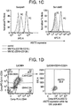
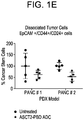
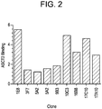

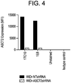
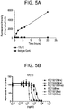


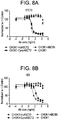
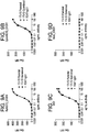


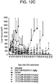

Classifications
-
- A—HUMAN NECESSITIES
- A61—MEDICAL OR VETERINARY SCIENCE; HYGIENE
- A61K—PREPARATIONS FOR MEDICAL, DENTAL OR TOILETRY PURPOSES
- A61K39/00—Medicinal preparations containing antigens or antibodies
- A61K39/395—Antibodies; Immunoglobulins; Immune serum, e.g. antilymphocytic serum
-
- C—CHEMISTRY; METALLURGY
- C07—ORGANIC CHEMISTRY
- C07K—PEPTIDES
- C07K16/00—Immunoglobulins [IGs], e.g. monoclonal or polyclonal antibodies
- C07K16/18—Immunoglobulins [IGs], e.g. monoclonal or polyclonal antibodies against material from animals or humans
- C07K16/28—Immunoglobulins [IGs], e.g. monoclonal or polyclonal antibodies against material from animals or humans against receptors, cell surface antigens or cell surface determinants
-
- A—HUMAN NECESSITIES
- A61—MEDICAL OR VETERINARY SCIENCE; HYGIENE
- A61K—PREPARATIONS FOR MEDICAL, DENTAL OR TOILETRY PURPOSES
- A61K47/00—Medicinal preparations characterised by the non-active ingredients used, e.g. carriers or inert additives; Targeting or modifying agents chemically bound to the active ingredient
- A61K47/50—Medicinal preparations characterised by the non-active ingredients used, e.g. carriers or inert additives; Targeting or modifying agents chemically bound to the active ingredient the non-active ingredient being chemically bound to the active ingredient, e.g. polymer-drug conjugates
- A61K47/51—Medicinal preparations characterised by the non-active ingredients used, e.g. carriers or inert additives; Targeting or modifying agents chemically bound to the active ingredient the non-active ingredient being chemically bound to the active ingredient, e.g. polymer-drug conjugates the non-active ingredient being a modifying agent
- A61K47/68—Medicinal preparations characterised by the non-active ingredients used, e.g. carriers or inert additives; Targeting or modifying agents chemically bound to the active ingredient the non-active ingredient being chemically bound to the active ingredient, e.g. polymer-drug conjugates the non-active ingredient being a modifying agent the modifying agent being an antibody, an immunoglobulin or a fragment thereof, e.g. an Fc-fragment
- A61K47/6801—Drug-antibody or immunoglobulin conjugates defined by the pharmacologically or therapeutically active agent
- A61K47/6803—Drugs conjugated to an antibody or immunoglobulin, e.g. cisplatin-antibody conjugates
- A61K47/6811—Drugs conjugated to an antibody or immunoglobulin, e.g. cisplatin-antibody conjugates the drug being a protein or peptide, e.g. transferrin or bleomycin
- A61K47/6817—Toxins
- A61K47/6819—Plant toxins
- A61K47/6825—Ribosomal inhibitory proteins, i.e. RIP-I or RIP-II, e.g. Pap, gelonin or dianthin
-
- A—HUMAN NECESSITIES
- A61—MEDICAL OR VETERINARY SCIENCE; HYGIENE
- A61K—PREPARATIONS FOR MEDICAL, DENTAL OR TOILETRY PURPOSES
- A61K47/00—Medicinal preparations characterised by the non-active ingredients used, e.g. carriers or inert additives; Targeting or modifying agents chemically bound to the active ingredient
- A61K47/50—Medicinal preparations characterised by the non-active ingredients used, e.g. carriers or inert additives; Targeting or modifying agents chemically bound to the active ingredient the non-active ingredient being chemically bound to the active ingredient, e.g. polymer-drug conjugates
- A61K47/51—Medicinal preparations characterised by the non-active ingredients used, e.g. carriers or inert additives; Targeting or modifying agents chemically bound to the active ingredient the non-active ingredient being chemically bound to the active ingredient, e.g. polymer-drug conjugates the non-active ingredient being a modifying agent
- A61K47/68—Medicinal preparations characterised by the non-active ingredients used, e.g. carriers or inert additives; Targeting or modifying agents chemically bound to the active ingredient the non-active ingredient being chemically bound to the active ingredient, e.g. polymer-drug conjugates the non-active ingredient being a modifying agent the modifying agent being an antibody, an immunoglobulin or a fragment thereof, e.g. an Fc-fragment
- A61K47/6801—Drug-antibody or immunoglobulin conjugates defined by the pharmacologically or therapeutically active agent
- A61K47/6803—Drugs conjugated to an antibody or immunoglobulin, e.g. cisplatin-antibody conjugates
- A61K47/6811—Drugs conjugated to an antibody or immunoglobulin, e.g. cisplatin-antibody conjugates the drug being a protein or peptide, e.g. transferrin or bleomycin
- A61K47/6817—Toxins
- A61K47/6831—Fungal toxins, e.g. alpha sarcine, mitogillin, zinniol or restrictocin
-
- A—HUMAN NECESSITIES
- A61—MEDICAL OR VETERINARY SCIENCE; HYGIENE
- A61K—PREPARATIONS FOR MEDICAL, DENTAL OR TOILETRY PURPOSES
- A61K47/00—Medicinal preparations characterised by the non-active ingredients used, e.g. carriers or inert additives; Targeting or modifying agents chemically bound to the active ingredient
- A61K47/50—Medicinal preparations characterised by the non-active ingredients used, e.g. carriers or inert additives; Targeting or modifying agents chemically bound to the active ingredient the non-active ingredient being chemically bound to the active ingredient, e.g. polymer-drug conjugates
- A61K47/51—Medicinal preparations characterised by the non-active ingredients used, e.g. carriers or inert additives; Targeting or modifying agents chemically bound to the active ingredient the non-active ingredient being chemically bound to the active ingredient, e.g. polymer-drug conjugates the non-active ingredient being a modifying agent
- A61K47/68—Medicinal preparations characterised by the non-active ingredients used, e.g. carriers or inert additives; Targeting or modifying agents chemically bound to the active ingredient the non-active ingredient being chemically bound to the active ingredient, e.g. polymer-drug conjugates the non-active ingredient being a modifying agent the modifying agent being an antibody, an immunoglobulin or a fragment thereof, e.g. an Fc-fragment
- A61K47/6835—Medicinal preparations characterised by the non-active ingredients used, e.g. carriers or inert additives; Targeting or modifying agents chemically bound to the active ingredient the non-active ingredient being chemically bound to the active ingredient, e.g. polymer-drug conjugates the non-active ingredient being a modifying agent the modifying agent being an antibody, an immunoglobulin or a fragment thereof, e.g. an Fc-fragment the modifying agent being an antibody or an immunoglobulin bearing at least one antigen-binding site
- A61K47/6849—Medicinal preparations characterised by the non-active ingredients used, e.g. carriers or inert additives; Targeting or modifying agents chemically bound to the active ingredient the non-active ingredient being chemically bound to the active ingredient, e.g. polymer-drug conjugates the non-active ingredient being a modifying agent the modifying agent being an antibody, an immunoglobulin or a fragment thereof, e.g. an Fc-fragment the modifying agent being an antibody or an immunoglobulin bearing at least one antigen-binding site the antibody targeting a receptor, a cell surface antigen or a cell surface determinant
-
- A—HUMAN NECESSITIES
- A61—MEDICAL OR VETERINARY SCIENCE; HYGIENE
- A61P—SPECIFIC THERAPEUTIC ACTIVITY OF CHEMICAL COMPOUNDS OR MEDICINAL PREPARATIONS
- A61P35/00—Antineoplastic agents
-
- G—PHYSICS
- G01—MEASURING; TESTING
- G01N—INVESTIGATING OR ANALYSING MATERIALS BY DETERMINING THEIR CHEMICAL OR PHYSICAL PROPERTIES
- G01N33/00—Investigating or analysing materials by specific methods not covered by groups G01N1/00 - G01N31/00
- G01N33/48—Biological material, e.g. blood, urine; Haemocytometers
- G01N33/50—Chemical analysis of biological material, e.g. blood, urine; Testing involving biospecific ligand binding methods; Immunological testing
- G01N33/53—Immunoassay; Biospecific binding assay; Materials therefor
- G01N33/574—Immunoassay; Biospecific binding assay; Materials therefor for cancer
- G01N33/57484—Immunoassay; Biospecific binding assay; Materials therefor for cancer involving compounds serving as markers for tumor, cancer, neoplasia, e.g. cellular determinants, receptors, heat shock/stress proteins, A-protein, oligosaccharides, metabolites
- G01N33/57492—Immunoassay; Biospecific binding assay; Materials therefor for cancer involving compounds serving as markers for tumor, cancer, neoplasia, e.g. cellular determinants, receptors, heat shock/stress proteins, A-protein, oligosaccharides, metabolites involving compounds localized on the membrane of tumor or cancer cells
-
- A—HUMAN NECESSITIES
- A61—MEDICAL OR VETERINARY SCIENCE; HYGIENE
- A61K—PREPARATIONS FOR MEDICAL, DENTAL OR TOILETRY PURPOSES
- A61K39/00—Medicinal preparations containing antigens or antibodies
- A61K2039/505—Medicinal preparations containing antigens or antibodies comprising antibodies
-
- C—CHEMISTRY; METALLURGY
- C07—ORGANIC CHEMISTRY
- C07K—PEPTIDES
- C07K2317/00—Immunoglobulins specific features
- C07K2317/20—Immunoglobulins specific features characterized by taxonomic origin
- C07K2317/21—Immunoglobulins specific features characterized by taxonomic origin from primates, e.g. man
-
- C—CHEMISTRY; METALLURGY
- C07—ORGANIC CHEMISTRY
- C07K—PEPTIDES
- C07K2317/00—Immunoglobulins specific features
- C07K2317/50—Immunoglobulins specific features characterized by immunoglobulin fragments
- C07K2317/52—Constant or Fc region; Isotype
- C07K2317/524—CH2 domain
-
- C—CHEMISTRY; METALLURGY
- C07—ORGANIC CHEMISTRY
- C07K—PEPTIDES
- C07K2317/00—Immunoglobulins specific features
- C07K2317/50—Immunoglobulins specific features characterized by immunoglobulin fragments
- C07K2317/56—Immunoglobulins specific features characterized by immunoglobulin fragments variable (Fv) region, i.e. VH and/or VL
- C07K2317/565—Complementarity determining region [CDR]
-
- C—CHEMISTRY; METALLURGY
- C07—ORGANIC CHEMISTRY
- C07K—PEPTIDES
- C07K2317/00—Immunoglobulins specific features
- C07K2317/70—Immunoglobulins specific features characterized by effect upon binding to a cell or to an antigen
- C07K2317/73—Inducing cell death, e.g. apoptosis, necrosis or inhibition of cell proliferation
-
- C—CHEMISTRY; METALLURGY
- C07—ORGANIC CHEMISTRY
- C07K—PEPTIDES
- C07K2317/00—Immunoglobulins specific features
- C07K2317/70—Immunoglobulins specific features characterized by effect upon binding to a cell or to an antigen
- C07K2317/76—Antagonist effect on antigen, e.g. neutralization or inhibition of binding
-
- C—CHEMISTRY; METALLURGY
- C07—ORGANIC CHEMISTRY
- C07K—PEPTIDES
- C07K2317/00—Immunoglobulins specific features
- C07K2317/70—Immunoglobulins specific features characterized by effect upon binding to a cell or to an antigen
- C07K2317/77—Internalization into the cell
-
- C—CHEMISTRY; METALLURGY
- C07—ORGANIC CHEMISTRY
- C07K—PEPTIDES
- C07K2317/00—Immunoglobulins specific features
- C07K2317/90—Immunoglobulins specific features characterized by (pharmaco)kinetic aspects or by stability of the immunoglobulin
- C07K2317/92—Affinity (KD), association rate (Ka), dissociation rate (Kd) or EC50 value
Definitions
- ASCT2 is a 541-amino-acid, multi-pass membrane protein with eight transmembrane domains.
- the molecular weight of ASCT2 varies from 55-75 KD depending on the various glycosylation profiles.
- ASCT2 In addition to transporting L-alanine, L-serine, and L-cysteine, ASCT2 also transports L-threonine and L-glutamine.
- ASCT2 functions as a cell surface receptor which is shared by type D simian retro virus and type C viruses.
- US2012039904 refers to antibodies or fragments thereof that recognize and bind an extracellular region of ASCT2.
- ADCs Antibody-drug conjugates
- An ADC may comprise a cytotoxin, which may be a small molecule that has been chemically modified to contain a linker. The linker is then used to conjugate the cytotoxin to the antibody or antigen-binding fragment thereof.
- Cytotoxicity is induced when the ADC binds to the antigen surface of a target-positive cell, is internalized and trafficked to the lysosome where the cytotoxin is released following either proteolysis of a cleavable linker (for example by cathepsin B found in the lysosome) or through proteolytic degradation of the antibody when a non-cleavable linker is used to attach the cytotoxin to the antibody.
- the cytotoxin then translocates out of the lysosome and into the cytosol where it can then bind to its target, depending on its mechanism of action.
- these cytotoxins induce cell cycle arrest which subsequently leads to apoptosis.
- Corresponding conjugates containing imaging agents also represent a promising new way to detect cancer cells in vivo or in vitro.
- the VH of the antibody or antigen binding fragment comprises an amino acid sequence of SEQ ID NO: 1 and the VL comprises an amino acid sequence of SEQ ID NO: 2.
- the VH comprises an amino acid sequence of SEQ ID NO: 3, and the VL comprises an amino acid sequence of SEQ ID NO: 4.
- the VH comprises an amino acid sequence of SEQ ID NO: 5, and the VL comprises an amino acid sequence of SEQ ID NO: 6.
- the VH comprises an amino acid sequence of SEQ ID NO: 7, and the VL comprises an amino acid sequence of SEQ ID NO: 8.
- the antibody comprises a heavy chain of an amino acid sequence of SEQ ID NO: 9.
- the antibody or antigen binding fragment comprises a light chain constant region selected from the group consisting of a human kappa constant region and a human lambda constant region.
- the antibody or antigen binding fragment is further conjugated to a cytotoxin selected from the group consisting of an antimicrobial agent, a therapeutic agent, a prodrug, a peptide, a protein, an enzyme, a lipid, a biological response modifier, a pharmaceutical agent, a lymphokine, a heterologous antibody, a fragment of a heterologous antibody, a detectable label, a polyethylene glycol (PEG), a radioisotope, and a combination of two or more of any said cytotoxins.
- the antibody or antigen binding fragment is conjugated to a cytotoxin.
- the cytotoxin is selected from a tubulysin derivative and a pyrrolobenzodiazepine.
- the tubulysin derivative is tubulysin AZ1508.
- the pyrrolobenzodiazepine is selected from SG3315 and SG3249.
- the antibody or antigen binding fragment binds to human ASCT2 and cynomolgus monkey ASCT2.
- a method of making an antibody or antigen binding fragment thereof according to the first aspect of the invention comprising culturing a host comprising a polynucleotide of the third aspect of the invention.
- the present invention provides antibodies and antigen-binding fragments thereof that specifically bind to ASCT2.
- the antibody, or antigen-binding fragment is conjugated to an agent, preferably a cytotoxin.
- an agent preferably a cytotoxin.
- Polynucleotides encoding the antibodies and antigen-binding fragments thereof, vectors containing the polynucleotides, and host cells expressing the antibodies are included.
- Compositions comprising the anti-ASCT2 antibodies or antigen-binding fragments thereof, and methods of making the anti-ASCT2 antibodies and antigen-binding fragments are also provided.
- Methods of using the novel anti-ASCT2 antibodies such as in diagnostic applications or in methods of treating a disease or disorder characterized by ASCT2 overexpression, e.g ., cancer, are further provided.
- Amino acids are referred to herein by their commonly known three letter symbols or by the one-letter symbols recommended by the IUPAC-IUB Biochemical Nomenclature Commission. Nucleotides, likewise, are referred to by their commonly accepted single-letter codes.
- ASCT2 refers to the system ASC amino acid transporter 2 protein, and/or active fragments thereof.
- ASCT2 is a transmembrane protein that mediates transport of small neutral amino acids, including glutamine, alanine, and serine, cysteine, and threonine, in a Na + -dependent manner.
- the RNA, DNA, and amino acid sequences of ASCT2 are known to those skilled in the art and can be found in many databases, for example, in the databases of the National Center for Biotechnology Information (NCBI).
- NCBI National Center for Biotechnology Information
- inhibitor refers to any statistically significant decrease in biological activity, including full blocking of the activity.
- inhibitor can refer to a decrease of about 10%, 20%, 30%, 40%, 50%, 60%, 70%, 80%, 90% or 100% in a biological activity or process.
- antibody can refer to an immunoglobulin molecule that recognizes and specifically binds to a target, such as a protein, polypeptide, peptide, carbohydrate, polynucleotide, lipid, or combinations of the foregoing through at least one antigen recognition site within the variable region of the immunoglobulin molecule.
- An antibody can be of any the five major classes of immunoglobulins: IgA, IgD, IgE, IgG, and IgM, or subclasses (isotypes) thereof (e.g. IgG1, IgG2, IgG3, IgG4, IgA1 and IgA2), based on the identity of their heavy-chain constant domains referred to as alpha, delta, epsilon, gamma, and mu, respectively.
- the different classes of immunoglobulins have different and well-known subunit structures and three-dimensional configurations.
- Antibodies can be naked or conjugated to other molecules such as toxins, radioisotopes, etc.
- ASCT2 antibody or “antibody that binds to ASCT2” or “anti-ASCT2” refers to an antibody that is capable of binding ASCT2 with sufficient affinity such that the antibody is useful as a therapeutic agent or a diagnostic reagent in targeting ASCT2.
- the extent of binding of an anti-ASCT2 antibody to an unrelated, non-ASCT2 protein is less than about 10% of the binding of the antibody to ASCT2 as measured, e.g ., by a radioimmunoassay (RIA), BIACORE® (using recombinant ASCT2 as the analyte and antibody as the ligand, or vice versa ), KINEXA®, or other binding assays known in the art.
- RIA radioimmunoassay
- BIACORE® using recombinant ASCT2 as the analyte and antibody as the ligand, or vice versa
- KINEXA® or other binding assays known in the art.
- an antibody that binds to ASCT2 has a dissociation constant (K D ) of ⁇ 1 ⁇ M, ⁇ 100 nM, ⁇ 10 nM, ⁇ 1 nM, ⁇ 0.1 nM, ⁇ 10 pM, ⁇ 1 pM, or ⁇ 0.1 pM.
- a “monoclonal antibody” refers to a homogeneous antibody population involved in the highly specific recognition and binding of a single antigenic determinant, or epitope. This is in contrast to polyclonal antibodies that typically include different antibodies directed against different antigenic determinants.
- the term “monoclonal antibody” encompasses both intact and full-length monoclonal antibodies as well as antibody fragments (such as Fab, Fab', F(ab')2, Fv), single chain (scFv) mutants, fusion proteins comprising an antibody portion, and any other modified immunoglobulin molecule comprising an antigen recognition site.
- “monoclonal antibody” refers to such antibodies made in any number of ways including, but not limited to, hybridoma, phage selection, recombinant expression, and transgenic animals.
- humanized antibody refers to an antibody derived from a non-human (e.g., murine) immunoglobulin, which has been engineered to contain minimal non-human ( e.g., murine) sequences.
- humanized antibodies are human immunoglobulins in which residues from the complementary determining region (CDR) are replaced by residues from the CDR of a non-human species (e.g., mouse, rat, rabbit, or hamster) that have the desired specificity, affinity, and capability ( Jones et al., 1986, Nature, 321:522-525 ; Riechmann et al., 1988, Nature, 332:323-327 ; Verhoeyen et al., 1988, Science, 239:1534-1536 ).
- the Fv framework region (FW) residues of a human immunoglobulin are replaced with the corresponding residues in an antibody from a non-human species that has the desired specificity, affinity, and capability.
- a “pharmaceutically acceptable carrier” refers to an ingredient in a pharmaceutical formulation, other than an active ingredient, which is nontoxic to a subject.
- a pharmaceutically acceptable carrier includes, but is not limited to, a buffer, excipient, stabilizer, or preservative.
- pharmaceutically acceptable carrier includes any and all solvents, dispersion media, coatings, antibacterial and antifungal agents, isotonic and absorption/resorption delaying agents, and the like that are physiologically compatible.
- variable region of an antibody refers to the variable region of the antibody light chain or the variable region of the antibody heavy chain, either alone or in combination.
- the variable regions of the heavy and light chain each consist of four framework regions (FW) connected by three complementarity-determining regions (CDRs), also known as hypervariable regions.
- the CDRs in each chain are held together in close proximity by the FW regions and, with the CDRs from the other chain, contribute to the formation of the antigen-binding site of antibodies.
- There are at least two techniques for determining CDRs (1) an approach based on cross-species sequence variability (i.e., Kabat et al.
- the "Kabat numbering system” is generally used when referring to a residue in the variable domain (approximately residues 1-107 of the light chain and residues 1-113 of the heavy chain) (e.g,, Kabat et al., Sequences of Immunological Interest, 5th Ed. Public Health Service, National Institutes of Health, Bethesda, Md. (1991 )).
- the Kabat numbering of residues can be determined for a given antibody by alignment at regions of homology of the sequence of the antibody with a "standard” Kabat numbered sequence. Chothia refers instead to the location of the structural loops ( Chothia and Lesk, J. Mol. Biol. 196:901-917 (1987 )).
- the end of the Chothia CDR-H1 loop when numbered using the Kabat numbering convention, varies between H32 and H34 depending on the length of the loop (this is because the Kabat numbering scheme places the insertions at H35A and H35B; if neither 35A nor 35B is present, the loop ends at 32; if only 35A is present, the loop ends at 33; if both 35A and 35B are present, the loop ends at 34).
- the AbM hypervariable regions represent a compromise between the Kabat CDRs and Chothia structural loops, and are used by Oxford Molecular's AbM antibody modeling software. Table 1, below lists the positions of the amino acids comprising the variable regions of the antibodies in each system.
- ImMunoGeneTics also provides a numbering system for the immunoglobulin variable regions, including the CDRs. See, e.g., Lefranc, M.P. et al., Dev. Comp. Immunol. 27: 55-77(2003 ).
- the IMGT numbering system is based on an alignment of more than 5,000 sequences, structural data, and characterization of hypervariable loops and allows for easy comparison of the variable and CDR regions for all species.
- VH-CDR1 is at positions 26 to 35
- VH-CDR2 is at positions 51 to 57
- VH-CDR3 is at positions 93 to 102
- VL-CDR1 is at positions 27 to 32
- VL-CDR2 is at positions 50 to 52
- VL-CDR3 is at positions 89 to 97.
- VH CDRs sequences described correspond to the classical Kabat numbering locations, namely Kabat VH-CDR1 is at positions 31-35, VH-CDR2 is a positions 50-65, and VH-CDR3 is at positions 95-102.
- VL-CDR1, VL-CDR2 and VL-CDR3 also correspond to classical Kabat numbering locations, namely positions 24-34, 50-56 and 89-97, respectively.
- human antibody means an antibody produced in a human or an antibody having an amino acid sequence corresponding to an antibody produced in a human made using any technique known in the art. This definition of a human antibody includes intact or full-length antibodies, fragments thereof, and/or antibodies comprising at least one human heavy and/or light chain polypeptide such as, for example, an antibody comprising murine light chain and human heavy chain polypeptides.
- chimeric antibodies refers to antibodies in which the amino acid sequence of the immunoglobulin molecule is derived from two or more species.
- the variable region of both light and heavy chains corresponds to the variable region of antibodies derived from one species of mammals (e.g., mouse, rat, rabbit, etc.) with the desired specificity, affinity, and capability while the constant regions are homologous to the sequences in antibodies derived from another (usually human) to avoid eliciting an immune response in that species.
- YTE refers to a mutation in IgG1 Fc that results in an increase in the binding to human FcRn and improves the serum half-life of the antibody having the mutation.
- a YTE mutant comprises a combination of three mutations, M252Y/S254T/T256E (EU numbering Kabat et al. (1991) Sequences of Proteins of Immunological Interest, U.S. Public Health Service, National Institutes of Health, Washington, D.C .), introduced into the heavy chain of an IgG1. See U.S. Patent No. 7,658,921 .
- the YTE mutant has been shown to increase the serum half-life of antibodies approximately four-times as compared to wild-type versions of the same antibody ( Dall'Acqua et al., J. Biol. Chem. 281:23514-24 (2006 ); Robbie et al., (2013) Antimicrob. Agents Chemother. 57, 6147-6153 ). See also U.S. Patent No. 7,083,784 .
- Binding affinity generally refers to the strength of the sum total of non-covalent interactions between a single binding site of a molecule (e.g., an antibody) and its binding partner (e.g., an antigen). Unless indicated otherwise, as used herein, "binding affinity” refers to intrinsic binding affinity which reflects a 1:1 interaction between members of a binding pair (e.g., antibody and antigen).
- the affinity of a molecule X for its partner Y can generally be represented by the dissociation constant (K D ). Affinity can be measured by common methods known in the art, including those described herein.
- Low-affinity antibodies generally bind antigen slowly and tend to dissociate readily, whereas high-affinity antibodies generally bind antigen faster and tend to remain bound longer.
- a variety of methods of measuring binding affinity are known in the art, any of which can be used for purposes of the present invention.
- IC 50 Potency of binding molecule is normally expressed as an IC 50 value, in ng/ml unless otherwise stated.
- IC 50 is the median inhibitory concentration of an antibody molecule. In functional assays, IC 50 is the concentration that reduces a biological response by 50% of its maximum. In ligand-binding studies, IC 50 is the concentration that reduces receptor binding by 50% of maximal specific binding level. IC 50 can be calculated by any number of means known in the art.
- the fold improvement in potency for the antibodies or polypeptides of the invention as compared to a reference antibody can be at least about 2-fold, at least about 4-fold, at least about 6-fold, at least about 8-fold, at least about 10-fold, at least about 20-fold, at least about 30-fold, at least about 40-fold, at least about 50-fold, at least about 60-fold, at least about 70-fold, at least about 80-fold, at least about 90-fold, at least about 100-fold, at least about 110-fold, at least about 120-fold, at least about 130-fold, at least about 140-fold, at least about 150-fold, at least about 160-fold, at least about 170-fold, or at least about 180-fold or more.
- Binding potency of an antibody is normally expressed as an EC 50 value, in nM or pM unless otherwise stated.
- EC 50 is the concentration of a drug that induces a median response between baseline and maximum after a specified exposure time. EC 50 can be calculated by any number of means known in the art.
- a “therapeutic antibody” is one that can be administered to a subject to treat or prevent a disease or condition.
- a “subject” is any individual, particularly a mammal, for whom diagnosis, prognosis, or therapy is desired. Mammalian subjects include humans, domestic animals, farm animals, sports animals, and zoo animals, e.g ., humans, non-human primates, dogs, cats, guinea pigs, rabbits, rats, mice, horses, cattle, etc.
- a subject is successfully "treated” for a disease or disorder, for example, cancer, according to the methods provided herein if the patient shows, e.g ., total, partial, or transient alleviation or elimination of symptoms associated with the disease or disorder.
- prevent refers to prophylactic or preventative measures that prevent and/or slow the development of a targeted pathologic condition or disorder.
- those in need of prevention include those prone to have or susceptible to the disorder.
- a disease or disorder is successfully prevented according to the methods provided herein if the patient develops, transiently or permanently, e.g ., fewer or less severe symptoms associated with the disease or disorder, or a later onset of symptoms associated with the disease or disorder, than a patient who has not been subject to the methods of the invention.
- composition refers to a preparation that is in such form as to permit the biological activity of the active ingredient to be effective, and which contains no additional components which are unacceptably toxic to a subject to which the composition would be administered.
- Such composition can be sterile, and can comprise a pharmaceutically acceptable carrier, such as physiological saline.
- label refers to a detectable compound or composition that is conjugated directly or indirectly to the binding molecule or antibody so as to generate a "labeled" binding molecule or antibody.
- the label can be detectable by itself (e.g., radioisotope labels or fluorescent labels) or, in the case of an enzymatic label, can catalyze chemical alteration of a substrate compound or composition that is detectable.
- polypeptide polypeptide
- peptide protein
- polymers of amino acids of any length can be linear or branched, it can comprise modified amino acids, and non-amino acids can interrupt it.
- the terms also encompass an amino acid polymer that has been modified naturally or by intervention; for example, disulfide bond formation, glycosylation, lipidation, acetylation, phosphorylation, or any other manipulation or modification, such as conjugation with a labeling component.
- polypeptides containing one or more analogs of an amino acid including, for example, unnatural amino acids, etc.
- the polypeptides can occur as single chains or associated chains.
- a “polynucleotide,” as used herein can include one or more "nucleic acids,” “nucleic acid molecules,” or “nucleic acid sequences,” refers to a polymer of nucleotides of any length, and includes DNA and RNA.
- the polynucleotides can be deoxyribonucleotides, ribonucleotides, modified nucleotides or bases, and/or their analogs, or any substrate that can be incorporated into a polymer by DNA or RNA polymerase.
- a polynucleotide can comprise modified nucleotides, such as methylated nucleotides and their analogs. The preceding description applies to all polynucleotides referred to herein, including RNA and DNA.
- vector means a construct, which is capable of delivering, and in some embodiments, expressing, one or more genes or sequences of interest in a host cell.
- vectors include, but are not limited to, viral vectors, naked DNA or RNA expression vectors, plasmid, cosmid or phage vectors, DNA or RNA expression vectors associated with cationic condensing agents, DNA or RNA expression vectors encapsulated in liposomes, and certain eukaryotic cells, such as producer cells.
- a polypeptide, antibody, polynucleotide, vector, cell, or composition that is "isolated” is a polypeptide, antibody, polynucleotide, vector, cell, or composition that is in a form not found in nature.
- Isolated polypeptides, antibodies, polynucleotides, vectors, cells or compositions include those which have been purified to a degree that they are no longer in a form in which they are found in nature.
- an antibody, polynucleotide, vector, cell, or composition that is isolated is substantially pure.
- nucleic acids or polypeptides refer to two or more sequences or subsequences that are the same or have a specified percentage of nucleotides or amino acid residues that are the same, when compared and aligned (introducing gaps, if necessary) for maximum correspondence, not considering any conservative amino acid substitutions as part of the sequence identity.
- percent identity can be measured using sequence comparison software or algorithms or by visual inspection. Various algorithms and software are known in the art that can be used to obtain alignments of amino acid or nucleotide sequences.
- sequence alignment algorithm is the algorithm described in Karlin et al., Proc. Natl. Acad. Sci. USA, 87:2264-2268 (1990 ), as modified by Karlin et al., Proc. Natl. Acad. Sci. USA, 90:5873-5877 (1993 ), and incorporated into the NBLAST and XBLAST programs ( Altschul et al., Nucleic Acids Res. 25:3389-3402 (1991 )).
- Gapped BLAST can be used as described by Altschul et al., Nucleic Acids Res. 25:3389-3402 (1997 ).
- BLAST-2 Altschul et al., Methods in Enzymol. 266:460-480 (1996 )
- ALIGN ALIGN-2
- ALIGN-2 Genentech, South San Francisco, CA
- Megalign Megalign
- the percent identity between two nucleotide sequences is determined using the GAP program in the GCG software package ( e.g., using a NWSgapdna.CMP matrix and a gap weight of 40, 50, 60, 70, or 90 and a length weight of 1, 2, 3, 4, 5, or 6).
- the GAP program in the GCG software package which incorporates the algorithm of Needleman and Wunsch (J. Mol. Biol. 48:444-453 (1970 )) can be used to determine the percent identity between two amino acid sequences (e.g., using either a BLOSUM 62 matrix or a PAM250 matrix, and a gap weight of 16, 14, 12, 10, 8, 6, or 4 and a length weight of 1, 2, 3, 4, 5).
- the percent identity between nucleotide or amino acid sequences is determined using the algorithm of Myers and Miller (CABIOS 4:11-17 (1989 )).
- the percent identity can be determined using the ALIGN program (version 2.0) and using a PAM120 with residue table, a gap length penalty of 12 and a gap penalty of 4.
- ALIGN program version 2.0
- PAM120 with residue table residue table
- gap length penalty 12
- gap penalty 4
- One skilled in the art can determine appropriate parameters for maximal alignment by particular alignment software.
- the default parameters of the alignment software are used.
- the percentage identity "X" of a first amino acid sequence to a second sequence amino acid is calculated as 100 x (Y/Z), where Y is the number of amino acid residues scored as identical matches in the alignment of the first and second sequences (as aligned by visual inspection or a particular sequence alignment program) and Z is the total number of residues in the second sequence. If the length of a first sequence is longer than the second sequence, the percent identity of the first sequence to the second sequence will be higher than the percent identity of the second sequence to the first sequence.
- a “conservative amino acid substitution” is one in which one amino acid residue is replaced with another amino acid residue having a similar side chain.
- Families of amino acid residues having similar side chains have been defined in the art, including basic side chains (e.g., lysine, arginine, histidine), acidic side chains (e.g., aspartic acid, glutamic acid), uncharged polar side chains ( e.g., asparagine, glutamine, serine, threonine, tyrosine, cysteine), nonpolar side chains ( e.g., glycine, alanine, valine, leucine, isoleucine, proline, phenylalanine, methionine, tryptophan), beta-branched side chains ( e.g., threonine, valine, isoleucine) and aromatic side chains ( e.g., tyrosine, phenylalanine, tryptophan, histidine).
- substitution of a phenylalanine for a tyrosine is a conservative substitution.
- conservative substitutions in the amino acid sequences of the binding molecules, antibodies, and antigen-binding fragments of the invention do not abrogate the binding of the binding molecule, antibody, or antigen-binding fragment containing the amino acid sequence, to the antigen(s), i.e., the ASCT2 to which the binding molecule, antibody, or antigen-binding fragment binds.
- Methods of identifying nucleotide and amino acid conservative substitutions which do not eliminate antigen-binding are well-known in the art. See, e.g., Brummell et al., Biochem.
- the anti-ASCT2 antibodies of the invention bind to ASCT2 on the surface of a cell and are internalized into the cell.
- an anti-ASCT2 antibody is internalized into ASCT2-expressing cells with an IC 50 at 10 minutes of about 100 ng/ml to about 1 ⁇ g/ml, about 100 ng/ml to about 500 ng/ml, about 100 ng/ml to about 250 ng/ml, about 250 ng/ml to about 500 ng/ml, about 350 ng/ml to about 450 ng/ml, about 500 ng/ml to about 1 ⁇ g/ml, about 500 ng/ml to about 750 ng/ml, about 750 ng/ml to about 850 ng/ml, or about 900 ng/ml to about 1 ⁇ g/ml.
- an anti-ASCT2 antibody is internalized into ASCT2-expressing cells with an IC 50 at 30 minutes of about 100 ng/ml to about 1 ⁇ g/ml, about 100 ng/ml to about 500 ng/ml, about 100 ng/ml to about 250 ng/ml, about 250 ng/ml to about 500 ng/ml, about 250 ng/ml to about 350 ng/ml, about 350 ng/ml to about 450 ng/ml, about 500 ng/ml to about 1 ⁇ g/ml, about 500 ng/ml to about 750 ng/ml, about 750 ng/ml to about 850 ng/ml, or about 900 ng/ml to about 1 ⁇ g/ml.
- an anti-ASCT2 antibody is internalized into ASCT2-expressing cells with an IC 50 at 120 minutes of about 50 ng/ml to about 500 ng/ml, about 50 ng/ml to about 100 ng/ml, about 100 ng/ml to about 200 ng/ml, about 200 ng/ml to about 300 ng/ml, about 300 ng/ml to about 400 ng/ml, or about 400 ng/ml to about 500 ng/ml.
- this disclosure provides an anti-ASCT2 antibody or antigen-binding fragment thereof comprising three heavy chain complementarity determining regions (HCDRs) and three light chain complementarity determining regions (LCDRs), wherein the HCDR1 has an amino acid sequence of SEQ ID NO: 10; the HCDR2 has an amino acid sequence of SEQ ID NO: 11; the HCDR3 has an amino acid sequence of SEQ ID NO: 12; the LCDR1 has an amino acid sequence of SEQ ID NO: 13; the LCDR2 has an amino acid sequence of SEQ ID NO: 14; and the LCDR3 has an amino acid sequence of SEQ ID NO: 15; or the HCDR1 has an amino acid sequence of SEQ ID NO: 16; the HCDR2 has an amino acid sequence of SEQ ID NO: 17; the HCDR3 has an amino acid sequence of SEQ ID NO: 18; the LCDR1 has an amino acid sequence of SEQ ID NO: 19; the LCDR2 has an amino acid sequence of SEQ ID NO: 20; and the LCDR3
- the VH comprises an amino acid sequence of SEQ ID NO: 1 or SEQ ID NO: 5; and the VL comprises an amino acid sequence of SEQ ID NO: 2 or SEQ ID NO: 6.
- the anti-ASCT2 antibody comprises a VH of an amino acid sequence of SEQ ID NO: 5 and a VL of an amino acid sequence of SEQ ID NO: 6.
- an anti-ASCT2 antibody comprises a VH of an amino acid sequence of SEQ ID NO: 3 or SEQ ID NO: 7, and a VL of an amino acid sequence of SEQ ID NO: 4 or SEQ ID NO: 8.
- the anti-ASCT2 antibody comprises a VH of an amino acid sequence of SEQ ID NO: 7 and a VL of an amino acid sequence of SEQ ID NO: 8.
- the disclosure provides an isolated antibody or antigen-binding fragment thereof which specifically binds to ASCT2 comprising a VH and a VL, where the VH and VL contain, respectively, amino acid sequences at least 70%, 75%, 80%, 85%, 90%, 95%, or 100% identical to reference amino acid sequences SEQ ID NO: 1 and SEQ ID NO: 2; SEQ ID NO: 3 and SEQ ID NO: 4; SEQ ID NO: 5 and SEQ ID NO: 6; or SEQ ID NO: 7 and SEQ ID NO: 8, , respectively.
- the disclosure provides an anti-ASCT2 antibody or antigen-binding fragment thereof comprising VH amino acid sequence SEQ ID NO: 5 and the VL amino acid sequence SEQ ID NO: 6. In one aspect, the disclosure provides an anti-ASCT2 antibody or antigen-binding fragment thereof comprising VH amino acid sequence SEQ ID NO: 7 and the VL amino acid sequence SEQ ID NO: 8.
- An anti-ASCT2 antibody or antigen-binding fragment thereof as described herein can be, e.g., a murine antibody, a humanized antibody, a chimeric antibody, a monoclonal antibody, a polyclonal antibody, a recombinant antibody, a multispecific antibody, or any combination thereof.
- An anti-ASCT2 antibody antigen-binding fragment can be an Fv fragment, an Fab fragment, an F(ab')2 fragment, an Fab' fragment, a dsFv fragment, an scFv fragment, or an sc(Fv)2 fragment.
- the disclosure provides an anti-ASCT2 antibody or antigen-binding fragment thereof that can bind to ASCT2 molecules across species, e.g., the antibody or fragment can bind to mouse ASCT2, rat ASCT2, rabbit ASCT2, human ASCT2 and/or cynomolgus monkey ASCT2.
- the antibody or fragment can bind to human ASCT2 and cynomolgus monkey ASCT2.
- the antibody or fragment can also bind to mouse ASCT2.
- an anti-ASCT2 antibody or antigen binding fragment thereof can specifically bind to ASCT2, e.g ., human ASCT2 and cynomolgus monkey ASCT2, but does not specifically bind to human ASCT1.
- An anti-ASCT2 antibody or antigen-binding fragment thereof as described herein can include, in addition to a VH and a VL, a heavy chain constant region or fragment thereof.
- the heavy chain constant region is a human heavy chain constant region, e.g., a human IgG constant region, e.g., a human IgG1 constant region.
- an agent such as a cytotoxic agent
- a cysteine residue is inserted between amino acid S239 and V240 in the CH2 region of IgG1. This cysteine is referred to as "a 239 insertion" or "239i.”
- a heavy chain constant region or fragment thereof e.g., a human IgG constant region or fragment thereof
- the IgG constant domain can contain one or more amino acid substitutions of amino acid residues at positions 251-257, 285-290, 308-314, 385-389, and 428-436, wherein the amino acid position numbering is according to the EU index as set forth in Kabat.
- the IgG constant domain can contain one or more of a substitution of the amino acid at Kabat position 252 with Tyrosine (Y), Phenylalanine (F), Tryptophan (W), or Threonine (T), a substitution of the amino acid at Kabat position 254 with Threonine (T), a substitution of the amino acid at Kabat position 256 with Serine (S), Arginine (R), Glutamine (Q), Glutamic acid (E), Aspartic acid (D), or Threonine (T), a substitution of the amino acid at Kabat position 257 with Leucine (L), a substitution of the amino acid at Kabat position 309 with Proline (P), a substitution of the amino acid at Kabat position 311 with Serine (S), a substitution of the amino acid at Kabat position 428 with Threonine (T), Leucine (L), Phenylalanine (F), or Serine (S), a substitution of the amino acid at Kabat position 433 with Arginine (R)
- the IgG constant domain can contain amino acid substitutions relative to a wild-type human IgG constant domain including a substitution of the amino acid at Kabat position 252 with Tyrosine (Y), a substitution of the amino acid at Kabat position 254 with Threonine (T), and a substitution of the amino acid at Kabat position 256 with Glutamic acid (E).
- This disclosure provides an anti-ASCT2 antibody or antigen-binding fragment thereof where the heavy chain is a human IgG1 YTE mutant.
- An anti-ASCT2 antibody or antigen-binding fragment thereof provided herein, e.g., as described above, can include, in addition to a VH and a VL, and optionally a heavy chain constant region or fragment thereof, a light chain constant region or fragment thereof.
- the light chain constant region is a kappa lambda light chain constant region, e.g., a human kappa constant region or a human lambda constant region.
- VH and/or VL amino acid sequence can be, e.g., 85%, 90%, 95%, 96%, 97%, 98% or 99% similar to a sequence set forth herein, and/or comprise 1, 2, 3, 4, 5 or more substitutions, e.g ., conservative substitutions relative to a sequence set forth herein.
- An ASCT2 antibody having VH and VL regions having a certain percent similarity to a VH region or VL region, or having one or more substitutions, e.g ., conservative substitutions can be obtained by mutagenesis (e.g., site-directed or PCR-mediated mutagenesis) of nucleic acid molecules encoding VH and/or VL regions described herein, followed by testing of the encoded altered antibody for binding to ASCT2 and optionally testing for retained function using the functional assays described herein.
- mutagenesis e.g., site-directed or PCR-mediated mutagenesis
- an anti-ASCT2 antibody or antigen-binding fragment thereof can bind to ASCT2-expressing cells with an IC 50 lower than about 500 nM, lower than about 350 nM, lower than about 250 nM, lower than about 150 nM, lower than about 100 nM, lower than about 75 nM, lower than about 60 nM, lower than about 50 nM, lower than about 40 nM, lower than about 30 nM, lower than about 20 nM, lower than about 15 nM, lower than about 10 nM, lower than about 5 nM, lowr than about 1 nM, lower than about 500 pM, lower than about 350 pM, lower than about 250 pM, lower than about 150 pM, lower than about 100 pM, lower than about 75 pM, lower than about 60 pM, lower than about 50 pM, lower than about 40 pM, lower than about 30 pM, lower than about 20 pM, lower than about 15 pM, lower than about
- Anti-ASCT2 antibodies and antigen fragments thereof, disclosed herein, can be conjugated to heterologous agents using site-specific or non-site specific methods of conjugation.
- the ADC comprises one, two, three, four or more therapeutic moieties. In some aspects, all therapeutic moieties are the same.
- site-specific conjugation of therapeutic moieties to antibodies using reactive amino acid residues at specific positions yields homogeneous ADC preparations with uniform stoichiometry.
- the site specific conjugation can be through a cysteine residue or a non-natural amino acid.
- the cytotoxic or imaging agent is conjugated to the antibody or antigen binding fragment thereof through at least one cysteine residue.
- each therapeutic moiety is chemically conjugated to the side chain of an amino acid at a specific Kabat position in the Fc region.
- the polypeptide having a leader sequence is a pre-protein and can have the leader sequence cleaved by the host cell to form the mature form of the polypeptide.
- the polynucleotides can also encode an ASCT2-binding pro-protein which is the mature protein plus additional 5' amino acid residues.
- the disclosure provides an isolated polynucleotide comprising a nucleic acid encoding an antibody VL, wherein the VL comprises an amino acid sequence at least 70%, 75%, 80%, 85%, 90%, 95%, or 100% identical to a reference amino acid sequence selected from the group consisting of SEQ ID NO: 2, SEQ ID NO: 4, SEQ ID NO: 6, and SEQ ID NO: 8.
- an antibody or antigen-binding fragment thereof comprising a VH or VL encoded by a polynucleotide as described above, can specifically bind to ASCT2, e.g., human or cynomolgus monkey ASCT2.
- ASCT2 e.g., human or cynomolgus monkey ASCT2.
- an antibody or antigen-binding fragment thereof can specifically bind to the same epitope as an antibody or antigen-binding fragment thereof comprising the VH and VL of 17c10 or 1e8.
- the disclosure provides a polynucleotide or combination of polynucleotides encoding a binding molecule, e.g., an antibody or antigen-binding fragment thereof, which specifically binds to ASCT2.
- the disclosure provides a composition, e.g ., a pharmaceutical composition, comprising a polynucleotide or vector as described above, optionally further comprising one or more carriers, diluents, excipients, or other additives.
- a composition e.g ., a pharmaceutical composition, comprising a polynucleotide or vector as described above, optionally further comprising one or more carriers, diluents, excipients, or other additives.
- the polynucleotide comprising a nucleic acid encoding a VH and the polynucleotide comprising a nucleic acid encoding a VL can reside in a single vector, or can be on separate vectors. Accordingly the disclosure provides one or more vectors comprising the polynucleotide composition described above.
- This disclosure further provides a host cell comprising a polynucleotide, polynucleotide composition, or vector as provided above, where host cell can, in some instances, express an antibody or antigen-binding fragment thereof that specifically binds to ASCT2.
- host cell can be utilized in a method of making an antibody or antigen-binding fragment thereof as provided herein, where the method includes (a) culturing the host cell and (b) isolating the antibody or antigen-binding fragment thereof expressed from the host cell.
- the polynucleotides comprise the coding sequence for the mature ASCT2-binding polypeptide, e.g., an anti-ASCT2 antibody or an antigen-binding fragment thereof, fused in the same reading frame to a marker sequence that allows, for example, purification of the encoded polypeptide.
- the marker sequence can be a hexa-histidine tag supplied by a pQE-9 vector to provide for purification of the mature polypeptide fused to the marker, in the case of a bacterial host, or the marker sequence can be a hemagglutinin (HA) tag derived from the influenza hemagglutinin protein when a mammalian host (e.g., COS-7 cells) is used.
- a mammalian host e.g., COS-7 cells
- Polynucleotide variants are also provided. Polynucleotide variants can contain alterations in the coding regions, non-coding regions, or both. In some embodiments polynucleotide variants contain alterations that produce silent substitutions, additions, or deletions, but do not alter the properties or activities of the encoded polypeptide. In some embodiments, polynucleotide variants are produced by silent substitutions due to the degeneracy of the genetic code. Polynucleotide variants can be produced for a variety of reasons, e.g., to optimize codon expression for a particular host (change codons in the human mRNA to those preferred by a bacterial host such as E. coli ). Vectors and cells comprising the polynucleotides described herein are also provided.
- the polynucleotide sequences encoding a particular isolated polypeptide of interest can be inserted into an expression vector and operatively linked to an expression control sequence appropriate for expression of the protein in a desired host. Proper assembly can be confirmed, e.g ., by nucleotide sequencing, restriction mapping, and/or expression of a biologically active polypeptide in a suitable host.
- the gene can be operatively linked to or associated with transcriptional and translational expression control sequences that are functional in the chosen expression host.
- recombinant expression vectors are used to amplify and express DNA encoding anti-ASCT2 antibodies or antigen-binding fragments thereof.
- Recombinant expression vectors are replicable DNA constructs which have synthetic or cDNA-derived DNA fragments encoding a polypeptide chain of an anti-ASCT2 antibody or and antigen-binding fragment thereof, operatively linked to suitable transcriptional or translational regulatory elements derived from mammalian, microbial, viral, or insect genes.
- Suitable mammalian host cell lines include HEK-293 and HEK-293T, the COS-7 lines of monkey kidney cells, described by Gluzman, Cell 23:175 (1981 ), and other cell lines including, for example, L cells, C127, 3T3, Chinese hamster ovary (CHO), HeLa, and BHK cell lines.
- ASCT2-binding molecules produced by a transformed host can be purified according to any suitable method.
- standard methods include chromatography (e.g., ion exchange, affinity, and sizing column chromatography), centrifugation, differential solubility, or by any other standard technique for protein purification.
- Affinity tags such as hexahistidine, maltose binding domain, influenza coat sequence, and glutathione-S-transferase, can be attached to the protein to allow easy purification by passage over an appropriate affinity column.
- Isolated proteins can also be physically characterized using such techniques as proteolysis, nuclear magnetic resonance and x-ray crystallography.
- supernatants from systems that secrete recombinant protein into culture media can be first concentrated using a commercially available protein concentration filter, for example, an Amicon or Millipore Pellicon ultrafiltration unit. Following the concentration step, the concentrate can be applied to a suitable purification matrix.
- a suitable purification matrix for example, an anion exchange resin can be employed, for example, a matrix or substrate having pendant diethylaminoethyl (DEAE) groups.
- the matrices can be acrylamide, agarose, dextran, cellulose, or other types commonly employed in protein purification.
- a cation exchange step can be employed. Suitable cation exchangers include various insoluble matrices comprising sulfopropyl or carboxymethyl groups.
- the route of administration of the ASCT2-binding molecule can be, for example, oral, parenteral, by inhalation, or topical.
- parenteral as used herein includes, e.g ., intravenous, intraarterial, intraperitoneal, intramuscular, subcutaneous, rectal, or vaginal administration. While all these forms of administration are clearly contemplated as being within the scope of the invention, another example of a form for administration would be a solution for injection, in particular for intravenous or intraarterial injection or drip.
- ASCT2-binding molecules provided herein can be administered in a pharmaceutically effective amount for the in vivo treatment of diseases or disorders characterized by ASCT2 overexpression, such as colorectal cancer, HNSCC, prostate cancer, lung cancer, pancreatic cancer, melanoma, endometrial cancer, hematological cancer (AML, MM, DLBCL), and cancers comprising CSCs.
- ASCT2 overexpression such as colorectal cancer, HNSCC, prostate cancer, lung cancer, pancreatic cancer, melanoma, endometrial cancer, hematological cancer (AML, MM, DLBCL), and cancers comprising CSCs.
- the disclosed binding molecules can be formulated so as to facilitate administration and promote stability of the active agent.
- Pharmaceutical compositions in accordance with the present invention can comprise a pharmaceutically acceptable, non-toxic, sterile carrier such as physiological saline, non-toxic buffers, preservatives and the like.
- a pharmaceutically effective amount of an ASCT2-binding molecule means an amount sufficient to achieve effective binding to a target and to achieve a benefit, e.g., to ameliorate symptoms of a disease or condition or to detect a substance or a cell.
- Suitable formulations for use in the therapeutic methods disclosed herein are described in Remington's Pharmaceutical Sciences, 22nd ed., Ed. Lloyd V. Allen, Jr. (2012 ).
- compositions provided herein can be orally administered in an acceptable dosage form including, e.g ., capsules, tablets, aqueous suspensions, or solutions. Certain pharmaceutical compositions also can be administered by nasal aerosol or inhalation. Such compositions can be prepared as solutions in saline, employing benzyl alcohol or other suitable preservatives, absorption promoters to enhance bioavailability, and/or other conventional solubilizing or dispersing agents.
- the amount of an ASCT2-binding molecule that can be combined with carrier materials to produce a single dosage form will vary depending upon the subject treated and the particular mode of administration.
- the composition can be administered as a single dose, multiple doses or over an established period of time in an infusion. Dosage regimens also can be adjusted to provide the optimum desired response.
- terapéuticaally effective dose or amount or “effective amount” is intended an amount of an ASCT2-binding molecule that, when administered, brings about a positive therapeutic response with respect to treatment of a patient with a disease or condition to be treated.
- the amount of at least one ASCT2-binding molecule to be administered is readily determined by one of ordinary skill in the art without undue experimentation given this disclosure. Factors influencing the mode of administration and the respective amount of at least one ASCT2-binding molecule include, but are not limited to, the severity of the disease, the history of the disease, and the age, height, weight, health, and physical condition of the individual undergoing therapy. Similarly, the amount of an ASCT2-binding molecule to be administered will be dependent upon the mode of administration and whether the subject will undergo a single dose or multiple doses of this agent.
- ASCT2-binding molecule e.g., an ASCT2-ADC, an anti-ASCT2 antibody, or antigen-binding fragment, variant, or derivative thereof, for use in the treatment of a disease or disorder characterized by ASCT2 overexpression, e.g ., colorectal cancer, HNSCC, prostate cancer, lung cancer, pancreatic cancer, or a hematological cancer.
- ASCT2 overexpression e.g ., colorectal cancer, HNSCC, prostate cancer, lung cancer, pancreatic cancer, or a hematological cancer.
- ASCT2-binding molecule e.g., an ASCT2-ADC, an anti-ASCT2 antibody or antigen-binding fragment, variant, or derivative thereof, in the manufacture of a medicament for treating a disease or disorder characterized by ASCT2 overexpression, e.g ., colorectal cancer, HNSCC, prostate cancer, lung cancer, pancreatic cancer, or a hematological cancer.
- ASCT2 overexpression e.g ., colorectal cancer, HNSCC, prostate cancer, lung cancer, pancreatic cancer, or a hematological cancer.
- This disclosure further provides a diagnostic method useful during diagnosis of diseases characterized by ASCT2-overexpression, such as certain cancers, which involves measuring the expression level of ASCT2 in cells or tissue from an individual and comparing the measured expression level with a standard ASCT2 expression in normal cells or tissue, whereby an increase in the expression level compared to the standard is indicative of a disorder treatable by an ASCT2-binding molecule provided herein.
- the ASCT2-binding molecules provided herein can be used to assay ASCT2 protein levels in a biological sample using classical immunohistological methods known to those of skill in the art. See Jalkanen et al., J. Cell Biol. 105:3087-3096 (1987 ); Jalkanen, et al., J. Cell. Biol. 101:976-985 (1985 ).
- Other antibody-based methods useful for detecting ASCT2 protein expression include immunoassays, such as ELISA, immunoprecipitation, or Western blotting.
- ASCT2 polypeptide expression level in a first biological sample is intended qualitatively or quantitatively measuring or estimating the level of ASCT2 polypeptide in a first biological sample either directly ( e.g., by determining or estimating absolute protein level) or relatively ( e.g., by comparing to the disease associated polypeptide level in a second biological sample).
- the ASCT2 polypeptide expression level in the first biological sample can be measured or estimated and compared to a standard ASCT2 polypeptide level, the standard being taken from a second biological sample obtained from an individual not having the disorder, or being determined by averaging levels from a population of individuals not having the disorder.
- ASCT2 polypeptide level is known, it can be used repeatedly as a standard for comparison.
- biological sample any biological sample obtained from an individual, cell line, tissue culture, or other source of cells potentially expressing ASCT2. Methods for obtaining tissue biopsies and body fluids from mammals are well known in the art.
- Kits comprising ASCT2-binding Molecules
- kits that comprise an ASCT2-binding molecule of the invention and that can be used to perform the methods described herein.
- a kit comprises at least one purified anti-ASCT2 antibody or an antigen-binding fragment thereof in one or more containers.
- a kit comprises at least one purified ASCT2-ADC in one or more containers.
- the kits contain all of the components necessary and/or sufficient to perform a detection assay, including all controls, directions for performing assays, and any necessary software for analysis and presentation of results.
- the disclosed ASCT2-binding molecules can be readily incorporated into one of the established kit formats which are well known in the art.
- ASCT2-binding molecules provided herein can be used in assays for immunospecific binding by any method known in the art.
- the immunoassays that can be used include, but are not limited to, competitive and non-competitive assay systems using techniques such as Western blot, RIA, ELISA, ELISPOT, "sandwich” immunoassays, immunoprecipitation assays, precipitin reactions, gel diffusion precipitin reactions, immunodiffusion assays, agglutination assays, complement-fixation assays, immunoradiometric assays, fluorescent immunoassays, and protein A immunoassays.
- Such assays are routine and well known in the art. See, e.g., Ausubel et al., eds, (1994) Current Protocols in Molecular Biology (John Wiley & Sons, Inc., NY) Vol. 1 .
- ASCT2-binding molecules provided herein can be employed histologically, as in immunofluorescence, immunoelectron microscopy, or non-immunological assays, for example, for in situ detection of ASCT2 or conserved variants or peptide fragments thereof.
- In situ detection can be accomplished by removing a histological specimen from a patient, and applying thereto a labeled ASCT2-binding molecule, e.g ., applied by overlaying the labeled ASCT2-binding molecule onto a biological sample. Through the use of such a procedure, it is possible to determine not only the presence of ASCT2, or conserved variants or peptide fragments, but also its distribution in the examined tissue.
- histological methods such as staining procedures
- the binding activity of a given lot of an ASCT2-binding molecule can be determined according to well-known methods. Those skilled in the art will be able to determine operative and optimal assay conditions for each determination by employing routine experimentation.
- Embodiments of the present disclosure can be further defined by reference to the following non-limiting examples, which describe in detail preparation of certain antibodies of the present disclosure and methods for using antibodies of the present disclosure. It will be apparent to those skilled in the art that many modifications, both to materials and methods, can be practiced without departing from the scope of the present disclosure.
- ASCT2 expression was evaluated by IHC across various cancerous tissues. Strong membraneous ASCT2 expression was observed in solid tumors including colon carcinoma, lung squamous cell carcinoma, head and neck cancer, and prostate cancer tissues, and in hematologic cancers such as AML, MM, and DLBCL. In addition, high ASCT2 expression was observed in ovarian endometrial cancer tissues and in melanoma tissues. Table 2, below, provides a summary of ASCT2 expression in human cancer tissues.
- ASCT2 expression was observed in cancer stem cells from AML and MM.
- ASCT2 in cancer stem cells was evaluated by flow cytometry using the ASCT2 antibody 17c10 conjugated with a fluorophore Alexa 647.
- the expression of ASCT2 in AML and MM patients was substantially higher than in normal bone marrow as described in FIG. 1A .
- flow cytometry sorting different subpopulations, such as CD38 + , CD38 - , CD34 + ; CD34 - ; CD38 + and CD34 + ; and CD38 - and CD34 - , cells were isolated and their stem cell properties were further characterized by performing a clonogenic assay on each subpopulation.
- FIG. 1B describes the high ASCT2 expression in the leukemic stem cell population, namely CD38 + , CD34 + population of AML patient samples.
- ASCT2 expression is also high in the bulk or non-leukemic stem cell populations in AML as described in FIG. 1C .
- ASCT2 expression was also evaluated in CD138+, CD19- (plasma cells) and CD138-, CD19+ (stem cells) cells of MM tumors. Histograms in FIG. 1C suggest high ASCT2 expression in plasma cells compared to the stem cells of MM.
- the data supports ASCT2 expression was observed in bone marrow from AML and MM patient samples compared to bone marrow from normal donor.
- ASCT2 is highly overexpressed in the leukemic stem cells (LSC) (CD34 + /CD38 + ) of AML patient samples.
- LSC leukemic stem cells
- CD138 + , CD19 - cells also defined as MM plasma cells show higher expression of ASCT2 compared to stem cell population (CD138 - , CD19 + ).
- ASCT2 expression was also observed in cancer stem cells from pancreatic tumors.
- Pancreatic solid tumor fragments were digested with collagen III and single cell suspension was made.
- Dissociated cells were stained with the antibody against cell surface proteins, EpCAM, CD44, CD24, and with ASCT2 antibody described earlier.
- Cell surface protein signatures for pancreatic cancer stem cells have been well characterized.
- EpCAM + CD44 + CD24 + cells are defined as cancer stem cells in pancreatic tumors ( Li, C et al. Cancer Res. 2007;67:1030-1037 ).
- Example of ASCT2 expression in the CSC population (EpCAM+, CD44+, CD24+) is described FIG. 1D .
- FIG. 1E demonstrates that ASCT2- PBD ADC ablates cancer stems cells populations.
- the data herein demonstrates targeting ASCT2 not only in solid tumors, but also in hematological cancers and cancer stem cells would be effective.
- Antibodies to ASCT2 were generated by DNA immunization ( Chowdhury et al., J. Immunol. Methods 249:147, 2001 ) of a plasmid harboring the human ASCT2 gene.
- the gene for human ASCT2 was cloned into expression plasmid pcDNA3.1 (Invitrogen, Carlsbad, CA).
- Eight-week old VelocImmune II mice (Regeneron, Tarrytown, NY) were injected intradermally at the base of tail every other week with 100 ⁇ g of the ASCT2 expression plasmid at 1 mg/mL in PBS.
- Test bleeds were collected at 2-week intervals starting on day 28 after the first injection, and assayed for ASCT2-specific antibodies by flow cytometry. Serial dilutions of test bleeds were incubated with 293F cells expressing either ASCT2 or an irrelevant cell surface protein. At days 56 and 70, mice with the highest specific titers were sacrificed. Lymphocytes from lymph nodes and spleen were isolated, and fused with myeloma cell line P3x/63Ag8.653 at a 1:1 ratio following the polyethylene glycol (Roche Diagnostics, Indianapolis, IN) fusion method. Fused cells were selected in hypoxanthine-aminopterin-thymidine (HAT)-containing hybridoma growth media.
- HAT hypoxanthine-aminopterin-thymidine
- Hybridoma supernatants were assessed for binding to HEK 293F cells expressing ASCT2. Supernatants that were found to bind specifically to ASCT2-expressing HEK 293F cells via flow cytometry were further confirmed for ASCT2-specific binding by flow cytometry staining with a panel of ASCT2-expressing cancer cell lines. Finally, the confirmed supernatants were converted into human IgG1s for further binding assessment.
- Hybridomas were subcloned by limiting dilution. Supernatants of Protein A-affinity purified IgG subclones were screened for ASCT2-specific antibodies by flow cytometry as described above for the parental hybridomas. The mRNA of subcloned hybridomas was isolated using Dynabeads mRNA Direct Kit (Invitrogen). The first-strand of cDNA was synthesized using SuperScript III reverse transcriptase (Invitrogen) and random hexamer primers. Human Ig VL and VH genes were amplified by PCR with a set of Novagen® degenerate Ig-primers (EMD Millipore, Catalog #69830).
- VH and VL products were cloned into plasmid pCR2.1-TOPO (Invitrogen) and sequenced.
- the VH and VL genes from each hybridoma were re-amplified by PCR, adding restriction enzyme sites for cloning into human IgGkappa pOE vector, where VL was cloned at BssHII/BsiWI site fused with human c-kappa, and VH was cloned at BsrGI/SalI site fused with human IgG-1 heavy chain constant region (or CH1 region for Fab generation).
- the resulting pOE plasmids were verified by DNA sequencing.
- Anti-ASCT2 antibodies were transiently expressed in either Hek293F (Invitrogen) or CHO-G22 cells.
- Hek293F cells For expression in Hek293F cells, transfection was performed using 293 fectinTM (Invitrogen; Cat. #12347-019) according to the manufacturer's protocol. The cells were cultured in FreeStyleTM 293 Expression Medium (Invitrogen; Cat. #12338-018), and the culture volume was doubled on days three and six post-transfection. Transfected Hek293F cells were cultured for a total of eleven days.
- CHO-G22 cells cells were transfected using 25 kDa linear Polyethylenimine (Polysciences, Warrington, PA) using the manufacturer's protocol. The cells were cultured in CD CHO medium (Invitrogen), and fed every other day with an in-house feed. Transfected CHO-G22 cells were cultured for a total of twelve days.
- FIG. 2 depicts a bar graph showing the fold change in binding of the isolated human IgGs 1e8, 3f7, 5a2, 9b3, 10c3, 16b8, 17c10, and 17a10 to cells expressing human ASCT2 as compared to mock transfected cells. As seen in the figure, several of the full length human IgGs were found to retain ASCT2 binding activity.
- the parental antibodies were tested in the Hum-ZAP antibody internalization assay (Advanced Targeting Systems, San Diego, CA) according to manufacturer's instructions. Briefly, ASCT2-positive WiDr cells were plated in culture media at a density of 1,000 cells per well of tissue culture-treated 96-well plates and allowed to adhere overnight at 37°C / 5% CO 2 . To prepare test articles, each parental antibody was incubated with a secondary antibody (goat anti-human IgG) conjugated with the ribosome inactivating protein, saporin, for 30 minutes at room temperature to form a secondary conjugate. Serial dilutions of this secondary conjugate were then prepared and added to wells containing cells.
- a secondary antibody goat anti-human IgG conjugated with the ribosome inactivating protein, saporin
- the CellTiter-Glo® Luminescent Viability Assay (Promega, Madison, WI) was used to determine relative cytotoxicity. Briefly, CellTiter-Glo® reagent was added to each well and allowed to incubate for 10 minutes at room temperature with mild shaking. The absorbance of each sample was read at 560 nM using a Perkin Elmer EnVision® luminometer. The relative proliferation rate (%) of cells treated with the parental antibodies 1E8 or 17C10, an anti-ASCT2 antibody chemically linked to saporin (hIgG-saporin), or an isotype control chemically linked to saporin was compared with that the relative viability of untreated control cells. As shown in FIG. 3A , the relative cell proliferation rate was lower in cells treated with anti-ASCT2 antibodies not chemically linked to saporin than in those cells treated with saporin-conjugated antibodies.
- Standard overlapping PCR methods were used to introduce a cysteine residue between amino acid S239 and V240 in the CH2 region of the anti-ASCT2 antibodies 1E8 and 17C10.
- This cysteine referred to as "239 insertion" or “239i,” will serve as the site of conjugation for cytotoxic drugs in the preparation of anti-ASCT2 ADC antibodies.
- the amino acid sequence of the heavy chain backbone containing the Maia insertion is shown in SEQ ID NO: 9.
- Antibodies containing the introduced cysteine were conjugated to a tubulysin payload (tubulysin AZ1508) or to a pyrrolobenzodiazepine (PBD) payload (SG3249 or SG3315), essentially as described below.
- All compounds evaluated for ADC payloads contain a linker and a maleimide group that is readily conjugated to a thiol residue of an antibody, forming a thiol-maleimide linkage.
- Cytotoxins comprising a maleimide group e.g., tubulysin 1508 may be conjugated to specific cysteine residues engineered into the anti-ASCT2 antibodies of the invention ( e.g. , 17c10, 1e8).
- cysteine residues engineered into the anti-ASCT2 antibodies of the invention
- one may use classical conjugation methods to attach a cytotoxic agent to the antibodies described. Methods for conjugation of cytotoxins to native lysine and cysteine residues on antibodies are well known in the art. Representative methods for site-specific (at engineered cysteine residues) and classic conjugation (at native cysteine residues) are provided below.
- a representative site-specific antibody-drug conjugation process includes the steps of (a) uncapping the side chains of the derivatizable amino acids (e.g ., cysteines), (b) oxidizing, (c) conjugating a payload (e.g., a cytotoxic agent such as tubulysin 1508), and (d) polishing by removing conjugation reagents and non-reacted payload.
- a payload e.g., a cytotoxic agent such as tubulysin 1508
- conjugation to an engineered cysteine may be carried out by formulating the antibody in 1X PBS with 1 mM ethylenediaminetetraacetic acid (EDTA).
- Mild reduction is used to generate free thiols by adding forty equivalences of tris(2-carboxyethyl)phosphine hydrochloride per antibody and incubating at 37°C for three hours.
- Three successive dialyses in 1X PBS with 1mM EDTA were used to remove the tris(2-carboxyethyl)phosphine hydrochloride.
- desalting columns may be used to remove the tris(2-carboxyethyl)phosphine hydrochloride.
- the antibody inter-chain disulfide bonds were allowed to re-form by addition of about 20 equivalences of dehydroabietic acid (dhAA) and incubation for about four hours at room temperature.
- dhAA dehydroabietic acid
- dimethyl sulfoxide was added to the anti-ASCT2 antibody to ten percent v/v. Eight or twelve equivalences of the tubulysin 1508 payload (for 2T and 4T drug loading, respectively) in dimethyl sulfoxide was added, and the mixture incubated at room temperature for about 1 hour. Alternatively, the incubation can be done at 4 °C for about 16 hours. The reaction was quenched by adding about 4 molar equivalents of N-acteyl cysteine (NAC) per payload (i.e., 32 or 48). The free payload was removed from the conjugated antibody by using Ceramic Hydroxyapatite following the manufacturer's recommendations.
- NAC N-acteyl cysteine
- the final product can be subjected to buffer-exchange.
- the conjugated antibodies can be analyzed by any method known in the art. In some instances, non-reducing and reducing SDS-PAGE may be used to confirm purity and conjugation to the heavy chain.
- ADCs with drugs randomly conjugated to native cysteine residues are prepared by partial reduction of the antibody followed by reaction with desired linker-drug.
- the antibody at a concentration of 5 mg/mL is partially reduced by addition of about 3 molar equivalents of DTT at pH 8.0, followed by incubation at about 37 °C for about 2 hours.
- the reduction reaction is then chilled in ice and the excess DTT is removed, for example, via diafiltration.
- the linker-drug is then added at a linker-drug/thiol molar ratio of about 1:10.
- the conjugation reaction is carried out in the presence of ⁇ 10% v/v of DMSO.
- reaction mixture was purified ( e.g., by hydrophobic interaction chromatography), and was be subjected to buffer-exchange into PBS. Drug load distribution was determined using standard methods, such as hydrophobic interaction chromatography and reduced reverse phase chromatography.
- binding was assessed following shRNA knockdown of ASCT2 expression. Briefly, WiDr cells were transduced with lentivirus expressing ASCT2 shRNA or non-target shRNA (NTshRNA). Binding of the two anti-ASCT2 hybridoma clones 17c10 and 1e8 was assessed at 72 hours post-infection. As seen in FIG. 4 , knocking down of ASCT2 expression significantly ablated binding of the respective clones, and further confirmed the antigen-specific binding of ASCT2 mAbs 17c10 and 1e8.
- FIG. 6 shows flow cytometry plots resulting from binding of anti-ASCT2 antibodies 17c10 and 1e8, and isotype control R347 to ASCT2-expressing cell lines.
- Results for human cancer cell line Cal27 are shown in FIG. 6A ; results for human cancer cell line FaDu are shown in FIG. 6B ; results for human cancer cell line SSC15 are shown in FIG. 6C ; results for human cancer cell line WiDr are shown in FIG. 6D ; results for CHOK1 cells stably expressing human ASCT2 are shown in FIG. 6E ; results for CHOK1 cells stably expressing cyno ASCT2 are shown in FIG. 6F ); results for cyno cancer cell line CynoMK1 are shown in FIG.
- the anti-ASCT2 antibody 17c10 does not have affinity for ASCT1 (SLC1A4), the other member of the SLC1A family.
- Silencing of ASCT1 expression by shRNAs does not ablate ASCT2-specific binding of 17c10 in SKMEL-2 cells as is seen in the graph shown in FIG. 7A .
- Knockdown efficiency of shRNA was further confirmed by western blot analysis.
- no change was observed in the cytotoxicity profile of cells in which ASCT1 expression was silenced by respective shRNAs as is seen in the graph shown in FIG. 7B . Results are summarized in Table 6.
- Anti-ASCT2-binding clones 17c10 and 1e8 conjugated to tubulysin AZ1508 were assessed for binding to cyno ASCT2 stably expressed in CHOK1 cells, human ASCT2 stably expressed in CHOK1 cells, and control molecules expressed in CHOK1 cells.
- ASCT2 antibody 17c10 ( FIG. 8A ) and ASCT2 antibody 1e8 ( FIG. 8B ) when conjugated to the tubulysin 1508 payload, show potent cytotoxic activity in CHOK1 cells expressing human and cyno ASCT2, but not in untransfected CHOK1 or CHOK1-ABCB5.
- the amino acid sequences of the VH and VL domains for 17c10 were aligned to the known human germline sequences in the VBASE database, and the closest germline was identified by sequence similarity. For the VH domain, this was IgVh4-34 ⁇ 01; for the VL domain, it was IgKv1-5 ⁇ 03.
- the germlining process involved reverting 1 framework residue in the VH domain and 5 residues in the VL domain. In the VH domain, the reversion mutation was at Kabat position 82a, where threonine (T) was reverted to serine (S).
- the mutations were at Kabat position 13, 21, 39, 70, and 76 where at Kabat position 13 threonine (T) was reverted to alanine (A); at Kabat position 21 leucine (L) was reverted to isoleucine (I); at Kabat position 39 Asparagine (N) was reverted to lysine (K); at Kabat position 70 aspartate (D) was reverted to glutamate (E), and at Kabat position 76 threonine (T) was reverted to serine (S).
- A Kabat position 13 threonine
- L leucine
- I isoleucine
- K Asparagine
- N was reverted to lysine
- K Asparagine
- D was reverted to glutamate
- E Kabat position 76 threonine
- S serine
- the 17c10 antibody was conjugated with a PBD (SG3315) or a Tubulysin (AZ1508) payload via a site-specific conjugation site, as described above.
- Drug-antibody ratio (DAR) was estimated to be about 2.0 for each asset.
- Cytotoxic assays were performed using cancer cells from various indications such as from pancreatic cancer, colon cancer, lung cancer, head and neck squamous carcinoma (HNSCC), prostate cancer, and an ASCT2 negative lung cancer. As shown in FIG. 10A to FIG. 10 F, the 17c10 ADC antibody conjugated to AZ1508 had higher cytotoxic activity than the control antibody bound to tubulysin.
- Anti-ASCT2 antibody 17c10 conjugated to SG3249 or SG3315 also had higher cytotoxic activity than control antibodies bound to tubulysin AZ1508, or bound to PBD SG3249, or bound to SG3315.
- a graph showing results from cytotoxic assays using 17c10 conjugated to SG3249 are shown in FIG. 11A
- a graph showing results from cytotoxic assays using 17c10 conjugated to SG3315 are shown in FIG. 11B .
- IC 50 values are summarized in Table 8, below.
- Table 8 Inhibition of Cancer Cell Proliferation by ASCT2-ADCs IC 50 (ng/ml) Indication Cell line 17c10-239i-AZ1508 17c10-239i-SG3315 17c10-239i-SG3249 Colon SW48 3.5 0.2 0.1 Colon HT29 2.5 2 1.8 Colon WiDR 1.9 0.25 0.4 Colon DLD1 17.1 11.5 10.3 Colon HCT116 25.42 6.54 5.625 HNSCC OE21 4.94 11.26 - HNSCC FADU 82.7 17.5 15.88 Lung-SSC H2170 4.1 3.7 3.5 Lung-SCLC H69 >1000 200 189.4 Lung-SC H2081 - - - Prostate 22RV1 34.44 4.299 - Prostate DU145 408.4 568.7 - Prostate PC-3 13.43 21.94 - Pancreatic cancer BXPC3 7.85 3.28 2.98 AML HL60 47.41 - 9.796 AML KG1 37.72 -
- mice All in vivo procedures were performed in accordance with institutional guidelines in an AAALAC-accredited facility and were approved by the Medlmmune, LLC Institutional Animal Care and Use Committee.
- WiDr 100 ⁇ l/10 6 cells/mouse
- PDX primary pancreatic tumors
- mice were injected intravenously with different doses of anti-ASCT2 ADCs (17c10-Az1508 or 17c10-SG3315 or 17c10-SG3249) or an isotype control drug-conjugated antibody.
- Body weight and the tumor volume of the treated xenograft mice were monitored for the respective time periods.
- the tumor volume was calculated using the following formula: (shortest diameter) 2 x (longest diameter) x 0.5, and the results are shown in FIG. 12A , FIG. 12B , and FIG. 12C .
- 17c10-SG3249 in several AML xenograft tumor models showed substantial increase in survival benefit compared to the other cohorts such as, SOC, untreated and isotype control ADC.
- 17c10-SG3249 demonstrated superior activity (median survival >205 days) compared to isotype control ADC (66 days).
- 17c10-SG3249 demonstrated robust tumor growth inhibition and survival benefit in several MM1.S multiple myeloma (MM) models (median survival 123.5 days vs 55.5 days for untreated control). Results for 17c10-SG3249 in several hematological malignancies is summarized in the Table 9, below.
- a purification method for the anti-ASCT2 mAbs was developed. Briefly, the harvested cell culture fluid was submitted to a protein A capture step performed using MAbSelect Sure resin (GE Healthcare) to capture the protein from the cell culture supernatant, and to remove process- and product related impurities. All process steps were performed at a linear flow rate of 300 cm/hr. The resin was equilibrated with 50 mM Tris, pH 7.4, and the conditioned medium was loaded onto the column to a load challenge of 30g/L resin. The column was re-equilibrated with 50 mM Tris, pH 7.4, and then exposed to two wash steps optimized to reduce impurities and decrease the excess of light chain present in the conditioned medium.
- MAbSelect Sure resin GE Healthcare
- CHT ceramic hydroxyapatite
- BioRad ceramic hydroxyapatite
- the column was operated in bind-elute mode at a linear flow rate of 180 cm/hr.
- the pH of the quenched antibody-drug reaction mixture was adjusted to pH 7.0 by addition of 1 M Tris base.
- the CHT column was pre-equilibrated with 300 mM sodium phosphate, pH 6.5, and equilibrated with 10 mM sodium phosphate, pH 6.5.
Landscapes
- Health & Medical Sciences (AREA)
- Life Sciences & Earth Sciences (AREA)
- Engineering & Computer Science (AREA)
- Bioinformatics & Cheminformatics (AREA)
- Chemical & Material Sciences (AREA)
- Immunology (AREA)
- Medicinal Chemistry (AREA)
- General Health & Medical Sciences (AREA)
- Molecular Biology (AREA)
- Cell Biology (AREA)
- Veterinary Medicine (AREA)
- Pharmacology & Pharmacy (AREA)
- Animal Behavior & Ethology (AREA)
- Public Health (AREA)
- Toxicology (AREA)
- Epidemiology (AREA)
- Organic Chemistry (AREA)
- Biochemistry (AREA)
- Hematology (AREA)
- Microbiology (AREA)
- Biomedical Technology (AREA)
- Urology & Nephrology (AREA)
- Oncology (AREA)
- Proteomics, Peptides & Aminoacids (AREA)
- Biophysics (AREA)
- Genetics & Genomics (AREA)
- Mycology (AREA)
- Analytical Chemistry (AREA)
- Hospice & Palliative Care (AREA)
- General Physics & Mathematics (AREA)
- Pathology (AREA)
- Physics & Mathematics (AREA)
- Food Science & Technology (AREA)
- Biotechnology (AREA)
- Botany (AREA)
- Nuclear Medicine, Radiotherapy & Molecular Imaging (AREA)
- General Chemical & Material Sciences (AREA)
- Chemical Kinetics & Catalysis (AREA)
- Peptides Or Proteins (AREA)
- Medicines Containing Antibodies Or Antigens For Use As Internal Diagnostic Agents (AREA)
Claims (20)
- Antikörper oder antigenbindendes Fragment davon, der bzw. das an ein Epitop von ASCT2 spezifisch bindet, wobei der Antikörper bzw. das antigenbindende Fragment drei Komplementarität bestimmende Regionen der schweren Kette (Heavy Chain Complementarity Determining Regions, HCDRs) einer Schwere-Kette-variable-Region (VH) und drei Komplementarität bestimmende Regionen der leichten Kette (Light Chain Complementarity Determining Regions, LCDRs) einer Leichte-Kette-variable-Region (VL) umfasst, wobei der Antikörper bzw. das antigenbindende Fragment Folgendes umfasst:(a) eine HCDR1 der Aminosäuresequenz unter SEQ ID NO: 10; eine HCDR2 der Aminosäuresequenz unter SEQ ID NO: 11; eine HCDR3 der Aminosäuresequenz unter SEQ ID NO: 12; eine LCDR1 der Aminosäuresequenz unter SEQ ID NO: 13; eine LCDR2 der Aminosäuresequenz unter SEQ ID NO: 14; und eine LCDR3 der Aminosäuresequenz unter SEQ ID NO: 15; oder(b) eine HCDR1 der Aminosäuresequenz unter SEQ ID NO: 16; eine HCDR2 der Aminosäuresequenz unter SEQ ID NO: 17; eine HCDR3 der Aminosäuresequenz unter SEQ ID NO: 18; eine LCDR1 der Aminosäuresequenz unter SEQ ID NO: 19; eine LCDR2 der Aminosäuresequenz unter SEQ ID NO: 20; und eine LCDR3 der Aminosäuresequenz unter SEQ ID NO: 21.
- Antikörper oder antigenbindendes Fragment nach Anspruch 1, wobei:(a) die VH eine Aminosäuresequenz unter SEQ ID NO: 1 umfasst und wobei die VL eine Aminosäuresequenz unter SEQ ID NO: 2 umfasst;(b) die VH eine Aminosäuresequenz unter SEQ ID NO: 3 umfasst und wobei die VL eine Aminosäuresequenz unter SEQ ID NO: 4 umfasst;(c) die VH eine Aminosäuresequenz unter SEQ ID NO: 5 umfasst und wobei die VL eine Aminosäuresequenz unter SEQ ID NO: 6 umfasst; oder(d) die VH eine Aminosäuresequenz unter SEQ ID NO: 7 umfasst und wobei die VL eine Aminosäuresequenz unter SEQ ID NO: 8 umfasst.
- Antikörper oder antigenbindendes Fragment gemäß einem der Ansprüche 1 oder 2, wobei die VH die Aminosäuresequenz unter SEQ ID NO: 5 und die VL die Aminosäuresequenz unter SEQ ID NO: 6 umfasst.
- Antikörper oder antigenbindendes Fragment gemäß einem der Ansprüche 1 oder 2, wobei die VH die Aminosäuresequenz unter SEQ ID NO: 7 und die VL die Aminosäuresequenz unter SEQ ID NO: 8 umfasst.
- Antikörper oder antigenbindendes Fragment gemäß einem der Ansprüche 1 bis 4, wobei der Antikörper bzw. das antigenbindende eine IgG-konstante-Region umfasst, die eine Cystein(C)-Insertion zwischen dem Serin (S) an Position 239 und dem Valin (V) an Position 240 umfasst, wobei die Nummerierung dem EU-Index bei Kabat entspricht.
- Antikörper oder antigenbindendes Fragment gemäß Anspruch 5, wobei der Antikörper eine schwere Kette der Aminosäuresequenz unter SEQ ID NO: 9 umfasst.
- Antikörper oder antigenbindendes Fragment gemäß einem der Ansprüche 1 bis 6, wobei nach Bindung des Antikörpers an ASCT2 auf der Zelloberfläche der Antikörper in die Zelle internalisiert.
- Antikörper oder antigenbindendes Fragment gemäß einem der Ansprüche 1 bis 7, der bzw. das eine Leichte-Kette-konstante-Region ausgewählt aus der Gruppe bestehend aus einer menschlichen kappa-Konstante-Region und einer menschlichen lambda-Konstante-Region umfasst.
- Antikörper oder antigenbindendes Fragment gemäß Anspruch 8, wobei der Antikörper eine menschliche kappa-Konstante-Region unter SEQ ID NO: 26 umfasst.
- Antikörper oder antigenbindendes Fragment gemäß einem der Ansprüche 1 bis 9, ferner konjugiert an ein Zytotoxin ausgewählt aus der Gruppe bestehend aus einem antimikrobiellen Mittel, einem Therapeutikum, einem Prodrug, einem Peptid, einem Protein, einem Enzym, einem Lipid, einem Modifikator einer biologischen Antwort, einem Pharmazeutikum, einem Lymphokin, einem heterologen Antikörper, einem Fragment eines heterologen Antikörpers, einer nachweisbaren Markierung, einem Polyethylenglykol (PEG), einem Radioisotop und einer Kombination von zwei oder mehr der genannten Zytotoxine.
- Antikörper oder antigenbindendes Fragment gemäß Anspruch 10, der bzw. das an ein Zytotoxin konjugiert ist.
- Antikörper oder antigenbindendes Fragment gemäß Anspruch 11, wobei das Zytotoxin ausgewählt ist aus einem Tubulysinderivat und einem Pyrrolobenzodiazepin.
- Antikörper oder antigenbindendes Fragment gemäß Anspruch 12, wobei es sich bei dem Tubulysinderivat um Tubulysin AZ1508 handelt.
- Antikörper oder antigenbindendes Fragment gemäß Anspruch 12, wobei das Pyrrolobenzodiapezin ausgewählt ist aus SG3315 und SG3249.
- Antikörper oder antigenbindendes Fragment gemäß einem der Ansprüche 1 bis 14, wobei der Antikörper an menschliches ASCT2 und Javaneraffe-ASCT2 bindet.
- Antikörper oder antigenbindendes Fragment gemäß einem der Ansprüche 1 bis 15, wobei der Antikörper nicht an menschliches ASCT1 spezifisch bindet.
- Pharmazeutische Zusammensetzung, umfassend einen Antikörper bzw. ein antigenbindendes Fragment nach einem der Ansprüche 1 bis 16 und einen pharmazeutisch annehmbaren Träger.
- Polynukleotid oder Kombination von Polynukleotiden, codierend den Antikörper bzw. das antigenbindende Fragment davon gemäß einem der Ansprüche 1 bis 16.
- Verfahren zur Erzeugung eines Antikörpers oder antigenbindenden Fragments davon nach einem der Ansprüche 1 bis 16, umfassend Kultivieren eines ein Polynukleotid nach Anspruch 18 umfassenden Polynukleotids.
- Antikörper oder antigenbindendes Fragment nach einem der Ansprüche 1 bis 16 oder pharmazeutische Zusammensetzung nach Anspruch 17 zur Verwendung bei der Behandlung einer durch Überexpression von ASCT2 gekennzeichneten Krebserkrankung, wobei die Behandlung Verabreichen einer wirksamen Menge des Antikörpers oder antigenbindenden Fragments bzw. der pharmazeutischen Zusammensetzung an ein diese benötigendes Individuum umfasst.
Priority Applications (5)
| Application Number | Priority Date | Filing Date | Title |
|---|---|---|---|
| EP20156129.7A EP3719039B1 (de) | 2015-11-10 | 2016-11-10 | Für asct2 spezifische bindemoleküle und verwendungen davon |
| SI201630757T SI3374398T1 (sl) | 2015-11-10 | 2016-11-10 | Vezavne molekule, specifične za ASCT2 in njihove uporabe |
| RS20200647A RS60389B1 (sr) | 2015-11-10 | 2016-11-10 | Vezujući molekuli specifični za asct2 i njihove upotrebe |
| PL16802197T PL3374398T3 (pl) | 2015-11-10 | 2016-11-10 | Cząsteczki wiążące swoiste dla ASCT2 i ich zastosowanie |
| HRP20200882TT HRP20200882T1 (hr) | 2015-11-10 | 2020-06-02 | Vezujuće molekule specifične za asct2 i njihova uporaba |
Applications Claiming Priority (3)
| Application Number | Priority Date | Filing Date | Title |
|---|---|---|---|
| US201562253371P | 2015-11-10 | 2015-11-10 | |
| US201562253774P | 2015-11-11 | 2015-11-11 | |
| PCT/US2016/061219 WO2017083451A1 (en) | 2015-11-10 | 2016-11-10 | Binding molecules specific for asct2 and uses thereof |
Related Child Applications (2)
| Application Number | Title | Priority Date | Filing Date |
|---|---|---|---|
| EP20156129.7A Division-Into EP3719039B1 (de) | 2015-11-10 | 2016-11-10 | Für asct2 spezifische bindemoleküle und verwendungen davon |
| EP20156129.7A Division EP3719039B1 (de) | 2015-11-10 | 2016-11-10 | Für asct2 spezifische bindemoleküle und verwendungen davon |
Publications (2)
| Publication Number | Publication Date |
|---|---|
| EP3374398A1 EP3374398A1 (de) | 2018-09-19 |
| EP3374398B1 true EP3374398B1 (de) | 2020-03-18 |
Family
ID=57421946
Family Applications (2)
| Application Number | Title | Priority Date | Filing Date |
|---|---|---|---|
| EP16802197.0A Active EP3374398B1 (de) | 2015-11-10 | 2016-11-10 | Für asct2 spezifische bindemoleküle und verwendungen davon |
| EP20156129.7A Active EP3719039B1 (de) | 2015-11-10 | 2016-11-10 | Für asct2 spezifische bindemoleküle und verwendungen davon |
Family Applications After (1)
| Application Number | Title | Priority Date | Filing Date |
|---|---|---|---|
| EP20156129.7A Active EP3719039B1 (de) | 2015-11-10 | 2016-11-10 | Für asct2 spezifische bindemoleküle und verwendungen davon |
Country Status (29)
| Country | Link |
|---|---|
| US (2) | US10829554B2 (de) |
| EP (2) | EP3374398B1 (de) |
| JP (1) | JP6898925B2 (de) |
| KR (1) | KR20180073675A (de) |
| CN (1) | CN108290949B (de) |
| AU (1) | AU2016352967A1 (de) |
| BR (1) | BR112018008983A2 (de) |
| CA (1) | CA3003468A1 (de) |
| CL (1) | CL2018001238A1 (de) |
| CO (1) | CO2018004930A2 (de) |
| CY (1) | CY1123525T1 (de) |
| DK (1) | DK3374398T3 (de) |
| ES (2) | ES2796560T3 (de) |
| HK (1) | HK1258317A1 (de) |
| HR (1) | HRP20200882T1 (de) |
| HU (1) | HUE050312T2 (de) |
| IL (1) | IL259036B (de) |
| LT (1) | LT3374398T (de) |
| MX (1) | MX2018005541A (de) |
| MY (1) | MY192146A (de) |
| PL (1) | PL3374398T3 (de) |
| PT (1) | PT3374398T (de) |
| RS (1) | RS60389B1 (de) |
| RU (1) | RU2710194C9 (de) |
| SG (2) | SG10202103712VA (de) |
| SI (1) | SI3374398T1 (de) |
| TW (1) | TWI726936B (de) |
| WO (1) | WO2017083451A1 (de) |
| ZA (1) | ZA201803841B (de) |
Families Citing this family (4)
| Publication number | Priority date | Publication date | Assignee | Title |
|---|---|---|---|---|
| JP2020503258A (ja) * | 2016-11-10 | 2020-01-30 | メディミューン,エルエルシー | Asct2特異的結合分子及びその使用 |
| WO2020056021A1 (en) * | 2018-09-11 | 2020-03-19 | The Johns Hopkins University | Force - dependent drug release system to enhance selective killing and minimize adverse effects in cancer treatment |
| WO2023133595A2 (en) | 2022-01-10 | 2023-07-13 | Sana Biotechnology, Inc. | Methods of ex vivo dosing and administration of lipid particles or viral vectors and related systems and uses |
| WO2024081820A1 (en) | 2022-10-13 | 2024-04-18 | Sana Biotechnology, Inc. | Viral particles targeting hematopoietic stem cells |
Family Cites Families (69)
| Publication number | Priority date | Publication date | Assignee | Title |
|---|---|---|---|---|
| JPS58180487A (ja) | 1982-04-16 | 1983-10-21 | Kyowa Hakko Kogyo Co Ltd | 抗生物質dc−81およびその製造法 |
| US4816567A (en) | 1983-04-08 | 1989-03-28 | Genentech, Inc. | Recombinant immunoglobin preparations |
| US4683195A (en) | 1986-01-30 | 1987-07-28 | Cetus Corporation | Process for amplifying, detecting, and/or-cloning nucleic acid sequences |
| US6492107B1 (en) | 1986-11-20 | 2002-12-10 | Stuart Kauffman | Process for obtaining DNA, RNA, peptides, polypeptides, or protein, by recombinant DNA technique |
| DE3546806C2 (de) | 1985-03-30 | 1991-03-28 | Marc Genf/Geneve Ch Ballivet | |
| US5618920A (en) | 1985-11-01 | 1997-04-08 | Xoma Corporation | Modular assembly of antibody genes, antibodies prepared thereby and use |
| DE3600905A1 (de) | 1986-01-15 | 1987-07-16 | Ant Nachrichtentech | Verfahren zum dekodieren von binaersignalen sowie viterbi-dekoder und anwendungen |
| US5681718A (en) | 1986-03-14 | 1997-10-28 | Celltech Limited | Methods for enhanced production of tissue plasminogen activator in cell culture using alkanoic acids or salts thereof |
| US5225539A (en) | 1986-03-27 | 1993-07-06 | Medical Research Council | Recombinant altered antibodies and methods of making altered antibodies |
| US4946778A (en) | 1987-09-21 | 1990-08-07 | Genex Corporation | Single polypeptide chain binding molecules |
| GB8823869D0 (en) | 1988-10-12 | 1988-11-16 | Medical Res Council | Production of antibodies |
| US5530101A (en) | 1988-12-28 | 1996-06-25 | Protein Design Labs, Inc. | Humanized immunoglobulins |
| AU652539B2 (en) | 1989-05-16 | 1994-09-01 | Medical Research Council | Co-expression of heteromeric receptors |
| CA2016842A1 (en) | 1989-05-16 | 1990-11-16 | Richard A. Lerner | Method for tapping the immunological repertoire |
| CA2016841C (en) | 1989-05-16 | 1999-09-21 | William D. Huse | A method for producing polymers having a preselected activity |
| US6172197B1 (en) | 1991-07-10 | 2001-01-09 | Medical Research Council | Methods for producing members of specific binding pairs |
| GB9015198D0 (en) | 1990-07-10 | 1990-08-29 | Brien Caroline J O | Binding substance |
| EP0814159B1 (de) | 1990-08-29 | 2005-07-27 | GenPharm International, Inc. | Transgene Mäuse fähig zur Produktion heterologer Antikörper |
| US5625126A (en) | 1990-08-29 | 1997-04-29 | Genpharm International, Inc. | Transgenic non-human animals for producing heterologous antibodies |
| US5545806A (en) | 1990-08-29 | 1996-08-13 | Genpharm International, Inc. | Ransgenic non-human animals for producing heterologous antibodies |
| US5661016A (en) | 1990-08-29 | 1997-08-26 | Genpharm International Inc. | Transgenic non-human animals capable of producing heterologous antibodies of various isotypes |
| US5633425A (en) | 1990-08-29 | 1997-05-27 | Genpharm International, Inc. | Transgenic non-human animals capable of producing heterologous antibodies |
| DK0564531T3 (da) | 1990-12-03 | 1998-09-28 | Genentech Inc | Berigelsesfremgangsmåde for variantproteiner med ændrede bindingsegenskaber |
| PT1024191E (pt) | 1991-12-02 | 2008-12-22 | Medical Res Council | Produção de auto-anticorpos a partir de reportórios de segmentos de anticorpo e exibidos em fagos |
| EP1291360A1 (de) | 1991-12-13 | 2003-03-12 | Xoma Corporation | Verfahren und Materialien zur Herstellung von modifizierten Antikörper Variablen Domänen und deren therapeutischen Verwendung |
| US5639641A (en) | 1992-09-09 | 1997-06-17 | Immunogen Inc. | Resurfacing of rodent antibodies |
| US5641870A (en) | 1995-04-20 | 1997-06-24 | Genentech, Inc. | Low pH hydrophobic interaction chromatography for antibody purification |
| ATE219517T1 (de) | 1995-08-18 | 2002-07-15 | Morphosys Ag | Protein-/(poly)peptidbibliotheken |
| US6706484B1 (en) | 1995-08-18 | 2004-03-16 | Morphosys Ag | Protein/(poly)peptide libraries |
| US5714352A (en) | 1996-03-20 | 1998-02-03 | Xenotech Incorporated | Directed switch-mediated DNA recombination |
| CA2339889C (en) | 1999-07-02 | 2012-01-31 | Morphosys Ag | Identification of specific binding partners binding to (poly)peptides encoded by genomic dna fragments or ests |
| US7658921B2 (en) | 2000-12-12 | 2010-02-09 | Medimmune, Llc | Molecules with extended half-lives, compositions and uses thereof |
| EP2357187A1 (de) | 2000-12-12 | 2011-08-17 | MedImmune, LLC | Moleküle mit längeren Halbwertszeiten, Zusammensetzungen und deren Verwendung |
| US7538195B2 (en) | 2002-06-14 | 2009-05-26 | Immunogen Inc. | Anti-IGF-I receptor antibody |
| WO2004005326A2 (de) | 2002-07-09 | 2004-01-15 | Morphochem Aktiengellschaft Fu | Tubulysinkonjugate |
| US7816377B2 (en) | 2002-07-09 | 2010-10-19 | R&D-Biopharmaceuticals Gmbh | Tubulysin analogues |
| GB0216648D0 (en) | 2002-07-18 | 2002-08-28 | Lonza Biologics Plc | Method of expressing recombinant protein in CHO cells |
| AU2003285878B2 (en) | 2002-11-07 | 2011-04-28 | Immunogen, Inc. | Anti-CD33 antibodies and method for treatment of acute myeloid leukemia using the same |
| DE10254439A1 (de) | 2002-11-21 | 2004-06-03 | GESELLSCHAFT FüR BIOTECHNOLOGISCHE FORSCHUNG MBH (GBF) | Tubulysine, Herstellungsverfahren und Tubulysin-Mittel |
| US20080187954A1 (en) | 2004-03-10 | 2008-08-07 | Lonza Ltd. | Method For Producing Antibodies |
| WO2006024497A1 (en) | 2004-08-30 | 2006-03-09 | Lonza Biologics Plc. | Affinity- plus ion exchange- chromatography for purifying antibodies |
| WO2006076734A2 (en) | 2005-01-14 | 2006-07-20 | Medical College Of Georgia Research Institute | Prodrugs of short-chain fatty acids and treatment methods |
| CA2647107A1 (en) | 2006-03-23 | 2007-09-27 | Novartis Ag | Anti-tumor cell antigen antibody therapeutics |
| US7691980B2 (en) | 2007-01-09 | 2010-04-06 | Bio-Rad Laboratories, Inc. | Enhanced capacity and purification of antibodies by mixed mode chromatography in the presence of aqueous-soluble nonionic organic polymers |
| FR2912314B1 (fr) | 2007-02-09 | 2012-08-03 | Geneuro Sa | Composition pharmaceutique comprenant des anticorps diriges contre l'enveloppe de herv-w. |
| US20110263650A1 (en) | 2007-07-20 | 2011-10-27 | Helmholtz-Zentrum Für Infektions-Forschung Gmbh | Tubulysin D Analogues |
| WO2009134279A1 (en) | 2007-07-20 | 2009-11-05 | The Regents Of The University Of California | Tubulysin d analogues |
| CA2703491C (en) | 2007-10-25 | 2017-06-13 | Endocyte, Inc. | Tubulysins and processes for preparing |
| PE20091163A1 (es) | 2007-11-01 | 2009-08-09 | Wyeth Corp | Anticuerpos para gdf8 |
| US8168586B1 (en) | 2007-11-21 | 2012-05-01 | Celera Corporation | Cancer targets and uses thereof |
| US8093364B2 (en) | 2008-01-18 | 2012-01-10 | Bio-Rad Laboratories, Inc. | Enhanced purification of antibodies and antibody fragments by apatite chromatography |
| JP5470817B2 (ja) | 2008-03-10 | 2014-04-16 | 日産自動車株式会社 | 電池用電極およびこれを用いた電池、並びにその製造方法 |
| CN103172735B (zh) * | 2008-07-17 | 2014-12-10 | 协和发酵麒麟株式会社 | 抗-系统asc氨基酸转运蛋白2(asct2)抗体 |
| WO2010031185A1 (en) | 2008-09-19 | 2010-03-25 | Protox Therapeutics Inc. | Treating cancer stem cells using targeted cargo proteins |
| CA2742968C (en) | 2008-11-07 | 2020-06-09 | Fabrus Llc | Combinatorial antibody libraries and uses thereof |
| JP5812869B2 (ja) * | 2010-01-15 | 2015-11-17 | 協和発酵キリン株式会社 | 抗システムascアミノ酸トランスポーター2(asct2)抗体 |
| KR20130096712A (ko) | 2010-08-06 | 2013-08-30 | 엔도사이트, 인코포레이티드 | 튜불리신을 제조하기 위한 방법 |
| US20150140555A1 (en) | 2012-05-25 | 2015-05-21 | National University Of Singapore | Method of identifying foetal erythroblast |
| CA2885340C (en) | 2012-10-12 | 2016-11-08 | Spirogen Sarl | Pyrrolobenzodiazepines and conjugates thereof |
| JP6136279B2 (ja) | 2013-01-15 | 2017-05-31 | 株式会社ジェイテクト | 転がり軸受装置 |
| TWI503850B (zh) | 2013-03-22 | 2015-10-11 | Polytronics Technology Corp | 過電流保護元件 |
| TWI510996B (zh) | 2013-10-03 | 2015-12-01 | Acer Inc | 控制觸控面板的方法以及使用該方法的可攜式電腦 |
| GB201317982D0 (en) | 2013-10-11 | 2013-11-27 | Spirogen Sarl | Pyrrolobenzodiazepines and conjugates thereof |
| MX2016013373A (es) | 2014-04-11 | 2017-05-02 | Medimmune Llc | Derivados de tubulisina. |
| RU2711485C2 (ru) * | 2014-04-11 | 2020-01-17 | МЕДИММЬЮН, ЭлЭлСи | Конъюгированные соединения, содержащие сконструированные с цистеином антитела |
| CA2943299A1 (en) | 2014-04-11 | 2015-10-15 | Medimmune, Llc | Bispecific her2 antibodies |
| WO2015155345A1 (en) | 2014-04-11 | 2015-10-15 | Medimmune Limited | Antibodies and antibody-drug conjugates |
| US9816280B1 (en) | 2016-11-02 | 2017-11-14 | Matthew Reitnauer | Portable floor |
| JP2020503258A (ja) | 2016-11-10 | 2020-01-30 | メディミューン,エルエルシー | Asct2特異的結合分子及びその使用 |
-
2016
- 2016-11-10 WO PCT/US2016/061219 patent/WO2017083451A1/en active Application Filing
- 2016-11-10 CN CN201680064905.2A patent/CN108290949B/zh active Active
- 2016-11-10 EP EP16802197.0A patent/EP3374398B1/de active Active
- 2016-11-10 KR KR1020187015380A patent/KR20180073675A/ko not_active Application Discontinuation
- 2016-11-10 RU RU2018120695A patent/RU2710194C9/ru active
- 2016-11-10 SI SI201630757T patent/SI3374398T1/sl unknown
- 2016-11-10 HU HUE16802197A patent/HUE050312T2/hu unknown
- 2016-11-10 RS RS20200647A patent/RS60389B1/sr unknown
- 2016-11-10 SG SG10202103712VA patent/SG10202103712VA/en unknown
- 2016-11-10 SG SG11201803068YA patent/SG11201803068YA/en unknown
- 2016-11-10 PL PL16802197T patent/PL3374398T3/pl unknown
- 2016-11-10 MX MX2018005541A patent/MX2018005541A/es unknown
- 2016-11-10 US US15/774,351 patent/US10829554B2/en active Active
- 2016-11-10 ES ES16802197T patent/ES2796560T3/es active Active
- 2016-11-10 ES ES20156129T patent/ES2959663T3/es active Active
- 2016-11-10 PT PT168021970T patent/PT3374398T/pt unknown
- 2016-11-10 CA CA3003468A patent/CA3003468A1/en active Pending
- 2016-11-10 AU AU2016352967A patent/AU2016352967A1/en not_active Abandoned
- 2016-11-10 TW TW105136601A patent/TWI726936B/zh active
- 2016-11-10 BR BR112018008983-4A patent/BR112018008983A2/pt active Search and Examination
- 2016-11-10 JP JP2018523019A patent/JP6898925B2/ja active Active
- 2016-11-10 MY MYPI2018000683A patent/MY192146A/en unknown
- 2016-11-10 EP EP20156129.7A patent/EP3719039B1/de active Active
- 2016-11-10 LT LTEP16802197.0T patent/LT3374398T/lt unknown
- 2016-11-10 DK DK16802197.0T patent/DK3374398T3/da active
-
2018
- 2018-04-30 IL IL259036A patent/IL259036B/en active IP Right Grant
- 2018-05-08 CL CL2018001238A patent/CL2018001238A1/es unknown
- 2018-05-09 CO CONC2018/0004930A patent/CO2018004930A2/es unknown
- 2018-06-08 ZA ZA2018/03841A patent/ZA201803841B/en unknown
-
2019
- 2019-01-16 HK HK19100685.9A patent/HK1258317A1/zh unknown
-
2020
- 2020-06-02 HR HRP20200882TT patent/HRP20200882T1/hr unknown
- 2020-06-11 CY CY20201100537T patent/CY1123525T1/el unknown
- 2020-10-01 US US17/060,711 patent/US11608375B2/en active Active
Non-Patent Citations (1)
| Title |
|---|
| None * |
Also Published As
Similar Documents
| Publication | Publication Date | Title |
|---|---|---|
| US20190367605A1 (en) | Binding Molecules Specific For ASCT2 And Uses Thereof | |
| AU2011320314B2 (en) | Novel EGFR-binding molecules and immunoconjugates thereof | |
| US20160017053A1 (en) | Non-Antagonistic EGFR-Binding Molecules and Immunoconjugates Thereof | |
| US11608375B2 (en) | Binding molecules specific for ASCT2 and uses thereof | |
| EP3095797A1 (de) | Dll3-antikörper und verfahren zur verwendung | |
| NZ743023B2 (en) | Binding molecules specific for asct2 and uses thereof | |
| US20230295293A1 (en) | BINDING MOLECULES AGAINST FRa | |
| AU2015252047A1 (en) | Novel EGFR-Binding Molecules and Immunoconjugates Thereof |
Legal Events
| Date | Code | Title | Description |
|---|---|---|---|
| STAA | Information on the status of an ep patent application or granted ep patent |
Free format text: STATUS: UNKNOWN |
|
| STAA | Information on the status of an ep patent application or granted ep patent |
Free format text: STATUS: THE INTERNATIONAL PUBLICATION HAS BEEN MADE |
|
| PUAI | Public reference made under article 153(3) epc to a published international application that has entered the european phase |
Free format text: ORIGINAL CODE: 0009012 |
|
| STAA | Information on the status of an ep patent application or granted ep patent |
Free format text: STATUS: REQUEST FOR EXAMINATION WAS MADE |
|
| 17P | Request for examination filed |
Effective date: 20180611 |
|
| AK | Designated contracting states |
Kind code of ref document: A1 Designated state(s): AL AT BE BG CH CY CZ DE DK EE ES FI FR GB GR HR HU IE IS IT LI LT LU LV MC MK MT NL NO PL PT RO RS SE SI SK SM TR |
|
| AX | Request for extension of the european patent |
Extension state: BA ME |
|
| DAV | Request for validation of the european patent (deleted) | ||
| STAA | Information on the status of an ep patent application or granted ep patent |
Free format text: STATUS: EXAMINATION IS IN PROGRESS |
|
| 17Q | First examination report despatched |
Effective date: 20190305 |
|
| GRAP | Despatch of communication of intention to grant a patent |
Free format text: ORIGINAL CODE: EPIDOSNIGR1 |
|
| STAA | Information on the status of an ep patent application or granted ep patent |
Free format text: STATUS: GRANT OF PATENT IS INTENDED |
|
| INTG | Intention to grant announced |
Effective date: 20191011 |
|
| RAP1 | Party data changed (applicant data changed or rights of an application transferred) |
Owner name: MEDIMMUNE, LLC |
|
| REG | Reference to a national code |
Ref country code: HK Ref legal event code: DE Ref document number: 1260476 Country of ref document: HK |
|
| GRAS | Grant fee paid |
Free format text: ORIGINAL CODE: EPIDOSNIGR3 |
|
| GRAA | (expected) grant |
Free format text: ORIGINAL CODE: 0009210 |
|
| STAA | Information on the status of an ep patent application or granted ep patent |
Free format text: STATUS: THE PATENT HAS BEEN GRANTED |
|
| AK | Designated contracting states |
Kind code of ref document: B1 Designated state(s): AL AT BE BG CH CY CZ DE DK EE ES FI FR GB GR HR HU IE IS IT LI LT LU LV MC MK MT NL NO PL PT RO RS SE SI SK SM TR |
|
| AX | Request for extension of the european patent |
Extension state: BA ME |
|
| REG | Reference to a national code |
Ref country code: GB Ref legal event code: FG4D |
|
| REG | Reference to a national code |
Ref country code: DE Ref legal event code: R096 Ref document number: 602016032179 Country of ref document: DE |
|
| REG | Reference to a national code |
Ref country code: AT Ref legal event code: REF Ref document number: 1245810 Country of ref document: AT Kind code of ref document: T Effective date: 20200415 Ref country code: IE Ref legal event code: FG4D |
|
| REG | Reference to a national code |
Ref country code: FI Ref legal event code: FGE |
|
| REG | Reference to a national code |
Ref country code: HR Ref legal event code: TUEP Ref document number: P20200882T Country of ref document: HR |
|
| REG | Reference to a national code |
Ref country code: DK Ref legal event code: T3 Effective date: 20200602 |
|
| REG | Reference to a national code |
Ref country code: NL Ref legal event code: FP |
|
| REG | Reference to a national code |
Ref country code: PT Ref legal event code: SC4A Ref document number: 3374398 Country of ref document: PT Date of ref document: 20200616 Kind code of ref document: T Free format text: AVAILABILITY OF NATIONAL TRANSLATION Effective date: 20200601 |
|
| REG | Reference to a national code |
Ref country code: SE Ref legal event code: TRGR |
|
| REG | Reference to a national code |
Ref country code: GR Ref legal event code: EP Ref document number: 20200401546 Country of ref document: GR Effective date: 20200716 |
|
| REG | Reference to a national code |
Ref country code: NO Ref legal event code: T2 Effective date: 20200318 |
|
| REG | Reference to a national code |
Ref country code: EE Ref legal event code: FG4A Ref document number: E019360 Country of ref document: EE Effective date: 20200604 |
|
| REG | Reference to a national code |
Ref country code: SK Ref legal event code: T3 Ref document number: E 34649 Country of ref document: SK |
|
| REG | Reference to a national code |
Ref country code: HR Ref legal event code: T1PR Ref document number: P20200882 Country of ref document: HR |
|
| REG | Reference to a national code |
Ref country code: ES Ref legal event code: FG2A Ref document number: 2796560 Country of ref document: ES Kind code of ref document: T3 Effective date: 20201127 Ref country code: HR Ref legal event code: ODRP Ref document number: P20200882 Country of ref document: HR Payment date: 20201105 Year of fee payment: 5 |
|
| REG | Reference to a national code |
Ref country code: HU Ref legal event code: AG4A Ref document number: E050312 Country of ref document: HU |
|
| REG | Reference to a national code |
Ref country code: DE Ref legal event code: R097 Ref document number: 602016032179 Country of ref document: DE |
|
| PLBE | No opposition filed within time limit |
Free format text: ORIGINAL CODE: 0009261 |
|
| STAA | Information on the status of an ep patent application or granted ep patent |
Free format text: STATUS: NO OPPOSITION FILED WITHIN TIME LIMIT |
|
| REG | Reference to a national code |
Ref country code: AT Ref legal event code: UEP Ref document number: 1245810 Country of ref document: AT Kind code of ref document: T Effective date: 20200318 |
|
| 26N | No opposition filed |
Effective date: 20201221 |
|
| REG | Reference to a national code |
Ref country code: HR Ref legal event code: ODRP Ref document number: P20200882 Country of ref document: HR Payment date: 20211110 Year of fee payment: 6 |
|
| PGFP | Annual fee paid to national office [announced via postgrant information from national office to epo] |
Ref country code: MK Payment date: 20210927 Year of fee payment: 6 |
|
| PGFP | Annual fee paid to national office [announced via postgrant information from national office to epo] |
Ref country code: SK Payment date: 20220926 Year of fee payment: 7 Ref country code: IE Payment date: 20220912 Year of fee payment: 7 Ref country code: EE Payment date: 20220922 Year of fee payment: 7 Ref country code: BG Payment date: 20220923 Year of fee payment: 7 |
|
| PGFP | Annual fee paid to national office [announced via postgrant information from national office to epo] |
Ref country code: LV Payment date: 20220921 Year of fee payment: 7 |
|
| REG | Reference to a national code |
Ref country code: HR Ref legal event code: ODRP Ref document number: P20200882 Country of ref document: HR Payment date: 20221109 Year of fee payment: 7 |
|
| PGFP | Annual fee paid to national office [announced via postgrant information from national office to epo] |
Ref country code: TR Payment date: 20221109 Year of fee payment: 7 Ref country code: SM Payment date: 20221031 Year of fee payment: 7 Ref country code: RS Payment date: 20221014 Year of fee payment: 7 Ref country code: RO Payment date: 20221013 Year of fee payment: 7 Ref country code: PT Payment date: 20221109 Year of fee payment: 7 Ref country code: NO Payment date: 20221108 Year of fee payment: 7 Ref country code: MC Payment date: 20221026 Year of fee payment: 7 Ref country code: LU Payment date: 20221027 Year of fee payment: 7 Ref country code: LT Payment date: 20221107 Year of fee payment: 7 Ref country code: FI Payment date: 20221109 Year of fee payment: 7 Ref country code: DK Payment date: 20221111 Year of fee payment: 7 Ref country code: CZ Payment date: 20221014 Year of fee payment: 7 Ref country code: AT Payment date: 20221025 Year of fee payment: 7 |
|
| PGFP | Annual fee paid to national office [announced via postgrant information from national office to epo] |
Ref country code: SI Payment date: 20221004 Year of fee payment: 7 Ref country code: IS Payment date: 20221006 Year of fee payment: 7 Ref country code: HU Payment date: 20221005 Year of fee payment: 7 Ref country code: HR Payment date: 20221109 Year of fee payment: 7 Ref country code: GR Payment date: 20221013 Year of fee payment: 7 Ref country code: BE Payment date: 20221019 Year of fee payment: 7 |
|
| PGFP | Annual fee paid to national office [announced via postgrant information from national office to epo] |
Ref country code: MT Payment date: 20221125 Year of fee payment: 7 |
|
| PGFP | Annual fee paid to national office [announced via postgrant information from national office to epo] |
Ref country code: PL Payment date: 20220915 Year of fee payment: 7 Ref country code: CY Payment date: 20221109 Year of fee payment: 7 |
|
| P01 | Opt-out of the competence of the unified patent court (upc) registered |
Effective date: 20230408 |
|
| PGFP | Annual fee paid to national office [announced via postgrant information from national office to epo] |
Ref country code: AL Payment date: 20221028 Year of fee payment: 7 |
|
| PGFP | Annual fee paid to national office [announced via postgrant information from national office to epo] |
Ref country code: NL Payment date: 20230915 Year of fee payment: 8 Ref country code: GB Payment date: 20230921 Year of fee payment: 8 |
|
| PGFP | Annual fee paid to national office [announced via postgrant information from national office to epo] |
Ref country code: SE Payment date: 20230912 Year of fee payment: 8 Ref country code: FR Payment date: 20230911 Year of fee payment: 8 |
|
| PGFP | Annual fee paid to national office [announced via postgrant information from national office to epo] |
Ref country code: ES Payment date: 20231211 Year of fee payment: 8 |
|
| PGFP | Annual fee paid to national office [announced via postgrant information from national office to epo] |
Ref country code: IT Payment date: 20231010 Year of fee payment: 8 Ref country code: DE Payment date: 20230912 Year of fee payment: 8 |
|
| PGFP | Annual fee paid to national office [announced via postgrant information from national office to epo] |
Ref country code: CH Payment date: 20240127 Year of fee payment: 8 |