EP3353738B1 - Spatial flicker removal at low frame rates in fluoroscopy - Google Patents
Spatial flicker removal at low frame rates in fluoroscopy Download PDFInfo
- Publication number
- EP3353738B1 EP3353738B1 EP16775122.1A EP16775122A EP3353738B1 EP 3353738 B1 EP3353738 B1 EP 3353738B1 EP 16775122 A EP16775122 A EP 16775122A EP 3353738 B1 EP3353738 B1 EP 3353738B1
- Authority
- EP
- European Patent Office
- Prior art keywords
- image
- imaging apparatus
- geometrical configuration
- ray
- imaging
- Prior art date
- Legal status (The legal status is an assumption and is not a legal conclusion. Google has not performed a legal analysis and makes no representation as to the accuracy of the status listed.)
- Active
Links
- 238000002594 fluoroscopy Methods 0.000 title description 7
- 238000003384 imaging method Methods 0.000 claims description 143
- 230000008859 change Effects 0.000 claims description 51
- 238000012545 processing Methods 0.000 claims description 35
- 238000000034 method Methods 0.000 claims description 26
- 238000004590 computer program Methods 0.000 claims description 19
- 230000000694 effects Effects 0.000 claims description 16
- 238000013213 extrapolation Methods 0.000 claims description 7
- 238000005259 measurement Methods 0.000 claims description 6
- 230000001419 dependent effect Effects 0.000 claims description 5
- 238000012800 visualization Methods 0.000 claims description 4
- 230000000717 retained effect Effects 0.000 claims description 2
- 239000000945 filler Substances 0.000 description 54
- 230000033001 locomotion Effects 0.000 description 38
- 230000009466 transformation Effects 0.000 description 25
- 230000005855 radiation Effects 0.000 description 10
- 238000013519 translation Methods 0.000 description 9
- 230000014616 translation Effects 0.000 description 9
- 238000006243 chemical reaction Methods 0.000 description 7
- 238000006073 displacement reaction Methods 0.000 description 7
- 230000003287 optical effect Effects 0.000 description 7
- 238000005070 sampling Methods 0.000 description 6
- 230000000007 visual effect Effects 0.000 description 6
- 239000011800 void material Substances 0.000 description 6
- 238000000844 transformation Methods 0.000 description 5
- 230000007704 transition Effects 0.000 description 5
- 238000002583 angiography Methods 0.000 description 4
- 238000003672 processing method Methods 0.000 description 4
- 238000012360 testing method Methods 0.000 description 4
- 239000013598 vector Substances 0.000 description 4
- 238000013459 approach Methods 0.000 description 3
- 230000007246 mechanism Effects 0.000 description 3
- 230000004044 response Effects 0.000 description 3
- 230000004043 responsiveness Effects 0.000 description 3
- 238000010521 absorption reaction Methods 0.000 description 2
- 230000008901 benefit Effects 0.000 description 2
- 230000000747 cardiac effect Effects 0.000 description 2
- 238000003780 insertion Methods 0.000 description 2
- 230000037431 insertion Effects 0.000 description 2
- 238000007781 pre-processing Methods 0.000 description 2
- 230000009467 reduction Effects 0.000 description 2
- 230000002123 temporal effect Effects 0.000 description 2
- 230000003936 working memory Effects 0.000 description 2
- 241001465754 Metazoa Species 0.000 description 1
- 206010028980 Neoplasm Diseases 0.000 description 1
- 208000031481 Pathologic Constriction Diseases 0.000 description 1
- 208000019155 Radiation injury Diseases 0.000 description 1
- 208000027418 Wounds and injury Diseases 0.000 description 1
- 238000009825 accumulation Methods 0.000 description 1
- 201000011510 cancer Diseases 0.000 description 1
- 238000004891 communication Methods 0.000 description 1
- 238000010276 construction Methods 0.000 description 1
- 239000002872 contrast media Substances 0.000 description 1
- 238000012937 correction Methods 0.000 description 1
- 230000006378 damage Effects 0.000 description 1
- 238000002405 diagnostic procedure Methods 0.000 description 1
- 238000010586 diagram Methods 0.000 description 1
- 230000006870 function Effects 0.000 description 1
- 208000014674 injury Diseases 0.000 description 1
- 230000003993 interaction Effects 0.000 description 1
- 238000013152 interventional procedure Methods 0.000 description 1
- 230000005865 ionizing radiation Effects 0.000 description 1
- 230000007774 longterm Effects 0.000 description 1
- 239000000463 material Substances 0.000 description 1
- 238000000691 measurement method Methods 0.000 description 1
- 230000010412 perfusion Effects 0.000 description 1
- 230000001766 physiological effect Effects 0.000 description 1
- 238000002601 radiography Methods 0.000 description 1
- 238000009877 rendering Methods 0.000 description 1
- 239000003381 stabilizer Substances 0.000 description 1
- 230000002195 synergetic effect Effects 0.000 description 1
- 230000001131 transforming effect Effects 0.000 description 1
Images


Classifications
-
- G—PHYSICS
- G06—COMPUTING; CALCULATING OR COUNTING
- G06T—IMAGE DATA PROCESSING OR GENERATION, IN GENERAL
- G06T3/00—Geometric image transformations in the plane of the image
- G06T3/40—Scaling of whole images or parts thereof, e.g. expanding or contracting
-
- A—HUMAN NECESSITIES
- A61—MEDICAL OR VETERINARY SCIENCE; HYGIENE
- A61B—DIAGNOSIS; SURGERY; IDENTIFICATION
- A61B6/00—Apparatus or devices for radiation diagnosis; Apparatus or devices for radiation diagnosis combined with radiation therapy equipment
- A61B6/44—Constructional features of apparatus for radiation diagnosis
- A61B6/4429—Constructional features of apparatus for radiation diagnosis related to the mounting of source units and detector units
- A61B6/4435—Constructional features of apparatus for radiation diagnosis related to the mounting of source units and detector units the source unit and the detector unit being coupled by a rigid structure
- A61B6/4441—Constructional features of apparatus for radiation diagnosis related to the mounting of source units and detector units the source unit and the detector unit being coupled by a rigid structure the rigid structure being a C-arm or U-arm
-
- H—ELECTRICITY
- H04—ELECTRIC COMMUNICATION TECHNIQUE
- H04N—PICTORIAL COMMUNICATION, e.g. TELEVISION
- H04N5/00—Details of television systems
- H04N5/30—Transforming light or analogous information into electric information
- H04N5/32—Transforming X-rays
-
- G—PHYSICS
- G06—COMPUTING; CALCULATING OR COUNTING
- G06T—IMAGE DATA PROCESSING OR GENERATION, IN GENERAL
- G06T2207/00—Indexing scheme for image analysis or image enhancement
- G06T2207/10—Image acquisition modality
- G06T2207/10116—X-ray image
- G06T2207/10121—Fluoroscopy
Definitions
- the invention relates to an image processing module, to an image processing method, to an imaging arrangement, to a computer program element, and to a computer readable medium.
- US 2010/0128955 reports a method of generating and archiving x-ray fluoroscopy images.
- the method includes obtaining x-ray fluoroscopy data comprising a plurality of image frames and performing image enhancement, including motion correction, based on a subset of the acquired plurality of x-ray fluoroscopy image frames to generate a single enhanced x-ray image.
- US 8,243,194 describes a method of frame interpolation, the method comprising: receiving first and second frames from an input video stream; generating an interpolated frame for arranging between the first and second frames in a processed video stream so that the second video stream has a different higher frame rate than the input video stream, wherein generating the interpolated frame comprises: identifying one or more moving objects within the first frame; segmenting the or each of the identified moving objects; and, determining motion parameters for each of the segments of the segmented objects.
- an image processing module (also referred to herein as a "system"), comprising:
- the image processing module further comprises a visualizer configured to effect on the display unit, a sequential visualization of, first, said first input image, then said at least one filler frame, and then of a second image (12) acquired at the second geometrical configuration.
- the visualizer is operative to effect, after said sequential visualization, a visualization of a second input image acquired at said second geometrical configuration of the imaging apparatus.
- the image processing module is capable of operating at real time, that is, whilst the change if effected.
- the insertion of the computed fillers into the sequence of acquired images affords generating a motion picture with smooth transitions thus compensating for the stop-motion effect.
- the one or more compute filler images are approximations of the second, subsequent image to be acquired. Hence, displaying in a time sequence, the first and image and then the filler frames will smoothen the visual experience.
- the specification of the change from the first to the second geometrical configuration is supplied in the form of hardware measurement data in relation to the imagining apparatus.
- These hardware measurements may be picked up at one or more manually or automatic actuators that effected or are to effect the specified change of the geometrical configuration of the imaging apparatus. This allows computing the filler frames as realistic approximations of the second image with high responsiveness thus low latency.
- a plurality of filler frames is computed by said up-sampler, one for each of a plurality of received specifications of changes in geometrical configuration of the imaging apparatus, the number of said plurality of filler frames being dependent on a refresh rate of said visualizer.
- the imaging apparatus is an X-ray imaging apparatus
- the geometrical configuration of the imaging apparatus includes any one or a combination of: (i) a position and/or orientation of a support on which the object to be imaged resides during an image acquisition; (ii) a position and/or orientation of an X-ray source of said X-ray imaging apparatus; (iii) a position and/or orientation of a detector of said imaging apparatus, and (iv) a position and/or orientation of a collimator component.
- the at least one filler frame comprises image information retained from the first input image and placeholder data that replaces image information from the first input image lost due to the application of the geometrical transformation.
- the placeholder data can be used for instance to alert the user (the viewer that is) visually to the fact that "artificial" information has been included in the filler image that is being displayed.
- the geometrical transformation includes moving, relative to a reference coordinate system, said first input image by an amount that corresponds to said change in geometrical configuration of the imaging apparatus. This further helps achieve smoothen the visual experience for the user.
- the image processing module allows creating an up-sampled image sequence from the earlier image and knowledge about the change of the geometrical configuration of the imaging apparatus. Knowledge of the later, follow up, acquisition image is not required thus increasing responsiveness of the proposed module as compared to image-based interpolation methods that rely on processing pairs of acquired image.
- the filler frames are extrapolated from, preferably, a single, available acquired image I1 and the specified imaging geometry change.
- the specification of change of the geometrical configuration is based on said hardware measurements acquired in relation to the imager supplying the first input image.
- the specification is supplied in quasi-real time.
- the specification may be based on information obtained from motion encoders or instead of or in addition to readings from mechanical/electrical/optical measuring techniques.
- the readings for the specification of the change of geometrical configuration may be supplied by suitable pick-up circuitry that interfaces with the one or more actuators that effect or are about to effect the change.
- Position and/or motion sensors arranged external to the imaging apparatus are also envisaged instead of or in addition to readings supplied internally by the respective actuator and/or motion encoders.
- the image processing module according to the present invention can be used with benefit in several ways.
- the proposed imaging module allows for a smoother viewing experience without changing the acquisition frame rate f of the imaging apparatus.
- the acquisition frame rate can be lowered to f 2 ⁇ f .
- the perceived smoothness, when displaying such a sequence will thus be inferior compared to a sequence recorded at the higher frame rate f .
- the proposed image processing module the low rate sequence can be up-sampled to secure a viewing experience at a smoothness level similar to that afforded by the image sequence recorded at the higher rate f. This allows effectively reducing X-ray dosage.
- an imaging arrangement comprising:
- a computer program element for controlling a module according to any one of the above mentioned embodiments, which computer program element, when being executed by a processing unit is adapted to perform the method steps of any one of the above mentioned embodiments of the method of image processing.
- the main field of application for the image processing module, the method for image processing, the imaging arrangement the computer program element and the computer readable medium envisaged herein is for X-ray imaging, in particular fluoroscopy or angiography.
- the imaging apparatus may be an X-ray imaging apparatus.
- Such X-ray imaging apparatus may be arranged for fluoroscopy, angiography, diagnostic X-ray and/or interventional X-ray.
- other applications for the proposed module are not excluded herein.
- the proposed image processing module may also be used more generally as an image stabiliser component in other imaging modalities.
- geometrical configuration of the imaging apparatus refers to any given mutual configuration in 3D space of the X-ray source and/or the detector relative to the region of interest to be imaged system. It further includes manipulations of the X-ray beam such as by collimation, etc., and that affect the field of view.
- imaging geometry The geometrical configuration of the imaging apparatus will be referred to hereinafter as “imaging geometry” for short.
- an imaging arrangement 100 comprising an imaging apparatus IM.
- the imaging apparatus comprises an X-ray source XR and an X-ray radiation sensitive detector D. More specific embodiments include fluoroscopic imaging equipment or angiographic imaging equipment. Although the main focus is on X-ray imaging and the invention will be explained in this context, other, in particular non-X-ray imaging equipment is not excluded herein.
- the X-ray source XR and/or the detector D is movable relative to an examination region envisaged between the X-ray source XR and/or the detector D.
- the examination region is suitably spaced to receive an object OB to be imaged.
- the imaging apparatus includes a support T, such an examination table, on which the object (or a part thereof) resides in the examination region during imaging.
- the X-ray source and/or detector D are arranged on a gantry.
- the gantry is rotatable and/or translatable around or relative to the examination region thus enabling motion of the source XR and/or the detector D relative to the object, or at least relative to a region of interest (ROI) thereof, in the examination region.
- ROI region of interest
- the X-ray imaging apparatus further includes collimation equipment (COL) to restrict the imager IM's field of view.
- collimation equipment COL
- One or more collimator blades formed from a high density material are positioned into the X-ray beam to block out unwanted parts of the radiation for a given imaging task. Positioning and/or orientation of the collimator blades are achieved automatically (by one or more actuators) or manually.
- the object OB to be imaged can be animate or inanimate.
- the animate "object” is an animal or human patient or a relevant part thereof.
- the imaging apparatus IM allows acquiring projection images of the ROI.
- the X-ray projection imagery encodes information on the internal configuration or constitution of the object.
- The, preferrably 2D (two-dimensional),imagery acquired by the X-ray imaging apparatus can be displayed on a monitor M.
- the X-ray source XR emits X-radiation which is projected across the region of interest in the examination region.
- the projected radiation interacts with matter in ROI.
- This interaction results in information on the internal constitution being modulated onto the radiation and the so modulated radiation is then incident on the detector D.
- the detector comprises a plurality of radiation sensitive elements (pixels) which produce electric signals in response to the incident radiation.
- the electric signals are converted by suitable DAS circuitry (not shown) into digital raw data.
- the raw data is then passed through one or more stages of signal processing circuitry to extract the modulated information to produce one or more digital images encoding the desired spatial distribution of a quantity of interest such as absorption (as in traditional radiography) or refraction (phase contrast imaging) or small angle scattering (dark field).
- the imaging apparatus may further include suitable interferometric equipment with which the radiation interacts additionally before being incident on the detector.
- an imaging geometry of the imager IM can be changed.
- the imaging geometry defines the mutual configuration in 3D space of the X-ray source and/or the detector relative to the region of interest to be imaged. That is, imagery from different projection directions can be acquired thanks to the adjustable imaging geometry.
- the imaging geometry is defined by position and/or orientation of system components, or more specially X-ray optical components, that have an effect on how the X-ray radiation interacts spatially with the detector.
- the imaging geometry definition also includes orientation and or position of the collimator blades (if any) relative to the X-ray beam.
- the change in imaging geometry can be achieved by rotating and/or translating the C-arm relative to the object OB to be imaged.
- degrees of freedom are indicated in Figure 1 by arrows a,b.
- at least the position of the X-ray source relative to the ROI (to be imaged) can be changed by moving the X-ray source from one position to another by rotating and or translating the C-arm G.
- Other embodiments envisage instead a fixed detector whereas it is only the X-ray source that is moveable.
- the table T may be translatable along a first axis relative to an (at least instantaneous) optical axis of the imaging apparatus IM or the table may independently translatable in two spatial dimensions, that is, in addition to a translation along a first axis, the table is translatable along a second axis across the first axis.
- the change in imaging geometry is effected by controlling suitable (preferably automatic) actuators A such as stepper motors or servo motors, etc. associated with the respective machine part of the imaging apparatus IM.
- the respective machine part may be for instance the X-ray source, or the gantry (having the source XR mounted thereon) and/or the exam table T, etc.
- the one or more actuators A are controlled by actuator controller circuitry AC.
- the actuator control circuitry AC responds to control signals issued from an operator console CON.
- the console CON includes a user input device such as a joy-stick arrangement JS which allows a user to precisely adjust the imaging geometry.
- the joy stick JS allows the user to rotate or translate the X-ray source and/or the detector and/or to translate or incline the examination table T and so on.
- the user input device allows the user to issue suitable control commands which are forwarded through a wired or wireless communication infrastructure to the actuator control circuitry AC.
- the control commands are then translated into by the actuator control circuitry AC into lower level machine signals to then activate the one or more actuators A to bring about the desired change in imaging geometry.
- the control commands may specify a rotation angle by which the C-arm is to be rotated or they may specify the distance (in centimeters or millimeters or in any other suitable unit of length) by which the examination table T is to be translated.
- a sequence of two or more X-ray images I1 and I2 are acquired in one or more imaging runs.
- a burst of X-ray exposures are issued to acquire one sequence of X-ray images 11,12 at a certain frame rate, for instance 5-10 fps.
- a change of imaging geometry may be called for and a new sequence of imagery is then acquired once the new geometry has been assumed by the X-ray imaging apparatus. It may also be the case that the imaging geometry is being changed whilst the images are acquired in a run.
- the sequence of projection images 11,12 are displayable in temporal sequence on the display unit M thereby creating essentially a motion picture that allows the user to monitor the internal dynamics of the object OB and/or the position of a (medical) device that resides in the object OB.
- a catheter is advanced through the patient's cardiovasculature to a lesioned site (a stricture for instance).
- the motion picture allows one to track the catheter position throughout its course. Also, physiological activities can be monitored to assess performance.
- cardiac activity or perfusion of a part of the cardio-vasculature can be monitored by displaying the motion picture based and the sequence of images acquired whilst a quantity of contrast agent resides at the region of interest to confer the required radio-opacity.
- the visual appearance or quality on the monitor of the motion picture may occasionally comprise non-smooth transitions which are perceived as jerky or flickering motions of image structures between subsequently acquired imagesI1 and 12. This may be caused by the change of imaging geometry either during a given imaging run or between two consecutive imaging runs.
- the proposed imaging arrangement 100 comprises an image processor or module IP that operates to compensate for image flicker by producing one or more filler frames that together encode motion that occurred during imaging geometry changes between two different image acquisitions.
- the filler frames are produced by applying one or more transformation to the earlier image.
- the transformation captures the motion caused by the imaging geometry change.
- the image processor may run as a software module on a workstation WS associated with the imaging apparatus IM.
- some or all components of the image processor IP may be arranged in hardware such as a suitably programmed FPGA (field-programmable-gate-array) or as a hardwired Integrated circuit (IC) chip.
- the lower part of Figure 1 shows a block diagram close-up of functional parts of the image processor IP.
- the image processor IP comprises: an input port IN and an output port OUT, an up-sampler US and a visualizer VIS.
- the image processor IP computes one or more filler frames I+ for inclusion between two consecutive image images I1 and I2 acquired by the imaging apparatus IM.
- the filler frames are computed based on an earlier acquired frame i, referenced herein as I1, and preferably real time information acquired from the imaging apparatus about the imaging geometry change that occurred since the X-ray image I1 was acquired or that is still to occur.
- the information specifies the change from a first imaging geometry (at which the earlier image I1 was acquired) to a second imaging geometry at which a later image, 12, was or is to be acquired. Both, the earlier image I1 and the specification of the imaging geometry change are received at one or more input ports IN.
- the up-sampler US computes a single or, preferably, a plurality of filler frames I + from the earlier image I1 for insertion as a sequence between the acquired frames 11,12 to so form an up-sampled imagery sequence.
- the earlier image I1 and the one or more filler frames are then displayed in sequence by operation of visualizer VIZ which interacts with video software that drives the display generation at the monitor M. More specifically, the sequential displaying effected by the visualizer VIS starts with the earlier frame I1, then the one or more filler frames are the displayed in sequence on the monitor M. Eventually it is then the later frame 12 that is displayed after the last filler frame I+ has been displayed.
- the display sequence segues smoothly from the earlier image I1 via the filler frames I+ into the later frame 12.
- the filler frames are essentially extrapolated from the earlier image and from the knowledge about the motion involved or to be involved in the change from the first to the second imaging geometry.
- Knowledge of the later X-ray image 12 itself is not required for the extrapolation of the filler frames. This allows reducing a lag time which is prevalent in previous approaches that rely on interpolation between two actually acquired images.
- processor IP is "informed" beforehand about the motion incurred by the imaging geometry change.
- the filler frames can then be computed even before the second imaging geometry is actually assumed or before the later X-ray image 12 is actually acquired.
- the filler frames are computed to together encode this motion such that, when these filler frames are displayed in a cine sequence, image flicker is eliminated or at least reduced when, eventually, the later image 12 is displayed.
- the filler frames are gradual transitions or "morphings" from the earlier image I1 to an estimate how the later image 12 may look like. In particular the last filler frame in the sequence is expected to be a good approximation of the next image 12. And by displaying in sequence the earlier image I1 and the filler frames I+, this transition between the earlier and later image can be softened to thereby achieve the flicker reduction.
- This real time specification of the change in imaging geometry can be acquired by general electrical optical sensors arranged at or in relation to the actuators.
- the change of imaging geometry information can also be obtained directly by interfacing with the actuators or can also be inferred by intercepting the control commands which are issued by the user input means for instance.
- the up-sampler US computes the filler frames by applying one or more image transformations to the earlier image I1 to achieve the flicker reduction effect.
- the transformation encodes at least in approximation, the specified imaging geometry change in terms of a suitable functional expression such as one or more vectors or matrices.
- the transformation is computed from the change of image geometry readings.
- the transformation is a shift operation.
- the shift transformation is applied to shift the earlier X-ray image I1 by an amount that corresponds or approximates a displacement used or to be used in the imaging geometry change. More generally, the transformation is a rigid or a non-rigid transformation or a transformation that allows accounting for magnification effects caused within the image signal path of the X-ray imaging apparatus.
- filler frames I+ are then displayed in sequence after display of the earlier frame I1 until eventually the second image frame 12 is displayed.
- Respective shift transformations, applied one-by-one to the earlier frame I1 to compute the one or more filler frames I+ are taken relative to a reference coordinate system.
- the reference coordinate system is specified by the edges of the earlier image I1 when displayed on the monitor M.
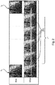
- FIG. 2 is an illustration of the operation of the proposed image processing apparatus IP.
- the upper row shows the two images I1 and I2 acquired at a given frame rate, for instance 6 fps.
- the acquistion frame rate is usually in the region of 3 fps -15 fps.
- the lower row shows four filler images I+ inserted between the two frames I1 and I2 and displayed in that sequence.
- the frame rate has been thus increased five-fold from 6 fps to 30 fps.
- the dashed line shows a reference base line and the arrow illustrates the effect of the pixel transformations are applied to the earlier image I1 as simple shifts, in this example in a positive direction along the vertical y axis of the image screen M.
- the filler images are, in this case, shifted copies of the earlier X-ray image I1. Accumulation of the individual shifts across the individual filler frames I+ are shown by the inclination of the dotted arrow. Because of the shifts, some image information is lost which presents as undefined or void regions 205.
- the void regions 205 can be rendered for instance by filling the void pixel positions with black (as in the Figure) or by another suitable color. A deliberately conspicuous rendering, relative to the surrounding reals image information, of said voids 205 may be chosen to clearly indicate to the viewer that the void regions 205 represent mocked information to which no interpretative (diagnostic or otherwise) value should be attached.
- the void regions 205 can be filled by interpolation from neighboring pixel information so as to effectively hide from the viewer the loss of image information.
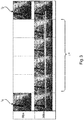
- the void pixel positions can be filled with the last pixel line in the direction of movement (i.e. the last line of the known image I1 is repeated several times).
- Figure 3 is an illustration of this. This embodiment helps avoid the sort of visual distraction on the viewer that may arise if the gap regions were filled in a conspicuous manner as per the earlier mentioned embodiment.
- the number of filler frames to be inserted between the two consecutive X-ray images 11,12 is adjusted to a ratio between the frame rate of the X-ray imager and the refresh rate of the display unit M.
- the refresh rate (usually about 60 fps or higher) defines the minimum time period required to display two consecutive images.
- the frame rate of the X-ray imager is usually significantly lower than the refresh rate of the monitor M.
- the image transformation may then be sub-divided into parts, which are then cumulatively applied in steps, part by part, to the earlier X-ray image to compute the required number N of filler frames, akin to the illustration in Figures 2 , 3 above.
- This number N which determines the up-sampling factor N+1, can be adjusted to the ratio between the monitor refresh rate and the frame rate of the X-ray imager.
- N the ratio between the monitor refresh rate and the frame rate of the X-ray imager.
- a consequence of using the proposed filler frames is an effective "image refresh rate" on the screen M significantly higher than the nominal X-ray exposure rate, in particular when a plurality of filler images are computed as above and inserted between consecutive X-ray images 11,12.
- the up-sampling factor prefferably adjustable based on user input.
- the user specifies the refresh rate and/or acquisition rate.
- This input is then forwarded to the up-sampler US where the up-sampling factor for computing the filler frames is the adjusted based on the ratio as explained above.
- the up-sampling factor is adjusted automatically.
- a first input image I1 acquired by the image apparatus is received.
- the frame I1 is assumed to have been acquired at a first imaging geometry by the imaging apparatus.
- a specification is received that specifies a change from a first to a second imaging geometry.
- the specification is schematically indicted in Fig 4 in coordinates "(X 1 ,Y 1 )" (indicating a "first” imaging geometry) and "(X 2 ,Y 2 )” (indicating a "second” imaging geometry), with the understanding that other formats, in particular in angular specifications, are not excluded herein.
- the specification for the change in imaging geometry can be embodied for instance by a control command, or a real time reading or measurements as supplied by one or more motion encoders associated with one or more actuators involved in bringing about the change in imaging geometry.
- Signal used to drive the actuators can also be used (after appropriate conversions) as an imaging geometry specification.
- the patient bed is translated by a certain amount (specifiable in cm or mm or other suitable unit), and this amount in included in said imaging geometry change specification.
- the translation amount can be obtained directly from a motion encoder associated with the actuator A that brings about the translation.
- the specification is obtained as velocity readings (speed and direction) to be applied to a relevant machine part.
- the reading is post-factum and specifies the displacement already experienced. In this later case, there will a slight lag in response time which, however, has been found in general to not appreciably disturb the real-time impression as experienced by the viewer.
- geometry change specifications are the rotation in degree by the X-ray source around the region of interest. More generally, the specification may also include inclination data to in respect of respective of changes of orientation around one more axes of the X-ray tube, detector plane or patient bed.
- the specification may specify a combination of motions such as one or more translations, possibly along different axis and one or more rotations, etc.
- the specification is preferably supplied from the imaging apparatus via its (automatic) actuators.
- the imaging apparatus may include manually operable actuators such as levers or other gearing mechanism.
- actuators such as levers or other gearing mechanism.
- some patient beds are translated by the user operating one or more handwheels coupled to the bed T via suitable gearing mechanisms.
- suitable internal sensors may be installed at the manual actuators to translate motions such as rotations/translations with the actuator mechanism into digital readings that correspond directly or indirectly (after suitable conversion) to the effected patient motion.
- suitable internal sensor equipment may be used instead.
- a system of one or more photo-electric sensors may be installed on the outside of the imaging equipment to measure the number of time a certain manually operable actuator (such as a hand or thumbwheel) is turned and these counts are then convertible into patient motion distances.
- a certain manually operable actuator such as a hand or thumbwheel
- photoelectric sensors in this manner is also suitable when measuring imaging geometry changes caused by automatic actuators.
- any measurement technique optical, electrical or mechanical to capture the change of imaging geometry specification is envisaged herein.
- step S420 where one or more filler images are then computed based on the specification of the change of geometry.
- the step 420 for computing the one or more filler frames I+ comprises two sub-steps.
- an image transformation is computed based on the specified imaging geometry change. For instance in one embodiment a translation or shift vector is extracted from the received specification of the imaging geometry change. In some instance, the motion (shift and/or rotation) as recorded on the specification can be directly taken (after conversion into pixel information) to indicate the transformation. This has been found to be true in particular for effective displacements ⁇ x ⁇ 1cm, whether or not the underlying motion is linear. This affords a convenient form of motion linearization where effects caused by the underlying projective geometry can be neglected. In other cases, where the imaging geometry change involves translations in excess of 1cm, one or more preprocessing stages, for instance for conversion operations, may be necessary to arrive at the effective motion components for definition of the required image transformation.
- this preprocessing may include decomposing the motions/displacements as recorded in the specification into components by projection onto the image plane of the detector at the imaging geometry at which the first image frame I1 has been acquired.
- the conversion may include taking the inverse of the actually specified displacement to account for possible mirror-effects caused by the imaging setup.
- the conversion stage/operation also takes into account X-ray optical magnification incurred in the image signal path. For instance, in C-arm X-ray imaging, a translation of the patient through the iso-center may incur a magnification factor of 2. In other words, the real motion in the examination region is recorded in the image plane by the detector as a magnified or "virtual" motion twice that of the real motion.
- magnification effects in relation to speed or distance may be known beforehand as a prior-data which can then be applied to adjust the geometry change specification by the appropriate amount. Otherwise, absent such a priori knowledge, the magnification factor may be obtained in a simple calibration measurement: merely two test images are acquired of a movable test object to compute the magnification factor from the known displacement or speed to which the test object is subjected to whilst the two (or more) test images are acquired.
- the transformation can thus be defined by a shift vector of 33 pixels in x direction.
- the one or more transformations determined in step S420b are then applied to, preferably, the latest available image frame I1 supplied by the imaging apparatus.
- the transformation is applied pixel-wisely to the image frame I1 to thereby rotate or translate, in one embodiment only translate, said frame relative to a reference coordinate system.
- the reference coordinate system (shown in Figs 2 , 3 as the horizontal dashed line) may be defined for instance by the window portion on the screen M on which the first image I1 is to be displayed.
- step S430 the first input image and the one or more filler frames are then rendered for display on the screen.
- the first image I1 is displayed first and then the filler(s)frames is/are sequentially displayed. Finally, it is then the second image 12 as supplied by the imaging apparatus that is displayed on the monitor M.
- the method allows computing an up-sampled sequence from the original image I1 as supplied by the imager and the one or more transformations computed from the change of imaging geometry specification.
- the filler frames are computed without knowledge of the follow-up image 12.
- the upsampled sequence presents to the viewer a more realistic visual experience with smooth transitions between the sequentially displayed image information. In particular, flicker caused by changes in the imaging geometry can be removed or at least reduced.
- the optimal display rate for this is generally the screen refresh rate (typically 60Hz), and the temporal up-sampling factor is chosen accordingly as explained earlier above.
- the transforming of image I1 to obtain the filler images at steps 2-5 is interlaced with the displaying step.
- some or all of the filler images are computed first, and the display steps are then performed after some or all of the transformation steps have been completed.
- the proposed image processing component and method can be used as an add-on for existing imagers by installing for instance suitable motion encoders to pick-up the imaging geometry change specifications.
- a computer program or a computer program element is provided that is characterized by being adapted to execute the method steps of the method according to one of the preceding embodiments, on an appropriate system.
- the computer program element might therefore be stored on a computer unit (such as the workstation), which might also be part of an embodiment of the present invention.
- This computing unit may be adapted to perform or induce a performing of the steps of the method described above.
- the computing unit may be adapted to control or operate components of the above-described module.
- the computing unit can be adapted to operate automatically and/or to execute the orders of a user.
- a computer program may be loaded into a working memory of a data processor.
- the data processor may thus be equipped to carry out the method of the invention.
- This exemplary embodiment of the invention covers both, a computer program that right from the beginning uses the invention and a computer program that by means of an up-date turns an existing program into a program that uses the invention.
- the computer program element might be able to provide all necessary steps to fulfill the procedure of an exemplary embodiment of the method as described above.
- a computer readable medium such as a CD-ROM
- the computer readable medium has a computer program element stored on it which computer program element is described by the preceding section.
- a computer program may be stored and/or distributed on a suitable medium (in particular, but not necessarily, a non-transitory medium), such as an optical storage medium or a solid-state medium supplied together with or as part of other hardware, but may also be distributed in other forms, such as via the internet or other wired or wireless telecommunication systems.
- a suitable medium in particular, but not necessarily, a non-transitory medium
- the computer program may also be presented over a network like the World Wide Web and can be downloaded into the working memory of a data processor from such a network.
- a medium for making a computer program element available for downloading is provided, which computer program element is arranged to perform a method according to one of the previously described embodiments of the invention.
Landscapes
- Engineering & Computer Science (AREA)
- Health & Medical Sciences (AREA)
- Life Sciences & Earth Sciences (AREA)
- Physics & Mathematics (AREA)
- Medical Informatics (AREA)
- General Physics & Mathematics (AREA)
- Theoretical Computer Science (AREA)
- Radiology & Medical Imaging (AREA)
- Surgery (AREA)
- Nuclear Medicine, Radiotherapy & Molecular Imaging (AREA)
- Optics & Photonics (AREA)
- Pathology (AREA)
- Biophysics (AREA)
- Biomedical Technology (AREA)
- Heart & Thoracic Surgery (AREA)
- Molecular Biology (AREA)
- High Energy & Nuclear Physics (AREA)
- Animal Behavior & Ethology (AREA)
- General Health & Medical Sciences (AREA)
- Public Health (AREA)
- Veterinary Medicine (AREA)
- Multimedia (AREA)
- Signal Processing (AREA)
- Apparatus For Radiation Diagnosis (AREA)
- Image Processing (AREA)
Applications Claiming Priority (2)
| Application Number | Priority Date | Filing Date | Title |
|---|---|---|---|
| EP15186918 | 2015-09-25 | ||
| PCT/EP2016/072379 WO2017050802A1 (en) | 2015-09-25 | 2016-09-21 | Spatial flicker removal at low frame rates in fluoroscopy |
Publications (2)
| Publication Number | Publication Date |
|---|---|
| EP3353738A1 EP3353738A1 (en) | 2018-08-01 |
| EP3353738B1 true EP3353738B1 (en) | 2020-07-29 |
Family
ID=54207358
Family Applications (1)
| Application Number | Title | Priority Date | Filing Date |
|---|---|---|---|
| EP16775122.1A Active EP3353738B1 (en) | 2015-09-25 | 2016-09-21 | Spatial flicker removal at low frame rates in fluoroscopy |
Country Status (5)
| Country | Link |
|---|---|
| US (1) | US10789674B2 (zh) |
| EP (1) | EP3353738B1 (zh) |
| JP (1) | JP6831373B2 (zh) |
| CN (1) | CN108027959B (zh) |
| WO (1) | WO2017050802A1 (zh) |
Families Citing this family (1)
| Publication number | Priority date | Publication date | Assignee | Title |
|---|---|---|---|---|
| DE102021210283A1 (de) | 2021-09-16 | 2023-03-16 | Siemens Healthcare Gmbh | Erzeugen eines Zwischenbildes |
Citations (1)
| Publication number | Priority date | Publication date | Assignee | Title |
|---|---|---|---|---|
| US8243194B2 (en) * | 2006-05-31 | 2012-08-14 | Vestel Elektronik Sanayi Ve Ticaret A.S. | Method and apparatus for frame interpolation |
Family Cites Families (19)
| Publication number | Priority date | Publication date | Assignee | Title |
|---|---|---|---|---|
| US5224141A (en) | 1991-02-06 | 1993-06-29 | General Electric Company | Fluoroscopic method with reduced x-ray dosage |
| US5400383A (en) | 1991-12-09 | 1995-03-21 | General Electric Company | Fluoroscopic imager with frame-filling apparatus |
| JPH11206753A (ja) * | 1998-01-28 | 1999-08-03 | Hitachi Medical Corp | X線撮像装置 |
| US6690386B2 (en) * | 2001-05-31 | 2004-02-10 | Dynapel Systems, Inc. | Medical image display system |
| US7522701B2 (en) | 2005-12-20 | 2009-04-21 | General Electric Company | System and method for image composition using position sensors |
| JP4986771B2 (ja) * | 2006-08-31 | 2012-07-25 | キヤノン株式会社 | 撮像装置、その駆動方法及び放射線撮像システム |
| WO2008107905A2 (en) * | 2007-03-08 | 2008-09-12 | Sync-Rx, Ltd. | Imaging and tools for use with moving organs |
| JP2008244846A (ja) * | 2007-03-27 | 2008-10-09 | Toshiba Corp | フレーム補間装置及びその方法 |
| US8260025B2 (en) | 2008-11-24 | 2012-09-04 | General Electric Company | Methods and apparatus for generating and archiving x-ray fluoroscopy images |
| CN102068281B (zh) * | 2011-01-20 | 2012-10-03 | 深圳大学 | 一种占位性病变超声图像的处理方法 |
| CN102499701B (zh) * | 2011-09-29 | 2014-08-06 | 华中科技大学 | X射线和荧光双模式活体成像系统的几何校准方法 |
| JP2013165485A (ja) * | 2012-01-11 | 2013-08-22 | Panasonic Corp | 画像処理装置、撮像装置およびコンピュータブログラム |
| CN104427939B (zh) * | 2012-07-05 | 2018-04-24 | 皇家飞利浦有限公司 | 用于多通道x射线成像的图像流的时间对准和信噪比增强 |
| CN104036452B (zh) * | 2013-03-06 | 2017-12-05 | 东芝医疗系统株式会社 | 图像处理装置和方法以及医学图像设备 |
| US9697624B2 (en) * | 2014-08-28 | 2017-07-04 | Shimadzu Corporation | Image processing apparatus, radiation tomography apparatus, and method of performing image processing |
| US9940541B2 (en) * | 2015-07-15 | 2018-04-10 | Fyusion, Inc. | Artificially rendering images using interpolation of tracked control points |
| JP6517087B2 (ja) * | 2015-06-05 | 2019-05-22 | キヤノンメディカルシステムズ株式会社 | X線診断装置及び画像処理プログラム |
| US10147211B2 (en) * | 2015-07-15 | 2018-12-04 | Fyusion, Inc. | Artificially rendering images using viewpoint interpolation and extrapolation |
| US10242474B2 (en) * | 2015-07-15 | 2019-03-26 | Fyusion, Inc. | Artificially rendering images using viewpoint interpolation and extrapolation |
-
2016
- 2016-09-21 JP JP2018515639A patent/JP6831373B2/ja active Active
- 2016-09-21 CN CN201680055539.4A patent/CN108027959B/zh active Active
- 2016-09-21 WO PCT/EP2016/072379 patent/WO2017050802A1/en active Application Filing
- 2016-09-21 EP EP16775122.1A patent/EP3353738B1/en active Active
- 2016-09-21 US US15/760,637 patent/US10789674B2/en active Active
Patent Citations (1)
| Publication number | Priority date | Publication date | Assignee | Title |
|---|---|---|---|---|
| US8243194B2 (en) * | 2006-05-31 | 2012-08-14 | Vestel Elektronik Sanayi Ve Ticaret A.S. | Method and apparatus for frame interpolation |
Non-Patent Citations (2)
| Title |
|---|
| STEFFENINO G ET AL: "Staff dose reduction during coronary angiography using low framing speed", BRITISH JOURNAL OF RADIOLOGY, BRITISH INSTITUTE OF RADIOLOGY, LONDON, GB, vol. 69, no. 825, 1 January 1996 (1996-01-01), pages 860 - 864, XP009509000 * |
| TITUS R KOENIG: "Skin Injuries from Fluoroscopically Guided Procedures: Part 2, Review of 73 Cases and Recommendations for Minimizing Dose Delivered to Patient", AMERICAN JOURNAL OF ROENTGENOLOGY, vol. 177, 1 January 2001 (2001-01-01), pages 13 - 20, XP055519761 * |
Also Published As
| Publication number | Publication date |
|---|---|
| CN108027959A (zh) | 2018-05-11 |
| WO2017050802A1 (en) | 2017-03-30 |
| JP2018528840A (ja) | 2018-10-04 |
| US10789674B2 (en) | 2020-09-29 |
| JP6831373B2 (ja) | 2021-02-17 |
| US20180253822A1 (en) | 2018-09-06 |
| CN108027959B (zh) | 2022-10-25 |
| EP3353738A1 (en) | 2018-08-01 |
Similar Documents
| Publication | Publication Date | Title |
|---|---|---|
| JP5677738B2 (ja) | X線コンピュータ断層撮影装置 | |
| US9044190B2 (en) | C-arm computerized tomography system | |
| US7103136B2 (en) | Fluoroscopic tomosynthesis system and method | |
| US20100201786A1 (en) | Method and apparatus for reconstructing an image | |
| US20120069951A1 (en) | Tomographic image displaying method and apparatus | |
| CN108463170B (zh) | X光图像显示设备和x光图像显示方法 | |
| US10537293B2 (en) | X-ray CT system, image display device, and image display method | |
| US20180275076A1 (en) | System and method for cabinet x-ray systems with camera | |
| US10206645B2 (en) | Multi-perspective interventional imaging using a single imaging system | |
| JP2009136518A (ja) | X線撮影装置及びx線撮影方法 | |
| WO2012099222A1 (ja) | X線診断装置、画像処理装置及び画像処理プログラム | |
| US20060241370A1 (en) | Medical x-ray imaging workflow improvement | |
| JP5631554B2 (ja) | X線診断装置 | |
| JP6580963B2 (ja) | 画像処理装置、画像処理方法およびx線診断装置 | |
| EP3353738B1 (en) | Spatial flicker removal at low frame rates in fluoroscopy | |
| EP3649957B1 (en) | Device and method for editing a panoramic radiography image | |
| JP2013000370A (ja) | 放射線画像撮影装置および方法 | |
| JP7187217B2 (ja) | 医用画像処理装置、x線診断装置及び医用画像処理プログラム | |
| EP3360482A1 (en) | Iso-centering in c-arm computer tomography | |
| JP7199958B2 (ja) | アンギオct装置 | |
| JP2005253572A (ja) | 画像処理装置、x線診断装置、医用画像情報システム、及びキャリブレーションテーブル付帯方法 | |
| JP5893102B2 (ja) | X線コンピュータ断層撮影装置 | |
| JP2022112175A (ja) | X線診断装置及び医用画像処理装置 |
Legal Events
| Date | Code | Title | Description |
|---|---|---|---|
| STAA | Information on the status of an ep patent application or granted ep patent |
Free format text: STATUS: UNKNOWN |
|
| STAA | Information on the status of an ep patent application or granted ep patent |
Free format text: STATUS: THE INTERNATIONAL PUBLICATION HAS BEEN MADE |
|
| PUAI | Public reference made under article 153(3) epc to a published international application that has entered the european phase |
Free format text: ORIGINAL CODE: 0009012 |
|
| STAA | Information on the status of an ep patent application or granted ep patent |
Free format text: STATUS: REQUEST FOR EXAMINATION WAS MADE |
|
| 17P | Request for examination filed |
Effective date: 20180425 |
|
| AK | Designated contracting states |
Kind code of ref document: A1 Designated state(s): AL AT BE BG CH CY CZ DE DK EE ES FI FR GB GR HR HU IE IS IT LI LT LU LV MC MK MT NL NO PL PT RO RS SE SI SK SM TR |
|
| AX | Request for extension of the european patent |
Extension state: BA ME |
|
| STAA | Information on the status of an ep patent application or granted ep patent |
Free format text: STATUS: EXAMINATION IS IN PROGRESS |
|
| 17Q | First examination report despatched |
Effective date: 20181114 |
|
| DAV | Request for validation of the european patent (deleted) | ||
| DAX | Request for extension of the european patent (deleted) | ||
| GRAP | Despatch of communication of intention to grant a patent |
Free format text: ORIGINAL CODE: EPIDOSNIGR1 |
|
| STAA | Information on the status of an ep patent application or granted ep patent |
Free format text: STATUS: GRANT OF PATENT IS INTENDED |
|
| INTG | Intention to grant announced |
Effective date: 20200221 |
|
| RAP1 | Party data changed (applicant data changed or rights of an application transferred) |
Owner name: KONINKLIJKE PHILIPS N.V. |
|
| GRAS | Grant fee paid |
Free format text: ORIGINAL CODE: EPIDOSNIGR3 |
|
| GRAA | (expected) grant |
Free format text: ORIGINAL CODE: 0009210 |
|
| STAA | Information on the status of an ep patent application or granted ep patent |
Free format text: STATUS: THE PATENT HAS BEEN GRANTED |
|
| AK | Designated contracting states |
Kind code of ref document: B1 Designated state(s): AL AT BE BG CH CY CZ DE DK EE ES FI FR GB GR HR HU IE IS IT LI LT LU LV MC MK MT NL NO PL PT RO RS SE SI SK SM TR |
|
| REG | Reference to a national code |
Ref country code: CH Ref legal event code: EP |
|
| REG | Reference to a national code |
Ref country code: AT Ref legal event code: REF Ref document number: 1296675 Country of ref document: AT Kind code of ref document: T Effective date: 20200815 |
|
| REG | Reference to a national code |
Ref country code: IE Ref legal event code: FG4D |
|
| REG | Reference to a national code |
Ref country code: DE Ref legal event code: R096 Ref document number: 602016040896 Country of ref document: DE |
|
| REG | Reference to a national code |
Ref country code: DE Ref legal event code: R084 Ref document number: 602016040896 Country of ref document: DE |
|
| REG | Reference to a national code |
Ref country code: LT Ref legal event code: MG4D |
|
| REG | Reference to a national code |
Ref country code: GB Ref legal event code: 746 Effective date: 20201208 |
|
| REG | Reference to a national code |
Ref country code: NL Ref legal event code: MP Effective date: 20200729 |
|
| REG | Reference to a national code |
Ref country code: AT Ref legal event code: MK05 Ref document number: 1296675 Country of ref document: AT Kind code of ref document: T Effective date: 20200729 |
|
| PG25 | Lapsed in a contracting state [announced via postgrant information from national office to epo] |
Ref country code: LT Free format text: LAPSE BECAUSE OF FAILURE TO SUBMIT A TRANSLATION OF THE DESCRIPTION OR TO PAY THE FEE WITHIN THE PRESCRIBED TIME-LIMIT Effective date: 20200729 Ref country code: FI Free format text: LAPSE BECAUSE OF FAILURE TO SUBMIT A TRANSLATION OF THE DESCRIPTION OR TO PAY THE FEE WITHIN THE PRESCRIBED TIME-LIMIT Effective date: 20200729 Ref country code: GR Free format text: LAPSE BECAUSE OF FAILURE TO SUBMIT A TRANSLATION OF THE DESCRIPTION OR TO PAY THE FEE WITHIN THE PRESCRIBED TIME-LIMIT Effective date: 20201030 Ref country code: SE Free format text: LAPSE BECAUSE OF FAILURE TO SUBMIT A TRANSLATION OF THE DESCRIPTION OR TO PAY THE FEE WITHIN THE PRESCRIBED TIME-LIMIT Effective date: 20200729 Ref country code: PT Free format text: LAPSE BECAUSE OF FAILURE TO SUBMIT A TRANSLATION OF THE DESCRIPTION OR TO PAY THE FEE WITHIN THE PRESCRIBED TIME-LIMIT Effective date: 20201130 Ref country code: HR Free format text: LAPSE BECAUSE OF FAILURE TO SUBMIT A TRANSLATION OF THE DESCRIPTION OR TO PAY THE FEE WITHIN THE PRESCRIBED TIME-LIMIT Effective date: 20200729 Ref country code: NO Free format text: LAPSE BECAUSE OF FAILURE TO SUBMIT A TRANSLATION OF THE DESCRIPTION OR TO PAY THE FEE WITHIN THE PRESCRIBED TIME-LIMIT Effective date: 20201029 Ref country code: ES Free format text: LAPSE BECAUSE OF FAILURE TO SUBMIT A TRANSLATION OF THE DESCRIPTION OR TO PAY THE FEE WITHIN THE PRESCRIBED TIME-LIMIT Effective date: 20200729 Ref country code: BG Free format text: LAPSE BECAUSE OF FAILURE TO SUBMIT A TRANSLATION OF THE DESCRIPTION OR TO PAY THE FEE WITHIN THE PRESCRIBED TIME-LIMIT Effective date: 20201029 Ref country code: AT Free format text: LAPSE BECAUSE OF FAILURE TO SUBMIT A TRANSLATION OF THE DESCRIPTION OR TO PAY THE FEE WITHIN THE PRESCRIBED TIME-LIMIT Effective date: 20200729 |
|
| PG25 | Lapsed in a contracting state [announced via postgrant information from national office to epo] |
Ref country code: RS Free format text: LAPSE BECAUSE OF FAILURE TO SUBMIT A TRANSLATION OF THE DESCRIPTION OR TO PAY THE FEE WITHIN THE PRESCRIBED TIME-LIMIT Effective date: 20200729 Ref country code: PL Free format text: LAPSE BECAUSE OF FAILURE TO SUBMIT A TRANSLATION OF THE DESCRIPTION OR TO PAY THE FEE WITHIN THE PRESCRIBED TIME-LIMIT Effective date: 20200729 Ref country code: LV Free format text: LAPSE BECAUSE OF FAILURE TO SUBMIT A TRANSLATION OF THE DESCRIPTION OR TO PAY THE FEE WITHIN THE PRESCRIBED TIME-LIMIT Effective date: 20200729 Ref country code: IS Free format text: LAPSE BECAUSE OF FAILURE TO SUBMIT A TRANSLATION OF THE DESCRIPTION OR TO PAY THE FEE WITHIN THE PRESCRIBED TIME-LIMIT Effective date: 20201129 |
|
| PG25 | Lapsed in a contracting state [announced via postgrant information from national office to epo] |
Ref country code: NL Free format text: LAPSE BECAUSE OF FAILURE TO SUBMIT A TRANSLATION OF THE DESCRIPTION OR TO PAY THE FEE WITHIN THE PRESCRIBED TIME-LIMIT Effective date: 20200729 |
|
| PG25 | Lapsed in a contracting state [announced via postgrant information from national office to epo] |
Ref country code: DK Free format text: LAPSE BECAUSE OF FAILURE TO SUBMIT A TRANSLATION OF THE DESCRIPTION OR TO PAY THE FEE WITHIN THE PRESCRIBED TIME-LIMIT Effective date: 20200729 Ref country code: CZ Free format text: LAPSE BECAUSE OF FAILURE TO SUBMIT A TRANSLATION OF THE DESCRIPTION OR TO PAY THE FEE WITHIN THE PRESCRIBED TIME-LIMIT Effective date: 20200729 Ref country code: RO Free format text: LAPSE BECAUSE OF FAILURE TO SUBMIT A TRANSLATION OF THE DESCRIPTION OR TO PAY THE FEE WITHIN THE PRESCRIBED TIME-LIMIT Effective date: 20200729 Ref country code: SM Free format text: LAPSE BECAUSE OF FAILURE TO SUBMIT A TRANSLATION OF THE DESCRIPTION OR TO PAY THE FEE WITHIN THE PRESCRIBED TIME-LIMIT Effective date: 20200729 Ref country code: IT Free format text: LAPSE BECAUSE OF FAILURE TO SUBMIT A TRANSLATION OF THE DESCRIPTION OR TO PAY THE FEE WITHIN THE PRESCRIBED TIME-LIMIT Effective date: 20200729 Ref country code: EE Free format text: LAPSE BECAUSE OF FAILURE TO SUBMIT A TRANSLATION OF THE DESCRIPTION OR TO PAY THE FEE WITHIN THE PRESCRIBED TIME-LIMIT Effective date: 20200729 |
|
| REG | Reference to a national code |
Ref country code: CH Ref legal event code: PL Ref country code: DE Ref legal event code: R097 Ref document number: 602016040896 Country of ref document: DE |
|
| PG25 | Lapsed in a contracting state [announced via postgrant information from national office to epo] |
Ref country code: AL Free format text: LAPSE BECAUSE OF FAILURE TO SUBMIT A TRANSLATION OF THE DESCRIPTION OR TO PAY THE FEE WITHIN THE PRESCRIBED TIME-LIMIT Effective date: 20200729 |
|
| PLBE | No opposition filed within time limit |
Free format text: ORIGINAL CODE: 0009261 |
|
| STAA | Information on the status of an ep patent application or granted ep patent |
Free format text: STATUS: NO OPPOSITION FILED WITHIN TIME LIMIT |
|
| REG | Reference to a national code |
Ref country code: BE Ref legal event code: MM Effective date: 20200930 |
|
| PG25 | Lapsed in a contracting state [announced via postgrant information from national office to epo] |
Ref country code: SK Free format text: LAPSE BECAUSE OF FAILURE TO SUBMIT A TRANSLATION OF THE DESCRIPTION OR TO PAY THE FEE WITHIN THE PRESCRIBED TIME-LIMIT Effective date: 20200729 Ref country code: LU Free format text: LAPSE BECAUSE OF NON-PAYMENT OF DUE FEES Effective date: 20200921 |
|
| 26N | No opposition filed |
Effective date: 20210430 |
|
| PG25 | Lapsed in a contracting state [announced via postgrant information from national office to epo] |
Ref country code: FR Free format text: LAPSE BECAUSE OF NON-PAYMENT OF DUE FEES Effective date: 20200929 |
|
| PG25 | Lapsed in a contracting state [announced via postgrant information from national office to epo] |
Ref country code: LI Free format text: LAPSE BECAUSE OF NON-PAYMENT OF DUE FEES Effective date: 20200930 Ref country code: IE Free format text: LAPSE BECAUSE OF NON-PAYMENT OF DUE FEES Effective date: 20200921 Ref country code: SI Free format text: LAPSE BECAUSE OF FAILURE TO SUBMIT A TRANSLATION OF THE DESCRIPTION OR TO PAY THE FEE WITHIN THE PRESCRIBED TIME-LIMIT Effective date: 20200729 Ref country code: BE Free format text: LAPSE BECAUSE OF NON-PAYMENT OF DUE FEES Effective date: 20200930 Ref country code: CH Free format text: LAPSE BECAUSE OF NON-PAYMENT OF DUE FEES Effective date: 20200930 |
|
| PG25 | Lapsed in a contracting state [announced via postgrant information from national office to epo] |
Ref country code: TR Free format text: LAPSE BECAUSE OF FAILURE TO SUBMIT A TRANSLATION OF THE DESCRIPTION OR TO PAY THE FEE WITHIN THE PRESCRIBED TIME-LIMIT Effective date: 20200729 Ref country code: MT Free format text: LAPSE BECAUSE OF FAILURE TO SUBMIT A TRANSLATION OF THE DESCRIPTION OR TO PAY THE FEE WITHIN THE PRESCRIBED TIME-LIMIT Effective date: 20200729 Ref country code: CY Free format text: LAPSE BECAUSE OF FAILURE TO SUBMIT A TRANSLATION OF THE DESCRIPTION OR TO PAY THE FEE WITHIN THE PRESCRIBED TIME-LIMIT Effective date: 20200729 |
|
| PG25 | Lapsed in a contracting state [announced via postgrant information from national office to epo] |
Ref country code: MK Free format text: LAPSE BECAUSE OF FAILURE TO SUBMIT A TRANSLATION OF THE DESCRIPTION OR TO PAY THE FEE WITHIN THE PRESCRIBED TIME-LIMIT Effective date: 20200729 Ref country code: MC Free format text: LAPSE BECAUSE OF FAILURE TO SUBMIT A TRANSLATION OF THE DESCRIPTION OR TO PAY THE FEE WITHIN THE PRESCRIBED TIME-LIMIT Effective date: 20200729 |
|
| PGFP | Annual fee paid to national office [announced via postgrant information from national office to epo] |
Ref country code: GB Payment date: 20220920 Year of fee payment: 7 |
|
| PGFP | Annual fee paid to national office [announced via postgrant information from national office to epo] |
Ref country code: DE Payment date: 20230928 Year of fee payment: 8 |