EP3314010B1 - Method for detecting chromosome aberrations - Google Patents
Method for detecting chromosome aberrations Download PDFInfo
- Publication number
- EP3314010B1 EP3314010B1 EP16731904.5A EP16731904A EP3314010B1 EP 3314010 B1 EP3314010 B1 EP 3314010B1 EP 16731904 A EP16731904 A EP 16731904A EP 3314010 B1 EP3314010 B1 EP 3314010B1
- Authority
- EP
- European Patent Office
- Prior art keywords
- locus
- chromosome
- specific hybridization
- signals
- marked
- Prior art date
- Legal status (The legal status is an assumption and is not a legal conclusion. Google has not performed a legal analysis and makes no representation as to the accuracy of the status listed.)
- Active
Links
Images


Classifications
-
- C—CHEMISTRY; METALLURGY
- C12—BIOCHEMISTRY; BEER; SPIRITS; WINE; VINEGAR; MICROBIOLOGY; ENZYMOLOGY; MUTATION OR GENETIC ENGINEERING
- C12Q—MEASURING OR TESTING PROCESSES INVOLVING ENZYMES, NUCLEIC ACIDS OR MICROORGANISMS; COMPOSITIONS OR TEST PAPERS THEREFOR; PROCESSES OF PREPARING SUCH COMPOSITIONS; CONDITION-RESPONSIVE CONTROL IN MICROBIOLOGICAL OR ENZYMOLOGICAL PROCESSES
- C12Q1/00—Measuring or testing processes involving enzymes, nucleic acids or microorganisms; Compositions therefor; Processes of preparing such compositions
- C12Q1/68—Measuring or testing processes involving enzymes, nucleic acids or microorganisms; Compositions therefor; Processes of preparing such compositions involving nucleic acids
- C12Q1/6813—Hybridisation assays
- C12Q1/6841—In situ hybridisation
-
- C—CHEMISTRY; METALLURGY
- C12—BIOCHEMISTRY; BEER; SPIRITS; WINE; VINEGAR; MICROBIOLOGY; ENZYMOLOGY; MUTATION OR GENETIC ENGINEERING
- C12Q—MEASURING OR TESTING PROCESSES INVOLVING ENZYMES, NUCLEIC ACIDS OR MICROORGANISMS; COMPOSITIONS OR TEST PAPERS THEREFOR; PROCESSES OF PREPARING SUCH COMPOSITIONS; CONDITION-RESPONSIVE CONTROL IN MICROBIOLOGICAL OR ENZYMOLOGICAL PROCESSES
- C12Q1/00—Measuring or testing processes involving enzymes, nucleic acids or microorganisms; Compositions therefor; Processes of preparing such compositions
- C12Q1/68—Measuring or testing processes involving enzymes, nucleic acids or microorganisms; Compositions therefor; Processes of preparing such compositions involving nucleic acids
- C12Q1/6844—Nucleic acid amplification reactions
- C12Q1/6858—Allele-specific amplification
-
- C—CHEMISTRY; METALLURGY
- C12—BIOCHEMISTRY; BEER; SPIRITS; WINE; VINEGAR; MICROBIOLOGY; ENZYMOLOGY; MUTATION OR GENETIC ENGINEERING
- C12Q—MEASURING OR TESTING PROCESSES INVOLVING ENZYMES, NUCLEIC ACIDS OR MICROORGANISMS; COMPOSITIONS OR TEST PAPERS THEREFOR; PROCESSES OF PREPARING SUCH COMPOSITIONS; CONDITION-RESPONSIVE CONTROL IN MICROBIOLOGICAL OR ENZYMOLOGICAL PROCESSES
- C12Q1/00—Measuring or testing processes involving enzymes, nucleic acids or microorganisms; Compositions therefor; Processes of preparing such compositions
- C12Q1/68—Measuring or testing processes involving enzymes, nucleic acids or microorganisms; Compositions therefor; Processes of preparing such compositions involving nucleic acids
- C12Q1/6876—Nucleic acid products used in the analysis of nucleic acids, e.g. primers or probes
- C12Q1/6883—Nucleic acid products used in the analysis of nucleic acids, e.g. primers or probes for diseases caused by alterations of genetic material
- C12Q1/6886—Nucleic acid products used in the analysis of nucleic acids, e.g. primers or probes for diseases caused by alterations of genetic material for cancer
-
- C—CHEMISTRY; METALLURGY
- C12—BIOCHEMISTRY; BEER; SPIRITS; WINE; VINEGAR; MICROBIOLOGY; ENZYMOLOGY; MUTATION OR GENETIC ENGINEERING
- C12Q—MEASURING OR TESTING PROCESSES INVOLVING ENZYMES, NUCLEIC ACIDS OR MICROORGANISMS; COMPOSITIONS OR TEST PAPERS THEREFOR; PROCESSES OF PREPARING SUCH COMPOSITIONS; CONDITION-RESPONSIVE CONTROL IN MICROBIOLOGICAL OR ENZYMOLOGICAL PROCESSES
- C12Q2565/00—Nucleic acid analysis characterised by mode or means of detection
- C12Q2565/10—Detection mode being characterised by the assay principle
- C12Q2565/101—Interaction between at least two labels
-
- C—CHEMISTRY; METALLURGY
- C12—BIOCHEMISTRY; BEER; SPIRITS; WINE; VINEGAR; MICROBIOLOGY; ENZYMOLOGY; MUTATION OR GENETIC ENGINEERING
- C12Q—MEASURING OR TESTING PROCESSES INVOLVING ENZYMES, NUCLEIC ACIDS OR MICROORGANISMS; COMPOSITIONS OR TEST PAPERS THEREFOR; PROCESSES OF PREPARING SUCH COMPOSITIONS; CONDITION-RESPONSIVE CONTROL IN MICROBIOLOGICAL OR ENZYMOLOGICAL PROCESSES
- C12Q2565/00—Nucleic acid analysis characterised by mode or means of detection
- C12Q2565/10—Detection mode being characterised by the assay principle
- C12Q2565/102—Multiple non-interacting labels
Definitions
- the present invention relates to the technical field of detection methods for chromosomal abnormalities or chromosome aberrations.
- the present invention relates to a method for the detection of at least two mutually different chromosome aberrations by means of in situ hybridization.
- the present invention further relates to a composition suitable for detecting at least two different chromosome aberrations and to the uses thereof according to the invention.
- the present invention furthermore relates to the uses of at least three mutually different, locus-specific hybridization probes marked with detection labels.
- the subject of the present invention is a kit for the detection of chromosome aberrations.
- ISH in situ hybridizations
- the in-situ hybridization is based on the hybridization or pairing of complementary bases of nucleic acid single strands, in particular DNA single strands, so that specific nucleic acid sequences can be detected in a sample, in particular in a tissue or cell preparation.
- nucleic acid single strands in particular DNA single strands
- synthetically produced probes are hybridized with nucleic acid single strands of the sample and then detected.
- fluorescence-marked nucleic acid fragments or fluorescence-marked hybridization probes can be used come.
- antigen-labeled probes especially hapten-labeled probes, can be used, which are then made visible with the help of antibodies through color reactions, so that an analysis by light microscopy is possible ((brightfield ISH (BrISH), chromogens ISH (CISH), silver ISH (SISH)).
- FISH FISH-linked immunosorbent assays
- nucleic acid fragments which address different genomic regions or are specific for this are each marked or coupled with different fluorescent dyes which differ from one another in their absorption and / or emission spectrum.
- the individual colors can be determined by using specific microscope filters, which guide precisely defined wavelength ranges of the light to excite the dyes on the preparation as well as precisely defined wavelength ranges of the emitted by the dyes Direct light to the evaluator, shown separately from each other (so-called single bandpass filter set).
- filters or filter sets which allow the simultaneous display of different fluorescent dyes and thus several nucleic acid fragments. In the case of two different fluorescent dyes, for example, one speaks of a dual bandpass filter set.
- FISH usually only analyzes two colors (orange / red and green) or three colors (orange / red and green simultaneously or together with a blue nuclear counterstain (DAPI)).
- DAPI blue nuclear counterstain
- the state of the art is the use of two haptens, mostly selected from the group of biotin, dinitrophenyl (DNP) and digoxigenin, and two antibody-coupled enzymes, mostly alkaline phosphatase and peroxidase.
- DNP dinitrophenyl
- digoxigenin two antibody-coupled enzymes, mostly alkaline phosphatase and peroxidase.
- Locus-specific probes are understood to mean those probes which address selected DNA sections of a chromosome, mostly individual genes or neighboring genes, with a total size of up to about 1,000 kb and are referred to as gene-specific probes or “single copy” probes.
- Repetitive sequence-specific probes are probes that address repetitive sequences and therefore address regions with a size of several 1,000 kb. These probes also include centromere or alpha satellite probes, for example.
- the area of a first breakpoint of the chromosome is flanked proximally and distally by nucleic acid fragments of the same color (e.g. orange)
- the area of a second breakpoint, i.e. H. of the reciprocal translocation partner is flanked proximally and distally by nucleic acid fragments of a second color (e.g. green).
- the normal situation i.e. H. without chromosomal breaks in the area of the two translocation partners, is characterized by a green and a spatially separated orange signal.
- the area of a break point is flanked proximally and distally by nucleic acid fragments marked in different ways or in different colors (e.g. distal orange, proximal green).
- the normal situation i.e. H. without a chromosomal break in this area, is characterized by a fusion signal.
- an aberrant situation i.e. H. if there is a chromosomal break between the probe fragments, the signals separate spatially from one another. The difference between the normal situation and the aberrant situation is thus characterized by the distance between the differently colored signals.
- Statements about involved translocation partners are not possible with this method. It only allows the conclusion that a specific chromosomal rearrangement has taken place.
- the disadvantage of this probe composition is that with the aid of the two colors used, only a single breaking point area and thus only a specific translocation or inversion can be detected.
- locus-specific probes are also supplemented by so-called repetitive sequence-specific probes, such as centromere or alpha satellite probes:
- the principle of detecting gain or loss of signals through the occurrence of deletions, aneuplodies and amplifications usually takes place in so-called dual-color probe approaches. With this principle, which is described below, and the signal patterns derived from it, it should be noted that a normal cell is usually diploid, i.e. H. each allele is present in duplicate.
- two different chromosomal areas are marked with differently marked or color-marked nucleic acid fragments (e.g. genomic region 1 or target region 1 in orange, genomic region 2 or target region 2 in green).
- the normal situation i.e. H. without gain or loss of these areas, is characterized by two orange and two green signals.
- an aberrant situation i.e. H. if genomic regions in which target regions 1 and / or 2 have been gained or lost, fewer green and / or orange signals are visible if they are lost, and more signals are visible if they are gained.
- many additional signals can be visible, which can also be represented as clusters.
- triple FISH approaches which address the detection of different translocation events that may cluster side by side in a chromosomal region (i.e., different genes that are in proximity are affected).
- different genes that are in proximity are affected.
- the signal pattern only two colors are analyzed that detect a single aberration, the third color being irrelevant.
- Three different locus-specific probes are used, each marked with a different label.
- the US 6 576 421 B1 describes a method for the detection of cancer-associated translocations in which three probes are used, two of which hybridize on one side of a breakpoint on a fusion chromosome and the third probe on the other side of the breakpoint. Each of the probes is marked with a marker that differs from those of the other probes.
- the US 6,344,315 B1 discloses methods of staining interphase chromosomal material using a nucleic acid probe greater than 50,000 bases in length and in which, for example, a biotin-labeled ABL probe is used, which is detected indirectly with the fluorochrome Texas red, and a digoxigenin-labeled BCR probe, which is detected indirectly with the fluorochrome FITC, to carry out a FISH method.
- the WO 93/21345 A1 describes methods for detecting a deletion of a gene in which two probes are used which are provided with fluorescent markers which can be distinguished from one another.
- the WO 2005/111235 A2 describes a method which involves the use of three colors for detection purposes.
- the chromosomal region that is marked by the third label of a probe is not directly affected by a change, so that if the chromosome structure changes, the first fusion signal is eliminated, so that a new split signal and a new fusion signal arise.
- This procedure uses probes that are each marked with only one label.
- only one possible translocation, which is specified by the probes, can be detected with this method.
- the WO 02/093130 A3 discloses a method for the detection of chromosomal translocations using two or, alternatively, four labels or dyes which flank the distal and proximal breakpoints of both breakpoints involved in a translocation. This method does not offer any possibilities to detect more than one translocation at the one breakpoint area that is flanked by the probes.
- mFISH multiplex FISH
- SKY-FISH spectral karyotyping
- multicolor FISH COBRA-FISH (Combined Binary Ratio labeling FISH) or 24-color FISH
- a total of twenty-four different dyes can be produced 'Chromosome Painting Probes' can be marked and differentiated.
- the chromosome arm-specific probes of all chromosomes can be marked differently in a so-called 42-color FISH.
- the aforementioned methods are only suitable for cells that are in the metaphase.
- the present invention is therefore based on the object of providing a method or a composition which is suitable for the detection or analysis of chromosome mutations or chromosome aberrations and at least largely avoids or at least mitigates the disadvantages of the prior art described above.
- the present invention is based on the object of providing a method which enables a reliable and simultaneous detection of several mutually different chromosome aberrations, in particular independent (i.e. non-reciprocal) chromosome aberrations, in particular in one approach.
- the present invention is likewise based on the object of providing a method which, in addition, also enables chromosome aberrations to be assigned to a specific chromosome or DNA region.
- the present invention proposes a method for the detection of at least two different chromosome aberrations according to patent claim 1; further advantageous refinements are the subject matter of the dependent claims.
- the present invention also relates to a composition for the detection of at least two different chromosomal aberrations according to the independent patent claim or a composition for use in prophylactic or therapeutic treatment or in the diagnosis or prognosis of diseases associated with chromosomal aberrations.
- the present invention also further provides the use of a composition according to the present invention according to the independent patent claim in this regard.
- the present invention relates to the use of at least three, preferably at least four mutually different locus-specific hybridization probes according to the independent patent claim.
- the subject matter of the present invention is the use of at least one location-specific hybridization probe marked with at least two detection labels according to the independent patent claim.
- the subject matter of the present invention is a kit or kit-of-parts or set for the detection of at least two different chromosome aberrations; further advantageous properties are the subject of the relevant sub-claim.
- the present invention proposes - according to a first aspect of the invention - a method for detecting at least two different chromosome aberrations, in particular structural and / or numerical chromosome aberrations, preferably structural chromosome aberrations, by means of in-situ hybridization by detection of chromosomes - and / or DNA areas in a biological sample, preferably in one or more cell (s) and / or in one or more cell nucleus (s), where the in- situ hybridization is carried out as an interphase in-situ hybridization, wherein the in situ hybridization is carried out with at least four mutually different, each marked with a first detection label, location-specific hybridization probes, wherein, to generate at least one mixed signal by two or more different detection labels located on a location-specific hybridization probe, at least one of the location-specific hybridization probes is marked with at least one further detection label different from the first detection label, based on the respective location-specific hybridization probe, so that
- the present invention is based on the basic principle, through the targeted generation of mixed signals in signal patterns generated by interphase in situ hybridizations, the simultaneous or simultaneous detection of several different chromosome aberrations, in particular independent, ie non-reciprocal chromosome aberrations, in a biological sample and its assignment to a detected chromosome or DNA area.
- the chromosome aberrations are mutually independent chromosome aberrations.
- the chromosome aberrations are not dependent on one another. It can also be provided that the chromosome aberrations are not reciprocal.
- the chromosome aberrations are not related to reciprocal or mutually dependent chromosome aberrations or are associated with such.
- the present invention is associated with numerous advantages and special features, which are discussed below in a non-restrictive manner and are to be assessed as an indication of the patentability of the present invention.
- chromosome aberrations in the context of an interphase in situ hybridization on the one hand a clear detection of several possible structural or numerical chromosome aberrations and on the other hand the clear differentiation of these chromosome aberrations or .
- the chromosome aberrations can be chromosome aberrations which are independent of one another and which are not mutually dependent or which are not reciprocal.
- any Detected chromosome aberrations can be assigned to a defined or specific DNA or chromosome area.
- the amount of sample required for the detection of chromosome aberrations can be significantly reduced with the aid of the method according to the invention. This is particularly advantageous against the background that the removal of tissue for examination purposes, in particular in connection with the diagnosis or detection or further analysis of cancer, is now usually done by means of fine needle biopsy, which only allows a limited amount of sample to be taken, whereas open biopsies, which also allow the removal of larger amounts of tissue, are carried out less and less frequently.
- the required amount of sometimes costly materials, such as enzymes, fluorescent dyes and the like, for performing the in-situ hybridization is reduced, so that the method is also advantageous with regard to economic and ecological aspects.
- chromosome aberrations synonymously also referred to as chromosome anomalies
- chromosome anomalies is understood in particular to be structural and numerical chromosome aberrations.
- structural chromosome aberrations there are changes in the structure of a chromosome, so that these are also referred to as chromosome mutations. In particular, these can be inversions, translocations, deletions, segmental duplications, insertions, duplications or amplifications.
- Numerical chromosome aberrations lead to a change in the number of chromosomes.
- genome mutation is also used synonymously. Numerical chromosome aberrations or genome mutations can in particular be aneuplodia or polyploidy.
- the method according to the invention is particularly suitable for the detection of structural chromosome aberrations.
- the in situ hybridization used according to the invention is based on the hybridization or pairing of complementary bases of nucleic acid single strands, in particular DNA single strands, so that specific nucleic acid sequences can be detected in a sample, such as a tissue or a cell preparation.
- a sample such as a tissue or a cell preparation.
- directly or indirectly labeled, synthetically produced, in particular locus-specific hybridization probes are hybridized with nucleic acid single strands of the sample and then detected.
- in-situ hybridization can take place or be carried out at different stages of the cell cycle of the cells or cell nuclei examined, with it being carried out in the metaphase when the chromosomes are in a condensed state, or in the interphase when the chromosomes decondensed exist, have established.
- the in situ hybridization it is not always possible to perform it on condensed chromosomes in the metaphase, in particular, for example, when examining the cells of solid tumors for chromosome aberrations. According to the invention, it is therefore provided that the in situ hybridization is carried out on cells or cell nuclei which are in the interphase.
- locus-specific hybridization probes are understood to mean probes specific to a certain chromosome area or DNA area or complementary to a certain chromosome area or DNA area of the DNA material or the genetic material in a sample to be examined.
- the hybridization probes used according to the invention are usually based on nucleic acids or nucleic acid fragments and are capable of specifically binding or hybridizing to the chromosome region or DNA region to be detected.
- the chromosome area or DNA area to be detected can have a variable length. In particular, it can be provided that a chromosome region or DNA region to be detected partially or completely comprises a single or a single gene. Equally, it can also be provided that a chromosome area or DNA area to be detected comprises several genes, preferably adjacent genes, preferably two genes, partially or completely.
- a location-specific hybridization probe is based on several, in particular a plurality of nucleic acid fragments (synonymous also probe fragments), which in their entirety are referred to as a location-specific hybridization probe.
- the location-specific hybridization probes are based on only a single nucleic acid fragment or are formed by a single nucleic acid fragment.
- detection labels denote substances or substances which are coupled to nucleic acids or nucleic acid fragments, in particular hybridization probes, for detection or detection purposes.
- the selection of suitable detection labels is within the usual skill of the person skilled in the art and does not require any further explanations at this point.
- the nucleic acid fragments marked with detection labels and bound or hybridized to the DNA or chromosome segment to be detected or detected by means of in situ hybridization can be subjected to the Methods known per se and adapted to the detection label used can be detected directly or indirectly, for example by means of fluorescence microscopy or, in particular, after enzymatic conversion or visualization by means of enzymatically converted dye substrates by bright field microscopy.
- a signal pattern is generated by the detection label on the locus-specific hybridization probes in the context of in-situ hybridization, which is the basis for examining a sample for any chromosome aberrations.
- detection label used in accordance with the invention relates below to the type or type of detection label and not to the numerical number of detection label molecules, i.e. Formulations such as "at least one detection label” refer to a specific type of detection label or the specific selection of a detection label.
- severe detection labels thus also relates to the selection of mutually different detection labels of different types and not to the number of detection label molecules used. It goes without saying that in the context of the labeling of hybridization probes, these are usually coupled to more than one detection label molecule.
- locus-specific hybridization probes used according to the invention thus hybridize specifically to a selected DNA or chromosome region of the genetic material in a sample and generate a signal pattern based on the coupled detection label in the context of in-situ hybridization.
- a signal pattern is understood to mean the entirety of all signals generated by the in-situ hybridization based on the location-specific hybridization probes marked with detection labels.
- a mixed signal within the meaning of the invention is thus a signal which is generated by at least two, but equally also several, different detection labels located on a location-specific hybridization probe.
- mixed signals are generated by the at least two, in particular several, detection labels of a locus-specific hybridization probe, these are also visible in the case of chromosome aberrations in the signal pattern of the in-situ hybridization or remain in place in the case of chromosome aberrations. Possible embodiments and configurations of the location-specific hybridization probes for generating mixed signals are explained in detail below.
- the first detection labels of the location-specific hybridization probes used are each identical.
- the location-specific hybridization probes used are marked with first detection labels that are different from one another.
- the location-specific hybridization probes used - purely by way of example and not by way of limitation - are used as the first detection label, e.g. have the same fluorescent dye or hapten.
- the location-specific hybridization probes used can for example - and likewise not restrictively - have fluorescent dyes that are different from one another, haptens or the like that are different from one another as the first detection label in each case. Marking with mutually different first detection labels has proven to be advantageous in particular with regard to the detection of chromosome aberrations associated with chromosome breaks, such as translocations or inversions.
- chromosome aberrations As far as the detection of chromosome aberrations according to the invention is concerned, it is according to the invention that at least two, in particular several different chromosome aberrations from a large number of possible chromosome aberrations are detected and / or detected in the sample.
- the method according to the invention is carried out as a multiplex method for the simultaneous detection of several mutually different chromosome aberrations.
- a particular advantage - as already stated above - of the method according to the invention over the methods known in the prior art for the detection of chromosome aberrations by means of in-situ hybridization is that samples, in particular based on cells or cells located in the interphase, are now also Cell nuclei, in a single hybridization approach simultaneously or simultaneously to several possible Chromosome aberrations can be examined by assigning them to a specific DNA or chromosome area.
- chromosome aberrations in the signal pattern are identified or assigned to the chromosome and / or DNA regions to be detected by means of the at least one mixed signal, in particular several mixed signals.
- Fig. 7 referenced, which exemplifies the detection according to the invention of chromosome aberrations in the form of amplifications.
- Fig. 7 four different chromosome areas are examined, with four different hybridization probes being used, three of which are marked with at least one further detection label to generate specific mixed signals (cf. Fig. 7 a) ).
- the "cluster" generated by an amplification of a detected chromosome area can be assigned in the signal pattern to a hybridization probe or a detected chromosome area (cf. Fig. 7 b) ).
- the marking of further location-specific hybridization probes with the at least one further detection label takes place in such a way that the location-specific hybridization probes marked with at least one further detection label each generate different mixed signals in the signal pattern.
- each area detected by a locus-specific hybridization probe can be assigned to a specific signal, in particular a mixed signal, within the signal pattern.
- each chromosome aberration to be detected in the signal pattern is assigned a detected chromosome and / or DNA region based on a specific mixed signal.
- locus-specific hybridization probes marked with at least one further detection label this is variable and depends in particular on the number of chromosome aberrations to be detected or examined: According to the invention, it is preferred if at least two, in particular at least three, preferably at least four, preferably at least five, particularly preferably at least six, very particularly preferably at least seven, further location-specific hybridization probes are marked with at least one further detection label different from the first.
- At least two, in particular at least three, preferably at least four, preferably at least five, particularly preferably at least six, very particularly preferably at least seven, further locus-specific hybridization probes in the signal pattern each for a chromosome and / or DNA Area-specific mixed signals can be generated.
- This previously described procedure according to the invention is particularly suitable for the case that chromosome aberrations which do not result from chromosome breaks, such as amplifications or deletions, are to be detected.
- the number of chromosomal aberrations to be detected or detected at the same time can thus also be increased.
- chromosome aberrations that result from chromosome breaks, such as translocations or inversions, and to assign them to a specific chromosome or DNA area:
- two location-specific hybridization probes each flank a chromosome segment, in particular a breakpoint area
- the locus-specific hybridization probes flanking a chromosome segment, in particular a breakpoint area being marked with mutually different detection labels, so that each one chromosome segment, in particular Breakpoint area, flanking locus-specific hybridization probes in the signal pattern a fusion signal is generated, in particular in the event that there is no chromosome aberration.
- “Flanking” in this context preferably means that that end of a hybridization probe which comes closest to the chromosome segment or breakpoint area hybridizes with a base which is at a distance of 0 to 1 Mbp from the chromosome segment or breakpoint area, in particular a distance of 0 to 500 kb, preferably a distance of 0 to 100 kb, preferably a distance of 0 to 10,000 bp and particularly preferably a distance of 0 to 1,000 bp.
- the DNA or chromosome areas to be detected are located distal and proximal of specifically selected chromosome segments, in particular potential breakpoint areas on a chromosome. It can be provided in particular that either the distal or the proximal chromosome segment is detected with a hybridization probe marked with at least one further detection label different from the first detection label.
- a breakpoint area is understood to mean those areas of a chromosome which can be affected by chromosome breaks.
- chromosome aberrations based on structural rearrangements can occur, in particular translocations or inversions.
- disease-specific chromosomal aberrations based on chromosome breaks, in particular translocations or inversions are known.
- breakpoint regions in the genetic material of a sample, in particular a tissue sample can be examined for the presence of chromosome aberrations.
- a fusion signal is generated by the two locus-specific hybridization probes, each flanking a breakpoint region, which is generated on the basis of the two mutually different detection labels of the first and second locus-specific hybridization probes, especially in the event that no chromosome aberration is present.
- fusion signals are generated by mutually different locus-specific hybridization probes, which are hybridized to the genetic material or the DNA in the sample in close proximity.
- a fusion signal is generated in the event that there is no chromosome aberration at a flanked breakpoint and the locus-specific hybridization probes used hybridize in the immediate vicinity with the genetic material or the DNA in the sample. If, on the other hand, there is a chromosome aberration in the area of a breakpoint, the location-specific hybridization probes used can no longer bind in the immediate vicinity due to the structural rearrangement of a DNA segment.
- two individual signals also synonymous with “split signals”
- the hybridization probes marked with at least one further detection label different from the first detection label generate mixed signals based on the first detection label and the at least one further detection label of the respective hybridization probe.
- a second hybridization probe flanking a chromosome segment, in particular a breakpoint area these can thus form a mixed and fusion signal in the signal pattern in the event that no chromosome aberration is present.
- chromosome aberrations based on the hybridization probes flanking a chromosome segment, in particular a breakpoint area, in normal cells or cell nuclei, one of which is marked with at least one additional detection label, an individual signal and an individual signal, which is accompanied by a mixed signal, are displayed in generated by the signal pattern.
- the mixed signals on the basis of the locus-specific hybridization probes marked with at least one further detection label to assign chromosome aberrations to defined breakpoint areas or detected chromosome or DNA areas.
- the hybridization probe marked with at least one further detection label different from the first generates a mixed and fusion signal in the signal pattern with the second locus-specific hybridization probe flanking a chromosome segment, in particular a breakpoint region, in particular in the event that none Chromosomal aberration is present.
- the hybridization probe marked with at least one further detection label different from the first and the second locus-specific hybridization probe flanking a chromosome segment, in particular the breakpoint region each generate an individual signal in the signal pattern, in particular the one marked with at least one further detection label Hybridization probe continues to generate a mixed signal in the signal pattern, in particular in the event that a chromosome aberration is present.
- chromosome aberrations are assigned to a detected chromosome and / or DNA area and / or a chromosome segment, in particular a breakpoint area, in the signal pattern by mixed signals.
- At least six, preferably at least eight, preferably at least ten, particularly preferably at least twelve, even more preferably at least fourteen different location-specific hybridization probes are used, two location-specific hybridization probes each having a chromosome segment, in particular a breakpoint area , flank or that a maximum of twenty-four different locus-specific hybridization probes are used, two locus-specific hybridization probes each flanking a chromosome segment, in particular a breakpoint area.
- each flanked chromosome segment, in particular the breakpoint area, and / or each chromosome to be detected and / or DNA region can be identified and / or assigned.
- a first breakpoint area to be examined is flanked by two hybridization probes, each of which has only one detection label, so that this breakpoint area in the signal pattern is only visible through a fusion signal or two single or split signals is.
- Each further breakpoint area to be examined is assigned a specific or individual mixed signal in the signal pattern by marking at least one of the two flanking hybridization probes with at least one further detection label, so that a large number of chromosome or DNA areas in the signal pattern, in particular breakpoint areas, can be shown distinguishable from one another.
- this can be formed by a single detection label, in particular a detection label X 1 .
- the detection label X is formed by several mutually different detection labels, preferably selected from the group of detection labels X 1 , X 2 ,... And / or X n , the index "n" being a natural one Whole number from 1 to 20, in particular 1 to 10, preferably 1 to 5, represents.
- the detection labels X 1 , X 2 ,... And / or X n are used to generate mutually different, in particular specific mixed signals in different ratios to one another.
- the detection label X is formed by several detection labels that differ from one another, preferably selected from the group of detection labels X 1 , X 2 , X 3 , X 4 , X 5 and / or X 6 .
- the detection label X 1 , X 2 , X 3 , X 4 , X 5 and / or X 6 it is preferred if the detection label X 1 , X 2 , X 3 , X 4 , X 5 and / or X 6 to generate different, in particular specific mixed signals are used in different ratios to one another.
- a location-specific hybridization probe can - in addition to the first detection label - have three further detection labels, the proportion of the first detection label being 20%, the proportion of the second detection label being 60% and the proportion of the third detection label being 20%, based on the three other detection labels.
- the procedure for analyzing chromosome aberrations in the signal pattern generated by means of the in situ hybridization it has proven to be particularly efficient if, in a first step, the fusion signals generated by the first detection label are detected and / or analyzed and in one subsequent step in the case of the occurrence of individual signals a detection and / or analysis of the mixed signals and their assignment to the detected chromosome and / or DNA areas takes place.
- this can be done by initially using a filter when analyzing the signal pattern, through which only the signals generated by the first detection label, i.e. H. the fusion signals or potential individual signals are visible. Since chromosome aberrations are only present in the signal pattern when individual signals occur, which require further analysis, the mixed signals are also displayed by using a different filter, in particular a filter suitable for displaying the mixed signals, based on which - in conjunction with the position of the fusion - or individual signals in the signal pattern - chromosome aberrations can be assigned to a specific chromosome or DNA area.
- the detection of the signal pattern takes place by means of computer-aided analysis.
- hybridization probes are marked with more than at least one further detection label, preferably at least two further, preferably several further, detection labels in different or defined ratios to one another in order to generate mixed signals.
- Computer-aided analyzes on the basis of measurements of the underlying color components or colors, also allow the differentiation of mixed signals, which would not be distinguishable from one another with the naked eye or when viewed in a fluorescence microscope.
- the detection of translocations or inversions is carried out using up to twenty-four location-specific hybridization probes, two location-specific hybridization probes each flanking a respective breakpoint area distally and proximally and individual location-specific hybridization probes of these probe pairs are simultaneously marked with further detection labels .
- up to twelve different breakpoint areas with up to twelve different break-apart approaches for representing up to twelve translocations or inversions can thus preferably be examined.
- a specific translocation is detected by identifying the separations of the probe pairs or the fusion signals of the probe pairs and on the basis of the respective mixed colors or mixed signals.
- translocations and inversions using up to twenty-four locus-specific Detect hybridization probes, with two probes distal and proximal flanking a respective breakpoint area, these probe pairs are each marked with the same labels A and B, one probe each with label A and the other probe each with label B, and individual probes of these Pairs of probes are marked simultaneously with further detection labels X.
- a specific translocation and / or inversion is detected by changing specific fusion and mixed signals AB / X in the case of chromosome aberration to form new and separate mixed signals A / X and / or B / X.
- no further label X can be used for a pair of probes either, so that the usual separate signals A and / or B arise only for this pair of probes in the case of the underlying aberration.
- the individual chromosome or DNA regions to be detected by a single locus-specific hybridization probe are concerned, in the context of the present invention they are preferably less than 5 Mbp, in particular less than 2 Mbp, preferably less than 1 Mbp, preferably less than 750 kbp, particularly preferably less than 500 kbp.
- the chromosome and / or DNA area to be detected by a single locus-specific hybridization probe has at least 500 bp, in particular at least 1 kbp, preferably at least 5 kbp, preferably at least 10 kbp.
- the chromosome and / or DNA area to be detected by a single locus-specific hybridization probe has a length in the range from 500 bp to 5 Mbp, in particular in the range from 1 kbp to 2 Mbp, preferably in the range from 5 kbp to 1 Mbp, preferably in the range from 10 kbp to 750 kbp, particularly preferably in the range from 10 kbp to 500 kbp.
- these are preferably in the form of nucleic acid fragments, in particular in the form of modified polynucleotides Polynucleotides, modified nucleic acid fragments, oligonucleotides and / or oligonucleotides.
- modified nucleic acid fragments can in particular be locked nucleic acids (LNA) or peptide nucleic acids (PNA).
- the locus-specific hybridization probes are each formed by a single nucleic acid fragment which each covers the chromosome and / or DNA area to be detected.
- the location-specific hybridization probes are each formed by a plurality of nucleic acid fragments (“probe fragments”) which each cover the chromosome and / or DNA area to be detected.
- probe fragments the individual nucleic acid fragments (“probe fragments”) of a location-specific hybridization probe have a length in the range from 5 to 2,000 bp, in particular in the range from 10 to 1,500 bp, preferably in the range from 50 to 1,000 bp
- nucleic acid fragments (“probe fragments”) of a location-specific hybridization probe to be marked with a further detection label different from the first detection label in addition to the first detection label to generate mixed signals.
- probe fragments the nucleic acid fragments of a location-specific hybridization probe to be marked with a further detection label different from the first detection label in addition to the first detection label to generate mixed signals.
- Hybridization probes marked in this way thus generate a mixed signal which is based only on two mutually different detection labels.
- nucleic acid fragments a location-specific hybridization probe next to the first detection label with several, in particular two to twenty, preferably two to ten, preferably two to six, different detection labels from the first detection label are marked. It can be provided in particular that the detection labels are used in mutually different amounts.
- a location-specific hybridization probe can - in addition to the first detection label - have three further detection labels, the proportion of the first detection label being 20%, the proportion of the second detection label being 60% and the proportion of the third detection label being 20%, based on the three other detection labels.
- the hybridization probes have at least two, preferably several, different detection labels in addition to the first detection label.
- the different detection labels in mutually different ratios, the total number of specific, in particular distinguishable mixed signals can be increased, which in turn enables the detection of a larger number of detectable chromosome aberrations.
- nucleic acid fragments of a hybridization-specific probe are marked with only one detection label. This can be the first detection label and / or any further detection label (cf. Fig. 2 I) ).
- a first portion of the nucleic acid fragments of a location-specific hybridization probe is marked only with the first detection label and further portions of the nucleic acid fragments of a location-specific hybridization probe are each marked with a further detection label different from the first detection label.
- the generation of mixed signals can thus take place according to one embodiment of the present invention in that the mutually different detection labels forming the mixed signal are present on mutually different nucleic acid fragments of a hybridization-specific probe (cf. Fig. 2 I) ).
- nucleic acid fragments of a hybridization-specific probe are marked with several different detection labels, in particular it can be the first detection label and / or any further detection label (cf. Fig. 2 II)).
- the mutually different detection labels forming the mixed signal are present or jointly on the same nucleic acid fragments of a hybridization-specific probe present on the nucleic acid fragments of a hybridization-specific probe (so-called “mixed probe”) (cf. Fig. 2 II)).
- mixed signals it is also possible for mixed signals to be generated if a spacing of a maximum of 3 Mbp, in particular a maximum of 2.5 Mbp, preferably a maximum of 2 Mbp, preferably a maximum of 1 Mbp, between individual hybridized nucleic acid fragments of a location-specific hybridization probe marked with at least one further detection label, particularly preferably a maximum of 500 kb, even more preferably a maximum of 200 kb.
- a "gap" of a maximum of 3 Mbp in particular a maximum of 2.5 Mbp, preferably a maximum of 2.5 Mbp, is between the individual hybridized nucleic acid fragments of a location-specific hybridization probe marked with at least one additional detection label 2 Mbp, preferably a maximum of 1 Mbp, particularly preferably a maximum of 500 kb, even more preferably a maximum of 200 kb (cf. Fig. 2 III)).
- Mixed signals can thus be generated in particular through the use of "mixed labels" for a single probe.
- mixed labels for a single probe.
- all fragments of a probe or optionally only individual fragments of a probe can be marked with several labels or II) identical fragments can be marked with different labels or III) alternately Fragments must be marked with different labels so that only one mixed signal is visible or detectable here too (cf. Fig. 2 I to III )).
- Mixed labels can also arise in the sense of the method according to the invention if individual fragments or fragment groups of a probe, which are marked with at least one label, and other individual fragments or fragment groups of the probe, which are marked with at least one further label, have a distance of 2 Mbp, optionally 1 Mbp, optionally 500 kb and optionally 200 kb.
- Mixing labels and mixed signals in the sense of the method according to the invention can thus also arise when two or more probes with the same sequence or nearly the same sequence are used, i.e. two or more probes addressing the same specific chromosomal regions or the same genomic sections, but are marked with different labels, whereby the mentioned probes only 95%, optionally 90%, optionally 80%, optionally 70%, optionally 60 %, optionally 50%, where differences arise either from sequence variations of basically similar sequences or from partial overlapping of only individual areas of the probes.
- suitable detection labels as such for carrying out the in situ hybridization is within the customary ability of the person skilled in the art and takes place as a function of the method used for carrying out the in situ hybridization.
- the hybridization probes can usually be labeled directly or indirectly by selecting suitable detection labels.
- the detection labels are selected from the group of dyes; Dye substrates; Chemiluminescent dyes, especially acridinum; Radioisotopes; Spin labels; Enzymes, especially alkaline phosphatase, horseradish peroxidase, soybean peroxidase and / or beta-galactosidase; Haptens, in particular digoxigenin, biotin, 2,4-dinitrophenol, 5 (6) -carboxyfluorescein, rhodamine, bromodeoxyuridine, acetylaminofluorene, trinitrophenol, trinitrophenol derivatives, estradiol and / or 2,4-dinitrophenol; Quantum dots; Beads; Aminohexylene; Pyrenees; and / or fluorescent dyes, in particular fluorescein, fluorescein derivative, 5 (6) -carboxyfluorescein, coumarin, coumarin derivative,
- in-situ hybridization it can be provided that this takes place with direct labeling of the hybridization probes, in particular by means of fluorescence in-situ hybridization (FISH).
- FISH fluorescence in-situ hybridization
- the in-situ hybridization with marking of the hybridization probes with fluorescent dyes in particular for the visible, infrared and / or ultraviolet emission range, preferably for the emission ranges green, orange / red, red, gold and / or blue, he follows.
- in-situ hybridization it can likewise be provided that this takes place with indirect marking of the hybridization probes, in particular by means of brightfield in-situ hybridization (BrISH).
- BrISH brightfield in-situ hybridization
- the in situ hybridization with labeling of the hybridization probes with haptens in particular biotin, digoxigenin and / or DNP, and subsequent detection by means of antibody-coupled alkaline phosphatase, antibody-coupled peroxidase and / or antibody-coupled beta-galactosidase.
- this is preferably carried out using specific single or multiple filter sets, which in particular allow the targeted display of fusion and mixed signals.
- the method according to the invention can be used to detect translocations, inversions, segmental duplications, deletions, insertions, duplications, aneuplodies and amplifications, in particular translocations and / or inversions.
- the chromosome aberrations are related to diseases, in particular malignancies, preferably carcinomas, sarcomas and / or leukemias.
- the genes to be examined for potential chromosome aberrations are preferably selected from the group of ALK, ROS1, RET, NRG1, NTRK1, CARS, EML4, FGFR2, FGFR3, KIF5B, TGF, BCR, ABL, ALK, BCL2, BCL6, BIRC3, CCND1, EGR1, ETV6, FGFR1, FGFR3, IGH, KMT2A, MYC, PML, RARA, RUNX1, RUNX1T1, EWSR1, CHOP, FUS, COL1A1, DDIT3, JAZF1, NR4A3, FOXO1, FUS, PAX3, PAX7, PDGFB USP6, WT1, HER2 / ERBB2, FGFR1, ALK, CCND1, CDK4, CD274, PDCD1 LG2, EGR1, EGFR, ESR1, ETV1, FGF3,4,19, FGFR2, FGFR3, FHIT (RCC), KRAS, MDM2, MDM4, MET
- the method according to the invention is used to detect inversions and / or translocations:
- the method according to the invention is used to detect different translocations and / or Inversions, in particular in lung tumors, where in particular the genes ALK, ROS1, RET, NRG1, NTRK1, CARS, EML4, FGFR2, FGFR3, KIF5B and / or TGF are affected.
- the method according to the invention for the detection of different translocations and / or inversions, in particular in lymphomas and leukemias wherein in particular the genes BCR, ABL, ALK, BCL2, BCL6, BIRC3, CCND1, EGR1, ETV6, FGFR1, FGFR3 , IGH, KMT2A, MYC, PML, RARA, RUNX1 and / or RUNX1T1 are used.
- the method according to the invention is used to detect different translocations and / or inversions, in particular in sarcomas, where in particular the genes EWSR1, CHOP, FUS, COL1A1, DDIT3, JAZF1, NR4A3, FOXO1, FUS, PAX3 , PAX7, PDGFB, SS18, TFE3, USP6 and / or WT1 are affected.
- the method according to the invention is used to detect inversions and / or translocations, the ALK and ROS1 genes in particular being affected.
- the method according to the invention is used for the detection of amplifications and / or deletions:
- amplifications or deletions up to twenty-four different locus-specific probes can be used, one probe addressing a respective genomic region and the different probes being marked with different labels in different combinations and ratios.
- the various locus-specific probes can be clearly distinguished from one another.
- a specific amplification or deletion is detected by counting the various mixed signals or mixed colors.
- the method according to the invention is preferably used to detect different amplifications and deletions, in particular in breast, intestinal and lung tumors, the genes HER2 / ERBB2, FGFR1, ALK, CCND1, CDK4, CD274, PDCD1LG2, EGR1, EGFR, ESR1, ETV1 , FGF3,4,19, FGFR2, FGFR3, FHIT (RCC), KRAS, MDM2, MDM4, MET, MYB, MYC, MYCN, PIK3CA, PTEN, SMARCB1, SOX2, TERT, TOP2A, TP53, TYMS and / or VHL affected are.
- Another object of the present invention is - according to a second aspect of the invention - a composition for the detection of at least two different chromosome aberrations, in particular structural and / or numerical chromosome aberrations, preferably structural chromosome aberrations, by means of in-situ hybridization, in particular by detection of chromosome and / or DNA regions in a biological sample, preferably in one or more cell (s) and / or in one or more cell nucleus (s), in particular by means of a method according to one of the preceding claims, wherein the composition is at least three, preferably at least four comprises location-specific hybridization probes which are different from one another, each marked with a first detection label, and wherein at least one of the location-specific hybridization probes has at least one further detection label different from the first, based on the respective one locus-specific hybridization probe, is labeled, and wherein a mixed signal can be generated by the two or more different detection labels located on a locus-specific hybridization probe.
- the present invention - according to this aspect of the invention a composition for use in prophylactic and / or therapeutic treatment and / or in the diagnosis and / or prognosis of diseases related to chromosomal aberrations, in particular malignancies, preferably carcinomas, sarcomas and / or leukemias, particularly preferably lung tumors, lymphomas, leukemias, sarcomas, breast carcinomas and / or colon cancer, the composition comprising at least three, preferably at least four different, location-specific hybridization probes, each marked with a first detection label, and at least one of the location-specific hybridization probes is marked with at least one further, different from the first detection label, based on the respective locus-specific hybridization probe.
- malignancies preferably carcinomas, sarcomas and / or leukemias
- the composition comprising at least three, preferably at least four different, location-specific hybridization probes, each marked with a first detection label, and at least one of the location-specific
- compositions according to the invention are intended or used for carrying out a method as described above.
- the subject matter of the present invention - according to a third aspect of the invention - is the use of a composition, in particular as described above, for the detection of at least two different chromosome aberrations, in particular structural and / or numerical chromosome aberrations, preferably structural chromosome aberrations, by means of in- Situ hybridization, in particular by detecting chromosome and / or DNA regions in a biological sample, preferably in one or more cell (s) and / or in one or more cell nucleus (s), in particular by means of the method described above and where a mixed signal can be generated by the two or more different detection labels located on a locus-specific hybridization probe.
- a composition in particular as described above, for the detection of at least two different chromosome aberrations, in particular structural and / or numerical chromosome aberrations, preferably structural chromosome aberrations, by means of in- Situ hybridization, in particular by detecting chromosome and / or DNA regions in a biological sample,
- Another object of the present invention is - according to a fourth aspect of the invention - the use of at least three, preferably at least four different, each labeled with a first detection label, locus-specific hybridization probes, at least one of the locus-specific hybridization probes with at least one other, from the first different detection labels, based on the respective locus-specific hybridization probe, is marked, for the detection of chromosome aberrations, in particular structural and / or numerical chromosome aberrations, by means of in-situ hybridization, in particular by detecting chromosome and / or DNA areas in a biological sample, preferably in one or more cell (s) and / or in one or more cell nucleus (s), preferably by means of a method as described above and wherein the two or more hybridize on a locus-specific basis ungssonde located from each other different detection label a mixed signal can be generated.
- the subject of the present invention is the use of at least three, preferably at least four mutually different, each labeled with a first detection label, location-specific hybridization probes, at least one of the location-specific hybridization probes with at least one further, different from the first detection label, based on the respective locus-specific hybridization probe is labeled, preferably in the context of a previously described method according to the invention, in the diagnosis and / or prognosis of diseases associated with chromosomal aberrations, in particular malignancies, preferably carcinomas, sarcomas and / or leukemias, particularly preferably lung tumors, lymphomas, leukemias, sarcomas, breast carcinomas and / or colon cancer.
- diseases associated with chromosomal aberrations in particular malignancies, preferably carcinomas, sarcomas and / or leukemias, particularly preferably lung tumors, lymphomas, leukemias, sarcomas, breast carcinomas and / or colon cancer
- the present invention also relates to the use of at least one location-specific hybridization probe marked with at least two detection labels together with at least one, in particular at least two, preferably at least three further, each marked with at least one first detection label, different from one another locus-specific hybridization probes for the detection of at least two different chromosome aberrations, in particular structural and / or numerical chromosome aberrations, preferably structural chromosome aberrations, by means of in-situ hybridization, in particular by detecting chromosome and / or DNA areas in a biological sample, preferably in a or more cell (s) and / or in one or more cell nucleus (s), preferably by means of a method as described above and wherein the two or more arise a location-specific hybridization probe located different from each other detection label a mixed signal can be generated.
- the subject matter of the present invention - according to a sixth aspect of the invention - is a kit or kit-of-parts or set for the detection of at least two different chromosome aberrations, in particular structural and / or numerical chromosome aberrations, preferably structural chromosome aberrations, by means of in-situ - Hybridization, in particular by detection of chromosome and / or DNA regions in a biological sample, preferably in one or more cells and / or in one or more cell nuclei, comprising at least two, in particular at least three, preferably at least four different location-specific hybridization probes each marked with a first detection label, at least one of the location-specific hybridization probes being marked with at least one further detection label different from the first, based on the respective hybridization probe, in particular where there s kit is intended and / or is used for carrying out the method described above and whereby the two or the more a mixed signal can be generated on a locus-specific hybridization probe that is different from one
- the at least three, preferably at least four mutually different locus-specific hybridization probes are present in a common composition, in particular in a composition as described above. It can likewise be provided that the at least three, preferably at least four mutually different locus-specific hybridization probes are present separately from one another in separate compositions.
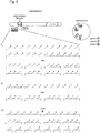
- Fig. 1 shows a schematic representation of a method according to the invention for the detection of two translocations using four probes and three labels, one probe being marked with two labels at the same time. It shows the signal pattern in normal cells and cells with translocation of the ALK gene in 2p23 or the ROS1 gene in 6q22. Both breakpoint regions (ALK and ROS1) are each flanked by label A and B of the quadruple ISH probe and each result in a fusion signal AB. One side of the ALK breakpoint region is also flanked with label C, so that there is a mixed label A / C.
- the ROS1 gene loci are marked by fusion signals A-B and ALK gene loci are marked by fusion signals A-B, which are accompanied by A / C mixed signals.
- the ALK gene affected by the translocation is marked by a separate signal of label B and a separate mixed signal A / C.
- the ROS1 gene affected by the translocation is marked by a separate signal of label A and a separate signal of label B.
- Fig. 2 shows a schematic representation of a method according to the invention relating to the use of several labels to represent mixed labels and mixed signals. For the sake of clarity, only two labels are listed.
- Mixed signals that are specific for a locus-specific probe and thus for a chromosomal region or a genomic section can arise if I) fragments of a probe are marked with different labels and / or II) all fragments of a probe or, optionally, only individual fragments of a probe are marked with several labels and / or III) alternating fragments are marked with different labels, so that here too only one mixed signal is finally visible or detectable.
- All or only individual fragments according to I) to III) can superimpose or overlap (not shown) and mixed labels can also arise if individual fragments or fragment groups according to I) to III) have a distance of up to 2 Mbp, e.g. in the "gap" shown.
- Fig. 3 shows a diagram of signal patterns when using a corresponding quadruple FISH probe "ZytoLight SPEC ALK & ROS1 Break Apart Dual-Mix NG-FISH Probe” from ZytoVision.
- the probe consists of green-marked polynucleotides (absorption at 503 nm and emission at 528 nm), which in 2p23 are directed against sequences proximal to the ALK breakpoint region and in 6q22 against sequences located proximal to the ROS1 breakpoint region, orange-marked polynucleotides ( Absorption at 547 nm and emission at 572 nm), which in 2p23 are directed against sequences distal to the ALK breakpoint region and in 6q22 against sequences distal to the ROS1 breakpoint region, as well as blue-labeled polynucleotides (absorption at 426 nm and emission at 480 nm), which in the region 2p23 are directed against sequences distal and prox
- the hybridization signals for the non-rearranged ALK gene appear as green-orange fluorescence fusion signals, which consist of green / blue and put together orange / blue fluorescence mixed signals.
- the hybridization signals for the non-rearranged ROS1 gene appear as green-orange fluorescence fusion signals.
- a 2p23 locus affected by an ALK translocation is identified by a separate green / blue mixed signal and a separate orange / blue mixed signal. (see. Figure 3b ).
- a 6q22 locus affected by a ROS1 translocation is identified by a separate green and a separate orange signal (cf. Figure 3c ).
- green and separate orange signals initially only allow the statement that an ALK or ROS1 translocation is present.
- a diagnostically relevant distinction between ALK or ROS1 translocation can then be made by including the blue fluorescence signals. If the separate green signals overlap blue signals (green / blue mixed signals) or if the separate orange signals overlap blue signals (orange / blue mixed signals), this indicates an ALK translocation. If the separate green and orange signals do not overlap with blue signals, this indicates a ROS1 translocation.
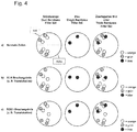
- Fig. 4 shows a diagram of signal patterns when using a corresponding quadruple FISH probe "ZytoLight SPEC ALK & ROS1 Break Apart Single-Mix NG-FISH Probe" from ZytoVision.
- the probe consists of green-marked polynucleotides (absorption at 503 nm and emission at 528 nm), which in 2p23 are directed against sequences proximal to the ALK breakpoint region and in 6q22 against sequences located proximal to the ROS1 breakpoint region, orange-marked polynucleotides ( Absorption at 547 nm and emission at 572 nm), which in 2p23 are directed against sequences distal to the ALK breakpoint region and in 6q22 against sequences distal to the ROS1 breakpoint region, as well as blue-labeled polynucleotides (absorption at 426 nm and emission at 480 nm), which in the region 2p23 are directed against sequences distal to the A
- the hybridization signals for the non-rearranged ALK gene appear as green-orange fluorescence fusion signals, which are composed of green and orange / blue mixed fluorescence signals.
- the hybridization signals for the non-rearranged ROS1 gene appear as green-orange fluorescence fusion signals.
- a 2p23 locus affected by an ALK translocation is identified by a separate green signal and a separate orange / blue mixed signal. (see. Figure 4b ).
- a 6q22 locus affected by a ROS1 translocation is identified by a separate green and a separate orange signal (cf. Figure 4c ).
- green and separate orange signals initially only allow the statement that an ALK or ROS1 translocation is present.
- a diagnostically relevant distinction between ALK or ROS1 translocation can then be made by including the blue fluorescence signals. If the separate orange signals overlap blue signals (orange / blue mixed signals), this indicates an ALK translocation. If the separate orange signals do not overlap with blue signals, this indicates a ROS1 translocation.
- Fig. 5 shows a diagram of signal patterns when using a corresponding six-fold FISH probe "ZytoLight SPEC ALK & ROS1 & RET Break Apart Dual-Mix NG-FISH Probe" from ZytoVision.
- the probe consists of green-marked polynucleotides (absorption at 503 nm and emission at 528 nm), the sequences located proximal to the ALK breakpoint region in 2p23, sequences located proximal to the ROS1 breakpoint region in 6q22 and proximal to the RET region in 10q11 Sequences located at the breakpoint region are directed, orange-marked polynucleotides (absorption at 547 nm and emission at 572 nm), the sequences located distal to the ALK breakpoint region in 2p23, sequences located distal to the ROS1 breakpoint region in 6q22 and sequences distal to the ROS1 breakpoint region in 10q11 Sequences located in the RET breakpoint region
- the hybridization signals for the non-rearranged ALK gene appear as green-orange fluorescence fusion signals, which are composed of green and orange / blue mixed fluorescence signals.
- the hybridization signals for the non-rearranged RET gene appear as green-orange fluorescence fusion signals, which are composed of green / blue mixed signals and orange signals.
- the hybridization signals for the non-rearranged ROS1 gene appear as green-orange fluorescence fusion signals.
- green-orange fusion signals appear when using a suitable green-orange dual bandpass filter set, four blue signals when using a suitable single bandpass filter set and when using one suitable triple bandpass filter sets two green-orange fusion signals, two green-orange / blue fusion and mixed signals and two green / blue-orange fusion and mixed signals (cf. Figure 5a ).
- a 2p23 locus affected by an ALK translocation is identified by a separate green signal and a separate orange / blue mixed signal. (see. Figure 5b ).
- a 6q22 locus affected by a ROS1 translocation is identified by a separate green and a separate orange signal (cf. Figure 5c ).
- a 10q11 locus affected by a RET translocation is identified by a separate orange signal and a separate green / blue mixed signal. (see. Fig. 5d ).
- green and separate orange signals initially only allow the statement that an ALK, ROS1 or RET translocation is present.
- a diagnostically relevant distinction between ALK, ROS1 or RET translocation can then be made by taking into account the blue fluorescence signals. If the separate orange signals overlap blue signals (orange / blue mixed signals), this indicates an ALK translocation. If the separate green signals overlap blue signals (green / blue mixed signals), this indicates a RET translocation. If neither the separate orange nor the separate green signals overlap with blue signals, this indicates a ROS1 translocation.
- Fig. 6 shows a diagram of signal patterns when using a corresponding six-fold FISH probe "ZytoLight SPEC ALK & ROS1 & RET Break Apart Dual-Mix II NG-FISH Probe" from ZytoVision GmbH.
- the probe consists of green-marked polynucleotides (absorption at 503 nm and emission at 528 nm), the sequences located proximal to the ALK breakpoint region in 2p23, sequences located proximal to the ROS1 breakpoint region in 6q22 and proximal to the RET region in 10q11 Sequences located at the breakpoint region are directed, red-marked polynucleotides (absorption at 580 nm and emission at 599 nm), the sequences located distal to the ALK breakpoint region in 2p23, sequences located distal to the ROS1 breakpoint region in 6q22 and sequences distal to the ROS1 breakpoint region in 10q11 Sequences located in the RET break
- the hybridization signals for the non-rearranged ALK gene appear as green-red fluorescence fusion signals which are composed of green and red / blue mixed fluorescence signals.
- the hybridization signals for the non-rearranged RET gene appear as green-red fluorescence fusion signals, which are composed of green / gold-yellow mixed signals and red signals.
- the hybridization signals for the non-rearranged ROS1 gene appear as green-red fluorescence fusion signals.
- a 2p23 locus affected by an ALK translocation is identified by a separate green signal and a separate red / blue mixed signal. (see. Figure 6b ).
- a 6q22 locus affected by a ROS1 translocation is identified by a separate green and a separate red signal (cf. Figure 6c ).
- a 10q11 locus affected by a RET translocation is identified by a separate red signal and a separate green / golden yellow mixed signal. (see. Fig. 6d ).
- green and separate red signals initially only allow the statement that there is basically an ALK, ROS1 or RET translocation.
- a diagnostically relevant distinction between ALK, ROS1 or RET translocation can then be made by taking into account the blue or golden-yellow fluorescence signals. If the separate red signals overlap blue signals (red / blue mixed signals), this indicates an ALK translocation. If the separate green signals overlap golden-yellow signals (green / golden-yellow mixed signals), this indicates a RET translocation. If neither the separate red nor the separate green signals overlap with blue or golden yellow signals, this indicates a ROS1 translocation.
- Fig. 7 shows a schematic representation of a method according to the invention for the detection of four numerical aberrations using four probes and four labels, three probes being marked simultaneously with two labels each, the labels used for the combination differing from one another in these three probes. It shows the signal pattern in normal cells and in cells with amplification of the MET gene in 7q31.
- the region 17q11.2-q12 of the ERBB2 gene is covered with label A, the region 7p12 of the EGFR gene with label A and also label D, so that a mixed label A / D is created, the region 8p11.23- p11.22 of the FGFR1 gene with label A and also label C, so that there is a mixed label A / C as well as the region 7q31 of the MET gene with label A and also label B, so that there is a mixed label A / B.
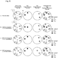
- Fig. 8 shows a schematic representation of a method according to the invention for the detection of seven translocations or amplifications using 14 probes and five labels each, one probe being marked with two labels at the same time and the ratio of the two labels per probe differing.
- the probes each flank a breakpoint region or address an amplification region as well as another region on the same chromosome (eg the centromere region) as on the upper left or upper right part of the Fig. 8 is shown.
- the two probes of a chromosome are marked with two different labels and the same label (e.g. probe 1: 25% green, 75% blue and probe 2: 25% yellow and 75% blue).
- the two probes each flanking a breakpoint region, thus each result in fusion and mixed signals from three labels in a cell that is not affected by a translocation.
- the two probes which each address an amplification region and a further region on the same chromosome, thus each produce separate mixed signals in a cell that is not affected by amplification (unless the distance between the two probes is so small, see above) that fusion and mixed signals arise).
- different mixed signals are generated so that a large number of probes with the same labels can be marked differentially (e.g. 4 probes for the breakpoint regions 1 to 4:25 in the example shown % to 75%; 50% to 50%; 75% to 25% and 100% to 0%).
- the breakpoint regions of a gene are marked by mixed signals AB and CB, which combine to form A / B / C fusion signals.
- the amplification regions of a gene are marked by the mixed signals AB and other regions on the same chromosome, for example centromere regions, are marked by the mixed signals CB.
- the gene affected by the translocation is identified by a separate signal of the label AB and one of these separate mixed signal CB marked.
- the gene affected by the amplification is marked by an amplified mixed signal from a label pair, eg AB.
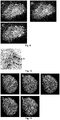
- Fig. 9 shows a FISH analysis to detect a translocation of the ROS1 region in 6q22 using the quadruple FISH probe "ZytoLight SPEC ALK & ROS1 Break Apart Single-Mix NG-FISH Probe" from ZytoVision.
- the probe consists of green-marked polynucleotides (absorption at 503 nm and emission at 528 nm), which in 2p23 are directed against sequences located proximal to the ALK breakpoint region and in 6q22 sequences located proximal to the ROS1 breakpoint region, orange-marked polynucleotides (absorption at 547 nm and emission at 572 nm), which in 2p23 are directed against sequences distal to the ALK breakpoint region and in 6q22 sequences located distal to the ROS1 breakpoint region, as well as blue-marked polynucleotides (absorption at 426 nm and emission at 480 nm) , which are directed in the region 6q22 proximal to the ROS1 breakpoint region located sequences.
- the hybridization signals for non-rearranged ROS1 and / or ALK genes appear as green-orange fluorescence fusion signals and for rearranged ROS1 and / or ALK genes as a separate green and separate orange signal (see Sect. Figure 9A showing the green and orange fluorescent signals).
- ROS1-specific green signals co-localize with blue fluorescence signals (s. Figure 9B , which shows the blue fluorescence signals), so that the unrearranged ROS1 gene is composed of orange and green / blue fluorescence mixed signals.
- the hybridization signals for the non-rearranged ALK gene appear as green-orange fluorescence fusion signals, without mixed signals with blue fluorescence signals.
- the 6q22 locus affected by a ROS1 translocation is indicated by a separate green and a separate orange signal (arrows in Figures 9A and C ).
- the separate green signal overlaps with a blue signal.
- This green / blue mixed signal indicates ROS1, not ALK, as the gene affected by the translocation (s. Figure 9C showing the blue, green, and orange fluorescent signals).
- the signal pattern can be made clearly visible with suitable filter sets.
- Fig. 10 shows a CISH analysis to detect a translocation of the ALK region in 2p23 using the quadruple CISH probe "ZytoDot SPEC ALK & ROS1 Break Apart Single-MIX NG-FISH Probe" from ZytoVision.
- the probe consists of digoxigenin-labeled polynucleotides that are directed against sequences located proximal to the ALK breakpoint region in 2p23 and sequences located proximal to the ROS1 breakpoint region in 6q22, DNP-labeled polynucleotides that are directed against sequences distal to the ALK breakpoint region in 2p23 and Sequences located distal to the ROS1 breakpoint region are directed in 6q22, as well as biotin-labeled polynucleotides which are directed in the region 6q22 distal to the ROS1 breakpoint region sequences.
- the markings were verified via primary (unlabeled) antibodies (Anti-DIG / Anti-DNP / Anti-BIO), which are detected by secondary polymerized enzyme-conjugated antibodies (HRP-Polymer / AP-Polymer / beta-GAL), as well as the enzymatic conversion of the Substrates (AP-RED / HRP-GREEN / beta-GAL-BLUE), which lead to the creation of strong, permanent, red (R), green (G) and blue (B) signals, which are light microscopically eg with a 40x dry lens can be represented leads.
- primary (unlabeled) antibodies Anti-DIG / Anti-DNP / Anti-BIO
- HRP-Polymer / AP-Polymer / beta-GAL secondary polymerized enzyme-conjugated antibodies
- AP-RED / HRP-GREEN / beta-GAL-BLUE the enzymatic conversion of the Substrates
- R red
- G green
- B
- Fig. 11 shows a FISH analysis to detect the amplification of the ERBB2 region using the fivefold FISH probe "ZytoLight SPEC ERBB2, EGFR, FGFR1, MET & SOX2 FiveCheck TM NG-FISH Probe” from ZytoVision.
- the probe consists of green-marked polynucleotides (absorption at 503 nm and emission at 528 nm) that target the region 17q11.2-q12 of the ERBB2 gene, the region 7p12 of the EGFR gene, the region 8p11.23-p11.
- the 7q31 region of the MET gene and the 3q26.3-q27 region of the SOX2 gene are directed as well as blue-marked polynucleotides (absorption at 426 nm and emission at 480 nm) that target the region of the EGFR gene and the SOX2 gene, golden yellow-marked polynucleotides (absorption at 532 nm and emission at 553 nm), which are against the region of the FGFR1 gene and red-marked polynucleotides (absorption at 580 nm and emission at 599 nm), which are directed against the region of the MET gene and the SOX2 gene.
- the single green signal is a non-amplified ERBB2 gene
- the green signal cluster identifies an ERBB2 gene amplification.
- Co-localizing green / blue mixed signals identify two copies of the EGFR gene
- co-localizing green / golden yellow mixed signals identify two copies of the FGFR1 gene
- co-localizing green / red mixed signals identify two copies of the MET gene
- co-localizing green / blue / red mixed signals identify two copies of the SOX2 gene (s. Figure 11E ).
- Example 1 FISH analysis for the detection of multiple numerical aberrations in different cell types using the five-fold FISH probe "SPEC ERBB2, EGFR, FGFR1, MET & SOX2 FiveCheck TM NG-FISH Probe "from ZytoVision GmbH
- the FISH was performed on 3 to 5 ⁇ m thick sections of formalin-fixed paraffin-embedded (FFPE) lung and breast cancer preparations with or without previously diagnosed ERBB2 gene amplification, which were mounted on coated glass slides and kept overnight at 58 ° C were baked.
- FFPE formalin-fixed paraffin-embedded
- the preparations were first heated on a hot plate for 10 minutes at 70 ° C. and then incubated twice in 100% xylene for 10 minutes each time at room temperature (RT).
- the preparations were then rehydrated by means of a descending series of ethanol (5 minutes each time at RT in 96%, 96%, 90%, 70% denatured ethanol) and incubation in ultrapure water (two times each two minutes at RT).
- a descending series of ethanol 5 minutes each time at RT in 96%, 96%, 90%, 70% denatured ethanol
- ultrapure water two times each two minutes at RT.
- the proteolytic pretreatment was carried out by dropping pepsin solution (Pepsin Solution, ZytoVision GmbH) onto the preparations and then incubating them in a humid chamber at 37 ° C. for 25 minutes. After subsequent incubation for 5 min in Wash Buffer SSC (ZytoVision GmbH), the preparations were dehydrated (one minute each at RT in ultrapure water, 70%, 90%, 96% ethanol). After the specimens had air-dried, 10 ⁇ l of the FISH probe ZytoLight SPEC ERBB2, EGFR, FGFR1, MET & SOX2 FiveCheck TM NG-FISH probe (ZytoVision GmbH) were applied directly to the sections using a pipette.
- pepsin solution Pepsin Solution, ZytoVision GmbH
- the probe was a mixture based on five locus-specific hybridization probes, the mixture of green-labeled polynucleotides (absorption at 503 nm and emission at 528 nm) that act against the region 17q11.2-q12 of the ERBB2 gene, the 7p12 region of the EGFR gene, the 8p11.23-p11.22 region of the FGFR1 gene, the 7q31 region of the MET gene and the 3q26.3-q27 region of the SOX2 gene were directed and were composed of blue-labeled polynucleotides ( Absorption at 426 nm and emission at 480 nm), which were directed against the region of the EGFR gene and the SOX2 gene, golden yellow-labeled polynucleotides (absorption at 532 nm and emission at 553 nm), which were directed against the region of the FGFR1- Gene, and red-labeled polynucleotides (absorption at 580 nm and
- the evaluation was then carried out on a fluorescence microscope (Axio Scope.A1 with HXP 120 V lighting unit, Carl Zeiss Microscopy GmbH) using suitable filter sets (Sp. Green HC mFISH filter set; Sp. Red HC mFISH filter set; Sp. Aqua HC mFISH filter set; ZyGold HC mFISH filter set (all AHF Analysentechnik AG)).
- the signal pattern could be interpreted as follows: Two green signals without spatially identical localization of signals of a different color identified the two ERBB2 gene copies of a diploid cell. Two green signals with spatially identical localization of two aqua signals identified the two EGFR gene copies, two green signals with spatially identical localization of two gold-yellow signals identified the two FGFR1 gene copies, two green signals with spatially identical localization of two red signals identified the two MET gene copies and two green signals with spatially identical localization of two red and two aqua signals identified the two SOX2 gene copies.
- the cell nuclei of the preparations with ERBB2 amplification showed a signal pattern comparable to the example described above, with the exception that, in addition to nine green signals, a green signal cluster or signal pattern, consisting of around fifteen signals that were inseparably close together, was observed (cf. Figure 11A ). This green signal pattern did not co-localize with signals of a different color and thus identified an ERBB2 gene amplification.
- Example 2 FISH analysis for the detection of translocations of the ALK or ROS1 regions in different cell types using the quadruple FISH probe "ZytoLight SPEC ALK & ROS1 Break Apart Single-Mix NG-FISH Probe” from ZytoVision GmbH_
- the FISH was performed on 3 to 5 ⁇ m thick sections of formalin-fixed paraffin-embedded (FFPE) cells of the cell lines HeLa (ATCC® CCL-2 TM), HCC78 (provided by Prof. Schildhaus, Göttingen) and H3122 ( provided by Prof. Schildhaus, Göttingen), which were mounted on coated glass slides and baked at 58 ° C overnight.
- FFPE formalin-fixed paraffin-embedded
- the preparations were first heated on a hot plate for 10 min at 70 ° C. and then incubated twice in 100% xylene for 10 min each time at room temperature (RT).
- the preparations were then rehydrated by means of a descending ethanol series (five minutes each at room temperature in 96%, 96%, 90%, 70% denatured ethanol) and incubation in ultrapure water (two times two minutes each at room temperature).
- RT room temperature
- ultrapure water two times two minutes each at room temperature.
- heat pretreatment for 15 minutes at 98 ° C. in Heat Pretration Solution Citric (ZytoVision GmbH), followed by two further incubation steps for two minutes in ultrapure water at room temperature.
- the proteolytic pretreatment was carried out by dripping a pepsin solution (Pepsin-Solution, ZytoVision GmbH) onto the preparations and subsequent incubation in a humid chamber at 37 ° C. for 15 minutes. After subsequent incubation for five minutes in wash buffer (Wash Buffer SSC, ZytoVision GmbH), the preparations were dehydrated (one minute each at RT in ultrapure water, 70%, 90%, 96% ethanol). After the specimens had dried in the air, 10 ⁇ l of the FISH probe ZytoLight SPEC ALK & ROS1 Break Apart Single-Mix NG-FISH Probe (ZytoVision GmbH) were applied directly to the sections using a pipette.
- a pepsin solution Pepsin-Solution, ZytoVision GmbH
- the probe was a mixture based on four locus-specific hybridization probes, the mixture of green-marked polynucleotides (absorption at 503 nm and emission at 528 nm), the sequences located proximal to the ALK breakpoint region in 2p23 and 6q22 were directed against sequences located proximal to the ROS1 breakpoint region, orange-marked polynucleotides (absorption at 547 nm and emission at 572 nm), the sequences located distal to the ALK breakpoint region in 2p23 and sequences located distal to the ROS1 breakpoint region in 6q22 were, as well as blue-marked polynucleotides (absorption at 426 nm and emission at 480 nm), which were directed in region 6q22 against sequences located proximal to the ROS1 breakpoint region.
- Example 3 FISH analysis to detect a translocation of the ROS1 region in 6q22 using the quadruple FISH probe "ZytoLight SPEC ALK & ROS1 Break Apart Single-Mix NG-FISH Probe" from ZytoVision GmbH
- a FISH analysis was carried out to detect a translocation of the ROS1 region in 6q22 using the quadruple FISH probe "ZytoLight SPEC ALK & ROS1 Break Apart Single-Mix NG-FISH Probe" from ZytoVision GmbH.
- the probe was a mixture based on four locus-specific hybridization probes, the mixture of green-marked polynucleotides (absorption at 503 nm and emission at 528 nm), the sequences located proximal to the ALK breakpoint region in 2p23 and 6q22 were directed against sequences located proximal to the ROS1 breakpoint region, orange-marked polynucleotides (absorption at 547 nm and emission at 572 nm), the sequences in 2p23 directed against sequences distal to the ALK breakpoint region and in 6q22 against sequences located distal to the ROS1 breakpoint region were, as well as blue-marked polynucleotides (absorption at 426 nm and emission at 480 nm), which were directed in region 6q22 against sequences located proximal to the ROS1 breakpoint region.
- the hybridization signals for non-rearranged ROS1 and / or ALK genes appeared as green-orange fluorescence fusion signals and for a rearranged ROS1 and / or ALK genes as a separate green and separate orange signal.
- ROS1-specific green signals co-localized with blue fluorescence signals, so that the non-rearranged ROS1 gene was composed of orange and green / blue mixed fluorescence signals.
- the hybridization signals for the non-rearranged ALK gene appeared as green-orange fluorescence fusion signals without mixed signals with blue fluorescence signals ( Figure 9A ).
- the 6q22 locus affected by a ROS1 translocation was indicated by a separate green and a separate orange signal ( Figure 9A Arrows).
- the separate green signal overlapped with a blue signal (blue signals Figure 9B ). This green / blue mixed signal indicated ROS1, not ALK, as the gene affected by the translocation.
- the signal pattern could be made clearly visible with suitable filter sets.
- Example 4 CISH analysis to detect a translocation of the ALK region in 2p23 using the quadruple CISH probe "ZytoDot SPEC ALK & ROS1 Break Apart Single-MIX NG-FISH Probe" from ZytoVision GmbH
- the probe was a mixture based on four locus-specific hybridization probes, the mixture of digoxigenin-labeled polynucleotides directed against sequences located proximal to the ALK breakpoint region in 2p23 and against sequences located proximal to the ROS1 breakpoint region in 6q22, DNP-labeled polynucleotides that in 2p23 against sequences distal to the ALK breakpoint region and in 6q22 against sequences distal to the ROS1 breakpoint region, and biotin-labeled polynucleotides in region 6q22 directed against sequences distal to the ROS1 breakpoint region.
- the markings were detected using primary (non-marked) antibodies (anti-DIG / anti-DNP / anti-BIO), which were detected by secondary polymerized enzyme-conjugated antibodies (HRP polymer / AP polymer / beta-GAL), as well as the enzymatic conversion of the substrates (AP-RED / HRP-GREEN / beta-GAL-BLUE), which led to the development of strong, permanent, red, green and blue signals, which could be visualized with a light microscope, for example with a 40x dry lens .
- Diploid or disome cell nuclei without rearrangements or translocations of the ALK or ROS1 genes showed two signals, each consisting of a red and a green signal, which were inseparably close together or partly overlapped or mixed and specific for the two copies of the ALK -Gens were ( Fig. 10 ).
- Example 5 FISH analysis to detect the amplification of the ERBB2 region using the fivefold FISH probe “ZytoLight SPEC ERBB2, EGFR, FGFR1, MET & SOX2 FiveCheck TM NG-FISH Probe” from ZytoVision
- the probe was a mixture based on five locus-specific hybridization probes, the mixture of green-labeled polynucleotides (absorption at 503 nm and emission at 528 nm) that act against the region 17q11.2-q12 of the ERBB2 gene, the 7p12 region of the EGFR gene, the 8p11.23-p11.22 region of the FGFR1 gene, the 7q31 region of the MET gene, and the region 3q26.3-q27 of the SOX2 gene as well as from blue-marked polynucleotides (absorption at 426 nm and emission at 480 nm) that were directed against the region of the EGFR gene and the SOX2 gene, golden-yellow-marked polynucleotides (absorption at 532 nm and emission at 553 nm), which were directed against the region of the FGFR1 gene and red-marked polynucleotides (absorption at 580 nm and emission at 599
- the single green signal was a non-amplified ERBB2 gene, the green signal cluster identified an ERBB2 gene amplification.
- Co-localizing green / blue mixed signals identified two copies of the EGFR gene, co-localizing green / gold-yellow mixed signals identified two copies of the FGFR1 gene, co-localizing green / red mixed signals identified two copies of the MET gene and co-localizing green / blue / red mixed signals identified two copies of the SOX2 gene.
Description
Die vorliegende Erfindung betrifft das technische Gebiet der Nachweisverfahren für Chromosomenanomalien bzw. Chromosomenaberrationen.The present invention relates to the technical field of detection methods for chromosomal abnormalities or chromosome aberrations.
Insbesondere betrifft die vorliegende Erfindung ein Verfahren zum Nachweis von mindestens zwei voneinander verschiedenen Chromosomenaberrationen mittels In-Situ-Hybridisierung. Weiterhin betrifft die vorliegende Erfindung eine zum Nachweis von mindestens zwei voneinander verschiedenen Chromosomenaberrationen geeignete Zusammensetzung sowie deren erfindungsgemäße Verwendungen. Weiterer Gegenstand der vorliegenden Erfindung sind zudem die Verwendungen von mindestens drei voneinander verschiedenen, mit Detektionslabeln markierten lokusspezifischen Hybridisierungssonden. Schließlich ist Gegenstand der vorliegenden Erfindung ein Kit zum Nachweis von Chromosomenaberrationen.In particular, the present invention relates to a method for the detection of at least two mutually different chromosome aberrations by means of in situ hybridization. The present invention further relates to a composition suitable for detecting at least two different chromosome aberrations and to the uses thereof according to the invention. The present invention furthermore relates to the uses of at least three mutually different, locus-specific hybridization probes marked with detection labels. Finally, the subject of the present invention is a kit for the detection of chromosome aberrations.
Vielen Tumorerkrankungen liegen strukturelle und numerische Chromosomenmutationen, wie Translokationen, Inversionen, segmentelle Duplikationen, Deletionen, Insertionen, Duplikationen, Aneuplodien und Amplifikationen, zu Grunde. Der Nachweis dieser Veränderungen als prädiktiver, prognostischer oder differentialdiagnostischer Marker erfolgt in der Regel durch In-Situ-Hybridisierungen (ISH).Many tumor diseases are based on structural and numerical chromosome mutations such as translocations, inversions, segmental duplications, deletions, insertions, duplications, aneuplodies and amplifications. Evidence of these changes as predictive, prognostic or differential diagnostic markers is usually provided by in situ hybridizations (ISH).
Die In-Situ-Hybridisierung beruht auf der Hybridisierung bzw. Paarung komplementärer Basen von Nukleinsäure-Einzelsträngen, insbesondere DNA-Einzelsträngen, so dass spezifische Nukleinsäuresequenzen in einer Probe, insbesondere in einem Gewebe oder Zellpräparat, detektiert werden können. Dazu werden direkt oder indirekt markierte, synthetisch hergestellte Sonden mit Nukleinsäure-Einzelsträngen der Probe hybridisiert und anschließend detektiert.The in-situ hybridization is based on the hybridization or pairing of complementary bases of nucleic acid single strands, in particular DNA single strands, so that specific nucleic acid sequences can be detected in a sample, in particular in a tissue or cell preparation. For this purpose, directly or indirectly labeled, synthetically produced probes are hybridized with nucleic acid single strands of the sample and then detected.
Zu Detektionszwecken können unter anderem fluoreszenzmarkierte Nukleinsäurefragmente bzw. fluoreszenzmarkierte Hybridisierungssonden (Fluoreszente ISH (FISH)) zum Einsatz kommen. Darüber hinaus können antigenmarkierte Sonden, insbesondere Hapten-markierte Sonden, zum Einsatz kommen, welche anschließend mit Hilfe von Antikörpern durch Farbreaktionen sichtbar gemacht werden, so dass eine lichtmikroskopische Analyse möglich ist ((Hellfeld ISH (BrISH), Chromogene ISH (CISH), Silber ISH (SISH)).For detection purposes, among other things, fluorescence-marked nucleic acid fragments or fluorescence-marked hybridization probes (fluorescent ISH (FISH)) can be used come. In addition, antigen-labeled probes, especially hapten-labeled probes, can be used, which are then made visible with the help of antibodies through color reactions, so that an analysis by light microscopy is possible ((brightfield ISH (BrISH), chromogens ISH (CISH), silver ISH (SISH)).
Der Vorteil der FISH ist, dass multiple genomische Bereiche simultan und voneinander deutlich unterscheidbar nachgewiesen werden können. Hierzu werden Nukleinsäurefragmente, die unterschiedliche genomische Bereiche adressieren bzw. hierfür spezifisch sind, jeweils mit unterschiedlichen Fluoreszenzfarbstoffen, die sich in ihrem Absorptions- und/oder Emissionsspektrum voneinander unterscheiden, markiert bzw. gekoppelt. Werden solche mehrfarbigen Sonden, welche separate, unterschiedliche Einzelsonden umfassen, z.B. an Metaphase-Chromosomen-Präparaten oder an Interphase-Zellkern-Präparaten verwendet, so können die einzelnen Farben durch Verwendung spezifischer Mikroskop-Filter, welche genau definierte Wellenlängenbereiche des Lichts zur Anregung der Farbstoffe auf das Präparat leiten sowie genau definierte Wellenlängenbereiche des von den Farbstoffen emittierten Lichts zum Auswerter leiten, getrennt voneinander dargestellt werden (sogenannter Single-Bandpass-Filterset). Darüber hinaus existieren auch Filter bzw. Filtersets, welche die simultane Darstellung unterschiedlicher Fluoreszenzfarbstoffe und somit mehrerer Nukleinsäurefragmente erlauben. Bei zwei unterschiedlichen Fluoreszenzfarbstoffen spricht man beispielsweise von einem Dual-Bandpass-Filterset.The advantage of FISH is that multiple genomic areas can be detected simultaneously and clearly distinguishable from one another. For this purpose, nucleic acid fragments which address different genomic regions or are specific for this are each marked or coupled with different fluorescent dyes which differ from one another in their absorption and / or emission spectrum. Are such multi-colored probes comprising separate, distinct individual probes, e.g. used on metaphase chromosome preparations or on interphase cell nucleus preparations, the individual colors can be determined by using specific microscope filters, which guide precisely defined wavelength ranges of the light to excite the dyes on the preparation as well as precisely defined wavelength ranges of the emitted by the dyes Direct light to the evaluator, shown separately from each other (so-called single bandpass filter set). In addition, there are also filters or filter sets which allow the simultaneous display of different fluorescent dyes and thus several nucleic acid fragments. In the case of two different fluorescent dyes, for example, one speaks of a dual bandpass filter set.
Der simultanen Darstellung sind jedoch deutliche Grenzen gesetzt, da je genomischer Region, die durch eine spezifische Sonde nachgewiesen wird, nur ein Label verwendet werden kann. Weiterhin liegen die Absorptions- und Emissionsbereiche der Farbstoffe häufig so nah beieinander, dass sie durch die Mikroskop-Filter-Sets nicht voneinander getrennt werden können. Aus diesen Gründen werden in der FISH simultan üblicherweise nur zwei Farben (orange/rot und grün) bzw. drei Farben (orange/rot und grün simultan bzw. gemeinsam mit einer blauen Kerngegenfärbung (DAPI)) analysiert. In etwa die gleichen Einschränkungen, die für die FISH darstellbar sind, gelten für die BrISH. Hier ist der Stand der Technik die Verwendung von zwei Haptenen, zumeist ausgewählt aus der Gruppe Biotin, Dinitrophenyl (DNP) und Digoxigenin, und zwei antikörpergekoppelten Enzymen, zumeist alkalische Phosphatase und Peroxidase.However, there are clear limits to the simultaneous display, since only one label can be used for each genomic region that is detected by a specific probe. Furthermore, the absorption and emission ranges of the dyes are often so close to one another that they cannot be separated from one another by the microscope filter sets. For these reasons, FISH usually only analyzes two colors (orange / red and green) or three colors (orange / red and green simultaneously or together with a blue nuclear counterstain (DAPI)). The same restrictions that can be represented for the FISH apply to the BrISH. Here, the state of the art is the use of two haptens, mostly selected from the group of biotin, dinitrophenyl (DNP) and digoxigenin, and two antibody-coupled enzymes, mostly alkaline phosphatase and peroxidase.
Die genannten Einschränkungen bei der simultanen Darstellung bzw. Analyse haben einen maßgeblichen Einfluss auf die Zusammensetzung (Komposition) bzw. die Abstimmung der Sonden für den Nachweis von strukturellen und numerischen Chromosomenaberrationen, die zur Diagnostik an Tumoren mit sich in der Interphase befindlichen Zellen nur mit sogenannten lokusspezifischen Sonden durchgeführt werden können, wobei gelegentlich parallel zum Nachweis von numerischen Chromosomenmutationen auch sogenannte Repetitive-Sequenzen-spezifische Sonden verwendet werden.The mentioned limitations in the simultaneous display or analysis have a decisive influence on the composition or the coordination of the probes for the detection of structural and numerical chromosome aberrations, which are only used for diagnosing tumors with cells in the interphase locus-specific probes can be carried out, so-called repetitive sequence-specific probes are occasionally used in parallel for the detection of numerical chromosome mutations.
Unter lokusspezifischen Sonden werden solche Sonden verstanden, welche ausgewählte DNA-Abschnitte eines Chromosoms, zumeist einzelne Gene oder benachbarte Gene, mit einer Größe von insgesamt bis zu etwa 1.000 kb adressieren und als genspezifische Sonden oder als 'single copy'-Sonden bezeichnet werden. Repetitive-Sequenzen-spezifischen Sonden sind Sonden, die repetitive Sequenzen adressieren und daher Regionen mit einer Größe von mehreren 1.000 kb adressieren. Diese Sonden umfassen beispielweise auch Zentromer- oder Alpha-Satelliten-Sonden.Locus-specific probes are understood to mean those probes which address selected DNA sections of a chromosome, mostly individual genes or neighboring genes, with a total size of up to about 1,000 kb and are referred to as gene-specific probes or “single copy” probes. Repetitive sequence-specific probes are probes that address repetitive sequences and therefore address regions with a size of several 1,000 kb. These probes also include centromere or alpha satellite probes, for example.
In Bezug auf die Detektion von Translokationen und Inversionen gibt es prinzipiell zwei maßgebliche Techniken und zugrundeliegende Sondenzusammensetzungen bzw. Sondenkompositionen: Das Prinzip des Entstehens von Fusionssignalen (sogenannte Dual-Color-Dual-Fusion-Ansätze) (
Bei Dual-Color-Dual-Fusion-Ansätzen wird der Bereich eines ersten Bruchpunkts des Chromosoms proximal und distal von Nukleinsäurefragmenten der gleichen Farbe (z. B. orange) flankiert, der Bereich eines zweiten Bruchpunkts, d. h. des reziproken Translokationspartners, wird proximal und distal von Nukleinsäurefragmenten einer zweiten Farbe (z. B. grün) flankiert. Die normale Situation, d. h. ohne chromosomale Brüche im Bereich der beiden Translokationspartner, wird hierbei durch ein grünes und ein räumlich getrenntes oranges Signal charakterisiert.In dual-color-dual-fusion approaches, the area of a first breakpoint of the chromosome is flanked proximally and distally by nucleic acid fragments of the same color (e.g. orange), the area of a second breakpoint, i.e. H. of the reciprocal translocation partner, is flanked proximally and distally by nucleic acid fragments of a second color (e.g. green). The normal situation, i.e. H. without chromosomal breaks in the area of the two translocation partners, is characterized by a green and a spatially separated orange signal.
Im Falle einer reziproken Translokation kommt es innerhalb der Bruchpunkte beider Translokationspartner zu Brüchen und der proximale Bereich des einen Translokationspartners fusioniert mit dem distalen Bereich des anderen Partners und vice versa. Es entstehen somit zwei grün/orange Signal-Paare, auch Fusionssignale genannt, da sich die unterschiedlich farbigen Signale oftmals überlappen. Der Nachteil dieser Sonden-Techniken ist, dass die Fusionssignale nur entstehen, wenn die Bruchpunkte beider Translokationspartner im Bereich der jeweiligen markierten Nukleinsäurefragmente liegen. Bei Translokationen, die nur einen der beiden Partner betreffen, entstehen keine Fusionssignale. Es kommt hierbei lediglich zur Entstehung eines zusätzlichen Signals mit der Farbe des Signals, welches charakteristisch für den durch die Translokation betroffenen Partner ist. Dies bedeutet, dass ein zusätzliches grünes Signal entsteht, wenn der Bruchpunkt der Translokation in dem Bereich lag, der von den mit grünem Fluorochrom markierten Nukleinsäuren überdeckt wurde. Nachteil dieser Sonden-Zusammensetzung ist, dass mit Hilfe der verwendeten zwei Farben nur die beiden Bruchpunktbereiche der gleichen Translokation bzw. Inversion markiert werden und somit nur eine spezifische Translokation bzw. Inversion detektiert werden kann.In the case of a reciprocal translocation, breaks occur within the breakpoints of both translocation partners and the proximal area of one translocation partner fuses with the distal area of the other partner and vice versa. This creates two green / orange signal pairs, also known as fusion signals, since the different colored signals often overlap. The disadvantage of these probe techniques is that the fusion signals only arise when the breakpoints of both translocation partners are in the area of the respective labeled nucleic acid fragments. In the case of translocations that only affect one of the two partners, no fusion signals arise. In this case, only an additional signal is generated with the color of the signal, which is characteristic of the partner affected by the translocation. This means that an additional green signal is generated if the breakpoint of the translocation was in the area that was covered by the nucleic acids marked with green fluorochrome. Disadvantage of this probe composition is that with the help of the two colors used only the two breakpoint areas of the same translocation or inversion are marked and thus only a specific translocation or inversion can be detected.
Bei Dual-Color-Break-Apart-Ansätzen wird der Bereich eines Bruchpunkts proximal und distal von unterschiedlich markierten oder farbig-markierten Nukleinsäurefragmenten flankiert (z. B. distal orange, proximal grün). Die normale Situation, d. h. ohne chromosomalen Bruch dieses Bereiches, wird dabei durch ein Fusionssignal charakterisiert. In einer aberranten Situation, d. h. wenn es zu einem chromosomalen Bruch zwischen den Sondenfragmenten kommt, trennen sich die Signale räumlich voneinander. Der Unterschied zwischen der normalen Situation und der aberranten Situation ist also gekennzeichnet durch den Abstand der unterschiedlich farbigen Signale. Aussagen über beteiligte Translokationspartner sind mit diesem Verfahren nicht möglich. Es lässt lediglich den Schluss zu, dass eine spezifische chromosomale Umlagerung stattgefunden hat. Nachteil dieser Sonden-Zusammensetzung ist, dass mit Hilfe der verwendeten zwei Farben nur ein einziger Bruchpunktbereich und somit nur eine spezifische Translokation bzw. Inversion detektiert werden kann.In the case of dual-color break-apart approaches, the area of a break point is flanked proximally and distally by nucleic acid fragments marked in different ways or in different colors (e.g. distal orange, proximal green). The normal situation, i.e. H. without a chromosomal break in this area, is characterized by a fusion signal. In an aberrant situation, i.e. H. if there is a chromosomal break between the probe fragments, the signals separate spatially from one another. The difference between the normal situation and the aberrant situation is thus characterized by the distance between the differently colored signals. Statements about involved translocation partners are not possible with this method. It only allows the conclusion that a specific chromosomal rearrangement has taken place. The disadvantage of this probe composition is that with the aid of the two colors used, only a single breaking point area and thus only a specific translocation or inversion can be detected.
Was Deletionen, Aneuplodien und Amplifikationen anbelangt, wird üblicherweise nur eine maßgebliche Technik und zugrundeliegende Sondenzusammensetzung, die sich im Allgemeinen auf lokusspezifische Sonden bezieht, eingesetzt. Unter Umständen werden jedoch lokusspezifische Sonden auch durch sogenannte Repetitive-Sequenzen-spezifische Sonden, wie beispielsweise Zentromer- oder Alpha-Satelliten-Sonden, ergänzt: Das Prinzip des Nachweises von Zugewinn oder Verlust von Signalen durch das Auftreten von Deletionen, Aneuplodien und Amplifikationen erfolgt üblicherweise in sogenannten Dual-Color-Probe-Ansätzen. Auch bei diesem Prinzip, welches nachfolgend beschrieben wird, und den daraus abgeleiteten Signalmustern ist zu beachten, dass eine normale Zelle in der Regel diploid ist, d. h. jedes Allel zweifach vorliegt.For deletions, aneuplodies, and amplifications, only one authoritative technique and underlying probe composition, which generally relates to locus specific probes, is typically used. Under certain circumstances, however, locus-specific probes are also supplemented by so-called repetitive sequence-specific probes, such as centromere or alpha satellite probes: The principle of detecting gain or loss of signals through the occurrence of deletions, aneuplodies and amplifications usually takes place in so-called dual-color probe approaches. With this principle, which is described below, and the signal patterns derived from it, it should be noted that a normal cell is usually diploid, i.e. H. each allele is present in duplicate.
Bei Dual-Color-Probe-Ansätzen werden zwei unterschiedliche chromosomale Bereiche mit unterschiedlich markierten oder farbig-markierten Nukleinsäurefragmenten markiert (z. B. genomische Region 1 oder Targetregion 1 in orange, genomische Region 2 oder Targetregion 2 in grün). Die normale Situation, d. h. ohne Zugewinn oder Verlust dieser Bereiche, wird durch zwei orange und zwei grüne Signale charakterisiert. In einer aberranten Situation, d. h. wenn es zu einem Zugewinn oder Verlust genomischer Bereiche in der die Targetregionen 1 und/oder 2 gekommen ist, sind bei Verlust weniger grüne und/oder orange Signale sichtbar, bei Zugewinn mehr Signale. Bei starken Genamplifikationen oder Amplifikationen genomischer Bereiche können viele zusätzliche Signale sichtbar sein, die sich auch als Cluster darstellen können.In dual-color probe approaches, two different chromosomal areas are marked with differently marked or color-marked nucleic acid fragments (e.g.
Mit den zuvor genannten Standardverfahren und Standardkompositionen unter Verwendung von zwei Standard-Signalen oder zwei Standard-Farben kann bzw. können somit im Falle struktureller Veränderungen nur eine (mögliche) Aberration und im Fall numerischer Chromosomenmutationen maximal zwei (mögliche) unterschiedliche Aberrationen pro Verfahren nachgewiesen werden.With the aforementioned standard methods and standard compositions using two standard signals or two standard colors, only one (possible) aberration can or can be in the case of structural changes and in the case of numerical changes Chromosome mutations a maximum of two (possible) different aberrations per procedure can be detected.
Neben den oben aufgeführten Zweifarb-Anwendungen werden in FISH-Analysen auch Dreifarb-, selten Vierfarb- und sehr selten Fünffarbsonden verwendet. Per Definition handelt es sich bei Verfahren, die gleichzeitig mindestens drei verschiedene Liganden oder Fluorochrome - ohne die Gegenfärbung - für die Markierung der Sonden verwenden, um Multicolor-FISH-Verfahren (mFISH-Verfahren). Dabei werden neben den deutlich stärkeren Standard-Fluoreszenzfarben orange/rot und grün die weiteren, zur Verfügung stehenden schwächeren Farben, z. B. gold bzw. goldgelb oder goldfarben, rot oder blau, verwendet. Daher werden im Allgemeinen beispielweise Vierfarb-FISH-Sonden nur zur Detektion von Deletionen oder Amplifikationen verwendet, weil hier zumeist bei den Markierungen der Sonden auf repetitive Sequenzen zur Verstärkung der Farbintensitäten z. B. von blau und gold zurückgegriffen werden kann. Derartige Verfahren werden unter anderem in der
Darüber hinaus werden im Stand der Technik dreifach-FISH-Ansätze beschrieben, welche den Nachweis von unterschiedlichen Translokationsereignissen adressieren, die in einer chromosomalen Region nebeneinander clustern können (d. h. es sind unterschiedliche Gene betroffen, die in Nachbarschaft liegen). Dabei werden für die entsprechende Auswertung der Signalmuster jeweils nur zwei Farben analysiert, die eine einzelne Aberration nachweisen, wobei die dritte Farbe jeweils keine Rolle spielt. Es werden drei unterschiedliche lokusspezifische Sonden verwendet, die jeweils mit einem anderen Label markiert sind.In addition, the prior art describes triple FISH approaches which address the detection of different translocation events that may cluster side by side in a chromosomal region (i.e., different genes that are in proximity are affected). For the corresponding evaluation of the signal pattern, only two colors are analyzed that detect a single aberration, the third color being irrelevant. Three different locus-specific probes are used, each marked with a different label.
Was die Hellfeld-ISH (BrISH) unter Verwendung von mehr als zwei Farben anbelangt, wird im Stand der Technik diesbezüglich die Durchführung einer chromogenen Dreifach-In-Situ-Hybridisierung beschrieben, die allgemein auf den Nachweis von drei repetitiven chromosomalen Bereichen abzielt. Nach dem derzeitigen Stand der Technik werden Translokationen mittels BrISH nur unter Verwendung von zwei Haptenen und somit zwei Farbstoffen nachgewiesen. In der Patentanmeldung
Die
Die
Die
Die
Die
Insgesamt lässt sich somit feststellen, dass es mit den aus dem Stand der Technik bekannten Verfahren nicht möglich ist, gleichzeitig bzw. simultan in effizienter Weise mehrere voneinander verschiedene Chromosomenaberrationen in Zellen bzw. Geweben mittels In-Situ-Hybridisierung zu detektieren.Overall, it can thus be stated that it is not possible with the methods known from the prior art to efficiently detect several different chromosome aberrations in cells or tissues simultaneously or simultaneously by means of in situ hybridization.
Eine simultane bzw. gleichzeitige Zuordnung definierter DNA- bzw. Chromosomenbereiche im Rahmen in In-Situ-Hybridisierungen ist bislang nur bei Verfahren zum Nachweis ganzer Chromosomen mit einer Größe beim Menschen zwischen etwa 50 Mbp und 250 Mbp oder großer chromosomaler Bereiche, z.B. Chromosomenarme, unter Verwendung von sogenannten 'Whole Chromosome Painting Probes' (WCP) oder 'Partial Chromosome Painting Probes' (PCP) bekannt. Mit den zugrunde liegenden Techniken, z. B. mFISH (multiplex FISH), SKY-FISH (spectral karyotyping), multicolor FISH, COBRA-FISH (Combined Binary Ratio labeling FISH) oder auch 24-color-FISH, können unter Nutzung von etwa vier bis sieben verschiedenen Fluoreszenzfarbstoffen insgesamt vierundzwanzig verschiedene 'Chromosome Painting Probes' markiert und unterschieden werden. In einem ähnlichen Verfahren können in einer sogenannten 42-color-FISH die Chromosomenarm-spezifischen Sonden aller Chromosomen unterschiedlich markiert werden. Die vorgenannten Verfahren sind jedoch nur für Zellen, welche sich in der Metaphase befinden, geeignet. Die vorgenannten Verfahren sind nur möglich, da mit den darin verwendeten Sonden nur genomische/chromosomale Bereiche in Metaphasen zumindest im Wesentlichen ohne Überlagerung chromosomalen Materials ausgewertet werden können. Eine Analyse von Zellen in der Interphase, welche im Allgemeinen erst eine Analyse des genetischen Materials von soliden Tumoren erlaubt, ist mit derartigen Verfahren nicht möglich. Darüber hinaus können die dabei zum Einsatz kommenden diese Sonden relativ einfach nachgewiesen werden, da sie große Bereiche adressieren. Die Auswertung dieser Analysen kann nicht manuell, d.h. unter Betrachtung der Signale am Fluoreszenzmikroskop erfolgen, sondern nur computerbasiert mit Hilfe geeigneter Auswertesoftware.A simultaneous or simultaneous assignment of defined DNA or chromosome areas in the context of in -situ hybridizations has so far only been possible with methods for the detection of whole chromosomes with a size in humans between about 50 Mbp and 250 Mbp or large chromosomal areas, e.g. chromosome arms Use of so-called 'Whole Chromosome Painting Probes' (WCP) or 'Partial Chromosome Painting Probes' (PCP) known. With the underlying techniques, e.g. B. mFISH (multiplex FISH), SKY-FISH (spectral karyotyping), multicolor FISH, COBRA-FISH (Combined Binary Ratio labeling FISH) or 24-color FISH, using around four to seven different fluorescent dyes, a total of twenty-four different dyes can be produced 'Chromosome Painting Probes' can be marked and differentiated. In a similar procedure, the chromosome arm-specific probes of all chromosomes can be marked differently in a so-called 42-color FISH. However, the aforementioned methods are only suitable for cells that are in the metaphase. The aforementioned procedures are only possible because with the therein used probes only genomic / chromosomal areas in metaphases can be evaluated at least essentially without overlaying chromosomal material. An analysis of cells in the interphase, which generally only allows an analysis of the genetic material of solid tumors, is not possible with such methods. In addition, these probes that are used can be detected relatively easily, as they address large areas. The evaluation of these analyzes cannot be done manually, ie by observing the signals on the fluorescence microscope, but only on a computer basis with the help of suitable evaluation software.
Die derzeit im Stand der Technik bekannten Methoden und Sondenzusammensetzungen für BrISH und FISH im Zusammenhang mit strukturellen und numerischen Chromosomenmutationen bzw. Chromosomenaberrationen, insbesondere im Zusammenhang mit Tumoren bzw. Krebserkrankungen, sind mit gewissen Nachteilen verbunden. So existieren keine Kompositionen lokusspezifischer Sonden und Methoden, welche eine sichere, einfache und schnelle Detektion und Diskriminierung mehrerer potentieller unterschiedlicher struktureller und/oder numerischer Chromosomenmutationen bzw. Chromosomenaberrationen erlauben.The methods and probe compositions for BrISH and FISH currently known in the prior art in connection with structural and numerical chromosome mutations or chromosome aberrations, in particular in connection with tumors or cancer diseases, are associated with certain disadvantages. There are no compositions of locus-specific probes and methods that are safe and simple and allow rapid detection and discrimination of several potentially different structural and / or numerical chromosome mutations or chromosome aberrations.
Insbesondere bei den strukturellen Chromosomenmutationen ist die simultane oder gleichzeitige Analyse mehrerer unterschiedlicher chromosomaler Mutationen, die sich nicht bedingen, d.h. kein chromosomales Material austauschen bzw. nicht reziprok sind, insbesondere in zugrunde liegenden Break-Apart-Ansätzen gar nicht oder nur sehr schwer möglich.Particularly in the case of structural chromosome mutations, the simultaneous or simultaneous analysis of several different chromosomal mutations that are not mutually dependent, i.e. do not exchange chromosomal material or are not reciprocal, especially not at all or only with great difficulty in underlying break-apart approaches.
Der vorliegenden Erfindung liegt daher die Aufgabe zugrunde, ein Verfahren bzw. eine Zusammensetzung bereitzustellen, welches bzw. welche zur Detektion bzw. Analyse von Chromosomenmutationen bzw. Chromosomenaberrationen geeignet ist und die zuvor geschilderten Nachteile des Standes der Technik zumindest weitgehend vermeidet oder aber wenigstens abschwächt.The present invention is therefore based on the object of providing a method or a composition which is suitable for the detection or analysis of chromosome mutations or chromosome aberrations and at least largely avoids or at least mitigates the disadvantages of the prior art described above.
Insbesondere liegt der vorliegenden Erfindung die Aufgabe zugrunde, ein Verfahren bereitzustellen, welches einen zuverlässigen und simultanen Nachweis mehrerer voneinander verschiedener Chromosomenaberrationen, insbesondere voneinander unabhängiger (d.h. nicht reziproker) Chromosomenaberrationen, insbesondere in einem Ansatz ermöglicht. Gleichermaßen liegt der vorliegenden Erfindung die Aufgabe zugrunde, ein Verfahren bereitzustellen, welches darüber hinaus auch die Zuordnung von Chromosomenaberrationen zu einem bestimmten Chromosomen- bzw. DNA-Bereich ermöglicht.In particular, the present invention is based on the object of providing a method which enables a reliable and simultaneous detection of several mutually different chromosome aberrations, in particular independent (i.e. non-reciprocal) chromosome aberrations, in particular in one approach. The present invention is likewise based on the object of providing a method which, in addition, also enables chromosome aberrations to be assigned to a specific chromosome or DNA region.
Zur Lösung der zuvor geschilderten Aufgabe schlägt die vorliegende Erfindung ein Verfahren zum Nachweis von mindestens zwei voneinander verschiedenen Chromosomenaberrationen gemäß Patentanspruch 1 vor; weitere vorteilhafte Ausgestaltungen sind Gegenstand der diesbezüglich abhängigen Patentansprüche.In order to achieve the object described above, the present invention proposes a method for the detection of at least two different chromosome aberrations according to
Weiterhin ist Gegenstand der vorliegenden Erfindung eine Zusammensetzung zum Nachweis von mindestens zwei voneinander verschiedenen Chromosomenaberrationen gemäß dem diesbezüglich unabhängigen Patentanspruch bzw. eine Zusammensetzung zur Verwendung bei der prophylaktischen bzw. therapeutischen Behandlung bzw. bei der Diagnose bzw. Prognose von im Zusammenhang mit Chromosomenaberrationen stehenden Erkrankungen.The present invention also relates to a composition for the detection of at least two different chromosomal aberrations according to the independent patent claim or a composition for use in prophylactic or therapeutic treatment or in the diagnosis or prognosis of diseases associated with chromosomal aberrations.
Wiederum weiterer Gegenstand der vorliegenden Erfindung ist die Verwendung einer Zusammensetzung nach der vorliegenden Erfindung gemäß dem diesbezüglich unabhängigen Patentanspruch.The present invention also further provides the use of a composition according to the present invention according to the independent patent claim in this regard.
Weiterhin betrifft die vorliegende Erfindung die Verwendung von mindestens drei, vorzugsweise mindestens vier voneinander verschiedenen lokusspezifischen Hybridisierungssonden gemäß dem diesbezüglich unabhängigen Patentanspruch.Furthermore, the present invention relates to the use of at least three, preferably at least four mutually different locus-specific hybridization probes according to the independent patent claim.
Weiterhin ist Gegenstand der vorliegenden Erfindung die Verwendung von mindestens einer, mit mindestens zwei Detektionslabeln markierten lokusspezifischen Hybridisierungssonde gemäß dem diesbezüglich unabhängigen Patentanspruch.Furthermore, the subject matter of the present invention is the use of at least one location-specific hybridization probe marked with at least two detection labels according to the independent patent claim.
Schließlich ist Gegenstand der vorliegenden Erfindung ein Kit bzw. Kit-of-parts oder Set zum Nachweis von mindestens zwei voneinander verschiedenen Chromosomenaberrationen; weitere, vorteilhafte Eigenschaften sind Gegenstand des diesbezüglichen Unteranspruchs.Finally, the subject matter of the present invention is a kit or kit-of-parts or set for the detection of at least two different chromosome aberrations; further advantageous properties are the subject of the relevant sub-claim.
Es versteht sich von selbst, dass im Folgenden besondere Ausgestaltungen, Ausführungsformen oder dergleichen, welche nur im Zusammenhang mit einem Erfindungsaspekt beschrieben sind, auch in Bezug auf die anderen Erfindungsaspekte entsprechend gelten, ohne dass dies einer ausdrücklichen Erwähnung bedarf.It goes without saying that in the following special configurations, embodiments or the like, which are only described in connection with one aspect of the invention, also apply accordingly with regard to the other aspects of the invention, without this needing to be explicitly mentioned.
Weiterhin ist bei allen nachstehend genannten relativen bzw. prozentualen, insbesondere gewichtsbezogenen Mengenangaben zu beachten, dass diese im Rahmen der vorliegenden Erfindung vom Fachmann derart auszuwählen sind, dass in der Summe der jeweiligen Inhaltsstoffe, Wirkstoffe, Zusatz- bzw. Hilfsstoff oder dergleichen stets 100% bzw. 100 Gew.-% resultieren. Dies versteht sich für den Fachmann aber von selbst.Furthermore, with all of the relative or percentage, in particular weight-related, quantitative information mentioned below, it should be noted that these are to be selected by the person skilled in the art within the scope of the present invention in such a way that the sum of the respective ingredients, active ingredients, additives or excipients or the like is always 100% or 100% by weight result. This goes without saying for the expert.
Im Übrigen gilt, dass der Fachmann anwendungsbezogen oder einzelfallbedingt von den nachfolgend aufgeführten Zahlen-, Bereichs- oder Mengenangaben abweichen kann, ohne dass er den Rahmen der vorliegenden Erfindung verlässt.In addition, the person skilled in the art can deviate from the numbers, ranges or quantities given below in relation to the application or in individual cases without departing from the scope of the present invention.
Zudem gilt, dass alle im Folgenden genannten Parameterangaben oder dergleichen grundsätzlich mit genormten oder explizit angegebenen Bestimmungsverfahren oder aber mit dem Fachmann an sich geläufigen Bestimmungsmethoden bestimmt bzw. ermittelt werden können.In addition, it applies that all of the parameter information or the like mentioned below can in principle be determined or determined using standardized or explicitly stated determination methods or with determination methods that are known per se to the person skilled in the art.
Zur Lösung der zuvor geschilderten Aufgabe schlägt die vorliegende Erfindung - gemäß einem ersten erfindungsgemäßen Aspekt - ein Verfahren zum Nachweis von mindestens zwei voneinander verschiedenen Chromosomenaberrationen, insbesondere strukturellen und/oder numerischen Chromosomenaberrationen, vorzugsweise strukturellen Chromosomenaberrationen, mittels In-Situ-Hybridisierung durch Detektion von Chromosomen- und/oder DNA-Bereichen in einer biologischen Probe, vorzugsweise in einer oder mehreren Zelle(n) und/oder in einem oder mehreren Zellkern(en),
wobei die In-Situ-Hybridisierung als Interphase-In-Situ-Hybridisierung durchgeführt wird,
wobei die In-Situ-Hybridisierung mit mindestens vier voneinander verschiedenen, mit jeweils einem ersten Detektionslabel markierten, lokusspezifischen Hybridisierungssonden durchgeführt wird, wobei, zur Erzeugung mindestens eines Mischsignals durch
zwei oder mehr sich auf einer lokusspezifischen Hybridisierungssonde befindende voneinander verschiedene Detektionslabel, mindestens eine der lokusspezifischen Hybridisierungssonden mit mindestens einem weiteren, von dem ersten Detektionslabel verschiedenen Detektionslabel, bezogen auf die jeweilige lokusspezifische Hybridisierungssonde, markiert ist, so dass ein Signalmuster erzeugt wird, und
wobei vorhandene Chromosomenaberrationen anhand des Signalmusters identifiziert und/oder einem Chromosomen- und/oder DNA-Bereich zugeordnet werden.To solve the problem described above, the present invention proposes - according to a first aspect of the invention - a method for detecting at least two different chromosome aberrations, in particular structural and / or numerical chromosome aberrations, preferably structural chromosome aberrations, by means of in-situ hybridization by detection of chromosomes - and / or DNA areas in a biological sample, preferably in one or more cell (s) and / or in one or more cell nucleus (s),
where the in- situ hybridization is carried out as an interphase in-situ hybridization,
wherein the in situ hybridization is carried out with at least four mutually different, each marked with a first detection label, location-specific hybridization probes, wherein, to generate at least one mixed signal by
two or more different detection labels located on a location-specific hybridization probe, at least one of the location-specific hybridization probes is marked with at least one further detection label different from the first detection label, based on the respective location-specific hybridization probe, so that a signal pattern is generated, and
existing chromosome aberrations being identified on the basis of the signal pattern and / or assigned to a chromosome and / or DNA area.
Mit anderen Worten liegt der vorliegenden Erfindung das Grundprinzip zugrunde, durch die zielgerichtete Erzeugung von Mischsignalen in durch Interphase-In-Situ-Hybridisierungen erzeugten Signalmustern die gleichzeitige bzw. simultane Detektion mehrerer, voneinander verschiedener Chromosomenaberrationen, insbesondere voneinander unabhängige, d.h. nicht reziproke Chromosomenaberrationen, in einer biologischen Probe sowie deren Zuordnung zu einem detektierten Chromosomen- bzw. DNA-Bereichen zu ermöglichen.In other words, the present invention is based on the basic principle, through the targeted generation of mixed signals in signal patterns generated by interphase in situ hybridizations, the simultaneous or simultaneous detection of several different chromosome aberrations, in particular independent, ie non-reciprocal chromosome aberrations, in a biological sample and its assignment to a detected chromosome or DNA area.
Insbesondere kann es somit im Rahmen der vorliegenden Erfindung vorgesehen sein, dass es sich bei den Chromosomenaberrationen um voneinander unabhängige Chromosomenaberrationen handelt. Mit anderen Worten ausgedrückt kann es erfindungsgemäß vorgesehen sein, dass die Chromosomenaberrationen nicht voneinander abhängig sind. Gleichermaßen kann es vorgesehen sein, dass die Chromosomenaberrationen nicht reziprok sind.In particular, it can thus be provided within the scope of the present invention that the chromosome aberrations are mutually independent chromosome aberrations. In other words, it can be provided according to the invention that the chromosome aberrations are not dependent on one another. It can also be provided that the chromosome aberrations are not reciprocal.
Gemäß einer besonderen Ausführungsform der vorliegenden Erfindung ist es zudem bevorzugt, dass die Chromosomenaberrationen nicht mit reziproken bzw. voneinander abhängigen Chromosomenaberrationen im Zusammenhang stehen bzw. mit solchen assoziiert sind.According to a particular embodiment of the present invention, it is also preferred that the chromosome aberrations are not related to reciprocal or mutually dependent chromosome aberrations or are associated with such.
Die vorliegende Erfindung ist mit zahlreichen Vorteilen und Besonderheiten verbunden, welche nachfolgend in nicht beschränkender Weise diskutiert sind und als Indiz für die Patentfähigkeit der vorliegenden Erfindung zu werten sind.The present invention is associated with numerous advantages and special features, which are discussed below in a non-restrictive manner and are to be assessed as an indication of the patentability of the present invention.
Im Rahmen der vorliegenden Erfindung ist es insgesamt vollkommen überraschend gelungen, ein Verfahren zum Nachweis von Chromosomenaberrationen bereitzustellen, welches im Rahmen einer Interphase-In-Situ-Hybridisierung einerseits einen eindeutigen Nachweis mehrerer möglicher struktureller bzw. numerischer Chromosomenaberrationen und andererseits die eindeutige Unterscheidung dieser Chromosomenaberrationen bzw. eindeutige Zuordnung der detektierten Chromosomenaberrationen zu bestimmten Chromosomen- bzw. DNA-Bereichen in einem einzigen Hybridsierungsansatz erlaubt. Insbesondere kann es sich bei den Chromosomenaberrationen um voneinander unabhängige Chromosomenaberrationen handeln, welche sich nicht bedingen bzw. welche nicht reziprok sind. Dies war bislang im Stand der Technik, insbesondere im Rahmen von Interphase-In-Situ-Hybridisierungen, nicht möglich.In the context of the present invention, it was completely surprisingly successful to provide a method for the detection of chromosome aberrations, which in the context of an interphase in situ hybridization on the one hand a clear detection of several possible structural or numerical chromosome aberrations and on the other hand the clear differentiation of these chromosome aberrations or . unambiguous assignment of the detected chromosome aberrations to certain chromosome or DNA areas in a single hybridization approach. In particular, the chromosome aberrations can be chromosome aberrations which are independent of one another and which are not mutually dependent or which are not reciprocal. Up to now, this was not possible in the prior art, in particular in the context of interphase in situ hybridizations.
Mit dem erfindungsgemäßen Verfahren lassen sich somit auf Chromosomenaberrationen zu untersuchende insbesondere biologische Proben, wie beispielsweise Schnitte aus Geweben, insbesondere Tumorgeweben, deutlich schneller und effizienter analysieren, da eine einzelne Probe gleichzeitig bzw. in einem Ansatz auf mehrere, voneinander verschiedene Chromosomenaberrationen untersucht werden kann. Darüber hinaus können etwaige detektierte Chromosomenaberrationen einem definierten bzw. bestimmten DNA- bzw. Chromosomenbereich zugeordnet werden.With the method according to the invention, particularly biological samples to be examined for chromosome aberrations, such as sections from tissue, especially tumor tissue, can thus be analyzed much faster and more efficiently, since a single sample can be examined simultaneously or in one approach for several different chromosome aberrations. In addition, any Detected chromosome aberrations can be assigned to a defined or specific DNA or chromosome area.
Weiterhin kann mit Hilfe des erfindungsgemäßen Verfahrens die für den Nachweis von Chromosomenaberrationen erforderliche Probenmenge signifikant reduziert werden. Dies ist insbesondere vor dem Hintergrund vorteilhaft, dass die Entnahme von Gewebe zu Untersuchungszwecken, insbesondere im Zusammenhang mit der Diagnose bzw. Erkennung oder weiterführenden Analyse von Krebserkrankungen, inzwischen üblicherweise mittels Feinnadelbiopsie erfolgt, welche nur die Entnahme einer begrenzten Probenmenge zulässt, wohingegen offene Biopsien, welche auch die Entnahme größerer Gewebemengen erlauben, zunehmend seltener durchgeführt werden.Furthermore, the amount of sample required for the detection of chromosome aberrations can be significantly reduced with the aid of the method according to the invention. This is particularly advantageous against the background that the removal of tissue for examination purposes, in particular in connection with the diagnosis or detection or further analysis of cancer, is now usually done by means of fine needle biopsy, which only allows a limited amount of sample to be taken, whereas open biopsies, which also allow the removal of larger amounts of tissue, are carried out less and less frequently.
Weiterhin wird aufgrund der hohen Effizienz des erfindungsgemäßen Verfahrens die erforderliche Menge an mitunter kostenintensiven Materialien, wie Enzymen, Fluoreszenzfarbstoffen und dergleichen, für die Durchführung der In-Situ-Hybridisierung reduziert, so dass das Verfahren auch im Hinblick auf ökonomische und ökologische Aspekte vorteilhaft ist.Furthermore, due to the high efficiency of the method according to the invention, the required amount of sometimes costly materials, such as enzymes, fluorescent dyes and the like, for performing the in-situ hybridization is reduced, so that the method is also advantageous with regard to economic and ecological aspects.
Zum besseren Verständnis der vorliegenden Erfindung werden nachfolgend die zentralen Begrifflichkeiten und Bezeichnungen des erfindungsgemäßen Verfahrens definiert:
Erfindungsgemäß werden unter dem Begriff der Chromosomenaberrationen, synonym auch als Chromosomenanomalien bezeichnet, insbesondere strukturelle und numerische Chromosomenaberrationen verstanden. Bei strukturellen Chromosomenaberrationen liegen Veränderungen im Aufbau eines Chromosoms vor, so dass diese auch als Chromosomenmutation bezeichnet werden. Insbesondere kann es sich dabei um Inversionen, Translokationen, Deletionen, segmentelle Duplikationen, Insertionen, Duplikationen bzw. Amplifikationen handeln. Numerische Chromosomenaberrationen hingegen führen zu einer Veränderung der Chromosomenanzahl. Synonym wird auch die Bezeichnung Genommutation verwendet. Bei numerischen Chromosomenaberrationen bzw. Genommutationen kann es sich insbesondere um Aneuplodien bzw. Polyploidien handeln. Das erfindungsgemäße Verfahren ist insbesondere zur Detektion struktureller Chromosomenaberrationen geeignet.For a better understanding of the present invention, the central terms and designations of the method according to the invention are defined below:
According to the invention, the term chromosome aberrations, synonymously also referred to as chromosome anomalies, is understood in particular to be structural and numerical chromosome aberrations. With structural chromosome aberrations, there are changes in the structure of a chromosome, so that these are also referred to as chromosome mutations. In particular, these can be inversions, translocations, deletions, segmental duplications, insertions, duplications or amplifications. Numerical chromosome aberrations, however, lead to a change in the number of chromosomes. The term genome mutation is also used synonymously. Numerical chromosome aberrations or genome mutations can in particular be aneuplodia or polyploidy. The method according to the invention is particularly suitable for the detection of structural chromosome aberrations.
Die erfindungsgemäß eingesetzte In-Situ-Hybridisierung beruht auf der Hybridisierung bzw. Paarung komplementärer Basen von Nukleinsäure-Einzelsträngen, insbesondere DNA-Einzelsträngen, so dass spezifische Nukleinsäuresequenzen in einer Probe, wie einem Gewebe oder einem Zellpräparat, detektiert werden können. Im Rahmen der In-Situ-Hybridisierung werden direkt oder indirekt markierte, synthetisch hergestellte insbesondere lokusspezifische Hybridisierungssonden mit Nukleinsäure-Einzelsträngen der Probe hybridisiert und anschließend detektiert.The in situ hybridization used according to the invention is based on the hybridization or pairing of complementary bases of nucleic acid single strands, in particular DNA single strands, so that specific nucleic acid sequences can be detected in a sample, such as a tissue or a cell preparation. In the context of in-situ hybridization, directly or indirectly labeled, synthetically produced, in particular locus-specific hybridization probes, are hybridized with nucleic acid single strands of the sample and then detected.
Grundsätzlich kann die In-Situ-Hybridisierung zu verschiedenen Stadien des Zellzyklus der untersuchten Zellen bzw. Zellkerne stattfinden bzw. durchgeführt werden, wobei sich eine Durchführung in der Metaphase, wenn die Chromosomen im kondensierten Zustand vorliegen, oder in der Interphase, wenn die Chromosomen dekondensiert vorliegen, etabliert haben. Je nach Ziel bzw. Zweck der In-Situ-Hybridisierung ist eine Durchführung an kondensierten Chromosomen in der Metaphase nicht immer möglich, insbesondere beispielsweise bei der Untersuchung der Zellen solider Tumore auf Chromosomenaberrationen. Erfindungsgemäß ist es daher vorgesehen, die In-Situ-Hybridisierung an Zellen bzw. Zellkernen, welche sich in der Interphase befinden, durchzuführen.In principle, in-situ hybridization can take place or be carried out at different stages of the cell cycle of the cells or cell nuclei examined, with it being carried out in the metaphase when the chromosomes are in a condensed state, or in the interphase when the chromosomes decondensed exist, have established. Depending on the goal or purpose of the in situ hybridization, it is not always possible to perform it on condensed chromosomes in the metaphase, in particular, for example, when examining the cells of solid tumors for chromosome aberrations. According to the invention, it is therefore provided that the in situ hybridization is carried out on cells or cell nuclei which are in the interphase.
Unter lokusspezifischen Hybridisierungssonden werden im Rahmen der vorliegenden Erfindung für einen bestimmten Chromosomenbereich bzw. DNA-Bereich spezifische bzw. zu einem bestimmten Chromosomenbereich bzw. DNA-Bereich des DNA-Materials bzw. des genetischen Materials in einer zu untersuchenden Probe komplementäre Sonden verstanden. Üblicherweise basieren die erfindungsgemäß verwendeten Hybridisierungssonden auf Nukleinsäuren bzw. Nukleinsäurefragmenten und sind imstande, an den zu detektierenden Chromosomenbereich bzw. DNA-Bereich spezifisch zu binden bzw. zu hybridisieren. Der zu detektierende Chromosomenbereich bzw. DNA-Bereich kann eine variable Länge aufweisen. Insbesondere kann es vorgesehen sein, dass ein zu detektierender Chromosomenbereich bzw. DNA-Bereich ein einziges bzw. ein einzelnes Gen teilweise oder vollständig umfasst. Gleichermaßen kann es auch vorgesehen sein, dass ein zu detektierender Chromosomenbereich bzw. DNA-Bereich mehrere Gene, vorzugsweise benachbarte Gene, bevorzugt zwei Gene, teilweise oder vollständig umfasst.In the context of the present invention, locus-specific hybridization probes are understood to mean probes specific to a certain chromosome area or DNA area or complementary to a certain chromosome area or DNA area of the DNA material or the genetic material in a sample to be examined. The hybridization probes used according to the invention are usually based on nucleic acids or nucleic acid fragments and are capable of specifically binding or hybridizing to the chromosome region or DNA region to be detected. The chromosome area or DNA area to be detected can have a variable length. In particular, it can be provided that a chromosome region or DNA region to be detected partially or completely comprises a single or a single gene. Equally, it can also be provided that a chromosome area or DNA area to be detected comprises several genes, preferably adjacent genes, preferably two genes, partially or completely.
Was die erfindungsgemäße Ausgestaltung der Hybridisierungssonden im Speziellen anbelangt, kann es insbesondere vorgesehen sein, dass eine lokusspezifische Hybridisierungssonde auf mehreren, insbesondere einer Vielzahl von Nukleinsäurefragmenten (synonym auch Sondenfragmente) basiert, welche in ihrer Gesamtheit als eine lokusspezifische Hybridisierungssonde bezeichnet werden. Darüber hinaus ist es möglich - wenn auch weniger bevorzugt - dass die lokusspezifischen Hybridisierungssonden auf nur einem einzelnen Nukleinsäurefragment basieren bzw. durch ein einzelnes Nukleinsäurefragment gebildet werden.With regard to the design of the hybridization probes according to the invention in particular, it can be provided in particular that a location-specific hybridization probe is based on several, in particular a plurality of nucleic acid fragments (synonymous also probe fragments), which in their entirety are referred to as a location-specific hybridization probe. In addition, it is possible - albeit less preferred - that the location-specific hybridization probes are based on only a single nucleic acid fragment or are formed by a single nucleic acid fragment.
Detektionslabel bezeichnen im Rahmen der vorliegenden Erfindung Stoffe bzw. Substanzen, welche zu Nachweis- bzw. Detektionszwecken an Nukleinsäuren bzw. Nukleinsäurefragmente, insbesondere Hybridisierungssonden, gekoppelt werden. Die Auswahl geeigneter Detektionslabel liegt im üblichen Können des Fachmanns und bedarf keiner weiteren Ausführungen an dieser Stelle. Die mit Detektionslabeln markierten und an den nachzuweisenden bzw. zu detektierenden DNA- bzw. Chromosomenabschnitt mittels In-Situ-Hybridisierung gebundenen bzw. hybridisierten Nukleinsäurefragmente können durch dem Fachmann an sich bekannte und an die verwendeten Detektionslabel angepasste Verfahren direkt oder indirekt nachgewiesen werden, beispielsweise mittels Fluoreszenzmikroskopie bzw. nach insbesondere enzymatischer Umsetzung bzw. Sichtbarmachung mittels enzymatisch umgesetzter Farbstoffsubstrate durch Hellfeldmikroskopie. Insbesondere wird durch die Detektionslabel an den lokusspezifischen Hybridisierungssonden im Rahmen der In-Situ-Hybridisierung ein Signalmuster erzeugt, welches die Grundlage der Untersuchung einer Probe auf etwaige Chromosomenaberrationen dient.In the context of the present invention, detection labels denote substances or substances which are coupled to nucleic acids or nucleic acid fragments, in particular hybridization probes, for detection or detection purposes. The selection of suitable detection labels is within the usual skill of the person skilled in the art and does not require any further explanations at this point. The nucleic acid fragments marked with detection labels and bound or hybridized to the DNA or chromosome segment to be detected or detected by means of in situ hybridization can be subjected to the Methods known per se and adapted to the detection label used can be detected directly or indirectly, for example by means of fluorescence microscopy or, in particular, after enzymatic conversion or visualization by means of enzymatically converted dye substrates by bright field microscopy. In particular, a signal pattern is generated by the detection label on the locus-specific hybridization probes in the context of in-situ hybridization, which is the basis for examining a sample for any chromosome aberrations.
Zudem bezieht sich der erfindungsgemäß verwendete Begriff "Detektionslabel" nachfolgend auf die Art bzw. den Typ von Detektionslabel und nicht auf die numerische Anzahl an Detektionslabel-Molekülen, d.h. Formulierungen wie "mindestens ein Detektionslabel" sind auf einen bestimmten Typ eines Detektionslabels bzw. die bestimmte Auswahl eines Detektionslabels gemeint ist. Der Begriff "mehrere Detektionslabel" bezieht sich somit ebenfalls auf die Auswahl voneinander verschiedener Detektionslabel unterschiedlichen Typs und nicht auf die eingesetzte Anzahl an Detektionslabel-Molekülen. Dass im Rahmen der Markierung von Hybridisierungssonden diese üblicherweise mit mehr als einem Detektionslabel-Molekül gekoppelt werden, versteht sich für den Fachmann von selbst.In addition, the term "detection label" used in accordance with the invention relates below to the type or type of detection label and not to the numerical number of detection label molecules, i.e. Formulations such as "at least one detection label" refer to a specific type of detection label or the specific selection of a detection label. The term “several detection labels” thus also relates to the selection of mutually different detection labels of different types and not to the number of detection label molecules used. It goes without saying that in the context of the labeling of hybridization probes, these are usually coupled to more than one detection label molecule.
Die erfindungsgemäß eingesetzten lokusspezifischen Hybridisierungssonden, insbesondere Sondenfragmente bzw. Nukleinsäurefragmente, hybridisieren somit spezifisch an einen ausgewählten DNA- bzw. Chromosomenbereich des genetischen Materials in einer Probe und erzeugen auf Basis der gekoppelten Detektionslabel im Rahmen der In-Situ-Hybridisierung ein Signalmuster. Unter einem Signalmuster wird im Rahmen der vorliegenden Erfindung die Gesamtheit aller durch die In-Situ-Hybridisierung erzeugten Signale auf Basis der mit Detektionslabeln markierten lokusspezifischen Hybridisierungssonden verstanden.The locus-specific hybridization probes used according to the invention, in particular probe fragments or nucleic acid fragments, thus hybridize specifically to a selected DNA or chromosome region of the genetic material in a sample and generate a signal pattern based on the coupled detection label in the context of in-situ hybridization. In the context of the present invention, a signal pattern is understood to mean the entirety of all signals generated by the in-situ hybridization based on the location-specific hybridization probes marked with detection labels.
In diesem Zusammenhang hat sich im Rahmen der vorliegenden Erfindung überraschend gezeigt, dass durch die Markierung von lokusspezifischen Hybridisierungssonden mit mindestens zwei voneinander verschiedenen Detektionslabeln in dem Signalmuster gut detektierbare und darüber hinaus von den übrigen Signalen gut zu unterscheidende Mischsignale erzeugt werden können, welche die Zuordnung einer aufgetretenen Chromosomenaberration zu einem detektierten DNA- bzw. Chromosomenbereich erlauben. Bei einem Mischsignal im Sinne der Erfindung handelt es sich somit um ein Signal, welches durch mindestens zwei, aber gleichermaßen auch mehrere, sich auf einer lokusspezifischen Hybridisierungssonde befindende voneinander verschiedene Detektionslabel erzeugt wird. Da Mischsignale durch die mindestens zwei, insbesondere mehreren Detektionslabel einer lokusspezifischen Hybridisierungssonde erzeugt werden, sind diese auch im Falle von Chromosomenaberrationen in dem Signalmuster der In-Situ-Hybridisierung sichtbar bzw. bleiben auch im Falle von Chromosomenaberrationen bestehen. Mögliche Ausführungsformen bzw. Ausgestaltungen der lokusspezifischen Hybridisierungssonden zur Erzeugung von Mischsignalen sind nachfolgend noch detailliert erläutert.In this context, it has surprisingly been shown in the context of the present invention that by labeling locus-specific hybridization probes with at least two detection labels that are different from one another in the signal pattern, mixed signals that are easily detectable and can also be easily distinguished from the other signals can be generated, which the assignment of a Allow any chromosome aberration that has occurred to a detected DNA or chromosome area. A mixed signal within the meaning of the invention is thus a signal which is generated by at least two, but equally also several, different detection labels located on a location-specific hybridization probe. Since mixed signals are generated by the at least two, in particular several, detection labels of a locus-specific hybridization probe, these are also visible in the case of chromosome aberrations in the signal pattern of the in-situ hybridization or remain in place in the case of chromosome aberrations. Possible embodiments and configurations of the location-specific hybridization probes for generating mixed signals are explained in detail below.
Bevorzugte Ausführungsformen des erfindungsgemäßen Verfahrens werden nachfolgend im Detail geschildert:
Gemäß einer ersten erfindungsgemäßen Ausführungsform kann es vorgesehen sein, dass die ersten Detektionslabel der eingesetzten lokusspezifischen Hybridisierungssonden jeweils gleich sind. Gemäß einer zweiten, gleichermaßen bevorzugten erfindungsgemäßen Ausführungsform kann es vorgesehen sein, dass die eingesetzten lokusspezifischen Hybridisierungssonden mit jeweils voneinander verschiedenen ersten Detektionslabeln markiert sind.Preferred embodiments of the method according to the invention are described in detail below:
According to a first embodiment according to the invention, it can be provided that the first detection labels of the location-specific hybridization probes used are each identical. According to a second, equally preferred embodiment according to the invention, it can be provided that the location-specific hybridization probes used are marked with first detection labels that are different from one another.
Mit anderen Worten kann es erfindungsgemäß vorgesehen sein, dass die eingesetzten lokusspezifischen Hybridisierungssonden - rein exemplarisch und nicht beschränkend - als erstes Detektionslabel z.B. den gleichen Fluoreszenzfarbstoff oder das gleiche Hapten aufweisen.In other words, it can be provided according to the invention that the location-specific hybridization probes used - purely by way of example and not by way of limitation - are used as the first detection label, e.g. have the same fluorescent dye or hapten.
Gleichermaßen können die eingesetzten lokusspezifischen Hybridisierungssonden beispielsweise - und ebenfalls nicht beschränkend - voneinander verschiedene Fluoreszenzfarbstoffe, voneinander verschiedene Haptenen oder dergleichen als jeweils erstes Detektionslabel aufweisen. Eine Markierung mit voneinander verschiedenen ersten Detektionslabeln hat sich insbesondere im Hinblick auf die Detektion von mit Chromosomenbrüchen einhergehenden Chromosomenaberrationen, wie Translokationen bzw. Inversionen, als vorteilhaft erwiesen.Equally, the location-specific hybridization probes used can for example - and likewise not restrictively - have fluorescent dyes that are different from one another, haptens or the like that are different from one another as the first detection label in each case. Marking with mutually different first detection labels has proven to be advantageous in particular with regard to the detection of chromosome aberrations associated with chromosome breaks, such as translocations or inversions.
Was darüber hinaus den erfindungsgemäßen Nachweis von Chromosomenaberrationen anbelangt, ist es erfindungsgemäß, dass mindestens zwei, insbesondere mehrere voneinander verschiedene Chromosomenaberrationen aus einer Vielzahl möglicher Chromosomenaberrationen in der Probe detektiert und/oder nachgewiesen wird bzw. werden.As far as the detection of chromosome aberrations according to the invention is concerned, it is according to the invention that at least two, in particular several different chromosome aberrations from a large number of possible chromosome aberrations are detected and / or detected in the sample.
Gleichermaßen kann es vorgesehen sein, dass mindestens zwei, insbesondere mehrere voneinander verschiedene Chromosomenaberrationen aus einer Vielzahl möglicher Chromosomenaberrationen gleichzeitig, insbesondere simultan, in der Probe nachgewiesen.Likewise, it can be provided that at least two, in particular several mutually different chromosome aberrations from a large number of possible chromosome aberrations are detected simultaneously, in particular simultaneously, in the sample.
Gemäß einer weiteren bevorzugten Ausführungsform der vorliegenden Erfindung kann es vorgesehen sein, dass das erfindungsgemäße Verfahren als Multiplex-Verfahren zum gleichzeitigen Nachweis mehrerer, voneinander verschiedener Chromosomenaberrationen durchgeführt wird.According to a further preferred embodiment of the present invention, it can be provided that the method according to the invention is carried out as a multiplex method for the simultaneous detection of several mutually different chromosome aberrations.
Besonderer Vorteil - wie zuvor bereits ausgeführt - des erfindungsgemäßen Verfahrens gegenüber den im Stand der Technik bekannten Verfahren zur Detektion von Chromosomenaberrationen mittels In-Situ-Hybridisierung liegt somit darin, dass nunmehr auch Proben, insbesondere auf Basis von sich in der Interphase befindenden Zellen bzw. Zellkernen, in einem einzelnen Hybridisierungsansatz simultan bzw. gleichzeitig auf mehrere etwaige Chromosomenaberrationen unter Zuordnung dieser zu einem bestimmten DNA- bzw. Chromosomenbereich untersucht werden können.A particular advantage - as already stated above - of the method according to the invention over the methods known in the prior art for the detection of chromosome aberrations by means of in-situ hybridization is that samples, in particular based on cells or cells located in the interphase, are now also Cell nuclei, in a single hybridization approach simultaneously or simultaneously to several possible Chromosome aberrations can be examined by assigning them to a specific DNA or chromosome area.
In diesem Zusammenhang kann es insbesondere vorgesehen sein, dass Chromosomenaberrationen in dem Signalmuster mittels des mindestens einen Mischsignals, insbesondere mehrerer Mischsignale, identifiziert bzw. den zu detektierenden Chromosomen- und/oder DNA-Bereichen zugeordnet werden. Diesbezüglich wird insbesondere auf
Erfindungsgemäß kann es somit insbesondere vorgesehen sein, dass die Markierung weiterer lokusspezifischer Hybridisierungssonden mit dem mindestens einen weiteren Detektionslabel derart erfolgt, dass die mit mindestens einem weiteren Detektionslabel markierten lokusspezifischen Hybridisierungssonden jeweils voneinander verschiedene Mischsignale in dem Signalmuster erzeugen.According to the invention, it can thus be provided in particular that the marking of further location-specific hybridization probes with the at least one further detection label takes place in such a way that the location-specific hybridization probes marked with at least one further detection label each generate different mixed signals in the signal pattern.
Gleichermaßen kann es vorgesehen sein - insbesondere für den Fall, dass Chromosomenaberrationen, welche nicht aus Chromosomenbrüchen resultieren, wie Amplifikationen wie Deletionen, nachgewiesen werden sollen -, dass die Markierung weiterer lokusspezifischer Hybridisierungssonden mit mindestens einem weiteren Detektionslabel derart erfolgt, dass durch jede, mit mindestens einem weiteren Detektionslabel markierte lokusspezifische Hybridisierungssonde in dem Signalmuster ein für einen Chromosomen- und/oder DNA-Bereich spezifisches Mischsignal erzeugt wird. In diesem Zusammenhang ist es im Rahmen der vorliegenden Erfindung somit bevorzugt, wenn jeder durch eine lokusspezifische Hybridisierungssonde detektierte Bereich einem spezifischen Signal, insbesondere einem Mischsignal, innerhalb des Signalmusters zugeordnet werden kann.Likewise, it can be provided - especially in the event that chromosome aberrations that do not result from chromosome breaks, such as amplifications such as deletions, are to be detected - that the marking of further location-specific hybridization probes with at least one further detection label takes place in such a way that each, with at least a further detection label labeled locus-specific hybridization probe in the signal pattern a specific mixed signal for a chromosome and / or DNA area is generated. In this context, it is therefore preferred within the scope of the present invention if each area detected by a locus-specific hybridization probe can be assigned to a specific signal, in particular a mixed signal, within the signal pattern.
Weiterhin ist es erfindungsgemäß bevorzugt, wenn die Markierung weiterer lokusspezifischer Hybridisierungssonden mit dem mindestens einen weiteren Detektionslabel derart erfolgt, dass jeder nachzuweisenden Chromosomenaberration in dem Signalmuster anhand eines spezifischen Mischsignals ein detektierter Chromosomen- und/oder DNA-Bereich zugeordnet wird.Furthermore, it is preferred according to the invention if the marking of further location-specific hybridization probes with the at least one further detection label takes place in such a way that each chromosome aberration to be detected in the signal pattern is assigned a detected chromosome and / or DNA region based on a specific mixed signal.
Was die Anzahl der mit mindestens einem weiteren Detektionslabel markierten, lokusspezifischen Hybridisierungssonden anbelangt, so ist diese variabel und hängt insbesondere von der Anzahl der nachzuweisenden bzw. zu untersuchenden Chromosomenaberrationen ab:
Erfindungsgemäß ist es bevorzugt, wenn mindestens zwei, insbesondere mindestens drei, vorzugsweise mindestens vier, bevorzugt mindestens fünf, besonders bevorzugt mindestens sechs, ganz besonders bevorzugt mindestens sieben, weitere lokusspezifische Hybridisierungssonden mit mindestens einem weiteren, von dem ersten verschiedenen Detektionslabel markiert sind. Gleichermaßen kann es vorgesehen sein, dass durch mindestens zwei, insbesondere mindestens drei, vorzugsweise mindestens vier, bevorzugt mindestens fünf, besonders bevorzugt mindestens sechs, ganz besonders bevorzugt mindestens sieben, weitere lokusspezifische Hybridisierungssonden in dem Signalmuster jeweils für einen Chromosomen- und/oder DNA-Bereich spezifische Mischsignale erzeugt werden.As far as the number of locus-specific hybridization probes marked with at least one further detection label is concerned, this is variable and depends in particular on the number of chromosome aberrations to be detected or examined:
According to the invention, it is preferred if at least two, in particular at least three, preferably at least four, preferably at least five, particularly preferably at least six, very particularly preferably at least seven, further location-specific hybridization probes are marked with at least one further detection label different from the first. Equally, it can be provided that at least two, in particular at least three, preferably at least four, preferably at least five, particularly preferably at least six, very particularly preferably at least seven, further locus-specific hybridization probes in the signal pattern each for a chromosome and / or DNA Area-specific mixed signals can be generated.
Diese zuvor geschilderte erfindungsgemäße Vorgehensweise ist insbesondere für den Fall dass Chromosomenaberrationen, welche nicht aus Chromosomenbrüchen resultieren, wie Amplifikationen oder Deletionen, nachgewiesen werden sollen, geeignet. Auf Basis einer Erhöhung der Anzahl von mit mindestens einem weiteren, vorzugsweise mehreren Detektionslabeln markierten Hybridisierungssonden, welche jeweils ein individuelles Mischsignal erzeugen, lässt sich somit auch die Zahl an gleichzeitig zu detektierenden bzw. nachzuweisenden Chromosomenaberrationen erhöhen.This previously described procedure according to the invention is particularly suitable for the case that chromosome aberrations which do not result from chromosome breaks, such as amplifications or deletions, are to be detected. On the basis of an increase in the number of hybridization probes marked with at least one further, preferably several detection labels, each of which generates an individual mixed signal, the number of chromosomal aberrations to be detected or detected at the same time can thus also be increased.
Gemäß einer weiteren, speziellen Ausführungsform der vorliegenden Erfindung ist es zudem möglich, Chromosomenaberrationen, welche aus Chromosomenbrüchen resultieren, wie z.B. Translokationen oder Inversionen, zu detektieren und einem bestimmten Chromosomen- bzw. DNA-Bereich zuzuordnen:
Gemäß dieser erfindungsgemäßen Ausführungsform kann es vorgesehen sein, dass jeweils zwei lokusspezifische Hybridisierungssonden einen Chromosomenabschnitt, insbesondere Bruchpunktbereich, flankieren, wobei die jeweils einen Chromosomenabschnitt, insbesondere Bruchpunktbereich, flankierenden lokusspezifischen Hybridisierungssonden mit voneinander verschiedenen Detektionslabeln markiert sind, so dass durch die jeweils einen Chromosomenabschnitt, insbesondere Bruchpunktbereich, flankierenden lokusspezifischen Hybridisierungssonden in dem Signalmuster ein Fusionssignal erzeugt wird, insbesondere für den Fall, dass keine Chromosomenaberration vorliegt.According to a further, special embodiment of the present invention, it is also possible to detect chromosome aberrations that result from chromosome breaks, such as translocations or inversions, and to assign them to a specific chromosome or DNA area:
According to this embodiment according to the invention, it can be provided that two location-specific hybridization probes each flank a chromosome segment, in particular a breakpoint area, the locus-specific hybridization probes flanking a chromosome segment, in particular a breakpoint area, being marked with mutually different detection labels, so that each one chromosome segment, in particular Breakpoint area, flanking locus-specific hybridization probes in the signal pattern a fusion signal is generated, in particular in the event that there is no chromosome aberration.
"Flankieren" bedeutet in diesem Zusammenhang vorzugsweise, dass dasjenige Ende einer Hybridisierungssonde, das dem Chromosomenabschnitt bzw. Bruchpunktbereich am nächsten kommt, mit einer Base hybridisiert, die einen Abstand von 0 bis 1 Mbp von dem Chromosomenabschnitt bzw. Bruchpunktbereich aufweist, insbesondere einen Abstand von 0 bis 500 kb, vorzugsweise einen Abstand von 0 bis 100 kb, bevorzugt einen Abstand von 0 bis 10.000 bp und besonders bevorzugt einen Abstand von 0 bis 1.000 bp."Flanking" in this context preferably means that that end of a hybridization probe which comes closest to the chromosome segment or breakpoint area hybridizes with a base which is at a distance of 0 to 1 Mbp from the chromosome segment or breakpoint area, in particular a distance of 0 to 500 kb, preferably a distance of 0 to 100 kb, preferably a distance of 0 to 10,000 bp and particularly preferably a distance of 0 to 1,000 bp.
Mit anderen Worten ist es gemäß dieser Ausführungsform der vorliegenden Erfindung vorgesehen, dass sich die zu detektierenden DNA- bzw. Chromosomenbereiche distal und proximal von spezifisch ausgewählten Chromosomenabschnitten, insbesondere potentiellen Bruchpunktbereichen auf einem Chromosom, befinden. Dabei kann es insbesondere vorgesehen sein, dass entweder der distal oder aber der proximal gelegene Chromosomenabschnitt mit einer mit mindestens einem weiteren, von dem ersten Detektionslabel verschiedenen Detektionslabel markierten Hybridisierungssonde detektiert wird.In other words, according to this embodiment of the present invention it is provided that the DNA or chromosome areas to be detected are located distal and proximal of specifically selected chromosome segments, in particular potential breakpoint areas on a chromosome. It can be provided in particular that either the distal or the proximal chromosome segment is detected with a hybridization probe marked with at least one further detection label different from the first detection label.
Unter einem Bruchpunktbereich werden im Rahmen der vorliegenden Erfindung diejenigen Bereiche eines Chromosoms verstanden, welche von Chromosomenbrüchen betroffen sein können. In Folge von Chromosomenbrüchen kann es zu Chromosomenaberrationen auf Basis struktureller Umlagerungen kommen, insbesondere zu Translokationen bzw. Inversionen. Für eine Reihe von Erkrankungen sind jeweils erkrankungsspezifische, auf Chromosomenbrüchen, insbesondere Translokationen bzw. Inversionen, beruhende Chromosomenaberrationen bekannt. Mit Hilfe des erfindungsgemäßen Verfahrens können somit mehrere, insbesondere bekannte Bruchpunktbereiche in dem genetischen Material einer Probe, insbesondere einer Gewebeprobe, auf das Vorliegen von Chromosomenaberrationen untersucht werden.In the context of the present invention, a breakpoint area is understood to mean those areas of a chromosome which can be affected by chromosome breaks. As a result of chromosome breaks, chromosome aberrations based on structural rearrangements can occur, in particular translocations or inversions. For a number of diseases, disease-specific chromosomal aberrations based on chromosome breaks, in particular translocations or inversions, are known. With the aid of the method according to the invention, several, in particular known, breakpoint regions in the genetic material of a sample, in particular a tissue sample, can be examined for the presence of chromosome aberrations.
Bei dieser erfindungsgemäßen Ausführungsform wird somit durch die beiden, jeweils einen Bruchpunktbereich flankierenden lokusspezifischen Hybridisierungssonden ein Fusionssignal erzeugt, welches auf Basis der beiden voneinander verschiedenen Detektionslabel der ersten und zweiten lokusspezifischen Hybridisierungssonde erzeugt wird, insbesondere für den Fall, dass keine Chromosomenaberration vorliegt.In this embodiment according to the invention, a fusion signal is generated by the two locus-specific hybridization probes, each flanking a breakpoint region, which is generated on the basis of the two mutually different detection labels of the first and second locus-specific hybridization probes, especially in the event that no chromosome aberration is present.
Im Unterschied zu den zuvor bereits beschriebenen Mischsignalen, welche durch verschiedene Detektionslabel einer lokusspezifischen Hybridisierungssonde erzeugt werden, werden Fusionssignale durch voneinander verschiedene lokusspezifische Hybridisierungssonde erzeugt, welche in unmittelbarer Nähe voneinander an das genetische Material bzw. die DNA in der Probe hybridisiert vorliegen.In contrast to the mixed signals already described above, which are generated by different detection labels of a location-specific hybridization probe, fusion signals are generated by mutually different locus-specific hybridization probes, which are hybridized to the genetic material or the DNA in the sample in close proximity.
Mit anderen Worten wird somit im Rahmen des erfindungsgemäßen Verfahrens ein Fusionssignal erzeugt, für den Fall, dass an einem flankierten Bruchpunkt keine Chromosomenaberration vorliegt und die eingesetzten lokusspezifischen Hybridisierungssonden in unmittelbarer Nähe mit dem genetischen Material bzw. der DNA in der Probe hybridisieren. Liegt hingegen im Bereich eines Bruchpunkts eine Chromosomenaberration vor, können die eingesetzten lokusspezifischen Hybridisierungssonden aufgrund der strukturellen Umlagerung eines DNA-Abschnitts nicht mehr in unmittelbarer Nähe binden. Anstelle eines Fusionssignals dann werden in dem Signalmuster zwei Einzelsignale (synonym auch "Split-Signal"), welche vorzugsweise voneinander verschieden sind, detektiert.In other words, within the scope of the method according to the invention, a fusion signal is generated in the event that there is no chromosome aberration at a flanked breakpoint and the locus-specific hybridization probes used hybridize in the immediate vicinity with the genetic material or the DNA in the sample. If, on the other hand, there is a chromosome aberration in the area of a breakpoint, the location-specific hybridization probes used can no longer bind in the immediate vicinity due to the structural rearrangement of a DNA segment. Instead of a fusion signal, two individual signals (also synonymous with “split signals”), which are preferably different from one another, are then detected in the signal pattern.
Darüber hinaus erzeugen die mit mindestens einem weiteren, von dem ersten Detektionslabel verschiedenen Detektionslabel markierten Hybridisierungssonden Mischsignale auf Basis des ersten Detektionslabels und des mindestens einen weiteren Detektionslabels der jeweiligen Hybridisierungssonde. Diese können mit einer zweiten, einen Chromosomenabschnitt, insbesondere Bruchpunktbereich, flankierenden Hybridisierungssonde in dem Signalmuster somit ein Misch- und Fusionssignal ausbilden, für den Fall, dass keine Chromosomenaberration vorliegt. Im Falle von Chromosomenaberrationen hingegen werden auf Basis der in normalen Zellen bzw. Zellkernen einen Chromosomenabschnitt, insbesondere einen Bruchpunktbereich, flankierenden Hybridisierungssonden, von denen eine mit mindestens einem weiteren Detektionslabel markiert ist, ein Einzelsignal sowie ein Einzelsignal, welches von einem Mischsignal begleitet wird, in dem Signalmuster erzeugt.In addition, the hybridization probes marked with at least one further detection label different from the first detection label generate mixed signals based on the first detection label and the at least one further detection label of the respective hybridization probe. With a second hybridization probe flanking a chromosome segment, in particular a breakpoint area, these can thus form a mixed and fusion signal in the signal pattern in the event that no chromosome aberration is present. In the case of chromosome aberrations, on the other hand, based on the hybridization probes flanking a chromosome segment, in particular a breakpoint area, in normal cells or cell nuclei, one of which is marked with at least one additional detection label, an individual signal and an individual signal, which is accompanied by a mixed signal, are displayed in generated by the signal pattern.
Im Rahmen der vorliegenden Erfindung ist es somit möglich, anhand der Mischsignale auf Basis der mit mindestens einem weiteren Detektionslabel markierten lokusspezifischen Hybridisierungssonden Chromosomenaberrationen definierten Bruchpunktbereichen bzw. detektierten Chromosomen- bzw. DNA-Bereichen zuzuordnen.In the context of the present invention, it is thus possible to use the mixed signals on the basis of the locus-specific hybridization probes marked with at least one further detection label to assign chromosome aberrations to defined breakpoint areas or detected chromosome or DNA areas.
Erfindungsgemäß ist es somit bevorzugt, wenn jeweils die mit mindestens einem weiteren, von dem ersten verschiedenen Detektionslabel markierte Hybridisierungssonde mit der zweiten einen Chromosomenabschnitt, insbesondere Bruchpunktbereich, flankierenden lokusspezifischen Hybridisierungssonde in dem Signalmuster ein Misch- und Fusionssignal erzeugt, insbesondere für den Fall, dass keine Chromosomenaberration vorliegt.According to the invention, it is therefore preferred if the hybridization probe marked with at least one further detection label different from the first generates a mixed and fusion signal in the signal pattern with the second locus-specific hybridization probe flanking a chromosome segment, in particular a breakpoint region, in particular in the event that none Chromosomal aberration is present.
Darüber hinaus kann es vorgesehen sein, dass die mit mindestens einem weiteren, von dem ersten verschiedenen Detektionslabel markierte Hybridisierungssonde und die zweite einen Chromosomenabschnitt, insbesondere Bruchpunktbereich, flankierende lokusspezifische Hybridisierungssonde in dem Signalmuster jeweils ein Einzelsignal erzeugen, insbesondere wobei die mit mindestens einem weiteren Detektionslabel markierte Hybridisierungssonde in dem Signalmuster weiterhin ein Mischsignal erzeugt, insbesondere für den Fall, dass eine Chromosomenaberration vorliegt.In addition, it can be provided that the hybridization probe marked with at least one further detection label different from the first and the second locus-specific hybridization probe flanking a chromosome segment, in particular the breakpoint region, each generate an individual signal in the signal pattern, in particular the one marked with at least one further detection label Hybridization probe continues to generate a mixed signal in the signal pattern, in particular in the event that a chromosome aberration is present.
Insbesondere kann es im Rahmen des erfindungsgemäßen Verfahrens somit vorgesehen sein, dass in dem Signalmuster Chromosomenaberrationen durch Mischsignale einem detektierten Chromosomen- und/oder DNA-Bereich und/oder einem Chromosomenabschnitt, insbesondere Bruchpunktbereich, zugeordnet werden.In particular, it can be provided within the scope of the method according to the invention that chromosome aberrations are assigned to a detected chromosome and / or DNA area and / or a chromosome segment, in particular a breakpoint area, in the signal pattern by mixed signals.
Gemäß dieser bevorzugten Ausführungsform des erfindungsgemäßen Verfahrens ist es somit möglich, eine Reihe von potentiellen Bruchpunktbereichen mittels Interphase-In-Situ-Hybridisierung simultan in einer einzelnen Probe darzustellen bzw. zu markieren. Für den Fall, dass keine Chromosomenaberration an den markierten Bruchpunkten stattgefunden hat, werden in dem Signalmuster Fusionssignale erzeugt, wohingegen das Auftreten von Einzelsignalen bzw. Split-Signalen das Vorliegen von Chromosomenaberrationen aufzeigt. Auf Basis der zusätzlich erzeugten Mischsignale können aberrante Einzelsignale bzw. Split-Signale einer bestimmten Hybridisierungssonde und somit einem bestimmten Bruchpunktbereich zugeordnet werden (vgl.
Insbesondere kann es im Rahmen der vorliegenden Erfindung vorgesehen sein, dass mindestens sechs, vorzugsweise mindestens acht, bevorzugt mindestens zehn, besonders bevorzugt mindestens zwölf, noch mehr bevorzugt mindestens vierzehn verschiedene lokusspezifische Hybridisierungssonden eingesetzt werden, wobei jeweils zwei lokusspezifische Hybridisierungssonden jeweils einen Chromosomenabschnitt, insbesondere Bruchpunktbereich, flankieren bzw. dass höchstens vierundzwanzig verschiedene lokusspezifische Hybridisierungssonden eingesetzt werden, wobei jeweils zwei lokusspezifische Hybridisierungssonden jeweils einen Chromosomenabschnitt, insbesondere Bruchpunktbereich, flankieren.In particular, it can be provided within the scope of the present invention that at least six, preferably at least eight, preferably at least ten, particularly preferably at least twelve, even more preferably at least fourteen different location-specific hybridization probes are used, two location-specific hybridization probes each having a chromosome segment, in particular a breakpoint area , flank or that a maximum of twenty-four different locus-specific hybridization probes are used, two locus-specific hybridization probes each flanking a chromosome segment, in particular a breakpoint area.
Zudem ist er erfindungsgemäß bevorzugt, wenn die Markierung weiterer lokusspezifischer Hybridisierungssonden mit mindestens einem weiteren, von dem ersten verschiedenen Detektionslabel derart erfolgt, dass in dem Signalmuster anhand von Fusions- und Mischsignalen jeder flankierte Chromosomenabschnitt, insbesondere Bruchpunktbereich, und/oder jeder zu detektierende Chromosomen- und/oder DNA-Bereich identifiziert und/oder zugeordnet werden kann.In addition, it is preferred according to the invention if the marking of further location-specific hybridization probes with at least one further detection label different from the first is carried out in such a way that in the signal pattern on the basis of fusion and mixed signals, each flanked chromosome segment, in particular the breakpoint area, and / or each chromosome to be detected and / or DNA region can be identified and / or assigned.
In diesem Zusammenhang ist es erfindungsgemäß insbesondere bevorzugt, wenn ein erster zu untersuchender Bruchpunktbereich von zwei Hybridisierungssonden, welche jeweils nur ein Detektionslabel aufweisen, flankiert wird, so dass dieser Bruchpunktbereich in dem Signalmuster lediglich durch ein Fusionssignal bzw. zwei Einzel- oder Split-Signale sichtbar ist. Jedem weiteren zu untersuchenden Bruchpunktbereich hingegen wird durch die Markierung von mindestens einer der beiden flankierenden Hybridisierungssonden mit mindestens einem weiteren Detektionslabel in dem Signalmuster ein spezifisches bzw. individuelles Mischsignal zugeordnet, so dass in dem Signalmuster insgesamt eine Vielzahl von Chromosomen- bzw. DNA-Bereichen, insbesondere Bruchpunktbereichen, voneinander unterscheidbar dargestellt werden kann.In this context, it is particularly preferred according to the invention if a first breakpoint area to be examined is flanked by two hybridization probes, each of which has only one detection label, so that this breakpoint area in the signal pattern is only visible through a fusion signal or two single or split signals is. Each further breakpoint area to be examined, however, is assigned a specific or individual mixed signal in the signal pattern by marking at least one of the two flanking hybridization probes with at least one further detection label, so that a large number of chromosome or DNA areas in the signal pattern, in particular breakpoint areas, can be shown distinguishable from one another.
Gemäß einer besonders bevorzugten Ausführungsform des erfindungsgemäßen Verfahrens kann es vorgesehen sein, dass
- (a) eine mit einem Detektionslabel A markierte erste lokusspezifische Hybridisierungssonde und eine mit einem Detektionslabel B markierte zweite lokusspezifische Hybridisierungssonde einen Chromosomenabschnitt, insbesondere Bruchpunktbereich, flankieren und in dem mittels In-Situ-Hybridisierung erzeugten Signalmuster ein Fusionssignal A-B erzeugen,
- (b) 2 bis 12 weitere lokusspezifische Hybridisierungssonden bis zu sechs weitere Chromosomenabschnitte, insbesondere Bruchpunktbereiche, flankieren, wobei ebenfalls jeweils eine der beiden einen Chromosomenabschnitt, insbesondere Bruchpunktbereich, flankierenden lokusspezifischen Hybridisierungssonden mit einem Detektionslabel A markiert ist und jeweils eine der beiden einen Chromosomenabschnitt, insbesondere Bruchpunktbereich, flankierenden lokusspezifischen Hybridisierungssonden mit einem Detektionslabel B markiert ist, so dass die jeweils einen Chromosomenabschnitt, insbesondere Bruchpunktbereich, flankierenden lokusspezifischen Hybridisierungssonden in dem mittels In-Situ-Hybridisierung erzeugten Signalmuster ein Fusionssignal A-B erzeugen und
- (c) mindestens eine, vorzugsweise mehrere der lokusspezifischen Hybridisierungssonden mit mindestens einem weiteren Detektionslabel X markiert sind, so dass die mit mindestens einem weiteren Detektionslabel markierten lokusspezifischen Hybridisierungssonden in dem mittels In-Situ-Hybridisierung erzeugten Signalmuster Fusions- und Mischsignale A-B/X erzeugen,
wobei sich in dem mittels In-Situ-Hybridisierung erzeugten Signalmuster die Fusions- und Mischsignale A-B/X bei Chromosomenaberrationen zu Mischsignalen A/X und/oder B/X verändern und/oder
wobei sich in dem mittels In-Situ-Hybridisierung erzeugten Signalmuster die Fusionssignale A-B bei Chromosomenaberrationen zu Einzelsignalen A und/oder B verändern,
so dass Chromosomenaberrationen anhand des mittels der In-Situ-Hybridisierung erzeugten Signalmusters einem Chromosomen- und/oder DNA-Bereich und/oder einem durch zwei lokusspezifische Hybridisierungssonden flankierten Chromosomenabschnitt, insbesondere Bruchpunktbereich, zugeordnet werden.
- (a) a first location-specific hybridization probe marked with a detection label A and a second location-specific hybridization probe marked with a detection label B flank a chromosome segment, in particular a breakpoint region, and generate a fusion signal AB in the signal pattern generated by means of in situ hybridization,
- (b) 2 to 12 further locus-specific hybridization probes flank up to six further chromosome sections, in particular breakpoint areas, with each also being one of the two locus-specific hybridization probes flanking a chromosome segment, in particular a breakpoint area, is marked with a detection label A and in each case one of the two locus-specific hybridization probes flanking a chromosome segment, in particular a breakpoint area, is marked with a detection label B, so that each one flanking a chromosome segment, in particular a breakpoint area locus-specific hybridization probes generate a fusion signal AB in the signal pattern generated by means of in situ hybridization and
- (c) at least one, preferably several of the location-specific hybridization probes are marked with at least one further detection label X, so that the location-specific hybridization probes marked with at least one further detection label generate fusion and mixed signals AB / X in the signal pattern generated by means of in-situ hybridization,
wherein in the signal pattern generated by means of in situ hybridization, the fusion and mixed signals AB / X change and / or in the event of chromosome aberrations to mixed signals A / X and / or B / X
where in the signal pattern generated by means of in situ hybridization, the fusion signals AB change to individual signals A and / or B in the event of chromosome aberrations,
so that chromosome aberrations are assigned to a chromosome and / or DNA area and / or a chromosome section, in particular a breakpoint area, flanked by two location-specific hybridization probes on the basis of the signal pattern generated by means of the in situ hybridization.
Was darüber hinaus das in der zuvor beschriebenen Ausführungsform vorgesehene Detektionslabel X anbelangt, kann dieses durch ein einzelnes Detektionslabel, insbesondere ein Detektionslabel X1, gebildet sein.As far as the detection label X provided in the embodiment described above is concerned, this can be formed by a single detection label, in particular a detection label X 1 .
Darüber hinaus kann es vorgesehen sein, dass das Detektionslabel X durch mehrere, voneinander verschiedene Detektionslabel gebildet ist, vorzugsweise ausgewählt aus der Gruppe der Detektionslabel X1, X2, ... und/oder Xn, wobei der Index "n" eine natürliche ganze Zahl von 1 bis 20, insbesondere 1 bis 10, vorzugsweise 1 bis 5, darstellt. Diesbezüglich kann es zudem vorgesehen sein, dass die Detektionslabel X1, X2, ... und/oder Xn zur Erzeugung von voneinander verschiedenen, insbesondere spezifischen Mischsignalen in unterschiedlichen Verhältnissen zueinander eingesetzt werden.In addition, it can be provided that the detection label X is formed by several mutually different detection labels, preferably selected from the group of detection labels X 1 , X 2 ,... And / or X n , the index "n" being a natural one Whole number from 1 to 20, in particular 1 to 10, preferably 1 to 5, represents. In this regard, it can also be provided that the detection labels X 1 , X 2 ,... And / or X n are used to generate mutually different, in particular specific mixed signals in different ratios to one another.
Gemäß einer weiteren Ausführungsform ist es auch möglich, dass das Detektionslabel X durch mehrere, voneinander verschiedene Detektionslabel gebildet ist, vorzugsweise ausgewählt aus der Gruppe der Detektionslabel X1, X2, X3, X4, X5 und/oder X6. In diesem Zusammenhang ist es bevorzugt, wenn die Detektionslabel X1, X2, X3, X4, X5 und/oder X6 zur Erzeugung von voneinander verschiedenen, insbesondere spezifischen Mischsignalen in unterschiedlichen Verhältnissen zueinander eingesetzt werden.According to a further embodiment, it is also possible that the detection label X is formed by several detection labels that differ from one another, preferably selected from the group of detection labels X 1 , X 2 , X 3 , X 4 , X 5 and / or X 6 . In this context, it is preferred if the detection label X 1 , X 2 , X 3 , X 4 , X 5 and / or X 6 to generate different, in particular specific mixed signals are used in different ratios to one another.
Im Rahmen der vorliegenden Erfindung ist es somit überraschend gelungen, dass selbst auf Basis weniger einzelner Detektionslabel eine Vielzahl von spezifischen Mischsignalen erzeugt werden kann, indem zur Erzeugung von Mischsignalen die lokusspezifischen Hybridisierungssonden mit mindestens einem weiteren Detektionslabel zur Erzeugung von Mischsignalen mit mehreren, voneinander verschiedenen Detektionslabeln markiert werden.In the context of the present invention, it was surprisingly successful that even on the basis of a few individual detection labels, a large number of specific mixed signals can be generated by the location-specific hybridization probes with at least one further detection label for generating mixed signals with several different detection labels to generate mixed signals be marked.
Insbesondere kann es erfindungsgemäß vorgesehen sein, dass zur Erzeugung von Mischsignalen im Rahmen der Markierung von lokusspezifischen Hybridisierungssonden mehrere, insbesondere zwei bis sechs, voneinander verschiedene Detektionslabel in jeweils voneinander verschiedenen (Mengen)Verhältnissen eingesetzt werden. Beispielsweise - und keinesfalls beschränkend - kann eine lokusspezifische Hybridisierungssonde - neben dem ersten Detektionslabel - drei weitere Detektionslabel aufweisen, wobei dabei der Anteil des ersten Detektionslabels 20 %, der Anteil des zweiten Detektionslabels 60 % und der Anteil des dritten Detektionslabels 20 % beträgt, bezogen auf die drei weiteren Detektionslabel.In particular, it can be provided according to the invention that several, in particular two to six, different detection labels are used in mutually different (quantity) ratios in order to generate mixed signals in the context of the labeling of location-specific hybridization probes. For example - and in no way limiting - a location-specific hybridization probe can - in addition to the first detection label - have three further detection labels, the proportion of the first detection label being 20%, the proportion of the second detection label being 60% and the proportion of the third detection label being 20%, based on the three other detection labels.
Gemäß einer wiederum weiteren Ausführungsform der vorliegenden Erfindung kann es zudem vorgesehen sein, dass
- (a) eine mit einem Detektionslabel A markierte erste lokusspezifische Hybridisierungssonde und eine mit einem Detektionslabel B markierte zweite lokusspezifische Hybridisierungssonde einen ersten Chromosomenabschnitt, insbesondere ersten Bruchpunktbereich, flankieren und in dem mittels In-Situ-Hybridisierung erzeugten Signalmuster ein Fusionssignal A-B erzeugen,
- (b) eine mit einem Detektionslabel A markierte dritte lokusspezifische Hybridisierungssonde und eine mit einem Detektionslabel B markierte vierte lokusspezifische Hybridisierungssonde einen zweiten Chromosomenabschnitt, insbesondere zweiten Bruchpunktbereich, flankieren und in dem mittels In-Situ-Hybridisierung erzeugten Signalmuster ein Fusionssignal A-B erzeugen, und
- (c) die dritte lokusspezifische Hybridisierungssonde und/oder die vierte lokusspezifische Hybridisierungssonde mit einem weiteren Detektionslabel X1 markiert sind und in dem mittels In-Situ-Hybridisierung erzeugten Signalmuster Fusions- und Mischsignale A-B/X1 erzeugen,
wobei in dem Signalmuster Chromosomenaberrationen des ersten Chromosomenabschnitts, insbesondere ersten Bruchpunktbereichs, durch Einzelsignale A und/oder B identifiziert werden und/oder wobei in dem Signalmuster Chromosomenaberrationen des zweiten Chromosomenabschnitts, insbesondere zweiten Bruchpunktbereichs, durch Mischsignale A/X1 und/oder B/X1 identifiziert und/oder dem zweiten Chromosomenabschnitt, insbesondere dem zweiten Bruchpunktbereich, zugeordnet werden.
- (a) a first location-specific hybridization probe labeled with a detection label A and a second location-specific hybridization probe labeled with a detection label B flank a first chromosome segment, in particular a first breakpoint area, and generate a fusion signal AB in the signal pattern generated by means of in situ hybridization,
- (b) a third location-specific hybridization probe marked with a detection label A and a fourth location-specific hybridization probe marked with a detection label B flank a second chromosome segment, in particular a second breakpoint region, and generate a fusion signal AB in the signal pattern generated by means of in-situ hybridization, and
- (c) the third locus-specific hybridization probe and / or the fourth locus-specific hybridization probe are marked with a further detection label X 1 and generate fusion and mixed signals AB / X 1 in the signal pattern generated by means of in situ hybridization,
where in the signal pattern chromosome aberrations of the first chromosome segment, in particular the first breakpoint area, are identified by individual signals A and / or B and / or where in the signal pattern chromosome aberrations of the second chromosome segment, in particular the second breakpoint area, are identified by mixed signals A / X 1 and / or B / X 1 and / or assigned to the second chromosome segment, in particular the second breakpoint area.
Was die Vorgehensweise zur Analyse von Chromosomenaberrationen in dem mittels der In-Situ-Hybridisierung erzeugten Signalmuster anbelangt, so hat es sich als besonders effizient erwiesen, wenn in einem ersten Schritt die durch die ersten Detektionslabel erzeugten Fusionssignale detektiert und/oder analysiert werden und in einem nachfolgenden Schritt im Falle des Auftretens von Einzelsignalen eine Detektion und/oder Analyse der Mischsignale und deren Zuordnung zu den detektierten Chromosomen- und/oder DNA-Bereichen erfolgt.As far as the procedure for analyzing chromosome aberrations in the signal pattern generated by means of the in situ hybridization is concerned, it has proven to be particularly efficient if, in a first step, the fusion signals generated by the first detection label are detected and / or analyzed and in one subsequent step in the case of the occurrence of individual signals a detection and / or analysis of the mixed signals and their assignment to the detected chromosome and / or DNA areas takes place.
Insbesondere kann dies erfolgen, indem zunächst bei der Analyse des Signalmusters ein Filter verwendet wird, durch welchen lediglich die durch die ersten Detektionslabel erzeugten Signale, d. h. die Fusionssignale bzw. potentiellen Einzelsignale, sichtbar sind. Da nur beim Auftreten von Einzelsignalen in dem Signalmuster Chromosomenaberrationen vorliegen, welche einer weiterführenden Analyse bedürfen, werden insbesondere durch Verwendung eines anderen Filters, insbesondere eines zur Darstellung der Mischsignale geeigneten Filters, auch die Mischsignale dargestellt, anhand derer - in Zusammenschau mit der Position der Fusions- bzw. Einzelsignale in dem Signalmuster - Chromosomenaberrationen einem spezifischen Chromosomen- bzw. DNA-Bereich zugeordnet werden können.In particular, this can be done by initially using a filter when analyzing the signal pattern, through which only the signals generated by the first detection label, i.e. H. the fusion signals or potential individual signals are visible. Since chromosome aberrations are only present in the signal pattern when individual signals occur, which require further analysis, the mixed signals are also displayed by using a different filter, in particular a filter suitable for displaying the mixed signals, based on which - in conjunction with the position of the fusion - or individual signals in the signal pattern - chromosome aberrations can be assigned to a specific chromosome or DNA area.
Zudem kann es gemäß einer besonderen Ausführungsform des erfindungsgemäßen Verfahrens vorgesehen sein, dass die Detektion des Signalmusters mittels computergestützter Analyse erfolgt. Dies ist insbesondere von Vorteil, wenn zur Erzeugung von Mischsignalen Hybridisierungssonden mit mehr als mindestens einem weiteren Detektionslabel, vorzugsweise mindestens zwei weiteren, vorzugsweise mehreren weiteren, Detektionslabeln in unterschiedlichen bzw. definierten Verhältnissen zueinander, markiert sind. Computergestützte Analysen erlauben anhand von Messungen der zugrundeliegenden Farbanteile bzw. Farben auch die Unterscheidung von Mischsignalen, welche mit bloßem Auge bzw. bei Betrachtung im Fluoreszenzmikroskop nicht voneinander unterscheidbar wären.In addition, according to a particular embodiment of the method according to the invention, it can be provided that the detection of the signal pattern takes place by means of computer-aided analysis. This is particularly advantageous if hybridization probes are marked with more than at least one further detection label, preferably at least two further, preferably several further, detection labels in different or defined ratios to one another in order to generate mixed signals. Computer-aided analyzes, on the basis of measurements of the underlying color components or colors, also allow the differentiation of mixed signals, which would not be distinguishable from one another with the naked eye or when viewed in a fluorescence microscope.
Erfindungsgemäß kann es somit insbesondere vorgesehen sein, dass der Nachweis von Translokationen bzw. Inversionen unter Verwendung von bis zu vierundzwanzig lokusspezifischen Hybridisierungssonden erfolgt, wobei jeweils zwei lokusspezifische Hybridisierungssonden distal und proximal einen jeweiligen Bruchpunktbereich flankieren und einzelne lokusspezifische Hybridisierungssonde dieser Sondenpaare gleichzeitig mit weiteren Detektionslabeln markiert sind. Vorzugsweise können somit können in einem Ansatz bis zu zwölf unterschiedliche Bruchpunktbereiche mit bis zu zwölf unterschiedlichen Break-Apart-Ansätzen zur Darstellung von bis zu zwölf Translokationen bzw. Inversionen untersucht werden. Der Nachweis einer spezifischen Translokation erfolgt über die Identifikation der Trennungen der Sondenpaare bzw. der Fusionssignale der Sondenpaare und anhand der jeweiligen Mischfarben bzw. Mischsignale.According to the invention, it can thus be provided in particular that the detection of translocations or inversions is carried out using up to twenty-four location-specific hybridization probes, two location-specific hybridization probes each flanking a respective breakpoint area distally and proximally and individual location-specific hybridization probes of these probe pairs are simultaneously marked with further detection labels . In one approach, up to twelve different breakpoint areas with up to twelve different break-apart approaches for representing up to twelve translocations or inversions can thus preferably be examined. A specific translocation is detected by identifying the separations of the probe pairs or the fusion signals of the probe pairs and on the basis of the respective mixed colors or mixed signals.
Es ist ebenfalls möglich, mithilfe des erfindungsgemäßen Verfahrens Translokationen und Inversionen unter Verwendung von bis zu vierundzwanzig lokusspezifischen Hybridisierungssonden nachzuweisen, wobei jeweils zwei Sonden distal und proximal einen jeweiligen Bruchpunktbereich flankieren, diese Sondenpaare jeweils mit den gleichen Labeln A und B markiert sind, wobei jeweils eine Sonde mit Label A und die andere Sonde jeweils mit Label B markiert ist, und einzelne Sonden dieser Sondenpaare gleichzeitig mit weiteren Detektionslabeln X markiert sind. Der Nachweis einer spezifischen Translokation und/oder Inversion erfolgt über Veränderung spezifischer Fusions- und Mischsignale A-B/X bei einer Chromosomenaberration zu neuen und separaten Mischsignalen A/X und/oder B/X. Dabei kann wahlweise auch bei einem Sondenpaar kein weiteres Label X verwendet werden, so dass nur für dieses Sondenpaar die üblichen separaten Signal A und/oder B im Falle der zugrunde liegenden Aberration entstehen.It is also possible, with the aid of the method according to the invention, to carry out translocations and inversions using up to twenty-four locus-specific Detect hybridization probes, with two probes distal and proximal flanking a respective breakpoint area, these probe pairs are each marked with the same labels A and B, one probe each with label A and the other probe each with label B, and individual probes of these Pairs of probes are marked simultaneously with further detection labels X. A specific translocation and / or inversion is detected by changing specific fusion and mixed signals AB / X in the case of chromosome aberration to form new and separate mixed signals A / X and / or B / X. Optionally, no further label X can be used for a pair of probes either, so that the usual separate signals A and / or B arise only for this pair of probes in the case of the underlying aberration.
Somit ist es erstmals auch möglich, dass in einer ersten Analyse mehrerer unterschiedlicher potentiell nachweisbarer struktureller Chromosomenmutationen nur der Signale A und B unter Nutzung spezifischer Filtersysteme, z.B. Doppelfilter für die Signale A und B, die nur die Signale A und B, nicht aber weitere Label X sichtbar machen, zunächst schnell die Aussage erfolgen kann, ob überhaupt ein Break Apart der Fusionssignale A-B erfolgt ist und generell eine Translokation bzw. Inversion vorliegt. Nur bei positiven Auftreten separater Signale A bzw. B erfolgt dann die Auswertung der Mischsignale unter Beteiligung der Label X und damit die eindeutige Zuordnung der zugrundeliegenden Translokation.Thus, for the first time, it is also possible, in a first analysis of several different, potentially detectable structural chromosome mutations, only signals A and B using specific filter systems, e.g. Double filters for the signals A and B, which only make the signals A and B, but not further labels X visible, first of all a quick statement can be made as to whether a break apart of the fusion signals A-B has taken place at all and whether there is generally a translocation or inversion. Only if separate signals A and B occur positively, the mixed signals are then evaluated with the participation of the label X and thus the unambiguous assignment of the underlying translocation.
Nachfolgend werden zudem weitere Besonderheiten bzw. Ausgestaltungsmöglichkeiten des erfindungsgemäßen Verfahrens beschrieben, welche für alle zuvor geschilderten möglichen Ausführungsformen des erfindungsgemäßen Verfahrens entsprechend gelten:
Was die einzelnen, durch jeweils eine einzelne lokusspezifische Hybridisierungssonde zu detektierenden Chromosomen- bzw. DNA-Bereiche anbelangt, so weisen diese im Rahmen der vorliegenden Erfindung vorzugsweise eine Länge von weniger als 5 Mbp, insbesondere weniger als 2 Mbp, vorzugsweise weniger als 1 Mbp, bevorzugt weniger als 750 kbp, besonders bevorzugt weniger als 500 kbp, auf. Gleichermaßen kann es vorgesehen sein, dass der durch eine einzelne lokusspezifische Hybridisierungssonde zu detektierende Chromosomen- und/oder DNA-Bereich von mindestens 500 bp, insbesondere mindestens 1 kbp, vorzugsweise mindestens 5 kbp, bevorzugt mindestens 10 kbp, aufweist. Schließlich kann es auch vorgesehen sein, dass der durch eine einzelne lokusspezifische Hybridisierungssonde zu detektierende Chromosomen- und/oder DNA-Bereich eine Länge im Bereich von 500 bp bis 5 Mbp, insbesondere im Bereich von 1 kbp bis 2 Mbp, vorzugsweise im Bereich von 5 kbp bis 1 Mbp, bevorzugt im Bereich von 10 kbp bis 750 kbp, besonders bevorzugt im Bereich von 10 kbp bis 500 kbp, aufweist.In the following, further special features or design options of the method according to the invention are described, which apply accordingly to all the possible embodiments of the method according to the invention described above:
As far as the individual chromosome or DNA regions to be detected by a single locus-specific hybridization probe are concerned, in the context of the present invention they are preferably less than 5 Mbp, in particular less than 2 Mbp, preferably less than 1 Mbp, preferably less than 750 kbp, particularly preferably less than 500 kbp. Likewise, it can be provided that the chromosome and / or DNA area to be detected by a single locus-specific hybridization probe has at least 500 bp, in particular at least 1 kbp, preferably at least 5 kbp, preferably at least 10 kbp. Finally, it can also be provided that the chromosome and / or DNA area to be detected by a single locus-specific hybridization probe has a length in the range from 500 bp to 5 Mbp, in particular in the range from 1 kbp to 2 Mbp, preferably in the range from 5 kbp to 1 Mbp, preferably in the range from 10 kbp to 750 kbp, particularly preferably in the range from 10 kbp to 500 kbp.
Was darüber hinaus die weitere Ausgestaltung der erfindungsgemäß eingesetzten lokusspezifischen Hybridisierungssonden anbelangt, so liegen diese vorzugsweise in Form von Nukleinsäurefragmenten, insbesondere in Form von Polynukleotiden, modifizierten Polynukleotiden, modifizierten Nukleinsäurefragmenten, Oligonukleotiden und/oder Oligonukleotiden, vor. Was speziell die modifizierten Nukleinsäurefragmente anbelangt, so kann es sich insbesondere um locked nucleic acids (LNA) oder Peptid-Nukleinsäuren (PNA) handeln.With regard to the further configuration of the location-specific hybridization probes used according to the invention, these are preferably in the form of nucleic acid fragments, in particular in the form of modified polynucleotides Polynucleotides, modified nucleic acid fragments, oligonucleotides and / or oligonucleotides. As far as the modified nucleic acid fragments are concerned, they can in particular be locked nucleic acids (LNA) or peptide nucleic acids (PNA).
Gemäß einer ersten Ausführungsform der vorliegenden Erfindung kann es zudem vorgesehen sein, dass die lokusspezifischen Hybridisierungssonden durch jeweils ein einzelnes Nukleinsäurefragment gebildet werden, welches jeweils den zu detektierenden Chromosomen- und/oder DNA-Bereich abdeckt.According to a first embodiment of the present invention, it can also be provided that the locus-specific hybridization probes are each formed by a single nucleic acid fragment which each covers the chromosome and / or DNA area to be detected.
Gemäß einer weiteren und darüber hinaus bevorzugten Ausführungsform der vorliegenden Erfindung kann es auch vorgesehen sein, dass die lokusspezifischen Hybridisierungssonden jeweils durch eine Vielzahl von Nukleinsäurefragmenten ("Sondenfragmenten") gebildet werden, welche jeweils den zu detektierenden Chromosomen- und/oder DNA-Bereich abdecken. Diesbezüglich ist es zudem bevorzugt, wenn die einzelnen Nukleinsäurefragmente ("Sondenfragmente") einer lokusspezifischen Hybridisierungssonde eine Länge im Bereich von 5 bis 2.000 bp, insbesondere im Bereich von 10 bis 1.500 bp, vorzugsweise im Bereich von 50 bis 1.000 bp, aufweisenAccording to a further and furthermore preferred embodiment of the present invention, it can also be provided that the location-specific hybridization probes are each formed by a plurality of nucleic acid fragments (“probe fragments”) which each cover the chromosome and / or DNA area to be detected. In this regard, it is also preferred if the individual nucleic acid fragments (“probe fragments”) of a location-specific hybridization probe have a length in the range from 5 to 2,000 bp, in particular in the range from 10 to 1,500 bp, preferably in the range from 50 to 1,000 bp
Was darüber hinaus die Erzeugung von Mischsignalen durch die Markierung von lokusspezifischen Hybridisierungssonden mit mindestens einem weiteren, von dem ersten verschiedenen Detektionslabel zur zielgerichteten Erzeugung von Mischsignalen anbelangt, so kann diese auf unterschiedliche Art und Weise erfolgen:
Gemäß einer diesbezüglich ersten Ausführungsform der vorliegenden Erfindung kann es vorgesehen sein, dass zur Erzeugung von Mischsignalen die Nukleinsäurefragmente ("Sondenfragmente") einer lokusspezifischen Hybridisierungssonde neben dem ersten Detektionslabel mit einem weiteren, von dem ersten Detektionslabel verschiedenen Detektionslabel markiert sind. Auf diese Art und Weise markierte Hybridisierungssonden erzeugen somit ein Mischsignal, welches lediglich auf zwei voneinander verschiedenen Detektionslabeln basiert.As far as the generation of mixed signals by the marking of location-specific hybridization probes with at least one further, different from the first detection label for the targeted generation of mixed signals is concerned, this can be done in different ways:
According to a first embodiment of the present invention in this regard, provision can be made for the nucleic acid fragments (“probe fragments”) of a location-specific hybridization probe to be marked with a further detection label different from the first detection label in addition to the first detection label to generate mixed signals. Hybridization probes marked in this way thus generate a mixed signal which is based only on two mutually different detection labels.
Darüber hinaus kann es gemäß einer weiteren Ausführungsform der vorliegenden Erfindung - insbesondere vor dem Hintergrund, die Bandbreite an spezifischen Mischsignalen zu erhöhen bzw. die Zahl spezifischer bzw. voneinander unterscheidbarer Mischsignale zu erhöhen - vorgesehen sein, dass zur Erzeugung von Mischsignalen die Nukleinsäurefragmente ("Sondenfragmente") einer lokusspezifischen Hybridisierungssonde neben dem ersten Detektionslabel mit mehreren, insbesondere zwei bis zwanzig, vorzugsweise zwei bis zehn, bevorzugt zwei bis sechs, von dem ersten Detektionslabel verschiedenen Detektionslabeln markiert sind. Dabei kann es insbesondere vorgesehen sein, dass die Detektionslabel in voneinander verschiedenen Mengen eingesetzt sind.In addition, according to a further embodiment of the present invention - in particular against the background of increasing the bandwidth of specific mixed signals or increasing the number of specific or mutually distinguishable mixed signals - it can be provided that the nucleic acid fragments ("probe fragments ") a location-specific hybridization probe next to the first detection label with several, in particular two to twenty, preferably two to ten, preferably two to six, different detection labels from the first detection label are marked. It can be provided in particular that the detection labels are used in mutually different amounts.
Erfindungsgemäß kann es somit vorgesehen sein, dass zur Erzeugung von Mischsignalen im Rahmen der Markierung von lokusspezifischen Hybridisierungssonden mehrere voneinander verschiedene Detektionslabel in jeweils voneinander verschiedenen Verhältnissen eingesetzt werden. Beispielsweise - und keinesfalls beschränkend - kann eine lokusspezifische Hybridisierungssonde - neben dem ersten Detektionslabel - drei weitere Detektionslabel aufweisen, wobei dabei der Anteil des ersten Detektionslabels 20 %, der Anteil des zweiten Detektionslabels 60 % und der Anteil des dritten Detektionslabels 20 % beträgt, bezogen auf die drei weiteren Detektionslabel.According to the invention, it can thus be provided that, in order to generate mixed signals in the context of the marking of locus-specific hybridization probes, several mutually different detection labels are used in different ratios. For example - and in no way limiting - a location-specific hybridization probe can - in addition to the first detection label - have three further detection labels, the proportion of the first detection label being 20%, the proportion of the second detection label being 60% and the proportion of the third detection label being 20%, based on the three other detection labels.
Gemäß dieser Ausführungsform kann es somit vorgesehen sein, dass die Hybridisierungssonden neben dem ersten Detektionslabel mindestens zwei, vorzugsweise mehrere voneinander verschiedene Detektionslabel aufweisen. Durch den Einsatz der verschiedenen Detektionslabel in voneinander verschiedenen Verhältnissen kann insgesamt die Zahl an spezifischen, insbesondere voneinander unterscheidbaren Mischsignalen erhöht werden, was wiederum den Nachweis einer größeren Zahl an detektierbaren Chromosomenaberrationen ermöglicht.According to this embodiment it can thus be provided that the hybridization probes have at least two, preferably several, different detection labels in addition to the first detection label. By using the different detection labels in mutually different ratios, the total number of specific, in particular distinguishable mixed signals can be increased, which in turn enables the detection of a larger number of detectable chromosome aberrations.
Im Rahmen der Markierung von lokusspezifischen Hybridisierungssonden kann es erfindungsgemäß weiterhin grundsätzlich vorgesehen sein, dass einzelne Nukleinsäurefragmente einer hybridisierungsspezifischen Sonde mit nur einem Detektionslabel markiert sind. Dabei kann es sich um das erste Detektionslabel und/oder um jedes weitere Detektionslabel handeln kann (vgl.
Anders ausgedrückt es erfindungsgemäß somit möglich, dass eine erste Portion der Nukleinsäurefragmente einer lokusspezifischen Hybridisierungssonde nur mit dem ersten Detektionslabel markiert ist und weitere Portionen der Nukleinsäurefragmente einer lokusspezifischen Hybridisierungssonde mit jeweils einem weiteren, von dem ersten Detektionslabel verschiedenen Detektionslabel markiert sind. Die Erzeugung von Mischsignalen kann somit gemäß einer Ausführungsform der vorliegenden Erfindung erfolgen, indem die das Mischsignal bildenden voneinander verschiedenen Detektionslabel auf voneinander verschiedenen Nukleinsäurefragmenten einer hybridisierungsspezifischen Sonde vorliegen (vgl.
Weiterhin kann es vorgesehen sein, dass einzelne Nukleinsäurefragmente einer hybridisierungsspezifischen Sonde mit mehreren, voneinander verschiedenen Detektionslabeln markiert sind, insbesondere wobei es sich dabei um das erste Detektionslabel und/oder um jedes weitere Detektionslabel handeln kann (vgl.
Gemäß dieser Ausführungsform der vorliegenden Erfindung kann es somit vorgesehen sein, dass die das Mischsignal bildenden, voneinander verschiedenen Detektionslabel auf gleichen Nukleinsäurefragmenten einer hybridisierungsspezifischen Sonde vorliegen bzw. gemeinsam auf den Nukleinsäurefragmenten einer hybridisierungsspezifischen Sonde vorliegen (sogenannte "Mischsonde") (vgl.
Darüber hinaus kann es im Rahmen auch vorgesehen, die beiden vorgenannten Ausführungsformen zur Erzeugung von Mischsignalen miteinander zu kombinieren, d.h. dass ein Teil der ein Mischsignal ausbildenden Nukleinsäurefragmente einer lokusspezifischen Hybridisierungssonde mit nur einem Detektionslabel markiert ist und ein weiterer oder mehrere weitere Teile der Nukleinsäurefragmente einer hybridisierungsspezifischen Sonde mit mindestens zwei verschiedenen Detektionslabeln markiert sind.In addition, it can also be provided within the framework to combine the two aforementioned embodiments for generating mixed signals, i.e. that part of the nucleic acid fragments forming a mixed signal of a location-specific hybridization probe is marked with only one detection label and another or several further parts of the nucleic acid fragments of a hybridization-specific probe are marked with at least two different detection labels.
Darüber hinaus ist es auch möglich, dass Mischsignale erzeugt werden, wenn zwischen einzelnen hybridisierten Nukleinsäurefragmenten einer mit mindestens einem weiteren Detektionslabel markierten lokusspezifischen Hybridisierungssonde eine Beabstandung von maximal 3 Mbp, insbesondere maximal 2,5 Mbp, vorzugsweise maximal 2 Mbp, bevorzugt maximal 1 Mbp, besonders bevorzugt maximal 500 kb, noch mehr bevorzugt maximal 200 kb, vorliegt. Anders ausgedrückt ist es somit erfindungsgemäß auch möglich, ein Mischsignal zu erzeugen, wenn zwischen den einzelnen hybridisierten Nukleinsäurefragmenten einer mit mindestens einem weiteren Detektionslabel markierten lokusspezifischen Hybridisierungssonde im hybridisierten Zustand eine "Lücke" von maximal 3 Mbp, insbesondere maximal 2,5 Mbp, vorzugsweise maximal 2 Mbp, bevorzugt maximal 1 Mbp, besonders bevorzugt maximal 500 kb, noch mehr bevorzugt maximal 200 kb, vorliegt (vgl.
Somit können Mischsignale insbesondere durch die Verwendung von "Mischlabeln" einer einzelnen Sonde erzeugt werden. Insbesondere können zur Entstehung der für eine chromosomale Region oder einen genomischen Abschnitt spezifischen Mischsignale wahlweise I) alle Fragmente einer Sonde oder wahlweise auch nur einzelne Fragmente einer Sonde mit mehreren Labeln markiert sein oder II) gleiche Fragmente mit jeweils unterschiedlichen Labeln markiert sein oder III) alternierend Fragmente mit unterschiedlichen Labeln markiert sein, so dass auch hier final nur ein Mischsignal sichtbar bzw. detektierbar ist (vgl.
Mischlabel und Mischsignale im Sinne des erfindungsmäßigen Verfahrens können dabei auch entstehen, wenn einzelne oder alle der unter I) bis III) vorgenannten Fragmente sich nur teilweise überlappen.Mixed labels and mixed signals within the meaning of the method according to the invention can also arise if some or all of the fragments mentioned under I) to III) only partially overlap.
Mischlabel können auch im Sinne des erfindungsmäßigen Verfahrens entstehen, wenn einzelnen Fragmente oder Fragmentgruppen einer Sonde, die mit mindestens einem Label markiert sind, und andere einzelnen Fragmente oder Fragmentgruppen der Sonde, die mit mindestens einem weiteren Label markiert sind, einen Abstand von 2 Mbp, wahlweise 1 Mbp, wahlweise 500 kb und wahlweise 200 kb aufweisen.Mixed labels can also arise in the sense of the method according to the invention if individual fragments or fragment groups of a probe, which are marked with at least one label, and other individual fragments or fragment groups of the probe, which are marked with at least one further label, have a distance of 2 Mbp, optionally 1 Mbp, optionally 500 kb and optionally 200 kb.
Mischlabel und Mischsignale im Sinne des erfindungsmäßigen Verfahrens können somit auch entstehen, wenn zwei oder mehr sequenzgleiche oder nahezu sequenzgleiche Sonden verwendet werden, d.h. zwei oder mehrere Sonden die gleiche spezifische chromosomale Regionen oder gleiche genomische Abschnitte adressieren, aber mit unterschiedlichen Labeln markiert sind, wobei die genannten Sonden auch nur zu 95 %, wahlweise 90 %, wahlweise 80 %, wahlweise 70 %, wahlweise 60 %, wahlweise 50 % übereinstimmen können, wobei Unterschiede entweder durch Sequenzvariationen grundsätzlicher ähnlicher Sequenzen oder durch teilweise Überlappung nur einzelnen Bereiche der Sonden entstehen.Mixing labels and mixed signals in the sense of the method according to the invention can thus also arise when two or more probes with the same sequence or nearly the same sequence are used, i.e. two or more probes addressing the same specific chromosomal regions or the same genomic sections, but are marked with different labels, whereby the mentioned probes only 95%, optionally 90%, optionally 80%, optionally 70%, optionally 60 %, optionally 50%, where differences arise either from sequence variations of basically similar sequences or from partial overlapping of only individual areas of the probes.
Die Auswahl von geeigneten Detektionslabeln als solche zur Durchführung liegt im üblichen Können des Fachmanns und erfolgt in Abhängigkeit von der eingesetzten Methode zur Durchführung der In-Situ-Hybridisierung. Üblicherweise kann durch die Auswahl geeigneter Detektionslabel eine direkte oder eine indirekte Markierung der Hybridisierungssonden erfolgen.The selection of suitable detection labels as such for carrying out the in situ hybridization is within the customary ability of the person skilled in the art and takes place as a function of the method used for carrying out the in situ hybridization. The hybridization probes can usually be labeled directly or indirectly by selecting suitable detection labels.
Besonders gute Ergebnisse werden im Rahmen der vorliegenden Erfindung erhalten, wenn die Detektionslabel ausgewählt sind aus der Gruppe von Farbstoffen; Farbstoffsubstraten; Chemilumineszenzfarbstoffen, insbesondere Acridinum; Radioisotopen; Spin-Labeln; Enzymen, insbesondere Alkalische Phosphatase, Meerrettich-Peroxidase, Sojabohnen-Peroxidase und/oder beta-Galactosidase; Haptenen, insbesondere Digoxigenin, Biotin, 2,4-Dinitrophenol, 5(6)-Carboxyfluorescein, Rhodamin, Bromdeoxyuridin, Acetylaminofluoren, Trinitrophenol, Trinitrophenol-Derivaten, Estradiol und/oder 2,4-Dinitrophenol; Quantum Dots; Beads; Aminohexylen; Pyrenen; und/oder Fluoreszenzfarbstoffen, insbesondere Fluorecein, Fluorescein-Derivat, 5(6)-Carboxyfluorescein, Coumarin, Coumarin-Derivat, Rhodamin, Rhodamin-Derivat, Tetramethylrhodamin, Lissamin, Texas Red, AMCA, TRITC, IR Farbstoff, Alexa-Farbstoff, Dyomics-Farbstoff, Phycoerythrine, Cascade Blue, Oregon Green 488, Pacific Blue und/oder Rhodamine Green.Particularly good results are obtained in the context of the present invention when the detection labels are selected from the group of dyes; Dye substrates; Chemiluminescent dyes, especially acridinum; Radioisotopes; Spin labels; Enzymes, especially alkaline phosphatase, horseradish peroxidase, soybean peroxidase and / or beta-galactosidase; Haptens, in particular digoxigenin, biotin, 2,4-dinitrophenol, 5 (6) -carboxyfluorescein, rhodamine, bromodeoxyuridine, acetylaminofluorene, trinitrophenol, trinitrophenol derivatives, estradiol and / or 2,4-dinitrophenol; Quantum dots; Beads; Aminohexylene; Pyrenees; and / or fluorescent dyes, in particular fluorescein, fluorescein derivative, 5 (6) -carboxyfluorescein, coumarin, coumarin derivative, rhodamine, rhodamine derivative, tetramethylrhodamine, lissamine, Texas Red, AMCA, TRITC, IR dye, Alexa dye, dyomics -Dye, Phycoerythrins, Cascade Blue, Oregon Green 488, Pacific Blue, and / or Rhodamine Green.
Was die Durchführung der In-Situ-Hybridisierung als solche anbelangt, so kann diese auf verschiedene Art und Weise erfolgen.As far as the implementation of the in situ hybridization as such is concerned, this can be done in various ways.
Insbesondere kann es gemäß einer ersten bevorzugten Ausführungsform der In-Situ-Hybridisierung vorgesehen sein, dass diese unter direkter Markierung der Hybridisierungssonden, insbesondere mittels Fluoreszenz-In-Situ-Hybridisierung (FISH), erfolgt.In particular, according to a first preferred embodiment of the in-situ hybridization, it can be provided that this takes place with direct labeling of the hybridization probes, in particular by means of fluorescence in-situ hybridization (FISH).
Gleichermaßen kann es vorgesehen sein, dass die In-Situ-Hybridisierung unter Markierung der Hybridisierungssonden mit Fluoreszenzfarbstoffen, insbesondere für den sichtbaren, infraroten und/oder ultravioletten Emissionsbereich, vorzugsweise für die Emissionsbereiche Grün, Orange/Rot, Rot, Gold und/oder Blau, erfolgt.Equally, it can be provided that the in-situ hybridization with marking of the hybridization probes with fluorescent dyes, in particular for the visible, infrared and / or ultraviolet emission range, preferably for the emission ranges green, orange / red, red, gold and / or blue, he follows.
Gemäß einer weiteren bevorzugten Ausführungsform der In-Situ-Hybridisierung kann es gleichermaßen vorgesehen sein, dass diese unter indirekter Markierung der Hybridisierungssonden, insbesondere mittels Hellfeld-In-Situ-Hybridisierung (BrISH), erfolgt.According to a further preferred embodiment of the in-situ hybridization, it can likewise be provided that this takes place with indirect marking of the hybridization probes, in particular by means of brightfield in-situ hybridization (BrISH).
Zudem kann es erfindungsgemäß vorgesehen sein, dass die In-Situ-Hybridisierung unter Markierung der Hybridisierungssonden mit Haptenen, insbesondere Biotin, Digoxigenin und/oder DNP, und anschließender Detektion mittels Antikörper-gekoppelter alkalischer Phosphatase, Antikörper-gekoppelter Peroxidase und/oder Antikörper-gekoppelter beta-Galactosidase erfolgt.In addition, it can be provided according to the invention that the in situ hybridization with labeling of the hybridization probes with haptens, in particular biotin, digoxigenin and / or DNP, and subsequent detection by means of antibody-coupled alkaline phosphatase, antibody-coupled peroxidase and / or antibody-coupled beta-galactosidase.
Was die Analyse von fluoreszenzbasierten In-Situ-Hybridisierungen anbelangt, so erfolgt diese vorzugsweise unter Verwendung spezifischer Einzel- bzw. Mehrfach-Filtersets, welche insbesondere die zielgerichtete Darstellung von Fusions- und Mischsignalen erlauben.As far as the analysis of fluorescence-based in-situ hybridizations is concerned, this is preferably carried out using specific single or multiple filter sets, which in particular allow the targeted display of fusion and mixed signals.
Darüber hinaus kann es vorteilhaft sein, insbesondere bei der Erzeugung von Mischsignalen auf Basis von mehr als mindestens einem weiteren Detektionslabel, insbesondere bei Erzeugung von Mischlabeln auf Basis von mindestens zwei weiteren, vorzugsweise mehreren weiteren, Detektionslabeln in unterschiedlichen bzw. definierten Verhältnissen zueinander, eine Auswertung mittels computergestützter Analyse vorzunehmen. Darüber hinaus kann es auch vorgesehen sein, insbesondere durch computergestützte Analyse, überlagerte Bilder bereitzustellen, welche eine gemeinsame Darstellung der Signalmuster verschiedener Einzel- bzw. Mehrfach-Filtersets erlauben.In addition, it can be advantageous, in particular when generating mixed signals based on more than at least one further detection label, in particular when generating mixed labels based on at least two further, preferably several further, detection labels in different or defined ratios to one another, an evaluation using computer-aided analysis. In addition, provision can also be made to provide superimposed images, in particular by means of computer-aided analysis, which allow a common representation of the signal patterns of different individual or multiple filter sets.
Grundsätzlich lässt sich mit dem erfindungsgemäßen eine Vielzahl verschiedener Arten von Chromosomenaberrationen detektieren. Insbesondere kann das erfindungsgemäße Verfahren zum Nachweis von Translokationen, Inversionen, segmentellen Duplikationen, Deletionen, Insertionen, Duplikationen, Aneuplodien und Amplifikationen, insbesondere Translokationen und/oder Inversionen, eingesetzt werden.In principle, a large number of different types of chromosome aberrations can be detected with the invention. In particular, the method according to the invention can be used to detect translocations, inversions, segmental duplications, deletions, insertions, duplications, aneuplodies and amplifications, in particular translocations and / or inversions.
Im Rahmen des erfindungsgemäßen Verfahrens kann es dabei vorgesehen sein, dass die Chromosomenaberrationen im Zusammenhang mit Erkrankungen, insbesondere Malignomen, vorzugsweise Karzinomen, Sarkomen und/oder Leukämien, stehen.In the context of the method according to the invention it can be provided that the chromosome aberrations are related to diseases, in particular malignancies, preferably carcinomas, sarcomas and / or leukemias.
Die auf potentielle Chromosomenaberrationen zu untersuchenden Gene sind vorzugsweise ausgewählt aus der Gruppe von ALK, ROS1, RET, NRG1, NTRK1, CARS, EML4, FGFR2, FGFR3, KIF5B, TGF, BCR, ABL, ALK, BCL2, BCL6, BIRC3, CCND1, EGR1, ETV6, FGFR1, FGFR3, IGH, KMT2A, MYC, PML, RARA, RUNX1, RUNX1T1, EWSR1, CHOP, FUS, COL1A1, DDIT3, JAZF1, NR4A3, FOXO1, FUS, PAX3, PAX7, PDGFB, SS18, TFE3, USP6, WT1, HER2/ERBB2, FGFR1, ALK, CCND1, CDK4, CD274, PDCD1 LG2, EGR1, EGFR, ESR1, ETV1, FGF3,4,19, FGFR2, FGFR3, FHIT (RCC), KRAS, MDM2, MDM4, MET, MYB, MYC, MYCN, PIK3CA, PTEN, SMARCB1, SOX2, TERT, TOP2A, TP53, TYMS und/oder VHL.The genes to be examined for potential chromosome aberrations are preferably selected from the group of ALK, ROS1, RET, NRG1, NTRK1, CARS, EML4, FGFR2, FGFR3, KIF5B, TGF, BCR, ABL, ALK, BCL2, BCL6, BIRC3, CCND1, EGR1, ETV6, FGFR1, FGFR3, IGH, KMT2A, MYC, PML, RARA, RUNX1, RUNX1T1, EWSR1, CHOP, FUS, COL1A1, DDIT3, JAZF1, NR4A3, FOXO1, FUS, PAX3, PAX7, PDGFB USP6, WT1, HER2 / ERBB2, FGFR1, ALK, CCND1, CDK4, CD274, PDCD1 LG2, EGR1, EGFR, ESR1, ETV1, FGF3,4,19, FGFR2, FGFR3, FHIT (RCC), KRAS, MDM2, MDM4, MET, MYB, MYC, MYCN, PIK3CA, PTEN, SMARCB1, SOX2, TERT, TOP2A, TP53, TYMS and / or VHL.
Besonders gute Ergebnisse werden im Rahmen des erfindungsgemäßen Verfahrens erzielt, wenn das erfindungsgemäße Verfahren zur Detektion von Inversionen und/oder Translokationen eingesetzt wird:
Im Rahmen einer bevorzugten Ausführungsform der vorliegenden Erfindung wird das erfindungsgemäße Verfahren zum Nachweis unterschiedlicher Translokationen und/oder Inversionen, insbesondere in Lungentumoren, wobei insbesondere die Gene ALK, ROS1, RET, NRG1, NTRK1, CARS, EML4, FGFR2, FGFR3, KIF5B und/oder TGF betroffen sind, eingesetzt. Weiterhin kann es vorgesehen sein, dass das erfindungsgemäße Verfahren zum Nachweis unterschiedlicher Translokationen und/oder Inversionen, insbesondere in Lymphomen und Leukämien, wobei insbesondere die Gene BCR, ABL, ALK, BCL2, BCL6, BIRC3, CCND1, EGR1, ETV6, FGFR1, FGFR3, IGH, KMT2A, MYC, PML, RARA, RUNX1 und /oder RUNX1T1 betroffen sind, eingesetzt wird.Particularly good results are achieved within the scope of the method according to the invention if the method according to the invention is used to detect inversions and / or translocations:
In the context of a preferred embodiment of the present invention, the method according to the invention is used to detect different translocations and / or Inversions, in particular in lung tumors, where in particular the genes ALK, ROS1, RET, NRG1, NTRK1, CARS, EML4, FGFR2, FGFR3, KIF5B and / or TGF are affected. Furthermore, it can be provided that the method according to the invention for the detection of different translocations and / or inversions, in particular in lymphomas and leukemias, wherein in particular the genes BCR, ABL, ALK, BCL2, BCL6, BIRC3, CCND1, EGR1, ETV6, FGFR1, FGFR3 , IGH, KMT2A, MYC, PML, RARA, RUNX1 and / or RUNX1T1 are used.
Gemäß einer weiteren bevorzugten Ausführungsform der vorliegenden Erfindung wird das erfindungsgemäße Verfahren zum Nachweis unterschiedlicher Translokationen und/oder Inversionen, insbesondere in Sarkomen, eingesetzt, wobei insbesondere die Gene EWSR1, CHOP, FUS, COL1A1, DDIT3, JAZF1, NR4A3, FOXO1, FUS, PAX3, PAX7, PDGFB, SS18, TFE3, USP6 und/oder WT1 betroffen sind.According to a further preferred embodiment of the present invention, the method according to the invention is used to detect different translocations and / or inversions, in particular in sarcomas, where in particular the genes EWSR1, CHOP, FUS, COL1A1, DDIT3, JAZF1, NR4A3, FOXO1, FUS, PAX3 , PAX7, PDGFB, SS18, TFE3, USP6 and / or WT1 are affected.
Auch kann es erfindungsgemäß vorgesehen sein, dass das erfindungsgemäße Verfahren zum Nachweis von Inversionen und/oder Translokationen eingesetzt wird, wobei insbesondere die Gene ALK und ROS1 betroffen sind.It can also be provided according to the invention that the method according to the invention is used to detect inversions and / or translocations, the ALK and ROS1 genes in particular being affected.
Darüber hinaus werden im Rahmen der vorliegenden Erfindung hervorragende Ergebnisse erzielt, wenn das erfindungsgemäße Verfahren zur Detektion von Amplifikationen und/oder Deletionen eingesetzt wird:
Zum Nachweis Amplifikationen bzw. Deletionen können bis zu vierundzwanzig verschiedene lokusspezifische Sonden eingesetzt werden, wobei jeweils eine Sonde eine jeweilige genomische Region adressiert und wobei die verschiedene Sonden mit verschiedenen Labeln in unterschiedlichen Kombinationen und Verhältnissen markiert. Anhand der entstehenden Mischsignale in dem Signalmuster lassen sich die verschiedenen lokusspezifischen Sonden eindeutig voneinander unterscheiden. Somit können in einem Verfahren bis zu vierundzwanzig unterschiedliche Amplifikations- und/oder Deletionsereignisse der betroffenen genomischen Regionen untersucht werden. Der Nachweis einer spezifischen Amplifikation oder Deletion erfolgt über die Auszählung der verschiedenen Mischsignale bzw. Mischfarben.In addition, excellent results are achieved within the scope of the present invention if the method according to the invention is used for the detection of amplifications and / or deletions:
For detection of amplifications or deletions, up to twenty-four different locus-specific probes can be used, one probe addressing a respective genomic region and the different probes being marked with different labels in different combinations and ratios. On the basis of the resulting mixed signals in the signal pattern, the various locus-specific probes can be clearly distinguished from one another. Thus, up to twenty-four different amplification and / or deletion events of the genomic regions concerned can be examined in one method. A specific amplification or deletion is detected by counting the various mixed signals or mixed colors.
Bevorzugt wird das erfindungsgemäße Verfahren zum Nachweis unterschiedlicher Amplifikationen und Deletionen, insbesondere in Brust-, Darm- und Lungentumoren eingesetzt, wobei insbesondere die Gene HER2/ERBB2, FGFR1, ALK, CCND1, CDK4 , CD274, PDCD1LG2, EGR1, EGFR, ESR1, ETV1, FGF3,4,19, FGFR2, FGFR3, FHIT (RCC), KRAS, MDM2, MDM4, MET, MYB, MYC, MYCN, PIK3CA, PTEN, SMARCB1, SOX2, TERT, TOP2A, TP53, TYMS und/oder VHL betroffen sind.The method according to the invention is preferably used to detect different amplifications and deletions, in particular in breast, intestinal and lung tumors, the genes HER2 / ERBB2, FGFR1, ALK, CCND1, CDK4, CD274, PDCD1LG2, EGR1, EGFR, ESR1, ETV1 , FGF3,4,19, FGFR2, FGFR3, FHIT (RCC), KRAS, MDM2, MDM4, MET, MYB, MYC, MYCN, PIK3CA, PTEN, SMARCB1, SOX2, TERT, TOP2A, TP53, TYMS and / or VHL affected are.
Insgesamt wurde somit im Rahmen der vorliegenden Erfindung überraschend gefunden, dass unter Verwendung lokusspezifischer Sonden mit Mischlabeln gut und sicher nachweisbare und auswertbare Mischsignale entstehen, die es erlauben, die von einer Aberration betroffenen chromosomalen Bereiche eindeutig zu identifizieren. Das erfindungsgemäße Verfahren erlaubt somit erstmals und überraschend den Nachweis mehrerer, verschiedener struktureller und/oder numerischer Chromosomenmutationen. Dies ist mit dem Stand der Technik nicht möglich.Overall, it was thus found, surprisingly, within the scope of the present invention that using locus-specific probes with mixed labels, mixed signals that can be easily and reliably detected and evaluated and that allow those affected by an aberration to arise clearly identify chromosomal areas. The method according to the invention thus for the first time and surprisingly allows the detection of several different structural and / or numerical chromosome mutations. This is not possible with the state of the art.
Weiterer Gegenstand der vorliegenden Erfindung ist - gemäß einem zweiten erfindungsgemäßen Aspekt - eine Zusammensetzung zum Nachweis von mindestens zwei voneinander verschiedenen Chromosomenaberrationen, insbesondere strukturellen und/oder numerischen Chromosomenaberrationen, vorzugsweise strukturellen Chromosomenaberrationen, mittels In-Situ-Hybridisierung, insbesondere durch Detektion von Chromosomen- und/oder DNA-Bereichen in einer biologischen Probe, vorzugsweise in einer oder mehreren Zelle(n) und/oder in einem oder mehreren Zellkern(en), insbesondere mittels eines Verfahrens nach einem der vorangehenden Ansprüche, wobei die Zusammensetzung mindestens drei, vorzugsweise mindestens vier voneinander verschiedene, mit jeweils einem ersten Detektionslabel markierte, lokusspezifische Hybridisierungssonden umfasst, und wobei mindestens eine der lokusspezifischen Hybridisierungssonden mit mindestens einem weiteren, von dem ersten verschiedenen Detektionslabel, bezogen auf die jeweilige lokusspezifische Hybridisierungssonde, markiert ist, und wobei durch die zwei oder mehr sich auf einer lokusspezifischen Hybridisierungssonde befindenden voneinander verschiedenen Detektionslabel ein Mischsignal erzeugbar ist.Another object of the present invention is - according to a second aspect of the invention - a composition for the detection of at least two different chromosome aberrations, in particular structural and / or numerical chromosome aberrations, preferably structural chromosome aberrations, by means of in-situ hybridization, in particular by detection of chromosome and / or DNA regions in a biological sample, preferably in one or more cell (s) and / or in one or more cell nucleus (s), in particular by means of a method according to one of the preceding claims, wherein the composition is at least three, preferably at least four comprises location-specific hybridization probes which are different from one another, each marked with a first detection label, and wherein at least one of the location-specific hybridization probes has at least one further detection label different from the first, based on the respective one locus-specific hybridization probe, is labeled, and wherein a mixed signal can be generated by the two or more different detection labels located on a locus-specific hybridization probe.
Gleichermaßen ist Gegenstand der vorliegenden Erfindung - gemäß diesem erfindungsgemäßen Aspekt - eine Zusammensetzung zur Verwendung bei der prophylaktischen und/oder therapeutischen Behandlung und/oder bei der Diagnose und/oder Prognose von im Zusammenhang mit Chromosomenaberrationen stehenden Erkrankungen, insbesondere Malignomen, vorzugsweise Karzinomen, Sarkomen und/oder Leukämien, besonders bevorzugt Lungentumoren, Lymphomen, Leukämien, Sarkomen, Mammakarzinomen und/oder Darmkrebs, wobei die Zusammensetzung mindestens drei, vorzugsweise mindestens vier voneinander verschiedene, mit jeweils einem ersten Detektionslabel markierte, lokusspezifische Hybridisierungssonden umfasst, und wobei mindestens eine der lokusspezifischen Hybridisierungssonden mit mindestens einem weiteren, von dem ersten verschiedenen Detektionslabel, bezogen auf die jeweilige lokusspezifische Hybridisierungssonde, markiert ist.Likewise, the present invention - according to this aspect of the invention - a composition for use in prophylactic and / or therapeutic treatment and / or in the diagnosis and / or prognosis of diseases related to chromosomal aberrations, in particular malignancies, preferably carcinomas, sarcomas and / or leukemias, particularly preferably lung tumors, lymphomas, leukemias, sarcomas, breast carcinomas and / or colon cancer, the composition comprising at least three, preferably at least four different, location-specific hybridization probes, each marked with a first detection label, and at least one of the location-specific hybridization probes is marked with at least one further, different from the first detection label, based on the respective locus-specific hybridization probe.
In diesem Zusammenhang kann es insbesondere vorgesehen sein, dass die erfindungsgemäßen Zusammensetzungen zur Durchführung eines Verfahrens, wie es zuvor beschrieben wurde, bestimmt sind bzw. eingesetzt werden.In this context, it can in particular be provided that the compositions according to the invention are intended or used for carrying out a method as described above.
In Bezug auf weitergehende Einzelheiten zu diesem Erfindungsaspekt kann auf die vorstehenden Ausführungen zu den übrigen erfindungsgemäßen Aspekten verwiesen werden, welche entsprechend auch in Bezug auf diesen Erfindungsaspekt gelten.With regard to further details on this aspect of the invention, reference can be made to the above statements on the other aspects of the invention, which also apply accordingly with regard to this aspect of the invention.
Darüber hinaus ist Gegenstand der vorliegenden Erfindung - gemäß einem dritten erfindungsgemäßen Aspekt - die Verwendung einer Zusammensetzung, insbesondere wie sie zuvor beschrieben wurde, zum Nachweis von mindestens zwei voneinander verschiedenen Chromosomenaberrationen, insbesondere strukturellen und/oder numerischen Chromosomenaberrationen, vorzugsweise strukturellen Chromosomenaberrationen, mittels In-Situ-Hybridisierung, insbesondere durch Detektion von Chromosomen- und/oder DNA-Bereichen in einer biologischen Probe, vorzugsweise in einer oder mehreren Zelle(n) und/oder in einem oder mehreren Zellkern(en), insbesondere mittels des zuvor beschriebenen Verfahrens und wobei durch die zwei oder mehr sich auf einer lokusspezifischen Hybridisierungssonde befindenden voneinander verschiedenen Detektionslabel ein Mischsignal erzeugbar ist.In addition, the subject matter of the present invention - according to a third aspect of the invention - is the use of a composition, in particular as described above, for the detection of at least two different chromosome aberrations, in particular structural and / or numerical chromosome aberrations, preferably structural chromosome aberrations, by means of in- Situ hybridization, in particular by detecting chromosome and / or DNA regions in a biological sample, preferably in one or more cell (s) and / or in one or more cell nucleus (s), in particular by means of the method described above and where a mixed signal can be generated by the two or more different detection labels located on a locus-specific hybridization probe.
In Bezug auf weitergehende Einzelheiten zu diesem Erfindungsaspekt kann auf die vorstehenden Ausführungen zu den übrigen erfindungsgemäßen Aspekten verwiesen werden, welche entsprechend auch in Bezug auf diesen Erfindungsaspekt gelten.With regard to further details on this aspect of the invention, reference can be made to the above statements on the other aspects of the invention, which also apply accordingly with regard to this aspect of the invention.
Weiterer Gegenstand der vorliegenden Erfindung ist zudem - gemäß einem vierten erfindungsgemäßen Aspekt - die Verwendung von mindestens drei, vorzugsweise mindestens vier voneinander verschiedenen, mit jeweils einem ersten Detektionslabel markierten, lokusspezifischen Hybridisierungssonden, wobei mindestens eine der lokusspezifischen Hybridisierungssonden mit mindestens einem weiteren, von dem ersten verschiedenen Detektionslabel, bezogen auf die jeweilige lokusspezifische Hybridisierungssonde, markiert ist, zum Nachweis von Chromosomenaberrationen, insbesondere strukturellen und/oder numerischen Chromosomenaberrationen, mittels In-Situ-Hybridisierung, insbesondere durch Detektion von Chromosomen- und/oder DNA-Bereichen in einer biologischen Probe, vorzugsweise in einer oder mehreren Zelle(n) und/oder in einem oder mehreren Zellkern(en), vorzugsweise mittels eines Verfahrens, wie es zuvor beschrieben wurde und wobei durch die zwei oder mehr sich auf einer lokusspezifischen Hybridisierungssonde befindenden voneinander verschiedenen Detektionslabel ein Mischsignal erzeugbar ist.Another object of the present invention is - according to a fourth aspect of the invention - the use of at least three, preferably at least four different, each labeled with a first detection label, locus-specific hybridization probes, at least one of the locus-specific hybridization probes with at least one other, from the first different detection labels, based on the respective locus-specific hybridization probe, is marked, for the detection of chromosome aberrations, in particular structural and / or numerical chromosome aberrations, by means of in-situ hybridization, in particular by detecting chromosome and / or DNA areas in a biological sample, preferably in one or more cell (s) and / or in one or more cell nucleus (s), preferably by means of a method as described above and wherein the two or more hybridize on a locus-specific basis ungssonde located from each other different detection label a mixed signal can be generated.
Gleichermaßen ist Gegenstand der vorliegenden Erfindung gemäß diesem Erfindungsaspekt die Verwendung von mindestens drei, vorzugsweise mindestens vier voneinander verschiedenen, mit jeweils einem ersten Detektionslabel markierten, lokusspezifischen Hybridisierungssonden, wobei mindestens eine der lokusspezifischen Hybridisierungssonden mit mindestens einem weiteren, von dem ersten verschiedenen Detektionslabel, bezogen auf die jeweilige lokusspezifische Hybridisierungssonde, markiert ist, vorzugsweise im Rahmen eines zuvor beschriebenen erfindungsgemäßen Verfahrens, bei der Diagnose und/oder Prognose von im Zusammenhang mit Chromosomenaberrationen stehenden Erkrankungen, insbesondere Malignomen, vorzugsweise Karzinomen, Sarkomen und/oder Leukämien, besonders bevorzugt Lungentumoren, Lymphomen, Leukämien, Sarkomen, Mammakarzinomen und/oder Darmkrebs.Likewise, the subject of the present invention according to this aspect of the invention is the use of at least three, preferably at least four mutually different, each labeled with a first detection label, location-specific hybridization probes, at least one of the location-specific hybridization probes with at least one further, different from the first detection label, based on the respective locus-specific hybridization probe is labeled, preferably in the context of a previously described method according to the invention, in the diagnosis and / or prognosis of diseases associated with chromosomal aberrations, in particular malignancies, preferably carcinomas, sarcomas and / or leukemias, particularly preferably lung tumors, lymphomas, leukemias, sarcomas, breast carcinomas and / or colon cancer.
In Bezug auf weitergehende Einzelheiten zu diesem Erfindungsaspekt kann auf die vorstehenden Ausführungen zu den übrigen erfindungsgemäßen Aspekten verwiesen werden, welche entsprechend auch in Bezug auf diesen Erfindungsaspekt gelten.With regard to further details on this aspect of the invention, reference can be made to the above statements on the other aspects of the invention, which also apply accordingly with regard to this aspect of the invention.
Auch ist Gegenstand der vorliegenden Erfindung zudem - gemäß einem fünften erfindungsgemäßen Aspekt - die Verwendung von mindestens einer mit mindestens zwei Detektionslabeln markierten lokusspezifischen Hybridisierungssonde zusammen mit mindestens einer, insbesondere mindestens zwei, vorzugsweise mindestens drei weiteren, jeweils mit mindestens einem ersten Detektionslabel markierten, voneinander verschiedenen lokusspezifischen Hybridisierungssonden zum Nachweis von mindestens zwei voneinander verschiedenen Chromosomenaberrationen, insbesondere strukturellen und/oder numerischen Chromosomenaberrationen, vorzugsweise strukturellen Chromosomenaberrationen, mittels In-Situ-Hybridisierung, insbesondere durch Detektion von Chromosomen- und/oder DNA-Bereichen in einer biologischen Probe, vorzugsweise in einer oder mehreren Zelle(n) und/oder in einem oder mehreren Zellkern(en), vorzugsweise mittels eines Verfahrens, wie es zuvor beschrieben wurde und wobei durch die zwei oder mehr sich auf einer lokusspezifischen Hybridisierungssonde befindenden voneinander verschiedenen Detektionslabel ein Mischsignal erzeugbar ist.According to a fifth aspect of the invention, the present invention also relates to the use of at least one location-specific hybridization probe marked with at least two detection labels together with at least one, in particular at least two, preferably at least three further, each marked with at least one first detection label, different from one another locus-specific hybridization probes for the detection of at least two different chromosome aberrations, in particular structural and / or numerical chromosome aberrations, preferably structural chromosome aberrations, by means of in-situ hybridization, in particular by detecting chromosome and / or DNA areas in a biological sample, preferably in a or more cell (s) and / or in one or more cell nucleus (s), preferably by means of a method as described above and wherein the two or more arise a location-specific hybridization probe located different from each other detection label a mixed signal can be generated.
In Bezug auf weitergehende Einzelheiten zu diesem Erfindungsaspekt kann auf die vorstehenden Ausführungen zu den übrigen erfindungsgemäßen Aspekten verwiesen werden, welche entsprechend auch in Bezug auf diesen Erfindungsaspekt gelten.With regard to further details on this aspect of the invention, reference can be made to the above statements on the other aspects of the invention, which also apply accordingly with regard to this aspect of the invention.
Schließlich ist Gegenstand der vorliegenden Erfindung - gemäß einem sechsten erfindungsgemäßen Aspekt - ein Kit bzw. Kit-of-parts oder Set zum Nachweis von mindestens zwei voneinander verschiedenen Chromosomenaberrationen, insbesondere strukturellen und/oder numerischen Chromosomenaberrationen, vorzugsweise strukturellen Chromosomenaberrationen, mittels In-Situ-Hybridisierung, insbesondere durch Detektion von Chromosomen- und/oder DNA-Bereichen in einer biologischen Probe, vorzugsweise in einer oder mehreren Zelle(n) und/oder in einem oder mehreren Zellkern(en), umfassend mindestens zwei, insbesondere mindestens drei, vorzugsweise mindestens vier voneinander verschiedene, mit jeweils einem ersten Detektionslabel markierte, lokusspezifische Hybridisierungssonden, wobei mindestens eine der lokusspezifischen Hybridisierungssonden mit mindestens einem weiteren, von dem ersten verschiedenen Detektionslabel, bezogen auf die jeweilige Hybridisierungssonde, markiert ist, insbesondere wobei das Kit zur Durchführung des zuvor geschilderten Verfahrens bestimmt ist und/oder eingesetzt wird und wobei durch die zwei oder mehr sich auf einer lokusspezifischen Hybridisierungssonde befindenden voneinander verschiedenen Detektionslabel ein Mischsignal erzeugbar ist.Finally, the subject matter of the present invention - according to a sixth aspect of the invention - is a kit or kit-of-parts or set for the detection of at least two different chromosome aberrations, in particular structural and / or numerical chromosome aberrations, preferably structural chromosome aberrations, by means of in-situ - Hybridization, in particular by detection of chromosome and / or DNA regions in a biological sample, preferably in one or more cells and / or in one or more cell nuclei, comprising at least two, in particular at least three, preferably at least four different location-specific hybridization probes each marked with a first detection label, at least one of the location-specific hybridization probes being marked with at least one further detection label different from the first, based on the respective hybridization probe, in particular where there s kit is intended and / or is used for carrying out the method described above and whereby the two or the more a mixed signal can be generated on a locus-specific hybridization probe that is different from one another.
In diesem Zusammenhang kann es insbesondere vorgesehen sein, dass die mindestens drei, vorzugsweise mindestens vier voneinander verschiedenen lokusspezifischen Hybridisierungssonden in einer gemeinsamen Zusammensetzung, insbesondere in einer Zusammensetzung wie sie zuvor geschildert wurde, vorliegen. Gleichermaßen kann es vorgesehen sein, dass die mindestens drei, vorzugsweise mindestens vier voneinander verschiedenen lokusspezifischen Hybridisierungssonden getrennt voneinander in separaten Zusammensetzungen vorliegen.In this connection it can be provided in particular that the at least three, preferably at least four mutually different locus-specific hybridization probes are present in a common composition, in particular in a composition as described above. It can likewise be provided that the at least three, preferably at least four mutually different locus-specific hybridization probes are present separately from one another in separate compositions.
In Bezug auf weitergehende Einzelheiten zu diesem Erfindungsaspekt kann auf die vorstehenden Ausführungen zu den übrigen erfindungsgemäßen Aspekten verwiesen werden, welche entsprechend auch in Bezug auf diesen Erfindungsaspekt gelten.With regard to further details on this aspect of the invention, reference can be made to the above statements on the other aspects of the invention, which also apply accordingly with regard to this aspect of the invention.
Nachfolgend wird die vorliegende Erfindung anhand von Zeichnungen und Beispielen näher beschrieben. Es zeigt:
-
Fig. 1 : Schematische Darstellung eines erfindungsgemäßen Verfahrens für die Detektion von zwei Translokationen. -
Fig. 2 : Schematische Darstellung eines erfindungsgemäßen Verfahrens betreffend die Verwendung von mehreren Labeln zur Darstellung von Mischlabeln und Mischsignalen. -
Fig. 3 : Schema zu Signalmustern bei Verwendung einer Vierfach-FISH-Sonde "ZytoLight SPEC ALK & ROS1 Break Apart Dual-Mix NG-FISH Probe" der Fa. ZytoVision GmbH. -
Fig. 4 : Schema zu Signalmustern bei Verwendung einer Vierfach-FISH-Sonde "ZytoLight SPEC ALK & ROS1 Break Apart Single-Mix NG-FISH Probe" der Fa. ZytoVision GmbH. -
Fig. 5 : Schema zu Signalmustern bei Verwendung einer Sechsfach-FISH-Sonde "ZytoLight SPEC ALK & ROS1 & RET Break Apart Single-Mix NG-FISH Probe" der Fa. ZytoVision GmbH. -
Fig. 6 : Schema zu Signalmustern bei Verwendung einer Sechsfach-FISH-Sonde "ZytoLight SPEC ALK & ROS1 & RET Break Apart Single-Mix II NG-FISH Probe" der Fa. ZytoVision GmbH. -
Fig. 7 : Schematische Darstellung eines erfindungsgemäßen Verfahrens für die Detektion von vier numerischen Aberrationen -
Fig. 8 : Schematische Darstellung eines erfindungsgemäßen Verfahrens für die Detektion von sieben Translokationen oder Amplifikationen. -
Fig. 9 : FISH-Analyse zum Nachweis einer Translokation der ROS1-Region in 6q22 unter Verwendung der Vierfach-FISH-Sonde "ZytoLight SPEC ALK & ROS1 Break Apart Single-Mix NG-FISH Probe" der Fa. ZytoVision GmbH. -
Fig. 10 : CISH-Analyse zum Nachweis einer Translokation der ALK-Region in 2p23 unter Verwendung der Vierfach-CISH-Sonde "ZytoDot SPEC ALK & ROS1 Break Apart Single-MIX NG-FISH Probe" der Fa. ZytoVision GmbH. -
Fig. 11 : FISH-Analyse zum Nachweis der Amplifikation der ERBB2-Region unter Verwendung der Fünffach-FISH-Sonde "ZytoLight SPEC ERBB2, EGFR, FGFR1, MET & SOX2 FiveCheck™ NG-FISH Probe" der Fa. ZytoVision.
-
Fig. 1 : Schematic representation of a method according to the invention for the detection of two translocations. -
Fig. 2 : Schematic representation of a method according to the invention relating to the use of several labels to represent mixed labels and mixed signals. -
Fig. 3 : Scheme of signal patterns when using a quadruple FISH probe "ZytoLight SPEC ALK & ROS1 Break Apart Dual-Mix NG-FISH Probe" from ZytoVision GmbH. -
Fig. 4 : Scheme of signal patterns when using a quadruple FISH probe "ZytoLight SPEC ALK & ROS1 Break Apart Single-Mix NG-FISH Probe" from ZytoVision GmbH. -
Fig. 5 : Scheme of signal patterns when using a sixfold FISH probe "ZytoLight SPEC ALK & ROS1 & RET Break Apart Single-Mix NG-FISH Probe" from ZytoVision GmbH. -
Fig. 6 : Scheme of signal patterns when using a sixfold FISH probe "ZytoLight SPEC ALK & ROS1 & RET Break Apart Single-Mix II NG-FISH Probe" from ZytoVision GmbH. -
Fig. 7 : Schematic representation of a method according to the invention for the detection of four numerical aberrations -
Fig. 8 : Schematic representation of a method according to the invention for the detection of seven translocations or amplifications. -
Fig. 9 : FISH analysis to detect a translocation of the ROS1 region in 6q22 using the quadruple FISH probe "ZytoLight SPEC ALK & ROS1 Break Apart Single-Mix NG-FISH Probe" from ZytoVision GmbH. -
Fig. 10 : CISH analysis to detect a translocation of the ALK region in 2p23 using the quadruple CISH probe "ZytoDot SPEC ALK & ROS1 Break Apart Single-MIX NG-FISH Probe" from ZytoVision GmbH. -
Fig. 11 : FISH analysis to detect the amplification of the ERBB2 region using the fivefold FISH probe "ZytoLight SPEC ERBB2, EGFR, FGFR1, MET & SOX2 FiveCheck ™ NG-FISH probe" from ZytoVision.
In der Interphase einer normalen Zelle (ohne ALK oder ROS1 Aberrationen) werden die ROS1-Gen-Loki durch Fusionssignale A-B markiert und ALK-Gen-Loki durch Fusionssignale A-B, die mit A/C Mischsignalen einhergehen, markiert. In der Interphase einer von einer ALK-Translokation betroffenen Zelle wird das durch die Translokation betroffene ALK-Gen durch ein separates Signal des Labels B sowie ein davon separates Mischsignal A/C markiert. In der Interphase einer von einer ROS1-Translokation betroffenen Zelle wird das durch die Translokation betroffene ROS1-Gen durch ein separates Signal des Labels A sowie ein separates Signal des Labels B markiert.In the interphase of a normal cell (without ALK or ROS1 aberrations) the ROS1 gene loci are marked by fusion signals A-B and ALK gene loci are marked by fusion signals A-B, which are accompanied by A / C mixed signals. In the interphase of a cell affected by an ALK translocation, the ALK gene affected by the translocation is marked by a separate signal of label B and a separate mixed signal A / C. In the interphase of a cell affected by a ROS1 translocation, the ROS1 gene affected by the translocation is marked by a separate signal of label A and a separate signal of label B.
Bei Verwendung geeigneter Filtersätze erscheinen die Hybridisierungssignale für das nicht-rearrangierte ALK-Gen als grün-orange Fluoreszenz-Fusionssignale, die sich aus grün/blauen und orange/blauen Fluoreszenz-Mischsignalen zusammensetzen. Die Hybridisierungssignale für das nicht-rearrangierte ROS1-Gen erscheinen als grün-orange Fluoreszenz-Fusionssignale.When using suitable filter sets, the hybridization signals for the non-rearranged ALK gene appear as green-orange fluorescence fusion signals, which consist of green / blue and put together orange / blue fluorescence mixed signals. The hybridization signals for the non-rearranged ROS1 gene appear as green-orange fluorescence fusion signals.
In der Interphase einer normalen Zelle (ohne ALK oder ROS1 Aberrationen) erscheinen vier grün-orange Fusionssignale bei Verwendung eines geeigneten grün-orange Dual-Bandpass-Filtersets, zwei blaue Signale bei Verwendung eines geeigneten Single-Bandpass-Filtersets sowie bei Verwendung eines geeigneten Triple-Bandpass-Filtersets zwei grün-orange Fusionssignale und zwei grün-orange/blaue Fusions-und Mischsignale (vgl.
Ein von einer ALK-Translokation betroffener 2p23-Lokus ist durch ein separates grün/blaues Mischsignal und ein separates orange/blaues Mischsignal gekennzeichnet. (vgl.
Ein von einer ROS1-Translokation betroffener 6q22-Lokus ist durch ein separates grünes und ein separates oranges Signal gekennzeichnet (vgl.
Bei der Verwendung geeigneter Dual-Bandpass-Filtersets für grüne und orangene Signale lassen grüne und davon separate orange Signale somit zunächst nur die Aussage zu, dass grundsätzlich eine ALK- oder ROS1-Translokation vorliegt. Eine diagnostisch möglicherweise relevante Unterscheidung zwischen ALK- oder ROS1-Translokation kann dann unter Einbeziehung der blauen Fluoreszenzsignale erfolgen. Überlappen die separaten grünen Signale blaue Signale (grün/blaue Mischsignale) bzw. überlappen die separaten orangen Signale blaue Signale (orange/blaue Mischsignale), indiziert dies eine ALK-Translokation. Überlappen die separaten grünen und orangen Signale sich nicht mit blauen Signalen, indiziert dies eine ROS1- Translokation.When using suitable dual bandpass filter sets for green and orange signals, green and separate orange signals initially only allow the statement that an ALK or ROS1 translocation is present. A diagnostically relevant distinction between ALK or ROS1 translocation can then be made by including the blue fluorescence signals. If the separate green signals overlap blue signals (green / blue mixed signals) or if the separate orange signals overlap blue signals (orange / blue mixed signals), this indicates an ALK translocation. If the separate green and orange signals do not overlap with blue signals, this indicates a ROS1 translocation.
Bei Verwendung geeigneter Filtersätze erscheinen die Hybridisierungssignale für das nicht-rearrangierte ALK-Gen als grün-orange Fluoreszenz-Fusionssignale, die sich aus grünen und orange/blauen Fluoreszenz-Mischsignalen zusammensetzen. Die Hybridisierungssignale für das nicht-rearrangierte ROS1-Gen erscheinen als grün-orange Fluoreszenz-Fusionssignale.When using suitable filter sets, the hybridization signals for the non-rearranged ALK gene appear as green-orange fluorescence fusion signals, which are composed of green and orange / blue mixed fluorescence signals. The hybridization signals for the non-rearranged ROS1 gene appear as green-orange fluorescence fusion signals.
In der Interphase einer normalen Zelle (ohne ALK oder ROS1 Aberrationen) erscheinen vier grün-orange Fusionssignale bei Verwendung eines geeigneten grün-orange Dual-Bandpass-Filtersets, zwei blaue Signale bei Verwendung eines geeigneten Single-Bandpass-Filtersets sowie bei Verwendung eines geeigneten Triple-Bandpass-Filtersets zwei grün-orange Fusionssignale und zwei grün-orange/blaue Fusions-und Mischsignale (vgl.
Ein von einer ALK-Translokation betroffener 2p23-Lokus ist durch ein separates grünes Signal und ein separates orange/blaues Mischsignal gekennzeichnet. (vgl.
Ein von einer ROS1-Translokation betroffener 6q22-Lokus ist durch ein separates grünes und ein separates oranges Signal gekennzeichnet (vgl.
Bei der Verwendung geeigneter Dual-Bandpass-Filtersets für grüne und orangene Signale lassen grüne und davon separate orange Signale somit zunächst nur die Aussage zu, dass grundsätzlich eine ALK- oder ROS1-Translokation vorliegt. Eine diagnostisch möglicherweise relevante Unterscheidung zwischen ALK- oder ROS1-Translokation kann dann unter Einbeziehung der blauen Fluoreszenzsignale erfolgen. Überlappen die separaten orangen Signale blaue Signale (orange/blaue Mischsignale), indiziert dies eine ALK-Translokation. Überlappen die separaten orangen Signale sich nicht mit blauen Signalen, indiziert dies eine ROS1-Translokation.When using suitable dual bandpass filter sets for green and orange signals, green and separate orange signals initially only allow the statement that an ALK or ROS1 translocation is present. A diagnostically relevant distinction between ALK or ROS1 translocation can then be made by including the blue fluorescence signals. If the separate orange signals overlap blue signals (orange / blue mixed signals), this indicates an ALK translocation. If the separate orange signals do not overlap with blue signals, this indicates a ROS1 translocation.
Bei Verwendung geeigneter Filtersätze erscheinen die Hybridisierungssignale für das nicht-rearrangierte ALK-Gen als grün-orange Fluoreszenz-Fusionssignale, die sich aus grünen und orange/blauen Fluoreszenz-Mischsignalen zusammensetzen. Die Hybridisierungssignale für das nicht-rearrangierte RET-Gen erscheinen als grün-orange Fluoreszenz-Fusionssignale, die sich aus grün/blauen Mischsignalen und orangen Signalen zusammensetzen. Die Hybridisierungssignale für das nicht-rearrangierte ROS1-Gen erscheinen als grün-orange Fluoreszenz-Fusionssignale.When using suitable filter sets, the hybridization signals for the non-rearranged ALK gene appear as green-orange fluorescence fusion signals, which are composed of green and orange / blue mixed fluorescence signals. The hybridization signals for the non-rearranged RET gene appear as green-orange fluorescence fusion signals, which are composed of green / blue mixed signals and orange signals. The hybridization signals for the non-rearranged ROS1 gene appear as green-orange fluorescence fusion signals.
In der Interphase einer normalen Zelle (ohne ALK, ROS1 oder RET Aberrationen) erscheinen sechs grün-orange Fusionssignale bei Verwendung eines geeigneten grün-orange Dual-Bandpass-Filtersets, vier blaue Signale bei Verwendung eines geeigneten Single-Bandpass-Filtersets sowie bei Verwendung eines geeigneten Triple-Bandpass-Filtersets zwei grün-orange Fusionssignale, zwei grün-orange/blaue Fusions-und Mischsignale und zwei grün/blaue-orange Fusions-und Mischsignale (vgl.
Ein von einer ALK-Translokation betroffener 2p23-Lokus ist durch ein separates grünes Signal und ein separates orange/blaues Mischsignal gekennzeichnet. (vgl.
Ein von einer ROS1-Translokation betroffener 6q22-Lokus ist durch ein separates grünes und ein separates oranges Signal gekennzeichnet (vgl.
Ein von einer RET-Translokation betroffener 10q11-Lokus ist durch ein separates oranges Signal und ein separates grün/blaues Mischsignal gekennzeichnet. (vgl.
Bei der Verwendung geeigneter Dual-Bandpass-Filtersets für grüne und orangene Signale lassen grüne und davon separate orange Signale somit zunächst nur die Aussage zu, dass grundsätzlich eine ALK-, ROS1- oder RET-Translokation vorliegt. Eine diagnostisch möglicherweise relevante Unterscheidung zwischen ALK-, ROS1- oder RET-Translokation kann dann unter Einbeziehung der blauen Fluoreszenzsignale erfolgen. Überlappen die separaten orangen Signale blaue Signale (orange/blaue Mischsignale), indiziert dies eine ALK-Translokation. Überlappen die separaten grünen Signale blaue Signale (grün/blaue Mischsignale), indiziert dies eine RET-Translokation. Überlappen weder die separaten orangen noch die separaten grünen Signale sich mit blauen Signalen, indiziert dies eine ROS1-Translokation.When using suitable dual bandpass filter sets for green and orange signals, green and separate orange signals initially only allow the statement that an ALK, ROS1 or RET translocation is present. A diagnostically relevant distinction between ALK, ROS1 or RET translocation can then be made by taking into account the blue fluorescence signals. If the separate orange signals overlap blue signals (orange / blue mixed signals), this indicates an ALK translocation. If the separate green signals overlap blue signals (green / blue mixed signals), this indicates a RET translocation. If neither the separate orange nor the separate green signals overlap with blue signals, this indicates a ROS1 translocation.
Bei Verwendung geeigneter Filtersätze erscheinen die Hybridisierungssignale für das nicht-rearrangierte ALK-Gen als grün-rote Fluoreszenz-Fusionssignale, die sich aus grünen und rot/blauen Fluoreszenz-Mischsignalen zusammensetzen. Die Hybridisierungssignale für das nicht-rearrangierte RET-Gen erscheinen als grün-rote Fluoreszenz-Fusionssignale, die sich aus grün/goldgelben Mischsignalen und roten Signalen zusammensetzen. Die Hybridisierungssignale für das nicht-rearrangierte ROS1-Gen erscheinen als grün-rote Fluoreszenz-Fusionssignale.When using suitable filter sets, the hybridization signals for the non-rearranged ALK gene appear as green-red fluorescence fusion signals which are composed of green and red / blue mixed fluorescence signals. The hybridization signals for the non-rearranged RET gene appear as green-red fluorescence fusion signals, which are composed of green / gold-yellow mixed signals and red signals. The hybridization signals for the non-rearranged ROS1 gene appear as green-red fluorescence fusion signals.
In der Interphase einer normalen Zelle (ohne ALK, ROS1 oder RET Aberrationen) erscheinen sechs grün-rote Fusionssignale bei Verwendung eines geeigneten grün-rot Dual-Bandpass-Filtersets, zwei blaue Signale bei Verwendung eines geeigneten Single-Bandpass-Filtersets sowie zwei goldgelbe Signale bei Verwendung eines geeigneten Single-Bandpass-Filtersets. (vgl.
Ein von einer ALK-Translokation betroffener 2p23-Lokus ist durch ein separates grünes Signal und ein separates rot/blaues Mischsignal gekennzeichnet. (vgl.
Ein von einer ROS1-Translokation betroffener 6q22-Lokus ist durch ein separates grünes und ein separates rotes Signal gekennzeichnet (vgl.
Ein von einer RET-Translokation betroffener 10q11-Lokus ist durch ein separates rotes Signal und ein separates grün/goldgelbes Mischsignal gekennzeichnet. (vgl.
Bei der Verwendung geeigneter Dual-Bandpass-Filtersets für grüne und rote Signale lassen grüne und davon separate rote Signale somit zunächst nur die Aussage zu, dass grundsätzlich eine ALK-, ROS1- oder RET-Translokation vorliegt. Eine diagnostisch möglicherweise relevante Unterscheidung zwischen ALK-, ROS1- oder RET-Translokation kann dann unter Einbeziehung der blauen bzw. goldgelben Fluoreszenzsignale erfolgen. Überlappen die separaten roten Signale blaue Signale (rot/blaue Mischsignale), indiziert dies eine ALK-Translokation. Überlappen die separaten grünen Signale goldgelbe Signale (grün/goldgelbe Mischsignale), indiziert dies eine RET-Translokation. Überlappen weder die separaten roten noch die separaten grünen Signale sich mit blauen bzw. goldgelben Signalen, indiziert dies eine ROS1- Translokation.When using suitable dual bandpass filter sets for green and red signals, green and separate red signals initially only allow the statement that there is basically an ALK, ROS1 or RET translocation. A diagnostically relevant distinction between ALK, ROS1 or RET translocation can then be made by taking into account the blue or golden-yellow fluorescence signals. If the separate red signals overlap blue signals (red / blue mixed signals), this indicates an ALK translocation. If the separate green signals overlap golden-yellow signals (green / golden-yellow mixed signals), this indicates a RET translocation. If neither the separate red nor the separate green signals overlap with blue or golden yellow signals, this indicates a ROS1 translocation.
In der Interphase einer normalen Zelle (ohne numerische ERBB2, EGFR, FGFR1 oder MET Aberration) werden sämtliche Loki durch Signale des Label A markiert. Eine Ko-Lokalisation eines Signals des Label A mit einem Signal des Label B führt zu einem Mischlabel A/B und markiert den MET-Gen-Lokus. Dem folgend markiert das Mischlabel A/C den FGFR1-Gen-Lokus und das Mischlabel A/D den EGFR-Gen-Lokus. Der ERBB2-Gen-Lokus zeichnet sich dadurch aus, dass es zu keiner Ko-Lokalisation mit einem anderen Label kommt. In der Interphase einer Zelle mit MET-Gen-Amplifikation kommt es zu einer Zunahme von Signalen des Labels A, welche mit Signalen des Labels B ko-lokalisieren, somit zu einer Zunahme von Signalen des Mischlabels A/B.In the interphase of a normal cell (without numerical ERBB2, EGFR, FGFR1 or MET aberration) all loci are marked by label A signals. Co-localization of a signal from label A with a signal from label B leads to a mixed label A / B and marks the MET gene locus. Following this, the mixed label A / C marks the FGFR1 gene locus and the mixed label A / D marks the EGFR gene locus. The ERBB2 gene locus is characterized by the fact that there is no co-localization with another label. In the interphase of a cell with MET gene amplification, there is an increase in signals of label A, which co-localize with signals of label B, thus to an increase in signals of mixed label A / B.
In der Interphase einer normalen Zelle (ohne Translokationen) werden die BruchpunktRegionen eines Gens durch Mischsignale A-B und C-B markiert, die sich zu A/B/C Fusionssignalen vereinen. In der Interphase einer normalen Zelle (ohne Amplifikationen) werden die Amplifikations-Regionen eines Gens durch die Mischsignale A-B und weitere Regionen auf demselben Chromosom, z.B. Zentromer-Regionen, durch die Mischsignale C-B markiert. In der Interphase einer von einer Translokation betroffenen Zelle wird das durch die Translokation betroffene Gen durch ein separates Signal des Labels A-B sowie ein davon separates Mischsignal C-B markiert. In der Interphase einer von einer Amplifikation betroffenen Zelle wird das durch die Amplifikation betroffene Gen durch ein vervielfältigtes Mischsignal eines Labelpaares, z.B. A-B, markiert.In the interphase of a normal cell (without translocations), the breakpoint regions of a gene are marked by mixed signals AB and CB, which combine to form A / B / C fusion signals. In the interphase of a normal cell (without amplifications), the amplification regions of a gene are marked by the mixed signals AB and other regions on the same chromosome, for example centromere regions, are marked by the mixed signals CB. In the interphase of a cell affected by a translocation, the gene affected by the translocation is identified by a separate signal of the label AB and one of these separate mixed signal CB marked. In the interphase of a cell affected by an amplification, the gene affected by the amplification is marked by an amplified mixed signal from a label pair, eg AB.
Bei Verwendung geeigneter Filtersätze erscheinen die Hybridisierungssignale für nicht-rearrangierte ROS1 und/oder ALK-Gene als grün-orange Fluoreszenz-Fusionssignale und für ein rearrangiertes ROS1 und/oder ALK-Gene als ein separates grünes und separates oranges Signal (s.
Bei Verwendung geeigneter Single-Bandpass-Filtersets zeigen sich 9 einzelne grüne Signale und ein grünes Signalcluster, welches die Fläche mehrerer einzelner grüner Signale einnimmt (s.
Durch Überlagerung der Bilder (s.
Um die Eigenschaften des erfindungsgemäßen Verfahrens weiterführend zu belegen, wurden zudem die nachfolgend beschriebenen In-Situ-Hybridisierungen durchgeführt:In order to further demonstrate the properties of the method according to the invention, the in-situ hybridizations described below were also carried out:
Die Durchführung der FISH erfolgte an 3 bis 5 µm dicken Schnitten von Formalin-fixierten Paraffin-eingebetteten (FFPE) Lungen- und Mammakarzinom-Präparaten ohne bzw. mit vorab diagnostizierter ERBB2-Genamplifikation, die auf beschichtete Glasobjektträger aufgezogen und über Nacht bei 58°C gebacken wurden.The FISH was performed on 3 to 5 µm thick sections of formalin-fixed paraffin-embedded (FFPE) lung and breast cancer preparations with or without previously diagnosed ERBB2 gene amplification, which were mounted on coated glass slides and kept overnight at 58 ° C were baked.
Zur Entfernung des Paraffins wurden die Präparate zunächst für 10 Minuten bei 70 °C auf einer Heizplatte erwärmt und anschließend zweimal für jeweils 10 Minuten bei Raumtemperatur (RT) in 100 % Xylol inkubiert. Danach wurden die Präparate mittels einer absteigenden Ethanolreihe (jeweils 5 Minuten bei RT in 96 %, 96 %, 90 %, 70 % vergälltem Ethanol) und Inkubation in Reinstwasser (zweimal jeweils zwei Minuten bei RT) rehydriert. Zur Permeabilisierung der Zellen schließt sich eine Hitzevorbehandlung für 15 Minuten bei 98 °C in Heat Pretratment Solution Citric (ZytoVision GmbH) an, gefolgt von zwei weiteren Inkubationsschritten für 2 Minuten in Reinstwasser bei RT. Die proteolytische Vorbehandlung erfolgte durch Auftropfen von Pepsin-Lösung (Pepsin Solution, ZytoVision GmbH) auf die Präparate und anschließende Inkubation in einer feuchten Kammer bei 37 °C für 25 Minuten. Nach anschließender Inkubation für 5 min in Wash Buffer SSC (ZytoVision Gmb) wurden die Präparate dehydriert (jeweils eine Minute bei RT in Reinstwasser, 70 %, 90 %, 96 % Ethanol). Nach Lufttrocknung der Präparate wurden jeweils 10 µl der FISH-Sonde ZytoLight SPEC ERBB2, EGFR, FGFR1, MET & SOX2 FiveCheck™ NG-FISH Probe (ZytoVision GmbH) mittels Pipette direkt auf die Schnitte aufgetragen.To remove the paraffin, the preparations were first heated on a hot plate for 10 minutes at 70 ° C. and then incubated twice in 100% xylene for 10 minutes each time at room temperature (RT). The preparations were then rehydrated by means of a descending series of ethanol (5 minutes each time at RT in 96%, 96%, 90%, 70% denatured ethanol) and incubation in ultrapure water (two times each two minutes at RT). To permeabilize the cells, this is followed by heat pretreatment for 15 minutes at 98 ° C in Heat Pretratment Solution Citric (ZytoVision GmbH), followed by two further incubation steps for 2 minutes in ultrapure water at room temperature. The proteolytic pretreatment was carried out by dropping pepsin solution (Pepsin Solution, ZytoVision GmbH) onto the preparations and then incubating them in a humid chamber at 37 ° C. for 25 minutes. After subsequent incubation for 5 min in Wash Buffer SSC (ZytoVision GmbH), the preparations were dehydrated (one minute each at RT in ultrapure water, 70%, 90%, 96% ethanol). After the specimens had air-dried, 10 μl of the FISH probe ZytoLight SPEC ERBB2, EGFR, FGFR1, MET & SOX2 FiveCheck ™ NG-FISH probe (ZytoVision GmbH) were applied directly to the sections using a pipette.
Bei der Sonde handelte es sich um eine Mischung auf Basis von fünf lokusspezifischen Hybridisierungsonden, wobei die Mischung aus grün-markierten Polynukleotiden (Absorption bei 503 nm und Emission bei 528 nm), die gegen die Region 17q11.2-q12 des ERBB2-Gens, die Region 7p12 des EGFR-Gens, die Region 8p11.23-p11.22 des FGFR1-Gens, die Region 7q31 des MET-Gens und die Region 3q26.3-q27 des SOX2-Gens gerichtet waren sowie aus blau-markierten Polynukleotiden (Absorption bei 426 nm und Emission bei 480 nm), die gegen die Region des EGFR-Gens und des SOX2-Gens gerichtet waren, goldgelb-markierten Polynukleotiden (Absorption bei 532 nm und Emission bei 553 nm), die gegen die Region des FGFR1-Gens gerichtet waren, und rot-markierten Polynukleotiden (Absorption bei 580 nm und Emission bei 599 nm), die gegen die Region des MET-Gens und des SOX2-Gens gerichtet waren, bestand. Anschließend wurden Deckgläser luftblasenfrei aufgelegt und die Kanten mit Fixogum (Marabu) versiegelt. Nach Denaturierung der Präparate für zehn Minuten bei 75 °C auf einer Heizplatte wurde die Hybridisierung in einer vorgewärmten feuchten Kammer bei 37 °C über Nacht (ungefähr 16 Stunden) in einem Wärmeofen durchgeführt.The probe was a mixture based on five locus-specific hybridization probes, the mixture of green-labeled polynucleotides (absorption at 503 nm and emission at 528 nm) that act against the region 17q11.2-q12 of the ERBB2 gene, the 7p12 region of the EGFR gene, the 8p11.23-p11.22 region of the FGFR1 gene, the 7q31 region of the MET gene and the 3q26.3-q27 region of the SOX2 gene were directed and were composed of blue-labeled polynucleotides ( Absorption at 426 nm and emission at 480 nm), which were directed against the region of the EGFR gene and the SOX2 gene, golden yellow-labeled polynucleotides (absorption at 532 nm and emission at 553 nm), which were directed against the region of the FGFR1- Gene, and red-labeled polynucleotides (absorption at 580 nm and emission at 599 nm) that were directed against the region of the MET gene and the SOX2 gene existed. Cover slips were then placed in a bubble-free position and the edges were sealed with Fixogum (Marabu). After denaturing the preparations for ten minutes at 75 ° C on a hot plate, hybridization was carried out in a preheated humid chamber at 37 ° C overnight (approximately 16 hours) in a heating oven.
Nach Hybridisierung wurde das Fixogum entfernt und die Präparate für drei Minuten bei 37 °C in Waschpuffer (1x Wash Buffer A, ZytoVision GmbH) in einer Glasküvette inkubiert. Nach Entfernung der Deckgläser erfolgte eine Stringenzwaschung für jeweils zweimal fünf Minuten bei 37 °C in Waschpuffer (1x Wash Buffer A, ZytoVision GmbH). Anschließend wurden die Präparate in einer aufsteigenden Ethanolreihe (jeweils eine Minute bei RT in 70 %, 90 %, 96 %) dehydriert und getrocknet, wobei die Präparate vor direktem Licht geschützt wurden. Nach Applikation der Gegenfärbung (20 µl DAPI DuraTect Solution (ZytoVision GmbH)) wurden Deckgläser luftblasenfrei aufgelegt und die Präparate lichtgeschützt für mindestens 30 Minuten bei RT inkubiert.After hybridization, the Fixogum was removed and the preparations were incubated for three minutes at 37 ° C. in wash buffer (1x Wash Buffer A, ZytoVision GmbH) in a glass cuvette. To The cover slips were washed with strings for two five minutes each time at 37 ° C. in wash buffer (1x Wash Buffer A, ZytoVision GmbH). The preparations were then dehydrated and dried in an ascending series of ethanol (one minute each at RT in 70%, 90%, 96%), the preparations being protected from direct light. After application of the counterstaining (20 μl DAPI DuraTect Solution (ZytoVision GmbH)), cover slips were placed free of air bubbles and the preparations were incubated for at least 30 minutes at room temperature, protected from light.
Anschließend erfolgte die Auswertung am Fluoreszenzmikroskop (Axio Scope.A1 mit Beleuchtungseinheit HXP 120 V, Carl Zeiss Microscopy GmbH) unter Verwendung geeigneter Filtersätze (Sp. Green HC mFISH Filterset; Sp. Red HC mFISH Filterset; Sp. Aqua HC mFISH Filterset; ZyGold HC mFISH Filterset (alle AHF Analysentechnik AG)).The evaluation was then carried out on a fluorescence microscope (Axio Scope.A1 with HXP 120 V lighting unit, Carl Zeiss Microscopy GmbH) using suitable filter sets (Sp. Green HC mFISH filter set; Sp. Red HC mFISH filter set; Sp. Aqua HC mFISH filter set; ZyGold HC mFISH filter set (all AHF Analysentechnik AG)).
Dabei zeigten sich in den Präparaten ohne ERBB2-Amplifikation bei Verwendung des grün-Filters in den Zellkernen jeweils zehn grüne Signale (oder im Falle einer ERBB2-Amplifikation mehr grüne Signale, siehe
In den Zellkernen der Präparate mit ERBB2-Amplifikation zeigte sich ein zum vorab beschriebenen Beispiel vergleichbares Signalmuster, mit der Ausnahme, dass neben neun grünen Signalen ein grünes Signalcluster bzw. Signalmuster, bestehend aus ungefähr fünfzehn untrennbar eng beieinanderliegenden Signalen, zu beobachten war (vgl.
Die Durchführung der FISH erfolgte an 3 bis 5 µm dicken Schnitten von Formalin-fixierten Paraffin-eingebetteten (FFPE) Zellen der Zelllinien HeLa (ATCC® CCL-2™), HCC78 (zur Verfügung gestellt von Prof. Schildhaus, Göttingen) und H3122 (zur Verfügung gestellt von Prof. Schildhaus, Göttingen), die auf beschichtete Glasobjektträger aufgezogen und über Nacht bei 58 °C gebacken wurden.The FISH was performed on 3 to 5 µm thick sections of formalin-fixed paraffin-embedded (FFPE) cells of the cell lines HeLa (ATCC® CCL-2 ™), HCC78 (provided by Prof. Schildhaus, Göttingen) and H3122 ( provided by Prof. Schildhaus, Göttingen), which were mounted on coated glass slides and baked at 58 ° C overnight.
Zur Entfernung des Paraffins wurden die Präparate zunächst für 10 min bei 70 °C auf einer Heizplatte erwärmt und anschließend zweimal für jeweils 10 min bei Raumtemperatur (RT) in 100 % Xylol inkubiert. Danach wurden die Präparate mittels einer absteigenden Ethanolreihe (jeweils fünf min bei RT in 96 %, 96 %, 90 %, 70 % vergälltem Ethanol) und Inkubation in Reinstwasser (zweimal jeweils zwei Minuten bei RT) rehydriert. Zur Permeabilisierung der Zellen schloss sich eine Hitzevorbehandlung für 15 Minuten bei 98 °C in Heat Pretratment Solution Citric (ZytoVision GmbH) an, gefolgt von zwei weiteren Inkubationsschritten für zwei Minuten in Reinstwasser bei RT. Die proteolytische Vorbehandlung erfolgte durch Auftropfen einer Pepsin-Lösung (Pepsin-Solution, ZytoVision GmbH) auf die Präparate und anschließender Inkubation in einer feuchten Kammer bei 37 °C für 15 Minuten. Nach anschließender Inkubation für fünf Minuten in Waschpuffer (Wash Buffer SSC, ZytoVision GmbH) wurden die Präparate dehydriert (jeweils eine Minute bei RT in Reinstwasser, 70%, 90%, 96% Ethanol). Nach Lufttrocknung der Präparate wurden jeweils 10 µl der FISH-Sonde ZytoLight SPEC ALK & ROS1 Break Apart Single-Mix NG-FISH Probe (ZytoVision GmbH) mittels Pipette direkt auf die Schnitte aufgetragen.To remove the paraffin, the preparations were first heated on a hot plate for 10 min at 70 ° C. and then incubated twice in 100% xylene for 10 min each time at room temperature (RT). The preparations were then rehydrated by means of a descending ethanol series (five minutes each at room temperature in 96%, 96%, 90%, 70% denatured ethanol) and incubation in ultrapure water (two times two minutes each at room temperature). To permeabilize the cells, this was followed by heat pretreatment for 15 minutes at 98 ° C. in Heat Pretration Solution Citric (ZytoVision GmbH), followed by two further incubation steps for two minutes in ultrapure water at room temperature. The proteolytic pretreatment was carried out by dripping a pepsin solution (Pepsin-Solution, ZytoVision GmbH) onto the preparations and subsequent incubation in a humid chamber at 37 ° C. for 15 minutes. After subsequent incubation for five minutes in wash buffer (Wash Buffer SSC, ZytoVision GmbH), the preparations were dehydrated (one minute each at RT in ultrapure water, 70%, 90%, 96% ethanol). After the specimens had dried in the air, 10 μl of the FISH probe ZytoLight SPEC ALK & ROS1 Break Apart Single-Mix NG-FISH Probe (ZytoVision GmbH) were applied directly to the sections using a pipette.
Bei der Sonde handelte es sich um eine Mischung auf Basis von vier lokusspezifischen Hybridisierungsonden, wobei die Mischung aus grün-markierten Polynukleotiden (Absorption bei 503 nm und Emission bei 528 nm), die in 2p23 gegen proximal zur ALK-Bruchpunktregion gelegene Sequenzen und in 6q22 gegen proximal zur ROS1-Bruchpunktregion gelegene Sequenzen gerichtet waren, orange-markierten Polynukleotiden (Absorption bei 547 nm und Emission bei 572 nm), die in 2p23 gegen distal zur ALK-Bruchpunktregion gelegene Sequenzen und in 6q22 gegen distal zur ROS1-Bruchpunktregion gelegene Sequenzen gerichtet waren, sowie blau-markierten Polynukleotiden (Absorption bei 426 nm und Emission bei 480 nm), die in der Region 6q22 gegen proximal zur ROS1-Bruchpunktregion gelegene Sequenzen gerichtet waren, bestand. Anschließend wurden Deckgläser luftblasenfrei aufgelegt und die Kanten mit Fixogum (Marabu) versiegelt. Nach Denaturierung der Präparate über eine Dauer von 10 Minuten bei 75 °C auf einer Heizplatte wurde die Hybridisierung in einer vorgewärmten feuchten Kammer bei 37 °C über Nacht (ca. 16 Stunden) in einem Wärmeofen durchgeführt.The probe was a mixture based on four locus-specific hybridization probes, the mixture of green-marked polynucleotides (absorption at 503 nm and emission at 528 nm), the sequences located proximal to the ALK breakpoint region in 2p23 and 6q22 were directed against sequences located proximal to the ROS1 breakpoint region, orange-marked polynucleotides (absorption at 547 nm and emission at 572 nm), the sequences located distal to the ALK breakpoint region in 2p23 and sequences located distal to the ROS1 breakpoint region in 6q22 were, as well as blue-marked polynucleotides (absorption at 426 nm and emission at 480 nm), which were directed in region 6q22 against sequences located proximal to the ROS1 breakpoint region. Cover slips were then placed in a bubble-free position and the edges were sealed with Fixogum (Marabu). After denaturation of the preparations for a period of 10 minutes at 75 ° C. on a hot plate, the hybridization was carried out in a preheated humid chamber at 37 ° C. overnight (approx. 16 hours) in a heating oven.
Nach Hybridisierung wurde das Fixogum entfernt und die Präparate wurden für drei Minuten bei 37 °C in Waschpuffer (1x Wash Buffer A, ZytoVision GmbH) in einer Glasküvette inkubiert. Nach Entfernung der Deckgläser erfolgte eine zweimalige Stringenzwaschung für jeweils fünf Minuten bei 37 °C in Waschpuffer (1x Wash Buffer A, ZytoVision GmbH). Anschließend wurden die Präparate in einer aufsteigenden Ethanolreihe (jeweils eine Minute bei RT in 70 %, 90 %, 96 % Ethanol) dehydriert und luftgetrocknet, wobei die Proben vor direktem Lichteinfall geschützt wurden. Nach Applikation der Gegenfärbung (20 µl DAPI DuraTect Solution, ZytoVision GmbH) wurden Deckgläser luftblasenfrei aufgelegt und die Präparate lichtgeschützt für mindestens 30 Minuten bei RT inkubiert.After hybridization, the fixogum was removed and the preparations were incubated for three minutes at 37 ° C. in wash buffer (1x Wash Buffer A, ZytoVision GmbH) in a glass cuvette. After removing the cover slips, strings were washed twice for five minutes each time at 37 ° C. in washing buffer (1x Wash Buffer A, ZytoVision GmbH). The preparations were then dehydrated in an ascending series of ethanol (one minute each at room temperature in 70%, 90%, 96% ethanol) and air-dried, the samples being protected from direct incidence of light. After applying the counterstaining (20 μl DAPI DuraTect Solution, ZytoVision GmbH), cover slips were placed free of air bubbles and the preparations were incubated for at least 30 minutes at room temperature, protected from light.
Anschließend erfolgte die Auswertung am Fluoreszenzmikroskop (Axio Scope.A1 mit Beleuchtungseinheit HXP 120 V, Carl Zeiss Microscopy GmbH) unter Verwendung geeigneter Filtersätze (Dualband Green / Orange-Red Filterset, AHF Analysentechnik; Sp. Aqua HC mFISH Filterset, AHF Analysentechnik).The evaluation was then carried out on the fluorescence microscope (Axio Scope.A1 with HXP 120 V lighting unit, Carl Zeiss Microscopy GmbH) using suitable filter sets (dual-band green / orange-red filter set, AHF analysis technology; Sp. Aqua HC mFISH filter set, AHF analysis technology).
Dabei zeigten sich bei Verwendung des orange/grün-Doppelfilters in den Zellkernen der HeLa-Zelllinie in der Mehrheit der analysierten Kerne jeweils sechs orange/grüne Fusionssignale, einzelne grüne und/oder orange Signale wurden nicht gesehen. Unter Verwendung des aqua-Filters wurden pro Zellkern jeweils drei aqua-Signale gesehen, deren räumliche Lage mit der von dreien der Fusionssignale identisch war. Das Signalmuster wurde, in Übereinstimmung mit der Literatur, als drei Kopien des ALK-Gens und drei Kopien des ROS1-Gens interpretiert. ALK oder ROS1-Translokationen lagen nicht vor.When using the orange / green double filter in the cell nuclei of the HeLa cell line, six orange / green fusion signals were found in the majority of the nuclei analyzed; individual green and / or orange signals were not seen. Using the aqua filter, three aqua signals were seen per cell nucleus, the spatial position of which was identical to that of three of the fusion signals. In agreement with the literature, the signal pattern was interpreted as three copies of the ALK gene and three copies of the ROS1 gene. There were no ALK or ROS1 translocations.
In den Zellkernen der Zelllinie H3122, für welche in der Literatur eine Translokation des ALK-Gens beschrieben wird, zeigten sich bei Verwendung des orange/grün-Doppelfilters in der Mehrheit der analysierten Kerne jeweils sieben orange/grüne Fusionssignale und ein einzelnes oranges Signal. Unter Verwendung des aqua-Filters wurden pro Zellkern jeweils zwei aqua-Signale gesehen, deren räumliche Lage mit der von zweien der Fusionssignale identisch war. Das Signalmuster wurde, in Übereinstimmung mit der Literatur, als sechs Kopien des ALK-Gens, eines davon von einer Translokation betroffen, und zwei Kopien des ROS1-Gens interpretiert.In the cell nuclei of the H3122 cell line, for which a translocation of the ALK gene is described in the literature, seven orange / green fusion signals and a single orange signal were found in the majority of the nuclei analyzed when the orange / green double filter was used. Using the aqua filter, two aqua signals were seen per cell nucleus, the spatial position of which was identical to that of two of the fusion signals. In accordance with the literature, the signal pattern was interpreted as six copies of the ALK gene, one of which was translocated, and two copies of the ROS1 gene.
In den Zellkernen der Zelllinie HCC78, für welche in der Literatur eine Translokation des ROS1-Gens beschrieben wird, zeigten sich bei Verwendung des orange/grün-Doppelfilters in der Mehrheit der analysierten Kerne jeweils vier orange/grüne Fusionssignale, zwei einzelne orange Signale und zwei einzelne, d.h. davon separate, grüne Signale. Unter Verwendung des aqua-Filters wurden pro Zellkern jeweils vier aqua-Signale gesehen, deren räumliche Lage mit zweien der Fusionssignale und den beiden separaten grünen Signalen identisch war. Das Signalmuster wurde, in Übereinstimmung mit der Literatur, als vier Kopien des ROS1-Gens, wobei zwei davon von einer Translokation betroffen waren, und 2 Kopien des ALK-Gens interpretiert.In the cell nuclei of the HCC78 cell line, for which a translocation of the ROS1 gene is described in the literature, four orange / green fusion signals, two individual orange signals and two were found in the majority of the nuclei analyzed when the orange / green double filter was used individual, ie separate, green signals. Using the aqua filter, four aqua signals were seen per cell nucleus, the spatial position of which was identical to two of the fusion signals and the two separate green signals. In accordance with the literature, the signal pattern was interpreted as four copies of the ROS1 gene, two of which were affected by a translocation, and two copies of the ALK gene.
Es wurde eine FISH-Analyse zum Nachweis einer Translokation der ROS1-Region in 6q22 unter Verwendung der Vierfach-FISH-Sonde "ZytoLight SPEC ALK & ROS1 Break Apart Single-Mix NG-FISH Probe" der Fa. ZytoVision GmbH durchgeführt. Bei der Sonde handelte es sich um eine Mischung auf Basis von vier lokusspezifischen Hybridisierungsonden, wobei die Mischung aus grün-markierten Polynukleotiden (Absorption bei 503 nm und Emission bei 528 nm), die in 2p23 gegen proximal zur ALK-Bruchpunktregion gelegene Sequenzen und in 6q22 gegen proximal zur ROS1-Bruchpunktregion gelegene Sequenzen gerichtet waren, orange-markierten Polynukleotiden (Absorption bei 547 nm und Emission bei 572 nm), die in 2p23 gegen distal zur ALK-Bruchpunktregion gelegene Sequenzen und in 6q22 gegen distal zur ROS1-Bruchpunktregion gelegene Sequenzen gerichtet waren, sowie blau-markierten Polynukleotiden (Absorption bei 426 nm und Emission bei 480 nm), die in der Region 6q22 gegen proximal zur ROS1-Bruchpunktregion gelegene Sequenzen gerichtet waren, bestand.A FISH analysis was carried out to detect a translocation of the ROS1 region in 6q22 using the quadruple FISH probe "ZytoLight SPEC ALK & ROS1 Break Apart Single-Mix NG-FISH Probe" from ZytoVision GmbH. The probe was a mixture based on four locus-specific hybridization probes, the mixture of green-marked polynucleotides (absorption at 503 nm and emission at 528 nm), the sequences located proximal to the ALK breakpoint region in 2p23 and 6q22 were directed against sequences located proximal to the ROS1 breakpoint region, orange-marked polynucleotides (absorption at 547 nm and emission at 572 nm), the sequences in 2p23 directed against sequences distal to the ALK breakpoint region and in 6q22 against sequences located distal to the ROS1 breakpoint region were, as well as blue-marked polynucleotides (absorption at 426 nm and emission at 480 nm), which were directed in region 6q22 against sequences located proximal to the ROS1 breakpoint region.
Bei Verwendung geeigneter Filtersätze erschienen die Hybridisierungssignale für nicht-rearrangierte ROS1 und/oder ALK-Gene als grün-orange Fluoreszenz-Fusionssignale und für ein rearrangiertes ROS1 und/oder ALK-Gene als ein separates grünes und separates oranges Signal. ROS1 spezifische grüne Signale ko-lokalisierten dabei mit blauen Fluoreszenzsignalen, so dass das nicht-rearrangierte ROS1-Gen sich aus orangen und grün/blauen Fluoreszenz-Mischsignalen zusammensetzte. Die Hybridisierungssignale für das nicht-rearrangierte ALK-Gen erschienen als grün-orange Fluoreszenz-Fusionssignale ohne Mischsignale mit blauen Fluoreszenzsignalen (
Weiterhin wurde eine CISH-Analyse zum Nachweis einer Translokation der ALK-Region in 2p23 unter Verwendung der Vierfach-CISH-Sonde "ZytoDot SPEC ALK & ROS1 Break Apart Single-MIX NG-FISH Probe" der Fa. ZytoVision GmbH durchgeführt. Bei der Sonde handelte es sich um eine Mischung auf Basis von vier lokusspezifischen Hybridisierungsonden, wobei die Mischung aus Digoxigenin-markierten Polynukleotiden, die in 2p23 gegen proximal zur ALK-Bruchpunktregion gelegene Sequenzen und in 6q22 gegen proximal zur ROS1-Bruchpunktregion gelegene Sequenzen gerichtet waren, DNP-markierten Polynukleotiden, die in 2p23 gegen distal zur ALK-Bruchpunktregion gelegene Sequenzen und in 6q22 gegen distal zur ROS1-Bruchpunktregion gelegene Sequenzen gerichtet waren, sowie Biotin-markierten Polynukleotiden, die in der Region 6q22 gegen distal zur ROS1-Bruchpunktregion gelegene Sequenzen gerichtet waren, bestand. Der Nachweis der Markierungen erfolgte über primäre (nicht markierte) Antikörper (Anti-DIG/Anti-DNP/Anti-BIO), die von sekundären polymerisierten Enzym-konjugierten Antikörpern (HRP-Polymer/AP-Polymer/beta-GAL) detektiert wurden, sowie die enzymatische Umsetzung der Substrate (AP-RED/HRP-GREEN/beta-GAL-BLUE), die zur Entstehung starker, permanenter, roter, grüner und blauer Signale führte, die licht-mikroskopisch z.B. mit einer 40x-Trockenlinse dargestellt werden konnten.Furthermore, a CISH analysis was carried out to detect a translocation of the ALK region in 2p23 using the quadruple CISH probe "ZytoDot SPEC ALK & ROS1 Break Apart Single-MIX NG-FISH Probe" from ZytoVision GmbH. The probe was a mixture based on four locus-specific hybridization probes, the mixture of digoxigenin-labeled polynucleotides directed against sequences located proximal to the ALK breakpoint region in 2p23 and against sequences located proximal to the ROS1 breakpoint region in 6q22, DNP-labeled polynucleotides that in 2p23 against sequences distal to the ALK breakpoint region and in 6q22 against sequences distal to the ROS1 breakpoint region, and biotin-labeled polynucleotides in region 6q22 directed against sequences distal to the ROS1 breakpoint region. The markings were detected using primary (non-marked) antibodies (anti-DIG / anti-DNP / anti-BIO), which were detected by secondary polymerized enzyme-conjugated antibodies (HRP polymer / AP polymer / beta-GAL), as well as the enzymatic conversion of the substrates (AP-RED / HRP-GREEN / beta-GAL-BLUE), which led to the development of strong, permanent, red, green and blue signals, which could be visualized with a light microscope, for example with a 40x dry lens .
Diploide bzw. disome Zellkerne ohne Rearrangierungen bzw. Translokationen der ALK bzw. ROS1 Gene zeigten zwei Signale, jeweils bestehend aus einem roten und einem grünen Signal, welche untrennbar dicht beieinander lagen bzw. sich teils überlappten oder vermischten und spezifisch für die beiden Kopien des ALK-Gens waren (
Diploide bzw. disome Zellkerne mit Rearrangierungen bzw. Translokationen eines ALK Gens, nicht jedoch der ROS1 Allele, zeigten ein rot-grünes Signal, welches spezifisch für das nicht-rearrangierte ALK-Allel war (
Diploide bzw. disome Zellkerne mit Rearrangierungen bzw. Translokationen eines ROS1 Gens, nicht jedoch der ALK Allele, zeigten ein rot-grün-blaues Signal, welches spezifisch für das nicht-rearrangierte ROS1-Allel war. Darüber hinaus zeigten sie ein einzelnes grünes Signal und ein einzelnes, davon separates, rot-blaues Signal, spezifisch für ein rearrangiertes ROS1-Allel sowie zwei rot-grüne Signale, welche spezifisch für die beiden Kopien des ALK-Gens waren.Diploid or disome cell nuclei with rearrangements or translocations of a ROS1 gene, but not the ALK alleles, showed a red-green-blue signal which was specific for the non-rearranged ROS1 allele. In addition, they showed a single green signal and a single, separate red-blue signal, specific for a rearranged ROS1 allele as well as two red-green signals which were specific for the two copies of the ALK gene.
Schließlich wurde eine FISH-Analyse zum Nachweis der Amplifikation der ERBB2-Region unter Verwendung der Fünffach-FISH-Sonde "ZytoLight SPEC ERBB2, EGFR, FGFR1, MET & SOX2 FiveCheck™ NG-FISH Probe" der Fa. ZytoVision durchgeführt. Bei der Sonde handelte es sich um eine Mischung auf Basis von fünf lokusspezifischen Hybridisierungsonden, wobei die Mischung aus grün-markierten Polynukleotiden (Absorption bei 503 nm und Emission bei 528 nm), die gegen die Region 17q11.2-q12 des ERBB2-Gens, die Region 7p12 des EGFR-Gens, die Region 8p11.23-p11.22 des FGFR1-Gens, die Region 7q31 des MET-Gens und die Region 3q26.3-q27 des SOX2-Gens gerichtet waren sowie aus blau-markierten Polynukleotiden (Absorption bei 426 nm und Emission bei 480 nm), die gegen die Region des EGFR-Gens und des SOX2-Gens, goldgelb-markierten Polynukleotiden (Absorption bei 532 nm und Emission bei 553 nm), die gegen die Region des FGFR1-Gens und rot-markierten Polynukleotiden (Absorption bei 580 nm und Emission bei 599 nm), die gegen die Region des MET-Gens und des SOX2-Gens gerichtet waren, bestand.Finally, a FISH analysis was carried out to detect the amplification of the ERBB2 region using the fivefold FISH probe "ZytoLight SPEC ERBB2, EGFR, FGFR1, MET & SOX2 FiveCheck ™ NG-FISH Probe" from ZytoVision. The probe was a mixture based on five locus-specific hybridization probes, the mixture of green-labeled polynucleotides (absorption at 503 nm and emission at 528 nm) that act against the region 17q11.2-q12 of the ERBB2 gene, the 7p12 region of the EGFR gene, the 8p11.23-p11.22 region of the FGFR1 gene, the 7q31 region of the MET gene, and the region 3q26.3-q27 of the SOX2 gene as well as from blue-marked polynucleotides (absorption at 426 nm and emission at 480 nm) that were directed against the region of the EGFR gene and the SOX2 gene, golden-yellow-marked polynucleotides (absorption at 532 nm and emission at 553 nm), which were directed against the region of the FGFR1 gene and red-marked polynucleotides (absorption at 580 nm and emission at 599 nm), which were directed against the region of the MET gene and the SOX2 gene, duration.
Bei Verwendung geeigneter Single-Bandpass-Filtersets zeigten sich neun einzelne grüne Signale und ein grünes Signalcluster, welches die Fläche mehrerer einzelner grüner Signale einnahm, vier blaue Signale, zwei goldgelbe Signale und vier rote Signale.When using suitable single-bandpass filter sets, nine individual green signals and one green signal cluster, which took up the area of several individual green signals, four blue signals, two golden-yellow signals and four red signals.
Durch Überlagerung der Bilder zeigte sich, dass ein einzelnes grünes Signal sowie das grüne Signalcluster nicht mit andersfarbigen Signalen ko-lokalisierten. Bei dem einzelnen grünen Signal handelte es sich um ein nicht-amplifiziertes ERBB2-Gen, das grüne-Signalcluster identifizierte eine ERBB2-Gen-Amplifikation. Ko-lokalisierende grün/blaue Mischsignale identifizierten zwei Kopien des EGFR-Gens, ko-lokalisierende grün/goldgelbe Mischsignale identifizierten zwei Kopien des FGFR1-Gens, ko-lokalisierende grün/rote Mischsignale identifizierten zwei Kopien des MET-Gens und ko-lokalisierende grün/blau/rote Mischsignale identifizierten zwei Kopien des SOX2-Gens.Overlaying the images showed that a single green signal and the green signal cluster did not co-localize with other colored signals. The single green signal was a non-amplified ERBB2 gene, the green signal cluster identified an ERBB2 gene amplification. Co-localizing green / blue mixed signals identified two copies of the EGFR gene, co-localizing green / gold-yellow mixed signals identified two copies of the FGFR1 gene, co-localizing green / red mixed signals identified two copies of the MET gene and co-localizing green / blue / red mixed signals identified two copies of the SOX2 gene.
Claims (15)
- A method for the detection of at least two chromosome abnormalities that are different from one another through the detection of chromosome regions and/or DNA regions in a biological sample by means of in situ hybridization,
characterized in that
the in situ hybridization is carried out as interphase in situ hybridization,
that the in situ hybridization is carried out with at least three locus-specific hybridization probes that are different from one another and each marked with a first detection label, wherein for generation of at least one mixed signal by two or more detection labels that are different from one another and located on a locus-specific hybridization probe, at least one of the locus-specific hybridization probes is marked with at least one further detection label, different from the first detection label, with reference to the respective locus-specific hybridization probe, so that a signal pattern is generated, and
that existing chromosome abnormalities are identified using the signal pattern and/or assigned to a chromosome region and/or DNA region. - The method according to claim 1, wherein the in situ hybridization is carried out with at least four locus-specific hybridization probes that are different from one another and which are each marked with a first detection label.
- The method according to one of claims 1 to 2, wherein the chromosome abnormalities are chromosome abnormalities that are independent of one another and/or wherein the chromosome abnormalities are not reciprocal.
- The method according to one of the preceding claims, wherein marking of further locus-specific hybridization probes takes place with the at least one further detection label, wherein the locus-specific hybridization probes that are marked with further detection labels each generate mixed signals in the signal pattern that are different from one another, and/or wherein further locus-specific hybridization probes are marked with the at least one further detection label, wherein a mixed signal specific for a chromosome region and/or DNA region is generated in the signal pattern by each locus-specific hybridization probe that is marked with at least one further detection label.
- The method according to one of the preceding claims, wherein at least two further locus-specific hybridization probes are marked with at least one further detection label that is different from the first detection label.
- The method according to one of the preceding claims, wherein two locus-specific hybridization probes in each instance flank a chromosome segment wherein the locus-specific hybridization probes that flank a chromosome segment, in each instance, are marked with detection labels that are different from one another, so that a fusion signal is generated in the signal pattern by means of the locus-specific hybridization probes that, in each instance, flank a chromosome segment.
- The method according claim 6, wherein a fusion signal is generated in the signal pattern by the locus-specific hybridization probes that, in each instance, flank a chromosome segment, in the event that no chromosome abnormality is present.
- The method according claim 6, wherein at least six different locus-specific hybridization probes are used, wherein two locus-specific hybridization probes, in each instance, flank a respective chromosome segment.
- The method according to one of claims 6 to 8, wherein(a) a first locus-specific hybridization probe marked with a detection label A and a second locus-specific hybridization probe marked with a detection label B flank a chromosome segment and generate a fusion signal A-B in the signal pattern generated by means of in situ hybridization,(b) 2 to 12 further locus-specific hybridization probes flank up to six further chromosome segments, wherein likewise, in each instance, one of the two locus-specific hybridization probes that flank a chromosome segment is marked with a detection label A and, in each instance, one of the two locus-specific hybridization probes that flank a chromosome segment is marked with a detection label B, so that the locus-specific hybridization probes that flank a chromosome segment, in each instance, generate a fusion signal A-B in the signal pattern generated by means of in situ hybridization, and(c) at least one of the locus-specific hybridization probes is marked with at least one further detection label X, so that the locus-specific hybridization probes marked with at least one further detection label generate fusion signals and mixed signals A-B/X in the signal pattern generated by means of in situ hybridization,
wherein the fusion signals and mixed signals A-B/X in the signal pattern generated by means of in situ hybridization change to mixed signals A/X and/or B/X in the event of chromosome abnormalities , or
wherein the fusion signals A-B in the signal pattern generated by means of in situ hybridization change to single signals A and/or B in the event of chromosome abnormalities,
so that chromosome abnormalities are assigned to a chromosome region and/or DNA region and/or to a chromosome segment flanked by two locus-specific hybridization probes, using the signal pattern generated by means of the in situ hybridization. - The method according to one of the preceding claims, wherein the locus-specific hybridization probes are formed, in each instance, by a plurality of nucleic acid fragments, which each cover the chromosome region and/or DNA region to be detected.
- A composition for detection of at least two chromosome abnormalities that are different from one another by means of in situ hybridization, wherein the composition comprises at least three locus-specific hybridization probes that are different from one another and each marked with a first detection label, and wherein at least one of the locus-specific hybridization probes is marked with at least one further detection label that is different from the first detection label, with reference to the respective locus-specific hybridization probe, and wherein a mixed signal can be generated by means of the two or more detection labels that are different from one another and located on a locus-specific hybridization probe.
- A composition for use in diagnosis and/or prognosis of illnesses that are associated with chromosome abnormalities, wherein the composition comprises at least three locus-specific hybridization probes that are different from one another and are each marked with a first detection label, and wherein at least one of the locus-specific hybridization probes is marked with at least one further detection label that is different from the first detection label, with reference to the respective locus-specific hybridization probe, and wherein a mixed signal can be generated by means of the two or more detection labels that are different from one another and located on one hybridization probe.
- Use of a composition according to one of claims 11 to 12 for the detection of at least two chromosome abnormalities that are different from one another by means of in situ hybridization, in particular by means of the method according to one of claims 1 to 10.
- A kit (kit-of-parts or set) for the detection of at least two chromosome abnormalities that are different from one another by means of in situ hybridization, comprising at least three locus-specific hybridization probes that are different from one another and are each marked with a first detection label, wherein at least one of the locus-specific hybridization probes is marked with at least one further detection label that is different from the first detection label, with reference to the respective hybridization probe, and wherein a mixed signal can be generated by means of the two or more detection labels that are different from one another and are located on one hybridization probe.
- The kit according to claim 14, wherein the at least three locus-specific hybridization probes that are different from one another are present in a common composition.
Priority Applications (1)
| Application Number | Priority Date | Filing Date | Title |
|---|---|---|---|
| PL16731904T PL3314010T3 (en) | 2015-06-23 | 2016-06-23 | Method for detecting chromosome aberrations |
Applications Claiming Priority (4)
| Application Number | Priority Date | Filing Date | Title |
|---|---|---|---|
| EP15001845 | 2015-06-23 | ||
| EP15002075 | 2015-07-13 | ||
| EP15002200.2A EP3109324B1 (en) | 2015-06-23 | 2015-07-24 | Method for detecting chromosome aberrations |
| PCT/EP2016/064631 WO2016207325A1 (en) | 2015-06-23 | 2016-06-23 | Method for detecting chromosomal abnormalities |
Publications (2)
| Publication Number | Publication Date |
|---|---|
| EP3314010A1 EP3314010A1 (en) | 2018-05-02 |
| EP3314010B1 true EP3314010B1 (en) | 2020-09-09 |
Family
ID=53757951
Family Applications (2)
| Application Number | Title | Priority Date | Filing Date |
|---|---|---|---|
| EP15002200.2A Active EP3109324B1 (en) | 2015-06-23 | 2015-07-24 | Method for detecting chromosome aberrations |
| EP16731904.5A Active EP3314010B1 (en) | 2015-06-23 | 2016-06-23 | Method for detecting chromosome aberrations |
Family Applications Before (1)
| Application Number | Title | Priority Date | Filing Date |
|---|---|---|---|
| EP15002200.2A Active EP3109324B1 (en) | 2015-06-23 | 2015-07-24 | Method for detecting chromosome aberrations |
Country Status (10)
| Country | Link |
|---|---|
| US (2) | US11174508B2 (en) |
| EP (2) | EP3109324B1 (en) |
| JP (2) | JP6630823B2 (en) |
| KR (2) | KR102190401B1 (en) |
| CN (3) | CN113667722A (en) |
| DK (2) | DK3109324T5 (en) |
| ES (2) | ES2702603T3 (en) |
| PL (1) | PL3314010T3 (en) |
| PT (1) | PT3314010T (en) |
| WO (1) | WO2016207325A1 (en) |
Families Citing this family (10)
| Publication number | Priority date | Publication date | Assignee | Title |
|---|---|---|---|---|
| US20180044722A1 (en) * | 2016-08-12 | 2018-02-15 | Agilent Technologies, Inc. | Tri-color probes for detecting multiple gene rearrangements in a fish assay |
| US20210285056A1 (en) * | 2018-07-27 | 2021-09-16 | Ventana Medical Systems, Inc. | Systems for automated in situ hybridization analysis |
| CN109920479B (en) * | 2019-03-13 | 2023-08-15 | 复旦大学附属妇产科医院 | Method for identifying chromosome inversion carrying state of embryo |
| KR102452413B1 (en) * | 2019-08-19 | 2022-10-11 | 주식회사 지씨지놈 | Method for detecting chromosomal abnormality using distance information between nucleic acid fragments |
| AU2020333348B2 (en) * | 2019-08-19 | 2023-11-23 | Green Cross Genome Corporation | Method for detecting chromosomal abnormality by using information about distance between nucleic acid fragments |
| CN115315507A (en) | 2020-03-30 | 2022-11-08 | 东方酵母工业株式会社 | Chromosome stabilizing agent for stem cells |
| EP4200863A1 (en) | 2020-08-24 | 2023-06-28 | Applied Materials, Inc. | Cell detection using segmentation based on nuclear staining and mfish images |
| WO2023091592A1 (en) * | 2021-11-19 | 2023-05-25 | Dovetail Genomics, Llc | Dendrimers for genomic analysis methods and compositions |
| KR102397822B1 (en) | 2021-11-19 | 2022-05-13 | 주식회사 클리노믹스 | Apparatus and method for analyzing cells using chromosome structure and state information |
| CN114512183B (en) * | 2022-01-27 | 2022-09-20 | 北京吉因加医学检验实验室有限公司 | Method and device for predicting MET gene amplification or polyploidy |
Family Cites Families (20)
| Publication number | Priority date | Publication date | Assignee | Title |
|---|---|---|---|---|
| US6344315B1 (en) * | 1986-01-16 | 2002-02-05 | The Regents Of The University Of California | Chromosome-specific staining to detect genetic rearrangements associated with chromosome 3 and/or chromosome 17 |
| IL92323A (en) | 1988-11-15 | 1995-10-31 | Univ Yale | Method of DNA hybridization for detecting target chromosomal DNA in situ |
| EP0787805A3 (en) | 1990-09-20 | 2004-01-02 | Amoco Corporation | Probe compositions for chromosome identification and method |
| US5784162A (en) * | 1993-08-18 | 1998-07-21 | Applied Spectral Imaging Ltd. | Spectral bio-imaging methods for biological research, medical diagnostics and therapy |
| US6025126A (en) * | 1991-10-28 | 2000-02-15 | Arch Development Corporation | Methods and compositions for the detection of chromosomal aberrations |
| DE69330750T2 (en) | 1992-03-04 | 2002-07-04 | Univ California | COMPARATIVE GENOM HYBRIDIZATION |
| WO1993021345A1 (en) * | 1992-04-21 | 1993-10-28 | The Regents Of The University Of California | Multicolor in situ hybridization methods for genetic testing |
| US5731153A (en) * | 1996-08-26 | 1998-03-24 | The Regents Of The University Of California | Identification of random nucleic acid sequence aberrations using dual capture probes which hybridize to different chromosome regions |
| US6174681B1 (en) | 1999-03-05 | 2001-01-16 | Mayo Foundation For Medical Education And Research | Method and probe set for detecting cancer |
| CA2447320A1 (en) | 2001-05-14 | 2002-11-21 | Cancer Genetics, Inc. | Methods of analyzing chromosomal translocations using fluorescence in situ hybridization (fish) |
| EP1432826B1 (en) | 2001-09-24 | 2012-12-12 | Life Technologies Corporation | Methods, kits and compositions pertaining to the suppression of detectable probe binding to randomly distributed repeat sequences in genomic nucleic acid |
| JP2007535966A (en) | 2004-05-04 | 2007-12-13 | ダコ デンマーク アクティーゼルスカブ | Method for detecting chromosomal abnormalities |
| US20080213762A1 (en) | 2004-12-08 | 2008-09-04 | Takeshi Yamamoto | Method of Gene Sequence Examination |
| EP3246417B1 (en) * | 2005-02-18 | 2019-09-11 | Abbott Molecular Inc. | Methods and probes for detecting esophageal cancer |
| US20070059747A1 (en) | 2005-09-02 | 2007-03-15 | Regents Of The University Of California | Methods and probe combinations for detecting melanoma |
| EP1978106A1 (en) | 2007-04-07 | 2008-10-08 | Universitätsklinikum Hamburg-Eppendorf | Detection of ESR1 amplification in endometrium cancer and ovary cancer |
| WO2012033828A2 (en) * | 2010-09-08 | 2012-03-15 | Cancer Genetics, Inc. | Methods for detecting human papillomavirus-associated cancers |
| CN103534359B (en) * | 2011-03-14 | 2016-07-06 | 文塔纳医疗系统公司 | Analyze method and the system thereof of chromosome translocation |
| DE102011100242A1 (en) * | 2011-05-02 | 2012-11-08 | Zytovision Gmbh | Method for detecting chromosomal aberration |
| JP6200133B2 (en) | 2012-07-12 | 2017-09-20 | 花王株式会社 | Methods for detecting ketmium globosum related species |
-
2015
- 2015-07-24 EP EP15002200.2A patent/EP3109324B1/en active Active
- 2015-07-24 ES ES15002200T patent/ES2702603T3/en active Active
- 2015-07-24 DK DK15002200.2T patent/DK3109324T5/en active
-
2016
- 2016-06-23 EP EP16731904.5A patent/EP3314010B1/en active Active
- 2016-06-23 JP JP2018517489A patent/JP6630823B2/en active Active
- 2016-06-23 DK DK16731904.5T patent/DK3314010T3/en active
- 2016-06-23 WO PCT/EP2016/064631 patent/WO2016207325A1/en active Application Filing
- 2016-06-23 PL PL16731904T patent/PL3314010T3/en unknown
- 2016-06-23 KR KR1020197028974A patent/KR102190401B1/en active IP Right Grant
- 2016-06-23 CN CN202110972867.3A patent/CN113667722A/en active Pending
- 2016-06-23 PT PT167319045T patent/PT3314010T/en unknown
- 2016-06-23 ES ES16731904T patent/ES2836134T3/en active Active
- 2016-06-23 CN CN201680034524.XA patent/CN107810276B/en active Active
- 2016-06-23 CN CN201810147310.4A patent/CN108467884B/en active Active
- 2016-06-23 US US15/737,536 patent/US11174508B2/en active Active
- 2016-06-23 KR KR1020177035975A patent/KR20180009763A/en active Search and Examination
-
2019
- 2019-07-24 JP JP2019135874A patent/JP2019213537A/en active Pending
-
2021
- 2021-08-11 US US17/399,714 patent/US20210371913A1/en active Pending
Non-Patent Citations (1)
| Title |
|---|
| None * |
Also Published As
| Publication number | Publication date |
|---|---|
| JP2018517436A (en) | 2018-07-05 |
| DK3109324T5 (en) | 2019-02-11 |
| JP6630823B2 (en) | 2020-01-15 |
| JP2019213537A (en) | 2019-12-19 |
| CN113667722A (en) | 2021-11-19 |
| US20210371913A1 (en) | 2021-12-02 |
| WO2016207325A1 (en) | 2016-12-29 |
| KR102190401B1 (en) | 2020-12-11 |
| DK3109324T3 (en) | 2019-01-07 |
| EP3109324B1 (en) | 2018-09-19 |
| US20180282795A1 (en) | 2018-10-04 |
| PT3314010T (en) | 2020-12-11 |
| EP3314010A1 (en) | 2018-05-02 |
| KR20180009763A (en) | 2018-01-29 |
| ES2702603T3 (en) | 2019-03-04 |
| PL3314010T3 (en) | 2021-05-04 |
| CN107810276B (en) | 2022-09-09 |
| CN108467884B (en) | 2023-01-17 |
| CN107810276A (en) | 2018-03-16 |
| CN108467884A (en) | 2018-08-31 |
| ES2836134T3 (en) | 2021-06-24 |
| US11174508B2 (en) | 2021-11-16 |
| KR20190116548A (en) | 2019-10-14 |
| DK3314010T3 (en) | 2020-12-14 |
| EP3109324A1 (en) | 2016-12-28 |
Similar Documents
| Publication | Publication Date | Title |
|---|---|---|
| EP3314010B1 (en) | Method for detecting chromosome aberrations | |
| EP1370691B1 (en) | Method for detecting cytosine methylation patterns having high sensitivity | |
| DE69836885T2 (en) | Detection of nucleic acids by fluorescence quenching | |
| WO2003057909A2 (en) | Method for detecting cytosine-methylation patterns by exponential ligation of hybridised probe oligo-nucleotides (mla) | |
| EP0894870B1 (en) | Extraction of fungal cells from clinical material | |
| EP2705158B1 (en) | Method for detecting a chromosomal aberration | |
| EP4276193A2 (en) | Composition and methods for hybridization | |
| EP1831246B1 (en) | Probe for detecting nucleic acids | |
| DE69937837T2 (en) | FLUORESCENT CONDENSES FOR COLORING CHROMOSOMES | |
| DE60210607T2 (en) | NUTRITIONAL FOR DETECTING AND / OR DISTINGUISHING ENTEROKOKKEN | |
| EP3321375A1 (en) | Method for the detection of deletions, duplications or inversions in the same critical region | |
| DE19610255A1 (en) | Nucleic acid sequences and method for the detection of translocations between chromosomes | |
| DE19616750A1 (en) | Method for the detection of microorganisms in mixtures | |
| DE10250948A1 (en) | Detecting target nucleic acid sequences in a sample comprising using complementary sequences are used at a probe quencher in contact with the sample under specified conditions | |
| DE10158283A1 (en) | Detecting methylation status of test DNA in a mixture, useful for diagnosis and prognosis of disease, comprises bisulfite treatment then selective amplification of test DNA | |
| WO1995030771A1 (en) | Process for analysing chromosome dna | |
| DE102005023055A1 (en) | Making DNA fragments for determining DNA methylation pattern involves digesting DNA with methylation-specific restriction enzyme; and ligating adapters to restriction fragments, followed by primer mediated-amplification of ligated fragments | |
| DE10337952A1 (en) | DNA-mediated representation of chromosomal bands, useful e.g. for detecting chromosomal anomalies, using set of probes for fluorescent in situ hybridization that are matched for efficiency in a single test |
Legal Events
| Date | Code | Title | Description |
|---|---|---|---|
| STAA | Information on the status of an ep patent application or granted ep patent |
Free format text: STATUS: THE INTERNATIONAL PUBLICATION HAS BEEN MADE |
|
| PUAI | Public reference made under article 153(3) epc to a published international application that has entered the european phase |
Free format text: ORIGINAL CODE: 0009012 |
|
| STAA | Information on the status of an ep patent application or granted ep patent |
Free format text: STATUS: REQUEST FOR EXAMINATION WAS MADE |
|
| 17P | Request for examination filed |
Effective date: 20171206 |
|
| AK | Designated contracting states |
Kind code of ref document: A1 Designated state(s): AL AT BE BG CH CY CZ DE DK EE ES FI FR GB GR HR HU IE IS IT LI LT LU LV MC MK MT NL NO PL PT RO RS SE SI SK SM TR |
|
| AX | Request for extension of the european patent |
Extension state: BA ME |
|
| DAV | Request for validation of the european patent (deleted) | ||
| DAX | Request for extension of the european patent (deleted) | ||
| STAA | Information on the status of an ep patent application or granted ep patent |
Free format text: STATUS: EXAMINATION IS IN PROGRESS |
|
| 17Q | First examination report despatched |
Effective date: 20190121 |
|
| REG | Reference to a national code |
Ref country code: DE Ref legal event code: R079 Ref document number: 502016011114 Country of ref document: DE Free format text: PREVIOUS MAIN CLASS: C12Q0001680000 Ipc: C12Q0001684100 |
|
| RIC1 | Information provided on ipc code assigned before grant |
Ipc: C12Q 1/6841 20180101AFI20200117BHEP |
|
| GRAP | Despatch of communication of intention to grant a patent |
Free format text: ORIGINAL CODE: EPIDOSNIGR1 |
|
| STAA | Information on the status of an ep patent application or granted ep patent |
Free format text: STATUS: GRANT OF PATENT IS INTENDED |
|
| INTG | Intention to grant announced |
Effective date: 20200316 |
|
| GRAS | Grant fee paid |
Free format text: ORIGINAL CODE: EPIDOSNIGR3 |
|
| GRAA | (expected) grant |
Free format text: ORIGINAL CODE: 0009210 |
|
| STAA | Information on the status of an ep patent application or granted ep patent |
Free format text: STATUS: THE PATENT HAS BEEN GRANTED |
|
| AK | Designated contracting states |
Kind code of ref document: B1 Designated state(s): AL AT BE BG CH CY CZ DE DK EE ES FI FR GB GR HR HU IE IS IT LI LT LU LV MC MK MT NL NO PL PT RO RS SE SI SK SM TR |
|
| REG | Reference to a national code |
Ref country code: GB Ref legal event code: FG4D Free format text: NOT ENGLISH |
|
| REG | Reference to a national code |
Ref country code: AT Ref legal event code: REF Ref document number: 1311634 Country of ref document: AT Kind code of ref document: T Effective date: 20200915 Ref country code: CH Ref legal event code: EP |
|
| REG | Reference to a national code |
Ref country code: IE Ref legal event code: FG4D Free format text: LANGUAGE OF EP DOCUMENT: GERMAN |
|
| REG | Reference to a national code |
Ref country code: DE Ref legal event code: R096 Ref document number: 502016011114 Country of ref document: DE |
|
| REG | Reference to a national code |
Ref country code: PT Ref legal event code: SC4A Ref document number: 3314010 Country of ref document: PT Date of ref document: 20201211 Kind code of ref document: T Free format text: AVAILABILITY OF NATIONAL TRANSLATION Effective date: 20201204 |
|
| REG | Reference to a national code |
Ref country code: DK Ref legal event code: T3 Effective date: 20201209 |
|
| REG | Reference to a national code |
Ref country code: LT Ref legal event code: MG4D |
|
| PG25 | Lapsed in a contracting state [announced via postgrant information from national office to epo] |
Ref country code: LT Free format text: LAPSE BECAUSE OF FAILURE TO SUBMIT A TRANSLATION OF THE DESCRIPTION OR TO PAY THE FEE WITHIN THE PRESCRIBED TIME-LIMIT Effective date: 20200909 Ref country code: SE Free format text: LAPSE BECAUSE OF FAILURE TO SUBMIT A TRANSLATION OF THE DESCRIPTION OR TO PAY THE FEE WITHIN THE PRESCRIBED TIME-LIMIT Effective date: 20200909 Ref country code: BG Free format text: LAPSE BECAUSE OF FAILURE TO SUBMIT A TRANSLATION OF THE DESCRIPTION OR TO PAY THE FEE WITHIN THE PRESCRIBED TIME-LIMIT Effective date: 20201209 Ref country code: NO Free format text: LAPSE BECAUSE OF FAILURE TO SUBMIT A TRANSLATION OF THE DESCRIPTION OR TO PAY THE FEE WITHIN THE PRESCRIBED TIME-LIMIT Effective date: 20201209 Ref country code: HR Free format text: LAPSE BECAUSE OF FAILURE TO SUBMIT A TRANSLATION OF THE DESCRIPTION OR TO PAY THE FEE WITHIN THE PRESCRIBED TIME-LIMIT Effective date: 20200909 Ref country code: FI Free format text: LAPSE BECAUSE OF FAILURE TO SUBMIT A TRANSLATION OF THE DESCRIPTION OR TO PAY THE FEE WITHIN THE PRESCRIBED TIME-LIMIT Effective date: 20200909 |
|
| PG25 | Lapsed in a contracting state [announced via postgrant information from national office to epo] |
Ref country code: RS Free format text: LAPSE BECAUSE OF FAILURE TO SUBMIT A TRANSLATION OF THE DESCRIPTION OR TO PAY THE FEE WITHIN THE PRESCRIBED TIME-LIMIT Effective date: 20200909 Ref country code: LV Free format text: LAPSE BECAUSE OF FAILURE TO SUBMIT A TRANSLATION OF THE DESCRIPTION OR TO PAY THE FEE WITHIN THE PRESCRIBED TIME-LIMIT Effective date: 20200909 |
|
| PG25 | Lapsed in a contracting state [announced via postgrant information from national office to epo] |
Ref country code: EE Free format text: LAPSE BECAUSE OF FAILURE TO SUBMIT A TRANSLATION OF THE DESCRIPTION OR TO PAY THE FEE WITHIN THE PRESCRIBED TIME-LIMIT Effective date: 20200909 Ref country code: RO Free format text: LAPSE BECAUSE OF FAILURE TO SUBMIT A TRANSLATION OF THE DESCRIPTION OR TO PAY THE FEE WITHIN THE PRESCRIBED TIME-LIMIT Effective date: 20200909 Ref country code: SM Free format text: LAPSE BECAUSE OF FAILURE TO SUBMIT A TRANSLATION OF THE DESCRIPTION OR TO PAY THE FEE WITHIN THE PRESCRIBED TIME-LIMIT Effective date: 20200909 |
|
| PG25 | Lapsed in a contracting state [announced via postgrant information from national office to epo] |
Ref country code: AL Free format text: LAPSE BECAUSE OF FAILURE TO SUBMIT A TRANSLATION OF THE DESCRIPTION OR TO PAY THE FEE WITHIN THE PRESCRIBED TIME-LIMIT Effective date: 20200909 Ref country code: IS Free format text: LAPSE BECAUSE OF FAILURE TO SUBMIT A TRANSLATION OF THE DESCRIPTION OR TO PAY THE FEE WITHIN THE PRESCRIBED TIME-LIMIT Effective date: 20210109 |
|
| REG | Reference to a national code |
Ref country code: DE Ref legal event code: R097 Ref document number: 502016011114 Country of ref document: DE |
|
| REG | Reference to a national code |
Ref country code: ES Ref legal event code: FG2A Ref document number: 2836134 Country of ref document: ES Kind code of ref document: T3 Effective date: 20210624 |
|
| PG25 | Lapsed in a contracting state [announced via postgrant information from national office to epo] |
Ref country code: SK Free format text: LAPSE BECAUSE OF FAILURE TO SUBMIT A TRANSLATION OF THE DESCRIPTION OR TO PAY THE FEE WITHIN THE PRESCRIBED TIME-LIMIT Effective date: 20200909 |
|
| PLBE | No opposition filed within time limit |
Free format text: ORIGINAL CODE: 0009261 |
|
| STAA | Information on the status of an ep patent application or granted ep patent |
Free format text: STATUS: NO OPPOSITION FILED WITHIN TIME LIMIT |
|
| 26N | No opposition filed |
Effective date: 20210610 |
|
| PG25 | Lapsed in a contracting state [announced via postgrant information from national office to epo] |
Ref country code: SI Free format text: LAPSE BECAUSE OF FAILURE TO SUBMIT A TRANSLATION OF THE DESCRIPTION OR TO PAY THE FEE WITHIN THE PRESCRIBED TIME-LIMIT Effective date: 20200909 |
|
| REG | Reference to a national code |
Ref country code: NL Ref legal event code: FP |
|
| PG25 | Lapsed in a contracting state [announced via postgrant information from national office to epo] |
Ref country code: MC Free format text: LAPSE BECAUSE OF FAILURE TO SUBMIT A TRANSLATION OF THE DESCRIPTION OR TO PAY THE FEE WITHIN THE PRESCRIBED TIME-LIMIT Effective date: 20200909 |
|
| PG25 | Lapsed in a contracting state [announced via postgrant information from national office to epo] |
Ref country code: LU Free format text: LAPSE BECAUSE OF NON-PAYMENT OF DUE FEES Effective date: 20210623 |
|
| PG25 | Lapsed in a contracting state [announced via postgrant information from national office to epo] |
Ref country code: IE Free format text: LAPSE BECAUSE OF NON-PAYMENT OF DUE FEES Effective date: 20210623 |
|
| P01 | Opt-out of the competence of the unified patent court (upc) registered |
Effective date: 20230516 |
|
| PG25 | Lapsed in a contracting state [announced via postgrant information from national office to epo] |
Ref country code: CY Free format text: LAPSE BECAUSE OF FAILURE TO SUBMIT A TRANSLATION OF THE DESCRIPTION OR TO PAY THE FEE WITHIN THE PRESCRIBED TIME-LIMIT Effective date: 20200909 |
|
| PG25 | Lapsed in a contracting state [announced via postgrant information from national office to epo] |
Ref country code: HU Free format text: LAPSE BECAUSE OF FAILURE TO SUBMIT A TRANSLATION OF THE DESCRIPTION OR TO PAY THE FEE WITHIN THE PRESCRIBED TIME-LIMIT; INVALID AB INITIO Effective date: 20160623 |
|
| PGFP | Annual fee paid to national office [announced via postgrant information from national office to epo] |
Ref country code: PT Payment date: 20230629 Year of fee payment: 8 Ref country code: NL Payment date: 20230525 Year of fee payment: 8 Ref country code: IT Payment date: 20230526 Year of fee payment: 8 Ref country code: FR Payment date: 20230523 Year of fee payment: 8 Ref country code: DK Payment date: 20230613 Year of fee payment: 8 Ref country code: DE Payment date: 20230526 Year of fee payment: 8 Ref country code: CZ Payment date: 20230530 Year of fee payment: 8 |
|
| PGFP | Annual fee paid to national office [announced via postgrant information from national office to epo] |
Ref country code: TR Payment date: 20230620 Year of fee payment: 8 Ref country code: PL Payment date: 20230530 Year of fee payment: 8 Ref country code: GR Payment date: 20230526 Year of fee payment: 8 Ref country code: AT Payment date: 20230525 Year of fee payment: 8 |
|
| PGFP | Annual fee paid to national office [announced via postgrant information from national office to epo] |
Ref country code: BE Payment date: 20230524 Year of fee payment: 8 |
|
| PGFP | Annual fee paid to national office [announced via postgrant information from national office to epo] |
Ref country code: GB Payment date: 20230518 Year of fee payment: 8 Ref country code: ES Payment date: 20230711 Year of fee payment: 8 Ref country code: CH Payment date: 20230702 Year of fee payment: 8 |