EP3266380B1 - Ermittlung von gewebedicke - Google Patents
Ermittlung von gewebedicke Download PDFInfo
- Publication number
- EP3266380B1 EP3266380B1 EP17179741.8A EP17179741A EP3266380B1 EP 3266380 B1 EP3266380 B1 EP 3266380B1 EP 17179741 A EP17179741 A EP 17179741A EP 3266380 B1 EP3266380 B1 EP 3266380B1
- Authority
- EP
- European Patent Office
- Prior art keywords
- tissue
- ultrasound
- processor
- catheter
- force
- Prior art date
- Legal status (The legal status is an assumption and is not a legal conclusion. Google has not performed a legal analysis and makes no representation as to the accuracy of the status listed.)
- Active
Links
Images

Classifications
-
- A—HUMAN NECESSITIES
- A61—MEDICAL OR VETERINARY SCIENCE; HYGIENE
- A61B—DIAGNOSIS; SURGERY; IDENTIFICATION
- A61B8/00—Diagnosis using ultrasonic, sonic or infrasonic waves
- A61B8/08—Clinical applications
- A61B8/0858—Clinical applications involving measuring tissue layers, e.g. skin, interfaces
-
- A—HUMAN NECESSITIES
- A61—MEDICAL OR VETERINARY SCIENCE; HYGIENE
- A61B—DIAGNOSIS; SURGERY; IDENTIFICATION
- A61B18/00—Surgical instruments, devices or methods for transferring non-mechanical forms of energy to or from the body
- A61B18/04—Surgical instruments, devices or methods for transferring non-mechanical forms of energy to or from the body by heating
- A61B18/12—Surgical instruments, devices or methods for transferring non-mechanical forms of energy to or from the body by heating by passing a current through the tissue to be heated, e.g. high-frequency current
- A61B18/14—Probes or electrodes therefor
- A61B18/1492—Probes or electrodes therefor having a flexible, catheter-like structure, e.g. for heart ablation
-
- A—HUMAN NECESSITIES
- A61—MEDICAL OR VETERINARY SCIENCE; HYGIENE
- A61B—DIAGNOSIS; SURGERY; IDENTIFICATION
- A61B8/00—Diagnosis using ultrasonic, sonic or infrasonic waves
- A61B8/12—Diagnosis using ultrasonic, sonic or infrasonic waves in body cavities or body tracts, e.g. by using catheters
-
- A—HUMAN NECESSITIES
- A61—MEDICAL OR VETERINARY SCIENCE; HYGIENE
- A61B—DIAGNOSIS; SURGERY; IDENTIFICATION
- A61B8/00—Diagnosis using ultrasonic, sonic or infrasonic waves
- A61B8/42—Details of probe positioning or probe attachment to the patient
- A61B8/4272—Details of probe positioning or probe attachment to the patient involving the acoustic interface between the transducer and the tissue
- A61B8/429—Details of probe positioning or probe attachment to the patient involving the acoustic interface between the transducer and the tissue characterised by determining or monitoring the contact between the transducer and the tissue
-
- A—HUMAN NECESSITIES
- A61—MEDICAL OR VETERINARY SCIENCE; HYGIENE
- A61B—DIAGNOSIS; SURGERY; IDENTIFICATION
- A61B8/00—Diagnosis using ultrasonic, sonic or infrasonic waves
- A61B8/52—Devices using data or image processing specially adapted for diagnosis using ultrasonic, sonic or infrasonic waves
- A61B8/5215—Devices using data or image processing specially adapted for diagnosis using ultrasonic, sonic or infrasonic waves involving processing of medical diagnostic data
- A61B8/5223—Devices using data or image processing specially adapted for diagnosis using ultrasonic, sonic or infrasonic waves involving processing of medical diagnostic data for extracting a diagnostic or physiological parameter from medical diagnostic data
-
- G—PHYSICS
- G16—INFORMATION AND COMMUNICATION TECHNOLOGY [ICT] SPECIALLY ADAPTED FOR SPECIFIC APPLICATION FIELDS
- G16H—HEALTHCARE INFORMATICS, i.e. INFORMATION AND COMMUNICATION TECHNOLOGY [ICT] SPECIALLY ADAPTED FOR THE HANDLING OR PROCESSING OF MEDICAL OR HEALTHCARE DATA
- G16H50/00—ICT specially adapted for medical diagnosis, medical simulation or medical data mining; ICT specially adapted for detecting, monitoring or modelling epidemics or pandemics
- G16H50/30—ICT specially adapted for medical diagnosis, medical simulation or medical data mining; ICT specially adapted for detecting, monitoring or modelling epidemics or pandemics for calculating health indices; for individual health risk assessment
-
- A—HUMAN NECESSITIES
- A61—MEDICAL OR VETERINARY SCIENCE; HYGIENE
- A61B—DIAGNOSIS; SURGERY; IDENTIFICATION
- A61B17/00—Surgical instruments, devices or methods
- A61B2017/00017—Electrical control of surgical instruments
- A61B2017/00022—Sensing or detecting at the treatment site
- A61B2017/00106—Sensing or detecting at the treatment site ultrasonic
-
- A—HUMAN NECESSITIES
- A61—MEDICAL OR VETERINARY SCIENCE; HYGIENE
- A61B—DIAGNOSIS; SURGERY; IDENTIFICATION
- A61B18/00—Surgical instruments, devices or methods for transferring non-mechanical forms of energy to or from the body
- A61B2018/00315—Surgical instruments, devices or methods for transferring non-mechanical forms of energy to or from the body for treatment of particular body parts
- A61B2018/00345—Vascular system
- A61B2018/00351—Heart
-
- A—HUMAN NECESSITIES
- A61—MEDICAL OR VETERINARY SCIENCE; HYGIENE
- A61B—DIAGNOSIS; SURGERY; IDENTIFICATION
- A61B18/00—Surgical instruments, devices or methods for transferring non-mechanical forms of energy to or from the body
- A61B2018/00571—Surgical instruments, devices or methods for transferring non-mechanical forms of energy to or from the body for achieving a particular surgical effect
- A61B2018/00577—Ablation
-
- A—HUMAN NECESSITIES
- A61—MEDICAL OR VETERINARY SCIENCE; HYGIENE
- A61B—DIAGNOSIS; SURGERY; IDENTIFICATION
- A61B18/00—Surgical instruments, devices or methods for transferring non-mechanical forms of energy to or from the body
- A61B2018/00636—Sensing and controlling the application of energy
- A61B2018/00696—Controlled or regulated parameters
- A61B2018/00702—Power or energy
-
- A—HUMAN NECESSITIES
- A61—MEDICAL OR VETERINARY SCIENCE; HYGIENE
- A61B—DIAGNOSIS; SURGERY; IDENTIFICATION
- A61B18/00—Surgical instruments, devices or methods for transferring non-mechanical forms of energy to or from the body
- A61B2018/00636—Sensing and controlling the application of energy
- A61B2018/00696—Controlled or regulated parameters
- A61B2018/00761—Duration
-
- A—HUMAN NECESSITIES
- A61—MEDICAL OR VETERINARY SCIENCE; HYGIENE
- A61B—DIAGNOSIS; SURGERY; IDENTIFICATION
- A61B90/00—Instruments, implements or accessories specially adapted for surgery or diagnosis and not covered by any of the groups A61B1/00 - A61B50/00, e.g. for luxation treatment or for protecting wound edges
- A61B90/06—Measuring instruments not otherwise provided for
- A61B2090/064—Measuring instruments not otherwise provided for for measuring force, pressure or mechanical tension
- A61B2090/065—Measuring instruments not otherwise provided for for measuring force, pressure or mechanical tension for measuring contact or contact pressure
-
- A—HUMAN NECESSITIES
- A61—MEDICAL OR VETERINARY SCIENCE; HYGIENE
- A61B—DIAGNOSIS; SURGERY; IDENTIFICATION
- A61B8/00—Diagnosis using ultrasonic, sonic or infrasonic waves
- A61B8/08—Clinical applications
- A61B8/0833—Clinical applications involving detecting or locating foreign bodies or organic structures
- A61B8/085—Clinical applications involving detecting or locating foreign bodies or organic structures for locating body or organic structures, e.g. tumours, calculi, blood vessels, nodules
-
- A—HUMAN NECESSITIES
- A61—MEDICAL OR VETERINARY SCIENCE; HYGIENE
- A61B—DIAGNOSIS; SURGERY; IDENTIFICATION
- A61B8/00—Diagnosis using ultrasonic, sonic or infrasonic waves
- A61B8/08—Clinical applications
- A61B8/0883—Clinical applications for diagnosis of the heart
Definitions
- Embodiments of the present invention relate to ultrasound imaging and to ablation procedures, especially as pertains to the heart.
- US Patent Application Publication 2016/0183915 describes determining wall thickness of a cavity by inserting a catheter into contact with a wall of a cavity in a body of a subject.
- the distal segment of the catheter is provided with a contact force sensor and an ultrasound transducer.
- the transducer is actuated to acquire ultrasound reflection data from the wall of the cavity, and while the transducer is actuated, the catheter is reciprocated against the wall of the cavity and the contact force measured between the catheter and the wall of the cavity.
- the reflection data is correlated with the contact force.
- a set of the correlated reflection data having the highest correlation with the contact force is identified.
- the tissue thickness between the inner surface and the identified set of the reflection data is calculated according to the time-of-flight therebetween.
- US Patent Application Publication 2009/0093806 describes a medical probe that includes a flexible insertion tube, having a distal end for insertion into a body cavity of a patient, and a distal tip, which is disposed at the distal end of the insertion tube and is configured to be brought into contact with tissue in the body cavity.
- a resilient member couples the distal tip to the distal end of the insertion tube and is configured to deform in response to pressure exerted on the distal tip when the distal tip engages the tissue.
- a position sensor within the probe senses a position of the distal tip relative to the distal end of the insertion tube, which changes in response to deformation of the resilient member.
- US Patent Application Publication 2011/0028848 describes a device for measuring a spatial location of a tissue surface, such as the interface between different types of tissues or between tissue and body fluids, which generally includes an elongate catheter body having a distal end portion, a plurality of localization elements carried by the distal end portion, and at least one pulse-echo acoustic element carried by the distal end portion.
- the localization elements allow the catheter to be localized (e.g., position and/or orientation) within a localization field, while the acoustic element allows for the detection of tissue surfaces where incoming acoustic energy will reflect towards the acoustic element.
- a suitable controller can determine the location of the detected tissue surface from the localization of the distal end portion of the catheter body.
- Tissue thicknesses can be derived from the detected locations of multiple (e.g., near and far) tissue surfaces. Maps and models of tissue thickness can also be generated.
- US Patent Application Publication 2014/0081262 describes various embodiments that concern delivering an ablation therapy to different areas of the cardiac tissue and, for each of the areas, sensing an ultrasound signal with at least one ultrasound sensor, the ultrasound signal responsive to the ultrasound energy reflected from the area of cardiac tissue.
- Such embodiments can further include for each of the plurality of different areas of the cardiac tissue, associating with each area an indication of the degree to which the area of cardiac tissue was lesioned by the delivery of the ablation therapy based on the ultrasound signal and representing a map of the different areas on a display.
- a user input can select one of the different areas and the indication associated with the selected one area can be represented on the map.
- US Patent 8,317,711 describes a dynamic ultrasound image catheter that includes a catheter body with an acoustic window on the distal end, an ultrasound phased array transducer assembly configured to rotate within the acoustic window through an angle of rotation, an acoustic coupling fluid filling a gap between the transducer array and the acoustic window, and a drive motor at the proximal end of the catheter body that is configured to rotate the transducer array.
- the drive motor may transmit a rotational force to the ultrasound phased array transducer by a drive wire or by tension wires coupled to drive spools.
- a system processor coupled to the drive motor controls rotation of the transducer array and estimates the angular orientation of the transducer array.
- PCT International Publication WO/2014/097014 describes a tracking, and point-of-view-based imaging, device that is configured for deriving a position of, and a direction from, a location at a distal tip of an elongated instrument, for performing coordinate system transformation in accordance with the position and direction, and for forming, from the location and based on a result of the transformation, a local view that moves with the tip.
- the device can keep, with the movement, a field of view of the local view fixed but the local view otherwise in synchrony with the position and the direction. From real-time ultrasound imaging, the local view and a more overall view that includes the tip but which does not move with said tip can be displayed.
- the distal tip can be that of a catheter and can be outfitted with a micromanipulator for surgery aided interactively by the combination of dynamic local and overall imaging.
- US Patent 8,562,546 describes a sensor system for measuring an elastic modulus and a shear modulus and a method for using the sensor system to evaluate a tissue by determining the presence of and/or characterizing abnormal growths.
- the method involves applying a set of forces of different magnitudes to one or more locations of tissue, detecting the corresponding displacements due to said applied forces, determining the forces acting on those locations of tissue which are a combination of forces from the applied voltages and the countering forces from tissue deformation, obtaining the elastic modulus and/or shear modulus for a plurality of locations, and determining abnormal growth invasiveness, malignancy or the presence of a tumor from said elastic and/or shear moduli.
- A1 wall thickness of a cavity is determined by inserting a catheter into contact with a wall of a cavity in a body of a subject.
- the distal segment of the catheter is provided with a contact force sensor and an ultrasound transducer.
- the transducer is actuated to acquire ultrasound reflection data from the wall of the cavity, and while the transducer is actuated, the catheter is reciprocated against the wall of the cavity and the contact force measured between the catheter and the wall of the cavity.
- the reflection data is correlated with the contact force.
- a set of the correlated reflection data having the highest correlation with the contact force is identified.
- the tissue thickness between the inner surface and the identified set of the reflection data is calculated according to the time-of-flight therebetween.
- WO 2015/113813 Al provides an ultrasound device and a method of assessing a bone of a subject.
- the ultrasound device assesses a bone of a subject in at least two modes comprising a first mode and a second mode.
- the ultrasound device comprises: a selecting unit configured to select a mode from the at least two modes; a first ultrasound probe configured to transmit an ultrasound signal to the bone; a second ultrasound probe configured to receive the ultrasound signal from the bone; an assessing unit configured to derive a first parameter indicating one or more characteristics of the bone based on the selected mode and the ultrasound signal received by the second ultrasound probe; and a coupler for coupling the first ultrasound probe and the second ultrasound probe, the coupler being configured to be switched to a first configuration in the first mode and to a second configuration in the second mode.
- US 2015/0018679 A1 discloses An ultrasonic measuring device including: an ultrasonic transducer device; a force sensor that measures pressing force; an emission unit that performs processing for emitting an ultrasonic beam; a reception unit that performs processing for receiving an ultrasonic echo obtained from the ultrasonic beam being reflected by a test subject; and a processing unit that performs analysis processing based on a reception signal from the reception unit and detection information from the force sensor, wherein the processing unit obtains elasticity information of a biological tissue layer of the test subject based on thickness information and pressing force information, the thickness information being thickness information of the biological tissue layer acquired based on the reception signal from the reception unit, and the pressing force information being pressing force information regarding the pressing force applied to the test subject from the force sensor.
- tissue thickness When performing an ablation of tissue, it is often helpful to know the thickness of the tissue, such that the parameters of the ablating signal may be appropriately set.
- One option for measuring tissue thickness is to use ultrasound imaging. For example, to prepare for a cardiac ablation, an ultrasound transducer inside the heart may transmit ultrasound signals into the cardiac tissue, and the tissue thickness may be ascertained from the times-of-flight of the reflections of these signals.
- Embodiments of the present invention address this challenge, by using a signal from a force sensor to learn the dependency of the tissue thickness on the force that is applied to the tissue.
- the signal from the force sensor is used to express the tissue thickness as a function of the applied force. Subsequently, the tissue thickness that would result from the desired ablation contact force is estimated, and the other ablation parameters are then set accordingly.
- a catheter is inserted into the heart of a subject.
- the distal end of the catheter comprises an ablation electrode for ablating cardiac tissue, a force sensor, and an ultrasound transducer.
- the force sensor measures the force applied to the tissue by the distal end
- the ultrasound transducer records ultrasound reflections from the tissue.
- a processor receives signals from the force sensor and the ultrasound transducer, and, using the signals, learns the dependency of the tissue thickness on the applied force.
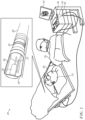
- FIG. 1 is a schematic illustration of a system 20 for performing a cardiac ablation, in accordance with some embodiments of the present invention.
- System 20 comprises a catheter 28, a proximal end of which is connected, via an electrical interface 40 (e.g., any suitable type of connector, jack, port, or plug), to a console 36, which comprises a processor 44.
- processor 44 receives, via electrical interface 40, electrical signals from catheter 28, processes these signals, and generates appropriate output in response thereto.
- catheter 28 is inserted by a physician 26 into the heart of a subject 24.
- the distal end 22 of catheter 28 comprises an ablation electrode 30, which is used to apply an ablating signal to cardiac tissue of subject 24.
- An ultrasound transducer 34 disposed at distal end 22 (e.g., inside ablation electrode 30), is used to transmit ultrasound signals, and receive reflections of the signals from the tissue. In response to the received reflections, ultrasound transducer 34 generates signals, which are received by processor 44.
- the ultrasound transducer is used for both (i) measuring tissue thickness prior to the ablation, in order to properly set the ablation parameters, and (ii) evaluate echogenic changes to the tissue caused by the ablation, in order to assess the outcome of the ablation.
- catheter 28 comprises a plurality of ultrasound transducers, which may be arranged within the ablation electrode in any suitable arrangement.
- the ultrasound transducers may be distributed around the circumference of the ablation electrode, at the distal tip of the ablation electrode and/or proximally to the distal tip.
- Such a plurality of ultrasound transducers may be used to transmit ultrasound signals from multiple locations, and/or in multiple directions, thus facilitating the performance of the techniques described herein.
- distal end 22 further comprises one or more temperature sensors 38, which may be used to record the temperature of the tissue during the ablation. Temperature sensors 38 generate signals indicative of the recorded temperatures, and communicate these signals to processor 44.
- Catheter 28 further comprises a force sensor 32, which may alternatively be referred to as a pressure sensor, at the distal end of the catheter.
- force sensor 32 operates as described in US Patent Application Publication 2009/0093806 .
- Force sensor 32 is configured to generate a signal that indicates both the magnitude and the direction of the mechanical force that is applied to the tissue by the distal end of the catheter.
- system 20 further comprises an electromagnetic tracking system, which tracks the position and orientation of the distal end of the catheter during the procedure, as described, for example, in US Patent 8,456,182 .
- console 36 further comprises a display 42, which displays appropriate output for the physician during the procedure.
- display 42 may show an electroanatomical map of the subject's heart, constructed, for example, using techniques described in US Patent 6,226,542 .
- display 42 may be driven by processor 44 to show output from the processing of signals received from ultrasound transducer 34 and force sensor 32, as described in detail hereinbelow.
- CARTO ® 3 System available from Biosense Webster, Inc., 3333 Diamond Canyon Road, Diamond Bar, CA 91765. This system may be modified by those skilled in the art to embody the principles of embodiments described herein.
- processor 44 may be embodied as a single processor, or a cooperatively networked or clustered set of processors.
- Processor 44 is typically a programmed digital computing device comprising a central processing unit (CPU), random access memory (RAM), non-volatile secondary storage, such as a hard drive or CD ROM drive, network interfaces, and/or peripheral devices.
- Program code, including software programs, and/or data are loaded into the RAM for execution and processing by the CPU and results are generated for display, output, transmittal, or storage, as is known in the art.
- the program code and/or data may be downloaded to the computer in electronic form, over a network, for example, or it may, alternatively or additionally, be provided and/or stored on non-transitory tangible media, such as magnetic, optical, or electronic memory.
- Such program code and/or data when provided to the processor, produce a machine or special-purpose computer, configured to perform the tasks described herein.
- FIG. 2 is a schematic illustration of a visual output 46 that includes a force signal 21 and an m-mode ultrasound image 23, which may be displayed on display 42 (e.g., overlaid over a displayed electroanatomical map) in accordance with some embodiments of the present invention.
- the physician presses the distal end of the catheter against the cardiac tissue.
- the force with which the catheter presses against the tissue varies.
- the physician may manually vary the contact force, and/or a linear actuator (not shown) incorporated into the distal end of the catheter may vary the contact force, as described, for example, in US Patent Application Publication 2016/0183915 . While the contact force varies, the ultrasound transducer transmits ultrasound signals into the tissue, and receives reflections of the signals from the tissue.
- Processor 44 receives, from force sensor 32, a force signal 21, which indicates the time-varying contact force "F" applied to the tissue.
- Processor 44 further receives, from the ultrasound transducer, one or more signals that are derived from ultrasound reflections received by the ultrasound transducer.
- Such signals from the ultrasound transducer may be used to generate an m-mode ultrasound image 23 of the tissue, which shows the portion 48 of tissue that is in front of the ultrasound transducer. Assuming a large-enough contrast between tissue portion 48 and the adjacent anatomical portion 50 of the subject, image 23 allows the thickness T of tissue portion 48 to be visualized.
- T is a function of the contact force F.
- the tissue becomes more compressed, and hence, the thickness decreases; conversely, as the force decreases, the tissue thickness increases.
- tissue thicknesses may be measured manually (e.g., by the physician) from image 23, and/or from the displayed electroanatomical map of the subject's heart.
- the thicknesses are obtained automatically by the processor, based on the times-of-flight of the ultrasound reflections received from the tissue.
- the processor may use techniques described in US Patent Application Publication 2016/0183915 to identify the reflections from the tissue interface 56 of interest.
- the aforementioned '915 publication describes correlating force signal 21 with the time-varying times-of-flight of the received reflections.
- the times-of-flight from tissue interface 56 will be highly correlated with force signal 21, while those from other tissue interfaces will be less correlated. That is, as the applied force increases, thus causing the distance from the ultrasound transducer to tissue interface 56 to decrease, the times-of-flight from tissue interface 56 will decrease, and vice versa, whereas other times-of-flight will be less correlated with force signal 21. Consequently, the processor may obtain the time-varying distance from the ultrasound transducer to tissue interface 56, which is the desired tissue thickness.
- This dependency - as learned, stored, and subsequently used by the processor - may be embodied in any suitable form, such as in the form of a lookup table of corresponding "T” and “F” values, and/or in the form of parameters derived by fitting a function to the acquired "T” and "F” values.
- the dependency of the tissue thickness on the contact force is learned for each of a plurality of portions of tissue. That is, as the physician moves the catheter along the tissue, force signal 21, and the signals based on the ultrasound reflections, are received, and are used to learn the dependency for each of the portions of tissue over which the catheter moves.
- the dependencies are first learned for a local area of an intended ablation site, an ablation is then performed at the local area, and then the catheter is moved to the next intended ablation site. In other embodiments, the learning is first performed for all of the intended ablation sites, and only afterwards are each of the sites ablated.
- force signal 21 and image 23 are typically displayed on display 42.
- force signal 21 typically includes both the magnitude and direction of the force
- the display of force signal 21 may indicate only the magnitude of the force, expressed, for example, in units of weight, such as grams.
- processor 44 may learn the dependency of the tissue thickness on the contact force, even without any signals or images being displayed on the display.
- FIG. 3 is a schematic illustration of a method for ascertaining thickness of tissue, in accordance with some embodiments of the present invention.
- distal end 22 of the catheter is perpendicular to the tissue during the acquisition of the force signal and the ultrasound reflections, the ultrasound reflections will be received from the portion of tissue to which force is being applied.
- the ultrasound transducer will face away from the portion of tissue that the catheter is pressing against, and consequently, the force signal and the ultrasound-reflection signals may not correspond to the same portion of tissue.
- the catheter contacts a first portion 52 of tissue, but ultrasound reflections are received from a second portion 54 of tissue.
- the processor is configured to determine, based on the directionality of the force signal, the portion of the tissue from which the ultrasound signals were received. (The directionality of the force signal is captured by the bending of force sensor 32, as depicted in Fig. 3 .) If the portion of tissue from which the ultrasound signals were received is not the currently-contacted portion of tissue, the processor selects, as the one or more ultrasound-reflection-based signals, signals derived from ultrasound reflections received during a different period of time.
- the ultrasound-reflection-based signals correspond to tissue portion 54, rather than to portion 52. Therefore, to learn the dependency for tissue portion 52, the processor selects ultrasound-reflection-based signals that correspond to tissue portion 52, these signals being acquired prior to or following the acquisition of the force signal for tissue portion 52. The processor similarly uses the correct set of signals to learn the dependency for tissue portion 54.
- T_54 may be calculated by multiplying the tissue thickness T_54', which is estimated based on the received ultrasound reflections, by the cosine of the appropriate angle theta ( ⁇ ), which may be ascertained from the force sensor.)
- ultrasound signals transmitted from ablation electrode 30 are generally transmitted from the ablation electrode in the direction of the central longitudinal axis 58 of the ablation electrode.
- a plurality of ultrasound transducers may be used to transmit ultrasound signals from multiple locations, and/or in multiple directions, such that, in the scenario depicted in Fig. 3 , T_52 may be measured, despite the ablation electrode not being perpendicular to the tissue.
- the learned dependency is used to set one or more ablation parameters for the ablation of the portion of tissue.
- processor 44 first estimates, using the learned dependency, the thickness of the portion of tissue that would result from the desired ablation contact force (which is typically in the range of 5-30 grams) being applied to the portion of tissue. Then, in response to the estimated thickness, the processor generates an output indicating at least one recommended parameter for the ablation. For example, the output may indicate a recommended power, and/or a recommended duration, of the ablating signal.
- the processor displays such an output on display 42, and the physician, in response to viewing the output on the display, then sets the ablation parameters accordingly.
- the processor alternatively or additionally to generating the output that indicates the recommended ablation parameter(s), the processor generates an output - e.g., a control signal directed to the generator that supplies the ablating signal - that automatically sets the parameter(s).
Landscapes
- Health & Medical Sciences (AREA)
- Life Sciences & Earth Sciences (AREA)
- Engineering & Computer Science (AREA)
- Surgery (AREA)
- Physics & Mathematics (AREA)
- Public Health (AREA)
- Medical Informatics (AREA)
- General Health & Medical Sciences (AREA)
- Biomedical Technology (AREA)
- Veterinary Medicine (AREA)
- Heart & Thoracic Surgery (AREA)
- Nuclear Medicine, Radiotherapy & Molecular Imaging (AREA)
- Molecular Biology (AREA)
- Animal Behavior & Ethology (AREA)
- Pathology (AREA)
- Biophysics (AREA)
- Radiology & Medical Imaging (AREA)
- Cardiology (AREA)
- Plasma & Fusion (AREA)
- Otolaryngology (AREA)
- Computer Vision & Pattern Recognition (AREA)
- Physiology (AREA)
- Acoustics & Sound (AREA)
- Data Mining & Analysis (AREA)
- Databases & Information Systems (AREA)
- Epidemiology (AREA)
- Primary Health Care (AREA)
- Ultra Sonic Daignosis Equipment (AREA)
- Surgical Instruments (AREA)
Claims (12)
- Einrichtung, umfassend:eine elektrische Schnittstelle (40);eine Konsole (36), umfassend einen Prozessor (44); und einen Katheter (28), umfassend an seinem distalen Ende (22) eine Ablationselektrode (30), einen Ultraschallwandler (34) und einen Kraftsensor (32);wobei das proximale Ende des Katheters (28) über die elektrische Schnittstelle (40) mit der Konsole (36) verbunden ist,wobei der Prozessor (44) konfiguriert ist, um über die elektrische Schnittstelle (40) zu empfangen:ein erstes Signal, von dem Kraftsensor (32), das eine zeitvariante Kontaktkraft angibt, die auf einen Abschnitt von Gewebe (48,52) ausgeübt wird, undein oder mehrere zweite Signale, von dem Ultraschallwandler (34), die aus den Ultraschallreflexionen abgeleitet sind, die von dem Abschnitt von Gewebe (48,52) empfangen werden; undwobei der Prozessor (44) ferner konfiguriert ist, um eine Dicke des Abschnitts von Gewebe (48,52) basierend auf den Laufzeiten der Ultraschallreflexionen zu erhalten, die von dem Abschnitt von Gewebe (48,52) empfangen werden, dadurch gekennzeichnet, dass der Prozessor (44) konfiguriert ist, um, unter Verwendung des ersten Signals und des zweiten Signals, die Dicke des Abschnitts von Gewebe (48,52) als eine Funktion der Kontaktkraft zu lernen, die auf den Abschnitt von Gewebe (48,52) ausgeübt wird.
- Einrichtung nach Anspruch 1, wobei die Funktion als Parameter ausgedrückt wird, die durch Anpassen der Funktion an das erste Signal und die zweiten Signale abgeleitet werden.
- Einrichtung nach Anspruch 1, wobei die Funktion als eine Nachschlagetabelle von entsprechenden Dicken- und Kraftwerten für den Abschnitt von Gewebe (48,52) ausgedrückt wird.
- Einrichtung nach einem der vorstehenden Ansprüche, wobei der Prozessor (44) ferner konfiguriert ist, um, basierend auf der erlernten Funktion, eine Ausgabe zu erzeugen, die mindestens einen empfohlenen Parameter für eine Ablation des Abschnitts von Gewebe (48,52) angibt, die mit einer bestimmten Kontaktkraft durchgeführt wird.
- Einrichtung nach Anspruch 4, wobei der Prozessor (44) konfiguriert ist, um die Ausgabe zu erzeugen durch:basierend auf der gelernten Funktion, Schätzen einer Dicke des Abschnitts von Gewebe (48,52), die sich aus der bestimmten Kontaktkraft ergeben würde, die während der Ablation des Abschnitts von Gewebe auf den Abschnitts von Gewebe (48,52) ausgeübt wird, undals Reaktion auf die geschätzte Dicke, Erzeugen der Ausgabe.
- Einrichtung nach Anspruch 4, wobei der empfohlene Parameter eine Leistung eines Ablationssignals einschließt.
- Einrichtung nach Anspruch 4, wobei der empfohlene Parameter eine Dauer eines Ablationssignals einschließt.
- Einrichtung nach einem der Ansprüche 1 bis 3, wobei der Prozessor (44) ferner konfiguriert ist, um, basierend auf der erlernten Funktion, mindestens einen Parameter für eine Ablation des Abschnitts von Gewebe (48,52) einzustellen.
- Einrichtung nach einem der Ansprüche 1 bis 3, wobei der Abschnitt von Gewebe ein Abschnitt von Herzgewebe ist.
- Einrichtung nach einem der Ansprüche 1 bis 3, wobei die zeitvariante Kraft durch ein distales Ende des Katheters auf den Abschnitt von Gewebe ausgeübt wird.
- Einrichtung nach Anspruch 10, wobei die Ultraschallreflexionen durch den Ultraschallwandler empfangen werden, der innerhalb des distalen Endes des Katheters angeordnet ist.
- Einrichtung nach einem der Ansprüche 1 bis 3, wobei der Prozessor (44) ferner konfiguriert ist:um, basierend auf einer Richtung der zeitvarianten Kontaktkraft während eines ersten Zeitraums, während der das erste Signal erfasst wird, sicherzustellen, dass Ultraschallreflexionen, die während des ersten Zeitraums empfangen werden, nicht von dem Abschnitt des Gewebes (48,52) reflektiert werden, undals Reaktion darauf, um, als das eine oder die mehreren zweiten Signale, ein oder mehrere Signale auszuwählen, die von Ultraschallreflexionen abgeleitet sind, die während eines zweiten Zeitraums empfangen werden, der sich von dem ersten Zeitraum unterscheidet.
Applications Claiming Priority (1)
| Application Number | Priority Date | Filing Date | Title |
|---|---|---|---|
| US15/202,979 US10646197B2 (en) | 2016-07-06 | 2016-07-06 | Ascertaining tissue thickness |
Publications (3)
| Publication Number | Publication Date |
|---|---|
| EP3266380A1 EP3266380A1 (de) | 2018-01-10 |
| EP3266380C0 EP3266380C0 (de) | 2024-12-25 |
| EP3266380B1 true EP3266380B1 (de) | 2024-12-25 |
Family
ID=59296739
Family Applications (1)
| Application Number | Title | Priority Date | Filing Date |
|---|---|---|---|
| EP17179741.8A Active EP3266380B1 (de) | 2016-07-06 | 2017-07-05 | Ermittlung von gewebedicke |
Country Status (7)
| Country | Link |
|---|---|
| US (1) | US10646197B2 (de) |
| EP (1) | EP3266380B1 (de) |
| JP (1) | JP7118603B2 (de) |
| CN (1) | CN107582101B (de) |
| AU (1) | AU2017203773A1 (de) |
| CA (1) | CA2972419A1 (de) |
| IL (1) | IL253144B (de) |
Families Citing this family (10)
| Publication number | Priority date | Publication date | Assignee | Title |
|---|---|---|---|---|
| US9763743B2 (en) | 2014-07-25 | 2017-09-19 | Arrinex, Inc. | Apparatus and method for treating rhinitis |
| US10646197B2 (en) * | 2016-07-06 | 2020-05-12 | Biosense Webster (Israel) Ltd. | Ascertaining tissue thickness |
| AU2018256964B2 (en) | 2017-04-28 | 2023-11-30 | Arrinex, Inc. | Systems and methods for locating blood vessels in the treatment of rhinitis |
| US11065058B2 (en) | 2018-05-23 | 2021-07-20 | Biosense Webster (Israel) Ltd. | Using a predetermined ablation-current profile |
| US20200178929A1 (en) * | 2018-12-07 | 2020-06-11 | Biosense Webster (Israel) Ltd. | Mapping endocardial sub-surface characteristics |
| US11642172B2 (en) * | 2019-03-05 | 2023-05-09 | Biosense Webster (Israel) Ltd. | Showing catheter in brain |
| US20220079499A1 (en) * | 2020-09-14 | 2022-03-17 | Biosense Webster (Israel) Ltd. | Identification of ablation gaps |
| EP4210616A4 (de) * | 2020-10-30 | 2024-10-30 | S4 Medical Corp | Elektromagnetische messung zur verwendung mit einer ablationsbehandlung |
| CN116744849A (zh) * | 2021-01-12 | 2023-09-12 | 波士顿科学医疗设备有限公司 | 微导管 |
| US20250325246A1 (en) * | 2022-04-27 | 2025-10-23 | Procept Biorobotics Corporation | Ultrasonic rod probe |
Family Cites Families (72)
| Publication number | Priority date | Publication date | Assignee | Title |
|---|---|---|---|---|
| US6301496B1 (en) | 1998-07-24 | 2001-10-09 | Biosense, Inc. | Vector mapping of three-dimensionally reconstructed intrabody organs and method of display |
| US6226542B1 (en) | 1998-07-24 | 2001-05-01 | Biosense, Inc. | Three-dimensional reconstruction of intrabody organs |
| US7137980B2 (en) * | 1998-10-23 | 2006-11-21 | Sherwood Services Ag | Method and system for controlling output of RF medical generator |
| US6892091B1 (en) | 2000-02-18 | 2005-05-10 | Biosense, Inc. | Catheter, method and apparatus for generating an electrical map of a chamber of the heart |
| US6554774B1 (en) | 2000-03-23 | 2003-04-29 | Tensys Medical, Inc. | Method and apparatus for assessing hemodynamic properties within the circulatory system of a living subject |
| CA2333224A1 (en) | 2001-01-31 | 2002-07-31 | University Technologies International Inc. | Non-invasive diagnostic method and apparatus for musculoskeletal systems |
| US6814733B2 (en) | 2002-01-31 | 2004-11-09 | Biosense, Inc. | Radio frequency pulmonary vein isolation |
| US6997924B2 (en) | 2002-09-17 | 2006-02-14 | Biosense Inc. | Laser pulmonary vein isolation |
| US7156816B2 (en) | 2002-11-26 | 2007-01-02 | Biosense, Inc. | Ultrasound pulmonary vein isolation |
| CN100443048C (zh) * | 2003-02-18 | 2008-12-17 | 松下电工株式会社 | 内脏脂肪计测装置、内脏脂肪计测方法、程序以及记录媒体 |
| CN100333694C (zh) * | 2003-11-17 | 2007-08-29 | 微星科技股份有限公司 | 超声波静脉检测装置及检测方法 |
| US8768026B2 (en) * | 2003-11-26 | 2014-07-01 | Hologic, Inc. | X-ray imaging with x-ray markers that provide adjunct information but preserve image quality |
| US8333764B2 (en) * | 2004-05-12 | 2012-12-18 | Medtronic, Inc. | Device and method for determining tissue thickness and creating cardiac ablation lesions |
| CN1663534A (zh) * | 2005-02-05 | 2005-09-07 | 黄晶 | 介入式超声组织硬度获取法及介入超声硬度检测仪 |
| US7517318B2 (en) | 2005-04-26 | 2009-04-14 | Biosense Webster, Inc. | Registration of electro-anatomical map with pre-acquired image using ultrasound |
| US10143398B2 (en) * | 2005-04-26 | 2018-12-04 | Biosense Webster, Inc. | Registration of ultrasound data with pre-acquired image |
| US7536218B2 (en) | 2005-07-15 | 2009-05-19 | Biosense Webster, Inc. | Hybrid magnetic-based and impedance-based position sensing |
| US8583220B2 (en) | 2005-08-02 | 2013-11-12 | Biosense Webster, Inc. | Standardization of catheter-based treatment for atrial fibrillation |
| US7877128B2 (en) | 2005-08-02 | 2011-01-25 | Biosense Webster, Inc. | Simulation of invasive procedures |
| US8657814B2 (en) * | 2005-08-22 | 2014-02-25 | Medtronic Ablation Frontiers Llc | User interface for tissue ablation system |
| US7756576B2 (en) | 2005-08-26 | 2010-07-13 | Biosense Webster, Inc. | Position sensing and detection of skin impedance |
| US7874987B2 (en) * | 2005-10-28 | 2011-01-25 | Biosense Webster, Inc. | Targets and methods for ultrasound catheter calibration |
| US7735349B2 (en) * | 2007-01-31 | 2010-06-15 | Biosense Websters, Inc. | Correlation of ultrasound images and gated position measurements |
| US7996057B2 (en) * | 2007-01-31 | 2011-08-09 | Biosense Webster, Inc. | Ultrasound catheter calibration with enhanced accuracy |
| US8317711B2 (en) | 2007-06-16 | 2012-11-27 | St. Jude Medical, Atrial Fibrillation Division, Inc. | Oscillating phased-array ultrasound imaging catheter system |
| US8357152B2 (en) | 2007-10-08 | 2013-01-22 | Biosense Webster (Israel), Ltd. | Catheter with pressure sensing |
| US10299753B2 (en) * | 2007-11-29 | 2019-05-28 | Biosense Webster, Inc. | Flashlight view of an anatomical structure |
| US8320711B2 (en) | 2007-12-05 | 2012-11-27 | Biosense Webster, Inc. | Anatomical modeling from a 3-D image and a surface mapping |
| AU2009246115A1 (en) | 2008-05-16 | 2009-11-19 | Drexel University | System and method for evaluating tissue |
| US8456182B2 (en) | 2008-09-30 | 2013-06-04 | Biosense Webster, Inc. | Current localization tracker |
| JP5025629B2 (ja) * | 2008-12-22 | 2012-09-12 | 株式会社東芝 | 超音波プローブ、プローブ着脱用移動台車、プローブ設置位置探索用移動台車、超音波プローブ着脱システム、および超音波プローブの取付方法 |
| US20110028848A1 (en) | 2009-07-31 | 2011-02-03 | Cem Shaquer | Methods and Apparatus for Detecting and Mapping Tissue Interfaces |
| US9907534B2 (en) | 2009-12-15 | 2018-03-06 | St. Jude Medical, Atrial Fibrillation Division, Inc. | Self-aiming directable acoustic transducer assembly for invasive medical device applications |
| US8758257B2 (en) * | 2009-12-24 | 2014-06-24 | Renzo Cecere | Instrument including a movement sensor for positioning an effective portion and method of using same |
| US8374670B2 (en) | 2010-01-22 | 2013-02-12 | Biosense Webster, Inc. | Catheter having a force sensing distal tip |
| BR112013000760A2 (pt) | 2010-07-25 | 2016-05-24 | Syneron Medical Ltd | método e aparelho para medir a espessura do tecido adiposo |
| US8731859B2 (en) * | 2010-10-07 | 2014-05-20 | Biosense Webster (Israel) Ltd. | Calibration system for a force-sensing catheter |
| US8801710B2 (en) * | 2010-12-07 | 2014-08-12 | Immersion Corporation | Electrosurgical sealing tool having haptic feedback |
| US20120165671A1 (en) * | 2010-12-27 | 2012-06-28 | Hill Anthony D | Identification of objects in ultrasound |
| US10524765B2 (en) * | 2010-12-27 | 2020-01-07 | St. Jude Medical, Atrial Fibrillation Division, Inc. | Refinement of an anatomical model using ultrasound |
| WO2012094308A1 (en) * | 2011-01-03 | 2012-07-12 | Massachusetts Institute Of Technology | Quantitative elastography |
| CN102103147B (zh) * | 2011-01-05 | 2012-04-18 | 王毅 | 超声自相关横向流速测量方法 |
| US8628473B2 (en) | 2011-04-13 | 2014-01-14 | St. Jude Medical, Inc. | Acoustic transducer for pulse-echo monitoring and control of thermally ablative lesioning in layered and nonlayered tissues, catheter contact monitoring, tissue thickness measurement and pre-pop warning |
| US8545408B2 (en) | 2011-05-23 | 2013-10-01 | St. Jude Medical, Inc. | Combination catheter for forward and side lesioning with acoustic lesion feedback capability |
| AU2012202857B2 (en) * | 2011-05-23 | 2014-10-30 | Biosense Webster (Israel), Ltd. | Monitoring tissue temperature while using an irrigated catheter |
| JP5930611B2 (ja) * | 2011-05-26 | 2016-06-08 | キヤノン株式会社 | 被検体情報取得装置 |
| JP5786166B2 (ja) * | 2011-07-19 | 2015-09-30 | 学校法人昭和大学 | 流水式超音波口腔洗浄装置 |
| CN103048098B (zh) * | 2011-10-17 | 2015-08-19 | 中国石油天然气集团公司 | 油管柱接头金属-金属密封面超声成像检查方法 |
| WO2013062039A1 (ja) * | 2011-10-27 | 2013-05-02 | オリンパスメディカルシステムズ株式会社 | 超音波観察装置 |
| JP5830614B2 (ja) * | 2012-01-31 | 2015-12-09 | ボストン サイエンティフィック サイムド,インコーポレイテッドBoston Scientific Scimed,Inc. | 超音波組織撮像のための流体に基づいた音響結合を有するアブレーションプローブ、および、アブレーションおよび超音波撮像システム |
| US9655595B2 (en) * | 2012-06-14 | 2017-05-23 | Arcitrax Inc. | System, method and device for prostate diagnosis and intervention |
| JP5368615B1 (ja) * | 2012-08-09 | 2013-12-18 | 国立大学法人 東京大学 | 超音波診断システム |
| US20140081262A1 (en) | 2012-09-20 | 2014-03-20 | Boston Scientific Scimed Inc. | Nearfield ultrasound echography mapping |
| US9615815B2 (en) | 2012-09-28 | 2017-04-11 | Clemson University Research Foundation | Devices that cooperate with ultrasound probes for muscoskeletal evaluations and related systems and methods |
| US11096741B2 (en) | 2012-10-10 | 2021-08-24 | Biosense Webster (Israel) Ltd. | Ablation power control based on contact force |
| EP2911589A1 (de) * | 2012-10-23 | 2015-09-02 | Koninklijke Philips N.V. | Vorrichtung zur bestimmung einer räumlichen konfiguration |
| US20140142438A1 (en) * | 2012-11-19 | 2014-05-22 | Biosense Webster (Israel), Ltd. | Using location and force measurements to estimate tissue thickness |
| CN104869899B (zh) | 2012-12-17 | 2017-10-27 | 皇家飞利浦有限公司 | 显微操纵器控制的局部视图与固定全面视图 |
| US9204820B2 (en) * | 2012-12-31 | 2015-12-08 | Biosense Webster (Israel) Ltd. | Catheter with combined position and pressure sensing structures |
| CN105163682B (zh) * | 2013-03-15 | 2018-06-01 | 圣犹达医疗用品电生理部门有限公司 | 力传感消融导管 |
| WO2014150376A1 (en) * | 2013-03-15 | 2014-09-25 | Muffin Incorporated | Internal ultrasound assembly fluid seal |
| JP2015016144A (ja) * | 2013-07-11 | 2015-01-29 | セイコーエプソン株式会社 | 超音波測定装置、超音波画像装置及び超音波測定方法 |
| US10114510B2 (en) * | 2013-12-23 | 2018-10-30 | Samsung Display Co., Ltd. | Apparatus and method for detecting surface shear force on a display device |
| JP6071101B1 (ja) | 2014-01-17 | 2017-02-01 | コーニンクレッカ フィリップス エヌ ヴェKoninklijke Philips N.V. | 被検者の骨を評価する超音波装置と方法 |
| JP6326833B2 (ja) * | 2014-01-31 | 2018-05-23 | セイコーエプソン株式会社 | 超音波デバイス、超音波デバイスの製造方法、プローブ、電子機器、超音波画像装置 |
| FI125492B (en) * | 2014-08-15 | 2015-10-30 | Teknologian Tutkimuskeskus Vtt Oy | pressure sensor |
| US20160081555A1 (en) * | 2014-09-18 | 2016-03-24 | Biosense Webster (Israel) Ltd. | Multi-range optical sensing |
| US9459201B2 (en) * | 2014-09-29 | 2016-10-04 | Zyomed Corp. | Systems and methods for noninvasive blood glucose and other analyte detection and measurement using collision computing |
| US10327734B2 (en) | 2014-12-30 | 2019-06-25 | Biosense Webster (Israel) Ltd. | Measurement of tissue thickness using ultrasound and force measurements |
| WO2016191870A1 (en) * | 2015-06-01 | 2016-12-08 | The Governors Of The University Of Alberta | Surface modeling of a segmented echogenic structure for detection and measurement of anatomical anomalies |
| CN105469547B (zh) * | 2015-11-19 | 2018-08-14 | 广东小天才科技有限公司 | 一种提示用户的方法及装置 |
| US10646197B2 (en) * | 2016-07-06 | 2020-05-12 | Biosense Webster (Israel) Ltd. | Ascertaining tissue thickness |
-
2016
- 2016-07-06 US US15/202,979 patent/US10646197B2/en active Active
-
2017
- 2017-06-05 AU AU2017203773A patent/AU2017203773A1/en not_active Abandoned
- 2017-06-22 IL IL253144A patent/IL253144B/en active IP Right Grant
- 2017-07-05 EP EP17179741.8A patent/EP3266380B1/de active Active
- 2017-07-05 JP JP2017131709A patent/JP7118603B2/ja active Active
- 2017-07-05 CA CA2972419A patent/CA2972419A1/en not_active Abandoned
- 2017-07-06 CN CN201710548827.XA patent/CN107582101B/zh active Active
Also Published As
| Publication number | Publication date |
|---|---|
| CA2972419A1 (en) | 2018-01-06 |
| CN107582101B (zh) | 2022-05-27 |
| JP2018000965A (ja) | 2018-01-11 |
| EP3266380C0 (de) | 2024-12-25 |
| CN107582101A (zh) | 2018-01-16 |
| US20180008229A1 (en) | 2018-01-11 |
| US10646197B2 (en) | 2020-05-12 |
| AU2017203773A1 (en) | 2018-01-25 |
| EP3266380A1 (de) | 2018-01-10 |
| IL253144B (en) | 2020-10-29 |
| JP7118603B2 (ja) | 2022-08-16 |
| IL253144A0 (en) | 2017-09-28 |
Similar Documents
| Publication | Publication Date | Title |
|---|---|---|
| EP3266380B1 (de) | Ermittlung von gewebedicke | |
| US10555672B2 (en) | Using location and force measurements to estimate tissue thickness | |
| EP3566646B1 (de) | Visualisierung des katheter-gewebe-kontakts durch kartenverzerrung | |
| EP3340918B1 (de) | Vorrichtung zur bestimmung eines bewegungsbezugs | |
| EP2401980B1 (de) | Druckfühlung für einen Katheter mit mehreren Armen | |
| US8523787B2 (en) | Detection of tenting | |
| US6106464A (en) | Apparatus and method for bone surface-based registration of physical space with tomographic images and for guiding an instrument relative to anatomical sites in the image | |
| EP2725984B1 (de) | Objektposenbasierte initialisierung eines ultraschallstrahlformers | |
| JP6537837B2 (ja) | 改良された心電図記録(ecg)グラフの表示 | |
| JP2008535560A (ja) | 身体ボリュームにおける誘導介入的医療デバイスのための3次元イメージング | |
| CN103220996A (zh) | 位置确定装置 | |
| CN105726065A (zh) | 使用超声测量和力测量来测量组织厚度 | |
| EP2854651B1 (de) | Pulmonale ultraschalltechniken für elastografie in der lunge | |
| EP3662841B1 (de) | Abbildung von endokardialen unteroberflächeneigenschaften | |
| JP7230003B2 (ja) | 対向するトランスデューサを使用するカテーテルプローブのナビゲーション方法及び装置 | |
| CN115398476A (zh) | 使用皮肤组织的超声测量值进行的解剖图像与位置跟踪系统的术前配准 | |
| EP3189785B1 (de) | Gewebetiefenschätzung unter verwendung von torgesteuerten ultraschall- und kraftmessungen | |
| AU2014221247B2 (en) | Detecting of tenting |
Legal Events
| Date | Code | Title | Description |
|---|---|---|---|
| PUAI | Public reference made under article 153(3) epc to a published international application that has entered the european phase |
Free format text: ORIGINAL CODE: 0009012 |
|
| STAA | Information on the status of an ep patent application or granted ep patent |
Free format text: STATUS: THE APPLICATION HAS BEEN PUBLISHED |
|
| AK | Designated contracting states |
Kind code of ref document: A1 Designated state(s): AL AT BE BG CH CY CZ DE DK EE ES FI FR GB GR HR HU IE IS IT LI LT LU LV MC MK MT NL NO PL PT RO RS SE SI SK SM TR |
|
| AX | Request for extension of the european patent |
Extension state: BA ME |
|
| STAA | Information on the status of an ep patent application or granted ep patent |
Free format text: STATUS: REQUEST FOR EXAMINATION WAS MADE |
|
| 17P | Request for examination filed |
Effective date: 20180608 |
|
| RBV | Designated contracting states (corrected) |
Designated state(s): AL AT BE BG CH CY CZ DE DK EE ES FI FR GB GR HR HU IE IS IT LI LT LU LV MC MK MT NL NO PL PT RO RS SE SI SK SM TR |
|
| STAA | Information on the status of an ep patent application or granted ep patent |
Free format text: STATUS: EXAMINATION IS IN PROGRESS |
|
| 17Q | First examination report despatched |
Effective date: 20191126 |
|
| RAP3 | Party data changed (applicant data changed or rights of an application transferred) |
Owner name: BIOSENSE WEBSTER (ISRAEL) LTD. |
|
| RAP3 | Party data changed (applicant data changed or rights of an application transferred) |
Owner name: BIOSENSE WEBSTER (ISRAEL) LTD. |
|
| REG | Reference to a national code |
Ref legal event code: R079 Ipc: A61B0008000000 Ref country code: DE Ref legal event code: R079 Ref document number: 602017086922 Country of ref document: DE Free format text: PREVIOUS MAIN CLASS: A61B0008120000 Ipc: A61B0008000000 |
|
| GRAP | Despatch of communication of intention to grant a patent |
Free format text: ORIGINAL CODE: EPIDOSNIGR1 |
|
| STAA | Information on the status of an ep patent application or granted ep patent |
Free format text: STATUS: GRANT OF PATENT IS INTENDED |
|
| RIC1 | Information provided on ipc code assigned before grant |
Ipc: G16H 50/30 20180101ALI20240320BHEP Ipc: A61B 8/12 20060101ALI20240320BHEP Ipc: A61B 8/08 20060101ALI20240320BHEP Ipc: A61B 8/00 20060101AFI20240320BHEP |
|
| INTG | Intention to grant announced |
Effective date: 20240411 |
|
| GRAJ | Information related to disapproval of communication of intention to grant by the applicant or resumption of examination proceedings by the epo deleted |
Free format text: ORIGINAL CODE: EPIDOSDIGR1 |
|
| STAA | Information on the status of an ep patent application or granted ep patent |
Free format text: STATUS: EXAMINATION IS IN PROGRESS |
|
| GRAP | Despatch of communication of intention to grant a patent |
Free format text: ORIGINAL CODE: EPIDOSNIGR1 |
|
| STAA | Information on the status of an ep patent application or granted ep patent |
Free format text: STATUS: GRANT OF PATENT IS INTENDED |
|
| INTC | Intention to grant announced (deleted) | ||
| INTG | Intention to grant announced |
Effective date: 20240912 |
|
| GRAS | Grant fee paid |
Free format text: ORIGINAL CODE: EPIDOSNIGR3 |
|
| GRAA | (expected) grant |
Free format text: ORIGINAL CODE: 0009210 |
|
| STAA | Information on the status of an ep patent application or granted ep patent |
Free format text: STATUS: THE PATENT HAS BEEN GRANTED |
|
| AK | Designated contracting states |
Kind code of ref document: B1 Designated state(s): AL AT BE BG CH CY CZ DE DK EE ES FI FR GB GR HR HU IE IS IT LI LT LU LV MC MK MT NL NO PL PT RO RS SE SI SK SM TR |
|
| REG | Reference to a national code |
Ref country code: GB Ref legal event code: FG4D |
|
| REG | Reference to a national code |
Ref country code: CH Ref legal event code: EP |
|
| REG | Reference to a national code |
Ref country code: DE Ref legal event code: R096 Ref document number: 602017086922 Country of ref document: DE |
|
| REG | Reference to a national code |
Ref country code: IE Ref legal event code: FG4D |
|
| U01 | Request for unitary effect filed |
Effective date: 20250113 |
|
| U07 | Unitary effect registered |
Designated state(s): AT BE BG DE DK EE FI FR IT LT LU LV MT NL PT RO SE SI Effective date: 20250120 |
|
| PG25 | Lapsed in a contracting state [announced via postgrant information from national office to epo] |
Ref country code: HR Free format text: LAPSE BECAUSE OF FAILURE TO SUBMIT A TRANSLATION OF THE DESCRIPTION OR TO PAY THE FEE WITHIN THE PRESCRIBED TIME-LIMIT Effective date: 20241225 |
|
| PG25 | Lapsed in a contracting state [announced via postgrant information from national office to epo] |
Ref country code: NO Free format text: LAPSE BECAUSE OF FAILURE TO SUBMIT A TRANSLATION OF THE DESCRIPTION OR TO PAY THE FEE WITHIN THE PRESCRIBED TIME-LIMIT Effective date: 20250325 |
|
| PG25 | Lapsed in a contracting state [announced via postgrant information from national office to epo] |
Ref country code: GR Free format text: LAPSE BECAUSE OF FAILURE TO SUBMIT A TRANSLATION OF THE DESCRIPTION OR TO PAY THE FEE WITHIN THE PRESCRIBED TIME-LIMIT Effective date: 20250326 |
|
| PG25 | Lapsed in a contracting state [announced via postgrant information from national office to epo] |
Ref country code: RS Free format text: LAPSE BECAUSE OF FAILURE TO SUBMIT A TRANSLATION OF THE DESCRIPTION OR TO PAY THE FEE WITHIN THE PRESCRIBED TIME-LIMIT Effective date: 20250325 |
|
| PG25 | Lapsed in a contracting state [announced via postgrant information from national office to epo] |
Ref country code: SM Free format text: LAPSE BECAUSE OF FAILURE TO SUBMIT A TRANSLATION OF THE DESCRIPTION OR TO PAY THE FEE WITHIN THE PRESCRIBED TIME-LIMIT Effective date: 20241225 |
|
| PG25 | Lapsed in a contracting state [announced via postgrant information from national office to epo] |
Ref country code: PL Free format text: LAPSE BECAUSE OF FAILURE TO SUBMIT A TRANSLATION OF THE DESCRIPTION OR TO PAY THE FEE WITHIN THE PRESCRIBED TIME-LIMIT Effective date: 20241225 |
|
| PG25 | Lapsed in a contracting state [announced via postgrant information from national office to epo] |
Ref country code: ES Free format text: LAPSE BECAUSE OF FAILURE TO SUBMIT A TRANSLATION OF THE DESCRIPTION OR TO PAY THE FEE WITHIN THE PRESCRIBED TIME-LIMIT Effective date: 20241225 |
|
| PGFP | Annual fee paid to national office [announced via postgrant information from national office to epo] |
Ref country code: GB Payment date: 20250529 Year of fee payment: 9 |
|
| PG25 | Lapsed in a contracting state [announced via postgrant information from national office to epo] |
Ref country code: IS Free format text: LAPSE BECAUSE OF FAILURE TO SUBMIT A TRANSLATION OF THE DESCRIPTION OR TO PAY THE FEE WITHIN THE PRESCRIBED TIME-LIMIT Effective date: 20250425 |
|
| U20 | Renewal fee for the european patent with unitary effect paid |
Year of fee payment: 9 Effective date: 20250606 |
|
| PG25 | Lapsed in a contracting state [announced via postgrant information from national office to epo] |
Ref country code: SK Free format text: LAPSE BECAUSE OF FAILURE TO SUBMIT A TRANSLATION OF THE DESCRIPTION OR TO PAY THE FEE WITHIN THE PRESCRIBED TIME-LIMIT Effective date: 20241225 |
|
| PG25 | Lapsed in a contracting state [announced via postgrant information from national office to epo] |
Ref country code: CZ Free format text: LAPSE BECAUSE OF FAILURE TO SUBMIT A TRANSLATION OF THE DESCRIPTION OR TO PAY THE FEE WITHIN THE PRESCRIBED TIME-LIMIT Effective date: 20241225 |
|
| PLBE | No opposition filed within time limit |
Free format text: ORIGINAL CODE: 0009261 |
|
| STAA | Information on the status of an ep patent application or granted ep patent |
Free format text: STATUS: NO OPPOSITION FILED WITHIN TIME LIMIT |
|
| 26N | No opposition filed |
Effective date: 20250926 |