EP3224360B1 - Method - Google Patents
Method Download PDFInfo
- Publication number
- EP3224360B1 EP3224360B1 EP15816204.0A EP15816204A EP3224360B1 EP 3224360 B1 EP3224360 B1 EP 3224360B1 EP 15816204 A EP15816204 A EP 15816204A EP 3224360 B1 EP3224360 B1 EP 3224360B1
- Authority
- EP
- European Patent Office
- Prior art keywords
- protein
- binding agent
- library
- binding
- target
- Prior art date
- Legal status (The legal status is an assumption and is not a legal conclusion. Google has not performed a legal analysis and makes no representation as to the accuracy of the status listed.)
- Active
Links
- 238000000034 method Methods 0.000 title claims description 178
- 108090000623 proteins and genes Proteins 0.000 claims description 244
- 102000004169 proteins and genes Human genes 0.000 claims description 240
- 239000011230 binding agent Substances 0.000 claims description 230
- 230000027455 binding Effects 0.000 claims description 151
- 238000009739 binding Methods 0.000 claims description 151
- 230000003993 interaction Effects 0.000 claims description 92
- 125000003729 nucleotide group Chemical group 0.000 claims description 78
- 239000002773 nucleotide Substances 0.000 claims description 77
- 108091028043 Nucleic acid sequence Proteins 0.000 claims description 35
- 150000001875 compounds Chemical class 0.000 claims description 31
- 230000003321 amplification Effects 0.000 claims description 27
- 238000003199 nucleic acid amplification method Methods 0.000 claims description 27
- 108091093088 Amplicon Proteins 0.000 claims description 24
- 239000000203 mixture Substances 0.000 claims description 23
- 239000000839 emulsion Substances 0.000 claims description 21
- 238000012163 sequencing technique Methods 0.000 claims description 21
- 239000007787 solid Substances 0.000 claims description 20
- 239000002299 complementary DNA Substances 0.000 claims description 19
- 238000002823 phage display Methods 0.000 claims description 19
- 238000006243 chemical reaction Methods 0.000 claims description 13
- 238000005304 joining Methods 0.000 claims description 13
- 239000000126 substance Substances 0.000 claims description 11
- 230000009870 specific binding Effects 0.000 claims description 9
- 238000012408 PCR amplification Methods 0.000 claims description 8
- 230000015572 biosynthetic process Effects 0.000 claims description 8
- 238000010790 dilution Methods 0.000 claims description 8
- 239000012895 dilution Substances 0.000 claims description 8
- 238000002955 isolation Methods 0.000 claims description 7
- 230000000295 complement effect Effects 0.000 claims description 5
- 238000009792 diffusion process Methods 0.000 claims description 4
- 108010052285 Membrane Proteins Proteins 0.000 claims description 2
- 108010089430 Phosphoproteins Proteins 0.000 claims description 2
- 102000007982 Phosphoproteins Human genes 0.000 claims description 2
- 238000000137 annealing Methods 0.000 claims description 2
- 108091005573 modified proteins Proteins 0.000 claims description 2
- 102000035118 modified proteins Human genes 0.000 claims description 2
- 235000018102 proteins Nutrition 0.000 description 214
- 230000004850 protein–protein interaction Effects 0.000 description 64
- 150000007523 nucleic acids Chemical group 0.000 description 50
- 102000039446 nucleic acids Human genes 0.000 description 43
- 108020004707 nucleic acids Proteins 0.000 description 43
- 238000001514 detection method Methods 0.000 description 40
- 239000000523 sample Substances 0.000 description 39
- 238000000926 separation method Methods 0.000 description 39
- 108010085220 Multiprotein Complexes Proteins 0.000 description 32
- 102000007474 Multiprotein Complexes Human genes 0.000 description 32
- 239000000427 antigen Substances 0.000 description 28
- 108091007433 antigens Proteins 0.000 description 28
- 102000036639 antigens Human genes 0.000 description 28
- 210000004027 cell Anatomy 0.000 description 27
- 239000012634 fragment Substances 0.000 description 26
- 238000003556 assay Methods 0.000 description 24
- 230000001413 cellular effect Effects 0.000 description 21
- 230000000694 effects Effects 0.000 description 21
- 238000007481 next generation sequencing Methods 0.000 description 21
- 239000000539 dimer Substances 0.000 description 20
- 108090000765 processed proteins & peptides Proteins 0.000 description 20
- 239000003795 chemical substances by application Substances 0.000 description 18
- 108020004414 DNA Proteins 0.000 description 17
- 230000008569 process Effects 0.000 description 17
- 108091023037 Aptamer Proteins 0.000 description 15
- 230000008045 co-localization Effects 0.000 description 14
- 230000002441 reversible effect Effects 0.000 description 14
- 238000001086 yeast two-hybrid system Methods 0.000 description 12
- 102000004196 processed proteins & peptides Human genes 0.000 description 11
- 238000005070 sampling Methods 0.000 description 11
- 238000000338 in vitro Methods 0.000 description 10
- 238000002493 microarray Methods 0.000 description 10
- 238000010381 tandem affinity purification Methods 0.000 description 10
- 108060003951 Immunoglobulin Proteins 0.000 description 9
- 238000002474 experimental method Methods 0.000 description 9
- 102000018358 immunoglobulin Human genes 0.000 description 9
- 230000006916 protein interaction Effects 0.000 description 9
- 240000004808 Saccharomyces cerevisiae Species 0.000 description 8
- 238000005516 engineering process Methods 0.000 description 8
- 230000004048 modification Effects 0.000 description 8
- 238000012986 modification Methods 0.000 description 8
- 238000010384 proximity ligation assay Methods 0.000 description 8
- 108090001008 Avidin Proteins 0.000 description 7
- 108010047041 Complementarity Determining Regions Proteins 0.000 description 7
- 238000004458 analytical method Methods 0.000 description 7
- 238000013459 approach Methods 0.000 description 7
- 238000004364 calculation method Methods 0.000 description 7
- 238000009826 distribution Methods 0.000 description 7
- 125000005647 linker group Chemical group 0.000 description 7
- 239000013598 vector Substances 0.000 description 7
- 108700026244 Open Reading Frames Proteins 0.000 description 6
- 208000037265 diseases, disorders, signs and symptoms Diseases 0.000 description 6
- 238000010494 dissociation reaction Methods 0.000 description 6
- 230000005593 dissociations Effects 0.000 description 6
- 230000004927 fusion Effects 0.000 description 6
- 230000004481 post-translational protein modification Effects 0.000 description 6
- 238000012360 testing method Methods 0.000 description 6
- 238000010396 two-hybrid screening Methods 0.000 description 6
- 241001515965 unidentified phage Species 0.000 description 6
- 108091034117 Oligonucleotide Proteins 0.000 description 5
- 108090000848 Ubiquitin Proteins 0.000 description 5
- 102000044159 Ubiquitin Human genes 0.000 description 5
- 230000001270 agonistic effect Effects 0.000 description 5
- 230000003042 antagnostic effect Effects 0.000 description 5
- 230000001580 bacterial effect Effects 0.000 description 5
- 239000011324 bead Substances 0.000 description 5
- 230000000875 corresponding effect Effects 0.000 description 5
- 230000018109 developmental process Effects 0.000 description 5
- 201000010099 disease Diseases 0.000 description 5
- 238000011304 droplet digital PCR Methods 0.000 description 5
- 229940079593 drug Drugs 0.000 description 5
- 239000003814 drug Substances 0.000 description 5
- 230000006870 function Effects 0.000 description 5
- 239000003446 ligand Substances 0.000 description 5
- 210000004962 mammalian cell Anatomy 0.000 description 5
- 238000005259 measurement Methods 0.000 description 5
- 229920001184 polypeptide Polymers 0.000 description 5
- 238000012216 screening Methods 0.000 description 5
- 238000005406 washing Methods 0.000 description 5
- YBJHBAHKTGYVGT-ZKWXMUAHSA-N (+)-Biotin Chemical compound N1C(=O)N[C@@H]2[C@H](CCCCC(=O)O)SC[C@@H]21 YBJHBAHKTGYVGT-ZKWXMUAHSA-N 0.000 description 4
- 108091032973 (ribonucleotides)n+m Proteins 0.000 description 4
- HEDRZPFGACZZDS-UHFFFAOYSA-N Chloroform Chemical compound ClC(Cl)Cl HEDRZPFGACZZDS-UHFFFAOYSA-N 0.000 description 4
- 101000825071 Homo sapiens Sclerostin domain-containing protein 1 Proteins 0.000 description 4
- 108700008625 Reporter Genes Proteins 0.000 description 4
- 102100022432 Sclerostin domain-containing protein 1 Human genes 0.000 description 4
- 230000008901 benefit Effects 0.000 description 4
- 238000001311 chemical methods and process Methods 0.000 description 4
- 238000010367 cloning Methods 0.000 description 4
- 230000009260 cross reactivity Effects 0.000 description 4
- 238000011161 development Methods 0.000 description 4
- 230000003100 immobilizing effect Effects 0.000 description 4
- 230000017730 intein-mediated protein splicing Effects 0.000 description 4
- 238000004519 manufacturing process Methods 0.000 description 4
- 238000004949 mass spectrometry Methods 0.000 description 4
- 125000004573 morpholin-4-yl group Chemical group N1(CCOCC1)* 0.000 description 4
- 238000003753 real-time PCR Methods 0.000 description 4
- 230000002829 reductive effect Effects 0.000 description 4
- 210000001519 tissue Anatomy 0.000 description 4
- 108020004635 Complementary DNA Proteins 0.000 description 3
- 239000004971 Cross linker Substances 0.000 description 3
- 102000004190 Enzymes Human genes 0.000 description 3
- 108090000790 Enzymes Proteins 0.000 description 3
- 241000588724 Escherichia coli Species 0.000 description 3
- 241001524679 Escherichia virus M13 Species 0.000 description 3
- 108010067902 Peptide Library Proteins 0.000 description 3
- 108091093037 Peptide nucleic acid Proteins 0.000 description 3
- 102000018120 Recombinases Human genes 0.000 description 3
- 108010091086 Recombinases Proteins 0.000 description 3
- 108060008539 Transglutaminase Proteins 0.000 description 3
- JLCPHMBAVCMARE-UHFFFAOYSA-N [3-[[3-[[3-[[3-[[3-[[3-[[3-[[3-[[3-[[3-[[3-[[5-(2-amino-6-oxo-1H-purin-9-yl)-3-[[3-[[3-[[3-[[3-[[3-[[5-(2-amino-6-oxo-1H-purin-9-yl)-3-[[5-(2-amino-6-oxo-1H-purin-9-yl)-3-hydroxyoxolan-2-yl]methoxy-hydroxyphosphoryl]oxyoxolan-2-yl]methoxy-hydroxyphosphoryl]oxy-5-(5-methyl-2,4-dioxopyrimidin-1-yl)oxolan-2-yl]methoxy-hydroxyphosphoryl]oxy-5-(6-aminopurin-9-yl)oxolan-2-yl]methoxy-hydroxyphosphoryl]oxy-5-(6-aminopurin-9-yl)oxolan-2-yl]methoxy-hydroxyphosphoryl]oxy-5-(6-aminopurin-9-yl)oxolan-2-yl]methoxy-hydroxyphosphoryl]oxy-5-(6-aminopurin-9-yl)oxolan-2-yl]methoxy-hydroxyphosphoryl]oxyoxolan-2-yl]methoxy-hydroxyphosphoryl]oxy-5-(5-methyl-2,4-dioxopyrimidin-1-yl)oxolan-2-yl]methoxy-hydroxyphosphoryl]oxy-5-(4-amino-2-oxopyrimidin-1-yl)oxolan-2-yl]methoxy-hydroxyphosphoryl]oxy-5-(5-methyl-2,4-dioxopyrimidin-1-yl)oxolan-2-yl]methoxy-hydroxyphosphoryl]oxy-5-(5-methyl-2,4-dioxopyrimidin-1-yl)oxolan-2-yl]methoxy-hydroxyphosphoryl]oxy-5-(6-aminopurin-9-yl)oxolan-2-yl]methoxy-hydroxyphosphoryl]oxy-5-(6-aminopurin-9-yl)oxolan-2-yl]methoxy-hydroxyphosphoryl]oxy-5-(4-amino-2-oxopyrimidin-1-yl)oxolan-2-yl]methoxy-hydroxyphosphoryl]oxy-5-(4-amino-2-oxopyrimidin-1-yl)oxolan-2-yl]methoxy-hydroxyphosphoryl]oxy-5-(4-amino-2-oxopyrimidin-1-yl)oxolan-2-yl]methoxy-hydroxyphosphoryl]oxy-5-(6-aminopurin-9-yl)oxolan-2-yl]methoxy-hydroxyphosphoryl]oxy-5-(4-amino-2-oxopyrimidin-1-yl)oxolan-2-yl]methyl [5-(6-aminopurin-9-yl)-2-(hydroxymethyl)oxolan-3-yl] hydrogen phosphate Polymers Cc1cn(C2CC(OP(O)(=O)OCC3OC(CC3OP(O)(=O)OCC3OC(CC3O)n3cnc4c3nc(N)[nH]c4=O)n3cnc4c3nc(N)[nH]c4=O)C(COP(O)(=O)OC3CC(OC3COP(O)(=O)OC3CC(OC3COP(O)(=O)OC3CC(OC3COP(O)(=O)OC3CC(OC3COP(O)(=O)OC3CC(OC3COP(O)(=O)OC3CC(OC3COP(O)(=O)OC3CC(OC3COP(O)(=O)OC3CC(OC3COP(O)(=O)OC3CC(OC3COP(O)(=O)OC3CC(OC3COP(O)(=O)OC3CC(OC3COP(O)(=O)OC3CC(OC3COP(O)(=O)OC3CC(OC3COP(O)(=O)OC3CC(OC3COP(O)(=O)OC3CC(OC3COP(O)(=O)OC3CC(OC3COP(O)(=O)OC3CC(OC3CO)n3cnc4c(N)ncnc34)n3ccc(N)nc3=O)n3cnc4c(N)ncnc34)n3ccc(N)nc3=O)n3ccc(N)nc3=O)n3ccc(N)nc3=O)n3cnc4c(N)ncnc34)n3cnc4c(N)ncnc34)n3cc(C)c(=O)[nH]c3=O)n3cc(C)c(=O)[nH]c3=O)n3ccc(N)nc3=O)n3cc(C)c(=O)[nH]c3=O)n3cnc4c3nc(N)[nH]c4=O)n3cnc4c(N)ncnc34)n3cnc4c(N)ncnc34)n3cnc4c(N)ncnc34)n3cnc4c(N)ncnc34)O2)c(=O)[nH]c1=O JLCPHMBAVCMARE-UHFFFAOYSA-N 0.000 description 3
- 239000002253 acid Substances 0.000 description 3
- 150000001413 amino acids Chemical group 0.000 description 3
- VYLDEYYOISNGST-UHFFFAOYSA-N bissulfosuccinimidyl suberate Chemical compound O=C1C(S(=O)(=O)O)CC(=O)N1OC(=O)CCCCCCC(=O)ON1C(=O)C(S(O)(=O)=O)CC1=O VYLDEYYOISNGST-UHFFFAOYSA-N 0.000 description 3
- 238000012512 characterization method Methods 0.000 description 3
- 239000003153 chemical reaction reagent Substances 0.000 description 3
- 230000002508 compound effect Effects 0.000 description 3
- 238000012790 confirmation Methods 0.000 description 3
- 230000009089 cytolysis Effects 0.000 description 3
- 229940088598 enzyme Drugs 0.000 description 3
- 238000000605 extraction Methods 0.000 description 3
- 229940072221 immunoglobulins Drugs 0.000 description 3
- 239000000463 material Substances 0.000 description 3
- 238000012261 overproduction Methods 0.000 description 3
- 238000004091 panning Methods 0.000 description 3
- 238000002360 preparation method Methods 0.000 description 3
- 238000003498 protein array Methods 0.000 description 3
- 230000009257 reactivity Effects 0.000 description 3
- 230000035945 sensitivity Effects 0.000 description 3
- 238000002198 surface plasmon resonance spectroscopy Methods 0.000 description 3
- 102000003601 transglutaminase Human genes 0.000 description 3
- QCVGEOXPDFCNHA-UHFFFAOYSA-N 5,5-dimethyl-2,4-dioxo-1,3-oxazolidine-3-carboxamide Chemical compound CC1(C)OC(=O)N(C(N)=O)C1=O QCVGEOXPDFCNHA-UHFFFAOYSA-N 0.000 description 2
- KDCGOANMDULRCW-UHFFFAOYSA-N 7H-purine Chemical compound N1=CNC2=NC=NC2=C1 KDCGOANMDULRCW-UHFFFAOYSA-N 0.000 description 2
- 208000023275 Autoimmune disease Diseases 0.000 description 2
- 241000894006 Bacteria Species 0.000 description 2
- 238000007400 DNA extraction Methods 0.000 description 2
- AHCYMLUZIRLXAA-SHYZEUOFSA-N Deoxyuridine 5'-triphosphate Chemical compound O1[C@H](COP(O)(=O)OP(O)(=O)OP(O)(O)=O)[C@@H](O)C[C@@H]1N1C(=O)NC(=O)C=C1 AHCYMLUZIRLXAA-SHYZEUOFSA-N 0.000 description 2
- 108010054477 Immunoglobulin Fab Fragments Proteins 0.000 description 2
- 102000001706 Immunoglobulin Fab Fragments Human genes 0.000 description 2
- 108010021625 Immunoglobulin Fragments Proteins 0.000 description 2
- 102000008394 Immunoglobulin Fragments Human genes 0.000 description 2
- 206010028980 Neoplasm Diseases 0.000 description 2
- 239000002033 PVDF binder Substances 0.000 description 2
- 108010076818 TEV protease Proteins 0.000 description 2
- 241000700605 Viruses Species 0.000 description 2
- 230000009471 action Effects 0.000 description 2
- 238000003491 array Methods 0.000 description 2
- 238000002819 bacterial display Methods 0.000 description 2
- 238000010378 bimolecular fluorescence complementation Methods 0.000 description 2
- 230000031018 biological processes and functions Effects 0.000 description 2
- 229960002685 biotin Drugs 0.000 description 2
- 235000020958 biotin Nutrition 0.000 description 2
- 239000011616 biotin Substances 0.000 description 2
- 210000001124 body fluid Anatomy 0.000 description 2
- 239000010839 body fluid Substances 0.000 description 2
- 239000000872 buffer Substances 0.000 description 2
- 201000011510 cancer Diseases 0.000 description 2
- 238000004113 cell culture Methods 0.000 description 2
- 238000000749 co-immunoprecipitation Methods 0.000 description 2
- 238000010276 construction Methods 0.000 description 2
- OPTASPLRGRRNAP-UHFFFAOYSA-N cytosine Chemical compound NC=1C=CNC(=O)N=1 OPTASPLRGRRNAP-UHFFFAOYSA-N 0.000 description 2
- 238000007847 digital PCR Methods 0.000 description 2
- 230000009977 dual effect Effects 0.000 description 2
- 238000010828 elution Methods 0.000 description 2
- 238000002866 fluorescence resonance energy transfer Methods 0.000 description 2
- 108020001507 fusion proteins Proteins 0.000 description 2
- 102000037865 fusion proteins Human genes 0.000 description 2
- 230000002068 genetic effect Effects 0.000 description 2
- UYTPUPDQBNUYGX-UHFFFAOYSA-N guanine Chemical compound O=C1NC(N)=NC2=C1N=CN2 UYTPUPDQBNUYGX-UHFFFAOYSA-N 0.000 description 2
- 238000010438 heat treatment Methods 0.000 description 2
- 238000005734 heterodimerization reaction Methods 0.000 description 2
- 238000009396 hybridization Methods 0.000 description 2
- 238000001727 in vivo Methods 0.000 description 2
- 150000002500 ions Chemical class 0.000 description 2
- 238000002372 labelling Methods 0.000 description 2
- 239000002960 lipid emulsion Substances 0.000 description 2
- 238000002824 mRNA display Methods 0.000 description 2
- 230000001404 mediated effect Effects 0.000 description 2
- 239000012528 membrane Substances 0.000 description 2
- 239000000178 monomer Substances 0.000 description 2
- 230000009871 nonspecific binding Effects 0.000 description 2
- 239000002245 particle Substances 0.000 description 2
- 239000008177 pharmaceutical agent Substances 0.000 description 2
- 230000010399 physical interaction Effects 0.000 description 2
- 230000004962 physiological condition Effects 0.000 description 2
- 239000004033 plastic Substances 0.000 description 2
- 229920002981 polyvinylidene fluoride Polymers 0.000 description 2
- 238000001556 precipitation Methods 0.000 description 2
- 239000002243 precursor Substances 0.000 description 2
- 230000001737 promoting effect Effects 0.000 description 2
- 230000004845 protein aggregation Effects 0.000 description 2
- 238000002818 protein evolution Methods 0.000 description 2
- 238000001742 protein purification Methods 0.000 description 2
- 238000003259 recombinant expression Methods 0.000 description 2
- 230000004044 response Effects 0.000 description 2
- 238000002702 ribosome display Methods 0.000 description 2
- 230000011664 signaling Effects 0.000 description 2
- 238000010561 standard procedure Methods 0.000 description 2
- 230000001225 therapeutic effect Effects 0.000 description 2
- RWQNBRDOKXIBIV-UHFFFAOYSA-N thymine Chemical compound CC1=CNC(=O)NC1=O RWQNBRDOKXIBIV-UHFFFAOYSA-N 0.000 description 2
- 230000009466 transformation Effects 0.000 description 2
- 238000010200 validation analysis Methods 0.000 description 2
- VOTJUWBJENROFB-UHFFFAOYSA-N 1-[3-[[3-(2,5-dioxo-3-sulfopyrrolidin-1-yl)oxy-3-oxopropyl]disulfanyl]propanoyloxy]-2,5-dioxopyrrolidine-3-sulfonic acid Chemical compound O=C1C(S(=O)(=O)O)CC(=O)N1OC(=O)CCSSCCC(=O)ON1C(=O)C(S(O)(=O)=O)CC1=O VOTJUWBJENROFB-UHFFFAOYSA-N 0.000 description 1
- VOXZDWNPVJITMN-ZBRFXRBCSA-N 17β-estradiol Chemical compound OC1=CC=C2[C@H]3CC[C@](C)([C@H](CC4)O)[C@@H]4[C@@H]3CCC2=C1 VOXZDWNPVJITMN-ZBRFXRBCSA-N 0.000 description 1
- YYDMSFVTLYEPOH-UHFFFAOYSA-N 2,5-dioxo-1-propanoyloxypyrrolidine-3-sulfonic acid Chemical compound CCC(=O)ON1C(=O)CC(S(O)(=O)=O)C1=O YYDMSFVTLYEPOH-UHFFFAOYSA-N 0.000 description 1
- LADFAOKPINUFBB-TWPNXFTKSA-N 5'-GGTTGGTGTGGTTGG-3' Chemical compound Cc1cn([C@H]2C[C@H](OP(O)(=O)OC[C@H]3O[C@H](C[C@@H]3OP(O)(=O)OC[C@H]3O[C@H](C[C@@H]3OP(O)(=O)OC[C@H]3O[C@H](C[C@@H]3OP(O)(=O)OC[C@H]3O[C@H](C[C@@H]3OP(O)(=O)OC[C@H]3O[C@H](C[C@@H]3OP(O)(=O)OC[C@H]3O[C@H](C[C@@H]3OP(O)(O)=O)n3cnc4c3nc(N)[nH]c4=O)n3cnc4c3nc(N)[nH]c4=O)n3cc(C)c(=O)[nH]c3=O)n3cc(C)c(=O)[nH]c3=O)n3cnc4c3nc(N)[nH]c4=O)n3cnc4c3nc(N)[nH]c4=O)[C@@H](COP(O)(=O)O[C@H]3C[C@@H](O[C@@H]3COP(O)(=O)O[C@H]3C[C@@H](O[C@@H]3COP(O)(=O)O[C@H]3C[C@@H](O[C@@H]3COP(O)(=O)O[C@H]3C[C@@H](O[C@@H]3COP(O)(=O)O[C@H]3C[C@@H](O[C@@H]3COP(O)(=O)O[C@H]3C[C@@H](O[C@@H]3COP(O)(=O)O[C@H]3C[C@@H](O[C@@H]3COP(O)(=O)O[C@H]3C[C@@H](O[C@@H]3CO)n3cnc4c3nc(N)[nH]c4=O)n3cnc4c3nc(N)[nH]c4=O)n3cc(C)c(=O)[nH]c3=O)n3cc(C)c(=O)[nH]c3=O)n3cnc4c3nc(N)[nH]c4=O)n3cnc4c3nc(N)[nH]c4=O)n3cc(C)c(=O)[nH]c3=O)n3cnc4c3nc(N)[nH]c4=O)O2)c(=O)[nH]c1=O LADFAOKPINUFBB-TWPNXFTKSA-N 0.000 description 1
- GFFGJBXGBJISGV-UHFFFAOYSA-N Adenine Chemical compound NC1=NC=NC2=C1N=CN2 GFFGJBXGBJISGV-UHFFFAOYSA-N 0.000 description 1
- 229930024421 Adenine Natural products 0.000 description 1
- 108010083359 Antigen Receptors Proteins 0.000 description 1
- 102000006306 Antigen Receptors Human genes 0.000 description 1
- 102000000584 Calmodulin Human genes 0.000 description 1
- 108010041952 Calmodulin Proteins 0.000 description 1
- 101710132601 Capsid protein Proteins 0.000 description 1
- 102000009016 Cholera Toxin Human genes 0.000 description 1
- 108010049048 Cholera Toxin Proteins 0.000 description 1
- 208000035473 Communicable disease Diseases 0.000 description 1
- 102000015792 Cyclin-Dependent Kinase 2 Human genes 0.000 description 1
- 108010024986 Cyclin-Dependent Kinase 2 Proteins 0.000 description 1
- 102000053602 DNA Human genes 0.000 description 1
- 238000000018 DNA microarray Methods 0.000 description 1
- 238000001712 DNA sequencing Methods 0.000 description 1
- 230000004568 DNA-binding Effects 0.000 description 1
- 108700022150 Designed Ankyrin Repeat Proteins Proteins 0.000 description 1
- 102100024746 Dihydrofolate reductase Human genes 0.000 description 1
- 238000002965 ELISA Methods 0.000 description 1
- 108010000912 Egg Proteins Proteins 0.000 description 1
- 102000002322 Egg Proteins Human genes 0.000 description 1
- 239000004593 Epoxy Substances 0.000 description 1
- 241000724791 Filamentous phage Species 0.000 description 1
- 102100039556 Galectin-4 Human genes 0.000 description 1
- 101000608765 Homo sapiens Galectin-4 Proteins 0.000 description 1
- 108090000144 Human Proteins Proteins 0.000 description 1
- 102000003839 Human Proteins Human genes 0.000 description 1
- 241000713772 Human immunodeficiency virus 1 Species 0.000 description 1
- 108010067060 Immunoglobulin Variable Region Proteins 0.000 description 1
- 102000017727 Immunoglobulin Variable Region Human genes 0.000 description 1
- ODKSFYDXXFIFQN-BYPYZUCNSA-N L-arginine Chemical compound OC(=O)[C@@H](N)CCCN=C(N)N ODKSFYDXXFIFQN-BYPYZUCNSA-N 0.000 description 1
- 229930064664 L-arginine Natural products 0.000 description 1
- 235000014852 L-arginine Nutrition 0.000 description 1
- ROHFNLRQFUQHCH-YFKPBYRVSA-N L-leucine Chemical compound CC(C)C[C@H](N)C(O)=O ROHFNLRQFUQHCH-YFKPBYRVSA-N 0.000 description 1
- ROHFNLRQFUQHCH-UHFFFAOYSA-N Leucine Natural products CC(C)CC(N)C(O)=O ROHFNLRQFUQHCH-UHFFFAOYSA-N 0.000 description 1
- 102000019298 Lipocalin Human genes 0.000 description 1
- 108050006654 Lipocalin Proteins 0.000 description 1
- YNAVUWVOSKDBBP-UHFFFAOYSA-N Morpholine Chemical group C1COCCN1 YNAVUWVOSKDBBP-UHFFFAOYSA-N 0.000 description 1
- 241001529936 Murinae Species 0.000 description 1
- 108010021466 Mutant Proteins Proteins 0.000 description 1
- 102000008300 Mutant Proteins Human genes 0.000 description 1
- 239000000020 Nitrocellulose Substances 0.000 description 1
- -1 Pb2+ Chemical class 0.000 description 1
- 239000004793 Polystyrene Substances 0.000 description 1
- 101710149951 Protein Tat Proteins 0.000 description 1
- 108010026552 Proteome Proteins 0.000 description 1
- CZPWVGJYEJSRLH-UHFFFAOYSA-N Pyrimidine Chemical compound C1=CN=CN=C1 CZPWVGJYEJSRLH-UHFFFAOYSA-N 0.000 description 1
- 102000044126 RNA-Binding Proteins Human genes 0.000 description 1
- 108700020471 RNA-Binding Proteins Proteins 0.000 description 1
- 108010003723 Single-Domain Antibodies Proteins 0.000 description 1
- 108010088160 Staphylococcal Protein A Proteins 0.000 description 1
- 239000006180 TBST buffer Substances 0.000 description 1
- 108010022394 Threonine synthase Proteins 0.000 description 1
- 108090000190 Thrombin Proteins 0.000 description 1
- 108091023040 Transcription factor Proteins 0.000 description 1
- 102000040945 Transcription factor Human genes 0.000 description 1
- 108700029229 Transcriptional Regulatory Elements Proteins 0.000 description 1
- 150000007513 acids Chemical class 0.000 description 1
- 230000006978 adaptation Effects 0.000 description 1
- 230000002730 additional effect Effects 0.000 description 1
- 229960000643 adenine Drugs 0.000 description 1
- 230000002411 adverse Effects 0.000 description 1
- 238000001042 affinity chromatography Methods 0.000 description 1
- 238000001261 affinity purification Methods 0.000 description 1
- 230000004075 alteration Effects 0.000 description 1
- AVKUERGKIZMTKX-NJBDSQKTSA-N ampicillin Chemical compound C1([C@@H](N)C(=O)N[C@H]2[C@H]3SC([C@@H](N3C2=O)C(O)=O)(C)C)=CC=CC=C1 AVKUERGKIZMTKX-NJBDSQKTSA-N 0.000 description 1
- 229960000723 ampicillin Drugs 0.000 description 1
- 239000012491 analyte Substances 0.000 description 1
- 230000000890 antigenic effect Effects 0.000 description 1
- 210000003719 b-lymphocyte Anatomy 0.000 description 1
- PERZMHJGZKHNGU-JGYWJTCASA-N bambermycin Chemical compound O([C@H]1[C@H](NC(C)=O)[C@@H](O)[C@@H]([C@H](O1)CO[C@H]1[C@@H]([C@@H](O)[C@H](O)[C@@H](CO)O1)O)O[C@@H]1O[C@@H]([C@H]([C@H](O)[C@H]1NC(C)=O)O[C@H]1[C@@H]([C@@H](O)[C@@H](O)[C@H](O1)C(=O)NC=1C(CCC=1O)=O)O)C)[C@H]1[C@@H](OP(O)(=O)OC[C@@H](OC\C=C(/C)CC\C=C\C(C)(C)CCC(=C)C\C=C(/C)CCC=C(C)C)C(O)=O)O[C@H](C(O)=O)[C@@](C)(O)[C@@H]1OC(N)=O PERZMHJGZKHNGU-JGYWJTCASA-N 0.000 description 1
- 102000005936 beta-Galactosidase Human genes 0.000 description 1
- 108010005774 beta-Galactosidase Proteins 0.000 description 1
- 230000033228 biological regulation Effects 0.000 description 1
- 210000004369 blood Anatomy 0.000 description 1
- 239000008280 blood Substances 0.000 description 1
- 229910052794 bromium Inorganic materials 0.000 description 1
- 210000004899 c-terminal region Anatomy 0.000 description 1
- 239000003054 catalyst Substances 0.000 description 1
- 230000008614 cellular interaction Effects 0.000 description 1
- 230000033077 cellular process Effects 0.000 description 1
- 210000001175 cerebrospinal fluid Anatomy 0.000 description 1
- 230000008859 change Effects 0.000 description 1
- MOOGIHKPTRJVRV-ZHVXJWHRSA-N chembl1814697 Chemical compound O=C1NC(=O)C(C)=CN1[C@@H]1O[C@H](COP(N)(=O)O[C@@H]2[C@H](O[C@H](C2)N2C(NC(=O)C(C)=C2)=O)COP(N)(=O)O[C@@H]2[C@H](O[C@H](C2)N2C3=C(C(NC(N)=N3)=O)N=C2)COP(N)(=O)O[C@@H]2[C@H](O[C@H](C2)N2C3=C(C(NC(N)=N3)=O)N=C2)COC(=O)CCCC[C@H]2[C@H]3NC(=O)N[C@H]3CS2)[C@@H](OP(N)(=O)OC[C@H]2O[C@H]([C@H](OP(N)(=O)OC[C@@H]3[C@H](C[C@@H](O3)N3C4=C(C(NC(N)=N4)=O)N=C3)OP(N)(=O)OC[C@@H]3[C@H](C[C@@H](O3)N3C(NC(=O)C(C)=C3)=O)OP(N)(=O)OC[C@@H]3[C@H](C[C@@H](O3)N3C4=C(C(NC(N)=N4)=O)N=C3)OP(N)(=O)OC[C@@H]3[C@H](C[C@@H](O3)N3C(NC(=O)C(C)=C3)=O)OP(N)(=O)OC[C@@H]3[C@H](C[C@@H](O3)N3C4=C(C(NC(N)=N4)=O)N=C3)OP(N)(=O)OC[C@@H]3[C@H](C[C@@H](O3)N3C4=C(C(NC(N)=N4)=O)N=C3)OP(N)(=O)OC[C@@H]3[C@H](C[C@@H](O3)N3C(NC(=O)C(C)=C3)=O)OP(N)(=O)OC[C@@H]3[C@H](C[C@@H](O3)N3C(NC(=O)C(C)=C3)=O)OP(N)(=O)OC[C@@H]3[C@H](C[C@@H](O3)N3C4=C(C(NC(N)=N4)=O)N=C3)OP(N)(=O)OC[C@@H]3[C@H](C[C@@H](O3)N3C4=C(C(NC(N)=N4)=O)N=C3)O)C2)N2C3=C(C(NC(N)=N3)=O)N=C2)C1 MOOGIHKPTRJVRV-ZHVXJWHRSA-N 0.000 description 1
- 238000010382 chemical cross-linking Methods 0.000 description 1
- 229910052801 chlorine Inorganic materials 0.000 description 1
- 238000002487 chromatin immunoprecipitation Methods 0.000 description 1
- 238000003776 cleavage reaction Methods 0.000 description 1
- 239000000470 constituent Substances 0.000 description 1
- 230000002596 correlated effect Effects 0.000 description 1
- 230000008878 coupling Effects 0.000 description 1
- 238000010168 coupling process Methods 0.000 description 1
- 238000005859 coupling reaction Methods 0.000 description 1
- 238000004132 cross linking Methods 0.000 description 1
- 239000003431 cross linking reagent Substances 0.000 description 1
- 230000002380 cytological effect Effects 0.000 description 1
- 229940104302 cytosine Drugs 0.000 description 1
- 238000013461 design Methods 0.000 description 1
- ANCLJVISBRWUTR-UHFFFAOYSA-N diaminophosphinic acid Chemical compound NP(N)(O)=O ANCLJVISBRWUTR-UHFFFAOYSA-N 0.000 description 1
- 102000004419 dihydrofolate reductase Human genes 0.000 description 1
- 108020001096 dihydrofolate reductase Proteins 0.000 description 1
- 238000006471 dimerization reaction Methods 0.000 description 1
- MGJYOHMBGJPESL-UHFFFAOYSA-L disodium;1-[8-(2,5-dioxo-3-sulfonatopyrrolidin-1-yl)oxy-8-oxooctanoyl]oxy-2,5-dioxopyrrolidine-3-sulfonate Chemical compound [Na+].[Na+].O=C1C(S(=O)(=O)[O-])CC(=O)N1OC(=O)CCCCCCC(=O)ON1C(=O)C(S([O-])(=O)=O)CC1=O MGJYOHMBGJPESL-UHFFFAOYSA-L 0.000 description 1
- 208000035475 disorder Diseases 0.000 description 1
- 238000009510 drug design Methods 0.000 description 1
- 230000008846 dynamic interplay Effects 0.000 description 1
- 235000014103 egg white Nutrition 0.000 description 1
- 210000000969 egg white Anatomy 0.000 description 1
- 230000002255 enzymatic effect Effects 0.000 description 1
- 210000003527 eukaryotic cell Anatomy 0.000 description 1
- 238000013401 experimental design Methods 0.000 description 1
- 238000001914 filtration Methods 0.000 description 1
- 239000000834 fixative Substances 0.000 description 1
- 238000005194 fractionation Methods 0.000 description 1
- 125000000524 functional group Chemical group 0.000 description 1
- 150000002243 furanoses Chemical group 0.000 description 1
- 238000001502 gel electrophoresis Methods 0.000 description 1
- 238000012248 genetic selection Methods 0.000 description 1
- 230000013595 glycosylation Effects 0.000 description 1
- 238000006206 glycosylation reaction Methods 0.000 description 1
- PCHJSUWPFVWCPO-UHFFFAOYSA-N gold Chemical compound [Au] PCHJSUWPFVWCPO-UHFFFAOYSA-N 0.000 description 1
- 239000010931 gold Substances 0.000 description 1
- 229910052737 gold Inorganic materials 0.000 description 1
- 230000012010 growth Effects 0.000 description 1
- 229910052736 halogen Inorganic materials 0.000 description 1
- 150000002367 halogens Chemical class 0.000 description 1
- 210000003783 haploid cell Anatomy 0.000 description 1
- 238000013537 high throughput screening Methods 0.000 description 1
- 238000013090 high-throughput technology Methods 0.000 description 1
- 230000002209 hydrophobic effect Effects 0.000 description 1
- 150000002463 imidates Chemical class 0.000 description 1
- 238000001114 immunoprecipitation Methods 0.000 description 1
- 230000006872 improvement Effects 0.000 description 1
- 238000000126 in silico method Methods 0.000 description 1
- 230000001524 infective effect Effects 0.000 description 1
- 238000012482 interaction analysis Methods 0.000 description 1
- 238000011835 investigation Methods 0.000 description 1
- 229910052740 iodine Inorganic materials 0.000 description 1
- 238000004255 ion exchange chromatography Methods 0.000 description 1
- 238000001155 isoelectric focusing Methods 0.000 description 1
- BPHPUYQFMNQIOC-NXRLNHOXSA-N isopropyl beta-D-thiogalactopyranoside Chemical compound CC(C)S[C@@H]1O[C@H](CO)[C@H](O)[C@H](O)[C@H]1O BPHPUYQFMNQIOC-NXRLNHOXSA-N 0.000 description 1
- 238000011173 large scale experimental method Methods 0.000 description 1
- 230000001665 lethal effect Effects 0.000 description 1
- 108020004999 messenger RNA Proteins 0.000 description 1
- 125000000956 methoxy group Chemical group [H]C([H])([H])O* 0.000 description 1
- MBAXWTVHCRPVFW-UHFFFAOYSA-N methyl 3-[(3-imino-3-methoxypropyl)disulfanyl]propanimidate Chemical compound COC(=N)CCSSCCC(=N)OC MBAXWTVHCRPVFW-UHFFFAOYSA-N 0.000 description 1
- 229920012128 methyl methacrylate acrylonitrile butadiene styrene Polymers 0.000 description 1
- 239000011325 microbead Substances 0.000 description 1
- 244000005700 microbiome Species 0.000 description 1
- 238000002156 mixing Methods 0.000 description 1
- 229940126619 mouse monoclonal antibody Drugs 0.000 description 1
- 239000002547 new drug Substances 0.000 description 1
- 229920001220 nitrocellulos Polymers 0.000 description 1
- 230000036961 partial effect Effects 0.000 description 1
- 230000001575 pathological effect Effects 0.000 description 1
- 230000000144 pharmacologic effect Effects 0.000 description 1
- 238000005191 phase separation Methods 0.000 description 1
- 238000000053 physical method Methods 0.000 description 1
- 230000008375 physiological alteration Effects 0.000 description 1
- 230000035479 physiological effects, processes and functions Effects 0.000 description 1
- 230000035790 physiological processes and functions Effects 0.000 description 1
- 229920002223 polystyrene Polymers 0.000 description 1
- 230000023603 positive regulation of transcription initiation, DNA-dependent Effects 0.000 description 1
- 230000001323 posttranslational effect Effects 0.000 description 1
- 238000012545 processing Methods 0.000 description 1
- 238000001498 protein fragment complementation assay Methods 0.000 description 1
- 239000003531 protein hydrolysate Substances 0.000 description 1
- 238000000734 protein sequencing Methods 0.000 description 1
- 230000016434 protein splicing Effects 0.000 description 1
- 238000010379 pull-down assay Methods 0.000 description 1
- 150000003230 pyrimidines Chemical class 0.000 description 1
- 238000011002 quantification Methods 0.000 description 1
- 238000011084 recovery Methods 0.000 description 1
- 230000009467 reduction Effects 0.000 description 1
- 238000012552 review Methods 0.000 description 1
- 210000003296 saliva Anatomy 0.000 description 1
- 230000007017 scission Effects 0.000 description 1
- 210000002966 serum Anatomy 0.000 description 1
- 150000003384 small molecules Chemical class 0.000 description 1
- 239000007790 solid phase Substances 0.000 description 1
- 241000894007 species Species 0.000 description 1
- 238000010972 statistical evaluation Methods 0.000 description 1
- 238000007619 statistical method Methods 0.000 description 1
- 239000000758 substrate Substances 0.000 description 1
- 238000000856 sucrose gradient centrifugation Methods 0.000 description 1
- 230000009897 systematic effect Effects 0.000 description 1
- 230000008685 targeting Effects 0.000 description 1
- 229960000278 theophylline Drugs 0.000 description 1
- ZFXYFBGIUFBOJW-UHFFFAOYSA-N theophylline Chemical compound O=C1N(C)C(=O)N(C)C2=C1NC=N2 ZFXYFBGIUFBOJW-UHFFFAOYSA-N 0.000 description 1
- 229960004072 thrombin Drugs 0.000 description 1
- 229940113082 thymine Drugs 0.000 description 1
- 230000001052 transient effect Effects 0.000 description 1
- 238000013519 translation Methods 0.000 description 1
- 238000011144 upstream manufacturing Methods 0.000 description 1
- 210000002700 urine Anatomy 0.000 description 1
- 238000012795 verification Methods 0.000 description 1
- 210000005253 yeast cell Anatomy 0.000 description 1
Images

Classifications
-
- C—CHEMISTRY; METALLURGY
- C40—COMBINATORIAL TECHNOLOGY
- C40B—COMBINATORIAL CHEMISTRY; LIBRARIES, e.g. CHEMICAL LIBRARIES
- C40B30/00—Methods of screening libraries
- C40B30/04—Methods of screening libraries by measuring the ability to specifically bind a target molecule, e.g. antibody-antigen binding, receptor-ligand binding
-
- G—PHYSICS
- G01—MEASURING; TESTING
- G01N—INVESTIGATING OR ANALYSING MATERIALS BY DETERMINING THEIR CHEMICAL OR PHYSICAL PROPERTIES
- G01N33/00—Investigating or analysing materials by specific methods not covered by groups G01N1/00 - G01N31/00
- G01N33/48—Biological material, e.g. blood, urine; Haemocytometers
- G01N33/50—Chemical analysis of biological material, e.g. blood, urine; Testing involving biospecific ligand binding methods; Immunological testing
- G01N33/68—Chemical analysis of biological material, e.g. blood, urine; Testing involving biospecific ligand binding methods; Immunological testing involving proteins, peptides or amino acids
- G01N33/6803—General methods of protein analysis not limited to specific proteins or families of proteins
- G01N33/6845—Methods of identifying protein-protein interactions in protein mixtures
-
- C—CHEMISTRY; METALLURGY
- C12—BIOCHEMISTRY; BEER; SPIRITS; WINE; VINEGAR; MICROBIOLOGY; ENZYMOLOGY; MUTATION OR GENETIC ENGINEERING
- C12N—MICROORGANISMS OR ENZYMES; COMPOSITIONS THEREOF; PROPAGATING, PRESERVING, OR MAINTAINING MICROORGANISMS; MUTATION OR GENETIC ENGINEERING; CULTURE MEDIA
- C12N15/00—Mutation or genetic engineering; DNA or RNA concerning genetic engineering, vectors, e.g. plasmids, or their isolation, preparation or purification; Use of hosts therefor
- C12N15/09—Recombinant DNA-technology
- C12N15/10—Processes for the isolation, preparation or purification of DNA or RNA
- C12N15/1034—Isolating an individual clone by screening libraries
- C12N15/1037—Screening libraries presented on the surface of microorganisms, e.g. phage display, E. coli display
-
- G—PHYSICS
- G01—MEASURING; TESTING
- G01N—INVESTIGATING OR ANALYSING MATERIALS BY DETERMINING THEIR CHEMICAL OR PHYSICAL PROPERTIES
- G01N2458/00—Labels used in chemical analysis of biological material
- G01N2458/10—Oligonucleotides as tagging agents for labelling antibodies
-
- G—PHYSICS
- G01—MEASURING; TESTING
- G01N—INVESTIGATING OR ANALYSING MATERIALS BY DETERMINING THEIR CHEMICAL OR PHYSICAL PROPERTIES
- G01N2500/00—Screening for compounds of potential therapeutic value
- G01N2500/04—Screening involving studying the effect of compounds C directly on molecule A (e.g. C are potential ligands for a receptor A, or potential substrates for an enzyme A)
Definitions
- the present invention relates generally methods and uses of kits for detecting binding interactions, in particular protein-protein interactions, and particularly to high throughput methods for labelling, analysing, detecting and measuring protein-protein interactions.
- Cellular architecture is defined by its complexes, the molecular machines that actually make a cell.
- Cell biology traditionally identifies proteins based on their individual actions as catalysts, signalling molecules, or building blocks of cells and microorganisms.
- the qualitative and quantitative characterization of complex protein-protein networks and the identification of major cell type specific interacting proteins are paramount to understanding the physiological processes and alterations of protein-protein interactions in a multitude of human diseases such as cancer, autoimmune diseases and other disorders.
- Detailed insights in protein-protein networks and the identification of disease-associated differences may lead to new ways for the rational design and development of specific drugs.
- the pattern of protein-protein interactions in a cell or tissue may also be used as a tool for molecular diagnostics.
- Proteins participate in complex interactions that represent the mechanistic foundation for much of the physiology and function of the cell. These protein-protein interactions are organized into extremelyly complex networks.
- the architecture of protein-protein interaction networks was proposed to be scale-free, with most of the proteins having only one or two connections but with relatively fewer 'hubs' possessing tens, hundreds or more links.
- the interaction networks are highly dynamic, allowing for rapid changes in the interactome, for example to external stimuli or even developmental processes. Interactions between core proteins and between two or more module proteins are likely to be mediated by domain-domain interactions. Interactions within and between attachment proteins are less likely to occur in this manner.
- the term 'sampling' is used for experimental designs where only a subset of the population is interrogated. Representative sampling is not common in the generation of protein interaction datasets, where sampling has often been guided by biological priorities. The 'coverage' summarizes which part of the total set of possible interactions has actually been tested. In light of current technologies, it is not valid to make inferences about the 'interactome', e.g. the set of all physical interactions that take place in a cell under the conditions being studied.
- peptide- and protein-library screening techniques such as the yeast two-hybrid strategy, which is a method to identify and clone genes for proteins that interact with a protein of interest; two-hybrid arrays, where large-scale experiments are carried out in a colony-array format, in which each yeast colony expresses a defined pair of 'bait' and 'prey' proteins that can be scored for reporter gene activity - indicating interaction - in an automated manner; phage display where a library of proteins is panned against a "bait” protein and affinity-purification/mass-spectrometry (AP-MS), especially to define all complexes in the cell (the 'complexome') and their constituent proteins ; and tandem affinity purification (TAP). TAP reveals interacting proteins as core, module, or attachment proteins, according to the frequency of their appearance in the various forms of that complex.
- yeast two-hybrid strategy is a method to identify and clone genes for proteins that interact with a protein
- the ideal method captures the information of interactome in a time and cost effective manner, enabling random sampling and high redundancy of sampling. It provides dynamic, original cellular context based, native protein-protein interaction based, and comprehensive, sufficiently large coverage of quantitative interaction data of even large, multi-unit protein complexes. It suppresses the effects of random variables, such as detecting of non-specific, accidentally interacting proteins. It, also, diminishes the effect of variables, which are any binding event related variables involved in the detection principle other than the original protein-protein interaction.
- Two-hybrid screens enable large scale interactome information generation.
- Affinity based methods especially those using mass spectrometry as the detection principle, generate a high amount of semi-quantitative interactome data, partly in the correct cellular context.
- they are influenced by random and binding (affinity) related variables. They detect accidental, non-specific binding events.
- affinity affinity
- To generate a random sampled, high coverage, comprehensive dataset would require a significant amount of time and expense, which compromises the benefit of its potential to detect the dynamic nature of interactome.
- FYI filtered yeast interactome
- Niro et al (Nuc.Acid Res. (2010) 38:9; e100 ) describe a method for determining a binding interaction between a phage display library of open reading frame fragments and transglutaminase (TG2). After two rounds of panning with TG2 the binding partners are sequenced with 454 sequencing.
- D'Angelo et al disclose a method for determining a binding interaction between a phage scFv library and two CDK2 proteins.
- a set of forward primers in combination with barcodes is used for linking and sequencing.
- Ravn et al disclose a method for determining a binding interaction between a phage scFv library and an anti-mouse monoclonal antibody and soluble biotinylated hIFN ⁇ .
- a set of forward primers in combination with barcodes is used for linking and sequencing.
- Ravn et al (Methods (2013) 60:1; 99-110 ) and WO2010/135558 disclose a method for determining a binding interaction between five phage scFv libraries and two antigens
- a method for determining a binding interaction between a phage scFv library and Ag85 is described by D'Angelo et al (MABS (2013) 6:1; 160-172 )
- WO2010/09589 discloses a method for determining binding interactions between antibodies and antigens by screening binding between e.g. phage antibody libraries against phage antigen libraries.
- Bowley et al (PNAS (2009) 105:5; 1380-1385 ) describes methods for determining binding interactions between antibodies and antigens by screening binding between combinatorial antibody libraries against eukaryotic antigen libraries.
- the present invention provides methods and uses of kits for detecting binding interactions, in particular protein-protein interactions at the cellular level.
- the methods and uses of can be used for simultaneously detecting all, or a subset of, interacting proteins in complex protein networks, preferably in the original context of cells.
- the methods and uses of provide dynamic, original cellular context based, native protein-protein interaction based, and comprehensive, sufficiently large coverage of quantitative and potentially kinetic interaction data of even large, multi-unit protein complexes.
- the methods described herein can be used for detecting protein-protein interactions using antibody display technology, using a plurality of antibody phages as the binding agents.
- the methods can also be used for detecting protein-protein interactions using aptamer technology, using a plurality of aptamers as binding agents.
- the complexity of the plurality of binding agents can be varied in wide ranges between a few binding agents to tens of thousands or hundreds of thousands or millions or tens of millions or hundreds of millions of binding agents.
- a complexity reduction method is devised (enrichment).
- More detailed interactions between target molecules can be identified and monitored. For example protein-protein interactions can be detected.
- the presence of two or more binding agents within a binding agent/target complex may indicate that two or more targets may be present within the complex. This indicates that the two or more targets may be interacting with, or bound to, each other.
- an identifiable part of the specific binding agent is known, for example the protein or nucleic acid sequence
- the targets can be identified.
- This method can be carried out using highly parallel PCR amplification by linking the identifiable nucleic acid sequences of bound displayed antibody phages i.e. those with predetermined binding characteristics e.g. with known epitope sequences, or known to bind to a specific molecule. This can be done preferably by emulsion PCR. This may be carried out at low protein complex concentrations, preferably in compartments.
- the interactions between targets e.g. protein-protein interactions can be detected by highly parallel PCR amplification, preferably using reduced complexity binding detection agents.
- the target-target e.g. protein-protein interaction information is gained by sequencing of the linked identifiable sequences, preferably by highly parallel DNA sequencing or by other sequence detection means. Varying the amount of input material e.g. the target, can be used to collect ligand binding kinetics data. In addition the method can be carried out in the presence and absence of compounds to determine whether the compounds have any effect on the target interaction, and whether this effect is agonistic or antagonistic.
- the method can also use protein display technology, displaying protein fragments of an organism and determining the binding characteristics of a multitude of displayed antibodies, each antibody having unique identifiable sequence information and each displayed protein fragments having identifiable sequence information.
- the identifiable sequence information for the displayed protein fragments is the sequence encoding the displayed amino acid sequence.
- the identity of the bound antibodies can be determined from the identifiable sequence information for each antibody-protein complex.
- the identity of the bound protein fragment, within each antibody-protein complex can be identified.
- the identity of the bound antibodies and the identity of the bound protein fragment can be determined from the linked identifiable sequence information for each antibody-protein complex.
- the binding, kinetic characteristics can also be determined using different amounts of the target e.g. proteins and binding agents such as, displayed proteins or display antibodies.
- the methods and compositions described herein may also be used to identify compounds which may agonize or antagonize such protein-protein interactions.
- the present disclosure provides methods and kits for detecting binding interactions with antagonistic (disrupting) or agonistic (promoting) compounds.
- the disclosure provides methods and kits for simultaneously detecting the binding interactions of antagonistic and/or agonistic compounds in complex protein networks, preferably in the original context of cells.
- the methods and kits provide original cellular context based, native protein-protein interaction based data, which is comprehensive, and has sufficiently large coverage of both quantitative and, potentially, kinetic interaction data, even for large, multi-unit protein complexes.
- the invention provides a method for determining a binding interaction between a binding agent and a target comprising
- the present invention describes methods of analysing and characterizing complex binding interactions, in particular protein-protein networks or interactomes.
- the method is based on the co-localization related identification of binding agents and optionally their targets, such as proteins, where information on the co-localization of binding agents and optionally their targets, in a plurality of compartments, are pair wise linked and translated to a nucleotide.
- the identity of the binding agent and optionally the targets may also be determined from the nucleotide sequence. This information can be revealed by sequencing.
- the present invention also describes methods of analysing and characterizing the effect of antagonistic (disrupting) or agonistic (promoting) compounds on target molecule interactions.
- the method is based on the identification of binding agents and their targets, such as proteins, in the presence and absence of the compound.
- the detection of complexes formed between the binding agents and their targets, and the identification of the binding agents is carried out, in a plurality of compartments, by pair wise linkage of unique identification sequences of the bound target specific binding agents which are then translated to a nucleotide.
- the alteration of the quantity of complexes and the identity of the binding agents and optionally the target involved can be revealed by sequencing.
- the binding agent is preferably an antibody, or an aptamer.
- the binding agent is a member of an antibody display library or a library of antibodies wherein each antibody is labelled with said unique nucleotide sequence.
- the target is also associated with a unique nucleotide sequence.
- the nucleotide sequence associated with a binding agent in the complex can be linked to a second nucleotide sequence associated with a second binding agent in the complex.
- This method can be used to identify a plurality of binding agents which bind to a single target.
- the target is a protein
- the method can identify antibodies which bind to different epitopes on the protein.
- the target can be a protein complex, and the method can identify a plurality of binding agents which bind to different proteins within the complex.
- the nucleotide sequence associated with one binding agent in the binding agent/target complex can be linked to a nucleotide sequence associated with a second binding agent in the binding agent/target complex.
- the identity of the binding agents is known (from the linked sequence), it may be possible to identify the components of the target, and thus, for example, the proteins in the target which naturally interact.
- the binding agent is an antibody with known binding characteristics, the protein bound by the antibody may be identified.
- the identity of the proteins within the target can be identified. This allows protein-protein interactions within the sample to be detected and identified.
- the method can be used to monitor the effect of a compound on the interaction.
- nucleotide sequence associated with a binding agent in the binding agent/target complex can be linked to a nucleotide sequence associated with a target within the binding agent/target complex.
- This method can be used to identify which binding agent interacts with which target. For example it can be used to identify which members of a binding agent library can form a complex with a known target. This information can be used to characterise the members of a binding agent library to gain binding characteristics information.
- the production of said random paired, linked nucleic acid products comprises utilising at least two pairs of PCR primers to amplify identical or non-identical amplicons; wherein the PCR primers at 5' end have sequence tags wherein amplification with tagged primers results in random paired, linked nucleic acid products. More preferably, amplification is emulsion PCR amplification and the production of said amplicons and random paired, linked nucleic acid products are parallel processes.
- said sequencing of said joined amplification products is a highly parallel sequencing method.
- the method of the present invention can be used to investigate the effect of a compound on the interaction between the binding agents and the targets or the interaction between two or more target molecules.
- the step of contacting the binding agent with a target can be carried out in the presence and absence of a compound, and results compared to determine whether the compound effects the binding interaction between the binding agent and the target or between the target molecules.
- This method can be utilised to identify potential pharmaceutical agents which can be used to treat medical diseases and conditions.
- the invention also provides the use of a kit for carrying out the method of the invention, the kit comprising
- the kit may further comprise a protein display library wherein each member of said library is associated with a unique nucleotide sequence.
- the ideal method captures information from the interactome in a time and cost effective manner, enabling random sampling and high redundancy of sampling.
- the method provides comprehensive coverage of quantitative interaction data, even for large, multi-unit protein complexes. This data is obtained in an original cellular context so can measure native protein-protein interactions, and can be used to detect dynamic interactions.
- the method suppresses the effects of random variables, such as detecting non-specific, accidentally interacting proteins. It also diminishes the effect of variables, which are any binding event related effect involved in the detection principle other than the original protein-protein interaction, for example self-binding or a specific binding.
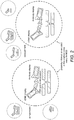
- FIG. 1 One embodiment of the present invention is summarized in Figure. 1 and the different components of the assay system are described in detail below.
- Figure. 2 A further embodiment of the present invention is summarized in Figure. 2 and the additional components of the system are described in detail below.
- the invention provides a method for determining a binding interaction between a binding agent and a target comprising
- the method can be carried out ex vivo, in vivo or in vitro.
- the binding agent is an antibody, aptamer, or based on an engineered protein scaffold.
- the binding agent may be a compound.
- the binding agent may be a member of an antibody display library or a library of antibodies wherein each antibody is labelled with a unique nucleotide sequence.
- the method may use displayed antibody agents as the binding agent, where the binding characteristics, for example, the target to which the binding agent binds is known and the unique nucleotide sequences associated with the plurality of displayed antibody agents are determined and the binding characteristics and unique nucleotide sequences are correlated with one another.
- the invention provides methods determining of the binding characteristics and relating these to the identifiable unique nucleotide sequence of the plurality of displayed antibody agents. This provides binding characteristic information.
- the binding agent used in the invention may be an antibody.
- antibody refers to immunoglobulin molecules and immunologically active portions of immunoglobulin molecules, i.e., molecules that contain an antigen binding site that specifically binds an antigen, whether natural or partly or wholly synthetically produced.
- antibody includes antibody fragments, derivatives, functional equivalents and homologues of antibodies, humanised antibodies, including any polypeptide comprising an immunoglobulin binding domain, whether natural or wholly or partially synthetic and any polypeptide or protein having a binding domain which is, or is homologous to, an antibody binding domain. Chimeric molecules comprising an immunoglobulin binding domain, or equivalent, fused to another polypeptide are therefore included.
- antibodies Cloning and expression of chimeric antibodies are described in EP-A-0120694 and EP-A-0125023 .
- Examples of antibodies are the immunoglobulin isotypes (e.g., IgG, IgE, IgM, IgD and IgA) and their isotypic subclasses; fragments which comprise an antigen binding domain such as Fab, scFv, Fv, dAb, Fd; and diabodies.
- Antibodies may be polyclonal or monoclonal.
- CDRs Complementarity determining regions
- immunoglobulins antibodies
- B-cells biologic response determining regions
- CDRs are part of the variable chains in immunoglobulins (antibodies), generated by B-cells, where these molecules bind to their specific antigen.
- CDRs are crucial to the diversity of antigen specificities generated by immunoglobulins.
- CDR1, CDR2 and CDR3 There are three CDRs (CDR1, CDR2 and CDR3), arranged non-consecutively, on the amino acid sequence of a variable domain of an immunoglobulin. Since the immunoglobulins are typically composed of two variable domains (on two different polypeptide chains, heavy and light chain), there are six CDRs for each antigen receptor that can collectively come into contact with the antigen.
- binding fragments are (i) the Fab fragment consisting of VL, VH, CL and CH1 domains; (ii) the Fd fragment consisting of the VH and CH1 domains; (iii) the Fv fragment consisting of the VL and VH domains of a single antibody; (iv) the dAb fragment ( Ward, E.S.
- an “antigen binding domain” is the part of an antibody which comprises the area which specifically binds to and is complementary to part or all of an antigen. Where an antigen is large, an antibody may only bind to a particular part of the antigen, which part is termed an epitope.
- An antigen binding domain may be provided by one or more antibody variable domains.
- An antigen binding domain may comprise an antibody light chain variable region (VL) and an antibody heavy chain variable region (VH).
- the binding agents may be based on engineered protein scaffolds.
- Protein scaffolds are derived from stable, soluble, natural protein structures which have been modified to provide a binding site for a target molecule of interest.
- engineered protein scaffolds include, but are not limited to, affibodies, which are based on the Z-domain of staphylococcal protein A that provides a binding interface on two of its a-helices ( Nygren, P. A. (2008). FEBS J 275(11): 2668-76 ); anticalins, derived from lipocalins, that incorporate binding sites for small ligands at the open end of a beta-barrel fold ( Skerra, A.
- Engineered protein scaffolds are typically targeted to bind the same antigenic proteins as antibodies.
- Short peptides may also be used to bind a target protein.
- Phylomers are natural structured peptides derived from bacterial genomes. Such peptides represent a diverse array of protein structural folds and can be used to inhibit/disrupt protein-protein interactions in vivo ( Watt, P. M. (2006). Nat Biotechnol 24(2): 177-83 )].
- the binding agent may be an aptamer.
- Aptamers are synthetic oligonucleotides (DNA or RNA) that recognize target molecules with high affinity and specificity through a combination of shape complementarity and non-covalent chemical bonds ( Blank & Blind, Current Opin. Chem. Biol., 2005, 9:336-342 ). These artificial ligands are quite easy to obtain in vitro and can be developed to recognise a large variety of different molecule classes which range from mere ions (e.g. Pb 2+ , Liu & Lu, 2003. J Am Chem Soc., 125, 6642-6643 ) to nucleotides, small molecules, proteins, viruses, and cells up to whole organisms ( encourager et al., 2006.
- High binding affinity aptamers have been selected through the well-known SELEX method ( Ellington & Szostak, 1990. Nature, 346, 818-822 ) for the detection of low molecular weight molecules like theophyllin ( Jenison et al., 1994. Science, 263, 1425-1429 ), L-arginine ( Geiger et al., 1996. Nucl. Acids Res., 24, 1029-1036 ), moenomycin ( Schuerer et al., 2001. Bioorg. Med. Chem., 92, 2557-2563 ), 17b-estradiol ( Kim et al., 2007. Biosens.
- Bioelectron., 22, 2525-2531 but also for larger molecules like thrombin (thrombin-binding aptamer:5'-GGTTGGTGTGGTTGG-3') ( Baldrich et al., Anal Chem. 2004, 76, 23,7053-63 ), cholera toxin or HIV-1 tat protein, among others (for review see Tombelli et al., 2007, Biomolec Eng., 24, 191-200 ).
- Some of the above mentioned aptamers have been used in ELISA-like assays on microplates or on the surface of biosensor transducers (QCM, SPR).
- An aptamer-modified AuNP colorimetric system has also been developed for the determination of the protein PDGF in a sandwich-based assay ( Huang et al., 2005, 77, 5735-5741 ).
- the binding agent may be part of a library, such as a displayed binding agent library, for example bacterial display, mRNA display, bacteriophage display, aptamer, ribosome display or yeast display libraries.
- a displayed binding agent library is an antibody bacteriophage display library.
- the library should be large enough so the library consists of a plurality of binding members which are expected to bind to at least 75% of the targets of interest within a target sample. More preferably the library is designed to bind to at least 80%, 85%, 90%, 95%, 97.5% or 99% of the targets of interest within a sample.
- the binding agent library comprises a plurality of binding members to protein or peptide sequences with 95% or higher coverage of expected or desired proteins within a sample.
- Each member of the library has a detectable, nucleic acid identity label, which is preferably unique to one member of the library.
- the unique nucleic acid identity labels are linked. "Linked" means the linking process has the potential to form random multimer nucleic acid products based on co-localization of these nucleic acid identity labels under suitable assay conditions.
- the multimeric product is a dimer.
- the suitable assay conditions include dismantling of bacteriophage particles, in separate compartments, for example by heat treatment in lipid emulsion, and specific consensus amplification of the unique sequences produce linkable amplicons. The joining of the linkable amplicons e.g.
- binding display specific nucleic acid domains form linked identity labels, which encodes the co-localisation information of the identity labels.
- the unique sequence is the binding display specific nucleic acid domains, for example the sequence which encodes one or more CDR regions.
- the joining reaction can be amplification based or involve other techniques.
- Amplification based joining can utilise two or more amplification primer pairs with identical binding abilities, but with complementary 5' tags or dimer linker sequences which result in the formation of polymerase extendable nucleic acid duplexes.
- the tags or dimer linker sequences mean that the sequence amplified by one primer pair will hybridise to sequences amplified by the second primer pair. The identity labels thereby become linked.
- Each member of the binding agent library is associated with a unique nucleotide sequence, which can be used to identify the binding agent.
- "Associated” as used herein means that the presence of the binding agent in the complex can be detected by the presence of the nucleic acid sequence within the linked sequence generated in the method.
- the nucleotide sequence may be attached as a label to the binding agent, be part of the binding agent itself e.g. aptamer, or be present within the binding agent e.g. nucleic acid within a phage.
- each member of the library can be labelled with unique nucleotide sequence.
- labelled refers to a nucleotide sequence which is attached to the member of the library.
- the unique nucleotide sequence can be the sequence which encodes one or more CDR regions or the displayed binding domain.
- a display library can be generated by inserting sequences encoding the amino acid sequence to be displayed into a phage at a known location. Universal primers that will amplify the inserted sequences can then be used and thus identify the binding sequence.
- the binding agent is an aptamer
- the aptamer itself can be the unique nucleotide sequence.
- the nucleotide sequence is an oligonucleotide and may comprise RNA or DNA, single or double stranded.
- Nucleotides used to label the binding agent or target are generally 5-150 bases in length, for example 10-40, or 20-30 bases in length.
- the nucleotides that form the nucleic acid can be chemically modified to increase the stability of the molecule, to improve its bioavailability or to confer additional activity on it.
- the pyrimidine bases may be modified at the 6 or 8 positions, and purine bases at the 5 position with CH3 or halogens such as I, Br or Cl.
- Modifications or pyrimidines bases also include 2 NH 3 , O 6 -CH 3 , N 6 -CH 3 and N 2 -CH 3 . Modifications at the 2'position are sugar modifications and include typically a NH 2 , F or OCH 3 group. Modifications can also include 3' and 5' modifications such as capping.
- modified nucleotides such as morpholino nucleotides, locked nucleic acids (LNA) and peptide nucleic acids (PNA) can be used.
- Morpholino oligonucleotides are assembled from different morpholino subunits, each of which contains one of the four genetic bases (adenine, cytosine, guanine, and thymine) linked to a 6-membered morpholine ring. The subunits are joined by non-ionic phosphorodiamidate intersubunit linkages to give a morpholino oligonucleotide.
- LNA monomers are characterised in that the furanose ring conformation is restricted by a methylene linker that connects the 2'-O position to the 4'-C position.
- PNA is an analogue of DNA in which the backbone is a pseudopeptide rather than a sugar.
- the binding agents are capable of detecting more than one target, preferably with different apparent affinities.
- the binding agents are capable of detecting a single target using different epitopes or binding sites, preferably with different apparent affinities.
- the binding characteristics of the members of the antibody phage library can be pre-determined. For example it can be determined which epitope is bound by the CDRs encoded and expressed binding agent (antibody) of the phage. This information can be associated with the unique nucleotide sequence which encodes the CDRs. Thus the epitope bound by the antibody expressed by the phage can be identified from the sequence of the unique nucleotide sequence. Once the epitope sequence present in the bound target is known, it may be possible to identify the protein or the group of proteins bound.
- Target as used herein is the molecule or group of molecules which forms a complex with the binding agent.
- the complex is usually formed under normal physiological conditions of the organism of interest.
- the target comprises a protein. More preferably the target is part of a protein sample.
- the protein sample may comprise a protein display library, preferably wherein each member of said library is associated with a unique nucleotide sequence.
- the protein display library is a cDNA phage display library.
- the target may be cross-linked to other targets within a plurality of targets e.g. a protein sample.
- a protein within a sample may be cross-linked to one or more other proteins within the sample.
- the target can be a known target. Binding agents which form a complex with the target can be identified, including compounds which interact with the target. Alternatively the target may be unknown, and the method of the invention is used to identify the target, or a plurality of target molecules which interact with one another.
- the target may be associated with a unique nucleotide sequence. "Associated" means that the presence of the target within the binding agent/target complex can be detected by the presence of the nucleic acid sequence within the linked sequence generated by the method.
- the nucleotide sequence may be attached as a label to the target, or be present within the target e.g. nucleic acid within a phage. Alternatively the nucleotide sequence may be part of an aptamer known to bind to the target.
- the binding agent/target complexes can be contacted with the aptamers to enable the target present to be identified through linkage of the unique nucleotide sequences, including the aptamer.
- the assay of the present invention can be applied to any protein sample.
- Proteins can be derived from any biological specimen including, but not limited to tissues, cytological specimens, body fluids, cell cultures or any other protein complex containing material.
- Body fluid samples include blood, saliva, urine, cerebrospinal fluid, or serum.
- the sample can be generated by recombinant expression methods.
- Preparation of proteins from specimens can be performed using standard methods known in the art.
- the specimen can be chemically treated before the extraction, e.g. different fixative chemicals or crosslinking agents can be used (e.g. BS3 - (bis(sulfosuccinimidyl)suberate).
- the protein sample can be crosslinked or not crosslinked.
- proteins can be produced, for example, by in vitro transcription-translation systems, or by recombinant expression systems. Depending on the experimental objective and the type of protein-protein interaction under investigation, proteins can be analysed either in their denatured or non-denatured form and/or crosslinked or not crosslinked form.
- the protein sample can be analysed in a plurality of conditions to collect information about the quantitative binding characteristics of plurality of protein-protein interactions. For example, the concentration or amount of the binding agent can be varied to determine dissociation constants and other kinetic parameters.
- the protein mixture can be preselected.
- the protein mixture can be an enrichment of specific proteins e.g. proteins from a specific cellular location, from a specific cell type, of a similar size or electrostatic charge, proteins with similar binding properties, similar sequence characteristics, or similar functions e.g. enzymes ( Current Protocols in Molecular Biology (2006) 20.0.1-20.0.6 CHAPTER 20 Analysis of Protein Interactions .).
- the specific proteins comprise phosphoproteins, membrane proteins or naturally, post-translational, artificially modified proteins.
- the proteins in the protein mixture of the method can be denatured or non-denatured and/or crosslinked or not crosslinked.
- the protein may be in the form of a protein display library.
- a protein display library comprises bacterial display, mRNA display, bacteriophage display, and ribosome display and yeast display libraries.
- the protein display library is a protein bacteriophage display library, more preferably a cDNA phage display library.
- the library should be large enough so that it consists of a plurality of peptide or protein members with at least 70% coverage of the proteins expected to be detected by the method in a sample. More preferably the library is large enough to provide 75%, 80%, 85%, 90%, 95%, 97.5%, 99% or higher coverage of the proteins or peptides in a sample.
- the display library provides coverage of any suitable biological entity e.g.
- Each member of the library is associated with a unique nucleotide sequence, i.e. each member has a unique detectable, nucleic acid identity labels. Preferably the unique nucleic acid identity labels are linked.
- Link means the linking process has the potential to form random multimer nucleic acid products based on co-localization of these nucleic acid identity labels under suitable assay conditions.
- the multimeric product is a dimer.
- the suitable assay conditions include dismantling of bacteriophage particles, preferably in separate compartments, for example by heat treatment in lipid emulsion, and specific consensus amplification of the unique sequences produce linkable amplicons.
- the joining of the linkable amplicons e.g. binding display specific nucleic acid domains, form linked identity labels, which encodes the co-localisation information of the identity labels.
- the joining reaction can be amplification based or involve other techniques.
- Amplification based joining can utilise two or more amplification primer pairs with identical binding abilities, but with complementary 5' tags or dimer linker sequences which result in the formation of polymerase extendable nucleic acid duplexes.
- the tags or dimer linker sequences mean that the sequence amplified by one primer pair will hybridise to sequences amplified by the second primer pair. The identity labels thereby become linked.
- the identity labels i.e. the associated unique nucleotide sequences used in the binding agent library and target library, such as the protein display library and the antibody library, may be different in their biological background, and so the amplification and joining process is based on two different primer pairs, e.g. one primer pair amplifies target sequences such as cDNA based identity labels and the second primer pair amplifies binding agent specific nucleotide sequences used as identity labels. Joining of the different labels makes it possible to link binding agent specific information to target information e.g. proteins encoded by displayed cDNAs. An example of this process is shown in Figure 2 .
- One binding agent preferably a displayed antibody phage
- a specific target e.g. a display protein target and the corresponding protein. This is termed specificity.
- a plurality of binding agents may recognize one target, such as a specific target e.g. display protein and the corresponding protein. This is termed redundancy.
- one binding agent can recognize more than one target, such as a protein species, based on the similarity of the target conformation due to, for example, protein conformation or protein sequences. This phenomenon is termed cross-reactivity.
- binding agent recognition of a target protein is based on conformation of protein or its protein sequence. This is known as its reactivity.
- Protein binding affinities of binding agents such as displayed binding agents can be calculated from the quantitative information of the sequencing datasets.
- the predetermined binding characteristics of the members of displayed binding agents may include reactivity and cross-reactivity with specificity and redundancy with calculated affinities.
- a varied concentration of the binding agent and/or target can be used to calculate quantitative parameters for the plurality of interactions, such as protein-protein interactions. Thus preferably the method is carried out using different concentrations of the binding agent and/or target.
- the quantitative nature of the detection enables the determination and calculation of the background, non-informative sequencing reads, produced by the non-specific co-localisation of identity labels or self-linking of the same identity labels.
- the detection of self-linking labels contains information about the quality of the datasets.
- enriched libraries can be used. Both the binding agent library and/or the target library can be enriched.
- the displayed targets, such as the proteins can be enriched to cover all potential binding partners in the experimental context.
- the displayed binding agents can be selected to have binding specificities enriched toward the detectable targets in the experimental context.
- the library may be limited to specific targets of interest. This may be done by panning based selection e.g. a target sample with controlled complexity is immobilised on solid surface and contacted to the binding agent library selecting the bound agents of the binding agent library by washing and subsequent elution of bound agents. Similarly, a binding agent sample with controlled complexity is immobilised on solid surface and contacted to the target library selecting the bound targets of the target library by washing and subsequent elution of bound targets.
- enrichment means the removal of self-binding displayed binding agents i.e removal of binding agents which bind to other binding agents under assay conditions. More preferably enrichment means reducing the complexity of the displayed binding agent libraries, but still ensuring it has high coverage for detection of targets in the experimental context. Reducing the complexity may involve reducing the total number of member within the library, eliminating members which bind to non-target proteins, or selecting only those members which bind to a target of interest.
- a binding agent library can be enriched by obtaining a protein mixture with components of interest e.g. studying known protein-protein interactions and validating dynamically their interactions or testing the effect of agonistic or antagonistic compounds. For example, immobilizing a target of interest e.g. protein mixture on a solid surface allows binding of the members of a binding agent library to the desired target e.g. protein. The binding agents bound to the target of interest can be separated from the unbound binding agents by dilution or other means. Bound members of the binding agent library can be eluted and used, as a reduced complexity library as described above, in the method of the invention.
- enriched binding agent libraries is generally advantageous as the separation methods (for example microarrays or emulsion based separations) have limited capacity to produce individual complexes within one compartment, so that linked identifiable sequences can be obtained.
- the reduced complexity of an enriched display agent libraries is directly translatable to the number of the separations that need to be carried out to obtain separation based, random paired, joined amplification products.
- the method of the invention is preferably carried out under physiological conditions so that the interactions can be detected in their original context i.e. in the same conditions as present in the cell. This provides information on the binding interactions that occur naturally.
- the step of contacting the binding agent library with the target is usually carried out in known buffer systems, from example in buffer systems that have already been used for studies of protein-protein interactions (e.g. TBST-buffer). Depending on the affinity, the reaction can be carried out at room temperature or 4°C. To get reproducible signals optimal time, optimal temperature and other assay conditions are determined including the steps of binding, washing and detection. Optimal conditions can be determined by the person skilled in the art.
- the binding agent/target complexes need to be separated, i.e. isolated from other complexes prior to linking the label sequences i.e. the associated unique nucleotide sequences.
- the separation is carried out by methods known in the art. For example separation can be carried out by dilution, specific binding, or separation by physical and/or chemical properties.
- the complexes are separated into compartments, such as emulsion droplets, micro-cavities etc., preferably diffusion limited or separated compartments.
- said separation comprises any one or more of solid surface binding, dilution or phase separation among the others, or providing diffusion limited or separated compartments.
- the separation limits the number of unbound binding agents in a compartment, to one, on average.
- the mean number of unbound binding agents in a compartment is one.
- the compartments may be individual droplets within an emulsion, or individual physical chambers such as microcavities.
- the complexes may be separated according to physical or chemical characteristics.
- said dilution is limited dilution.
- the compartmentalization (e.g. effective separation or isolation of large number reactions) is based on the Poisson distribution based separation of single unbound phages; e.g. emulsions and microarray are the best known state-of-art method.
- Separating the binding agent/target complexes sufficiently prior to further analysis will provide the circumstances where pairs of nucleic acid labels are generated, which are based on co-localization of binding agents. If the complexes are not separated sufficiently nucleic acid labels from members of different complexes will become linked and thus provide false information. Separation reduces the amount of non-specific co-localisations of nucleic acid identity labels and allows the identification of specific binding partners especially when complex protein mixtures are investigated. For example separation may result, on average, in a single unbound binding agent per compartment, where linking will provide only self-linked nucleotide sequences, consequently reducing the possibility of random linking between the members of the binding agent library.
- any agents is based on Poisson distribution the necessary measures to achieve suitable separation of agents can be calculated. This preferably results in a single complex within one compartment.
- an emulsion is used, which can be utilised in the amplification and linking of specific nucleic acid domains to form linked identity labels.
- Emulsion amplification methods are well known to the person skilled in the art, for example Schütze et al., Anal. Biochem. 2011 March 1; 410 (1):155-7 .
- the binding agent/target complex is immobilised following separation.
- the binding agent/target complexes can be captured onto a surface, for example as part of an array. This can help to maintain the separation between the complexes. Preferably separation of the complexes is maintained, for example, during the linking step.
- the binding agent /target complexes are optionally immobilized on solid support surfaces, including, but not limited to, membranes e.g. polyvinylidene fluoride (PVDF) or nitrocellulose, plastic surfaces (e.g. polystyrene) or can be covalently coupled to appropriate beads (e.g. Epoxy-activated beads).
- PVDF polyvinylidene fluoride
- plastic surfaces e.g. polystyrene
- appropriate beads e.g. Epoxy-activated beads.
- the binding or coupling to solid surfaces is performed by standard methods for proteins (" Antibodies, a Laboratory Manual.” Harlow, E., and Lane, D., eds. Cold Spring Harbor Press, Cold Spring Harbor N.Y., 1988 ) or by specific binding including antibodies or other specific binding interaction e.g. biotin- avidin.
- the immobilization may comprise (a) obtaining solid support with universal recognized protein complex binding ability i.e. the solid support is capable of binding all members of the binding agent library or target molecules.
- the amount of the recognized protein complexes and the number of available binding sites on the solid support should be balanced so as to achieve sufficient separation between bound recognized protein complexes.
- said solid support comprises a membrane, a plastic surface, or a bead. More preferably, the solid support is a bead and separation is achieved wherein on average one binding agent/target complex is bound to one bead. More preferably, the universal recognized protein complex binding ability is provided by an anti-bacteriophage antibody.
- separation comprises physical separation in reaction chambers or physical-chemical separation in droplets within an emulsion.
- the production of said random paired, linked nucleic acid products comprises utilising at least two pairs of PCR primers to amplify identical or non-identical amplicons; wherein the PCR primers at 5' end have sequence tags wherein amplification with tagged primers results in random paired, linked nucleic acid products.
- amplification is emulsion PCR amplification and the production of said amplicons and random paired, linked nucleic acid products are parallel processes.
- said sequencing of said joined amplification products is a highly parallel sequencing method. Separation allows the specific formation of linked identity labels, wherein molecule that are co-localized in separated or diffusion limited compartments (e.g.
- the linking process can provide a linked identity label with two identical identity labels e.g. where only one binding agent is present in the compartment. Similarly in the case of more than one binding agent recognizing a target, different linked identity labels can be produced with the same binding specificities.
- the linking step joins the identity label of one or more members of the binding agent library present. In addition the linking step may join the identity label of a member of a binding agent library to a nucleic acid sequence associated with the target.
- the linking process does not rely on the nucleic acids interacting with each other due to their close proximity e.g. by hybridising with each other.
- the sequences are joined together by using a linking process e.g. by amplification methods as described herein.
- the use of these methods instead of relying on close physical proximity ligation assays, such as those in US7306904 , allows multiple interactions to be detected in parallel.
- the labels do not need to be in close proximity to be linked and so detected. They simply need to be within the same compartment.
- the unique nucleotide sequences associated with members of the complex can be linked by a method comprising:
- Each pair of primers comprises a forward primer and a reverse primer.
- the sequences of these primers are designed so that they allow amplification of the identifiable sequences.
- these are universal primers i.e. they bind to all of the identifiable sequence within a library e.g. all the members of a binding agent library, or target library.
- the sequence between the primers which is amplified is unique to one member of the library, allowing identification.
- the pairs of PCR primers are designed to produce a dimerised linked nucleic acid sequence, otherwise multimer linked nucleic acid sequence is produced. This is achieved by having dimer linker sequences at the 5' end of at least two primers amplifying the member amplicons of the dimer or the multimer.
- these amplicons can form partially overlapping and polymerase extendable hybridisation products at their 3' ends. (for example as shown in Figure 2 )
- the amplification is emulsion PCR amplification with suitable separation of the nucleotide sequences associated with the binding agents, or binding agent and target.
- the linked nucleotide sequences from more than one complex are combined prior to identifying the linked binding agents and/or targets present.
- the identity of the binding agent(s) and/or target(s) can be determined, for example, by sequencing the linked nucleotide sequences. This can be carried out using a highly parallel system.
- the linked sequences can be combined so that a single reaction can be carried out to identify all the linked sequences. For example all of the linked nucleotides can be sequenced in a single reaction.
- the linked sequences can be determined quantitatively, to measure the relative abundances of the linked sequences.
- the co-localization information can be inferred from the linked identity label.
- the plurality of linked identity labels i.e. unique nucleotide sequences associated with the binding agent and optionally the target, in the form of the counts of sequencing reads, provides information of the identities of the members of binding agent/target complexes and their relative abundance. Other information is also inferable, wherein information of all linked identity labels is taken into account. Examples include but are not limited to, comparing relative affinities to predetermined affinities of the binding agents, comparing relative abundances calculated for different binding agents, determination of bound and not bound target or protein ratios.
- the sequence of all CDR regions can be determined using NGS sequencing.
- the predetermined binding characteristics of the binding agents are, preferably, based on a single CDR region sequence identity, in one preferred embodiment, the full sequence information of antibody binding agents can be associated with its predetermined binding characteristics.
- the method of the invention can comprise the step of contacting the binding agent library with a target in the presence or absence of a compound; to determine whether said compound effects the binding interaction.
- This method can be used to determine the effects of a compound or other chemical moieties on a plurality of binding interactions, for example protein-protein interactions.
- the method is used to detect compounds or chemical moieties which can promote or disrupt certain protein-protein or other detectable interactions wherein the compounds or chemical moieties act as drugs or eliminate or suppress the pathological consequences of such situations.
- such drugs can be used to treat different disease including, but not limited to, cancer, infectious diseases, autoimmune diseases and others.
- a compound as used herein refers to two or more atoms which are connected by covalent bonds.
- a chemical moiety is part of a compound, which forms a functional group.
- the compound may be a known pharmaceutical agent.
- the method of the invention can comprise the step of contacting the binding agent library with a target in the presence or absence of a compound; to determine whether said compound effects the binding interactions.
- the invention provides methods to determine interactome data.
- binding agents such as the displayed antibody agents are co-localized according to their binding characteristics i.e. two or more binding agents are present within one compartment as they bind the same target, or bind to targets which themselves interact or are bound to one another.
- Their identifiable sequences i.e. unique nucleotide sequences are linked carrying the co-localization information in the form of linked identities.
- information on both the protein-protein interactions and identities of proteins can be determined.
- the invention also describes a method for determining protein-protein interaction in a specimen obtained comprising:
- the protein-protein interaction data can be validated by statistical means, including background determination and subtraction of level of protein-protein interactions, determination of apparent relative affinities and relative abundances of proteins and protein-protein interactions.
- the unique nucleotide sequence of the binding agent allows the binding agent present to be identified, and so the predetermined protein binding characteristics of said binding agents is used to infer the presence of a protein-protein interaction in said protein mixture in said specimen.
- the determination of the effect of the compound is determined using high throughput experimental setup.
- a method for determining protein binding characteristics of the members of a binding agent library comprising:
- the protein binding characteristics of the members of the binding agent library can be determined from the information within the sequence linked nucleic acid products. Detection of linked unique nucleotide sequence associated with the members of displayed agent libraries and the protein indicates recognition and binding to certain members of the displayed protein library. The sequence can provide information as to which members of the library bind to which proteins.
- the protein binding characteristics of a plurality of said binding agents can be calculated. All the binding characteristics information for all members of said binding agent library can be combined, as binding information.
- the said binding agent libraries and/or targets e.g. protein samples
- the said binding agent libraries and/or targets are used in varied concentration in plurality of measurements to allow calculation of quantitative binding information including dissociation constants of said plurality of protein-protein interactions.
- the methods to calculate these measures are known to the person skilled in the art.
- the invention also relates to uses of kits in carrying out the methods of the invention.
- the kit comprises
- the kit may also comprise a means for detecting protein-protein interactions wherein reagents, and optionally materials are provided to carry out any one or more of the following steps: separation, immobilization, detecting unique nucleotide sequences, linking of nucleotide sequences, and/or detecting linked nucleotide sequences to gain said co-localization information.
- the kit may also further comprise instructions for carrying out the methods of the invention and utilising the kit.
- kits may further comprise a protein display library wherein each member of said library is associated with a unique nucleotide sequence.
- the method of the present invention is a novel approach to capture the information of the interactome in a time and cost effective manner, enabling random sampling and high redundancy of sampling. It provides dynamic, original cellular context based e.g. physiological, native protein-protein interaction based, and comprehensive coverage of quantitative interaction data of even large, multi-unit protein complexes. It suppresses the effects of random variables, such as detecting non-specific, accidentally interacting proteins. It, also, diminishes the effect of variables, such as any binding event related variables involved in the detection principle other than the original protein-protein interaction.
- the method is suitable for the in vitro detection of proteins that interact not only with the bait protein, but also with DNA, RNA and chemical compounds.
- PCR based detection of a proximity ligation assay is used to determine the relative expression of a target.
- PLA proximity ligation assay
- the assay is capable of detecting several targets in a given experiment. However, as it is necessary to prepare a large number of specific antibodies which are artificially labelled, it is infeasible at the interactome level. Without highly specific antibodies, cross-reactivity reduces the ability of the proximity ligation assay to clearly distinguish specific and non-specific interactions. The parallel detection of targets at very large numbers becomes costly and cumbersome.
- PLA proximity ligation assay
- PLA is described as an extremely sensitive and specific assay (4PLA) for detection of complex target structures such as microvesicles in which the target is first captured via an immobilized antibody and subsequently detected by using four other antibodies with attached DNA strands.
- 4PLA extremely sensitive and specific assay
- the low specificity is not an issue, and can even be used as validation information.
- the methods of invention use co-localization and compartmentalization, wherein the amplified identity labels freely diffuse in compartments enabling more relaxed steric conditions.
- Dual Expression Recombinase Based (DERB) destiny vectors individually encode two sets of recombinase recognizable sequences for inserting the protein open reading frame (ORF) of interest, two sets of promoters and reporter tags in frame with the ORFs for detecting interactions.
- ORF protein open reading frame
- Introduction of the vectors into living cells enables the detection of protein interactions by fluorescence resonance energy transfer (FRET) or bimolecular fluorescence complementation (BiFC).
- FRET fluorescence resonance energy transfer
- BiFC bimolecular fluorescence complementation
- the DERB platform shows advantages over current commercialized systems by introducing recombinase based cloning and compatible accepting vectors validated through proof-of-principle experiments and the identification of an unknown interaction.
- the system needs large numbers of screened interactions and consequently large efforts and costs, which suits only robotic systems at the interactome level and using artificial testing conditions (fusion proteins and artificial promoters).
- the yeast two-hybrid (Y2H) screen is specific implementation of protein-fragment complementation assay, or PCA, where the identification of protein-protein interactions is based on two protein fragments, each covalently linked to incomplete fragments of a third protein (e.g. DHFR, which acts as a reporter). Interaction between the proteins brings the fragments of the reporter protein in close enough proximity to allow them to form a functional reporter protein whose activity can be measured.
- This principle can be applied to many different reporter proteins, as the yeast two-hybrid screen using GAL4 transcription factor.
- the yeast two-hybrid screen investigates the interaction between artificial fusion proteins inside the nucleus of yeast. The method has a high false-positive rate, which makes it necessary to verify the identified interactions by other means.
- the method is out of cellular context and the lack of natural environment limits its use for interactions governed by cellular context specific modifications of the protein or in the case of low affinity interactions.
- the interactome level it requires further optimisation and the use of arrays, to make it possible to construct the very large interactome datasets. This involves high costs, but still do not overcome all the limitations of this assay type.
- a dual bait system can be used which improves the accuracy of library screens with an immediate selection to eliminate false positives.
- a coiled-coil mediated heterodimerization functional interaction trap assay has been described, where coiled-coil heterodimerization domains are substituted for modular protein binding domains. This can be useful for validating functionally relevant protein-protein interactions, directing enzymes to specific substrates, and screening fusion libraries for functionally important interaction partners.
- M2H mammalian cell based two hybrid
- a variation of detecting protein-protein interactions in Y2H screens is to use a specific PCR based sequencing method, termed Stitch-seq.
- This is PCR stitching, which places a pair of sequences encoding interacting proteins on the same PCR amplicon.
- PCR stitching consists of two rounds of PCR. In the first round, X and Y (present on the Y2H DB-X and AD-Y vectors) are amplified with DB- and AD vector-specific upstream primers, respectively. Amplicons from the first round as templates used to produce a concatenated PCR product composed of X and Y ORFs connected by an 82-bp linker sequence. The PCR products are pooled and sequenced by next-generation DNA sequencing to produce stitched ISTs (sISTs). Stitch-seq has removed the bottleneck of some Y2H protocols, but still does not solve the problems associated with key steps.
- a patent describing the following improvements to the PCA 1) reporter genes (and methods for detecting their expression) that readily permit the analysis of large libraries (>10 7 in size) and whose selectivity can be easily “tuned,” modified, and/or monitored, 2) methods for the simultaneous and independent measurement of multiple interactions (as judged by expression of different reporter genes), and 3) construction of libraries using a phagemid-based system that provides a) an efficient, automatable method for performing library vs. library experiments and b) a method to simplify the analysis of positive candidates from any screen/selection performed in the prokaryotic PCA.
- the use of this phagemid-based technology to screen library vs. library involves crossing libraries e.g.
- Two leucine zipper libraries semi-randomized at the positions adjacent to the hydrophobic core, were genetically fused to either one of two designed fragments of the enzyme murine dihydrofolate reductase (mDHFR), and cotransformed into E. coli. Interaction between the library polypeptides was required for reconstitution of the enzymatic activity of mDHFR, allowing bacterial growth. This strategy however is limited by the transformation efficiency that can be achieved in bacterial cells.
- mDHFR murine dihydrofolate reductase
- the Tandem affinity purification (TAP) method can be seen as a more specific version of co-immunoprecipitation, allows the high-throughput identification of proteins interactions.
- the accuracy of the method can be compared to those of small-scale co-immunoprecipitation experiments and the interactions are detected within the correct cellular context.
- the TAP method applies the fusion of the TAP tag to the C-terminus of the protein under study.
- the TAP tag consists of calmodulin binding peptide (CBP) from the N-terminal, followed by tobacco etch virus protease (TEV protease) cleavage site and Protein A. It is capable to provide the real determination of protein partners quantitatively in the correct cellular context, however the method, at the interactome level, requires large effort and would incur large costs to cover the entire proteome with the used constructs.
- the tagless strategy was developed to perform systematic, highly extensive biochemical fractionation of the soluble human protein interactome using multiple separation techniques including nondenaturing high-performance multibed ion-exchange chromatography, sucrose gradient centrifugation and isoelectric focusing.
- the method needs verification and statistical analysis to produce reliable datasets, also needs significant amount a sample material and its cost limits its usage for random sampling and high redundancy of sampling.
- Protein microarrays have introduced a new approach to identify and characterize protein interactions, providing the ability to rapidly identify new interactions between thousands of proteins in a single experiment. Since the location and identity of each protein on the array is known, interaction maps can be developed rapidly from iterative probing of protein arrays. Because a protein microarray experiment is performed within a day, and interactions are assessed in the context of thousands of other proteins, interaction profiling on microarrays can greatly accelerate the rate at which novel protein interactions are discovered. Additionally, the in vitro nature of protein microarray experiments permits control over probing conditions that affect protein interactions such as protein concentration, post-translational modifications, and presence of cofactors, which may not be possible with other methods such as yeast two-hybrid screening. However, the classical one probe at a time approach is not suitable for large interactome level experiments.
- a version of protein array based detection is used, where cellular protein lysate or synthetic peptide mixes is applied to the protein array with immobilized bait protein/ peptide.
- the nonspecific proteins/peptides are washed off under various stringent conditions and only the proteins specifically interacting with the bait protein/peptide remain on the chip.
- the captured interacting protein/peptide complexes are then analyzed by SELDI-TOF mass spectrometry and their identities are confirmed by their predicted distinctive masses. It is highly promising approach, however protein sequencing by SELDI-TOF is limited by several factors (amount of protein, separation, post-translational modifications) and the interactions lack the natural context.
- the protein mixture can also be immobilised on a solid support, and contacted with a plurality of unlabelled protein-protein interaction domains under appropriate binding conditions. In the presence of at least one labelled selected protein-protein interaction domain, (the labelled protein-protein interaction domain being different from the unlabelled protein-protein interaction domains) the binding of the labelled protein-protein interaction domain is measured. This method is interaction domain specific, which limits its application.
- a cell-free display technology combined with next generation sequencing (NGS) can improve both the coverage and reliability of interactome datasets.
- the completely cell-free method gives a high-throughput and a large detection space, testing the interactions without using clones.
- the quantitative information provided by NGS reduces the number of false positives.
- the method is suitable for the in vitro detection of proteins that interact not only with the bait protein, but also with DNA, RNA and chemical compounds.
- the method employs a complete in vitro treatment with cDNA libraries (extracted from cells and tissues) and selection on target proteins to gain selected cDNA sequences for NGS. Selections using the method are conducted under cell-free conditions, and subsequent sequencing by NGS is not limited by cloning steps using any kind of cells. This method applies one at time approach in out of cellular context, which limits its ability to generate large datasets at the interactome level.
- inteins are peptide sequences capable of directing protein trans-splicing in vitro.
- An intein is an intervening protein sequence in a protein precursor that is excised from the protein precursor during protein splicing.
- Two hybrid fusion constructs are provided, where one has a first test agent and an N-terminal intein fragment or N-intein, and the other has a second test agent and a C-terminal intein fragment or C-intein.
- one or both fusion constructs may have a reporter that undergoes detectable changes upon trans-splicing of the fusion constructs. Both the throughput and the cellular context free features are significant disadvantages of this method.
- Phage display is an enrichment process that requires multiple cycles to infer protein-protein interaction data. The enrichments are performed in vitro, which biases the interactions and favour high affinity interaction in detection. Certain proteins (particularly larger ones) are not well suited for analysis by phage display.
- a major disadvantage of bacterium-based ORF phage display is that proteins displayed on phage surface lack appropriate post-translational modifications, such as glycosylation.
- Phage display as used in the art, is limited to reproducing natural binding events, as described above.
- antibody repertoires expressed as phage display are extremely successful at providing pharmacological leads for therapeutic antibodies or detection antibodies for diagnostic assays.
- the present invention applied the phage display technology in the latter context, where the extremely large phage antibody libraries contain plurality of specificities covering almost any identifiable targets.
- a variation of the phage display relates to a method for the selection and identification of interaction partners.
- Target molecules ligands
- ligands are immobilized on the surface of a solid phase carrier such that they are position addressable in a two-dimensional grade and contacted to protein display viruses.
- the interaction partner is identified by detection and determination of the position of the binding between the immobilized ligand and the interaction partner.
- the preferred detection method described is surface plasmon resonance (SPR).
- crosslinking is often used to "fix" protein interactions in place before trying to isolate/identify interacting proteins.
- Common cross-linkers for this application include the non-cleavable [NHS-ester] cross-linker, [bis-sulfosuccinimidyl suberate] (BS3); a cleavable version of BS3, [dithiobis(sulfosuccinimidyl propionate)](DTSSP); and the [imidoester] crosslinker [dimethyl dithiobispropionimidate] (DTBP) that is popular for fixing interactions in ChIP assays.
- BS3 non-cleavable [NHS-ester] cross-linker
- DTSSP [dithiobis(sulfosuccinimidyl propionate)
- DTBP [imidoester] crosslinker [dimethyl dithiobispropionimidate]
- a technology has been developed for identifying proteins that specifically bind predicted transcriptional regulatory elements using phage-display library of genomically encoded peptides, which bind to a surface immobilized double-stranded DNA, containing a DNA motif sequence of interest. After enrichment for a specific DNA-protein interaction, the bound phages are amplified, and the inserts from the enriched phage are sequenced to determine the interacting proteins using labelling and hybridization to DNA microarray.
- One or more analytes have been reportedly measured using a chemFET array.
- the array may include any of a variety of chemical substances that provide relevant information regarding a chemical process or chemical processes of interest including binding of an antibody to an antigen.
- the ability to measure levels or concentrations of one or more analytes, in addition to merely detecting the presence of an analyte provides valuable information in connection with the chemical process or processes.
- the present invention provides a powerful, versatile, in vitro system for detecting and characterizing protein-protein interactions, and for selecting compounds capable of modulating protein-protein interactions.
- the system can be used with great convenience and can be easily adapted to high-throughput screening procedures.
- pBluescript II SK(+) phagemid vector (Agilent, 212205) f1 origin in (+) orientation, Sac-->Kpn polylinker orientation in host strain: XL1-Blue MRF' have been used to generate pBluescript II SK(+) phage and M13KO7 Helper Phage (NEB, N0315S) was purchased. The phages were titrated to determine the number of infective phages. The phages were diluted serially by factor of 10 to achieve lower than single phage per compartment dilution. The dilutions was subjected to digital PCR using QX200 Droplet Digital PCR (ddPCRTM) System.
- ddPCRTM Droplet Digital PCR
- 5'CTCAAGTCGGTGACGGTGAT3' (M13KO7 specific forward), 5'GACAAAAGGGCGACATTCAA3' (M13KO7 specific reverse) and/or 5'TCTTGATCCGGCAAACAAAC3' (pBluescript II SK(+) specific forward), 5'TTTTCTGCGCGTAATCTGCT3' (pBluescript II SK(+) specific reverse) with the probes 5'CTGGTAGCGGTGGTTTTT3' (pBluescript II SK(+) specific probe FAM-MGB labeled), 5'CCGTCAATATTTACCTTCCC3' (M13KO7 specific probe VIC-MGB labeled) were used to amplify the compartmentalized phages, the amplification were recorded in two different channels and the droplet generation , PCR and detection were according to the manufacturer protocol.
- pBluescript II SK(+) phagemid vector (Agilent, 212205) f1 origin in (+) orientation, Sac-->Kpn polylinker orientation in host strain: XL1-Blue MRF' have been used to generate pBluescript II SK(+) phage and M13KO7 Helper Phage (NEB, N0315S) was purchased. Phages was labeled with 20 molar excess of biotin according to the manufacturer instruction using EZ-Link Sulfo-NHS-Biotin (Thermo, 21326) and precipitated using isoelectric point precipitation as described ( Dong D, Sutaria S, Hwangbo JY, Chen P.
- Antibody displaying M13 phages were purchased from Source BioScience (6001_hDAb) including a phagemid antibody library (-3x10 9) ( Dudgeon K, Famm K, Christ D. Sequence determinants of protein aggregation in human VH domains. Protein Eng Des Sel. 2008 Oct 28 .) KM13 helper phage, TG1Tr bacterial strain and anti-beta-galactosidase & anti-bovine ubiquitin antibodies display phages.
- the control phages were sequenced by NGS (ThermoFisher Scientific PGM, Ion XpressTM Plus Fragment Library Kit, 4471269) and their binding affinities were verified by ELISA against the respective antigens.
- the antigens are the mono-biotinylated ubiquitin (b-UBI) from LifeSensors (#S1280), beta-galactosidase biotin labeled (G5025) (b-BGAL). Avidin from egg white (A9275) is also from Sigma-Aldrich.
- Control phage specific probes are also designed: anti-beta-galactosidase - FAM GCTAGGGCTATGTATCC MGB; anti-bovine ubiquitin - VIC TGGGTCGATGTTTGACTAC MGB.
- Avidin (36 nM) (or it was omitted), b-UBI (72nM) and b-BGAL (72 nM for the monomer) were combined and incubated for one hour at room temperature to form complexes. The ten times diluted complexes were combined and incubated overnight with control phages at 1.5 nM.
- the phage bound complexes are diluted 2 ⁇ 106 times and according to the protocol of (QX200 Droplet Digital PCR (ddPCRTM) System) emulsion droplets are generated and amplified using PCR conditions: ddPCR Supermix for Probes (no dUTP) (186-3023), general forward primer concentration is 800 nM, dimerisation capable general reverse primers concentration is 50 nM. In some cases control phage specific probes are also added at 250 nM.
- the amplified droplets are chloroform extracted according to the manufacturer protocol to recover amplified dimer products.
- the dimers are successfully detected by the dimer specific real-time PCR proving their correct dimerised structure. Increased 'linkage'-es were detected in the presence of avidin due to control phages and the avidin/antigen complex binding if the control specific probes were included in the emulsion PCR reaction, indicating detection of anti-beta-galactosidase and anti-bovine ubiquitin phages were localized at the same droplet at a higher rate than only by chance.
- Antibody displaying M13 phages were purchased from Source BioScience (6001_hDAb) including a phagemid antibody library (-3x10 9) ( Dudgeon K, Famm K, Christ D. Sequence determinants of protein aggregation in human VH domains. Protein Eng Des Sel. 2008 Oct 28 .) KM13 helper phage and TG1Tr bacterial strain. The library was amplified, infected with KM13 helper phage and the phages were harvested according to the protocol ( Lee CM, lorno N, Sierro F, Christ D. Selection of human antibody fragments by phage display. Nat Protoc. 2007;2(11):3001-8 .).
- dimer specific real-time PCR reaction is designed: forward - CGGACTGTTGAAAGTTGTTTAGCA, reverse - GGTCACCGTCTCACCTCTAC and probe - VIC-CATACAGATGCGCATCC-MGB.
- the 10 12 antigen bait library and enriched antibody library phages were combined and incubated overnight at room temperature.
- the phage complexes are diluted 2 ⁇ 10 6 times and according to the protocol of (QX200 Droplet Digital PCR (ddPCRTM) System) emulsion droplets are generated and amplified using PCR conditions: ddPCR Supermix for Probes (no dUTP) (186-3023), general forward primer concentration is 800 nM, dimerisation capable general reverse primers concentration is 50 nM.
- the amplified droplets are chloroform extracted according to the manufacturer protocol to recover amplified dimer products.
- the amplified dimer products are NGS sequenced and specific bait library and enriched antibody library dimerised products are detected indicating specific sequenced based interactions between the members of the antigen bait library and hedged antibody library.
- nM of protein in the cell volume of E. coli is approximately 1 molecule/cell and 2,000 molecules/mammalian (HeLa) cell, and the characteristic concentration for a signalling proteins (as an example here) are in the range 10 nM-1 microM.
- dissociation constant (Kd) of phage display antibodies is in the range of 10 nM and down to 0.1 nM and off-rates of 10 (-3) to 10 (-4) s -1 and as these phages can be routinely selected, saturation binding stoichiometry is expected for most proteins/epitopes and off-rates provide enough time to compartmentalise complexes without early dissociation.
- the interactome complexity in the range of 10(+4-5), desired interaction/phage multiplexity is about 10, which corresponds 10 5 individual phages Furthermore i.e. 0.1 nM per individual phage (at 10 16 /ml), and as every protein of more than 10 6 per 10000 fL (0.01% of total protein) has a concentration of larger than 1nM and the average 0.1-1nM Kd of an antibody phage (HuCAL GOLD subnanomolar probability: 30%) ( J. Mol. Biol. (2008) 376, 1182-1200 ) can provide a saturation of 50-5%. This is corresponding to the co-localised saturation of 25-0.25% (co-localised saturation means that two bound phages with distinct specificities are localised in the same compartment).
- a fully randomised, primary, high coverage antibody phage library complexity are up to 10 (+13) of individual phage clones and the number of manageable emulsion PCR or microarray compartments are in the range of 10 5 -10 8 (the current number of a NGS chip compartments), these disproportional numbers need to be merged by reducing the complexity of antibody phage libraries and increase the abundance of phages with binding capacity against targeted proteins (targeting partial or full interactome),
- a specific selection process is devised - selecting phages from fully randomised libraries using library (antibody) selection against library (cDNA) method results in low complexity, affinity enriched, na ⁇ ve, general purpose phage library, moreover the process can be monitored by detecting the antibody-protein bindings during the selection process or even binding kinetics information can be extracted using for example different amount of input protein display phages.
- the method is based on the compartment based identification of proteins/protein complexes, in which protein-antibody identities of a single protein complex per compartment translated to DNA.
- the identification of all linked DNA fragments are used to quantitatively determine the interactions, however unbound phages accidentally trapped in the same compartment can contribute to a background.
- This background can handled by simple statistical means as it is a random event, which can be differentiated from specific events.
- the distribution of binding agents during compartmentalisation is governed by Poisson distribution, thus counting the occurrences of each binding agents (by the determination of their relative abundances of binding agents by NGS after limited compartment based PCR amplification), if the number of compartments are known, thus the background detection of proteins/protein complexes can be calculated.
- a multitude of specific binding events will be used to identify the exact target proteins of the antibodies as in multi-protein complexes linkage information is due to the co-localisation, and indicates direct binding.
- the background detection is calculated any variation of detection of proteins/protein complexes is due to real binding effects, which can be calculated by simple subtraction by removing the calculated background detection of proteins/protein complexes (or Poisson corrected subtraction as proteins/protein complexes bound binding agents change the overall number of binding agents).
- Scatchard plot or other binding kinetics calculations can be constructed to calculate the Kds or other parameters for the antibody protein interactions .
- the interactome internal kinetics data for all or several interactions also can be calculated using different concentration of the interacting proteins (changing the experimental conditions or using spiked analysis).
Landscapes
- Life Sciences & Earth Sciences (AREA)
- Health & Medical Sciences (AREA)
- Engineering & Computer Science (AREA)
- Chemical & Material Sciences (AREA)
- Molecular Biology (AREA)
- Biomedical Technology (AREA)
- Genetics & Genomics (AREA)
- Biotechnology (AREA)
- Bioinformatics & Cheminformatics (AREA)
- Organic Chemistry (AREA)
- Physics & Mathematics (AREA)
- Zoology (AREA)
- Wood Science & Technology (AREA)
- Biochemistry (AREA)
- General Health & Medical Sciences (AREA)
- General Engineering & Computer Science (AREA)
- Immunology (AREA)
- Microbiology (AREA)
- Biophysics (AREA)
- Hematology (AREA)
- Urology & Nephrology (AREA)
- Proteomics, Peptides & Aminoacids (AREA)
- Analytical Chemistry (AREA)
- Crystallography & Structural Chemistry (AREA)
- Plant Pathology (AREA)
- Medicinal Chemistry (AREA)
- Virology (AREA)
- General Physics & Mathematics (AREA)
- Pathology (AREA)
- Cell Biology (AREA)
- Food Science & Technology (AREA)
- Bioinformatics & Computational Biology (AREA)
- Chemical Kinetics & Catalysis (AREA)
- General Chemical & Material Sciences (AREA)
- Measuring Or Testing Involving Enzymes Or Micro-Organisms (AREA)
- Spectroscopy & Molecular Physics (AREA)
- Investigating Or Analysing Biological Materials (AREA)
Applications Claiming Priority (2)
| Application Number | Priority Date | Filing Date | Title |
|---|---|---|---|
| GBGB1420852.4A GB201420852D0 (en) | 2014-11-24 | 2014-11-24 | Method |
| PCT/GB2015/053573 WO2016083793A1 (en) | 2014-11-24 | 2015-11-24 | Method |
Publications (2)
| Publication Number | Publication Date |
|---|---|
| EP3224360A1 EP3224360A1 (en) | 2017-10-04 |
| EP3224360B1 true EP3224360B1 (en) | 2019-10-09 |
Family
ID=52292436
Family Applications (1)
| Application Number | Title | Priority Date | Filing Date |
|---|---|---|---|
| EP15816204.0A Active EP3224360B1 (en) | 2014-11-24 | 2015-11-24 | Method |
Country Status (10)
Families Citing this family (10)
| Publication number | Priority date | Publication date | Assignee | Title |
|---|---|---|---|---|
| WO2017214211A1 (en) * | 2016-06-09 | 2017-12-14 | Igc Bio, Inc. | Methods for identifying a high affinity antibody |
| AU2018216180A1 (en) * | 2017-01-31 | 2019-09-05 | The Governors Of The University Of Alberta | Display of molecules on silently genetically encoded nanoscale carriers for determining synergistic molecular interactions |
| US20200355685A1 (en) * | 2017-11-06 | 2020-11-12 | Tsinghua University | Reagent set for detecting interactions between biomolecules and their regulatory factors, and applications |
| KR20210038592A (ko) | 2018-07-23 | 2021-04-07 | 더 거버너스 오브 더 유니버시티 오브 알버타 | 유전자 인코딩된 이환형 펩타이드 라이브러리 |
| EP3990919B1 (en) | 2019-06-25 | 2023-09-13 | Actome GmbH | Method and kit for measuring of analytes in bi-component systems and uses thereof |
| CN115066434A (zh) | 2019-12-03 | 2022-09-16 | 阿拉玛生物科学公司 | 核酸连接的免疫-夹心测定(nulisa) |
| EP4330678A1 (en) * | 2021-04-26 | 2024-03-06 | Ecole Polytechnique Federale De Lausanne (Epfl) | High complexity microcompartment-based interaction screening |
| EP4242656A1 (en) * | 2022-03-10 | 2023-09-13 | Actome GmbH | Quantitative detection method and corresponding device |
| KR20250039992A (ko) | 2022-06-29 | 2025-03-21 | 액토움 게엠베하 | 단일 세포 내 생체분자의 검출 |
| GB202218453D0 (en) | 2022-12-08 | 2023-01-25 | Actome Gmbh | Assay system |
Family Cites Families (11)
| Publication number | Priority date | Publication date | Assignee | Title |
|---|---|---|---|---|
| GB8308235D0 (en) | 1983-03-25 | 1983-05-05 | Celltech Ltd | Polypeptides |
| US4816567A (en) | 1983-04-08 | 1989-03-28 | Genentech, Inc. | Recombinant immunoglobin preparations |
| EP0672142B1 (en) | 1992-12-04 | 2001-02-28 | Medical Research Council | Multivalent and multispecific binding proteins, their manufacture and use |
| US7306904B2 (en) | 2000-02-18 | 2007-12-11 | Olink Ab | Methods and kits for proximity probing |
| GB0422431D0 (en) * | 2004-10-08 | 2004-11-10 | Affitech As | Method |
| CN103134938B (zh) * | 2007-10-12 | 2017-03-01 | 卡里斯生命科学瑞士控股有限责任公司 | 适体在蛋白质组学中的用途 |
| GB0903316D0 (en) | 2009-02-26 | 2009-04-08 | Affitech As | Method |
| CN102459591B (zh) * | 2009-05-20 | 2015-05-13 | 诺维莫尼公司 | 合成性的多肽文库以及用于建立具有天然多样性的多肽变体的方法 |
| EP2452195A4 (en) * | 2009-07-07 | 2012-12-05 | Agency Science Tech & Res | METHODS OF IDENTIFYING A PAIR OF LIAISON PARTNERS |
| DK3399035T3 (da) * | 2010-09-27 | 2021-05-17 | Vipergen Aps | Fremgangsmåde til fremstilling af et beriget bibliotek |
| US9209965B2 (en) | 2014-01-14 | 2015-12-08 | Microsemi Semiconductor Ulc | Network interface with clock recovery module on line card |
-
2014
- 2014-11-24 GB GBGB1420852.4A patent/GB201420852D0/en not_active Ceased
-
2015
- 2015-11-24 KR KR1020177017162A patent/KR102422494B1/ko active Active
- 2015-11-24 JP JP2017546051A patent/JP6871168B2/ja active Active
- 2015-11-24 WO PCT/GB2015/053573 patent/WO2016083793A1/en active Application Filing
- 2015-11-24 EP EP15816204.0A patent/EP3224360B1/en active Active
- 2015-11-24 ES ES15816204T patent/ES2763563T3/es active Active
- 2015-11-24 SG SG11201703689SA patent/SG11201703689SA/en unknown
- 2015-11-24 SG SG10202008300RA patent/SG10202008300RA/en unknown
- 2015-11-24 CN CN201580063963.9A patent/CN107430130B/zh active Active
- 2015-11-24 US US15/528,979 patent/US10605814B2/en active Active
- 2015-11-24 CA CA2968488A patent/CA2968488A1/en active Pending
-
2020
- 2020-03-30 US US16/833,995 patent/US11421347B2/en active Active
-
2022
- 2022-08-22 US US17/892,745 patent/US20230175171A1/en active Pending
Non-Patent Citations (1)
| Title |
|---|
| None * |
Also Published As
| Publication number | Publication date |
|---|---|
| US20170269098A1 (en) | 2017-09-21 |
| US10605814B2 (en) | 2020-03-31 |
| CN107430130B (zh) | 2020-07-24 |
| JP6871168B2 (ja) | 2021-05-12 |
| US11421347B2 (en) | 2022-08-23 |
| GB201420852D0 (en) | 2015-01-07 |
| KR20170086627A (ko) | 2017-07-26 |
| US20230175171A1 (en) | 2023-06-08 |
| CN107430130A (zh) | 2017-12-01 |
| SG11201703689SA (en) | 2017-06-29 |
| JP2017536846A (ja) | 2017-12-14 |
| EP3224360A1 (en) | 2017-10-04 |
| CA2968488A1 (en) | 2016-06-02 |
| KR102422494B1 (ko) | 2022-07-19 |
| US20210003585A1 (en) | 2021-01-07 |
| SG10202008300RA (en) | 2020-10-29 |
| WO2016083793A1 (en) | 2016-06-02 |
| ES2763563T3 (es) | 2020-05-29 |
Similar Documents
| Publication | Publication Date | Title |
|---|---|---|
| US11421347B2 (en) | Methods for labelling, analyzing, detecting and measuring protein-protein interactions | |
| US11993865B2 (en) | Selection of affinity reagents | |
| CN103134938B (zh) | 适体在蛋白质组学中的用途 | |
| WO2016011364A1 (en) | Methods and compositions to identify, quantify, and characterize target analytes and binding moieties | |
| US20210269863A1 (en) | Systems and methods for proteomic activity analysis using dna-encoded probes | |
| JP2017525390A (ja) | 組換えタンパク質調製物中の残留宿主細胞タンパク質の検出 | |
| US10954550B2 (en) | Method and system for screening nanobody | |
| Jara-Acevedo et al. | Screening phage-display antibody libraries using protein arrays | |
| Konthur et al. | High-throughput applications of phage display in proteomic analyses | |
| US20240117526A1 (en) | Methods and compositions for protein detection | |
| Pérez-Gamarra et al. | Array-in-well binding assay for multiparameter screening of phage displayed antibodies | |
| CN119384696A (zh) | 分离反配体的方法 | |
| Lipstein | High-Throughput Functional Screening with DNA Nanoswitches | |
| CA3236602A1 (en) | Methods and compositions for protein detection | |
| Keresztessy et al. | Development of an antibody control system using phage display | |
| US20180119202A1 (en) | Proximity-enhanced nucleic acid-amplified protein detection | |
| McGinnis | Generation of Affinity Reagents to Challenging Targets Through Phage Display | |
| Saragozza | High-throughput antibody validation platforms | |
| Boeke et al. | Rapid Identification of Monospecific Monoclonal Antibodies Using a Human Proteome Microarray | |
| Larman | Molecular display of synthetic oligonucleotide libraries and their analysis with high throughput DNA sequencing | |
| HK1237832A1 (en) | Detecting residual host cell proteins in recombinant protein preparations | |
| HK1237832B (en) | Detecting residual host cell proteins in recombinant protein preparations |
Legal Events
| Date | Code | Title | Description |
|---|---|---|---|
| STAA | Information on the status of an ep patent application or granted ep patent |
Free format text: STATUS: THE INTERNATIONAL PUBLICATION HAS BEEN MADE |
|
| PUAI | Public reference made under article 153(3) epc to a published international application that has entered the european phase |
Free format text: ORIGINAL CODE: 0009012 |
|
| STAA | Information on the status of an ep patent application or granted ep patent |
Free format text: STATUS: REQUEST FOR EXAMINATION WAS MADE |
|
| 17P | Request for examination filed |
Effective date: 20170613 |
|
| AK | Designated contracting states |
Kind code of ref document: A1 Designated state(s): AL AT BE BG CH CY CZ DE DK EE ES FI FR GB GR HR HU IE IS IT LI LT LU LV MC MK MT NL NO PL PT RO RS SE SI SK SM TR |
|
| AX | Request for extension of the european patent |
Extension state: BA ME |
|
| DAV | Request for validation of the european patent (deleted) | ||
| DAX | Request for extension of the european patent (deleted) | ||
| STAA | Information on the status of an ep patent application or granted ep patent |
Free format text: STATUS: EXAMINATION IS IN PROGRESS |
|
| 17Q | First examination report despatched |
Effective date: 20180403 |
|
| GRAP | Despatch of communication of intention to grant a patent |
Free format text: ORIGINAL CODE: EPIDOSNIGR1 |
|
| STAA | Information on the status of an ep patent application or granted ep patent |
Free format text: STATUS: GRANT OF PATENT IS INTENDED |
|
| INTG | Intention to grant announced |
Effective date: 20181206 |
|
| GRAJ | Information related to disapproval of communication of intention to grant by the applicant or resumption of examination proceedings by the epo deleted |
Free format text: ORIGINAL CODE: EPIDOSDIGR1 |
|
| STAA | Information on the status of an ep patent application or granted ep patent |
Free format text: STATUS: EXAMINATION IS IN PROGRESS |
|
| GRAP | Despatch of communication of intention to grant a patent |
Free format text: ORIGINAL CODE: EPIDOSNIGR1 |
|
| STAA | Information on the status of an ep patent application or granted ep patent |
Free format text: STATUS: GRANT OF PATENT IS INTENDED |
|
| RAP1 | Party data changed (applicant data changed or rights of an application transferred) |
Owner name: ACTOME GMBH |
|
| INTG | Intention to grant announced |
Effective date: 20190429 |
|
| GRAS | Grant fee paid |
Free format text: ORIGINAL CODE: EPIDOSNIGR3 |
|
| GRAA | (expected) grant |
Free format text: ORIGINAL CODE: 0009210 |
|
| STAA | Information on the status of an ep patent application or granted ep patent |
Free format text: STATUS: THE PATENT HAS BEEN GRANTED |
|
| AK | Designated contracting states |
Kind code of ref document: B1 Designated state(s): AL AT BE BG CH CY CZ DE DK EE ES FI FR GB GR HR HU IE IS IT LI LT LU LV MC MK MT NL NO PL PT RO RS SE SI SK SM TR |
|
| REG | Reference to a national code |
Ref country code: GB Ref legal event code: FG4D |
|
| REG | Reference to a national code |
Ref country code: CH Ref legal event code: EP |
|
| REG | Reference to a national code |
Ref country code: IE Ref legal event code: FG4D |
|
| REG | Reference to a national code |
Ref country code: DE Ref legal event code: R096 Ref document number: 602015039590 Country of ref document: DE |
|
| REG | Reference to a national code |
Ref country code: AT Ref legal event code: REF Ref document number: 1188836 Country of ref document: AT Kind code of ref document: T Effective date: 20191115 |
|
| REG | Reference to a national code |
Ref country code: NO Ref legal event code: T2 Effective date: 20191009 |
|
| REG | Reference to a national code |
Ref country code: SE Ref legal event code: TRGR |
|
| REG | Reference to a national code |
Ref country code: NL Ref legal event code: FP |
|
| REG | Reference to a national code |
Ref country code: LT Ref legal event code: MG4D |
|
| PG25 | Lapsed in a contracting state [announced via postgrant information from national office to epo] |
Ref country code: PL Free format text: LAPSE BECAUSE OF FAILURE TO SUBMIT A TRANSLATION OF THE DESCRIPTION OR TO PAY THE FEE WITHIN THE PRESCRIBED TIME-LIMIT Effective date: 20191009 Ref country code: LT Free format text: LAPSE BECAUSE OF FAILURE TO SUBMIT A TRANSLATION OF THE DESCRIPTION OR TO PAY THE FEE WITHIN THE PRESCRIBED TIME-LIMIT Effective date: 20191009 Ref country code: PT Free format text: LAPSE BECAUSE OF FAILURE TO SUBMIT A TRANSLATION OF THE DESCRIPTION OR TO PAY THE FEE WITHIN THE PRESCRIBED TIME-LIMIT Effective date: 20200210 Ref country code: GR Free format text: LAPSE BECAUSE OF FAILURE TO SUBMIT A TRANSLATION OF THE DESCRIPTION OR TO PAY THE FEE WITHIN THE PRESCRIBED TIME-LIMIT Effective date: 20200110 Ref country code: BG Free format text: LAPSE BECAUSE OF FAILURE TO SUBMIT A TRANSLATION OF THE DESCRIPTION OR TO PAY THE FEE WITHIN THE PRESCRIBED TIME-LIMIT Effective date: 20200109 Ref country code: FI Free format text: LAPSE BECAUSE OF FAILURE TO SUBMIT A TRANSLATION OF THE DESCRIPTION OR TO PAY THE FEE WITHIN THE PRESCRIBED TIME-LIMIT Effective date: 20191009 Ref country code: LV Free format text: LAPSE BECAUSE OF FAILURE TO SUBMIT A TRANSLATION OF THE DESCRIPTION OR TO PAY THE FEE WITHIN THE PRESCRIBED TIME-LIMIT Effective date: 20191009 |
|
| PG25 | Lapsed in a contracting state [announced via postgrant information from national office to epo] |
Ref country code: HR Free format text: LAPSE BECAUSE OF FAILURE TO SUBMIT A TRANSLATION OF THE DESCRIPTION OR TO PAY THE FEE WITHIN THE PRESCRIBED TIME-LIMIT Effective date: 20191009 Ref country code: RS Free format text: LAPSE BECAUSE OF FAILURE TO SUBMIT A TRANSLATION OF THE DESCRIPTION OR TO PAY THE FEE WITHIN THE PRESCRIBED TIME-LIMIT Effective date: 20191009 Ref country code: IS Free format text: LAPSE BECAUSE OF FAILURE TO SUBMIT A TRANSLATION OF THE DESCRIPTION OR TO PAY THE FEE WITHIN THE PRESCRIBED TIME-LIMIT Effective date: 20200224 |
|
| REG | Reference to a national code |
Ref country code: ES Ref legal event code: FG2A Ref document number: 2763563 Country of ref document: ES Kind code of ref document: T3 Effective date: 20200529 |
|
| PG25 | Lapsed in a contracting state [announced via postgrant information from national office to epo] |
Ref country code: AL Free format text: LAPSE BECAUSE OF FAILURE TO SUBMIT A TRANSLATION OF THE DESCRIPTION OR TO PAY THE FEE WITHIN THE PRESCRIBED TIME-LIMIT Effective date: 20191009 |
|
| REG | Reference to a national code |
Ref country code: DE Ref legal event code: R097 Ref document number: 602015039590 Country of ref document: DE |
|
| PG2D | Information on lapse in contracting state deleted |
Ref country code: IS |
|
| PG25 | Lapsed in a contracting state [announced via postgrant information from national office to epo] |
Ref country code: LU Free format text: LAPSE BECAUSE OF NON-PAYMENT OF DUE FEES Effective date: 20191124 Ref country code: CZ Free format text: LAPSE BECAUSE OF FAILURE TO SUBMIT A TRANSLATION OF THE DESCRIPTION OR TO PAY THE FEE WITHIN THE PRESCRIBED TIME-LIMIT Effective date: 20191009 Ref country code: MC Free format text: LAPSE BECAUSE OF FAILURE TO SUBMIT A TRANSLATION OF THE DESCRIPTION OR TO PAY THE FEE WITHIN THE PRESCRIBED TIME-LIMIT Effective date: 20191009 Ref country code: EE Free format text: LAPSE BECAUSE OF FAILURE TO SUBMIT A TRANSLATION OF THE DESCRIPTION OR TO PAY THE FEE WITHIN THE PRESCRIBED TIME-LIMIT Effective date: 20191009 Ref country code: DK Free format text: LAPSE BECAUSE OF FAILURE TO SUBMIT A TRANSLATION OF THE DESCRIPTION OR TO PAY THE FEE WITHIN THE PRESCRIBED TIME-LIMIT Effective date: 20191009 Ref country code: RO Free format text: LAPSE BECAUSE OF FAILURE TO SUBMIT A TRANSLATION OF THE DESCRIPTION OR TO PAY THE FEE WITHIN THE PRESCRIBED TIME-LIMIT Effective date: 20191009 Ref country code: IS Free format text: LAPSE BECAUSE OF FAILURE TO SUBMIT A TRANSLATION OF THE DESCRIPTION OR TO PAY THE FEE WITHIN THE PRESCRIBED TIME-LIMIT Effective date: 20200209 |
|
| PLBE | No opposition filed within time limit |
Free format text: ORIGINAL CODE: 0009261 |
|
| STAA | Information on the status of an ep patent application or granted ep patent |
Free format text: STATUS: NO OPPOSITION FILED WITHIN TIME LIMIT |
|
| PG25 | Lapsed in a contracting state [announced via postgrant information from national office to epo] |
Ref country code: SK Free format text: LAPSE BECAUSE OF FAILURE TO SUBMIT A TRANSLATION OF THE DESCRIPTION OR TO PAY THE FEE WITHIN THE PRESCRIBED TIME-LIMIT Effective date: 20191009 Ref country code: SM Free format text: LAPSE BECAUSE OF FAILURE TO SUBMIT A TRANSLATION OF THE DESCRIPTION OR TO PAY THE FEE WITHIN THE PRESCRIBED TIME-LIMIT Effective date: 20191009 |
|
| 26N | No opposition filed |
Effective date: 20200710 |
|
| PG25 | Lapsed in a contracting state [announced via postgrant information from national office to epo] |
Ref country code: SI Free format text: LAPSE BECAUSE OF FAILURE TO SUBMIT A TRANSLATION OF THE DESCRIPTION OR TO PAY THE FEE WITHIN THE PRESCRIBED TIME-LIMIT Effective date: 20191009 |
|
| PG25 | Lapsed in a contracting state [announced via postgrant information from national office to epo] |
Ref country code: CY Free format text: LAPSE BECAUSE OF FAILURE TO SUBMIT A TRANSLATION OF THE DESCRIPTION OR TO PAY THE FEE WITHIN THE PRESCRIBED TIME-LIMIT Effective date: 20191009 |
|
| PG25 | Lapsed in a contracting state [announced via postgrant information from national office to epo] |
Ref country code: HU Free format text: LAPSE BECAUSE OF FAILURE TO SUBMIT A TRANSLATION OF THE DESCRIPTION OR TO PAY THE FEE WITHIN THE PRESCRIBED TIME-LIMIT; INVALID AB INITIO Effective date: 20151124 Ref country code: MT Free format text: LAPSE BECAUSE OF FAILURE TO SUBMIT A TRANSLATION OF THE DESCRIPTION OR TO PAY THE FEE WITHIN THE PRESCRIBED TIME-LIMIT Effective date: 20191009 |
|
| PG25 | Lapsed in a contracting state [announced via postgrant information from national office to epo] |
Ref country code: TR Free format text: LAPSE BECAUSE OF FAILURE TO SUBMIT A TRANSLATION OF THE DESCRIPTION OR TO PAY THE FEE WITHIN THE PRESCRIBED TIME-LIMIT Effective date: 20191009 |
|
| PG25 | Lapsed in a contracting state [announced via postgrant information from national office to epo] |
Ref country code: MK Free format text: LAPSE BECAUSE OF FAILURE TO SUBMIT A TRANSLATION OF THE DESCRIPTION OR TO PAY THE FEE WITHIN THE PRESCRIBED TIME-LIMIT Effective date: 20191009 |
|
| P01 | Opt-out of the competence of the unified patent court (upc) registered |
Effective date: 20230530 |
|
| PGFP | Annual fee paid to national office [announced via postgrant information from national office to epo] |
Ref country code: NL Payment date: 20241120 Year of fee payment: 10 |
|
| PGFP | Annual fee paid to national office [announced via postgrant information from national office to epo] |
Ref country code: DE Payment date: 20241023 Year of fee payment: 10 |
|
| PGFP | Annual fee paid to national office [announced via postgrant information from national office to epo] |
Ref country code: NO Payment date: 20241119 Year of fee payment: 10 |
|
| PGFP | Annual fee paid to national office [announced via postgrant information from national office to epo] |
Ref country code: BE Payment date: 20241119 Year of fee payment: 10 |
|
| PGFP | Annual fee paid to national office [announced via postgrant information from national office to epo] |
Ref country code: GB Payment date: 20241121 Year of fee payment: 10 |
|
| PGFP | Annual fee paid to national office [announced via postgrant information from national office to epo] |
Ref country code: FR Payment date: 20241128 Year of fee payment: 10 |
|
| PGFP | Annual fee paid to national office [announced via postgrant information from national office to epo] |
Ref country code: AT Payment date: 20241118 Year of fee payment: 10 |
|
| PGFP | Annual fee paid to national office [announced via postgrant information from national office to epo] |
Ref country code: IE Payment date: 20241120 Year of fee payment: 10 |
|
| PGFP | Annual fee paid to national office [announced via postgrant information from national office to epo] |
Ref country code: IT Payment date: 20241129 Year of fee payment: 10 Ref country code: ES Payment date: 20241213 Year of fee payment: 10 |
|
| PGFP | Annual fee paid to national office [announced via postgrant information from national office to epo] |
Ref country code: SE Payment date: 20241120 Year of fee payment: 10 |
|
| PGFP | Annual fee paid to national office [announced via postgrant information from national office to epo] |
Ref country code: CH Payment date: 20241201 Year of fee payment: 10 |
|
| REG | Reference to a national code |
Ref country code: AT Ref legal event code: UEP Ref document number: 1188836 Country of ref document: AT Kind code of ref document: T Effective date: 20191009 |