EP3207153B1 - Telomertest mit hohem durchsatz - Google Patents
Telomertest mit hohem durchsatz Download PDFInfo
- Publication number
- EP3207153B1 EP3207153B1 EP15784106.5A EP15784106A EP3207153B1 EP 3207153 B1 EP3207153 B1 EP 3207153B1 EP 15784106 A EP15784106 A EP 15784106A EP 3207153 B1 EP3207153 B1 EP 3207153B1
- Authority
- EP
- European Patent Office
- Prior art keywords
- dna
- stela
- primer
- telomere
- pcr
- Prior art date
- Legal status (The legal status is an assumption and is not a legal conclusion. Google has not performed a legal analysis and makes no representation as to the accuracy of the status listed.)
- Active
Links
Images










Classifications
-
- C—CHEMISTRY; METALLURGY
- C12—BIOCHEMISTRY; BEER; SPIRITS; WINE; VINEGAR; MICROBIOLOGY; ENZYMOLOGY; MUTATION OR GENETIC ENGINEERING
- C12Q—MEASURING OR TESTING PROCESSES INVOLVING ENZYMES, NUCLEIC ACIDS OR MICROORGANISMS; COMPOSITIONS OR TEST PAPERS THEREFOR; PROCESSES OF PREPARING SUCH COMPOSITIONS; CONDITION-RESPONSIVE CONTROL IN MICROBIOLOGICAL OR ENZYMOLOGICAL PROCESSES
- C12Q1/00—Measuring or testing processes involving enzymes, nucleic acids or microorganisms; Compositions therefor; Processes of preparing such compositions
- C12Q1/68—Measuring or testing processes involving enzymes, nucleic acids or microorganisms; Compositions therefor; Processes of preparing such compositions involving nucleic acids
- C12Q1/6876—Nucleic acid products used in the analysis of nucleic acids, e.g. primers or probes
- C12Q1/6883—Nucleic acid products used in the analysis of nucleic acids, e.g. primers or probes for diseases caused by alterations of genetic material
-
- C—CHEMISTRY; METALLURGY
- C12—BIOCHEMISTRY; BEER; SPIRITS; WINE; VINEGAR; MICROBIOLOGY; ENZYMOLOGY; MUTATION OR GENETIC ENGINEERING
- C12Q—MEASURING OR TESTING PROCESSES INVOLVING ENZYMES, NUCLEIC ACIDS OR MICROORGANISMS; COMPOSITIONS OR TEST PAPERS THEREFOR; PROCESSES OF PREPARING SUCH COMPOSITIONS; CONDITION-RESPONSIVE CONTROL IN MICROBIOLOGICAL OR ENZYMOLOGICAL PROCESSES
- C12Q1/00—Measuring or testing processes involving enzymes, nucleic acids or microorganisms; Compositions therefor; Processes of preparing such compositions
- C12Q1/68—Measuring or testing processes involving enzymes, nucleic acids or microorganisms; Compositions therefor; Processes of preparing such compositions involving nucleic acids
- C12Q1/6876—Nucleic acid products used in the analysis of nucleic acids, e.g. primers or probes
- C12Q1/6883—Nucleic acid products used in the analysis of nucleic acids, e.g. primers or probes for diseases caused by alterations of genetic material
- C12Q1/6886—Nucleic acid products used in the analysis of nucleic acids, e.g. primers or probes for diseases caused by alterations of genetic material for cancer
-
- C—CHEMISTRY; METALLURGY
- C12—BIOCHEMISTRY; BEER; SPIRITS; WINE; VINEGAR; MICROBIOLOGY; ENZYMOLOGY; MUTATION OR GENETIC ENGINEERING
- C12Q—MEASURING OR TESTING PROCESSES INVOLVING ENZYMES, NUCLEIC ACIDS OR MICROORGANISMS; COMPOSITIONS OR TEST PAPERS THEREFOR; PROCESSES OF PREPARING SUCH COMPOSITIONS; CONDITION-RESPONSIVE CONTROL IN MICROBIOLOGICAL OR ENZYMOLOGICAL PROCESSES
- C12Q1/00—Measuring or testing processes involving enzymes, nucleic acids or microorganisms; Compositions therefor; Processes of preparing such compositions
- C12Q1/68—Measuring or testing processes involving enzymes, nucleic acids or microorganisms; Compositions therefor; Processes of preparing such compositions involving nucleic acids
- C12Q1/6869—Methods for sequencing
- C12Q1/6874—Methods for sequencing involving nucleic acid arrays, e.g. sequencing by hybridisation
-
- G—PHYSICS
- G01—MEASURING; TESTING
- G01N—INVESTIGATING OR ANALYSING MATERIALS BY DETERMINING THEIR CHEMICAL OR PHYSICAL PROPERTIES
- G01N21/00—Investigating or analysing materials by the use of optical means, i.e. using sub-millimetre waves, infrared, visible or ultraviolet light
- G01N21/62—Systems in which the material investigated is excited whereby it emits light or causes a change in wavelength of the incident light
- G01N21/63—Systems in which the material investigated is excited whereby it emits light or causes a change in wavelength of the incident light optically excited
- G01N21/64—Fluorescence; Phosphorescence
-
- G—PHYSICS
- G01—MEASURING; TESTING
- G01N—INVESTIGATING OR ANALYSING MATERIALS BY DETERMINING THEIR CHEMICAL OR PHYSICAL PROPERTIES
- G01N21/00—Investigating or analysing materials by the use of optical means, i.e. using sub-millimetre waves, infrared, visible or ultraviolet light
- G01N21/62—Systems in which the material investigated is excited whereby it emits light or causes a change in wavelength of the incident light
- G01N21/63—Systems in which the material investigated is excited whereby it emits light or causes a change in wavelength of the incident light optically excited
- G01N21/64—Fluorescence; Phosphorescence
- G01N21/6486—Measuring fluorescence of biological material, e.g. DNA, RNA, cells
-
- C—CHEMISTRY; METALLURGY
- C12—BIOCHEMISTRY; BEER; SPIRITS; WINE; VINEGAR; MICROBIOLOGY; ENZYMOLOGY; MUTATION OR GENETIC ENGINEERING
- C12Q—MEASURING OR TESTING PROCESSES INVOLVING ENZYMES, NUCLEIC ACIDS OR MICROORGANISMS; COMPOSITIONS OR TEST PAPERS THEREFOR; PROCESSES OF PREPARING SUCH COMPOSITIONS; CONDITION-RESPONSIVE CONTROL IN MICROBIOLOGICAL OR ENZYMOLOGICAL PROCESSES
- C12Q1/00—Measuring or testing processes involving enzymes, nucleic acids or microorganisms; Compositions therefor; Processes of preparing such compositions
- C12Q1/68—Measuring or testing processes involving enzymes, nucleic acids or microorganisms; Compositions therefor; Processes of preparing such compositions involving nucleic acids
-
- G—PHYSICS
- G01—MEASURING; TESTING
- G01N—INVESTIGATING OR ANALYSING MATERIALS BY DETERMINING THEIR CHEMICAL OR PHYSICAL PROPERTIES
- G01N35/00—Automatic analysis not limited to methods or materials provided for in any single one of groups G01N1/00 - G01N33/00; Handling materials therefor
Definitions
- the disclosure relates to a high throughput method for determining telomere length of mammalian chromosomal DNA; primers for use in said method; a kit comprising said primers; use of said method to diagnose or prognose or to determine the risk of developing a telomere shortening disease such as cancer, ageing, neurological disorders including Alzheimer's disease, Parkinson's disease and other dementias, brain infarction, heart disease, chronic HIV infection, chronic hepatitis, skin diseases, chronic inflammatory bowel disease including ulcerative colitis, anaemia, atherosclerosis, Barrett's oesophagus and cancers including pre-cancerous conditions, infertility, telomere syndromes including dyskeratosis congenita, aplastic anaemia, idiopathic pulmonary fibrosis, familial myelodysplastic syndrome-acute myeloid leukaemia, Hoyeraal-Hreiderasson syndrome, Revesz syndrome, Coats plus syndrome, bone m
- Telomeres are nucleoprotein structures composed of repetitive DNA sequences (up to 25kb of predominantly the tandem repeat sequence TTAGGG, termed the telomere repeat array) that cap the ends of linear eukaryotic chromosomes, protecting them from deterioration or fusion with adjacent chromosomes.
- TTAGGG tandem repeat sequence
- proximal 1-2kb of human telomere repeat arrays also contain telomere variant repeats (TVRs) that include amongst others TCAGGG and TGAGGG (e.g. Baird et al EMBO 1995 and Letsolo et al NAR 2010 ).
- TVRs telomere variant repeats
- telomere compensates for this by synthesizing new telomere repeats but in somatic cells this enzyme is inactive and so, over time, the telomere shortens. Telomere ends are, however, maintained in certain cell types such as germ cells, stem cells and certain white blood cells, by telomerase catalysing the RNA templated addition of telomere repeats.
- telomere length is a key determinant of telomeric function and it has been shown that short dysfunctional telomeres can drive genomic instability and tumourigenesis in mouse models. Furthermore, deregulation of telomerase has been shown to drive oncogenesis. Additionally, the loss of telomeres in somatic cells has been linked to replicative senescence which typically would provide genomic stability preventing cancer. Conversely, it has also been shown that malignant cells can bypass this senescence and become immortalised by the aberrant activation of telomerase. Numerous diseases are known to exist as a consequence of genetic abnormalities and, specifically telomere shortening.
- telomere syndromes including dyskeratosis congenita, aplastic anaemia, idiopathic pulmonary fibrosis, familial myelodysplastic syndrome-acute myeloid leukaemia, Hoyeraal-Hreiderasson syndrome, Revesz syndrome, Coats plus syndrome, bone marrow failure, and cryptogenic liver cirrhosis. Additionally, a determination of telomere length has application in assessing an individual's suitability to be a transplantation donor, for example a bone marrow donor.
- telomere length can provide prognostic information in many human malignancies including chronic lymphoid leukemia (CLL).
- CLL chronic lymphoid leukemia
- telomeres As telomeres get shorter there is less probe target, and this means the shortened telomeres are not detectable. This is significant because it is the shortened telomeres that become dysfunctional and are subject to fusion, causing the genomic instability that can drive the progression of human cancers.
- Q-PCR-based methods have also been described for the estimation of telomere repeat content ( WO 2004068110 US). These methods allow for high throughput analysis however their linearity for the detection of short telomeres ( ⁇ 4 kb) has not been established 18 .
- telomere analysis using existing low-resolution techniques is not a sufficiently informative prognostic tool.
- telomere length analysis allows complete resolution of telomere lengths at specific chromosome ends, including telomere lengths at which telomere end-end fusions can occur 16,20 . It therefore permits detection of short telomeres that are potentially dysfunctional and capable of fusion; these telomeres cannot reliably be detected with any other method.
- telomere length below the fusogenic threshold was highly prognostic and that the mean of the fusogenic range (2.26 kb) provides an optimum prognostic resolution.
- Telomere length below the fusion threshold was the most powerful predictor of survival in CLL and this was particularly prognostic in early-stage patients 24 .
- Telomere dysfunction has been implicated in the progression of many tumour types and we have now established that the same thresholds are prognostic in breast cancer and myelodysplastic syndromes . We therefore consider that our threshold may provide clinical utility in many different tumour types.
- HT-STELA High Throughput STELA
- HT-STELA is a PCR based technique that requires a linker oligonucleotide and amplifies specific human telomeres.
- the resolution and detection of telomere length distributions is not achieved through gel electrophoresis and Southern hybridisation.
- the PCR products generated during the STELA PCR are resolved via capillary electrophoresis (using a Fragment AnalyzerTM Advanced Analytical) and detected by a camera that monitors the incorporation of a fluorescent intercalating dye into the PCR product.
- This altered resolution and detection step significantly decreases the amount of time taken to generate a telomere length measurement.
- the use of these different resolution and detection techniques require process modification if they are to work with the requisite sensitivity needed to reliably measure telomere length, thus, a number of steps have been devised to ensure the sensitivity of the method.
- telomere length of mammalian chromosomal DNA comprising:
- the telomere is a region of repetitive nucleotide sequences predominantly composed of (TTAGGG) n at each end of a chromosome, thus reference to a region 3843bp-30bp from said telomere repeat array is reference to a region upstream from the start of the array (characterized predominantly by TTAGGG repeats) that caps the chromosome end.
- telomere length distribution 'smear' As opposed to a banding pattern.
- a telomere length distribution smear it allows a more rapid analysis of mean telomere length as it requires less human intervention in the analyses of data within the (PROSizeTM) software; it requires less reactions per sample; it increases the total amount of product and it ensures that non-specific background bands present are not incorporated into the sizing analysis.
- telomere length distribution 'smear' we have discovered that we need to use an optimum amount of DNA and an optimum number of PCR cycles.
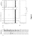
- telomere length distribution 'smear' A typical telomere length distribution band is shown in Figure 1a , in contrast a typical telomere length distribution smear is shown in Figure 4 .
- telomere length distributions that are representative of telomere shortening.
- selection of an optimised cycle number and amount of DNA provides a better representation of the telomere length distributions.
- an optimised PCR cycle number results in less artefactual amplification, less heterogeneity and so a more accurate representation of the telomere length distribution.
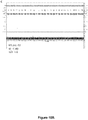
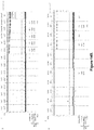
- PCR cycle numbers 21 and 23 produce a robust signal (between 27-65 RFU) and a reliable estimation of telomere length ( fig. 11 ) in a system where the lowest signal to provide a reliable telomere length estimate was 25 RFUs or 25% (Tel Peak area LM peak area, where Tel Peak area is the area under the curve of the telomere-specific products, and the LM area is the area under the curve for the lower marker). Both are determined using the ProSize software. Most ideally, PCR cycle number of 23 is used.
- the amount of chromosomal DNA used for PCR amplification is between 25-30ng.
- the amount of chromosomal DNA used for PCR amplification is selected from the group comprising: 25ng, 26ng, 27ng, 28ng, 29ng and 30ng. Most ideally, 25ng of chromosomal DNA are used and 22 PCR cycles are executed to provide the amplification product. More preferably still, 30ng of chromosomal DNA was subjected to 23 PCR cycles.
- said primer is provided at a concentration of 0.1, 0.2, 0.3, 0.4, 0.5, 0.6, 0.7 or 0.8 ⁇ M, but most ideally at 0.1 or 0.2 ⁇ M.
- said chromosomal DNA is extracted from at least one cell, and ideally a plurality of cells, using an automated extraction method with a view to providing an equal amount of DNA into the PCR reaction each time the reaction is undertaken.
- An example of a suitable DNA extraction system is the Promega DNA IQTM system. Using this system, DNA was extracted from cell suspensions and subsequently diluted to the single molecule level to provide an equal number of amplifiable molecules with the original STELA method.
- telomere length distribution smear for use in HT-STELA, this was done by determining the optimal elution volumes from the Promega DNA IQTM system and subsequent volume of DNA solution added to the HT-STELA reactions. It was found that this provided a reliable telomere length distribution smear without the need to quantify input DNA. This increases the speed and reliability of the STELA process and allows for automation of DNA extraction, dilution and PCR setup. It also increases the ranges of samples that can be analysed, allowing for example, the use of FTA DNA cards with fingersticks or buccal swabs.
- this technology also allows individuals to provide the samples at home and send them in the standard post for telomere testing.
- HT-STELA provides a powerful tool allowing a rapid, accurate prognostic evaluation of cancer patients.
- said cell can be extracted from any biological sample.
- biological sample refers to any sample, or processed derivative thereof, taken from a subject whose telomere length is to be analysed. This includes but is not limited to complex samples, such as whole blood, tissue samples or tumour samples, or purified cell samples thereof.
- a processed derivative of a biological sample may be used such as, but not limited to, cells harvested previously isolated and extracted from a biological sample and, if necessary, re-suspended in an appropriate medium.
- FTA cards are a commonly used format for the convenient sampling and storage of blood samples for subsequent downstream DNA analysis. It provides a quick method for patients that may require a telomere test, to provide a sample at home, without the requirement of a clinician or phlebotomist. A small blood sample is spotted onto an FTA card then returned to the testing facility by standard mail services.
- the DNA IQ system appears to be saturated at 4X10 5 cells and so the preferred method involves the use of between 2x10 5 and 6x10 5 and preferably 4X10 5 cells.
- telomere lengths can display considerable heterogeneity, for example in samples derived from peripheral blood of normal individuals, which can lead to unfavourable PCR amplification products.
- pre-digesting the chromosomal DNA prior to performing HT-STELA reduces the complexity of sample DNA providing a simpler target for amplification.
- said chromosomal DNA is digested with a restriction endonuclease that cleaves non-telomeric DNA.
- a restriction endonuclease that cleaves non-telomeric DNA.
- telomere length it is advisable to use a restriction endonuclease that does not cleave the telomeric region and so, more ideally, said restriction enzyme is selected on the basis it does not cleave between the 5' end of said primer and the telomere repeat array or chromosome telomere end.
- said detecting the length of the amplified product involves the use of markers: a lower marker that was larger than the size of the larger primer complexes and primer-dimers (200bp) but smaller than the smallest fragment size of a telomere generated with HT-STELA (such as 882bp or 415bp); and an upper marker that was larger than the longest mean telomere length observed in our cohorts of CLL.
- a lower marker less than 882bp or 415bp was chosen.
- the marker is selected from the group comprising: 882bp, 881bp 880bp, 879bp, 878bp, 877bp, 876bp, 875bp, 874bp, 873bp, 872bp, 871bp, 870bp, 410bp, 409bp, 408bp, 407bp, 406bp, 405bp, 404bp, 403bp, 402bp, 401bp, 400bp, 399bp, 398bp, 397bp, 396bp, 395bp, 394bp, 393bp, 392bp, 391bp, and 390bp.
- an upper marker greater than 10kb was chosen such as a marker selected form the group comprising: 11kb, 12kb, 13kb, 14kb, 15kb, 16kb, 17kb, 18kb, 19kb and 20kb, including all 0.1kb integers therebewteen.
- primers are designed to anneal with a region adjacent a telomere repeat array of chromosomal DNA of chromosomes XpYp, 7q, 12q and 17p. More ideally still, telomere-adjacent primers are designed incorporating telomere specific nucleotides at the 3' end.
- the 7q chromosome primers are designed around i.e. to be complementary to the 7q-specific nucleotides (with respect to the beginning of the telomere repeat array, or hg38 human genome reference sequence) 1115T (chr7:159334755 hg38), 551A (chr7:159335319 hg38), 195G (chr7:159335675 hg38) and, to a lesser extent, 29G (chr7:159335841 hg38).
- These primers produced robust telomere length distribution smears.
- the 12q chromosome primers are designed around i.e. to be complementary to the 12q specific nucleotides (with respect to the beginning of the telomere repeat array, or hg38 human genome reference sequence) 350G (chr12:133264524 hg38) and 550T (chr12:133264324 hg38). These primers generated robust telomere length distribution smears.
- the XpYp chromosome primers are designed around i.e. to be complementary to the XpYp specific nucleotides (with respect to the beginning of the telomere repeat array, or hg38 human genome reference sequence) 397C (XpYpE5)(chrX: 10432 hg38) and 861C (XpYpC)(chrX:10896 hg38).
- XpYpE5 XpYpE5
- XpYpC 861C
- the 17p chromosome primers are designed around i.e. to be complementary to the 17p specific nucleotides 292C (17pseq1rev), 1018T (17pseq2rev), 1837C (17pseq4rev), 2559C (17p2) and 3018G (17p7) (with respect to the beginning of the telomere repeat array). These primers generated robust telomere length distribution smears.
- primers designed to amplify other chromosomal telomeres including 2p, 4q, 4p, 5p, 11q, 16p and 18q, that also generate telomere-specific products with the orginal STELA method, may also provide utility with HT-STELA.
- primers include: 2p2 (chr2: 10755 hg38), 4qK1 (chr4:190122098 hg38), 4p4 (chr4:10457 hg38), 5p3 (chr5:12240 hg38), 11q13B (chr11:135076467 hg38), 16prev1 (chr16:10241 hg38) and 18qrev4M (chr18:80262147 hg38).
- primers that anneal up to at least 3.843kb from the start of the telomere repeat array yield robust telomere profiles.
- primers that anneal within 30bp of the telomere repeat array did not produce efficient or specific amplification. Accordingly, when working the invention we use primers that anneal to a region of the chromosome that is between 3843bp-30bp from the start of the telomere repeats TTAGGG.
- the term "primer(s)” describes an oligonucleotide that hybridizes under physiological or reaction conditions (see HT-STELA PCR, page 20) to end chromosome DNA.
- primers are designed to be complementary to at least one and ideally a plurality of said chromosome end specific nucleotides.
- the oligonucleotide be constructed and arranged so as to bind selectively with the target under physiological/reaction conditions, i.e., to hybridize substantially more to the target sequence than to any other sequence in the target cell under physiological/reaction conditions.
- the oligonucleotides should comprise at least 7 26 and more preferably, at least 15 consecutive bases which are complementary to the target. Most preferably, the oligonucleotides comprise a complementary sequence of 20-30 bases.
- detecting said amplification product(s) involves a resolution (or separation) process using capillary electrophoresis
- a resolution (or separation) process using capillary electrophoresis an example of a suitable apparatus for undertaking this work is the Fragment AnalyzerTM (available from Advanced Analytical).
- Other capillary gel systems are available from Agilent and Lifetechnologies.
- a fluorescent dye ideally one that intercalates into the amplification product.
- telomere length ( fig. 11 ) in a system where the lowest signal to provide a reliable telomere length estimate was 25 RFUs, this equates to, or can be expressed as 25% of (the area under the curve for the HT-STELA signal divided by the area under the curve for lower marker as shown in Figure 11 i.e. Tel Peak area/LM peak area). Please see page 35, Quality Control.
- said method is undertaken to diagnose or prognose or to determine the risk of developing a condition selected from the group comprising: cancer, ageing, neurological disorders including Alzheimer's disease, Parkinson's disease and other dementias, brain infarction, heart disease, chronic HIV infection, chronic hepatitis, skin diseases, chronic inflammatory bowel disease including ulcerative colitis, anaemia, atherosclerosis, Barrett's oesophagus and cancers including pre-cancerous conditions, infertility, telomere syndromes including dyskeratosis congenita, aplastic anaemia, idiopathic pulmonary fibrosis, familial myelodysplastic syndrome-acute myeloid leukaemia, Hoyeraal-Hreiderasson syndrome, Revesz syndrome, Coats plus syndrome, bone marrow failure, and cryptogenic liver cirrhosis.
- a condition selected from the group comprising: cancer, ageing, neurological disorders including Alzheimer's disease, Parkinson's disease and other dementias, brain infar
- said method is undertaken to assess an individual's suitability to be a transplantation donor, for example a bone marrow donor.
- kits for determining telomere length of mammalian chromosomal DNA comprising: at least one oligonucleotide primer able to anneal to a region adjacent a telomere repeat array of chromosomal DNA wherein said region is between 3843bp-30bp from said telomere repeat array.
- a plurality of primers are provided and ideally said primers are able to anneal to at least said region in one of the following chromosomes 7q, 12q, 17p, XpYp, 2p, 4q, 4p, 5p, 11q, 16p and 18q.
- said kit comprises reagents for carrying out said method.
- a primer selected from the group comprising: Chromosome end Primer name Genomic Coordinates (HG38) Sequence 2p 2p2 chr2: 10755 GAGCTGCGTTTTGCTGAGCAC (SEQ ID No: 1) 4q 4qK1 chr4:190122098 ACGGTGGCATGCCTCTCTC (SEQ ID No: 2) 4p 4p4 chr4:10457 TCTGCGCCTGCGATGGCGCTATG (SEQ ID No: 3) 5p 5p3 chr5:12240 GCATTCTCTTCACCACAGATGTTG (SEQ ID No: 4) 7q 7q1533C chr7:159334337 CCCACACAGTCATCTATTGTT (SEQ ID No: 5) 7q1115T chr7:159334755 GAGGTGCAGTAGTGGGGATCTAACT (SEQ ID No: 6) 7q843T chr7:159335027 GGGACAGCAT
- any feature disclosed herein may be replaced by an alternative feature serving the same or a similar purpose.
- Oligonucleotides were designed based on human DNA sequences obtained from H. Reithman at the Wistar institute (http:/www.wistar.upenn.edu/Reithman) and National Centre for Biotechnology Information (NCBI). The oligonucleotides were synthesised by MWG-Biotech AG (Ebersberg, Germany). All primers used during this study are listed in table 1.
- CD19+ CLL cells were isolated using MACS whole blood CD19 microbeads. A volume of whole blood containing 2X10 6 white blood cells was taken and brought up to 1000ul with 1XPBS. 20ul MACS whole blood microbeads were then added before mixing thoroughly and incubating at room temperature for 15 minutes. After incubation blood samples were loaded onto AutoMACS Pro separator (Miltenyi Biotech) and CD19+ cells were positively selected using the posselwb program.
- Cell lysis was achieved through incubation of the cell pellet with 400 ⁇ l lysis buffer and 10 ⁇ l proteinase K at 56°C for at least one hour. Cell lysates were then loaded into the cartridges and placed in the Maxwell 16 instrument. The forensic mode with LEV hardware was ran to extract DNA from the cell lysates. Extracted DNA was eluted into 40 ⁇ l elution buffer.
- Genomic DNA from cell pellets containing over 3X10⁶ cells was extracted by standard phenol/chloroform extraction (Sambrook et al., 1989). Briefly, cells were lysed overnight at 45°C in 300-500 ⁇ l lysis buffer (10mM Tris-Hcl pH8, 100mM NaCl, 5mM EDTA pH8, 0.5% SDS) containing 30 ⁇ g RNase A (Sigma; stock 10mg/ml) and 60 ⁇ g proteinase K (Sigma; stock 20mg/ml). After brief centrifugation 300-500 ⁇ l phenol/chloroform was added to the cell lysate and rotated for 20mins at room temperature.
- the mixture was centrifuged at 13000rpm for 5mins to separate the phases, the aqueous and interphase phases were removed and added to an eppendorf containing 300 ⁇ l phenol/chloroform. This was rotated again for 20mins and then centrifuged (13000rpm) for 5 minutes. The aqueous phase was removed and to this was added 30 ⁇ l 3M sodium acetate pH5.3 (Sigma) and 900 ⁇ l ice-cold 100% ethanol to precipitate the DNA. The mix was left at -20°C for at least an hour. After brief centrifugation (13000rpm, 1min) the DNA pellet was washed in 70% ice-cold ethanol and air dried in the micro-flow hood. DNA was then resuspended in 10mM Tris-Hcl pH8.
- DNA concentrations were determined in triplicate either by Hoechst 33258 flurometry as described previously (Baird et al., 2003) or using a Nanodrop 3300 fluorospectrometer (Thermo scientific). Briefly an equal amount of a 2X dilution of Quant-iT Picogreen dsDNA reagent (Life Technologies) and DNA were mixed and incubated at room temperature for 5 minutes. 2ul was loaded onto the pedestal to measure the fluorescence. The fluorescence measurement was then converted to DNA concentration using a standard curve constructed using DNA of known quantity.
- DNA was diluted to 250pg/ ⁇ l in 10mM Tri-Hcl (pH 8) containing 250pM Tel2 linker. Multiple 10 ⁇ l reactions were set up per sample (typically 6) each containing 1X Taq buffer (75mM Tris-HCI (pH8.8), 20mM (NH4)SO4, 0.01% Tween-20) (Abgene), 2mM MgCl2,, 1.2mM dNTPs, telomere-specific primer (0.5 ⁇ M), teltail primer (0.5 ⁇ M), and 1U Taq/PWO (Abgene/Roche) at a ratio of 10:1. 1 ⁇ l DNA/tel2 mix (250pg) was added to each reaction. These reactions were then cycled in a Bio-Rad DNA Engine Tetrad® Thermal Cycler using the following conditions: 94°C (20 sec), 65°C (30 sec), 68°C (8 mins) for 22 cycles.
- DNA was diluted to 10ng/ ⁇ l in 10mM Tris-Hcl (pH8) containing 250pM Tel2 linker. Multiple 30ul reactions were set up per sample (typically 3-5) each containing 1X Taq buffer (75mM Tris-HCI (pH8.8), 20mM (NH4)SO4, 0.01% Tween-20) (Abgene), 2mM MgCl2, 1.2mM dNTPs, telomere-specific primer (0.2 ⁇ M), teltail primer (0.2 ⁇ M), and 1U Taq/PWO (Abgene/Roche) at a ratio of 10:1. 3 ⁇ l DNA/tel2 mix (30ng) was added to each reaction. These reactions were then cycled in a Bio-Rad DNA Engine Tetrad® Thermal Cycler using the following conditions: 94°C (20 sec), 65°C (30 sec), 68°C (5 mins) for 23 cycles
- DNA fragments were resolved using a 40cm long, 0.5% Tris-acetate-EDTA agarose gel submerged in 1XTAE cooled to 4°C by a circulating cooling system. 4ul STELA PCR reactions containing 1X ficol-based loading dye (5% bromophenol blue, 5% xylene, 15% ficol) were loaded in the gel and ran through the length of the gel at 120V for 16 hours.
- 1X ficol-based loading dye 5% bromophenol blue, 5% xylene, 15% ficol
- HT-STELA PCR reactions were loaded onto a 96 well plate (sample plate) along with a 75bp-15kb ladder in one well.
- Another 96 well plate (marker plate) was loaded which contained 22ul of the combined lower (400bp) and upper (15kb) markers in every well. Both plates were placed into fragment analyser along with a deep well 96 well plate containing 1.1ml 1X inlet buffer. Both marker DNA fragments and HT-STELA PCR fragments were then resolved through DNF-930 dsDNA gel containing 0.001X intercalating gel using the following conditions: prerun (6kV, 30 seconds), marker injection (1kV, 10 seconds), sample injection (4kV, 50 seconds), separation 6kV, 120 minutes).
- the resolved STELA PCR products were depurinated by washing the gel twice in depurination buffer (0.25M HCl) for 6 minutes. After rinsing, the gel was then washed in denaturation buffer (1.5M NaCl/0.5MNaOH) for 15 minutes. The DNA was then transferred onto a positively charged membrane (Hybond XL, Amersham) by alkaline Southern blotting with denaturation buffer for 4-6 hours.
- depurination buffer 0.25M HCl
- denaturation buffer 1.5M NaCl/0.5MNaOH
- probe DNA and ladder (1:1 of 1kb:2.5kb) in TE buffer (10mM Tric-Hcl and 1mM EDTA) was labelled using Ready-To-Go DNA labelling beads (GE Healthcare).
- This kit generates labelled probes using random hexaprime labelling with [ ⁇ -33P] dCTP.
- the membrane was washed with 0.1X sodium chloride sodium citrate (SSC)/0.1% sodium dodecyl sulphate (SDS) several times at 60°C. The washed blots were then dried in the hybridization oven at 60°C for ⁇ 30mins.
- SSC sodium chloride sodium citrate
- SDS sodium dodecyl sulphate
- Radiolabelled southern blots were placed in a cassette with a phosphoimager screen (Amersham) for 24 hours.
- the phosphimager screen was then scanned using the Typhoon 9410 biomolecular imager (GE healthcare).
- Peripheral blood was obtained from healthy individuals using contact-activated lancets (BD). DNA was extracted directly from peripheral blood samples (300 ⁇ l) using DNA IQ casework pro kit cartridges and eluted into 50 ⁇ l elution buffer (Promega). Extracted DNA was digested using EcoR1. Briefly, 17.3 ⁇ l DNA was added to 20 ⁇ l reactions containing RE 1X buffer, acetylated BSA (1ug/ ⁇ l) and EcoR1 restriction enzyme (5U). Reactions were then incubated at 37°C for 2 hours followed by a 5 min incubation at 65°C to heat inactivate the restriction enzyme. Following digestion was then quantified using NanoDrop 3300 fluorospectrophotometer (Thermo Scientific) and diluted to ⁇ 10ng/ul.
- BD contact-activated lancets
- HT-STELA PCR reactions were loaded onto a 96 well plate (sample plate) along with a 75bp-15kb ladder in one well.
- Another 96 well plate (marker plate) was loaded which contained 22ul of combined lower (400bp) and upper (15 or 20kb) markers. Both plates were placed into the Fragment Analyzer (AATI) along with a deep well 96 well plate containing 1.1ml 1X inlet buffer. Both marker DNA fragments and HT-STELA PCR fragments were then resolved through DNF-930 dsDNA gel containing 0.001X intercalating gel using the following conditions: prerun (6kV, 30 seconds), marker injection (1kV, 10 seconds), sample injection (4kV, 75 seconds), separation 6kV, 120 minutes).
- Genomic DNA was added directly to 400 ⁇ l lysis buffer which was then loaded into DNA IQ casework pro kit cartridges or DNA IQ reference sample kit cartridges (Promega) and placed in the Maxwell 16 instrument.
- DNA IQ casework pro kit DNA was extracted using the forensic mode with low elution volume (LEV) hardware. DNA was eluted into 80 ⁇ l elution buffer.
- SEV standard elution volume
- 125 ⁇ l blood was pipetted onto an FTA card sample target and left to air-dry overnight.
- Two 3mm punches were cut from each dried blood spot and incubated at 65°C with shaking (1400rpm) with 40 ⁇ l 10mg/ml proteinase K and 360 ⁇ l casework extraction buffer (Promega).
- the eluted cell lysate was removed from the stripped FTA cardpunches by adding both the punches and supernatant to spin baskets (Promega) inserted into Eppendorf tubes that were centrifuged at 12000rpm for two minutes. Cell lysates were then loaded into DNA IQ casework pro kit cartridges (Promega) and placed in the Maxwell 16 instrument.
- the forensic mode with LEV hardware was ran to extract DNA from the cell lysates. Extracted DNA was eluted into 50 ⁇ l elution buffer.
- HT-STELA PCR reactions were loaded onto a 96 well plate (sample plate) along with a 75bp-15kb ladder in one well.
- Another 96 well plate (marker plate) was loaded which contained 22 ⁇ l of the combined lower (400bp) and upper (15kb) markers in every well. Both plates were placed into Fragment AnalyzerTM along with a deep well 96 well plate containing 1.1ml 1X inlet buffer. Both marker DNA fragments and HT-STELA PCR fragments were then resolved through DNF-930 dsDNA gel containing 0.001X intercalating gel using the following conditions: prerun (6kV, 30 seconds), marker injection (1kV, 10 seconds), sample injection (4kV, 75 seconds), separation 6kV, 120 minutes).
- the output data from the fragment analyser was viewed in ProSize software (Advanced Analytical). The following peak analysis settings were applied when analysing the data: peak width - 50 seconds, minimum peak height - 25 RFU with no valley to valley baseline. Baseline setpoints were determined manually for each sample. Mean telomere length was calculated using the smear analysis function within the ProSize software. The smear range was manually set for each sample. The mean telomere length was calculated from the smear analysis result for all reactions within each sample reaction set.
- STELA PCR was performed using DNA extracted from a clonal population of the HT1080 fibrosarcoma cell line (HT1080 cl.2). This DNA was used as it displays a homogenous telomere length profile at XpYp that is similar to what is observed in CLL patients. Based on the reaction conditions for the original STELA method for the XpYp telomere, 250pg DNA was added to 30 ⁇ l STELA PCR reactions which were then subject to 22 PCR cycles using an annealing temperature of 65°C. 4 ⁇ l of each reaction was used for the standard STELA analysis and the remainder of the PCR products resolved and detected using the Fragment AnalyzerTM (Advanced Analytical; fig. 1 ).
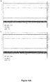
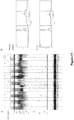
- telomere repeat containing probe fig. 1a
- these same PCR products are not abundant enough to be to be detected fluorescently with the Fragment AnalyzerTM ( fig. 1b(i) ).
- An electropherogram of one of the capillaries through which the generated PCR products were electrophoresed further confirms the lack of any fluorescent signal generated from the STELA PCR products ( fig. 1b(ii) ).
- the lower and upper markers are clearly visible as are large amounts of artefacts ⁇ 200bp formed in the STELA PCR.
- the STELA PCR conditions therefore required modification to ensure that telomere length distributions could be detected by capillary electrophoresis (using the Fragment AnalyzerTM).
- the first attempt to increase the amount of STELA PCR product generated was to increase the number of PCR cycles. 250pg DNA extracted from a clonal population of MRC5 fibroblasts was added to multiple 30 ⁇ l PCR reactions and subject to either 22, 24, 26 or 28 cycles ( fig. 2 ). A small number of STELA-PCR products were generated when the reactions were subject to either 22 or 24 PCR cycles however the intensity of these products was not sufficient to result in their detection using the Fragment AnalyzerTM ( fig. 2a &b). However when STELA PCR reactions were cycled 26 times a larger amount of PCR product was generated, resulting in more intensely hybridising bands on the STELA Southern blot.
- telomere adjacent primer used to amplify telomeric DNA in the original STELA method.
- XpYpE2 resides 408 nucleotides from the start of the XpYp telomere.
- a number of similar primers were designed around this region and used in STELA PCR to replace XpYpE2, in an attempt to increase the PCR efficiency and thereby increase the amount of PCR product generated.
- Five additional primers were designed (E3, E4, E5, E6 and E7) ( fig. 3a ).
- the XpYpE5 primer results in a more efficient PCR and the generation of more PCR product compared to XpYpE2 ( fig. 3c ).
- the average relative fluorescence was 430.3 units, approximately double the relative fluorescence achieved by the STELA PCR reaction containing XpYpE2 (244.3 RFU). Therefore subsequent HT STELA PCR was performed using XpYpE5 as opposed to XpYpE2.
- telomere length distribution smear would be beneficial: it would allow a more rapid analysis of mean telomere length as it would require less human intervention in the analyses of data within the PROSizeTM software; it would require less reactions per sample; it would increase the total amount of product and it would ensure that non-specific background bands present are not incorporated into the sizing analysis.
- a DNA quantity titration was used to determine the optimum amount of DNA to add to the STELA PCR reactions.
- 10ng/ ⁇ l dilutions are made from the concentrated stock DNA samples. 1 ⁇ l (10ng) were taken from this dilution and added to 40 ⁇ l Tris-Hcl (pH8) containing tel2 linker oligonucleotide. 1 ⁇ l (250pg) of this DNA/tel2 mix was then added to each PCR reaction. On the Fragment AnalyzerTM this approach produced a faint banding pattern only after 28 PCR cycles ( fig. 2b ).
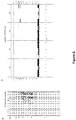
- telomere length distributions As smears using the Fragment AnalyzerTM, increasing amounts of DNA was added to the DNA tel2 mix. Specifically, a 50ng/ ⁇ l dilution was made from stock HT1080 cl.2 DNA. 1 ⁇ l (50ng), 5 ⁇ l (250ng) or 10 ⁇ l (500ng) of this dilution was added to DNA/tel2 mixes. 1 ⁇ l of these DNA/tel2 mixes were added to multiple 30 ⁇ l HT-STELA reactions giving a final concentration of 1.25, 6.25 and 12.5ng/reaction respectively. When the generated STELA PCR products were resolved and detected by the Fragment AnalyzerTM all three input DNA quantities resulted in visible telomere length distribution smears ( fig. 4a )
- the HT1080 cl.2 XpYp telomere length distribution is homogenous.
- XpYp telomere length distributions from CLL patients are also expected to be homogenous previous work in which the XpYp telomere length distribution was studied in a panel of CLL patients by STELA revealed that the telomere length heterogeneity varies somewhat between patients.
- the amount of input DNA that would create a telomere length distribution smear for a homogenous telomere length distribution such as HT1080 cl.2 will not necessarily produce a smear in a sample that displays a more heterogeneous XpYp telomere length distribution.
- a DNA sample from a CLL patient (4408) was selected that displayed a heterogeneous XpYp telomere length distribution.
- DNA from this patient was used for an input DNA titration ranging from 1.25-47.5ng/reaction ( fig. 4b ).
- the input DNA amounts that produced a telomere length distribution smear in the HT1080 cl.2 cell population, did not produce a similar smear for patient 4408, instead a clear banding pattern was observed.
- the amount of DNA in the reactions was increased to 25ng DNA and above, the telomere length distribution was resolved as a smear in this CLL sample ( fig. 4b ); the smear was robust with a strong signal and was reproducible.
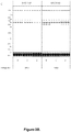
- HT-STELA and STELA was performed on a panel of CLL samples, as well as on clonal populations of the HT1080 (cl.5) and MRC5 (cl.5) cell lines ( fig.6 ).
- the original STELA method revealed the expected high degree of variability in both the mean telomere length, as well the levels of heterogeneity of the XpYp telomere length distributions between samples ( fig. 6a ).
- a useful feature of the MRC5 cl.5 DNA was that it displayed a bimodal XpYp telomere length distribution (Baird et al., 2003), providing a test of the resolution for the Fragment AnalyzerTM in determining whether it is able to successfully amplify and resolve the two allelic telomere length distributions separately ( fig. 6a ).
- telomere length distribution smear was produced for all samples.
- intensity of the telomere length distribution smears differed between samples, suggesting differences in PCR efficiency between reaction sets ( fig. 6b ).
- MRC5 cl5 exhibits a clear bimodal XpYp telomere length distribution ( fig. 6a ).
- analysis by HT-STELA results in the detection of only the lower allele telomere allele on the output gel. Therefore, when the mean telomere length was measured for this sample, only the lower allele was represented.
- Mean telomere length was measured by the smear analysis function within the ProSize software (Advanced Analytical). Overall these discrepancies resulted in an underestimation in telomere length by a mean of 892bp when measured by HT-STELA. This will be problematic when using the technology in a prognostic setting.
- a cycle titration was performed in which HT-STELA PCR reactions containing 10.06ng, 20.12ng or 30.18ng of HT1080 cl.5 DNA were cycled for between 22-28 cycles ( fig. 7a ); it was apparent that the heterogeneity of the product sizes generated was dependent on the number of PCR cycles.
- a homogenous telomere length distribution was observed with a mean telomere length very similar to the mean telomere length measured by the original STELA method (5.822kb) ( fig. 6a ).
- telomere length measurements after 28 PCR cycles observed in fig. 7 was very similar to the HT1080 cl.5 mean telomere length measurement made when measured along with the CLL4 panel (3.618kb) ( fig. 6c ), suggesting this may be the cause of the mean telomere length measurement inaccuracies observed in the panel CLL of samples previously tested ( fig.6 ). These data are consistent with amplification of non-telomeric products with high cycle numbers.
- a cycle titration was also undertaken using DNA from a clonal population of the MRC5 fibroblast cell line (clone 5), this clone displays a bi-allelic telomere length distribution however when analysed using HT-STELA a single telomere length distributions could be detected ( fig. 6 ).
- Three reaction sets were constructed containing either 6.6ng, 13.2ng or 19.8ng MRC5 cl.5 DNA. These reactions were then subject to 22, 24, 26 or 28 PCR cycles ( fig. 7b ). It was clear from these experiments that both the number of cycles and the amount of input DNA influenced the distributions obtained.
- the ratio was 1:2.522 which is not as high as the 2 ⁇ l and 3 ⁇ l reaction sets indicating that the ratio between alleles was also influenced by the amount of input DNA, with a reduction in the amount of input DNA improving the ratio of allelic amplification.
- Another potential source of sizing inaccuracies within the HT-STELA method is the lower (75bp) and upper (20kb) markers provided with the DNF-930 dsDNA kit (Advanced Analytical) that are used for inter-capillary normalisation.
- DNF-930 dsDNA kit Advanced Analytical
- STELA-PCR a large number of fragments less than 200bp in size are generated, these are composed of primer complexes and primer-dimers.
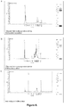
- the intensity of these fragments on the Fragment AnalyzerTM masked the position of the lower 75bp marker ( fig. 8a ) making it difficult to identify this marker. This led to incorrect normalisation of the STELA products and subsequently inaccurate telomere length measurements. A number of strategies were taken to resolve this issue.
- PCR purification was performed using the 96 well plate QIAquick columns (Qiagen) in an attempt to remove these smaller DNA fragments. Although this purification removed a significant amount of the fragments ⁇ 200bp it was not sufficient to completely eliminate them and the same problem remained ( fig.8b ). Additionally, the PCR purification was inconsistent between samples and also removed some of the telomere derived PCR products thereby reducing their signal intensity. It was therefore not possible to purify away the fragments that prevented detection of the lower mark and this strategy was not pursued further.
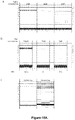
- telomere length of an additional panel of CLL samples as well as the clonal populations of MRC5 (cl.5) and HT1080 (cl.5) was analysed ( fig.9 ). Strong, robust telomere length distribution smears were obtained for the samples.
- MRC5 cl.5 displayed a clear bi-allelic telomere length distribution and HT18080 XpYp telomeres formed a homogenous distribution similar to what is seen with the original STELA method ( fig. 9a ).
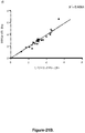
- the mean telomere length determined with HT-STELA was compared with the original STELA method.
- telomere length measurement accuracy was important to incorporate a quality control measure into the HT-STELA assay to ensure that the technique maintains the high level of telomere length measurement accuracy observed in the original STELA method.
- an important aspect of the output data generated by HT-STELA was the total signal generated as defined by the relative fluorescence units (RFU) detectable with Fragment AnalyzerTM given for each telomere length distribution smear.
- telomere length measurement becomes unreliable a PCR cycle titration was performed: HT1080 cl.2 DNA was added to HT-STELA reactions which were then subject to 18-23 PCR cycles.
- the rationale behind this experiment was that as PCR cycle number decreases the amount of PCR product generated and hence the RFU value also decreases. The point at which telomere length measurement becomes inaccurate can then be monitored.
- the signal intensity of the HT1080 cl.2 telomere smears decreased as a function of decreasing cycle numbers, such that after 18 cycles no telomere signal could be detected.
- the peak RFU value is indicative of the amount of PCR product generated and detected this will not always be consistent, for several reasons: Firstly RFU will be subject to variation between different fragment analyzer machines. Secondly the RFU measurement is taken from the highest point of a peak on the electropherogram. The heterogeneity of a telomere length distribution and hence the width of a telomere smear peak on the electropherogram is not taken into account. Therefore robust heterogeneous telomere length distributions whose RFU signal is more dispersed may be discarded as their peak height is lower than for homogenous distributions. Although this may not pose too much of a problem in CLL patients as XpYp telomere length in this disease has been shown to be homogenous it is more pertinent when applying the same criteria to other cancers which show more telomere length heterogeneity.
- telomere length distribution peak area may provide a more appropriate measure of PCR product formation as it encapsulates all HT-STELA PCR products detected by the fragment analyzer.
- Tel peak area may provide a more appropriate measure of PCR product formation as it encapsulates all HT-STELA PCR products detected by the fragment analyzer.
- machine-to-machine variability problem still existed and required normalization.
- One constant in the electrophoresis and detection process were the markers.
- telomere smear peak area value such as the lower marker peak area (LM peak area)
- LM peak area the lower marker peak area
- any marker may be selected and used consistently, would negate any such variation as differences in signal detection of a sample plate between fragment analyzers would be mirrored by the same detection differences between the lower marker on a marker plate. Additionally slight capillary-to-capillary PCR product detection variation within the same assay would also be resolved.
- This approach was applied to the cycle titration experiment ( fig.11 ) and as mentioned earlier a consistent estimation of telomere length was obtained when HT-STELA PCR reactions were subject to 21 -23 PCR cycles. A high Tel peak area/LM peak area percentage was also obtained for these PCR reactions (between 25.87-61.02%.
- the use of the Fragment AnalyzerTM provides a fast high-throughput method to measure mean telomere length in a large number of CLL patients.
- One key aspect of the STELA is the extraction and accurate quantification of DNA. Standard protocols based on phenol/chloroform extraction are time consuming, however this has largely been overcome by various methods that have been developed to allow rapid automated extraction of DNA. However one limiting factor is the requirement for quantification of each extracted DNA followed by subsequent dilution to a standard normalised concentration prior to the PCR steps of the process.
- the quantification steps can be overcome by extraction systems designed to extract defined amounts of DNA. We tested the DNA IQ Casework Pro kit (Promega) that is run on the Maxwell 16 automated DNA extraction system (Promega). This system utilises paramagnetic beads that are calibrated to absorb 100ng of DNA from a cell preparation; therefore as long as the system is saturated, an equal amount of DNA should be extracted from each sample.
- CLL B-cells from which 100ng DNA can be extracted and provide a consistent HT-STELA profile. Differing numbers of CLL B-cells (5X10 3 , 1X10 4 , 1.6X10 4 , 5X10 4 , 2X10 5 , 5X10 5 , 1X10 6 and 2X10 6 ) isolated from a blood sample of a CLL patient ( fig. 12a ) were collected before being lysed for 1 hour and loaded into DNA IQ Casework Pro kits on the Maxwell instrument for DNA extraction. DNA was eluted into 80 ⁇ l of elution buffer and the tel2 oligonucleotide was added directly to the elution buffer at a final concentration of 250pM.
- HT-STELA was performed on the DNA/tel2 mixes and the PCR products resolved and detected with the Fragment AnalyzerTM ( fig. 12b ). Fewer PCR products were generated from the DNA extracted from 1X10 4 and 5 X10 4 cells compared to DNA extracted from greater cell numbers suggesting that this number of cells does not contain enough DNA (100ng) to saturate the DNA IQ system.
- XpYp HT-STELA was performed on DNA extracted from 2X10 5 to 6X10 5 cells a relatively consistent telomere length profile was obtained ( fig. 12b ), however the number of bands and therefore the amount of DNA extracted from 8X10 5 and above appeared to decrease suggesting that this cell number is too great for the DNA IQ system and actually inhibits the extraction of 100ng DNA.
- the DNA IQ system appears to be saturated at 4X10 5 cells. As sufficient DNA is extracted from this amount of cells to provide a robust HT-STELA profile, this number cell was chosen for subsequent use. Furthermore this cell number is relatively small and thus small samples can be reliably analysed.
- HT-STELA was performed using the eluted DNA ( fig. 12c ).
- telomere length distribution profile was observed between the 40 ⁇ l eluate replicates and between the 80 ⁇ l eluate replicates, with a more intense banding pattern observed in the 40 ⁇ l elution compared to the 80 ⁇ l elution.
- DNA concentration given by the Nanodrop 3300 was higher for the 40 ⁇ l eluate replicates it was not double the concentration of the 80 ⁇ l eluate; indicated that DNA elution is less efficient at 40 ⁇ l compared to 80 ⁇ l.
- telomere length distributions given using DNA extracted from the different eluate replicates from the DNA IQ system was encouraging, however they consisted of a banding pattern as opposed to the desired telomere length distribution smear.
- an input DNA titration was performed using DNA from one of the replicates (80-4): 1 ⁇ l, 3 ⁇ l or 5 ⁇ l was added to multiple HT-STELA PCR reactions ( fig. 12d ).
- 1 ⁇ l DNA is added per reaction the similar banding pattern to what was seen in fig. 12c was evident, however when the input DNA was increased to 3 ⁇ l and 5 ⁇ l a robust telomere length distribution smear was observed ( fig. 12d ).
- HT-STELA was then repeated for all the 4X10 5 replicates but with 3 ⁇ l DNA added to PCR reactions as opposed to 1 ⁇ l ( fig. 12e ). Robust telomere length smears were generated instead of banding patterns across all the PCR reactions for every replicate. These experiments allowed us to optimise the DNA IQ system to provide robust HT-STELA smears.
- tel2 linker oligonucleotide was added to the eluted DNA to a final concentration of 250pM and 3 ⁇ l DNA/tel2 mix was added to the 30 ⁇ l HT-STELA PCR reactions, containing 0.2uM XpYpE5 and cycled 23 times.
- the PCR products were resolved and detected using the Fragment AnalyzerTM ( fig. 13a ).
- Mean XpYp telomere length for each individual reaction was measured using the smear analysis function within the ProSize software and the resulting mean telomere lengths for the three replicates for each patient were combined and averaged giving a final XpYp mean telomere length.
- HT-STELA Patient stratification based on XpYp telomere length determined using STELA together with the fusogenic telomere length threshold, provides high resolution prognostic information for CLL patients (Lin et al., 2014).
- the entire HT-STELA process from a blood sample of a CLL patient, to the generation of a robust telomere length profile can be performed within 7.5 hours.
- the development of HT-STELA now facilitates the application of this technology for the routine prognostic assessment of CLL patients, as well as patients with other tumour types for which the prognostic thresholds are informative.
- telomere-adjacent primers were designed incorporating telomere specific nucleotides at the 3' ends. It is this design parameter which gives specificity to one chromosome end as a mismatch will occur when the primer anneals to other subtelomeric sequences from other chromosome ends that exhibit high levels of sequence homology, resulting in failure of the Taq polymerase to extend from the primer in a PCR reaction and hence failure to produce a PCR product from other telomeres.
- telomere adjacent regions were designed for telomere adjacent regions at the 7q and 12q chromosome ends.
- primers specific to the 17p telomere adjacent region were already available as were additional XpYp specific primers. These primers were tested under the same conditions as XpYpE5 to determine whether they could generate telomere length distribution smears using HT1080 cl.2 DNA for their respective chromosome end ( fig. 14 ).
- telomere length distribution smears ( fig. 14a ).
- the mean molecular weight of the fragments calculated using the smear analysis tool on the ProSize software showed the expected increase that was dependent on the distance from the start of the telomere repeat array.
- Calculation of the mean telomere length from these telomere length distribution smears by subtracting the distance from the primer to the start of the telomere repeat array resulted in a similar mean telomere length estimate irrespective of the distance from the start of the telomeres.
- 7q telomere length estimates with HT-STELA were comparable to those determined with the original STELA method ( fig. 14a ; fig. 16 ).
- HT-STELA development for the 12q chromosome end was also successful for some of the primers designed.
- Primers 350G and 550T (350 & 550 nucleotides from the TTAGGG repeats respectively) generated telomere length distribution smears with comparable RFU values.
- the 12q mean telomere length calculated by HT-STELA was very similar to that calculated using the original STELA method ( fig. 14b ).
- the primer designed around the 1388G 12q specific nucleotide produced a very weak telomere distribution. The RFU values for this are too low for a mean telomere length measurement to be given with confidence and will therefore not be used in future experiments
- telomere-adjacent region at the 17p chromosome end had previously been designed specific to the telomere-adjacent region at the 17p chromosome end and a further 8 primers were designed extending up to 6877bp from the telomere repeat array. All of the primers up to 3843bp from the telomere repeat tested in HT-STELA yielded smears consistent with telomeric amplification ( fig. 14c ). With the exception of 3843 bp (17p8) the HT-STELA smears could be quantified. The smear generated with 17p8 could not be quantified as it was too close to the 15 kb upper marker.
- telomere adjacent primer seems to be diminished if it located less 30 nucleotides from the telomeric repeat tract.
- 7q29G and 17pseq1B performed poorly compared to other primers designed further from the telomeric repeat tract at their respective chromosome ends.
- XpYpM and XpYpO (1653 and 1625nt from TTAGGG respectively), or primers XpYpD, XpYpF, XpYpH and XpYpl all greater than 4.8 kb from the telomere, did not generate any PCR product whatsoever ( fig. 14d ).
- XpYpE5 produced a telomere length distribution smear with a mean telomere length similar to what was generated using the original STELA method.
- XpYpB (2539 bp from the telomere) produce a smear of the expected size.
- XpYpC also produced a similar telomere length distribution, but with a higher RFU value than XpYpE5 suggesting that XpYpC is more efficient at generating XpYp telomere products and therefore XpYpC and could be used instead of XpYpE5 for subsequent XpYp HT-STELA ( fig. 14d ).
- XpYpJ also produced a telomere length distribution smear however it was very weak.
- XpYpJ is located less than 30nt from the telomeric repeat tract and has failed to produce a robust telomere length distribution smear suggesting that there is a lower limit distance from the TTAGGG repeat tract below which primers cannot be designed which can sufficiently amplify telomeres.
- telomere lengths observed in samples derived from peripheral blood of normal individuals as detailed above can display considerable heterogeneity, with a diverse range of lengths ranging from close to zero to 25 kb or more. This can occasionally present a particular problem for PCR based amplification of telomeres, whereby the shorter telomeres are likely to amplify more efficiently than the longer telomeres, resulting in an underestimate of mean telomere length.
- DNA samples were extracted and digested with the restriction enzyme as detailed in the Material and Methods.
- the choice of enzyme will depend upon the nature of the telomere repeat array to be amplified and it will be necessary to ensure that it does not cut between the 5' end of the oligonucleotide primer and chromosome telomere end.
- EcoR1 was chosen because it does not cut DNA between the 5' end of the XpYpC primer used for HT-STELA and the telomere repeat array, such that non-telomeric loci and other telomeres maybe cut, but the XpYp telomere will remain intact.
- Other enzymes could also conform to this specific requirement.
- the DNA samples with, or without, EcoR1 digestion were subjected to HT-STELA and analysed with a Fragment Analyzer (AATI) ( fig. 17 ). It was apparent that EcoR1 digestion prior to a HT-STELA successfully reduced the background signal, resulting the improved definition of the telomere smear.
- AATI Fragment Analyzer
- telomere length was established for the analysis of telomere length in fresh/frozen cell and tissue samples obtained from either haematological or solid tumours.
- the ability to analyse previously extracted DNA samples expands the utility of HT-STELA, by for example, allowing the analysis of patient cohorts from clinical trials many of which will only have tumour DNA samples available, or indeed the analysis of any archived DNA sample.
- the conditions under which the DNA IQ system is saturated by DNA was determined. To do this increasing amounts (200, 500, 1000 and 2000ng) of HT1080 cl2 DNA was added to DNA IQ lysis buffer and DNA extracted using the standard protocols on the Maxwell instrument (Promega). This was performed twice: once using the DNA IQ casework pro kit (used when extracting DNA from CLL cell pellets) and also using the DNA IQ reference kit ( fig. 19 ). The two kits differ in that a low elution volume (LEV) protocol is used with the casework pro kit whereas a standard elution volume (SEV) protocol is used with the reference sample kit. DNA is eluted into 80 ⁇ l using the casework pro kit and into 300 ⁇ l using the reference sample kit.
- LUV low elution volume
- SEV standard elution volume
- eluted DNA Differing amounts of eluted DNA was then tested in HT-STELA reactions.
- 3 6 or 9 ⁇ l eluted DNA was added for each input DNA quantity added to the DNA IQ system.
- 3 10 and 20 ⁇ l of eluted DNA was added to HT-STELA reactions. Again this was performed for each DNA quantity added to the DNA IQ system.
- the HT-STELA reactions were then subject to PCR under the same conditions as previously described and the resulting PCR products were resolved by capillary electrophoresis using the Fragment AnalyzerTM (Advanced Analytical; fig. 19 ).
- the quantity of DNA added to the DNA IQ casework pro kit cartridges had little impact on the resulting telomere length distribution smears. Whether adding 200, 500, 1000 or 2000ng to the system the resulting telomere length distribution smears appeared similar both in terms of MTL and normalised tel peak area % suggesting that the system is already at saturation point when 200ng is added to it ( fig. 19a ).
- a factor which made more of an impression on the final telomere length distribution smear was the amount of eluted DNA added to HT-STELA reactions. The increase in DNA quantity added to HT-STELA reactions is reflected by the increase in signal intensity of the telomere length distribution smears for each DNA quantity set and for both kit types.
- telomere length distribution smear For the DNA IQ casework pro kit, regardless of input DNA quantity, adding 6 ⁇ l or less of the eluted DNA to the HT-STELA PCR reactions is not sufficient to generate a telomere length distribution smear with a normalised tel peak area % higher than the lower 25% threshold that we had previously defined as a quality threshold. Adding 9 ⁇ l of eluted DNA to HT-STELA reactions produced a more robust telomere length distribution smears which, with exception of the 500ng samples, surpassed the lower normalised tel peak area % threshold. The MTL given by all of the 9 ⁇ l replicates ranged from 6.084-6.357kb which is comparable to the HT1080 cl2 positive control MTL of 6.368kb.
- the input DNA amount added to the DNA IQ reference sample cartridges affected the telomere length distribution smears observed on the HT-STELA output gel ( fig. 19b ). Adding DNA eluted from 200ng of input DNA into the HT-STELA reactions resulted in barely detectable telomere length distribution smears regardless of how much of the DNA was added to the reactions. Only when input DNA amount was increased to 500ng and above did the telomere length distribution smears become apparent. However this only occurred when 10 ⁇ l or more eluted DNA was included in the HT-STELA reactions. This suggests that the DNA IQ system using the reference sample kit is not saturated using these DNA quantities.
- the XpYp mean telomere length measured from the PCR products generated from the normalised DNA samples was accurate ranging from 5.868kb-6.106kb which spans the 5.956kb measured MTL of the positive control HT1080 cl2 DNA. This MTL range gave an overall coefficient of variance of 1.65% ( fig.20b ).
- the %CV between the input DNA quantity replicates was similarly very low - 1.397% for 200ng, 2.152% for 500ng and and 1.473% for 1000ng.
- FTA cards are a commonly used format for the convenient sampling and storage of blood samples for subsequent downstream DNA analysis. It provides a quick method for patients that may require a telomere test, to provide a sample at home, without the requirement of a clinician or phlebotomist. A small blood sample is spotted onto an FTA card then returned to the testing facility by standard mail services. This will also have utility in the analysis of trial cohorts that have samples stored on FTA card.
- 125 ⁇ l blood from three CLL patients was spotted onto FTA cards and allowed to air-dry overnight.
- the three patients had differing white blood cell (WBC) counts; patient 71 had a WBC count of 45X10 6 cells/ml whereas patient 74 had a count of 6X10 6 cells/ml and patient 75 had a WBC count of 101X10 6 cells/ml.
- WBC white blood cell
- DNA was extracted from two 3mm punches of a dried blood spot with casework extraction buffer (Promega) for 1 hour at 65°C with shaking at 1400rpm. The eluted blood cell lysates were then separated from the stripped punches and DNA was extracted with the Maxwell automated DNA extraction system using the LEV blood kit. Negative control punches were also included (FTA card with no blood). As stated in the protocol (Promega) LEV blood cartridges were used for the DNA extraction and DNA was eluted into 50 ⁇ l elution buffer. 3 ⁇ l of eluted DNA was added to each HT-STELA reaction in the same manner as previously mentioned.
- telomere length distribution smears were generated from DNA extracted from the dried blood of patients 71 and 75.
- the calculated mean telomere length (MTL) of these patients using this FTA-STELA method was very similar to MTL measured by STELA: for patient 71 the MTL for FTA-STELA was 3.889kb compared to 4.169kb measured by STELA. Similarly, for patient 75 the FTA-STELA derived MTL 2.036kb was very similar to the STELA MTL of 1.856kb.
- telomere length distribution profile could be generated for patient 74. This doesn't appear to be due to the low WBC count (6X10 6 cells/ml) as the number of cells on two 3mm punches of this dried blood spot was estimated to contain 7.5X10 4 cells which, although less than the than the 4X10 5 cells required to produce robust telomere length profile, there should still have been sufficient DNA to yield a detectable profile ( fig. 12b ). Alternatively this negative result may be due to a technical issue that occurred within the experiment. The number of cells on two 3mm punches of the dried blood spots of patients 71 and 75 was estimated to be 5.63X10 5 and 1.575X10⁶.
- telomere length distribution smear could still be generated from patients with a WBC count as low as 16X10 6 cells/ml a two 3mm FTA card punches from such a patient was estimated to contain 2X10 5 cells, a cell number that we have previously shown to produce a HT-STELA profile ( fig. 12b ).
- Patient 89 with a WBC count of 20X10 6 cells/ml. DNA was extracted from the dried blood spot of this patient as well as from the FTA sample of patient 91 (WBC count - 69 X10 6 cells/ml), together with patients 71 and 75.
- Patient 74 was discounted as a telomere length profiles could not be achieved with either STELA, HT-STELA or FTA STELA.
- the concentrations of the eluted DNAs indicate that the DNA IQ system successfully normalised the DNA eluted from each of the samples ( fig. 21b ).
- the DNA concentration ranged from 4.576 to 6.027ng/ ⁇ l, a lower range than observed when DNA was extracted using the LEV blood kit.
- Telomere length distribution smears were generated from all four eluted DNA samples ( fig. 21b ). Three of these four telomere length distribution smears were robust and exceeded the 25% lower threshold for tel peak area %.
- telomere length distributions was generated from DNA extracted from the dried blood of patient 89 (WBC count - 20) indicating that patients with a WBC count less than 32X10 6 can have XpYp MTL measured by FTA-STELA.
- the lower limit of WBC count for FTA-STELA is still unknown and will require elucidation in future assays.
- the MTL measured by FTA-STELA for patient 89 was 2.962kb, very similar to the MTL measured by STELA (2.75kb).
- the FTA-STELA MTL of patients 71 and 75 were similarly accurate: Patient 71 FTA-STELA MTL was 3.889kb compared to 4.169 as measured by STELA. Likewise patient 75 FTA-STELA MTL of 2.068kb was very similar to the STELA MTL of 1.856kb.
- telomere length distribution smear was generated from the DNA extracted from the dried blood spot of patient 91 as reflected by the reduced tel peak area % ( fig. 21b ).
- a reduction in signal intensity has previously been shown to result in a decrease in accuracy in measuring mean XpYp telomere length and also increases variation between biological replicates ( fig. 11 ).
- reduced accuracy in MTL measurement was seen in this sample as the FTA-STELA MTL of 6.293kb is a significant overestimation compared to the STELA measurement of 5.008kb.
- the HT-STELA method can be adapted for the analysis of blood samples stored on FTA cards, this method will likely be applicable to other card based sample storage systems. This adaptation will provide considerable additional utility to the HT-STELA method by allowing patients to use standard self-sampling kits and send these direct to the testing facility for analysis. This will provide considerable cost savings in terms of blood sample acquisition and upstream processing.
- Telomere length analysis as representative of telomere dysfunction, provides a highly prognostic tool in human diseases, permitting considerable discrimination for clinical outcome following treatment. Moreover, prognostic power should enable clinicians to confidently predict the clinical course of these diseases and tailor treatments accordingly.
- telomere length in a high throughput context represents a significant contribution to the ability of clinicians to determine, manage and treat telomere shortening illnesses.
Landscapes
- Chemical & Material Sciences (AREA)
- Life Sciences & Earth Sciences (AREA)
- Health & Medical Sciences (AREA)
- Proteomics, Peptides & Aminoacids (AREA)
- Organic Chemistry (AREA)
- Engineering & Computer Science (AREA)
- Analytical Chemistry (AREA)
- Wood Science & Technology (AREA)
- Immunology (AREA)
- Zoology (AREA)
- Pathology (AREA)
- Genetics & Genomics (AREA)
- General Health & Medical Sciences (AREA)
- Physics & Mathematics (AREA)
- Biochemistry (AREA)
- Molecular Biology (AREA)
- General Engineering & Computer Science (AREA)
- Microbiology (AREA)
- Biotechnology (AREA)
- Bioinformatics & Cheminformatics (AREA)
- Biophysics (AREA)
- Nuclear Medicine, Radiotherapy & Molecular Imaging (AREA)
- General Physics & Mathematics (AREA)
- Hospice & Palliative Care (AREA)
- Oncology (AREA)
- Biomedical Technology (AREA)
- Measuring Or Testing Involving Enzymes Or Micro-Organisms (AREA)
Claims (25)
- Hochdurchsatzverfahren zum Bestimmen der Telomerlänge von chromosomaler Säugetier-DNA, umfassend:i) Annealing eines Primers an eine Region neben einem Telomer-Repeat-Array chromosomaler DNA, wobei die Region zwischen 3843 bp und 30 bp von dem Telomer-Repeat-Array entfernt liegt;ii) PCR-Amplifikation von 20 ng bis 35 ng der chromosomalen DNA unter Verwendung von 21 bis 23 Zyklen, um ein Amplifikationsprodukt zu erzeugen, das das Telomer-Repeat-Array umfasst;iii) Erfassen der Länge des Amplifikationsprodukts.
- Verfahren nach Anspruch 1, wobei Teil ii) das PCR-Amplifizieren von 25 ng bis 30 ng der chromosomalen DNA unter Verwendung von 21 bis 23 Zyklen umfasst, um ein Amplifikationsprodukt zu erzeugen.
- Verfahren nach Anspruch 1 oder 2, wobei Teil iii) das Anbringen eines Fluoreszenzsignals oder einer Markierung an das PCR-Amplifikationsprodukt umfasst, das ein Signal von mindestens 25 RFU liefert; oder
ein Signal, das mindestens 25 % Telomer-Peakfläche unter einer für das erzeugte Produkt repräsentativen Kurve/Marker-Peakfläche unter einer für das erzeugte Markerprodukt repräsentativen Kurve beträgt, wenn 0,5 ng/µl Marker bei einer Injektionsrate von 1 kV, 10 Sekunden in einer Kapillarelektrophorese verwendet werden. - Verfahren nach Anspruch 1, 2 oder 3, wobei die chromosomale DNA 23 PCR-Zyklen oder 30 ng chromosomale DNA 23 PCR-Zyklen unterzogen wurden.
- Verfahren nach einem der vorhergehenden Ansprüche, wobei die Primerkonzentration ausgewählt ist aus der Gruppe umfassend:
0,1, 0,2, 0,3, 0,4, 0,5, 0,6, 0,7 oder 0,8 µM. - Verfahren nach einem der vorhergehenden Ansprüche, wobei die chromosomale DNA aus mindestens einer Zelle unter Verwendung eines automatisierten Extraktionsverfahrens extrahiert wird.
- Verfahren nach Anspruch 6, wobei die Zelle aus einer beliebigen biologischen Probe oder einem verarbeiteten Derivat davon extrahiert wird.
- Verfahren nach Anspruch 7, wobei die biologische Probe eine komplexe Zellprobe oder eine gereinigte Zellprobe ist.
- Verfahren nach einem der Ansprüche 7 oder 8, wobei die biologische Probe Vollblut umfasst.
- Verfahren nach einem der Ansprüche 6 bis 9, wobei für das Extraktionsverfahren etwa 6 × 10⁵ Zellen oder weniger verwendet werden.
- Verfahren nach Anspruch 10, wobei für das Extraktionsverfahren zwischen 2 × 10⁵ und 6 × 10⁵ Zellen verwendet werden.
- Verfahren nach Anspruch 10 oder Anspruch 11, wobei für das Extraktionsverfahren 4 × 10⁵ Zellen verwendet werden.
- Verfahren nach einem der vorhergehenden Ansprüche, umfassend das Verdauen der chromosomalen DNA mit einer Restriktionsendonuklease, die nicht-telomere DNA spaltet, bevor das Verfahren durchgeführt wird.
- Verfahren nach Anspruch 13, wobei die Restriktionsendonuklease nicht zwischen dem 5'-Ende des Primers und dem Telomer-Repeat-Array oder dem 5'-Ende des Primers und dem Chromosomentelomerende spaltet.
- Verfahren nach einem der vorhergehenden Ansprüche, wobei Teil iii) die Verwendung eines unteren Markers beinhaltet, der größer als 200 bp, aber kleiner als die kleinste Fragmentgröße eines mit dem Verfahren dieser Ansprüche erzeugten Telomers ist.
- Verfahren nach Anspruch 15, wobei der untere Marker ausgewählt ist aus der Gruppe umfassend:
882 bp, 881 bp, 880 bp, 879 bp, 878 bp, 877 bp, 876 bp, 875 bp, 874 bp, 873 bp, 872 bp, 871 bp, 870 bp, 410 bp, 409 bp, 408 bp, 407 bp, 406 bp, 405 bp, 404 bp, 403 bp, 402 bp, 401 bp, 400 bp, 399 bp, 398 bp, 397 bp, 396 bp, 395 bp, 394 bp, 393 bp, 392 bp, 391 bp und 390 bp einschließlich aller dazwischen liegenden 0,1-kb-Ganzzahlen. - Verfahren nach einem der vorhergehenden Ansprüche, wobei Teil iii) die Verwendung eines oberen Markers beinhaltet, ausgewählt aus der Gruppe umfassend:
10 kb, 11 kb, 12 kb, 13 kb, 14 kb, 15 kb, 16 kb, 17 kb, 18 kb, 19 kb und 20 kb einschließlich aller dazwischen liegenden 0,1 kb-Ganzzahlen. - Verfahren nach einem der vorhergehenden Ansprüche, wobei der Primer geeignet ist, um an telomerspezifische Nukleotide am 3'-Ende zu binden.
- Verfahren nach einem der vorhergehenden Ansprüche, wobei der Primer so ausgelegt ist, dass er an das 7q-Chromosom und damit an mindestens eines der folgenden 7q-spezifischen Nukleotide in Bezug auf den Beginn des Telomer-Repeat-Arrays oder der hg38-Referenzsequenz des menschlichen Genoms bindet:chr7: 159334755 hg38, 1115T, beispielhaft dargestellt durch den Primer 7q1115T der vorliegenden Tabelle 1;chr7: 159335319 hg38, 551A, beispielhaft dargestellt durch den Primer 7q551A der vorliegenden Tabelle 1;chr7: 159335675 hg38, 195G, beispielhaft dargestellt durch den Primer 7q195G der vorliegenden Tabelle 1 und chr7: 159335841 hg38, 29G, beispielhaft dargestellt durch den Primer 7q29G der vorliegenden Tabelle 1.
- Verfahren nach einem der vorhergehenden Ansprüche, wobei der Primer so ausgelegt ist, dass er an das 12q-Chromosom und damit an mindestens eines der folgenden 12q-spezifischen Nukleotide in Bezug auf den Beginn des Telomer-Repeat-Arrays oder der hg38-Referenzsequenz des menschlichen Genoms bindet:
chr12: 133264524 hg38, 350G, beispielhaft dargestellt durch den Primer 12q350G der vorliegenden Tabelle 1 und chr12: 133264324 hg38, 550T, beispielhaft dargestellt durch den Primer 12q550T der vorliegenden Tabelle 1. - Verfahren nach einem der vorhergehenden Ansprüche, wobei der Primer so ausgelegt ist, dass er an das XpYp-Chromosom und damit an mindestens eines der folgenden XpYpspezifischen Nukleotide in Bezug auf den Beginn des Telomer-Repeat-Arrays oder der hg38-Referenzsequenz des menschlichen Genoms bindet:
chrX: 10432 hg38, 397C, beispielhaft dargestellt durch den Primer XpYpE5 der vorliegenden Tabelle 1, und chrX: 10896 hg38, 861C, beispielhaft dargestellt durch den Primer XpYpC der vorliegenden Tabelle 1. - Verfahren nach einem der vorhergehenden Ansprüche, wobei der Primer so ausgelegt ist, dass er an das 17p-Chromosom und damit an mindestens eines der folgenden 17p-spezifischen Nukleotide in Bezug auf den Beginn des Telomer-Repeat-Arrays bindet:292C, wie durch den Primer 17pseq1rev der vorliegenden Tabelle 1 beispielhaft dargestellt;1018T, wie durch den Primer 17pseq2rev der vorliegenden Tabelle 1 beispielhaft dargestellt;1837C, wie durch den Primer 17pseq4rev der vorliegenden Tabelle 1 beispielhaft dargestellt;2559C, wie durch den Primer 17p2 der vorliegenden Tabelle 1 beispielhaft dargestellt, und 3018G, wie durch den Primer 17p7 der vorliegenden Tabelle 1 beispielhaft dargestellt.
- Verfahren nach einem der vorhergehenden Ansprüche, wobei der Primer ausgewählt ist aus der Gruppe umfassend:
dazu bestimmt, um an das 2p-Chromosom zu binden, und desgleichen der Primer 2p2 der vorliegenden Tabelle 1 (chr2: 10755 hg38), das 4q-Chromosom und desgleichen der Primer 4qK1 der vorliegenden Tabelle 1 (chr4: 190122098 hg38), das 4p-Chromosom und desgleichen der Primer 4p4 der vorliegenden Tabelle 1 (chr4: 10457 hg38), das 5p-Chromosom und desgleichen der Primer 5p3 der vorliegenden Tabelle 1 (chr5: 12240 hg38), das 11q-Chromosom und desgleichen der Primer 11q13B der vorliegenden Tabelle 1 (chr11: 135076467 hg38), das 16p-Chromosom und desgleichen der Primer 16prev1 der vorliegenden Tabelle 1 (chr16: 10241 hg38) sowie das 18q-Chromosom und desgleichen der Primer 8qrev4M der vorliegenden Tabelle 1 (chr18: 80262147 hg38). - Verfahren nach einem der vorhergehenden Ansprüche, wobei das Verfahren durchgeführt wird, um das Risiko der Entwicklung eines Zustands zu diagnostizieren oder zu prognostizieren oder zu bestimmen, ausgewählt aus der Gruppe, umfassend:
Krebs, Alterung, neurologische Erkrankungen, darunter Alzheimer, Parkinson und andere Demenzerkrankungen, Hirninfarkt, Herzerkrankungen, chronische HIV-Infektion, chronische Hepatitis, Hautkrankheiten, chronisch entzündliche Darmerkrankungen einschließlich Colitis ulcerosa, Anämie, Atherosklerose, Barrett-Ösophagus und Krebserkrankungen einschließlich Präkanzerosen, Unfruchtbarkeit, Telomersyndrome einschließlich Dyskeratosis congenita, aplastische Anämie, idiopathische Lungenfibrose, familiäres myelodysplastisches Syndrom, akute myeloische Leukämie, Hoyeraal-Hreiderasson-Syndrom, Revesz-Syndrom, Morbus Coats, Knochenmarkversagen und kryptogene Leberzirrhose. - Verfahren nach einem der Ansprüche 1 bis 24, wobei das Verfahren durchgeführt wird, um die Eignung eines Individuums als Transplantationsspender zu bewerten.
Applications Claiming Priority (2)
| Application Number | Priority Date | Filing Date | Title |
|---|---|---|---|
| GBGB1418144.0A GB201418144D0 (en) | 2014-10-14 | 2014-10-14 | High throughput sequencing |
| PCT/GB2015/053023 WO2016059398A1 (en) | 2014-10-14 | 2015-10-14 | High throughput sequencing |
Publications (2)
| Publication Number | Publication Date |
|---|---|
| EP3207153A1 EP3207153A1 (de) | 2017-08-23 |
| EP3207153B1 true EP3207153B1 (de) | 2020-11-11 |
Family
ID=52001373
Family Applications (1)
| Application Number | Title | Priority Date | Filing Date |
|---|---|---|---|
| EP15784106.5A Active EP3207153B1 (de) | 2014-10-14 | 2015-10-14 | Telomertest mit hohem durchsatz |
Country Status (7)
| Country | Link |
|---|---|
| US (1) | US10851417B2 (de) |
| EP (1) | EP3207153B1 (de) |
| JP (1) | JP7117846B2 (de) |
| AU (1) | AU2015332207B2 (de) |
| CA (1) | CA2964621C (de) |
| GB (1) | GB201418144D0 (de) |
| WO (1) | WO2016059398A1 (de) |
Families Citing this family (7)
| Publication number | Priority date | Publication date | Assignee | Title |
|---|---|---|---|---|
| US10378065B2 (en) * | 2011-08-15 | 2019-08-13 | University College Cardiff Consultants Limited | Prognostic mean telomere length |
| US10604794B2 (en) | 2017-05-10 | 2020-03-31 | The Board Of Regents Of The University Of Texas System | Method to measure the shortest telomeres |
| WO2018215375A1 (en) * | 2017-05-22 | 2018-11-29 | General Electric Company | Improvements in the recovery of nucleic acids from solid supports |
| GB201818792D0 (en) * | 2018-11-19 | 2019-01-02 | Telonostix Ltd | Compounds for telomere length-related treatment of cancer |
| CN110408684A (zh) * | 2019-08-22 | 2019-11-05 | 陕西九州医学检验有限公司 | 一种端粒长度检测试剂盒与方法及生物学年龄评价方法 |
| EP4347882A4 (de) * | 2021-05-26 | 2025-04-30 | Centarix Biotech Ltd | Verfahren zur identifizierung von kritisch kurzen telomeren |
| EP4490324A4 (de) * | 2022-03-07 | 2026-05-20 | Arima Genomics Inc | Onkogene strukturvarianten |
Family Cites Families (14)
| Publication number | Priority date | Publication date | Assignee | Title |
|---|---|---|---|---|
| JPH10507366A (ja) | 1994-10-21 | 1998-07-21 | ゼネカ・リミテッド | Pcrによるテロメアdnaの可変性の特性決定法 |
| US5741677A (en) | 1995-06-07 | 1998-04-21 | Geron Corporation | Methods for measuring telomere length |
| US6197557B1 (en) | 1997-03-05 | 2001-03-06 | The Regents Of The University Of Michigan | Compositions and methods for analysis of nucleic acids |
| WO2003000927A2 (en) * | 2001-06-23 | 2003-01-03 | University Of Wales College Of Medicine | Method for the determination of telomere length |
| CA2453360A1 (en) * | 2001-07-11 | 2003-01-23 | Applera Corporation | Internal calibration standards for electrophoretic analyses |
| US7695904B2 (en) * | 2002-01-31 | 2010-04-13 | University Of Utah Research Foundation | Reducing non-target nucleic acid dependent amplifications: amplifying repetitive nucleic acid sequences |
| JP5686493B2 (ja) | 2003-01-24 | 2015-03-18 | ユニバーシティ・オブ・ユタUniversity Of Utah | テロメアの長さを決定することによって死亡の危険性を予測する方法 |
| US8163480B2 (en) * | 2006-10-05 | 2012-04-24 | Quest Diagnostics Investments Incorporated | Nucleic acid size detection method |
| EP2191013A1 (de) | 2007-08-10 | 2010-06-02 | Tina Holding APS | Verfahren zur abschätzung der telomerlänge |
| US20120122700A1 (en) * | 2010-11-11 | 2012-05-17 | University Of South Florida | Materials and methods for determining subtelomere dna sequence |
| GB201113968D0 (en) * | 2011-08-15 | 2011-09-28 | Univ Cardiff | Prognostic methadology |
| CN102618633A (zh) | 2012-02-03 | 2012-08-01 | 常州亚当生物技术有限公司 | 一种用改良stela法测细胞中最短端粒长度的方法 |
| US20140024034A1 (en) * | 2012-07-17 | 2014-01-23 | Indiana University Research And Technology Corporation | Novel primers for detecting human chromosome end-to-end telemore fusion |
| WO2014152676A1 (en) * | 2013-03-15 | 2014-09-25 | New York University | A method for a single cell analysis of telomer length |
-
2014
- 2014-10-14 GB GBGB1418144.0A patent/GB201418144D0/en not_active Ceased
-
2015
- 2015-10-14 US US15/518,363 patent/US10851417B2/en active Active
- 2015-10-14 EP EP15784106.5A patent/EP3207153B1/de active Active
- 2015-10-14 CA CA2964621A patent/CA2964621C/en active Active
- 2015-10-14 AU AU2015332207A patent/AU2015332207B2/en active Active
- 2015-10-14 WO PCT/GB2015/053023 patent/WO2016059398A1/en not_active Ceased
- 2015-10-14 JP JP2017519317A patent/JP7117846B2/ja active Active
Non-Patent Citations (1)
| Title |
|---|
| None * |
Also Published As
| Publication number | Publication date |
|---|---|
| WO2016059398A1 (en) | 2016-04-21 |
| EP3207153A1 (de) | 2017-08-23 |
| AU2015332207A1 (en) | 2017-04-27 |
| JP7117846B2 (ja) | 2022-08-15 |
| CA2964621A1 (en) | 2016-04-21 |
| GB201418144D0 (en) | 2014-11-26 |
| CA2964621C (en) | 2022-12-06 |
| AU2015332207B2 (en) | 2021-02-18 |
| US10851417B2 (en) | 2020-12-01 |
| JP2017532040A (ja) | 2017-11-02 |
| US20170306404A1 (en) | 2017-10-26 |
Similar Documents
| Publication | Publication Date | Title |
|---|---|---|
| AU2015332207B2 (en) | High throughput sequencing | |
| EP3307885B1 (de) | Integrierte reinigung und messung der dna-methylierung und ko-messung von mutationen und/oder mrna-expressionsniveaus in einer automatisierten reaktionskartusche | |
| EP1759011B1 (de) | Nachweis chromosomaler störungen | |
| JP5680304B2 (ja) | 迅速な法医学的dna分析法 | |
| EP3056574B1 (de) | Verfahren zur erkennung von wiederholungssequenzmotiven in nukleinsäure | |
| US20100203532A1 (en) | Enrichment of a Target Sequence | |
| JP6144623B2 (ja) | 核酸測定用の核酸プローブ | |
| JP2020503858A (ja) | 自動反応カートリッジにおける統合された、DNAメチル化の精製及び測定並びに変異及び/又はmRNA発現レベルの同時測定 | |
| WO2018233236A1 (en) | Methods and kits of constructing sequencing library for use in detecting chromosome copy number variation | |
| AU2016286088A1 (en) | Single nucleotide polymorphism in HLA-B*15:02 and use thereof | |
| US20220364173A1 (en) | Methods and systems for detection of nucleic acid modifications | |
| EP4074840A1 (de) | Pcr-verfahren und pcr-kit zur erhöhung der allelunterscheidung | |
| Kwok | Approaches to allele frequency determination | |
| Shi et al. | Detection of ultrarare somatic mutation in the human TP53 gene by bidirectional pyrophosphorolysis‐activated polymerization allele‐specific amplification | |
| CN110709522A (zh) | 生物样本核酸质量的测定方法 | |
| Aldape et al. | Real‐time quantitative polymerase chain reaction: a potential tool for genetic analysis in neuropathology | |
| Wu et al. | Microarray-based Ms-SNuPE: near-quantitative analysis for a high-throughput DNA methylation | |
| CN116926066A (zh) | 引物组合产品、用于质量检测的试剂盒和基因组质量检测方法 | |
| KR101136008B1 (ko) | 옥사닌 염기를 포함한 라이게이션 단편서열을 이용한 리가제 기반 단일염기다형성 분석방법 및 그 단편서열을 포함하는 단일염기다형성 분석용 시스템 | |
| EP3455376B1 (de) | Verfahren zur herstellung einer vielzahl an dna-sonden und verfahren zur analyse von genomischer dna unter verwendung der dna-sonden | |
| JP7043901B2 (ja) | 核酸検出方法及び核酸検出装置、並びに、核酸検出キット | |
| EP4516925A1 (de) | Dna-nachweisverfahren und dna-nachweiskit unter besonderer betrachtung der fluoreszenzintensitätsmessung und schmelzkurvenanalyse | |
| McCarthy et al. | MethylMeter: a quantitative, sensitive, and bisulfite-free method for analysis of DNA methylation | |
| Fajkus et al. | Analysis of telomeres and telomerase | |
| KR101513276B1 (ko) | 변형 mlpa 를 이용한 카피 수 다형성의 다중 분석 방법 |
Legal Events
| Date | Code | Title | Description |
|---|---|---|---|
| STAA | Information on the status of an ep patent application or granted ep patent |
Free format text: STATUS: THE INTERNATIONAL PUBLICATION HAS BEEN MADE |
|
| PUAI | Public reference made under article 153(3) epc to a published international application that has entered the european phase |
Free format text: ORIGINAL CODE: 0009012 |
|
| STAA | Information on the status of an ep patent application or granted ep patent |
Free format text: STATUS: REQUEST FOR EXAMINATION WAS MADE |
|
| 17P | Request for examination filed |
Effective date: 20170420 |
|
| AK | Designated contracting states |
Kind code of ref document: A1 Designated state(s): AL AT BE BG CH CY CZ DE DK EE ES FI FR GB GR HR HU IE IS IT LI LT LU LV MC MK MT NL NO PL PT RO RS SE SI SK SM TR |
|
| AX | Request for extension of the european patent |
Extension state: BA ME |
|
| DAV | Request for validation of the european patent (deleted) | ||
| DAX | Request for extension of the european patent (deleted) | ||
| REG | Reference to a national code |
Ref country code: HK Ref legal event code: DE Ref document number: 1245338 Country of ref document: HK |
|
| STAA | Information on the status of an ep patent application or granted ep patent |
Free format text: STATUS: EXAMINATION IS IN PROGRESS |
|
| 17Q | First examination report despatched |
Effective date: 20190722 |
|
| REG | Reference to a national code |
Ref country code: DE Ref legal event code: R079 Ref document number: 602015061918 Country of ref document: DE Free format text: PREVIOUS MAIN CLASS: C12Q0001680000 Ipc: C12Q0001688600 |
|
| GRAP | Despatch of communication of intention to grant a patent |
Free format text: ORIGINAL CODE: EPIDOSNIGR1 |
|
| STAA | Information on the status of an ep patent application or granted ep patent |
Free format text: STATUS: GRANT OF PATENT IS INTENDED |
|
| RIC1 | Information provided on ipc code assigned before grant |
Ipc: C12Q 1/6886 20180101AFI20200424BHEP |
|
| INTG | Intention to grant announced |
Effective date: 20200602 |
|
| GRAS | Grant fee paid |
Free format text: ORIGINAL CODE: EPIDOSNIGR3 |
|
| GRAA | (expected) grant |
Free format text: ORIGINAL CODE: 0009210 |
|
| STAA | Information on the status of an ep patent application or granted ep patent |
Free format text: STATUS: THE PATENT HAS BEEN GRANTED |
|
| AK | Designated contracting states |
Kind code of ref document: B1 Designated state(s): AL AT BE BG CH CY CZ DE DK EE ES FI FR GB GR HR HU IE IS IT LI LT LU LV MC MK MT NL NO PL PT RO RS SE SI SK SM TR |
|
| REG | Reference to a national code |
Ref country code: GB Ref legal event code: FG4D |
|
| REG | Reference to a national code |
Ref country code: CH Ref legal event code: EP |
|
| REG | Reference to a national code |
Ref country code: AT Ref legal event code: REF Ref document number: 1333526 Country of ref document: AT Kind code of ref document: T Effective date: 20201115 |
|
| REG | Reference to a national code |
Ref country code: DE Ref legal event code: R096 Ref document number: 602015061918 Country of ref document: DE |
|
| REG | Reference to a national code |
Ref country code: IE Ref legal event code: FG4D |
|
| REG | Reference to a national code |
Ref country code: CH Ref legal event code: NV Representative=s name: COSMOVICI INTELLECTUAL PROPERTY SARL, CH |
|
| REG | Reference to a national code |
Ref country code: NL Ref legal event code: FP |
|
| REG | Reference to a national code |
Ref country code: AT Ref legal event code: MK05 Ref document number: 1333526 Country of ref document: AT Kind code of ref document: T Effective date: 20201111 |
|
| PG25 | Lapsed in a contracting state [announced via postgrant information from national office to epo] |
Ref country code: NO Free format text: LAPSE BECAUSE OF FAILURE TO SUBMIT A TRANSLATION OF THE DESCRIPTION OR TO PAY THE FEE WITHIN THE PRESCRIBED TIME-LIMIT Effective date: 20210211 Ref country code: FI Free format text: LAPSE BECAUSE OF FAILURE TO SUBMIT A TRANSLATION OF THE DESCRIPTION OR TO PAY THE FEE WITHIN THE PRESCRIBED TIME-LIMIT Effective date: 20201111 Ref country code: PT Free format text: LAPSE BECAUSE OF FAILURE TO SUBMIT A TRANSLATION OF THE DESCRIPTION OR TO PAY THE FEE WITHIN THE PRESCRIBED TIME-LIMIT Effective date: 20210311 Ref country code: RS Free format text: LAPSE BECAUSE OF FAILURE TO SUBMIT A TRANSLATION OF THE DESCRIPTION OR TO PAY THE FEE WITHIN THE PRESCRIBED TIME-LIMIT Effective date: 20201111 Ref country code: GR Free format text: LAPSE BECAUSE OF FAILURE TO SUBMIT A TRANSLATION OF THE DESCRIPTION OR TO PAY THE FEE WITHIN THE PRESCRIBED TIME-LIMIT Effective date: 20210212 |
|
| PG25 | Lapsed in a contracting state [announced via postgrant information from national office to epo] |
Ref country code: SE Free format text: LAPSE BECAUSE OF FAILURE TO SUBMIT A TRANSLATION OF THE DESCRIPTION OR TO PAY THE FEE WITHIN THE PRESCRIBED TIME-LIMIT Effective date: 20201111 Ref country code: IS Free format text: LAPSE BECAUSE OF FAILURE TO SUBMIT A TRANSLATION OF THE DESCRIPTION OR TO PAY THE FEE WITHIN THE PRESCRIBED TIME-LIMIT Effective date: 20210311 Ref country code: LV Free format text: LAPSE BECAUSE OF FAILURE TO SUBMIT A TRANSLATION OF THE DESCRIPTION OR TO PAY THE FEE WITHIN THE PRESCRIBED TIME-LIMIT Effective date: 20201111 Ref country code: PL Free format text: LAPSE BECAUSE OF FAILURE TO SUBMIT A TRANSLATION OF THE DESCRIPTION OR TO PAY THE FEE WITHIN THE PRESCRIBED TIME-LIMIT Effective date: 20201111 Ref country code: BG Free format text: LAPSE BECAUSE OF FAILURE TO SUBMIT A TRANSLATION OF THE DESCRIPTION OR TO PAY THE FEE WITHIN THE PRESCRIBED TIME-LIMIT Effective date: 20210211 Ref country code: AT Free format text: LAPSE BECAUSE OF FAILURE TO SUBMIT A TRANSLATION OF THE DESCRIPTION OR TO PAY THE FEE WITHIN THE PRESCRIBED TIME-LIMIT Effective date: 20201111 |
|
| REG | Reference to a national code |
Ref country code: LT Ref legal event code: MG9D |
|
| PG25 | Lapsed in a contracting state [announced via postgrant information from national office to epo] |
Ref country code: HR Free format text: LAPSE BECAUSE OF FAILURE TO SUBMIT A TRANSLATION OF THE DESCRIPTION OR TO PAY THE FEE WITHIN THE PRESCRIBED TIME-LIMIT Effective date: 20201111 |
|
| PG25 | Lapsed in a contracting state [announced via postgrant information from national office to epo] |
Ref country code: RO Free format text: LAPSE BECAUSE OF FAILURE TO SUBMIT A TRANSLATION OF THE DESCRIPTION OR TO PAY THE FEE WITHIN THE PRESCRIBED TIME-LIMIT Effective date: 20201111 Ref country code: SK Free format text: LAPSE BECAUSE OF FAILURE TO SUBMIT A TRANSLATION OF THE DESCRIPTION OR TO PAY THE FEE WITHIN THE PRESCRIBED TIME-LIMIT Effective date: 20201111 Ref country code: LT Free format text: LAPSE BECAUSE OF FAILURE TO SUBMIT A TRANSLATION OF THE DESCRIPTION OR TO PAY THE FEE WITHIN THE PRESCRIBED TIME-LIMIT Effective date: 20201111 Ref country code: SM Free format text: LAPSE BECAUSE OF FAILURE TO SUBMIT A TRANSLATION OF THE DESCRIPTION OR TO PAY THE FEE WITHIN THE PRESCRIBED TIME-LIMIT Effective date: 20201111 Ref country code: EE Free format text: LAPSE BECAUSE OF FAILURE TO SUBMIT A TRANSLATION OF THE DESCRIPTION OR TO PAY THE FEE WITHIN THE PRESCRIBED TIME-LIMIT Effective date: 20201111 Ref country code: CZ Free format text: LAPSE BECAUSE OF FAILURE TO SUBMIT A TRANSLATION OF THE DESCRIPTION OR TO PAY THE FEE WITHIN THE PRESCRIBED TIME-LIMIT Effective date: 20201111 |
|
| REG | Reference to a national code |
Ref country code: DE Ref legal event code: R097 Ref document number: 602015061918 Country of ref document: DE |
|
| PG25 | Lapsed in a contracting state [announced via postgrant information from national office to epo] |
Ref country code: DK Free format text: LAPSE BECAUSE OF FAILURE TO SUBMIT A TRANSLATION OF THE DESCRIPTION OR TO PAY THE FEE WITHIN THE PRESCRIBED TIME-LIMIT Effective date: 20201111 |
|
| PLBE | No opposition filed within time limit |
Free format text: ORIGINAL CODE: 0009261 |
|
| STAA | Information on the status of an ep patent application or granted ep patent |
Free format text: STATUS: NO OPPOSITION FILED WITHIN TIME LIMIT |
|
| 26N | No opposition filed |
Effective date: 20210812 |
|
| PG25 | Lapsed in a contracting state [announced via postgrant information from national office to epo] |
Ref country code: IT Free format text: LAPSE BECAUSE OF FAILURE TO SUBMIT A TRANSLATION OF THE DESCRIPTION OR TO PAY THE FEE WITHIN THE PRESCRIBED TIME-LIMIT Effective date: 20201111 Ref country code: AL Free format text: LAPSE BECAUSE OF FAILURE TO SUBMIT A TRANSLATION OF THE DESCRIPTION OR TO PAY THE FEE WITHIN THE PRESCRIBED TIME-LIMIT Effective date: 20201111 |
|
| PG25 | Lapsed in a contracting state [announced via postgrant information from national office to epo] |
Ref country code: SI Free format text: LAPSE BECAUSE OF FAILURE TO SUBMIT A TRANSLATION OF THE DESCRIPTION OR TO PAY THE FEE WITHIN THE PRESCRIBED TIME-LIMIT Effective date: 20201111 |
|
| PG25 | Lapsed in a contracting state [announced via postgrant information from national office to epo] |
Ref country code: ES Free format text: LAPSE BECAUSE OF FAILURE TO SUBMIT A TRANSLATION OF THE DESCRIPTION OR TO PAY THE FEE WITHIN THE PRESCRIBED TIME-LIMIT Effective date: 20201111 |
|
| REG | Reference to a national code |
Ref country code: CH Ref legal event code: PL |
|
| PG25 | Lapsed in a contracting state [announced via postgrant information from national office to epo] |
Ref country code: IS Free format text: LAPSE BECAUSE OF FAILURE TO SUBMIT A TRANSLATION OF THE DESCRIPTION OR TO PAY THE FEE WITHIN THE PRESCRIBED TIME-LIMIT Effective date: 20210311 |
|
| REG | Reference to a national code |
Ref country code: NL Ref legal event code: MM Effective date: 20211101 |
|
| REG | Reference to a national code |
Ref country code: BE Ref legal event code: MM Effective date: 20211031 |
|
| PG25 | Lapsed in a contracting state [announced via postgrant information from national office to epo] |
Ref country code: MC Free format text: LAPSE BECAUSE OF FAILURE TO SUBMIT A TRANSLATION OF THE DESCRIPTION OR TO PAY THE FEE WITHIN THE PRESCRIBED TIME-LIMIT Effective date: 20201111 |
|
| PG25 | Lapsed in a contracting state [announced via postgrant information from national office to epo] |
Ref country code: NL Free format text: LAPSE BECAUSE OF NON-PAYMENT OF DUE FEES Effective date: 20211101 Ref country code: LU Free format text: LAPSE BECAUSE OF NON-PAYMENT OF DUE FEES Effective date: 20211014 Ref country code: BE Free format text: LAPSE BECAUSE OF NON-PAYMENT OF DUE FEES Effective date: 20211031 |
|
| PG25 | Lapsed in a contracting state [announced via postgrant information from national office to epo] |
Ref country code: LI Free format text: LAPSE BECAUSE OF NON-PAYMENT OF DUE FEES Effective date: 20211031 Ref country code: CH Free format text: LAPSE BECAUSE OF NON-PAYMENT OF DUE FEES Effective date: 20211031 |
|
| PG25 | Lapsed in a contracting state [announced via postgrant information from national office to epo] |
Ref country code: IE Free format text: LAPSE BECAUSE OF NON-PAYMENT OF DUE FEES Effective date: 20211014 |
|
| PG25 | Lapsed in a contracting state [announced via postgrant information from national office to epo] |
Ref country code: HU Free format text: LAPSE BECAUSE OF FAILURE TO SUBMIT A TRANSLATION OF THE DESCRIPTION OR TO PAY THE FEE WITHIN THE PRESCRIBED TIME-LIMIT; INVALID AB INITIO Effective date: 20151014 |
|
| P01 | Opt-out of the competence of the unified patent court (upc) registered |
Effective date: 20230514 |
|
| PG25 | Lapsed in a contracting state [announced via postgrant information from national office to epo] |
Ref country code: CY Free format text: LAPSE BECAUSE OF FAILURE TO SUBMIT A TRANSLATION OF THE DESCRIPTION OR TO PAY THE FEE WITHIN THE PRESCRIBED TIME-LIMIT Effective date: 20201111 |
|
| PG25 | Lapsed in a contracting state [announced via postgrant information from national office to epo] |
Ref country code: MK Free format text: LAPSE BECAUSE OF FAILURE TO SUBMIT A TRANSLATION OF THE DESCRIPTION OR TO PAY THE FEE WITHIN THE PRESCRIBED TIME-LIMIT Effective date: 20201111 |
|
| REG | Reference to a national code |
Ref country code: HK Ref legal event code: WD Ref document number: 1245338 Country of ref document: HK |
|
| PG25 | Lapsed in a contracting state [announced via postgrant information from national office to epo] |
Ref country code: TR Free format text: LAPSE BECAUSE OF FAILURE TO SUBMIT A TRANSLATION OF THE DESCRIPTION OR TO PAY THE FEE WITHIN THE PRESCRIBED TIME-LIMIT Effective date: 20201111 |
|
| PG25 | Lapsed in a contracting state [announced via postgrant information from national office to epo] |
Ref country code: MT Free format text: LAPSE BECAUSE OF FAILURE TO SUBMIT A TRANSLATION OF THE DESCRIPTION OR TO PAY THE FEE WITHIN THE PRESCRIBED TIME-LIMIT Effective date: 20201111 |
|
| PGFP | Annual fee paid to national office [announced via postgrant information from national office to epo] |
Ref country code: GB Payment date: 20250903 Year of fee payment: 11 |
|
| PGFP | Annual fee paid to national office [announced via postgrant information from national office to epo] |
Ref country code: DE Payment date: 20251022 Year of fee payment: 11 |
|
| PGFP | Annual fee paid to national office [announced via postgrant information from national office to epo] |
Ref country code: FR Payment date: 20251018 Year of fee payment: 11 |