EP3204414B1 - Novel anti-nodal antibodies and methods of using same - Google Patents
Novel anti-nodal antibodies and methods of using same Download PDFInfo
- Publication number
- EP3204414B1 EP3204414B1 EP15849199.3A EP15849199A EP3204414B1 EP 3204414 B1 EP3204414 B1 EP 3204414B1 EP 15849199 A EP15849199 A EP 15849199A EP 3204414 B1 EP3204414 B1 EP 3204414B1
- Authority
- EP
- European Patent Office
- Prior art keywords
- nodal
- antibody
- cancer
- cells
- antibodies
- Prior art date
- Legal status (The legal status is an assumption and is not a legal conclusion. Google has not performed a legal analysis and makes no representation as to the accuracy of the status listed.)
- Active
Links
Images


Classifications
-
- C—CHEMISTRY; METALLURGY
- C07—ORGANIC CHEMISTRY
- C07K—PEPTIDES
- C07K16/00—Immunoglobulins [IG], e.g. monoclonal or polyclonal antibodies
- C07K16/18—Immunoglobulins [IG], e.g. monoclonal or polyclonal antibodies against material from animals or humans
- C07K16/22—Immunoglobulins [IG], e.g. monoclonal or polyclonal antibodies against material from animals or humans against growth factors ; against growth regulators
-
- A—HUMAN NECESSITIES
- A61—MEDICAL OR VETERINARY SCIENCE; HYGIENE
- A61K—PREPARATIONS FOR MEDICAL, DENTAL OR TOILETRY PURPOSES
- A61K31/00—Medicinal preparations containing organic active ingredients
- A61K31/655—Azo (—N=N—), diazo (=N2), azoxy (>N—O—N< or N(=O)—N<), azido (—N3) or diazoamino (—N=N—N<) compounds
-
- A—HUMAN NECESSITIES
- A61—MEDICAL OR VETERINARY SCIENCE; HYGIENE
- A61K—PREPARATIONS FOR MEDICAL, DENTAL OR TOILETRY PURPOSES
- A61K39/00—Medicinal preparations containing antigens or antibodies
- A61K39/395—Antibodies; Immunoglobulins; Immune serum, e.g. antilymphocytic serum
- A61K39/39533—Antibodies; Immunoglobulins; Immune serum, e.g. antilymphocytic serum against materials from animals
- A61K39/3955—Antibodies; Immunoglobulins; Immune serum, e.g. antilymphocytic serum against materials from animals against proteinaceous materials, e.g. enzymes, hormones, lymphokines
-
- A—HUMAN NECESSITIES
- A61—MEDICAL OR VETERINARY SCIENCE; HYGIENE
- A61K—PREPARATIONS FOR MEDICAL, DENTAL OR TOILETRY PURPOSES
- A61K39/00—Medicinal preparations containing antigens or antibodies
- A61K39/395—Antibodies; Immunoglobulins; Immune serum, e.g. antilymphocytic serum
- A61K39/39533—Antibodies; Immunoglobulins; Immune serum, e.g. antilymphocytic serum against materials from animals
- A61K39/39558—Antibodies; Immunoglobulins; Immune serum, e.g. antilymphocytic serum against materials from animals against tumor tissues, cells, antigens
-
- A—HUMAN NECESSITIES
- A61—MEDICAL OR VETERINARY SCIENCE; HYGIENE
- A61K—PREPARATIONS FOR MEDICAL, DENTAL OR TOILETRY PURPOSES
- A61K39/00—Medicinal preparations containing antigens or antibodies
- A61K39/395—Antibodies; Immunoglobulins; Immune serum, e.g. antilymphocytic serum
- A61K39/44—Antibodies bound to carriers
-
- A—HUMAN NECESSITIES
- A61—MEDICAL OR VETERINARY SCIENCE; HYGIENE
- A61K—PREPARATIONS FOR MEDICAL, DENTAL OR TOILETRY PURPOSES
- A61K45/00—Medicinal preparations containing active ingredients not provided for in groups A61K31/00 - A61K41/00
- A61K45/06—Mixtures of active ingredients without chemical characterisation, e.g. antiphlogistics and cardiaca
-
- A—HUMAN NECESSITIES
- A61—MEDICAL OR VETERINARY SCIENCE; HYGIENE
- A61P—SPECIFIC THERAPEUTIC ACTIVITY OF CHEMICAL COMPOUNDS OR MEDICINAL PREPARATIONS
- A61P35/00—Antineoplastic agents
-
- A—HUMAN NECESSITIES
- A61—MEDICAL OR VETERINARY SCIENCE; HYGIENE
- A61P—SPECIFIC THERAPEUTIC ACTIVITY OF CHEMICAL COMPOUNDS OR MEDICINAL PREPARATIONS
- A61P35/00—Antineoplastic agents
- A61P35/02—Antineoplastic agents specific for leukemia
-
- A—HUMAN NECESSITIES
- A61—MEDICAL OR VETERINARY SCIENCE; HYGIENE
- A61P—SPECIFIC THERAPEUTIC ACTIVITY OF CHEMICAL COMPOUNDS OR MEDICINAL PREPARATIONS
- A61P35/00—Antineoplastic agents
- A61P35/04—Antineoplastic agents specific for metastasis
-
- C—CHEMISTRY; METALLURGY
- C12—BIOCHEMISTRY; BEER; SPIRITS; WINE; VINEGAR; MICROBIOLOGY; ENZYMOLOGY; MUTATION OR GENETIC ENGINEERING
- C12N—MICROORGANISMS OR ENZYMES; COMPOSITIONS THEREOF; PROPAGATING, PRESERVING, OR MAINTAINING MICROORGANISMS; MUTATION OR GENETIC ENGINEERING; CULTURE MEDIA
- C12N15/00—Mutation or genetic engineering; DNA or RNA concerning genetic engineering, vectors, e.g. plasmids, or their isolation, preparation or purification; Use of hosts therefor
- C12N15/09—Recombinant DNA-technology
- C12N15/11—DNA or RNA fragments; Modified forms thereof; Non-coding nucleic acids having a biological activity
- C12N15/113—Non-coding nucleic acids modulating the expression of genes, e.g. antisense oligonucleotides; Antisense DNA or RNA; Triplex- forming oligonucleotides; Catalytic nucleic acids, e.g. ribozymes; Nucleic acids used in co-suppression or gene silencing
- C12N15/1136—Non-coding nucleic acids modulating the expression of genes, e.g. antisense oligonucleotides; Antisense DNA or RNA; Triplex- forming oligonucleotides; Catalytic nucleic acids, e.g. ribozymes; Nucleic acids used in co-suppression or gene silencing against growth factors, growth regulators, cytokines, lymphokines or hormones
-
- G—PHYSICS
- G01—MEASURING; TESTING
- G01N—INVESTIGATING OR ANALYSING MATERIALS BY DETERMINING THEIR CHEMICAL OR PHYSICAL PROPERTIES
- G01N33/00—Investigating or analysing materials by specific methods not covered by groups G01N1/00 - G01N31/00
- G01N33/48—Biological material, e.g. blood, urine; Haemocytometers
- G01N33/50—Chemical analysis of biological material, e.g. blood, urine; Testing involving biospecific ligand binding methods; Immunological testing
- G01N33/53—Immunoassay; Biospecific binding assay; Materials therefor
- G01N33/543—Immunoassay; Biospecific binding assay; Materials therefor with an insoluble carrier for immobilising immunochemicals
-
- G—PHYSICS
- G01—MEASURING; TESTING
- G01N—INVESTIGATING OR ANALYSING MATERIALS BY DETERMINING THEIR CHEMICAL OR PHYSICAL PROPERTIES
- G01N33/00—Investigating or analysing materials by specific methods not covered by groups G01N1/00 - G01N31/00
- G01N33/48—Biological material, e.g. blood, urine; Haemocytometers
- G01N33/50—Chemical analysis of biological material, e.g. blood, urine; Testing involving biospecific ligand binding methods; Immunological testing
- G01N33/68—Chemical analysis of biological material, e.g. blood, urine; Testing involving biospecific ligand binding methods; Immunological testing involving proteins, peptides or amino acids
- G01N33/6863—Cytokines, i.e. immune system proteins modifying a biological response such as cell growth proliferation or differentiation, e.g. TNF, CNF, GM-CSF, lymphotoxin, MIF or their receptors
-
- A—HUMAN NECESSITIES
- A61—MEDICAL OR VETERINARY SCIENCE; HYGIENE
- A61K—PREPARATIONS FOR MEDICAL, DENTAL OR TOILETRY PURPOSES
- A61K39/00—Medicinal preparations containing antigens or antibodies
- A61K2039/505—Medicinal preparations containing antigens or antibodies comprising antibodies
-
- C—CHEMISTRY; METALLURGY
- C07—ORGANIC CHEMISTRY
- C07K—PEPTIDES
- C07K2317/00—Immunoglobulins specific features
- C07K2317/20—Immunoglobulins specific features characterized by taxonomic origin
- C07K2317/24—Immunoglobulins specific features characterized by taxonomic origin containing regions, domains or residues from different species, e.g. chimeric, humanized or veneered
-
- C—CHEMISTRY; METALLURGY
- C07—ORGANIC CHEMISTRY
- C07K—PEPTIDES
- C07K2317/00—Immunoglobulins specific features
- C07K2317/30—Immunoglobulins specific features characterized by aspects of specificity or valency
- C07K2317/34—Identification of a linear epitope shorter than 20 amino acid residues or of a conformational epitope defined by amino acid residues
-
- C—CHEMISTRY; METALLURGY
- C07—ORGANIC CHEMISTRY
- C07K—PEPTIDES
- C07K2317/00—Immunoglobulins specific features
- C07K2317/40—Immunoglobulins specific features characterized by post-translational modification
- C07K2317/41—Glycosylation, sialylation, or fucosylation
-
- C—CHEMISTRY; METALLURGY
- C07—ORGANIC CHEMISTRY
- C07K—PEPTIDES
- C07K2317/00—Immunoglobulins specific features
- C07K2317/50—Immunoglobulins specific features characterized by immunoglobulin fragments
- C07K2317/54—F(ab')2
-
- C—CHEMISTRY; METALLURGY
- C07—ORGANIC CHEMISTRY
- C07K—PEPTIDES
- C07K2317/00—Immunoglobulins specific features
- C07K2317/50—Immunoglobulins specific features characterized by immunoglobulin fragments
- C07K2317/55—Fab or Fab'
-
- C—CHEMISTRY; METALLURGY
- C07—ORGANIC CHEMISTRY
- C07K—PEPTIDES
- C07K2317/00—Immunoglobulins specific features
- C07K2317/60—Immunoglobulins specific features characterized by non-natural combinations of immunoglobulin fragments
- C07K2317/62—Immunoglobulins specific features characterized by non-natural combinations of immunoglobulin fragments comprising only variable region components
- C07K2317/622—Single chain antibody (scFv)
-
- C—CHEMISTRY; METALLURGY
- C07—ORGANIC CHEMISTRY
- C07K—PEPTIDES
- C07K2317/00—Immunoglobulins specific features
- C07K2317/70—Immunoglobulins specific features characterized by effect upon binding to a cell or to an antigen
- C07K2317/73—Inducing cell death, e.g. apoptosis, necrosis or inhibition of cell proliferation
-
- C—CHEMISTRY; METALLURGY
- C07—ORGANIC CHEMISTRY
- C07K—PEPTIDES
- C07K2317/00—Immunoglobulins specific features
- C07K2317/70—Immunoglobulins specific features characterized by effect upon binding to a cell or to an antigen
- C07K2317/76—Antagonist effect on antigen, e.g. neutralization or inhibition of binding
-
- C—CHEMISTRY; METALLURGY
- C07—ORGANIC CHEMISTRY
- C07K—PEPTIDES
- C07K2317/00—Immunoglobulins specific features
- C07K2317/90—Immunoglobulins specific features characterized by (pharmaco)kinetic aspects or by stability of the immunoglobulin
- C07K2317/92—Affinity (KD), association rate (Ka), dissociation rate (Kd) or EC50 value
-
- G—PHYSICS
- G01—MEASURING; TESTING
- G01N—INVESTIGATING OR ANALYSING MATERIALS BY DETERMINING THEIR CHEMICAL OR PHYSICAL PROPERTIES
- G01N2800/00—Detection or diagnosis of diseases
- G01N2800/70—Mechanisms involved in disease identification
- G01N2800/7023—(Hyper)proliferation
- G01N2800/7028—Cancer
Definitions
- the present invention relates to anti-Nodal antibodies and use of the anti-Nodal antibodies for diagnosing, preventing and treating a Nodal-related disorder or disease.
- Aggressive tumor cells share a number of characteristics with embryonic progenitors. During vertebrate development, multipotent precursor cells are gradually specified to particular fates through the autocrine or paracrine delivery of signaling molecules, and during cancer progression, malignant cells similarly release and receive cues that promote tumor growth and metastasis. Aggressive tumor cells, such as melanoma cells, display stem cell-like plasticity as demonstrated by their molecular signature that signifies a dedifferentiated, multipotent plastic phenotype (capable of responding to microenvironmental factors as well as influencing other cells via epigenetic mechanisms) ( Bittner et al., 2000, Nature 406:536-540 ; Hendrix et al., 2003, Nat. Rev.
- Nodal is a highly conserved morphogen belonging to the transforming growth factor beta (TGF ⁇ ) super family ( Schier et al., 2003, Annu. Rev. Cell Dev. Biol. 19:589-621 ). By acting as an organizing signal before gastrulation, Nodal initiates embryonic axis formation, and previous studies demonstrated that the ectopic expression of Nodal induces mesendodermal fates in ectopic positions ( Whitman, 2001, Dev. Cell 1:605-617 ; Schier, 2003, Annu. Rev. Cell Dev. Biol. 19:589-621 ; Iannaccone et al., 1992, Dev. Dyn.
- TGF ⁇ transforming growth factor beta
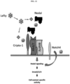
- Nodal Activation of Nodal includes binding to the co-receptor Cripto-1 and subsequent phosphorylation of the type I and type II activin-like kinase receptors (ALK).
- ALK activin-like kinase receptors
- SMAD2 and SMAD3 are activated ( Lee et. al., 2006, Nature Medicine 12:882-884 ).
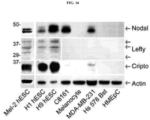
- human embryonic stem cells express Nodal and secrete endogenous inhibitors of Nodal such as Lefty A/B ( Besser, D., 2004, J. Biol. Chem. 279:45076-45084 ).
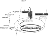
- Lefty A and Lefty B human homologs to murine Lefty 2 and Lefty 1, respectively, are separated by approximately 50 kb on chromosome q42 and are 96% identical to each other ( Kosaki et. al., 1999, Am. J. Hum. Genet. 64:712-21 ).
- Lefty A and Lefty B are members of the TGF ⁇ superfamily, and are considered amongst the most powerful inhibitors of Nodal.
- Nodal is reactivated and aberrantly upregulated in many different forms of aggressive cancer; however, Lefty is silenced - allowing Nodal to signal in an unregulated manner ( Postovit et. al., 2008, Proc. Natl. Acad. Sci. U.S.A. 105:4329-4334 .
- YAN, LT ET AL. "Preparation of Nodal Antibody and Development of its ELISA Kit. Journal of Chongqing University of Technology", NATURAL SCIENCE, vol. 26, no. 9, 2012, pages 31-36 discloses generation of monoclonal and polyclonal antibodies against nodal mature peptide and their application in ELISA, western blot FACS, histochemistry.
- STRIZZI, LET AL. "Nodal as a Biomarker for Melanoma Progression and a New Therapeutic Target for Clinical Intervention", EXPERT REVIEW OF DERMATOL, vol. 4, no. 1, 2009, pages 67-78 discloses potential therapeutic efficacy of function-blocking anti nodal antibody in a murine melanoma model.
- the antibody of the present invention as defined in the attached claim set differs from the antibodies of said prior art in the CDR sequence, wherein the claimed antibody has a heavy chain variable domain comprising the amino acid sequence of SEQ ID NO:3, and a light chain variable do-main comprising the amino acid sequence of SEQ ID NO: 1.
- the present invention relates to anti-Nodal antibodies and methods of using the same (e.g., use of the anti-Nodal antibodies for diagnosing, preventing and treating a Nodal-related disorder or disease).
- the invention provides antibodies as defined in the claims that bind to Nodal.
- the specification further relates to a monoclonal antibody that binds to Nodal, wherein the antibody comprises: (a) an HVR-H1 comprising the amino acid sequence of SEQ ID NO:7; (b) an HVR-H2 comprising the amino acid sequence of SEQ ID NO:8; (c) an HVR-H3 comprising the amino acid sequence of SEQ ID NO:9; (d) an HVR-L1 comprising the amino acid sequence of SEQ ID NO:4; (e) an HVR-L2 comprising the amino acid sequence of SEQ ID NO:5; and (f) an HVR-L3 comprising the amino acid sequence of SEQ ID NO:6.
- the antibody may further comprise at least one human framework region.
- the human framework region may comprise a human VH Acceptor 2 framework.
- the human framework region may comprise a human VL kappa subgroup I consensus framework.
- the specification further relates to monoclonal antibodies that bind to Nodal, wherein the antibodies comprise a heavy chain variable domain having at least 70%, at least 75%, at least 80%, at least 85%, at least 90%, or at least 95% sequence identity to the amino acid sequence of SEQ ID NO:2.
- the monoclonal antibodies may comprise a light chain variable domain having at least 70%, at least 75%, at least 80%, at least 85%, at least 90%, or at least 95% sequence identity to the amino acid sequence of SEQ ID NO:1.
- the antibody may further comprise at least one human framework region.
- the human framework region may comprise a human VH Acceptor 2 framework.
- the human framework region may comprise a human VL kappa subgroup I consensus framework.
- the specification further relates to a monoclonal antibody that binds to Nodal, wherein the antibody comprises an HVR-H1 comprising the amino acid sequence of SEQ ID NO: 10; an HVR-L1 comprising the amino acid sequence of SEQ ID NO:4; an HVR-L2 comprising the amino acid sequence of SEQ ID NO:5; and an HVR-L3 comprising the amino acid sequence of SEQ ID NO:6.
- the antibody may further comprise at least one human framework region.
- the human framework region may comprise a human VH Acceptor 2 framework.
- the human framework region may comprise a human VL kappa subgroup I consensus framework.
- the specification further relates to a monoclonal antibody that binds to Nodal, wherein the antibody comprises an HVR-H1 comprising the amino acid sequence of SEQ ID NO:7; an HVR-L1 comprising the amino acid sequence of SEQ ID NO:4; an HVR-L2 comprising the amino acid sequence of SEQ ID NO:5; and an HVR-L3 comprising the amino acid sequence of SEQ ID NO:6.
- the antibody may further comprise at least one human framework region.
- the human framework region may comprise a human VH Acceptor 2 framework.
- the human framework region may comprise a human VL kappa subgroup I consensus framework.
- the specification further relates to an isolated antibody that binds to an epitope in SEQ ID NO: 13.
- An isolated antibody may be provided that binds to the Nodal pre-helix loop region.
- the anti-Nodal antibody may be an antibody fragment selected from a Fab, Fab'-SH, Fv, scFv, or (Fab') 2 fragment as defined in claim 2.
- the invention also provides an in vitro method of inhibiting Nodal activity using an anti- Nodal antibody as defined in the claims.
- the invention provides an in vitro method of inhibiting Nodal activity comprising exposing a cell that expresses Nodal to an antibody according to claim 1.
- the invention provides an effective amount of an anti-Nodal antibody for use in a method of treating a disorder associated with increased expression or activity of Nodal, wherein said disorder is cancer.
- the antibody may be administered with at least one or more therapeutic agents - in a combinatorial or sequential manner. There is no limitation in this regard. Indeed, a variety of agents may be administered with an antibody of the invention including, but not limited to, a chemotherapeutic agent or agents described herein.
- the therapeutic agent may be Lefty protein (e.g., recombinant Lefty protein).
- the invention also provides an in-vitro method for diagnosing a cancer in a subject, comprising contacting a sample (e.g., a biological sample described herein) from the subject with an antibody of claim 1 and determining the amount of antibody that is bound to the sample, and wherein an increase in Nodal as compared to a normal noncancerous control is indicative of a cancer.
- a sample e.g., a biological sample described herein
- an antibody of claim 1 e.g., a biological sample described herein
- an increase in Nodal as compared to a normal noncancerous control is indicative of a cancer.
- any cancer described herein may be detected (e.g., including, but not limited to, glioblastoma, neuroblastoma, melanoma, breast cancer, pancreatic cancer, ovarian cancer, bladder cancer, colon cancer, prostate cancer, and leukemia).
- the cancer may express Nodal protein.
- Any type of biological sample from a subject/patient may be tested for Nodal expression including, but not limited to, tissue, blood, feces, plasma, bodily fluid, serum, saliva, lung effusion, sputum, urine, and intestinal scraping.
- the binding of an antibody to Nodal may be detected by a label.
- a label There is no limitation by the type of label.
- a variety of labels may be used including, but not limited to, an enzymatic label, a fluorescent label, a chemiluminescent label, a radioactive label, and a dye label.
- Nodal may be detected using any immunoassay known in the art including, but not limited to, an enzyme linked immunosorbent assay or radioimmunoassay or variations thereof.
- Nodal may be also detected using any label-free technology where the antibody is used to capture the protein. Label-free technologies include SPR, Bio-Layer Interferometry (BLI), Long Period Gratings (LPG), etc.
- antibody refers to monoclonal antibodies (including full length monoclonal antibodies), polyclonal antibodies, and multispecific antibodies (e.g., antibodies specific for more than one target) as well as antibody fragments (e.g., that exhibit binding to the same target as full length antibody).
- Antibodies of the invention may be any type (e.g., IgG, IgE, IgM, IgD, IgA), class (e.g., IgG1, IgG2, IgG3, IgG4, IgA1 and IgA2) or subclass.
- anti-Nodal antibody refers to an antibody which binds specifically to human Nodal (e.g., binds in a way that Nodal activity is inhibited).
- An “anti-Nodal antibody” may be an antibody that binds to human Nodal that allows detection, diagnosis, or predetermination of a disease or disorder associated with Nodal expression and/or activity, or that is used in a therapeutic composition as defined in the claims (e.g., to treat and/or prevent Nodal-related disorder or disease).
- neutralizable epitope refers to a determinant portion of a protein, binding of which by an appropriate antibody will result in inhibition of a function of the protein.
- a “neutralizable Nodal epitope” is a determinant portion of the Nodal protein, the binding of which by an antibody inhibits Nodal interaction with Cripto-1 or the Cripto-1 coreceptor complex, or inhibits downstream signaling from Nodal or its complex with Cripto-1.
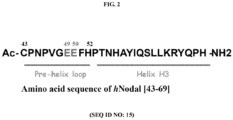
- a neutralizable Nodal epitope may be a polypeptide comprising the amino acid sequence of SEQ ID NO: 13, 15, or 17.
- neutralizing antibody refers to an antibody which is capable of specifically binding to a neutralizable epitope on a protein and substantially inhibiting or eliminating a biologically (e.g., complex formation, downstream signaling, etc.) activity of the protein.
- variable refers to structural features of the variable domain itself that differ extensively in sequence among all antibodies and the portions of the antibody that provide specificity for binding between the antibody and its specific target. These structural features within the variable domain are called hypervariable regions (HVRs) or complementarity determining regions (CDRs) and occur in both the light chain and heavy chain variable domains. There are three heavy chain HVRs or CDRs (HVRH1 or CDRH1 or H1, HVRH2 or CDRH2 or H2, and HVRH3 or CDRH3 or H3). Likewise, there are three light chain CDRs (HVRL1 or CDRL1 or L1, HVRL2 or CDRL2 or L2, and HVRL3 or CDRL3 or L3).
- variable domains of native heavy and light chains each comprise four FR regions, largely adopting a ⁇ -sheet configuration, connected by three CDRs, which form loops connecting, and in some cases forming part of, the ⁇ -sheet structure.
- the CDRs in each chain are held together in close proximity by the FR regions and, with the CDRs from the other chain, contribute to the formation of the target binding site of antibodies (see Kabat, et al. Sequences of Proteins of Immunological Interest, National Institutes of Health, Bethesda, Md. ,1987 ).
- numbering of immunoglobulin amino acid residues is done according to the immunoglobulin amino acid residue numbering system of Kabat, et al., unless otherwise indicated.
- the residues that make up these six CDRs have been characterized by Kabat as follows: residues 24-34 (CDRL1), 50-56 (CDRL2) and 89-97 (CDRL3) in the light chain variable region and 31-35 (CDRH1), 50-65 (CDRH2) and 95-102 (CDRH3) in the heavy chain variable region; Kabat et al., (1991) Sequences of Proteins of Immunological Interest, 5th Ed.
- fully human framework means a framework with an amino acid sequence found naturally in humans.
- fully human frameworks include, but are not limited to, KOL, NEWM, REI, EU, TUR, TEI, LAY and POM (See, e.g., Kabat et al., (1991) Sequences of Proteins of Immunological Interest, US Department of Health and Human Services, NIH, USA ; and Wu et al., (1970) J. Exp. Med. 132, 211-250 ).
- Humanized antibodies described herein may have fully human frameworks, or frameworks with one or more amino acids changed (e.g., to accommodate CDRs as described herein).
- Humanized forms of non-human (e.g., murine) antibodies may be chimeric immunoglobulins, immunoglobulin chains or fragments thereof (such as Fv, Fab, Fab', F(ab') 2 or other target-binding subsequences of antibodies) which contain minimal sequence derived from non-human immunoglobulin.
- the humanized antibody will comprise substantially all of at least one, and typically two, variable domains, in which all or substantially all of the CDR regions correspond to those of a non-human immunoglobulin and all or substantially all of the FR regions are those of a human immunoglobulin template sequence.
- the humanized antibody may also comprise at least a portion of an immunoglobulin constant region (Fc), typically that of a human immunoglobulin template chosen.
- humanized antibodies have one or more CDRs (one, two, three, four, five, six) which are altered with respect to the original antibody (e.g., affinity matured), which are also termed one or more CDRs "derived from" one or more CDRs from the original antibody.
- antibody fragment refers to a portion of a full-length antibody, generally the target binding or variable region.
- antibody fragments include F(ab), F(ab'), F(ab') 2 and Fv fragments.
- the phrase "functional fragment or analog" of an antibody is a compound having qualitative biological activity in common with a full-length antibody.
- a functional fragment or analog of an anti-Nodal antibody is one which can bind to Nodal in such a manner so as to prevent or substantially reduce the ability of Nodal to bind to its receptor (e.g., Cripto-1 or Alk4/7/ActRIIB receptor complex) and/or initiate signaling (e.g. through the Alk4/7/ActRIIB receptor complex).
- “functional fragment” with respect to antibodies refers to Fv, F(ab) and F(ab') 2 fragments.
- An “Fv” fragment consists of a dimer of one heavy and one light chain variable domain in a tight, non-covalent association (V H -V L dimer). It is in this configuration that the three CDRs of each variable domain interact to define a target binding site on the surface of the V H -V L dimer.
- V H -V L dimer tight, non-covalent association
- Single-chain Fv or “sFv” antibody fragments comprise the V H and V L domains of an antibody, wherein these domains are present in a single polypeptide chain.
- the Fv polypeptide further comprises a polypeptide linker between the V H and V L domains which enables the sFv to form the desired structure for target binding.
- diabodies refers to small antibody fragments with two antigen-binding sites, which fragments comprise a heavy chain variable domain (V H ) connected to a light chain variable domain (V L ) in the same polypeptide chain.
- the F(ab) fragment contains the constant domain of the light chain and the first constant domain (CH1) of the heavy chain.
- F(ab') fragments differ from F(ab) fragments by the addition of a few residues at the carboxyl terminus of the heavy chain CH1 domain including one or more cysteines from the antibody hinge region.
- F(ab') fragments are produced by cleavage of the disulfide bond at the hinge cysteines of the F(ab') 2 pepsin digestion product.
- Monoclonal antibody refers to an antibody obtained from a population of substantially homogeneous antibodies, i.e., the individual antibodies comprising the population are identical except for possible naturally occurring mutations that may be present in minor amounts.
- Monoclonal antibodies herein specifically include "chimeric" antibodies (immunoglobulins) in which a portion of the heavy and/or light chain is identical with or homologous to corresponding sequences in antibodies derived from a particular species or belonging to a particular antibody class or subclass, which the remainder of the chain(s) is identical with or homologous to corresponding sequences in antibodies derived from another species or belonging to another antibody class or subclass, as well as fragments of such antibodies, so long as they exhibit the desired biological activity.
- Monoclonal antibodies are highly specific, being directed against a single target site. Furthermore, in contrast to conventional (polyclonal) antibody preparations which typically include different antibodies directed against different determinants (epitopes), each monoclonal antibody is directed against a single determinant on the target. In addition to their specificity, monoclonal antibodies are advantageous in that they may be synthesized by the hybridoma culture, uncontaminated by other immunoglobulins.
- the modifier "monoclonal" indicates the character of the antibody as being obtained from a substantially homogeneous population of antibodies, and is not to be construed as requiring production of the antibody by any particular method.
- Such antibodies may be of any immunoglobulin class including IgG, IgM, IgE, IgA, and any subclass thereof.
- a hybridoma producing a mAb of the present invention may be cultivated in vitro, in situ or in vivo.
- bispecific antibody is refers to any immunoreactive agent having two different antigen-binding regions defined by different antibody sequences.
- the different targets may be epitopes on separate target species (e.g., Nodal and Cripto-1 or different epitopes in one target species (e.g., Nodal).
- Non-limiting examples of drugs that may be included in the antibody-drug-conjugates are mitotic inhibitors, antitumor antibiotics, immunomodulating agents, gene therapy vectors, alkylating agents, antiangiogenic agents, antimetabolites, boron-containing agents, chemoprotective agents, hormones, antihormone agents, corticosteroids, photoactive therapeutic agents, oligonucleotides, radionuclide agents, topoisomerase inhibitors, tyrosine kinase inhibitors, and radiosensitizers.
- the term "patient” preferably refers to a human in need of treatment (e.g., to treat cancer, or a precancerous condition or lesion).
- the term “patient” can also refer to non-human animals, preferably mammals such as dogs, cats, horses, cows, pigs, sheep and non-human primates, among others, that are in need of treatment.
- the term "purified” or “to purify” refers to the removal of contaminants from a sample.
- anti-Nodal antibodies may be purified by removal of contaminating non-immunoglobulin proteins; they are also purified by the removal of immunoglobulins that do not bind to the same antigen.
- the removal of non-immunoglobulin proteins and/or the removal of immunoglobulins that do not bind the particular antigen results in an increase in the percentage of antigen specific immunoglobulins in the sample.
- recombinant antigen-specific polypeptides are expressed in bacterial host cells and the polypeptides are purified by the removal of host cell proteins; the percentage of recombinant antigen-specific polypeptides is thereby increased in the sample.
- an "isolated" antibody or antibody fragment is one that has been identified and separated and/or recovered from a component of its natural environment. Contaminant components of its natural environment are materials that would interfere with diagnostic or therapeutic uses for the antibody or fragment thereof, and may include enzymes, hormones, and other proteinaceous or non-proteinaceous solutes.
- the isolated antibody may be purified (1) to greater than 95% by weight of polypeptides as determined by the Lowry method, and preferably, more than 99% by weight, (2) to a degree sufficient to obtain at least 15 residues of N-terminal or internal amino acid sequence by use of a spinning cup sequenator, (3) to homogeneity by SDS-page under reducing or nonreducing conditions using Coomassie blue, or silver stain, or (4) using chromatography.
- An isolated antibody includes the antibody in situ within recombinant cells since at least one component of the polypeptide's natural environment will not be present. Ordinarily, however, an isolated antibody will be prepared by a least one purification step.
- An anti-idiotypic (anti-Id) antibody is an antibody which recognizes unique determinants generally associated with the antigen-binding site of an antibody.
- An Id antibody can be prepared by immunizing an animal of the same species and genetic type (e.g., mouse strain) as the source of the mAb with the mAb to which an anti-Id is being prepared. The immunized animal will recognize and respond to the idiotypic determinants of the immunizing antibody by producing an antibody to these idiotypic determinants (the anti-Id antibody).
- substantially identical with respect to an antibody chain polypeptide sequence may be construed as an antibody chain exhibiting at least 70%, or 80%, or 90%, or 95% sequence identity to the reference polypeptide sequence.
- nucleic acid sequence may be construed as a sequence of nucleotides exhibiting at least about 85%, or 90%, or 95%, or 97% sequence identity to the reference nucleic acid sequence.
- Typical eukaryotic host cells are mammalian, such as Chinese hamster ovary or cells of human origin.
- the introduced DNA sequence may be from the same species as the host cell of a different species from the host cell, or it may be a hybrid DNA sequence, containing some foreign and some homologous DNA.
- the terms “cell,” “cell line,” and “cell culture” include progeny. It is also understood that all progeny may not be precisely identical in DNA content, due to deliberate or inadvertent mutations. Variant progeny that have the same function or biological property, as screened for in the originally transformed cell, are included.
- the "host cells” used in the present invention generally are prokaryotic or eukaryotic hosts.
- vector means a DNA construct containing a DNA sequence which is operably linked to a suitable control sequence capable of effecting the expression of the DNA in a suitable host.
- control sequences include a promoter to effect transcription, an optional operator sequence to control such transcription, a sequence encoding suitable mRNA ribosome binding sites, and sequences which control the termination of transcription and translation.
- the vector may be a plasmid, a phage particle, or simply a potential genomic insert. Once transformed into a suitable host, the vector may replicate and function independently of the host genome, or may in some instances, integrate into the genome itself.
- plasmid and vector are sometimes used interchangeably, as the plasmid is the most commonly used form of vector. However, it is intended to include such other forms of vectors which serve equivalent function as and which are, or become, known in the art.
- label when used herein refers to a detectable compound or composition which can be conjugated directly or indirectly to a molecule or protein, e.g., an antibody.
- the label may itself be detectable (e.g., radioisotope labels or fluorescent labels) or, in the case of an enzymatic label, may catalyze chemical alteration of a substrate compound or composition which is detectable.
- solid phase means a non-aqueous matrix to which the antibody of the present invention can adhere.
- solid phases include those formed partially or entirely of glass (e.g., controlled pore glass), polysaccharides (e.g., agarose), polyacrylamides, polystyrene, polyvinyl alcohol, and silicones.
- the solid phase can comprise the well of an assay plate; in others it is a purification column (e.g., an affinity chromatography column).
- cancers include chronic myeloid leukemia, acute lymphoblastic leukemia, Philadelphia chromosome positive acute lymphoblastic leukemia (Ph+ ALL), squamous cell carcinoma, small-cell lung cancer, non-small cell lung cancer, glioma, gastrointestinal cancer, renal cancer, ovarian cancer, liver cancer, colorectal cancer, endometrial cancer, kidney cancer, prostate cancer, thyroid cancer, neuroblastoma, pancreatic cancer, glioblastoma multiforme, cervical cancer, stomach cancer, bladder cancer, hepatoma, breast cancer, colon carcinoma, and head and neck cancer, gastric cancer, germ cell tumor, pediatric sarcoma, sinonasal natural killer, multiple myeloma, acute myelogenous leukemia (AML), chronic lymphocytic leukemia (CML), and other cancers described herein.
- AML acute myelogenous leukemia
- CML chronic lymphocytic leukemia
- Leukemia refers to progressive, malignant diseases of the blood-forming organs and is generally characterized by a distorted proliferation and development of leukocytes and their precursors in the blood and bone marrow. Leukemia is generally clinically classified on the basis of (1) the duration and character of the disease-acute or chronic; (2) the type of cell involved; myeloid (myelogenous), lymphoid (lymphogenous), or monocytic; and (3) the increase or non-increase in the number of abnormal cells in the blood-leukemic or aleukemic (subleukemic).
- Leukemia includes, for example, acute nonlymphocytic leukemia, chronic lymphocytic leukemia, acute granulocytic leukemia, chronic granulocytic leukemia, acute promyelocytic leukemia, adult T-cell leukemia, aleukemic leukemia, a leukocythemic leukemia, basophylic leukemia, blast cell leukemia, bovine leukemia, chronic myelocytic leukemia, leukemia cutis, embryonal leukemia, eosinophilic leukemia, Gross' leukemia, hairy-cell leukemia, hemoblastic leukemia, hemocytoblastic leukemia, histiocytic leukemia, stem cell leukemia, acute monocytic leukemia, leukopenic leukemia, lymphatic leukemia, lymphoblastic leukemia, lymphocytic leukemia, lymphogenous leukemia, lymphoid leukemia, lymphosarcoma cell le
- overexpression of Nodal protein and "aberrant expression of Nodal protein” are intended to indicate a reactivation of the embryonic Nodal signaling pathway or an abnormal level of expression of the Nodal protein in a cell (e.g., within a cancer or tumor or other disease process in which Nodal is elevated) within a specific tissue or organ of a patient relative to the level of expression in a normal cell from that tissue or organ.
- patients having a cancer characterized by overexpression or aberrant expression of Nodal can be determined by standard assays known in the art.
- Overexpression or aberrant expression can be measured in fixed cells of frozen or paraffin-embedded tissue sections using immunohistochemical (IHC) detection. When coupled with histological staining, localization of the targeted protein can be determined and extent of its expression (e.g., within a tumor) can be measured both qualitatively and semi-quantitatively.
- IHC immunohistochemical
- Nodal positive cancer refers to a cancer disease such as breast cancer, melanoma or other type of cancer described herein which is characterized by an overexpression or aberrant expression of Nodal.
- relapsed cancer refers to the uncontrolled growth of abnormal cells in tumor patients who initially responded to previous therapy, but in whom the therapeutic response was not maintained.
- relapsed Nodal positive cancer refers to the uncontrolled growth of abnormal cells characterized by Nodal protein overexpression or aberrant expression in tumor patients who initially responded to previous therapy with an anti-Nodal antibody, but in whom the therapeutic response was not maintained during treatment with the anti-Nodal antibody.
- a therapeutic response can be established based on the medical judgment of a practitioner ascertained by the results from clinical and laboratory data that are generally known in the art to assess patient treatment. Such data may be obtained, by way of example, from clinical examination, cytological and histological techniques, endoscopy and laparoscopy, ultrasound, CT and MRI scans, chest X-ray and mammography, and measuring the concentration of tumor markers, such as Nodal. Preferably RECIST criteria may be used to determine tumor response (RE). ( Therasse et al., J. Nat. Cancer Institute. 92 (2000) 205-216 ).
- tumor response for solid tumors is categorized in dependency of the volume progression or regression of the tumors (e.g. measured via CT) into four levels: complete response (CR) or partial response (PR), stable disease (SD) and progressive disease (PD).
- EORTC European Organization for Research and Treatment of Cancer
- FDG-PET 2-[18F]-Fluoro-2-deoxyglucose positron emission tomography
- RE and NR may be established based on data acquired by the combination of computer tomography (CT) and 2-(18F)-Fluoro-2-deoxyglucose positron emission tomography (FDG-PET) ( Kellof, G. J., et al, Clin Canc Res 11 (2005) 2785-2808 and Young H., et al., Eur J Canc 35 (1999) 1773-82 ) using both the RECIST and FDG-PET criteria described above.
- CT computer tomography
- FDG-PET 2-(18F)-Fluoro-2-deoxyglucose positron emission tomography
- method for manufacturing a medicament refers to the manufacturing of a medicament for use in an indication as specified herein and in particular for use in the treatment of tumors, tumor metastases, or cancer in general.
- treating means reversing, alleviating, inhibiting the progress of, or preventing, either partially or completely, the growth of tumors, tumor metastases, or other cancer-causing or neoplastic cells in a patient.
- treatment refers to the act of treating.
- a method of treating when applied to, for example, cancer refers to a procedure or course of action that is designed to reduce or eliminate the number of cancer cells in a patient, or to alleviate the symptoms of a cancer.
- a method of treating does not necessarily mean that the cancer cells or other disorder will, in fact, be eliminated, that the number of cells or disorder will, in fact, be reduced, or that the symptoms of a cancer or other disorder will, in fact, be alleviated.
- a method of treating cancer will be performed even with a low likelihood of success, but which, given the medical history and estimated survival expectancy of a patient, is nevertheless deemed an overall beneficial course of action.
- the term "metastasis” refers to the transmission of cancerous cells from the primary tumor to one or more sites elsewhere in a patient. Means to determine if a cancer has metastasized are known in the art and include bone scan, chest X-ray, CAT scan, MRI scan, and tumor marker tests.
- medicament for preventing metastasis or “medicament for reducing metastasis” as used herein refer to use of a medicament as a prophylactic agent against metastasis in patient (e.g., with cancer (e.g., to inhibit or reduce a further transmission of cancerous cells from the primary tumor to one or more sites elsewhere in a patient).
- cancer e.g., to inhibit or reduce a further transmission of cancerous cells from the primary tumor to one or more sites elsewhere in a patient.
- metastasis of the primary, metastatic tumor or cancer may be prevented, delayed, or inhibited.
- Therapeutic compounds as described herein include, but are not limited to, antibodies as described herein (including fragments, analogs and derivatives thereof as described herein) and nucleic acids encoding antibodies of the invention as described herein (including fragments, analogs and derivatives thereof and anti-idiotypic antibodies as described herein).
- compositions or agent co-administered with an antibody composition include, but are not limited to, alkylating agents or agents with an alkylating action, such as cyclophosphamide (CTX; e.g. CYTOXAN), chlorambucil (CHL; e.g. LEUKERAN), cisplatin (CisP; e.g. PLATINOL busulfan (e.g.
- CX cyclophosphamide
- CHL chlorambucil
- isP e.g. PLATINOL busulfan
- MYLERAN melphalan
- BCNU carmustine
- streptozotocin triethylenemelamine
- TEM mitomycin C
- anti-metabolites such as methotrexate (MTX), etoposide (VP 16; e.g. VEPESID), 6-mercaptopurine (6 MP), 6-thiocguanine (6TG), cytarabine (Ara-C), 5-fluorouracil (5-FU), capecitabine (e.g. XELODA), dacarbazine (DTIC), and the like
- antibiotics such as actinomycin D, doxorubicin (DXR; e.g.
- ADRIAMYCIN daunorubicin (daunomycin), bleomycin, mithramycin and the like
- alkaloids such as vinca alkaloids such as vincristine (VCR), vinblastine, and the like
- antitumor agents such as paclitaxel (e.g. TAXOL) and paclitaxel derivatives, the cytostatic agents, glucocorticoids such as dexamethasone (DEX; e.g.
- DECADRON corticosteroids
- corticosteroids such as prednisone, nucleoside enzyme inhibitors such as hydroxyurea, amino acid depleting enzymes such as asparaginase, leucovorin and other folic acid derivatives, and similar, diverse antitumor agents.
- the following agents may also be used as additional agents: amifostine (e.g. ETHYOL), dactinomycin, mechlorethamine (nitrogen mustard), streptozocin, cyclophosphamide, lomustine (CCNU), doxorubicin lipo (e.g. DOXIL), gemcitabine (e.g. GEMZAR), daunorubicin lipo (e.g.
- DAUNOXOME procarbazine, mitomycin, docetaxel (e.g. TAXOTERE), aldesleukin, carboplatin, oxaliplatin, cladribine, camptothecin, CPT 11 (irinotecan), 10-hydroxy 7-ethyl-camptothecin (SN38), floxuridine, fludarabine, ifosfamide, idarubicin, mesna, interferon beta, interferon alpha, mitoxantrone, topotecan, leuprolide, megestrol, melphalan, mercaptopurine, plicamycin, mitotane, pegaspargase, pentostatin, pipobroman, plicamycin, tamoxifen, teniposide, testolactone, thioguanine, thiotepa, uracil mustard, vinorelbine, chlorambucil.
- Radiation therapy is a standard treatment for controlling unresectable or inoperable tumors and/or tumor metastases. Improved results have been seen when radiation therapy has been combined with chemotherapy. Radiation therapy is based on the principle that high-dose radiation delivered to a target area will result in the death of reproductive cells in both tumor and normal tissues.
- the radiation dosage regimen is generally defined in terms of radiation absorbed dose (Gy), time and fractionation, and must be carefully defined by the oncologist.
- the amount of radiation a patient receives will depend on various considerations, but the two most important are the location of the tumor in relation to other critical structures or organs of the body, and the extent to which the tumor has spread.
- a typical course of treatment for a patient undergoing radiation therapy will be a treatment schedule over a 1 to 6 week period, with a total dose of between 10 and 80 Gy administered to the patient in a single daily fraction of about 1.8 to 2.0 Gy, 5 days a week.
- a total dose of between 10 and 80 Gy administered to the patient in a single daily fraction of about 1.8 to 2.0 Gy, 5 days a week.
- the combination treatment as described herein and radiation There might be synergy when tumors in human patients are treated with the combination treatment as described herein and radiation.
- the inhibition of tumor growth by means of the agents comprising the combination or single therapy as described herein is enhanced when combined with radiation, optionally with additional chemotherapeutic or anticancer agents.
- compositions comprise a therapeutically effective amount of anti-Nodal antibodies and a physiologically acceptable carrier.
- physiologically acceptable may mean approved by a regulatory agency of the Federal or a state government or listed in the U.S. Pharmacopeia or other generally recognized pharmacopeia for use in animals, and more particularly in humans.
- a pharmaceutically acceptable carrier is any carrier known in the art for the delivery of an agent to a subject.
- carrier refers to a diluent, adjuvant, excipient, or vehicle with which the therapeutic is administered.
- physiological carriers can be sterile liquids, such as water and oils, including those of petroleum, animal, vegetable, or synthetic origin, such as peanut oil, soybean oil, mineral oil, sesame oil and the like.
- Water is a preferred carrier when the pharmaceutical composition is administered intravenously.
- Saline solutions and aqueous dextrose and glycerol solutions can also be employed as liquid carriers, particularly for injectable solutions.
- Suitable pharmaceutical excipients include starch, glucose, lactose, sucrose, gelatin, malt, rice, flour, chalk, silica gel, sodium stearate, glycerol monostearate, talc, sodium chloride, dried skim milk, glycerol, propylene, glycol, water, ethanol and the like.
- compositions can also contain minor amounts of wetting or emulsifying agents, or pH buffering agents.
- These compositions can take the form of solutions, suspensions, emulsion, tablets, pills, capsules, powders, sustained-release formulations and the like.
- the composition can be formulated as a suppository, with traditional binders and carriers such as triglycerides.
- Oral formulation can include standard carriers such as pharmaceutical grades of mannitol, lactose, starch, magnesium stearate, sodium saccharine, cellulose, magnesium carbonate, etc. Examples of suitable carriers are described in "Remington's Pharmaceutical Sciences" by E. W. Martin.
- compositions will contain an effective amount of the antibody, preferably in purified form, together with a suitable amount of carrier so as to provide the form for proper administration to the patient.
- a composition comprising a therapeutically effective amount of anti-Nodal antibodies and a physiologically acceptable carrier may be specifically formulated for the particular mode of administration.
- the composition may be formulated in accordance with routine procedures as a pharmaceutical composition adapted for intravenous administration to human beings.
- compositions for intravenous administration are solutions in sterile isotonic aqueous buffer.
- the composition may also include a solubilizing agent and a local anesthetic such as lignocaine to ease pain at the site of the injection.
- the ingredients are supplied either separately or mixed together in unit dosage form, for example, as a dry lyophilized powder or water free concentrate in a hermetically sealed container such as an ampoule or sachette indicating the quantity of active agent.
- composition is to be administered by infusion
- it can be dispensed with an infusion bottle containing sterile pharmaceutical grade water or saline.
- an ampoule of sterile water for injection or saline can be provided so that the ingredients may be mixed prior to administration.
- the invention also relates to a pharmaceutical pack comprising one or more containers filled with one or more of the ingredients of the pharmaceutical compositions described herein.
- Optionally associated with such container(s) can be a notice in the form prescribed by a governmental agency regulating the manufacture, use or sale of pharmaceuticals or biological products, which notice reflects approval by the agency of manufacture, use or sale for human administration.
- the container may include a package insert.
- package insert refers to instructions customarily included in commercial packages of a therapeutic product, which may include information about the indications, usage, dosage, administration, contraindications and/or warnings concerning the use of such therapeutic products.
- the invention also relates to therapeutic formulations that are prepared for storage as lyophilized formulations or aqueous solutions by mixing anti-Nodal antibodies having the desired degree of purity with optional "pharmaceutically-acceptable" carriers, excipients or stabilizers typically employed in the art (all of which are termed "excipients”), i.e., buffering agents, stabilizing agents, preservatives, isotonifiers, non-ionic detergents, antioxidants, and other miscellaneous additives. See Remington's Pharmaceutical Sciences, 16th edition, Osol, Ed. (1980 ). Such additives must be nontoxic to the recipients at the dosages and concentrations employed.
- Preservatives may be added to retard microbial growth, and may be added in amounts ranging from 0.2%-1% (w/v).
- exemplary preservatives include phenol, benzyl alcohol, meta-cresol, methyl paraben, propyl paraben, octadecyldimethylbenzyl ammonium chloride, benzalconium halides (e.g., chloride, bromide, iodide), hexamethonium chloride, and alkyl parabens such as methyl or propyl paraben, catechol, resorcinol, cyclohexanol, and 3-pentanol.
- stabilizers may be used, for example, polhydric sugar alcohols, preferably trihydric or higher sugar alcohols, such as glycerin, erythritol, arabitol, xylitol, sorbitol and mannitol.
- Stabilizers refer to a broad category of excipients which can range in function from a bulking agent to an additive which solubilizes the therapeutic agent or helps to prevent denaturation or adherence to the container wall.
- Typical stabilizers can be polyhydric sugar alcohols (enumerated above); amino acids such as arginine, lysine, glycine, glutamine, asparagine, histidine, alanine, ornithine, L-leucine, 2-phenylalanine, glutamic acid, threonine, etc., organic sugars or sugar alcohols, such as lactose, trehalose, stachyose, mannitol, sorbitol, xylitol, ribitol, myoinisitol, galactitol, glycerol and the like, including cyclitols such as inositol; polyethylene glycol; amino acid polymers; sulfur containing reducing agents, such as urea, glutathione, thioctic acid, sodium thioglycolate, thioglycerol, alpha.-monothioglycerol and sodium thiosulfate; low
- proteins such as human serum albumin, bovine serum albumin, gelatin or immunoglobulins
- hydrophylic polymers such as polyvinylpyrrolidone monosaccharides, such as xylose, mannose, fructose, glucose; disaccharides such as lactose, maltose, sucrose and trisaccacharides such as raffinose; and polysaccharides such as dextran.
- Stabilizers may be present in the range from 0.1 to 10,000 weights per part of weight active protein.
- Non-ionic surfactants or detergents may be used, such as, polysorbates (20, 40, 60, 80, etc.), polyoxamers (184, 188 etc.), PLURONIC, polyols, etc.
- Non-ionic surfactants may be present in a range of about 0.05 mg/ml to about 1.0 mg/ml, although less (e.g., below 0.05 mg/ml) or more (e.g., 1.0 mg/ml) may be used.
- Additional miscellaneous excipients include bulking agents, (e.g., starch), chelating agents (e.g., EDTA), antioxidants (e.g., ascorbic acid, methionine, vitamin E), and cosolvents.
- the formulation herein may also contain more than one active compound as necessary for the particular indication being treated, preferably those with complementary activities that do not adversely affect each other. For example, it may be desirable to further provide an immunosuppressive agent.
- Such molecules are suitably present in combination in amounts that are effective for the purpose intended.
- the active ingredients may also be encapsulated in microcapsule prepared, for example, by coascervation techniques or by interfacial polymerization, for example, hydroxymethylcellulose or gelatin-microcapsule and poly-(methylmethacylate) microcapsule, respectively, in colloidal drug delivery systems (for example, liposomes, albumin micropheres, microemulsions, nano-particles and nanocapsules) or in macroemulsions.
- colloidal drug delivery systems for example, liposomes, albumin micropheres, microemulsions, nano-particles and nanocapsules
- sustained-release preparations may be used. Suitable examples of sustained-release preparations include semi-permeable matrices of solid hydrophobic polymers containing the antibody variant, which matrices are in the form of shaped articles, e.g., films, or microcapsules.
- sustained-release matrices include polyesters, hydrogels (for example, poly(2-hydroxyethyl-methacrylate), poly(vinylalcohol)), polylactides, copolymers of L-glutamic acid and ethyl-L-glutamate, non-degradable ethylene-vinyl acetate, degradable lactic acid-glycolic acid copolymers such as the LUPRON DEPOT (injectable microspheres composed of lactic acid-glycolic acid copolymer and leuprolide acetate), and poly-D-(-)-3-hydroxybutyric acid.
- polyesters for example, poly(2-hydroxyethyl-methacrylate), poly(vinylalcohol)
- polylactides copolymers of L-glutamic acid and ethyl-L-glutamate
- non-degradable ethylene-vinyl acetate non-degradable ethylene-vinyl acetate
- Nodal immunogen and methods of generating antibodies using the same are disclosed.
- Nodal immunogen may be produced recombinantly or made using synthetic methods.
- Nodal immunogen may also be isolated and/or purified from a natural source (e.g., endogenous, cellular Nodal protein). Multiple forms of the Nodal immunogen useful for preparing antibodies will be readily apparent to those in the art.
- Nodal protein e.g., full length human Nodal or portions thereof
- Cells expressing Nodal may be used as immunogen.
- Such cells can be derived from a natural source (e.g., cancer cell lines) or may be cells which have been transformed by recombinant techniques to over-express Nodal.
- a gene or a cDNA encoding human Nodal may be cloned into a plasmid or other expression vector and expressed in any of a number of expression systems according to methods well known to those of skill in the art.
- a variety of nucleotide sequences encoding Nodal protein or polypeptides may be used based upon the known degeneracy of the genetic code.
- Non-human Nodal proteins may also be used as immunogen.
- Nodal protein may be fused and/or conjugated to other proteins/fragments and/or immunogenic substances (e.g., key hole limpet hemocyanin (KLH)). Conjugation and/or fusion may be used to assist in protein purification, e.g., by permitting the fusion protein to be isolated and purified by affinity chromatography, but can also be used to increase immunogenicity.
- Fusion proteins can be produced by culturing a recombinant cell transformed with a fusion nucleic acid sequence that encodes a protein including the fusion segment attached to either the carboxyl and/or amino terminal end of the protein.
- Fusion segments may include, but are not limited to, immunoglobulin Fc regions, glutathione-S-transferase, ⁇ -galactosidase, a poly-histidine segment capable of binding to a divalent metal ion, and maltose binding protein.
- Antibodies as described herein include, but are not limited to, polyclonal, monoclonal, monovalent, bispecific, heteroconjugate, multispecific, human, humanized or chimeric antibodies, single chain antibodies, single-domain antibodies, Fab fragments, F(ab') fragments, fragments produced by a Fab expression library, anti-idiotypic (anti-Id) antibodies (including, e.g., anti-Id antibodies to antibodies of the invention), and epitope-binding fragments of any of the above.
- An immunogenic composition comprising a Nodal immunogen can be used to immunize a mammal, such as a mouse, rat, rabbit, guinea pig, monkey, or human, to produce polyclonal antibodies.
- a Nodal immunogen can be conjugated to a carrier protein, such as bovine serum albumin, thyroglobulin, keyhole limpet hemocyanin or other carrier described herein.
- a carrier protein such as bovine serum albumin, thyroglobulin, keyhole limpet hemocyanin or other carrier described herein.
- various adjuvants can be used to increase the immunological response.
- adjuvants include, but are not limited to, Freund's adjuvant, mineral gels (e.g., aluminum hydroxide), and surface active substances (e.g.
- Such antibodies may be of any immunoglobulin class including IgG, IgM, IgE, IgA, IgD and any subclass thereof.
- the hybridoma producing monoclonal antibody as described herein may be cultivated in vitro or in vivo.
- humanized antibodies can be produced using recombinant methods.
- Antibodies which specifically bind to a particular antigen can contain antigen binding sites which are either partially or fully humanized, as disclosed in U.S. Pat. No. 5,565,332 .
- Completely human antibodies may be used and may be particularly desirable for therapeutic treatment of human patients.
- Human antibodies can be made by a variety of methods known in the art including phage display methods using antibody libraries derived from human immunoglobulin sequences.
- Single-chain antibodies also can be constructed using a DNA amplification method, such as PCR, using hybridoma cDNA as a template (See, e.g., Thirion et al., 1996, Eur. J. Cancer Prev. 5, 507-11 ).
- Single-chain antibodies can be mono- or bispecific, and can be bivalent or tetravalent. Construction of tetravalent, bispecific single-chain antibodies is taught, for example, in Coloma & Morrison, 1997, Nat. Biotechnol. 15, 159-63 . Construction of bivalent, bispecific single-chain antibodies is taught, for example, in Mallender & Voss, 1994, J. Biol. Chem. 269, 199-206 .
- Antibodies which specifically bind to a particular antigen also can be produced by inducing in vivo production in the lymphocyte population or by screening immunoglobulin libraries or panels of highly specific binding reagents as disclosed in the literature (See, e.g., Orlandi et al., Proc. Natl. Acad. Sci. 86, 3833 3837, 1989 ; Winter et al., Nature 349, 293 299, 1991 ).
- Antibody fragments are disclosed which recognize a specific epitopes (e.g., human Nodal (e.g., pre-helix loop region)) generated by any known technique.
- antibody fragments were derived via proteolytic digestion of intact antibodies (see, e.g., Morimoto, et al., J Biochem Biophys Methods 24:107 (1992 ); Brennan, et al., Science 229:81 (1985 )).
- Fab and F(ab').sub.2 fragments may be produced by proteolytic cleavage of immunoglobulin molecules, using enzymes such as papain (to produce Fab fragments) or pepsin (to produce F(ab') 2 fragments).
- Heteroconjugate antibodies can also be utilized.
- Heteroconjugate antibodies are composed of two covalently joined antibodies. Such antibodies have, for example, been proposed to target immune system cells to unwanted cells ( U.S. Pat. No. 4,676,980 ).
- monoclonal antibodies that bind Nodal and inhibit Nodal activity (e.g., inhibit Nodal binding to Cripto-1 and/or to Alk4/7/ActRIIB receptor complex, inhibit signaling downstream of Nodal or its complexes, downregulate Nodal expression, etc.).
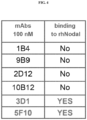
- monoclonal antibodies that bind Nodal and inhibit Nodal mediated cellular signaling may include the antibodies designated 1B4, 9B9, 2D12, 10B12, 3D1 and 5F10.
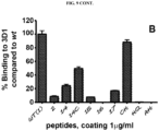
- Preferably the antibody designated 3D1 is used.
- the anti-Nodal antibodies were identified and characterized as described in the examples.
- Antibodies disclosed herein may be described or specified in terms of the epitope(s) or portion(s) of Nodal which they recognize or specifically bind.
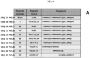
- the epitope(s) or polypeptide portion(s) may be specified as described herein, e.g., by N-terminal and C-terminal positions, by size in contiguous amino acid residues, or listed in the Tables and Figures.
- Antibodies as disclosed herein may also be described or specified in terms of their cross-reactivity. Antibodies that bind Nodal polypeptide, which have at least 95%, at least 90%, at least 85%, at least 80%, at least 75%, at least 70%, at least 65%, at least 60%, at least 55%, and at least 50% identity (as calculated using methods known in the art and described herein) to Nodal polypeptide are also included. As described herein, anti- Nodal antibodies may also bind with a K D of less than about 10 -7 M, less than about 10 -6 M, or less than about 10 -5 M to other proteins.
- Antibodies as disclosed herein may also be described or specified in terms of their binding affinity to a polypeptide of the invention.
- Preferred binding affinities include those with an equilibrium dissociation constant or K D from 10 -8 M to 10 -15 M, 10 -8 M to 10 -12 M, 10 -8 M to 10 -10 M, or 10 -10 M to 10 -12 M.
- antibodies that competitively inhibit binding of an antibody to an epitope as determined by any method known in the art for determining competitive binding, for example, the immunoassays described herein. The antibody may competitively inhibit binding to the epitope by at least 95%, at least 90%, at least 85%, at least 80%, at least 75%, at least 70%, at least 60%, or at least 50%.
- the nucleic acid encoding it is isolated and inserted into a replicable vector for further cloning (amplification of the DNA) or for expression.
- DNA encoding the antibody is readily isolated and sequenced using conventional procedures (e.g., by using oligonucleotide probes that are capable of binding specifically to genes encoding the heavy and light chains of the antibody variant). Standard techniques for cloning and transformation may be used in the preparation of cell lines expressing the antibodies discloed herein.
- antibodies as described herein can be produced by any method known in the art for the synthesis of antibodies, in particular, by chemical synthesis or preferably, by recombinant expression techniques.
- recombinant expression of an antibody as described herein, or fragment, derivative, or analog thereof, requires construction of an expression vector containing a polynucleotide that encodes the antibody or a fragment of the antibody.
- the vector for the production of the antibody may be produced by recombinant DNA technology.
- An expression vector is constructed containing antibody coding sequences and appropriate transcriptional and translational control signals. These methods include, for example, in vitro recombinant DNA techniques, synthetic techniques, and in vivo genetic recombination.
- the expression vector is transferred to a host cell by conventional techniques and the transfected cells are then cultured by conventional techniques to produce an antibody of the invention.
- vectors encoding both the heavy and light chains may be co-expressed in the host cell for expression of the entire immunoglobulin molecule.
- mammalian cells such as CHO, in conjunction with a vector such as the major intermediate early gene promoter element from human cytomegalovirus, are an effective expression system for antibodies ( Foecking, et al., Gene 45:101 (1986 ); Cockett, et al., Bio/Technology 8:2 (1990 )).
- a host cell strain may be chosen which modulates the expression of the inserted sequences, or modifies and processes the gene product in the specific fashion desired. Such modifications (e.g., glycosylation) and processing (e.g., cleavage) of protein products may be important for the function of the protein.
- Different host cells have characteristic and specific mechanisms for the post-translational processing and modification of proteins and gene products. Appropriate cell lines or host systems can be chosen to ensure the correct modification and processing of the foreign protein expressed.
- eukaryotic host cells which possess the cellular machinery for proper processing of the primary transcript, glycosylation, and phosphorylation of the gene product may be used.
- mammalian host cells include, but are not limited to, CHO, COS, HEK293, NIH3T3, or myeloma cells.
- An antibody as described herein may be purified by any method known in the art for purification of an immunoglobulin molecule, for example, by chromatography (e.g., ion exchange, affinity, particularly by affinity for the specific antigen after Protein A, and size-exclusion chromatography), centrifugation, differential solubility, or by any other standard technique for the purification of proteins.
- chromatography e.g., ion exchange, affinity, particularly by affinity for the specific antigen after Protein A, and size-exclusion chromatography
- centrifugation e.g., centrifugation, differential solubility, or by any other standard technique for the purification of proteins.
- the antibodies or fragments thereof can be fused to heterologous polypeptide sequences described herein or otherwise known in the art, to facilitate purification.
- Conjugates of antibodies or antibody fragments with one or more additional functional agents are encompassed.
- antibody-drug-conjugates or fragment-drug-conjugates are provided (e.g., wherein the functional agent is a drug). Binding of the antibody to its target epitope may localize the functional agent (e.g., drug) to target cells, thereby increasing the efficacy of the functional agent. the efficacy of the functional agent and antibody may be additive. Synergy(ies) between the mode of action of the functional agent and antibody may result in greater than additive increase in efficacy of the conjugate over the individual components.
- the antibody variant can be produced intracellularly, in the periplasmic space, or directly secreted into the medium. If the antibody variant is produced intracellularly, as a first step, the particulate debris, either host cells or lysed fragments, may be removed, for example, by centrifugation or ultrafiltration. Carter, et al., Bio/Technology 10:163 (1992 ) describe a procedure for isolating antibodies which are secreted to the periplasmic space of E. coli.
- supernatants from such expression systems are generally first concentrated using a commercially available protein concentration filter, for example, an AMICON or MILLIPORE ultrafiltration unit.
- Antibody generated and/or isolated from the cells can be purified using, for example, hydroxylapatite chromatography, gel elecrophoresis, dialysis, and affinity chromatography.
- the suitability of protein A as an affinity ligand depends on the species and isotype of any immunoglobulin Fc domain that is present in the antibody variant.
- Protein A can be used to purify antibodies that are based on human IgG1, IgG2 or IgG4 heavy chains ( Lindmark, et al., J Immunol Meth 62:1 (1983 )).
- a mixture comprising antibody and contaminants may be further subjected to low pH hydrophobic interaction chromatography using an elution buffer (e.g., at a pH between about 2.5-4.5, preferably performed at low salt concentrations (e.g., from about 0-0.25 M salt)).
- an elution buffer e.g., at a pH between about 2.5-4.5, preferably performed at low salt concentrations (e.g., from about 0-0.25 M salt)
- an antibody as described herein may be used to detect Nodal inin vitro vivo diagnostic methods.
- an antibody as described herein may be used in immunoassays (e.g., ELISA (e.g., sandwich ELISA, direct ELISA, indirect ELISA, competitive ELISA, etc.), Western blot, immunohistochemistry, protein array, immuno-PCR, etc.) for qualitatively and quantitatively measuring levels of Nodal in a biological sample (e.g., See, e.g., Harlow, et al., Antibodies: A Laboratory Manual, Cold Spring Harbor Laboratory Press, 2nd ed. (1988 )). There is no limiation to any particular type of biological sample.
- samples may be characterized using the diagnostic methods described herein, including, but not limited to, any sample in which Nodal protein is found.
- the sample may be tissue, a bodily fluid, blood, serum, urine, saliva, sputum, or a lung effusion.
- a sample may comprise cells expressing or suspected of expressing Nodal, Cripto-1, or both Nodal and Cripto-1.
- a sample may comprise cells that don't express Nodal, but test positive for Nodal due to paracrine signaling.
- An antibody as described herein may be recombinantly fused or conjugated to molecules useful as labels in detection assays. Any suitable antibodies, antibody fragments, bispecific antibodies, conjugated antibodies, etc.
- Detection of Nodal in a sample may provide diagnostic and/or prognostic information for a clinician. Detection of Nodal in a sample may be indicative of, or diagnostic for, an aggressive form of cancer. Nodal diagnostics described herein may be performed with one or more additional diagnostic assays to determine the type of cancer a subject suffers from, and/or to determine an appropriate treatmetn course of action for the subject (e.g., treatment with anti-Nodal antibodies, other cancer treatments, etc.).
- An antibody as described herein may be modified (e.g., via covalent attachment of a moiety to the antibody).
- an antibody is modified in such a way that the attachment of a moiety thereto does not interfere with the antibody binding to Nodal.
- An antibody may be modified via biotinylation, attachment to an enzyme, or any other type of moiety binding that allows detection of the antibody.
- an antibody or fragments thereof is conjugated to a diagnostic agent.
- the antibodies can be used diagnostically in vitro, for example, to detect expression of a target of interest in specific cells, tissues, plasma, blood or serum; or to monitor the development or progression of an immunologic response as part of a clinical testing procedure to, e.g., determine the efficacy of a given treatment regimen. Detection can be facilitated by coupling the antibody to a detectable substance. Examples of detectable substances are known in the art an include, but are not limited to, various enzymes, prosthetic groups, fluorescent materials, luminescent materials, bioluminescent materials, radioactive materials, positron emitting metals using various positron emission tomographies, and nonradioactive paramagnetic metal ions.
- the detectable substance may be coupled or conjugated either directly to the antibody (or fragment thereof) or indirectly, through an intermediate (such as, for example, a linker known in the art) using techniques known in the art.
- a label may be indirectly conjugated to the antibody (e.g., biotin-avidin conjugation).
- An immunoassay e.g., an enzyme linked immunosorbent assay or radioimmunoassay
- An antibody as described herein may be used in any known detection assay in the art including, but not limited to competitive binding assays, direct and indirect sandwich assays, and immunoprecipitation assays.
- Detection of Nodal can be achieved also using label-free techniques employing anti-Nodal antibodies as described herein immobilized on the surface of suitable biochips or biosurfaces as those used for SPR, Bio-Layer Interferometry (BLI), Long Period Gratings (LPG).
- BLI Bio-Layer Interferometry
- LPG Long Period Gratings
- Detection of an anti-Nodal antibody may be via use of an antibody that detects the anti-Nodal antibody (e.g., a secondary antibody).
- an antibody, and derivatives and analogs thereof, which specifically bind to Nodal can be used for diagnostic purposes to detect, diagnose, or monitor diseases, disorders, and/or conditions associated with the aberrant expression, overexpression and/or activity of Nodal.
- Nodal e.g., overexpression or aberrant expression of Nodal
- a biological sample from a subjection or patient e.g., a subject or patient having or suspected of having cancer
- Nodal specific antibodies as described herein and comparing the level of expression with standard (e.g., noncancerous control) expression level, whereby an increase or decrease in Nodal expression and/or activity compared to the standard expression level is indicative of aberrant expression and/or activity.
- Anti-Nodal antibody can be used to detect Nodal in any sample. Detecting may comprise contacting the sample with anti-Nodal antibody and determining the amount of antibody that is bound to the sample. For immunohistochemistry, the sample may be fresh or frozen or may be embedded in paraffin and fixed with a preservative such as formalin, for example.
- Various labels known in the art may be used in the detection methods described herein including, but not limited to, enzyme labels; radioisotopes, luminescent labels, fluorescent labels, rhodamine, and biotin.
- detection may comprise contacting the biosensors or biosurfaces with immobilized anti-Nodal antibodies of the invention and determining the amount of free protein using suitable devices for any specific biosensor or biosurface.
- a diagnostic assay for diagnosing a disease or disorder comprising characterizing the expression of Nodal in biological sample of a subject/patient using one or more antibodies as described herein and comparing the level of Nodal expression with a standard protein expression level, whereby an increase or decrease in the assayed expression level compared to the standard expression level is indicative of a particular disorder or disease.
- a method of detecting Nodal in a biological sample or a prepared biological sample may comprise contacting an antibody as described herein with the sample and observing anti-Nodal antibody bound to Nodal in the sample or determining the amount of the anti- Nodal antibody bound to Nodal in the sample.
- An antibody as described herein may be provided in a kit (e.g., for the detection of Nodal (e.g., for in vitro diagnostic use).
- a kit e.g., for the detection of Nodal (e.g., for in vitro diagnostic use).
- the art knows well that the kit may include various other reagents useful for the detection of Nodal in a sample.
- Nodal and Cripto-1 were studied in order to determine if the interaction could be targeted for generation of Nodal-specific antibodies that could inhibit and/or neutralize Cripto-1-mediated Nodal signaling.
- Various regions of Cripto-1 and Nodal, and the interactions between the same, are shown in FIG 1 .
- the pre-helix loop and helix3 region of Nodal are involved in the Nodal recognition of Cripto-1.
- Amino acids E49 and E50 are important residues for binding to the Cripto-1 EGF-like domain (See, e.g., Calvanese L. et al Biopolymers, 2010 ).
- Nodal immunogens were utilized in an effort to generate anti-Nodal antibodies.
- a strategy was developed whereby Nodal a peptide immunogen was generated and used to immunize mice. Antibodies generated were then screened to identify antibodies that bound with specificity to the peptide used as immunogen and full length wild-type Nodal, but that did not bind with specificity to mutant Nodal protein fragments.
- Nodal human peptide (43-69) containing the residues involved in the interaction of Nodal with the EGF-like domain of Cripto-1-1 was created (See FIG. 2 ).
- the following synthetic immunogen was used to immunize mice in order to generate anti-Nodal antibodies:
- Nodal (44-69) peptide and the mutated variant Nodal (44-69) E49A-E50A were synthesized as both acetylated and amidated derivatives or as only amidated derivatives.
- the immunogen was prepared by conjugating via the N-terminus only the amidated derivative to KLH and used to immunize Balb/c mice. Peptides a and b , reported below
- FIG. 3 shows hybridoma clones comparatively screened by ELISA to identify those secreting monoclonal antibodies (mAbs) selectively recognizing E49E50 residues of the Nodal pre-helix loop region. Monoclonal antibodies (mAbs) selectively recognizing the wild type peptide were selected for further investigation.
- Antibodies that selectively recognized wild type human Nodal (E49E50) were further screened in order to identify mAbs that bound full length Nodal.
- SPR surface plasmon resonance
- Recombinant human Nodal protein rhNodal, E. coli derived His238 Leu347, with an N-terminal Met Accession # Q96S42, cat. num 3218ND, R&D System
- 3D1 and 5F10 See FIG. 4 .
- the antibodies capable of binding full length human Nodal only clone 3D1 displayed selectivity for wild type Nodal peptide (See FIG.3 ).
- FIG. 5 shows SPR dose-dependent binding assays to Nodal for antibody 3D1 and antibody 5F10.
- Data were generated immobilizing hNodal on a CM5 sensor chip and injecting 3D1 at the indicated concentrations.
- 3D1 binds with high affinity to hNodal.
- the KD was about 1.3 nM.
- Data have been generated immobilizing hNodal on a CM5 sensor chip and injecting the 5F10 at the indicated concentrations.
- 5F10 binds less strongly than 3D1 to hNodal.
- the KD was about 64 nM.
- 3D1 prevents the binding of soluble Cripto-1 to the immobilized Nodal.
- Increasing amounts of 3D1 were incubated with 5 nM Nodal and injected over the Cripto-1-derivatized chip which resulted in the reduction of binding between Cripto-1 and Nodal.
- the antibody at 1:1 molar ratio prevents the binding of h Nodal to h Cripto-1-1 by about 70%.
- 3D1 can be used to prevent binding between Cripto-1 and Nodal (e.g., for therapeutic uses described herein (e.g., to inhibit and/or neutralize Cripto-1-mediated Nodal signaling)).
- FIG. 9A shows the set of synthetic peptides used to map residues involved in 3D1 recognition.
- FIG. 9B shows binding of peptides reported in FIG. 9A to 3D1. Peptides were immobilized at 1 ⁇ g/mL and probed with 3D1 at 5 ⁇ g/mL . Data were normalized to the binding of the peptide h NODAL (44-69) wild-type (WT).
- FIG 9C shows dose-dependent binding of 3D1 to the immobilized h NODAL (44-69) WT and to the E49A-E50A doubly mutated variant.
- the data indicates that 3D1 interacts with residues encompassing the 43-56 region. Most interactions were established with C43, P46, V47, E49, E50.
- the effect of Y58 can be on the conformation of the peptide.
- FIG. 10 shows the following:
- Size exclusion analysis of 3D1 by a Sepharose S200 column is shown in FIG. 11 .
- One single peak at the elution volume expected for an IgG is observed. No presence of aggregates was observed.
- the nucleic acid sequences of the Heavy and Light Chains of 3D1 were determined.
- 3D1 sequences of the Heavy and Light chains were sequenced using primers that amplify the variable regions of both chains containing the complementary determining regions (CDR or ABR, Antigen Binding Regions).
- the heavy chain obtained with primer n° 10 is likely coding a non-functional protein, because of an extra base located at the 3'-end of the CDR3 mRNA. This extra base, originating during the recombination process, changes the reading frame of the RNA, likely originating a premature stop codon down- stream. Indeed, in this sequence it was possible to detect the CDR1, CDR2 and the N-terminus of the CDR3, but not the C-terminus of the CDR3 and the N-terminus of the FRAME 4.
- 3D1 sequences were as follows:
- Nodal is an embryonic morphogen critical in early development and re-expressed in aggressive tumor cells.
- Nodal is an embryonic growth factor belonging to the TGF-beta superfamily.
- Nodal signals via binding to Cripto-1- 1/Alk4/7/ActRIIB receptor complex.
- Nodal maintains hESC pluripotency and is involved in axis formation and L-R patterning.
- Lefty also member of the TGF-beta superfamily, is the natural inhibitor of Nodal.
- Nodal is capable of inducing its own expression. In humans, Nodal expression is largely restricted to embryonic tissues, and is lost in most normal adult tissues. Nodal is reactivated in aggressive tumor cells, while Lefty is silenced. As in development, Notch expression can upregulate Nodal in tumor cells.
- Nodal expression in normal human tissue lysates was examined (See FIG. 14 ; see also Figure 56 ).
- a polyclonal anti-Nodal antibody from Santa Cruz (H-110; 1:1000) was used with WB grade normal human tissue lysates purchased from ABCAM. 30 ⁇ g of total protein was loaded for each sample. Membrane was incubated with rabbit anti-Nodal for 1 hr at RT. Bands were detected with West Pico reagent. Lysates from H9 hESCs was used as positive control in the first lane.
- Nodal is a valuable prognostic biomarker: Immunohistochemistry analyses (red/brown stain) in patient tissues. Nodal is also detected in Dysplastic nevi ; Glioblastoma; Neuroblastoma; Pancreatic cancer; Leukemia; Ovarian cancer; Bladder cancer; and Colon cancer. Nodal protein has been observed to be highly expressed by hESCs and metastatic tumor cells; Lefty (Nodal's inhibitor) is not expressed by tumor cells (See FIG. 16 ).
- Mel-2 Human embryonic stem cell line
- H1 Human embryonic stem cell line
- H9 Human embryonic stem cell line
- C8161 Aggressive human melanoma cell line
- Melanocyte Normal human pigmented cell line
- MDA-MB-231 Aggressive human breast carcinoma cell line
- Hs578 Bst Normal human breast cell line
- HMEpC Normal human breast epithelial cell line.
- FIG. 18 depicts Notch4 signaling directly regulating Nodal expression. Knockdown of Notch expression (by siRNA) in C8161 and MV3 melanoma cells results in Nodal down-regulation. Experimental upregulation of Notch4 in Nodal-negative, non-aggressive UACC1273 melanoma cells results in the upregulation of Nodal (See FIG. 19 ).
- Nodal Enhancer Element is located 10 Kb upstream of the Nodal gene and contains 2 putative RBPJ binding sites for Notch.
- ChIP confirms Notch4 ICD directly binding to RBPJ-1 binding sites in the NDE (See FIG. 20 ).
- FIG. 21 shows that nonaggressive, Nodal-negative C81-61 melanoma cells are tumorigenic after transfection with Nodal cDNA
- a typical calibration curve was obtained using a sandwich ELISA assay developed to detect Nodal (detection limit 50 pg/well) (See FIG. 22 ).
- the assay used monoclonal anti-Nodal antibody (designated 3D1) to coat an ELISA dish to capture rNodal (recombinant Nodal) from solutions with different concentrations of rNodal and a second, rabbit monoclonal anti-Nodal antibody to detect the captured rNodal.
- the resulting captured rNodal-antibody complex was then detected with an anti-rabbit antibody conjugated to horseradish peroxidase and a QuantaRed enhanced chemifluorescent horseradish peroxidase substrate.
- a specific and sensitive assay utilizing 3D1 antibody of the invention was developed based on xMAP technology for the detection of Nodal in plasma/serum samples and is shown in FIG. 23 .
- Untreated cells, or cells treated with either IgG Control or 3D1 anti-Nodal antibody for 72 hours were cultured in soft agar for three weeks (soft agar clonogenic assay).
- Cell treated with 3D1 demonstrated a decrease in their ability to form non-adherent spheroidal clusters (a decrease in clonogenicity) compared to untreated cells and cells treated with IgG control antibody.
- C8161 cells were treated with either 4 ⁇ g/ml of either Santa Cruz (SC), or 3D1 function blocking anti-Nodal antibodies, or IgG Control and cultured in three dimensional cultures for 24 hours to measure their ability to engage in vasculogenic mimicry (VM).
- SC Santa Cruz
- VM vasculogenic mimicry
- 3D1 antibody could alter phosphorylation of Smad2 or MAPK.
- 3D1 anti-Nodal antibody reduced phosphorylation of Smad2 and MAPK.
- 3D1 anti-Nodal antibody also reduced lung colonization of metastatic melanoma cells in mice.
- Lung tumor burden was determined as the mean percentage of lung occupied by C8161 melanoma cell colonies calculated from at least 5 different lung sections per individual mouse (See FIG. 26 ).
- Experimental Protocol 250,000 C8161 metastatic melanoma cells were injected i.v . in Nude mice and lung colonization established. Following colonization, animals received by i.p. delivery either 3D1 monoclonal anti-Nodal antibody (100 ⁇ g/injection) or control IgG over the next 10 days, every other day (5 total injections).
- hLefty Nodal inhibition in melanoma tumor cells via treatment with human-derived Lefty (hLefty) induces apoptosis and reduces clonogenicity.
- HBSS Hank's Basic Salt Solution
- hLefty (50ng/ml) to melanoma tumors in vivo induces apoptosis of Nodal-positive tumor cells; while treatment of metastatic melanoma cells in vitro with hLefty at 10, 50 and 100ng/ml down-regulates Nodal expression and diminishes clonogenicity in soft agar.
- FIG. 27 shows that hLefty is effective in inhibiting Nodal protein expression in metastatic melanoma cells at 10, 50 and 100 ng/ml.
- FIG. 28 shows that three week clonogenic assay +/hLefty demonstrates that hLefty is effective in inhibiting clonogenicity of metastatic melanoma cells.
- DTIC dacarbazine
- DTIC dacarbazine
- a working hypothesis developed during development of embodiments of the invention was that DTIC therapy is ineffective because this agent would not target Nodal-expressing subpopulations of tumor cells. Accordingly, it was tested whether treating with DTIC and targeting Nodal-expressing cells would have an enhanced killing effect over DTIC alone.
- DTIC treatment leaves a residual cell population that continues to express Nodal (See FIGS. 29A and 29B ).
- combining DTIC and anti-Nodal antibody treatment induced cell death (See FIG. 30 ).
- C8161 metastatic melanoma cells were either untreated or treated with 5 ⁇ g/ml DTIC for 72 hours, allowed to recover for 72 hours, then incubated for an additional 72 hours with 3 ⁇ g/ml rabbit IgG, or left untreated. End point changes in percent cell death vs. controls ( FIG. 30 ) were evaluated by flow cytometry (p ⁇ 0.05).
- Nodal knockdown impairs growth and aggressive behavior in human breast cancer cell lines.
- Nodal knockdown impairs breast tumor growth in vivo.
- FIG. 34 shows that Nodal knockdown cells are arrested in G1 with increased p27 expression.
- FIG. 35 shows that Nodal signaling regulates C-myc expression.
- FIG. 36 shows that Nodal regulates p27 and c-myc protein levels and post-translational modifications through ERK activation in breast cancer cells.
- Probes When unbound to target, cell is dark. Probes contain a gold nanoparticle conjugated to double stranded oligos, one of which contains a fluorephore that is quenched by its proximity to the gold core.
- Target mRNA
- mRNA mRNA
- the flare releases and the cell fluoresces.
- the nanoparticles come into contact with their target, they bind to the capture strand on the nanoparticle and replace the reporter strand.
- the reporter strand now unquenched, fluoresces and can be detected using any fluorescence detection platform.
- This method can be used for live cell sorting using FACS; Sorted tumor cells analyzed by RT-PCR for gene expression; Western blot for protein expression; for clonogenic potential in soft agar; and for tumorigenic potential in vivo. Confocal microscopy can be performed for image analysis of Nodal mRNA together with CD133, another stem cell marker.
- SMARTFLARE enters the cell; detects the mRNA of interest; exits the cell allowing further experimentation (See FIG. 38 ).
- SMARTFLARES were utilized to measure Nodal expression in both metastatic as well as non-aggressive melanoma cells.
- aggressive metastatic melanoma cells C8161 (A), MV3 (B), SK-MEL28 (C) and non-aggressive melanoma cells UACC1273 (D) were treated with 100 ⁇ M Cy3-labeled SMARTFLARES prepared to detect Nodal mRNA for 16 hours in culture, then the live cells were imaged using a Zeiss LSM 700 confocal microscope equipped with a LSM 700 XL S1 incubation system for live cell imaging. The detection of Nodal mRNA as a Cy3 fluorescent green signal is seen in the aggressive melanoma cells (A, B, C), while no signal is seen in the non-aggressive UACC1273 cells (D).
- Melanoma subpopulations can be sorted based on high vs. low Nodal expression (See FIG. 40 ).
- C8161 cells were treated with Nodal mRNA SMARTFLARES for 16 hours, then sorted into subpopulations based on a high or low fluorescent signal representing Nodal expressing (Nodal High) and Nodal deficient (Nodal Low) subpopulations using a BD Bioscience FACSAria II Cell Sorter.
- Nodal mRNA in the two subpopulations was determined by real-time polymerase chain reaction (RT-PCR) assay (A) and the relative expression of Nodal protein (detected at 39 kilodaltons as pro-Nodal) determined by Western blot analysis (Epitomics rabbit monoclonal anti-Nodal antibody; B).
- RT-PCR real-time polymerase chain reaction
- Nodal protein detected at 39 kilodaltons as pro-Nodal
- Western blot analysis Episomnal anti-Nodal antibody
- Nodal-high-expressing melanoma subpopulations concurrently express the CD133 cancer stem cell marker (See FIG. 41 ).
- a Nodal High population of C8161 cells sorted using Nodal mRNA SMARTFLARES was plated on glass coverslips and fixed and permeabilized with methanol. The cells were then treated with a rabbit primary antibody to Nodal (Santa Cruz; H-110) and a mouse primary antibody to CD133 (Millipore; 17A6.1), followed by an Alexa dye 488-conjugated secondary antibody against the rabbit antibody (green; Nodal) and Alexa dye 594-conjugated secondary antibody against the mouse antibody (red; CD133).
- CD133 expression appears enriched in the Nodal High sorted subpopulation (A) and Western blot analysis of the Unsorted vs.
- the Nodal High subpopulations for CD133 protein expression shows an increase in the amount of CD133 glycosylated CD133 (130 kilodalton) protein in the Nodal High selected subpopulation vs.
- the Unsorted subpopulation B; corrected for the amount of protein loaded per lane using ⁇ -Actin protein as a control).
- hNodal antigen Fig. 42
- the hNodal(44-67) peptide referred to the 1-110 residues numbering and corresponding to the mature form of the endogenous protein (238-347 a.a.), is involved in the binding to the co-receptor Cripto-1-1 and contains two glutamic acid residues, E49 and E50, that stabilize the interaction.
- h Nodal(44-67) and the mutated variants were synthesized with either a free amino group at the N-terminus (used for conjugation with KLH and immunogen preparation), or acetylated and amidated to better mimic the peptide within the protein structure
- hybridoma supernatants were carried out by ELISA coating the hNodal(44-67) BSA-conjugated peptide. Nine clones were positive. The pre-selected hybridoma supernatants were screened using in parallel hNodal(44-67) and its mutated variant hNodal(44-67)E49A-E50A. The assay was performed coating the unconjugated peptides at 330 nM and testing the hybridoma supernatants at a total protein concentration of 5.0 ⁇ g/mL (33 nM).
- Binding curves for the two mAbs are reported in Fig. 44b ,c.
- Kinetics parameters are reported in Table 3a,b. Table 2.
- F(ab') 2 fragment was next isolated from the digestion mixture by a two-step purification procedure that included Protein G affinity and size-exclusion chromatography ( FIG. 46a ). SEC chromatographic profile showed absence of aggregates and the presence of a highly homogenous product; moreover, its elution volume was in agreement with the expected molecular weight (-110 kDa, Fig. 46c ). Integrity and purity of the products were confirmed after each step by SDS-PAGE analysis under reducing and non-reducing conditions ( Fig. 46b,d ). Fab' fragment was obtained by selective reduction of the hinge-region disulfide bonds of F(ab') 2 using mercaptoethylamine.
- Fab' was alkylated with IAM, then it was purified by size-exclusion chromatography to confirm its identity and to evaluate the presence of potential aggregates. As indicated by the single peak eluted at 13.65 mL ( Fig. 47b ), no aggregates were detected.
- 3D1 Fab' was also characterized by LC-ESI-TOF MS after selective reduction of the disulfide bridge connecting the light and the heavy chain. LC-MS analysis performed on the separated chains ( Fig.
- ELISA assays were performed to further assess the specificity of the 3D1 mAb for the region of Nodal(44-56) involved in the binding with the co-receptor Cripto-1-1.
- New Nodal peptides were therefore screened for binding to 3D1. These peptides were: hNodal(1-38) (SEQ ID NO:27), mimicking the protein N-terminal portion; hNodal(39-75) (SEQ ID NO:28), mimicking the central region; and hNodal(76-110) (SEQ ID NO: 29) mimicking the C-terminal portion of the mature form of human Nodal.
- hNodal(44-56) SEQ ID NO:25 was used to block the staining of endogenous Nodal expressed in human embryonic stem cells.
- the experiment was performed by probing the blots with either 3D1 alone and after pre-incubation with the peptide. Both forms of endogenous Nodal were no longer detectable when the mAb was pre-incubated with the peptide ( Fig. 53 ), indicating that 3D1 recognizes the same epitope also on the native protein.
- the wild-type hNodal(44-67) peptide, its mutated variants, and human GDF peptides 5, and 6/7, were prepared by step-wise solid phase synthesis as C-terminally amidated variants following standard Fmoc chemistry using an automatic SYRO system.
- a Rink-amide MBHA resin with a substitution grade of 0.57 mmol/g and amino acid derivatives with standard protections were used in all syntheses.
- Polypeptides were assembled under canonical conditions of peptide synthesis, using for each coupling reaction HATU/DIEA pre-activation and a five-fold excess of Fmoc-protected amino acids. Coupling and deprotection times were kept at 30 and 20 minutes, respectively.
- Standard side-chain protection groups for Fmoc chemistry were used for all residues.
- N-terminal acetylation was performed on the resin using acetic anhydride at 0.5 M in DMF with 5% DIEA, 20 min at room temperature.
- the cleavage of peptides from the solid support was performed by treatment with a trifluoroacetic acid (TFA)/tri-isopropylsilane (TIS)/water (90/5/5, v/v/v) mixture for 90 min at room temperature.
- TFA trifluoroacetic acid
- TIS tri-isopropylsilane
- Crude peptides were precipitated in cold di-ethyl-ether, dissolved in a water/acetonitrile (1/1, v/v) mixture and lyophilized.
- peptides were dissolved in a solution of H 2 O/CH 3 CN 95/5 v/v, containing 0.1% TFA. They were then purified by reverse-phase HPLC (RP-HPLC) on a WATERS Prep 150 LC preparative system using a semi-preparative 10 ⁇ 1 cm ID C18 monolythic Onyx column, applying a linear gradient of 0.05% TFA in CH 3 CN from 5% to 70% over 10 min at a flow rate of 15 mL/min, and monitoring the absorbance at 214 nm. The collected fractions were lyophilized.
- RP-HPLC reverse-phase HPLC
- LC-MS liquid chromatography-mass spectrometry analysis
- Non-acetylated hNodal(44-67) peptide was conjugated with 3.0 mg carrier proteins (KLH and BSA) in 2.0 mL of 20 mM phosphate buffer pH 7.0 containing 0.2% v/v glutaraldehyde (stock solution 25%), by stirring the mixture for 3 h at room temperature. The reaction was blocked by adding 1.0 mL of 1.0 M glycine in water, then solutions were extensively dialyzed against PBS buffer pH 7.4 before being lyophilized. The amount of peptide-protein conjugate was determined using the Bradford assay.
- BALB/c mice were housed and handled according to the institutional guidelines. Five-week old female BALBalb/c mice (Jackson Lab) were immunized by sub cutaneous injection with 300 ⁇ L of suspension containing 100 ⁇ g of KLH-conjugated hNodal(44-67) peptide mixture emulsified with Complete Freund's Adjuvant. Before immunization, 250 ⁇ L blood samples were taken from each mouse from the caudal vein and used as the pre-immune control (T 0 samples).
- mice were boosted with the same amount of immunogen in incomplete Freund's adjuvant at day 18 and at day 30; blood samples were taken from the caudal vein (250 ⁇ L) before every subsequent immunization and tested by ELISA to monitor antibody titer.
- a final antigen boost was administered sub-cutis in to the mice showing the highest antibody titer seven days before being sacrificed and splenectomised as described below.
- Cells harvested from spleens of sacrificed animals were fused with myeloma SP2/0 (ATCC) cells at a ratio of 5:1 in RPMI-GM containing polyethylene glycol (PEG) 1300-1600 and 7.5% DMSO.
- hybridoma clone named 3D1 with strong reactivity with hNodal(44-67) peptide (but not with the mutated variant) was re-cloned twice by limited dilution and its reactivity was re-confirmed by ELISA.
- Sub-cloned hybridoma cells were cultured in RPMI-HAT containing 10% FBS and slowly adopted to serum-free medium.
- the OPTI-MEM medium containing 10% FBS.
- Adapted cells were cultured in Optimem medium and then transferred to the Bioreactor (Bio Cell Line, Becton Dickinson) for large scale production.
- 96-well ELISA plates were coated with 50 ⁇ L of the BSA-conjugated hNodal(44-67), 1 ⁇ g/mL in PBS, overnight at 4 °C. The plate was then washed three times with PBS containing 0.05% Tween 20 (PBS-T). Non-specific sites of the plate were blocked with 5% BSA and incubated at R.T. for 120 min. Wells were then washed three times with PBS-T and serum was (50 ⁇ L) added to the wells in three-fold serial dilutions starting from 1:100 to 1:100,000. The plate was incubated at 37 °C for 1 h and washed again with PBS-T.
- PBS-T PBS containing 0.05% Tween 20
- hNodal(44-67) and hNodal(44-67)E49A-E50A were coated at 330 nM diluted in PBS on polystyrene conical flat bottom 96-well plates at 4 °C overnight (100 ⁇ L/well). After incubation, the coated wells were washed three times with PBS containing 0.005% Tween-20 (PBS-T) and non-specific binding sites were blocked by incubating with 1% BSA in PBS (300 ⁇ L/well) for 1 h at 37 °C.
- PBS-T PBS containing 0.005% Tween-20
- the dose-dependent binding of 3D1 to immobilized hNodal(44-67) and hNodal(44-67)E49A-E50A was performed by coating the peptides at 60 nM and using 3D1 at concentrations between 0.1 and 66 nM.
- Peptides used for the epitope mapping and for the specificity assay of the 3D1 mAb were coated at 1.0 ⁇ g/mL diluted in PBS; 3D1 mAb, dissolved in PBS, was used as primary antibody at 1.0 ⁇ g/mL.
- Antibodies and F(ab')2/Fab' fragments were purified by standard procedures. After centrifugation at 4000 rpm for 20 min at 4 °C, hybridoma cell-culture supernatants, containing monoclonal IgGs, were filtered on a 0.22 ⁇ m filter and loaded onto a HiTrap TM Protein G HP column (GE Healthcare). Purifications were performed on an ⁇ KTA FPLC TM instrument (GE Healthcare) at a flow rate of 0.6 mL/min, monitoring the absorbance at 280 nm. After washing away the unbound material using PBS (pH 7.4) as loading buffer, bound antibodies were recovered by changing drastically the pH conditions using 100 mM Glycine pH 2.7 as elution buffer.
- Eluted antibodies were quickly neutralized by adding 1/10 volume 2 M TRIS pH 9.0. To ensure the stability of purified mAbs and of related fragments they were buffer-exchanged into PBS (pH 7.4) and concentrated using an appropriate centrifugal filter (Millipore, cityDarmstadt, state, USAGermany). The concentration was estimated by using the Bradford assay. Purity of concentrated proteins was evaluated by SDS-PAGE and Coomassie blue staining.
- Size-exclusion chromatography purifications were performed on a Superdex TM 200 HR 10/300 (GE Healthcare, Piscataway, NJ, USA) gel filtration column, using PBS pH 7.4 as running buffer, at a constant flow rate of 0.5 mL/min, with an elution volume of 25 mL and monitoring the absorbance at 280 nm.
- Residual reactive groups on the sensor chip surface were deactivated by addition of 1.0 M ethanolamine hydrochloride, pH 8.5.
- Antibody binding was tested at 20 ⁇ L/min injecting solutions of 3D1 (60 ⁇ L) in HBS-EP at increasing concentrations (6-100 nM). A 10 mM NaOH solution was used to regenerate the chip surface.
- 3D1 and 5F10 able to bind the full-length protein. Binding of 3D1 was carried out at concentrations ranging between 6 and 100 nM. For 5F10 higher concentrations were required (100, 250, 500 and 750 nM). Dissociations were monitored for at least 500 s.
- the whole antibody was immobilized at 5 ⁇ g/mL in 10 mM NaAc buffer pH 5 at a flow rate of 5 ⁇ L/min by standard amine coupling chemistry on a CM5 sensor chip. The immobilization level reached with the mAb was about 5350 RU.
- the 3D1 Fab' was immobilized at 10 ⁇ g/mL in 10 mM NaAc buffer pH 4.5 at a flow rate of 5 ⁇ L/min, following the standard amine coupling chemistry.
- the amount of immobilized Fab' was about 2570 RU.
- Residual reactive groups were deactivated by 1M ethanolamine hydrochloride, pH 8.5.
- the blank channel was prepared as already described.
- Real-time binding analyses were performed at a flow rate of 20 ⁇ L/min, using HBS-EP buffer, injecting a constant sample volume of 60 ⁇ L.
- Peptides opportunely diluted in HBS-EP buffer were injected at the following concentrations: 0.5, 1, 2.5, 5, 10, and 20 ⁇ M. Dissociations were monitored for at least 500 s; the regeneration solution was 5 mM NaOH.
- 3D1 was first subjected to a deglycosylation reaction with the Peptide-N-glycosidase F (PNGase F), supplied by New England Biolabs (Beverly, city, MA, USA). Following a first preliminary small-scale attempt, the large-scale deglycosylation reaction was performed incubating 1.5 mg of the mAb, diluted in PBS (pH 7.4), with 11 units of PNGase F at 37 °C. The time for optimal deglycosylation was identified as being 48 h.
- PNGase F Peptide-N-glycosidase F
- Pepsinolysis reaction monitored by SDS-PAGE, was optimized in 20 mM sodium acetate buffer, pH 4.0, using a final w/w ratio of pepsin to antibody 1:25 and incubating the mixture in a 37 °C water bath for 4 h.
- Fab' fragments were produced by reducing selectively the hinge-region disulfide bonds of F(ab')2 using 5 mM 2-Mercaptoethylamine. 20 mM sodium acetate buffer pH 4.0 was added to the F(ab')2 fragments in PBS pH 7.4 to adjust the pH at 6.0 and 2 mM EDTA was also added. The mixture was incubated for 3 h at 37 °C. After incubation, PBS was added to the mixture to adjust the pH to neutrality. Reduction of F(ab')2 to Fab' fragments was checked and confirmed by 12% SDS-PAGE gel under non-reducing conditions. After reduction, Fab' fragments were incubated with 25 mM IAM (Iodoacetamide) for 30 min at room temperature in the dark to block reactive thiols.
- IAM Iodoacetamide
- the sample analyzed by mass spectrometry was reduced using 20 mM DTT for 1 h at 37 °C.
- Mass spectrometry analyses were performed on an Agilent 1290 Infinity LC System coupled to an Agilent 6230 time-of-flight (TOF) LC/MS System.
- the liquid chromatography Agilent 1290 LC module was coupled with a photodiode array (PDA) detector and a 6230 time-of-flight MS detector, along with a binary solvent pump degasser, column heater, and autosampler. Based on manufacturer recommendations, the pump was connected to a gradient binary solvent system: solvent A, 0.01% TFA in H2O (v/v) and solvent B, 0.01% TFA in CH3CN (v/v).
- Nitrogen was used as the drying and nebulizer gas.
- the instrument acquired data using the following parameters: drying gas temperature, 325 °C; drying gas flow, 10 L/min; nebulizer, 20 psi; sheath gas temperature, 400 °C; sheath gas flow, 11 L/min; VCap. 3.500 V; nozzle, 0 V; fragmentor, 200 V; skimmer, 65 V and octapole RF Vpp was 750.
- the instrument state was set to extended dynamic range mode (2 GHz). Tuning and calibration were performed before sample runs. Data collection and integration were performed using MassHunter workstation software (version B.05.00). Data were stored in both centroid and profile formats during acquisition.
- a constant flow of Agilent TOF reference solution through the reference nebulizer allowed the system to continuously correct for any mass drift by using two independent reference lock-mass ions, purine (m/z 119.03632) and HP-922 (m/z 922.000725), to ensure mass accuracy and reproducibility.
- Target compounds were detected and reported from accurate-mass scan data using Agilent MassHunter Qualitative software.
- H9 and H14 human embryonic stem cell total protein lysates obtained as previously described, were separated on a 15% SDS-PAGE gel under reducing conditions and the resolved proteins were transferred onto a PVDF membrane. 50 ng of rhNodal protein were used as positive control.
- a Western blotting procedure was conducted: membranes were blocked, then probed with the 3D1 alone at 4.0 ⁇ g/mL in 2.5% NFDM in TBS-T and with the 3D1 mAb at 4.0 ⁇ g/mL pre-incubated with its antigen, the hNodal(44-56) peptide at 10 ⁇ g/mL.
- Detection was achieved with GAM-HRP (Bio-Rad) 1:1000 (1.0 ⁇ g/mL). Blots were developed with Enhanced Chemiluminescence Western Blot Substrate (Pierce, Rockford, IL, USA) following the manufacturer's instructions and were acquired by using a Chemidoc XRS video densitometer (Bio-Rad, Hercules, CA, USA).
- Cell lysates were cleared by centrifugation at 12,000 rpm for 20 min at 4 °C and the supernatants were quantified by Bradford assay. 50 ⁇ g of cell lysate from each sample were applied to a 15% SDS-PAGE gel under reducing conditions and electrophoresed for 3 h at 80 V. Separated proteins were transferred onto a polyvinylidene difluoride (PVDF) membrane (Millipore), previously soaked in methanol 100% for 5 min.
- PVDF polyvinylidene difluoride
- the blot was blocked with 5% milk (NFDM) in Tris-buffered saline containing 0.1% Tween-20 (TBS-T) for 1 h at room temperature under shaking; next, the membrane was probed overnight at 4 °C with 3D1 mAb as primary antibody at 2 ⁇ g/mL in 2.5% NFDM in TBS-T under shaking. The membrane was then triple-washed with TBS-T and incubated with GAM-HRP as secondary antibody diluted 1:1000 in TBS-T for 1 h at room temperature under agitation. The target protein was detected using the enhanced chemi-luminescent substrate method with the SuperSignal West Pico kit provided by Pierce Chemical Co. following the manufacturer's instructions. Signals were acquired with the ChemiDoc XRS video densitometer using the Quantity One software.
- LCP, A375 and WM266 melanoma cells were collected by centrifugation. After aspiration of supernatant, the cells were resuspended in 500 ⁇ L of PBS and formaldehyde was added to obtain a final concentration of 4%. Cells were fixed for 10 min at room temperature. After fixation, cells were permeabilized by adding 500 ⁇ L of PBS-Tween 20. 1 ⁇ 10 6 cells were aliquoted into each vial; 1 mL of PBS (incubation buffer) was added to each vial and cells were washed by centrifugation at 4000 rpm for 5 min at 4 °C.
- Cells were resuspended in 100 ⁇ L of solution of 3D1 anti-Nodal antibody (primary antibody), diluted in PBS at the following concentrations: 0.1, 0.5, 1.0 and 2.0 ⁇ g/mL. A standard IgG1 isotype was used as negative control.
- Cells were incubated with the primary antibody for 1 h at 4 °C under shaking. After incubation, cells were washed by centrifugation in 1 mL of PBS, resuspended and incubated with a FITC-conjugated anti-mouse antibody (secondary antibody), diluted 1: 1000 (1.0 ⁇ g/mL) in PBS, for 40 min at 4 °C in the dark under stirring. Cells were washed by centrifugation with 1 mL of incubation buffer, re-suspended in 500 ⁇ L of PBS and analyzed on flow cytometer. Fluorescence was evaluated using the BD FACScalibur System.
- 3D1 binds antigen h Nodal[44-67], whereas it recognizes less robustly the mutated peptide variant h Nodal[44-67]E49A-E50A ( Fig. 56a ), indicating that it preferentially interacts with the two glutamic residues involved in the binding with Cripto-1-1 and potentially has a neutralization activity for the Nodal/Cripto-1-1 receptor complex interaction.
- Binding of anti-Nodal mAb to rhNodal The ability of 3D1 to bind rh Nodal was assessed by SPR by immobilizing the protein on Biacore sensor chips and injecting the purified antibody at increasing concentrations ( Figure 57b ). Association and dissociation rate constants together with thermodynamic dissociation constants (K D ) were determined for each run and averaged. A K D value of 1.42 nM was estimated in this way for the interaction between the 3D1 mAb and the protein.
- Competition assays were performed using Biacore sensor chips derivatized with rh Cripto-1-1. The binding between immobilized rh Cripto-1-1 and rh Nodal was assessed by injecting solutions of the Nodal protein at increasing concentrations. rh Nodal was incubated at 5 nM with 3D1 mAb at concentrations matching 1:0.5, 1:1 and 1:2 molar ratios (protein:mAb). The 3D1 mAb at 5 nM (1:2 molar ratio) inhibited the binding of Nodal to Cripto-1-1 by approximately 70% ( Fig. 56c ), demonstrating the capacity of 3D1 to block the interaction between Nodal and Cripto-1-1 co-receptor.
- Nodal induces phenotypic switching whereby melanoma cells are capable of assuming an endothelial-like phenotype via formation of capillary-like structures in a process known as vasculogenic mimicry (VM), which can be recapitulated in a three-dimensional (3D) culture system for in vitro studies.
- VM vasculogenic mimicry
- Sections representative of C8161 lung colonies from 3D1 mAb and IgG treated mice were processed for IHC staining to determine the effects on Nodal related signaling.
- Residual reactive groups on the sensor chip surface were deactivated by addition of 1.0 M ethanolamine hydrochloride, pH 8.5.
- Antibody binding was tested at 20 ⁇ L/min injecting solutions of 3D1 (60 ⁇ L) in HBS at increasing concentrations (6.0 nM - 100 nM).
- a 10 mM NaOH solution was used to regenerate the chip surface.
- rh Cripto-1-1 was covalently immobilized at 5 ⁇ g/mL in 10 mM Sodium Acetate buffer pH 4.5 at a flow rate of 5 ⁇ L/min onto a CM5 sensor chip, reaching an immobilization level of 600 RU.
- rh Nodal at 5 nM in HBS was incubated with 3D1 at increasing concentrations. To achieve Nodal:antibody final molar ratios of 1.0:0.5, 1:1 and 1.0:2.0. Each mixture was incubated for 30 min at room temperature before passing on the sensor chip. 60 ⁇ L of each solution was injected at a flow rate of 20 ⁇ L/min. On each biochip, an underivatized surface was prepared and used as control blank. Analyses were carried out at a flow rate of 20 ⁇ L/min, injecting a constant volume of 60 ⁇ L of antibody or competition solutions opportunely diluted in the HBS running buffer at various concentrations. The contact time was 3 minutes for the binding. Dissociations were monitored for at least 3 additional minutes.
- C8161 melanoma cells (5,000 cells/well) were suspended in 0.35% agarose, RPMI 1640;10% serum with of either 4 ⁇ g/ml mouse IgG control (Jackson ImmunoResearch, West Groove, PA) or 3D1 mAb (2.8 ⁇ g/ml) and were then overlayed onto a solidified layer of 0.5% agar; RPMI 1640; 10% serum in 6-well dishes. Cell clusters were allowed to form and were scored (50 cells or larger clusters) after 3 weeks in culture. Triplicate wells were averaged from separate experiments and presented as a percentage (mean ⁇ SEM) of IgG control.
- Three-dimensional matrices were prepared by spreading 75 ⁇ l of ice-cold Matrigel (average 12-15 mg/ml; Corning, Bedford, MA) into 12-well culture dishes and polymerized for one hour at 37 0 C. C8161 human melanoma cells (1 ⁇ 10 5 cells/well) were then plated onto the prepared Matrigel matrices in the presence of either 4 ⁇ g/ml mouse IgG control or 3D1 mAb (2.8 ⁇ g/ml). No additional antibody was added.
- Tubular network formation was then observed after 24 hr and images captured digitally using a Zeiss model 25 inverted microscope (Carl Zeiss, Inc, Thornwood, NY) and Hitachi HV-C20 CCD camera (Hitachi Denshi Ltd., Woodbury, NY). Mean values were calculated from images of at least four different fields of both the IgG control and 3D1 mAb treated cultures, and then analyzed using the AngioSys software package (TCS CellWorks, Ltd., Buckingham, UK) with the mean number ( ⁇ SEM) of junctions and tubules calculated from the analyzed fields.
- mice were separated into treatment and control groups.
- the treatment group received a total of 500 ⁇ g of 3D1 mAb versus 500 ⁇ g of irrelevant isotype IgG in the control group administered over 10 days (alternate day intraperitoneal injection (IP) of 100 ⁇ g of either 3D1 mAb or IgG).
- IP intraperitoneal injection
- Lung tumor burden was determined by evaluating the mean percentage of lung tissue occupied by C8161 in at least 4 separate fields at low power (10X objective) in lungs from at least 4 separate mice for each group and performed by 2 different observers. A final mean ⁇ SEM was calculated from the separate means determined by the individual observers.
- Nodal antibodies and 3D1 mAb By epitope mapping of commercially available Nodal antibodies and 3D1 mAb, the latter was chosen as the capture antibody for coating ELISA dishes, while a rabbit monoclonal anti-Nodal (Abcam) served for detecting the bound Nodal.
- the immune complex was subsequently quantified using a horseradish peroxidase-conjugated anti-rabbit antibody in conjunction with QuantaRed enhanced chemifluorescent horseradish peroxidase substrate (ThermoFisher).
- the established sandwich ELISA assay detects Nodal as a recombinant protein in conditioned media from human embryonic stem cells and in biological fluids with a detection limit (calculated using recombinant Nodal) of 75 pg/well.
- Human serum samples from breast cancer patients were purchased from BioOptions (Brea, CA).
Landscapes
- Health & Medical Sciences (AREA)
- Life Sciences & Earth Sciences (AREA)
- Chemical & Material Sciences (AREA)
- Engineering & Computer Science (AREA)
- Immunology (AREA)
- Medicinal Chemistry (AREA)
- General Health & Medical Sciences (AREA)
- Molecular Biology (AREA)
- Biomedical Technology (AREA)
- Hematology (AREA)
- Urology & Nephrology (AREA)
- Microbiology (AREA)
- Public Health (AREA)
- Veterinary Medicine (AREA)
- Pharmacology & Pharmacy (AREA)
- Animal Behavior & Ethology (AREA)
- Biochemistry (AREA)
- Organic Chemistry (AREA)
- Biotechnology (AREA)
- Cell Biology (AREA)
- Epidemiology (AREA)
- Physics & Mathematics (AREA)
- Genetics & Genomics (AREA)
- Bioinformatics & Cheminformatics (AREA)
- Food Science & Technology (AREA)
- Analytical Chemistry (AREA)
- Pathology (AREA)
- General Physics & Mathematics (AREA)
- Proteomics, Peptides & Aminoacids (AREA)
- Mycology (AREA)
- Oncology (AREA)
- Biophysics (AREA)
- General Chemical & Material Sciences (AREA)
- Nuclear Medicine, Radiotherapy & Molecular Imaging (AREA)
- Chemical Kinetics & Catalysis (AREA)
- Zoology (AREA)
- General Engineering & Computer Science (AREA)
- Wood Science & Technology (AREA)
- Endocrinology (AREA)
- Plant Pathology (AREA)
Applications Claiming Priority (2)
| Application Number | Priority Date | Filing Date | Title |
|---|---|---|---|
| US201462060974P | 2014-10-07 | 2014-10-07 | |
| PCT/US2015/054515 WO2016057683A2 (en) | 2014-10-07 | 2015-10-07 | Novel anti-nodal antibodies and methods of using same |
Publications (4)
| Publication Number | Publication Date |
|---|---|
| EP3204414A2 EP3204414A2 (en) | 2017-08-16 |
| EP3204414A4 EP3204414A4 (en) | 2018-03-21 |
| EP3204414B1 true EP3204414B1 (en) | 2024-12-04 |
| EP3204414C0 EP3204414C0 (en) | 2024-12-04 |
Family
ID=55653941
Family Applications (1)
| Application Number | Title | Priority Date | Filing Date |
|---|---|---|---|
| EP15849199.3A Active EP3204414B1 (en) | 2014-10-07 | 2015-10-07 | Novel anti-nodal antibodies and methods of using same |
Country Status (4)
| Country | Link |
|---|---|
| US (3) | US9688750B2 (enExample) |
| EP (1) | EP3204414B1 (enExample) |
| JP (1) | JP6574257B2 (enExample) |
| WO (1) | WO2016057683A2 (enExample) |
Families Citing this family (11)
| Publication number | Priority date | Publication date | Assignee | Title |
|---|---|---|---|---|
| US10450343B2 (en) | 2013-03-21 | 2019-10-22 | Sanofi-Aventis Deutschland Gmbh | Synthesis of cyclic imide containing peptide products |
| CA2907454C (en) | 2013-03-21 | 2021-05-04 | Sanofi-Aventis Deutschland Gmbh | Synthesis of hydantoin containing peptide products |
| JP6574257B2 (ja) | 2014-10-07 | 2019-09-11 | アン アンド ロバート エイチ. ルーリー チルドレンズ ホスピタル オブ シカゴ | 新規の抗Nodal抗体及びその使用方法 |
| WO2018127494A1 (en) * | 2017-01-03 | 2018-07-12 | Deinobiotics | Methods of producing lipolanthipeptides |
| US11331019B2 (en) | 2017-08-07 | 2022-05-17 | The Research Foundation For The State University Of New York | Nanoparticle sensor having a nanofibrous membrane scaffold |
| US20190365922A1 (en) * | 2018-06-05 | 2019-12-05 | University Of Mississippi Medical Center | Method for the production of precisely sized macro- and micro-elp containing particles for the delivery of therapeutic agents |
| SG11202100721SA (en) | 2018-07-23 | 2021-02-25 | Univ Alberta | Genetically-encoded bicyclic peptide libraries |
| US20200239561A1 (en) * | 2019-01-26 | 2020-07-30 | Tairx Inc. | Nodal regulation of cancer drug resistance gene |
| US20230310478A1 (en) * | 2020-09-02 | 2023-10-05 | Ann And Robert H. Lurie Children's Hospital Of Chicago | Methods and compositions for the treatment of pulmonary hypertension and cancer |
| KR102763290B1 (ko) * | 2021-09-16 | 2025-02-05 | 동아대학교 산학협력단 | 신규한 샤르코-마리-투스병 제1a형 진단 또는 예후 예측용 바이오마커 및 이의 용도 |
| CN119592689A (zh) * | 2024-11-19 | 2025-03-11 | 北京肿瘤医院(北京大学肿瘤医院) | SMAD2、p38 MAPK作为评估免疫治疗黏膜黑色素瘤效果的标志物 |
Family Cites Families (18)
| Publication number | Priority date | Publication date | Assignee | Title |
|---|---|---|---|---|
| US4376110A (en) | 1980-08-04 | 1983-03-08 | Hybritech, Incorporated | Immunometric assays using monoclonal antibodies |
| JPS6147500A (ja) | 1984-08-15 | 1986-03-07 | Res Dev Corp Of Japan | キメラモノクロ−ナル抗体及びその製造法 |
| EP0173494A3 (en) | 1984-08-27 | 1987-11-25 | The Board Of Trustees Of The Leland Stanford Junior University | Chimeric receptors by dna splicing and expression |
| GB8422238D0 (en) | 1984-09-03 | 1984-10-10 | Neuberger M S | Chimeric proteins |
| JPS61134325A (ja) | 1984-12-04 | 1986-06-21 | Teijin Ltd | ハイブリツド抗体遺伝子の発現方法 |
| US4676980A (en) | 1985-09-23 | 1987-06-30 | The United States Of America As Represented By The Secretary Of The Department Of Health And Human Services | Target specific cross-linked heteroantibodies |
| DE69228247T2 (de) | 1991-08-10 | 1999-07-08 | Medical Research Council, London | Behandlung von Zellpopulationen |
| US5565332A (en) | 1991-09-23 | 1996-10-15 | Medical Research Council | Production of chimeric antibodies - a combinatorial approach |
| ES2156149T3 (es) | 1992-12-04 | 2001-06-16 | Medical Res Council | Proteinas de union multivalente y multiespecificas, su fabricacion y su uso. |
| US20020086351A1 (en) | 1998-08-20 | 2002-07-04 | Reinhard Ebner | Human nodal and lefty homologues |
| CA2658786A1 (en) | 2006-07-28 | 2008-01-31 | Children's Memorial Hospital | Methods of inhibiting tumor cell aggressiveness using the microenvironment of human embryonic stem cells |
| CN101965365A (zh) * | 2007-10-19 | 2011-02-02 | 伊缪纳斯制药株式会社 | 能够特异性结合Aβ寡聚体的抗体及其应用 |
| CN101952242B (zh) | 2007-12-24 | 2015-05-20 | DPx控股有限公司 | 肾素抑制剂的汇集合成和其中有用的中间产物 |
| EP3130607B1 (en) * | 2008-10-06 | 2024-04-17 | Minerva Biotechnologies Corporation | Muc1* antibodies |
| IT1395574B1 (it) | 2009-09-14 | 2012-10-16 | Guala Dispensing Spa | Dispositivo di erogazione |
| JP2013508287A (ja) | 2009-10-14 | 2013-03-07 | ヤンセン バイオテツク,インコーポレーテツド | 抗体を親和性成熟する方法 |
| EP3539985A1 (en) * | 2012-02-24 | 2019-09-18 | AbbVie Stemcentrx LLC | Anti sez6 antibodies and methods of use |
| JP6574257B2 (ja) | 2014-10-07 | 2019-09-11 | アン アンド ロバート エイチ. ルーリー チルドレンズ ホスピタル オブ シカゴ | 新規の抗Nodal抗体及びその使用方法 |
-
2015
- 2015-10-07 JP JP2017538915A patent/JP6574257B2/ja active Active
- 2015-10-07 EP EP15849199.3A patent/EP3204414B1/en active Active
- 2015-10-07 WO PCT/US2015/054515 patent/WO2016057683A2/en not_active Ceased
- 2015-10-07 US US14/877,617 patent/US9688750B2/en active Active
-
2017
- 2017-06-27 US US15/634,355 patent/US10508147B2/en active Active
-
2018
- 2018-02-13 US US15/895,771 patent/US10494428B2/en active Active
Non-Patent Citations (1)
| Title |
|---|
| CALVANESE LUISA ET AL: "Structural investigations on the Nodal-Cripto binding: A theoretical and experimental approach", BIOPOLYMERS (PEPTIDE SCIENCE), vol. 93, no. 11, 1 November 2010 (2010-11-01), pages 1011 - 1021, XP055814858, ISSN: 0006-3525, DOI: 10.1002/bip.21517 * |
Also Published As
| Publication number | Publication date |
|---|---|
| US20160130335A1 (en) | 2016-05-12 |
| WO2016057683A3 (en) | 2016-06-02 |
| US10508147B2 (en) | 2019-12-17 |
| US20180298090A1 (en) | 2018-10-18 |
| EP3204414A4 (en) | 2018-03-21 |
| WO2016057683A2 (en) | 2016-04-14 |
| JP2017538439A (ja) | 2017-12-28 |
| US20180319875A1 (en) | 2018-11-08 |
| JP6574257B2 (ja) | 2019-09-11 |
| US9688750B2 (en) | 2017-06-27 |
| EP3204414A2 (en) | 2017-08-16 |
| EP3204414C0 (en) | 2024-12-04 |
| US10494428B2 (en) | 2019-12-03 |
Similar Documents
| Publication | Publication Date | Title |
|---|---|---|
| EP3204414B1 (en) | Novel anti-nodal antibodies and methods of using same | |
| JP7270522B2 (ja) | 増殖分化因子15(gdf-15)に対するモノクローナル抗体 | |
| JP7607498B2 (ja) | 成長分化因子15(gdf-15)に対するモノクローナル抗体およびがん悪液質およびがんを処置するためのその使用 | |
| RU2746413C1 (ru) | Антитело, специфически связывающееся с muc1, и его применение | |
| KR102618312B1 (ko) | 항muc16 항체 및 그의 용도 | |
| KR20200016899A (ko) | 활성화가능 항-pdl1 항체, 및 이의 이용 방법 | |
| TW202400651A (zh) | 抗cd200r1抗體 | |
| HK40098451A (zh) | 抗ccr8单克隆抗体及其用途 | |
| IL315506A (en) | ROR-1 binding molecule and its use | |
| WO2020158959A1 (ja) | 抗mc16抗体 | |
| HK40072804A (en) | Antibody-drug conjugates targeting claudin 18.2 | |
| HK40072804B (en) | Antibody-drug conjugates targeting claudin 18.2 | |
| EA046745B1 (ru) | Способ лечения рака активируемым антителом против pdl1 | |
| HK1231096B (en) | Monoclonal antibodies to growth and differentiation factor 15 (gdf-15), and uses thereof for treating cancer cachexia and cancer | |
| HK1231096A1 (en) | Monoclonal antibodies to growth and differentiation factor 15 (gdf-15), and uses thereof for treating cancer cachexia and cancer | |
| HK1210950B (en) | Monoclonal antibodies to growth and differentiation factor 15 (gdf-15) |
Legal Events
| Date | Code | Title | Description |
|---|---|---|---|
| STAA | Information on the status of an ep patent application or granted ep patent |
Free format text: STATUS: THE INTERNATIONAL PUBLICATION HAS BEEN MADE |
|
| PUAI | Public reference made under article 153(3) epc to a published international application that has entered the european phase |
Free format text: ORIGINAL CODE: 0009012 |
|
| STAA | Information on the status of an ep patent application or granted ep patent |
Free format text: STATUS: REQUEST FOR EXAMINATION WAS MADE |
|
| 17P | Request for examination filed |
Effective date: 20170502 |
|
| AK | Designated contracting states |
Kind code of ref document: A2 Designated state(s): AL AT BE BG CH CY CZ DE DK EE ES FI FR GB GR HR HU IE IS IT LI LT LU LV MC MK MT NL NO PL PT RO RS SE SI SK SM TR |
|
| AX | Request for extension of the european patent |
Extension state: BA ME |
|
| DAV | Request for validation of the european patent (deleted) | ||
| DAX | Request for extension of the european patent (deleted) | ||
| A4 | Supplementary search report drawn up and despatched |
Effective date: 20180215 |
|
| RIC1 | Information provided on ipc code assigned before grant |
Ipc: G01N 33/574 20060101ALI20180210BHEP Ipc: C07K 16/22 20060101AFI20180210BHEP Ipc: A61K 39/395 20060101ALI20180210BHEP |
|
| STAA | Information on the status of an ep patent application or granted ep patent |
Free format text: STATUS: EXAMINATION IS IN PROGRESS |
|
| 17Q | First examination report despatched |
Effective date: 20190227 |
|
| GRAP | Despatch of communication of intention to grant a patent |
Free format text: ORIGINAL CODE: EPIDOSNIGR1 |
|
| STAA | Information on the status of an ep patent application or granted ep patent |
Free format text: STATUS: GRANT OF PATENT IS INTENDED |
|
| INTG | Intention to grant announced |
Effective date: 20240626 |
|
| GRAS | Grant fee paid |
Free format text: ORIGINAL CODE: EPIDOSNIGR3 |
|
| GRAA | (expected) grant |
Free format text: ORIGINAL CODE: 0009210 |
|
| STAA | Information on the status of an ep patent application or granted ep patent |
Free format text: STATUS: THE PATENT HAS BEEN GRANTED |
|
| AK | Designated contracting states |
Kind code of ref document: B1 Designated state(s): AL AT BE BG CH CY CZ DE DK EE ES FI FR GB GR HR HU IE IS IT LI LT LU LV MC MK MT NL NO PL PT RO RS SE SI SK SM TR |
|
| REG | Reference to a national code |
Ref country code: GB Ref legal event code: FG4D |
|
| REG | Reference to a national code |
Ref country code: CH Ref legal event code: EP |
|
| REG | Reference to a national code |
Ref country code: DE Ref legal event code: R096 Ref document number: 602015090577 Country of ref document: DE |
|
| REG | Reference to a national code |
Ref country code: IE Ref legal event code: FG4D |
|
| U01 | Request for unitary effect filed |
Effective date: 20241205 |
|
| U07 | Unitary effect registered |
Designated state(s): AT BE BG DE DK EE FI FR IT LT LU LV MT NL PT RO SE SI Effective date: 20241216 |
|
| PG25 | Lapsed in a contracting state [announced via postgrant information from national office to epo] |
Ref country code: HR Free format text: LAPSE BECAUSE OF FAILURE TO SUBMIT A TRANSLATION OF THE DESCRIPTION OR TO PAY THE FEE WITHIN THE PRESCRIBED TIME-LIMIT Effective date: 20241204 |
|
| PG25 | Lapsed in a contracting state [announced via postgrant information from national office to epo] |
Ref country code: ES Free format text: LAPSE BECAUSE OF FAILURE TO SUBMIT A TRANSLATION OF THE DESCRIPTION OR TO PAY THE FEE WITHIN THE PRESCRIBED TIME-LIMIT Effective date: 20241204 |
|
| PG25 | Lapsed in a contracting state [announced via postgrant information from national office to epo] |
Ref country code: NO Free format text: LAPSE BECAUSE OF FAILURE TO SUBMIT A TRANSLATION OF THE DESCRIPTION OR TO PAY THE FEE WITHIN THE PRESCRIBED TIME-LIMIT Effective date: 20250304 |
|
| PG25 | Lapsed in a contracting state [announced via postgrant information from national office to epo] |
Ref country code: RS Free format text: LAPSE BECAUSE OF FAILURE TO SUBMIT A TRANSLATION OF THE DESCRIPTION OR TO PAY THE FEE WITHIN THE PRESCRIBED TIME-LIMIT Effective date: 20250304 |
|
| PG25 | Lapsed in a contracting state [announced via postgrant information from national office to epo] |
Ref country code: SM Free format text: LAPSE BECAUSE OF FAILURE TO SUBMIT A TRANSLATION OF THE DESCRIPTION OR TO PAY THE FEE WITHIN THE PRESCRIBED TIME-LIMIT Effective date: 20241204 |
|
| PG25 | Lapsed in a contracting state [announced via postgrant information from national office to epo] |
Ref country code: PL Free format text: LAPSE BECAUSE OF FAILURE TO SUBMIT A TRANSLATION OF THE DESCRIPTION OR TO PAY THE FEE WITHIN THE PRESCRIBED TIME-LIMIT Effective date: 20241204 |
|
| PG25 | Lapsed in a contracting state [announced via postgrant information from national office to epo] |
Ref country code: IS Free format text: LAPSE BECAUSE OF FAILURE TO SUBMIT A TRANSLATION OF THE DESCRIPTION OR TO PAY THE FEE WITHIN THE PRESCRIBED TIME-LIMIT Effective date: 20250404 |
|
| PG25 | Lapsed in a contracting state [announced via postgrant information from national office to epo] |
Ref country code: SK Free format text: LAPSE BECAUSE OF FAILURE TO SUBMIT A TRANSLATION OF THE DESCRIPTION OR TO PAY THE FEE WITHIN THE PRESCRIBED TIME-LIMIT Effective date: 20241204 |
|
| PG25 | Lapsed in a contracting state [announced via postgrant information from national office to epo] |
Ref country code: CZ Free format text: LAPSE BECAUSE OF FAILURE TO SUBMIT A TRANSLATION OF THE DESCRIPTION OR TO PAY THE FEE WITHIN THE PRESCRIBED TIME-LIMIT Effective date: 20241204 |
|
| PLBE | No opposition filed within time limit |
Free format text: ORIGINAL CODE: 0009261 |
|
| STAA | Information on the status of an ep patent application or granted ep patent |
Free format text: STATUS: NO OPPOSITION FILED WITHIN TIME LIMIT |
|
| 26N | No opposition filed |
Effective date: 20250905 |
|
| U20 | Renewal fee for the european patent with unitary effect paid |
Year of fee payment: 11 Effective date: 20251027 |
|
| PGFP | Annual fee paid to national office [announced via postgrant information from national office to epo] |
Ref country code: GB Payment date: 20251027 Year of fee payment: 11 |